Effect of Water on the Rheology of Experimentally Deformed Quartzite
Upload
independentCategory
view
0download
0
Hypertension favors theinflammatory processin rats with experimentallyinduced periodontitis
C. F. Bonato1,3, C. C. F. do-Amaral3,L. Belini1,3, L. M. P. Salzedas2,S. H. P. Oliveira3
1Undergraduate Dentistry Student, School ofDentistry of AraÅatuba, Universidade EstadualPaulista, UNESP, AraÅatuba, S¼o Paulo, Brazil,2Department of Pathology and ClinicalPropedeutics, School of Dentistry of AraÅatuba,Universidade Estadual Paulista, UNESP,AraÅatuba, S¼o Paulo, Brazil and 3Departmentof Basic Sciences, School of Dentistry ofAraÅatuba, Universidade Estadual Paulista,UNESP, AraÅatuba, S¼o Paulo, Brazil
Bonato CF, do-Amaral CCF, Belini L, Salzedas LMP, Oliveira SHP. Hypertension
favors the inflammatory process in rats with experimentally induced periodontitis.
J Periodont Res 2012; 47: 783–792. � 2012 John Wiley & Sons A/S
Background and Objective: Cardiovascular diseases are significantly correlated
with chronic periodontitis. The aim of this study was to evaluate bone-loss level,
neutrophil migration, CXCL2/CINC-2a, CXCL5/LIX, CCL20/MIP-3a and
tumor necrosis factor-a (TNF-a) production, inducible nitric oxide synthase
(iNOS) expression and C-reactive protein (CRP) release in spontaneously hyper-
tensive rats (SHRs) and normotensive (WTK) rats after experimental induction of
periodontal disease.
Material and Methods: Periodontitis was induced by placement of silk yarn liga-
tures around the first molar counterparts. The levels of CRP, CCL20/MIP-3a and
CXCL5/LIX were evaluated in the peripheral blood, and bone-loss level, neu-
trophil recruitment, the production of myeloperoxidase, CXCL2, CXCL5, CCL20
and TNF-a, and the expression of iNOS were evaluated in the gingival tissue.
Histological sections were taken to evaluate and measure bone resorption and
neutrophil recruitment in the furcation region.
Results: Rats with periodontitis had alveolar bone resorption. SHRs with
periodontitis showed marked bone loss and increased neutrophil infiltration in
comparison with WTK rats. SHRs with periodontitis showed increased levels of
TNF-a and CXCL2, and a slight tendency for increased levels of CXCL5, in the
gingival tissue but no increase in the level of CCL20. In SHRs, even without
periodontitis, the levels of TNF-a, CXCL2, CXCL5 and CCL20 showed a slight
tendency to increase. In the WTK rats, TNF-a, CXCL2 and CXCL5 levels were
increased with periodontitis, but the level of CCL20 was not. iNOS was expressed
in the gingival tissue of WTK rats and SHRs with periodontitis; however, SHRs
appeared to express a higher level of iNOS than did WKT rats. The CRP level was
elevated in both types of rats with periodontitis; however, the CRP level was
higher in SHRs with periodontitis than in WTK rats with periodontitis.
Conclusion: In SHRs, the hypertensive condition per se seems to favor the
inflammatory processes that become potentiated with periodontitis, when com-
pared with WKT rats.
Sandra Helena Penha Oliveira, PhD,Department of Basic Science, School ofDentistry of AraÅatuba, Universidade EstadualPaulista, Rua Jos� Bonif�cio, 1193 VilaMendonÅa, 16015-050, AraÅatuba-SP, BrazilTel: +55 18 3636 2814Fax: +55 18 3636 3332e-mail: [email protected]
Key words: hypertension; periodontitis; tumornecrosis factor-a; chemokines; bone loss
Accepted for publication May 2, 2012
J Periodont Res 2012; 47: 783–792All rights reserved
� 2012 John Wiley & Sons A/S
JOURNAL OF PERIODONTAL RESEARCH
doi:10.1111/j.1600-0765.2012.01496.x

Periodontal disease is amongst the
most common infectious diseases of
humans and is characterized by bacte-
rial-induced inflammatory destruction
of tooth-supporting tissues, including
alveolar bone. These manifestations
are mediated by periodontal pathogens
in the biofilm, which can induce chan-
ges in lipid metabolism and increases in
C-reactive protein (CRP), fibrinogen,
cytokines and chemokines, and trigger
the coagulation system (1). Although
the primary cause of periodontitis is
bacterial infection, several systemic
diseases seem to be initiated with the
development of destructive periodontal
disease, or the presence of systemic
disease may favor the development of
periodontitis (2).
Cardiovascular disease, the leading
cause of death in most countries, is
significantly correlated with chronic
periodontitis (3,4). Although this cor-
relation is probably because the two
diseases have various common risk
factors, most epidemiological studies
have interpreted poor oral health, and
mainly periodontal disease, as being a
risk factor for coronary heart disease
and stroke (5). On the other hand,
hypertension can affect periodontal
membrane vessels, inducing malfunc-
tion of small arterioles and gingival
bleeding, causing alteration of the
tooth-supporting alveolar bone (6–8).
In this context, a number of hypothe-
ses have been put forward; these
include common susceptibility, inflam-
mation via increased levels of circu-
lating cytokines and inflammatory
mediators in both diseases, direct
infection of the blood vessels and the
possibility of cross-reactivity interfer-
ing with innate immunity and the
adaptive response.
Spontaneously hypertensive rats
(SHRs) were originally inbred from
Wistar rats and their Wistar–Kyoto
inbred nonhypertensive controls (9).
These rats develop hypertension at
about 4–6 wk of age without physio-
logical, pharmacological or surgical
intervention (10); however, environ-
mental factors also affect the develop-
ment of hypertension (11). SHRs have
an increased cardiac output with nor-
mal total peripheral resistance. As the
SHR progresses into the established
hypertension state, the cardiac output
returns to normal and the hypertro-
phied blood vessels produce an
increase in the total peripheral resis-
tance (12). The importance of this
model has been attributed to the simi-
larity of its pathophysiology with
essential hypertension in humans (11)
and it can be used to investigate the
interaction of hypertension conditions
and periodontitis. The relationship
between hypertension and periodonti-
tis was previously evaluated in different
studies (7,13–16). However, few studies
have focused on the impact of hyper-
tension on inflammatory parameters
observed in periodontal local tissue
(7,13,14,17). Considering the avail-
ability of arterial-hypertension experi-
mental models and the hypothesis that
hypertension increases the risk of
development of periodontal disease,
the present study was conducted to
examine the influence of hypertension
and/or periodontitis in the inflamma-
tory response in the periodontium and
to systemically compare hypertensive
and normotensive animals. For this
purpose, bone-loss level, neutrophil
recruitment, production of tumor
necrosis factor-a (TNF-a), CXCL2/
CINC-2a, CXCL5/LIX and CCL20/
MIP-3a, expression of inducible nitric
oxide synthase (iNOS) mRNA and
release of CRP were evaluated in SHRs
and the results were compared with
those obtained for normotensive
(WTK) rats.
Material and methods
Animals and ethical aspects
All experimental procedures involving
the use of animals were reviewed and
approved by the Institutional Animal
Welfare Committee of the School of
Dentistry, of Aracatuba, Universidade
Estadual Paulista (Aracatuba, SP,
Brazil) (protocol #52/06). A total of 30
male WTK rats and 30 male SHRs,
weighing 200–250 g, were used. These
animals were housed in temperature-
controlled rooms and received water
and food ad libitum. In order to obtain
all the gingival samples from the
first-molar perimeter used in this study,
the rats were killed with an overdose
of halothane (Tanohalo; Cristalia,
Campinas, SP, Brazil).
Experimental design
The rats were anesthetized with 80 mg/
mL of ketamine hydrochloride (Dop-
alen; Vetbrands, Paulınia, SP, Brazil)
and with 10 mg/mL of xylazine
hydrochloride (Anasedan; Vetbrands),
and a silk thread ligature was placed
bilaterally around the mandibular first
molars in a submarginal position to
induce experimental periodontitis. The
rats were divided into four groups:
SHRs with periodontal disease (n =
15); SHRs without periodontal disease
(n = 15); WTK rats with periodontal
disease (n = 15); and WTK rats
without periodontal disease (n = 15).
SHRs and WTK rats were killed 14 d
after ligature placement by halothane
inhalation. The fragments of gingival
tissue adjacent to the first molar were
removed with scalpel blades, frozen in
liquid nitrogen and then stored at
)80�C until use. The jaw was dissected
and stored in alcohol. Five rats of each
group were randomly selected, and
their mandibles were processed for
histological analysis.
Histologic procedures andhistometric analysis
The mandibles were removed and fixed
in 4% neutral formalin solution for
48 h, then demineralized by immersion
for 45 d in a solution containing 10%
ethylenediaminetetracetic acid. After
this procedure, the mandibles were
dehydrated in an ascending series of
ethanol solutions, xylene and embed-
ded in paraffin. Serial sections (6 lm)
were obtained in a mesio-distal direc-
tion, stained with hematoxylin and
eosin, then analyzed by light micros-
copy to establish bone loss and char-
acteristics of periodontal ligament in
the furcation region of the mandibular
first molars. After excluding the first
and last sections in which the furcation
area was totally evident, five equally
distant sections of each tooth were
selected for histometric analysis and
captured by a digital camera connected
to a light microscope. The area (mm2)
of periodontal ligament for unligated
784 Bonato et al.

teeth and the area of bone loss for
ligated teeth in the furcation region
were determined histometrically (18),
as the area between the inter-radicular
bone crest and the furcation roof.
Another masked, trained and cali-
brated examiner conducted the histo-
metric analysis using image-analysis
software (AxioVision Rel. 4.8; Carl
Zeiss, Frankfurt, Germany). The area
between the inter-radicular bone crest
and the furcation roof was also deter-
mined from a radiograph of using
Digora (Software Digora, SOREDEX,
Milwaukee, WI, USA).
Histological analysis of neutrophilrecruitment
Whole mandibules were removed and
fixed in 4% neutral formalin. The
mandibules were maintained in for-
malin for a further 24 h before being
embedded in paraffin using standard
histological techniques. Six-microme-
ter mandibular tissue sections were
stained with hematoxylin and eosin for
analysis of neutrophil accumulation.
Slides were blinded and coded. Neu-
trophil counts were analyzed by
counting the number of neutrophils
surrounding the area between the inter-
radicular bone crest and the furcation
roof in 10 high-power fields (· 1000).
Determination of themyeloperoxidase levels in gingivaltissues of SHR and WTK animals
Samples of gingival tissue of SHRs and
WTK rats, with or without periodontal
disease, were collected 14 d after sur-
gery. The samples were placed in buffer
1 (0.1 M NaCl + 0.015 M Na2EDTA
in 0.02 M NaPO4, pH 4.7), at 2 mL/1 g
of tissue sample, then homogenized
three times using a Polytron� (Omni
International, Marietta, GA, USA) at
15,700 g, centrifuged for 15 min at
15,700 g and the supernatant dis-
carded. The same volume of buffer 1, as
used initially, was added to the pellet
and the solution was agitated. Then,
800 lL of 0.2% NaCl was added for
30 s, followed by 800 lL of 1.6%
NaCl + 5% glucose. The samples were
centrifuged for 15 min at 15,700 g, and
the supernatant was again discarded.
Buffer 2 (0.5% H-TAB in 200 mL of
0.05 M NaPO4, pH 5.4) was added, at
the same volume as initially used for
buffer 1, to the pellet the samples were
homogenized using a vortex, subjected
to two freeze–thaw cycles in liquid
nitrogen, then centrifuged for 15 min at
9300 g. The supernatant was collected
for myeloperoxidase determination. In
a 96-well plate, 5 lL of sample was
incubated with 45 lL of 0.08 M
NaPO4. In the next step, 25 lL of 3,
3¢,5, 5¢-Tetramethylbenzidine (Sigma
Chemical, St Louis, MO, USA) solu-
tion was added to each well for 5 min.
Then, 100 lL of 30% H2O2 was added
to each well, and after 5 min, 50 lL of
H2SO4 was added to each well to stop
the reaction. The readout was taken
in an ELISA plate-reader spectro-
photometer (Spectramax; Molecular
Devices, Sunnyvale, CA, USA) at a
wavelength of 450 nm. The results were
expressed as weight (g) of gingival tis-
sue divided by amount of neutrophils
recruted to the inflammatory focus and
the results were correlated with a stan-
dard curve created previously with
30,000 neutrophils.
Determination of cytokine TNF-a andchemokines CXCL2/CINC-2a, CXCL5/LIX and CCL20/MIP-3a by ELISA
Gingival tissues were collected,
weighed and kept in frozen liquid
nitrogen in order to measure the
amounts produced of TNF-a, CXCL2/
CINC-2a, CXCL5/LIX and CCL20/
MIP-3a. One day before the experi-
ment, the samples were homogenized in
phosphate-buffered saline plus
protease-inhibitor tablets using a
Polytron�. After centrifugation, the
supernatant was collected and kept at
)80�C until use. The 96-well plate was
coated with monoclonal antibodies
(R&D Systems, Minneapolis, MN,
USA) to TNF-a, CXCL2/CINC-2a,CXCL5/LIX and CCL20/MIP-3a.Samples and recombinant chemokines
were added to the wells and, after 2 h,
unbound proteins were washed away
and an enzyme-linked polyclonal anti-
body was added to the wells. This
antibody acted as a link between the
cytokines or chemokines and a dye
agent. A color change proportional to
the amount of TNF-a, CXCL2/CINC-
2a, CXCL5/LIX and CCL20/MIP-3awas observed. This was quantified by
comparing the absorbance of the sam-
ples, measured using a plate reader at
450 nm, with those of known dilutions.
The concentrations of TNF-a, CXCL2/
CINC-2a, CXCL5/LIX and CCL20/
MIP-3a were calculated (in pg/mL) by
comparison with a standard curve.
Evaluation of CRP
CRP levels were measured in periph-
eral blood plasma using a specific,
commercially available ELISA kit
(DSL, Genese, Brazil) in accordance
with the manufacturer�s instructions.
The readout was taken in a spectro-
photometer at a wavelength of 450 nm.
Determining expression of iNOSmRNA in gingival tissue from SHRsand WTK rats with experimentallyinduced periodontal disease
Total RNA was extracted from the
gingival tissue. Then, iNOS expression
was detected by RT-PCR.
RNA extraction
Total RNA was extracted from gingi-
val tissue using the Trizol� reagent
(Invitrogen, Life Technologies, Grand
Island, NY, USA) according to the
protocol recommended by the manu-
facturer.
RT-PCR
RT-PCR was performed as described
previously, with some modifications
(19). Total RNA was extracted from
gingival tissue using Trizol� reagent.
First-strand cDNA was synthesized
from 2 lg of total RNA. The resulting
cDNA was amplified using the Taq
polymerase (Invitrogen, Life Technol-
ogies). Sequences of the primers for
amplification were as follows: b-actin,sense, 5¢-GAA GCT GTG CTA TGT
TGC CCT AGA-3¢, and antisense,
5¢-GTA CTC CTG CTT GCT GAT
CCA CAT-3¢; 445 bp (20); and iNOS,
sense, 5¢-ATC CCG AAA CGC TAC
ACT T-3¢, and antisense, 5¢-TCT GGC
GAA GAA CAA TCC-3¢; 314 bp (21).
Hypertension increases inflammation in periodontitis 785

Detection of mRNA for b-actin was
used as the internal control. The PCR
products were electrophoresed and
visualized under ultraviolet light on a
1.8% agarose gel (Invitrogen, Life
Technologies) containing 10 mg/mL of
ethidium bromide (Sigma Chemical, St
Louis, MO, USA). The agarose gel was
scanned and analyzed by a computer
program to obtain a numeric value
that permitted a semiquantitative
comparison between target iNOS and
the constitutive control b-actin.
Statistical analysis
Statistical analysis was performed
using the independent-samples two-
tailed Student�s t-test to compare the
means between the groups as well as
the correlation within the group. A
general linear analysis of variance test
was used when testing the correlation
coefficients between the hypertensive
and normotensive rats with period-
ontal disease and the control rats. The
statistical program GRAPHPAD PRISM
5.0 was used. Values of p < 0.001 were
considered significant.
Results
Experimentally induced periodontitisin normotensive and hypertensiverats
Figure 1A shows a significant reduc-
tion in the proportion of mineralized
tissue in rats with experimentally
induced periodontitis. The evaluation
demonstrated that rats with periodon-
titis had alveolar bone resorption, but
that SHRs showed increased alveolar
bone resorption compared with the
WTK animals. In addition, SHRs
showed marked bone loss in compari-
son with normal rats.
Histometric data
As shown in Fig. 1, in all groups
without periodontal disease (Fig. 1C,
D, I and J), the periodontal ligament,
alveolar bone crest and furcation
region were intact. The groups with
experimentally induced periodontitis
(Fig. 1F, G, L and M) demonstrated
areas of bone resorption, indicating
that ligatures are able to induce bone
loss. The intergroup comparison of
unligated groups did not show a
statistically significant difference in the
periodontal ligament area. The mean
periodontal ligament or bone-loss
areas of WTK rats without periodontal
disease, of WTK rats with periodon-
tal disease, of SHRs without peri-
odontal disease and of SHRs with
periodontal disease were 0.081 ±
0.012 mm2, 0.842 ± 0.138 mm2, 0.124
± 0.024 mm2 and 1.513 ± 0.519 mm2,
respectively.
Neutrophil recruitment into gingivaltissue of SHRs and WTK rats
Figure 1B shows significant recruit-
ment of neutrophils into the inter-
radicular bone crest and furcation roof
in SHRs and WKT rats with peri-
odontitis. The SHRs without peri-
odontal disease showed a slight, but
nonsignificant, increase of neutrophil
migration when compared with WKT
rats. The numbers of lymphocytes,
macrophages and eosinophils in the
sites of inflammation were nonsignifi-
cant (data not shown).
Myeloperoxidase level in gingivaltissue of SHRs and WTK rats
Figure 2 illustrates neutrophil migra-
tion into gingival tissue after peri-
odontal disease induction in SHRs and
WTK rats. SHRs with experimentally
induced periodontitis showed a higher
level of myeloperoxidase at the
inflammation site than WTK rats,
which was correlated with the number
of neutrophils recruited into the
inflammatory tissue.
TNF-a, CXCL2/CINC-2a, CXCL5/LIXand CCL20/MIP-3a production ingingival tissue of SHRs and WTK rats
Figure 3A illustrates the amount of
TNF-a produced in gingival tissue
after induction of periodontal disease
in normotensive and hypertensive rats.
SHRs and WTK rats with experimen-
tally induced periodontal disease
showed an increased level of TNF-a in
the gingival inflammation tissue. In
rats without periodontitis, the gingival
tissue of SHRs showed a slight,
but nonsignificant, tendency for an
increased production of TNF-awhen compared with WKT rats.
Figure 3B–D illustrates the production
of CXCL2, CXCL5 and CCL20 in
normotensive and hypertensive rats.
In SHRs, even without periodontal
disease, the levels of CXCL2, CXCL5
and CCL20 were increased, and after
periodontal disease induction, a
further increase was observed of
CXCL2 and CXCL5 chemokines. The
CCL20 level was not statistically sig-
nifcant but it had a slight tendency to
be lower in SHRs with periodontal
disease than in SHRs without period-
ontal disease. In the WTK rats, the
levels of CXCL2 and CXCL5 chemo-
kines were increased with periodontal
disease induction, but the level of
CCL20 was not.
CCL20/MIP-3a and CXCL5/LIX inperipheral plasma from SHRs andWTK rats
In the peripheral plasma from SHRs
and WTK rats, Fig. 4 illustrates that in
SHRs, even without periodontal dis-
ease, the levels of CCL20 and CXCL5
chemokines were increased. The CINC-
2a level was analyzed in all samples, but
no detection was observed. The stan-
dard curve of the assaywas good, but all
samples did not have any amount of
CINC-2a in the period observed.
Expression of iNOS mRNA ingingival tissue from SHRs and WTKrats
Expression of iNOS mRNA was
observed in gingival tissue from WTK
rats and SHRs with experimentally
induced periodontal disease; however,
SHRs appeared to express higher
levels of iNOS mRNA compared with
WKT rats with periodontal disease
(Fig. 5).
CRP levels in gingival tissue of SHRsand WTK rats
The level of CRP was elevated in rats
with experimentally induced peri-
odontal disease; however, the systemic
concentration of protein was increased
786 Bonato et al.

A B
C D E
F G H
I J K
L M N
0
2
4
6
8
1050
60
70
80
90
Num
ber
of n
eutr
ophi
lspe
r 10
HP
F
Without PD With PD
WTKSHR
*
*
0.0
0.5
1.0
1.5
2.0
Without PD With PD
WTKSHR
Bon
e lo
ss (
mm
2 )
*
**#
Fig. 1. Bone-loss level and neutrophil migration in spontaneously hypertensive rats (SHRs) and in normotensive (WTK) rats, with and
without periodontal disease. (A) Periodontal ligament area in hypertensive and normotensive rats with and without periodontal disease. The
area (mm2) of periodontal ligament for unligated teeth and bone loss for ligated teeth in the furcation region was determined histometrically as
the area between the inter-radicular bone crest and the furcation roof. The results are expressed as mean ± standard error of the mean of the
area between the inter-radicular bone crest and the furcation roof. *p < 0.001 (WTK rats without periodontal disease vs. WTK rats with
periodontal disease), **p < 0.001 (SHRs without periodontal disease vs. SHRs with periodontal disease) and #p < 0.05 (WTK rats with
periodontal disease vs. SHRs with periodontal disease) (results obtained by analysis of variance with Bonferroni correction). (B) Neutrophil
recruitment into the inter-radicular bone crest and into furcation roof tissue was counted in 10 high-power fields (HPF; · 1000 magnification).
The results are expressed as mean ± standard error of the mean of a group of 15. *p < 0.001 (WKT rats without periodontal disease vs.
WKT rats with periodontal disease) and **p < 0.001 (SHRs with periodontal disease vs. SHRs without periodontal disease; and WKT rats
with periodontal disease vs. SHRs with periodontal disease) (analysis of variance with Bonferroni correction). (C–N) Photomicrographs with
hematoxylin and eosin staining of histological aspects, furcation sections and cellular components in SHRs and WTK rats with or without
periodontal disease. Mandibular sections of (C) WTK rats without periodontal disease, (F) WTK rats with periodontal disease, (I) SHRs
without periodontal disease and (L) SHRs with periodontal disease (original magnification, 16·; scale bar = 1 mm). (D, G, J, M) Area
between the inter-radicular bone crest and the furcation roof. The boxed area represents the measure used to establish the histometric analysis
(original magnification, 40·; scale bar = 200 lm). (E, K) Note the high number of fibroblasts and the small number of neutrophils in
nonligated groups compared with ligated groups (H, N) (original magnification 1000·, scale bar = 20 lm; asterisk indicates fibroblasts;
arrows indicate neutrophils). PD, periodontal disease.
Hypertension increases inflammation in periodontitis 787

in SHRs with periodontitis compared
with WTK rats (Fig. 6).
Discussion
Hypertension and periodontitis are
inflammatory diseases that stimulate
specific and nonspecific defense mech-
anisms, with synthesis of various pro-
inflammatory mediators (8, 22, 23 24).
Thus, the present study focused on the
impact of hypertension and/or peri-
odontitis on alveolar bone and the
inflammatory process in the gingival
tissue of rats.
Experimental studies have demon-
strated that ligation-induced period-
ontitis results in degeneration of
collagen and causes an increase in the
numbers of neutrophils and osteoclasts
in the alveolar process (25), which
results in bone resorption. This occurs
in both normotensive and hypertensive
animals, but few studies have claimed
that the inflammatory response and
bone loss, which characterize peri-
odontal disease, are exacerbated in
SHRs (7,18). In the present study, it
was found that SHRs had higher bone
loss in the presence of periodontal
disease than did WTK rats. It is worth
reporting that in the presence of peri-
odontal disease, a higher rate of bone
loss was found in SHRs (7). Cardio-
vascular disease and osteoporosis are
major public health problems that pose
increased morbidity and mortality.
Although traditionally viewed as sep-
arate diseases, with an increased inci-
dence with aging, evidence indicates
similar pathophysiological mechanisms
underlying these diseases.
Myeloperoxidase is stored in neu-
trophils and macrophages and is
released into the extracellular fluid on
establishment of the inflammatory
process. It is involved in oxidative
stress and has been considered as one
of the possible markers of atheroscle-
rotic plaque instability. In the experi-
ments conducted in the present study,
the presence of neutrophils was
0
5
10
15
WTK
SHR
Without PD
*
With PD
40
60
80
100
#
Neu
trop
hils
/g ti
ssue
Fig. 2. Myeloperoxidase level in spontane-
ously hypertensive rats (SHRs) and in nor-
motensive (WTK) rats with and without
periodontal disease. Gingival tissue of
SHRs and WTK rats was collected,
homogenized and the myeloperoxidase level
was measured. The results are expressed as
mean ± standard error of the mean of a
group of six animals in three independent
experiments, with similar results obtained
each time. *p < 0.001 (SHRs with peri-
odontal disease vs. SHRs without peri-
odontal disease) and p < 0.001 (WTK rats
with periodontal disease vs. WTK rats
without periodontal disease) (analysis of
variance with Bonferroni correction). PD,
periodontal disease.
0
50
100
150
200500
1000
1500
Without PD With PD
WTK
SHR
*#
TN
F-α
(pg
/mL)
0
500
1000
1500
*
Without PD With PD
CX
CL2
/CIN
C-2
α(p
g/m
L)
#
0
500
1000
1500
2000
Without PD With PD
*
CX
CL5
/LIX
(pg
/mL)
0
500
1000
1500
CC
L20/
MIP
-3α
(pg/
mL)
Without PD With PD
BA
DC
Fig. 3. Levels of tumor necrosis factor-a (TNF-a), CXCL2/CINC-2a, CXCL5/LIX and CCL20/MIP-3a in gingival tissue from spontane-
ously hypertensive rats (SHRs) and from normotensive (WTK) rats, with and without periodontal disease. Gingival tissue of SHRs and WTK
rats was collected, homogenized and the TNF-a (A), CXCL2/CINC-2a (B), CXCL5/LIX (C) and CCL20/MIP-3a (D) levels were measured
by ELISA. The results, of a group of six animals in three independent experiments (with similar results obtained each time), are expressed as
mean ± standard error of the mean. *p < 0.001 (WTK rats with periodontal disease vs. WTK rats without periodontal disease) and
p < 0.05 (SHRs with periodontal disease vs. SHRs without periodontal disease) (analysis of variance with Bonferroni correction). PD,
periodontal disease.
788 Bonato et al.

observed at the site of injury, by
determining the level of myeloperox-
idase dosage, which is considered a
marker of activated neutrophils (26).
Correlating with the myeloperoxidase
level, we observed neutrophil recruit-
ment into the gingival tissue in hyper-
tensive and normotensive rats with
periodontal disease. However, SHRs
showed an increased recruitment of
neutrophils to the site of injury com-
pared with WTK rats after experimen-
tal induction of periodontitis and even
in its absence. These data strengthen
the idea that the accumulation of neu-
trophils may be higher in hypertensive
subjects owing to the presence of the
primary disease, and that the second
source of inflammation – periodontitis
– results in the recruitment of an even
larger number of inflammatory cells.
Neutrophil accumulation in tissues is a
characteristic of acute inflammatory
conditions, including host defence
against bacterial infection (27,28) and
also in the initiation and development
of atherosclerosis (29,30). In our study,
the majority of recruited cells observed
in the inflammatory gingival tissue were
neutrophils, with few lymphocytes,
macrophages or eosinophils detected.
Studies have shown that neutrophil
infiltration and activation in inflamed
tissue is mediated by cytokines, such as
TNF-a, and mainly by CXC chemo-
kines (31,32). Activated neutrophils are
able to produce these mediators, lead-
ing to an amplification of the loop of
polymorphonuclear accumulation and
activation (33,34). In our model we
observed an increased level of TNF-aalready in the SHRs, which was
enhanced after induction of peri-
odontitis.
TNF-a is a mediator of immune and
inflammatory responses (35). TNF-a,together with other molecules, is
known to modify heart function
through different mechanisms (36).
Data from the literature show that
patients with myocardial infarction
have higher levels of TNF-a (37). As
described previously, we observed that
normotensive and hypertensive rats
with periodontal disease had elevated
levels of TNF-a; however, the hyper-
tensive rats without periodontal dis-
ease had a slight tendency for increased
TNF-a compared with normotensive
rats. Possibly, high blood pressure and
the presence of atherosclerosis in SHRs
are pre-existing factors that increase
the concentrations of TNF-a and
chemokines in gingival tissue, even
without the induction of periodontal
disease. Data from the literature have
0
500
1000
1500WTK
*
SHR
#
CC
L20/
MIP
-3α
(pg/
mL)
0
500
1000
1500 WTKSHR
Without PD With PD
Without PD With PD
CX
CL5
/LIX
(pg
/mL) *
#
Fig. 4. Levels of CCL20/MIP-3a and
CXCL5/LIX in peripheral blood plasma
from spontaneously hypertensive rats
(SHRs) and from normotensive (WTK)
rats, with and without periodontal disease.
Peripheral blood plasma was collected from
SHRs and from WTK rats and the CCL20/
MIP-3a and CXCL5/LIX levels were mea-
sured by ELISA. The results, of a group of
six animals in three independent experi-
ments (with similar results obtained each
time), are expressed as mean ± standard
error of the mean. *p < 0.001 (SHRs
without periodontal disease vs. WTK rats
without periodontal disease) and p < 0.001
(SHRs with periodontal disease vs. WTK
rats with periodontal disease) (analysis of
variance with Bonferroni correction). PD,
periodontal disease.
0
5
10
15
- PD - PD
WTK SHR
*
*
iNO
S/β
-act
in
WTK
WTK+P
D
SHRSHR+P
D
β-actin
iNOS
Fig. 5. Expression of inducible nitric oxide
synthase (iNOS) mRNA in gingival tissue
from spontaneously hypertensive rats
(SHRs) and from normotensive (WTK)
rats, with and without periodontal disease.
Gingival tissue was collected from SHRs
and from WTK rats 15 d after experimental
induction of periodontal disease, homoge-
nized and analyzed by RT-PCR. Two
micrograms of total RNA purified from
gingival fibroblasts was used in RT-PCR
analysis (1.8%agarose gel), and the products
of the PCR were used for iNOS and b-actindetection. The iNOS/b-actin ratio was
determined using densitometric analysis.
The results represent the mean ± standard
error of the mean of three independent
experiments, with similar results obtained
each time (analysis of variance with
Bonferroni�s Multiple Comparison Test).
*p < 0.001 (statistically significant for
comparison of WTK rats vs. WTK
rats + periodontal disease and for SHRs
vs. SHRs + periodontal disease). PD,
periodontal disease.
0
100
200
300 WTKSHR *
Without PD With PD
#
C-r
eact
ive
ptn
(μg/
mL)
Fig. 6. C-reactive protein level in peripheral
blood plasma from spontaneously hyper-
tensive rats (SHRs) and normotensive
(WTK) rats, with and without periodontal
disease. Peripheral blood plasma of SHRs
and WTK rats was collected and the
C-reactive protein level was measured by
ELISA. The results are expressed as
mean ± standard error of the mean of a
group of six animals in three independent
experiments, with similar results obtained
each time. *p < 0.001 (SHRs with peri-
odontal disease vs. SHRs without peri-
odontal disease) and p < 0.05 (WTK rats
with periodontal disease vs. WTK rats
without periodontal disease) (analysis of
variance with Bonferroni correction). PD,
periodontal disease.
Hypertension increases inflammation in periodontitis 789
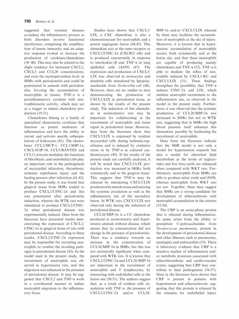
suggested that systemic diseases
co-induce the inflammatory process as
both disorders involve functional
interference, comprising the amplifica-
tion of innate immunity and an adap-
tive response evoked to increase the
production of cytokines/chemokines
(38–40). This may also be related to the
slight tendency for increased CXCL2,
CXCL5 and CCL20 concentrations,
and even the myeloperoxidase level, in
SHRs with periodontitis and could be
potentiated in animals with periodon-
titis, favoring the accumulation of
neutrophils in tissues. TNF-a is a
proinflammatory cytokine with neu-
trophilotactic activity, which may act
as a trigger to induce chemokine pro-
duction (19,31).
Chemokines belong to a family of
specialized chemotactic cytokines that
function as potent mediators of
inflammation and have the ability to
recruit and activate specific subpopu-
lations of leukocytes (41). The chemo-
kines CCL2/MCP-1, CCL3/MIP-1a,CXCL10/IP-10, CCL5/RANTES and
CCL11/eotaxin modulate the functions
of fibroblasts, and endothelial cells play
an important role in the pathogenesis
of myocardial infarction, thrombosis,
ischemia reperfusion injury and the
healing process after infarction (41,42).
In the present study, it was found that
gingival tissue from SHRs tended to
produce CXCL2/CINC-2a and this
was potentiated after periodontitis
induction, whereas the WTK rats were
stimulated to produce CXCL2/CINC-
2a when periodontal disease was
experimentally induced. Data from the
literature have presented results dem-
onstrating the expression of CXCL2/
CINC-2a in gingival tissue of rats with
periodontal disease. According to these
results, CXCL2/CINC-2a expression
may be responsible for recruiting neu-
trophils to combat the invading path-
ogen in periodontal disease (43). In the
model used in the present study, the
recruitment of neutrophils was ob-
served in hypertensive rats, and this
migration was enhanced in the presence
of periodontal disease. It may be sug-
gested that CXCL2 and TNF-a work
in a coordinated manner to induce
neutrophil migration to the inflamma-
tory focus.
Studies have shown that CXCL5/
LIX, a CXC chemokine, is also a
chemoattractant for neutrophils and a
potent angiogenic factor (44,45). This
chemokine acts at the same receptor as
CXCL2/CINC-2a (CXCR2) (46) and
is produced concurrently in response
to interleukin-1b and TNF-a in lung
epithelial cells (A459) (47). The
expression and production of CXCL5/
LIX was observed in monocytes and
dendritic cells stimulated by lipopoly-
saccharide from Escherichia coli (48).
However, there are no studies to date
demonstrating the production of
CXCL5/LIX in periodontal tissue, as
shown by the results of the present
study. The induction of this chemoki-
ne in normotensive rats may be
important for collaborating in the
recruitment of neutrophils and tissue
repair in periodontal tissue. However,
data from the literature show that
CXCL5/LIX is expressed by resident
myocardial cells during ischemia/rep-
erfusion and is induced by oxidative
stress or by TNF-a in cultured car-
diomyocytes (49). If the results of the
present study are carefully analyzed, it
will be noted that CXCL5/LIX pro-
duction was increased in SHRs, both
systemically and in the gingival tissue.
This suggests that TNF-a may be
released by stimulating CXCL5/LIX
productionbymuscletissueandentering
the systemic circulation as well as the
inflammatory site of the secondary
lesion. In WTK rats, CXCL5/LIX was
observed only during the induction of
periodontal disease.
CCL20/MIP-3a is a CC chemokine
produced in normotensive and hyper-
tensive animals without disease, which
means that its concentration did not
change in the presence of periodontitis.
There was a tendency towards an
increase in the concentration of
CCL20/MIP-3a in SHRs, but this was
not statistically significant when com-
pared with WTK rats. It is known that
CXCL2/CINC-2a and CCL20/MIP-3aare important in the recruitment of
neutrophils and T lymphocytes, by
interacting with endothelial cells at the
lesion site (50,51). The authors suggest
that, as a result of resident cells sti-
mulation with TNF-a, the presence of
CXCL2/CINC-2a and/or CCL20/
MIP-3a and/or CXCL5/LIX released
by them may facilitate the accumula-
tion of neutrophils at the site of injury.
Moreover, it is known that in hyper-
tension, accumulation of neutrophils
occurs, both systemically and at the
lesion site, and that these neutrophils
are capable of producing mainly
chemokines and TNF-a (52). TNF-a is
able to mediate the influx of neu-
trophils induced by CXCL1/KC and
CXCL5/LIX (31). These findings
strengthen the possibility that TNF-ainduces CINC-2a and LIX, which
mediate neutrophil recruitment to the
inflammation site, as observed in the
model in the present study. Further-
more, it was observed that the systemic
production of CCL20/MIP-3a was
increased in SHRs but not in WTK
rats, suggesting that in SHRs the high
blood pressure itself enhances this
chemokine, possibly by facilitating the
recruitment of neutrophils.
Data from the literature indicate
that the SHR model is not only a
model for hypertension research but
also a model for abnormal lipid
metabolism as the levels of triglyce-
rides and free fatty-acids are enhanced
when compared with WKT rats. Fur-
thermore, neutrophils from SHRs are
able to produce nitric oxide and iNOS,
whereas neutrophils from WKT rats
are not. Together, these data suggest
that SHRs are a strong candidate for
development of atherosclerosis with
neutrophil accumulation in the arteries
(53,54).
The CRP is an acute-phase protein
that is released during inflammation.
Its name arises from the ability to
precipitate C-polysaccharides from
Streptococcus pneumoniae, present in
the development of periodontal disease
and other illnesses such as pneumonia,
meningitis and endocarditis (55). There
is laboratory evidence that CRP is a
sensitive marker of inflammation and/
or metabolic processes associated with
atherothrombotic and cardiovascular
events, suggesting that CRP may con-
tribute to their pathogenesis (56,57).
Data in the literature have shown that
CRP is present in patients with
hypertension and atherosclerosis, sug-
gesting that this protein is released by
the stimulus for endothelial injury
790 Bonato et al.

(58,59). The results of the present study
demonstrate that both SHRs and
WTK rats have high levels of CRP
following experimental induction of
periodontal disease. However, there
was no statistically significant differ-
ence between the groups with peri-
odontitis, although there was a
tendency towards increased protein
concentration in the group of SHRs
with periodontal disease.
The enzyme responsible for nitric
oxide production – nitric oxide syn-
thase – was identified in various cell
types, suggesting that it has a physio-
logical and pathophysiological role,
mainly through its direct action on
inflammed tissue, bone and immuno-
pathology. In hypertensive individuals
the level of nitric oxide is increased to
provide greater inhibition of norepi-
nephrine and cause vasodilation in or-
der to decrease blood pressure (60). In
periodontal disease the production of
nitric oxide is increased, suggesting
that it is an inflammatory marker of
periodontitis (61). According to the
results of the present study, the
expression of iNOS mRNA was
increased in gingival tissue from rats
with experimentally induced periodon-
tal disease. In addition, this expression
was higher in SHRs with periodontitis.
Corroborating the results of the present
study, other authors have shown that
SHR animals stimulated with NaCl are
able to express iNOS with higher
intensity than are Wistar rats, and that
the expression was increased upon
stimulation with atrial natriuretic pep-
tide (62). Taken together, the hyper-
tensive state of the genetic SHR seems
to favor the inflammatory process,
measured using markers such as
CXCL2, CXCL5, CCL20, TNF-a,iNOS and CRP, with greater intensity
than in normotensive rats. Neverthe-
less, the periodontitis potentiated the
inflammatory process in SHRs.
Acknowledgement
This work was supported by Sao Paulo
State Research Foundation- FAPESP
(grant# 2006/59794-2 to SHPO) and
(grant# 2006/58993-1 to CFB). The
authors declare no financial or com-
mercial conflict of interest.
References
1. Stanford TW Jr. Periodontal disease and
cardiovascular disease: is there an associ-
ation? Tex Dent J 2005;122:1198–1201.
2. Fowler EB, Breault LG, Cuenin MF.
Periodontal disease and its association
with systemic disease. Mil Med 2001;166:
85–89.
3. Kinane DF, Lowe GD. How periodontal
disease may contribute to cardiovascular
disease. Periodontol 2000 2000;23:121–126.
4. Beck JD, Offenbacher S. The association
between periodontal diseases and cardio-
vascular diseases: a state-of-the-science
review. Ann Periodontol 2001;6:9–15.
5. Loos BG, Craandijk J, Hoek FJ, Wert-
heim-van Dillen PM, van der Velden U.
Elevation of systemic markers related to
cardiovascular diseases in the peripheral
blood of periodontitis patients. J Period-
ontol 2000;71:1528–1534.
6. Ford PJ, Yamazaki K, Seymour GJ.
Cardiovascular and oral disease interac-
tions: what is the evidence? Prim Dent
Care 2007;14:59–66.
7. Leite CLA, Redins CA, Vasquez EC,
Meyrelles SS. Experimental-Induced peri-
odontitis is exacerbated in Spontaneously
Hypertensive Rats. Clinical and Experi-
mental. Hypertension 2005;6:523–531.
8. Tsioufis C, Kasiakogias A, Thomopoulos
C, Stefanadis C. Periodontitis and blood
pressure: the concept of dental hyperten-
sion. Atherosclerosis 2011;219:1–9.
9. Okamoto K, Aoki K. Development of a
strain of spontaneously hypertensive rats.
Jpn Circ J 1963;27:282–293.
10. Zicha J, Kunes J. Ontogenetic aspects of
hypertension development: analysis in the
rat. Physiol Rev 1999;79:1227–1282.
11. Trippodo NC, Frohlic ED. Similarities of
genetic spontaneous hypertension. Circ
Res 1981;48:309–319.
12. Smith TL, Hutchins PM. Central hemo-
dynamics in the developmental stage of
spontaneous hypertension in the unanes-
thetized rat. Hypertension 1979;1:508–517.
13. Arendt DM, Elzay RP. Radiographic
analysis of periodontal bone loss in
hypertensive and normotensive patients.
Va Dent J 1976;53:15–19.
14. Perlstein MI, Bissada NF. Influence of
obesity and hypertension on the severity
of periodontitis in rats. Oral Surg Oral
Med Oral Pathol 1977;43:707–719.
15. Angeli F, Verdecchia P, Pellegrino C et al.
Association between periodontal disease
and left ventricle mass in essential hyper-
tension. Hypertension 2003;41:488–492.
16. Holmlund A, Holm G, Lind L. Severity of
periodontal disease and number of
remaining teeth are related to the preva-
lence of myocardial infarction and hyper-
tension in a study based on 4,254 subjects.
J Periodontol 2006;77:1173–1178.
17. Castelli WA, Diaz-Perez R, Nasjleti CE,
Caffesse RG. Effect of renovascular
hypertension on the morphology of oral
blood vessels. Oral Surg Oral Med Oral
Pathol 1978;46:576–582.
18. Bastos MF, Brilhante FV, Goncalves TED
et al. Hypertension may affect tooth-
supporting alveolar bone quality: a study
in rats. J Periodontal 2010;81:1075–1083.
19. Ramos CD, Fernandes KS, Canetti C,
Teixeira MM, Silva JS, Cunha FQ. Neu-
trophil recruitment in immunized mice
depends on MIP-2 inducing the sequential
release of MIP-1a, TNF-a and LTB4. Eur
J Immunol 2006;36:2025–2034.
20. Kim IS, Song JK, Zhang YL et al.
Biphasic electric current stimulates proli-
feration and induces VEGF production in
osteoblasts. Biochim Biophys Acta 2006;
1763:907–916.
21. Tu XK, Yang WZ, Wang CH et al.
Zileuton reduces inflammatory reaction
and brain damage following permanent
cerebral ischemia in rats. Inflammation
2010;33:344–352.
22. Libby P. Current concepts of the patho-
genesis of the acute coronary syndromes.
Circulation 2001;104:365–372.
23. Blake GJ, Ridker PM. Novel clinical
markers of vascular wall inflammation.
Circ Res 2001;89:763–771.
24. Pihlstrom BL, Michalowicz BS, Johnson
NW. Periodontal diseases. Lancet 2005;
366:1809–1820.
25. Bezerra MM, Lima V, Alencar VBM et al.
Selective cycloxigenase-2 inhibition pre-
vents alveolar bone loss in experimental
periodontitis in rats. J Periodontol 2000;
71:1009–1014.
26. Montanher AB, Zucolotto SM, Schenkel
FP, Frode TS. Evidence of anti-
inflammatory effects of Passiflora edulis in
an inflammation model. J Ethnopharmacol
2007;109:281–288.
27. Liu RK, Cao CF, Meng HX, Gao Y.
Polymorphonuclear Neutrophils and their
mediators in gingival tissues from gener-
alized aggressive periodontitis. J Period-
ontol 2001;72:1545–1553.
28. Nussbaum G, Shapira L. How has neu-
trophil research improved our under-
standing of periodontal pathogenesis.
J Clin Periodontol 2011;38:49–59.
29. Ionita MG, van den Borne P, Catanzariti
LM et al. High neutrophil numbers in
human carotid atherosclerotic plaques are
associated with characteristics of rupture-
prone lesions. Arterioscler Thromb Vasc
Biol 2010;30:1842–1848.
30. Drechsler M, Megens RT, van Zandvoort
M, Weber C, Soehnlein O. Hyperlipid-
emia-triggered neutrophilia promotes ear-
ly atherosclerosis. Circulation 2010;122:
1837–1845.
31. Vieira SM, Lemos HP, Grespan R et al.
A crucial role of TNF-a in mediating
Hypertension increases inflammation in periodontitis 791

neutrophil influx induced by endoge-
nously generated or exogenous chemo-
kines, KC/CXCL1 and LIX/CXCL5. Br J
Pharmacol 2009;158:779–789.
32. Baggiolini M, Walz A, Kunkel SL. Neu-
trophil-activating peptide-1/interleukin-8,
a novel cytokine that activates neutroph-
ils. J Clin Invest 1989;84:1045–1049.
33. Cassatela MA. The production of cyto-
kines by polymorphonuclear neutrophils.
Immunol Today 1995;16:21–26.
34. Jablonska E, Jablonski J, Holownia A.
Role of neutrophils in release of some
cytokines and their soluble receptors.
Immunol Lett 1999;70:191–197.
35. Beutler B, Cerami A. The history, prop-
erties, and biological effects of cachectin.
Biochemistry 1988;27:7575–7582.
36. Mann DL. Tumor necrosis factor-induced
signal transduction and left ventricular
remodeling. J Card Fail 2002;6(Sup-
pl):S379–S386.
37. Levine B, Kalman J, Mayer L, Fillit HM,
Packer M. Elevated circulating levels of
tumornecrosis factor inseverechronicheart
failure.NEngl JMed 1990;323:236–241.
38. Claudino M, Ceoli DS, Alberti S et al.
Alloxan-induced diabetes triggers the
development of periodontal disease in
rats. PLoS ONE 2007;2:e1320.
39. Golub LM, Payne JB, Reinhardt Ra,
Nieman G. Can Systemic diseases
co-induce (not just exacerbate) periodon-
titis? A Hypothetical ‘‘two-hit’’ model J
Dent Res 2006;85:102–105.
40. Trombone AP, Claudino M, Colavite P
et al. Periodontitis and arthritis interac-
tion in mice involves a shared hyper-
inflammatory genotype and functional
immunological interferences. Genes
Immun 2010;11:479–489.
41. Bucova M, Bernadic M, Buckingham T.
C-reactive protein, cytokines and inflam-
mation in cardiovascular diseases. Bratisl
Lek Listy 2008;109:333–340.
42. Dewald O, Zymek P, Winkelmann K et al.
CCL2/Monocyte Chemoattractant Pro-
tein-1 regulates inflammatory responses
critical to healing myocardial infarcts.
Circ Res 2005;96:881–889.
43. Miyauchi M, Kitagawa S, Hiraoka M
et al. Immunolocalization of CXC
chemokines and recruitment of polymor-
phonuclear leukocytes in the rat molar
periodontal tissue after topical application
of lipopolysaccharide. Histochem Cell Biol
2004;121:291–297.
44. Koch AE, Polverini PJ, Kunkel SL et al.
Interleukin-8 as a macrophage-derived
mediator of angiogenesis. Science 1992;
258:1798–1801.
45. Koch AE, Volin MV, Woods JM et al.
Regulation of angiogenesis by the C-X-C
chemokines interleukin-8 and epithelial
neutrophil activating peptide 78 in the
rheumatoid joint. Arthritis Rheum 2001;
44:31–40.
46. Wuyts A, Proost P, Lenaerts JP, Ben-
Baruch A, Van Damme J, Wang JM.
Differential usage of the CXC chemokine
receptors 1 and 2 by interleukin-8, gran-
ulocyte chemotactic protein-2 and epithe-
lial cell-derived neutrophil attractant-78.
Eur J Biochem 1998;255:67–73.
47. Chang MS, McNinch J, Basu R, Simonet
S. Cloning and characterization of the
human neutrophil-activating peptide
(ENA-78) gene. J Biol Chem 1994;269:
25277–25282.
48. Barksby HE, Nile CJ, Jaedicke KM,
Taylor JJ, Preshaw PM. Differential
expression of immunoregulatory genes in
monocytes in response to Porphyromonas
gingivalis and Escherichia coli lipopoly-
saccharide. Clin Exp Immunol 2009;156:
479–487.
49. Changdrasekar B, Smith JB, Freeman
GL. Ischemia-Reperfusion of rat myo-
cardium activates nuclear Factor-jB and
induces neutrophil infiltration via lipo-
polysaccharide-induced CXC chemokine.
Circulation 2001;103:2296–2302.
50. Sato K, Kawasaki H, Nagayama H et al.
Chemokine receptor expressions and
responsiveness of cord blood T cells.
J Immunol 2001;166:1659–1666.
51. Scapini P, Laudanna C, Pinardi C et al.
Neutrophils produce biologically active
macrophage inflammatory protein-3alpha
(MIP- 3alpha)/CCL20 and MIP-3beta/
CCL19. Eur J Immunol 2001;31:1981–
1988.
52. Hopps E, Lo Presti R, Caimi G.
Pathophysiology of polymorphonuclear
leukocyte in arterial hypertension. Clin
Hemorheol Microcirc 2009;41:209–218.
53. Chatterjee M, Saluja R, Kanneganti S,
Chinta S, Dikshit M. Biochemical and
molecular evaluation of neutrophil NOS
in spontaneously hypertensive rats. Cell
Mol Biol 2007;53:84–93.
54. Iritani N, Fukura E, Nara Y, Yamori Y.
Lipid metabolism in spontaneously
hypertensive rats (SHR). Atherosclerosis
1977;28:217–222.
55. Hirschfield GM, Pepys MB. C-reactive
protein and cardiovascular disease: new
insights from an old molecule. QJM
2003;96:793–807.
56. Nijmeijer R, Lagrand WK, Visser CA,
Meijer CI, Niessen HW, Hack CE. CRP, a
major culprit in complement-mediated tis-
sue damage in acute myocardial infarction?
Int Immunopharmacol 2001;1:403–414.
57. Pepys MB. The Lumleian Lecture.
C-reactive protein and amyloidosis: from
proteins to drugs? In: Williams G, ed.
Horizons in Medicine. London: Royal
College of Physicians of London,
1999:397–414.
58. Jialal I, Devaraj S, Venugopal SK.
C-Reactive protein: risk marker or medi-
ator in atherothrombosis? Hypertension
2004;44:6–11.
59. Yosef-Levi IM, Grad E, Danenberg HD.
C-reactive protein and atherothrombosis–
a prognostic factor or a risk factor?
Harefuah 2007;146:970–974.
60. Bhandary UV, Tse W, Yang B, Know-
les MR, Demaine AG. Endothelial
nitric oxide synthase polymorphisms are
associated with hypertension and car-
diovascular disease in renal transplan-
tation. Nephrology (Carlton) 2008;
13:348–355.
61. Menaka KB, Ramesh A, Thomas B,
Kumari NS. Estimation of nitric oxide as
an inflammatory marker in periodontitis.
J Indian Soc Periodontol 2009;13:75–78.
62. Costa MA, Elesgaray R, Caniffi C, Fellet
A, Laughlin MM, Arranz C. Role of nitric
oxide as a key mediator on cardiovascular
actions of atrial natriuretic peptide in
spontaneously-hypertensive rats. Am J
Physiol Heart Circ Physiol 2010;298:
H778–H786.
792 Bonato et al.
Copyright © 2022 FDOKUMEN