Phenotypic and functional properties of murine yS T cell clones derived from malaria immunized, a(3...
8
International Immunology, VW. 8, No. 3, pp 359-366 © 1996 Oxford University Press Phenotypic and functional properties of murine yS T cell clones derived from malaria immunized, a(3 T cell-deficient mice Moriya Tsuji, Cassandra L. Eyster 1 , Rebecca L. O'Brien 1 , Willi K. Born 1 , Mitali Bapna, Martin Reichel, Ruth S. Nussenzweig and Fidel Zavala Department of Medical and Molecular Parasitology, New York University School of Medicine, 341 East 25th Street, New York, NY 10010, USA department of Medicine, National Jewish Center for Immunology and Respiratory Medicine, 1400 Jackson Street, Denver, CO 80262, USA Keywords: hpopolysaccharide, lymphokine profile, malaria, murine 78 T cells, surface phenotype, TCR Abstract Six murine T cell clones expressing 76 TCR were generated from malaria Immunized, ap" T cell- deficient mice. Phenotypic characterization of these clones has revealed that, In contrast to conventional ap" T cells, there Is a considerable degree of heterogeneity among these 76 clones with regard to their surface markers and their lymphokine profile. One clone was found to display significant anti-parasite activity In vivo upon adoptive transfer. We attempted to determine whether the protective clone differs in one or more key characteristics from the non-protective clones. Although no obvious pattern peculiar to the protective 78 clone was observed, It appears that more than one parameter may, In combination, define a distinct protective phenotype, and thus explain the functional difference between the protective and non-protective 76 clones. Introduction Lymphocytes expressing 78 TCR constitute a relatively recently defined subset of T lymphocytes. Since the identifica- tion of the yand 5 genes (1,2), considerable information has been obtained regarding their structure and organization (3). These data, particularly those on murine and human 76 T cells, have revealed that these cells displayed a rather restricted receptor repertoire compared with that of 0$ T cells. The function of 76 T cells, and their mechanism of recognition of 'self and 'foreign' antigens, as well as the nature of the respective ligands, remains largely unknown. Increased numbers of 76 T cells have repeatedly been reported to occur in various human and murine microbial infections, in certain auto-immune diseases, and in some tumors (3). In most of these instances it is still unclear whether these 76 T cells contribute to the protective immune mechanisms and/or the immunopathology of the disease We considered that these and other questions could be more easily investigated by generating and characterizing the basic features of a series of murine 76 clones. Our earlier success in generating two 76 T cell clones from mutant mice lacking TCR a chain genes (a-/-) and immunized with irradiated malaria parasites led us to use this approach to obtain additional y6 T cell clones. An attractive feature of this experimental system was our finding that passive transfer of one of the two initial 76 clones had a clear inhibitory effect on parasite development in the liver (4). We therefore set out to determine whether a parasite derived ligand was being recognized by this and other potentially protective clones. We also aimed to identify some of the essential characteristics of a series of 76 clones and determine whether these would provide a basis for distinguishing protective from non-protect- ive clones. Methods Mice We used 6-to 10-week-old(129/SvxC57BL/6)F 1 mice in these studies. In some experiments we used 6- to 8-week-old C57BL/6, BALB/c, A/J, SJL and C3H/HeJ mice as well as several C57BL/10 (B10) congenic mice. They were all pur- chased from The Jackson Laboratories (Bar Harbor, ME). Mutant mice lacking the p^microglobulin gene {farn-l-) ar| d those lacking the Ap b gene (Ap b -/-) had a (129/OlaxC57BL/6) Correspondence to. M. Tsuji Transmitting editor. R. R. Hardy Received 22 August 1995, accepted 28 November by guest on October 1, 2014 http://intimm.oxfordjournals.org/ Downloaded from
Transcript of Phenotypic and functional properties of murine yS T cell clones derived from malaria immunized, a(3...

International Immunology, VW. 8, No. 3, pp 359-366 © 1996 Oxford University Press
Phenotypic and functional properties ofmurine yS T cell clones derived from malariaimmunized, a(3 T cell-deficient mice
Moriya Tsuji, Cassandra L. Eyster1, Rebecca L. O'Brien1, Willi K. Born1,Mitali Bapna, Martin Reichel, Ruth S. Nussenzweig and Fidel Zavala
Department of Medical and Molecular Parasitology, New York University School of Medicine, 341 East25th Street, New York, NY 10010, USAdepartment of Medicine, National Jewish Center for Immunology and Respiratory Medicine, 1400Jackson Street, Denver, CO 80262, USA
Keywords: hpopolysaccharide, lymphokine profile, malaria, murine 78 T cells, surface phenotype, TCR
Abstract
Six murine T cell clones expressing 76 TCR were generated from malaria Immunized, ap" T cell-deficient mice. Phenotypic characterization of these clones has revealed that, In contrast toconventional ap" T cells, there Is a considerable degree of heterogeneity among these 76 cloneswith regard to their surface markers and their lymphokine profile. One clone was found to displaysignificant anti-parasite activity In vivo upon adoptive transfer. We attempted to determine whetherthe protective clone differs in one or more key characteristics from the non-protective clones.Although no obvious pattern peculiar to the protective 78 clone was observed, It appears that morethan one parameter may, In combination, define a distinct protective phenotype, and thus explainthe functional difference between the protective and non-protective 76 clones.
Introduction
Lymphocytes expressing 78 TCR constitute a relativelyrecently defined subset of T lymphocytes. Since the identifica-tion of the yand 5 genes (1,2), considerable information hasbeen obtained regarding their structure and organization (3).These data, particularly those on murine and human 76 T cells,have revealed that these cells displayed a rather restrictedreceptor repertoire compared with that of 0$ T cells.
The function of 76 T cells, and their mechanism of recognitionof 'self and 'foreign' antigens, as well as the nature ofthe respective ligands, remains largely unknown. Increasednumbers of 76 T cells have repeatedly been reported to occurin various human and murine microbial infections, in certainauto-immune diseases, and in some tumors (3). In most ofthese instances it is still unclear whether these 76 T cellscontribute to the protective immune mechanisms and/or theimmunopathology of the disease We considered that theseand other questions could be more easily investigated bygenerating and characterizing the basic features of a seriesof murine 76 clones.
Our earlier success in generating two 76 T cell clones frommutant mice lacking TCR a chain genes (a-/-) and immunizedwith irradiated malaria parasites led us to use this approach
to obtain additional y6 T cell clones. An attractive feature ofthis experimental system was our finding that passive transferof one of the two initial 76 clones had a clear inhibitory effecton parasite development in the liver (4). We therefore set outto determine whether a parasite derived ligand was beingrecognized by this and other potentially protective clones.We also aimed to identify some of the essential characteristicsof a series of 76 clones and determine whether these wouldprovide a basis for distinguishing protective from non-protect-ive clones.
Methods
Mice
We used 6-to 10-week-old(129/SvxC57BL/6)F1 mice in thesestudies. In some experiments we used 6- to 8-week-oldC57BL/6, BALB/c, A/J, SJL and C3H/HeJ mice as well asseveral C57BL/10 (B10) congenic mice. They were all pur-chased from The Jackson Laboratories (Bar Harbor, ME).Mutant mice lacking the p^microglobulin gene {farn-l-) a r |dthose lacking the Apb gene (Apb-/-) had a (129/OlaxC57BL/6)
Correspondence to. M. Tsuji
Transmitting editor. R. R. Hardy Received 22 August 1995, accepted 28 November
by guest on October 1, 2014
http://intimm
.oxfordjournals.org/D
ownloaded from
360 Characteristics of murine y6 T cell clones
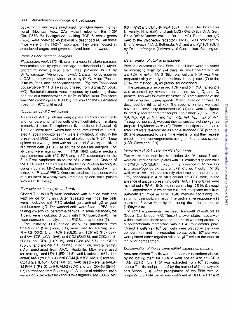
background, and were purchased from Genpharm Interna-tional (Mountain View, CA) Mutant mice on the (129/OlaxC57BL/6) background, lacking TCR P chain genes(p-/-), were obtained as previously described (4). All thesemice were of the ^-2*^ haplotype. They were housed inautoclaved cages, and given sterilized food and water.
Parasites and bacterial antigens
Plasmodium yoehi (17X NL strain), a rodent malaria parasite,was maintained by cyclic passage as described (4) Myco-bacterium bovis (Tokyo strain) was provided to us byDr A. Yamazaki (Ajinomoto, Tokyo). Listeria monocytogenes(LO28 strain) were provided to us by Dr G. Milon (PasteurInstitute, Paris) and lipopolysaccharide (LPS; from Escherichiacoli serotype 0111.B4) was purchased from Sigma (St Louis,MO). Bacterial extracts were prepared by sonicating thesebacteria at a concentration of 107/ml in RPMI 1640. The lysatewas then centrifuged at 10,000 g for 3 min and the supernatantfrozen at -20°C until used.
Generation of yd T cell clones
A series of y& T cell clones were generated from spleen cellsand non-parenchymal liver cells of afi T cell-deficient, malariaimmunized mice. The spleen or liver cells of individual apT cell-deficient mice, which had been immunized with irradi-ated P yoelii sporozoites (4), were stimulated, in vitro, in thepresence of MHC-matched normal spleen cells (H-2b). Thesespleen cells were pulsed with an extract of P yoeln parasitizedred blood cells (PRBC), as source of parasite antigens. Theyd> cells were maintained in RPMI 1640 culture medium,supplemented with 10% FCS and a 2% supernatant of theEL-4 T cell lymphoma, as source of IL-2 and IL-4. Cloning ofthe T cells was carried out by the limiting dilution technique,using irradiated MHC-matched spleen cells, pulsed with anextract of P. yoelii PRBC. Once established, the clones werere-stimulated bi-weekly with irradiated spleen cells pulsedwith a PRBC extract.
Flow cytometric analysis and mAb
Cloned T cells (106) were incubated with purified mAb andkept on ice for 45 min. After repeated washings, the cellswere incubated with FITC-labeled goat anti-rat IgG or goatanti-hamster IgG. The washed cells were fixed in PBS, con-taining 2% (w/v) of paraformaldehyde. In some instances, theT cells were incubated directly with FITC-labeled mAb. Thefluorescence was analyzed in a FACScan cytometer (5).
The following FITC-labeled mAb, all purchased fromPharMingen (San Diego, CA), were used for staining, anti-Thy-1.2 (53-2.1), anti-TCR 8 (GL3), anti-TCR aP (H57-597),anti-Vyt TCR (UC3-10A6), anti-CD2 (RM2-5), anti-CD3e (145-2C11), anti-CD4 (H129.19), anti-CD8a (53-6.7), anti-CD8b(53-5.8) and anti-NK 1.1 (PK136). In addition, several rat IgGmAb, purchased from ATCC (Rockville, MD), were usedfor staining: anti-LFA-1 (FD441.8), anti-L-selectin (MEL-14),anti-ICAM-1 (YN1/1.7.4), anti-CD44 (KM703, KM201) and anti-CD45Ra (TIB164). Other rat IgG mAb used were: anti-VLA-4/LPAM-1 (R1-2), anti-ICAM-2 (3C4) and anti-CD49e (5H10-27) (purchased from PharMingen). A series of additional mAbwere kindly provided by various investigators: anti-CD45 (M1/
9.3.4 H2-2) and CD45Rb (4B4) (by Dr E. Pure, The RockefellerUniversity, New York); and anti-CD2 (RM2-2) (by Dr A. Sen,Dana-Farber Cancer Institute, Boston, MA). The hamster IgGmAb to the Vitronectin receptor (H9-2B8) was provided byDr E. Shevach (NIAID, Bethesda, MD) and anti-V/TCR (GL1)by Dr L. Lefrancois (University of Connecticut, Farmington,CT).
Determination of TCR y5 phenotype
Prior to extraction of their RNA, all cell lines were activatedby incubating them for 2-4 days, in flasks coated with ananti-TCR y5 mAb [3A10 (6)]. Total cellular RNA was thenprepared using vanadyl ribonucleoside complexes (7) or theLiCI-urea method (8), as previously described.
The presence of expressed TCR y and 8 mRNA transcriptswas assessed by reverse transcription, using C5 and CT
primers. This was followed by further PCR amplification of thecDNA generated, using specific V and C region primers, asdescribed by Bill et al. (9). The specific primers we usedhave been previously described (10,11) and were designedto amplify rearranged transcripts containing: VY1, V^, V^,Vy4, V^, V ^ or VY7 and V61, V ^ , V54, V55, V86 or V57.Throughout our study we used the nomenclature of the y genesproposed by Maeda et al. (12). Those transcripts that becameamplified were re-amplified as single-stranded PCR products(9) and sequenced to determine whether or not they repres-ented in-frame rearrangements using the Sequenase system(USB, Cleveland, OH).
Stimulation of y&T cells, proliferation assay
Two weeks after their last stimulation, 3X10³ cloned T cellswere cultured in 96-well plates with 106 irradiated spleen cellsof (129/SvxC57BL/6)F1 mice, in the presence of M. bovis orL. monocytogenes extracts or LPS. Cloned T cells, 1X10⁵/well, were also incubated directly with these bacterial extracts,LPS, concanavalin A or plate-bound anti-CD3 mAb, in theabsence of antigen presenting cells (APC). The cultures weremaintained in RPM11640 medium containing 10% FCS, exceptin the experiments in which we cultured the spleen cells fromP2m-deficient mice in RPMI 1640 medium containing 1%serum of p2m-deficient mice. The proliferative response wasassessed 3 days later by measuring the incorporation of[3H]thymidine
In some experiments, we used Transwell 24-well plates(Costar, Cambridge, MA). These Transwell plates have a wellwithin a well and these two compartments were separated bya polycarbonate membrane with a 0.4 urn diameter pore.Cloned T cells (2X10⁵ per well) were placed in the innercompartment and the irradiated spleen cells, 106 per well,were placed either together with the yb T cells in the inner orthe outer compartment.
Determination of the cytokine mRNA expression patterns
Activated cloned T cells were obtained as described above,by incubating them for 18 h in wells coated with anti-CD3emAb (2C11) Total RNA was extracted from 107 activatedcloned T cells and prepared by the method of Chomczynskiand Sacchi (13). After precipitation of the RNA with 2-propanol, the RNA pellet was dissolved in DEPC water and
by guest on October 1, 2014
http://intimm
.oxfordjournals.org/D
ownloaded from

Characteristics of marine y5 T cell clones 361
its concentration measured at OD 260 nm Total RNA (2 ng)were subjected to first strand cDNA synthesis using theGeneAmp RNA PCR Kit (Perkin Elmer, Foster City, CA) aspreviously described (14).
After reverse transcription, 80 ml of PCR mix was added toeach tube, to give a final concentration of 2 5 U/100 mlAmpliTaq DNA polymerase (Perkin Elmer), 0.15 mM 5' primer,0 15 mM 3' primer, 2 mM MgCI2 and 1xPCR buffer IIPreviously described primers, specific for murine IL-2, IL-4,IL-5, IL-6, IL-10, IFN-y, tumor necrosis factor (TNF)-a andgranulocyte macrophage colony stimulating factor, were used(15,16). After heating to 95°C for 2 min, the cDNA wasamplified for 35 cycles, each cycle consisting of 95°C for1 min, 55°C for 1 min and 72°C for 1 5 min To ascertain thatcomparable amounts of RNA were added to each PCRreaction, primers for a 'housekeeping gene', i e. hypoxan-thine-guanine phosphoribosyl transferase (HPRT), were usedas control (15)
A sample of the PCR reaction products was run on a 1%agarose gel and visualized by UV light illumination, afterethidium bromide staining. The gel was then denatured, bysoaking for 30 min in several volumes of 0 6 M NaCI, 0.2 NNaOH, under constant agitation. The gel was briefly rinsedin deionized water and neutralized by soaking it with constantagitation in several volume of 0 24 M Tris, pH 7 4, 0.6 M NaCIat room temperature
The DNA was transferred to a nylon membrane which wasUV cross-linked, and blots were pre-hybridized at 42°C for6-8 h in Denhardt's solution containing 6xSSC and 0.1%SDS. They were hybridized at 42°C, overnight, with ^P-labeled oligoprobes, in the same buffer with 50 mg/ml dena-tured single-stranded DNA The oligonucleotide probes wereinternal to the PCR primers ensuring the identity of the segmentwhich had been amplified (15,16). After hybridization, theblots were washed for 30 min in 1 xSSC, 0 1% SDS and thenin 0.5XSSC, 0.1% SDS at 42°C for 30 min Autoradiographswere exposed at -70°C, using Kodak XAR-2 film.
Passive transfer of y5 clones to naive mice, followed bysporozoite challenge
•yS T cell clones were harvested 10-14 days after re-stimula-tion. Ten million cells of the respective clones, or normalspleen cells from a£ T cell-deficient mice, were resuspendedin RPMI 1640 medium containing recombinant human IL-2 at2X103 units/ml, immediately before transfer. A total of 0 5 mlof the cell suspension was injected i.v. into each mouse. Allmice were challenged with 3X10⁵ sporozoites of P. yoelii 4 hafter the cell transfer.
Quantification of P. yoeln ribosomal RNA in the liver of sporozo-ite-challenged mice
The amount of parasite ribosomal RNA present in the liverof individual sporozoite-challenged mice was measured asdescribed previously (17). In brief, total RNA was isolatedfrom the livers of mice killed 42 h after they had beenchallenged with viable sporozoites. RNA was prepared usingChomczynski and Sacchi's method (13). After one-tenth ofthe whole liver RNA was precipitated with 2-propanol, theRNA pellet was dissolved in 10 mM Tris-HCI/1 mM EDTA, pH
8.0, and denatured at 65°C in 20xSSC (1XSSC = 10 mMNaCI/15 mM sodium citrate, pH 7 0) plus 1.1 (v/v) of 37%(v/v) formaldehyde. This preparation was diluted 1:45 and0.2 ml samples were blotted onto nylon membranes. TheRNA was fixed to the filters by UV cross-linking. Hybridizationwas performed overnight at 42°C in 5xSSPE (1 xSSPE = 180mM NaCI/10 mM sodium phosphate/1 mM EDTA, pH 7.7)/1%SDS with hepann at 500 mg/ml, using three ^P-labeledoligonucleotide probes (106 c.p.m/ml; sp. act > 2 X 1 0 8
c.p m./mg). These probes have been described previously(17).
Results
Two T cell clones which express the y5 TCR, 291-H4 and219-1, had previously been generated by us, from spleensof malaria immunized ap T cell-deficient mice (4). Morerecently, we generated four additional murine y6 T cell clones.Three of these, 291-F6, 269-B8 and 269-B10, were derivedfrom spleens cells, while another y5 T cell clone, 269-L1,was derived from non-parenchymal liver cells of malariaimmunized ap T cell-deficient mice. In the current study, wepresent the results of the characterization of these six distinctyd T cell clones, with regard to their surface markers, lympho-kine production, their usage of TCR and carbohydrate ligand.
Cell surface phenotype of y8T cell clones
The expression on the cell surface of these y5 T cell clones,of molecules known to play an important role in the in vivoactivity of T cells, was determined by FACS analysis, using aseries of antibodies We selected antibodies which detectmolecules known to be important in cell adhesion and homing,including LFA-1, CD2, VLA-5, VLA-4/LPAM-1, L-selectin,ICAM, CD51, CD44 and CD11b. We also used antibodiescapable of detecting phenotypic markers characteristic ofT cells, including Thy-1, CD4 and CD8.
Table 1 summarizes our findings on the cell surface pheno-type expressed by these y6 clones. All six clones express y5TCR, Thy-1 and CD3. Three of the clones are CD4-CD8"(291-H4, 291-F6 and 219-1), while two others are CD4+, andone (269-B8) is CD8+ (aa homodimer). A leukocyte commonantigen and certain adhesion molecules are uniformly presenton all six 76 T cell clones. These include CD44, CD45, VLA-4,ICAM-1, ICAM-2 and LFA-1. The expression of other surfacemarkers, such as CD2, CD45Ra, CD45Rb, CD49e and CD51,varied greatly in individual y5 clones.
Cytokine mRNA expression of the y6T cell clones
The pattern of lymphokines produced by these y5 T cellclones was established by incubating these cells with anti-CD3 mAb bound to the culture plates. Using cell lysates ofthese activated T cell clones, the mRNA expression of aseries of lymphokines was measured using RT-PCR analysis.Consistent with earlier observations (18,19), we found that allsix y5 T cell clones express high levels of mRNA of IL-10 andgranulocyte macrophage colony stimulating factor (Fig. 1).Individual clones displayed considerable differences in thelevel of expression of mRNA of certain lymphokines, such asTNF-a The differences in mRNA levels were even more
by guest on October 1, 2014
http://intimm
.oxfordjournals.org/D
ownloaded from

362 Characteristics of munne yS T cell clones
Table 1. Cell surface markers of six murine yd T cell clones
291-H4 291-F6 219-1 269-L1 269-B10 269-B8
T cell markers8
Thy-1 + b
CD3 +y5TCR +CD4CD8
Leukocyte common antigensCD45 +CD45Ra +CD45Rb +
Adhesion molecules0
CD44 +VLA-4 +ICAM-1 +ICAM-2 +LFA-1 +CD2 +CD49e(VLA-5)CD51 +
aNone of the Y5 clones were positive for the ap TCR nor the NK1 1 markerbA '+ ' indicates the presence of the respective surface markers, while a '-' indicates the absence of a given marker or molecule.CAII clones were negative for L-selectin and CD11b
striking for IL-4, IL-5 and IFN-y, which were barely or notdetectable in certain clones The mRNA corresponding toIL-2 was only detected in a single y5 clone, 219-1.
TCR rearrangements and diversity of y5T cell clones
All six yb T cell clones were examined for the presence oftranscripts resulting from various V5 and Vy rearrangements(see Table 2). One clone (291-F6) was found to express aVT1 gene aberrantly rearranged to J^2. Such rearrangementshave previously been found to occur, at a low frequency, inother y5 T cell clones and hybridomas (20). All but two of theclones expressed more than one functionally rearrangedy chain.
The TCR sequence data combined with the results ofantibody staining revealed that all these T cell clones expresseither V71 or VT7 on their surface, in combination with a varietyof V8 chains. Furthermore, we found that several clones, whichdiffered in various other features, expressed identical TCR,with identical junctional regions. This might reflect clonalexpansion, followed by in vivo or in vitro differentiation, orpreferential rearrangement.
Activation of y6T cell clones
None of the six yb clones displayed reactivity with parasiteantigens contained in extracts of P. yoe/;7-infected red bloodcells (data not shown). This lack of reactivity with PRBCoccurred in spite of the fact that all six y5 T cell clones weregenerated from mice immunized with irradiated P. yoeliisporozoites and had been expanded, in vitro, in the presenceof extracts of PRBC.
Three of the 76 T cell clones, 291-H4, 219-1 and 269-B8,were stimulated by incubation with concanavalin A, whichelicited a considerable proliferative response. In contrast, the76 T cell clones 291-F6, 269-L1 and 269-B10, which lack
H4 219-1 F6 L1 B8 B10A.
HPRT
IL2
IL4
IL5
IL6
B.HPRT
IL10
IFN-y
TNF-a
GM-CSF
Fig. 1. Cytokine mRNA expression of the y5 T cell clones RT-PCR,electrophoresis, Southern blotting, hybridization and autoradiographywere performed as described in Methods. Expression of thehousekeeping gene, HPRT, demonstrated that approximatelyequivalent amounts of RNA were used for all yd clones. The resultsrepresent those obtained in one of three similar experiments.
by guest on October 1, 2014
http://intimm
.oxfordjournals.org/D
ownloaded from

Characteristics of murine y6 T cell clones 363
Table 2. TCR expression in the 76 T cell clones
Clone In-frame TCR transcripts
N/D
Antibody staining Deduced TCR
291-H48
219-1
291-F6
269-B8
269-L1 b
269-B10
VVVVT1
VV54V71
VV2
V 5VT1
VVS6
AVWIYYCAALMEAVWIYYCAALMEAVWN
ACWDYYCACGGKAVWIASGYAVWIASGYAVWICSYGALWE
SAQREKMGRRDTASAQREKMGRRDTA
(V1/J2 join)
QAGIGGLAIKYRRDTRIKYRRDTRPRLPHIGGIRA
GTSWYSSGTDKLGTSWYSSGTDKLSSG
SSGYSSGTDKLGTSWLGTSWLSGTSWYSSGTDKL
V r 7 + VT7/V)s4
V//V64
VT7/Vj2
VT1/V55
VT1/V56
aProtective clonebDenved from the liver, while all other clones were derived from spleen cells.
CD2 molecules on their surface, failed to proliferate withconcanavalin A. However, all of these CD2~ clones wereactivated by incubation with immobilized anti-CD3 mAb(Fig 2)
Most of these y8 clones, with the exception of 269-B8,reacted with and proliferated when incubated with LPS, in thepresence of syngeneic or allogeneic irradiated spleen cells.
The extracts of certain Gram-positive bacteria such as M.bovis and L. monocytogenes also stimulated some, but notall of the 76 clones (Fig. 3). Clones 291-H4, 291-F6 and 219-1proliferated with these bacterial extracts, in the presence ofirradiated spleen cells. The cells of one of these clones, 219-1,also reacted with the extracts of Listeria and Mycobactena inthe absence of spleen cells. The same bacterial extractalso activated clone 269-B10, which proliferated only in theabsence of spleen cells (data not shown) Proliferation in theabsence of spleen or other APC was also observed uponincubation of clones 269-B10 and 269-L1 with LPS.
Lack of genetic restriction of the stimulation ofyST cells by LPS
The conditions required for presentation of LPS to murine y8T cells were further defined by using a clone 291-H4. Thisclone proliferates strongly with LPS, in the presence ofirradiated spleen cells, whether they were syngeneic orderived from various MHC non-matched strains of mice, whichinclude B10 (H-2"), B10.D2 (H-^) , B10.RIII (H-?), B10.M(H-2f), B10.SM (1-1-2 ,̂ B10.BR (H-2*), B10.AKM (H-2"1),B10.PL (H-2"), BALB/C (H-2"), A/J (H-28) and SJL (H-28).Among several strains tested, only C3H/HeJ mice, known tobe LPS non-responders, failed to present LPS to the y8 clone(data not shown).
To establish whether MHC class I, class l-like or class IImolecules were presenting LPS to clone H4, the spleen cellsof mice which lack class I (fem-/-) or class II molecules
100
•oc
1 10
ofa-
l l .
JL
JH4 219-1 88 F6 L1 B10
Fig. 2. Activation of yS T cell clones by concanavalin A and anti-CD3mAb. One hundred thousand cells of the respective T cell cloneswere incubated with 1 ng/ml of concanavalin A (open columns) orwith plate-bound anti-CD3 mAb (shaded columns). In Figs 2-4, theexperiments were done with triplicated samples and the proliferativeresponse was assessed 3 days later by measuring the incorporationof [3H]thymidine In Figs 2 and 3, the results are expressed as astimulation index, calculated as the c.p.m. of cultures in the presenceof antigen, divided by the c p.m. in the absence of antigen Theresults shown in Figs 2-4 represent those obtained in one of foursimilar experiments.
(Apb-/-) were used as APC. These pyn-deficient mice lacknot only the classical MHC class I molecules, but also fail toexpress MHC class l-like molecules, such as Qa-2, T22 andT23 (21). Cells of the y5 clone H4 incubated with LPS andco-cultured with spleen cells from these class I- or classll-deficient mice proliferated very intensively, even more than
by guest on October 1, 2014
http://intimm
.oxfordjournals.org/D
ownloaded from

364 Characteristics of murine yS T cell clones
«
100 r
2 1 9 - 1 L1 B8 B10
Fig. 3. Reactivity of y5 T cell clones with various bacterial antigens,in the presence of spleen cells. Three thousand cells of the respectiveT cell clones were incubated with 106 irradiated spleen cells of (129/SvxC57BL/6)F, mice, in the presence of extracts of M. bovis (cross-hatched columns) or L monocytogenes (shaded columns), or1 (ig/ml of LPS (solid columns)
5
~ 4
o
x 3
20.o 2
1
n
-
/
t/
p-i
*
/
9
/
/
•
0 0.001 0.01 0.1 1
Antigen ( / x g / m l )
Fig. 4. Spleen cells from fcm and also from class ll-deficient micestimulate clone 291-H4 in the presence of LPS. Three thousand cellsof clone, 291-H4, were incubated with 1 ng/ml of LPS in the presenceof 106 irradiated spleen cells from (^-deficient (open circles), classll-deficient (filled circles) or normal mice (inverted triangles)
those in the presence of spleen cells from immunologicallyintact mice (Fig. 4).
To determine whether cell contact is required for the LPS-induced stimulation of this y5 clone, we used Transwell cultureplates in which the compartments are separated by a semi-permeable membrane Clone H4 proliferated strongly inresponse to LPS, when the cell were incubated together withthe spleen cells. In contrast, almost no proliferation wasobserved when this clone and the spleen cells were separated
Injected Spleen H 4 219 -1with cells F6 B8 B10 L1
Fig. 5. In vivo anti-plasmodial activity of passively transferred y& T cellclones Ten million cells of the respective T cell clones or spleencells from <x0 T cell-deficient non-immunized mice were adoptivelytransferred to naive (129/SvxC57BL/6)F1 mice, by tail vein injectionFour hours later, all mice were challenged with 3X10⁵ P yoeliisporozoites and after 42 h the amount of P. yoelii rRNA present inthe liver of these mice and the percentage of inhibition wasdetermined Each experimental group consisted of four mice and theresults represent those obtained in one of three similar experiments.
by the semi-permeable membrane, reflecting the need fordirect interaction between these cells for LPS stimulation tooccur (data not shown).
Passive transfer of y§ clones induces inhibition of developmentof plasmodial liver stages
Our previous studies had shown that, in malaria-immunizeda(3 T cell-deficient mice, a significant inhibition of the develop-ment of the parasite's liver stages occurred upon challenge.It was also determined that the protective immunity of thesemice was mediated by 76 T cells (4).
Although none of these clones displayed a detectablereactivity with parasite (blood stages) antigens, it becameimportant to verify whether the additional clones we obtainedmight have anti-parasitic activity in vivo. Each of the •$ cloneswas adoptively transferred to naive mice, then inoculated 4h later with viable sporozoites. In the current studies, weconfirmed that the transfer of clone 291-H4, but not ofclone 219-1, resulted in a significant inhibition of parasitedevelopment. Four additional 76 clones failed to displayprotective activity, although we detected a minimal inhibitoryactivity of the liver stages upon transfer of clone 269-L1(Fig. 5).
Discussion
The generation and maintenance of murine 76 T cell cloneshave been rather difficult, so that only few murine 76 T celllines and clones have been characterized. This endeavor hasbeen somewhat more successful for human y5 T cell lines,
by guest on October 1, 2014
http://intimm
.oxfordjournals.org/D
ownloaded from
clones and hybridomas, and several subpopulations of humany8 cells have been identified (3). However, the role andparticipation of y8 T cells in the immune response is certainlymore difficult to define in man than in an experimental mousemodel, in which the immune system can be subjected tovarious manipulations
The murine y8 clones, characterized in the current study,were obtained from a|3 T cell-deficient mice (a- / - or p-/-)and immunized with irradiated sporozoites of a rodent malariaparasite, P. yoelii (4). The characterization of the y5 T cellclones, obtained from these animals, was of particular interest.First, because these malaria immunized mutant mice dis-played a very reproducible protective response to parasitechallenge, resulting in a significant decrease of parasitedevelopment in their livers. This protective effect was medi-ated by yd cells, since it was abolished by the administrationof an anti-TCR 5 mAb to the immunized mice (4\An additionalreason for interest in this experimental system was the findingthat the passive transfer of a yd clones, 291-H4, generatedfrom these immunized mutant mice, conferred a considerabledegree of protection against malaria challenge to naivemice (4).
These earlier findings raised a series of basic questions,which we have addressed by the experiments reported in thepresent manuscript One of these questions relates to thenature of the mechanism which mediates the inhibition ofparasite development, in the immunized ap-deficient mice,and also upon passive transfer of the 'protective' 78 clone.Another basic question was whether the 'protective' andpossibly also other clones recognize a parasite derived moietyas ligand, and whether this ligand is a molecule sharedwith other pathogens or modified host cells. It was also ofconsiderable interest to determine whether the protectiveclone 291-H4 differed in one or more key characteristics (TCRrepertoire, surface markers, lymphokine profile, etc.) fromnon-protective clones and whether these characteristics mightbe predictive of a protective role.
The fact that we recently succeeded in generating severaladditional y8 T cell clones, from malaria immunized mutantmice, provided the opportunity to probe some of these issues,enabling us to identify and compare several characteristicsof this set of murine y8 clone. Of the six y5 clones we obtained,only one, 291-H4, showed significant anti-parasite activityin vivo, upon passive transfer.
We had shown earlier that the level of expression of CD44and VLA-4, on the surface of CD8+ a0 T cell clones, correlatesclosely with the in vivo protective anti-malaria activity of theseclones (5). We therefore searched for cell surface markersthat may determine or correlate with the protective capacityof the y5 clones. While all the clones shared a number of thesurface markers we investigated, including VLA-4 and CD44,other surface molecules were expressed by only one or fewy8 clones. In contrast to CD8+ a0 T cells, VLA-4 and CD44appear not to play a role in the protective activity of yd T cells.Taken together, the data on the surface antigens of theseclones failed to provide a pattern peculiar to the single clone291-H4, capable of conferring protection against malaria. Thepatterns of surface antigens also provided no clear distinctionbetween the single Y5 T cell clone derived from liver, 269-L1,
Characteristics of murine yd T cell clones 365
and all the other y8 clones derived from the spleens of themalaria immunized mutant mice.
There have been few studies on the lymphokine productionby murine y8 T cells. The lymphokine profile of the singleprotective clone, 291-H4, was compared with that of the othery5 clones to determine whether it produced a unique set oflymphokines The absence of detectable mRNA for IFN-y intwo of the 76 clones, particularly in the protective clone291-H4, was rather unexpected. IFN-y has been known tomediate protective immunity against liver stages of malariaparasites (22,23), and the role of the IFN-y receptor and ofinducible nitric oxide synthase, in this process, has beenclearly established (14) The protective clone 291-H4appeared to express the highest levels of TNF-a. Only oneother clone 291-F6 expressed TNF-a mRNA at similar butsomewhat lower levels. It is conceivable that TNF-a maycontribute to the anti-parasite activity of the 291-H4 clone.
As a third approach toward identifying possible uniquefeatures of the protective clone 291-H4, we compared its TCRgene transcripts and surface-expressed TCR chains withthose of the other clones. Based on cDNA sequencing andantibody staining, 291-H4 expressed V77, a V gene rarelyfound in y5 cells from normal mouse liver or spleen (3,19).Surprisingly, V77 was expressed in three of the six clonesderived from the malaria immunized mice, in two independentcloning experiments Two of these clones, 291-H4 and 219-1,appear to be related because they express identical TCRgene transcripts. These clones, however, differ in cytokineand cell surface marker expression, suggestive of functionalspecialization. The over-representation of Vy7 in this set of y5clones is likely to be caused by a response of these cells tothe immunization The fact that the only protective clone,291-H4, expresses the over-represented VYgene is consistentwith this interpretation. Moreover, the TCR expressed by291-H4, in combination with high levels of TNF-a expression,unique to this clone, and perhaps certain cell surface markers(CD2 or CD45Ra in this study), may well define a distinctprotective phenotype, and thus explain the functional differ-ence between 291-H4 and the other yS clones.
It was noteworthy that neither the protective clone 291-H4,nor any of the other y6 clones, generated from the malariaimmunized animals, and expanded in the presence of PRBC,proliferated upon addition of an extract of PRBC. This suggeststhat the observed protection was either due to recognition bythe y5 cells, of a liver stage antigen, absent from the bloodstages of the parasite or that it was non-specific.
This is an important, still unresolved issue with regard tomost y5 cells, i e the identity of the ligand(s) recognizedby these T cells. A recent advance in this area was thedemonstration by Tanaka et al. (24) that the ligands recog-nized by human V ^ V ^ cells consist of small non-peptidicantigens, which are protease resistant and phosphatasesensitive. Since these antigens correspond to widely occurringnatural compounds, this finding may explain the broad reactiv-ity of many y5 cells, including most of our clones, with anumber of different bacterial extracts.
In conclusion, we found that within a series of y5 clones,generated from malaria immunized, aP-deficient mutant mice,individual y6 clones differed considerably with regard to all
by guest on October 1, 2014
http://intimm
.oxfordjournals.org/D
ownloaded from
366 Characteristics of murine yS T cell clones
the parameters we examined. Although, we observed no veryobvious pattern peculiar to the protective y8 clone, it appearsthat there may be a set of parameters which, in combination,are unique to the protective clone and thus may define aprotective phenotype. Further experimental manipulation ofthese parameters will be necessary to test this possibility.The observed features of TCR gene expression among clonesderived from malaria immunized mice is suggestive of specificreactivity and clonal expansion, but a malaria-specific ligandwas not identified. Further studies are required to determinewhether the putative ligand triggering this 76 T cell responseis pathogen or host derived.
Acknowledgements
We thank Ms Rita Altszuler and Mr Chui Ng for their excellent technicalassistance, and Dr John Hirst for assistance with FACS analysis. Thiswork was supported by grant AI-27458 from National Institutes ofHealth, grant DPE-0453-A-00-5012-00 from the Agency of Interna-tional Development and a American Cancer Society Institutional grantIRG-14-35 M T is a recipient of the Whitehead Presidential Fellowshipfor Junior Faculty.
Abbreviations
APC antigen-presenting cellsB10 C57BL/10f^n P2-microglobulinLPS lipopolysaccharidePRBC parasitized red blood cellsTNF tumor necrosis factor
References
1 Saito, H., Kranz, D. M., Takagaki, Y, Hayday, A. C, Eisen, HN. and Tonegawa, S. 1984. Complete primary structure of aheterodimeric T-cell receptor deduced from cDNA sequencesNature 309:757.
2 Chien, Y. H., Iwashima, M., Kaplan, K. B., Elliott, J F. and David,M. M. 1987. A new T-cell receptor gene located within the alphalocus and expressed early in T-cell differentiation. Nature327 677.
3 Haas, W., Perreira, P. and Tonegawa, S. 1993. y6 cells. AnnuRev Immunol 11 625.
4 Tsuji, M., Mombaerts, P., Lefrancois, L, Nussenzweig, R. S.,Zavala, F and Tonegawa, S. 1994.76 T cells contribute to immunityagainst the liver stages of malaria in cx0 T-cell-deficient mice.Proc. NatlAcad Sci USA 91:345.
5 Rodrigues, M., Nussenzweig, R. S., Romero, P. and Zavala, F.1992 The in vivo cytotoxic activity of CD8+ T cell clones correlateswith their levels of expression of adhesion molecules. J. Exp.Med. 175:895.
6 Itohara, S., Nakanishi, N , Kanagawa, O. Kubo, R. and TonegawaS. 1989. Monoclonal antibodies specific to naive murine T-cellreceptor >fr analysis of y5 T cells during thymic ontogeny and inperipheral lymphoid organs. Proc. Natl Acad. Sci. USA 86:5094.
7 Kumagai, N., Benedict, S. H., Mills, G. B. and Gelfand, E. W1987. Requirements for the simultaneous presence of phorbolesters and calcium ionophores in the expression of humanT lymphocytes. J. Immunol. 139:1393.
8 Auffary, C. and Rougeon, F. 1980. Purification of mouseimmunoglobulin heavy-chain mRNAs from total myeloma tumorRNA Eur J. Btochem. 107:303
9 Bill, J , Appel, V. B and Palmer, E 1988. An analysis of T-cellreceptor variable region gene expression in majorhistocompatibility complex disparate mice Proc Natl Acad. SciUSA 85:9184.
10 O'Brien, R. L, Fu, Y.-X., Cranfill, R., Dallas, A., Reardon, C, Lang,J., Carding, S. R., Kubo, R. and Born, W. 1992. Heat shock proteinHsp-60 reactive y5 cells- a large, diversified T lymphocyte subsetwith highly focused specificity. Proc NatlAcad Sci USA 89.4348
11 Heyborne, K. D., Cranfill, R L, Carding, S R , Born, W K. andO'Brien, R L. 1992. Characterization of 76 T lymphocytes at thematernal-fetal interface J. Immunol 149:2872.
12 Maeda, K., Nakanishi, N., Rodgers, B. L, Haser, W G., Shrtara,K , Yoshida, H., Takagaki, Y, Augustm, A A. and Tonegawa, S.1987. Expression of the T-cell receptor y-chain gene products onthe surface of peripheral T cells and T-cell blasts generated byallogeneic mixed lymphocyte reactions. Proc. Natl Acad. Sci.USA 84:6539.
13 Chomczynski, P and Sacchi, N 1987 Single step method ofRNA isolation by acid guanidium thiocyanate-phenol-chloroformextraction. Anal Biochem. 162.156
14 TSUJI, M , Miyahira, Y, Nussenzweig, R. S., Aguet, M , Reichel, Mand Zavala, F. 1995 Development of antimalaria immunity in micelacking IFN-y receptor. J. Immunol. 154.5338.
15 Svetic, A., Finkelman, F. D., Jian, Y C , Dieffenbach, C W., Scott,D. E., McCarthy, K. F, Steinberg, A. D. and Gause, W. C. 1991Cytokine gene expression after in vivo primary immunization withgoat antibody to mouse IgD antibody. J. Immunol. 147:2391
16 Gazzmelli, R. T, Eltoum, I , Wynn, T A. and Sher, A 1993. Acutecerebral toxoplasmosis is induced by in vivo neutralization ofTNF-a and correlates with the down-regulated expression ofinducible nitric oxide synthase and other markers of macrophageactivation. J. Immunol. 151:3672
17 Arreaza, G., Corredor, V and Zavala, F. 1991 Plasmodium yoelirquantification of the exoerythrocytic stages based on the use ofribosomal RNA probes. Exp Parasitol. 72'103.
18 Eichelberger, M., Allan, W., Carding, S. R., Bottomly, K andDoherty, P. C. 1991. Activation status of the CD4-CD8" y5-T cellsrecovered from mice with influenza pneumonia. J Immunol.147:2069.
19 Bluestone, J A , Cron, R. Q., Barrett, T A., Houlden, B , Sperling,A I., Dent, A., Hedrick, S., Rellahan, B. and Matis, L A 1991.Repertoire development and ligand specificity of murine TCR yScells. Immunol. Rev. 120.5.
20 Roark, C E., \follmer, M. K , Cranfill, R. L, Carding, S. R., Born,W. K. and O'Brien, R. L. 1993. Liver y5 T cells' TCR junctionsreveal differences in HSP-60 reactive cells in liver and spleen JImmunol. 150-4867
21 Correa, I., Bix, M., Liao, N. S., Zijlstra, M., Jaenish, R. and Raulet,D. 1992. Most gamma delta T cells develop normally in beta 2-microglobulin deficient mice. Proc. Natl Acad. Sci. USA 89653.
22 Ferreira, A , Schofield, L., Enea, V., Schellekens, H., van derMeide, P., Collins, W. E., Nussenzweig, R. S. and Nussenzweig,V. 1986. Inhibition of development of exo-erythrocytic forms ofmalaria parasites by y-interferon. Science 232.881.
23 Schofield, L, villaquiran, J., Ferreira, A , Schellekens, H.,Nussenzweig, R S. and Nussenzweig, V. 1987 y-\nlerteron,CD8+ T cells and antibodies required for immunity to malariasporozoites Nature 330:664.
24 Tanaka, Y, Morita, C. T, Tanaka, Y, Nieves, E., Brenner, M. B.and Bloom, B. R 1995. Natural and synthetic non-peptide antigensrecognized by human T cells. Nature 375:155.
by guest on October 1, 2014
http://intimm
.oxfordjournals.org/D
ownloaded from