Histone Deacetylation Critically Determines T Cell Subset Radiosensitivity
Upload
independentCategory
view
0download
0
Functional optimisation of soluble TCRs
1
Design of soluble recombinant T cell receptors for
antigen targeting and T cell inhibition
Bruno Laugel*, Jonathan M. Boulter†, Nikolai Lissin‡, Annelise Vuidepot‡, Yi Li‡,
Emma Gostick*, Laura E. Crotty§, Daniel C. Douek§, Joris Hemelaar¶, David A.
Price§#, Bent K. Jakobsen‡ and Andrew K. Sewell*||**
*The T-cell Modulation Group, The Peter Medawar Building for Pathogen Research,
University of Oxford, South Parks Road, Oxford OX1 3SY, UK †Department of Medical Biochemistry and Immunology, University of Wales College of
Medicine, Heath Park, Cardiff CF14 4XN, UK ‡Avidex Ltd., 57 Milton Park, Abingdon, Oxon OX14 4RX, UK
§Human Immunology Section, Vaccine Research Center, National Institute of Allergy and
Infectious Diseases, 40 Convent Drive, National Institutes of Health, Bethesda, MD
20892, USA ¶Harvard Medical School, Department of Pathology, 200 Longwood Avenue, Boston, MA
02115, USA
||Correspondence to: Andrew Sewell
Tel: (+)44-1865-281539
Fax: (+)44-1865-281530
Email: [email protected]
Running title: Functional optimisation of soluble TCRs
JBC Papers in Press. Published on November 4, 2004 as Manuscript M409427200
Copyright 2004 by The American Society for Biochemistry and Molecular Biology, Inc.

Functional optimisation of soluble TCRs
2
Summary The use of recombinant T cell receptors (TCRs) to target therapeutic interventions has been hindered by the
naturally low affinity of TCR interactions with peptide-major histocompatibility complex (pMHC) ligands.
Here, we use multimeric forms of soluble heterodimeric αβ TCRs for specific detection of target cells
pulsed with cognate peptide, discrimination of quantitative changes in antigen display at the cell surface,
identification of virus-infected cells, inhibition of antigen-specific cytotoxic T lymphocyte (CTL) activation
and identification of cross-reactive peptides. Notably, the A6 TCR specific for the immunodominant HLA
A2-restricted HTLV-1 Tax11-19 epitope bound HLA A2-HuD87-95 (KD = 120 µM by surface plasmon
resonance), an epitope implicated as a causal antigen in the paraneoplastic neurological degenerative
disorder anti-Hu syndrome. A mutant A6 TCR that exhibited dramatically increased affinity for cognate
antigen (KD = 2.5 nM) without enhanced cross-reactivity was generated; this TCR demonstrated potent
biological activity even as a monomeric molecule. These data provide insights into TCR repertoire selection
and delineate a framework for the selective modification of TCRs in vitro that could enable specific
therapeutic intervention in vivo.

Functional optimisation of soluble TCRs
3
Introduction Peptide-major histocompatibility complex (pMHC) antigens displayed on the surface of target cells are
recognized by T cells via their specific T cell receptor (TCR) (1). The TCR coreceptors CD8 or CD4 bind to
invariant domains of pMHC class I (pMHCI) or pMHC class II (pMHCII) respectively, and are known to
facilitate the process of antigen recognition by T cells (2). Recent advances have enabled the generation of
high quality soluble TCR, pMHCI, pMHCII, CD8 and CD4 proteins; these, in turn, have allowed the
biophysical characterization of the interactions between these molecules. Accordingly, the TCR and CD4/8
co-receptors have been shown to have very low affinities (KD ~ 10-3-10-6 M) for cognate pMHC. Despite
these low affinity interactions, however, the process of antigen engagement can initiate T cell recognition of
antigen-presenting cells (APCs) bearing fewer than 10 copies of a specific pMHC complex (3,4). The
mechanisms by which these weak recognition events result in such exquisite sensitivity are not fully
understood.
The production of soluble recombinant αβ T cell receptors has proved challenging. The main technical
pitfall is heterodimeric instability in the absence of anchoring transmembrane domains and α/β pairing
through an interchain disulphide bond. One of the commonest protein engineering strategies used in TCR
studies to date has been the construction of single-chain TCRs (scTCRs). This technique, which takes
advantage of the structural similarities between antibodies and TCRs, is based upon the single-chain Fv
technology used to generate antibody fragments (5). In short, for the TCR, it involves the cloning and
expression of a unique chimerical open-reading-frame where the Vα and Vβ domains are paired with a
protein linker (6,7). These reagents have been successfully used in structural and biophysical studies (7,8)
but widespread application of the method has proven more difficult. Alternative approaches have included
shuffling the variable (V) and constant (C) domains of the TCR to the C-region of an immunoglobulin κ
light chain to generate a soluble heterodimeric protein (9); the resultant TCR was shown to react with
several anti-TCR antibodies, but was not shown to bind specific antigen.
In this study, we generate soluble versions of the human HLA A2-restricted JM22 and A6 TCRs specific for
dominant viral epitopes derived from the influenza matrix and human T cell leukaemia virus type 1 (HTLV-

Functional optimisation of soluble TCRs
4
1) Tax proteins respectively. Unlike most of the recombinant TCRs described to date, these proteins
comprise an α- and a β-chain that are expressed separately and paired by a non-native disulfide bond (10).
We overcome limitations imposed by the intrinsically low binding affinity and correspondingly short half-
life of the monomeric TCR/pMHCI interaction by building multimeric forms of these proteins. The exquisite
binding sensitivity and specificity exhibited by these multimeric TCRs allowed us to monitor quantitative
modifications of antigen display on APCs and, in the case of A6, to investigate the binding parameters of the
TCR with several syngeneic cross-reactive ligands. These latter data, which represent the first biophysical
demonstration of the self-reactivity of a human TCR, have important implications for our understanding of T
cell repertoire selection by thymic editing. Further, we characterize these soluble multimeric TCRs
functionally and show that such reagents can be used to inhibit antigen-specific CD8+ T cell activation.
Finally, we employ a novel method that allows the generation of TCRs with cognate ligand affinities in the
antibody range; these latter reagents hold the potential to revolutionize immunotherapeutics by enabling
targeted drug delivery and antigen-selective immunosuppression.

Functional optimisation of soluble TCRs
5
Experimental Procedures
B cell lines
LBL 721.174 cells, T1 cells and EBV-immortalized HLA A2+ B cells (PK) were maintained at 37°C in
RPMI medium supplemented with 10% fetal calf serum (FCS), L-glutamine (2 mM), penicillin (100 U/ml)
and streptomycin (100 µg/ml) (R10 medium).
T cell lines
The E6 CTL line was derived from peripheral blood mononuclear cells (PBMCs) obtained from an HTLV-1
infected donor. Cells specific for the HLA A2-restricted HTLV-1 Tax11-19 epitope were initially expanded by
exposure to peptide-pulsed autologous PBMC in R10, and then further stimulated with peptide-pulsed PK B
cells and mixed irradiated allogeneic feeder PBMC from 3 unrelated donors in Isocove’s modified
Dulbecco’s Minimum Essential Medium (D-MEM) (Sigma) supplemented with FCS and antibiotics as
above, together with PHA (4 µg/ml) and T-STIM (10%; Becton Dickinson). The specificity of the line was
confirmed by pMHCI tetramer staining. The 003 CTL clone specific for the HLA A2-restricted HIV-1
epitope SLYNTVATL (p17 Gag; residues 77-85) was isolated and maintained as previously described (11).
Manufacture of soluble heterodimeric TCRs
The generation of soluble TCR heterodimers was based on the procedure described by Boulter et al. (10).
Each TCR chain was individually cloned in the bacterial expression vector pGMT7 and expressed in E.coli
BL21-DE3(pLysS). Residues threonine 48 and serine 57 of, respectively, the α− and β-chain TCR constant
region domains were both mutated to cysteine. A biotinylation target motif (12) was also fused to the C-
terminus of the TCR beta chain to allow tetramerization with extravidin. Expression, refolding, purification
and biotinylation of soluble TCR heterodimers have been described previously (13). Development and
production of the high affinity A6c134 TCR are described elsewhere1.

Functional optimisation of soluble TCRs
6
Multimerization of T cell receptors
Tetramerization of TCR heterodimers was performed by the addition of extravidin or R-phycoerythrin-
labelled extravidin (Sigma) in aliquots, in order to saturate its binding sites, to a total TCR:extravidin molar
ratio of 4:1.
Flow cytometry
106 B cells per staining were pelleted and pulsed with the appropriate concentration of HLA A2-restricted
peptides diluted in RPMI for 90 min at 37 °C. Cells were then washed once in 5 ml FACS buffer (PBS with
2mM EDTA and 2% FCS) and resuspended in 100 µl TCR tetramer solution at 100 µg/ml (with respect to
TCR). Incubation was carried out at 37 °C for 30 min except when specified. Cells were washed twice in 5
ml FACS buffer prior to analysis. All samples were collected on a FACSCalibur flow cytometer and data
were analyzed with CellQuest (BD BioSciences) software; a minimum of 5000 live cells was analyzed per
sample.
Peptides
All HLA A2-restricted peptides (>95% purity; Invitrogen, Paisley, UK) were dissolved in DMSO and
diluted in RPMI medium to the desired concentrations. Binding affinity of peptides to HLA A2 was
estimated using the algorithm developed by Rammensee et al (14) available online at the following url:
http://syfpeithi.bmi-heidelberg.com/Scripts/MHCServer.dll/EpitopePrediction.htm.
CTL inhibition assay
HLA A2+ B cells (PK) were pulsed with the indicated peptide concentrations as described above or infected
with vaccinia virus. 5x10³ target cells were then incubated with TCR tetramers or monomers, or bovine
serum albumin (BSA), at a final concentration of 10 µg/ml for 4 hr at 37 °C in 50 µl R5 medium. CTLs
maintained overnight in R5 were then added to the targets at a 5:1 (Tax-specific CTL) or 3:1 (HIV-1 Gag-
specific) effector:target ratio in 100 µl final volume and incubation was carried out for a further 4 hr at 37°C.
Sample supernatant was then harvested and MIP1β concentration quantified in duplicate assays by ELISA
according to the manufacturer’s instructions (R&D technologies, Abingdon, UK). Absorbance was

Functional optimisation of soluble TCRs
7
measured at 450 nm with a Bio-Rad 550 microplate reader; standard curves were constructed with each
assay.
Vaccinia virus
The construction of the recombinant vaccinia virus expressing the pX region of HTLV-1 used in this study
was reported in Parker et al. (15). PK B cells were infected with viral particles at a multiplicity of infection
(MOI)=5 in D-MEM with 0.1% BSA for 90 min at 37 °C. Cells were then washed once, resuspended in D-
MEM supplemented with 10% FCS and antibiotics as above and then incubated for 12 hr to allow for
protein expression.
Surface plasmon resonance
A Biacore 3000TM machine and CM-5 sensor chips were used. Approximately 5000 RU (response units) of
streptavidin was covalently linked to the chip surface in all four flow-cells using the amino-coupling kit
according to manufacturer’s instructions. Biotinylated pMHCI proteins and biotinylated control protein
(OX68) were bound to the sensor surfaces by flowing dilute solutions (50 µg/ml) of protein over the relevant
streptavidin-coated flow cell. 500 RU (for kinetics measurements) or 1000RU (equilibrium affinity
measurements) of protein ligand were bound to each flow cell. If biotinylated TCR monomers were to be
used, surfaces were blocked with 1 mM biotin in HEPES buffered saline (HBS). Soluble A6, JM22 and 1G4
TCRs were then flowed over the relevant flow-cells at a rate of 5 µl/minute (equilibrium) or of 50 µl/min
(kinetics) at the concentrations indicated. The 1G4 HLA A2-restricted, NY-ESO specific TCR was used as a
negative-binding control. All measurements were performed at 25°C using HBS buffer. Responses were
recorded in real time and analyzed using BIAevaluation software (BIAcore, Uppsala, Sweden). Equilibrium
dissociation constants (KDs) were determined assuming a 1:1 interaction (A + B <-> AB) by plotting specific
equilibrium binding responses against protein concentrations followed by non-linear least squares fitting of
the Langmuir binding equation: AB = B*ABmax/(KD + B), and were confirmed by linear Scatchard plot
analysis using Origin 6.0 software (Microcal; Northampton, MA). Kinetic binding parameters (kon and koff)
were determined using BIAevaluation software. koff values for TCR tetramers were estimated using
dissociation phase data at least 10 minutes after reagent binding to prevent interference from the small

Functional optimisation of soluble TCRs
8
fraction of weakly bound TCR tetramers on the BIAcore™ chip surface.

Functional optimisation of soluble TCRs
9
Results
Comparative kinetic parameters of A6 and JM22 TCRs in monomeric and multimeric form
The TCRs derived from the CTL clones A6 and JM22 were selected for this study. Each of these TCRs is
specific for a peptide presented in the context of HLA A*0201 (HLA A2 from hereon). The A6 TCR
recognizes an antigenic peptide that comprises residues 11-19 of the lymphotrophic retrovirus HTLV-1
transcription factor Tax (16). The co-crystal structure of the A6 TCR and its cognate ligand has been
determined (17). This TCR/pMHCI interaction has the highest affinity for a human TCR and a syngeneic
ligand measured to date (18). The JM22 TCR is specific for residues 58-66 of influenza A matrix protein
(19). The affinity and kinetic parameters of this interaction have been characterized (20) and the crystal
structure of the TCR complexed to its cognate ligand has recently been determined (21). Whereas
dissociation rates are similar for both TCRs and fall within the spectrum of values observed for all
TCR/pMHC interactions studied to date (22), the on-rate measured for the A6 TCR is unusually fast and
accounts for most of the difference in affinity between these two TCR/pMHCI interactions. Equilibrium
binding analyses of biotinylated A6 and JM22 TCRs in monomeric and multimeric forms are shown in
Figure 1. The multimers took longer than the monomers to reach equilibrium binding. This difference is
believed to reflect the steric rearrangements required for binding of the tetrameric molecules. Importantly,
binding specificity of the multimers was not affected and no response was monitored with irrelevant pMHCI
complexes. The most striking feature of this data is the substantial reduction in dissociation rate exhibited by
the multimerized TCRs compared to monomers. TCR tetramers exhibited complex dissociation kinetics,
with a fraction of TCR tetramers (14% for A6 and 43% for JM22) exhibiting a more rapid off-rate (koff); this
likely represents those reagents binding via only two pMHCI molecules or those binding in a sterically
compromised manner. The vast majority of the TCR tetramers, presumably those bound to three pMHCI
molecules, have a considerably slower (true) koff. The greatly reduced koff of tetramerized TCR results in
reagents that bind with a half-life of 154 min and 43 min for the A6 and JM22 TCR tetramers respectively
(Figure 1). Thus the A6 and JM22 TCR tetramers exhibit a 1339- and 444-fold slower dissociation rate than
the respective monomeric interactions. The dissociation of A6 TCR tetramers did not exhibit concentration
dependence (Supplementary Material Figure 2). The greatly increased half-life of tetramerized TCRs

Functional optimisation of soluble TCRs
10
provides a basis for the use of such reagents in cellular binding assays where the half-life of interaction is
critical for stable adhesion to the cell surface.
Soluble A6 and JM22 T cell receptors bind antigen-pulsed B cells specifically
Experimental parameters with multimerized TCRs were tested systematically to define the optimal staining
conditions described in the Experimental Procedures. Figure 2 shows a typical staining of peptide-pulsed
HLA A2+ B cells with the A6 and JM22 TCR multimers under optimal conditions. Background staining of
unpulsed cells was negligible as the mean fluorescence intensity (MFI) was similar to the value obtained for
unstained cells (data not shown). Antigen-specific staining was observed with peptide concentrations of
10-5 M and more in all cases. The A6 TCR multimer stained cognate peptide-loaded APC more efficiently
than the JM22 TCR multimer (compare Figures 2A and 2B). This difference in staining at high
concentrations of exogenous peptide probably reflects ligand affinity and off-rate differences (Figure 1).
TCR multimers do not exhibit cross-reactivity with unrelated self and non-self peptides
Multimerized A6 and JM22 TCRs do not stain HLA A2+ B cells without the addition of exogenous antigen
implying that they do not cross-react with the repertoire of self-peptides displayed by HLA A2 molecules on
the cell surface at physiological densities. In addition, experiments in which B cells pulsed with either the
HTLV-1 Tax11-19 peptide or the influenza matrix58-66 peptide were stained with the irrelevant TCR tetramer
failed to show detectable cross-staining with non-cognate ligand (Figure 3). These results indicate that the
recombinant proteins bind to the surface of APC in a peptide-dependent manner and that non-specific
binding does not occur. Thus, in vitro protein engineering and refolding do not seem to affect the binding
specificity of these TCRs. However, the possibility remained that these reagents could bind high amounts of
other unrelated, non-self peptides displayed by HLA A2 molecules on the cell surface. To rule out this
possibility, we stained HLA A2+ B cells pulsed with high concentrations (200 µM) of several HLA A2-
restricted peptides derived from different pathogens, some of them exhibiting a degree of sequence
similarity with HTLV-1 Tax11-19 and influenza matrix58-66 at non-anchor residues. No non-specific binding of
TCR multimers to cells pulsed with any of these peptides was detected (Figure 3). Even though this does not

Functional optimisation of soluble TCRs
11
entirely rule out the possibility of cross-reactivity, these results confirm that the A6 and JM22 TCRs retain
cognate antigen specificity in soluble multimeric form.
TCR multimers can sense quantitative modifications of antigen display at the cell surface and stain APCs
processing endogenous antigen
Monoclonal antibodies (mAbs) have been used previously to examine the presentation of specific pMHC
(23-33). Generation of such reagents is laborious and involves extensive screening of mAbs, although some
recent technical advances may facilitate this process (34,35). The use of soluble TCRs may prove to be
advantageous in this respect. O’Herrin et al. used soluble TCR dimers to monitor quantitatively and
qualitatively the up-regulation of pMHC molecules by IFNγ (36). We repeated this result in our systems;
HLA A2+ B cells cultured in medium supplemented with 50 U/ml IFNγ for 48 hrs, pulsed with peptide and
then stained with soluble TCR multimers exhibited higher levels of specific staining than cells grown in
IFNγ-free medium (data not shown). As the staining of TCR multimers seemed to reflect the up-regulation
of MHC molecules induced by IFNγ at the cell surface, we examined whether we could monitor similar
effects induced by other biomolecules. β2-microglobulin (β2m) is known to enhance the activation of CTLs
when added exogenously in bioassays (37,38). Although the precise molecular events responsible for this
effect are not completely understood, it is clearly associated with an increase in the number of antigenic
pMHCI complexes on the cell surface. The effect of β2m on TCR multimer staining was monitored using
cells pulsed with varying concentrations of peptide in the presence or absence of high concentrations of
exogenous β2m (Figure 4). In the absence of β2m, the minimum peptide concentration for which specific
staining occured above background (detection threshold) was 10 µM (Figures 2, 4A&4B). At 10 µM peptide
concentration, the MFI values were substantially increased in the presence of exogenous β2m (from 10.43 to
36.28 for the A6 TCR multimer, and from 10.76 to 21.00 for the JM22 TCR multimer in the experiment
shown) (Figure 4A&B). Further, the addition of exogenous β2m lowered the detection threshold, for both
the A6 and JM22 TCR multimers, to allow specific staining of cells pulsed with 1 µM cognate peptide
(Figure 4A&B). These data are consistent with previous observations from studies using conformation-
specific antibodies and highlight the dramatic effect of exogenous β2m on the density of cell surface pMHCI
complexes. The effect of β2m on TCR binding was dose-dependent with a dynamic range from 1 – 100

Functional optimisation of soluble TCRs
12
µg/ml (data not shown). The stainings shown in this study were carried out with higher β2m concentrations
to ensure optimal effect. In Figure 4C, similar concentrations of exogenous β2m were used to facilitate
staining of HLA A2+ cells infected with recombinant vaccinia virus grown in medium supplemented with
IFNγ. Cells were stained 12 hours post-infection by vaccinia expressing the HTLV-1 Tax protein or an
irrelevant vaccinia. The A6 TCR multimer stained only HLA A2+ B cells infected with vaccinia expressing
the Tax protein (Figure 4C). That the detection of naturally processed ligand required the presence of
exogenous β2m indicates that β2m-mediated enhancement of pMHCI antigen density on the cell surface
does not operate through peptide exhange catalysis, but likely reflects an effect on conformational
stabilization.
Increasing the order of multimerization does not enhance the sensitivity of antigen detection
The ability of TCR multimers to detect APCs bearing cognate peptide on their cell surface is a function of
the reduced dissociation rate compared to the monomeric molecule. We therefore reasoned that increasing
the order of multimerization might lead to further incremental increases in detection threshold. However,
microbeads coated with fluorochrome-conjugated TCRs were not able to distinguish lower levels of cell
surface antigen than TCR tetramers; addition of a cross-linker induced further increases in background
staining (Supplementary Material Figure 3 and data not shown).
Antigen-specific inhibition of cytotoxic T lymphocyte (CTL) activation by soluble TCRs
The detection threshold of specific antigens on the cell surface using TCR multimers in combination with
flow cytometry, even under optimal conditions, is limited to a relatively high peptide concentration
(i.e. 10-6 M); this probably corresponds to a cell surface antigen density in excess of that produced by natural
intracellular processing (34). To test whether binding could occur at lower antigen densities, a more
sensitive experimental system was required. Previously, we have shown that phycoerythrin (PE)-conjugated
pMHCI multimers can induce functional effects in CD8+ T cells at concentrations more than 2 orders of
magnitude lower than can be detected by flow cytometry (Sewell, unpublished observations). We reasoned
that TCR multimers could compete for antigens displayed on the cell surface of APCs with TCRs expressed
by T cells specific for the same epitope, and therefore inhibit activation of CTL. Figure 5A shows the effect

Functional optimisation of soluble TCRs
13
of pre-incubating target cells with multimerized TCRs on the activation of an HLA A2-restricted HTLV-1
Tax11-19 specific CTL line. The A6 TCRs inhibited macrophage inflammatory protein (MIP)1β release by
over two-fold for peptide concentrations ranging from 10-5 M to 10-8 M as well as for peptide produced
endogenously by recombinant vaccinia virus. CTL activation did not seem to be completely abrogated by
pre-incubation with the TCRs, since for higher peptide concentrations (>10-7 M), MIP1β release remained
above background. Pre-incubation with the JM22 TCR tetramer failed to decrease MIP1β production,
indicating that the inhibition was antigen-specific. TCR binding thus occurs at low antigen density, more
related to physiological levels, and with peptides produced in an endogenous manner, conditions in which
flow cytometry fails to detect any binding of these reagents. This result highlights the limitations of the latter
technique in terms of sensitivity when the amount of ligand on the cell surface is restricted. The limits of
detection of PE on a FACSCalibur are >150 molecules/cell (BD Biosciences Scientific Support, UK).
pMHCI tetramers are thought to cross-link at least three different TCRs on the T cell surface (39). Assuming
the same kind of binding for TCR tetramers, >450 antigenic pMHCI molecules/cell will be required to
observe positive staining by flow cytometry in our experiments. Interestingly, TCR monomers failed to
mediate inhibition of CTL activation (Figure 5C). This probably reflects a requirement for multimerization
in order to achieve stable binding of soluble TCRs, but could also indicate that inhibition of CTL activation
is mediated through steric hindrance due to the large PE molecule in the complex rather than specific
masking of the antigen. Such steric effects might be expected to prevent access of CTL to cognate and non-
cognate pMHCI molecules on the APC surface. Figure 5B shows the effect of A6 TCR multimers on the
activation of a CTL clone specific for an HLA A2-restricted HIV-1 p17 Gag-derived peptide
(SLYNTVATL; residues 77-85). In this experiment, HLA A2+ B cells pulsed with both the Tax11-19 peptide
and the HIV-1 p17 Gag77-85 peptide were stained with A6 TCR tetramers and assayed for their ability to
elicit activation of either HIV-1 Gag- or HTLV-1 Tax-specific CTLs. Incubation of the targets with A6 TCR
multimers inhibited activation of the HTLV-1 CTL but did not inhibit activation of the HIV-1 specific CTLs
by these same targets. We observed similar TCR-specific inhibition of CTL activation using tetramers made
with unconjugated streptavidin, thus excluding steric hindrance due to the large PE moiety in the observed
CTL inhibition (data not shown).

Functional optimisation of soluble TCRs
14
The A6 recombinant TCR multimers show specificity similar to the native cellular protein
Given that functional effects can occur below the level of detection by flow cytometry, cross-reactivity
becomes an even more significant concern when considering the application of these reagents for cellular
targeting and therapeutic intervention in vivo. We therefore undertook experiments to characterize this
potential confounding factor further. In an effort to understand the importance of individual amino-acid side
chains of a peptide in the TCR/pMHC interaction for T cell activation, Hausmann et al. extensively tested
the effect of single amino-acid substitutions on the effector functions of CTL clones specific for the HLA
A2-restricted HTLV-1 Tax11-19 antigen (40). From these substitutions they inferred different recognition
motifs, which were used to search databases for protein sequences matching these motifs. Several peptides,
all of them partial agonists eliciting significant levels of activation in A6 CTL, were identified. These
“mimotopes” represented a good system to test whether the recombinant A6 TCR retained the specificity of
the native molecule expressed on the T cell surface. Staining was carried out with seven of these peptides,
chosen according to their activation potential. One was derived from the Tel1 protein of Saccharomyces
cerevisiae (Tel1p549-557 MLWGYLQYV) and the other six from human proteins (HuD87-95 LGYGFVNYI,
BENE54-62 LLQGWVMYV, Phosphofructokinase572-580 TMGGYCGYL, Protein tyrosine phosphatase1073-1081
DLKGFLSYL, Protein tyrosine kinase32-40 SLHGYKKYL and HuR61-69 LGYGFVNYV). Flow cytometry
was used to determine that tetramerized A6 TCR bound to the surface of HLA A2+ APC pulsed with just
two of these mimitopes (Figure 6). These were HuD87-95, a nonameric peptidic fragment derived from a
protein expressed in neurons, and Tel1p. Given the characteristically weak nature of TCR/pMHC
interactions, the affinity of a particular TCR for cross-reactive, non-optimal ligands was expected to be very
low. Accordingly, A6 TCR multimers only stained APC pulsed with very high concentrations of cross-
reactive peptides. The detection threshold was 0.5 and 1 mM for Tel1p and HuD87-95 respectively; staining of
HLA A2+ B-cells pulsed with 0.1 mM of the Tax11-19 peptide is shown for comparison (Figure 6). Although
weak, binding was peptide-specific and concentration dependent. These results highlight the fact that the
recombinant molecules retain the specificity of the native molecule and confirm the cellular data published

Functional optimisation of soluble TCRs
15
by Hausmann et al. (40). The Tel1p and HuD87-95 peptides selected using the A6 TCR tetramers were taken
forward for further analysis by SPR.
SPR analysis of the affinity between A6 TCR and cross-reactive ligands
A6 TCR tetramer staining of APCs pulsed with HuD87-95 and Tel1p was inefficient compared to staining of
APCs pulsed with Tax11-19, likely reflecting differences in TCR/pMHCI binding affinities. However, other
parameters not assessed in this study, such as the efficiency of peptide binding to HLA A2, could influence
staining of APCs with TCR multimers. Estimations of relative binding affinities of both cross-reactive
peptides and Tax11-19 peptide for HLA A2 were calculated. Predictive scores are shown in Table I and
indicate that both peptides are likely to bind HLA A2, although more weakly in the case of HuD87-95. In
order to measure the actual affinities of the interactions between the antigen and the A6 TCR, soluble HLA
A2 molecules folded around the HuD87-95 and Tel1p peptides were synthesized and purified. Figures 7A&C
show the binding response data of soluble A6 TCR at a range of concentrations (224 µM and two-fold
dilutions thereof) injected over, respectively, HLA A2-Tel1p and HLA A2-HuD87-95 complexes immobilized
on the BIAcore flow cell. The equilibrium binding response of both interactions is plotted for each TCR
concentration in a non-linear fit of the Langmuir isotherm (Figures 7 B&D). Fits of three independent
measurements gave a mean KD of 38.6 µM for HLA A2-Tel1p and 123.3 µM for HLA A2-HuD87-95. These
values and the corresponding standard deviations are shown in Table I. A6 TCR/ HLA A2-HuD87-95 binding
did not reach saturation; this is likely to result in a lower accuracy of the KD estimation. The 1G4 TCR
specific for the tumor epitope HLA A2-NY-ESO (10) failed to bind to any of these ligands (data not shown).
Positive control data are shown for comparison (Figures 7E&F). Altogether, these results show that the
cross-reactive ligands exhibit significant binding affinities with the A6 TCR.
A high affinity variant of the A6 TCR inhibits CTL activation as a monomeric molecule
Although multimeric TCRs could inhibit the activation of CTL with identical specificities, the detection of
cross-reactivity with self-ligands is concerning in terms of translation of this technology into a therapeutic
setting. Further, the administration of multimeric TCRs may be problematic in vivo, and monomeric A6
TCR failed to inhibit the activation of HTLV-1-specific CTLs. The absence of a detectable biological effect
Functional optimisation of soluble TCRs
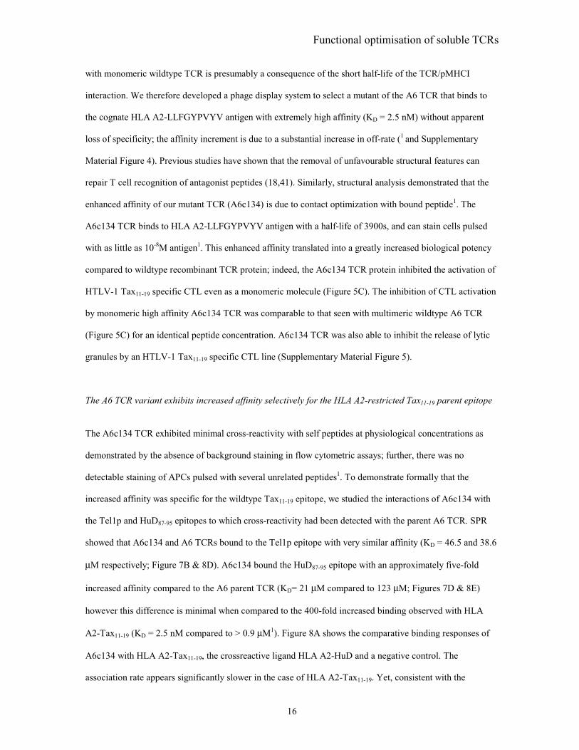
16
with monomeric wildtype TCR is presumably a consequence of the short half-life of the TCR/pMHCI
interaction. We therefore developed a phage display system to select a mutant of the A6 TCR that binds to
the cognate HLA A2-LLFGYPVYV antigen with extremely high affinity (KD = 2.5 nM) without apparent
loss of specificity; the affinity increment is due to a substantial increase in off-rate (1 and Supplementary
Material Figure 4). Previous studies have shown that the removal of unfavourable structural features can
repair T cell recognition of antagonist peptides (18,41). Similarly, structural analysis demonstrated that the
enhanced affinity of our mutant TCR (A6c134) is due to contact optimization with bound peptide1. The
A6c134 TCR binds to HLA A2-LLFGYPVYV antigen with a half-life of 3900s, and can stain cells pulsed
with as little as 10-8M antigen1. This enhanced affinity translated into a greatly increased biological potency
compared to wildtype recombinant TCR protein; indeed, the A6c134 TCR protein inhibited the activation of
HTLV-1 Tax11-19 specific CTL even as a monomeric molecule (Figure 5C). The inhibition of CTL activation
by monomeric high affinity A6c134 TCR was comparable to that seen with multimeric wildtype A6 TCR
(Figure 5C) for an identical peptide concentration. A6c134 TCR was also able to inhibit the release of lytic
granules by an HTLV-1 Tax11-19 specific CTL line (Supplementary Material Figure 5).
The A6 TCR variant exhibits increased affinity selectively for the HLA A2-restricted Tax11-19 parent epitope
The A6c134 TCR exhibited minimal cross-reactivity with self peptides at physiological concentrations as
demonstrated by the absence of background staining in flow cytometric assays; further, there was no
detectable staining of APCs pulsed with several unrelated peptides1. To demonstrate formally that the
increased affinity was specific for the wildtype Tax11-19 epitope, we studied the interactions of A6c134 with
the Tel1p and HuD87-95 epitopes to which cross-reactivity had been detected with the parent A6 TCR. SPR
showed that A6c134 and A6 TCRs bound to the Tel1p epitope with very similar affinity (KD = 46.5 and 38.6
µM respectively; Figure 7B & 8D). A6c134 bound the HuD87-95 epitope with an approximately five-fold
increased affinity compared to the A6 parent TCR (KD= 21 µM compared to 123 µM; Figures 7D & 8E)
however this difference is minimal when compared to the 400-fold increased binding observed with HLA
A2-Tax11-19 (KD = 2.5 nM compared to > 0.9 µM1). Figure 8A shows the comparative binding responses of
A6c134 with HLA A2-Tax11-19, the crossreactive ligand HLA A2-HuD and a negative control. The
association rate appears significantly slower in the case of HLA A2-Tax11-19. Yet, consistent with the

Functional optimisation of soluble TCRs
17
findings of Li et al.1, the most striking feature is the dramatically slower dissociation rate exhibited by the
A6c134 monomers. This is illustrated in the first two injection cycles, during which dissociation of the TCR
is barely detectable, and by the saturation of immobilized complexes as concentration of the analyte
increases. Altogether, these results indicate that TCR affinity can be selectively increased for a specific
peptide without concomitant major increases in the binding of related ligands.

Functional optimisation of soluble TCRs
18
Discussion
In this study, we generated soluble human TCRs that, like membrane-bound TCRs, comprised two
polypeptide chains; these were expressed separately from two distinct bacterial vectors and paired by a non-
native disulfide bond. This strategy of protein engineering produces a protein similar to the native globular
TCR that is extremely stable. The A6 and JM22 TCRs studied here appear structurally authentic, maintained
their ligand specificity in this form and were used to optimize the targeting of cells expressing cognate
antigen. To overcome the intrinsically low affinity of TCR/pMHC interactions, we adopted two approaches.
First, we increased overall avidity by multimerization to reduce the composite dissociation rate; this
approach has been exploited successfully to allow identification of antigen-specific T cells with soluble
pMHC molecules (39,42). Second, we adopted mutational strategies to engineer a TCR from its A6 parent
that had dramatically increased affinity for cognate ligand due to a reduction in off-rate. The TCR yielded by
this latter approach was more sensitive than multimeric forms of the parent TCR in terms of its ability to
detect cognate antigen on the target cell surface, and exhibited potent biological activity with minimal cross-
reactivity. The soluble heterodimeric TCRs manufactured in this study were utilized in a variety of
functional assays including specific detection of target cells pulsed with cognate peptide, discrimination of
quantitative changes in antigen display at the cell surface, identification of virus-infected cells, inhibition of
antigen-specific CTL activation and identification of cross-reactive peptides.
Part of this work has developed systems that utilize TCRs for the specific identification of cell surface
pMHCI complexes. In this regard, several noteworthy features emerge from the data. First, PE-conjugated
TCR multimers failed to emit a signal detectable by standard flow cytometric techniques when target cells
were pulsed with low antigen concentrations (Figure 2). However, functional assays clearly demonstrated
specific and biologically relevant binding at antigen densities well below the fluorescence detection limit
(Figure 5). The requirement for high antigen density in order to visualize TCR multimer binding is therefore
a methodological issue. In terms of detection sensitivity, the apparent discrepancy between TCR and pMHCI
multimers, which are widely used to identify and characterize antigen-specific T cell populations directly ex
vivo by flow cytometry, can be explained by respective ligand density differences on the target cell surface.
A T cell is thought to bear approximately 30,000-100,000 TCRs (43), all of identical idiotypic specificity; at

Functional optimisation of soluble TCRs
19
physiological antigen densities, however, only a small fraction of the 50,000 or so MHCI molecules on the
surface of an APC (44) will present the epitope cognate for the TCR clonotype. Thus, it appears that flow
cytometric detection of specific pMHCI at low antigen densities using soluble recombinant TCR-based
reagents is limited by poor signal intensity. Second, despite these limitations, TCR multimers proved to be
useful tools that enabled the discrimination of quantitative changes in antigen presentation on the target cell
surface. Indeed, the effects of both β2m and IFNγ on the loading of exogenous peptide could be monitored
by flow cytometry using these reagents. It is well documented that the addition of exogenous β2m can
enhance the apparent sensitivity of CTL to antigen (37). Other studies have shown that the addition of
exogenous β2m concomitant with peptide caused a vast increase in the number of conformationally correct
pMHCI molecules on the cell surface as monitored with conformation-specific antibodies (38,45). In the
present study, the addition of exogenous β2m at a given concentration of peptide increased the number of
TCR-cognate ligands by at least two-fold and sufficiently increased the sensitivity of flow cytometric
detection to allow the visualization of endogenously processed pMHCI antigen (Figure 4). These
observations could be extended to enable tracking of specific viral antigen expression in many settings. The
synergistic effects of IFNγ, which enhances processing and presentation of endogenously generated antigen
(46), might further enhance the general applicability of either TCR multimers or high affinity variant TCRs
for this purpose. Third, TCR multimers were used to screen for and identify syngeneic cross-reactive
pMHCI ligands (Figure 6); this approach eliminates the effects of adhesion/costimulatory molecular
interactions that potentially confound cellular screening assays (47,48) and has recently been validated in
class II-restricted systems (49). Surface plasmon resonance studies confirmed binding, with measured
affinities for both HLA A2-Tel1p and HLA A2- HuD87-95 lying at the lower end of the spectrum of
previously defined TCR/pMHC interactions (22) (Figure 7). In order to ensure an efficient adaptative
immune response, it is advantageous that a single T cell clonotype can potentially engage in a functionally
productive manner with several foreign peptides (50). On the molecular level, the corollary of this
hypothesis is that a TCR clonotype will exhibit sufficient intrinsic conformational diversity to cross-react
with several molecular mimics and less structurally related pMHCI complexes (51-53). Although
alloreactive and/or xenoreactive ligands have been identified for other TCRs (51,53-56), few cross-reactive
syngeneic interactions between naturally occurring pMHCI and cognate TCR have been identified (57-60).

Functional optimisation of soluble TCRs
20
Indeed, to the best of our knowledge, this study is the first to characterize the interaction between a
pathogen-reactive human TCR and a cross-reactive self-peptide in biophysical terms.
The cross-reactivity identified in this study has several important implications. First, it instructs on the limits
of thymic selection. Of the cross-reactive human peptides capable of eliciting functional responses through
the A6 TCR in the study by Hausmann et al., only HuD87-95 bound the corresponding TCR tetramer in our
hands by flow cytometry. Thus, this peptide likely ranks at the upper end of the A6 TCR affinity spectrum in
terms of interactions with human peptides; indeed, this assumption was confirmed by SPR analysis for those
peptides tested (data not shown). In a broader context, we have examined the binding of TCRs from several
human immunodominant anti-viral CTL to their cognate ligand in addition to the A6 and JM22 TCRs
studied here. All of these TCRs have a relatively high affinity for their cognate ligand (KD 0.9-10 µM). The
TCR/pMHCI interaction of anti-tumour CTL appears to be weaker than this (KD = 20-40 µM) (Boulter et
al., manuscript in preparation). Thus, the affinity of the interaction between HLA A2-HuD87-95 and the A6
TCR (KD =120 µM) is only 4 times weaker than that of TCRs capable of mediating functional T cell
responses in the periphery. These data suggest that thymic selection operates within narrow limits, and are
consistent with studies in murine systems (61). Second, the cross-reactivity of A6 with HLA A2-HuD87-95
might be relevant in autoimmunity. Paraneoplastic neurological degenerations (PNDs) are disorders that
develop in patients with coexistent malignancies. PNDs are believed to be triggered by an anti-tumour
immune response directed against neuronal antigens expressed inside tumour cells (62). Curiously, the HuD
protein has been implicated as a source of such antigen in one form of PND called anti-Hu syndrome (62-
64). It is believed that the expression of HuD by small cell lung cancer triggers an immune response against
this protein, which is exclusively neuron-expressed in healthy individuals (63,64). A recent study examined
the recognition and processing of 14 computer-predicted HLA A*0201-restricted, HuD-derived epitopes
(64). Only one of these peptides, comprising HuD residues 86-95, was generated through the MHCI antigen-
processing pathway; HuD86-95 was found to be immunogenic in experimental systems (64). HuD86-95
contains the shorter 9mer HuD87-95 peptide (LGYGFVNYI) that cross-reacts with the A6 TCR in our study;
HLA A2-HuD87-95 binds the A6 TCR tetramer to a slightly greater extent than the longer HuD86-95 peptide
(data not shown). Cross-recognition of a neuron-derived epitope by a Tax11-19-specific TCR might be

Functional optimisation of soluble TCRs
21
relevant to HTLV-1-associated pathology. 1-2% of HTLV-1 infected individuals, particularly those with a
high viral load, develop HTLV-1-associated myelopathy/tropical spastic paraparsis (HAM/TSP). The HLA
A2-restricted Tax11-19 epitope is highly immunodominant in HLA A2+ individuals infected with HTLV-1,
and appears to elicit especially large CTL responses in the setting of HAM/TSP (65,66). Further, HTLV-1-
specific lymphocytes are enriched in the cerebrospinal fluid of individuals with HAM/TSP (65). These
findings suggest that such CTL might cause HTLV-1-associated myeloneuropathy (65-67); this could
potentially result either from direct recognition of viral antigen in central nervous system (CNS) tissue or
from cross-recognition of CNS-derived self antigens such as HuD87-95. The latter hypothesis is supported by
elegant studies showing that Tax11-19-stimulated T cell lines from two HLA A2+ individuals with HTLV-1-
associated myelopathy killed targets pulsed with HuD86-95 peptide (40).
The ability of TCR multimers to target cells expressing specific antigens provides a vehicle for the delivery
of therapeutic interventions selectively to sites of disease. In comparison to systemic delivery systems,
targeted therapeutics might both enhance efficacy and minimize side-effects. We show here, for example,
that both wildtype multimeric TCRs and engineered high affinity monomeric TCRs can be used to conceal
specific antigens from cognate CTL (Figure 5). In future, it might be possible to use similar technology in
vivo to down-modulate cellular autoimmune reactions. Alternatively, soluble TCRs could be used to deliver
cytotoxic agents or to redirect pathogen-specific immune responses; this latter principle has recently been
demonstrated using pMHCI-conjugated antibodies that allowed antiviral CTL to induce regression of human
tumour xenografts in a murine model (68). However, the observation of TCR cross-reactivity is a potentially
confounding factor in the context of targeted therapy. In this respect, the development of engineered TCRs
with artificially enhanced binding affinities for cognate ligand represents a dramatic advance ((69,70) and 1).
Importantly, this increased affinity is not accompanied by concomitant increases in cross-reactivity (Figure
8)1. Thus, a strategy for the optimization of targeted therapeutics is defined that could be developed for
multiple applications, most notably perhaps within the fields of tumour immunology and autoimmunity.

Functional optimisation of soluble TCRs
22
REFERENCES
1. Davis, M. M., Boniface, J. J., Reich, Z., Lyons, D., Hampl, J., Arden, B., and Chien, Y. (1998) Annu Rev Immunol 16, 523-544
2. Konig, R. (2002) Curr Opin Immunol 14, 75-83. 3. Irvine, D. J., Purbhoo, M. A., Krogsgaard, M., and Davis, M. M. (2002) Nature 419, 845-849 4. Purbhoo, M. A., Irvine, D. J., Huppa, J. B., and Davis, M. M. (2004) Nat Immunol 5, 524-530 5. Huston, J. S., Levinson, D., Mudgett-Hunter, M., Tai, M. S., Novotny, J., Margolies, M. N., Ridge,
R. J., Bruccoleri, R. E., Haber, E., Crea, R., and et al. (1988) Proc Natl Acad Sci U S A 85, 5879-5883
6. Hoo, W. F., Lacy, M. J., Denzin, L. K., Voss, E. W., Jr., Hardman, K. D., and Kranz, D. M. (1992) Proc Natl Acad Sci U S A 89, 4759-4763
7. Plaksin, D., Polakova, K., McPhie, P., and Margulies, D. H. (1997) J Immunol 158, 2218-2227 8. Lake, D. F., Salgaller, M. L., van der Bruggen, P., Bernstein, R. M., and Marchalonis, J. J. (1999)
Int Immunol 11, 745-751 9. Gregoire, C., Rebai, N., Schweisguth, F., Necker, A., Mazza, G., Auphan, N., Millward, A.,
Schmitt-Verhulst, A. M., and Malissen, B. (1991) Proc Natl Acad Sci U S A 88, 8077-8081 10. Boulter, J. M., Glick, M., Todorov, P. T., Baston, E., Sami, M., Rizkallah, P., and Jakobsen, B. K.
(2003) Protein Eng 16, 707-711 11. Sewell, A. K., Harcourt, G. C., Goulder, P. J., Price, D. A., and Phillips, R. E. (1997) Eur J
Immunol 27, 2323-2329 12. Cull, M. G., and Schatz, P. J. (2000) Methods Enzymol 326, 430-440 13. Willcox, B. E., Gao, G. F., Wyer, J. R., O'Callaghan, C. A., Boulter, J. M., Jones, E. Y., van der
Merwe, P. A., Bell, J. I., and Jakobsen, B. K. (1999) Protein Sci 8, 2418-2423 14. Rammensee, H., Bachmann, J., Emmerich, N. P., Bachor, O. A., and Stevanovic, S. (1999)
Immunogenetics 50, 213-219 15. Parker, C. E., Daenke, S., Nightingale, S., and Bangham, C. R. (1992) Virology 188, 628-636 16. Utz, U., Koenig, S., Coligan, J. E., and Biddison, W. E. (1992) J Immunol 149, 214-221 17. Garboczi, D. N., Ghosh, P., Utz, U., Fan, Q. R., Biddison, W. E., and Wiley, D. C. (1996) Nature
384, 134-141 18. Ding, Y. H., Baker, B. M., Garboczi, D. N., Biddison, W. E., and Wiley, D. C. (1999) Immunity 11,
45-56 19. Lehner, P. J., Wang, E. C., Moss, P. A., Williams, S., Platt, K., Friedman, S. M., Bell, J. I., and
Borysiewicz, L. K. (1995) J Exp Med 181, 79-91 20. Willcox, B. E., Gao, G. F., Wyer, J. R., Ladbury, J. E., Bell, J. I., Jakobsen, B. K., and van der
Merwe, P. A. (1999) Immunity 10, 357-365 21. Stewart-Jones, G. B., McMichael, A. J., Bell, J. I., Stuart, D. I., and Jones, E. Y. (2003) Nat
Immunol 4, 657-663 22. Van Der Merwe, P. A., and Davis, S. J. (2003) Annu Rev Immunol 21, 659-684 23. Aharoni, R., Teitelbaum, D., Arnon, R., and Puri, J. (1991) Nature 351, 147-150. 24. Chung, D. H., Belyakov, I. M., Derby, M. A., Wang, J., Boyd, L. F., Berzofsky, J. A., and
Margulies, D. H. (2001) J Immunol 167, 699-707 25. Dadaglio, G., Nelson, C. A., Deck, M. B., Petzold, S. J., and Unanue, E. R. (1997) Immunity 6,
727-738 26. Eastman, S., Deftos, M., DeRoos, P. C., Hsu, D. H., Teyton, L., Braunstein, N. S., Hackett, C. J.,
and Rudensky, A. (1996) Eur J Immunol 26, 385-393. 27. Messaoudi, I., LeMaoult, J., and Nikolic-Zugic, J. (1999) J Immunol 163, 3286-3294 28. Morkowski, S., Raposo, G., Kleijimeer, M., Geuze, H. J., and Rudensky, A. Y. (1997) Eur J
Immunol 27, 609-617. 29. Murphy, D. B., Rath, S., Pizzo, E., Rudensky, A. Y., George, A., Larson, J. K., and Janeway, C. A.,
Jr. (1992) J Immunol 148, 3483-3491. 30. Polakova, K., Plaksin, D., Chung, D. H., Belyakov, I. M., Berzofsky, J. A., and Margulies, D. H.
(2000) J Immunol 165, 5703-5712 31. Puri, J., Arnon, R., Gurevich, E., and Teitelbaum, D. (1997) J Immunol 158, 2471-2476.

Functional optimisation of soluble TCRs
23
32. Reay, P. A., Matsui, K., Haase, K., Wulfing, C., Chien, Y. H., and Davis, M. M. (2000) J Immunol 164, 5626-5634.
33. Zhong, G., Reis e Sousa, C., and Germain, R. N. (1997) Proc Natl Acad Sci U S A 94, 13856-13861.
34. Cohen, C. J., Sarig, O., Yamano, Y., Tomaru, U., Jacobson, S., and Reiter, Y. (2003) J Immunol 170, 4349-4361
35. Biddison, W. E., Turner, R. V., Gagnon, S. J., Lev, A., Cohen, C. J., and Reiter, Y. (2003) J Immunol 171, 3064-3074
36. O'Herrin, S. M., Lebowitz, M. S., Bieler, J. G., al-Ramadi, B. K., Utz, U., Bothwell, A. L., and Schneck, J. P. (1997) J Exp Med 186, 1333-1345
37. Shields, M. J., Kubota, R., Hodgson, W., Jacobson, S., Biddison, W. E., and Ribaudo, R. K. (1998) J Biol Chem 273, 28010-28018
38. Rock, K. L., Rothstein, L. E., Gamble, S. R., and Benacerraf, B. (1990) Proc Natl Acad Sci U S A 87, 7517-7521
39. McMichael, A. J., and O'Callaghan, C. A. (1998) J Exp Med 187, 1367-1371 40. Hausmann, S., Biddison, W. E., Smith, K. J., Ding, Y. H., Garboczi, D. N., Utz, U., Wiley, D. C.,
and Wucherpfennig, K. W. (1999) J Immunol 162, 5389-5397 41. Baker, B. M., Gagnon, S. J., Biddison, W. E., and Wiley, D. C. (2000) Immunity 13, 475-484 42. Altman, J. D., Moss, P. A., Goulder, P. J., Barouch, D. H., McHeyzer-Williams, M. G., Bell, J. I.,
McMichael, A. J., and Davis, M. M. (1996) Science 274, 94-96 43. Schodin, B. A., Tsomides, T. J., and Kranz, D. M. (1996) Immunity 5, 137-146 44. Yewdell, J. W. (2001) Trends Cell Biol 11, 294-297 45. Otten, G. R., Bikoff, E., Ribaudo, R. K., Kozlowski, S., Margulies, D. H., and Germain, R. N.
(1992) J Immunol 148, 3723-3732 46. Hengel, H., Lucin, P., Jonjic, S., Ruppert, T., and Koszinowski, U. H. (1994) J Virol 68, 289-297 47. Hemmer, B., Vergelli, M., Pinilla, C., Houghten, R., and Martin, R. (1998) Immunol Today 19,
163-168 48. Hiemstra, H. S., van Veelen, P. A., Willemen, S. J., Benckhuijsen, W. E., Geluk, A., de Vries, R.
R., Roep, B. O., and Drijfhout, J. W. (1999) Eur J Immunol 29, 2385-2391 49. Crawford, F., Huseby, E., White, J., Marrack, P., and Kappler, J. W. (2004) PLoS Biol 2, E90 50. Mason, D. (1998) Immunol Today 19, 395-404 51. Garcia, K. C., Tallquist, M. D., Pease, L. R., Brunmark, A., Scott, C. A., Degano, M., Stura, E. A.,
Peterson, P. A., Wilson, I. A., and Teyton, L. (1997) Proc Natl Acad Sci U S A 94, 13838-13843 52. Holler, P. D., and Kranz, D. M. (2004) Mol Immunol 40, 1027-1031 53. Reiser, J. B., Darnault, C., Gregoire, C., Mosser, T., Mazza, G., Kearney, A., van der Merwe, P. A.,
Fontecilla-Camps, J. C., Housset, D., and Malissen, B. (2003) Nat Immunol 4, 241-247 54. Reiser, J. B., Darnault, C., Guimezanes, A., Gregoire, C., Mosser, T., Schmitt-Verhulst, A. M.,
Fontecilla-Camps, J. C., Malissen, B., Housset, D., and Mazza, G. (2000) Nat Immunol 1, 291-297 55. Corr, M., Slanetz, A. E., Boyd, L. F., Jelonek, M. T., Khilko, S., al-Ramadi, B. K., Kim, Y. S.,
Maher, S. E., Bothwell, A. L., and Margulies, D. H. (1994) Science 265, 946-949 56. Lang, H. L., Jacobsen, H., Ikemizu, S., Andersson, C., Harlos, K., Madsen, L., Hjorth, P.,
Sondergaard, L., Svejgaard, A., Wucherpfennig, K., Stuart, D. I., Bell, J. I., Jones, E. Y., and Fugger, L. (2002) Nat Immunol 3, 940-943
57. Hiemstra, H. S., Schloot, N. C., van Veelen, P. A., Willemen, S. J., Franken, K. L., van Rood, J. J., de Vries, R. R., Chaudhuri, A., Behan, P. O., Drijfhout, J. W., and Roep, B. O. (2001) Proc Natl Acad Sci U S A 98, 3988-3991
58. Hagerty, D. T., and Allen, P. M. (1995) J Immunol 155, 2993-3001 59. Quaratino, S., Thorpe, C. J., Travers, P. J., and Londei, M. (1995) Proc Natl Acad Sci U S A 92,
10398-10402 60. Wucherpfennig, K. W., and Strominger, J. L. (1995) Cell 80, 695-705 61. Alam, S. M., Travers, P. J., Wung, J. L., Nasholds, W., Redpath, S., Jameson, S. C., and Gascoigne,
N. R. (1996) Nature 381, 616-620 62. Albert, M. L., and Darnell, R. B. (2004) Nat Rev Cancer 4, 36-44 63. Szabo, A., Dalmau, J., Manley, G., Rosenfeld, M., Wong, E., Henson, J., Posner, J. B., and
Furneaux, H. M. (1991) Cell 67, 325-333

Functional optimisation of soluble TCRs
24
64. Plonquet, A., Garcia-Pons, F., Fernandez, E., Philippe, C., Marquet, J., Rouard, H., Delfau-Larue, M. H., Kosmatopoulos, K., Lemonnier, F., Farcet, J. P., Gherardi, R. K., and Langlade-Demoyen, P. (2003) J Neuroimmunol 142, 93-100
65. Elovaara, I., Koenig, S., Brewah, A. Y., Woods, R. M., Lehky, T., and Jacobson, S. (1993) J Exp Med 177, 1567-1573
66. Utz, U., Banks, D., Jacobson, S., and Biddison, W. E. (1996) J Virol 70, 843-851 67. Jacobson, S., Shida, H., McFarlin, D. E., Fauci, A. S., and Koenig, S. (1990) Nature 348, 245-248 68. Lev, A., Noy, R., Oved, K., Novak, H., Segal, D., Walden, P., Zehn, D., and Reiter, Y. (2004) Proc
Natl Acad Sci U S A 101, 9051-9056 69. Shusta, E. V., Holler, P. D., Kieke, M. C., Kranz, D. M., and Wittrup, K. D. (2000) Nat Biotechnol
18, 754-759 70. Holler, P. D., Holman, P. O., Shusta, E. V., O'Herrin, S., Wittrup, K. D., and Kranz, D. M. (2000)
Proc Natl Acad Sci U S A 97, 5387-5392

Functional optimisation of soluble TCRs
25
Footnotes
**This work was funded by the Wellcome Trust. AKS is a Wellcome Trust Senior Fellow.
#DAP is a Medical Research Council Clinician Scientist.
1Li et al submitted
We thank Anton van der Merwe for helpful suggestions and critical reading of the manuscript

Functional optimisation of soluble TCRs
26
Figure legends Figure 1. Binding of A6 and JM22 TCR monomers and tetramers to HLA A2-Tax and HLA A2-Flu
complexes. Streptavidin was linked to a BIAcore™ CM-5 chip by amine coupling, biotin-tagged pMHCI
was loaded onto each flow cell, and data were collected at 25°C with a flow rate of 5 µl/min. 5 µl of each
biotinylated TCR monomer at 1 mg/ml was flowed over all flow cells, as was 25 µl of each TCR tetramer at
50 µg/ml. Negligible response was observed to non-cognate pMHCI for both TCR monomers and tetramers.
To facilitate visual comparison of monomer and tetramer binding events, the much larger monomer response
values were normalized to the peak values for the tetramers. Kinetic binding parameters for the tetramers
were estimated using BIAEvaluation™ software (see table). Monomer KDs were taken from experiments
with unbiotinylated A6 TCR (Supplementary Material Figure 1) or JM22 TCR (20). There are two apparent
off-rates for the TCR tetramers: (i) a minority fast off-rate thought to correspond to those tetramers binding
less than three antigens; and, (ii) a slow (true) off-rate for those tetramers likely binding three pMHCI
molecules. The latter off-rate is shown in the table. Some irreversible binding of biotinylated TCR
monomers is observed owing to the incomplete blocking of the streptavidin-coated chip surface with soluble
biotin.
Figure 2. TCR multimer staining of APCs. HLA A2+ B-cells were pulsed with exogenous peptides at the
indicated concentrations and stained with A6 or JM22 TCR multimers as described in the Experimental
Procedures.
Figure 3. TCR multimers exhibit exquisite staining specificity. HLA A2+ B cells were pulsed with a
variety of A2-restricted peptides at 200 µM or incubated with medium only (unpulsed cells) and stained with
the indicated TCR tetramers as described in the Experimental Procedures. Bars show the standard deviation
from the mean fluorescence intensity (MFI) values from 2 independent stainings.
Figure 4. Characterization of TCR binding by flow cytometry. LBL721.174 cells were incubated for 90
min with the HLA A2-HTLV-1 Tax (A) or HLA A2-Flu matrix (B) peptides at the indicated concentrations

Functional optimisation of soluble TCRs
27
or with medium only in presence of either 200 µg/ml BSA (empty bars) or 200 µg/ml β2m (filled bars) and
stained with the relevant TCR tetramer conjugated with extravidin-PE as described in the Experimental
Procedures. (C) PK B cells (HLA A2+) infected with recombinant vaccinia virus (see Experimental
Procedures) were stained 12 hr after infection as above.
Figure 5. TCR-mediated specific inhibition of CTL activation. (A) HLA A2+ B cells (PK) were pulsed
with the indicated concentrations of HLA A2-HTLV-1 Tax11-19 peptide or infected with vaccinia virus
expressing the full-length Tax protein and used as targets in a CTL activation assay (see Experimental
Procedures). Bars show the standard deviation from the mean of two replicate assays. Data shown are
representative of three experiments. (B) HLA A2+ B cells (PK) were pulsed with 1 µM HLA A2 HTLV-1
Tax peptide and either 0.1 µM (grey filling) or 10 µM (black filling) HLA A2-HIV Gag peptide, or pulsed
with 0.1 µM HTLV-1 Tax peptide (inset). Cells were then pre-incubated with 100 µg/ml A6 Tax TCR
tetramer or BSA and used in CTL activation assays with 003 HIV Gag CTLs or with E6 Tax CTLs, as
indicated. Bars show the standard deviation from the mean of three replicate assays. Background MIP1β
release by E6 CTL was 60 pg/ml. (C) HLA A2+ B cells (PK) were pulsed with 1 µM HLA A2-HTLV-1 Tax
peptide and incubated with 10 µg/ml A6 wt monomers/tetramers, A6c134 monomer or 1G4 TCR tetramer
prior to CTL activation assay as described in the Experimental Procedures. Data are expressed as percentage
of MIP1β release compared to an assay performed in absence of TCR (100%). Bars show the standard
deviation from the mean of three replicate assays and are representative of 2 independent experiments.
Background MIP1β release without addition of antigenic peptide was 91.8 pg/ml (or 14.3% of that with
added antigen). The A6c134 monomer was able to inhibit CTL activation even at 0.1 µg/ml (data not
shown).
Figure 6. A6 TCR tetramer staining of HLA A2+ B cells pulsed with cross-reactive peptides. PK B cells
were pulsed with indicated concentrations of HuD87-95 (A) and Tel1p (B) peptides in the presence of 200
µg/ml β2m for 90 min and stained as described in the Experimental Procedures. Staining of cells pulsed with
with 0.1 mM Tax11-19 peptide is shown for comparison.

Functional optimisation of soluble TCRs
28
Figure 7. Affinity of the A6 TCR for HLA A2-Tel1p and HLA A2-HuD87-95. Kinetic binding
experiments for A6 TCR flowed over immobilized A2-Tel1p (A), A2-HuD87-95 (C) and A2-Tax11-19 (E).
Equilibrium binding specific response is plotted against A6 TCR concentration and a non-linear fit of the
Langmuir binding isotherm is shown for A2-Tel1p (B), A2-HuD87-95 (D) and A2-Tax11-19 (F). A6 TCR was
flowed over the BIAcore chip at concentrations of 224 µM and two-fold dilutions thereof down to 1.75 µM.
Saturation binding with A2-Tax11-19 was reached at low concentration. Only data with lower concentrations
of TCR is shown in F.
Figure 8. Affinity of the A6c134 TCR for HLA A2-Tel1p and HLA A2-HuD87-95. Kinetic binding
experiments with A6c134 TCR flowed over immobilized ligands are shown. Overlay of the binding
responses for A6c134 flowed over A2-Tax11-19 (solid line), A2-HuD87-95 (dotted line) and control A2-
hTERT540 complexes (dashed line) (A). Kinetic binding experiments for A6c134 flowed over A2-Tel1p (B)
and A2-HuD87-95 (C). Equilibrium binding specific response is plotted against A6c134 TCR concentration
and a non-linear fit of the Langmuir binding isotherm is shown for A2-Tel1p (D) and A2-HuD87-95 (E).
A6c134 TCR was flowed over the BIAcore chip at concentrations of 48 µM and two-fold dilutions thereof,
down to 0.0937 µM.

Table I. Summary of binding data of A6 TCR with various ligands measured by SPR.
LIGAND
HLA A2-Tax
HLA A2-Tel1p
HLA A2-HuD
11-19
86-95
Bindingprediction*
28
28
19
SEQUENCE
LLFGYPVYV
MLWGYLQYV
LGYGFVNYI
Calculated KD in independentBIAcore experiments
Average KD ± SD
(µµµµM)
0.95 0.82 1.4
45.7 46.5 23.6
115.5 151.3 103.2 123.3±35.4
38.6±20.9
*Binding prediction of peptide for HLA A2 (see reference 14)
1.06±0.3

A B
Laugel et al. Figure 1
JM22 monomer JM22 tetramer A6 monomer A6 tetramer
k on (M-1 s-1) 3 × 10⁴ ° - 7 × 10⁴ $ -
k off (s-1) 0.12 ° 2.7 × 10⁻⁴ 0.1 $ 7.5 × 10⁻⁵
Approx KD (M) 6.6 × 10⁻⁶ ° - 1.4 × 10⁻⁶ $ -
t1/2 (s) 5.7 ° 2580 6.9 $ 9240
° (20) $ (data from experiment shown in Supplementary Material Figure 1)

100
0100 101 102 103
no peptide no peptide10 µM 10 µM
100 µM 100 µM
Cel
l cou
nt
FL-2 fluorescence intensity
JM22 Flu A6 TaxA
Laugel et al Figure 2
B100
0100 101 102 103

Laugel et al. Figure 3
unpulsed cells
HTLV-1 Tax LLFGYPVYV
EBV GLCTLVAML
HCV-22 GLQDCTMLV
HCV-33 ALYDVVTKL
HIV Gag SLYNTVATL
Flu matrix GILGFVFTC
Mean Fluorescence Intensity
A6 Tax TCR Flu TCR
0 20 40 60 80 20 40 60 800 100

Laugel et al. Figure 4
5
10
15
20
25
0
10
20
30
40
200 µg/ml BSA200 µg/ml β2m
200 µg/ml BSA200 µg/ml β2m
Tax11-19 Flu matrix58-66
Mea
n Fl
uore
scen
ce I
nten
sity
irrelevantvaccinia
A B C
Tax vaccinia
Cou
nts
Fluorescence intensity
1 µMnone 10 µM 1 µMnone 10 µM 101100
[Peptide]

Laugel et al. Figure 5
250
500
003 E6
Non
e
1G4
1mer
A6
1mer
s
A6
4mer
s
A6c
134
1mer
Rel
ativ
e M
IP1β
rele
ase
(%)
0
20
40
60
80
100
[MIP1β] (pg/ml)
10-5
10-6
10-7
10-8
tax-vaccinia
unpulsed cells
0 200 400 600
JM22 tetramer
A6 tetramer
BSA
[Pep
tid
e]
(M)
[MIP
1β]
(p
g/m
l)
A6 Tax TCR
BSA
BA C

Laugel et al. Figure 6
No peptide
2.5mM
1mM
Tax 0.1mMTax 0.1mM
No peptide
1mM0.5mM
2.5mM
HuD87-95 Tel1p
A B
Cel
l cou
nt
FL-2 fluorescence intensity

B
0
1 0 0
2 0 0
3 0 0
KD= 38.6 µµµµM
0 1 0 0 2 0 0 3 0 0
0 1 0 2 0 3 0 4 0 5 0 6 0 7 00
5 0
1 0 0
1 5 0
KD= 0.95 µµµµM
F
A2-Tel1p
A
48028150
28200
28250
28300
28350
28400
28450
28500
280 300 320 340 360 380 400 420 440 460
Res
po
nse
Un
its C
28100
28200
28250
275 295 315 335 355 375 395 415 435 455 475
28150
28050
28000
27950
D
1 0 0 2 0 0 3 0 00
5 0
1 0 0
1 5 0
KD= 123.3 µµµµM
0
28400
28500
28600
28700
28800
28900
29000
29100
29200
320 340 360 380 400 420 440 460
E
Sp
ecif
ic R
esp
on
se
A2-HuD
A2-Tax
Time (s) TCR concentration (µµµµM)
Laugel et al. Figure 7

Laugel et al. Figure 8
Res
po
nse
Un
its
Time (s)
28300
28350
28400
28450
28500
28550
28600
560 580 600 620 640 660 680 700
A2-Tel1p
28050
28100
28150
28200
28250
28300
560 580 600 620 640 660 680 700
A2-HuD
0 10 20 30 40 50
0
10
20
30
40
50
60
0 10 20 30 40 50
0
50
100
150
200
250
Sp
ecif
ic R
esp
on
se
TCR concentration (µµµµM)
B C
D E
KD=46.5 µµµµM KD=21 µµµµM
0
500
1000
1500
2000
2500
0 200 400 600 800 1000 1200 1400
A

-20
0
20
40
60
80
100
120
150 200 250 300 350 400 450 500 550 600
Laugel et al. Supplementary Material Figure 1
Bin
din
g R
esp
on
se
Time (s)
Kinetic parameters of the A6/HLA-A2 interaction. Streptavidin was covalently linked to a BIAcore CM-5 chip by aminecoupling at 5,000 RU and biotinylated pMHC protein or biotinylated control protein were bound to sensor surface atapproximately 500 RU in each flow-cell. A6 TCR solutions at a maximum concentration of 1 µM and two-fold dilutionsthereof, down to 0.0625 µM, were flowed over the cells at a fast flow-rate (50 µl/min.). Binding response curves (plainlines) and corresponding fits (dashed lines) are shown as overlays. The calculated association (kon) and dissociation rateconstants (koff) corresponding to this figure are shown in the table of Figure 1.

Laugel et al. Supplementary Material Figure 2
-20
0
20
40
60
80
100
120
140
160
180
-500 -100 300 700 1100 1500 1900 2300 2700 3100 3500Time (s)
Bin
din
g R
esp
on
se
1 µM0.25 µM0.125 µM
Binding of A6 TCR tetramers to HLA-A2 Tax. Streptavidin was covalently linked to a BIAcore CM-5 chip byamine coupling at 5,000 RU and biotinylated pMHC protein or biotinylated control protein were bound to sensorsurface at approximately 500 RU in each flow-cell. Each A6 TCR tetramer solution at the indicated concentrationwas flowed over a separate flow-cell of the chip at the injection rate of 50 µl/min. The half-life of tetramerinteraction calculated for the linear part of the dissociation phase of each tetramer concentration differs by <3.5%.

No antigen
10 -6 M Tax peptide
10 -5 M Tax peptide
Cel
l cou
nt
FL-1 Mean Fluorescence Intensity
Laugel et al. Supplementary Material Figure 3
Staining of B-cells with microbeads-linked TCRs. Purified TCR at a concentration of 1mg/ml was labeled with Alexa Fluor 488by standard N-hydroxy-succinidimide linkage to -NH2 groups (Molecular Probes, Oregon) and purified according to the manufacturer’s directions. Labeled TCR was then mixed with microMACS streptavidin-conjugated magnetic microbeads(Miltenyi Biotech, Germany) at a ratio of 20 TCR molecules per streptavidin molecule for 15 min at room temperature. TCR-streptavidinbead multimers were then purified on microMACS columns and eluted in 150 µl PBS. For staining, T1 cells were suspended at 106 cells/ml in R10 containing varying concentration of Tax11-19 peptide, as indicated, for 90 minutes at 370C. Cells were washed in PBS, resuspendedin 50 µl aliquots at 3x10⁶ cells/ml, mixed with 16 µl of TCR multimers, and incubated at room temperature for 2 hrs. Cells were then brought up to a total volume of 200 µl with PBS without washing and immediately analyzed by flow cytometry.

Time (s)
Res
pons
e (R
U)
-50
0
50
100
150
200
250
2000 3000 4000 5000 6000 7000 8000
Laugel et al. Supplementary Material Figure 4
Binding of A6c134 monomers to HLA A2-Tax. Streptavidin was linked to a Biacore™ CM-5 chip by amine coupling, biotin-tagged pMHCI was loaded onto each flow cell at a low density (500 RU), and data were collected at 25°C with a flow rate of 50 µl/min. 120 µl of TCR monomer (0.5 µM) was injected during the association phase.

100101102103104
100 101 102 103 104100101102103104
100 101 102 103
100101102103104
100 101 102 103
100101102103104
100 101 102 103
Laugel et al. Supplementary Material Figure 5
0.12%
0.27%
Anti-Lamp FITC
Ant
i-C
D8
APC
2.07%
HLA-A2 Tax11-19 Tetramer-PE
Ant
i-C
D8
APC
A
B C
ED
2.33%
TCR mediated inhibition of lysosomal membrane associated proteins (LAMPs) up-regulation. LAMPs are proteins present in the membrane of cytotoxic granules and are displayed on the cell-surface following CTL activation, when the granules fuse with the cytoplasmic membrane (Betts et al. 2003. Sensitive and viable identification of antigen-specific CD8+ T cells by a flow cytometric assay for degranulation. J Immunol Methods 281:65-78). Soluble A6c134 TCR inhibited antigen-induced LAMP upregulation by HLA-A2 Tax11-19 specific CTLs (E). Approximately 25,000 cells of the D1 line containing about 2% of HLA-A2 Tax11-19 specific cells (A) were incubated either without
peptide (B) or in presence of 10-6 M (C, D and E) of Tax11-19; and with BSA (B and C), with the irrelevant 1G4 TCR (D) or with A6c134 (E) at a final concentration of 10 µg/ml. The assay was carried out over 1 hour in presence of the protein transport inhibitor monensin (Golgistop, BD Pharmingen) and anti-LAMPs FITC (BD Pharmingen) antibody. Cells were then washed with FACS buffer and stained with anti-CD8 APC antibody prior to the FACS analysis. Percentage of total cells in gate is indicated in B-E.
2.13%
100101102103104
100 101 102 103
104
104
104
Copyright © 2022 FDOKUMEN