T-Cell Maturation, Activation and Differentiation
27
As indicated in Chapter 2, the thymus occupies a central role in T-cell biology. Aside from being the main source of all T cells, it is where T cells diversify and then are shaped into an effective primary T-cell repertoire by an extraordinary pair of selection processes. One of these, positive selection, permits the survival of only those T cells whose TCRs are capable of recognizing self-MHC molecules. It is thus responsible for the creation of a self-MHC-restricted repertoire of T cells. The other, negative selection, eliminates T cells that react too strongly with self-MHC or with self-MHC plus self- peptides. It is an extremely important factor in generating a primary T-cell repertoire that is self-tolerant. As shown in Figure 10-1, when T-cell precursors arrive at the thymus, they do not express such signature surface mark- ers of T cells as the T-cell receptor, the CD3 complex, or the coreceptors CD4 and CD8. In fact, these progenitor cells have chapter 10 ■ T-Cell Maturation and the Thymus ■ Thymic Selection of the T-Cell Repertoire ■ T H -Cell Activation ■ T-Cell Differentiation ■ Cell Death and T-Cell Populations ■ Peripheral T-Cells T-Cell Maturation, Activation, and Differentiation T recognition by most T cells from recognition by B cells is MHC restriction. In most cases, both the maturation of progenitor T cells in the thymus and the acti- vation of mature T cells in the periphery are influenced by the involvement of MHC molecules. The potential antigenic diversity of the T-cell population is reduced during matura- tion by a selection process that allows only MHC-restricted and nonself-reactive T cells to mature. The final stages in the maturation of most T cells proceed along two different de- velopmental pathways, which generate functionally distinct CD4 and CD8 subpopulations that exhibit class II and class I MHC restriction, respectively. Activation of mature peripheral T cells begins with the interaction of the T-cell receptor (TCR) with an antigenic peptide displayed in the groove of an MHC molecule. Al- though the specificity of this interaction is governed by the TCR, its low avidity necessitates the involvement of corecep- tors and other accessory membrane molecules that strengthen the TCR-antigen-MHC interaction and trans- duce the activating signal. Activation leads to the prolifera- tion and differentiation of T cells into various types of effector cells and memory T cells. Because the vast majority of thymocytes and peripheral T cells express the T-cell receptor rather than the T-cell receptor, all references to the T-cell receptor in this chapter denote the receptor un- less otherwise indicated. Similarly, unless otherwise indi- cated, all references to T cells denote those receptor- bearing T cells that undergo MHC restriction. T-Cell Maturation and the Thymus Progenitor T cells from the early sites of hematopoiesis begin to migrate to the thymus at about day 11 of gestation in mice and in the eighth or ninth week of gestation in humans. In a manner similar to B-cell maturation in the bone marrow, T- cell maturation involves rearrangements of the germ-line TCR genes and the expression of various membrane mark- ers. In the thymus, developing T cells, known as thymocytes, proliferate and differentiate along developmental pathways that generate functionally distinct subpopulations of mature T cells. Engagement of TcR by Peptide: MHC Initiates Signal Transduction ζ ζ γ δ ε 8536d_ch10_221 8/29/02 2:03 PM Page 221 mac83 Mac 83:379_kyw:
Transcript of T-Cell Maturation, Activation and Differentiation
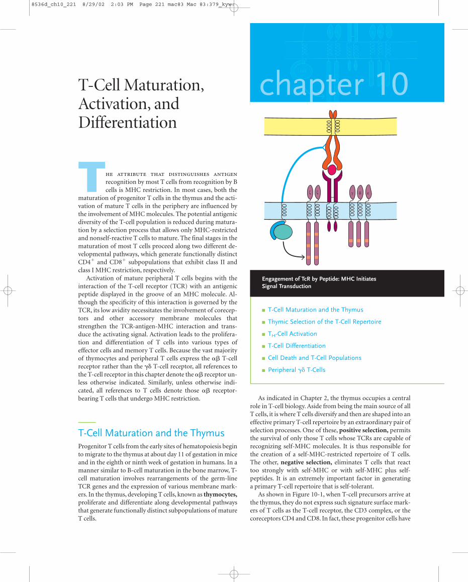
As indicated in Chapter 2, the thymus occupies a centralrole in T-cell biology. Aside from being the main source of allT cells, it is where T cells diversify and then are shaped into aneffective primary T-cell repertoire by an extraordinary pair ofselection processes. One of these, positive selection, permitsthe survival of only those T cells whose TCRs are capable ofrecognizing self-MHC molecules. It is thus responsible forthe creation of a self-MHC-restricted repertoire of T cells.The other, negative selection, eliminates T cells that reacttoo strongly with self-MHC or with self-MHC plus self-peptides. It is an extremely important factor in generating a primary T-cell repertoire that is self-tolerant.
As shown in Figure 10-1, when T-cell precursors arrive atthe thymus, they do not express such signature surface mark-ers of T cells as the T-cell receptor, the CD3 complex, or thecoreceptors CD4 and CD8. In fact, these progenitor cells have
chapter 10
� T-Cell Maturation and the Thymus
� Thymic Selection of the T-Cell Repertoire
� TH-Cell Activation
� T-Cell Differentiation
� Cell Death and T-Cell Populations
� Peripheral �� T-Cells
T-Cell Maturation,Activation, andDifferentiation
T
recognition by most T cells from recognition by Bcells is MHC restriction. In most cases, both the
maturation of progenitor T cells in the thymus and the acti-vation of mature T cells in the periphery are influenced bythe involvement of MHC molecules. The potential antigenicdiversity of the T-cell population is reduced during matura-tion by a selection process that allows only MHC-restrictedand nonself-reactive T cells to mature. The final stages in thematuration of most T cells proceed along two different de-velopmental pathways, which generate functionally distinctCD4� and CD8� subpopulations that exhibit class II andclass I MHC restriction, respectively.
Activation of mature peripheral T cells begins with theinteraction of the T-cell receptor (TCR) with an antigenicpeptide displayed in the groove of an MHC molecule. Al-though the specificity of this interaction is governed by theTCR, its low avidity necessitates the involvement of corecep-tors and other accessory membrane molecules thatstrengthen the TCR-antigen-MHC interaction and trans-duce the activating signal. Activation leads to the prolifera-tion and differentiation of T cells into various types ofeffector cells and memory T cells. Because the vast majorityof thymocytes and peripheral T cells express the �� T-cellreceptor rather than the �� T-cell receptor, all references tothe T-cell receptor in this chapter denote the �� receptor un-less otherwise indicated. Similarly, unless otherwise indi-cated, all references to T cells denote those �� receptor-bearing T cells that undergo MHC restriction.
T-Cell Maturation and the ThymusProgenitor T cells from the early sites of hematopoiesis beginto migrate to the thymus at about day 11 of gestation in miceand in the eighth or ninth week of gestation in humans. In amanner similar to B-cell maturation in the bone marrow, T-cell maturation involves rearrangements of the germ-lineTCR genes and the expression of various membrane mark-ers. In the thymus, developing T cells, known as thymocytes,proliferate and differentiate along developmental pathwaysthat generate functionally distinct subpopulations of matureT cells.
Engagement of TcR by Peptide: MHC Initiates Signal Transduction
ζ ζ γ δ ε
8536d_ch10_221 8/29/02 2:03 PM Page 221 mac83 Mac 83:379_kyw:
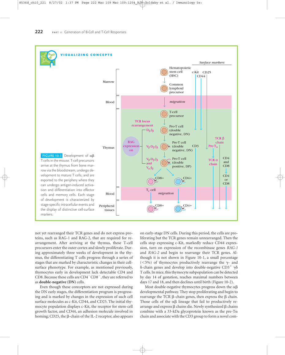
not yet rearranged their TCR genes and do not express pro-teins, such as RAG-1 and RAG-2, that are required for re-arrangement. After arriving at the thymus, these T-cellprecursors enter the outer cortex and slowly proliferate. Dur-ing approximately three weeks of development in the thy-mus, the differentiating T cells progress through a series ofstages that are marked by characteristic changes in their cell-surface phenotype. For example, as mentioned previously,thymocytes early in development lack detectable CD4 andCD8. Because these cells are CD4�CD8�, they are referred toas double-negative (DN) cells.
Even though these coreceptors are not expressed duringthe DN early stages, the differentiation program is progress-ing and is marked by changes in the expression of such cellsurface molecules as c-Kit, CD44, and CD25. The initial thy-mocyte population displays c-Kit, the receptor for stem-cellgrowth factor, and CD44, an adhesion molecule involved inhoming; CD25, the �-chain of the IL-2 receptor, also appears
222 P A R T I I Generation of B-Cell and T-Cell Responses
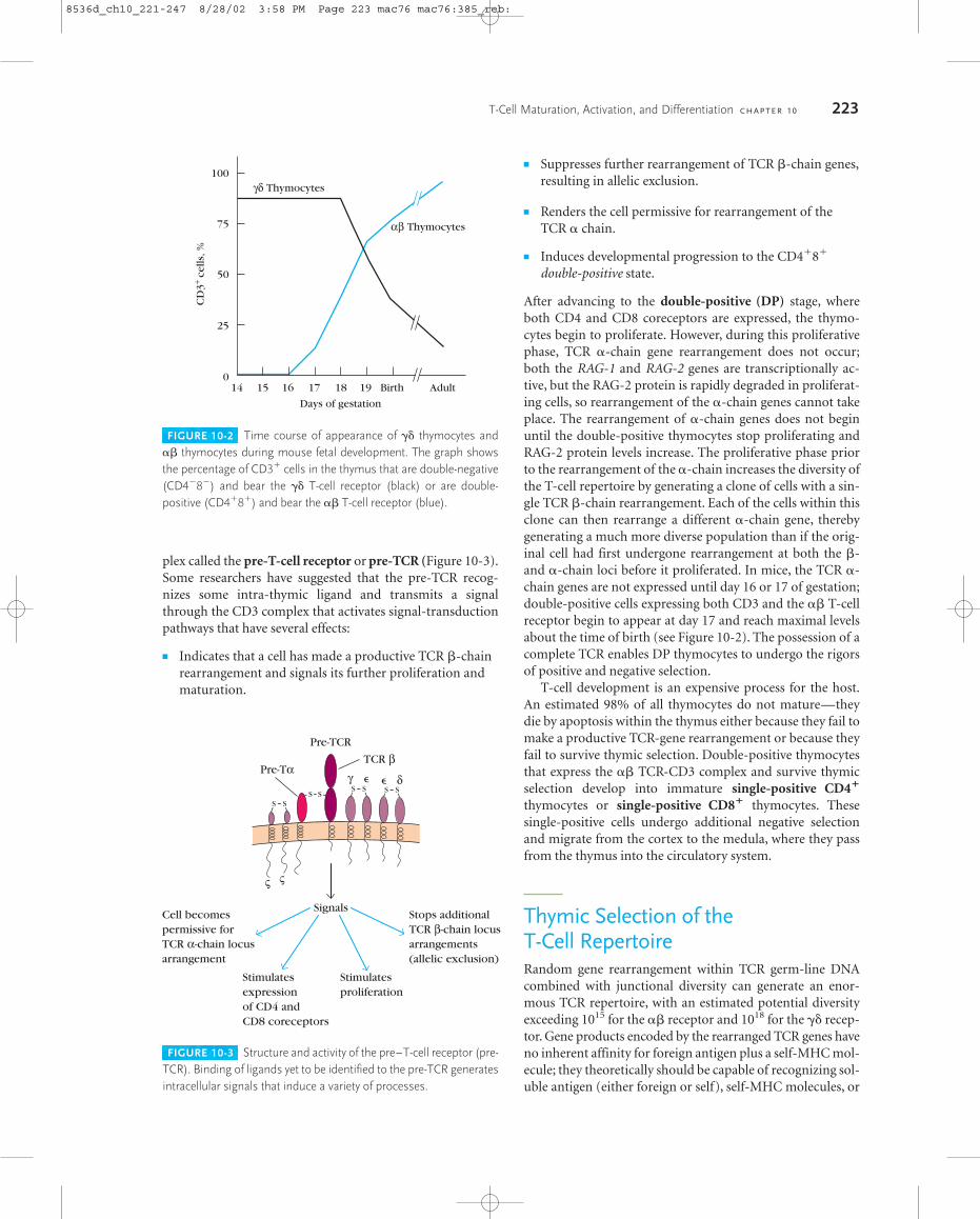
on early-stage DN cells. During this period, the cells are pro-liferating but the TCR genes remain unrearranged. Then thecells stop expressing c-Kit, markedly reduce CD44 expres-sion, turn on expression of the recombinase genes RAG-1and RAG-2 and begin to rearrange their TCR genes. Al-though it is not shown in Figure 10-1, a small percentage(�5%) of thymocytes productively rearrange the �- and �-chain genes and develop into double-negative CD3� ��T cells. In mice, this thymocyte subpopulation can be detectedby day 14 of gestation, reaches maximal numbers betweendays 17 and 18, and then declines until birth (Figure 10-2).
Most double-negative thymocytes progress down the ��developmental pathway. They stop proliferating and begin torearrange the TCR �-chain genes, then express the � chain.Those cells of the �� lineage that fail to productively re-arrange and express � chains die. Newly synthesized � chainscombine with a 33-kDa glycoprotein known as the pre-T�chain and associate with the CD3 group to form a novel com-
V I S U A L I Z I N G C O N C E P T S
FIGURE 10-1 Development of ��
T cells in the mouse. T-cell precursorsarrive at the thymus from bone mar-row via the bloodstream, undergo de-velopment to mature T cells, and areexported to the periphery where theycan undergo antigen-induced activa-tion and differentiation into effectorcells and memory cells. Each stage of development is characterized bystage-specific intracellular events andthe display of distinctive cell-surfacemarkers.
Hematopoieticstem cell(HSC)
Commonlymphoidprecursor
T-cellprecursor
c-Kit
CD3
CD44
Pre-Tα
TCR βchain
TCR αchain
CD4andCD8
CD4or
CD8
CD4+CD8+
CD4+CD8+
CD25
Pro-T cell(doublenegative, DN)
Pre-T cell(doublenegative, DN)
Pro-T cell(doublepositive, DP)
migration
migration
Surface markers
Peripheraltissues
Marrow
Blood
Blood
Thymus
RAGexpression
on
Dβ-Jβ
Vβ-Dβ-Jβ
Vβ-Dβ-JβandVα-Jβ
TCR locusrearrangement
Tc cell
8536d_ch10_221 8/27/02 1:37 PM Page 222 Mac 109 Mac 109:1254_BJN:Goldsby et al. / Immunology 5e:
plex called the pre-T-cell receptor or pre-TCR (Figure 10-3).Some researchers have suggested that the pre-TCR recog-nizes some intra-thymic ligand and transmits a signalthrough the CD3 complex that activates signal-transductionpathways that have several effects:
� Indicates that a cell has made a productive TCR �-chainrearrangement and signals its further proliferation andmaturation.
T-Cell Maturation, Activation, and Differentiation C H A P T E R 10 223
� Suppresses further rearrangement of TCR �-chain genes,resulting in allelic exclusion.
� Renders the cell permissive for rearrangement of theTCR � chain.
� Induces developmental progression to the CD4�8�
double-positive state.
After advancing to the double-positive (DP) stage, whereboth CD4 and CD8 coreceptors are expressed, the thymo-cytes begin to proliferate. However, during this proliferativephase, TCR �-chain gene rearrangement does not occur;both the RAG-1 and RAG-2 genes are transcriptionally ac-tive, but the RAG-2 protein is rapidly degraded in proliferat-ing cells, so rearrangement of the �-chain genes cannot takeplace. The rearrangement of �-chain genes does not beginuntil the double-positive thymocytes stop proliferating andRAG-2 protein levels increase. The proliferative phase priorto the rearrangement of the �-chain increases the diversity ofthe T-cell repertoire by generating a clone of cells with a sin-gle TCR �-chain rearrangement. Each of the cells within thisclone can then rearrange a different �-chain gene, therebygenerating a much more diverse population than if the orig-inal cell had first undergone rearrangement at both the �-and �-chain loci before it proliferated. In mice, the TCR �-chain genes are not expressed until day 16 or 17 of gestation;double-positive cells expressing both CD3 and the �� T-cellreceptor begin to appear at day 17 and reach maximal levelsabout the time of birth (see Figure 10-2). The possession of acomplete TCR enables DP thymocytes to undergo the rigorsof positive and negative selection.
T-cell development is an expensive process for the host.An estimated 98% of all thymocytes do not mature—theydie by apoptosis within the thymus either because they fail tomake a productive TCR-gene rearrangement or because theyfail to survive thymic selection. Double-positive thymocytesthat express the �� TCR-CD3 complex and survive thymicselection develop into immature single-positive CD4�
thymocytes or single-positive CD8� thymocytes. These single-positive cells undergo additional negative selectionand migrate from the cortex to the medula, where they passfrom the thymus into the circulatory system.
Thymic Selection of the T-Cell RepertoireRandom gene rearrangement within TCR germ-line DNAcombined with junctional diversity can generate an enor-mous TCR repertoire, with an estimated potential diversityexceeding 1015 for the �� receptor and 1018 for the �� recep-tor. Gene products encoded by the rearranged TCR genes have no inherent affinity for foreign antigen plus a self-MHC mol-ecule; they theoretically should be capable of recognizing sol-uble antigen (either foreign or self), self-MHC molecules, or
FIGURE 10-2 Time course of appearance of �� thymocytes and�� thymocytes during mouse fetal development. The graph showsthe percentage of CD3� cells in the thymus that are double-negative(CD4�8�) and bear the �� T-cell receptor (black) or are double-positive (CD4�8�) and bear the �� T-cell receptor (blue).
FIGURE 10-3 Structure and activity of the pre–T-cell receptor (pre-TCR). Binding of ligands yet to be identified to the pre-TCR generatesintracellular signals that induce a variety of processes.
100
75
50
25
017 18161514
CD
3+ ce
lls, %
Days of gestation
19 Birth Adult
αβ Thymocytes
γδ Thymocytes
Pre-TCR
TCR β
δγ
ς ς
Pre-Tα
SignalsCell becomespermissive forTCR α-chain locusarrangement
Stimulatesexpressionof CD4 andCD8 coreceptors
Stimulatesproliferation
Stops additionalTCR β-chain locusarrangements(allelic exclusion)
S SS S
S S
S S
8536d_ch10_221-247 8/28/02 3:58 PM Page 223 mac76 mac76:385_reb:
antigen plus a nonself-MHC molecule. Nonetheless, the mostdistinctive property of mature T cells is that they recognizeonly foreign antigen combined with self-MHC molecules.
As noted, thymocytes undergo two selection processes inthe thymus:
� Positive selection for thymocytes bearing receptorscapable of binding self-MHC molecules, which results inMHC restriction. Cells that fail positive selection areeliminated within the thymus by apoptosis.
� Negative selection that eliminates thymocytes bearinghigh-affinity receptors for self-MHC molecules alone orself-antigen presented by self-MHC, which results inself-tolerance.
Both processes are necessary to generate mature T cells thatare self-MHC restricted and self-tolerant. As noted already,some 98% or more of all thymocytes die by apoptosis withinthe thymus. The bulk of this high death rate appears to reflecta weeding out of thymocytes that fail positive selection be-cause their receptors do not specifically recognize foreignantigen plus self-MHC molecules.
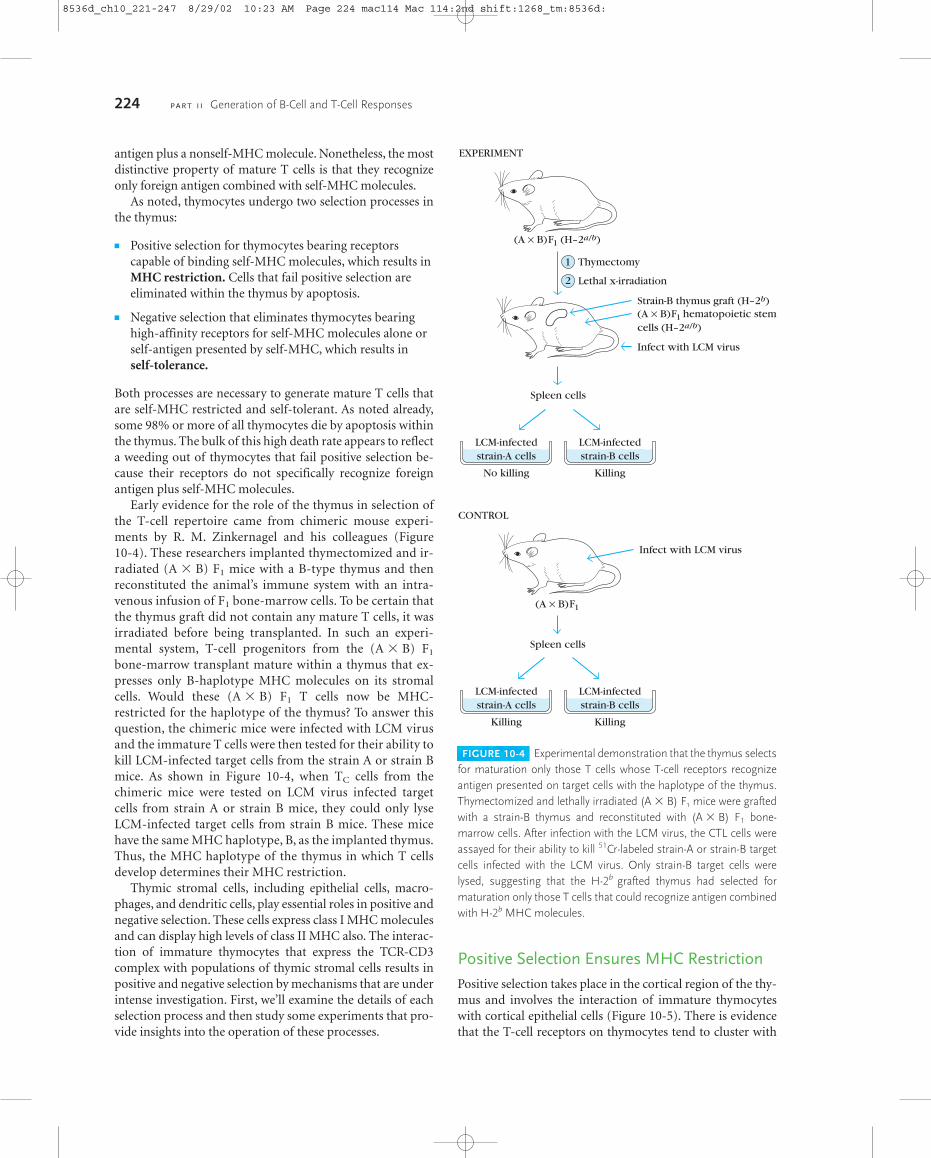
Early evidence for the role of the thymus in selection ofthe T-cell repertoire came from chimeric mouse experi-ments by R. M. Zinkernagel and his colleagues (Figure 10-4). These researchers implanted thymectomized and ir-radiated (A � B) F1 mice with a B-type thymus and thenreconstituted the animal’s immune system with an intra-venous infusion of F1 bone-marrow cells. To be certain thatthe thymus graft did not contain any mature T cells, it wasirradiated before being transplanted. In such an experi-mental system, T-cell progenitors from the (A � B) F1
bone-marrow transplant mature within a thymus that ex-presses only B-haplotype MHC molecules on its stromalcells. Would these (A � B) F1 T cells now be MHC-restricted for the haplotype of the thymus? To answer thisquestion, the chimeric mice were infected with LCM virusand the immature T cells were then tested for their ability tokill LCM-infected target cells from the strain A or strain Bmice. As shown in Figure 10-4, when TC cells from thechimeric mice were tested on LCM virus infected targetcells from strain A or strain B mice, they could only lyseLCM-infected target cells from strain B mice. These micehave the same MHC haplotype, B, as the implanted thymus.Thus, the MHC haplotype of the thymus in which T cellsdevelop determines their MHC restriction.
Thymic stromal cells, including epithelial cells, macro-phages, and dendritic cells, play essential roles in positive andnegative selection. These cells express class I MHC moleculesand can display high levels of class II MHC also. The interac-tion of immature thymocytes that express the TCR-CD3complex with populations of thymic stromal cells results inpositive and negative selection by mechanisms that are underintense investigation. First, we’ll examine the details of eachselection process and then study some experiments that pro-vide insights into the operation of these processes.
Positive Selection Ensures MHC RestrictionPositive selection takes place in the cortical region of the thy-mus and involves the interaction of immature thymocyteswith cortical epithelial cells (Figure 10-5). There is evidencethat the T-cell receptors on thymocytes tend to cluster with
224 P A R T I I Generation of B-Cell and T-Cell Responses
FIGURE 10-4 Experimental demonstration that the thymus selectsfor maturation only those T cells whose T-cell receptors recognizeantigen presented on target cells with the haplotype of the thymus.Thymectomized and lethally irradiated (A � B) F1 mice were graftedwith a strain-B thymus and reconstituted with (A � B) F1 bone-marrow cells. After infection with the LCM virus, the CTL cells wereassayed for their ability to kill 51Cr-labeled strain-A or strain-B targetcells infected with the LCM virus. Only strain-B target cells werelysed, suggesting that the H-2b grafted thymus had selected for maturation only those T cells that could recognize antigen combinedwith H-2b MHC molecules.
Lethal x-irradiation
Thymectomy
EXPERIMENT
(A × B)F1 (H–2a/b)
Strain-B thymus graft (H–2b)(A × B)F1 hematopoietic stemcells (H–2a/b)
Infect with LCM virus
Spleen cells
CONTROL
Infect with LCM virus
(A × B)F1
Spleen cells
Killing Killing
LCM-infectedstrain-B cells
LCM-infectedstrain-A cells
No killing Killing
LCM-infectedstrain-B cells
LCM-infectedstrain-A cells
1
2
8536d_ch10_221-247 8/29/02 10:23 AM Page 224 mac114 Mac 114:2nd shift:1268_tm:8536d:
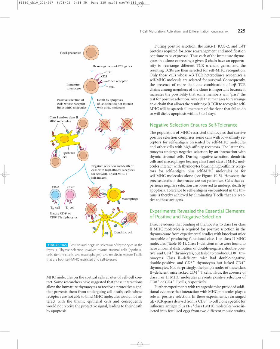
MHC molecules on the cortical cells at sites of cell-cell con-tact. Some researchers have suggested that these interactionsallow the immature thymocytes to receive a protective signalthat prevents them from undergoing cell death; cells whosereceptors are not able to bind MHC molecules would not in-teract with the thymic epithelial cells and consequentlywould not receive the protective signal, leading to their deathby apoptosis.
During positive selection, the RAG-1, RAG-2, and TdTproteins required for gene rearrangement and modificationcontinue to be expressed. Thus each of the immature thymo-cytes in a clone expressing a given � chain have an opportu-nity to rearrange different TCR �-chain genes, and theresulting TCRs are then selected for self-MHC recognition.Only those cells whose �� TCR heterodimer recognizes aself-MHC molecule are selected for survival. Consequently,the presence of more than one combination of �� TCRchains among members of the clone is important because itincreases the possibility that some members will “pass” thetest for positive selection. Any cell that manages to rearrangean � chain that allows the resulting �� TCR to recognize self-MHC will be spared; all members of the clone that fail to doso will die by apoptosis within 3 to 4 days.
Negative Selection Ensures Self-ToleranceThe population of MHC-restricted thymocytes that survivepositive selection comprises some cells with low-affinity re-ceptors for self-antigen presented by self-MHC moleculesand other cells with high-affinity receptors. The latter thy-mocytes undergo negative selection by an interaction withthymic stromal cells. During negative selection, dendriticcells and macrophages bearing class I and class II MHC mol-ecules interact with thymocytes bearing high-affinity recep-tors for self-antigen plus self-MHC molecules or forself-MHC molecules alone (see Figure 10-5). However, theprecise details of the process are not yet known. Cells that ex-perience negative selection are observed to undergo death byapoptosis. Tolerance to self-antigens encountered in the thy-mus is thereby achieved by eliminating T cells that are reac-tive to these antigens.
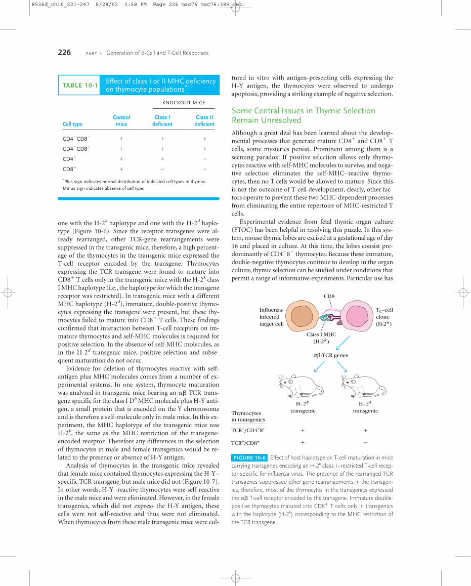
Experiments Revealed the Essential Elementsof Positive and Negative SelectionDirect evidence that binding of thymocytes to class I or classII MHC molecules is required for positive selection in thethymus came from experimental studies with knockout miceincapable of producing functional class I or class II MHCmolecules (Table 10-1). Class I–deficient mice were found tohave a normal distribution of double-negative, double-posi-tive, and CD4� thymocytes, but failed to produce CD8� thy-mocytes. Class II–deficient mice had double-negative,double-positive, and CD8� thymocytes but lacked CD4�
thymocytes. Not surprisingly, the lymph nodes of these classII–deficient mice lacked CD4� T cells. Thus, the absence ofclass I or II MHC molecules prevents positive selection ofCD8� or CD4� T cells, respectively.
Further experiments with transgenic mice provided addi-tional evidence that interaction with MHC molecules plays arole in positive selection. In these experiments, rearranged��-TCR genes derived from a CD8� T-cell clone specific forinfluenza antigen plus H-2k class I MHC molecules were in-jected into fertilized eggs from two different mouse strains,
T-Cell Maturation, Activation, and Differentiation C H A P T E R 10 225
FIGURE 10-5 Positive and negative selection of thymocytes in thethymus. Thymic selection involves thymic stromal cells (epithelialcells, dendritic cells, and macrophages), and results in mature T cellsthat are both self-MHC restricted and self-tolerant.
T-cell receptorImmaturethymocyte
Positive selection ofcells whose receptorbinds MHC molecules
Death by apoptosisof cells that do not interactwith MHC molecules
CD8CD3
CD4
T-cell precursor
Class I and/or class IIMHC molecules
Epithelialcell
Rearrangement of TCR genes
Negative selection and death ofcells with high-affinity receptorsfor self-MHC or self-MHC +self-antigen
CD4+ CD8+
TH cell TC cell
Mature CD4+ orCD8+ T lymphocytes
Macrophage
Dendritic cell
8536d_ch10_221-247 8/28/02 3:58 PM Page 225 mac76 mac76:385_reb:
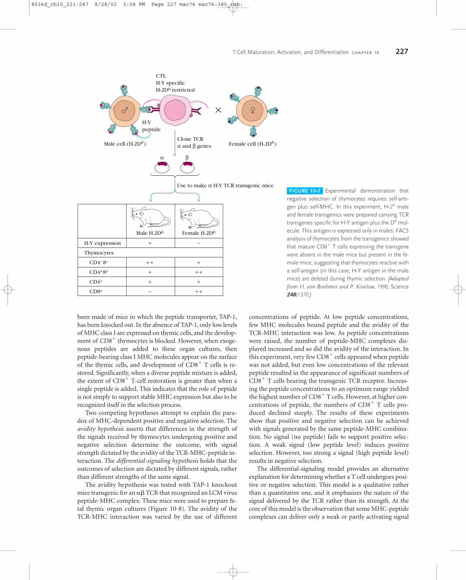
one with the H-2k haplotype and one with the H-2d haplo-type (Figure 10-6). Since the receptor transgenes were al-ready rearranged, other TCR-gene rearrangements weresuppressed in the transgenic mice; therefore, a high percent-age of the thymocytes in the transgenic mice expressed the T-cell receptor encoded by the transgene. Thymocytes expressing the TCR transgene were found to mature intoCD8� T cells only in the transgenic mice with the H-2k classI MHC haplotype (i.e., the haplotype for which the transgenereceptor was restricted). In transgenic mice with a differentMHC haplotype (H-2d), immature, double-positive thymo-cytes expressing the transgene were present, but these thy-mocytes failed to mature into CD8� T cells. These findingsconfirmed that interaction between T-cell receptors on im-mature thymocytes and self-MHC molecules is required forpositive selection. In the absence of self-MHC molecules, asin the H-2d transgenic mice, positive selection and subse-quent maturation do not occur.
Evidence for deletion of thymocytes reactive with self-antigen plus MHC molecules comes from a number of ex-perimental systems. In one system, thymocyte maturationwas analyzed in transgenic mice bearing an �� TCR trans-gene specific for the class I Db MHC molecule plus H-Y anti-gen, a small protein that is encoded on the Y chromosomeand is therefore a self-molecule only in male mice. In this ex-periment, the MHC haplotype of the transgenic mice was H-2b, the same as the MHC restriction of the transgene- encoded receptor. Therefore any differences in the selectionof thymocytes in male and female transgenics would be re-lated to the presence or absence of H-Y antigen.
Analysis of thymocytes in the transgenic mice revealedthat female mice contained thymocytes expressing the H-Y–specific TCR transgene, but male mice did not (Figure 10-7).In other words, H-Y–reactive thymocytes were self-reactivein the male mice and were eliminated. However, in the femaletransgenics, which did not express the H-Y antigen, thesecells were not self-reactive and thus were not eliminated.When thymocytes from these male transgenic mice were cul-
tured in vitro with antigen-presenting cells expressing the H-Y antigen, the thymocytes were observed to undergoapoptosis, providing a striking example of negative selection.
Some Central Issues in Thymic SelectionRemain UnresolvedAlthough a great deal has been learned about the develop-mental processes that generate mature CD4� and CD8� Tcells, some mysteries persist. Prominent among them is aseeming paradox: If positive selection allows only thymo-cytes reactive with self-MHC molecules to survive, and nega-tive selection eliminates the self-MHC–reactive thymo-cytes, then no T cells would be allowed to mature. Since thisis not the outcome of T-cell development, clearly, other fac-tors operate to prevent these two MHC-dependent processesfrom eliminating the entire repertoire of MHC-restricted Tcells.
Experimental evidence from fetal thymic organ culture(FTOC) has been helpful in resolving this puzzle. In this sys-tem, mouse thymic lobes are excised at a gestational age of day16 and placed in culture. At this time, the lobes consist pre-dominantly of CD4�8� thymocytes. Because these immature,double-negative thymocytes continue to develop in the organculture, thymic selection can be studied under conditions thatpermit a range of informative experiments. Particular use has
226 P A R T I I Generation of B-Cell and T-Cell Responses
TABLE 10-1Effect of class I or II MHC deficiencyon thymocyte populations*
KNOCKOUT MICE
Control Class I Class II Cell type mice deficient deficient
CD4�CD8� � � �
CD4�CD8� � � �
CD4� � � �
CD8� � � �
*Plus sign indicates normal distribution of indicated cell types in thymus.
Minus sign indicates absence of cell type.
FIGURE 10-6 Effect of host haplotype on T-cell maturation in micecarrying transgenes encoding an H-2b class I–restricted T-cell recep-tor specific for influenza virus. The presence of the rearranged TCRtransgenes suppressed other gene rearrangements in the transgen-ics; therefore, most of the thymocytes in the transgenics expressedthe �� T-cell receptor encoded by the transgene. Immature double-positive thymocytes matured into CD8� T cells only in transgenicswith the haplotype (H-2k) corresponding to the MHC restriction ofthe TCR transgene.
Thymocytesin transgenics
TCR+/CD4+8+
TCR+/CD8+
H–2k
transgenic
+
+
+
−
Influenza-infectedtarget cell
TC-cellclone(H-2k)
CD8
Class I MHC(H-2k)
αβ -TCR genes
H–2d
transgenic
8536d_ch10_221-247 8/28/02 3:58 PM Page 226 mac76 mac76:385_reb:
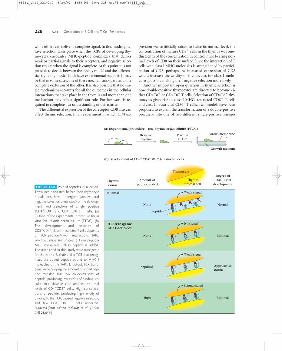
been made of mice in which the peptide transporter, TAP-1,has been knocked out. In the absence of TAP-1, only low levelsof MHC class I are expressed on thymic cells, and the develop-ment of CD8� thymocytes is blocked. However, when exoge-nous peptides are added to these organ cultures, thenpeptide-bearing class I MHC molecules appear on the surfaceof the thymic cells, and development of CD8� T cells is re-stored. Significantly, when a diverse peptide mixture is added,the extent of CD8� T-cell restoration is greater than when asingle peptide is added. This indicates that the role of peptideis not simply to support stable MHC expression but also to berecognized itself in the selection process.
Two competing hypotheses attempt to explain the para-dox of MHC-dependent positive and negative selection. Theavidity hypothesis asserts that differences in the strength ofthe signals received by thymocytes undergoing positive andnegative selection determine the outcome, with signalstrength dictated by the avidity of the TCR-MHC-peptide in-teraction. The differential-signaling hypothesis holds that theoutcomes of selection are dictated by different signals, ratherthan different strengths of the same signal.
The avidity hypothesis was tested with TAP-1 knockoutmice transgenic for an �� TCR that recognized an LCM viruspeptide-MHC complex. These mice were used to prepare fe-tal thymic organ cultures (Figure 10-8). The avidity of theTCR-MHC interaction was varied by the use of different
concentrations of peptide. At low peptide concentrations,few MHC molecules bound peptide and the avidity of theTCR-MHC interaction was low. As peptide concentrationswere raised, the number of peptide-MHC complexes dis-played increased and so did the avidity of the interaction. Inthis experiment, very few CD8� cells appeared when peptidewas not added, but even low concentrations of the relevantpeptide resulted in the appearance of significant numbers ofCD8� T cells bearing the transgenic TCR receptor. Increas-ing the peptide concentrations to an optimum range yieldedthe highest number of CD8� T cells. However, at higher con-centrations of peptide, the numbers of CD8� T cells pro-duced declined steeply. The results of these experimentsshow that positive and negative selection can be achievedwith signals generated by the same peptide-MHC combina-tion. No signal (no peptide) fails to support positive selec-tion. A weak signal (low peptide level) induces positiveselection. However, too strong a signal (high peptide level)results in negative selection.
The differential-signaling model provides an alternativeexplanation for determining whether a T cell undergoes posi-tive or negative selection. This model is a qualitative ratherthan a quantitative one, and it emphasizes the nature of thesignal delivered by the TCR rather than its strength. At thecore of this model is the observation that some MHC-peptidecomplexes can deliver only a weak or partly activating signal
T-Cell Maturation, Activation, and Differentiation C H A P T E R 10 227
FIGURE 10-7 Experimental demonstration thatnegative selection of thymocytes requires self-anti-gen plus self-MHC. In this experiment, H-2b maleand female transgenics were prepared carrying TCRtransgenes specific for H-Y antigen plus the Db mol-ecule. This antigen is expressed only in males. FACSanalysis of thymocytes from the transgenics showedthat mature CD8� T cells expressing the transgenewere absent in the male mice but present in the fe-male mice, suggesting that thymocytes reactive witha self-antigen (in this case, H-Y antigen in the malemice) are deleted during thymic selection. [Adaptedfrom H. von Boehmer and P. Kisielow, 1990, Science248:1370.]
Use to make α H-Y TCR transgenic mice
Male H-2Db Female H-2Db
H-Y expression
Thymocytes
CD4−8−
CD4+8+
CD4+
CD8+
+
+ +
+
+
−
−
+
+ +
+
+ +
Clone TCRα and β genes Female cell (H-2Db)Male cell (H-2Db)
CTLH-Y specificH-2Db restricted
H-Y peptide
α β
×
8536d_ch10_221-247 8/28/02 3:58 PM Page 227 mac76 mac76:385_reb:
while others can deliver a complete signal. In this model, pos-itive selection takes place when the TCRs of developing thy-mocytes encounter MHC-peptide complexes that deliverweak or partial signals to their receptors, and negative selec-tion results when the signal is complete. At this point it is notpossible to decide between the avidity model and the differen-tial-signaling model; both have experimental support. It maybe that in some cases, one of these mechanisms operates to thecomplete exclusion of the other. It is also possible that no sin-gle mechanism accounts for all the outcomes in the cellularinteractions that take place in the thymus and more than onemechanism may play a significant role. Further work is re-quired to complete our understanding of this matter.
The differential expression of the coreceptor CD8 also canaffect thymic selection. In an experiment in which CD8 ex-
pression was artificially raised to twice its normal level, theconcentration of mature CD8� cells in the thymus was one-thirteenth of the concentration in control mice bearing nor-mal levels of CD8 on their surface. Since the interaction of Tcells with class I MHC molecules is strengthened by partici-pation of CD8, perhaps the increased expression of CD8would increase the avidity of thymocytes for class I mole-cules, possibly making their negative selection more likely.
Another important open question in thymic selection ishow double-positive thymocytes are directed to become ei-ther CD4�8� or CD4�8� T cells. Selection of CD4�8� thy-mocytes gives rise to class I MHC–restricted CD8� T cellsand class II–restricted CD4� T cells. Two models have beenproposed to explain the transformation of a double-positiveprecursor into one of two different single-positive lineages
228 P A R T I I Generation of B-Cell and T-Cell Responses
FIGURE 10-8 Role of peptides in selection.Thymuses harvested before their thymocytepopulations have undergone positive andnegative selection allow study of the develop-ment and selection of single positive(CD4�CD8� and CD4�CD8�) T cells. (a)Outline of the experimental procedure for invitro fetal thymic organ culture (FTOC). (b)The development and selection ofCD8�CD4� class I–restricted T cells dependson TCR peptide-MHC I interactions. TAP-1
knockout mice are unable to form peptide-MHC complexes unless peptide is added.The mice used in this study were transgenicfor the � and � chains of a TCR that recog-nizes the added peptide bound to MHC Imolecules of the TAP-1 knockout/TCR trans-genic mice. Varying the amount of added pep-tide revealed that low concentrations ofpeptide, producing low avidity of binding, re-sulted in positive selection and nearly normallevels of CD4�CD8� cells. High concentra-tions of peptide, producing high avidity ofbinding to the TCR, caused negative selection,and few CD4�CD8� T cells appeared.[Adapted from Ashton Rickardt et al. (1994)Cell 25:651.]
(a) Experimental procedure—fetal thymic organ culture (FTOC)
(b) Development of CD8+ CD4− MHC I–restricted cells
Thymusdonor
Amount ofpeptide added
Thymocyte
Thymicstromal cell
Degree ofCD8+ T-cell
development
None
Peptide
Normal
None Minimal
Optimal Approachesnormal
High Minimal
Removethymus
Place inFTOC
Porous membrane
Growth medium
Normal
TCR-transgenicTAP-1 deficient
Weak signal
No signal
Weak signal
Strong signal
8536d_ch10_221-247 8/28/02 3:58 PM Page 228 mac76 mac76:385_reb:
(Figure 10-9). The instructional model postulates that themultiple interactions between the TCR, CD8� or CD4�
coreceptors, and class I or class II MHC molecules instructthe cells to differentiate into either CD8� or CD4� single-positive cells, respectively. This model would predict that aclass I MHC–specific TCR together with the CD8 coreceptorwould generate a signal that is different from the signal in-duced by a class II MHC–specific TCR together with theCD4 coreceptor. The stochastic model suggests that CD4 orCD8 expression is switched off randomly with no relation tothe specificity of the TCR. Only those thymocytes whoseTCR and remaining coreceptor recognize the same class ofMHC molecule will mature. At present, it is not possible tochoose one model over the other.
TH-Cell ActivationThe central event in the generation of both humoral and cell-mediated immune responses is the activation and clonal ex-pansion of TH cells. Activation of TC cells, which is generallysimilar to TH-cell activation, is described in Chapter 14. TH-cell activation is initiated by interaction of the TCR-CD3complex with a processed antigenic peptide bound to a classII MHC molecule on the surface of an antigen-presentingcell. This interaction and the resulting activating signals alsoinvolve various accessory membrane molecules on the TH
cell and the antigen-presenting cell. Interaction of a TH cellwith antigen initiates a cascade of biochemical events that in-duces the resting TH cell to enter the cell cycle, proliferating
and differentiating into memory cells or effector cells. Manyof the gene products that appear upon interaction with anti-gen can be grouped into one of three categories dependingon how early they can be detected after antigen recognition(Table 10-2):
� Immediate genes, expressed within half an hour ofantigen recognition, encode a number of transcriptionfactors, including c-Fos, c-Myc, c-Jun, NFAT, and NF-�B
� Early genes, expressed within 1–2 h of antigenrecognition, encode IL-2, IL-2R (IL-2 receptor), IL-3,IL-6, IFN-�, and numerous other proteins
� Late genes, expressed more than 2 days after antigenrecognition, encode various adhesion molecules
These profound changes are the result of signal-transductionpathways that are activated by the encounter between theTCR and MHC-peptide complexes. An overview of some ofthe basic strategies of cellular signaling will be useful back-ground for appreciating the specific signaling pathways usedby T cells.
Signal-Transduction Pathways Have SeveralFeatures in CommonThe detection and interpretation of signals from the environ-ment is an indispensable feature of all cells, including those ofthe immune system. Although there are an enormous numberof different signal-transduction pathways, some commonthemes are typical of these crucial integrative processes:
T-Cell Maturation, Activation, and Differentiation C H A P T E R 10 229
FIGURE 10-9 Proposed models for the roleof the CD4 and CD8 coreceptors in thymic se-lection of double positive thymocytes leadingto single positive T cells. According to the in-structive model, interaction of one coreceptorwith MHC molecules on stromal cells resultsin down-regulation of the other coreceptor. According to the stochastic model, down-regulation of CD4 or CD8 is a random process.
INSTRUCTIVE MODELCD8engagement signal
CD4engagement signal
STOCHASTIC MODEL
CD4lo8hi
CD4hi8lo
CD4+8+
CD4+8+
CD4lo8hi
CD4hi8lo
RandomCD4
RandomCD8
CD4+8+
CD4+8+
CD4−8+ T cell
CD4+8− T cell
CD4−8+ T cell
Able to bindAg + class I MHC
Able to bindAg + class II MHC
Not able to bindAg + class II MHC
Not able to bindAg + class I MHC
Apoptosis
CD4+8− T cell
Apoptosis
8536d_ch10_221-247 8/28/02 3:58 PM Page 229 mac76 mac76:385_reb:

� Signal transduction begins with the interaction between asignal and its receptor. Signals that cannot penetrate thecell membrane bind to receptors on the surface of thecell membrane. This group includes water-solublesignaling molecules and membrane-bound ligands(MHC-peptide complexes, for example). Hydrophobicsignals, such as steroids, that can diffuse through the cellmembrane are bound by intracellular receptors.
� Signals are often transduced through G proteins,membrane-linked macromolecules whose activities arecontrolled by binding of the guanosine nucleotides GTPand GDP, which act as molecular switches. Bound GTPturns on the signaling capacities of the G protein;
hydrolysis of GTP or exchange for GDP turns off thesignal by returning the G protein to an inactive form.There are two major categories of G proteins. Small Gproteins consist of a single polypeptide chain of about 21 kDa. An important small G protein, known as Ras,is a key participant in the activation of an importantproliferation-inducing signal-transduction cascadetriggered by binding of ligands to their receptor tyrosinekinases. Large G proteins are composed of �, �, and �subunits and are critically involved in many processes,including vision, olfaction, glucose metabolism, andphenomena of immunological interest such as leukocytechemotaxis.
230 P A R T I I Generation of B-Cell and T-Cell Responses
TABLE 10-2 Time course of gene expression by TH cells following interaction with antigen
Time mRNA Ratio of activated toGene product Function expression begins Location nonactivated cells
IMMEDIATE
c-Fos Protooncogene; 15 min Nucleus � 100nuclear-binding protein
c-Jun Cellular oncogene; 15–20 min Nucleus ?transcription factor
NFAT Transcription factor 20 min Nucleus 50
c-Myc Cellular oncogene 30 min Nucleus 20
NF-�B Transcription factor 30 min Nucleus � 10
EARLY
IFN-� Cytokine 30 min Secreted � 100
IL-2 Cytokine 45 min Secreted � 1000
Insulin receptor Hormone receptor 1 h Cell membrane 3
IL-3 Cytokine 1–2 h Secreted � 100
TGF-� Cytokine � 2 h Secreted � 10
IL-2 receptor (p55) Cytokine receptor 2 h Cell membrane � 50
TNF-� Cytokine 1–3 h Secreted � 100
Cyclin Cell-cycle protein 4–6 h Cytoplasmic � 10
IL-4 Cytokine � 6 h Secreted � 100
IL-5 Cytokine � 6 h Secreted � 100
IL-6 Cytokine � 6 h Secreted � 100
c-Myb Protooncogene 16 h Nucleus 100
GM-CSF Cytokine 20 h Secreted ?
LATE
HLA-DR Class II MHC molecule 3–5 days Cell membrane 10
VLA-4 Adhesion molecule 4 days Cell membrane � 100
VLA-1, VLA-2, VLA-3, VLA-5 Adhesion molecules 7–14 days Cell membrane � 100, ?, ?, ?
SOURCE: Adapted from G. Crabtree, Science 243:357.
8536d_ch10_221 8/27/02 1:37 PM Page 230 Mac 109 Mac 109:1254_BJN:Goldsby et al. / Immunology 5e:
� Signal reception often leads to the generation within thecell of a “second messenger,” a molecule or ion that candiffuse to other sites in the cell and evoke changes.Examples are cyclic nucleotides (cAMP, cGMP), calciumion (Ca2�), and membrane phospholipid derivativessuch as diacylglycerol (DAG) and inositol triphosphate(IP3).
� Protein kinases and protein phosphatases are activated orinhibited. Kinases catalyze the phosphorylation of targetresidues (tyrosine, serine, or threonine) of key elementsin many signal-transduction pathways. Phosphatasescatalyze dephosphorylation, reversing the effect ofkinases. These enzymes play essential roles in manysignal-transduction pathways of immunological interest.
� Many signal transduction pathways involve the signal-induced assembly of some components of the pathway.Molecules known as adaptor proteins bind specificallyand simultaneously to two or more different moleculeswith signaling roles, bringing them together andpromoting their combined activity.
� Signals are amplified by enzyme cascades. Each enzyme inthe cascade catalyzes the activation of many copies of thenext enzyme in the sequence, greatly amplifying thesignal at each step and offering many opportunities tomodulate the intensity of a signal along the way.
� The default setting for signal-transduction pathways isOFF. In the absence of an appropriately presented signal,transmission through the pathway does not take place.
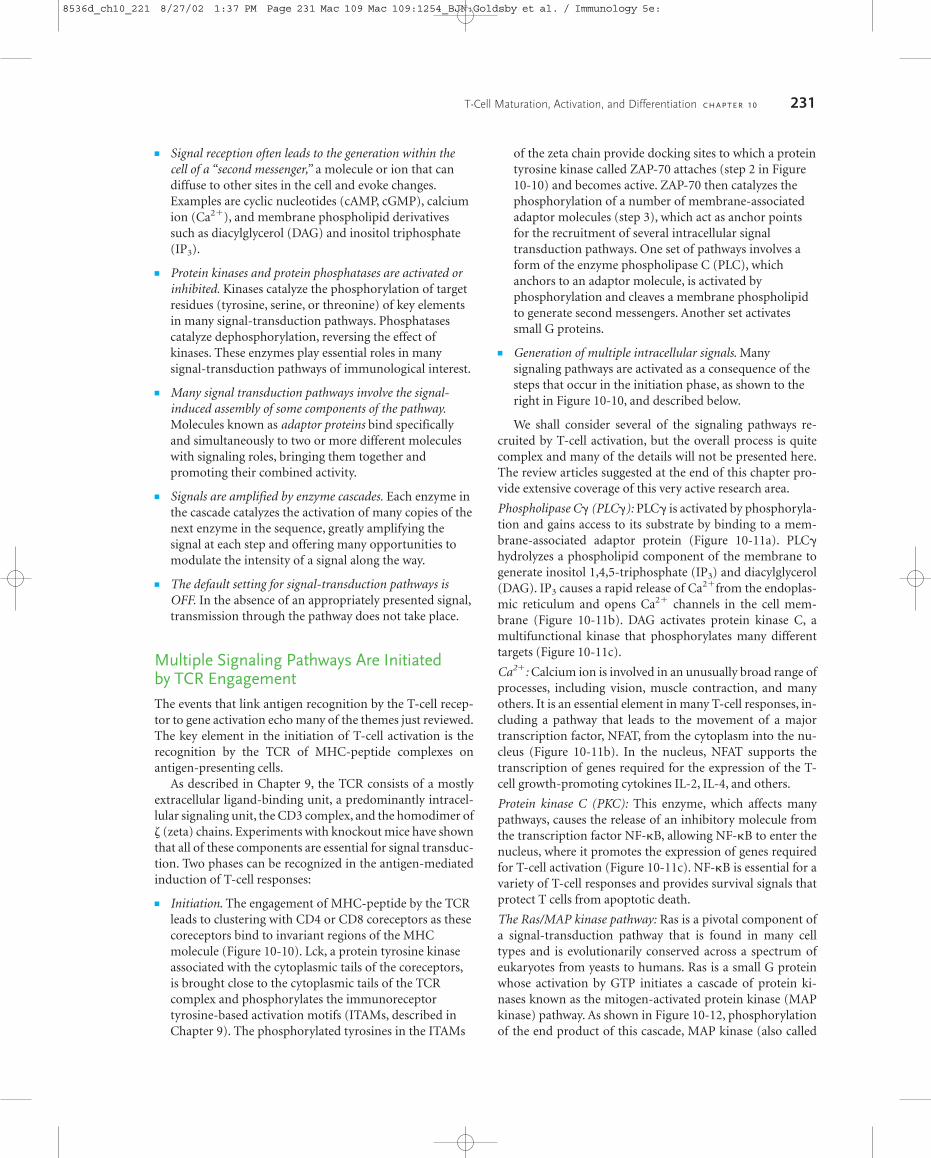
Multiple Signaling Pathways Are Initiated by TCR EngagementThe events that link antigen recognition by the T-cell recep-tor to gene activation echo many of the themes just reviewed.The key element in the initiation of T-cell activation is therecognition by the TCR of MHC-peptide complexes on antigen-presenting cells.
As described in Chapter 9, the TCR consists of a mostlyextracellular ligand-binding unit, a predominantly intracel-lular signaling unit, the CD3 complex, and the homodimer of (zeta) chains. Experiments with knockout mice have shownthat all of these components are essential for signal transduc-tion. Two phases can be recognized in the antigen-mediatedinduction of T-cell responses:
� Initiation. The engagement of MHC-peptide by the TCRleads to clustering with CD4 or CD8 coreceptors as thesecoreceptors bind to invariant regions of the MHCmolecule (Figure 10-10). Lck, a protein tyrosine kinaseassociated with the cytoplasmic tails of the coreceptors,is brought close to the cytoplasmic tails of the TCRcomplex and phosphorylates the immunoreceptortyrosine-based activation motifs (ITAMs, described inChapter 9). The phosphorylated tyrosines in the ITAMs
of the zeta chain provide docking sites to which a proteintyrosine kinase called ZAP-70 attaches (step 2 in Figure10-10) and becomes active. ZAP-70 then catalyzes thephosphorylation of a number of membrane-associatedadaptor molecules (step 3), which act as anchor pointsfor the recruitment of several intracellular signaltransduction pathways. One set of pathways involves aform of the enzyme phospholipase C (PLC), whichanchors to an adaptor molecule, is activated byphosphorylation and cleaves a membrane phospholipidto generate second messengers. Another set activatessmall G proteins.
� Generation of multiple intracellular signals. Manysignaling pathways are activated as a consequence of thesteps that occur in the initiation phase, as shown to theright in Figure 10-10, and described below.
We shall consider several of the signaling pathways re-cruited by T-cell activation, but the overall process is quitecomplex and many of the details will not be presented here.The review articles suggested at the end of this chapter pro-vide extensive coverage of this very active research area.
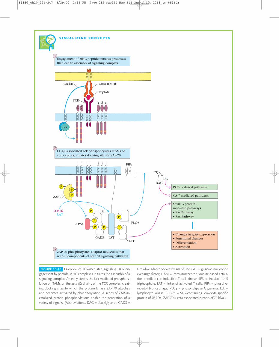
Phospholipase C� (PLC�): PLC� is activated by phosphoryla-tion and gains access to its substrate by binding to a mem-brane-associated adaptor protein (Figure 10-11a). PLC�hydrolyzes a phospholipid component of the membrane togenerate inositol 1,4,5-triphosphate (IP3) and diacylglycerol(DAG). IP3 causes a rapid release of Ca2�from the endoplas-mic reticulum and opens Ca2� channels in the cell mem-brane (Figure 10-11b). DAG activates protein kinase C, amultifunctional kinase that phosphorylates many differenttargets (Figure 10-11c).
Ca2�: Calcium ion is involved in an unusually broad range ofprocesses, including vision, muscle contraction, and manyothers. It is an essential element in many T-cell responses, in-cluding a pathway that leads to the movement of a majortranscription factor, NFAT, from the cytoplasm into the nu-cleus (Figure 10-11b). In the nucleus, NFAT supports thetranscription of genes required for the expression of the T-cell growth-promoting cytokines IL-2, IL-4, and others.
Protein kinase C (PKC): This enzyme, which affects manypathways, causes the release of an inhibitory molecule fromthe transcription factor NF-�B, allowing NF-�B to enter thenucleus, where it promotes the expression of genes requiredfor T-cell activation (Figure 10-11c). NF-�B is essential for avariety of T-cell responses and provides survival signals thatprotect T cells from apoptotic death.
The Ras/MAP kinase pathway: Ras is a pivotal component ofa signal-transduction pathway that is found in many celltypes and is evolutionarily conserved across a spectrum ofeukaryotes from yeasts to humans. Ras is a small G proteinwhose activation by GTP initiates a cascade of protein ki-nases known as the mitogen-activated protein kinase (MAPkinase) pathway. As shown in Figure 10-12, phosphorylationof the end product of this cascade, MAP kinase (also called
T-Cell Maturation, Activation, and Differentiation C H A P T E R 10 231
8536d_ch10_221 8/27/02 1:37 PM Page 231 Mac 109 Mac 109:1254_BJN:Goldsby et al. / Immunology 5e:
V I S U A L I Z I N G C O N C E P T S
FIGURE 10-10 Overview of TCR-mediated signaling. TCR en-gagement by peptide-MHC complexes initiates the assembly of asignaling complex. An early step is the Lck-mediated phosphory-lation of ITAMs on the zeta (�) chains of the TCR complex, creat-ing docking sites to which the protein kinase ZAP-70 attaches and becomes activated by phosphorylation. A series of ZAP-70-catalyzed protein phosphorylations enable the generation of a variety of signals. (Abbreviations: DAG = diacylglycerol; GADS =
Grb2-like adaptor downstream of Shc; GEF = guanine nucleotideexchange factor; ITAM = immunoreceptor tyrosine-based activa-tion motif; Itk = inducible T cell kinase; IP3 = inositol 1,4,5triphosphate; LAT = linker of activated T cells; PIP2 = phospho-inositol biphosphage; PLC� = phospholipase C gamma; Lck =lymphocyte kinase; SLP-76 = SH2-containing leukocyte-specificprotein of 76 kDa; ZAP-70 = zeta associated protein of 70 kDa.)
1Engagement of MHC-peptide initiates processes that lead to assembly of signaling complex.
CD4/8-associated Lck phosphorylates ITAMs of coreceptors, creates docking site for ZAP-70
3ZAP-70 phosphorylates adaptor molecules that recruit components of several signaling pathways
ζ ζ
γ �δ
Class II MHC
Peptide
CD4/8
TCR
P
PP
P
P
P
P
PP
Lck
ZAP-70
SLP-76LAT
LATGADS
SLP67
ItK
PLCγ
GEF
DAG
PIP2
IP3
PkC-mediated pathways
CA2+-mediated pathways
Small G-protein—mediated pathways• Ras Pathway• Rac Pathway
• Changes in gene expression• Functional changes• Differentiation• Activation
2
8536d_ch10_221-247 8/29/02 2:31 PM Page 232 mac114 Mac 114:2nd shift:1268_tm:8536d:
ERK), allows it to activate Elk, a transcription factor neces-sary for the expression of Fos. Phosphorylation of Fos byMAP kinase allows it to associate with Jun to form AP-1,which is an essential transcription factor for T-cell activation.
Co-Stimulatory Signals Are Required for Full T-Cell ActivationT-cell activation requires the dynamic interaction of multiplemembrane molecules described above, but this interaction,by itself, is not sufficient to fully activate naive T cells. NaiveT cells require more than one signal for activation and subse-quent proliferation into effector cells:
� Signal 1, the initial signal, is generated by interaction ofan antigenic peptide with the TCR-CD3 complex.
T-Cell Maturation, Activation, and Differentiation C H A P T E R 10 233
� A subsequent antigen-nonspecific co-stimulatory signal,signal 2, is provided primarily by interactions betweenCD28 on the T cell and members of the B7 family onthe APC.
There are two related forms of B7, B7-1 and B7-2 (Figure10-13). These molecules are members of the immunoglobu-lin superfamily and have a similar organization of extracel-lular domains but markedly different cytosolic domains.Both B7 molecules are constitutively expressed on dendriticcells and induced on activated macrophages and activated Bcells. The ligands for B7 are CD28 and CTLA-4 (also knownas CD152), both of which are expressed on the T-cell mem-brane as disulfide-linked homodimers; like B7, they aremembers of the immunoglobulin superfamily (Figure 10-13). Although CD28 and CTLA-4 are structurally similarglycoproteins, they act antagonistically. Signaling through
FIGURE 10-11 Signal-transduction path-ways associated with T-cell activation. (a) Phospholipase C� (PLC) is activated byphosphorylation. Active PLC hydrolyzes aphospholipid component of the plasmamembrane to generate the second mes-sengers, DAG and IP3. (b) Protein kinase C(PKC) is activated by DAG and Ca2�.Among the numerous effects of PKC isphosphorylation of IkB, a cytoplasmic pro-tein that binds the transcription factor NF-�B and prevents it from entering thenucleus. Phosphorylation of IkB releasesNF-�B, which then translocates into thenucleus. (c) Ca2�-dependent activation ofcalcineurin. Calcineurin is a Ca2�/calmod-ulin dependent phosphatase. IP3 mediatesthe release of Ca2� from the endoplasmicreticulum. Ca2� binds the protein calmod-ulin, which then associates with and acti-vates the Ca2�/calmodulin-dependentphosphatase calcineurin. Active calcine-urin removes a phosphate group fromNFAT, which allows this transcription fac-tor to translocate into the nucleus.
DAG
Cytoplasm
Nucleus
IP3
IntracellularCa2+ stores
IκB/NF-κB
NF-κB
NF-κB
IκBPhospho-lipase Cγ(inactive)
PKC
(active)ATP ADP
ATPADP
ZAP-70
+
+
+
Ca2+
Calmodulin-Ca2+
Calcineurin-calmodulin-Ca2+
(active)
CalmodulinCalcineurin(inactive)
NFAT NFAT
Transcriptional activationof several genes
NFAT
P
P
PP
(c)
(a) (b)
8536d_ch10_221-247 8/28/02 3:58 PM Page 233 mac76 mac76:385_reb:

CD28 delivers a positive co-stimulatory signal to the T cell;signaling through CTLA-4 is inhibitory and down-regulatesthe activation of the T cell. CD28 is expressed by both restingand activated T cells, but CTLA-4 is virtually undetectable onresting cells. Typically, engagement of the TCR causes the in-duction of CTLA-4 expression, and CTLA-4 is readily de-
234 P A R T I I Generation of B-Cell and T-Cell Responses
FIGURE 10-13 TH-cell activation requires a co-stimulatory signalprovided by antigen-presenting cells (APCs). Interaction of B7 familymembers on APCs with CD28 delivers the co-stimulatory signal. En-gagement of the closely related CTLA-4 molecule with B7 producesan inhibitory signal. All of these molecules contain at least one im-munoglobulin-liké domain and thus belong to the immunoglobulinsuperfamily. [Adapted from P. S. Linsley and J. A. Ledbetter, 1993,Annu. Rev. Immunol. 11:191.]
FIGURE 10-12 Activation of the small G protein, Ras. Signals fromthe T-cell receptor result in activation of Ras via the action of specificguanine nucleotide exchange factors (GEFs) that catalyze the ex-change of GDP for GTP. Active Ras causes a cascade of reactionsthat result in the increased production of the transcription factor Fos.Following their phosphorylation, Fos and Jun dimerize to yield thetranscription factor AP-1. Note that all these pathways have impor-tant effects other than the specific examples shown in the figure.
CytoplasmMAPkinasepathway
Nucleus
+
+
Pi
Ras-GDP(inactive)
Ras-GDP(active)
P
P
P
P
P
GTP
GDP
TCR-mediated signals
Raf
MEK
MAP kinase
GEFs
Elk
Elk
Jun
AP-1
Jun
Fos
Fos Fos
Transcriptionalactivation ofseveral genes
SS
S S
S S
SS S S
S S
S SS S
B7
S SS S
B7
TH cell
CD28 is expressed by bothresting and activated T cells
CTLA-4
APC
Both B7 molecules are expressed on dendritic cells, activated macrophages, and activated B cells
CD28
CTLA-4 is expressedon activated T cells
tectable within 24 hours of stimulation, with maximal ex-pression within 2 or 3 days post-stimulation. Even thoughthe peak surface levels of CTLA-4 are lower than those ofCD28, it still competes favorably for B7 molecules because ithas a significantly higher avidity for these molecules thanCD28 does. Interestingly, the level of CTLA-4 expression isincreased by CD28-generated co-stimulatory signals. Thisprovides regulatory braking via CTLA-4 in proportion to theacceleration received from CD28. Some of the importance ofCTLA-4 in the regulation of lymphocyte activation and pro-liferation is revealed by experiments with CTLA-4 knockoutmice. T cells in these mice proliferate massively, which leadsto lymphadenopathy (greatly enlarged lymph nodes),splenomegaly (enlarged spleen), and death at 3 to 4 weeks after birth. Clearly, the production of inhibitory signals byengagement of CTLA-4 is important in lymphocyte home-ostasis.
CTLA-4 can effectively block CD28 co-stimulation bycompetitive inhibition at the B7 binding site, an ability thatholds promise for clinical use in autoimmune diseases andtransplantation. As shown in Figure 10-14, an ingeniouslyengineered chimeric molecule has been designed to explorethe therapeutic potential of CTLA-4. The Fc portion ofhuman IgG has been fused to the B7-binding domain ofCTLA-4 to produce a chimeric molecule called CTLA-4Ig.The human Fc region endows the molecule with a longerhalf-life in the body and the presence of B7 binding domains
8536d_ch10_221-247 8/28/02 3:58 PM Page 234 mac76 mac76:385_reb:
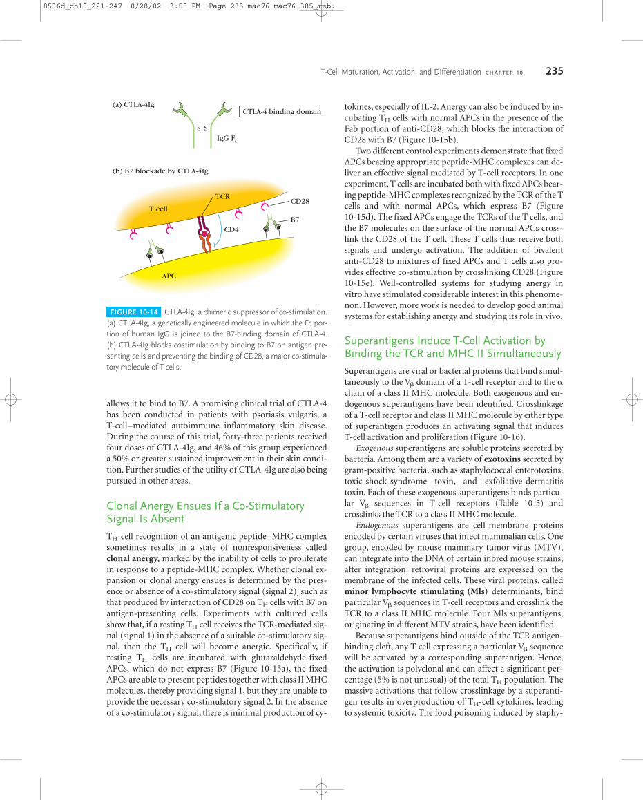
allows it to bind to B7. A promising clinical trial of CTLA-4has been conducted in patients with psoriasis vulgaris, a T-cell–mediated autoimmune inflammatory skin disease.During the course of this trial, forty-three patients receivedfour doses of CTLA-4Ig, and 46% of this group experienceda 50% or greater sustained improvement in their skin condi-tion. Further studies of the utility of CTLA-4Ig are also beingpursued in other areas.
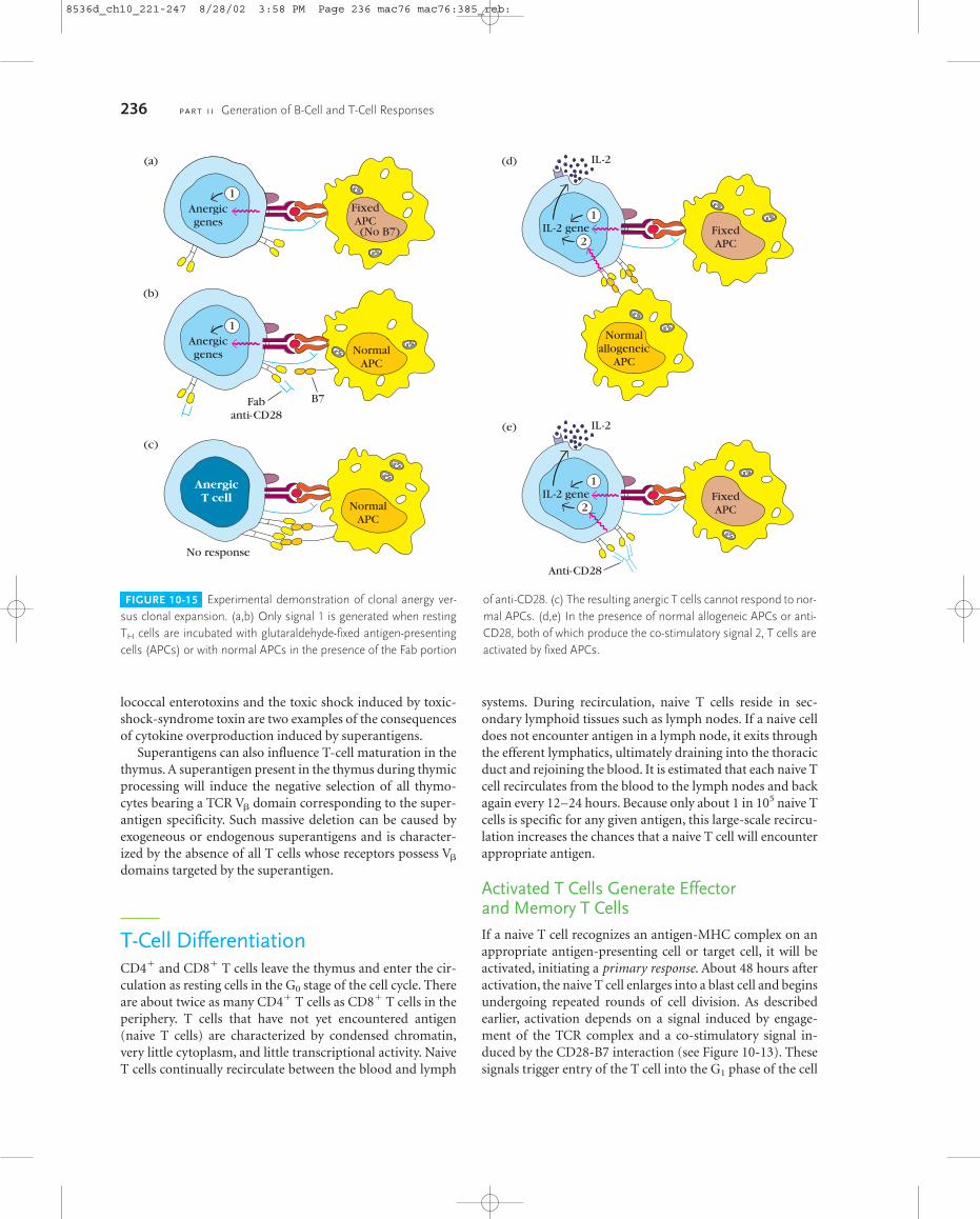
Clonal Anergy Ensues If a Co-StimulatorySignal Is AbsentTH-cell recognition of an antigenic peptide–MHC complexsometimes results in a state of nonresponsiveness calledclonal anergy, marked by the inability of cells to proliferatein response to a peptide-MHC complex. Whether clonal ex-pansion or clonal anergy ensues is determined by the pres-ence or absence of a co-stimulatory signal (signal 2), such asthat produced by interaction of CD28 on TH cells with B7 onantigen-presenting cells. Experiments with cultured cellsshow that, if a resting TH cell receives the TCR-mediated sig-nal (signal 1) in the absence of a suitable co-stimulatory sig-nal, then the TH cell will become anergic. Specifically, ifresting TH cells are incubated with glutaraldehyde-fixedAPCs, which do not express B7 (Figure 10-15a), the fixedAPCs are able to present peptides together with class II MHCmolecules, thereby providing signal 1, but they are unable toprovide the necessary co-stimulatory signal 2. In the absenceof a co-stimulatory signal, there is minimal production of cy-
T-Cell Maturation, Activation, and Differentiation C H A P T E R 10 235
FIGURE 10-14 CTLA-4Ig, a chimeric suppressor of co-stimulation.(a) CTLA-4Ig, a genetically engineered molecule in which the Fc por-tion of human IgG is joined to the B7-binding domain of CTLA-4. (b) CTLA-4Ig blocks costimulation by binding to B7 on antigen pre-senting cells and preventing the binding of CD28, a major co-stimula-tory molecule of T cells.
(a) CTLA-4Ig
(b) B7 blockade by CTLA-4Ig
CTLA-4 binding domain
S S
IgG Fc
CD4B7
CD28T cell
APC
TCR
tokines, especially of IL-2. Anergy can also be induced by in-cubating TH cells with normal APCs in the presence of theFab portion of anti-CD28, which blocks the interaction ofCD28 with B7 (Figure 10-15b).
Two different control experiments demonstrate that fixedAPCs bearing appropriate peptide-MHC complexes can de-liver an effective signal mediated by T-cell receptors. In oneexperiment, T cells are incubated both with fixed APCs bear-ing peptide-MHC complexes recognized by the TCR of the Tcells and with normal APCs, which express B7 (Figure 10-15d). The fixed APCs engage the TCRs of the T cells, andthe B7 molecules on the surface of the normal APCs cross-link the CD28 of the T cell. These T cells thus receive bothsignals and undergo activation. The addition of bivalentanti-CD28 to mixtures of fixed APCs and T cells also pro-vides effective co-stimulation by crosslinking CD28 (Figure10-15e). Well-controlled systems for studying anergy invitro have stimulated considerable interest in this phenome-non. However, more work is needed to develop good animal systems for establishing anergy and studying its role in vivo.
Superantigens Induce T-Cell Activation byBinding the TCR and MHC II SimultaneouslySuperantigens are viral or bacterial proteins that bind simul-taneously to the V� domain of a T-cell receptor and to the �chain of a class II MHC molecule. Both exogenous and en-dogenous superantigens have been identified. Crosslinkageof a T-cell receptor and class II MHC molecule by either typeof superantigen produces an activating signal that inducesT-cell activation and proliferation (Figure 10-16).
Exogenous superantigens are soluble proteins secreted bybacteria. Among them are a variety of exotoxins secreted bygram-positive bacteria, such as staphylococcal enterotoxins,toxic-shock-syndrome toxin, and exfoliative-dermatitistoxin. Each of these exogenous superantigens binds particu-lar V� sequences in T-cell receptors (Table 10-3) andcrosslinks the TCR to a class II MHC molecule.
Endogenous superantigens are cell-membrane proteinsencoded by certain viruses that infect mammalian cells. Onegroup, encoded by mouse mammary tumor virus (MTV),can integrate into the DNA of certain inbred mouse strains;after integration, retroviral proteins are expressed on themembrane of the infected cells. These viral proteins, calledminor lymphocyte stimulating (Mls) determinants, bindparticular V� sequences in T-cell receptors and crosslink theTCR to a class II MHC molecule. Four Mls superantigens,originating in different MTV strains, have been identified.
Because superantigens bind outside of the TCR antigen-binding cleft, any T cell expressing a particular V� sequencewill be activated by a corresponding superantigen. Hence,the activation is polyclonal and can affect a significant per-centage (5% is not unusual) of the total TH population. Themassive activations that follow crosslinkage by a superanti-gen results in overproduction of TH-cell cytokines, leadingto systemic toxicity. The food poisoning induced by staphy-
8536d_ch10_221-247 8/28/02 3:58 PM Page 235 mac76 mac76:385_reb:
lococcal enterotoxins and the toxic shock induced by toxic-shock-syndrome toxin are two examples of the consequencesof cytokine overproduction induced by superantigens.
Superantigens can also influence T-cell maturation in thethymus. A superantigen present in the thymus during thymicprocessing will induce the negative selection of all thymo-cytes bearing a TCR V� domain corresponding to the super-antigen specificity. Such massive deletion can be caused byexogeneous or endogenous superantigens and is character-ized by the absence of all T cells whose receptors possess V�
domains targeted by the superantigen.
T-Cell DifferentiationCD4� and CD8� T cells leave the thymus and enter the cir-culation as resting cells in the G0 stage of the cell cycle. Thereare about twice as many CD4� T cells as CD8� T cells in theperiphery. T cells that have not yet encountered antigen(naive T cells) are characterized by condensed chromatin,very little cytoplasm, and little transcriptional activity. NaiveT cells continually recirculate between the blood and lymph
systems. During recirculation, naive T cells reside in sec-ondary lymphoid tissues such as lymph nodes. If a naive celldoes not encounter antigen in a lymph node, it exits throughthe efferent lymphatics, ultimately draining into the thoracicduct and rejoining the blood. It is estimated that each naive Tcell recirculates from the blood to the lymph nodes and backagain every 12–24 hours. Because only about 1 in 105 naive Tcells is specific for any given antigen, this large-scale recircu-lation increases the chances that a naive T cell will encounterappropriate antigen.
Activated T Cells Generate Effector and Memory T CellsIf a naive T cell recognizes an antigen-MHC complex on anappropriate antigen-presenting cell or target cell, it will beactivated, initiating a primary response. About 48 hours afteractivation, the naive T cell enlarges into a blast cell and beginsundergoing repeated rounds of cell division. As describedearlier, activation depends on a signal induced by engage-ment of the TCR complex and a co-stimulatory signal in-duced by the CD28-B7 interaction (see Figure 10-13). Thesesignals trigger entry of the T cell into the G1 phase of the cell
236 P A R T I I Generation of B-Cell and T-Cell Responses
FIGURE 10-15 Experimental demonstration of clonal anergy ver-sus clonal expansion. (a,b) Only signal 1 is generated when restingTH cells are incubated with glutaraldehyde-fixed antigen-presentingcells (APCs) or with normal APCs in the presence of the Fab portion
of anti-CD28. (c) The resulting anergic T cells cannot respond to nor-mal APCs. (d,e) In the presence of normal allogeneic APCs or anti-CD28, both of which produce the co-stimulatory signal 2, T cells areactivated by fixed APCs.
(a)
(b)
NormalAPC
Fabanti-CD28
B7
(c)
NormalAPC
No response
(d)
FixedAPC
1Anergicgenes
1Anergicgenes
AnergicT cell
1IL-2 gene
2
Normalallogeneic
APC
IL-2
(e)
FixedAPC
1IL-2 gene
2
IL-2
Anti-CD28
FixedAPC(No B7)
8536d_ch10_221-247 8/28/02 3:58 PM Page 236 mac76 mac76:385_reb:

cycle and, at the same time, induce transcription of the genefor IL-2 and the � chain of the high-affinity IL-2 receptor. Inaddition, the co-stimulatory signal increases the half-life ofthe IL-2 mRNA. The increase in IL-2 transcription, togetherwith stabilization of the IL-2 mRNA, increases IL-2 produc-
tion by 100-fold in the activated T cell. Secretion of IL-2 andits subsequent binding to the high-affinity IL-2 receptor in-duces the activated naive T cell to proliferate and differenti-ate (Figure 10-17). T cells activated in this way divide 2–3times per day for 4–5 days, generating a large clone of prog-eny cells, which differentiate into memory or effector T-cellpopulations.
The various effector T cells carry out specialized functionssuch as cytokine secretion and B-cell help (activated CD4� TH
cells) and cytotoxic killing activity (CD8� CTLs). The genera-tion and activity of CTL cells are described in detail in Chapter14. Effector cells are derived from both naive and memory cellsafter antigen activation. Effector cells are short-lived cells,whose life spans range from a few days to a few weeks. The ef-fector and naive populations express different cell-membranemolecules, which contribute to different recirculation patterns.
As described in more detail in Chapter 12, CD4� effectorT cells form two subpopulations distinguished by the differ-ent panels of cytokines they secrete. One population, calledthe TH1 subset, secretes IL-2, IFN-�, and TNF-�. The TH1subset is responsible for classic cell-mediated functions, suchas delayed-type hypersensitivity and the activation of cyto-toxic T lymphocytes. The other subset, called the TH2 subset,secretes IL-4, IL-5, IL-6, and IL-10. This subset functionsmore effectively as a helper for B-cell activation.
The memory T-cell population is derived from both naiveT cells and from effector cells after they have encounteredantigen. Memory T cells are antigen-generated, generally
T-Cell Maturation, Activation, and Differentiation C H A P T E R 10 237
FIGURE 10-16 Superantigen-mediated crosslinkage of T-cell re-ceptor and class II MHC molecules. A superantigen binds to all TCRsbearing a particular V� sequence regardless of their antigenic speci-ficity. Exogenous superantigens are soluble secreted bacterial pro-teins, including various exotoxins. Endogenous superantigens aremembrane-embedded proteins produced by certain viruses; they in-clude Mls antigens encoded by mouse mammary tumor virus.
APC
TCR
Peptide for whichTCR is not specific
Class II MHC
TH cell
Vβ
Superantigen
Endogenoussuperantigen ismembrane-bound
αβ
βα
TABLE 10-3 Exogenous superantigens and their V� specificity
V� SPECIFICITY
Superantigen Disease∗ Mouse Human
Staphylococcal enterotoxins
SEA Food poisoning 1, 3, 10, 11, 12, 17 nd
SEB Food poisoning 3, 8.1, 8.2, 8.3 3, 12, 14, 15, 17, 20
SEC1 Food poisoning 7, 8.2, 8.3, 11 12
SEC2 Food poisoning 8.2, 10 12, 13, 14, 15, 17, 20
SEC3 Food poisoning 7, 8.2 5, 12
SED Food poisoning 3, 7, 8.3, 11, 17 5, 12
SEE Food poisoning 11, 15, 17 5.1, 6.1–6.3, 8, 18
Toxic-shock-syndrome toxin (TSST1) Toxic-shock syndrome 15, 16 2
Exfoliative-dermatitis toxin (ExFT) Scalded-skin syndrome 10, 11, 15 2
Mycoplasma-arthritidis supernatant Arthritis, shock 6, 8.1–8.3 nd(MAS)
Streptococcal pyrogenic exotoxins Rheumatic fever, shock nd nd(SPE-A, B, C, D)
∗Disease results from infection by bacteria that produce the indicated superantigens.
8536d_ch10_221-247 8/28/02 3:58 PM Page 237 mac76 mac76:385_reb:

long-lived, quiescent cells that respond with heightened reac-tivity to a subsequent challenge with the same antigen, gen-erating a secondary response. An expanded population ofmemory T cells appears to remain long after the populationof effector T cells has declined. In general, memory T cellsexpress many of the same cell-surface markers as effector Tcells; no cell-surface markers definitively identify them asmemory cells.
Like naive T cells, most memory T cells are resting cells inthe G0 stage of the cell cycle, but they appear to have lessstringent requirements for activation than naive T cells do.For example, naive TH cells are activated only by dendriticcells, whereas memory TH cells can be activated bymacrophages, dendritic cells, and B cells. It is thought thatthe expression of high levels of numerous adhesion mole-cules by memory TH cells enables these cells to adhere to abroad spectrum of antigen-presenting cells. Memory cellsalso display recirculation patterns that differ from those ofnaive or effector T cells.
A CD4+CD25+ Subpopulation of T cellsNegatively Regulates Immune ResponsesInvestigators first described T cell populations that could sup-press immune responses during the early 1970s. These cellswere called suppressor T cells (Ts) and were believed to beCD8+ T cells. However, the cellular and molecular basis of theobserved suppression remained obscure, and eventually greatdoubt was cast on the existence of CD8+ suppressor T cells.Recent research has shown that there are indeed T cells thatsuppress immune responses. Unexpectedly, these cells haveturned out to be CD4+ rather than CD8+ T cells. Within thepopulation of CD4+CD25+ T cells, there are regulatory T cellsthat can inhibit the proliferation of other T cell populations invitro. Animal studies show that members of the CD4+CD25+
population inhibit the development of autoimmune diseasessuch as experimentally induced inflammatory bowel disease,experimental allergic encephalitis, and autoimmune diabetes.The suppression by these regulatory cells is antigen specificbecause it depends upon activation through the T cell recep-tor. Cell contact between the suppressing cells and their tar-gets is required. If the regulatory cells are activated by antigenbut separated from their targets by a permeable barrier, nosuppression occurs. The existence of regulatory T cells thatspecifically suppress immune responses has clinical implica-tions. The depletion or inhibition of regulatory T cells fol-lowed by immunization may enhance the immune responsesto conventional vaccines. In this regard, some have suggestedthat elimination of T cells that suppress responses to tumorantigens may facilitate the development of anti-tumor immu-nity. Conversely, increasing the suppressive activity of regula-tory T cell populations could be useful in the treatment ofallergic or autoimmune diseases. The ability to increase theactivity of regulatory T cell populations might also be usefulin suppressing organ and tissue rejection. Future work on thisregulatory cell population will seek deeper insights into themechanisms by which members of CD4+CD25+ T cell popu-lations regulate immune responses. There will also be deter-mined efforts to discover ways in which the activities of thesepopulations can be increased to diminish unwanted immuneresponses and decreased to promote desirable ones.
Antigen-Presenting Cells Have CharacteristicCo-Stimulatory PropertiesOnly professional antigen-presenting cells (dendritic cells,macrophages, and B cells) are able to present antigen to-gether with class II MHC molecules and deliver the co-stim-ulatory signal necessary for complete T-cell activation thatleads to proliferation and differentiation. The principal co-stimulatory molecules expressed on antigen-presenting cellsare the glycoproteins B7-1 and B7-2 (see Figure 10-13). Theprofessional antigen-presenting cells differ in their ability todisplay antigen and also differ in their ability to deliver theco-stimulatory signal (Figure 10-18).
238 P A R T I I Generation of B-Cell and T-Cell Responses
FIGURE 10-17 Activation of a TH cell by both signal 1 and co-stimulatory signal 2 up-regulates expression of IL-2 and the high-affinity IL-2 receptor, leading to the entry of the T cell into the cell cycle and several rounds of proliferation. Some of the cells differenti-ate into effector cells, others into memory cells.
CD28 B7
IL-2
IL-2 geneIL-2R gene
NormalAPC
1
2
IL-2
IL-2receptor
Activation
Population of memory and effector cells
Severaldivisions
G1
G2
M S
M M E E E E
8536d_ch10_221-247 8/28/02 3:58 PM Page 238 mac76 mac76:385_reb:

Dendritic cells constitutively express high levels of class Iand class II MHC molecules as well as high levels of B7-1 andB7-2. For this reason, dendritic cells are very potent activa-tors of naive, memory, and effector T cells. In contrast, allother professional APCs require activation for expression ofco-stimulatory B7 molecules on their membranes; conse-quently, resting macrophages are not able to activate naive Tcells and are poor activators of memory and effector T cells.Macrophages can be activated by phagocytosis of bacteria orby bacterial products such as LPS or by IFN-�, a TH1-derivedcytokine. Activated macrophages up-regulate their expres-sion of class II MHC molecules and co-stimulatory B7 mole-cules. Thus, activated macrophages are common activators ofmemory and effector T cells, but their effectiveness in acti-vating naive T cells is considered minimal.
B cells also serve as antigen-presenting cells in T-cell acti-vation. Resting B cells express class II MHC molecules but failto express co-stimulatory B7 molecules. Consequently, rest-ing B cells cannot activate naive T cells, although they can ac-tivate the effector and memory T-cell populations. Uponactivation, B cells up-regulate their expression of class IIMHC molecules and begin expressing B7. These activated Bcells can now activate naive T cells as well as the memory andeffector populations.
Cell Death and T-Cell PopulationsCell death is an important feature of development in all multicellular organisms. During fetal life it is used to moldand sculpt, removing unnecessary cells to provide shape and form. It also is an important feature of lymphocytehomeostasis, returning T- and B-cell populations to their ap-propriate levels after bursts of antigen-induced proliferation.Apoptosis also plays a crucial role in the deletion of poten-tially autoreactive thymocytes during negative selection andin the removal of developing T cells unable to recognize self(failure to undergo positive selection).
Although the induction of apoptosis involves differentsignals depending on the cell types involved, the actual deathof the cell is a highly conserved process amongst vertebratesand invertebrates. For example, T cells may be induced to dieby many different signals, including the withdrawal ofgrowth factor, treatment with glucocorticoids, or TCR sig-naling. Each of these signals engages unique signaling path-ways, but in all cases, the actual execution of the cell involvesthe activation of a specialized set of proteases known as cas-pases. The role of these proteases was first revealed by studiesof developmentally programmed cell deaths in the nematode
T-Cell Maturation, Activation, and Differentiation C H A P T E R 10 239
FIGURE 10-18 Differences in the properties of professional antigen-presenting cells affect their ability to present antigen and
induce T-cell activation. Note that activation of effector and memoryT cells does not require the co-stimulatory B7 molecule.
Antigen uptake
Class II MHC expression
Co-stimulatoryactivity
T-cell activation
Endocytosisphagocytosis(by Langerhans cells)
Phagocytosis
Inducible(−)
Inducible B7(−)
Inducible( + + )
Inducible B7( + + )
Effector T cellsMemory T cells
Phagocytosis Receptor-mediatedendocytosis
Receptor-mediatedendocytosis
Constitutive( + + )
Constitutive(+ + + )
Constitutive B7( + + + )
Naive T cellsEffector T cellsMemory T cells
Constitutive( + + + )
Inducible B7(−)
Inducible B7( + + )
Effector T cellsMemory T cells
Naive T cellsEffector T cellsMemory T cells
RestingResting
Class IIMHC Class II
MHC
Class IIMHC
Class IMHC
Class IMHC
Class IMHC
Class IMHC
B7
Activated
B Lymphocyte
Activated
Dendritic cell
B7
Class IIMHC
Class IMHC
Macrophage
LPS
INF-γ
B7
(−)
8536d_ch10_221-247 8/28/02 3:58 PM Page 239 mac76 mac76:385_reb:
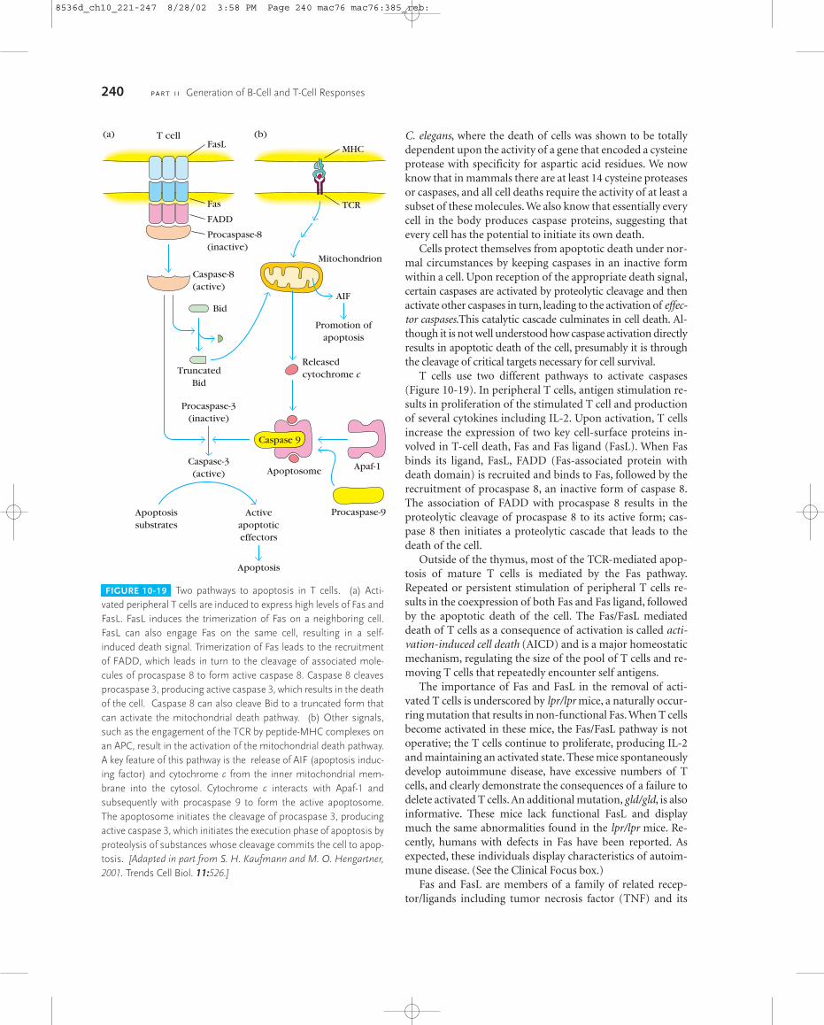
C. elegans, where the death of cells was shown to be totallydependent upon the activity of a gene that encoded a cysteineprotease with specificity for aspartic acid residues. We nowknow that in mammals there are at least 14 cysteine proteasesor caspases, and all cell deaths require the activity of at least asubset of these molecules. We also know that essentially everycell in the body produces caspase proteins, suggesting thatevery cell has the potential to initiate its own death.
Cells protect themselves from apoptotic death under nor-mal circumstances by keeping caspases in an inactive formwithin a cell. Upon reception of the appropriate death signal,certain caspases are activated by proteolytic cleavage and thenactivate other caspases in turn, leading to the activation of effec-tor caspases.This catalytic cascade culminates in cell death. Al-though it is not well understood how caspase activation directlyresults in apoptotic death of the cell, presumably it is throughthe cleavage of critical targets necessary for cell survival.
T cells use two different pathways to activate caspases(Figure 10-19). In peripheral T cells, antigen stimulation re-sults in proliferation of the stimulated T cell and productionof several cytokines including IL-2. Upon activation, T cellsincrease the expression of two key cell-surface proteins in-volved in T-cell death, Fas and Fas ligand (FasL). When Fasbinds its ligand, FasL, FADD (Fas-associated protein withdeath domain) is recruited and binds to Fas, followed by therecruitment of procaspase 8, an inactive form of caspase 8.The association of FADD with procaspase 8 results in theproteolytic cleavage of procaspase 8 to its active form; cas-pase 8 then initiates a proteolytic cascade that leads to thedeath of the cell.
Outside of the thymus, most of the TCR-mediated apop-tosis of mature T cells is mediated by the Fas pathway.Repeated or persistent stimulation of peripheral T cells re-sults in the coexpression of both Fas and Fas ligand, followedby the apoptotic death of the cell. The Fas/FasL mediateddeath of T cells as a consequence of activation is called acti-vation-induced cell death (AICD) and is a major homeostaticmechanism, regulating the size of the pool of T cells and re-moving T cells that repeatedly encounter self antigens.
The importance of Fas and FasL in the removal of acti-vated T cells is underscored by lpr/lpr mice, a naturally occur-ring mutation that results in non-functional Fas. When T cellsbecome activated in these mice, the Fas/FasL pathway is notoperative; the T cells continue to proliferate, producing IL-2and maintaining an activated state. These mice spontaneouslydevelop autoimmune disease, have excessive numbers of Tcells, and clearly demonstrate the consequences of a failure todelete activated T cells. An additional mutation, gld/gld, is alsoinformative. These mice lack functional FasL and displaymuch the same abnormalities found in the lpr/lpr mice. Re-cently, humans with defects in Fas have been reported. As expected, these individuals display characteristics of autoim-mune disease. (See the Clinical Focus box.)
Fas and FasL are members of a family of related recep-tor/ligands including tumor necrosis factor (TNF) and its
240 P A R T I I Generation of B-Cell and T-Cell Responses
FIGURE 10-19 Two pathways to apoptosis in T cells. (a) Acti-vated peripheral T cells are induced to express high levels of Fas andFasL. FasL induces the trimerization of Fas on a neighboring cell.FasL can also engage Fas on the same cell, resulting in a self-induced death signal. Trimerization of Fas leads to the recruitmentof FADD, which leads in turn to the cleavage of associated mole-cules of procaspase 8 to form active caspase 8. Caspase 8 cleavesprocaspase 3, producing active caspase 3, which results in the deathof the cell. Caspase 8 can also cleave Bid to a truncated form thatcan activate the mitochondrial death pathway. (b) Other signals,such as the engagement of the TCR by peptide-MHC complexes onan APC, result in the activation of the mitochondrial death pathway.A key feature of this pathway is the release of AIF (apoptosis induc-ing factor) and cytochrome c from the inner mitochondrial mem-brane into the cytosol. Cytochrome c interacts with Apaf-1 andsubsequently with procaspase 9 to form the active apoptosome.The apoptosome initiates the cleavage of procaspase 3, producingactive caspase 3, which initiates the execution phase of apoptosis byproteolysis of substances whose cleavage commits the cell to apop-tosis. [Adapted in part from S. H. Kaufmann and M. O. Hengartner,2001. Trends Cell Biol. 11:526.]
(a) (b)T cell
Fas TCR
MHC
FADD
Procaspase-8(inactive)
Caspase-8(active)
FasL
Mitochondrion
Procaspase-3(inactive)
Caspase-3(active)
Apoptosissubstrates
Apoptosis
Activeapoptoticeffectors
Caspase 9
Procaspase-9
Apoptosome
Releasedcytochrome c
Apaf-1
Promotion ofapoptosis
AIFBid
TruncatedBid
8536d_ch10_221-247 8/28/02 3:58 PM Page 240 mac76 mac76:385_reb:
ligand, TNFR (tumor necrosis factor receptor). Like Fas andFasL, membrane-bound TNFR interacts with TNF to induceapoptosis. Also similar to Fas/FasL-induced apoptosis,TNF/TNFR-induced death is the result of the activation ofcaspase 8 followed by the activation of effector caspases suchas caspase 3.
In addition to the activation of apoptosis through deathreceptor proteins like Fas and TNFR, T cells can die throughother pathways that do not activate procaspase 8. For exam-ple, negative selection in the thymus induces the apoptoticdeath of developing T cells via a signaling pathway that orig-inates at the TCR. We still do not completely understand whysome signals through the TCR induce positive selection andothers induce negative selection, but we know that thestrength of the signal plays a critical role. A strong, negativelyselecting signal induces a route to apoptosis in which mitochondria play a central role. In mitochondrially depen-dent apoptotic pathways, cytochrome c, which normally resides in the inner mitochondrial membrane, leaks into thecytosol. Cytochrome c binds to a protein known as Apaf-1(apoptotic protease-activating factor-1) and undergoes anATP-dependent conformational change and oligomeriza-tion. Binding of the oligomeric form of Apaf-1 to procaspase9 results in its transformation to active caspase 9. The com-plex of cytochrome c/Apaf-1/caspase 9, called the apopto-some, proteolytically cleaves procaspase 3 generating activecaspase 3, which initiates a cascade of reactions that kills thecell (Figure 10-19). Finally, mitochondria also release an-other molecule, AIF (apoptosis inducing factor), which playsa role in the induction of cell death.
Cell death induced by Fas/FasL is swift, with rapid activa-tion of the caspase cascade leading to cell death in 2–4 hours.On the other hand, TCR-induced negative selection appears to be a more circuitous process, requiring the activation of sev-eral processes including mitochondrial membrane failure, therelease of cytochrome c, and the formation of the apopto-some before caspases become involved. Consequently, TCR-mediated negative selection can take as long as 8–10 hours.
An important feature in the mitochondrially induced celldeath pathway is the regulatory role played by Bcl-2 familymembers. Bcl-2 and Bcl-XL both reside in the mitochondr-ial membrane. These proteins are strong inhibitors of apop-tosis, and while it is not clear how they inhibit cell death, onehypothesis is that they somehow regulate the release of cy-tochrome c from the mitochondria. There are at least threegroups of Bcl-2 family members. Group I members are anti-apoptotic and include Bcl-2 and Bcl-xL. Group II and GroupIII members are pro-apoptotic and include Bax and Bak inGroup II and Bid and Bim in Group III. There is clear evi-dence that levels of anti-apoptotic Bcl-2 family members playan important role in regulating apoptosis in lymphocytes.Bcl-2 family members dimerize, and the anti-apoptoticgroup members may control apoptosis by dimerizing withpro-apoptotic members, blocking their activity. As indicatedin Figure 10-19, cleavage of Bid, catalyzed by caspase 8 gen-
erated by the Fas pathway, can turn on the mitochondrialpathway. Thus signals initiated through Fas can also involvethe mitochondrial death pathway.
While it is apparent there are several ways a lymphocytecan be signaled to die, all of these pathways to cell death con-verge upon the activation of caspases. This part of the cell-death pathway, the execution phase, is common to almost alldeath pathways known in both vertebrates and invertebrates,demonstrating that apoptosis is an ancient process that hasbeen conserved throughout evolution.
Peripheral �� T CellsIn 1986, a small population of peripheral-blood T cells wasdiscovered that expressed CD3 but failed to stain with mon-oclonal antibody specific for the �� T-cell receptor, indicat-ing an absence of the �� heterodimer. Many of these cellseventually were found to express the �� receptor.
gd T Cells Are Far Less Pervasive Than ab T CellsIn humans, less than 5% of T cells bear the �� heterodimer,and the percentage of �� T cells in the lymphoid organs ofmice has been reported to range from 1% to 3%. In additionto their presence in blood and lymphoid tissues, they also ap-pear in the skin, intestinal epithelium, and pulmonary epithe-lium. Up to 1% of the epidermal cells in the skin of mice are�� T cells. In general, �� T cells are not MHC-restricted, andmost do not express the coreceptors CD4 and CD8 present onpopulations of �� T cells. Although the potential of the � and� TCR loci to generate diversity is great, very little diversity isfound in this type of T cell. In fact, as pointed out in Chapter9, most of the �� T cells in humans have an identical combi-nation of �� chains (�9 and �2).
�� T Cells Recognize Nonpeptide LigandsNot all T cells are self-MHC restricted and recognize onlypeptide antigens displayed in the cleft of the self-MHC mol-ecule. Indeed, Chapters 2 and 8 describe �� TCR-bearing Tcells (NK1-T cells and CD1-restricted T cells) that are not re-stricted by conventional MHC molecules. In one study, a ��T-cell clone was found to bind directly to a herpes-virus pro-tein without requiring antigen processing and presentationtogether with MHC. Human �� T cells have been reportedthat display MHC-independent binding of a phospholipidderived from M. tuberculosis, the organism responsible fortuberculosis (see Chapter 9). This finding suggests that inmany cases the TCR receptors of �� T cells bind to epitopesin much the same way that the immunoglobulin receptors of B cells do. The fact that most human �� T cells all have the same specificity suggests that like other components ofthe innate immune system, they recognize and respond to
T-Cell Maturation, Activation, and Differentiation C H A P T E R 10 241
8536d_ch10_221-247 8/29/02 10:23 AM Page 241 mac114 Mac 114:2nd shift:1268_tm:8536d:
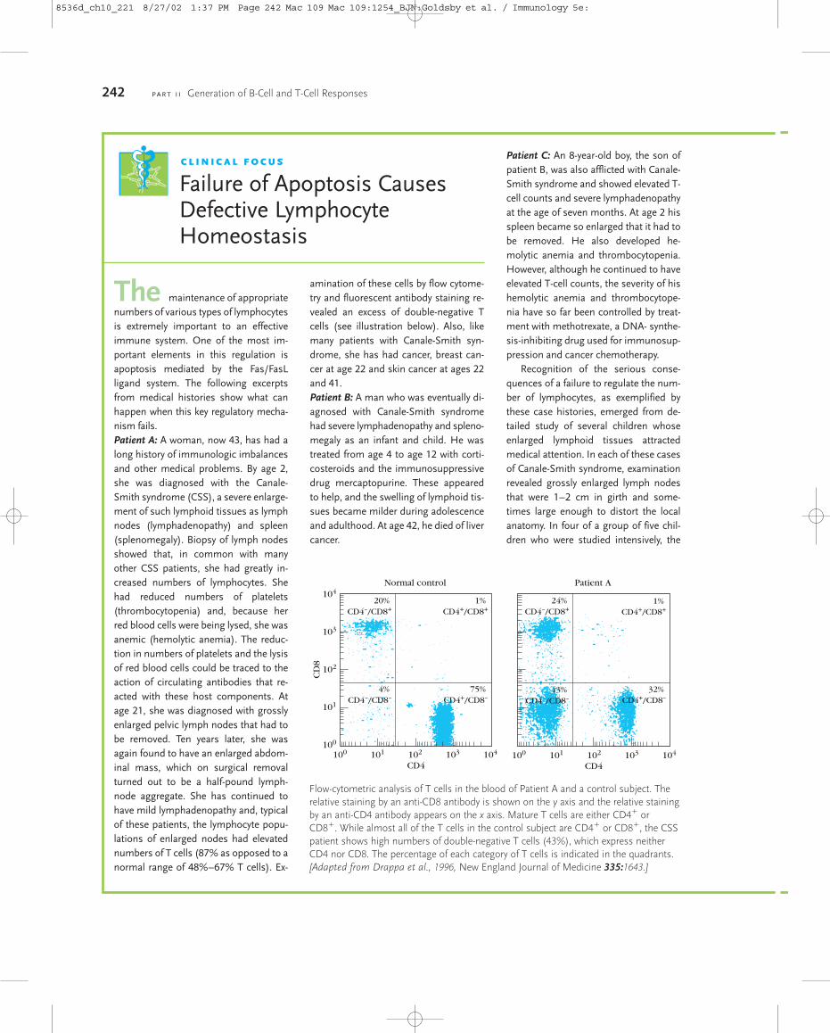
242 P A R T I I Generation of B-Cell and T-Cell Responses
amination of these cells by flow cytome-try and fluorescent antibody staining re-vealed an excess of double-negative Tcells (see illustration below). Also, likemany patients with Canale-Smith syn-drome, she has had cancer, breast can-cer at age 22 and skin cancer at ages 22and 41.Patient B: A man who was eventually di-agnosed with Canale-Smith syndromehad severe lymphadenopathy and spleno-megaly as an infant and child. He wastreated from age 4 to age 12 with corti-costeroids and the immunosuppressivedrug mercaptopurine. These appearedto help, and the swelling of lymphoid tis-sues became milder during adolescenceand adulthood. At age 42, he died of livercancer.
Patient C: An 8-year-old boy, the son ofpatient B, was also afflicted with Canale-Smith syndrome and showed elevated T-cell counts and severe lymphadenopathyat the age of seven months. At age 2 hisspleen became so enlarged that it had tobe removed. He also developed he-molytic anemia and thrombocytopenia.However, although he continued to haveelevated T-cell counts, the severity of hishemolytic anemia and thrombocytope-nia have so far been controlled by treat-ment with methotrexate, a DNA- synthe-sis-inhibiting drug used for immunosup-pression and cancer chemotherapy.
Recognition of the serious conse-quences of a failure to regulate the num-ber of lymphocytes, as exemplified bythese case histories, emerged from de-tailed study of several children whose enlarged lymphoid tissues attractedmedical attention. In each of these casesof Canale-Smith syndrome, examinationrevealed grossly enlarged lymph nodesthat were 1–2 cm in girth and some-times large enough to distort the localanatomy. In four of a group of five chil-dren who were studied intensively, the
The maintenance of appropriatenumbers of various types of lymphocytesis extremely important to an effective immune system. One of the most im-portant elements in this regulation isapoptosis mediated by the Fas/FasL ligand system. The following excerptsfrom medical histories show what canhappen when this key regulatory mecha-nism fails.Patient A: A woman, now 43, has had along history of immunologic imbalancesand other medical problems. By age 2,she was diagnosed with the Canale-Smith syndrome (CSS), a severe enlarge-ment of such lymphoid tissues as lymphnodes (lymphadenopathy) and spleen(splenomegaly). Biopsy of lymph nodesshowed that, in common with manyother CSS patients, she had greatly in-creased numbers of lymphocytes. Shehad reduced numbers of platelets(thrombocytopenia) and, because herred blood cells were being lysed, she wasanemic (hemolytic anemia). The reduc-tion in numbers of platelets and the lysisof red blood cells could be traced to theaction of circulating antibodies that re-acted with these host components. Atage 21, she was diagnosed with grosslyenlarged pelvic lymph nodes that had tobe removed. Ten years later, she wasagain found to have an enlarged abdom-inal mass, which on surgical removalturned out to be a half-pound lymph-node aggregate. She has continued tohave mild lymphadenopathy and, typicalof these patients, the lymphocyte popu-lations of enlarged nodes had elevatednumbers of T cells (87% as opposed to anormal range of 48%–67% T cells). Ex-
C L I N I C A L F O C U S
Failure of Apoptosis CausesDefective LymphocyteHomeostasis
CD4 CD4
CD
8
103
104
102
101
100
101
Normal control Patient A
100 100102 103 104
20% CD4–/CD8+
1% CD4+/CD8+
4% CD4–/CD8–
75% CD4+/CD8–
101 102 103 104
24% CD4–/CD8+
1% CD4+/CD8+
43% CD4–/CD8–
32% CD4+/CD8–
Flow-cytometric analysis of T cells in the blood of Patient A and a control subject. Therelative staining by an anti-CD8 antibody is shown on the y axis and the relative stainingby an anti-CD4 antibody appears on the x axis. Mature T cells are either CD4� orCD8�. While almost all of the T cells in the control subject are CD4� or CD8�, the CSSpatient shows high numbers of double-negative T cells (43%), which express neitherCD4 nor CD8. The percentage of each category of T cells is indicated in the quadrants.[Adapted from Drappa et al., 1996, New England Journal of Medicine 335:1643.]
8536d_ch10_221 8/27/02 1:37 PM Page 242 Mac 109 Mac 109:1254_BJN:Goldsby et al. / Immunology 5e:
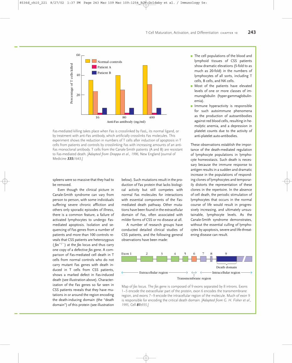
T-Cell Maturation, Activation, and Differentiation C H A P T E R 10 243
below). Such mutations result in the pro-duction of Fas protein that lacks biologi-cal activity but still competes withnormal Fas molecules for interactionswith essential components of the Fas-mediated death pathway. Other muta-tions have been found in the extracellulardomain of Fas, often associated withmilder forms of CSS or no disease at all.
A number of research groups haveconducted detailed clinical studies ofCSS patients, and the following generalobservations have been made:
� The cell populations of the blood andlymphoid tissues of CSS patientsshow dramatic elevations (5-fold to asmuch as 20-fold) in the numbers oflymphocytes of all sorts, including Tcells, B cells, and NK cells.
� Most of the patients have elevated levels of one or more classes of im-munoglobulin (hyper-gammaglobulin-emia).
� Immune hyperactivity is responsiblefor such autoimmune phenomena as the production of autoantibodiesagainst red blood cells, resulting in he-molytic anemia, and a depression inplatelet counts due to the activity ofanti-platelet auto-antibodies.
These observations establish the impor-tance of the death-mediated regulationof lymphocyte populations in lympho-cyte homeostasis. Such death is neces-sary because the immune response toantigen results in a sudden and dramaticincrease in the populations of respond-ing clones of lymphocytes and temporar-ily distorts the representation of theseclones in the repertoire. In the absenceof cell death, the periodic stimulation oflymphocytes that occurs in the normalcourse of life would result in progres-sively increasing, and ultimately unsus-tainable, lymphocyte levels. As theCanale-Smith syndrome demonstrates,without the essential culling of lympho-cytes by apoptosis, severe and life-threat-ening disease can result.
spleens were so massive that they had tobe removed.
Even though the clinical picture inCanale-Smith syndrome can vary fromperson to person, with some individualssuffering severe chronic affliction andothers only sporadic episodes of illness,there is a common feature, a failure ofactivated lymphocytes to undergo Fas-mediated apoptosis. Isolation and se-quencing of Fas genes from a number ofpatients and more than 100 controls re-veals that CSS patients are heterozygous( fas�/�) at the fas locus and thus carryone copy of a defective fas gene. A com-parison of Fas-mediated cell death in Tcells from normal controls who do notcarry mutant Fas genes with death in-duced in T cells from CSS patients,shows a marked defect in Fas-induceddeath (see illustration above). Character-ization of the Fas genes so far seen inCSS patients reveals that they have mu-tations in or around the region encodingthe death-inducing domain (the “deathdomain”) of this protein (see illustration
Anti-Fas antibody (ng/ml)
Per
cen
tage
of
T c
ells
kill
ed
20
40
60
Normal controls
Patient A
Patient B
16 80 400
Fas-mediated killing takes place when Fas is crosslinked by FasL, its normal ligand, orby treatment with anti-Fas antibody, which artificially crosslinks Fas molecules. Thisexperiment shows the reduction in numbers of T cells after induction of apoptosis in Tcells from patients and controls by crosslinking Fas with increasing amounts of an anti-Fas monoclonal antibody. T cells from the Canale-Smith patients (A and B) are resistantto Fas-mediated death. [Adapted from Drappa et al., 1996, New England Journal ofMedicine 335:1643.]
Map of fas locus. The fas gene is composed of 9 exons separated by 8 introns. Exons1–5 encode the extracellular part of the protein, exon 6 encodes the transmembraneregion, and exons 7–9 encode the intracellular region of the molecule. Much of exon 9is responsible for encoding the critical death domain. [Adapted from G. H. Fisher et al.,1995, Cell 81:935.]
Death domain
Exon 1 2 3 4 5 6 7 8 9
Extracellular region Intracellular region
Transmembrane region
8536d_ch10_221 8/27/02 1:37 PM Page 243 Mac 109 Mac 109:1254_BJN:Goldsby et al. / Immunology 5e:
molecular patterns that are found in certain pathogens butnot in humans. Thus they may play a role as first lines of de-fense against certain pathogens, expressing effector functionsthat help control infection and secreting cytokines that pro-mote an adaptive immune response by �� T cells and B cells.
SUMMARY� Progenitor T cells from the bone marrow enter the thymus
and rearrange their TCR genes. In most cases these thymo-cytes rearrange �� TCR genes and become �� T cells.A smallminority rearrange �� TCR genes and become �� T cells.
� The earliest thymocytes lack detectable CD4 and CD8 andare referred to as double-negative cells. During develop-ment, the majority of double-negative thymocytes developinto CD4�CD8� �� T cells or CD4�CD8� �� T cells.
� Positive selection in the thymus eliminates T cells unableto recognize self-MHC and is the basis of MHC restriction.Negative selection eliminates thymocytes bearing high-affinity receptors for self-MHC molecules alone or self-antigen plus self-MHC and produces self-tolerance.
� TH-cell activation is initiated by interaction of the TCR-CD3 complex with a peptide-MHC complex on an anti-gen-presenting cell. Activation also requires the activity ofaccessory molecules, including the coreceptors CD4 andCD8. Many different intracellular signal-transductionpathways are activated by the engagement of the TCR.
� T-cells that express CD4 recognize antigen combined witha class II MHC molecule and generally function as TH cells;T cells that express CD8 recognize antigen combined witha class I MHC molecule and generally function as TC cells.
� In addition to the signals mediated by the T-cell receptorand its associated accessory molecules (signal 1), activa-tion of the TH cell requires a co-stimulatory signal (signal2) provided by the antigen-presenting cell. The co-stimu-latory signal is commonly induced by interaction betweenmolecules of the B7 family on the membrane of the APCwith CD28 on the TH cell. Engagement of CTLA-4, a closerelative of CD28, by B7 inhibits T-cell activation.
� TCR engagement with antigenic peptide-MHC may in-duce activation or clonal anergy. The presence or absenceof the co-stimulatory signal (signal 2) determines whetheractivation results in clonal expansion or clonal anergy.
� Naive T cells are resting cells (G0) that have not encounteredantigen.Activation of naive cells leads to the generation of ef-fector and memory T cells. Memory T cells, which are moreeasily activated than naive cells, are responsible for secondaryresponses. Effector cells are short lived and perform helper,cytotoxic, or delayed-type hypersensitivity functions.
� The T-cell repertoire is shaped by apoptosis in the thymusand periphery.
� �� T cells are not MHC restricted. Most in humans bindfree antigen, and most have the same specificity. They mayfunction as part of the innate immune system.
ReferencesAshton-Rickardt, P. G., A. Bandeira, J. R. Delaney, L. Van Kaer,
H. P. Pircher, R. M. Zinkernagel, and S. Tonegawa. 1994.Evidence for a differential avidity model of T-cell selection inthe thymus. Cell 74:577.
Drappa, M. D., A. K. Vaishnaw, K. E. Sullivan, B. S. Chu, and K. B. Elkon. 1996. Fas gene mutations in the Canale-Smith syndrome, an inherited lymphoproliferative disorder associ-ated with autoimmunity. New England Journal of Medicine335:1643.
Dutton, R. W., L. M. Bradley, and S. L. Swain. 1998. T-cell mem-ory. Annu. Rev. Immunol. 16:201.
Ellmeier, W., S. Sawada, and D. R. Littman. 1999. The regulationof CD4 and CD8 coreceptor gene expression during T-cell de-velopment. Annu. Rev. Immunol. 17:523.
Hayday, A. 2000. �� Cells: A right time and right place for a con-served third way of protection. Annu. Rev. Immunol. 18:1975.
Herman, A., J. W. Kappler, P. Marrack, and A. M. Pullen. 1991.Superantigens: mechanism of T-cell stimulation and role inimmune responses. Annu. Rev. Immunol. 9:745.
Lanzavecchia, A., G. Lezzi, and A. Viola. 1999. From TCR en-gagement to T-cell activation: a kinetic view of T-cell behavior.Cell 96:1.
Myung, P. S., N. J. Boerthe, and G. A. Koretzky. 2000. Adapterproteins in lymphocyte antigen-receptor signaling. Curr. Opin.Immunol. 12:256.
Osborne, B. A. 1996. Apoptosis and maintenance of homeostasisin the immune system. Curr. Opin. Immunol. 8:245.
Osborne, B., A. 2000. Transcriptional control of T-cell develop-ment. Curr. Opin. Immunol. 12:301.
Owen, J. J. T., and N. C. Moore. 1995. Thymocyte–stromal-cellinteractions and T-cell selection. Immunol. Today 16:336.
Salomon, B., and J. A. Bluestone. 2001. Complexities ofCD28/B7: CTLA-4 costimulatory pathways in autoimmunityand transplantation. Annu. Rev. Immunol. 19:225.
Schreiber, S. L., and G. R. Crabtree. 1992. The mechanism of ac-tion of cyclosporin A and FK506. Immunol. Today 13:136.
Thompson, C. B. and J. C. Rathmell. 1999. The central effectorsof cell death in the immune system. Annu. Rev. Immunol.17:781.
Vaishnaw, A. K., J. R. Orlinick, J. L. Chu, P. H. Krammer, M. V.Chao, and K. B. Elkon. 1999. The molecular basis for apoptoticdefects in patients with CD95 (Fas/Apo-1) mutations. Journalof Clinical Investigation 103:355.
USEFUL WEB SITES
http://www.ncbi.nlm.nih.gov/Omim/http://www.ncbi.nlm.nih.gov/htbinpost/Omim/getmim
The Online Mendelian Inheritance in Man Web site containsa subsite that features ten different inherited diseases associ-ated with defects in the TCR complex or associated proteins.
244 P A R T I I Generation of B-Cell and T-Cell Responses
8536d_ch10_221-247 8/29/02 10:23 AM Page 244 mac114 Mac 114:2nd shift:1268_tm:8536d:

http://www.ultranet.com/~jkimball/BiologyPages/A/Apoptosis.html
http://www.ultranet.com/~jkimball/BiologyPages/B/B_and_Tcells.html
These subsites of John Kimball’s Biology Pages Web site pro-vide a clear introduction to T-cell biology and a good basicdiscussion of apoptosis.
http://www.bioscience.org/knockout/knochome.htm
Within the Frontiers in Bioscience Database of Gene Knock-outs, one can find information on the effects of knockouts ofmany genes involved in the development and function of cellsof the T cells.
Study QuestionsCLINICAL FOCUS QUESTION Over a period of several years, agroup of children and adolescents are regularly dosed with com-pound X, a life-saving drug. However, in addition to its beneficialeffects, this drug interferes with Fas-mediated signaling.
a. What clinical manifestations of this side effect of com-pound X might be seen in these patients?
b. If white blood cells from an affected patient are stainedwith a fluorescein-labeled anti-CD4 and a phycoerythrin-labeled anti-CD8 antibody, what might be seen in theflow-cytometric analysis of these cells? What patternwould be expected if the same procedure were performedon white blood cells from a healthy control?
1. You have a CD8� CTL clone (from an H-2k mouse) that hasa T-cell receptor specific for the H-Y antigen. You clone the�� TCR genes from this cloned cell line and use them to pre-pare transgenic mice with the H-2k or H-2d haplotype.
a. How can you distinguish the immature thymocytes fromthe mature CD8� thymocytes in the transgenic mice?
b. For each transgenic mouse listed in the table below, indi-cate with (�) or (�) whether the mouse would have im-mature double-positive and mature CD8� thymocytesbearing the transgenic T-cell receptor.
3. Antigenic activation of TH cells leads to the release or induc-tion of various nuclear factors that activate gene transcrip-tion.
a. What transcription factors that support proliferation ofactivated TH cells are present in the cytoplasm of restingTH cells in inactive forms?
b. Once in the nucleus, what might these transcription fac-tors do?
4. You have fluorescein-labeled anti-CD4 and rhodamine-labeled anti-CD8. You use these antibodies to stain thymo-cytes and lymph-node cells from normal mice and fromRAG-1 knockout mice. In the diagrams below, draw theFACS plots that you would expect.
T-Cell Maturation, Activation, and Differentiation C H A P T E R 10 245
Transgenic Immature Mature mouse thymocytes thymocytes
female
male
female
maleH-2d
H-2d
H-2k
H-2k
CD8�
c. Explain your answers for the H-2k transgenics.d. Explain your answers for the H-2d transgenics.
2. Cyclosporin and FK506 are powerful immunosuppressivedrugs given to transplant recipients. Both drugs prevent theformation of a complex between calcineurin andCa2+/calmodulin. Explain why these compounds suppress T-cell–mediated aspects of transplant rejection. Hint: see Figure10-11.
CD8
Normal mice RAG-1 knockout mice
Thymus
Normal mice RAG-1 knockout mice
Lymph node
CD
4C
D4
CD
4C
D4
CD8
CD8 CD8
5. In order to demonstrate positive thymic selection experi-mentally, researchers analyzed the thymocytes from normalH-2b mice, which have a deletion of the class II IE gene, andfrom H-2b mice in which the class II IA gene had beenknocked out.
a. What MHC molecules would you find on antigen-pre-senting cells from the normal H-2b mice?
b. What MHC molecules would you find on antigen-pre-senting cells from the IA knockout H-2b mice?
c. Would you expect to find CD4� T cells, CD8� T cells, orboth in each type of mouse? Why?
6. In his classic chimeric-mouse experiments, Zinkernageltook bone marrow from mouse 1 and a thymus from mouse2 and transplanted them into mouse 3, which was thymec-tomized and lethally irradiated. He then challenged the re-constituted mouse with LCM virus and removed its spleencells. These spleen cells were then incubated with LCM-in-fected target cells with different MHC haplotypes, and thelysis of the target cells was monitored. The results of two
8536d_ch10_221-247 8/29/02 10:23 AM Page 245 mac114 Mac 114:2nd shift:1268_tm:8536d:
such experiments using H-2b strain C57BL/6 mice and H-2d
strain BALB/c mice are shown in the table on the above.
a. What was the haplotype of the thymus-donor strain inexperiment A and experiment B?
b. Why were the H-2b target cells not lysed in experiment Abut were lysed in experiment B?
c. Why were the H-2k target cells not lysed in either experi-ment?
7. Fill in the blank(s) in each statement below (a–k) with themost appropriate term(s) from the following list. Terms maybe used once, more than once, or not at all.
8. You wish to determine the percentage of various types ofthymocytes in a sample of cells from mouse thymus usingthe indirect immunofluorescence method.
a. You first stain the sample with goat anti-CD3 (primaryantibody) and then with rabbit FITC-labeled anti-goat Ig(secondary antibody), which emits a green color. Analysisof the stained sample by flow cytometry indicates that70% of the cells are stained. Based on this result, howmany of the thymus cells in your sample are expressingantigen-binding receptors on their surface? Would all beexpressing the same type of receptor? Explain your an-swer. What are the remaining unstained cells likely to be?
b. You then separate the CD3� cells with the fluorescence-activated cell sorter (FACS) and restain them. In this case,the primary antibody is hamster anti-CD4 and the sec-ondary antibody is rabbit PE-labeled anti-hamster-Ig,which emits a red color. Analysis of the stained CD3�
cells shows that 80% of them are stained. From this re-sult, can you determine how many TC cells are present inthis sample? If yes, then how many TC cells are there? Ifno, what additional experiment would you perform inorder to determine the number of TC cells that are pre-sent?
9. Many of the effects of engaging the TCR with MHC-peptidecan be duplicated by the administration of ionomycin plus aphorbol ester. Ionomycin is a Ca2+ ionophore, a compoundthat allows calcium ions in the medium to cross the plasmamembrane and enter the cell. Phorbol esters are analogues ofdiacylglycerol (DAG). Why does the administration of phor-bol and calcium ionophores mimic many effects of TCR en-gagement?
10. What effects on cell death would you expect to observe inmice carrying the following genetic modifications? Justifyyour answers.
a. Mice that are transgenic for BCL-2 and over-express thisprotein.
b. Mice in which caspase 8 has been knocked out.c. Mice in which caspase 3 has been knocked out.
11. Several basic themes of signal transduction were identifiedand discussed in this chapter. What are these themes? Con-sider the signal-transduction processes of T-cell activationand provide an example for each of six of the seven themesdiscussed.
246 P A R T I I Generation of B-Cell and T-Cell Responses
a. Lck and ZAP-70 are ______ .b. ______ is a T-cell membrane protein that has cytosolic
domains with phosphatase activity.c. Dendritic cells express ______ constitutively, whereas B
cells must be activated before they express this mem-brane molecule.
d. Calcineurin, a ______ , is involved in generating the ac-tive form of the transcription factor NFAT.
e. Activation of TH cells results in secretion of ______ andexpression of its receptor, leading to proliferation anddifferentiation.
f. The co-stimulatory signal needed for complete TH-cellactivation is triggered by interaction of ______ on the Tcell and ______ on the APC.
g. Knockout mice lacking class I MHC molecules fail toproduce thymocytes bearing ______ .
h. Macrophages must be activated before they express______ molecules and ______ molecules.
i. T cells bearing ______ are absent from the lymph nodesof knockout mice lacking class II MHC molecules.
j. PIP2 is split by a ______ to yield DAG and IP3.k. In activated TH cells, DAG activates a ______ , which acts
to generate the transcription factor NF-�B.l. ______ stimulates and ______ inhibits T-cell activation
when engaged by ______ or on antigen-presenting cells.
Thymectomized,x-irradiated recipient
Lysis of LCM-infected target cells
Experiment Bone-marrow donor
A
B C57BL/6 � BALB/cC57BL/6 � BALB/c
C57BL/6 � BALB/cC57BL/6 � BALB/c
���
���
H-2bH-2kH-2d
protein phosphatase(s) CD8 Class I MHC CD45
protein kinase(s) CD4 Class II MHC B7
CD28 IL-2 IL-6 CTLA-4
8536d_ch10_221-247 8/29/02 2:31 PM Page 246 mac114 Mac 114:2nd shift:1268_tm:8536d:

T-Cell Maturation, Activation, and Differentiation C H A P T E R 10 247
8536d_ch10_221-247 8/28/02 3:58 PM Page 247 mac76 mac76:385_reb: