Plasma B Lymphocyte Stimulator and B Cell Differentiation in Idiopathic Pulmonary Fibrosis Patients
Upload
massgeneralCategory
view
4download
0
PROGRESS IN HEMATOLOGY Epigenetic regulation of lymphopoiesis
Ikaros fingers on lymphocyte differentiation
Toshimi Yoshida • Katia Georgopoulos
Received: 6 July 2014 / Revised: 15 July 2014 / Accepted: 15 July 2014
� The Japanese Society of Hematology 2014
Abstract The Ikaros family of DNA-binding proteins are
critical regulators of lymphocyte differentiation. In multi-
potent, hematopoietic progenitors, Ikaros supports tran-
scriptional priming of genes promoting lymphocyte
differentiation. Ikaros targets the Nucleosome Remodeling
Deacetylase (NuRD) complex to lymphoid lineage genes,
thereby increasing chromatin accessibility and transcrip-
tional priming. After lymphoid lineage specification, Ikaros
expression is raised to levels characteristic of intermediate
B cell and T cell precursors, which is necessary to support
maturation and prevent leukemogenesis. Loss of Ikaros in
T cell precursors allows the NuRD complex to repress
lymphocyte genes and extends its targeting to genes that
support growth and proliferation, causing their activation
and triggering a cascade of events that leads to leukemo-
genesis. Loss of Ikaros in B cell precursors blocks differ-
entiation and perpetuates stromal adhesion by enhancing
integrin signaling. The combination of integrin and cyto-
kine signaling in Ikaros-deficient pre-B cells promotes their
survival and self-renewal. The stages of lymphocyte dif-
ferentiation that are highly dependent on Ikaros are
underscored by changes in Ikaros transcription, supported
by a complex network of stage-specific regulatory net-
works that converge upon the Ikzf1 locus. It is increasingly
apparent that understanding the regulatory networks that
operate upstream and downstream of Ikaros is critical not
only for our understanding of normal lymphopoiesis, but
also in placing the right finger on the mechanisms that
support hematopoietic malignancies in mouse and human.
Keywords Ikaros � Mi-2b � NuRD � LMPP � T-ALL �B-ALL � Lymphocyte precursors � Epigenetic regulation
Introduction
Ikaros is an essential regulator of lymphocyte differentia-
tion with two major contributions in this developmental
system. The first is in early hematopoietic progenitors
where it provides lymphoid lineage differentiation poten-
tial. The second is at the proliferative stages of T and B cell
precursor differentiation in mediating transition to a qui-
escent state where recombination of the second antigen
receptor chain and selection of the T and B cell repertoire
takes place. A stepwise increase in Ikaros expression at key
developmental stages, i.e. in lymphoid lineage restricted
progenitors and lymphocyte precursors, is required for
Ikaros to perform its unique roles in the hemo-lymphoid
pathways. Loss of Ikaros activity results in profound dif-
ferentiation defects and leukemic transformation of B and
T cell precursors. Studies of Ikaros function in mouse
models and human GWAS studies are providing support
for the involvement of Ikaros not only in normal lympho-
cyte differentiation but also in the development of high-risk
leukemias caused by IKZF1 mutations. Here we will
review past and recent studies that together provide new
insight into the mechanisms by which Ikaros contributes to
normal lymphocyte differentiation and its aberrant
manifestations.
T. Yoshida (&) � K. Georgopoulos (&)
Cutaneous Biology Research Center, Massachusetts General
Hospital, Harvard Medical School, Bldg.149-3, 13th st,
Charlestown, MA 02129, USA
e-mail: [email protected]
K. Georgopoulos
e-mail: [email protected]
123
Int J Hematol
DOI 10.1007/s12185-014-1644-5

Structure–function of Ikaros and its family members
Ikaros [1, 2] and its family members are Krupple-type zinc-
(Zn-) finger proteins with two highly conserved Zn-finger
domains at their N- and C-terminus (Fig. 1a). The N-ter-
minal Zn-finger domain is comprised of four highly con-
served Zn-fingers encoded by exons three though five that
support sequence-specific DNA binding. The second and
Ikaros (Ik-1)Aiolos, Helios, Eos
Ik-2
Ik-3
Ik-4
Ik-5
Ik-6
Ik-7
Exon: 2/3 4 5 6 7 8
DNA-bindingZn finger domain
Protein interactionZn finger domain
Ikaros
E2A
RUNX1
Binding site Motif
Ikaros
Aiolos Helios
MBD3
MTA2
p66
HDAC1/2Rbp46/48
Mi-2β
Mi-2βPHD Chromo
ATPase/Helicase
dn
Log10 (P-value)
-1.3x10³
-1.93x10²
-1.04x10³
-2.47x10²
a
b
c dMi-2β
NuRDIkaros (+)
K9AcK4me3
Aio AioIkaros
Ik Ik
K27me3
polIIactive
PRC
polII poised
NuRD Ikaros (–)
Mi-2β
Aio Aio
Mi-2β
polIICTCF ETS1
activepoisedpolII
NuRD Ikaros (–)
RUNX1 E2A
Lymphoid genes Metabolism genes
Fig. 1 Schematic representation of the Ikaros isoforms, DNA-
binding motifs and of the Ikaros-NuRD complex and its mode of
action in lymphocytes. a Exon composition containing Zn finger
motifs involved in DNA binding and protein dimerization is shown
for Ikaros isoforms and Ikaros family members (Aiolos, Helios and
Eos). Exons are shown as light blue boxes. Dark blue bars indicate
zinc fingers. b Transcription factor binding motifs identified in the
vicinity of Ikaros enrichment peaks at enhancer regions in thymo-
cytes. Two highly enriched Ikaros binding motifs identified by de
novo motif search on its chromatin binding sites. c Structure of the
Ikaros-NuRD complex and of Mi-2b. The NuRD complex contains
Class I histone deacetylases (HDAC1/2) and the ATP-dependent
chromatin remodeler Mi-2b (and a). d A model of negative and
positive regulation by the NuRD complex. Targeting of the Mi-2b-
NuRD complex to permissive chromatin (H3K4me3, H3K9Ac) is
restricted to lymphoid genes by the Ikaros family proteins. Our
hypothesis is that NuRD’s repressive activities are poised by Ikaros
extensive DNA binding at its target sites. Reduction in Ikaros activity
either through posttranslational modification of the protein or through
Ikaros inactivating mutations increases chromatin access of the
Mi-2b-NuRD complex and loss of lymphoid gene expression. Upon
loss of Ikaros, the NuRD complex also re-distributes to new sites
associated with promoters of transcriptionally poised genes that
support cell growth, proliferation and metabolism causing their
activation in part by displacing the PRC2 complex. Ik Ikaros, Aio
Aiolos, polII RNA polymerase II, PRC polycomb repressive complex
T. Yoshida, K. Georgopoulos
123

third Zn-fingers of the N-terminal domain are required to
provide sequence-specific binding to a core motif
A/GGGAA [3–5] and are indispensable for lymphocyte
differentiation [6–8]. A recent study has revealed that the
first and fourth Ikaros zinc fingers make distinct contribu-
tions to lymphoid development or leukemogenesis, sug-
gesting that these two fingers alter the overall sequence
specificity and gene targets [9].
Chromatin enrichment studies on the Ikaros proteins in
lymphocytes provided strong selection for the core motif
AGGAA that was previously predicted as an Ikaros binding
site both by modeling the amino acid composition of
Kruppel type Zn-fingers 2 and 3 and by in vitro DNA-
binding, site-selection studies with Ikaros proteins [3, 5, 10,
11]. Interestingly, the Ikaros DNA-binding specificity on
chromatin is similar to that described for a variety of Ets
factors on lymphoid-specific regulatory elements [12]. Ika-
ros chromatin enrichment sites and motifs are found in both
enhancers and promoters with relative distributions depen-
dent on cell type. At T cell specific enhancers, the most
frequent partners of Ikaros are the E-box E2A proteins and
Runx1, supporting a functional interaction between these
factors during T cell differentiation [10] (Fig. 1b).
The Ikaros C-terminal domain contains two Zn-fingers
encoded by exon eight and is required for oligomerization
with self and other family members (Aiolos, Helios and
Eos) [13–16]. Oligomerization increases Ikaros DNA-
binding activity in in vitro and is critical for Ikaros function
in in vivo [13, 17].
A number of Ikaros isoforms can be generated by dif-
ferential splicing of exons three to eight [3, 4, 13]. This is in
part due to non-canonical splice acceptor sites at exons four,
six and seven. The major Ikaros isoforms encountered from
the early to the late stages of hemo-lymphopoiesis are the
DNA-binding isoforms Ik-1 and Ik-2 (Fig. 1a), whereas
splicing variants lacking the N-terminal Zn-fingers and
unable to bind DNA are normally produced at very low
frequency [4, 14, 15]. However, genomic alterations and
deletions of the Zn-finger encoding exons cause an increase
in the frequency of isoforms that lack DNA-binding activity
(e.g. Ik-6) (reviewed by Ref. [18]). These interfere with the
activity of DNA-binding Ikaros isoforms and family mem-
bers by competing for incorporation into the Nucleosome
Remodeling Histone Deacetylase (NuRD) chromatin
remodeling complex [19–24] and are frequently referred to
as Ikaros dominant negative (dn) (Fig. 1a, c) [6, 13].
Ikaros is an integral component of the NuRD complex
in lymphocytes
Biochemical purification of Ikaros proteins has identified a
stable association with the 2MDa NuRD complex in the
nucleus of both lymphocytes and erythroleukemia cells
[23–25] (Fig. 1c). The major components of the NuRD
complex are HDAC1, HDAC2, MTA1, MTA2, MBD3,
Rbp46/48, and Mi-2b. Mi-2b is a 220 kDa ATP-dependent
chromatin remodeler that modulates the topology of the
chromatin by sliding nucleosomes relative to DNA [26, 27]
(Fig. 1c). Unlike Ikaros whose expression is largely limited
to the hematopoietic system, components of the NuRD
complex are ubiquitously expressed.
Mouse genetic studies investigating the transcriptional
properties of Ikaros and Mi-2b, a unique component of the
NuRD complex, have provided dual functions for the
Ikaros-NuRD complex as a repressor as well as an activator
of gene expression through its histone deactylase and
nucleosome remodeling activities [10, 25, 28–31].
Although Ikaros and Mi-2b are in the same complex,
antagonistic interactions have been observed such as in the
regulation of Cd4 expression as well as in the cell fate
decisions of the HSC [30, 31]. Down-regulation of the stem
cell self-renewing genes (e.g. Mpl, Tek, Ndn, Mdmdc2,
Tgm2, Ebi3) and the early myeloid promoting genes (e.g.
Csf1r, Egr1, Il6ra, Il6st) in Mi-2b deficient HSC correlates
with the loss of self-renewal leading to transient expansion
and exhaustion of the HSC as well as impaired differenti-
ation into the myeloid lineage [31]. Given that Ikaros
down-regulates the stem cell program in the lymphoid-
primed multipotent progenitor (LMPP, see the following
section) and up-regulates lymphoid programs in the HSC
and LMPP, Ikaros and Mi-2b may have opposing roles on
the regulation of stem cell self-renewal as well as in cell
fate decisions towards the lymphoid versus myeloid path-
ways in the earliest lympho-myeloid progenitors.
Recent studies on genome-wide chromatin mapping and
gene expression profiling have provided new mechanistic
insights into the function of Ikaros in the NuRD complex
[10]. In wild-type DP thymocytes, Mi-2b binding sites are
largely limited to where Ikaros binds to the genome. In
these cells, the Ikaros-NuRD complex binds to active genes
that are members of pathways that support lymphocyte
differentiation. Reduction in Ikaros proteins increases
chromatin access to the NuRD complex resulting in
increased nucleosome remodeling and histone deacetyla-
tion with subsequent loss in lymphoid gene expression. In
addition, the NuRD complex is re-distributed to trans-
criptionally poised genes located in permissive chromatin,
causing activation of these genes, frequently by interfering
with local activity of the Polycomb repressive complex.
Many of these poised genes are associated with cell
growth, proliferation, migration and metabolism such as
those of the Notch signaling pathway [10, 32] (Fig. 1d).
This is likely the underlying cause of the developmental
block and malignant transformation of Ikaros mutant DP
thymocytes and provides the mechanism by which Ikaros
Ikaros, a guardian of lymphocyte differentiation
123
serves as a tumor suppressor by antagonizing NuRD
complex chromatin remodeling activities both directly at
lymphoid-specific genes and indirectly at growth and pro-
liferation promoting genes.
Priming lymphoid potential in multipotent progenitors;
the first task
The first restriction that a multipotent HSC undergoes
towards becoming a lymphocyte produces an LMPP in the
bone marrow (Fig. 2a). We and others have independently
identified the LMPP using either the cell surface receptor
Flt3 or an Ikaros-based reporter that is strongly up-regu-
lated from the HSC to the LMPP [33–35]. The LMPP has
robust lymphoid and myeloid differentiation potential and
minimal potential for erythroid and megakaryocyte dif-
ferentiation. Further restrictions along the lymphoid path-
ways give rise to the early lymphoid precursor (ELP) [36,
37], the common lymphoid progenitor (CLP) [38] and the
early thymic precursor (ETP) [39]. These further differ-
entiate into pro-B and double negative 2 (DN2) pro-T cell
precursors, respectively (Fig. 2a).
The human counterpart of the LMPP was also identified
in CD34? cord blood cells and as a source of CD34?
acute myeloid leukemia (AML), providing support for a
HS
CLM
PP
GM
PM
EP
-.$ /011$
stem
s-myly
lymphoid
myeloid
Down in HSC/LMPP/GMPUp in HSC/LMPP/GMP
Eryth
roid
Signature:
wt Ik null
Lympho-m
yeloid
HS
CLM
PP
GM
PM
EP
proB
MEP
Stem
LMPP
GMPpro-B
s-eryr-myly
s-myly
MEPCebpaCebpbGfi1LmoCsf1rCsf2rb1Csf2rb2Lyzs
Satb1Sox4Flt3Notch1LtbBtlaCd52LckClnkIgh sterileDntt
MplTie1Tie2procrSelpSocs2Socs3Sox6
Ð3.0 0 3.0
T
BLMPPB, T, M
HSCB, T, M, E
GMPB, T, M
Myeloid
ErythrocyteMegakaryocyte
MEP
ELPB, T, M
DN1/ETPB, T, M
DN2T, M
DN3/4 DP
Ikaros gene family expression
CLPB, T, M
proBFr.AB, T, M
LargepreB
Small pre-B
a
b c
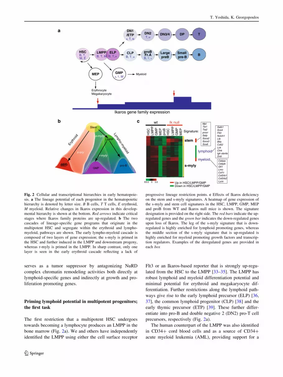
Fig. 2 Cellular and transcriptional hierarchies in early hematopoie-
sis. a The lineage potential of each progenitor in the hematopoietic
hierarchy is denoted by letter size. B B cells, T T cells, E erythroid,
M myeloid. Relative changes in Ikaros expression in this develop-
mental hierarchy is shown at the bottom. Red arrows indicate critical
stages where Ikaros family proteins are up-regulated. b The two
cascades of lineage-specific gene programs that originate in the
multipotent HSC and segregate within the erythroid and lympho-
myeloid, pathways are shown. The early lympho-myeloid cascade is
composed of two layers of gene expression; the s-myly is primed in
the HSC and further induced in the LMPP and downstream progeny,
whereas r-myly is primed in the LMPP. In sharp contrast, only one
layer is seen in the early erythroid cascade reflecting a lack of
progressive lineage restriction points. c Effects of Ikaros deficiency
on the stem and s-myly signatures. A heatmap of gene expression of
the s-myly and stem cell signatures in the HSC, LMPP, GMP, MEP
and proB from WT and Ikaros null mice is shown. The signature
designation is provided on the right side. The red bars indicate the up-
regulated genes and the green bar indicates the down-regulated genes
upon loss of Ikaros. The leg of the s-myly signature that is down-
regulated is highly enriched for lymphoid promoting genes, whereas
the middle section of the s-myly signature that is up-regulated is
highly enriched for myeloid promoting growth factors and transcrip-
tion regulators. Examples of the deregulated genes are provided in
each box
T. Yoshida, K. Georgopoulos
123

key role in human hemo-lymphopoiesis and in leukemo-
genesis [40–42]. This revised road map of early hemato-
poiesis first described in the mouse is highly conserved in
the human indicating the importance of mouse genetic
models for the study of normal hematopoiesis and lym-
phoid disorders in the human.
Studies on mouse genetic models have revealed that while
Ikaros is not required for development of the LMPP, it is
critical for its ability to differentiate into lymphoid restricted
progenitors such as the CLP and the ETP [34, 39], the gen-
eration of which usually identifies with lymphoid lineage
specification. Notably, the E2A transcription factor is also
involved in lymphoid priming and shares many downstream
targets [10, 43, 44]. However, E2A may also acts upstream of
Ikaros as it is required for the development of the LMPP from
the multipotent HSC [43]. These genetic studies are in line
with biochemical studies that have identified E2A DNA-
binding motifs in the context of Ikaros chromatin enrichment
sites in lymphoid specific enhancers [10].
While pro-B and NK are completely absent in Ikaros
null mice, the ETP (DN1) in the thymus is still detected
albeit at a highly reduced level [39]. Contribution of other
Ikaros family members, such as Helios expressed in the
HSC and through early stages of T cell but not B cell
differentiation may provide some rescue in T cell differ-
entiation. Ikaros dn mice show more profound defects with
no T cell progenitors detected indicating interference with
Helios function [6].
Priming of lymphoid-specific gene expression
in multipotent progenitors
The establishment of lineage restriction can be observed at
the molecular level by the induction and propagation of
lineage-affiliated transcriptional programs that support
lineage-specific differentiation. Comparative analyses of
genome-wide transcriptional profiles of the HSC, LMPP,
granulocyte-macrophage progenitor (GMP) and mega-
karyocyte-erythrocyte progenitor (MEP) [45] established
nine lineage-affiliated signatures that fit into two tran-
scriptional cascades that originate in the HSC [44]. One is
propagated in the erythroid pathway, while the other, in the
lympho-myeloid pathways (Fig. 2b, c). The front-runner in
the lympho-myeloid cascade is a ‘‘stem-myeloid-lym-
phoid’’ (s-myly) signature, that is composed of genes
primed in the HSC and further up-regulated in the LMPP.
The second runner is a ‘‘restricted-myeloid-lymphoid’’
(r-myly) signature that comprises of genes primed in the
LMPP and further induced in the downstream myeloid and
lymphoid progenitors [44] (Fig. 2b, c).
Progenitor comparative transcriptional analysis has
revealed that progenitor commitment into the lymphoid or
myeloid lineages is gradual, whereas commitment into the
erythroid lineage is rapid. Notably, the lymphoid and
myeloid potential and their supporting gene expression
programs are both maintained in the nominal myeloid
progenitor (GMP) and B cell progenitor (CLP, pro-B)
populations [44, 46, 47]. The latent T cell activity of the
GMP observed in vitro in mice and human [40, 44] is
consistent with the remaining expression of lymphoid genes
in these cells (Fig. 2a, b). Although these progenitors may
not support lymphoid differentiation in vivo, this study
supports the idea of progressive lineage restriction along the
lymphoid or myeloid pathways where either B cell or T cell
potential is lost first followed by myeloid potential [48–50].
A similar approach to transcriptional profiling and compu-
tational analyses on human HSC and progenitors have
likewise revealed transcriptional ‘landscapes’ that cross the
lineage and population boundaries [42].
The multilineage (erythroid, myeloid and lymphoid)
priming in the HSC observed by clustering of lineage-
specific gene expression profiles has been further con-
firmed at the single cell level. Erythroid and myeloid gene
expression programs are primed together with lymphoid
gene expression at a similar frequency, suggesting there is
equal opportunity for all three fates that is further modu-
lated by environmental inputs [44]. Lineage-specific
priming occurs in cells with an active stem cell gene
expression program that is rapidly extinguished upon
lineage restriction (Fig. 2b).
Transcriptional analyses of Ikaros null HSC and pro-
genitors have revealed that up-regulation of the lymphoid
genetic program and down-regulation of stem cell and
myeloid programs controlled by Ikaros underlie this critical
stage of lymphoid lineage specification at the LMPP stage
[44] (Fig. 2c). A failure of such events results in a block of
differentiation towards CLP and ETP and augmentation of
the myeloid differentiation from the LMPP [34, 44].
Examples of Ikaros target genes during this process are
summarized in Fig. 2c. It will be important to determine
whether maintenance of stem cell gene expression in
mutant myeloid progenitors can contribute to aberrant
expansion and whether this is linked to myeloid prolifer-
ative disorders in human. Thus the first major role of Ikaros
in early multipotent progenitors is to awaken their lym-
phoid potential by increasing local chromatin accessibility
through the NuRD complex while suppressing alternate
fates of myeloid differentiation and self-renewal.
Proliferative expansion of lymphocyte precursors
is harnessed by Ikaros
After lymphoid lineage specification, Ikaros expression is
again increased at the small pre-B cell stage in the bone
Ikaros, a guardian of lymphocyte differentiation
123

marrow and in double positive (DP) T cell precursors in the
thymus. These are equivalent stages of B and T cell dif-
ferentiation where precursor cells come out of cycle to
undergo the second antigen receptor rearrangement and
selection (Fig. 2a). Notably, when Ikaros activity is
reduced in human and mice, B and T cell leukemias arise
from the preceding proliferative stages (Fig. 3a). Thus I-
karos plays an important role as a tumor suppressor at the
lymphoid precursor stages where proliferation and Rag
expression provide a high risk for leukemic transformation.
Studies using germline knock-out mouse models have
shown that homozygocity for Ikaros null and heterozygo-
city for Ikaros dn mutations result in a rapid development
of T-ALL with a thymic origin. Leukemogenesis kinetics
are faster in cells with the Ikaros dn isoforms due to
interference with other Ikaros family members such as
Helios and Aiolos that are also expressed in these cells [6,
17]. One of the mechanisms of leukemic transformation in
the thymus may relate to altered TCR signaling [51, 52].
Recent studies have revealed that Ikaros suppresses Notch-
dependent leukemia development by directly repressing
transcription of Notch1 through upstream regulatory ele-
ments and promoters that include a cryptic intragenic
promoter in DP thymocytes that supports expression of a
ligand-independent Notch signaling [32, 53, 54]. Deletion
of Ikaros in DP thymocytes unleashes deregulated Notch
signaling in these cells.
A new high-risk model of B-ALL supported by Ikaros
mutations
Acute lymphoblastic leukemias (ALLs) are neoplasms of
lymphoid precursors and are common among childhood
malignancies [55]. With the advent of high-resolution
genome-wide profiling approaches, a variety of genetic
alterations, deletions and mutations have been newly
identified on the ALL genomes that cooperate with previ-
ously characterized chromosomal alterations [56–58].
Notably, deletions and mutations in the genes encoding key
transcription factors for early lymphoid development (e.g.
PAX5, IKZF1, IKZF3, EBF, TCF3 and LEF) were identi-
fied in 40 % of B-ALL cases [56]. Among these factors,
deletion and mutation in the IKZF1 locus that encodes
IKAROS is highly associated with BCR-ABL1-positive
B-ALL that display poor prognosis [57]. These genetic
P-PI3K/AKT/STAT5TAT5
HSC
LMPP
CLPpro-Bpro-T L.pre-B
pre-T
B/T
B-ALLT-ALL
Restriction
Pot
entia
l Priming
Specification
Commitment
ΔIk
WT large pre-B IkE5Δ/Δ large pre-B
Proliferation Self-renewal
Integrins
pre-BCRIL-7RGFR
Survival
ECM ECM
Self-renewalP-PI3K/AKT/STA
Proliferation
Survival
WT small pre-B
ProliferationSurvival
Differentiation
pFAKpFAK
pFAKpFAK
Differentiation
ΔIk, Ikdn, IkE5Δ/Δ
Ikaros
IkE5Δ/Δ
S.pre-BDP
a
b
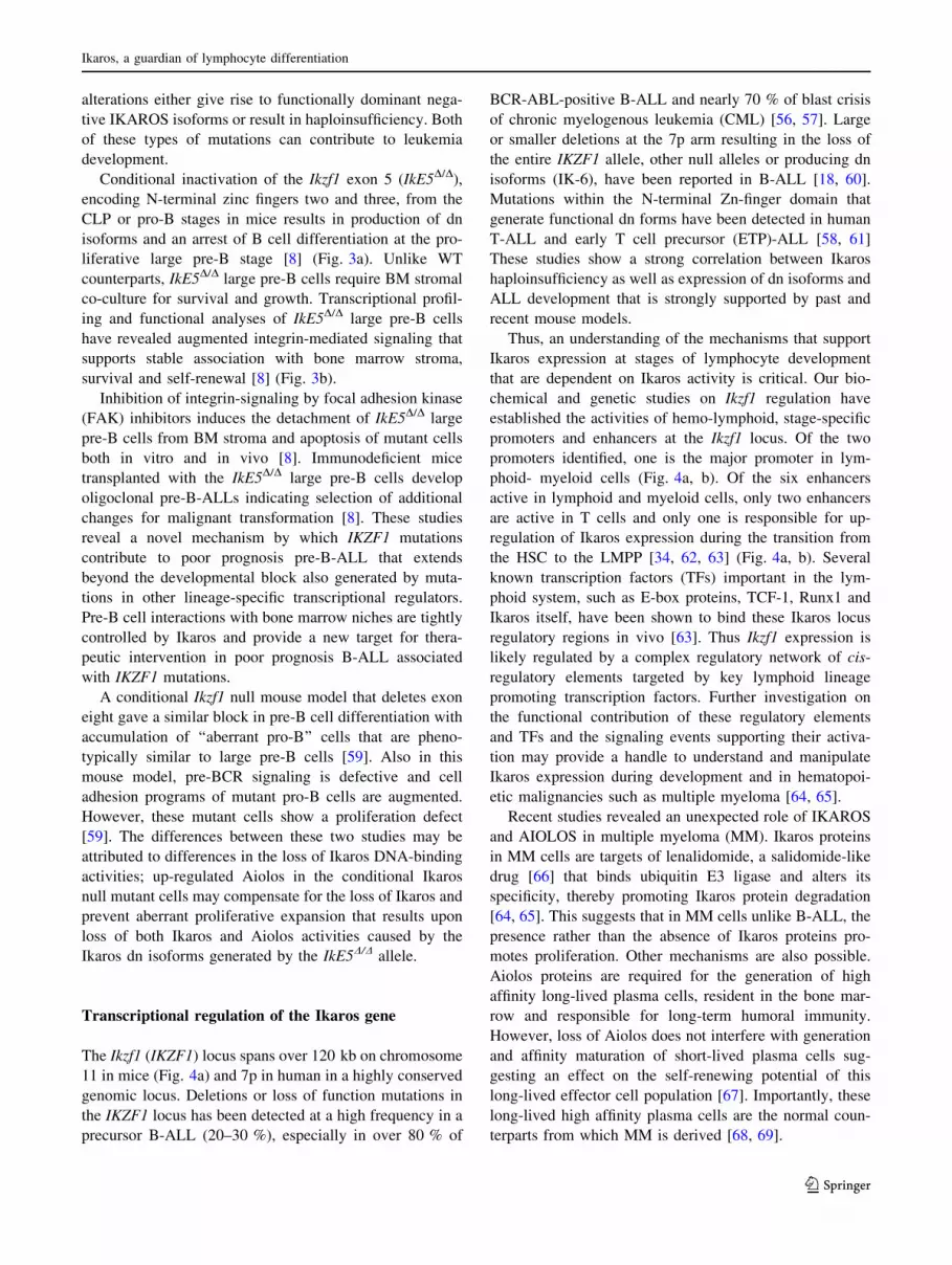
Fig. 3 Effects of Ikaros
mutations in early lymphoid
development and homeostasis.
a A summary of Ikaros’ roles in
early lymphocyte differentiation
as revealed by various mouse
genetic models. DIk germline
Ikaros null mutation, Ikdn.
germline Ikaros dominant
negative mutation, IkE5D/D B
cell specific conditional Ikaros
dominant negative mutation.
b A diagram of interactions
between integrin, IL-7R and
pre-BCR signaling in WT large
pre-B cells and the effects of an
Ikaros dominant negative
mutation (IkE5D/D) on this
signaling network. The strength
of the effect of individual
signaling pathways on cellular
properties such as survival, self-
renewal and proliferation is
depicted by letter size. Green
arrows and red bars indicate
positive and negative
interactions, respectively. pFAK
phosphorylated FAK, IL-7R
Interleukin-7 receptor, GHR
growth factor and/or cytokine
receptor
T. Yoshida, K. Georgopoulos
123
alterations either give rise to functionally dominant nega-
tive IKAROS isoforms or result in haploinsufficiency. Both
of these types of mutations can contribute to leukemia
development.
Conditional inactivation of the Ikzf1 exon 5 (IkE5D/D),
encoding N-terminal zinc fingers two and three, from the
CLP or pro-B stages in mice results in production of dn
isoforms and an arrest of B cell differentiation at the pro-
liferative large pre-B stage [8] (Fig. 3a). Unlike WT
counterparts, IkE5D/D large pre-B cells require BM stromal
co-culture for survival and growth. Transcriptional profil-
ing and functional analyses of IkE5D/D large pre-B cells
have revealed augmented integrin-mediated signaling that
supports stable association with bone marrow stroma,
survival and self-renewal [8] (Fig. 3b).
Inhibition of integrin-signaling by focal adhesion kinase
(FAK) inhibitors induces the detachment of IkE5D/D large
pre-B cells from BM stroma and apoptosis of mutant cells
both in vitro and in vivo [8]. Immunodeficient mice
transplanted with the IkE5D/D large pre-B cells develop
oligoclonal pre-B-ALLs indicating selection of additional
changes for malignant transformation [8]. These studies
reveal a novel mechanism by which IKZF1 mutations
contribute to poor prognosis pre-B-ALL that extends
beyond the developmental block also generated by muta-
tions in other lineage-specific transcriptional regulators.
Pre-B cell interactions with bone marrow niches are tightly
controlled by Ikaros and provide a new target for thera-
peutic intervention in poor prognosis B-ALL associated
with IKZF1 mutations.
A conditional Ikzf1 null mouse model that deletes exon
eight gave a similar block in pre-B cell differentiation with
accumulation of ‘‘aberrant pro-B’’ cells that are pheno-
typically similar to large pre-B cells [59]. Also in this
mouse model, pre-BCR signaling is defective and cell
adhesion programs of mutant pro-B cells are augmented.
However, these mutant cells show a proliferation defect
[59]. The differences between these two studies may be
attributed to differences in the loss of Ikaros DNA-binding
activities; up-regulated Aiolos in the conditional Ikaros
null mutant cells may compensate for the loss of Ikaros and
prevent aberrant proliferative expansion that results upon
loss of both Ikaros and Aiolos activities caused by the
Ikaros dn isoforms generated by the IkE5D/D allele.
Transcriptional regulation of the Ikaros gene
The Ikzf1 (IKZF1) locus spans over 120 kb on chromosome
11 in mice (Fig. 4a) and 7p in human in a highly conserved
genomic locus. Deletions or loss of function mutations in
the IKZF1 locus has been detected at a high frequency in a
precursor B-ALL (20–30 %), especially in over 80 % of
BCR-ABL-positive B-ALL and nearly 70 % of blast crisis
of chronic myelogenous leukemia (CML) [56, 57]. Large
or smaller deletions at the 7p arm resulting in the loss of
the entire IKZF1 allele, other null alleles or producing dn
isoforms (IK-6), have been reported in B-ALL [18, 60].
Mutations within the N-terminal Zn-finger domain that
generate functional dn forms have been detected in human
T-ALL and early T cell precursor (ETP)-ALL [58, 61]
These studies show a strong correlation between Ikaros
haploinsufficiency as well as expression of dn isoforms and
ALL development that is strongly supported by past and
recent mouse models.
Thus, an understanding of the mechanisms that support
Ikaros expression at stages of lymphocyte development
that are dependent on Ikaros activity is critical. Our bio-
chemical and genetic studies on Ikzf1 regulation have
established the activities of hemo-lymphoid, stage-specific
promoters and enhancers at the Ikzf1 locus. Of the two
promoters identified, one is the major promoter in lym-
phoid- myeloid cells (Fig. 4a, b). Of the six enhancers
active in lymphoid and myeloid cells, only two enhancers
are active in T cells and only one is responsible for up-
regulation of Ikaros expression during the transition from
the HSC to the LMPP [34, 62, 63] (Fig. 4a, b). Several
known transcription factors (TFs) important in the lym-
phoid system, such as E-box proteins, TCF-1, Runx1 and
Ikaros itself, have been shown to bind these Ikaros locus
regulatory regions in vivo [63]. Thus Ikzf1 expression is
likely regulated by a complex regulatory network of cis-
regulatory elements targeted by key lymphoid lineage
promoting transcription factors. Further investigation on
the functional contribution of these regulatory elements
and TFs and the signaling events supporting their activa-
tion may provide a handle to understand and manipulate
Ikaros expression during development and in hematopoi-
etic malignancies such as multiple myeloma [64, 65].
Recent studies revealed an unexpected role of IKAROS
and AIOLOS in multiple myeloma (MM). Ikaros proteins
in MM cells are targets of lenalidomide, a salidomide-like
drug [66] that binds ubiquitin E3 ligase and alters its
specificity, thereby promoting Ikaros protein degradation
[64, 65]. This suggests that in MM cells unlike B-ALL, the
presence rather than the absence of Ikaros proteins pro-
motes proliferation. Other mechanisms are also possible.
Aiolos proteins are required for the generation of high
affinity long-lived plasma cells, resident in the bone mar-
row and responsible for long-term humoral immunity.
However, loss of Aiolos does not interfere with generation
and affinity maturation of short-lived plasma cells sug-
gesting an effect on the self-renewing potential of this
long-lived effector cell population [67]. Importantly, these
long-lived high affinity plasma cells are the normal coun-
terparts from which MM is derived [68, 69].
Ikaros, a guardian of lymphocyte differentiation
123
Conclusions and future directions
The balanced production of lymphocytes is important for
organismal health. Disruption of the mechanisms that
support this process can cause disorders ranging from
immune cell deficiencies to cancers of hematopoietic but
also non-hematopoietic cell origin. Ikaros activity in the
HSC–MPP identifies with lymphoid lineage potential,
while Ikaros activity at later stages of lymphocyte differ-
entiation regulates normal proliferative expansion and
differentiation to a non-proliferative stage. Deletions and
mutations in the IKZF1 locus in both coding and non-
coding regions (i.e. highly conserved regulatory regions)
may interfere with IKAROS activity and provide several
independent ways to support leukemia development.
Past studies on Ikaros in lymphocyte differentiation are
paving the road to new studies that seek to delineate Ikaros-
based regulatory and signaling networks in both normal
development and immune cell based diseases. As such
these studies bear important therapeutic implications for
human health.
Acknowledgments We thank Drs. Taku Naito (Toho University
Faculty of Medicine), John R. Seavitt (Baylor College of Medicine)
and Bruce A. Morgan (Massachusetts General Hospital) for critical
reading of the manuscript.
Conflict of interest The author has no conflicts of interest to
disclose.
References
1. Georgopoulos K, Moore DD, Derfler B. Ikaros, an early lym-
phoid-specific transcription factor and a putative mediator for T
cell commitment. Science. 1992;258:808–12.
a
b
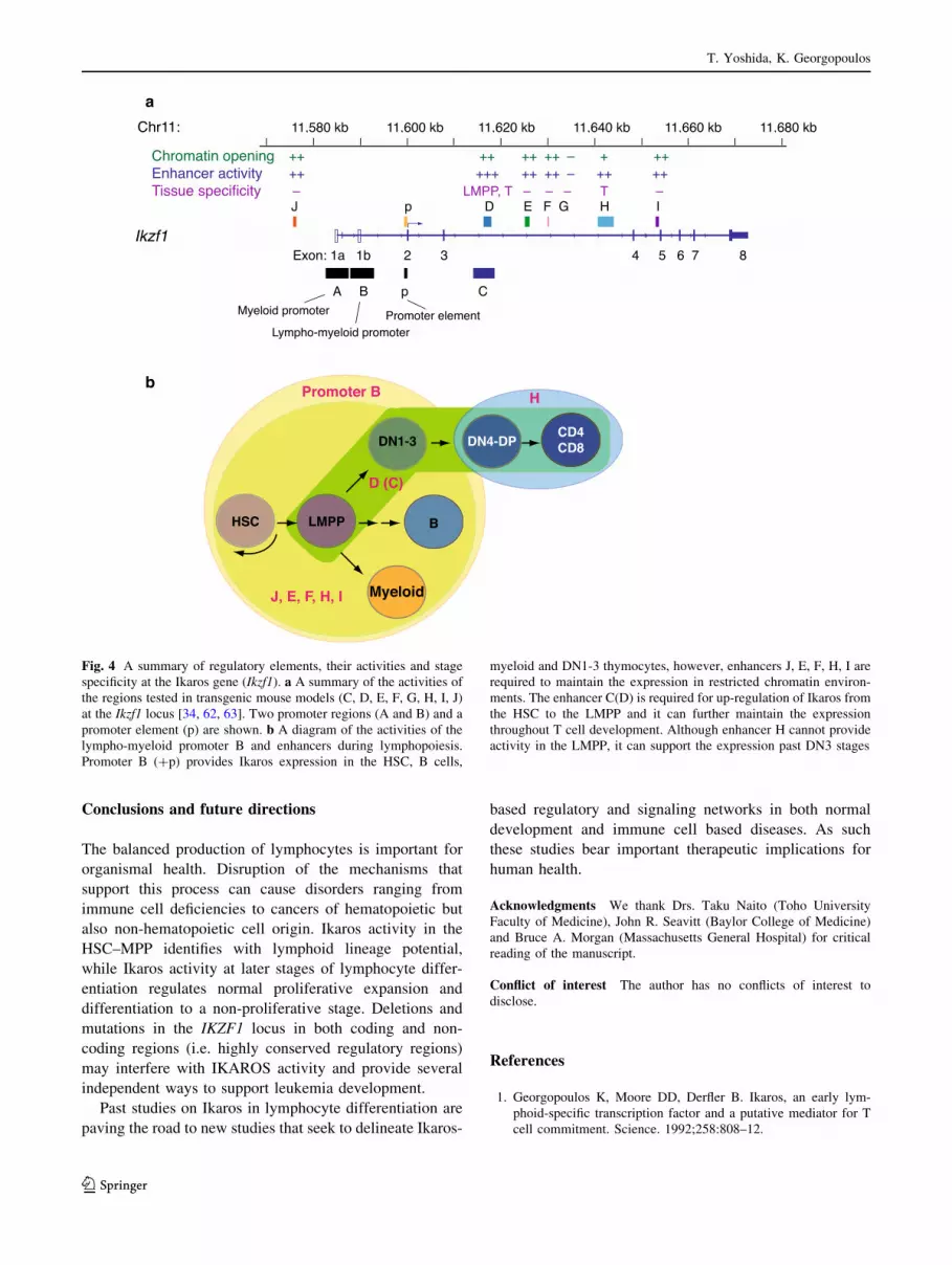
Fig. 4 A summary of regulatory elements, their activities and stage
specificity at the Ikaros gene (Ikzf1). a A summary of the activities of
the regions tested in transgenic mouse models (C, D, E, F, G, H, I, J)
at the Ikzf1 locus [34, 62, 63]. Two promoter regions (A and B) and a
promoter element (p) are shown. b A diagram of the activities of the
lympho-myeloid promoter B and enhancers during lymphopoiesis.
Promoter B (?p) provides Ikaros expression in the HSC, B cells,
myeloid and DN1-3 thymocytes, however, enhancers J, E, F, H, I are
required to maintain the expression in restricted chromatin environ-
ments. The enhancer C(D) is required for up-regulation of Ikaros from
the HSC to the LMPP and it can further maintain the expression
throughout T cell development. Although enhancer H cannot provide
activity in the LMPP, it can support the expression past DN3 stages
T. Yoshida, K. Georgopoulos
123

2. Hahm K, Ernst P, Lo K, Kim GS, Turck C, Smale ST. The
lymphoid transcription factor LyF-1 is encoded by specific,
alternatively spliced mRNAs derived from the Ikaros gene. Mol
Cell Biol. 1994;14:7111–23.
3. Molnar A, Georgopoulos K. The Ikaros gene encodes a family of
functionally diverse zinc finger DNA-binding proteins. Mol Cell
Biol. 1994;14:8292–303.
4. Molnar A, Wu P, Largespada DA, Vortkamp A, Scherer S, Co-
peland NG, Jenkins NA, Bruns G, Georgopoulos K. The Ikaros
gene encodes a family of lymphocyte-restricted zinc finger DNA
binding proteins, highly conserved in human and mouse.
J Immunol. 1996;156:585–92.
5. Koipally J, Heller EJ, Seavitt JR, Georgopoulos K. Unconven-
tional potentiation of gene expression by Ikaros. J Biol Chem.
2002;277:13007–15.
6. Georgopoulos K, Bigby M, Wang JH, Molnar A, Wu P, Winandy
S, Sharpe A. The Ikaros gene is required for the development of
all lymphoid lineages. Cell. 1994;79:143–56.
7. Papathanasiou P, Perkins AC, Cobb BS, Ferrini R, Sridharan R,
Hoyne GF, Nelms KA, Smale ST, Goodnow CC. Widespread
failure of hematolymphoid differentiation caused by a recessive
niche-filling allele of the Ikaros transcription factor. Immunity.
2003;19:131–44.
8. Joshi I, Yoshida T, Jena N, Qi X, Zhang J, Van Etten RA,
Georgopoulos K. Loss of Ikaros DNA-binding function confers
integrin-dependent survival on pre-B cells and progression to
acute lymphoblastic leukemia. Nat Immunol. 2014;15:294–304.
9. Schjerven H, McLaughlin J, Arenzana TL, Frietze S, Cheng D,
Wadsworth SE, Lawson GW, Bensinger SJ, Farnham PJ, Witte
ON, et al. Selective regulation of lymphopoiesis and leukemo-
genesis by individual zinc fingers of Ikaros. Nat Immunol.
2013;14:1073–83.
10. Zhang J, Jackson AF, Naito T, Dose M, Seavitt J, Liu F, Heller
EJ, Kashiwagi M, Yoshida T, Gounari F, et al. Harnessing of the
nucleosome-remodeling-deacetylase complex controls lympho-
cyte development and prevents leukemogenesis. Nat Immunol.
2011;13:86–94.
11. Cobb BS, Morales-Alcelay S, Kleiger G, Brown KE, Fisher AG,
Smale ST. Targeting of Ikaros to pericentromeric heterochro-
matin by direct DNA binding. Genes Dev. 2000;14:2146–60.
12. Hollenhorst PC, Shah AA, Hopkins C, Graves BJ. Genome-wide
analyses reveal properties of redundant and specific promoter
occupancy within the ETS gene family. Genes Dev.
2007;21:1882–94.
13. Sun L, Liu A, Georgopoulos K. Zinc finger-mediated protein
interactions modulate Ikaros activity, a molecular control of
lymphocyte development. EMBO J. 1996;15:5358–69.
14. Morgan B, Sun L, Avitahl N, Andrikopoulos K, Ikeda T, Gonz-
ales E, Wu P, Neben S, Georgopoulos K. Aiolos, a lymphoid
restricted transcription factor that interacts with Ikaros to regulate
lymphocyte differentiation. EMBO J. 1997;16:2004–13.
15. Kelley CM, Ikeda T, Koipally J, Avitahl N, Wu L, Georgopoulos
K, Morgan BA. Helios, a novel dimerization partner of Ikaros
expressed in the earliest hematopoietic progenitors. Curr Biol.
1998;8:508–15.
16. Honma Y, Kiyosawa H, Mori T, Oguri A, Nikaido T, Kanazawa
K, Tojo M, Takeda J, Tanno Y, Yokoya S, et al. Eos: a novel
member of the Ikaros gene family expressed predominantly in the
developing nervous system. FEBS Lett. 1999;447:76–80.
17. Wang JH, Nichogiannopoulou A, Wu L, Sun L, Sharpe AH,
Bigby M, Georgopoulos K. Selective defects in the development
of the fetal and adult lymphoid system in mice with an Ikaros null
mutation. Immunity. 1996;5:537–49.
18. Kastner P, Dupuis A, Gaub MP, Herbrecht R, Lutz P, Chan S.
Function of Ikaros as a tumor suppressor in B cell acute lym-
phoblastic leukemia. Am J Blood Res. 2013;3:1–13.
19. Tong JK, Hassig CA, Schnitzler GR, Kingston RE, Schreiber SL.
Chromatin deacetylation by an ATP-dependent nucleosome
remodelling complex. Nature. 1998;395:917–21.
20. Wade PA, Jones PL, Vermaak D, Wolffe AP. A multiple subunit
Mi-2 histone deacetylase from Xenopus laevis cofractionates
with an associated Snf2 superfamily ATPase. Curr Biol.
1998;8:843–6.
21. Xue Y, Wong J, Moreno GT, Young MK, Cote J, Wang W.
NURD, a novel complex with both ATP-dependent chromatin-
remodeling and histone deacetylase activities. Mol Cell.
1998;2:851–61.
22. Wade PA, Gegonne A, Jones PL, Ballestar E, Aubry F, Wolffe
AP. Mi-2 complex couples DNA methylation to chromatin
remodelling and histone deacetylation. Nat Genet. 1999;23:62–6.
23. Kim J, Sif S, Jones B, Jackson A, Koipally J, Heller E, Winandy
S, Viel A, Sawyer A, Ikeda T, et al. Ikaros DNA-binding proteins
direct formation of chromatin remodeling complexes in lym-
phocytes. Immunity. 1999;10:345–55.
24. O’Neill DW, Schoetz SS, Lopez RA, Castle M, Rabinowitz L,
Shor E, Krawchuk D, Goll MG, Renz M, Seelig HP, et al. An
ikaros-containing chromatin-remodeling complex in adult-type
erythroid cells. Mol Cell Biol. 2000;20:7572–82.
25. Sridharan R, Smale ST. Predominant interaction of both Ikaros
and Helios with the NuRD complex in immature thymocytes.
J Biol Chem. 2007;282:30227–38.
26. Zhang Y, LeRoy G, Seelig HP, Lane WS, Reinberg D. The
dermatomyositis-specific autoantigen Mi2 is a component of a
complex containing histone deacetylase and nucleosome remod-
eling activities. Cell. 1998;95:279–89.
27. Becker PB, Horz W. ATP-dependent nucleosome remodeling.
Annu Rev Biochem. 2002;71:247–73.
28. Shimono Y, Murakami H, Kawai K, Wade PA, Shimokata K,
Takahashi M. Mi-2 beta associates with BRG1 and RET finger
protein at the distinct regions with transcriptional activating and
repressing abilities. J Biol Chem. 2003;278:51638–45.
29. Williams CJ, Naito T, Arco PG, Seavitt JR, Cashman SM, De
Souza B, Qi X, Keables P, Von Andrian UH, Georgopoulos K.
The chromatin remodeler Mi-2beta is required for CD4 expres-
sion and T cell development. Immunity. 2004;20:719–33.
30. Naito T, Gomez-Del Arco P, Williams CJ, Georgopoulos K.
Antagonistic interactions between Ikaros and the chromatin
remodeler Mi-2beta determine silencer activity and Cd4 gene
expression. Immunity. 2007;27:723–34.
31. Yoshida T, Hazan I, Zhang J, Ng SY, Naito T, Snippert HJ, Heller
EJ, Qi X, Lawton LN, Williams CJ, et al. The role of the chromatin
remodeler Mi-2beta in hematopoietic stem cell self-renewal and
multilineage differentiation. Genes Dev. 2008;22:1174–89.
32. Gomez-del Arco P, Kashiwagi M, Jackson AF, Naito T, Zhang J,
Liu F, Kee B, Vooijs M, Radtke F, Redondo JM, et al. Alternative
promoter usage at the Notch1 locus supports ligand-independent
signaling in T cell development and leukemogenesis. Immunity.
2010;33:685–98.
33. Adolfsson J, Mansson R, Buza-Vidas N, Hultquist A, Liuba K,
Jensen CT, Bryder D, Yang L, Borge OJ, Thoren LA, et al.
Identification of Flt3? lympho-myeloid stem cells lacking ery-
thro-megakaryocytic potential a revised road map for adult blood
lineage commitment. Cell. 2005;121:295–306.
34. Yoshida T, Ng SY, Zuniga-Pflucker JC, Georgopoulos K. Early
hematopoietic lineage restrictions directed by Ikaros. Nat
Immunol. 2006;7:382–91.
35. Lai AY, Kondo M. Asymmetrical lymphoid and myeloid lineage
commitment in multipotent hematopoietic progenitors. J Exp
Med. 2006;203:1867–73.
36. Igarashi H, Gregory SC, Yokota T, Sakaguchi N, Kincade PW.
Transcription from the RAG1 locus marks the earliest lympho-
cyte progenitors in bone marrow. Immunity. 2002;17:117–30.
Ikaros, a guardian of lymphocyte differentiation
123

37. Yokota T, Sudo T, Ishibashi T, Doi Y, Ichii M, Orirani K,
Kanakura Y. Complementary regulation of early B-lymphoid
differentiation by genetic and epigenetic mechanisms. Int J
Hematol. 2013;98:382–9.
38. Kondo M, Weissman IL, Akashi K. Identification of clonogenic
common lymphoid progenitors in mouse bone marrow. Cell.
1997;91:661–72.
39. Allman D, Sambandam A, Kim S, Miller JP, Pagan A, Well D,
Meraz A, Bhandoola A. Thymopoiesis independent of common
lymphoid progenitors. Nat Immunol. 2003;4:168–74.
40. Doulatov S, Notta F, Eppert K, Nguyen LT, Ohashi PS, Dick JE.
Revised map of the human progenitor hierarchy shows the origin
of macrophages and dendritic cells in early lymphoid develop-
ment. Nat Immunol. 2010;11:585–94.
41. Goardon N, Marchi E, Atzberger A, Quek L, Schuh A, Soneji S,
Woll P, Mead A, Alford KA, Rout R, et al. Coexistence of
LMPP-like and GMP-like leukemia stem cells in acute myeloid
leukemia. Cancer Cell. 2011; 138–152.
42. Laurenti E, Doulatov S, Zandi S, Plumb I, Chen J, April C, Fan
JB, Dick JE. The transcriptional architecture of early human
hematopoiesis identifies multilevel control of lymphoid com-
mitment. Nat Immunol. 2013;14:756–63.
43. Dias S, Mansson R, Gurbuxani S, Sigvardsson M, Kee BL. E2A
proteins promote development of lymphoid-primed multipotent
progenitors. Immunity. 2008;29:217–27.
44. Ng SY, Yoshida T, Zhang J, Georgopoulos K: Genome-wide
Lineage-Specific Transcriptional Networks Underscore Ikaros-
Dependent Lymphoid Priming in Hematopoietic Stem Cells.
Immunity. 2009.
45. Akashi K, Traver D, Miyamoto T, Weissman IL. A clonogenic
common myeloid progenitor that gives rise to all myeloid lin-
eages. Nature. 2000;404:193–7.
46. Rumfelt LL, Zhou Y, Rowley BM, Shinton SA, Hardy RR.
Lineage specification and plasticity in CD19- early B cell pre-
cursors. J Exp Med. 2006;203:675–87.
47. Mansson R, Zandi S, Anderson K, Martensson IL, Jacobsen SE,
Bryder D, Sigvardsson M. B-lineage commitment prior to surface
expression of B220 and CD19 on hematopoietic progenitor cells.
Blood. 2008;112:1048–55.
48. Wada H, Masuda K, Satoh R, Kakugawa K, Ikawa T, Katsura Y,
Kawamoto H. Adult T-cell progenitors retain myeloid potential.
Nature. 2008;452:768–72.
49. Bell JJ, Bhandoola A. The earliest thymic progenitors for T cells
possess myeloid lineage potential. Nature. 2008;452:764–7.
50. Kawamoto H, Ikawa T, Masuda K, Wada H, Katsura Y. A map
for lineage restriction of progenitors during hematopoiesis: the
essence of the myeloid-based model. Immunol Rev.
2010;238:23–36.
51. Winandy S, Wu P, Georgopoulos K. A dominant mutation in the
Ikaros gene leads to rapid development of leukemia and lym-
phoma. Cell. 1995;83:289–99.
52. Winandy S, Wu L, Wang JH, Georgopoulos K. Pre-T cell
receptor (TCR) and TCR-controlled checkpoints in T cell dif-
ferentiation are set by Ikaros. J Exp Med. 1999;190:1039–48.
53. Jeannet R, Mastio J, Macias-Garcia A, Oravecz A, Ashworth T,
Geimer Le Lay AS, Jost B, Le Gras S, Ghysdael J, Gridley T,
et al. Oncogenic activation of the Notch1 gene by deletion of its
promoter in Ikaros-deficient T-ALL. Blood. 2010;116:5443–54.
54. Ashworth TD, Pear WS, Chiang MY, Blacklow SC, Mastio J, Xu
L, Kelliher M, Kastner P, Chan S, Aster JC. Deletion-based
mechanisms of Notch1 activation in T-ALL: key roles for RAG
recombinase and a conserved internal translational start site in
Notch1. Blood. 2010;116:5455–64.
55. Urayama KY, Chokkalingam AP, Manabe A, Mizutani S. Current
evidence for an inherited genetic basis of childhood acute lym-
phoblastic leukemia. Int J Hematol. 2013;97:3–19.
56. Mullighan CG, Goorha S, Radtke I, Miller CB, Coustan-Smith E,
Dalton JD, Girtman K, Mathew S, Ma J, Pounds SB, et al.
Genome-wide analysis of genetic alterations in acute lympho-
blastic leukaemia. Nature. 2007;446:758–64.
57. Mullighan CG, Miller CB, Radtke I, Phillips LA, Dalton J, Ma J,
White D, Hughes TP, Le Beau MM, Pui CH, et al. BCR-ABL1
lymphoblastic leukaemia is characterized by the deletion of I-
karos. Nature. 2008;453:110–4.
58. Zhang J, Ding L, Holmfeldt L, Wu G, Heatley SL, Payne-Turner
D, Easton J, Chen X, Wang J, Rusch M, et al. The genetic basis of
early T-cell precursor acute lymphoblastic leukaemia. Nature.
2012;481:157–63.
59. Schwickert TA, Tagoh H, Gultekin S, Dakic A, Axelsson E,
Minnich M, Ebert A, Werner B, Roth M, Cimmino L, et al.
Stage-specific control of early B cell development by the tran-
scription factor Ikaros. Nat Immunol. 2014;15:283–93.
60. Dupuis A, Gaub MP, Legrain M, Drenou B, Mauvieux L, Lutz P,
Herbrecht R, Chan S, Kastner P. Biclonal and biallelic deletions
occur in 20% of B-ALL cases with IKZF1 mutations. Leukemia.
2013;27:503–7.
61. Marcais A, Jeannet R, Hernandez L, Soulier J, Sigaux F, Chan S,
Kastner P. Genetic inactivation of Ikaros is a rare event in human
T-ALL. Leuk Res. 2010;34:426–9.
62. Kaufmann C, Yoshida T, Perotti EA, Landhuis E, Wu P, Geor-
gopoulos K. A complex network of regulatory elements in Ikaros
and their activity during hemo-lymphopoiesis. EMBO J.
2003;22:2211–23.
63. Yoshida T, Landhuis E, Dose M, Hazan I, Zhang J, Naito T,
Jackson AF, Wu J, Perroti EA, Kaufmann C, et al.: Transcrip-
tional regulation of the Ikzf1 locus. Blood 2013.
64. Gandhi AK, Kang J, Havens CG, Conklin T, Ning Y, Wu L, Ito
T, Ando H, Waldman MF, Thakurta A, et al. Immunomodulatory
agents lenalidomide and pomalidomide co-stimulate T cells by
inducing degradation of T cell repressors Ikaros and Aiolos via
modulation of the E3 ubiquitin ligase complex CRL4(CRBN.). Br
J Haematol. 2014;164:811–21.
65. Lu G, Middleton RE, Sun H, Naniong M, Ott CJ, Mitsiades CS,
Wong KK, Bradner JE, Kaelin WG Jr. The myeloma drug le-
nalidomide promotes the cereblon-dependent destruction of Ika-
ros proteins. Science. 2014;343:305–9.
66. Watanabe R, Tokuhira M, Kizaki M. Current approaches for the
treatment of multiple myeloma. Int J Hematol. 2013;97:333–44.
67. Cortes M, Georgopoulos K. Aiolos is required for the generation
of high affinity bone marrow plasma cells responsible for long-
term immunity. J Exp Med. 2004;199:209–19.
68. Anderson KC, Carrasco RD. Pathogenesis of myeloma. Annu
Rev Pathol. 2011;6:249–74.
69. Hosen N. Multiple myeloma-initiating cells. Int J Hematol.
2013;97:306–12.
T. Yoshida, K. Georgopoulos
123
Copyright © 2022 FDOKUMEN