
Antibodies to both ICAM-1 and LFA-1 do not protect the kidney against toxic (HgCl2) injury
Upload
independentCategory
view
1download
0
1467
J. Exp. Med.
The Rockefeller University Press • 0022-1007/99/05/1467/12 $2.00Volume 189, Number 9, May 3, 1999 1467–1478http://www.jem.org
Lymphocyte Migration in Lymphocyte Function-associated Antigen (LFA)-1–deficient Mice
By Cornelia Berlin-Rufenach,
*
i
Florian Otto,
‡
Meg Mathies,
*
Juergen Westermann,
§
Michael J. Owen,
‡
Alf Hamann,
i
¶
and Nancy Hogg
*
From the
*
Leukocyte Adhesion Laboratory and the
‡
Lymphocyte Molecular Biology Laboratory, Imperial Cancer Research Fund, London WC2A 3PX, United Kingdom; the
§
Center of Anatomy, Medical School of Hannover, D-30623 Hannover, Germany; the
i
Department of Immunology, Medical Clinic, University Hospital Eppendorf, D-20246 Hamburg, Germany; and
¶
Charité Clinic, Humboldt University, D-10117 Berlin, Germany
Summary
Using lymphocyte function-associated antigen (LFA)-1
2
/
2
mice, we have examined the role ofLFA-1 and other integrins in the recirculation of lymphocytes. LFA-1 has a key role in migra-tion to peripheral lymph nodes (pLNs), and influences migration into other LNs. Second, the
a
4 integrins,
a
4
b
7 and
a
4
b
1, have a hitherto unrecognized ability to compensate for the lackof LFA-1 in migration to pLNs. These findings are confirmed using normal mice and blockingLFA-1 and
a
4 monoclonal antibodies. Unexpectedly, vascular cell adhesion molecule (VCAM)-1,which is essential in inflammatory responses, serves as the ligand for the
a
4 integrins on pLNhigh endothelial venules. VCAM-1 also participates in trafficking into mesenteric LNs andPeyer’s patch nodes where mucosal addressin cell adhesion molecule 1 (MAdCAM-1), the
a
4
b
7-specific ligand, dominates. Both
a
4
b
1, interacting with ligand VCAM-1, and also LFA-1participate in substantial lymphocyte recirculation through bone marrow. These observationssuggest that organ-specific adhesion receptor usage in mature lymphocyte recirculation is not asrigidly adhered to as previously considered, and that the same basic sets of adhesion receptorsare used in both lymphocyte homing and inflammatory responses.
Key words: adhesion • integrin • homing • bone marrow • lymphocyte
T
he integrin LFA-1 (CD11a/CD18), which belongs tothe
b
2 family of integrin receptors, is expressed by allleukocytes and has a central role in the functions of thesecells. When leukocytes respond to inflammatory signals,LFA-1 acts together with other adhesion receptors to directthe cells into injured tissues (see references 1–4). Thus, theselectin adhesion receptors allow transient leukocyte contactwith stimulated endothelium, causing a rolling movementwhich is further arrested by the action of the
a
4 integrins.This stage is followed by a signaling event that activatesLFA-1 to promote firm adhesion of the leukocytes andtheir migration across the endothelium towards the area ofinjury.
In contrast, mature lymphocytes, but not myeloid cells,circulate continually through secondary lymphoid tissue
of the peripheral lymph nodes (pLNs),
1
mesenteric LNs(mLNs), and Peyer’s patches (PPs), thereby increasing theopportunity for an encounter with antigen. To migrateacross the specialized high endothelial venules (HEVs) ofthese LNs, lymphocytes have been proposed to use organ-specific sets of adhesion receptors (4, 5). Migration intomucosal tissue such as PPs is greatly dependent on the
a
4integrin,
a
4
b
7, which has no apparent role in homing tothe pLNs (6, 7). On the other hand, homing of naive lym-phocytes to pLNs has been considered to be dependent
upon L-selectin (CD62L) but not to involve the
a
4 inte-grins (8, 9). However, parallels are emerging between the
C. Berlin-Rufenach, F. Otto, and M. Mathies contributed equally to thismanuscript.
1
Abbreviations used in this paper:
CT, CellTracker™; ES, embryonic stem;HEV, high endothelial venule; HUVEC, human umbilical vein endothe-lial cell; ICAM, intercellular adhesion molecule; MAdCAM-1, mucosaladdressin cell adhesion molecule 1; pLN, peripheral lymph node; PNAd,peripheral node addressin; PP, Peyer’s patch; VCAM, vascular cell adhe-sion molecule.
on February 4, 2014
jem.rupress.org
Dow
nloaded from
Published May 3, 1999

1468
Role of LFA-1 and
a
4 Integrins in Lymphocyte Recirculation
processes of lymphocyte recirculation and the response to in-flammatory signals. LFA-1 influences migration into mucosaltissue as well as the pLNs (10), and there is now evidencethat recirculating lymphocytes also undergo an activationstep when they contact the HEVs. LFA-1 can be activatedby signals arising from the binding of lymphocyte L-selectinto HEV ligand, GlyCAM-1 (11). HEV-expressed chemo-kines B lymphocyte chemoattractant (BLC) and secondarylymphoid tissue chemokine (SLC) cause adhesion of naiveB cells (12) and T cells (13), respectively, and SLC has beendemonstrated to activate LFA-1.
Ligands for most of the lymphocyte adhesion receptorshave been identified on the HEVs. L-selectin recognizesperipheral node addressin (PNAd), a group of fucosylatedand sialylated LewisX-expressing proteins on pLN HEVs(see reference 14). LFA-1 functions through recognition ofits ligands intercellular adhesion molecule (ICAM)-1 orICAM-2 (see reference 15). The
a
4
b
7 integrin recognizesMAdCAM-1, which is expressed on HEVs in PPs andmLNs (16, 17), and VCAM-1 and fibronectin. The alter-native
a
4 integrin,
a
4
b
1, which has a major role in in-flammation (18–20), also recognizes ligands VCAM-1 andfibronectin. VCAM-1 is expressed by endothelia under in-flammatory conditions (21, 22) and is thought to be absenton normal LN endothelium (22, 23). Fibronectin is gen-erally associated only with the subendothelial matrix,but there is a report that a spliced variant is expressed inrheumatoid arthritis tissue (24). Thus, the ligand bindingactivities of the two
a
4 integrins are distinctive, in thatMAdCAM-1 is involved in
a
4
b
7-mediated naive lympho-cyte homing to mucosal sites whereas VCAM-1 primarilyserves as the ligand for
a
4
b
1 at inflammatory sites.Recently, two studies using LFA-1–deficient mice have
confirmed many of the previous assignments of LFA-1function, for example, in assays for T cell function such asMLR and delayed-type hypersensitivity responses (25, 26).In this study, we have used LFA-1
2
/
2
mice to reexaminethe role of LFA-1 in the recirculation of lymphocytes andto investigate alternative mechanisms used in its absence.We confirm an important role for LFA-1 in LN localiza-tion, identify an unexpected role for
a
4 integrins,
a
4
b
7and
a
4
b
1, in pLN homing that is mediated by HEV-expressed VCAM-1, and finally demonstrate that
a
4
b
1/VCAM-1 together with LFA-1 is involved in migration oflymphocytes to bone marrow.
Materials and Methods
Generation of LFA-1 Mutant Mice.
A 7-kb PCR product am-plified from murine 129/Sv genomic DNA and containing exon 2of the
Lfa-1
gene was subcloned into pSP72. A selection cassettecontaining stop codons in all three frames, an independent ribo-somal entry sequence (IRES) followed by the LacZ gene with anSV40 polyadenylation signal, and a neomycin phosphotransferasegene was introduced into exon 2. Culture and transfection ofGK129 embryonic stem (ES) cells was done as described previ-ously (27). EcoRI-digested ES cell DNA was analyzed by South-ern blot analysis using probe A, and homologous recombination
was confirmed using probe B (see Fig. 1 a). Blastocyst injectionand breeding have been described previously (27). Mice weregenotyped from EcoRI-digested tail DNA by Southern blot (datanot shown) or PCR (Fig. 1 b). A multiplex PCR using primerson either side of the selection cassette and one within the neo-mycin resistance gene yielded fragments of 90 bp (wild-type)and 660 bp (LFA-1–deficient), respectively.
Animal Husbandry.
All mice were kept under specific patho-gen–free conditions and in accordance with United KingdomHome Office regulations. The mice were monitored on a six-monthly basis for a wide selection of parasites, bacteria, and fungiand were regularly found to be pathogen free. LN sections fromsentinel mice belonging to litters used in the reported experi-ments were inspected for the presence of germinal centers by he-matoxylin and eosin staining and for positive MAdCAM-1 stain-ing in pLNs (28). These latter indicators of an inflammatoryresponse were routinely negative for mice housed in London andHamburg.
mAbs and Other Reagents.
The following purified rat mAbswere used in this study: LFA-1
a
subunit mAbs H35.89.9 (IgG2b)and H68 (IgG2a), both obtained from Dr. Michel Pierres (Centred’Immunologie, INSERM-CNRS de Marseille-Luminy, Marseille,France);
a
4 subunit mAb PS/2 (IgG2b); anti–VCAM-1 mAbMK2.7 (IgG1); anti–MAdCAM-1 mAb MECA-367 (IgG2a),obtained from American Type Culture Collection; anti-PNAdmAb MECA-79 (IgM; obtained from Drs. Mark Singer andSteve Rosen, University of California, San Francisco, CA);MECA-325 (IgG1), obtained from Dr. A. Duijvestijn (Universityof Limburg, Maastricht, Netherlands); CD54 mAb YN1/1.7(IgG2a; obtained from Dr. Fumio Takei, Terry Fox Laboratory,BC Cancer Research Centre, Vancouver, Canada); CD4, CD8,and B220 mAbs (PharMingen); and control rat IgG1 mAb PyLT-1(IgG1; Imperial Cancer Research Fund) and control null rat IgG1and IgG2a mAbs (PharMingen). Secondary detection antibodieswere FITC-conjugated goat anti–rat Ig (Jackson Immuno-Research Laboratories) and biotinylated rabbit anti–rat Ig (VectorLaboratories). Fab fragments were prepared by papain digestionusing either the ImmunoPure
®
Fab kit (Pierce Chemical Co.)or HPLC to remove any residual intact IgG (7).
Preparation of Cells for Flow Cytometry.
Single cell suspensionsfrom 8–12-wk-old mice were prepared by mincing or rubbingbetween glass slides with frequent rinsing with 5% FCS/RPMI or0.2% BSA/PBS. Bone marrow cells were harvested by flushingwith 5% FCS/RPMI via cut ends of tibias and femurs, followedby disaggregation and filtering through nylon gauze. Cells werewashed and incubated at 5
3
10
6
cells/ml with specific primarymAbs, then FITC-conjugated goat anti–rat Ig (1:200; JacksonImmunoResearch Laboratories) at 4
8
C for 30 min. Cells werefixed in 1% HCHO/PBS before analysis on a FACScan™ instru-ment (Becton Dickinson).
In Vivo Homing Experiments Using CellTracker™ FluorescentDye–labeled Lymphocytes.
Lymphocytes from pLNs and mLNs ofLFA-1
1
/
1
and LFA-1
2
/
2
mice were prepared and adjusted to 5
3
10
6
cells/ml in 5% FCS/RPMI for labeling as described (29).LFA-1
1
/
1
lymphocytes were incubated with 0.2 mM CellTracker™(CT) Green 5-chloromethylfluorescein diacetate (CMFDA; Molec-ular Probes) and LFA-1
2
/
2
lymphocytes with 5 mM CT Orange5-chloromethyl tetramethylrhodamine (CMTMR) at 37
8
C for45 min. The two cell preparations were washed and counted,then mixed together to achieve a 1:1 ratio of differentially labeledLFA-1
1
/
1
and LFA-1
2
/
2
lymphocytes for injection into recipient8–12-wk-old C57BL/6 mice for a period of 1 h. A correctionfactor was applied to the subsequent data if the lymphocyte ratio
on February 4, 2014
jem.rupress.org
Dow
nloaded from
Published May 3, 1999

1469
Berlin-Rufenach et al.
was not precisely 1:1. Reversing the CT dyes had no effect onresults. The lymphocytes were frequently tested for activationstatus before and after the labeling procedure by measuring thelevel of L-selectin that can be enzymatically cleaved after an acti-vating stimulus (30). In general, a total of 2
3
10
7
cells in a vol-ume of 200
m
l was injected per tail vein. In experiments involv-ing antibody blocking, the cells and mAbs were coinjected. Fabfragments were used in amounts of 250–400
m
g per mouse. Irrel-evant control Fab fragments were without effect.
In Vivo Homing Experiments Using
51
Cr-labeled Lymphocytes.
Lymphocytes obtained as above were labeled with 20
m
Ci
51
Cr/mlas described previously (29), and dead cells removed by centrif-ugation on a 17% Nycodenz cushion (Nycomed). 5
3
10
6
lymphocytes in 200
m
l were injected into the tail vein of recipi-ent 8–12-wk-old C57BL/6 mice. mAbs were coinjected as Fabfragments and at 300
m
g/200
m
l. Sample groups of four micewere used in each condition. The mice were killed after 1 h, andthe distribution of radioactivity was determined for the differentorgans and for the residual body mass. PBLs were calculated for ablood volume of 1.5 ml.
Labeling, Injection, and Detection of Donor Cells in the Host pLNs.
LN cells were labeled by dissolving 1 mg digoxigenin in 0.5 mlDMSO and incubating the lymphocytes for 15 min at a finalconcentration of 5
3
10
7
cells/ml with 60
m
g digoxigenin. Afterwashing, 0.8
3
10
7
cells were injected intravenously, and pLNswere removed 30 min later and frozen in liquid nitrogen. To lo-calize the donor cells in the HEVs, two antigens were revealedsimultaneously as described (31). In brief, cryostat sections wereincubated with the anti-HEV mAb MECA-325, which was re-vealed by the alkaline phosphatase anti-alkaline phosphatase(APAAP) technique (blue [31]). The donor cells were identifiedby a peroxidase-conjugated antidigoxigenin antibody (brown) asdescribed previously (31). The area of the HEVs (excluding thelumen) together with the area within 4-cell diameters surround-ing the vessel was then determined (31), and the number of in-jected cells adhering to and within the endothelium, and withinthe tissue was analyzed.
Immunohistochemistry.
Sections of 6
m
m were cut from snap-frozen tissues of pLN, mLN, and PP and placed on silane-coatedslides which were fixed in acetone for 10 min and allowed to airdry. Slides were washed in standard Tris-buffered saline (TBS),
pH 7.6, and primary mAb was added for 1 h at room tempera-ture. mAbs were used at optimal dilution: 10
m
g/ml for anti–VCAM-1 mAb MK2.7 (IgG1) and IgG1 isotype control mAbPyLT-1, MECA-79 at 4
m
g/ml, and MECA-367 as tissue culturesupernatant. Sections were also tested with null IgG1 and IgG2amAbs (PharMingen; data not shown). Biotinylated rabbit anti–ratIg (1:100; Vector Laboratories) was added for 35 min. A tertiarystage was carried out using the StreptABComplex/HRP kit fol-lowing the manufacturer’s instructions (K377; Dako), and theslides were incubated again for 35 min before exposure to 1 mg/ml3,3
9
diaminobenzidine.
Results
Disruption of the Lfa-1 Gene. Lfa-1
, the gene for theLFA-1
a
subunit, was mutated in ES cells using a replace-ment-type targeting vector and the strategy shown in Fig. 1 a.G418-resistant colonies were identified by Southern blotanalysis of EcoR1-digested ES cell genomic DNA usingthe probe shown in Fig. 1 a. Of 400 G418-resistant ES cellcolonies, 10 correctly targeted colonies were identified.Three different homologously targeted ES cell clones wereinjected into C57BL/6 blastocysts, and all of them weretransmitted through the germline. Southern blot analysis ofmouse tail DNA enabled identification of LFA-1 wild-type(
1
/
1
), heterozygous (
1
/
2
), and gene-deleted status (
2
/2)(data not shown). This was confirmed by PCR analysis(Fig. 1 b).
Absence of LFA-1 Expression and Function In Vitro. Toestablish that the capacity for LFA-1 synthesis had beencompletely ablated, leukocyte populations were examinedfor LFA-1 expression and function. Thymocytes fromLFA-12/2 mice were compared with LFA-11/1 littermatesand shown to be devoid of LFA-1 expression (Fig. 1 c).The lack of LFA-1 surface expression was also demon-strated for leukocytes from other lymphoid tissue, includ-ing pLN, mLN, PP, spleen, bone marrow, and blood (datanot shown). To test for absence of LFA-1 function, control
Figure 1. Generation of LFA-1–deficient mice, showing (a) the design of thetargeting construct and the site of insertion of the selectable cassette within exon2 of the LFA-1a gene; and (b) PCR analysis of the wild-type, heterozygous, andhomozygous LFA-1 mutant mice genotypes (B, BamH1; E, EcoR1; K, Kpn1;N, Not1; S, Sac1). (c) Flow cytometric analysis of thymocytes from wild-typeand homozygous LFA-1 mutant mice stained with anti–LFA-1 mAb H68 (solidline) or no primary antibody (dotted line). The mean fluorescence intensities areindicated.
on February 4, 2014
jem.rupress.org
Dow
nloaded from
Published May 3, 1999

1470 Role of LFA-1 and a4 Integrins in Lymphocyte Recirculation
phorbol ester–treated LFA-11/1 thymocytes were adheredto murine ICAM-1–transfected COS-7 cells, and this ad-hesion could be blocked with anti–LFA-1 mAb H68 (datanot shown). In contrast, no adhesion was evident withLFA-12/2 thymocytes. Therefore, testing for both expres-sion and function of the LFA-1 receptor provided furtherproof that the LFA-12/2 mice were totally deficient in thisb2 integrin.
Status of Secondary Lymphoid Tissue. Analysis of LFA-12/2
compared with LFA-11/1 mice revealed an increase in spleensize and a decrease in size of the pLNs, as observed previ-ously (25). For 30-g male mice (n 5 29), the LFA-12/2
spleens were 1.7 times larger than those of LFA-11/1 mice(182.0 6 56.4 compared with 107.8 6 30.0 mg). For 25-gfemale mice (n 5 37), the same comparison yielded a1.2-fold increase in weight (137.6 6 33.0 compared with118.5 6 42.3 mg). Second, the pLNs from LFA-12/2 micewere smaller than those from LFA-11/1 mice, with an av-erage decreased lymphocyte number for LFA-12/2 mice ofz30% that of the LFA-11/1 littermates (3.54 6 0.74 3106 compared with 12.15 6 2.90 3 106; n 5 9).
To discover whether alteration had occurred in lympho-cyte numbers in general or in a particular subtype, we nextanalyzed CD4 and CD8 T cell subsets and used mAb B220to detect B cells. In the pLNs, substantial loss in numbers ofCD4 and CD8 T cells as well as a deficiency in B cells wasobserved (Fig. 2). Furthermore, there was no difference innaive versus memory phenotype in LFA-12/2 and LFA-11/1
mice as indicated by expression levels of L-selectin, CD44,and CD45RB antigens (data not shown). In the other LNs,wild-type and LFA-1–deficient mice were similar in termsof lymphocyte numbers and subsets, as was expected fromthe fact that these LNs were comparable in size betweenthe two types of mice. In spleen, there was a significant in-crease in CD4 T cells and CD8 cells (Fig. 2).
Lymphocyte Homing in LFA-1–deficient Mice. To gainfurther information about the diminished cellularity of the
pLNs and to investigate trafficking capabilities of LFA-1–deficient lymphocytes, short-term migration studies wereperformed. The approach taken was to label LFA-12/2 andLFA-11/1 lymphocytes with two distinguishable CT orangeand green fluorescent dyes and inject equivalent numbersinto the tail vein of C57BL/6 recipients. This allowed di-rect comparison of the homing activity of the lymphocyteswithin each LN setting (29). An example of the method-ology is illustrated in Fig. 3 a. When the pLN lymphocyteswere examined for proportions of CT orange to green after1 h of homing, it was observed that z13% of LFA-12/2
cells compared with LFA-11/1 cells had migrated into thepLNs (Fig. 3 a). We then examined the relative ability ofLFA-12/2 cells to gain entry into other secondary lym-phoid tissues (Fig. 3 b). When compared with LFA-11/1,the LFA-12/2 cells were most poorly represented in pLNs(0.21 6 0.01), followed by mLNs (0.51 6 0.02) and PPs(0.68 6 0.03). In contrast, there was a marked increase inthe LFA-12/2 cells in the spleen (1.29 6 0.04) which islikely due to redistribution of lymphocytes from LN tospleen. These findings on the effect of LFA-1 absence onthe trafficking of lymphocytes were confirmed using thetechnique of tracking 51Cr-labeled lymphocytes to comparehoming in LFA-12/2 and LFA-11/1 mice (data not shown).When CD4, CD8, and B2201 lymphocyte subsets wereanalyzed separately, the ratio of migrated to injected lym-phocytes was identical among the subsets whether derivedfrom wild-type or LFA-12/2 mice, indicating that the lackof LFA-1 caused equal difficulty for all types of lympho-cytes to gain entry into the LNs (data not shown).
LFA-12/2 Lymphocytes Show Limited Adhesion to LNHEVs. The above findings showed that LFA-12/2 cellsmigrated less effectively, particularly to the pLNs, withinthe 1-h period but did not reveal the stage at which thelack of LFA-1 had its effect. To discover where LFA-1 de-ficiency caused difficulty, we turned to histochemistry andexamined pLN tissue sections from mice injected with
Figure 2. A comparison of lymphocytenumbers in the pLNs and spleens of wild-type and LFA-1–deficient mutant mice. Inthe pLNs, decreased numbers of CD4 (P ,0.001), CD8 (P , 0.001) T cells and B cellsare present in LFA-12/2 compared withwild-type mice. The spleens of these miceshow increased numbers of CD4 (P ,0.0001) and CD8 (P , 0.02) cells. Whitebars, LFA-11/1; gray bars, LFA-12/2. Statis-tical analysis was performed using the t test.
on February 4, 2014
jem.rupress.org
Dow
nloaded from
Published May 3, 1999

1471 Berlin-Rufenach et al.
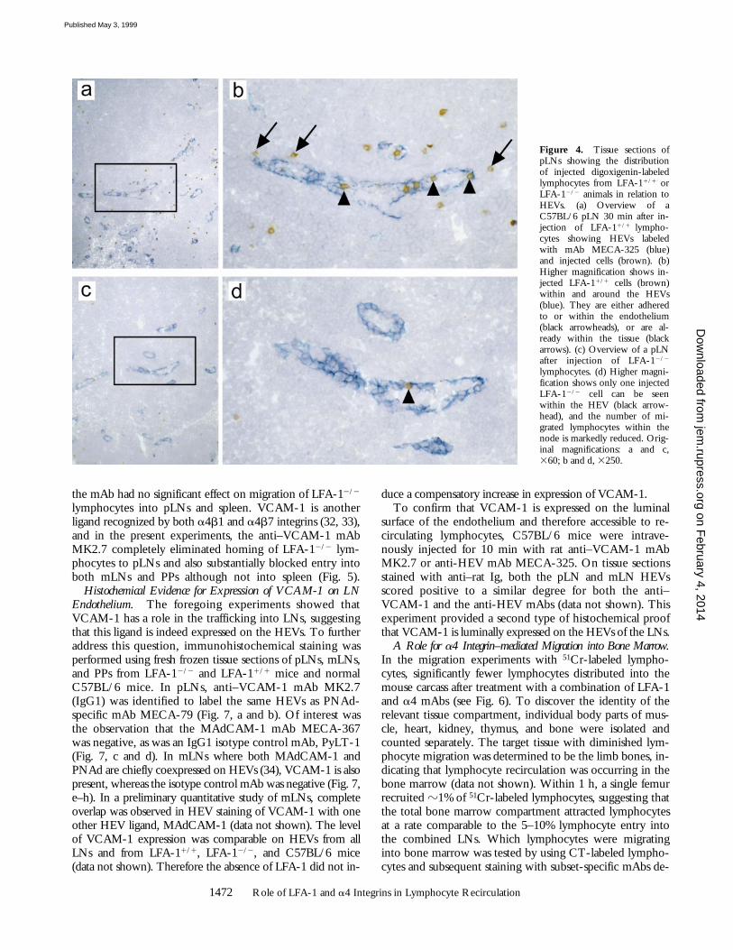
LFA-12/2 and LFA-11/1 lymphocytes for 30 min. Asshown in Fig. 4, the LFA-1–deficient lymphocytes boundless well to HEVs than the LFA-1-expressing lymphocytes.The total numbers of lymphocytes at the HEV level and sur-rounding 4-cell diameters were 285 6 70 cells/mm2 forLFA-11/1 (Fig. 4, a and b) and 85 6 10 cells/mm2 for LFA-12/2 lymphocytes (Fig. 4, c and d) (n 5 3). These data sug-gest that loss of LFA-1 expression reduces lymphocyte ad-herence to and transmigration across HEVs by 70%, therebycausing substantially diminished recruitment into pLNs.When the cells were divided between those adhering to andwithin the HEVs and those found within 4-cell diametersbeyond HEVs, the following proportions were observed: forLFA-11/1 lymphocytes, 73 6 7 and 276 2%; for LFA-12/2
lymphocytes, 78 6 1 and 22 6 1%, respectively. These datasuggest that the major block is at the level of the HEVs. Ifthe deficiency had been acting selectively at the level of thetransmigration step, a larger accumulation of lymphocytes atthe HEV level would have been expected.
A General Role for a4 Integrins and LFA-1 in LN Homing.Although LFA-1 was obviously playing a critical role at theHEV level, a proportion of lymphocytes remained capableof migrating into lymphoid tissue (Fig. 3 b, and Fig. 4). Ofother adhesion receptors that might substitute for LFA-1,the a4 integrins were attractive candidates, as they were activein migration in other situations. In the next series of experi-ments, the migratory behavior of CT-labeled LFA-12/2 andLFA-11/1 cells was compared in host animals simulta-neously injected with Fab fragments of a4 mAb or a4b7-specific mAb. The a4 mAb completely prevented the mi-gration of the residual numbers of LFA-12/2 lymphocyteshoming to pLNs, mLNs, and PPs (Fig. 5). In addition, thea4b7-specific mAb blocked lymphocyte entry into pLNsto z22% and completely prevented migration into mLNsand PPs. There was no significant effect of a4 or a4b7mAbs on migration into spleen.
These results were duplicated in a 51Cr lymphocyte la-beling experiment in which a4 mAb abolished all entryinto the pLNs, mLNs, and PPs of LFA-12/2 lymphocytes(data not shown). The a4b7 mAb blocked entry intomLNs and PPs and inhibited entry into pLNs to z35%(data not shown). It was possible that the a4-dependentLFA-12/2 cells migrating into pLNs were a specific subsetof lymphocytes expanded in the LFA-1–deficient environ-ment. However, phenotyping of these cells using a thirdfluorescent tag of Tricolor-conjugated anti–rat Ig showedthe migrated LFA-12/2 and LFA-11/1 lymphocytes to haveidentical phenotypic profiles with regard to their levels ofa4 and a4b7 integrins and L-selectin (data not shown).
The suggestion that a4 integrins, a4b7 acting togetherwith a4b1, have a role in migration to the pLNs has notpreviously been recognized. To further confirm the find-ings and to gain information about tissues other than second-ary LNs, 51Cr-labeled lymphocytes from normal BALB/cmice were coinjected with 300 mg of Fab fragments fromeither a4 mAb, LFA-1 mAb, or both into host BALB/cmice. The findings with the single mAbs were as reportedpreviously (7, 10; Fig. 6), but the combination of a4 andLFA-1 mAbs totally prevented migration into pLNs as wellas mLNs and PPs (Fig. 6). There was also an appreciabledecrease in migration into intestinal tissue and into the“body” (see below). This mAb blockade provoked an in-crease in circulating blood cells as well as an increase in mi-gration into the spleen. Put together, these observationsprovide evidence that the a4 integrins operating togetherwith LFA-1 have an essential role in migration to pLNs, asfound previously for other secondary LNs.
Ligands for a4 Integrins on LN HEVs. The identificationof the ligand(s) recognized by the a4 integrins, particularlyin pLNs, was next explored. Using a MAdCAM-1–specificmAb, homing to both PPs and mLNs was substantially pre-vented, as described previously (16, 17; Fig. 5). However,
Figure 3. Differential rates of homing of CT-labeled lymphocytes fromwild-type and LFA-12/2 mice. (a) Two-color FACS® analysis of the cellmixture of LFA-12/2 and LFA-11/1 cells injected intravenously into a wild-type C57BL/6 mouse (left) and the fluorescent cells recovered from thepLNs after a 1-h homing period (right). For details, see Materials and Meth-ods. (b) Homing experiments show that within 1 h, LFA-12/2 lymphocytesmigrate into pLNs, mLNs, and PPs with 21, 51, and 68% of the efficiency ofLFA-11/1 lymphocytes, respectively. Conversely, LFA-1–deficient lympho-cytes occur in larger numbers in the spleen than do wild-type cells (ratio of1.29). These data are averaged from 10–12 experiments. In all cases, themean ratios are significantly different from 1.0 (P , 0.05) as assessed by t test.
on February 4, 2014
jem.rupress.org
Dow
nloaded from
Published May 3, 1999
1472 Role of LFA-1 and a4 Integrins in Lymphocyte Recirculation
Figure 4. Tissue sections ofpLNs showing the distributionof injected digoxigenin-labeledlymphocytes from LFA-11/1 orLFA-12/2 animals in relation toHEVs. (a) Overview of aC57BL/6 pLN 30 min after in-jection of LFA-11/1 lympho-cytes showing HEVs labeledwith mAb MECA-325 (blue)and injected cells (brown). (b)Higher magnification shows in-jected LFA-11/1 cells (brown)within and around the HEVs(blue). They are either adheredto or within the endothelium(black arrowheads), or are al-ready within the tissue (blackarrows). (c) Overview of a pLNafter injection of LFA-12/2
lymphocytes. (d) Higher magni-fication shows only one injectedLFA-12/2 cell can be seenwithin the HEV (black arrow-head), and the number of mi-grated lymphocytes within thenode is markedly reduced. Orig-inal magnifications: a and c,360; b and d, 3250.
the mAb had no significant effect on migration of LFA-12/2
lymphocytes into pLNs and spleen. VCAM-1 is anotherligand recognized by both a4b1 and a4b7 integrins (32, 33),and in the present experiments, the anti–VCAM-1 mAbMK2.7 completely eliminated homing of LFA-12/2 lym-phocytes to pLNs and also substantially blocked entry intoboth mLNs and PPs although not into spleen (Fig. 5).
Histochemical Evidence for Expression of VCAM-1 on LNEndothelium. The foregoing experiments showed thatVCAM-1 has a role in the trafficking into LNs, suggestingthat this ligand is indeed expressed on the HEVs. To furtheraddress this question, immunohistochemical staining wasperformed using fresh frozen tissue sections of pLNs, mLNs,and PPs from LFA-12/2 and LFA-11/1 mice and normalC57BL/6 mice. In pLNs, anti–VCAM-1 mAb MK2.7(IgG1) was identified to label the same HEVs as PNAd-specific mAb MECA-79 (Fig. 7, a and b). Of interest wasthe observation that the MAdCAM-1 mAb MECA-367was negative, as was an IgG1 isotype control mAb, PyLT-1(Fig. 7, c and d). In mLNs where both MAdCAM-1 andPNAd are chiefly coexpressed on HEVs (34), VCAM-1 is alsopresent, whereas the isotype control mAb was negative (Fig. 7,e–h). In a preliminary quantitative study of mLNs, completeoverlap was observed in HEV staining of VCAM-1 with oneother HEV ligand, MAdCAM-1 (data not shown). The levelof VCAM-1 expression was comparable on HEVs from allLNs and from LFA-11/1, LFA-12/2, and C57BL/6 mice(data not shown). Therefore the absence of LFA-1 did not in-
duce a compensatory increase in expression of VCAM-1.To confirm that VCAM-1 is expressed on the luminal
surface of the endothelium and therefore accessible to re-circulating lymphocytes, C57BL/6 mice were intrave-nously injected for 10 min with rat anti–VCAM-1 mAbMK2.7 or anti-HEV mAb MECA-325. On tissue sectionsstained with anti–rat Ig, both the pLN and mLN HEVsscored positive to a similar degree for both the anti–VCAM-1 and the anti-HEV mAbs (data not shown). Thisexperiment provided a second type of histochemical proofthat VCAM-1 is luminally expressed on the HEVs of the LNs.
A Role for a4 Integrin–mediated Migration into Bone Marrow.In the migration experiments with 51Cr-labeled lympho-cytes, significantly fewer lymphocytes distributed into themouse carcass after treatment with a combination of LFA-1and a4 mAbs (see Fig. 6). To discover the identity of therelevant tissue compartment, individual body parts of mus-cle, heart, kidney, thymus, and bone were isolated andcounted separately. The target tissue with diminished lym-phocyte migration was determined to be the limb bones, in-dicating that lymphocyte recirculation was occurring in thebone marrow (data not shown). Within 1 h, a single femurrecruited z1% of 51Cr-labeled lymphocytes, suggesting thatthe total bone marrow compartment attracted lymphocytesat a rate comparable to the 5–10% lymphocyte entry intothe combined LNs. Which lymphocytes were migratinginto bone marrow was tested by using CT-labeled lympho-cytes and subsequent staining with subset-specific mAbs de-
on February 4, 2014
jem.rupress.org
Dow
nloaded from
Published May 3, 1999

1473 Berlin-Rufenach et al.
tected with Tricolor-conjugated anti–rat Ig. The ratio ofmigrated to injected CD4/CD8/B220 subsets was z0.5/1.0/2.0. This advantage for B cell and disadvantage for CD4T cells were independent of LFA-1 expression status.
LFA-11/1 and LFA-12/2 cells did not significantly differ intheir trafficking to bone marrow (ratio of LFA-12/2 to LFA-11/1 was 1.1; data not shown), suggesting that the migrationwas not solely reliant on LFA-1. However, when LFA-12/2
lymphocytes were coinjected with a4 mAb, the trafficking tobone marrow was completely abolished (Fig. 8). These resultssuggest that migration into bone marrow can be accom-plished either by LFA-1 or in its absence by an a4 integrin.As the a4b7 mAb had a partial effect on migration, whereasa4 mAb blocked migration completely, we conclude that inbone marrow, in contrast to the migration into LNs, a4b1dominates a4b7. Finally, the VCAM-1 mAb, but not MAd-
Figure 6. The effect of anti-a4and LFA-1 mAbs on the migra-tion of 51Cr-labeled BALB/c lym-phocytes within 1 h after tail veininjection into host BALB/c mice.Distribution of radioactivity invarious organs when lymphocyteswere coinjected with anti-a4mAb PS/2 (hatched bars), anti–LFA-1 mAb H35.89.9 (cross-hatched bars), anti–LFA-1 and a4mAbs together (black bars), andcontrol (white bars). The a4 mAbinhibited entry of lymphocytesinto PPs and intestine. The LFA-1mAb inhibited entry of lympho-cytes into pLNs and redistributionto the spleen. The mAb combina-tion inhibited entry of lympho-cytes into pLNs, mLNs, PPs,intestine, and the “body” and in-creased redistribution into spleen.Note the increased numerical scalefor the organs shown on the right.Four animals were used per exper-imental group (n 5 2; bars, 6SD).
Figure 5. The effect of vari-ous mAbs on the homing ofLFA-1–deficient lymphocytes topLNs, mLNs, PPs, and spleen. Ineach case, the relative ratio ofLFA-12/2 to LFA-11/1 migra-tion (0.21, 0.51, 0.68, and 1.29,respectively; see Fig. 3 b) is set at100% in order to assess the fur-ther reduction in migrationcaused by specific antibodies.Anti–a4 integrin (PS/2) blockedthe residual ability of LFA-12/2
lymphocytes to enter all threeLNs, but did not significantly af-fect their entry into spleen. Anti-a4b7 mAb DATK32 mirroredthe effects of a4 mAb on mLNsand PPs, but reduced pLN mi-gration to z25%. Anti–VCAM-1mAb (MK2.7) eliminated themigration of LFA-12/2 cells intopLNs and partially limited theirentry into mLNs and PPs. Con-versely, anti–MAdCAM-1 mAb(MECA-367) blocked the hom-ing of LFA-12/2 cells into PPs,had a lesser effect on mLNs, andshowed no inhibition of lym-phocyte entry into pLNs orspleen. Four or five animals wereused per experimental group (n 52 or 3; bars, 6SD).
on February 4, 2014
jem.rupress.org
Dow
nloaded from
Published May 3, 1999

1474 Role of LFA-1 and a4 Integrins in Lymphocyte Recirculation
CAM-1 mAb, had an inhibitory effect. Considered together,these results demonstrate that migration of recirculating lym-phocytes into bone marrow can be mediated by either LFA-1or the a4 integrins, and, as in pLNs, that VCAM-1 serves asthe chief ligand for the latter receptors.
Discussion
This study shows that LFA-1 has a key role in migration tothe pLNs, other LNs, and bone marrow but not into the
spleen. Also revealed is a hitherto unrecognized ability of thea4 integrins, a4b7 and a4b1, to compensate for the lack ofLFA-1 in lymphocyte trafficking to pLNs and other lymphoidtissues, including bone marrow. In general, these findingshighlight common features between lymphocyte homing andthe response to inflammatory stimuli and extend the validity ofthe multistep model of adhesion and transmigration.
The LFA-12/2 mouse described in this report has, as its keyphenotypic characteristic, pLNs that contain z30% normallymphocyte numbers, as also noted previously (25). The de-creased trafficking of LFA-12/2 lymphocytes to the pLNs toz20–30% of wild-type lymphocytes suggested that LFA-12/2
lymphocytes, irrespective of which subset, had difficultygaining entry to the LNs. This was confirmed by microscopicstudies showing that circulating LFA-12/2 lymphocytesbound poorly to HEVs. Migration into mLNs and PPs wasalso depressed, but LN cell counts were normal, suggestingcompensatory measures were in operation in these tissuesbut not in pLNs. The decrease in migration of lymphocytesto pLNs is consistent with previous work using function-blocking LFA-1 mAbs (10) and has recently been reported inanother study using LFA-1–deficient mice (35). Thus, thegeneral importance of LFA-1 in mature lymphocyte traffick-ing to secondary lymphoid tissue is confirmed.
In our studies, LFA-12/2 lymphocytes were able to gainentry into the pLNs, albeit at a lower level, suggesting in-volvement of further receptors. We here demonstrate theseadditional receptors to be the a4 integrins. Skewed recep-tor usage towards increased expression of a4 integrins inLFA-12/2 mice as a possible cause of experimental bias wasexcluded (data not shown). The fact that the presence ofmAbs to both LFA-1 and a4 integrins caused completeblockade of lymphocyte entry into normal pLNs and otherLNs strongly implies that a4 has a critical role in migrationof normal lymphocytes into pLNs. That this role for a4 in-tegrin has not previously been observed might be becauseLFA-1 has a larger role than a4 integrin in adherence to
Figure 7. Histochemical demonstration of the presence of a4 integrinligands in LFA-11/1 pLNs (a–d) and mLNs (e–h). The panels show stain-ing of identical tissue sections of HEVs in the pLNs with mAbs specificfor (a) VCAM-1, (b) PNAd, (c) MAdCAM-1, and (d) an isotype-matched IgG1 control mAb and similarly, in the mLNs for (e) VCAM-1,(f) PNAd, (g) MAdCAM-1, and (h) IgG1 control mAb. Original magnifi-cation: a–h, 3150.
Figure 8. The effect of various mAbs on the relative entry of LFA-12/2
to LFA-11/1 lymphocytes into bone marrow. In the absence of antibody,the relative ratio is 1.1. Coinjection of a4 mAb or VCAM-1 mAb com-pletely inhibited the migration of LFA-12/2 cells, whereas a4b7 mAb has apartial effect and MAdCAM-1 mAb is without effect.
on February 4, 2014
jem.rupress.org
Dow
nloaded from
Published May 3, 1999

1475 Berlin-Rufenach et al.
pLN HEVs than HEVs of other organs. This is in goodagreement with intravital microscopy studies which showLFA-1 and L-selectin are the essential codependent pLN-specific adhesion pair for lymphocyte adherence to pLNvessels (36). Put together, the data suggest that a4 integrinshave a less prominent role in adherence to pLN HEVs, butpotentially a larger role in the transmigration step.
Our data also suggest unexpectedly that a4b7 acts as themajor a4 receptor in conjunction with a4b1 in lympho-cyte recirculation into pLNs. There has previously been noevidence to link a4b7 with pLN migration, although itsdominant role in trafficking into mucosal tissue of themLNs and PPs is well documented (3, 17) and is confirmedhere. The integrin a4b1 has usually been associated withinflammatory responses (18–20) rather than with lympho-cyte recirculation. However, as these two a4 integrins mightbe acting either in synergy or in sequence, the extent of thecontribution of a4b1 must await a potent murine CD29blocking mAb. The results imply that the succession of ad-hesive events in lymphocyte recirculation is similar to thatin inflammatory responses, and that the a4 integrins in co-operation with L-selectin and LFA-1 have a more specificand necessary role than previously perceived.
The data presented here identify VCAM-1 and notMAdCAM-1 as ligand on pLN HEVs for a4 integrins inspite of a4b7 involvement. The result was unexpected, asa4b1 has been identified as the major VCAM-1 binding in-tegrin in inflammatory responses (37) in studies using trans-fectant (38) and cell lines (17). VCAM-1 also made a signifi-cant contribution to trafficking into mLNs and, to a lesserextent, PPs. This cooperative activity with MAdCAM-1 wasalso unexpected, as it has previously been considered that en-try into these LNs was regulated only by MAdCAM-1. Thefindings presented here suggest that potential roles for a4b7in addition to a4b1 should be sought in other circum-stances where VCAM-1 is the major ligand.
One explanation for having overlooked the importanceof VCAM-1 as an HEV ligand is its reported absence fromnormal lymphoid tissue (22, 23, 39), with the exception ofa report on its expression on rat HEVs (40). In the presentstudy, expression occurred at comparable levels on HEVsin pLNs, mLNs, and PP LNs and without obvious differ-ence between LFA-12/2 mice, their wild-type littermates,C57BL/6, or BALB/c mice. Such broad and constant pres-ence of VCAM-1 implies it is constitutively expressedrather than as a consequence of an inflammatory signal or acompensatory mechanism in LFA-12/2 mice.
The involvement of VCAM-1 raises the issue as towhether lymphocytes can use this ligand for migrationacross HEVs into the LN as well as adhesion to the luminalsurface of HEVs. In this study, the lack of LFA-1 decreasedmigration across HEVs by z25% only, suggesting involve-ment of other receptor/ligand pair(s) (data not shown). ForHUVECs, VCAM-1 is reported to be restricted to an api-cal location and therefore available for adhesion but not fortransendothelial migration (41). However, monocytes cantransmigrate HUVECs, making use of a4/VCAM-1 inde-pendently of b2 integrins (42). In mice, two major forms of
VCAM-1 have been identified, the common seven-domainform and also a three-domain glycosylphosphatidylinositol(GPI)-linked splice variant which is induced by inflamma-tion (43, 44). It is of interest that in polarized epithelial cells,the GPI-linked VCAM-1 was found apically, whereas theseven-domain VCAM-1 was localized to the basolateral sur-face, where it could theoretically serve as a ligand for trans-migration (45). The issue of where VCAM-1 is expressed,particularly in murine HEVs, warrants further investigation.
Migration of recirculating lymphocytes to bone marrow,a primary lymphoid tissue, has been noted in the past (46).Bone marrow serves as a site of hematopoiesis and for Bcell maturation in the mammal. It can also act as a reservoirfor a primary immune response in the absence of a spleenand presence of L-selectin mAb (47). In this study, we findan unexpectedly high degree of mature lymphocyte recir-culation in which B cells dominate into bone marrow. Wesuggest here that bone marrow, but not the thymus (datanot shown), should be considered as a major part of thelymphocyte recirculation network.
Migration to bone marrow is restricted by adhesionmechanisms. Both human (48) and murine (49) hemato-poietic progenitors make use of a4 integrin to lodge in thebone marrow. The a4 integrins are also reported to benecessary for correct lymphocyte development (50). Weshow that normal lymphocyte recirculation through the bonemarrow is regulated by LFA-1 and the a4 integrins and, incontrast to the LNs, a4b1 substantially dominates a4b7. Inthis context, it is again VCAM-1 and not MAdCAM-1which acts as ligand for the a4 integrins. VCAM-1 is ex-pressed constitutively by bone marrow sinusoidal vascula-ture (51, 52), and as progenitor cells have been demon-strated to roll on bone marrow endothelial VCAM-1 (48),it can be speculated that other lymphocytes might behavesimilarly. However VCAM-1 is also expressed constitu-tively by stromal reticular cells (53, 54), and it is possiblethat lymphocytes may be retarded not only at the level ofthe endothelium but also within the bone marrow matrix.Factors produced by the stromal cells might be beneficialfor the maintenance of the recirculating lymphocytes. Forexample, the interaction with VCAM-1 might suppresslymphocyte apoptosis (55). On the other hand, the incom-ing cells might contribute to the regulation of hematopoiesis.
In summary, the observations presented here extend andvalidate the concept of a multistage adhesion response requir-ing selectins and integrins. Rather than exclusive LN-specificuse of receptors such as LFA-1 and L-selectin in trafficking topLNs or a4b7 to mucosal sites, we have presented evidencethat LFA-1 and the a4 integrins can operate in migration ofunstimulated lymphocytes to the pLNs as well as other sec-ondary LNs and bone marrow. These general features, in-cluding the use of VCAM-1 as an a4 integrin ligand, resem-ble the response to an inflammatory signal. Our findingssuggest a unifying hypothesis for migration of lymphocytesacross HEVs into secondary lymphoid tissue and also establishfurther parallels between the activities of adhesion receptorsin the “homing” context and the mechanisms used in themore sporadic responses to tissue injury and inflammation.
on February 4, 2014
jem.rupress.org
Dow
nloaded from
Published May 3, 1999

1476 Role of LFA-1 and a4 Integrins in Lymphocyte Recirculation
We thank Ian Rosewell for blastocyst injection, and Tracy Crafton and Peter Hagger for mouse breeding.For carrying out the histochemical staining procedures, we are indebted to Phillipa Munson and KeithMiller, Department of Histopathology, University College London Medical School, and George Elia andRichard Poulsom, Histopathology Laboratory, Imperial Cancer Research Fund. We also gratefully ac-knowledge the statistical help of Henry Potts, Imperial Cancer Research Fund Medical Statistics Group,Oxford. We thank Britta Engelhardt for the murine ICAM-1 construct; and Mark Singer, Steve Rosen,Fumio Takei, and Michel Pierres for mAbs.
C. Berlin-Rufenach was supported by an EMBO Fellowship. F. Otto was supported by Deutsche For-schungsgemeinschaft fellowship Ot 134/1-1. M. Mathies was supported by The Claremont Colleges, Clare-mont, CA. This work was funded by the Deutsche Forschungsgemeinschaft (A. Hamann) and the ImperialCancer Research Fund.
Address correspondence to Nancy Hogg, Leukocyte Adhesion Laboratory, Imperial Cancer Research Fund,Lincoln’s Inn Fields, London WC2A 3PX, UK. Phone: 44-171-269-3255; Fax: 44-171-269-3093; E-mail:[email protected]
F. Otto’s present address is University of Freiburg Medical Center, 79106 Freiburg, Germany, and M.Mathies’s is Joint Science Department, Claremont Colleges, Claremont, CA 91711.
Received for publication 17 September 1998 and in revised form 25 February 1999.
References1. Butcher, E.C. 1991. Leukocyte-endothelial cell recognition:
three (or more) steps to specificity and diversity. Cell. 67:1033–1036.
2. Springer, T.A. 1994. Traffic signals for lymphocyte recircula-tion and leukocyte emigration: the multistep paradigm. Cell.76:301–314.
3. Bargatze, R.F., M.A. Jutila, and E.C. Butcher. 1995. Distinctroles of L-selectin and integrins a4b7 and LFA-1 in lympho-cyte homing to Peyer’s patch-HEV in situ: the multistepmodel confirmed and refined. Immunity. 3:99–108.
4. Salmi, M., and S. Jalkanen. 1997. How do lymphocytesknow where to go: current concepts and enigmas of lympho-cyte homing. Adv. Immunol. 64:139–218.
5. Butcher, E.C., and L.J. Picker. 1996. Lymphocyte homingand homeostasis. Science. 272:60–66.
6. Hu, M.C.-T., D.T. Crowe, I.L. Weissman, and B. Holz-mann. 1992. Cloning and expression of mouse integrin bp(b7):a functional role in Peyer’s Patch-specific lymphocyte hom-ing. Proc. Natl. Acad. Sci. USA. 89:8254–8258.
7. Hamann, A., D.P. Andrew, D. Jablonski-Westrich, B. Holz-mann, and E.C. Butcher. 1994. Role of a4-integrins in lym-phocyte homing to mucosal tissues in vivo. J. Immunol. 152:3282–3293.
8. Gallatin, W.M., I.L. Weissman, and E.C. Butcher. 1983. Acell-surface molecule involved in organ-specific homing oflymphocytes. Nature. 304:30–34.
9. Arbones, M.L., D.C. Ord, K. Ley, L.K. Ratech, C. May-nard-Curry, G. Otten, D.J. Capon, and T.F. Tedder. 1994.Lymphocyte homing and leukocyte rolling and migration areimpaired in L-selectin-deficient mice. Immunity. 1:247–260.
10. Hamann, A., D.J. Westrich, A. Duijvestijn, E.C. Butcher, H.Baisch, R. Harder, and H.G. Thiele. 1988. Evidence for anaccessory role of LFA-1 in lymphocyte-high endothelium in-teraction during homing. J. Immunol. 140:693–699.
11. Hwang, S.T., M.S. Singer, P.A. Giblin, T.A. Yednock, K.B.Bacon, S.I. Simon, and S.D. Rosen. 1996. GlyCAM-1, aphysiologic ligand for L-selectin, activates b2 integrins on
naive peripheral lymphocytes. J. Exp. Med. 184:1343–1348.12. Gunn, M.D., V.N. Ngo, K.M. Ansel, E.H. Ekland, J.G.
Cyster, and L.T. Williams. 1998. A B-cell-homing chemo-kine made in lymphoid follicles activates Burkitt’s lymphomareceptor-1. Nature. 391:799–803.
13. Gunn, M.D., K. Tangemann, C. Tam, J.G. Cyster, S.D.Rosen, and L.T. Williams. 1998. Proc. Natl. Acad. Sci. USA.95:258–263.
14. Rosen, S.D., and C.R. Bertossi. 1994. The selectins and theirligands. Curr. Opin. Cell Biol. 6:663–673.
15. Gahmberg, C.G., M. Tolvanen, and P. Kotovuori. 1997.Structure and function of human leukocyte b2 integrins andtheir cellular ligands. Eur. J. Biochem. 245:215–232.
16. Streeter, P.R., E.L. Berg, B.T.N. Rouse, R.F. Bargatze, andE.C. Butcher. 1988. A tissue-specific endothelial cell mole-cule involved in lymphocyte homing. Nature. 331:41–46.
17. Berlin, C., E.L. Berg, M.J. Briskin, D.P. Andrew, P.J.Kilshaw, A. Holzmann, I.L. Weissman, A. Hamann, and E.C.Butcher. 1993. a4b7 integrin mediates lymphocyte bindingto the mucosal vascular addressin MAdCAM-1. Cell. 74:185–195.
18. Issekutz, T.B. 1991. Inhibition of in vivo lymphocyte migra-tion to inflammation and homing to lymphoid tissues by theTA-2 monoclonal antibody. J. Immunol. 147:4178–4184.
19. Yednock, T.A., C. Cannon, L.C. Fritz, F. Sanchez-Madrid,L. Steinmann, and N. Karin. 1992. Prevention of experi-mental autoimmune encephalomyelitis by antibodies againsta4b1 integrin. Nature. 356:63–66.
20. Weg, V.B., T.J. Williams, R.R. Lobb, and S. Nourshargh.1993. A monoclonal antibody recognizing very late activa-tion antigen-4 inhibits eosinophil accumulation in vivo. J.Exp. Med. 177:561–566.
21. Osborn, L. 1990. Leukocyte adhesion to endothelium in in-flammation. Cell. 62:3–6.
22. Rice, G.E., J.M. Munro, C. Corless, and M.P. Bevilacqua.1991. Vascular and nonvascular expression of INCAM-110.A target for mononuclear leukocyte adhesion in normal and
on February 4, 2014
jem.rupress.org
Dow
nloaded from
Published May 3, 1999

1477 Berlin-Rufenach et al.
inflamed human tissues. Am. J. Pathol. 138:385–393.23. Hahne, M., M. Lenter, U. Jager, S. Isenmann, and D.
Vestweber. 1993. VCAM-1 is not involved in LPAM-1 me-diated binding of lymphoma cells to high endothelial venulesof mucosa-associated lymph nodes. Eur. J. Cell Biol. 61:290–298.
24. Elices, M.J., V. Tsai, D. Strahl, A.S. Goel, V. Tollefson, T.Arrhenius, E.A. Wayner, F.C. Gaeta, J.D. Fikes, and G.S.Firestein. 1994. Expression and functional significance of al-ternatively spliced CS1 fibronectin in rheumatoid arthritismicrovasculature. J. Clin. Invest. 93:405–416.
25. Schmits, R., T.M. Kündig, D.M. Baker, G. Shumaker, J.J.L.Simard, G. Duncan, A. Wakeham, A. Shahinian, A. van derHeiden, M.F. Bachmann, et al. 1996. LFA-1–deficient miceshow normal CTL responses to virus but fail to reject immu-nogenic tumor. J. Exp. Med. 183:1415–1426.
26. Shier, P., G. Otulakowski, K. Ngo, J. Panakos, E. Chour-mouzis, L. Christjansen, C.Y. Lau, and W.-P. Fung-Leung.1996. Impaired immune responses toward alloantigens andtumor cells but normal thymic selection in mice deficient inthe b2 integrin leukocyte function-associated antigen-1. J.Immunol. 157:5375–5386.
27. Otto, F., A.P. Thornell, T. Crompton, A. Denzel, K.C.Gilmour, I.R. Rosewell, G.W.H. Stamp, R.S.P. Bedding-ton, S. Mundlos, B.R. Olsen, et al. 1997. Cbfa1, a candidategene for cleidocranial dysplasia syndrome, is essential for os-teoblast differentiation and bone development. Cell. 89:765–771.
28. Szabo, M.C., E.C. Butcher, and L.M. McEvoy. 1997. Spe-cialization of mucosal follicular dendritic cells revealed bymucosal addressin-cell adhesion molecule-1 display. J. Immu-nol. 158:5584–5588.
29. Hamann, A., and P. Jonas. 1997. Lymphocyte migration invivo: the mouse model. In Immunology Methods Manual.Vol. 3. I. Lefkovits, editor. Academic Press Ltd., London.1333–1341.
30. Kishimoto, T.K., M.A. Jutila, E.L. Berg, and E.C. Butcher.1989. Neutrophil Mac-1 and MEL-14 adhesion proteins in-versely regulated by chemotactic factors. Science. 245:1238–1241.
31. Westermann, J., U. Geismar, A. Sponholz, U. Bode, S.M.Sparshott, and E.B. Bell. 1997. CD41 T cells of both the na-ive and the memory phenotype enter rat lymph nodes andPeyer’s patches via high endothelial venules: within the tissuetheir migratory behaviour differs. Eur. J. Immunol. 27:3174–3181.
32. Ruegg, C., A.A. Postigo, E.E. Sikorski, E.C. Butcher, R.Pytela, and D.J. Erle. 1992. Role of integrin a4b7/a4bP inlymphocyte adherence to fibronectin and VCAM-1 and inhomotypic cell clustering. J. Cell Biol. 117:179–189.
33. Strauch, U.G., A. Lifka, U. Gosslar, P.J. Kilshaw, J. Cle-ments, and B. Holzmann. 1994. Distinct binding specificitiesof integrins a4b7 (LPAM-1), a4b1 (VLA-4), and aIELb7.Int. Immun. 6:263–275.
34. Streeter, P.R., B.T.N. Rouse, and E.C. Butcher. 1988. Im-munohistologic and functional characterization of a vascularaddressin involved in lymphocyte homing into peripherallymph nodes. J. Immunol. 107:1853–1862.
35. Andrew, D.P., J.P. Spellberg, H. Takimoto, R. Schmits,T.W. Mak, and M.M. Zukowski. 1998. Transendothelialmigration and trafficking of leukocytes in LFA-1-deficientmice. Eur. J. Immunol. 28:1959–1969.
36. Warnock, R.A., S. Askari, E.C. Butcher, and U.H. von An-
drian. 1998. Molecular mechanisms of lymphocyte homingto peripheral lymph nodes. J. Exp. Med. 187:205–216.
37. Rott, L.S., M.J. Briskin, D.P. Andrew, E.L. Berg, and E.C.Butcher. 1996. A fundamental subdivision of circulating lym-phocytes defined by adhesion to mucosal addressin cell adhe-sion molecule-1. J. Immunol. 156:3727–3736.
38. Chan, B.M., M.J. Elices, E. Murphy, and M.E. Hemler.1992. Adhesion to vascular cell adhesion molecule 1 and fi-bronectin. J. Biol. Chem. 267:8366–8370.
39. Henninger, D.D., J. Panes, M. Eppihimer, J. Russell, M.Gerritsen, D.C. Anderson, and D.N. Granger. 1997. Cyto-kine-induced VCAM-1 and ICAM-1 expression in differentorgans of the mouse. J. Immunol. 158:1825–1832.
40. May, M.J., G. Entwistle, M.J. Humphries, and A. Ager.1993. VCAM-1 is a CS1 peptide-inhibitable adhesion mole-cule expressed by lymph node high endothelium. J. Cell Sci.106:109–119.
41. Oppenheimer-Marks, N., L.S. Davis, D.T. Bogue, J. Ram-berg, and P.E. Lipsky. 1991. Differential utilization ofICAM-1 and VCAM-1 during the adhesion and transendo-thelial migration of human T lymphocytes. J. Immunol. 147:2913–2921.
42. Chuluyan, H.E., and A.C. Issekutz. 1995. VLA-4 integrincan mediate CD11/CD18-independent transendothelial mi-gration of human monocytes. J. Clin. Invest. 92:2768–2777.
43. Moy, P., R. Lobb, R. Tizard, D. Olson, and C. Hession.1993. Cloning of an inflammation-specific phosphatidylinositol-linked form of murine vascular cell adhesion mole-cule-1. J. Biol. Chem. 268:8835–8841.
44. Hahne, M., M. Lenter, U. Jager, and D. Vestweber. 1994. Anovel soluble form of mouse VCAM-1 is generated from aglycolipid-anchored splicing variant. Eur. J. Immunol. 24:421–428.
45. Pirozzi, G., R.W. Terry, and M.A. Labow. 1994. Murinevascular cell adhesion molecule-1 (VCAM-1) proteins en-coded by alternatively spliced mRNAs are differentially tar-geted in polarized cells. Cell. Adhes. Commun. 2:549–556.
46. Smith, M.E., and W.L. Ford. 1983. The recirculating lym-phocyte pool of the rat: a systematic description of the migra-tory behaviour of recirculating lymphocytes. Immunology. 49:83–94.
47. Tripp, R.A., R.A. Topham, S.R. Watson, and P.C.Doherty. 1997. Bone marrow can function as a lymphoid or-gan during a primary immune response under conditions ofdisrupted lymphocyte trafficking. J. Immunol. 158:3716–3720.
48. Mazo, I.B., J.-C. Gutierrez-Ramos, P.S. Frenette, R.O.Hynes, D.D. Wagner, and U.H. von Andrian. 1998. Hema-topoietic progenitor cell rolling in bone marrow microves-sels: parallel contributions by endothelial selectins and vascu-lar cell adhesion molecule 1. J. Exp. Med. 188:465–474.
49. Papayannopoulou, T., C. Craddock, B. Nakamoto, G.V.Priestley, and N.S. Wolf. 1995. The VLA4/VCAM-1 adhe-sion pathway defines contrasting mechanisms of lodgementof transplanted murine hemopoietic progenitors betweenbone marrow and spleen. Proc. Natl. Acad. Sci. USA. 92:9647–9651.
50. Arroyo, A.G., Y.T. Yang, H. Rayburn, and R.O. Hynes.1996. Differential requirements for a4 integrins during fetaland adult hematopoiesis. Cell. 85:997–1008.
51. Schweitzer, K.M., A.M. Draeger, P. van der Valk, S.F.T.Thijsen, A. Zevenbergen, A.P. Theijsmeijer, C.E. van derSchoot, and M.M.A.C. Langenhuijsen. 1996. Constitutive
on February 4, 2014
jem.rupress.org
Dow
nloaded from
Published May 3, 1999
1478 Role of LFA-1 and a4 Integrins in Lymphocyte Recirculation
expression of E-selectin and vascular cell adhesion molecule-1on endothelial cells of hematopoietic tissues. Am. J. Pathol.148:165–175.
52. Jacobsen, K., P.W. Kravitz, P.W. Kincade, and D.G. Os-mond. 1996. Adhesion receptors on bone marrow stromalcells: in vivo expression of vascular cell adhesion molecule-1by reticular cells and sinusoidal endothelium in normal andgamma-irradiated mice. Blood. 87:73–82.
53. Miyake, K., K. Medina, K. Ishiara, M. Kimoto, R. Auer-bach, and P.W. Kincade. 1991. A VCAM-like adhesionmolecule on murine bone marrow stromal cells mediatesbinding of lymphocyte precursors in culture. J. Cell Biol. 114:
557–565.54. Simmons, P.J., B. Masinovsky, B.M. Longenecker, R. Be-
renson, B. Torok-Storb, and W.M. Gallatin. 1992. Vascularcell adhesion molecule-1 expressed by bone marrow stromalcells mediates the binding of hematopoietic progenitor cells.Blood. 80:388–395.
55. Koopman, G., R.M.J. Keehnen, E. Lindhout, W. Newman,Y. Shimizu, G.A. van Seventer, C. de Groot, and S.T. Pals.1994. Adhesion through the LFA-1 (CD11a/CD18)-ICAM-1(CD54) and the VLA-4 (CD49d)-VCAM-1 (CD106) path-ways prevents apoptosis of germinal center B cells. J. Immu-nol. 152:3760–3767.
on February 4, 2014
jem.rupress.org
Dow
nloaded from
Published May 3, 1999
Copyright © 2022 FDOKUMEN