Enhanced LTP in Mice Deficient in the AMPA Receptor GluR2
12
Neuron, Vol. 17, 945–956, November, 1996, Copyright 1996 by Cell Press Enhanced LTP in Mice Deficient in the AMPA Receptor GluR2 Zhengping Jia,* § Nadia Agopyan,* § Peter Miu, ² (Hollmann et al., 1991; Verdoorn et al., 1991). Compari- son of the amino acid sequence of the GluR2 subunit Zhigang Xiong, ‡ Jeff Henderson,* Robert Gerlai,* with that of the other subunits revealed a positively Franco A. Taverna,* Alexander Velumian, ² charged arginine (R) at position 586 of the transmem- John MacDonald, ‡ Peter Carlen, ² brane segment 2, instead of the neutral glutamine (Q) Wanda Abramow-Newerly,* and John Roder* found at the homologous position in the other subunits. *Samuel Lunenfeld Research Institute This position, termed the Q/R site, was subject to RNA Mount Sinai Hospital editing (Sommer et al., 1991) and is the main site control- 600 University Avenue ling divalent ion permeation of recombinant AMPA re- Toronto, Ontario M5G 1X5 ceptors (AMPARs) (Hume et al., 1991; Burnashev et al., ² Playfair Neuroscience Unit 1992a; Dingledine et al., 1992). Coexpression of the Toronto Hospital GluR2 subunit with GluR1, 3, and 4 showed that AMP- Toronto, Ontario ARs containing the Q/R edited GluR2 subunit exhibited ‡ Department of Physiology lower Ca 21 permeability and outwardly rectifying I/V rela- University of Toronto tionships. Conversely, overexpression of unedited Toronto, Ontario GluR2 yielded higher Ca 21 permeability in the neurons of gene-targeted mice (Brusa et al., 1995). The studies indicate that the channel properties of the GluR2 subunit Summary dominate those of the heteromeric receptor complex. Therefore, one would predict that Ca 21 permeability in AMPA receptors (AMPARs) are not thought to be in- the CNS could be controlled by regulating expression volved in the induction of long-term potentiation (LTP), of the GluR2 subunit. Indeed, the relative abundance of but may be involved in its expression via second mes- GluR2 mRNA correlates negatively with the Ca 21 perme- senger pathways. However, one subunit of the AMP- ability of native AMPARs in brain slices (Geiger et al., ARs, GluR2, is also known to control Ca 21 influx. To 1995). Thus, in principal neurons of the hippocampus test whether GluR2 plays any role in the induction of (i.e., pyramidal and granule neurons), where GluR2 is LTP, we generated mice that lacked this subunit. In highly expressed, AMPARs exhibit low Ca 21 permeabil- GluR2 mutants, LTP in the CA1 region of hippocampal ity, whereas in cerebellar Bergman glial cells (Burnashev slices was markedly enhanced (2-fold) and nonsatu- et al., 1992b), hippocampal basket cells, and neocortical rating, whereas neuronal excitability and paired-pulse nonpyramidal cells (Koh et al., 1995a), where expression of GluR2 is low or absent, AMPARs show higher Ca 21 facilitation were normal. The 9-fold increase in Ca 21 permeability. permeability, in response to kainate application, sug- While these data clearly show the dominant role of gests one possible mechanism for enhanced LTP. Mu- the GluR2 subunit in inhibiting Ca 21 influx via AMPARs, tant mice exhibited increased mortality, and those the functional consequence of Ca 21 entry through syn- surviving showed reduced exploration and impaired aptically activated AMPARs remains unknown. Since motor coordination. These results suggest an impor- Ca 21 -permeable AMPAR channels are blocked by intra- tant role for GluR2 in regulating synaptic plasticity and cellular polyamines (Kamboj et al., 1995; Koh et al., behavior. 1995b; Bowie and Mayer, 1995) at positive membrane potentials and N-methyl-D-aspartate (NMDA) receptor Introduction channels are blocked by extracellular Mg 21 at negative membrane potentials (MacDonald and Wojtowicz, 1982; In the mammalian CNS, a-amino-3-hydroxy-5-methyl- Nowak et al., 1984), it was hypothesized that at excit- 4-isoxazolepropionate (AMPA) receptor subtypes of the atory synapses, where AMPARs and NMDA receptors glutamate receptors are the principal mediators of fast are colocalized, Ca 21 influx through the AMPARs would excitatory synaptic transmission. Four genes, GluR1, be comparable with that through NMDARs at resting 2, 3, and 4 (also called GluRA, B, C, and D) encode membrane potential (Burnashev et al., 1995). Thus, hy- heteromeric receptors with high affinity for AMPA, but perpolarization would favor Ca 21 influx through AMP- low affinity for kainic acid (Hollmann and Heinemann, ARs, and depolarization would favor Ca 21 influx through 1994). The relative levels of expression of these genes, NMDA receptors, which has been implicated in long- as well as splicing and editing of their mRNAs, account term potentiation (LTP) (Bliss and Collingridge, 1993). for the wide differences in Ca 21 permeability and gating However, the lack of specific AMPAR subunit antago- between cells (Geiger et al., 1995). Homomeric channels, nists has hindered studies that would elucidate the func- assembled from GluR2 subunits, are not permeable to tional role of Ca 21 influx through the AMPARs. In an Ca 21 and showed outwardly rectifying current/voltage attempt to address the issue of whether Ca 21 influx through the AMPARs plays any modulatory role in syn- (I/V) relations. In contrast, receptors assembled from aptic transmission and synaptic plasticity, we have gen- GluR1, GluR3, and GluR4 subunits are highly permeable erated mutant mice lacking the GluR2 protein. Isolated to Ca 21 , and showed a doubly rectifying I/V relation hippocampal CA1 neurons showed enhanced Ca 21 per- meation and LTP was markedly enhanced in hippocam- pal slices. While several knockouts have decreased LTP § These authors contributed equally to this work.
Transcript of Enhanced LTP in Mice Deficient in the AMPA Receptor GluR2
Neuron, Vol. 17, 945–956, November, 1996, Copyright 1996 by Cell Press
Enhanced LTP in Mice Deficientin the AMPA Receptor GluR2
Zhengping Jia,*§ Nadia Agopyan,*§ Peter Miu,† (Hollmann et al., 1991; Verdoorn et al., 1991). Compari-son of the amino acid sequence of the GluR2 subunitZhigang Xiong,‡ Jeff Henderson,* Robert Gerlai,*with that of the other subunits revealed a positivelyFranco A. Taverna,* Alexander Velumian,†charged arginine (R) at position 586 of the transmem-John MacDonald,‡ Peter Carlen,†brane segment 2, instead of the neutral glutamine (Q)Wanda Abramow-Newerly,* and John Roder*found at the homologous position in the other subunits.*Samuel Lunenfeld Research InstituteThis position, termed the Q/R site, was subject to RNAMount Sinai Hospitalediting (Sommer et al., 1991) and is the main site control-600 University Avenueling divalent ion permeation of recombinant AMPA re-Toronto, Ontario M5G 1X5ceptors (AMPARs) (Hume et al., 1991; Burnashev et al.,†Playfair Neuroscience Unit1992a; Dingledine et al., 1992). Coexpression of theToronto HospitalGluR2 subunit with GluR1, 3, and 4 showed that AMP-Toronto, OntarioARs containing the Q/R edited GluR2 subunit exhibited‡Department of Physiologylower Ca21 permeability and outwardly rectifying I/V rela-University of Torontotionships. Conversely, overexpression of uneditedToronto, OntarioGluR2 yielded higher Ca21 permeability in the neuronsof gene-targeted mice (Brusa et al., 1995). The studiesindicate that the channel properties of the GluR2 subunit
Summary dominate those of the heteromeric receptor complex.Therefore, one would predict that Ca21 permeability in
AMPA receptors (AMPARs) are not thought to be in- the CNS could be controlled by regulating expressionvolved in the induction of long-term potentiation (LTP), of the GluR2 subunit. Indeed, the relative abundance ofbut may be involved in its expression via second mes- GluR2 mRNA correlates negatively with the Ca21 perme-senger pathways. However, one subunit of the AMP- ability of native AMPARs in brain slices (Geiger et al.,ARs, GluR2, is also known to control Ca21 influx. To 1995). Thus, in principal neurons of the hippocampustest whether GluR2 plays any role in the induction of (i.e., pyramidal and granule neurons), where GluR2 isLTP, we generated mice that lacked this subunit. In highly expressed, AMPARs exhibit low Ca21 permeabil-GluR2 mutants, LTP in the CA1 region of hippocampal ity, whereas in cerebellar Bergman glial cells (Burnashevslices was markedly enhanced (2-fold) and nonsatu- et al., 1992b), hippocampal basket cells, and neocorticalrating, whereas neuronal excitability and paired-pulse nonpyramidal cells (Koh et al., 1995a), where expression
of GluR2 is low or absent, AMPARs show higher Ca21facilitation were normal. The 9-fold increase in Ca21
permeability.permeability, in response to kainate application, sug-While these data clearly show the dominant role ofgests one possible mechanism for enhanced LTP. Mu-
the GluR2 subunit in inhibiting Ca21 influx via AMPARs,tant mice exhibited increased mortality, and thosethe functional consequence of Ca21 entry through syn-surviving showed reduced exploration and impairedaptically activated AMPARs remains unknown. Sincemotor coordination. These results suggest an impor-Ca21-permeable AMPAR channels are blocked by intra-tant role for GluR2 in regulating synaptic plasticity andcellular polyamines (Kamboj et al., 1995; Koh et al.,behavior.1995b; Bowie and Mayer, 1995) at positive membranepotentials and N-methyl-D-aspartate (NMDA) receptor
Introduction channels are blocked by extracellular Mg21 at negativemembrane potentials (MacDonald and Wojtowicz, 1982;
In the mammalian CNS, a-amino-3-hydroxy-5-methyl- Nowak et al., 1984), it was hypothesized that at excit-4-isoxazolepropionate (AMPA) receptor subtypes of the atory synapses, where AMPARs and NMDA receptorsglutamate receptors are the principal mediators of fast are colocalized, Ca21 influx through the AMPARs wouldexcitatory synaptic transmission. Four genes, GluR1, be comparable with that through NMDARs at resting2, 3, and 4 (also called GluRA, B, C, and D) encode membrane potential (Burnashev et al., 1995). Thus, hy-heteromeric receptors with high affinity for AMPA, but perpolarization would favor Ca21 influx through AMP-low affinity for kainic acid (Hollmann and Heinemann, ARs, and depolarization would favor Ca21 influx through1994). The relative levels of expression of these genes, NMDA receptors, which has been implicated in long-as well as splicing and editing of their mRNAs, account term potentiation (LTP) (Bliss and Collingridge, 1993).for the wide differences in Ca21 permeability and gating However, the lack of specific AMPAR subunit antago-between cells (Geiger et al., 1995).Homomeric channels, nists hashindered studies that would elucidate the func-assembled from GluR2 subunits, are not permeable to tional role of Ca21 influx through the AMPARs. In anCa21 and showed outwardly rectifying current/voltage attempt to address the issue of whether Ca21 influx
through the AMPARs plays any modulatory role in syn-(I/V) relations. In contrast, receptors assembled fromaptic transmission and synaptic plasticity, we have gen-GluR1, GluR3, and GluR4 subunits are highly permeableerated mutant mice lacking the GluR2 protein. Isolatedto Ca21, and showed a doubly rectifying I/V relationhippocampal CA1 neurons showed enhanced Ca21 per-meation and LTP was markedly enhanced in hippocam-pal slices. While several knockouts have decreased LTP§These authors contributed equally to this work.
Neuron946
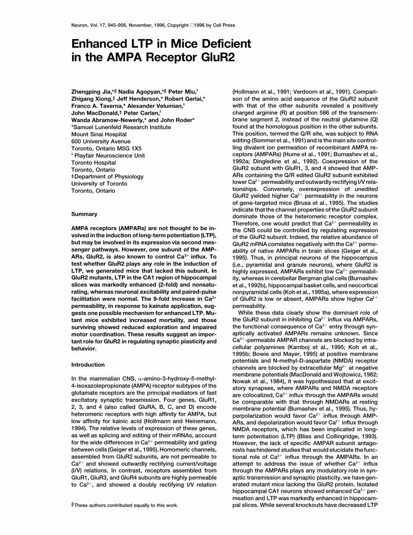
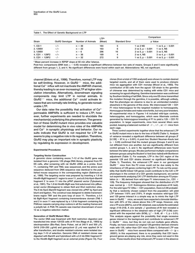
Figure 1. Targeted Disruption of the GluR2Gene
(A) The GluR2 cDNA showing the hydropho-bic membrane domains (TM) and the anti-GluR2 antibody epitope (3A111), the isogenic129 strain genomic clone (exons, openboxes), targeting vector, and the mutantGluR2 locus expected following homologousrecombination. B, BamHI; H, HindIII; Bg, BglII;K, KpnI; N, NotI.(B) Southern blot analysis of genomic DNAisolated from the tails of a representative F2litter of 12 mice derived from a heterozygous(1/2) 129 3 CD1 F1 inter-cross. DNA wasdigested with BamHI and probed with a 0.4kb fragment 59 to exon 10, yielding diagnostic11.5 kb restriction fragments from wild type(1/1) and 7.0 kb from mutant alleles (2/2).(C) Western blot analysis. Brain plasma mem-brane proteins were loaded at 50 mg per laneand probed with the antibodies indicated.(D) Sagittal sections of hippocampus from2/2 (left) or 1/1 (right) mice, stained withCresyl violet. Serial sections in the coronal,sagittal, and horizontal planes of the entireCNS of wild-type (n 5 2) and GluR22/2 mutant(n 5 2) mice revealed no difference in cellularmorphology or in the organization and extentof fiber tracts (see Experimental Procedures).Staining with cytochrome C oxidase, a grossmeasure of presynaptic input, was also nor-mal in mutants.
(e.g., Silva et al., 1992), this is the first knockout showing were used to produce aggregation chimeras with CD1morulae (Wood et al., 1993). Only one ES clone transmit-elevated LTP and behavioral abnormalities associated
with it. Our data raise the possibility that Ca21 influx ted the GluR2 mutation through the germline. Heterozy-gous mice from a CD1 3 129 cross were intercrossedvia AMPARs alone may be able to induce long-lasting
increases in synaptic efficacy, suggesting an important to produce 477 F2 offspring, of which 22% were 1/1,54% 1/2, and 24% 2/2 (Figure 1B). This 1:2:1 Mende-and unexpected role in synaptic plasticity.lian ratio suggests there was no embryonic lethality inthe mutants. Mice from this cross were used inall experi-Resultsments (see Figures 1–6), except that represented in Ta-ble 1. Western blot analysis of brain protein with a GluR2Targeted Disruption of the GluR2 Gene
To disrupt the GluR2 locus, an isogenic targeting vector antibody (3A11), produced against the N-terminal do-main (Puchalski et al., 1994), showed no detectablewas designed to delete transmembrane region 1 and
the pore loop, which are essential for receptor function GluR2 protein, either full-length (Figure 1C) or truncated,even at a 100-fold excess loading and a 1% level of(Figure 1A) (Hollmann and Heinemann, 1994). R1 embry-
onic stem (ES) cells (strain 129) were electropororated detection (data not shown). Enhanced chemilumines-cence (ECL) detection showed that 1/2 mice hadwith this vector and selected in G418 and gancyclovir
(Nagy et al., 1993). Double resistant clones were z50% 6 10% (n 5 3) of GluR2 protein compared with1/1. In mutant mice, the level of the other glutamatescreened for the desired homologous recombination by
Southern blotting using a probe 59 to exon 10 (Figure receptors (GluR1, 4, 6, and 7; NMDAR1, 2A, and 2B)was not altered. Since there are no GluR3 antibodies1A). Four ES clones contained the targeting events, and
Inactivation of the GluR2 Locus947
available, we used one that recognizes the C-terminusof both GluR2 and GluR3. GluR3 is known to be ex-pressed at lower levels in brain than GluR2. As shownin Figure 1C, the lane from GluR22/2 mice showed aband indicating the presence of GluR3 in the GluR2knockout mice. Based on the normal ratios of GluR2and 3, it is unlikely that GluR3 is overexpressed, sincethe band is less intense than the GluR2 band. However,we cannot exclude the unlikely possibility that GluR3 isdownregulated in the absence of GluR2. This experi-ment was repeated several times with similar results,using enriched synaptic membranes as well as whole-protein fractions from brain. Therefore, as best we canjudge, the loss of GluR2 was selective and did not resultin any developmental compensation or downregulationin other glutamate receptors. GluR22/2 mice were alsofound to possess all major neuroanatomic loci and fiberpathways in grossly normal proportion. The hippocam-pus exhibited normal cellularity in the CA regions 1–3and dentate gyrus (Figure 1D). Golgi staining of the indi-vidual pyramidal neurons also revealed normal morphol-ogy. Fiber pathways within the hippocampus, such asthe perforant path, also appeared normal, as did thestructure of the cerebellum, the dorsal laminae of thespinal cord, and retina (data not shown).
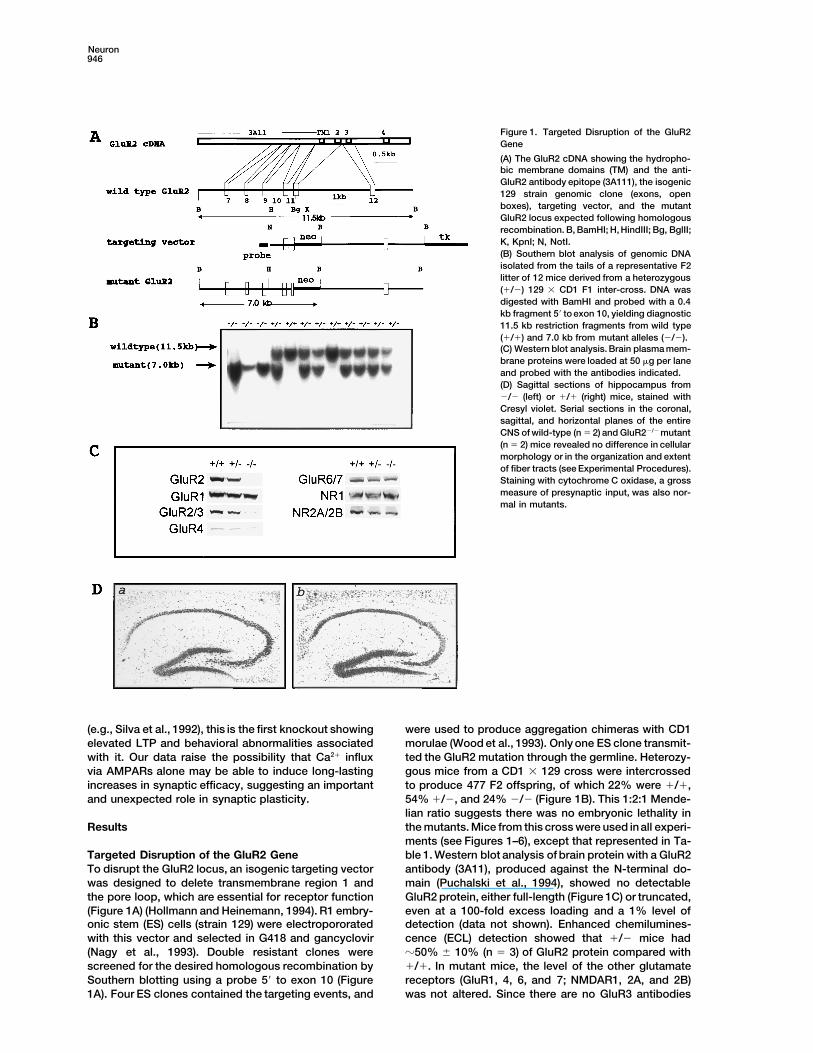
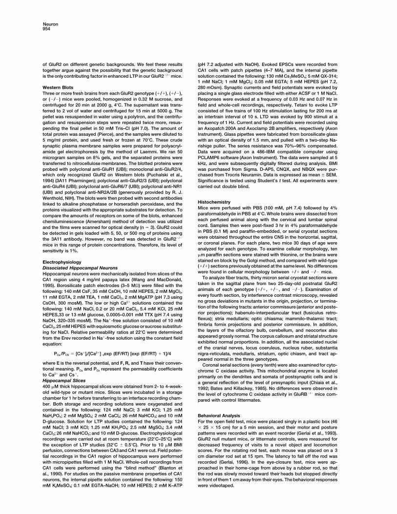
General Appearance of GluR2 Mutant MiceFigure 2. Enhanced Ca21 Permeability in CA1 Neurons LackingThe GluR2 heterozygous (1/2) mice appeared healthy,GluR2
well groomed, and fertile. The GluR2 homozygous mu-(A) Representative kainate-evoked (100 mM) whole-cell currents fortants were born viable and showed no apparent abnor- (1/1) neurons recorded in low (open circles) and high (closed cir-
malities in general appearance until postnatal 2–3 cles) extracellular Ca21. The I/V relationships (right) for averagedweeks, when the mutant mice (n 5 50) could be easily steady-state currents from seven such neurons per condition.
Curves were fitted by third to sixth order polynomials from whichidentified by their much smaller size and reduced weightinterpolated reversal potentialswere calculated. Erev:21.8 6 1.3 mV(11.5 6 0.8 gm in 1/1 mice versus 6.5 6 1.3 gm in 2/2and 21.0 6 1.5 mV.mice). In addition, depending on the litter size, z20%(B) Currents in mutant (2/2) neurons are illustrated, and averaged
of the mutants died at 2–3 weeks after birth. Both the currents of seven such cells are also plotted.growth retardation and mortality rate of the mutants (C and D) Recordings were also made in a solution containing 10were dramatically reduced by separating the littermates mM Ca21 but lacking Na1. Representative responses from (1/1) (C)
and (2/2) (D) neurons are shown.from the parents to reduce the litter size. Therefore,reduced feeding as a result of competition between lit-termates may contribute to an increased mortality rate
Relative Ca21 Permeability in Neuronsin mutants. Mutant mice (including dying mice) showedfrom GluR2 Mutant Miceno sign of seizure activity as measured by observation,Currents evoked by the nondesensitizing agonist, kai-EEG, or post-mortem analysis of pyramidal cell dropoutnate (Burnashev et al., 1992a), in isolated CA1 pyramidalin the hippocampus (data not shown). Surviving mutantneurons from wild-type mice exhibited little or no rectifi-mice recovered at 3 weeks of age, and by 6–7 weekscation, while their reversal potentials were insensitivewere similar to wild-type controls in both size andto change from low to high extracellular Ca21 (Erev:weight. Adult mutants appeared healthy and fully capa-21.8 6 1.3 mV and 21.0 6 1.5 mV [Figure 2A]). Thisble of caring for themselves. No apparent change insuggests that most of the AMPARs in normal CA1 neu-lifespan was observed up to 12 months of age. Behav-rons contain the edited GluR2 subunit and have a lowioral analysis of 13 1/1 and 10 2/2 mice showed thatpermeability to Ca21. Cells taken from mutant mice dem-the lack of GluR2 expression significantly impaired nov-onstrated both an enhanced inward rectification andelty-induced exploratoryactivities in the open-field sucha Ca21-dependent shift of the reversal potential (Erev)as rearing (frequency in 2/2 mice, 0.1 6 0.1 versus(Figure 2B), as predictedfor the loss of the GluR2subunit2.2 6 1.1 in 1/1 mice; p < .05) and object exploration(Hollmann et al., 1991; Verdoorn et al., 1991; Geiger et(frequency in 2/2 mice, 2.9 6 1.3 versus 9.1 6 1.9 inal., 1995; Burnashev et al., 1995; Jonas et al., 1994). The1/1 mice; p < .02), decreased self-directed behaviorsErev shifted from 24.1 6 1.9 mV to 16.0 6 2.3 mV, whichincluding grooming (duration in 2/2 mice 3.3 6 2 sindicates a substantial increase in Ca21 permeability inversus 8.8 6 1.9 in 1/1 mice; p < .006), disrupted motormutant channels. In addition, the degree of inward recti-coordination (fall latency on rotorod at 15 rpm in 2/2fication (Puchalski et al., 1994) of these currents wasmice, 4.8 6 0.9 s versus 1/1, 28 6 5; p < 0.001), andenhanced. The shift in the reversal potential for NMDA-impaired eye closure reflexes to approaching objects (1
of 6 2/2 mice versus 10 of 10 1/1 mice; p < .001). evoked currents (n 5 6) was similar (low Ca21, 22.0 6
Neuron948
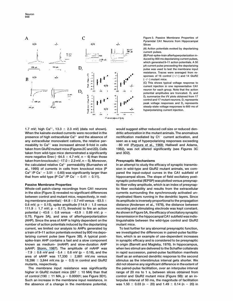
Figure 3. Passive Membrane Properties ofPyramidal CA1 Neurons from HippocampalSlices
(A) Action potentials evoked by depolarizingcurrent pulses.(B) Post-spike-train afterhyperpolarization in-duced by 800 ms depolarizing current pulses,which generated 9–11 action potentials. A 50pA current pulse preceeding the depolarizingpulse was used to test the membrane inputresistance. Traces were averaged from re-sponses of 16 control (1/1) and 14 GluR2(2/2) mutant mice.(C) This shows typical voltage response tocurrent injection in one representative CA1neuron for each group. Note that the actionpotential amplitudes are truncated. D1 andD2 summarize the I/V plots obtained from 17control and 17 mutant neurons. D1 representspeak voltage responses and D2 representssteady-state voltage responses to 800 ms ofhyperpolarizing current injection.
1.7 mV; high Ca21, 13.3 6 2.3 mV) (data not shown). would suggest either reduced cell size or reduced den-dritic arborization in the mutant animals. The anomalousWhen the kainate-evoked currents were recorded in the
presence of high extracellular Ca21 and the absence of rectification mediated by C12 current activation, andseen as a sag of hyperpolarizing responses exceedingany extracellular monovalent cations, the relative per-
meability to Ca21 was increased almost 9-fold in cells 280 mV (Purpura et al., 1968; Halliwell and Adams,1982), was not altered significantly (see Figures 3Ctaken from GluR2mutant mice (Figures2C and 2D). Cells
taken from wild-type mice demonstrated a significantly and 3D2).more negative Erev (264.5 6 4.7 mV, n 5 6) than thosetaken from knockouts (217.0 6 2.2 mV, n 5 5). Moreover, Presynaptic Mechanisms
In an attempt to study the efficacy of synaptic transmis-the calculated relative Ca21 permeability (Burnashev etal., 1995) of currents in cells from knockout mice (P sion in wild-type and GluR2 mutant animals, we com-
pared the input-output curves in the CA1 subfield ofCa21/P Cs1 5 3.51 6 0.60) was significantly larger thanthat from wild type (P Ca21/P Cs1 5 0.41 6 0.11). hippocampal slices. The slope of field excitatory post-
synaptic potential (fEPSP) was plotted versus presynap-tic fiber volley amplitude, which is an index of presynap-Passive Membrane Properties
Whole-cell patch-clamp recordings from CA1 neurons tic fiber excitability and results from the extracellularcurrents surrounding the synchronously activated un-in the slice (Figure 3) revealed no significant differences
between control and mutant mice, respectively, in rest- myelinated fibers running in the dendritic layers. Sinceits amplitude is inversely proportional to the propagationing membrane potential (264.8 6 0.7 mV versus 263.5 6
0.6 mV; p 5 0.15), spike amplitude (114.9 6 1.5 versus distance (Andersen et al., 1978), the distance betweenrecording and stimulating electrode was kept constant.111.9 6 1.7 mV; p 5 0.17), threshold to fire an action
potential (243.6 6 0.8 versus 243.9 6 0.89 mV; p 5 As shown inFigure 5A, theefficacy of excitatory synaptictransmission in the hippocampalCA1 subfield was indis-0.75; Figure 3A), and area of afterhyperpolarization
(AHP). Since the area of AHP is highly dependent on the tinguishable between the wild-type control and GluR2mutant mice.number of action potentials induced by the depolarizing
current, we limited our analysis to AHPs generated by To test further for any abnormal presynaptic function,we investigated the differences in paired-pulse facilita-a train of 9–11 action potentials evoked by 800 ms depo-
larizing current pulse (see Figure 3B). A typical post- tion, which is an example of use-dependent increasesin synaptic efficacy and is considered to be presynapticspike-train AHP contains a fast and a slow component
known as medium- (mAHP) and slow-duration AHP in origin (Barrett and Magleby, 1976). In hippocampus,when two stimuli are delivered to the Schaffer collaterals(sAHP; Storm, 1987). The amplitude of mAHP was
27.9 6 0.6 mV and 25.4 6 0.6 mV (p 5 0.18), and the in rapid succession, paired-pulse facilitation manifestsitself as an enhanced dendritic response to the secondarea of sAHP was 17,330 6 2,881 mV.ms versus
18,396 6 3,844 mV.ms (p 5 0.5) in control and GluR2 stimulus as the interstimulus interval gets shorter. Wedid not observe any significant difference in theextent ofmutants, respectively.
The membrane input resistance was significantly the paired-pulse facilitation, over an interpulse intervalrange of 20 ms to 1 s, between slices obtained fromhigher in GluR2 mutant mice (267 6 10 MA) than that
of control (190 6 11 MA, p 5 0.00001; see Figure 3D1). control and GluR2 mutant mice (Figure 5B): at an in-terpulse interval of 50 ms, the magnitude of facilitationSuch an increase in the membrane input resistance, in
the absence of a change in the membrane potential, was 1.50 6 0.03 (n 5 20) and 1.49 6 0.14 (n 5 20) in

Inactivation of the GluR2 Locus949
Figure 4. Whole-Cell Recordings of EvokedEPSCs in CA1 Neurons
(A) and (B) show a representative normalizedI/V plot of AMPA and NMDA currents, from1/1 (n 5 7) and 2/2 (n 5 7) CA1 neurons.AMPA and NMDA currents were isolated by50 mM AP5 and 5 mM NBQX, respectively, inthe continuous presence of 10 mM BMI, andwere normalized to their respective max-imums.(C) Representative traces of synaptic cur-rents from a wild-type (1/1) and a mutantCA1neuron (2/2) recordedin whole-cell volt-age clamp in the presence of 10 mM BMI ata holding potential of 140 mV (upper twotraces, NMDA component) and 280 mV(lower two traces, AMPA component). Thefirst trace at Vh 5 140 mV was recorded inthe presence of 5 mM NBQX, which was sub-sequently blocked by addition of 50 mMD-AP5 (second upper trace). The first traceat Vh 5 280 mV was recorded in the presenceof 10 mM BMI, which was blocked by 5 mMNBQX.(D) Summary histogram showing ratio ofNMDA receptor channel current at 140 mV tosynaptic currents at 280 mV, obtained fromseven control (1/1) neurons and seven mu-tant (2/2) neurons.
control and GluR2 mutant slices, respectively. These not significantly different, thereby indicating that NMDAreceptors have their usual voltage dependence (see Fig-data suggest that in GluR2 mutant mice, the excitability
of Schaeffer collateral fibers and neurotransmitter re- ure 4B). However, the I/V relation of the AMPA compo-nent was significantly different in the GluR2 mutantlease is likely to be normal.mice: in cells obtained from control animals, AMPA cur-rents were quite linear, while those obtained from GluR2Postsynaptic Mechanisms
Synaptic activity in hippocampal CA1 pyramidal cells, mutant mice demonstrated a significant rectification atthe positive holding potentials (see Figure 4A). The re-evoked by stimulation of Schaeffer collateral-commis-
sural axons, was recorded by both extracellular field and duction of outward AMPA current at positive holdingpotentials is in agreement with previous reports ob-whole-cell recordings. In extracellular field recordings,
excitatory postsynaptic potentials (EPSPs) appeared to tained from cells lacking the GluR2 subunit (Hollmannet al., 1991; Verdoorn et al., 1991; Geiger et al., 1995;be normal in GluR2 mutant mice. D-AP5 (50–100 mM),
a selective NMDA receptor antagonist, did not have a Koh et al., 1995a).Figure 4C illustrates that in both control and GluR2significant effect on the extracellular synaptic responses
obtained at low frequency stimulation (LFS) in both con- mutant mice, AMPA and NMDA components of evokedEPSCs were completely blocked by NBQX (5 mM) andtrol and GluR2 mutant mice, while NBQX (5 mM), an
AMPA/kainate receptor antagonist, blocked all synaptic D-AP5 (50 mM), respectively. The evoked synapticEPSC, recorded in the presence of 10 mM BMI, is pre-transmission in both the presence and absence of
D-AP5. These results suggested that synaptic transmis- dominantly mediated by AMPAR activation at a holdingpotential of 280 mV (Clark and Collingridge, 1995;sion at low frequency is predominantly AMPA mediated
and that the lack of GluR2 subunit did not perturb synap- Spruston et al., 1995). There was no significant differ-ence in the average synaptic current decay time con-tic transmission. However, since theamplitude of synap-
tic currents depends on the stimulus intensity, and stim- stant at 280 mV (18.6 6 3.1 ms in GluR2 mutant miceand 19.4 6 2.3 ms in control mice; P 5 0.85). The decayulus intensities are not precisely comparable from slice
to slice, we carried out whole-cell voltage-clamp re- time constants of AMPAR-mediated EPSCs measuredin CA1 neurons of both control and GluR2 mutant micecordings under conditions in which we minimized the
number of synapses activated, by increasing the extra- are similar to values reported by other groups undersimilar recording conditions (Kato et al., 1993; Perkelcellular Ca21 and magnesium concentration to 4 mM
each, and reducing the stimulation intensity. and Nicoll, 1993; Clark and Collingridge, 1995). The aver-age decay time constant for the NMDA component, re-Whole-cell voltage-clamp data, recorded in the pres-
ence of both GABAA- and GABAB-mediated inhibitory corded in the presence of NBQX (5–10 mM) and at aholding potential of 140 mV, for control and GluR2 mu-postsynaptic currents (IPSCs), did not show any obvious
difference among the cells obtained from wild-type con- tant mice, was 174.1 6 20.5 ms and 166.3 6 22.7 ms,respectively (P 5 0.80).trol and GluR2 mutant slices (see Figure 4). The I/V
relation of the NMDA component in neurons obtained The AMPA current amplitudes, measured 7 ms fromstimulation artefact (Clark and Collingridge, 1995), werefrom seven control and seven GluR2 mutant mice was
Neuron950
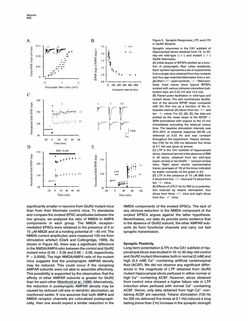
Figure 5. Synaptic Responses, LTP, and LTDin GluR2 Mutants
Synaptic responses in the CA1 subfield ofhippocampal slices obtained from 15- to 30-day-old wild-type (1/1) and mutant (2/2)GluR2 littermates.(A) Initial slopes of fEPSPs plotted as a func-tion of presynaptic fiber volley amplitude.Each symbol represents a set of experimentsfrom a single slice obtained from four mutantsand four age-matched littermates from a sin-gle litter (1/1: open symbols; 2/2: filled sym-bols). Inset traces show typical fEPSPsevoked with various stimulus intensities (cali-bration bars are 0.22 mV and 12.5 ms).(B) Paired pulse facilitation in wild-type andmutant slices. The plot summarizes facilita-tion of the second fEPSP slope comparedwith the first one as a function of the in-terpulse interval (20 slices from ten 1/1 andten 2/2 mice). For (C), (D), (E), the data areplotted as the mean slope of the fEPSP 6
SEM (normalized with respect to the 10 minimmediately preceding the tetanus) versustime. The baseline stimulation intensity was30%–50% of maximal response (30–80 ms)delivered at 0.03 Hz and was constantthroughout the experiment. Tetanic stimula-tion (100 Hz for 200 ms delivered five timesat 0.1 Hz) was given at arrows.(C) LTP in the CA1 subfield of hippocampalslices, tetanized (arrow) inthe absence of BMIin 20 slices, obtained from ten wild-type(open circles) or ten GluR22/2 (closed circles)mice. Right panel shows representativetraces (averages of 10) at the times indicatedby arabic numerals on the graph in (C).(D) LTP in the presence of 10 mM BMI from5 slices from five 1/1 mice and 12 slices fromten 2/2 mice.(E) Effects of LFS (1 Hz for 900 s) on potentia-tion induced by tetanic stimulation: fourslices from three 1/1 mice and eight slicesfrom five 2/2 mice.
significantly smaller in neurons from GluR2 mutant mice NMDA components of the evoked EPSCs. The lack ofany obvious reduction in the NMDA component of thethan from their littermate control mice. To standarize
and compare the evoked EPSC amplitudes between the evoked EPSCs argues against the latter hypothesis.Nevertheless, our data do provide some evidence thattwo groups, we analyzed the ratio of NMDA to AMPA
components in each group. The NMDA receptor– in the absence of GluR2 subunit, the other AMPAR sub-mediated EPSCs were obtained in the presence of 5 or units do form functional channels and carry out fast10 mM NBQX and at a holding potential of 140 mV. The synaptic transmission.NMDA current amplitudes were measured 100 ms fromstimulation artefact (Clark and Collingridge, 1995). As
Synaptic Plasticityshown in Figure 4D, there was a significant differenceLong-term potentiation (LTP) in the CA1 subfield of hip-in the NMDA/AMPA ratio between the control and GluR2pocampalslices was studied in 16- to 30-day-old controlmutant mice (0.40 6 0.08 and 0.98 6 0.09, respectively;and GluR2 mutant littermates both in normal (2 mM) andP 5 0.0006). The high NMDA/AMPA ratio of the mutanthigh (3.4 mM) Ca21-containing artificial cerebrospinalmice suggests that the postsynaptic AMPAR densityfluid (ACSF). We did not observe any significant differ-may be reduced. This could occur if the remainingences in the magnitude of LTP obtained from GluR2AMPAR subunits were not able to assemble effectively.mutant hippocampal slices perfused in either normal orThis possibility is supported by the observation that thehigh Ca21-containing ACSF. However, slices obtainedaffinity of other AMPAR subunits is greater for GluR2from control mice showed a higher failure rate in LTPthan for each other (Wenthold et al., 1996). Alternatively,induction when perfused with normal Ca21-containingthe reduction in postsynaptic AMPAR density may beACSF. Hence, only data obtained from high Ca21-con-caused by reduced cell size or dendritic aborization, astaining ACSF are reported. Tetanic stimulation (100 Hzmentioned earlier. If one assumes that both AMPA andfor 200 ms delivered five times at 0.1 Hz) induced a longNMDA receptor channels are colocalized postsynapti-
cally, then one would expect a similar reduction in the lasting (more than 2 hr) increase in the synaptic strength
Inactivation of the GluR2 Locus951
in the control slices (Figure 5C). The normalized EPSPslope for control mice at 60 min after tetanus was167% 6 15% of the average slope before stimulation(n 5 20). In slices obtained from GluR2 mutants, theextent of long-lasting increase in the synaptic strengthwas enhanced by 2-fold. The normalized EPSP slope forGluR2 mutant mice at 60 min after tetanus was 252% 6
17.8% (P 5 0.005) of the average slope before stimula-tion (n 5 20). When tetanic stimulation was deliveredin the presence of bicuculline (i.e., in the absence ofGABAergic transmission), the magnitude of potentiationseen was indistinguishable from that obtained in theabsence of bicuculline. At 60 min after tetanus, the nor-malized EPSP slope for the control and GluR2 mutantmice was 158% 6 5.9% (n 5 5) and 295% 6 17% (n 5
12; P 5 0.0003), respectively (Figure 5D).It was possible that neurons from GluR2 mutant mice
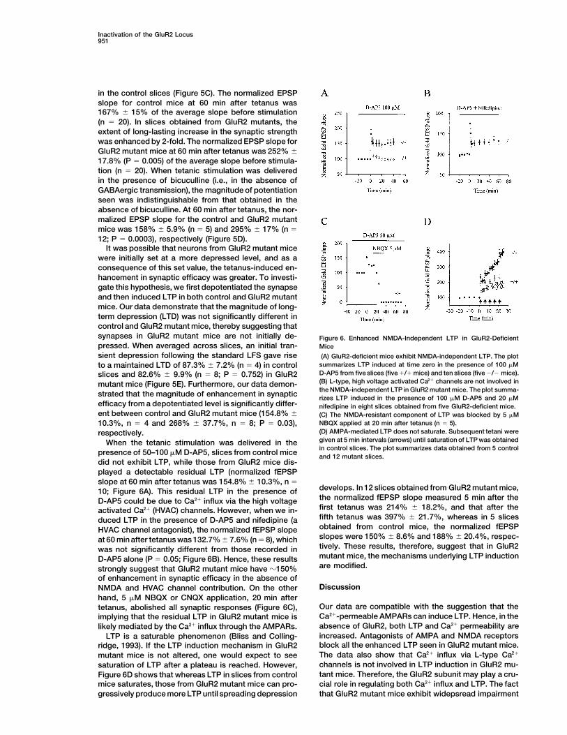
were initially set at a more depressed level, and as aconsequence of this set value, the tetanus-induced en-hancement in synaptic efficacy was greater. To investi-gate this hypothesis, we first depotentiated the synapseand then induced LTP in both control and GluR2 mutantmice. Our data demonstrate that the magnitude of long-term depression (LTD) was not significantly different incontrol and GluR2 mutant mice, thereby suggesting thatsynapses in GluR2 mutant mice are not initially de- Figure 6. Enhanced NMDA-Independent LTP in GluR2-Deficientpressed. When averaged across slices, an initial tran- Micesient depression following the standard LFS gave rise (A) GluR2-deficient mice exhibit NMDA-independent LTP. The plot
summarizes LTP induced at time zero in the presence of 100 mMto a maintained LTD of 87.3% 6 7.2% (n 5 4) in controlD-AP5 from five slices (five 1/1 mice) and ten slices (five 2/2 mice).slices and 82.6% 6 9.9% (n 5 8; P 5 0.752) in GluR2(B) L-type, high voltage activated Ca21 channels are not involved inmutant mice (Figure 5E). Furthermore, our data demon-the NMDA-independent LTP in GluR2 mutant mice. The plot summa-
strated that the magnitude of enhancement in synaptic rizes LTP induced in the presence of 100 mM D-AP5 and 20 mMefficacy from a depotentiated level is significantly differ- nifedipine in eight slices obtained from five GluR2-deficient mice.ent between control and GluR2 mutant mice (154.8% 6 (C) The NMDA-resistant component of LTP was blocked by 5 mM
NBQX applied at 20 min after tetanus (n 5 5).10.3%, n 5 4 and 268% 6 37.7%, n 5 8; P 5 0.03),(D) AMPA-mediated LTP does not saturate. Subsequent tetani wererespectively.given at 5 min intervals (arrows) until saturation of LTP was obtainedWhen the tetanic stimulation was delivered in thein control slices. The plot summarizes data obtained from 5 control
presence of 50–100 mM D-AP5, slices from control mice and 12 mutant slices.did not exhibit LTP, while those from GluR2 mice dis-played a detectable residual LTP (normalized fEPSPslope at 60 min after tetanus was 154.8% 6 10.3%, n 5
develops. In 12 slices obtained from GluR2 mutant mice,10; Figure 6A). This residual LTP in the presence ofthe normalized fEPSP slope measured 5 min after theD-AP5 could be due to Ca21 influx via the high voltagefirst tetanus was 214% 6 18.2%, and that after theactivated Ca21 (HVAC) channels. However, when we in-fifth tetanus was 397% 6 21.7%, whereas in 5 slicesduced LTP in the presence of D-AP5 and nifedipine (aobtained from control mice, the normalized fEPSPHVAC channel antagonist), the normalized fEPSP slopeslopes were 150% 6 8.6% and 188% 6 20.4%, respec-at 60 min after tetanus was 132.7% 6 7.6% (n 5 8), whichtively. These results, therefore, suggest that in GluR2was not significantly different from those recorded inmutant mice, the mechanisms underlying LTP inductionD-AP5 alone (P 5 0.05; Figure 6B). Hence, these resultsare modified.strongly suggest that GluR2 mutant mice have z150%
of enhancement in synaptic efficacy in the absence ofDiscussionNMDA and HVAC channel contribution. On the other
hand, 5 mM NBQX or CNQX application, 20 min afterOur data are compatible with the suggestion that thetetanus, abolished all synaptic responses (Figure 6C),Ca21-permeable AMPARs can induce LTP. Hence, in theimplying that the residual LTP in GluR2 mutant mice isabsence of GluR2, both LTP and Ca21 permeability arelikely mediated by the Ca21 influx through the AMPARs.increased. Antagonists of AMPA and NMDA receptorsLTP is a saturable phenomenon (Bliss and Colling-block all the enhanced LTP seen in GluR2 mutant mice.ridge, 1993). If the LTP induction mechanism in GluR2The data also show that Ca21 influx via L-type Ca21mutant mice is not altered, one would expect to seechannels is not involved in LTP induction in GluR2 mu-saturation of LTP after a plateau is reached. However,tant mice. Therefore, the GluR2 subunit may play a cru-Figure 6D shows that whereas LTP in slices from controlcial role in regulating both Ca21 influx and LTP. The factmice saturates, those from GluR2 mutant mice can pro-
gressively produce more LTP until spreading depression that GluR2 mutant mice exhibit widepsread impairment
Neuron952
in behavior suggests that GluR2 is also critical for normal for the enhanced synaptic efficacy observed in theGluR2 mutants.brain function.
The magnitude of the rise in postsynaptic Ca21 duringLTP induction, and the level of NMDA receptor function,GluR Expressionhave been shown to have significant effect on the gener-The results suggest that we have created a null mutationation of LTP (Malenka, 1991). Previous reports alsoin the GluR2 locus. The neoR gene inserted into exonshowed that trans-ACPD, a metabotropic glutamate re-11 has stop codons in all three reading frames and aceptor agonist, enhanced NMDA receptor currents inpoly(A) site. No full-length GluR2 protein was seen inCA1 cellsand, when applied with a weak tetanus incapa-overloaded gels at a 1% level of detection. Although able of inducing LTP by itself, generated LTP (Aniksztejnshort truncated GluR2 transcript could be made fromet al., 1992; Bashir et al., 1993; O’Connor et al., 1994,exons 1–10, we detected no truncated GluR2 protein1995). Since NMDA receptor function is strongly modu-in 100-fold overloaded Western blots. Since the GluR2lated by kinase activity (Gerber et al., 1989; MacDonaldantibody recognizes the N-terminus of the protein, itet al., 1989; Chen and Huang 1991, 1992; Kelso et al.,should have detected any protein made. However, we1992; Yamazaki et al., 1992), it is conceivable that insaw no extra band in the range of 1–200 kd on WesternGluR2 mutants the Ca21 influx from AMPARs could mod-blots, not even a GluR–neo fusion. Likewise, an antibodyify the NMDA receptor function through protein phos-with dual specificity for GluR3 and the C-terminus ofphorylation or direct binding to calmodulin (Ehlers et al.,GluR2, showed a loss of the comigrating GluR2 protein1996). However, this is unlikely, since in GluR2 mutantin GluR2 mutants. Finally, the 9-fold change in Ca21
mice, whole-cell data did not reveal any change in thepermeability in GluR2 2/2 neurons was in the range thatNMDA component of the synaptic current. Moreover,would be predicted from the loss of GluR2 (Burnashev etaddition of NMDA-independent LTP in GluR2 mutantsal., 1995). Therefore, we likely have created a null muta-to NMDA-dependent LTP in control mice reconstitutedtion in the GluR2 locus. GluR2 loss itself might also alterthe enhanced LTP seen in GluR2 mutants. In addition,the stoichiometry of GluR1, 3, and 4 assembly in theLTD, another NMDA-dependent phenomenon, was notmembrane, but this does not alter postsynaptic trans-altered. These findings strongly support the hypothesismission, although AMPA responses were smaller in mu-that Ca21 influx, via AMPARs lacking the GluR2 subunit,tants.is not modifying the NMDA component and is sufficientfor producing LTP.
Relative Ca21 Permeability of AMPA Receptors Several studies have suggested the presence ofIt has been shown in vitro that AMPARs containing the NMDA receptor–independent LTP in the CA1 SchaefferQ/R edited GluR2 subunit exhibit lower Ca21 permeabil- collateral synapse. Perfusion of a high extracellular Ca21
ity and distinct gating properties, compared with recep- solution induces a long-lasting enhancement in the syn-tor channels assembled without this subunit (Hollmann aptic strength (Turner et al., 1982), which is not blockedand Heinemann, 1994; Burnashev et al., 1992a). As ex- by the NMDA receptor antagonist APV (Grover andpected, the loss of this GluR2 subunit in individual CA1 Teyler, 1990a). NMDA receptor–independent LTP waspyramidal neurons from GluR2 mutant mice demon- also induced in CA1 by using a 200 Hz, rather thanstrated a 9-fold increase in relative Ca21 permeability the usual 100 Hz tetanic stimulation (Grover and Teyler,following kainate application, when compared with that 1990b). Postsynaptic injection of BAPTA, a Ca21 chela-of control mice. Indeed, this shift was similar in magni- tor, or bath application of nifedipine, the dihydropyridinetude to that observed for Ca21-permeable NMDA recep- Ca21 channel (L-type) antagonist, blocked this form oftors (Koh et al., 1995a). These results, together with LTP, suggesting that in the presence of APV, HVACobservations in hippocampal slices from GluR2 editing– channels provided the necessary Ca21 influx requireddeficient mice (Brusa et al., 1995), support a crucial for LTP at the Schaeffer collateral synapse. Such arole for the GluR2 subunit in inhibiting Ca21 influx via mechanism could underlie the NMDA receptor–inde-AMPARs in vivo. pendent enhancement in GluR2 mutants. However,
when we perfused slices with nifedipine, in the presenceGluR2 Subunit and Synaptic Plasticity of D-AP5, the residual LTP was unaffected, thereby indi-One could argue that enhanced synaptic plasticity seen cating the lack of L-type HVAC channel involvement inin GluR2 null mutant mice was due to changes in either NMDA-independent LTP in GluR2 mutant mice. There-passive membrane properties, or the conductances for fore, our data raise the possibility that Ca21 influx viathe inhibitory components of the synaptic response. AMPARs devoid of GluR2 subunit is sufficient to induceHowever, our data indicate that there is no difference LTP, in a NMDA-independent manner. A recent studyin either the passive membrane properties or the IPSCs onspinal cordneurons, where AMPAreceptors are natu-among the slices obtained from control and GluR2 null rally devoid of the GluR2 subunits and are thereforemutant mice. One might also presume that Ca21 influx Ca21 permeable, showed that synaptic strength variedthrough the AMPARs would be sufficient to block the in an activity-dependent way (Gu et al., 1996). Further-Cl2 ionophore of the GABAA receptors (Inoue et al., 1986) more, it is interesting to note that LTP in GluR2 mutantand thereby give rise to the enhanced LTP. However, slices did not saturate, thus indicating that the mecha-the magnitude of enhancement in synaptic strength was nism(s) underlying LTP induction is altered. One reasonalso the same under conditions where GABAergic trans- for normal saturable LTP could be that Ca21 influxmission was blocked by bicuculline, suggesting that through the NMDAR channel activates calmodulin,
which is known to bind and inactivate the NMDAR1changes in the IPSCs are not the underlying mechanism
Inactivation of the GluR2 Locus953
Table 1. The Effect of Genetic Background on LTP
ComparisonLTPa
Strain GluR2 Genotype Number of Animals (Mean) Standard Error p Value
1. CD-1 1/1 n 5 36 164 5 1 vs 2 NS 1 vs 5, p , 0.0012. 129SV 1/1 n 5 22 163 6 2 vs 3, p , 0.001 1 vs 4, NS3. 129SV 2/2 n 5 6 296 16 2 vs 5, p , 0.001 3 vs 5, NS4. CD1 3 129F2 1/1 n 5 36 169 4 2 vs 4, NS 3 vs 4, p , 0.0015. CD1 3 129F2 2/2 n 5 32 272 9 1 vs 3, p , 0.001 4 vs 5, p , 0.001
a Mean percent increase in fEPSP slope at 60 min after tetanus.Post-hoc comparisons (SNK test; a 5 0.05) revealed a significant difference between two sets of means. Groups 3 and 5 were significantlydifferent from groups 1, 2, and 4. No significant differences were found within each set. Abbreviations: NS, not significant.
clones (from a total of 1350 analyzed) were shown to contain desiredchannel (Ehlers et al., 1996). Therefore, normal LTP maytargeted events, and all of them were used to produce chimericbe self-limiting. However, in GluR22/2 mice, the addi-mice via aggregation with CD1 morulae (Wood et al., 1993). Thetional Ca21 influx will not inactivate the AMPAR channel,contribution of ES cells from the agouti 129 strain to the germline
thereby leading to an ever-increasingLTP at higher stim- of chimerae was determined by mating with white CD1 mice andulation intensities. Alternatively, downstream signaling screening for agoutioffspring. Germline transmission was confirmed
by Southern blotting of tailDNA. Since only one ES clone transmittedcomponents may limit LTP in normal animals. Inthe mutation through the germline, it is possible, although unlikely,GluR22/2 mice, the additional Ca21 could activate ki-that the phenotype we observe is due to an unintended mutationnases that are normally rate limiting, to generate nonsat-elsewhere in the genome of this clone. We intercrossed 129 3 CD1urable LTP.F1 mice heterozygous for the targeted mutation to homozygosity.
Our data raise the possibility that activation of Ca21Homozygous females and males were fertile, butvery poor breeders.
permeable AMPARs is sufficient to induce LTP; how- All experimental mice in Figures 1–6 included wild-type controls,heterozygotes, and homozygotes, which were littermate controlsever, further experiments are needed to elucidate thegenerated by heterozygous breeding of F1 to yield a 129 3 CD1 F2mechanism(s) underlying this phenomenon. The genera-population. In larger experiments, mice from several litters weretion of these GluR2 mutant mice provides one valuablepooled, keeping a balanced number of 2/2, 1/2, or 1/1 mice frommodel for determining the in vivo roles of the AMPARseach cage.
and Ca21 in synaptic physiology and behavior. Our re- Three control experiments together show that the enhanced LTPsults indicate that GluR2 is not required for LTP but in GluR2 mutant mice is due to the loss of GluR2 (Table 1). Analysis
of variance revealed a significant difference between all groups (Fseems to play a negative role in its regulation. Therefore,[4,126] 5 67.4; p < 0.001). A post-hoc Student Neuman–Keuls testGluR2 may play an important role in synaptic plasticity(a 5 0.05) confirmed that the mutant groups (Groups 3 and 5) areby regulating its expression in development.not different from one another, but are significantly different fromcontrol groups 1, 2, and 4. No significant differences were found
Experimental Procedures between the latter groups. We also performed multiple comparisionsbetween groups using the Student’s t test (two-tailed)with Bonferoni
Targeting Vector Construction adjustment (Table 1). For example, LTP in the CA1 region of theA genomic clone containing exons 7–12 of the GluR2 gene was parental 129 and CD1 strains showed no significant differencesisolated from a genomic 129 phage DNA library, prepared from R1 (Table 1). Therefore, the enhanced LTP seen in our genotypedES cells, after screening with rat GluR2 cDNA as a probe. Exon GluR22/2 mice from the F2 cross could not be due solely to the11, containing TM1 and TM2, was sequenced, and the amino acid inheritance of 129 genes conferring high LTP. To rule out the possi-sequence deduced from DNA sequence was identical to the pub- bility that GluR2-linked 129 genes could contribute to the CA1-LTPlished mouse sequence in the corresponding region (Sakimura et phenotype in the context of a CD1 genetic background, we carriedal., 1990). The targeting vector was prepared by inserting a 1.8 kb out experiments analyzing CA1 LTP distribution among an F2 popu-HindIII–BglII fragment 59 region to exon 11, and a 6.0 kb KpnI–BamHI lation (n 5 36) derived from wild-type F1 intercrosses (i.e., CD1 3fragment 39 to exon 11 into the pPNT plasmid vector (Tybulewicz 129). The frequency histogram showed that the distribution of LTPet al., 1991). The 1.8 kb HindIII–BglII was first subcloned into pBlue- was normal (p 5 0.31 Kolmogorov–Smirnov goodness-of-fit test).script vector (Stratagene) to obtain NotI and XhoI restriction sites. For the wild-type F2 129sv 3 CD1 population, there is 0.99 probabil-The 6 kb KpnI–BamHI fragment was cloned into pPNT by KpnI and ity that a randomly chosen subject from the population will yieldblunt end ligation. The construct was designed to delete transmem- LTP between 118% and 218%. If single recessive wild-type CD1brane region TM1 and the pore loop of exon 11. Therefore, a 0.4 and 129 genes interact in a simple way as to mimic the high LTPkb BglII–KpnI fragment containing transmembrane domain (TM) 1 seen in GluR22/2 mice, we would have expected a bimodal distribu-and 2 in exon 11 was replaced by a 1.8 kb fragment containing the tion with 25% of the values above this LTP range. However, onlyPGKneo cassete carrying stop codons in all the reading frames and one LTP score (260%), out of 36 obtained, was outside the estimateda poly(A) tail. A PGK-TK cassete was inserted downstream of exon range (118–218). A one sample x2 goodness-of-fit test revealed a12 as a negative selection marker. significant difference in the obtained ratio of high LTP (1/36) com-
pared with the expected ratio (9/36), (x2 5 9.48, df 5 1; p < 0.01).Generation of GluR2 Mutant Mice This analysis argues against the possibility that single recessiveThe vector DNA was linearized with NotI restriction digestion and genes interact in the background and contribute to enhanced LTPtransfected into strain 129 ES cells of R1 line (Nagy et al., 1993) by in GluR22/2 mice, but they do not rule out multiple gene effects.electroporation (Bio-Rad, Gene Pulser, set at 240 V and 500 mF). Finally, we have crossed our original 129 germline–derived chimericG418 (150–250 mg/ml) and gancyclovir (2 mm) was applied 24 hr male with 129, rather than CD1 mice (Table 1). Enhanced LTP wasafter transfection, and double resistant colonies were isolated dur- seen in GluR22/2 mice from several litters compared with 1/1 (p <ing days 7–10 of selection. Genomic DNA of resistant clones was .00001). In this experiment, there is no possibility that CD1 back-digested with BamHI and hybridized with a 0.5 kb DNA fragment 59 ground genes are playing any role, since they were not used in this
cross. These results show that enhanced LTP is seen in the absenceto the HindIII–BglII fragment as an external probe (Figure 1A). Four
Neuron954
of GluR2 on different genetic backgrounds. We feel these results (pH 7.2 adjusted with NaOH). Evoked EPSCs were recorded fromCA1 cells with patch pipettes (4–7 MA), and the internal pipettetogether argue against the possibility that the genetic background
is the only contributing factor in enhanced LTP in our GluR22/2 mice. solution contained the following: 130 mM Cs2MeSO4; 5 mM QX-314;1 mM NaCl; 1 mM MgCl2; 0.05 mM EGTA; 5 mM HEPES (pH 7.2,280 mOsm). Synaptic currents and field potentials were evoked byWestern Blots
Three or more fresh brains from each GluR2 genotype (1/1), (1/2), placing a single glass electrode filled with either ACSF or 1 M NaCl.Responses were evoked at a frequency of 0.03 Hz and 0.07 Hz inor (2/2) mice were pooled, homogenized in 0.32 M sucrose, and
centrifuged for 20 min at 2000 g, 48C. The supernatant was trans- field and whole-cell recordings, respectively. Tetani to evoke LTPconsisted of five trains of 100 Hz stimulation lasting for 200 ms atferred to 2 vol of water and centrifuged for 15 min at 5000 g. The
pellet was resuspended in water using a polytron, and the centrifu- an intertrain interval of 10 s. LTD was evoked by 900 stimuli at afrequency of 1 Hz. Current and field potentials were recorded usinggation and resuspension steps were repeated twice more, resus-
pending the final pellet in 50 mM Tris–Cl (pH 7.0). The amount of an Axopatch 200A and Axoclamp 2B amplifiers, respectively (AxonInstrument). Glass pipettes were fabricated from borosilicate glasstotal protein was assayed (Pierce), and the samples were diluted to
5 mg/ml protein, and used fresh or frozen at 708C. These crude with an optical density of 1.5 mm, and pulled with a two-step Na-rishige puller. The series resistance was 70%–96% compensated.synaptic plasma membrane samples were prepared for polyacryl-
amide gel electrophoresis by the method of Laemmi. We ran 50 Data were acquired on a 486-IBM compatible computer usingPCLAMP6 software (Axon Instrument). The data were sampled at 5microgram samples on 8% gels, and the separated proteins were
transferred to nitrocellulose membranes. The blotted proteins were kHz, and were subsequently digitally filtered during analysis. BMIwas purchased from Sigma. D-AP5, CNQX, and NBQX were pur-probed with polyclonal anti-GluR1 (UBI); monoclonal anti-GluR2/4,
which only recognized GluR2 on Western blots (Puchalski et al., chased from Trocris Neuramin. Data is expressed as mean 6 SEM.Significance is tested using Student’s t test. All experiments were1994) (3A11 Pharmingen); polyclonal anti-GluR2/3 (UBI); polyclonal
anti-GluR4 (UBI); polyclonal anti-GluR6/7 (UBI); polyclonal anti-NR1 carried out double blind.(UBI) and polyclonal anti-NR2A/2B (generously provided by R. J.Wenthold, NIH). The blots were then probed with second antibodies
Histochemistrylinked to alkaline phosphatase or horseradish peroxidase, and theMice were perfused with PBS (100 mM, pH 7.4) followed by 4%proteins visualized with the appropriate substrates for detection. Toparaformaldehyde in PBS at 48C. Whole brains were dissected fromcompare the amounts of receptors on some of the blots, enhancedeach perfused animal along with the cervical and lumbar spinalchemiluminescence (Amersham) method of detection was utilizedcord. Samples then were post-fixed 3 hr in 4% paraformaldehydeand the films were scanned for optical density (n 5 3). GluR2 couldin PBS (0.1 M) and paraffin-embedded, or serial cryostat sectionsbe detected in gels loaded with 5, 50, or 500 mg of proteins usingwere obtained throughout the entire CNS in the horizontal, sagittal,the 3A11 antibody. However, no band was detected in GluR22/2
or coronal planes. For each plane, two mice 30 days of age weremice in this range of protein concentrations. Therefore, its level ofanalyzed for each genotype. To examine cellular morphology, tensensitivity is 1%.mm paraffin sections were stained with thionine, or the brains werestained en block by the Golgi method, and compared with wild-typeElectrophysiology(1/1) sections previously obtained at the same level. No differencesDissociated Hippocampal Neuronswere found in cellular morphology between 1/1 and 2/2 mice.Hippocampal neurons were mechanically isolated from slices of the
To analyze fiber tracts, thirty micron serial cryostat sections wereCA1 region using 4 mg/ml papaya latex (Wang and MacDonald,taken in the sagittal plane from two 25-day-old postnatal GluR21995). Borosilicate patch electrodes (3–5 MV) were filled with theanimals of each genotype (1/1, 1/2, and 2/2). Examination offollowing: 140 mM CsF, 35 mM CsOH, 10 mM HEPES, 2 mM MgCl2,every fourth section, by interference contrast microscopy, revealed11 mM EGTA, 2 mM TEA, 1 mM CaCl2, 2 mM MgATP (pH 7.3 usingno gross deviations in mutants in the origin, projection, or termina-CsOH, 300 mosM). The low or high Ca21 solutions contained thetion of the following tracts: anterior commissure (anterior and poste-following: 140 mM NaCl, 0.2 or 20 mM CaCl2, 5.4 mM KCl, 25 mMrior projections); habenulo-interpeduncular tract (fusiculus retro-HEPES,33 or 13 mM glucose, 0.0005–0.001 mM TTX (pH 7.4 usingflexus); stria medullaris; optic chiasma; mammilo-thalamic tract;NaOH, 320–335 mosM). The Na1-free solution consisted of 10 mMfimbria fornix projections and posterior commissure. In addition,CaCl2, 25 mM HEPES with equiosmotic glucose orsucrose substitut-the layers of the olfactory bulb, cerebellum, and neocortex alsoing for NaCl. Relative permeability ratios at 228C were determinedappeared grossly normal. The corpus callosum and striatal structurefrom the Erev recorded in Na1-free solution using the constant fieldexhibited normal proportions. In addition, all the associated nucleiequation:of the cranial nerves, locus coeruleus, nucleus ruber, substantianigra-reticulata, medullaris, striatum, optic chiasm, and tract ap-PCa /PCS 5 [Cs1]i/[Ca21] oexp (EF/RT) [exp (EF/RT) 1 1]/4peared normal in the three genotypes.
where E is the reversal potential, and F, R, and T have their conven- Coronal serial sections (every tenth) were also examined for cyto-tional meaning. PCa and PCs represent the permeability coefficients chrome C oxidase activity. This mitochondrial enzyme is locatedto Ca21 and Cs1. primarily on the dendrites and somata of postsynaptic cells and isHippocampal Slices a general reflection of the level of presynaptic input (Chiaia et al.,400 mM thick hippocampal slices were obtained from 2- to 4-week- 1992; Bates and Killackey, 1985). No differences were observed inold wild-type or mutant mice. Slices were incubated in a storage the level of cytochrome C oxidase activity in GluRB2/2 mice com-chamber for 1 hr before transferring to an interface recording cham- pared with control littermates.ber. Both storage and recording solutions were oxygenated andcontained in the following: 124 mM NaCl; 3 mM KCl; 1.25 mMNaH2PO4; 2 mM MgSO4; 2 mM CaCl2; 26 mM NaHCO3; and 10 mM Behavioral Analysis
For the open field test, mice were placed singly in a plastic box (46D-glucose. Solution for LTP studies contained the following: 124mM NaCl; 3 mM KCl; 1.25 mM KH2PO4; 2.5 mM MgSO4; 3.4 mM 3 25 3 15 cm) for a 5 min session, and their motor and posture
patterns were recorded with an event recorder (Gerlai et al., 1993).CaCl2; 26 mM NaHCO3; and 10 mM D-glucose. Electrophysiologicalrecordings were carried out at room temperature (228C–258C) with GluR2 null mutant mice, or littermate controls, were measured for
decreased frequency of visits to a novel object and locomotionthe exception of LTP studies (328C 6 0.58C). Prior to 10 mM BMIperfusion, connections between CA3and CA1 were cut. Field poten- scores. For the rotating rod test, each mouse was placed on a 3
cm diameter rod set at 15 rpm. The latency to fall off the rod wastial recordings in the CA1 region of hippocampus were performedwith micropipettes filled with 1 M NaCl. Whole-cell recordings from recorded (Gerlai, 1996). In the eye-closure test, mice were ap-
proached in their home-cage from above by a rubber rod, so thatCA1 cells were performed using the “blind method” (Blanton etal., 1990). For studies on the passive membrane properties of CA1 the rod was slowly moved toward their heads but stopped directly
in front of them 1 cm away from their eyes. The behavioral responsesneurons, the internal pipette solution contained the following: 150mM K2MeSO4; 0.1 mM EGTA–NaOH; 10 mM HEPES; 2 mM K–ATP were videotaped.
Inactivation of the GluR2 Locus955
Acknowledgments Clark, K.A., and Collingridge, G.L. (1995). Synaptic potentiation ofdual component excitatory postsynaptic currents in the rat hippo-campus. J. Physiol. (Lond.) 482, 39–52.This work was supported by the Medical Research Council of Can-
ada and the Ontario Mental Health Foundation. Zhengping Jia was Dingledine, R., Hume, R.I., and Heinemann, S.F. (1992). Structurala Networks of Centres of Excellence Neuroscience fellow and Nadia determinant of barium permeation and rectification in non-NMDAAgopyan was a Medical Research Council Centennial fellow. We glutamate receptor channels. J. Neurosci. 12, 4080–4087.thank S. Heinemann for the GluR2 cDNA clone, Y. M. Lu for some
Ehlers, M.D., Zhang, S., Bernhardt, J.P., and Huganir, R.L. (1996).of the data in Table 1, and C. Janus for statistical analysis.Inactivation of NMDA receptors by direct interaction of calmodulinThe costs of publication of this article were defrayed in part bywith the NR1 subunit. Cell 84, 746–755.the payment of page charges. This article must therefore be hereby
marked “advertisement” in accordance with 18 USC Section 1734 Geiger, J.R.P., Melcher, T., Koh, D.S., Sakman, B., Seeburg, P.H.,solely to indicate this fact. Jonas, P., and Monyer, H. (1995). Relative abundance of subunit
mRNAs determines gating and Ca21 permeabilityof AMPAreceptorsReceived June 18, 1996; revised September 19, 1996. in principal neurons and interneurons in rat CNS. Neuron 15,193–
204.References Gerber, G., Kangrga, I., Ryu, P.D., Larew, J.S.A., and Randic, M.
(1989). Multiple effects of phorbol esters in the rat spinal dorsalAndersen, P., Silfvenius, H., Sundberg, S.H., Sveen, O., and horn. J. Neurosci. 9, 3606–3617.Wingstrom, H. (1978). Functional characteristics of unmyelinated
Gerlai, R. (1996). Gene targeting studies of mammalian behaviour:fibers in the hippocampal cortex. Brain Res. 144, 11–18.is it the mutation or the background genotype? Trends Neurosci.Aniksztejn, L., Otani, S., and Ben-Ari, Y. (1992). Quisqualate metabo-19, 177–189.tropic receptors modulate NMDA currents and facilitate inductionGerlai, R., Friend, W., Becker, L., O’Hanlon, R., Marks, A., and Roder,of long term potentiation through protein kinase C. Eur. J. Neurosci.J. (1993). Female transgenic mice carrying multiple copies of the4, 500–505.human gene for S100b are hyperactive. Behav. Brain Res. 55, 51–59.Barrett, E.F., and Magleby, K.L. (1976). Physiology of the cholinergic
transmission. In Biology of Cholinergic Function, (A.M. Goldberg Grover, L.M., and Teyler, T.J. (1990a). Effects of extracellular potas-and I. Hanin, eds. (New York: Raven Press), pp. 29–100. sium concentration and postsynaptic membrane potential on Ca21
induced potentiation in area CA1 of rat hippocampus. Brain Res.Bashir, Z.I., Bortolotto, Z.A., Davies, C.H., Beretta, N., Irving, A.J.,506, 53–61.Seal, A.J., Henley, J.M., Jane, D.E., Watkins, J.C., and Collingridge,
G.L. (1993). Induction of LTP in the hippocampus needs synaptic Grover, L.M., and Teyler, T.J. (1990b). Two components of longactivation of glutamate metabotropic receptors. Nature 363, term potentiation induced by different patternsof afferentactivation.347–349. Nature 347, 477–479.Bates, S.C.A., and Killackey, H.P. (1985). The organization of the Gu, J.G., Albuquerque, C., Lee, C.J., and MacDermott, A.B. (1996).neonatal rat’s brainstem trigernimal complex and its role in the Synaptic strengthening through activation of Ca21 permeable AMPAformation of central trigernimal pattersn. J. Comp. Neurol. 240, receptors. Nature 381, 793–796.265–287.
Halliwell, J.W., and Adams, P.R. (1982). Voltage-clamp analysis ofBlanton, M., Lotorco, J., and Kreigstein, A. (1990). Whole-cell re- muscarinic excitation in hippocampal neurones. Brain Res. 250,cording from neurons in slices of reptilian and mammalian cortex. 71–92.J. Neurosci. Meth. 30, 203–210.
Hollmann, M., and Heinemann, S. (1994). Cloned glutamate recep-Bliss, T.V.P., and Collingridge, G.L. (1993). A synaptic model oftors. Ann. Rev. Neurosci. 17, 31–108.memory: long-term potentiation in the hippocampus. Nature 361,Hollmann, M., Hartley, M., and Heinemann, S. (1991). Ca21 perme-31–39.ability of KA-AMPA glutamate receptor channels depends on sub-Bowie, D., and Mayer, M.L. (1995). Inward rectification of both AMPAunit composition. Science 52, 81–85.and kainate subtype glutamate receptors generated by polyamine-
mediated ion channel block. Neuron 15, 453–462. Hume, I.R., Dingledine,R., and Heinemann, S.F. (1991). Identificationof a site in glutamate receptor subunit that controls Ca21 permeabil-Brusa, R., Zimmerman, F., Koh, D.S., Feldmeyer, D., Gass, P., See-ity. Science 253, 1028–1031.burg, P.H., and Sprengel, R. (1995). Early-onset epilepsy and postna-
tal lethality associated with an editing-deficient GluR-B allele in Inoue, M., Oomura, Y., Yakushiji, T., and Akaike, N. (1986). Intracellu-mice. Science 270, 1677–1680. lar Ca21 ions decrease the affinity of the GABA receptor. Nature
324, 156–158.Burnashev, N., Monyer, H., Seeburg, P.H., and Sakmann, B. (1992a).Divalent ion permeability of AMPA receptor channels is dominated Jonas, P., Racca, C., Sakman, B., Seeburg, P.H., Monyer, H. (1994).by the edited form of a single subunit. Neuron 8, 189–198. Differences in Ca21 permeability of AMPA-type glutamate receptorBurnashev, N., Khodorova, A., Jonas, P., Helm, P.J., Wisden, W., channels in neocortical neurons caused by differential GluR-B sub-Monyer, H., Seeburg, P.H., and Sakmann, B. (1992b). Ca21 perme- unit expression. Neuron 12, 1281–1289.able AMPA/KA receptors in fusiform cerebellar glial cells. Science
Kamboj, S.K., Swanson, J.T., and Cull-Candy, S.J. (1995). Intracellu-256, 1566–1570.
lar spermine confers rectification on rat calicum-permeable AMPABurnashev, N., Zhou, Z., Neher, E., and Sakman, B . (1995). Fractional and kainate receptors. J. Physiol. 486, 297–303.Ca21 currents through recombinant GluR channels of the NMDA,
Kato, K., Clifford, D.B., and Zorumski, C.F. (1993). Long term potenti-AMPA and kainate receptor subtypes. J. Physiol. (Lond.) 485, 403–ation during whole cell recordings in rat hippocampal slices. Neuro-418.science 53, 39–47.
Chen, L., and Huang, L.Y.M. (1991). Sustained potentiation of NMDAKelso, S.R., Nelson, T.E., and Leonard, J.P. (1992). Protein kinasereceptor–mediated glutamate responses through activation of pro-C–mediated enhancement of NMDA currents by metabotropic gluta-tein kinase C by a m opioid. Neuron 7, 319–326.mate receptors in Xenopus oocytes. J.Physiol. (Lond.)449, 705–718.
Chen, L., and Huang, L.Y.M. (1992). Protein kinase C reduces Mg21
Koh, D.S., Geiger, J.R.P., Jonas, P., and Sakman, B. (1995a). Ca21block of NMDA receptor channels as a mechanism of modulation.permeable AMPA and NMDA receptor channels in basket cells ofNature 356, 521–523.rat hippocampal dendate gyrus. J. Physiol. 485, 383–402.Chiaia, N.L., Bennett-Clarke, C.A., and Rhoades, R.W. (1992). Differ-
ential effects of peripheral damage on vibrissa-related patterns in Koh, D.S., Burnashev, N., and Jonas, P. (1995b). Block of nativeCa21-permeable AMPA receptors in rat brain by intracellular polam-trigernimal nucleus principalis subnucleus interpolaris and sub-
nucleus candidates. Neuroscience 49, 141–156. ines generates double rectification. J. Physiol. 486, 305–312.
Neuron956
MacDonald, J.F., and Wojtowicz, J.M. (1982). The effect of L-gluta- Wood, S.A., Allen, N.D., Rossant, J., Auerbach, A., and Nagy, A.(1993). Non-injection methods for the production of embryonic stemmate and its analogues upon the membrane conductance of central
murine neurones in culture. Can. J. Physiol. Pharmcol. 60, 282–296. cell embryo chimeras. Nature 365, 87–89.
Yamazaki, M., Mori, M., Araki, K., Mori, K.J., and Mishina, M. (1992).MacDonald, J.F., Mody, I., and Salter, M.W. (1989). Regulation ofCloning, expression and modulation of a mouse NMDA receptorN-methyl-D-aspartate receptors revealed by intracellular dialysis ofsubunit. FEBS Lett. 300, 39–45.murine neurons in culture. J. Physiol. (Lond.) 414, 17–34.
Malenka, R.C. (1991). Postsynaptic factors control the duration ofsynaptic enhancement in area CA1 of the hippocampus. Neuron 6,53–60.
Nagy, A., Rossant, J., Nagy, R., Abramow-Newerly, W., Roder, J.C.(1993). Derivation of completely cell culture–derived mice from early-passage embryonic stem cells. Proc. Nucl. Acids. Res. 90, 8424–8428.
Nowak, L., Bregestovski, P., Ascher, P., Herbet, A., and Prochiantz,A. (1984). Magnesium gates glutamate-activated channels in mousecentral neurones. Nature 307, 462–465.
O’Connor, J.J., Rowan, M.J., and Anwyl, R. (1994). Long-lastingenhancement of NMDA receptor–mediated synaptic transmissionby metabotropic glutamate receptor activation. Nature 367,557–560.
O’Connor, J.J., Rowan, M.J., and Anwyl, R. (1995). Tetanically in-duced similar increase in the AMPA and NMDA receptor compo-nents of the excitatory postsynaptic current: investigations of theinvolvement of mGlu receptors. J. Neurosci. 15, 2013–2020.
Perkel, D.J., and Nicoll, R.A. (1993). Evidence for all-or-none regula-tion of neurotransmitter release: implications for long term potentia-tion. J. Physiol. (Lond.) 471, 481–500.
Puchalski, R.B., Louis, J.C., Brose, N., Traynelis, S.F., Egebjerg,J., Kukekov, V., Wenthold, R.J., Rogers, S.W., Lin, F., Moran, T.,Morrison, J.H., and Heinemann, S.F. (1994). Selective RNA editingand subunit assembly of native glutamate receptors. Neuron 13,131–147.
Purpura, D.P., Prelevic, S., and Santini, M. (1968). Hyperpolarizingincrease in membrane conductance in hippocampal neurones. BrainRes. 7, 310–312.
Sakimura, K., Bujo, H., Kushiya, E., Araki, K., Yamazaki, M., Yama-zaki, M., Meguro, H., Warashina, A., Numa, S., Mishina, M. (1990).Functional expression from cloned cDNAs of glutamate receptorspecies responsive to kainate and quisqualate. FEBS Lett. 272,73–80.
Silva, A., Stevens, L.F., Tonegawa, S., and Wang, Y. (1992). Deficienthippocampal long-term potentiation in a-Ca21-calmodulin kinase IImutant mice. Science 257, 201–206.
Sommer, B., Kohler, M., Sprengel, R., and Seeburg, P.H. (1991).RNA editing in brain controls a determinant of ion flow in glutamategated channel. Cell 67, 11–19.
Spruston, N., Jonas, P., and Sakman, B. (1995). Dendritic glutamatereceptor channels in rat hippocampal CA3 and CA1 pyramidal neu-rons. J. Physiol. 482, 325–352.
Storm, J.F. (1987). Action potential repolarization and a fast afterhy-perpolarization in rat hippocampal pyramidal cells. J. Physiol.(Lond.) 385, 733–759.
Turner, R.W., Baimbridge, K.G., and Miller, J.J. (1982). Calcium in-duced long term potentiation in the hippocampus. Neuroscience 7,1411–1416.
Tybulewicz, V.L., Crawford, C.E., Jackson, P.K., Bronson, R.T., andMulligan, R.C. (1991). Neonatal lethality and lymphopenia in micewith a homozygous disruption of the C-abl photo-oncogene. Cell65, 1153–1163.
Verdoorn, T.A., Burnashev, N., Monyer, H., Seeburg, P.H., and Sak-man, B. (1991). Structural determinants of ion flow through recombi-nant glutamate receptor channels. Science 252, 1715–1718.
Wang, L.-Y., and MacDonald, J.F. (1995). Modulation by Mg21 ofthe affinity of NMDA-receptors for glycine in murine hippocampalneurons. J. Physiol. 486, 83–95.
Wenthold, R.J., Petralia, R.S., Blahos II, J., and Niedzielski, A.S.(1996). Evidence for multiple AMPA receptor complexes in hippo-campal CA1/CA2 neurons. J. Neurosci. 16, 1982–1989.