Evaluation of lymphocyte apoptosis in patients with oral cancer
10
J Appl Oral Sci. Abstract Submitted: May 08, 2020 Modification: July 09, 2020 Accepted: July 14, 2020 Evaluation of lymphocyte apoptosis in patients with oral cancer Objectives: To evaluate apoptotic levels of peripheral blood mononuclear cells (PBMCs) and apoptotic regulatory proteins (Bax and Bcl-2) in lymphocyte subsets of oral cancer (OC) patients and healthy controls (HC). Methodology: The percentage of apoptotic cells and lymphocyte counts were measured in the first cohort using PBMCs obtained from 23 OC patients and 6 HC. In the second cohort, (OC, 33; HC, 13), the mean fluorescence intensity (MFI) of Bax and Bcl-2 in CD19 + B, CD4 + T, CD8 + T, and CD16 + 56 + natural killer (NK) cells was determined via flow cytometry. Results: The percentage of apoptotic cells was higher in the PBMCs of OC patients than in HC patients, particularly in patients with stage IV cancer (p<0.05). However, lymphocyte counts were significantly lower in stage IV patients (p<0.05). NK CD19 + B and CD16 + 56 + cell counts were significantly lower in OC patients compared with HC patients (p<0.001 and p<0.01, respectively), but CD4 + T cells were interestingly significantly higher in OC patients (p<0.001). While Bax MFI was slightly higher, Bcl-2 MFI was significantly lower for all four lymphocyte subsets in OC samples, particularly in stage IV patients, when compared with HC. Consequently, Bax/Bcl-2 ratios showed an upward trend from HC to OC patients, particularly those in stage IV. We found similar trends in Bax and Bcl-2 MFI for tumor stage, tumor size, and lymph node involvement. Conclusions: The increased lymphocyte apoptosis in stage IV OC patients may be related to higher Bax levels and lower Bcl-2 levels. The Bax/Bcl-2 ratio in lymphocytes may be useful to determine the prognosis of OC patients, and could be considered a mean for supportive treatment in the future. Keywords: Apoptosis. Bax/Bcl-2. Lymphocyte. Oral cancer. Neoplasm staging. Fardeela BIN-ALEE 1,2 Areeya ARAYATAWEEGOOL 1 Supranee BURANAPRADITKUN 3,4,5 Patnarin MAHATTANASAKUL 6,7 Napadon TANGJATURONRASME 7 Apiwat MUTIRANGURA 1 Nakarin KITKUMTHORN 8 Original Article http://dx.doi.org/10.1590/1678-7757-2020-0124 1 Chulalongkorn University, Faculty of Medicine, Department of Anatomy, Center of Excellence in Molecular Genetics of Cancer and Human Diseases, Bangkok, Thailand. 2 Chulalongkorn University, Faculty of Medicine, Program of Medical Science, Bangkok, Thailand. 3 Chulalongkorn University, Faculty of Medicine, Department of Medicine, Division of Allergy and Clinical Immunology, Bangkok, Thailand. 4 King Chulalongkorn Memorial Hospital, Bangkok, Thailand. 5 Chulalongkorn University, Faculty of Medicine, Center of Excellence in Vaccine Research and Development (Chula Vaccine Research Center- Chula VRC), Bangkok, Thailand. 6 Thai Red Cross Society, King Chulalongkorn Memorial Hospital, Department of Otolaryngology, Head and Neck Surgery, Bangkok, Thailand. 7 Chulalongkorn University, Faculty of Medicine, Department of Otolaryngology, Head and Neck Surgery, Bangkok, Thailand. 8 Mahidol University, Faculty of Dentistry, Department of Oral Biology, Bangkok, Thailand. Corresponding address: Nakarin Kitkumthorn Department of Oral Biology - Faculty of Dentistry - Mahidol University. Payathai Rd. - Ratchathewi - Bangkok - 10400 - Thailand. Phone: (66)2200-7849 e-mail: [email protected] 2020;28:e20200124 1/10
-
Upload
khangminh22 -
Category
Documents
-
view
0 -
download
0
Transcript of Evaluation of lymphocyte apoptosis in patients with oral cancer
J Appl Oral Sci.
Abstract
Submitted: May 08, 2020Modification: July 09, 2020
Accepted: July 14, 2020
Evaluation of lymphocyte apoptosis in patients with oral cancer
Objectives: To evaluate apoptotic levels of peripheral blood mononuclear cells (PBMCs) and apoptotic regulatory proteins (Bax and Bcl-2) in lymphocyte subsets of oral cancer (OC) patients and healthy controls (HC). Methodology: The percentage of apoptotic cells and lymphocyte counts were measured in the first cohort using PBMCs obtained from 23 OC patients and 6 HC. In the second cohort, (OC, 33; HC, 13), the mean fluorescence intensity (MFI) of Bax and Bcl-2 in CD19+ B, CD4+ T, CD8+ T, and CD16+56+ natural killer (NK) cells was determined via flow cytometry. Results: The percentage of apoptotic cells was higher in the PBMCs of OC patients than in HC patients, particularly in patients with stage IV cancer (p<0.05). However, lymphocyte counts were significantly lower in stage IV patients (p<0.05). NK CD19+ B and CD16+56+ cell counts were significantly lower in OC patients compared with HC patients (p<0.001 and p<0.01, respectively), but CD4+ T cells were interestingly significantly higher in OC patients (p<0.001). While Bax MFI was slightly higher, Bcl-2 MFI was significantly lower for all four lymphocyte subsets in OC samples, particularly in stage IV patients, when compared with HC. Consequently, Bax/Bcl-2 ratios showed an upward trend from HC to OC patients, particularly those in stage IV. We found similar trends in Bax and Bcl-2 MFI for tumor stage, tumor size, and lymph node involvement. Conclusions: The increased lymphocyte apoptosis in stage IV OC patients may be related to higher Bax levels and lower Bcl-2 levels. The Bax/Bcl-2 ratio in lymphocytes may be useful to determine the prognosis of OC patients, and could be considered a mean for supportive treatment in the future.
Keywords: Apoptosis. Bax/Bcl-2. Lymphocyte. Oral cancer. Neoplasm staging.
Fardeela BIN-ALEE1,2
Areeya ARAYATAWEEGOOL1
Supranee BURANAPRADITKUN3,4,5
Patnarin MAHATTANASAKUL6,7
Napadon TANGJATURONRASME7
Apiwat MUTIRANGURA1
Nakarin KITKUMTHORN8
Original Articlehttp://dx.doi.org/10.1590/1678-7757-2020-0124
1Chulalongkorn University, Faculty of Medicine, Department of Anatomy, Center of Excellence in Molecular Genetics of Cancer and Human Diseases, Bangkok, Thailand.2Chulalongkorn University, Faculty of Medicine, Program of Medical Science, Bangkok, Thailand.3Chulalongkorn University, Faculty of Medicine, Department of Medicine, Division of Allergy and Clinical Immunology, Bangkok, Thailand.4King Chulalongkorn Memorial Hospital, Bangkok, Thailand.5Chulalongkorn University, Faculty of Medicine, Center of Excellence in Vaccine Research and Development (Chula Vaccine Research Center- Chula VRC), Bangkok, Thailand.6Thai Red Cross Society, King Chulalongkorn Memorial Hospital, Department of Otolaryngology, Head and Neck Surgery, Bangkok, Thailand.7Chulalongkorn University, Faculty of Medicine, Department of Otolaryngology, Head and Neck Surgery, Bangkok, Thailand.8Mahidol University, Faculty of Dentistry, Department of Oral Biology, Bangkok, Thailand.
Corresponding address:Nakarin Kitkumthorn
Department of Oral Biology -Faculty of Dentistry - Mahidol University.
Payathai Rd. - Ratchathewi -Bangkok - 10400 - Thailand.
Phone: (66)2200-7849e-mail: [email protected]
2020;28:e202001241/10

J Appl Oral Sci. 2020;28:e202001242/10
Introduction
Oral cancer (OC) is a worldwide common type of
malignancy.1 This cancer originates from the stratified
squamous epithelium of the oral cavity and is called
oral squamous cell carcinoma (OSCC).2 It is often
diagnosed at advanced stages, entailing fatalities
and low survival rates.3 During metastasis (stage
IV), OSCC patients experience weakness and fatigue,
are susceptible to infection, and commonly present
leukopenia.4-6 Low lymphocyte counts (lymphopenia)
may lead to a decrease in immune system’s ability to
inhibit cancer development, promoting tumor growth
and worsening prognosis.7
OC has an immune-escape mechanism. Malignant
cells are associated with immune suppression, enabling
cancer cells to evade the host’s immune surveillance.7,8
The impaired function of the immune system might be
directly associated with head and neck squamous cell
carcinomas (HNSCC) growth and metastasis.8 It has
also been reported that tumor cells can escape immune
surveillance, inhibit immune function,9 and induce
immunogenic cell death and lymphocyte apoptosis,
changing lymphocyte homeostasis.7
Apoptosis is a form of programmed cell death
that plays a critical role in normal development
and homeostasis of adult tissues, including cell
turnover, immune system development, embryonic
development, and chemical-induced cell death.10,11
Bcl-2-associated X (Bax), a pro-apoptotic protein,
and B-cell lymphoma-2 (Bcl-2), an anti-apoptotic
protein, are interrelated members of the Bcl-2 family
proteins, associated with mechanisms that regulate
the permeabilization of the mitochondrial outer
membrane, a critical step of apoptosis.12 Defects
in mechanisms of apoptosis are involved in tumor
pathogenesis. Tumor cells can acquire resistance to
apoptosis by Bax downregulation or mutation and Bcl-
2 upregulation. Bcl-2 and Bax expression is regulated
by p53, a tumor suppressor gene.13 A previous study
on cancer tissues found that the expression of Bax
is strongly correlated with good clinical outcomes in
HNSCC patients.14 Takemura and Noguchi15 (2002)
corroborate with these results, reporting that patients
with OSCC along with Bax expression had better
prognosis than those without Bax expression. Some
studies demonstrated that lower levels of Bcl-2 and
higher levels of Bax are associated with overall clinical
improvement in patients with OSCC.16,17
Under normal circumstances, apoptosis plays a
key role within the immune system. A previous study
observed lower levels of Bcl-2 in T cells of patients
with HNSCC in comparison to healthy donors.18 Several
studies, conducted in various conditions, reported
that Bcl-2 and Bax are crucial for the survival and
proliferation of several types of cells, such as CD4+ T,
B, and natural killer (NK) cells.18-20
Our study focused on lymphocyte apoptosis,
hypothesizing that lymphopenia in OC patients is
associated with lymphocyte apoptosis. For that, we
measured the levels of Bax and Bcl-2 in common
lymphocyte subsets – CD19+ B, CD4+ T, CD8+ T, and
CD16+56+ NK cells – for both OC patients and healthy
controls (HC).
Methodology
Ethical statementThis study was approved by the Institutional Review
Board of the Faculty of Medicine of Chulalongkorn
University (IRB No. 228/63), and was conducted
according to the ethical principles established by the
Declaration of Helsinki . All participants agreed to
participate by signing a consent form before the start
of the study.
SamplesIn the first cohort, peripheral blood mononuclear
cells (PBMCs) were collected for detecting apoptosis.
In total, 25 samples were obtained from OC patients
(stage I: 4; stage II: 1; stage III: 4; and stage IV:
16) and 6 from HC (Table 1). In the second cohort, the
levels of Bax and Bcl-2 apoptotic regulatory proteins
were measured in 33 OC samples (stage I: 6; stage
II: 3; stage III: 5; and stage IV: 19) and 13 HC
samples (Table 1). All samples were collected from
the Department of Otolaryngology, Head and Neck
Surgery, Faculty of Medicine, Chulalongkorn University,
Thailand. OSCC was confirmed via histopathological
analysis, conducted by a pathologist (NK), and clinical
staging was recorded using the tumor, node, and
metastasis (TNM) staging system (PM, NT, and VK).
PBMCs were isolated from heparinized blood using
Ficoll–Paque density gradient centrifugation, following
manufacturer’s instructions (Axis-Shield PoC AS, Oslo,
Norway).
Evaluation of lymphocyte apoptosis in patients with oral cancer
J Appl Oral Sci. 2020;28:e202001243/10
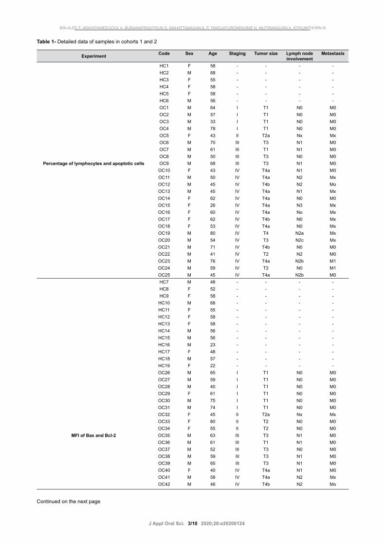
Experiment Code Sex Age Staging Tumor size Lymph nodeinvolvement
Metastasis
HC1 F 58 - - - -HC2 M 68 - - - -HC3 F 55 - - - -HC4 F 58 - - - -HC5 F 58 - - - -HC6 M 56 - - - -OC1 M 64 I T1 N0 M0OC2 M 57 I T1 N0 M0OC3 M 33 I T1 N0 M0OC4 M 78 I T1 N0 M0OC5 F 43 II T2a Nx MxOC6 M 70 III T3 N1 M0OC7 M 61 III T1 N1 M0OC8 M 50 III T3 N0 M0
Percentage of lymphocytes and apoptotic cells OC9 M 68 III T3 N1 M0OC10 F 43 IV T4a N1 M0OC11 M 50 IV T4a N2 MxOC12 M 45 IV T4b N2 MoOC13 M 45 IV T4a N1 MxOC14 F 62 IV T4a N0 M0OC15 F 26 IV T4a N3 MxOC16 F 60 IV T4a No MxOC17 F 62 IV T4b N0 MxOC18 F 53 IV T4a N0 MxOC19 M 80 IV T4 N2a MxOC20 M 54 IV T3 N2c MxOC21 M 71 IV T4b N0 M0OC22 M 41 IV T2 N2 M0OC23 M 76 IV T4a N2b M1OC24 M 59 IV T2 N0 M1OC25 M 45 IV T4a N2b M0HC7 M 48 - - - -HC8 F 52 - - - -HC9 F 58 - - - -
HC10 M 68 - - - -HC11 F 55 - - - -HC12 F 58 - - - -HC13 F 58 - - - -HC14 M 56 - - - -HC15 M 56 - - - -HC16 M 23 - - - -HC17 F 48 - - - -HC18 M 57 - - - -HC19 F 22 - - - -OC26 M 65 I T1 N0 M0OC27 M 59 I T1 N0 M0OC28 M 40 I T1 N0 M0OC29 F 61 I T1 N0 M0OC30 M 75 I T1 N0 M0OC31 M 74 I T1 N0 M0OC32 F 45 II T2a Nx MxOC33 F 80 II T2 N0 M0OC34 F 55 II T2 N0 M0
MFI of Bax and Bcl-2 OC35 M 63 III T3 N1 M0OC36 M 61 III T1 N1 M0OC37 M 52 III T3 N0 M0OC38 M 59 III T3 N1 M0OC39 M 65 III T3 N1 M0OC40 F 40 IV T4a N1 M0OC41 M 58 IV T4a N2 MxOC42 M 46 IV T4b N2 Mo
Table 1- Detailed data of samples in cohorts 1 and 2
Continued on the next page
BIN-ALEE F, ARAYATAWEEGOOL A, BURANAPRADITKUN S, MAHATTANASAKUL P, TANGJATURONRASME N, MUTIRANGURA A, KITKUMTHORN N

J Appl Oral Sci. 2020;28:e202001244/10
Apoptosis of PBMCs and lymphocyte countPBMCs and gated lymphocytes were used to
analyze data on apoptosis. The first cohort investigated
lymphocyte count and the presence of apoptosis in
samples from OC patients and HC. One million cells
were stained with Annexin V Alexa Fluor 488 and
propidium iodide (Biolegend, San Diego, CA, USA) for
30 min at room temperature. Fluorescence-activated
cell sorting (FACS) buffer was added to the cells for
quantifying cell types via flow cytometry (LSRII, BD
biosciences, CA, USA). Frequencies of lymphocytes
were gated from forward scatter FCS and side scatter
(SSC). Data were analyzed using the FlowJo program
(Ashland, OR, USA).
Immune cell subsets with Bax and Bcl-2 measurements
The second cohort categorized PBMCs into four
types of immune cells based on cell-surface markers,
as follows: CD19+ B, CD4+ T, CD8+ T, and CD16+CD56+
NK. The mean fluorescence intensities (MFI) of Bax and
Bcl-2 were measured within these lymphocyte subsets.
One million PBMCs were washed with phosphate-
buffered saline with 2% fetal bovine serum (FBS; FACS
buffer) and stained with cell-surface markers for 20
min at 4°C. The cell-surface markers contained PE-
DZ594-labeled anti-CD16 (clone 3G8) and anti-CD56
(clone 5.1H11), PerCP-Cy5.5-labeled anti-CD19 (clone
HIB19), PE-Cy7-labeled anti-CD3 (clone UCHT1),
Alexa Fluor 700-labeled anti-CD8 (clone SK1), and
APC-Cy7-labeled anti-CD4 (clone RPA-T4) (Biolegend,
San Diego, CA, USA). After washing the cells twice
in FACS buffer, they were fixed/permeabilized and
stained in Alexa Fluor 488 anti-Bax (clone 2D2) and PE
anti-Bcl-2 (clone 100) (Biolegend, San Diego, CA, USA)
antibodies for 30 min at 4°C. Then, cells were again
washed in FACS buffer, fixed in 2% paraformaldehyde
(2% PFA), and analyzed via flow cytometry. Figure 1
shows an example of the gating strategies used in
flow cytometry for detecting Bax and Bcl-2 in CD19+
B, CD4+ T, CD8+ T, and CD16+56+ NK cells among
HC (dash line), low-expression OC (black line), and
high-expression OC (solid black line).
Statistical analysisAll statistical analyses were performed using SPSS
v. 22 (SPSS Inc., Chicago, IL, USA). One-way analysis
of variance (ANOVA) and unpaired t-test calculated
significant differences in the first cohort. Kruskal–
Wallis test compared MFI among the groups in the
second cohort. Pearson’s correlation coefficient (R)
was used to assess correlations between apoptosis and
different cell counts for each group. A p-value of less
than 0.05 (<0.05) was considered statically significant.
Results
Percentage of lymphocytes and apoptotic PBMCs between OC patients and HC
Given the small sample of OC patients in the
stages I, II, and III, we merged them into one group
(OC stage I–III). The percentage of lymphocytes was
higher in HC group, followed by OC stage I–III, and
OC stage IV. The percentage of lymphocytes was
significantly lower in OC stage IV than in HC samples
(p<0.05; Figure 2A). However, the percentage of
apoptotic cells gated from lymphocytes (Figure 2B)
OC43 M 45 IV T4a N1 MxOC44 F 61 IV T4a N0 M0OC45 F 26 IV T4a N3 MxOC46 F 54 IV T4a No MxOC47 F 63 IV T4b N0 MxOC48 F 59 IV T4a N0 MxOC49 M 74 IV T4 N2a Mx
MFI of Bax and Bcl-2 OC50 M 58 IV T3 N2c MxOC51 M 70 IV T4b N0 M0OC52 M 46 IV T2 N2 M0OC53 M 42 IV T4a N2 M0OC54 M 40 IV T3 N2 M0OC55 F 54 IV T4a N2b M0OC56 M 76 IV T4a N2b M1OC57 M 60 IV T2 N0 M1OC58 M 47 IV T4a N2b M0
Continued from previous page
F = female, HC = healthy control, M = male, OC = oral cancer patient
Evaluation of lymphocyte apoptosis in patients with oral cancer

J Appl Oral Sci. 2020;28:e202001245/10
gradually increased from HC, to OC stage I–III, and
OC stage IV. We found a significant difference between
patients with OC stage IV and HC (p<0.05; Figure 2C),
but no significant correlations between the percentage
of lymphocytes and apoptotic cells within OC samples
(R2=0.03; p=0.43; Figure 2D).
Percentage of lymphocyte subtypes and Bax/Bcl-2 MFI and ratio between OC patients and HCFigure 3A shows lymphocyte subsets. OC samples
presented significantly higher CD4+ T cells (p<0.001)
and significantly lower CD8+ T cells than HC. CD19+ B
cells and CD16+56+ NK cells were significantly lower
in OC than in HC samples (p<0.001 and p<0.01,
respectively). Bax MFI of all four cell types was slightly
higher in OC samples than in HC samples (Figure 3B).
The two groups had similar Bcl-2 MFI levels (Figure
3C). Bax/Bcl-2 ratio in CD19+ B, CD8+ T, and CD16+56+
NK cells were slightly higher in OC samples than in
HC samples. The mean ± SD of OC vs. HC in CD19+
B cells were 2.42±1.08 vs. 1.95±0.41 (p=0.13) ,
in CD8+ T cells 1.96±0.85 vs. 1.66±0.52 (p=0.24),
and in CD16+56+ NK cells 2.60±1.21 vs. 2.02±0.53
(p=0.11) (Figure 3D).
Figure 1- Gating strategy of PBMCs via flow cytometry. Lymphocytes were gated from FSC and SSC based on size and granularity. CD3 was used to identify T cells (CD3+) or non-T cells (CD3-). Staining with CD4 and CD8 antibodies against CD4 and CD8 T cells from CD3+ T cells and CD19 Ab for B cells and CD16/56 Ab for NK cells from CD3- T cells. Gated Bax and Bcl-2 expression of immune cells represented by mean fluorescence intensity (MFI). Bax and Bcl-2 expression in HC (dash line), low-expression OC (black line), and high-expression OC (solid black line)
Figure 2- Percentage of lymphocytes. (A), Gated lymphocytes (B) and apoptotic cells (C). Correlations observed between the percentage of lymphocytes and apoptotic cells in OC cells (D) among the PBMCs obtained from the OC patients and HC. *: p<0.05
BIN-ALEE F, ARAYATAWEEGOOL A, BURANAPRADITKUN S, MAHATTANASAKUL P, TANGJATURONRASME N, MUTIRANGURA A, KITKUMTHORN N

J Appl Oral Sci. 2020;28:e202001246/10
Percentage of lymphocyte subtypes and Bax/Bcl-2 MFI and ratio between OC patients in stage I–III and stage IV
The percentage of CD16+56+ NK cells was
significantly lower in OC stage IV than in stage I–III
(p<0.05; Figure 4A). Bax MFI of all four cell types
was slightly higher in stage IV than in stage I–III
samples (Figure 4B). Conversely, Bcl-2 MFI of all four
cell types was significantly lower in stage IV (p<0.05;
Figure 4C). Bax/Bcl-2 ratio in CD19+ B, CD4+ T, CD8+
T, and CD16+56+ NK cells had slightly higher levels in
OC stage IV than in stage I-III samples. The mean ±
SD of OC vs. HC in CD19+ B cells were 2.69±1.16 vs.
2.01±0.82 (p=0.09), in CD4+ T 1.74±0.71 vs. 1.36
Figure 3- Comparison between OC and HC samples. Frequency lymphocyte subsets (A) and MFI of Bax (B), Bcl-2**: P<0.01 (C), and Bax/Bcl-2 ratio (D) of the four lymphocyte cell types. *: p<0.05, **: p<0.01, ***: p<0.001
Figure 4- Comparison between stage I–III and stage IV OC samples. Frequency of lymphocyte subsets (A) and MFI of Bax (B), Bcl-2 (C), and Bax/Bcl-2 ratio (D) of the four lymphocyte cell types. *: p<0.05
Evaluation of lymphocyte apoptosis in patients with oral cancer

J Appl Oral Sci. 2020;28:e202001247/10
±0.56 (p=0.13), in CD8+ T 2.18±0.96 vs. 1.62±0.49
(p=0.07), and in CD16+56+ NK cells 2.91±1.30 vs.
2.12±0.92 (p=0.08) Figure 4D).
Percentage of lymphocytes subtypes and Bax/Bcl-2 MFI and ratio according to tumor size and lymph node metastasis
Regarding tumor size, the percentage of
CD16+CD56+ NK cells was significantly lower in T4
than in T1, T2, and T3 of OC samples (Figure 5A). Bax
Figure 5- Comparison based on tumor size among T1, T2, T3, and T4 samples. Percentage of cells in the four lymphocyte subtypes (A). MFI of Bax, Bcl-2, and Bax/Bcl-2 of the four lymphocyte cell types (B-D). *: p<0.05, **: p<0.01
BIN-ALEE F, ARAYATAWEEGOOL A, BURANAPRADITKUN S, MAHATTANASAKUL P, TANGJATURONRASME N, MUTIRANGURA A, KITKUMTHORN N
Figure 6- Comparison based on lymph node metastasis among N0, N1, N2, and N3 of samples. Percentage of cells in the four lymphocyte subtypes (A). MFI of Bax, Bcl-2, and Bax/Bcl-2 of the lymphocytes cell types (B–D)

J Appl Oral Sci. 2020;28:e202001248/10
MFI of all four cell types gradually increased from T2 to
T3 and T4 (Figure 5B). Bcl-2 MFI of all four cell types
– particularly CD19+ B cells and CD8+ T cells – was
considerably lower in T4 tumors (Figure 5C). Bax/Bcl-2
ratio was significantly higher in T4 than in T2 tumors
for all four cell types, mainly for CD19+ B cells and
CD4+ T cells (p<0.01) and CD8+ T and CD16+56+ NK
cells (p<0.05) (Figure 5D). Likewise, Bax/Bcl-2 ratio
for CD8+ T and CD16+56+ NK cells was significantly
higher in T4 than in T3 (Figure 5D; p < 0.05).
As for the number of metastatic lymph nodes,
the percentage of CD4+ T cells and CD16+CD56+ NK
cells was lower in N3, whereas for CD19+ B and CD8+
T-cells it was higher (Figure 6A). Both Bax and Bcl-2
MFI were significantly different for all four cell types
in N3 when compared with N0, N1, and N2 (Figure
6B, 6C). Bax/Bcl-2 ratio in CD19+ B, CD4+ T, CD8+ T,
and CD16+56+ NK cells had slightly higher levels in N3
than N0, N1, and N2 (data not shown), but without
statically significant difference. N3 p-value, when
compared with N0, N1, and N2 for all four cell types,
ranged from 0.48 to 0.99 (Figure 6D).
Discussion
The immune system is an important defense
mechanism to eliminate tumor cells, and the decrease
of its functional response is intrinsically associated
with cancer’s growth, metastasis, and its recurrence.21
OC is an aggressive tumor, particularly in TNM stage
IV – which corresponds to the metastatic stage.3
Lymphocytes are an important type of immune cells
that have been targeted for the treatment of OC22
for being capable of recognizing cancer antigen and
destroying cancer cells. In a recent study, we found
that PBMCs in cancer patients may alter epigenetic
regulation and gene expression as an effect of head
and neck23 and breast cancer.24 Previous studies
showed that lymphocyte apoptosis is related to
weakness and tumor progression.8,9 Based on these
findings, we hypothesized that cancer cells can alter
the levels of proteins associated with lymphocyte
function, including apoptosis.
In this study, we found significantly lower CD19+
B and CD16+56+ NK cell counts in OC patients than
in HC. The percentage of CD16+56+ NK cells was
significantly lower among OC stage IV patients. Ye, et
al.25 (2017) reported that circulating tumor cells (often
found in advanced-stage cancer) were associated with
a decrease in the number of T lymphocyte subsets
and NK cells in the peripheral blood of patients with
advanced non-small cell lung cancer. As observed
in patients with metastatic breast cancer, NK cells
activity was also lower in patients with advanced-
stage colorectal and prostate cancer.26 NK cells are
associated with innate immune system and contribute
to the first-line defense against cancer and virus
infection.27 These cells are responsible for producing
tumor necrosis factor (TNF), interferon gamma (IFNγ),
interleukin-4 (IL-4), and interleukin-13 (IL-13)28 and
exerting cytolytic activities against tumor cells.27 Many
reports have shown that NK cells failure is associated
with tumor growth.19 NK cells role in tumor immune
surveillance comprises: inducing ligands activation,
decreasing major histocompatibility complex (MHC)
class I expression, retargeting via antibody-dependent
cell-mediated cytotoxicity, and releasing granzyme,
perforin, or cytokines to kill tumor cells.29 NK cells
have acted as good prognostic markers in OC patients.
Our results show that NK cells were reduced in OC
patients in stage IV, T4, and N3 (advanced-stage),
what suggests that increasing NK cell counts may be
a useful alternative-supportive treatment in patients
with OSCC.30,31
We also found that T helper (Th) cells were
significantly higher in OC patients than in HC. This
may be explained by the role of Th cells in anti-tumor
response in promoting immune response, including the
expansion of B cells and cytotoxic T cells, to eliminate
tumor cells by secreting cytokines such as TNF and
IFNγ.32 However, the percentage of Th cells was slightly
lower in OC patients with N3 tumors, which may
occur because these tumors were highly metastatic
and may have escaped the T cell-mediated immune
response mechanism by the adaptation of primary
tumor antigens.33
Several studies on OC demonstrated that circulating
peripheral blood lymphocytes, particularly T cells, are
significantly lower during tumor progression.21 A
study conducted by Reichert, et al.34 (2002) showed
that T cells were present in lower number in the
blood circulation and tumor microenvironment due
to apoptosis in patients with head and neck cancer,
indicating that apoptosis may play a crucial role in the
development and progression of some cancers.
The anti-apoptotic Bcl-2 protein and pro-apoptotic
Bax protein are involved in the intrinsic apoptosis
Evaluation of lymphocyte apoptosis in patients with oral cancer

J Appl Oral Sci. 2020;28:e202001249/10
pathway and respond to cellular stresses, such as
DNA damage, γ-irradiation, and oncogene activation.35
During normal cell growth, Bax and Bcl-2 levels are
balanced. Many studies show that an imbalance
between Bax and Bcl-2, with increased levels of Bax
and decreased levels of Bcl-2, affects lymphocytes
proliferation and survival (such as Th, B, and NK cells)
in patients with cancer.18,20
Our results show that Bax mean MFI was higher in
all four lymphocyte types in OC patients, but Bcl-2 MFI
of all cell types was significantly lower in OC stage IV.
This may be explained by the influence of Bax in cancer
development, and of Bcl-2 in its progression to stage
IV. We also found a high Bax/Bcl-2 ratio in OC stage
IV patients. These results corroborate those reported
by Kim, et al.36 (2004) who found a high Bax/Bcl-2
ratio in circulating CD8+ T cells of patients with HNSCC.
Tumor size, lymph node involvement, and Bax/Bcl-2
ratio were also higher in advance-stage tumors in our
study, which may suggest that Bax/Bcl-2 ratio levels
are associated with OSCC aggressiveness. However,
further studies with a larger cohort are necessary to
clarify this correlation.
The limited number of patients with OC stage I–III
poses a limitation for our study. We suggest further
studies to be conducted with a larger number of
patients to confirm our findings. In conclusion, our
results show that lymphocyte apoptosis and Bax/Bcl-2
ratios were higher in patients with OC in stage 4, T4,
and N3 tumors, indicating that they play an important
role in cancer prognosis. These target molecules may
prove useful for supportive treatment in the future.
AcknowledgmentsWe thank our Head and Neck Cancer Research
Group members for their great effort. This research
was supported by The Chulalongkorn Academic
Advancement into Its 2nd Century Project in
cooperation with The Thailand Research Fund Grants
No. RSA6180078 and National Science and Technology
Development Agency (grant No. FDA-CO-2561-8477-
TH). FB was granted by The Office of the Higher
Education Commission of Thailand.
Disclosure statementThe authors declare no conflict of interest.
Authors' contributionsBin-Alee, Fardeela: Data curation (Lead);
Formal analysis (Lead); Investigation (Lead);
Methodology (Lead); Writing original draft (Lead).
Arayataweekul, Areeya: Investigation (Equal);
Methodology (Equal); Resources (Lead). Baranaprad, Supranee: Investigation (Lead); Methodology (Equal);
Software (Lead). Mahattanasakul, Patnarin: Resources (Lead). Tangjaturonrasme, Napadon: Resources (Equal). Mutirangura, Apiwat: Writing,
review & editing (Equal). Kitkumthorn, Nakarin: Conceptualization (Lead); Investigation (Equal);
Project administration (Lead); Resources (Equal);
Validation (Equal); Writing, review & editing (Lead).
References1- Bray F, Ferlay J, Soerjomataram I, Siegel RL, Torre LA, Jemal A. Global cancer statistics 2018: GLOBOCAN estimates of incidence and mortality worldwide for 36 cancers in 185 countries. CA Cancer J Clin. 2018;68(6):394-424. doi: 10.3322/caac.214922- Jadhav KB, Gupta N. Clinicopathological prognostic implicators of oral squamous cell carcinoma: need to understand and revise. N Am J Med Sci. 2013;5(12):671-9. doi: 10.4103/1947-2714.1232393- Warnakulasuriya S. Global epidemiology of oral and oropharyngeal cancer. Oral Oncol. 2009;45(4-5):309-16. doi: 10.1016/j.oraloncology.2008.06.0024- Joseph N, Dovedi SJ, Thompson C, Lyons J, Kennedy J, Elliott T, et al. Pre-treatment lymphocytopaenia is an adverse prognostic biomarker in muscle-invasive and advanced bladder cancer. Ann Oncol. 2016;27(2):294-9. doi: 10.1093/annonc/mdv5465- Ray-Coquard I, Cropet C, Van Glabbeke M, Sebban C, Le Cesne A, Judson I, et al. Lymphopenia as a prognostic factor for overall survival in advanced carcinomas, sarcomas, and lymphomas. Cancer Res. 2009;69(13):5383-91. doi: 10.1158/0008-5472.CAN-08-38456- Wu ES, Oduyebo T, Cobb LP, Cholakian D, Kong X, Fader AN, et al. Lymphopenia and its association with survival in patients with locally advanced cervical cancer. Gynecol Oncol. 2016;140(1):76-82. doi: 10.1016/j.ygyno.2015.11.0137- Zhao J, Huang W, Wu Y, Luo Y, Wu B, Cheng J, et al. Prognostic role of pretreatment blood lymphocyte count in patients with solid tumors: a systematic review and meta-analysis. Cancer Cell Int. 2020;20:15. doi: 10.1186/s12935-020-1094-58- Tano T, Okamoto M, Kan S, Nakashiro K, Shimodaira S, Koido S, et al. Prognostic impact of expression of Bcl-2 and Bax genes in circulating immune cells derived from patients with head and neck carcinoma. Neoplasia. 2013;15(3):305-14. doi: 10.1593/neo.1215289- Walsh JE, Lathers DM, Chi AC, Gillespie MB, Day TA, Young MR. Mechanisms of tumor growth and metastasis in head and neck squamous cell carcinoma. Curr Treat Options Oncol. 2007;8(3):227-38. doi: 10.1007/s11864-007-0032-210- Galluzzi L, Vitale I, Abrams JM, Alnemri ES, Baehrecke EH, Blagosklonny MV, et al. Molecular definitions of cell death subroutines: recommendations of the Nomenclature Committee on Cell Death 2012. Cell Death Differ. 2012;19(1):107-20. doi: 10.1038/cdd.2011.9611- Elmore S. Apoptosis: a review of programmed cell death. Toxicol Pathol. 2007;35(4):495-516. doi: 10.1080/0192623070132033712- Shamas-Din A, Kale J, Leber B, Andrews DW. Mechanisms of action of Bcl-2 family proteins. Cold Spring Harb Perspect Biol. 2013;5(4):a008714. doi: 10.1101/cshperspect.a008714
BIN-ALEE F, ARAYATAWEEGOOL A, BURANAPRADITKUN S, MAHATTANASAKUL P, TANGJATURONRASME N, MUTIRANGURA A, KITKUMTHORN N

J Appl Oral Sci.
13- Miyashita T, Krajewski S, Krajewska M, Wang HG, Lin HK, Liebermann DA, et al. Tumor suppressor p53 is a regulator of bcl-2 and bax gene expression in vitro and in vivo. Oncogene. 1994;9(6):1799-805.14- Xie X, Clausen OP, De Angelis P, Boysen M. The prognostic value of spontaneous apoptosis, Bax, Bcl-2, and p53 in oral squamous cell carcinoma of the tongue. Cancer. 1999;86(6):913-20. doi: 10.1002/(sici)1097-0142(19990915)86:6<913::aid-cncr4>3.0.co;2-a15- Takemura T, Noguchi M, Kinjyo H, Kubota H, Kido Y, Miyazaki A, et al. The correlation between Bax expression and neoadjuvant chemotherapeutic effects in patients with advanced oral squamous cell carcinoma. J Jpn Stomatol Soc. 2002;51(4):266-72. doi: 10.11277/stomatology1952.51.26616- Kato K, Kawashiri S, Yoshizawa K, Kitahara H, Yamamoto E. Apoptosis-associated markers and clinical outcome in human oral squamous cell carcinomas. J Oral Pathol Med. 2008;37(6):364-71. doi: 10.1111/j.1600-0714.2008.00642.x17- Zhang M, Zhang P, Zhang C, Sun J, Wang L, Li J, et al. Prognostic significance of Bcl-2 and Bax protein expression in the patients with oral squamous cell carcinoma. J Oral Pathol Med. 2009;38(3):307-13. doi: 10.1111/j.1600-0714.2008.00689.x18- Manicassamy S, Gupta S, Huang ZF, Sun ZM. Protein kinase C-theta-mediated signals enhance CD4(+) T cell survival by up-regulating Bcl-x(L). J Immunol. 2006;176(11):6709-16. doi: 10.4049/jimmunol.176.11.670919- Liphaus BL, Bittencourt Kiss MH, Carrasco S, Goldenstein-Schainberg C. Increased Fas and Bcl-2 expression on peripheral mononuclear cells from patients with active juvenile-onset systemic lupus erythematosus. J Rheumatol. 2007;34(7):1580-4.20- Pillet AH, Theze J, Rose T. Interleukin (IL)-2 and IL-15 have different effects on human natural killer lymphocytes. Hum Immunol. 2011;72(11):1013-7. doi: 10.1016/j.humimm.2011.07.31121- Grimm M, Feyen O, Hofmann H, Teriete P, Biegner T, Munz A, et al. Immunophenotyping of patients with oral squamous cell carcinoma in peripheral blood and associated tumor tissue. Tumor Biol. 2016;37(3):3807-16. doi: 10.1007/s13277-015-4224-222- Whiteside TL. Apoptosis of immune cells in the tumor microenvironment and peripheral circulation of patients with cancer: implications for immunotherapy. Vaccine. 2002;20 Suppl 4:A46-51. doi: 10.1016/s0264-410x(02)00387-023- Arayataweegool A, Srisuttee R, Mahattanasakul P, Tangjaturonsasme N, Kerekhanjanarong V, Kitkumthorn N, et al. Head and neck squamous cell carcinoma drives long interspersed element-1 hypomethylation in the peripheral blood mononuclear cells. Oral Dis. 2019;25(1):64-72. doi: 10.1111/odi.12944
24- Puttipanyalears C, Kitkumthorn N, Buranapraditkun S, Keelawat S, Mutirangura A. Breast cancer upregulating genes in stromal cells by LINE-1 hypermethylation and micrometastatic detection. Epigenomics. 2016;8(4):475-86. doi: 10.2217/epi-2015-000725- Ye L, Zhang F, Li HJ, Yang LF, Lv TF, Gu W, et al. Circulating tumor cells were associated with the number of t lymphocyte subsets and NK cells in peripheral blood in advanced non-small-cell lung cancer. Dis Markers. 2017;2017:5727815. doi: 10.1155/2017/572781526- Santos MF, Mannam VK, Craft BS, Puneky LV, Sheehan NT, Lewis RE, et al. Comparative analysis of innate immune system function in metastatic breast, colorectal, and prostate cancer patients with circulating tumor cells. Exp Mol Pathol. 2014;96(3):367-74. doi: 10.1016/j.yexmp.2014.04.00127- Hu WL, Wang GS, Huang DS, Sui MH, Xu YB. Cancer immunotherapy based on natural killer cells: Current progress and new opportunities. Front Immunol. 2019;10:1205. doi: 10.3389/fimmu.2019.0120528- Ruffell B, DeNardo DG, Affara NI, Coussens LM. Lymphocytes in cancer development: Polarization towards pro-tumor immunity. Cytokine Growth Factor Rev. 2010;21(1):3-10. doi: 10.1016/j.cytogfr.2009.11.00229- Li Y, Sun R. Tumor immunotherapy: new aspects of natural killer cells. Chin J Cancer Res. 2018;30(2):173-96. doi: 10.21147/j.issn.1000-9604.2018.02.0230- Agarwal R, Chaudhary M, Bohra S, Bajaj S. Evaluation of natural killer cell (CD57) as a prognostic marker in oral squamous cell carcinoma: an immunohistochemistry study. J Oral Maxillofac Pathol. 2016;20(2):173-7. doi: 10.4103/0973-029X.18593331- Turkseven MR, Oygur T. Evaluation of natural killer cell defense in oral squamous cell carcinoma. Oral Oncol. 2010;46(5):e34-7. doi: 10.1016/j.oraloncology.2010.02.01932- Borst J, Ahrends T, Babala N, Melief CJ, Kastenmuller W. CD4(+) T cell help in cancer immunology and immunotherapy. Nat Rev Immunol. 2018;18(10):635-47. doi: 10.1038/s41577-018-0044-033- Kumagai K, Hamada Y, Gotoh A, Kobayashi H, Kawaguchi K, Horie A, et al. Evidence for the changes of antitumor immune response during lymph node metastasis in head and neck squamous cell carcinoma. Oral Surg Oral Med Oral Pathol Oral Radiol Endod. 2010;110(3):341-50. doi: 10.1016/j.tripleo.2010.03.03034- Reichert TE, Strauss L, Wagner EM, Gooding W, Whiteside TL. Signaling abnormalities, apoptosis, and reduced proliferation of circulating and tumor-infiltrating lymphocytes in patients with oral carcinoma. Clin Cancer Res. 2002;8(10):3137-45.35- Meier P, Finch A, Evan G. Apoptosis in development. Nature. 2000;407(6805):796-801. doi: 10.1038/3503773436- Kim JW, Tsukishiro T, Johnson JT, Whiteside TL. Expression of pro- and antiapoptotic proteins in circulating CD8+ T cells of patients with squamous cell carcinoma of the head and neck. Clin Cancer Res. 2004;10(15):5101-10. doi: 10.1158/1078-0432.CCR-04-0309
2020;28:e2020012410/10
Evaluation of lymphocyte apoptosis in patients with oral cancer