Food Restriction‐Like Effects of Dehydroepiandrosterone: Decreased Lymphocyte Numbers and...
10
Food Restriction-Like Effects of Dehydroepiandrosterone: Decreased Lymphocyte Numbers and Functions with Increased Apoptosis (44415) FERNANDO CATALINA,* VINAY KUMAR,* LEON MILEWICH,² AND MICHAEL BENNETT* ,1 Departments of Pathology* and Obstetrics and Gynecology,² University of Texas Southwestern Medical Center, Dallas, Texas 75235–9072 Abstract. Both dietary dehydroepiandrosterone (DHEA) and food restriction can pre- vent or modulate the initiation or progression of a number of diseases in rodents and prolong life span. We sought to determine if these interventions have common mecha- nisms of action in regulating lymphocyte functions and cell numbers. We observed that male C57BL/6 mice receiving DHEA in the diet (0.45%, w/w) ate approximately 50% as much food as mice on the DHEA-free diet, and this was reflected in decreased body weights throughout a 10-week period. Mice either fed the DHEA-containing diet or pair-fed to the DHEA-treated mice had decreased spleen and thymus weights and lymphocyte cell numbers compared to mice having free access to the control diet. Mice were fed these diets for 2 weeks before and 2 weeks after exposure to sublethal irradiation (500 cGy). In mice fed DHEA or pair-fed, there was a decrease in spleen cell numbers, and B cells were the most severely affected. The frequency of apoptosis in peripheral blood cells increased from <5% in nonirradiated controls to >50% within 4 days after starting DHEA or pair-feeding. Shortly after irradiation, >87% of blood lym- phocytes were hypodiploid (apoptotic) in all groups. By 9 days only 27% of lympho- cytes in mice on the control diet were hypodiploid compared to 62% in DHEA and 74% in pair-fed mice. In addition, both DHEA and pair-fed mice had significant reductions in T-cell function (contact hypersensitivity to dinitrofluorobenzene), B cell function (antibody response to trinitrophenolated-lipopolysaccharide), and NK cell function (lung clearance of radiolabeled YAC-1 tumor cells) 2 weeks after irradiation. In a complementary study, peripheral blood lymphocytes from naı ¨ve mice were treated overnight with various concentrations of either DHEA or hydrocortisone 21-acetate. Only glucocorticoid-treated cells underwent apoptosis. Thus, DHEA induces apopto- sis in vivo but not in vitro. We conclude that dietary DHEA induces apoptosis and decreased lymphocyte production and function in C57BL/6 mice largely by reducing food intake. [P.S.E.B.M. 1999, Vol 221] D ehydroepiandrosterone (DHEA) is a steroid pro- hormone present in relatively high concentrations in blood of young human adults; these levels gradu- ally decrease to approximately 10% by the eighth decade of life (1). Clinical studies revealed decreased serum and/or urinary concentrations of DHEA significantly lower in pa- tients with Alzheimer’s disease (2), HIV infection (3), sys- temic lupus erythematosus (4), coronary atherosclerosis (5), breast cancer (6, 7), and obesity (8). In studies with animal models of diseases, dietary DHEA (0.2%–1.0% w/w of food) was effective in preventing or inhibiting neoplasia (9), diabetes (10), lupus erythematosus (11), immune decline (12), hemolytic anemia (13), nephrosis (14), atherosclerosis (15), and obesity (16). Despite the association between decreased serum DHEA levels and disease states in humans, little is known about the mode(s) of DHEA action. Many of the diseases in This work was supported in part by NIH MSTP Training Grant 5T32 GM08014 and NIH Grants CA36922, CA47073, AI38939, and AI20451. 1 To whom requests for reprints should be addressed at Department of Pathology, University of Texas Southwestern Medical Center, 5323 Harry Hines Boulevard, Dallas, TX 75235–9072. E-mail: [email protected] Received November 3, 1998. [P.S.E.B.M. 1999, Vol 221] Accepted March 3, 1999. 0037-9727/99/2214-0326$14.00/0 Copyright © 1999 by the Society for Experimental Biology and Medicine 326 DHEA, FOOD INTAKE, AND APOPTOSIS
-
Upload
independent -
Category
Documents
-
view
4 -
download
0
Transcript of Food Restriction‐Like Effects of Dehydroepiandrosterone: Decreased Lymphocyte Numbers and...
Food Restriction-Like Effects ofDehydroepiandrosterone: Decreased
Lymphocyte Numbers and Functions withIncreased Apoptosis (44415)
FERNANDO CATALINA ,* V INAY KUMAR,* L EON MILEWICH,† AND MICHAEL BENNETT* ,1
Departments of Pathology* and Obstetrics and Gynecology,† University of Texas Southwestern Medical Center,Dallas, Texas 75235–9072
Abstract. Both dietary dehydroepiandrosterone (DHEA) and food restriction can pre-vent or modulate the initiation or progression of a number of diseases in rodents andprolong life span. We sought to determine if these interventions have common mecha-nisms of action in regulating lymphocyte functions and cell numbers. We observedthat male C57BL/6 mice receiving DHEA in the diet (0.45%, w/w) ate approximately 50%as much food as mice on the DHEA-free diet, and this was reflected in decreased bodyweights throughout a 10-week period. Mice either fed the DHEA-containing diet orpair-fed to the DHEA-treated mice had decreased spleen and thymus weights andlymphocyte cell numbers compared to mice having free access to the control diet.Mice were fed these diets for 2 weeks before and 2 weeks after exposure to sublethalirradiation (500 cGy). In mice fed DHEA or pair-fed, there was a decrease in spleen cellnumbers, and B cells were the most severely affected. The frequency of apoptosis inperipheral blood cells increased from <5% in nonirradiated controls to >50% within 4days after starting DHEA or pair-feeding. Shortly after irradiation, >87% of blood lym-phocytes were hypodiploid (apoptotic) in all groups. By 9 days only 27% of lympho-cytes in mice on the control diet were hypodiploid compared to 62% in DHEA and 74%in pair-fed mice. In addition, both DHEA and pair-fed mice had significant reductionsin T-cell function (contact hypersensitivity to dinitrofluorobenzene), B cell function(antibody response to trinitrophenolated-lipopolysaccharide), and NK cell function(lung clearance of radiolabeled YAC-1 tumor cells) 2 weeks after irradiation. In acomplementary study, peripheral blood lymphocytes from naı ¨ve mice were treatedovernight with various concentrations of either DHEA or hydrocortisone 21-acetate.Only glucocorticoid-treated cells underwent apoptosis. Thus, DHEA induces apopto-sis in vivo but not in vitro. We conclude that dietary DHEA induces apoptosis anddecreased lymphocyte production and function in C57BL/6 mice largely by reducingfood intake. [P.S.E.B.M. 1999, Vol 221]
Dehydroepiandrosterone (DHEA) is a steroid pro-hormone present in relatively high concentrationsin blood of young human adults; these levels gradu-
ally decrease to approximately 10% by the eighth decade oflife (1). Clinical studies revealed decreased serum and/orurinary concentrations of DHEA significantly lower in pa-tients with Alzheimer’s disease (2), HIV infection (3), sys-temic lupus erythematosus (4), coronary atherosclerosis (5),breast cancer (6, 7), and obesity (8). In studies with animalmodels of diseases, dietary DHEA (0.2%–1.0% w/w offood) was effective in preventing or inhibiting neoplasia (9),diabetes (10), lupus erythematosus (11), immune decline(12), hemolytic anemia (13), nephrosis (14), atherosclerosis(15), and obesity (16).
Despite the association between decreased serumDHEA levels and disease states in humans, little is knownabout the mode(s) of DHEA action. Many of the diseases in
This work was supported in part by NIH MSTP Training Grant 5T32 GM08014 andNIH Grants CA36922, CA47073, AI38939, and AI20451.1 To whom requests for reprints should be addressed at Department of Pathology,University of Texas Southwestern Medical Center, 5323 Harry Hines Boulevard,Dallas, TX 75235–9072. E-mail: [email protected]
Received November 3, 1998. [P.S.E.B.M. 1999, Vol 221]Accepted March 3, 1999.
0037-9727/99/2214-0326$14.00/0Copyright © 1999 by the Society for Experimental Biology and Medicine
326 DHEA, FOOD INTAKE, AND APOPTOSIS
which DHEA is effective have an immune component (4,11–13). Low doses of DHEA or its sulfate enhances theeffectiveness of vaccines against various pathogens, par-ticularly in aged rodents (17–29). At a higher dietary DHEAdose of 0.45% (w/w), there is a decrease in lymphopoiesis(30), but little effect on various lymphocyte functions inmice (31, 32). In long-lived (C3H.SW X B10.RIII)F1 mice,dietary DHEA increased T-cell and NK-cell function (12).
DHEA administration to mice and rats can significantlydecrease food intake (12, 14, 33, 34); thus, one of the pos-sible mechanisms by which dietary DHEA may exert itsbeneficial effects is through the induction of food restric-tion. Whereas dietary DHEA (0.2%–1.0%) may reduce foodintake (10), lower levels of DHEA applied subcutaneouslyor topically to stimulate the immune system do not affectfood consumption or body weights. Limiting an animal’sfood intake has been found to be therapeutic in many of thesame disease models in which dietary DHEA administrationis also beneficial (9, 12, 13, 35–41). On the basis of theseobservations we conducted pair-feeding experiments to de-termine the possible involvement of food restriction on theobserved effects of dietary DHEA on cells of the immunesystem. Because restriction of food intake has been reportedto induce apoptosis in both normal and neoplastic cells (40,42), we analyzed the incidence of apoptosis in peripheralblood lymphocytes of both DHEA-treated and pair-fed ani-mals before and after irradiation (500 cGy). We also com-pared the effects of DHEA and hydrocortisone 21-acetateon the rates of apoptosis on lymphocytes maintained inovernight cultures.
Materials and MethodsAnimals. Conventionally housed male C57BL/6
mice (10–12 weeks old) were obtained from the Departmentof Microbiology Colony at this University. Initially all stud-ies were conducted with mice housed individually (1/cage);later, mice were housed in groups of four to six per cage. Nodifferences were observed in body weight, food intake, orimmune parameters whether housed singly or in groups.Mice were caged singly for experiments described in Fig-ures 1 and 5. Animals were housed at 22 ± 2°C and main-tained on a 12-hr light/dark cycle with lights out at 19:00 hr.The use of mice has been approved by the Institution’sAnimal Care and Use Committee (IACRAC).
Diets. Fourteen days prior to any experimental ma-nipulations, mice were placed on pelleted purified AIN(American Institute of Nutrition)-76A diet (Dyets Inc.,Bethlehem, PA) containing either no additives (control diet)or DHEA (0.45%, w/w) (Sigma, St. Louis, MO). All ani-mals except pair-fed mice had free access to food, and allmice received water freely. Pair-fed animals were given thecontrol diet. The amount of control diet fed to pair-fed micewas determined by the mean weight of the food consumedby DHEA-fed mice on the previous day. The diet for pair-feeding was administered once per day between 09:00 hrand 10:00 hr.
Determination of Food Intake and Body Weight.The weight of the food consumed by mice on the DHEA-containing diet, accounting for the weight of spilled food,was determined daily between 09:00 hr and 10:00 hr. Whenmice were housed four to six animals per cage, only themean daily intake per mouse could be obtained by dividingthe total daily food intake per cage by the number of micein the group. Mice were weighed weekly during the courseof the studies.
Irradiation. Mice were exposed to 500 cGy ofgamma radiation in a GammaCell 40 small animal irradiatorcontaining two 137Cs sources (Atomic Energy Ltd., Ot-tawa, Ontario, Canada) at a rate of 68 cGy/min.
Contact Hypersensitivity. This response is a testof T-cell function and was performed as described (43).Briefly, the abdomen of mice was shaved and painted on 2consecutive days with 20ml 0.5% dinitrofluorobenzene(DNFB, Sigma) in acetone/olive oil (4:1, v/v) solution. Fivedays later, ear swelling was elicited by applying 10ml of0.2% DNFB solution to both sides of the pinna of one ear.Response was determined by measuring the difference inear thickness between pre-elicitation and 24 hr post-elicitation using a Fowler Precision Tools caliper (Lux Sci-entific Instrument Corp., New York, NY).
T Cell-Independent IgM Antibody Response.The IgM response to trinitrophenolated lipopolysaccharide(TNP-LPS, Sigma), was used to measure B-cell function.Mice were immunized by i.p. injection of 100mg TNP-LPS;6 and 10 days later mice were bledvia the tail vein; serawere diluted in PBS containing 1.0% BSA (Sigma), andIgM concentrations were determined by ELISA (44).
Lung Clearance of Radiolabeled YAC-1 TumorCells. This is an assay for natural killer (NK) cell function.YAC-1 tumor cells (31) (5 × 10⁷) were incubated in RPMIcontaining 5-fluorodeoxyuridine (2.5mg/ml; Sigma); 20min later, 30 mCi [125I]5-iodo-28-deoxyuridine (121IdU,Amersham, Arlington Heights, IL) was added and the cellswere incubated for 90 min at 37°C in 5% CO2/air. The cellswere washed three times in RPMI and adjusted to 2 × 10⁶
cells/ml. Cell viabilities were >95% by trypan blue exclu-sion. 1 × 10⁶ cells (0.5 ml) were injected into micevia thelateral tail vein. Four hours later, the mice were sacrificedby CO2 inhalation, the lungs were removed, and125I radio-activity was determined. The results are expressed as thegeometric mean (95% confidence limits) percentage recov-ery of the total injected radioactivity (45). Thelack of re-tention of radiolabeled YAC-1 cells in the lungs is a mea-sure of NK activity.
Flow Cytometry for Lymphocyte Surface Mark-ers. Animals were bled from tail veins to obtain bloodlymphocytes. Other mice were sacrificed CO2 inhalation.Spleens were removed and gently crushed between thefrosted ends of slides in RPMI media (GIBCO BRL, GrandIsland, NY). Each spleen was processed individually. Cellswere washed twice in RPMI. Erythrocytes were lysed byhypotonic shock; splenocytes were then passed through ny-
DHEA, FOOD INTAKE, AND APOPTOSIS 327
lon mesh to make single cell suspensions. Cells werecounted using an electronic particle counter (Coulter Elec-tronics Inc., Hialeah, FL). Splenocytes (2 × 10⁶) were sus-pended in 5% calf serum in PBS, and cells were stained withthe following conjugated monoclonal antibodies: F(ab)82
DTAF-goat anti-mouse IgG + IgM (H + L) (JacksonImmunoResearch, Avondale, PA), FITC-anti-TCRab, PE-anti-NK1.1, (Pharmingen, San Diego, CA), or the irrelevantisotype controls FITC-Rat IgG2a, PE-mouse IgG2a, PE-Hamster IgG (Pharmingen). Collection gates were set oncells of lymphocyte size and internal complexity based onforward and 90 scatter using a FACScan flow cytometer(Becton-Dickinson, Mountain View, CA).
Flow Cytometry for Apoptosis. In vitro studies.Peripheral blood was collected from the lateral tail vein ofuntreated mice and placed in Alsever’s solution (GIBCOBRL). Erythrocytes were lysed by hypotonic shock, cellswere enumerated using the Coulter electronic particlecounter, and lymphocytes (2 × 10⁶ cells/well in 0.2 ml) wereplated in 96-well plates containing various concentrations ofDHEA or hydrocortisone 21-acetate. DHEA and hydrocor-tisone 21-acetate stock solutions in ethanol were diluted tothe appropriate concentrations with medium. Medium andethanol-containing medium were used as controls in theflow cytometry studies. Cells were incubated overnight at37°C in 5% CO2/air. The next day the cells were placed inFalcon 2054 tubes (Becton-Dickinson) for staining andFACS analysis.
In vivo studies. At different time points after the startof either the DHEA or pair-fed diet, peripheral blood wascollected by tail bleeding. Erythrocytes were lysed by hy-potonic saline, and lymphocytes were placed in Falcon 2054tubes for staining and analysis by flow cytometry.
Propidium iodide staining and analysis. Cells werestained as described (46). Briefly, 1 × 10⁵ to 1 × 10⁶ cellswere incubated overnight at 4°C in the presence of hypo-tonic propidium iodide (PI) solution [0.1% Triton X-100(Baker Chemical Co. Phillipsburg, NJ), 0.1% sodium ci-trate (Fisher Scientific, Fair Lawn, NJ), 50mg/ml PI(Sigma)]. The following day the cells were analyzed usinga FACScan flow cytometer. Cellular debris was gated outbased on low forward scatter. PI fluorescence was moni-tored in the red fluorescence channels. Apoptotic cells (hy-podiploid) appear as a broad peak, staining with consider-ably lower intensity than the narrow peak of healthy (dip-loid) cells.
Statistics. Differences between groups were deter-mined using two-way ANOVA followed by Duncan’s rangetest, one-way ANOVA followed by Scheffe’spost hoctest,or Studentst test as indicated in the figure legends.
ResultsWe examined the effect of dietary DHEA (0.45%, w/w)
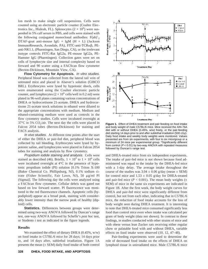
on food intake in C57BL/6 mice for 28 days; 14 days priorto, and 14 days after, sublethal irradiation. Figure 1Apresents the mean (± SEM) daily food intake of both control
and DHEA-treated mice from six independent experiments.The intake of pair-fed mice is not shown because food ad-ministered was equal to the intake by the DHEA-fed micewith a 1-day delay. The average intake throughout thecourse of the studies was 3.04 ± 0.06 g/day (mean ± SEM)for control mice and 1.53 ± 0.05 g/day for DHEA-treatedand pair-fed mice (P < 0.001). The mean body weights (±SEM) of mice in the same six experiments are indicated inFigure 1B. After the first week, the body weight curves forDHEA and pair-fed mice were significantly different fromcontrol, but not from each other, indicating that in C57BL/6mice, the reduction of food intake accounts for the loss ofbody weight seen during DHEA treatment. It is interestingto note that DHEA-treated mice consumed significantly lessfood than control mice even when intake was calculated pergram of body weight (data not shown). In contrast to thesefindings, in studies conducted with other strains of mice andwith obese versus lean Zucker rats receiving either regularchow or palatable food with and without DHEA, variableeffects on food intake were observed (10, 12, 47–48).
Pair-feeding experiments were used to determine therole of decreased food intake on the effects of DHEA onlymphoid tissue in unirradiated mice. Male C57BL/6 mice
Figure 1. Effect of DHEA treatment and pair-feeding on food intakeand body weight of male C57BL/6 mice. Mice received the AIN-76Adiet with or without DHEA (0.45%, w/w) freely, or the pair-feedingdiet starting 14 days prior to and after sublethal irradiation (500 cGy).Daily food intake and weekly body weights were monitored. Valuespresented are from six experiments, with four to six mice/group or aminimum of 24 mice per experimental group. *Significantly differentfrom control (P < 0.01) by two-way ANOVA with repeated measuresfollowed by Duncan’s range test.
328 DHEA, FOOD INTAKE, AND APOPTOSIS
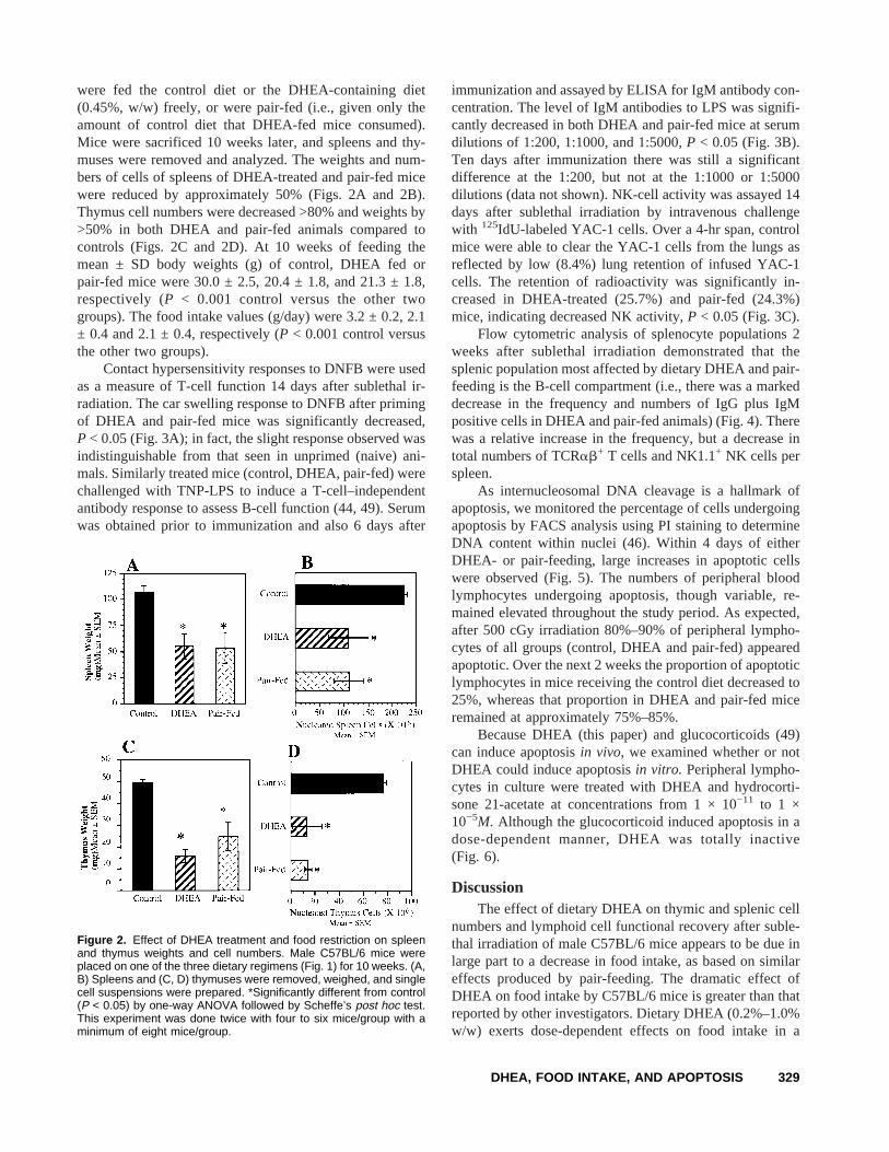
were fed the control diet or the DHEA-containing diet(0.45%, w/w) freely, or were pair-fed (i.e., given only theamount of control diet that DHEA-fed mice consumed).Mice were sacrificed 10 weeks later, and spleens and thy-muses were removed and analyzed. The weights and num-bers of cells of spleens of DHEA-treated and pair-fed micewere reduced by approximately 50% (Figs. 2A and 2B).Thymus cell numbers were decreased >80% and weights by>50% in both DHEA and pair-fed animals compared tocontrols (Figs. 2C and 2D). At 10 weeks of feeding themean ± SD body weights (g) of control, DHEA fed orpair-fed mice were 30.0 ± 2.5, 20.4 ± 1.8, and 21.3 ± 1.8,respectively (P < 0.001 control versus the other twogroups). The food intake values (g/day) were 3.2 ± 0.2, 2.1± 0.4 and 2.1 ± 0.4, respectively (P < 0.001 control versusthe other two groups).
Contact hypersensitivity responses to DNFB were usedas a measure of T-cell function 14 days after sublethal ir-radiation. The car swelling response to DNFB after primingof DHEA and pair-fed mice was significantly decreased,P < 0.05 (Fig. 3A); in fact, the slight response observed wasindistinguishable from that seen in unprimed (naive) ani-mals. Similarly treated mice (control, DHEA, pair-fed) werechallenged with TNP-LPS to induce a T-cell–independentantibody response to assess B-cell function (44, 49). Serumwas obtained prior to immunization and also 6 days after
immunization and assayed by ELISA for IgM antibody con-centration. The level of IgM antibodies to LPS was signifi-cantly decreased in both DHEA and pair-fed mice at serumdilutions of 1:200, 1:1000, and 1:5000,P < 0.05 (Fig. 3B).Ten days after immunization there was still a significantdifference at the 1:200, but not at the 1:1000 or 1:5000dilutions (data not shown). NK-cell activity was assayed 14days after sublethal irradiation by intravenous challengewith 125IdU-labeled YAC-1 cells. Over a 4-hr span, controlmice were able to clear the YAC-1 cells from the lungs asreflected by low (8.4%) lung retention of infused YAC-1cells. The retention of radioactivity was significantly in-creased in DHEA-treated (25.7%) and pair-fed (24.3%)mice, indicating decreased NK activity,P < 0.05 (Fig. 3C).
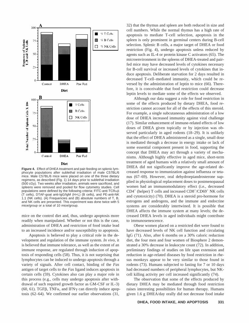
Flow cytometric analysis of splenocyte populations 2weeks after sublethal irradiation demonstrated that thesplenic population most affected by dietary DHEA and pair-feeding is the B-cell compartment (i.e., there was a markeddecrease in the frequency and numbers of IgG plus IgMpositive cells in DHEA and pair-fed animals) (Fig. 4). Therewas a relative increase in the frequency, but a decrease intotal numbers of TCRab+ T cells and NK1.1+ NK cells perspleen.
As internucleosomal DNA cleavage is a hallmark ofapoptosis, we monitored the percentage of cells undergoingapoptosis by FACS analysis using PI staining to determineDNA content within nuclei (46). Within 4 days of eitherDHEA- or pair-feeding, large increases in apoptotic cellswere observed (Fig. 5). The numbers of peripheral bloodlymphocytes undergoing apoptosis, though variable, re-mained elevated throughout the study period. As expected,after 500 cGy irradiation 80%–90% of peripheral lympho-cytes of all groups (control, DHEA and pair-fed) appearedapoptotic. Over the next 2 weeks the proportion of apoptoticlymphocytes in mice receiving the control diet decreased to25%, whereas that proportion in DHEA and pair-fed miceremained at approximately 75%–85%.
Because DHEA (this paper) and glucocorticoids (49)can induce apoptosisin vivo, we examined whether or notDHEA could induce apoptosisin vitro. Peripheral lympho-cytes in culture were treated with DHEA and hydrocorti-sone 21-acetate at concentrations from 1 × 10⁻¹¹ to 1 ×10⁻⁵M. Although the glucocorticoid induced apoptosis in adose-dependent manner, DHEA was totally inactive(Fig. 6).
DiscussionThe effect of dietary DHEA on thymic and splenic cell
numbers and lymphoid cell functional recovery after suble-thal irradiation of male C57BL/6 mice appears to be due inlarge part to a decrease in food intake, as based on similareffects produced by pair-feeding. The dramatic effect ofDHEA on food intake by C57BL/6 mice is greater than thatreported by other investigators. Dietary DHEA (0.2%–1.0%w/w) exerts dose-dependent effects on food intake in a
Figure 2. Effect of DHEA treatment and food restriction on spleenand thymus weights and cell numbers. Male C57BL/6 mice wereplaced on one of the three dietary regimens (Fig. 1) for 10 weeks. (A,B) Spleens and (C, D) thymuses were removed, weighed, and singlecell suspensions were prepared. *Significantly different from control(P < 0.05) by one-way ANOVA followed by Scheffe’s post hoc test.This experiment was done twice with four to six mice/group with aminimum of eight mice/group.
DHEA, FOOD INTAKE, AND APOPTOSIS 329

given strain of mice, but the effects vary in different animalspecies and/or strains. For example, Colemanet al. (10)observed that food intake was decreased in only one(C57BL/6J) of two strains tested in the narrow ancestralC57BL mouse lineage. Most investigators have found no oronly transient effects of DHEA (9, 50–53), whereas othershave observed a significant reduction in food intake. A dietwe and others have used may contribute to more dramaticeffects on food intake due to better absorption (10, 30–32).The suppression of food intake by DHEA in Zucker rats wasassociated with alterations of hypothalamic serotonin con-tent (47), raising the possibility that DHEA may have aneuromodulatory effect on appetite regulation.
Both food restriction and dietary DHEA can preventand/or treat many of the same diseases in animal models, aswell as inhibit immune decline (1–10, 35–42); this list in-cludes autoimmune disease (35–37), nephrosis (14, 39) andneoplasia (9, 40, 41, 54). Also, both dietary DHEA and foodrestriction greatly extend the life expectancy of laboratoryrodents (14, 55). These observations indicate a possible rolefor food restriction in the effects of DHEA. For many yearslittle was known about the mechanism by which food re-striction mediates its various effects. As early as the 1960sit was noted that food restriction inhibited cellular prolif-eration, and it was proposed that proliferation was inhibitedthrough suppression of DNA synthesis (56, 57). More re-cently, food restriction has been shown not only to inhibitproliferation, but also to eliminate pre-neoplastic cellsthrough induction of apoptosis (40). The protective effect of
food restriction against cancer was associated with modu-lation of the rates of apoptosis and proliferation (42). In ourstudies, DHEA administration and food restriction dramati-cally increased the number of apoptotic blood lymphocytesin mice within 4 days. The high rates of apoptosis weresurprising, because apoptotic cells are usually rapidly en-gulfed and removed by phagocytic cells. The action ofDHEA is likely mediated through food restriction, becausepair-fed mice behave in a similar manner. DHEA, unlikeglucocorticoids, does not directly induce cells to undergoapoptosisin vitro (Fig. 6), indicating that DHEA and foodrestriction act indirectly. However, it is conceivable that thestress or toxicity of DHEA and/or food restriction stimu-lated the secretion of corticosterone or other direct media-tors of apoptosis, or induced the expression ofFason lym-phocytes andFas ligand on other cell types or other lym-phocytes. The effects of DHEA observed here may havebeen mostly indirect and may have obscured any directeffects of DHEA on the immune system.
It is possible that apoptosis detected by flow cytometryas described (46) is a result of the manipulations involved inisolating peripheral blood lymphocytes, and not a result ofthe treatment. However, the fact that apoptotic cells are notdetectable in lymphocytes of mice on the control diet untilafter the mice are sublethally irradiated indicates that themanipulations performed here do not induce healthy cells toundergo apoptosis. It is still possible that apoptotic cells arenot present in the blood of DHEA-treated or pair-fed mice,but that these cells are more susceptible than cells from
Figure 3. Effect of DHEA treatment and food restriction on recovery of T, B, and NK lymphocyte functions after sublethal irradiation. MaleC57BL/6 mice were placed on one of the three dietary regimens (Fig. 1). Two weeks after starting the diets, the mice were exposed to sublethalirradiation (500 cGy). Two weeks after irradiation mice were examined for one of the following parameters: (A) contact hypersensitivityresponse to DNFB (T cell response); (B) IgM response to TNP-LPS (B cell response); and (C) clearance of radiolabeled YAC-1 tumor cells(NK cell response). *Significantly different from control (P < 0.05) by one-way ANOVA followed by Scheffe’s post hoc test in A and C, andtwo-way ANOVA followed by Duncan’s range test in B. This experiment was done twice with four mice/group or a total of eight mice/group.
330 DHEA, FOOD INTAKE, AND APOPTOSIS
mice on the control diet and, thus, undergo apoptosis morereadily when manipulated. Whether or not this is the case,administration of DHEA and restriction of food intake leadto an increased incidence and/or susceptibility to apoptosis.
Apoptosis is believed to play a critical role in the de-velopment and regulation of the immune system.In vivo, itis believed that immune tolerance, as well as the extent of animmune response, are regulated through induction of apop-tosis of responding cells (58). Thus, it is not surprising thatlymphocytes can be induced to undergo apoptosis through avariety of signals. After cell contact, binding of theFasantigen of target cells to theFas ligand induces apoptosis incertain cells (59). Cytokines also can play a major role inthis process (e.g., cells may undergo apoptosis after with-drawal of such required growth factor as GM-CSF or IL-3)(60, 61). TGFb, TNFa, and IFNg can directly induce apop-tosis (62–64). We confirmed our earlier observations (31,
32) that the thymus and spleen are both reduced in size andcell numbers. While the normal thymus has a high rate ofapoptosis to mediate T-cell selection, apoptosis in thespleen is only prominent in germinal centers during B-cellselection. Splenic B cells, a major target of DHEA or foodrestriction (Fig. 4), undergo apoptosis unless reduced byagents such as IL-4 or protein kinase C activators (65). Themicroenvironment in the spleens of DHEA-treated and pair-fed mice may have decreased levels of cytokines necessaryfor B-cell survival or increased levels of cytokines that in-duce apoptosis. Deliberate starvation for 2 days resulted indecreased T-cell–mediated immunity, which could be re-versed by the administration of leptin to mice (66). There-fore, it is conceivable that food restriction could decreaseleptin levels to mediate some of the effects we observed.
Although our data suggest a role for food restriction insome of the effects produced by dietary DHEA, food re-striction cannot account for all of the effects of this steroid.For example, a single subcutaneous administration of a lowdose of DHEA increased immunity against viral challenge(17). Similar enhancement of immune-related effects of lowdoses of DHEA given topically or by injection was ob-served particularly in aged rodents (18–29). It is unlikelythat the effect of DHEA administered as a single, small doseis mediated through a decrease in energy intake or lack ofsome essential component present in food, supporting theconcept that DHEA may act through a variety of mecha-nisms. Although highly effective in aged mice, short-termtreatment of aged humans with a relatively small amount ofDHEA did not significantly improve the age-related de-creased response to immunization against influenza or teta-nus (67–69). However, oral dehydroepiandrosterone sup-plied in physiological replacement doses to postmenopausalwomen had an immunomodulatory effect (i.e., decreasedCD4+ (helper) T cells and increased CD8+/CD69+ NK cellsand cytotoxicity) (70). DHEA is a steroid pro-hormone forestrogens and androgens, and the immune and endocrinesystems are considerably intertwined. It is possible thatDHEA affects the immune system at many levels; the de-creased DHEA levels in aged individuals might contributeto immunosenescence.
Obese women placed on a restricted diet were found tohave decreased levels of NK cell function and circulatingIgG (71). Also, after 6 months on a 30% caloric reductiondiet, the four men and four women of Biosphere 2 demon-strated a 30% decrease in leukocyte count (72). In addition,preliminary findings of studies on life span extension andreduction in age-related diseases by food restriction in rhe-sus monkeys appear to be very similar to those found inrodents (73). Humans subjected to fasting for 7 or 10 dayshad decreased numbers of peripheral lymphocytes, but NK-cell killing activity per cell increased significantly (74).
The observation that some of the effects produced bydietary DHEA may be mediated through food restrictionraises interesting possibilities for human therapy. Humansgiven 1.6 g DHEA/day orally did not decrease food intake
Figure 4. Effect of DHEA treatment and pair-feeding on splenic lym-phocyte populations after sublethal irradiation of male C57BL/6mice. Male C57BL/6 mice were placed on one of the three dietaryregimens, as described (Fig. 1) 14 days prior to sublethal irradiation(500 cGy). Two weeks after irradiation, animals were sacrificed, andspleens were removed and pooled for flow cytometry studies. Cellpopulations were defined by the following criteria: FITC-anti-TCRab(T cells), DTAF-goat anti-IgG/IgM (H+L) (B cells), and PE-anti-NK1.1 (NK cells). (A) Frequencies and (B) absolute numbers of T, B,and NK cells are presented. This experiment was done twice with 5mice/group or a total of 10 mice/group.
DHEA, FOOD INTAKE, AND APOPTOSIS 331

Figure 5. Effect of DHEA treat-ment and pair-feeding of maleC57BL/6 mice on apoptosis ratesof peripheral blood lymphocytes invivo. Male C57BL/6 mice wereplaced on one of the three dietaryregimens (as described in Fig. 1)14 days prior to sublethal irradia-tion (500 cGy). The feeding regi-mens were continued for 2 weeksafter irradiation. Blood was ob-tained from the lateral tail vein,erythrocytes were lysed, and cellswere stained with propidium io-dide for FACS analysis to deter-mine the percentage of hypodip-loid cells. *Significantly differentfrom control (P < 0.05) by one-way ANOVA followed by Scheffe’spost hoc test. This experimentwas done twice with four mice/group or a total of eight mice/group.
Figure 6. Effect of DHEA and hydrocortisone 21-acetate in vitro on apoptosis of cultured peripheralblood lymphocytes. Peripheral blood lymphocytesobtained from untreated male C57BL/6 mice werecultured overnight in 96 well plates (2 × 10⁶ per well)in the presence of either DHEA or hydrocortisone 21acetate at the concentrations indicated. Cells wereharvested and stained with propidium iodide, as de-scribed in Materials and Methods, and analyzed byflow cytometry. Gray bars represent hypodiploid cells(%; mean ± SD) observed in cultures with media con-taining 0.1% ethanol. *Significantly different from ei-ther medium or 0.1% ethanol-containing medium andfrom DHEA-treated cells (P < 0.001) by Student’s ttest. This experiment was done twice with very simi-lar results.
332 DHEA, FOOD INTAKE, AND APOPTOSIS

(75–77). However, these doses are much lower when ex-pressed in DHEA/kg body weight/day than doses used inanimal studies. The dose of DHEA that can be administeredsafely to human subjects is not precisely known, but it islimited by the possible adverse effects of increasing thelevels of the androgenic and estrogenic metabolites ofDHEA. Oral doses of 3 g/day were safe in a study of treat-ment of patients with multiple sclerosis (78).
Whether the benefits of reducing food intake are due toimmunological or other mechanisms remains to be deter-mined, yet it is important to note that some immunologicaleffects are observed in both laboratory animals (35–42) andhumans (71, 72, 74). Unfortunately, the reality of humancompliance to a restricted diet is likely to fall short of whatcan be imposed on caged laboratory animals.
In conclusion, dietary DHEA decreased food intake inmale C57BL/6 mice, and this decrease apparently led toincreased apoptosis of lymphocytes, with decreased lym-phoid cell numbers and functions. Adrenalectomy reversedthe inhibitory effects of food restriction on skin tumor pro-motion by phorbol esters in mice (79), supporting the po-tential importance of adrenal steroids (e.g., glucocorticoids)in at least some of the effects observed. Defective apoptosisis thought to contribute to autoimmune disease and immu-nosenescence (80, 81), whereas increased apoptosis is pro-tective in liver cancer models (40, 42). DHEA, by reducingfood intake, may exert some of its beneficial effects throughapoptosis. The pleiotropic effects of DHEA on metabolic,hormonal, or brain functions argue that apoptosis is not theonly important effect of this abundant human steroid (82).
1. Orentreich N, Brind JL, Rizer RL, Vogelman JH. Age changes and sexdifferences in serum dehydroepiandrosterone sulfate concentrationsthroughout adulthood. J Clin Endorcrinol Metab59:551–555, 1984.
2. Schneider LS, Hinsey M, Lyness S. Plasma dehydroepiandrosteronesulfate in Alzheimer’s disease. Biol Psychiatry31:205–208, 1992.
3. Mulder JW, Frissen PH, Krijnen P, Endert E, de Wolf F, Goudsmit J,Masterson JG, Lange JM. Dehydroepiandrosterone as predictor forprogression to AIDS in asymptomatic human immunodeficiency vi-rus–infected men. J Infect Dis165:413–418, 1992.
4. Suzuki T, Suzuki N, Engelman EG, Misushima Y, Sakane T. Lowserum levels of dehydroepiandrosterone may cause deficient IL-2 pro-duction by lymphocytes in patients with systemic lupus erythematosus(SLE). Clin Exp Immunol99:251–255, 1995.
5. Herrington KJ, Gordon GB, Achuff SC, Trejo JF, Weisman HF,Kriterovich PO Jr. Pearson TA. Plasma dehydroepiandrosterone anddehydroepiandrosterone sulfate in patients undergoing diagnosticcoronary angiography. J Am Coll Cardiol16:862–870, 1990.
6. Bulbrook RD, Hayword JL. Abnormal urinary steroid excretion andsubsequent breast cancer: A prospective study in the Island of Guern-sey. Lancet1:519–522, 1967.
7. Helzsouer KJ, Gordon GB, Alberg AJ, Bush TL, Comstock GW. Re-lationship of prediagnostic serum levels of dehydroepiandrosteroneand dehydroepiandrosterone sulfate to the risk of developing premeno-pausal breast cancer. Cancer Res52:1–4, 1992.
8. De Pergola G, Cospite MR, Giagulli VA, Giorgino F, Garruti G,Cignarelli M, Giorgino R. Insulin-like growth factor-1 (IGF-1) anddehydroepiandrosterone sulfate in obese women. Int J Obes RelatMetab Disord17:481–483, 1993.
9. Schwartz AG. Inhibition of spontaneous breast cancer formation in
female C3H (Avy/a) mice by long-term treatment with dehydroepi-androsterone. Cancer Res39:1129–1132, 1979.
10. Coleman DL, Schwizer RW, Leiter EH. Effect of genetic backgroundon the therapeutic effects of dehydroepiandrosterone (DHEA) in dia-betes-obesity mutants and in aged mice. Diabetes33:26–32, 1994.
11. Matsunaga A, Miller BC, Cottam GL. Dehydroepiandrosterone pre-vention of autoimmune disease in NZB/W F1 mice: Lack of an effecton associated immunological abnormalities. Biochem Biophys Acta992:265–271, 1989.
12. Weindruch R, McFeeters G, Walford RL. Food intake reduction andimmunologic alterations in mice fed dehydroepiandrosterone. ExpGerontol19:297–304, 1984.
13. Tannen RH, Schwartz AG. Reduced weight gain and delay ofCoomb’s positive hemolytic anemia in NZB mice treated with dehy-droepiandrosterone (DHEA). Fed Proc41:463(A), 1982.
14. Pashko LL, Fairman DK, Schwartz AG. Inhibition of proteinuria de-velopment in aging Sprague-Dawley rats and C57BL/6 mice by long-term treatment with dehydroepiandrosterone. J Gerontol41:433–438,1986.
15. Arad Y, Badimon JJ, Badimon L, Hembree WC, Ginsberg HN. De-hydroepiandrosterone feeding prevents aortic fatty streak formationand cholesterol in cholesterol-fed rabbit. Arteriosclerosis9:159–166,1989.
16. Mohan PF, Ihnen JS. Levin BE, Cleary MP. Effects of dehydroepi-androsterone treatment in rats with diet-induced obesity. J Nutr120:1103–1114, 1990.
17. Loria RM, Inge TH, Cook SS, Sazaki AK, Regelson W. Protectionagainst acute lethal viral infections with the native steroid dehydro-epiandrosterone (DHEA). J Med Virol26:301–314, 1988.
18. Araneo BA, Woods ML II, Daynes RA. Reversal of the immunose-nescent phenotype by dehydroepiandrosterone: Hormone treatmentprovides an adjuvant effect on the immunization of aged mice withrecombinant hepatitis B surface antigen. J Infect Dis167:830–840,1993.
19. Daynes RA, Araneo BA. Natural regulators of T-cell lymphokine pro-duction in vivo. J Immunotherapy12:174–179, 1992.
20. Padgett DA, Loria RM.In vitro potentiation of lymphocyte activationby dehydroepiandrosterone, androstenediol, and androstenetriol. J Im-munol 153:1544–1552, 1994.
21. Ben-Nathan D, Lachmi B, Lustig S, Feurstein G. Protection by dehy-droepiandrosterone in mice infected with viral encephalitis. Arch Virol120:263–271, 1991.
22. Ben-Nathan D, Lustig S, Kobiler D, Danenberg HD, Lupu E, FeursteinG. Dehydroepiandrosterone protects mice inoculated with West Nilevirus and exposed to cold stress. J Med Virol38:159–166, 1992.
23. Rasmussen KR, Martin EG, Healey MC. Effects of dehydroepiandros-terone in immunosuppressed rats infected withCriptosporidium par-vum.J Parasitol79:364–370, 1993.
24. Garg M, Bondada S. Reversal of age-associated decline in immuneresponse to Pnu-immune vaccine by supplementation with the steroidhormone dehydroepiandrosterone. Infect Immun61:2238–2241, 1993.
25. Rasmussen KR, Healey MC, Cheng L, Yang S. Effects of dehydro-epiandrosterone in immunosuppressed adult mice infected withCris-tosporidium parvum.J Parasitol81:429–433, 1995.
26. Danenberg HD, Ben-Yehuda A, Zakay-Rones Z, Friedman G. Dehy-droepiandrosterone (DHEA) treatment reverses the impaired immuneresponse of old mice to influenza vaccination and protects from influ-enza infection. Vaccine13:1445–1448, 1995.
27. Danenberg HD, Ben-Yehuda A, Zakai-Rones Z, Friedman G. Dehy-droepiandrosterone enhances influenza immunization in aged mice.Ann N Y Acad Sci774:297–299, 1995.
28. Araghi-Niknam M, Zhang Z, Jiang S, Call O, Eskelson CD, WatsonRR. Cytokine dysregulation and increased oxidation is prevented bydehydroepiandrosterone in mice infected with murine leukemia retro-virus. Proc Soc Exp Biol Med216:386–391, 1997.
29. Fallon PG, Richardson EJ, Jones FM, Duane DW. Dehydroepiandros-
DHEA, FOOD INTAKE, AND APOPTOSIS 333

terone sulfate treatment of mice modulates infection withSchistosomamansoni.Clin Diagn Lab Immunol5:251–253, 1998.
30. Risdon G, Cope J, Bennett M. Mechanisms of chemoprevention bydietary dehydroepiandrosterone. Am J Pathol136:759–769, 1990.
31. Risdon G, Moore TA, Kumar V, Bennett M. Inhibition of murinenatural killer cell differentiation by dehydroepiandrosterone. Blood78:2387–2391, 1991.
32. Risdon G, Kumar V, Bennett M. Differential effects of DHEA onmurine lymphopoiesis and myelopoiesis. Exp Hematol19:128–131,1991.
33. Wright BE, Browne ES, Svec F, Porter JP. Divergent effect of dehy-droepiandrosterone on energy intake of Zucker rats. Physiol Behav53:39–43, 1993.
34. Cleary MP, Shepherd A, Jenks B. Effect of dehydroepiandrosterone ongrowth in lean and obese Zucker rats. J Nutr114:1242–1251, 1984.
35. Weindruch R, Gottesman SR, Walford RL. Modification of age-related immune decline in mice dietary restricted from or after mid-adulthood. Proc Natl Acad Sci U S A 79:898–902, 1982.
36. Mizutani H, Engelman RW, Kinjoh K, Kurata Y, Ikehara S, Mat-suzawa Y, Good RA. Calorie restriction prevents the occlusive coro-nary vascular disease of autoimmune (NZW X BXSB)F1 mice. ProctNatl Acad Sci U S A91:4402–4406, 1994.
37. Fernandes G, Venkatraman J, Khare A, Horbach GJM, Friedrich W.Modulation of gene expression in autoimmune disease and aging byfood restriction and dietary lipids. Proc Soc Exp Biol Med193:16–22,1990.
38. Fernandes G. Chronobiology of immune functions: Cellular and hu-moral aspects. In: Touitou Y, Haus E, Eds. Biologic rhythms in clinicaland laboratory medicine. Berlin: Springer-Verlag, pp 493–508, 1992.
39. Fujita Y, Ichikawa M, Kurimoto F, Rikimaru T. Effects of feed re-striction and switching the diet on proteinuria in male Wistar rats. JGerontol39:531–537, 1984.
40. Grasl-Kraupp B, Bursch W, Ruttkay-Nedecky B, Wagner A, Lauer B,Schulte-Hermann R. Food restriction eliminates preneoplastic cellsthrough apoptosis and antagonizes carcinogenesis in rat liver. ProcNatl Acad Sci U S A91:9995–9999, 1994.
41. Volk MJ, Pugh TD, Kim M, Frith CH, Daynes RA, Ershler WB,Weindruch R. Dietary restriction from middle age attenuates age-associated lymphoma development and interleukin 6 dysregulation inC57BL/6 mice. Cancer Res54:3054–3061, 1994.
42. James SJ, Muskhelishvili L. Rates of apoptosis and proliferation varywith caloric intake and may influence incidence of spontaneous hep-atoma in C57BL/6 X C3H F1 Mice. Cancer Res54:5508–5510, 1994.
43. Toews GB, Bergstresser PR, Streilein JW. Epidermal cell density de-termines whether contact hypersensitivity or unresponsiveness followsskin painting with DNFB. J Immunol124:445–453, 1980.
44. Dahlgren UI, Hanson LA. Effect of oestradiol on the secretory immunesystem in the rat: An increase in biliary IgM antibodies against aT-cell–independent antigen. Immunology74:74–77, 1991.
45. Hackett J, Bosma GC, Bosma MJ, Bennett M, Kumar V. Transplant-able progenitors of natural killer cells are distinct from those of T andB lymphocytes. Proc Natl Acad Sci U S A 83:3427–3431, 1986.
46. Nicoletti I, Migliorati G, Pagliacci MC, Grignani F, Riccardi C. Arapid and simple method for measuring thymocyte apoptosis by prop-idium iodide staining and flow cytometry. J Immunol Methods139:271–279, 1991.
47. Abadie JM, Wright B, Correa G, Browne ES, Porter JP, Svec F. Effectof dehydroepiandrosterone on neurotransmitter levels and appetiteregulation of the obese Zucker rat. The Obesity Research Program.Diabetes42:662–669, 1993.
48. Svec F, Abadie J, Browne ES, Porter JR. Dehydroepiandrosterone andmacronutrient selection by obese Zucker rats. Appetite25:143–154,1995.
49. Garvey BA, Fraker PJ. Suppression of the antigenic response of mu-rine bone marrow B cells by physiological concentrations of gluco-corticoids. Immunology74:519–523, 1991.
50. Schwartz AG, Tannen RH. Inhibition of 7, 12-dimethylbenz[a]-
anthracene-and urethane-induced lung tumor formation in A/J mice bylong-term treatment with dehyroepiandrosterone. Carcinogenesis2:1335–1338, 1981.
51. Yen TT, Allan JA, Pearson DV, Acton JM, Greenberg MM. Preven-tion of obesity in Avy/a mice by dehydroepiandrosterone. Lipids12:409–413, 1977.
52. Granholm NK, Staaber LD, Wilkin PJ. Effect of dehydroepiandros-terone on obesity and glucose-6-phosphate dehydrogenase activity inthe lethal yellow mouse (129/Sv-Ay Aw). J Exp Zool242:67–74, 1987.
53. Schwartz AG, Hard JC, Pashko LL, Abou-Gharbia M, Swem D. De-hydroepiandrosterone: An antiobesity and anticarcinogenic agent. NutrCancer3:46–53, 1981.
54. Nyce JW, Magee PN, Hard GC, Schwartz AG. Inhibition of 1,2-dimethylhydrazine-induced colon tumorigenesis in BALB/c mice bydehydroepiandrosterone. Carcinogenesis5:57–62, 1984.
55. Masoro EJ. Dietary restriction and aging. J Am Geriatr Soc41:994–999, 1993.
56. Potter VR, Gebert RA, Pitot HC, Peraino C, Lamar C. Systematicoscillations in metabolic activity in rat liver and in hepatomas. I.Morris hepatoma No. 7793. Cancer Res26:1547–1560, 1966.
57. Barbiroli B, Potter RV. DNA synthesis and interaction between con-trolled feeding schedules and partial hepatectomy in rats. Science172:738–741, 1971.
58. Cohen JJ, Drake RC, Fadok VA, Sellins KS. Apoptosis and pro-grammed cell death in immunity. Annu Rev Immunol10:267–293,1992.
59. Stadler T, Hahn S, Erh P.Fasantigen is the major target molecule forCD4+ T cell–mediated cytotoxicity. J Immunol152:1127–1133, 1994.
60. Brach MA, de Vos S, Gruss HS, Herrmann F. Prolongation of survivalof human polymorphonuclear neutrophils by granulocyte-macrophagecolony-stimulating factor is caused by inhibition of programmed celldeath. Blood80:2920–2924, 1992.
61. Mekori YA, Oh CK, Metcalfe DD. IL-3-dependent murine mast cellsundergo apoptosis on removal of IL-3. Prevention of apoptosis byc-kit ligand. J Immunol151:3775–3784, 1993.
62. Lotem J, Sachs L. Hematopoietic cytokines inhibit apoptosis inducedby transforming growth factorb1 and cancer chemotherapy com-pounds in myeloid leukemia cells. Blood80:1750–1757, 1992.
63. Hernandez-Caselles T, Stutman O. Immune functions of tumor necro-sis factor. I. Tumor necrosis factor induces apoptosis of mouse thy-mocytes and can also stimulate or inhibit IL-6-induced proliferationdepending on the concentration of mitogenic costimulation. J Immunol151:3999–4012, 1993.
64. Novelli F, Pieffo FD, Celle PFD, Bertini, S Affaticati P, Garotta G,Forni G. Environmental signals influencing expression of the IFN-greceptor on human T cells control whether IFN-g promotes prolifera-tion or apoptosis. J Immunol152:496–504, 1994.
65. Illera VA, Perandones CE, Stunz LL, Mower DA, Ashman RF. Ap-optosis in splenic B lymphocytes: Regulation by protein kinase C andIL-4. J Immunol151:2965–2973, 1993.
66. Lord GM, Matarese G, Howard JK, Baker RJ, Bloom SR, Lechler RI.Leptin modulates the T-cell immune response and reverses starvation-induced immunosuppression. Nature394:897–901, 1998.
67. Evans TG, Judd ME, Dowell T, Poe S, Daynes RA, Araneo BA. Theuse of oral dehydroepiandrosterone sulfate as an adjuvant in tetanusand influenza vaccination of the elderly. Vaccine14:1531–1537, 1996.
68. Danenberg HD, Ben-Yehuda A, Zakai-Rones Z, Gross DJ, FriedmanG. Dehydroepiandrosterone treatment is not beneficial to the immuneresponse to influenza in elderly subjects. J Clin Endocrinol Metab82:2911–2914, 1997.
69. Degelau J, Guay D, Hallgren H. The effect of DHEAS on influenzavaccination in aging adults. J Am Geriatr Soc45:747–751, 1997.
70. Casson PR, Anderson RN, Herrod HG, Stentz FB, Straughn AB, Abra-ham GE, Buster JE. Oral dehydroepiandrosterone in physiologicaldoses modulates immune function in postmenopausal women. Am JObstet Gynecol169:1536–1539, 1993.
71. Kelley DS, Daudu PA, Branch LB, Johnson HL, Taylor PC, Mackey
334 DHEA, FOOD INTAKE, AND APOPTOSIS

B. Energy restriction decreases number of circulating natural killercells and serum levels of immunoglobulins in overweight women. EurJ Clin Nutr 48:9–18, 1993.
72. Walford RL, Harris SB, Gunion MW. The calorically restricted low-fat nutrient-dense diet in Biosphere 2 significantly lowers blood glu-cose, total leukocyte count, cholesterol, and blood pressure in humans.Proc Natl Acad Sci U S A89:11533–11537, 1992.
73. Lane MA, Ingram DK, Roth GS. Beyond the rodent model: Calorierestriction in rhesus monkeys. Age20:45–56, 1997.
74. Komaki G, Kamazawa F, Sogawa H, Mine K, Tamai H, Okamura S,Kubo C. Alterations in lymphocyte subsets and pituitary-adrenalgland–related hormones during fasting. Am J Clin Nutr66:147–152,1997.
75. Nestler JE, Barlascini CO, Clore JN, Blackard WG. Dehydroepiandros-terone reduces serum low-density lipoprotein levels and body fat doesnot alter insulin sensitivity in normal men. J Clin Endocrinol Metab66:57–61, 1988.
76. Morola JF, Yen SS. The effects of oral dehydroepiandrosterone onendocrine-metabolic parameters in postmenopausal women. J Clin En-docrinol Metab71:696–704, 1990.
77. Usiskin KS, Butterworth S, Clore JN, Ginsberg HN, Blackard WG,Nestler JE. Lack of effect of dehydroepiandrosterone in obese men. IntJ Obes Relat Metab Disord14:457–463, 1990.
78. Calabrese VP, Isaacs ER, Regelson W. Dehydroepiandrosterone inmultiple sclerosis: Positive effects on the fatigue syndrome in a non-randomized study. In: Kalimi M, Regelson W, Eds. The Biologic roleof Dehydroepiandrosterone (DHEA), New York: Walter de Gruyter,pp 95–100, 1990.
79. Pashko LL, Schwartz AG. Reversal of food restriction–induced inhi-bition of mouse skin tumor promotion by adrenalectomy. Carcinogen-esis13:1925–1928, 1992.
80. Mountz JD, Wu J, Cheng J, Zhou T. Autoimmune disease: A problemof defective apoptosis. Arthritis Rheum10:1415–1420, 1994.
81. Miller RA. Accumulation of hyporesponsive, calcium extrudingmemory T cells as a key feature of age-dependent immune dysfunc-tion. Clin Immunol Immunopathol58:305–317, 1991.
82. Svec F, Porter JR. The actions of exogenous dehydroepiandrosteronein experimental animals and humans. Proc Soc Exp Biol Med218:174–191, 1992.
DHEA, FOOD INTAKE, AND APOPTOSIS 335