Novel culture strategy for human stem cell proliferation and neuronal differentiation
Upload
khangminh22Category
view
1download
0
African Journal of Biotechnology Vol. 10(34), pp. 6348-6363, 11 July, 2011 Available online at http://www.academicjournals.org/AJB DOI: 10.5897/AJB10.044 ISSN 1684–5315 © 2011 Academic Journals
Review
Glutathione, cell proliferation and differentiation
Hamid Reza Ahmadi Ashtiani1, Ali Karimi Bakhshandi2, Mohammad Rahbar3, Akram Mirzaei2, Akram Malekpour2 and Hossein Rastegar4*
1Department of Clinical Biochemistry, School of Medical Science, Tarbiat Modarres University, Tehran, Iran.
2Department of Biology, Science and Research Branch, Islamic Azad University, Tehran, Iran.
3Department of Microbiology, Iranian Reference Health Laboratory, Tehran, Iran.
4Food and Drug Control Laboratory and Research Center, Tehran, Iran.
Accepted 8 April, 2011
All organisms require an equivalent source for living. Reduced glutathione is the most abundant thiol containing protein in mammalian cells and organs. Glutathione was discovered by Hopkins in 1924 who published his findings in JBC. It is a three peptide containing glutamic acid, cystein and glycin and is found in reduced and oxide forms in cell. High concentration of glutathione and its high reduced/oxide potential makes GSH a powerful antioxidant and the first defense line against free radicals. However, glutathione is the most efficient tool for detoxification of xenobiotic. In several studies, the effect of GSH on different cell types has been investigated and so, in this study, a review of the glutathione function, focusing on cell proliferation and differentiation would be carried out. Key words: Glutathione, proliferation, differentiation.
INTRODUCTION Glutathione (GSH) is a major source of reducing equiv-alents in mammalian cells (Shi et al., 2000). In mammalian tissue and cells, glutathione is the major non-protein thiol agent (Mullick et al., 1986) and it is essential for the survival of eukaryotic, but not in prokaryotic cells (Miranada et al., 1996) (Figure 1). In fact, GSH plays an important role in cell proliferation. Nonetheless, most GSH co-localizes with nuclear DNA when cells are proliferating (Pallardo et al., 2009). HISTORY OF GLUTATHIONE Sir Frederick Gowland Hopkins in 1924 made the discovery and characterization of glutathione, which is described in classical paper of the Journal of Biological Chemistry (JBC) (Figure 2). It had been recognized that, glutathione under-went reversible oxidation-reduction, which involved a disulfide linkage between two molecules of GSH
*Corresponding author. E-mail: [email protected]. Tel: 09125135247, 021-66496152. Fax: 021-66404330.
glutathione disulfide (GSSG). Although, the discovery of glutathione certainly ranks among the major discoveries in biochemistry, Hopkins is unfortunately remembered for his error regarding the structure of glutathione, which he had concluded was a dipeptide of glutamic acid and cysteine. Moreover, the structure of glutathione was con-troversial for several years. In 1927, Hunter and Eagles described a product that was isolated using the same procedure employed by Hopkins which had significantly less sulfur per mass and was possibly a tripeptide. After seeing a preprint version of the Hunter and Eagles paper provided by the editors of JBC, with permission of the authors, Hopkins responded that, their preparation of glutathione was impure and reasserted that glutathione was a dipeptide. In 1929, after developing a new procedure for preparing crystalline glutathione, Hopkins recognized that “Hunter and Eagles were right in doubting that the substance was a simple dipeptide of glutamic acid and cysteine. He then showed that, glutathione is indeed a tripeptide of glutamic acid, glycine and cysteine. Although, he did not determine the precise structure, he suggested that it was Glu-Cys-Gly (The structure of glutathione is, in fact, γ-L-glutamyl-L-cysteinylglycine) (Hopkins, 2002).

Ashtiani et al. 6349
Figure 1. Pathways of glutaredoxin and thioredoxin systems (Pallardo et al 2009)
Figure 2. Sir Frederick Gowland Hopkins (Hopkins 2002)
STRUCTURE OF GLUTATHIONE
Glutathione is a small molecule made up of three amino acids (glycine, cysteine and glutamic acid) (Mullick et al., 1986) (Figure 3). Glutathione find in two forms: 1. Monomer form that is single molecule and named reduction glutathione (GSH). 2. Dimmer form that two single molecule is under effect of disulfide bond. Dimmer form is named GSSG (Mullick et al 1986). GSSG is transformed under the effect of the enzyme that is named reductase glutathione and which produced a reduction glutathione. Reduction glutathione is an active form of glutathione, and more than 90% of the glutathione is in the reduction form in cells and tissue. The metabolism of hydroperoxides via the glutathione (GSH), 3 GSH reductase and GSH peroxidase pathway, represents one of the major cellular defense mechanisms
against oxidative stress. GSH is a tripeptide (γ-glutamylcysteinylglycine) that cycles between its oxidized GSSG and its reduced GSH state of detoxify hydro-peroxides in concert with GSH peroxidase, using the reducing equivalents in NADPH and GSH reductase. In the absence of oxidative stress, 90 to 95% of GSH is in its reduced state (Mullick et al., 1986) (Figure 4). REVIEW ON GLUTATHIONE PHYSIOLOGIC ROLES Anti-oxidant role The high density of glutathione and the reduction/ xidation potential made this syntax a powerful anti-oxidant and first defense struck against the free radical in cells. Glutathione is a cofactor for peroxidase glutathione enzyme that has an important role in ventilate of peroxides lipid (Hayes et al., 1999; Abedelahi et al., 2010). Dehydroascorbat reductase enzyme, with the use of glutathione, reduces dehydroascorbate to ascorbat. Glutathione impound antioxidants like vitamin E and carotenoids in reduction statute (Jones, 2002; J M, 1991).
There are two kinds of anti oxidant systems: anti oxidants like catalase, glutathione peroxidase (GPx) and glutathione S-transferase (GST) and non enzymes anti oxidants like albumin, ascorbate, solphidrile group and vitamin E (Sharma and Agarwal, 1996; Aitken, 1995). Glutathione is one of the endogenous thiols that plays role in cells such as antioxidant defenses (Ahmadi Ashtiani et al 2011).
Detoxification role
Glutathione used cells material to remove poison from drugs, insecticide syntax, heavy metal and other xenobiotic material and neutralized them before repercussion with the cells component like nucleic acids and proteins (Chevion et al., 1982; Behroozikhah et al., 1992; Allameh et al., 2002; Behroozikhah et al., 1993). In

6350 Afr. J. Biotechnol.
Figure 3. The structure of glutathione. A: Stereo view of F0 - Fc electron density map of glutathione in the Sj GST binding site contoured at the 3σ level. B: Atom assignments.
addition, the chemical response and enzyme attendance is also used to remove the poison activation of glutathione. However, the intracellular concentration of this tripeptide plays an important role in determining the sensitivity of cells to radiation and drug induced cytotoxicity. The glutathione syntax with xenobiotic in the chemical response is proximate, whereas in glutathione enzyme syntax, the attendance of glutathione S-transferase enzyme is conjugated with the poison syntax (Hayes et al., 1999; Jones, 2002). Conjugations trans-formed via membrane bails to the cell are deserted from the body to form mercaptorice acid. Nevertheless, a high number of poison syntax caused a decrease in glutathione and in the vulnerable cells versus oxidative shocks (Hayes et al., 1999; Behroozikhah et al., 1993; Pompella et al., 2003). Allameh et al. suggested that the glutathione system, like other systems, is incorrect in immature animal and mature rats in comparison with the immature rats that demonstrated more sensitivity to aphlatoxin (Behroozikhah et al., 1993). Regulation role Glutathionilation has an important role in the regulation proteins turnover and a role in outer cell proteins stability and custody from cystein stems against oxidation and regulation of many enzyme and reproduction agents (Pastore et al., 2003). Oxidized GSH (GSSG) has been
assumed to be the major source of the oxidizing equivalents for disulfide-bond formation associated with protein folding, of which a lot of the proteins became glutathionilated (Cotgreave et al., 1998; Klatt et al., 2000).
Immune role Glutathione has a major role in the immune system turnover in lymphocytes activation (Droge et al., 1986). Different subclass lymphocyte in activation is dependent on different counts of glutathione. A lack or decrease of glutathione inhibit cytotoxic T lymphocyte and CD8
+,
whereas CD4+ cells in this statute have more activity
(Gmunder et al., 1991). Moreover, a metabolism of some cytokine-like interleukin-2 is needed by glutathione. The relevance between glutathione and the immune system is not discovered completely, although in this process, the response is perhaps between the nuclear factor KB and glutathione (Mihm et al., 1995). Allameh et al. (2008, 2007) suggested that in bedridden rats, the glutathione system (especially glutathione S-transferase) have an important role in the decrease of tort oxidative to central nervous system (CNS). Human T lymphocytes depleted GSH, using Buthionine sulphoximine (BSO) were unable to proliferate in response to mitogenic lectins, which suggested a direct relationship between the proliferative response and GSH availability.
Ashtiani et al. 6351
Figure 4. Glutathione substrate (in thick lines) in the GST binding site. A: Glutathone in Sj
GST. B: Glutathione in µGST. C: S-benzyl-glutathione in α GST. D: Glutathione sulfonate in π GST.
BIOSYNTHESIS AND ANALYSIS OF GLUTATHIONE Liver is an important place for synthase, except for glutathione plasma. Synthesis in two responses has been catalyzed to γ-glutamylcysteine and glutathione synthe-tase (Allameh et al., 1986; Mabrouk et al., 1998).
γ- Glutamylcysteine synthesis enzyme is the most important cramp enzyme in glutathione synthase way. This enzyme is heterodimeric and it has two subunit (30 kilo Dalton) and heavy unit (73 kilo Dalton) lights that are bonded to each other with disulphide bond (Griffith and Mulcahy, 1999). Heavy unit have catalytic activity enzyme, but light unit regulates enzyme Km for glutamate (Griffith and Mulcahy, 1999). The glutathione analysis in and out of the cell is accomplished by two γ-glutamyl transferase and dipeptidase enzyme that is found on the surface of the epithelial cells membrane. γ- Glutamyl transferase enzyme separates the basis of γ-glutamyl from glutathione molecule and in other stages; dipeptidase enzyme separates the glisil root (Lu, 2000). Amino acids from glutathione analysis can come back to the cell, and like reflexes 1 and 2, they can be used for synthase inner cell glutathione (Meister, 1973). It is
important to know that, kidney, lung and intestine are salient consumer plasma glutathione (Lu, 2000) (Figure 5 and 6).
ROLE OF THE AMINO ACID FORMED GLUTATHIONE IN PROLIFERATION AND DIFFERENTIATION Cysteine Cysteine is a thiol containing amino acid and a rate-limiting precursor of glutathione. It is still unresolved as to whether the proliferative effect of cysteine or cystine is entirely mediated by a change in the intracellular glutathione status. In the presence of DL-buthionine-[S, R]-sulfoximine (BSO), in CaCO-2 cell line, a glutathione synthesis inhibitor that exogenously administered cysteine did not change the intracellular glutathione content, but increased the intracellular cysteine, as well as the cystine level. Both cysteine and cystine have proliferative effects in CaCO-2 cells independent of an increase in intracellular glutathione. Cysteine exists in equilibrium between the reduced and oxidized form

6352 Afr. J. Biotechnol.
Figure 5. Synthesize and degradation of Glutathione.
Figure 6. Synthesize and degradation of Glutathione.
known as cystine. Cystine predominates in the extra-cellular environment, while cysteine predominates in the intracellular environment. T lymphocytes are not able to take up cystine from the surrounding environment and convert it to cysteine. Neither GSH nor cysteine levels are required for T cell proliferation, but rather GSH and cysteine are rate limiting only in the absence of other low mloecular weight reduced thiols. This suggests that reduced thiols, but not necessarily GSH, are required for T cell proliferation. In addition, depletion of the intra-cellular thiols selectively impairs IL-2 secretion without affecting the expression of T cell activation surface markers or cell viability. Inhibition of T cell proliferation by BSO is completely overcome by exogenous IL-2. These results support the hypothesis that T cell proliferation is dependent on thiol-mediated regulation of IL-2 secretion, and suggest that redox regulation of IL-2 secretion is an obligatory step in T cell proliferative responses. Induction of cyclin D1, phosphorylation of Rb, and subsequent facilitation of G1-to-S phase transition were involved in the proliferative effect of exogenous cysteine (Noda and
Iwakiri, 2002) (Figure 7). Glycine Glycine is included in saliva in large quantities and it reportedly has important roles in antibacterial activities and the inhibition of tumor growth as a precursor of nucleotide synthesis in cell proliferation. Glycine promotes three-dimensional formations of mouse salivary-gland-derived progenitor cells (mSGP), which are negative for immature markers and positive for differentiation markers. In cell-cycle analysis, cell-cycle progression is delayed at the S-phase by glycine supplementation. Glycine also suppresses the phosphorylation of p42/p44MAPK. These results suggest that glycine suppresses proliferation and promotes the differentiation of mSGP cells, and that it has inhibitory effects on growth factor signaling and cell-cycle progression. Glycine inhibited the cell-cycle progression in the S-phase, but not in the G0/G1- phase. These

Figure 7. Cysteine chemical structure (Noda and Iwakiri 2002).
Figure 8. Glycine chemical structure.
effects of glycine were independent of apoptosis. There-fore, glycine regulated the cell cycle and cell proliferation without toxicity. The cell-cycle that is captured at the S-phase usually indicates a delay of DNA synthesis or DNA repair. Glycine exerts an effect on the differentiation of mSGP cells at an early stage. The S-phase delay in non-proliferating cells treated with glycine might be caused by a delay in DNA synthesis or a delay in the G2/M transition accompanying the inhibition of growth factors. Since glycine prevents increases in [Ca
2+] in other types
of white blood cells and calcium which is important in the activation of T lymphocytes, it was hypothesized that glycine would prevent T lymphocyte proliferation and be immunosuppressive by a mechanism involving the activation of a glycine-gated chloride channel (Mihm et al., 1995) (Figure 8). Glutamic acid Glutamic acid is one of the amino acid in glutathione that plays a role in proliferation and cell apoptosis. The significant increase in the area of p53 expression by the action of asparagine, lysine and glutamic acid in immature lymphoid tissue in the immunohistochemical study indicates that the decrease in cell proliferation in the growth zone at this period is related to the enhancement of apoptosis (Suzuki et al., 2006). GLUTATHIONE AND BRAIN The cells of the adult human brain consume <20% of the
Ashtiani et al. 6353
Figure 8. Glutamic acid chemical structure.
oxygen utilized by the body, although the brain comprises only 2% of the body weight. Reactive oxygen species, which are produced continuously during oxidative metabolism, are generated at high rates within the brain. Therefore, the defense against the toxic effects of reactive oxygen species is an essential task within the brain. An important component of the cellular detoxi-fication of reactive oxygen species is the antioxidant glutathione. This indicates the generation of a large quantity of ROS during oxidative phosphorylation in the brain. The best evidence of an altered glutathione metabolism, as an important factor contributing to the pathogenesis of a neurodegenerative disease, has been found in Parkinson's disease. The involvement of a compromised glutathione system in a neurological disorder is reviewed by Schulz et al. in this issue. In this context, insufficient mitochondrial functions are consi-dered to play an important role, while in the presence of astroglial cells, neurons are protected against the ROS-induced toxicity of various compounds and treatments. In coculture, neurons are protected against H2O2 toxicity even at a cellular ratio of 1 astroglial cell to 20 neurons. Neurons in culture become damaged by extracellular ROS which are detoxified in the presence of astroglial cells. GSH is important for this function, because the protective effect of astroglial cells is diminished when these cells contain low levels of GSH. For the synthesis of GSH, a metabolic interaction between neurons and astroglial cells takes place. Only the availability of cysteine determines the level of neuronal GSH. If neurons are cultured in the presence of astroglial cells, the GSH content of the neurons increases strongly, indicating that, in the presence of astroglial cells, a cysteine precursor is provided from the astroglial cells to the neurons improving neuronal GSH synthesis. The dipeptide CysGly, which is generated from extracellular GSH by the gGT reaction, is utilized efficiently in micro-molar concentrations as a precursor for neuronal GSH. Inhibition of gGT prevented totally the astroglial-induced effect on the GSH content in neurons demonstrating that CysGly is most probably the GSH precursor provided by astroglial cells to neurons (Gutterer, 2000) (Figure 9).
6354 Afr. J. Biotechnol.
Figure 10. Scheme of biphasic ROS signal transduction.
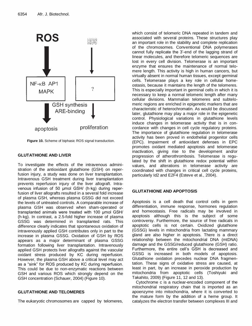
GLUTATHIONE AND LIVER To investigate the effects of the intravenous admini-stration of the antioxidant glutathione (GSH) on reper-fusion injury, a study was done on liver transplantation. Intravenous GSH treatment during liver transplantation prevents reperfusion injury of the liver allograft. Intra-venous infusion of 50 μmol GSH/ (h·kg) during reper-fusion of liver allografts resulted in a several fold increase of plasma GSH, whereas plasma GSSG did not exceed the levels of untreated controls. A comparable increase of plasma GSH was observed when sham-operated or transplanted animals were treated with 100 μmol GSH/ (h·kg). In contrast, a 2.5-fold higher increase of plasma GSSG was determined in transplanted rats. This difference clearly indicates that spontaneous oxidation of intravenously applied GSH contributes only in part to the increase in plasma GSSG. Oxidation of GSH by ROS appears as a major determinant of plasma GSSG formation following liver transplantation. Intravenously applied GSH protects liver allografts against the vascular oxidant stress produced by KC during reperfusion. However, the plasma GSH above a critical level may act as a “sink” for ROS produced by KC during reperfusion. This could be due to non-enzymatic reactions between GSH and various ROS which strongly depend on the GSH concentration (Schauer, 2004) (Figure 10).
GLUTATHIONE AND TELOMERES
The eukaryotic chromosomes are capped by telomeres,
which consist of telomeric DNA repeated in tandem and associated with several proteins. These structures play an important role in the stability and complete replication of the chromosomes. Conventional DNA polymerases cannot fully replicate the 3’-end of the lagging strand of linear molecules, and therefore telomeric sequences are lost in every cell division. Telomerase is an important enzyme that ensures the maintenance of normal telo-mere length. This activity is high in human cancers, but virtually absent in normal human tissues, except germinal cells. Telomerase plays a key role in cellular home-ostasis, because it maintains the length of the telomeres. This is especially important in germinal cells in which it is necessary to keep a normal telomeric length after many cellular divisions. Mammalian telomeres and subtelo-meric regions are enriched in epigenetic markers that are characteristic of heterochromatin. As would be discussed later, glutathione may play a major role in the epigenetic control. Physiological variations in glutathione levels induce changes in telomerase activity that is in con-cordance with changes in cell cycle regulatory proteins. The importance of glutathione regulation in telomerase activity has been proved in endothelial progenitor cells (EPC). Impairment of antioxidant defenses in EPC promotes oxidant mediated apoptosis and telomerase inactivation, giving rise to the development and/or progression of atherothrombosis. Telomerase is regu-lated by the shift in glutathione redox potential within values, and alterations in telomerase activity are coordinated with changes in critical cell cycle proteins, particularly Id2 and E2F4 (Esteve et al., 2004).
GLUTATHIONE AND APOPTOSIS
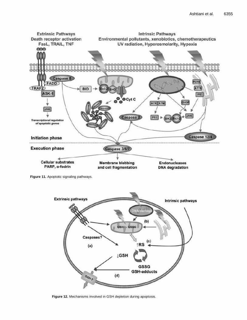
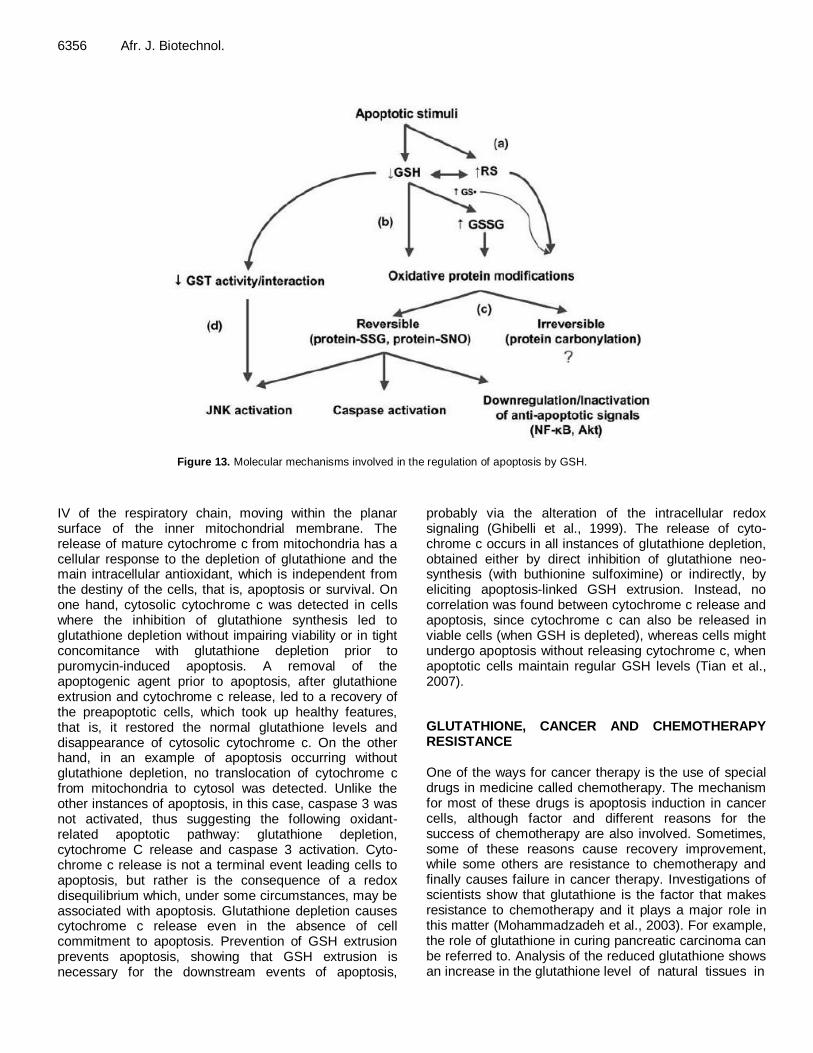
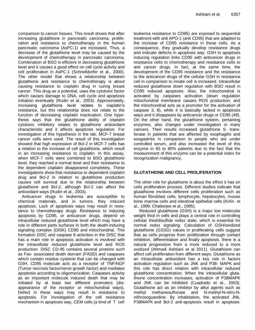
Apoptosis is a cell death that control cells in germ differentiation, immune response, hormones regulation and homeostasis. Free radicals may be involved in apoptosis although this is the subject of some controversy. Furthermore, the source of free radicals in apoptotic cells is not certain. Oxidized glutathione (GSSG) levels in mitochondria from lactating mammary gland are also higher in apoptosis. There is a direct relationship between the mitochondrial DNA (mtDNA) damage and the GSSG/reduced glutathione (GSH) ratio. Furthermore, the entire cell’s GSH is decreased and GSSG is increased in both models of apoptosis. Glutathione oxidation precedes nuclear DNA fragmen-tation. These signs of oxidative stress are caused, at least in part, by an increase in peroxide production by mitochondria from apoptotic cells (Toshiyuki and Takahito, 2009) (Figure 11, 12 and 13).
Cytochrome c is a nuclear-encoded component of the mitochondrial respiratory chain that is imported as an apoenzyme into mitochondria, where it is converted to the mature form by the addition of a heme group. It catalyzes the electron transfer between complexes III and
Ashtiani et al. 6355
Figure 11. Apoptotic signaling pathways.
Figure 12. Mechanisms involved in GSH depletion during apoptosis.
6356 Afr. J. Biotechnol.
Figure 13. Molecular mechanisms involved in the regulation of apoptosis by GSH.
IV of the respiratory chain, moving within the planar surface of the inner mitochondrial membrane. The release of mature cytochrome c from mitochondria has a cellular response to the depletion of glutathione and the main intracellular antioxidant, which is independent from the destiny of the cells, that is, apoptosis or survival. On one hand, cytosolic cytochrome c was detected in cells where the inhibition of glutathione synthesis led to glutathione depletion without impairing viability or in tight concomitance with glutathione depletion prior to puromycin-induced apoptosis. A removal of the apoptogenic agent prior to apoptosis, after glutathione extrusion and cytochrome c release, led to a recovery of the preapoptotic cells, which took up healthy features, that is, it restored the normal glutathione levels and disappearance of cytosolic cytochrome c. On the other hand, in an example of apoptosis occurring without glutathione depletion, no translocation of cytochrome c from mitochondria to cytosol was detected. Unlike the other instances of apoptosis, in this case, caspase 3 was not activated, thus suggesting the following oxidant-related apoptotic pathway: glutathione depletion, cytochrome C release and caspase 3 activation. Cyto-chrome c release is not a terminal event leading cells to apoptosis, but rather is the consequence of a redox disequilibrium which, under some circumstances, may be associated with apoptosis. Glutathione depletion causes cytochrome c release even in the absence of cell commitment to apoptosis. Prevention of GSH extrusion prevents apoptosis, showing that GSH extrusion is necessary for the downstream events of apoptosis,
probably via the alteration of the intracellular redox signaling (Ghibelli et al., 1999). The release of cyto-chrome c occurs in all instances of glutathione depletion, obtained either by direct inhibition of glutathione neo-synthesis (with buthionine sulfoximine) or indirectly, by eliciting apoptosis-linked GSH extrusion. Instead, no correlation was found between cytochrome c release and apoptosis, since cytochrome c can also be released in viable cells (when GSH is depleted), whereas cells might undergo apoptosis without releasing cytochrome c, when apoptotic cells maintain regular GSH levels (Tian et al., 2007). GLUTATHIONE, CANCER AND CHEMOTHERAPY RESISTANCE One of the ways for cancer therapy is the use of special drugs in medicine called chemotherapy. The mechanism for most of these drugs is apoptosis induction in cancer cells, although factor and different reasons for the success of chemotherapy are also involved. Sometimes, some of these reasons cause recovery improvement, while some others are resistance to chemotherapy and finally causes failure in cancer therapy. Investigations of scientists show that glutathione is the factor that makes resistance to chemotherapy and it plays a major role in this matter (Mohammadzadeh et al., 2003). For example, the role of glutathione in curing pancreatic carcinoma can be referred to. Analysis of the reduced glutathione shows an increase in the glutathione level of natural tissues in
comparison to cancer tissues. This result shows that after increasing glutathione in pancreatic carcinoma, prolife-ration and resistance to chemotherapy in the human pancreatic carcinoma (AsPC-1) are increased. Thus, a decrease of the glutathione level may be caused by the development of chemotherapy in pancreatic carcinoma. Combination of BSO is efficient in decreasing glutathione level and it causes a good effect on cell cycle activity and cell proliferation in AsPC-1 (Schnelldorfer et al., 2000). The other model that shows a relationship between glutathione and resistance to chemotherapy is about causing resistance to cisplatin drug in curing breast cancer. This drug as a potential, uses the cytotoxic factor which causes damage to DNA, cell cycle and apoptosis initiation eventually (Rudin et al., 2003). Approximately, increasing glutathione level relates to cisplatin’s resistance, but this relationship does not relate to the function of decreasing cisplatin inactivation. One hypo-thesis says that the glutathione ability of cisplatin cytotoxic inhibitory relates to glutathione antioxidant characteristic and it affects apoptosis regulation. For investigation of this hypothesis in the lab, MCF-7 breast cancer cells were used. The result of this investigation showed that high expression of Bcl-2 in MCF-7 cells has a relation to the increase of cell glutathione, which result in an increasing resistance to cisplatin. In this assay, when MCF-7 cells were combined to BSO glutathione level, they reached a normal level and their resistance to the dependent cisplatin disappeared completely. These investigations show that resistance to dependent cisplatin drug and Bcl-2 in relation to glutathione production causes cell survival due to the relationship between glutathione and Bcl-2, although Bcl-2 can affect the antioxidant ways (Rudin et al., 2003).
Anticancer drugs in leukemia are susceptible to chemical materials, and in tumors, they induced apoptosis. Lack of apoptosis ways may result in resis-tance to chemotherapy drugs. Resistance to inducing apoptosis by CD95, or anticancer drugs, depend on intracellular reduced glutathione level which may have a role in different parts surfaces in both the death-inducing signaling complex (DISK) CD95 and mitochondrial. This formation DISC and caspase 8 activities in the DISC that has a main role in apoptosis activation is involved with the intracellular reduced glutathione level and ROS production. DISC CD-95 contains several proteins such as Fas- associated death domain (FADD) and caspases which contain residue cysteine that can be changed with GSH. CD95 molecule acts as a receptor of TNF/NGF (Tumor necrosis factor/nerve growth factor) and mediates apoptosis according to oligomerization. Caspases activity as an important molecule induced death that may be initiated by at least two different promoters (dis-appearance of the receptor or mitochondrial ways). Defect in these ways may result in resistance to apoptosis. For investigation of the cell resistance mechanism in apoptosis way, CEM cells (a kind of T cell
Ashtiani et al. 6357 leukemia resistance to CD95) are exposed to sequential treatment with anti APO-1 (anti CD95) that are adapted to the increase of CD95 resistance in these cells. As a consequence, they gradually develop resistance drugs and indicate defects in apoptosis way. GSH in apoptosis inducing regulation links CD95 with anticancer drugs in resistance cells to chemotherapy and resistance cells to anti cancer drugs. In fact, at the same time, the development of the CD95 resistance and the resistance to the anticancer drugs of the cellular GSH in resistance cell in comparison to innate cell is increased. Intracellular reduced glutathione down regulation with BSO result in CD95 induced apoptosis. Also, the mitochondrial is activated by caspases activation (down regulation mitochondrial membrane causes ROS production, and the mitochondrial acts as a promoter for the activation of caspase 3, 8), while it is basically lacked in apoptosis ways and it disappears by anticancer drugs or CD95 (48). On the other hand, the glutathione system, pertaining enzymes, also changes under investigation in many cancers. Their results increased glutathione S- trans-ferase in patients that are affected by esophagitis and esogastritis in comparison to people who are under controlled serum, and also increased the level of this enzyme in 60 to 80% patients due to the fact that the measurement of this enzyme can be a potential index for recognization malignancy. GLUTATHIONE AND CELL PROLIFERATION The other role for glutathione is about the effect it has on cells proliferation process. Different studies indicate that glutathione involves different cells proliferation such as human fibroblast cells, lymphocyte, hepatocytes, mouse bone marrow cells and intestinal epithelial cells (Kirlin et al., 1999; Chatterjee et al., 1995).
Reduced glutathione (GSH) is a major low molecular weight thiol in cells and plays a central role in controlling cellular thiol/disulfide redox state, which is essential for normal redox signaling. Calculation of GSH/oxidized glutathione (GSSG) values in proliferating cells suggest that as cells progress from proliferation through contact inhibition, differentiation and finally apoptosis, there is a natural progression from a more reduced to a more oxidized (Ahmadi Ashtiani et al 2011). Glutathione can affect cell proliferation from different ways. Glutathione as an intracellular antioxidant has a key role in factors activation regulation such as JNK and P38- MAPK
and
this role has direct relation with intracellular reduced glutathione concentration. When the intracellular gluta-thione concentration increases, activation of P38MAPK and JNK can be inhibited (Cuadrado et al., 2003). Glutathione act as an inhibitor by alkyl agents such as methyl methanesulfonate and N-methyl-N-nitro-N-nithrosoguandine. By inhabitation, the activated JNk, P38MAPK and Bcl-2 anti-apoptosis result in apoptosis
6358 Afr. J. Biotechnol. inhabitation and increased proliferation as well (Cuadrado et al., 2003).
The relationship between the reduced glutathione and ROS is one of the other effective agents on cell prolife-ration. ROS, especially hydrogen peroxide and super-oxide, plays a key role in cellular death and proliferation. This compound usually stimulate cellular growth in submicromolar concentration, while in high concentration (more than 10 to 30 micromolar), it can result in necrosis cells and apoptosis (Day and Suzuki, 2005). In fact, the high concentration of this compound causes the activation of the transcription factors such as MAPK and NF-кB, following the cellular death that occurs, whereas in low concentration, it causes an activation of other transcription factors such as ARE (for example Nrf2). ARE transcription factor has a role in enzyme γ-GCS gene expression regulation. This enzyme is a key enzyme for the control of glutathione synthesis and is produced in response to xenobiotic. The result of this activation occurs after cellular proliferation, due to the increase of the reduced glutathione concentration. The following figure indicates that as long as ROS is produced in high concentration, cellular death occurs and when ROS is produced in low concentration, glutathione is synthesized and the cellular proliferation occurs (Day and Suzuki, 2005).
Glutathione can also affect tumor necrosis factor-α (TNF-α). TNF-α is a cytokine that is derived from micro-phage – monocyte cells that have a key role in infection and immunology. Cytokine TNF-α plays a lot of roles in biology such as antitumor and cellular proliferation inhibitor. The reactive oxygen intermediate (ROI) molecules stimulate the ability of cell death TNF-α, while TNF-α by intermediation in the normal process of electron transfer in mitochondrial membrane produces ROI which involves dead cells and damage to DNA (Schnelldorfer et al., 2000). Resistance to TNF-α refers to antioxidant cells’ ability in tumor cells and also the high concentration of the reduced glutathione intracellular which is a resistance agent to TNF-α (Obrador et al., 1997). Also, resistance to TNF-α is influenced by the high glutathione level which prevents the death of cells and thus, maintains gluta-thione (Obrador et al., 1997). In addition, the high concentration of the reduced glutathione intracellular, by other mechanisms, affects cell proliferation and stimu-lates mitogenic ability in cells. Reduced glutathione has a role in DNA synthesis regulation. Also, glutathione with activity regulation of protein kinase C (PKC) and intracellular pH controls tumor proliferation (Obrador et al., 1997). Evacuation reduced glutathione in cancer cells with compounds such as BSO, which prevented them from activating the protein kinase C (PKC) and which decreased the intracellular pH (Terradez et al., 1993). Glutathione plays a key role as a regulator in T lympho-cytes proliferation. In the study where T lymphocytes were performed after using BSO inhibitor, glutathione decreased after the T lymphocytes proliferation was
controlled (Friesen et al., 2004). The investigation indicated that a decrease of the reduced glutathione concentration prevented the lymphocytes development from phase G1 to phase S cell cycle. However, the agents that increased the cellular reduced glutathione, increased the proliferation of lymphocytes in response to interleukin-2, while they prevented the reduced gluta-thione synthesis interleukin-2 level. In another study, the reduced glutathione impression on HT-29 cells derived a human colon carcinoma cell that has been investigated and which affected the sodium butyrate of this cell. This material caused a decrease in the reduced glutathione and finally in the cellular proliferation (Kirlin et al., 1999). In other study, it was indicated that when the treatment of CaCO2 cells with BSO cellular glutathione level decreased alone, there was no change in GSH (GSSG). However, in this case, there was a little effect on the cellular proliferation, but the treatment of cell with diamide and 1,3-bis (2 chloroethyl)-1 nitrosourea (BCNU) (inhibitor reductase glutathione enzyme activity) decree-sed the GSH level and increased the GSSG level. Besides, the ratio of GSH: GSSG decreased. The decrease of the inhibitory effects, by N-acetylcysteine (NAC), on these changes is related to the decrease of the cellular proliferation. On the other hand, compounds such as BSO decreased the reduced glutathione level, but there was no change in GSH (GSSG ratio). In this survey, they concluded that although BSO has no effect on cellular proliferation, its presence can improve BCNU and diamide the inhibitory effect (Taahiro and Ryuich, 2001). In other survey about glutathione effect on cellular proliferation, its role improves the intestinal cells proliferation. In this study, the role of glutathione on apoptosis or proliferation is investigated by NAC and BSO. Documents show that glutathione concentration balance is a key mechanism in intestinal cells prolife-ration control (Buccigrossi et al., 2009). DIFFERENTIATION AND GLUTATHIONE Glutathione plays a major role in differentiation process, although studies indicate that glutathione plays a role in the cellular proliferation process. The finding of the study that was performed on the skeletal muscle cells of C2C12 suggests that GSH level 24 h after cells stimulation to differentiation increased and later decreased. Sub-sequently, after 5 days, it reached the first level which indicated that GSH has a stimulatory effect on cellular proliferation. Also, it was observed that by adding BSO and decreasing GSH during the first day, cells differentiation rate decreased in the third day and this decrease continued until the seventh day. In addition, with the increasing of GSH, the skeletal muscle differentiation cells increased as well. Skeletal muscle differentiation is a complex process whose regulation includes several processes. Cellular glutathione, during

24 h after the myoblast stimulation, increased because of stimulation of L-cysteine ligase glutamate activity. Glutathione involves the formation of myotube from satellite myoblast by activation of nuclear factor kappa B (NF-KB). However, a recovery of the optimal glutathione level in muscle mass differentiation in chronic inflame-mation has been useful (Ardite et al., 2004). In other study that was performed on the mouse fibroblast cells, the researcher concluded that while cells division (6 to 24 h after culture) percent of Bcl-2 was very high, this amount was decreased after this time (48 h to 5 days after culture) and at the same time that a decrease in the cellular growth occurred. Nevertheless, glutathione in fibroblast 3T3 occurred before cellular growth increased. High level of telomerase activity was followed 24 h after the culture occurred and at the same time that the maximum level of glutathione in cells happened. Also, telomerase activity was maximally seen than the reduction condition. After 24 h culture, 3T3 fibroblast initiated growth, while for 24 to 48 h, it increased incredibly after culture cells percents in S+G2/M. After 72 h culture, cell growth decreased and after 6 days, most of the cells are in G0/G1. Glutathione concentration is found to be at its maximum in all cells after 24 h culture, when high percent cells are coming or preparing for division. While cell populations are in G0/G1 phase, glutathione is very low (Markovic et al., 2007). Thus, glutathione nuclear distribution has a main role in cell cycle deve-lopment during the first phase of the cellular culture, and when cells initiated growth, glutathione can be replaced in the nuclear cell (Markovic et al., 2007; Mihm et al., 1995).
Studies indicate that the mouse bone marrow stromal cells (BMSCs) can be differentiated from the neuron cells by adding 2- mercaptoethanol. When the bone marrow stromal cells are found with a little glutathione passage differentiation, the neuron cells occur, but using glutathione monoethyl ester (GEE) to increase the intracellular reduced glutathione concentration, there has been no effect on the neuron marker expression, so it can be said that the extracellular reduced glutathione is very effective on the stromal stem cell differentiation to neuron cells, but the intracellular reduced glutathione has no any effect on this trend (Sagara and Makino, 2008). In other study that was performed on the mouse preadipocyt cells differentiation to adipocytes, it was concluded that the presence of ROS can accelerate adipocyte differentiation, while when the reduced glutathione presents its ROS level; it also decreased following the decrease of adipocyte differentiation. Thus, it is concluded that GSH has an effective compound in decreasing adipocyte number (Toshiyuki and Takahito, 2009). In another study, Kim et al. looked for the relationship between GSH and macrophage differen-tiation derived from the bone marrow monocyte and their phagocyte activation. They indicated that: (1) GSH/GSSG ratio during bone marrow monocyte (BMMs) differen-
Ashtiani et al. 6359 tiation to macrophage cells decreased. BMMs were taken from Tibia mouse and were exposed with M-CSF diffe-rentiation to macrophage. (2) A decrease of the GSH level is prevented from the macrophage differentiation. (3) Inhibitory effects of GSH depletion on differentiation are performed by C-fms gene down expression regulation. (4) GSH can regulate phagocyte activity in relation to macrophage (Jin-Man and Hyunsoo, 2004). To evaluate this, a study about the presence of glutathione system evacuation (BSO) and glutathione system supporter (NAC) was done. Moreover, the investigation of the cells position by these compounds suggests a conclusion of the level of GSH. With the presence of BSO, preventing the composition of the macrophage occurs with a down regulation of C-fms gene, but it does not have any effect on the phagocytic activity since the macrophage phagocytic activity relates to the increase of the ROS level, and when the cellular GSH level is low, more levels of ROS in the cells occur. While saturation of cells by N-acetyl cysteine does not have any effect on macrophage differentiation, the phagocytic activity decreased because the saturation of cells with GSH position of reduced redox in cell increases, thereby decreasing the phagocytic activation of macrophage. M-CFS as one of the growth factors is necessary for BMMs development, while contamination between M-CFS and the acceptor (which stimulates the macrophage differen-tiation), as a set of development reactions, mediate macrophage. M-CSF linkage with its receptor causes spontaneous phosphorylation tyrosine kinase following the fact that cellular signals are increased for proliferation, differentiation and survival. M-CSF stimulate C-fms gene expression by linkage of several transcription factors to C-fms gene promoter. However, in a perfor-mance of the down regulation from C-fms gene, the GSH level decreased and resulted in a dependent differen-tiation decrease of M-CSF (Jin-Man and Hyunsoo, 2004). Allameh et al. (2009) investigated the hepatocytes differentiation trend from mesenchymal cells. It was concluded that the derived bone marrow of the cellular GSH level decreased (20%≈) in the differentiated hepatocytes from mesenchymal cells in comparison to the control group, which implied that the differentiation hepatogenic of the MSC was derived from the human bone marrow with involved factors regulation in GSH conjugation ways (Allameh et al., 2009).
FETUS AND GLUTATHIONE
Increasing the intracellular ROS causes thermal shock delays in the development of the fetus or fetus death. Reports indicate that the thermal shock defects the fetus mitochondrial function and morphology. Although the BSO decreases fetus development, high level of glutathione is necessary for the cleavage of the ROS witch to be produced by thermal shock. In fact, mito-

6360 Afr. J. Biotechnol. chondrial glutathione maintains cell vitality by ROS elimination (Miki et al., 2008). β- Mercaptoethanol can decrease the produced ROS witch, which directly causes an increase in the glutathione level. The fetus witch that is exposed to thermal shock, without mercaptoethanol, has high level of ROS and it decreased fetuses’ development in comparison to the fetus witch that is not exposed to thermal shock. β- Mercaptoethanol is a reduced agent and acts as an antioxidant which disappear from hydroxyl radical with (sulfhydryl) the SH group. Indication of this compound stimulates glutathione synthesis and also induces thiol that stimulates cystine reduced reaction to cysteine, and finally results to the synthesis of glutathione (Miki et al., 2008). GSH has a main role in growth, development and fetus organization. In fact, this compound decreases the intracellular ROS level that has the ability to destroy the effect of the fetus formation and helps in fetus formation. In the study that was done on the regulation of diabetic and normal mice, it was concluded that hyperglycemia resulted in a dis-ruption of the organization by causing free radicals and intracellular GSH depletion in fetus tissues. Intracellular GSH in the fetus tissue of diabetic pregnant mice in comparison to normal mice is low and also the enzyme level witch synthesis GSH in diabetic mice fetus is low because of the decrease that relates to the mRNA expression of this enzyme in this mouse. BSO is a special inhibitor of GSH synthesis which decreases intracellular GSH level and causes nervous damages, while GSH esters in diabetic mice maintain GSH in fetus and hilling of damaging nervous (Sakamaki et al., 1999). REFERENCES
Abedelahi A, Salehnia M, Allameh A, Davoodi D (2010). sodium selenit
improves the in vitro follicular development by reducing the reative
oxygen species level and increasing the total antioxidant capacity and glutathione peroxide activity. Hum. Rep. 25: 977-985.
Ahmadi Ashtiani HR, Allameh A, Rastegar H, Soleimani M, Barkhordari
E (2011). Inhibition of cyclooxygenase-2 and inducible nitric oxide synthase by silymarin in proliferating mesenchymal stem cell: comparing with glutathione modifiers. J. Natural Med. (In press).
Aitken RJ (1995). Free radicals, Lipid peroxidation and sperm function. Reprod. Fertil. Dev. 7: 65-70.
Allameh A, Alikhani N (2002). Acetaminophen- glutathione conjugate
formation in a coupled cytochrome P-450 glutathione S-transferase assaye system mediated by subcellular preparation from adult and weanling rat tissues. Toxico. Vit. 16: 637-41.
Allameh A, Esmaeli Sh, Kazemnejad S, Soleimani M (2009). differenitial expression of glutathione S-transferases P1-1 and A1-1 at protein and mRNA levels in hepatocytes derived from human bone marrow
mesenchymal stem cells. Toxico. Vit. 23: 674–679. Allameh A, Maleklou N, Zargari M, Sanaati MH (2008). The influence of
uric acid treatments on liver glutathione system prevent oxidative
damages in experimental autoimmune encephalomyelitis mice. Neuro. Sci. Let. 439: 111-115.
Ardite E, Barbera JA, Roca J, Jose C, Checa F (2004). Glutathione
depletion impairs myogenic differentiation of murine skeletal muscle C2C12 cells through sustained NF-KB activation. Am. J. Pathol. 165(3): 719-728.
Behroozikhah M, Saidee M, Allameh A (1992). Comparison of aflatoxin B1-DNA binding and glutathione conjugate formation by liver preparations from rats at different ages. Cancer Let. 66: 69-76.
Behrozikhah M, Allameh A (1995). The role of glutathione in aflatoxin
B1 metabolism in different ages in rat. Sabzevar Univ. Med. J., pp. 35-21.
Buccigrossi V, Mari C, Giannattasio A, Oliva M, Ruberto E, Caiazzo M (2009). Role of glutathione in the control of intestinal cell proliferation. Abstract of XVI Nat. Cong. SIGENP/ Diges. Liver Dis. 41: 199-239.
Chatterjee A, Chattopadhyay A, Lawlor CJ (1995). Effect of glutathione on sister-chromatid exchanges in normal and buthionine sulfoximine-treated mice. Mutat. Res. 327(1-2): 171-177.
Chevion M, Navok T, Glaser G, Mager J (1982).The chemistry of favism-inducing compounds, the properties of isouramil and divicine and their reaction with glutathione. Eur. J. Biochem. 127: 405-409.
Cotgreave IA, Gerdes RG (1998). Recent trends in glutathione biochemistry. Glutathione-protein interactions: a molecular link between oxidative stress and cell proliferation. Biochem. Biophys.
Res. Commun. 242: 1-9. Cuadrado A, Fern لndez LG, Gonz لlez L, Su لrez Y, Losada A, Alcaide
V (2003). AplidinTM induces apoptosis in human cancer cells via
glutathione depletion and sustained activation of the epidermal growth factor receptor, Src, JNK, and p38 MAPK. J. Biol. Chem. 278: 241-250.
Day R, Suzuki Y (2005). Cell proliferation, reactive oxygen and cellular glutathione. Dose-Resp. 3: 425-442.
Droge W, Pottmeyer-Gerber C, Schmidt H, Nick S (1986). Glutathione
augments the activation of cytotoxic T lymphocytes in vivo. Immunobiology, 172: 151-56.
Esteve CBS, JM, JRV A, Sastre J, Jose´ Vin A, Pallardo FV (2004).
Glutathione Regulates Telomerase Activity in 3T3 Fibroblasts. Am. Soc. Biochem Mol. Biol. 242-247.
Friesen C, Kiess Y, Debatin KM (2004). A critical role o glutathione in
determining apoptosis sensitivity and resistance in leukemia cells. Cell Death Different, 11: 73-85.
Ghibelli L, Copppla S, Fanelli C, Rotilio G, Civitareal P, Scovasii AI
(1999). Glutathione depletion causes cytochrome c release even in the absence of cell commitment to apoptosis. FASEB J. 13(14): 2031-2036.
Gmunder H, Droge W (1991). Differential effects of glutathione depletion on T cell subsets. Cell Immunol. 138: 229-37.
Griffith OW, Mulcahy RT (1999).The enzymes of glutathione
biosynthesis: glutamylcysteine synthetase. Adv. Enzymol. Relat. Areas Mol. Biol. 73: 209-267.
Gutterer DR, Jan M (2000). Glutathione metabolism in brain. J.
Biochem. FEBS. 267: 4912-4916. Hayes J, McLellan L (1999). Glutathione and glutathione-dependent
enzymes represent a co- ordinately regulated defence against
oxidative stress. Free Radic. Res. 31(4): 273-300. Hopkins FG (2002). The Discovery of Glutathione. J Biol. Chem.
277(24): 13. Jin-Man K, Hyunsoo K (2004). Intracellular glutathione status regulates
mouse bone marrow monocyte derived macrophage differentiation and phagocytic activity. Biochem. Biophys. Res. Com. 325: 101–108.
Jones D (2002). Redox potential of GSH/GSSG couple: assay and
biological significance. Methods Enzymol. 348: 93-112. Jones DP (2002). Redox potential of GSH/GSSG couple: assay and
biological significance. Methods Enzymol. 348: 93-112.
Kirlin WG, Cai J, Thompson SA, Diaz D, Kavanagh TJ, Jones DP (1999). Glutathione redox potential in response to differentiation and enzyme inducers. Free Radic. Biol. Med. 27: 1208-1218.
Klatt P, Lamas S (2000). Regulation of protein functions by S-glutathionylation in response to oxidative and nitrosative stress. Eur. J. Biochem. 267: 4928-44.
Lu SC (2000). Regulation of glutathione synthesis. Curr. Top Cell Regul. 36: 95-116.
Mabrouk GM, Jois M, Brosnan JT (1998).Cell signaling and the
hormonal stimulation of the hepatic glycine cleavage enzyme system by glucagons. Biochem, J. 330: 759-763.
Markovic J, Borras C, Ortega A, Sastre J, Vina J, Pallardo FV (2007).
Glutathione is recruited into the nucleus in early phases of cell proliferation. J. Biolog Chem; 282(28): 20416–20424.
Meister A (1973).On the enzymology of amino acid transport. Science,
180: 33-39. Mihm S, Galter D, Faseb J, Droge W (1995). Modulation of transcription

factor NF-kB activity by intracellular glutathione levels and by variations of the extracellular cysteine supply, 9: 246-252.
Miki S, Kenichi Y, Shuji K (2008). Heat shock derived reactive oxygen
species induce embryonic mortality in vitro early stage bovine embryos. J. Reprod. Dev. 54(6): 496-501.
Miranada-Vizuette A, Toribio F, Holmgren A, Lopez J, Pueyo C (1996).
The Level Of Ribonucleotide Reductase, Thioredoxin, Glutaredoxin 1 And GSH Are Balanced in Escherichia coli K12. J. Biol. Chem. 271: 19099-19103.
Mohammadzadeh GS, NasseriMoghadam S, Rasaee MJ, Zaree AB, Mahmoodzadeh H, Allameh A (2003). Measurement of glutathione S-transferase and its classπ in plasma and tissue biopsies obtained
after laparoscopy and endoscopy from subjects with esophagus and gastric cancer. Clin. Biochem. 36: 283-288
Mullick J, Allameh A, Raj H (1986). Metabolism of glutathione in
microorganisms. In: KG M, VP S, KL G, editors. frontiers in applied microbiology: Rastogy and Company, pp. 118-32.
Noda T, Iwakiri R (2002) .Exogenous cysteine and cystine promote cell
proliferation in CaCo-2 cells. Cell Prolif. 35: 117-129. Obrador E, Navarro J, Mompo J, Asensi M, Pellicer JA, Estrela JM
(1997). Glutathione and the rate of cellular proliferation determine
tumour cell sensitivity to tumour necrosis factor in vivo. Biochem. J. 325: 183-189.
Pallardo FV, Markovic J, Garcيa JL , Viٌa J (2009). Role of nuclear
glutathione as a key regulator of cell proliferation. Mol. Aspects Med. 30: 77-85.
Pastore A, Federici G, Bertini E, Piemonte F (2003). Analysis of glutathione: implication in redox and detoxification. Clin. Chim. Acta. 333: 19-39.
Pompella A, Visvikis A, Paolicchi A, Tata VD, Casini AF (2003). The changing faces of glutathione, a cellular protagonist. Biochem. Pharmacol. 66: 1499-503.
Rudin CM, Yang Z, Schumaker LM, David J, Weele V, Newkirk K (2003). Inhibition of glutathione synthesis reverses Bcl-2-mediated Cisplatin resistance. Cancer Res. 63: 312-318.
Sagara J, Makino N (2008).Glutathione induces neuronal differentiation in rat bone marrow stromal cells. Neurochem. Res. 33: 16-21.
Sakamaki H, Akazawa S, Ishibashi M, Izumino K, Takino H, YamasakI
H (1999). Significance of glutathione dependent antioxidant system in diabetes induced embryonic malformation. Diabetes, 48: 1138-1144.
Ashtiani et al. 6361 Schauer RJ (2004). Intravenous administration of glutathione protects
parenchymal and non-parenchymal liver cells against reperfusion injury following rat liver transplantation. World J. Gastroenterol. 10(6):
864-870. Schnelldorfer T, Gansauge S, Gansauge F, Schlosser S, Beger HG,
Nussler AK (2000). Glutathione depletion causes cell growth
inhibition and enhanced apoptosis in pancreatic cancer cells. Cancer. 89(7): 1440-1447.
Sharma RK, Agarwal A (1996). Role of reactive oxygen species in male
infertility. Urol, 48(6): 835- 850. Shi ZZ, Osei-Frimpong J, Kala G, Kala SV, Barrios RJ, Habib GM
(2000). Glutathione synthesis is essential for mouse development but
not for cell growth in culture. PANS, 97: 5101-5106. Suzuki Masatoshi, Nelson, Aaron D (2006). Glutamate enhances
proliferation and neurogenesis in human neural progenitor cell
cultures derived from the fetalcortex. Eur. J. Neurosci. 24: 645–653. Taahiro N, Ryuich J (2001). Induction of mild intracellular redox
imbalance inhibits proliferation of CaCo-2 cells. FASEB J. 15: 2131-
2139. Terradez P, Asensi M, Lasso De La Vega MC , Puertes IR , Viٌa J, Estrela
JM (1993). Depletion of tumour glutathione in vivo by buthionine
sulphoximine: modulation by the rate of cellular proliferation and inhibition of cancer growth. Biochem. J. 292: 477-483.
Tian J, Washizawa N, Gu LH, Levin MS, Wang L, Rubin DC (2007).
Local Glutathione Redox Status Does Not Regulate Ileal Mucosal Growth after Massive Small Bowel Resection in Rats. J. Nutr. 137: 320-325.
Toshiyuki T, Takahito T (2009). Carnosic acid and carnosol inhibit adipocyte differentiation in mouse 3T3-L1 cells through induction of phase2 enzymes and activation of glutathione metabolism. Biochem.
Biophys. Res. Commun. 382(3): 549-554. Zargari M, Allameh A, Sanaati MH, Tiraihi T, Lavasani Sh, Emadiyan O
(2007). Relationship between the clinical scoring and demyelination
in central nerves system with total antioxidant capacity of plasma during experimental autoimmune encephalomyelitis development in mice. Neuro. Sci. Let. 412: 24-28.
Copyright © 2022 FDOKUMEN