Forced Diuresis with Matched Isotonic Intravenous Hydration ...
Upload
khangminh22Category
view
0download
0
Running head: INTRAVENOUS GLUTATHIONE AND DISEASE STATES 1
Skin Lightening Treatments: A Review on the Effect of Intravenous Glutathione
in the Disease States of Women
Buhle Mthombeni
A Senior Thesis submitted in partial fulfillment
of the requirements for graduation
in the Honors Program
Liberty University
Spring 2020

INTRAVENOUS GLUTATHIONE AND DISEASE STATES 2
Acceptance of Senior Honors Thesis
This Senior Honors Thesis is accepted in partial
fulfillment of the requirements for graduation from the
Honors Program of Liberty University.
_____________________________
Abigail Solitro, Ph.D.
Thesis Chair
______________________________
Matthew Brynteson, Ph.D.
Committee Member
______________________________
James H. Nutter, D.A.
Honors Director
______________________________
Date

INTRAVENOUS GLUTATHIONE AND DISEASE STATES 3
Abstract
The skin bleaching industry is a global business with a vast array of anti-melanogenic choices
including glutathione. Glutathione is synthesized in vivo but has been used as a bodily
supplement by medical personnel to aid in preventative medicine. Known for its antioxidant
properties, glutathione has been used for its anti-melanogenic effects. Intravenous glutathione
requires more investigation to determine its safety for usage. It continues to be distributed to the
cosmetic industry despite antagonism from the Philippine FDA. This study will research the
potential effects of intravenous glutathione on women and it will propose the biochemical
mechanisms of glutathione in induced disease states in women. The aim is to educate people
about safer methods for skin lightening.
Keywords: skin lightening, intravenous glutathione, pheomelanin, Fitzpatrick skin types,
Stevens-Johnson syndrome.

INTRAVENOUS GLUTATHIONE AND DISEASE STATES 4
Skin Lightening Treatments: A Review on the Effect of Intravenous Glutathione
in the Disease States of Women
Skin lightening, more commonly known as skin bleaching, is globally practiced by
women who are of darker shades of skin. It is a form of body modification performed to alter
appearance and temporarily alleviate psychological issues that stem from self-hatred and low
self-esteem.1 Racism and colonialism have attributed greatly to the psychological pressures of
having lighter skin. Fairer skin is viewed as more attractive, tasteful and healthy.2 Statistically
speaking, women of African descent participate the most in skin lightening, with 70% of women
in Nigeria, 30-40% of women in Pretoria, South Africa, 50% of women in Mali, and eight out of
every ten women in the Ivory coast participating in this practice.3,4 In a study done by Schroff et
al., it was suggested that Indian women were generally two times more likely to use lightening
products than Indian men. The statistics in Europe and Asia suggest 27-77% usage in different
communities. The toxicity of the ingredients, such as mercury and hydroquinone, has been
sufficient to ban the products from circulation in an open market in countries like Ghana,
Zimbabwe, South Africa and Nigeria.5 The constitutive skin types that lighten their complexion
tend to fall under the Fitzpatrick skin types IV-VII; individuals with skin types that are I-III
would normally lighten their skin in cases of hyperpigmentation, melasma, or acne scars that
may alter the color of the areas affected.6 The percentage of women who bleach their skin in the
world correlates with the skin type distribution as illustrated by Table 1, which is based on
modified figures from studies performed by Holcomb et al. and Bino et al.10, 11 Manufacturers
and skin lightening product distributors do not warn users of the dangers of lightening skin

INTRAVENOUS GLUTATHIONE AND DISEASE STATES 5
because of how lucrative the industry is.15 The product market is estimated to reach $8 billion by
2026 globally.7
Table 1
Fitzpatrick scale of skin types and their associated characteristics as they relate to the
distribution of women who would practice skin lightening.
Fitzpatrick Scale
Photo type Sunburn and tanning
criteria
Constitutive skin color
Heritage of photo type
prevalence (but not
limited to)
I Always burn, never tan Very light Northern Europe,
United Kingdom
II Always burn,
sometimes tan
Light Europe, Scandinavia
III Sometimes burn,
always tan
Intermediate Southern Europe,
Central Europe
IV Never burn, always tan Tan Mediterranean, Asia,
Latino
V Moderately pigmented Brown East India, Africa,
Native Americans
VI Heavily pigmented Dark brown to black Aboriginal, Africa

INTRAVENOUS GLUTATHIONE AND DISEASE STATES 6
The agents for skin lightening are classified into different categories based on their mode
of action on the skin. Hydroquinone and its derivatives arbutin and kojic acid, azelaic acid,
mercury, and botanicals are classified as chemical tyrosine kinase inhibitors; niacinamide is a
melanocyte transfer inhibitor; alpha and beta hydroxy acid are vitamin A derivatives and are
classified as accelerators of epidermal turnover; vitamin C, vitamin E, ubiquinone, glutathione
and coenzyme Q-10 are antioxidants; and topical corticosteroids are anti-inflammatory in
nature.8 These have been used as agents to halt the production of eumelanin, which causes the
brown pigment in skin. Many African countries have banned the distribution and sales of skin
bleaching creams containing these agents due to harmful side effects such as hypopigmentation,
skin sensitization and melanoma.9
The effects of oral and topical applications of glutathione have been studied in higher
proportions compared to intravenous glutathione. These studies have helped in establishing
safety data. The Food and Drug Administration (FDA) of the Philippines declared dietary oral
glutathione supplements as safe for use but have not approved the oral supplements in the case of
skin lightening. Although the oral glutathione has not been declared safe to use, it is still
manufactured and distributed. Due to the desire for an efficient treatment that is not localized to
one part of the body the demand for intravenous glutathione has increased. However, due to
limited information about the safety of intravenous applications of glutathione, organizations
such as the Philippine Dermatological Society have issued a warning that the use of intravenous
glutathione at high doses may be harmful to the recipient.12

INTRAVENOUS GLUTATHIONE AND DISEASE STATES 7
Glutathione
Glutathione (GSH) is a ubiquitous, low molecular weight thiol, and tripeptide consisting
of γ-L-glutamyl-L-cysteinyl-glycine. GSH has one of the greatest antioxidant properties and is
important to normal cell function because it maintains cellular redox balance. Glutathione is
abundant in cells but it is remarkably reduced in pathological conditions such as HIV/AIDS,
Parkinson’s disease, malnutrition, cancer, strokes, myocardial infarctions and others.49
Glutathione is found in two forms, reduced (GSH) and oxidized (GSSG), as illustrated by Figure
1.13 Glutathione is synthesized de novo from glutamate, cysteine and glycine through ATP
hydrolysis. One oxidized glutathione molecule (GSSG) is made from two reduced glutathione
molecules (GSH), which explains the higher ratio of GSH to GSSG intracellularly. Oxidative
stress is indicated by a reduction in the ratio, as GSH has been used up to convert reactive
oxygen species (ROS) like hydrogen peroxide (H2O2) to water, and by the decrease of NADPH,
which reduces GSSG.

INTRAVENOUS GLUTATHIONE AND DISEASE STATES 8
Figure 1. The glutathione redox cycle illustrating the function of the two glutathione
variants. Figure 1 is adapted from a study performed by Sonthalia et al., and Ilkhani et al.15, 16
The ratio of these two forms of glutathione can indicate oxidative health. A normal ratio
of GSH/GSSG is greater than 100, while oxidative stress is indicated by a ratio of 10 or less.13
De novo glutathione synthesis is catalyzed by glutamate cysteine ligase and glutathione
synthetase enzymes through a two-step process that requires ATP. GSH is found in large
concentrations of 5 mM intracellularly.13, 14 The most important functions of glutathione known
to date are i) detoxification of xenobiotics, ii) catalysis of exchange reactions, iii) scavenging of
free radicals and reactive oxygen species (ROS), iv) transport of amino acids across cell
2

INTRAVENOUS GLUTATHIONE AND DISEASE STATES 9
membranes, v) acting as a coenzyme in some processes of cellular metabolism and vi)
maintenance of thiol groups of proteins and other molecules.15, 17
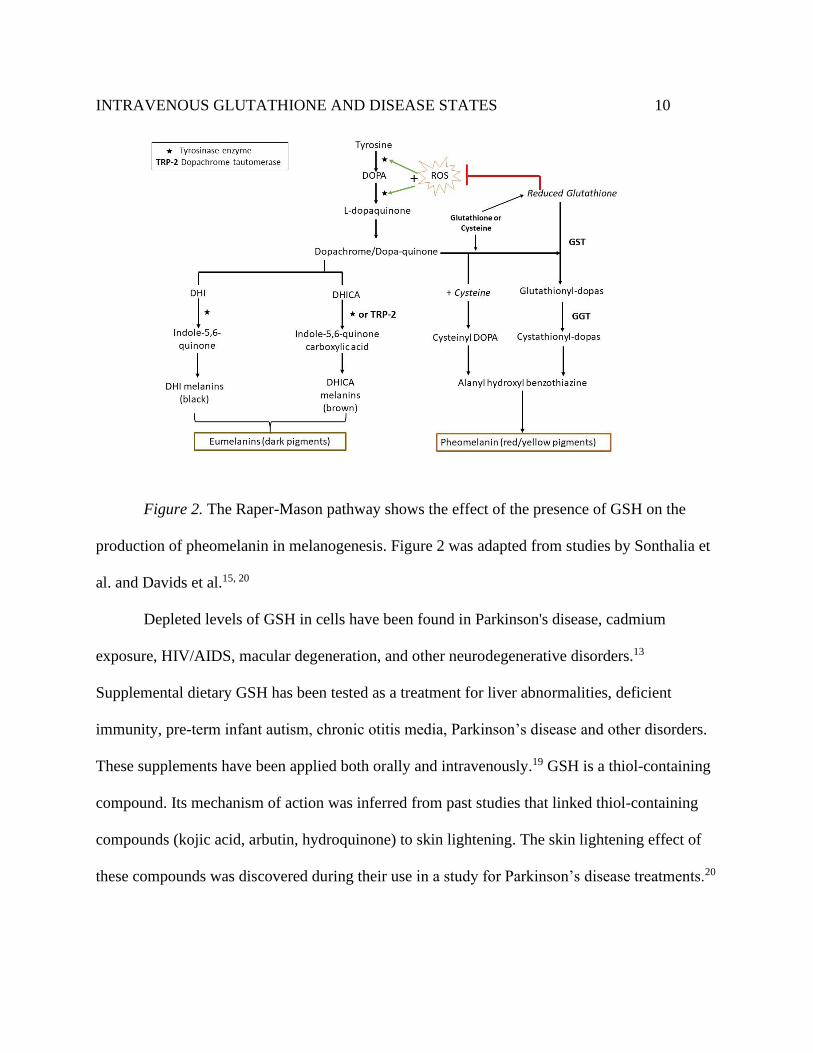
Glutathione’s popularity in skin lightening treatments comes as a result of its anti-
melanogenic properties when it works as a tyrosinase inhibitor during melanogenesis (see Figure
2).18 ROS directly activate tyrosinase. However, when GSH binds to the ROS, they are oxidized
into a non-reactive form, inhibiting tyrosinase. GSH directly inhibits tyrosinase when its thiol
groups react with the copper active site. Tyrosinase is mostly involved in the catalysis of
dihydroxyindole (DHI) and dihydroxyindole carboxylic acid (DHICA) into DHI melanins and
DHICA melanins, respectively. The presence of thiols such as GSH at the dopachrome stage of
the pathway results in binding with dopaquinone to make thiolodopas. This pathway is favored
due to the inhibition of the tyrosinases, causing the increased production of pheomelanins. Thiol
groups found in GSH can directly inhibit tyrosinase by binding to its copper-containing active
site. The production of the brown pigment eumelanin is halted, resulting in the switch to
production of pheomelanin, which is a red/orange pigment.20 This is one of three postulated
mechanisms of action for GSH.
INTRAVENOUS GLUTATHIONE AND DISEASE STATES 10
Figure 2. The Raper-Mason pathway shows the effect of the presence of GSH on the
production of pheomelanin in melanogenesis. Figure 2 was adapted from studies by Sonthalia et
al. and Davids et al.15, 20
Depleted levels of GSH in cells have been found in Parkinson's disease, cadmium
exposure, HIV/AIDS, macular degeneration, and other neurodegenerative disorders.13
Supplemental dietary GSH has been tested as a treatment for liver abnormalities, deficient
immunity, pre-term infant autism, chronic otitis media, Parkinson’s disease and other disorders.
These supplements have been applied both orally and intravenously.19 GSH is a thiol-containing
compound. Its mechanism of action was inferred from past studies that linked thiol-containing
compounds (kojic acid, arbutin, hydroquinone) to skin lightening. The skin lightening effect of
these compounds was discovered during their use in a study for Parkinson’s disease treatments.20

INTRAVENOUS GLUTATHIONE AND DISEASE STATES 11
Compounds that act as scavengers for ROS, such as GSH, can slow down the effects of
melanogenesis. Furthermore, if the compound has redox properties like glutathione, it can
decrease melanin production by interacting with o-quinones or the thiol group at the active site
of the enzyme tyrosinase.21 The active site of tyrosinase contains Cu2+ and soft Lewis bases as
thiol groups are inclined to chelate soft Lewis acids such as Cu2+.48, 52 Furthermore, when
cysteine residues are present in active site pockets of tyrosinase, they cause disulfide
interchanges with the thiol groups. A larger network of inhibition is created when cysteine
residues increase around the tyrosine active site.48
Oral Glutathione
Topical creams are slowly fading away in usage as their effects are restricted to the area
of application, making oral lightening treatments the alternative. Oral glutathione treatments
have increased in popularity due to the impression that they give the whole body a lighter
appearance. However, this is contrary to scientific research.14 Torula yeast (Candida utilis) has
been the main source of oral glutathione, which has been used as a dietary supplement. This
derived glutathione has been the primary ingredient in supplements. Secondary ingredients
include antioxidants like vitamin C.15
Dosing of oral glutathione has been inconsistent between manufacturers as a result of
insufficient studies, making it difficult to assess the side effects of this treatment. Oral
glutathione has been made available in the form of pills and solutions.15 One manufacturer,
Flawless Beauty and Skin, recommends a dosage of three pills that contain 900 mg of L-
glutathione.22 Minor ingredients include N-acetyl-L-cysteine (300 mg), alpha lipoic acid (225
mg), L-methionine (150 mg), vitamin E (150 IU), vitamin B2, (7.5 mg), and selenomethionine

INTRAVENOUS GLUTATHIONE AND DISEASE STATES 12
(300 mcg). According to the manufacturer, these ingredients were combined to enhance
glutathione serum levels in the body. This product emphasizes that it is not approved by the
FDA even though it is manufactured in the United States of America.22
In comparison, an Indian manufacturer recommends a dosage of 10-20 mg per kg body
weight for use as an antioxidant or anti-aging medication. In contrast, in the case of skin
lightening, the recommended dosage is 20-40 mg per kg body weight. Furthermore, the duration
of treatment is dependent on the client’s skin color, with darker shades of skin requiring
treatment for over two years, and lighter shades of skin needing one to three months of treatment
until the desired outcome has been achieved. This product is recommended in combination with
vitamin C (2000 mg per day) for the glutathione to be absorbed most efficiently. Each
glutathione pill taken contains 1000 mg, and a client is prescribed one pill twice a day.24 These
two products are small examples of how diverse the industry is in dosage and duration
recommendations. Indeed, there is no scientific basis for the recommended dosage.
Studies done by Kovacs-Nolan et al. on the fate of oral GSH have shown that it can cross
the intestinal epithelium, but it is readily oxidized to GSSG.25 GSH is quickly digested by
gamma-glutamyl transferase (GGT) in the upper jejunum into its constituent amino acids:
cysteine, glycine and glutamate.26 It has been found that intracellular GSH is more effective
when its constituent amino acids are taken on their own instead of GSH itself, as there is a higher
concentration of GGT in the liver which further degrades the oral GSH that has been absorbed
into the bloodstream after digestion. Oral cysteine is degraded in the digestive tract, but
supplementing it in the form of N-acetylcysteine (NAC) increases the levels of cysteine because
it is protected from degradation.13 Oral glutathione has a low bioavailability, and this may be the

INTRAVENOUS GLUTATHIONE AND DISEASE STATES 13
motive behind the pursuit of intravenous (IV) glutathione.27 The estimated half-life of oral
glutathione in plasma is 1.6 min.39 Additionally, it should be noted that any skin lightening
treatments sought from this method are temporary as pheomelanin production is low and
inconsistent.28
Intravenous (IV) Glutathione
IV glutathione has been recently introduced in the cosmetic industry to speed up the skin
lightening process, and to have long-lasting effects. This method is controversial due to the lack
of safety data regarding its use. Few research studies have been performed to date concerning IV
glutathione despite many women, especially in the Philippines, using this as their primary skin
lightening agent.15 The Philippine FDA released a statement warning of the toxic consequences
to the nervous system, liver and kidneys and the development of the rare Stevens-Johnson
syndrome with the continued used of IV glutathione. The greatest concern from the FDA is the
incorrect administration of this treatment by untrained and non-medical personnel. In the black-
market areas that provide this service safety precautions may not be observed. The use of
injections and a drip in non-sterile conditions can lead to the transmission of HIV as needles are
shared, and the spread of different types of hepatitis. Overall, clients can experience embolisms
caused by the introduction of intravenous air bubbles, and sepsis caused by pathogens and
counterfeit glutathione.29 On a cellular level, increased serum levels of intravenous glutathione
can lead to reductive stress that is just as toxic to the body as oxidative stress.32
Research Question
Despite insufficient data to support the use of glutathione as a skin lightening treatment,
it is still used. This study will investigate the ability of glutathione to lighten skin. First, the

INTRAVENOUS GLUTATHIONE AND DISEASE STATES 14
mechanism of action that glutathione takes to lighten skin will be investigated to bridge the gap
that exists between topical, oral and IV administration. Then, the effects of oral and topical
glutathione in the body will be investigated by studying their biochemical mechanisms to
compare them to IV glutathione. This study will also explore the side effects of glutathione when
used for its melanogenic properties. Finally, by looking into the pathogenesis of disease, possible
effects of IV administration of glutathione will be mapped to determine the potential long-term
consequences of skin lightening.
Literature Summary of Glutathione Skin Lightening Products
IV glutathione has not been officially recognized as a skin lightening agent. Its mode of
action has not been observed. Furthermore, only a few studies have been conducted to date to
observe the efficacy of oral and topical glutathione in skin lightening. The following studies
were published over four years ago, and there is a need for more recent data as glutathione is
increasing in popularity. Table 2a and 2b are a compilation of known studies that may aid in a
proposal of a mechanism of action. These tables are adapted from Table 1 published from the
study by Sonthalia et al.15

INTRAVENOUS GLUTATHIONE AND DISEASE STATES 15
Table 2a
Investigations of glutathione as a skin lightening treatment topically and orally.
Type of
Glutathione
Application
Topical Oral (capsules)
Study
Reference
Watanabe et al. 22 Weschawalit & Asawanoda
44
Arjinpathana and
Asawononda 46
Year 2014 2016 2010
Subject
Information
30 healthy Filipino
females; tan;
Fitzpatrick skin types III
or IV;
30-50 years of age
60 healthy females
randomized into 3 groups;
20-50 years of age
60 healthy medical
school students; 18
males and 42 females;
19-22 years of age
Study
Design
Randomized, double-
blind, placebo-
controlled, matched-pair
study
Randomized, placebo
controlled, three-arm study
Randomized, double-
blind, placebo-
controlled, two-arm
study
Methods
Applied
0.5 g of lotion comprised
of 2% (w/w) GSSG or
placebo without GSSG
randomly assigned and
spread evenly to either
250 mg/day of GSH,
250 mg/day of GSSG or
placebo (dibasic calcium
phosphate), randomly
assigned to subjects
250 mg glutathione or
placebo capsules
taken twice daily on
an empty stomach for
4 weeks, block-

INTRAVENOUS GLUTATHIONE AND DISEASE STATES 16
left or right cheek of
each subject
randomized
assignment to subjects
Parameters
Evaluated
Measured every 2 weeks
for 10 weeks:
1) Skin whitening using
Mexameter for melanin
index (mexameter is a
tool used to measure
melanin and hemoglobin
levels through
absorption/reflection of
light)
2) Skin moisture
3) Skin firmness using a
Triplesense TR-3 sensor
device
4) Efficacy against
wrinkle reduction by
observing “crow’s feet”
5) Skin smoothening
Measured every 4 weeks:
1) Melanin index
2) Presence of UV spots
3) Transepidermal water
loss
4) Water contents
5) Elasticity
Measured at baseline
and after 4 weeks:
1) Melanin index
using a Mexameter:
Sun exposed areas:
i) Face (both sides),
2.5 cm caudally from
the lateral canthi
ii) Extensor surfaces
of both forearms, 7
cm above the ulnar
styloid processes
Sun protected areas:
i) Upper, inner arms,
10 cm from axillary
vault
ii) Standardized
photographs were
taken using a VISIA
CR system to
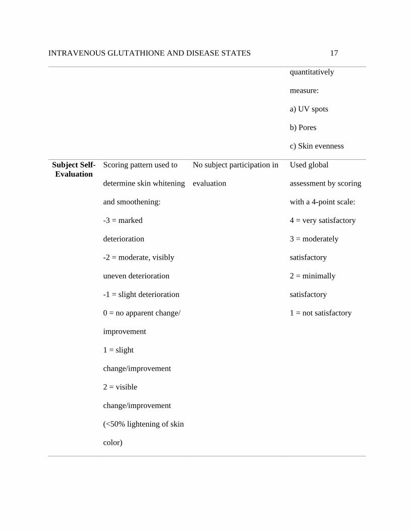
INTRAVENOUS GLUTATHIONE AND DISEASE STATES 17
quantitatively
measure:
a) UV spots
b) Pores
c) Skin evenness
Subject Self-
Evaluation
Scoring pattern used to
determine skin whitening
and smoothening:
-3 = marked
deterioration
-2 = moderate, visibly
uneven deterioration
-1 = slight deterioration
0 = no apparent change/
improvement
1 = slight
change/improvement
2 = visible
change/improvement
(<50% lightening of skin
color)
No subject participation in
evaluation
Used global
assessment by scoring
with a 4-point scale:
4 = very satisfactory
3 = moderately
satisfactory
2 = minimally
satisfactory
1 = not satisfactory

INTRAVENOUS GLUTATHIONE AND DISEASE STATES 18
3 = marked
improvement/ significant
change (uniform skin
whitening, >80% of
application area)
Safety and
Side Effects
Listed
Well-tolerated; all
subjects completed
study; 1 subject had mild
erythema on days 2 and
3; no reports of
unwanted symptoms
Well-tolerated
Well-tolerated; 1
subject in glutathione
group experienced
flatulence; 1 subject
in placebo group
experienced
constipation
Follow-up None after 10 weeks of
study
None after study duration None after study
duration
Study
Results
1) Skin lightening
melanin index:
A) Placebo - decreased
slightly after 10 weeks
(Week 0: 274.13 [+/-]
25.81 vs. Week 10:
265.50 [+/-] 25.82)
1) Both GSH and GSSG
caused decreased melanin
indices
2) GSH more effective in
wrinkle improvement
3) GSH and GSSG
increased skin elasticity in
1) GSH group had
greater decrease in
melanin index than
placebo group on sun-
exposed areas
2) Smaller number of
UV spots developed
in subjects on

INTRAVENOUS GLUTATHIONE AND DISEASE STATES 19
B) GSSG lotion –
decreased after 10 weeks
(Week 0: 272.77 [+ or -]
26.17 vs. Week 10:
243.47 [+ or -] 26.31)
2) Moisture index values
were higher at GSSG
sites than placebo sites
3) Curvature and keratin
values were significantly
lower in GSSG sites than
placebo sites
4) Elasticity values did
not show a remarkable
difference between
placebo and GSSG lotion
both sun exposed and sun-
protected skin
glutathione treatment;
an increase in skin
evenness and a
reduction in pore size
were reported with
the treatment
3) Subjects reported
an average score of
3.06 for satisfaction
Limitations Small study size, short
duration and no follow-
up; limited to women of
Fitzpatrick skin type III-
VI; results may be
Study had insufficient data
to analyze limitations
Plasma glutathione
levels not measured;
study duration was
short and no follow
up

INTRAVENOUS GLUTATHIONE AND DISEASE STATES 20
limited to Filipino
women
Proposed
Mechanism
of Action
A) Conversion of GSSG
to GSH in epidermis
B) GSH activates
pheomelanin pathway
Unavailable GSH activates
pheomelanin pathway
Table 2b
Investigations of glutathione as a skin lightening treatment orally and intravenously.
Type of
Glutathione
Application
Oral (buccal lozenges) Intravenous
Study
Reference
Handog et al. 45 Zubair et al. 32
Year 2015 2016
Subject
Information
30 Filipino females who work as
medical personnel, Fitzpatrick
skin types IV – V, with melanin
indices of ≥ 20 out of 99;
22-42 years of age
50 female test subjects enrolled, 32 had
data recorded from them and were
divided into 2 groups: Group A skin
types match group B skin types;
25-47 years of age
Study Design Open label, singe–arm pilot study Placebo-controlled
Methods
Applied
Identical bottles containing 30
lozenges (500 mg of GSH) were
given to each subject
Group A: 16 subjects given IV
glutathione and vitamin C (injection of
GSH Detox Forte ®, 1200 mg)

INTRAVENOUS GLUTATHIONE AND DISEASE STATES 21
Subjects kept one lozenge in their
mouth, against their inner cheek
(buccal mucosa)
every morning, until completely
dissolved
Control Group B: 16 subjects given IV
saline as placebo (injection of 5 mL
saline, 10 mL distilled water)
2 unexposed body sites were chosen
(upper inner arm below axilla and upper
outer thigh of all patients).
Parameters
Evaluated
Evaluation done every 2 weeks
for 8 weeks:
1) Portable Mexameter used to
measure melanin index
2) Melanin indices taken from
sun-exposed area (extensor
surface of wrist) and sun-
protected area (mid sternum)
3) Liver function tests, serum
glutamic pyruvic transaminase
(SGPT), complete blood count
(CBC) and serum glutamic
oxaloacetic transaminase (SGOT)
were done at baseline
Administration for 6 weeks and 2
independent observers evaluated:
Skin tone measured with Taylor
hyperpigmentation scale (tool consisting
of 15 uniquely colored plastic cards with
hues that apply to Fitzpatrick skin types
I-IV and is used to visually assess
complexion), cards are used to analyze
change from darker shades to light
shades 47

INTRAVENOUS GLUTATHIONE AND DISEASE STATES 22
Subject Self-
Evaluation
Used global assessment scores to
determine skin lightening:
None = 0
Mild change = 1
Moderate = 2
Obvious = 3
Very marked = 4
No subject participation in evaluation
Safety and
Side Effects
Listed
Well-tolerated; 29 out of 30
subjects completed the study; 30th
subject complained of soreness in
gums due to lozenge
administration; another subject
complained of chalky taste but
completed study.
CBC, SGPT and SGOT values
remained normal at week 8.
Not well-tolerated; 32 out of 50
completed study; 8 subjects from group
A had deranged liver function tests
(defined as 1.5 times above reference
range), and 1 subject developed
anaphylactic shock; all were excluded
from results.
9 control from group B who matched the
skin types of subjects excluded from
group A were also excluded to keep
study standardizations
Side effects in 25 subjects in group A:
Feeling warmth during injection (44%)
Abdominal cramps (40%)

INTRAVENOUS GLUTATHIONE AND DISEASE STATES 23
Abnormal liver functions (32%)
Feeling of heart sinking (28%)
Diarrhea (16%)
Paresthesia (16%)
Dizziness (12%)
Anaphylactic shock (4%)
Vomiting (4%)
Follow-Up None after the duration of study 2, 4, 6 months after end of
administration
Study Results 1) In sun-exposed area:
At week 8, 100% of subjects
experienced a reduction in their
melanin indices (p<0.0001) from
week 0; significant skin
lightening effects only showed
after week 2
2) In sun-protected area:
At week 8, 100% of subjects
experienced a reduction in their
melanin indices from week 0
3) 27/30 subjects (90%) evaluated
their skin as having undergone
1) GSH group A skin improvement,
n=16
0 months (after 12 injections): 6 (37.5%)
2 months: 3 (18.7%)
4 months: 3 (18.7%)
6 months: 1 (6.2%)
2) Placebo group B skin improvement,
n=16
0 months (after 12 injections): 3 (18.7%)
2 months: 2 (12.5%)
4 months: 0 (0%)
6 months: 0 (0%)
Not very effective for skin lightening

INTRAVENOUS GLUTATHIONE AND DISEASE STATES 24
moderate lightening (score 2 out
of 4); 3/30 subjects (10%)
evaluated their skin having
undergone mild skin lightening
(score 1 out 4)
Limitations Small study population and
duration
No control used and no follow-up
Blood GSH levels were not
measured
Blood GSH levels were not measured
Taylor hyperpigmentation tool is more
useful when used subjectively but a
Mexameter is more useful to measure
melanin index objectively and accurately
50
Small study population and short
duration
Mechanism of
Action
GSH used mucosal route to get to
epidermis all around the body
GSH absorption increased when
administered with vitamin C
Analysis of the Literature
Most of the above studies were conducted in Southeastern Asia countries, where
residents usually have the Fitzpatrick skin types III-IV and olive/tan skin (Table 1). The results
displayed that glutathione in general does help with skin lightening, but the most effective
treatment was through oral glutathione administration. Oral administration also appeared to be
the safest because it had minor or no side effects on the subjects compared to IV glutathione.

INTRAVENOUS GLUTATHIONE AND DISEASE STATES 25
GSH and GSSG both had the same effect in reducing the melanin index of the skin. This would
be expected because GSSG can be easily reduced into GSH by glutathione reductase (see Figure
1) to be used as GSH in the Raper-Mason pathway. These studies suggested that glutathione has
skin lightening properties when applied topically and orally, and the proposed mechanism was
through the shifting of the Raper-Mason pathway to produce pheomelanin instead of eumelanin.
According to the study done by Zubair et al., IV glutathione does not appear to retain its skin
lightening properties over time.32 The missing link in the studies was the lack of data pertaining
to serum glutathione levels. By monitoring the levels of GSH in the blood, the metabolism of IV
glutathione may be investigated, and mechanisms of action proposed.
Proposed Disease States Directly and Indirectly Correlated to IV Glutathione
The study done by Zubair et al. in 2016 to test the efficacy of IV glutathione against a
placebo in skin-lightening is one of the only studies published to date. The data from this
experiment is helpful because it gives the potential effects of using IV glutathione over long
periods of time. Of all the side effects, notably 40% of volunteers from group A experienced
abdominal cramps, 32% reported deranged liver functions and 16% reported diarrhea and
paresthesia. The control group B did not record any adverse side effects, which suggests that
intravenous glutathione was the cause for the side effects. The main objective of the experiment
was to observe changes in skin color: 37.5% of women were observed to have a change in skin
color immediately after the completion of 12 injections. By the last observation at six months,
only 1 out of the 16 women who completed the treatment showed a consistency in skin color
improvement.32 Nine women had to stop treatment due to adverse side effects. Eight out of the

INTRAVENOUS GLUTATHIONE AND DISEASE STATES 26
nine women experienced issues with their livers, and one suffered from anaphylaxis due to the
treatment (Table 2b).
Reductive Stress Can Cause Oxidative Stress
Reductive stress is the opposite of oxidative stress and happens as a result of the redox
imbalance of the species GSH/GSSG, nicotinamide adenine dinucleotide (NAD+)/NADH and
phosphorylated NAD+ (NADP+)/NADPH; these species are important in the maintenance of a
homeostatic environment for cells. An increase in the formation of GSSG is a marker of
oxidative stress.33 Increased levels of intracellular GSH can induce pro-oxidant activity and halt
its antioxidant properties towards ROS.34, 35 Reductive stress is known to mostly contribute to
inflammatory disease in contexts such as cancer, pulmonary hypertension and hypertrophic
cardiomyopathy.33 ROS are beneficial in the body for redox homeostasis, and well-functioning
cardiovascular and immune systems. For example, in redox regulation, the ROS react with the
amino acid cysteine on proteins, which is important in signaling pathways. ROS are also
important in the cell cycle and in influencing apoptosis. ROS can bind to the mitogen-activated
kinase, which triggers the apoptotic cascade.51 Consistent reductive stress caused by a shortage
of ROS can cause oxidative stress as a result of negative feedback: redox proteins can donate
excess electrons to O2 which generates ROS such as superoxide (O2- ). An increased
concentration of ROS leads to oxidative stress, which triggers increased action by the ROS
scavengers, and may trigger a reductive stress crisis again.36 This cycle can cause cellular
damage as homeostasis is disrupted.
Increased production of pheomelanin. GSH is imperative for the melanogenesis
pathway to produce pheomelanin, and its continued production is sustained by high levels of
INTRAVENOUS GLUTATHIONE AND DISEASE STATES 27
cysteine that interact with the DOPA-quinone as illustrated in Figure 2.43 The use of IV
glutathione, as demonstrated by the study conducted by Zubair et al., can alter skin color.
However, the changes were temporary.32 People with lighter skin types (Fitzpatrick skin types I-
III) are 70 times more at risk to develop skin cancer compared to those who fall under the
Fitzpatrick skin types IV-VI.37 Eumelanin is regarded as the more protective of the two types of
melanin as it is not implicated in melanogenesis as much as pheomelanin.37 While the exact
mechanism is unknown, pheomelanin production increases ROS in melanocytes, and therefore
decreases glutathione stores within the cell. The introduction of high levels of intravenous GSH
can help to recover depleted levels of the antioxidant in the cell and maintain the level of
cysteine for pheomelanin production, rather than exacerbate oxidative stress in melanocytes.38
A study performed by Nasti and Timares about the roles of eumelanin and pheomelanin
in the susceptibility to skin cancer concluded that there was a correlation between different
strains of mice (agouti and yellow coats) with high pheomelanin production and a higher risk of
developing melanoma.37 More research needs to be done to determine the direct link between
pheomelanin and melanoma, and how other factors such as UV radiation contribute to it.
It has been found that GSH is vital for metastatic melanoma growth. In a study done by
Carretero et al. to investigate the link between glutathione content and the activity of metastatic
liver cancer cells using B16 melanoma cells, it was observed that GSH regulated energy
metabolism and increased cell growth.42 GSH is produced in vivo, but low levels of cysteine
cause a lag in GSH synthesis. When cysteine was supplemented in this study, growth of the B16
melanoma cells was expedited, as cysteine is the rate-limiting step in GSH production.42
INTRAVENOUS GLUTATHIONE AND DISEASE STATES 28
Exogenous intravenous supplementation of glutathione could possibly increase cancer growth as
it may supply the much-needed cysteine when GSH is broken down intracellularly.
Stevens-Johnson syndrome (SJS) and toxic epidermal necrolysis (TEN). These are
rare, life-threatening conditions that are caused by an adverse immune reaction to medications.
Medications for these conditions include anticonvulsants, allopurinol, sulfonamides, antibiotics
and non-steroidal anti-inflammatory drugs. SJS and TEN physically manifest themselves in the
form of mucocutaneous blisters that may result in epidermal detachment and tissue necrosis.30
Skin lesions appear, followed by symptoms that resemble a respiratory infection such as fever,
headache and sore throat. People who suffer from HIV/AIDS, lupus, or compromised immune
systems are usually more susceptible to suffering from SJS and TEN.31 The Philippine FDA
mentioned that the use of intravenous glutathione can cause SJS/TENS. The mechanism of how
this could occur is still under investigation, and little is known about the role of glutathione in
this disease state.
Theoretically, glutathione may increase the risk of SJS/TEN occurring through cellular
redox imbalance. High levels of ROS that could be caused by the reductive stress/oxidative
stress cycle trigger apoptosis to occur. There are different mechanisms of the pathogenesis of
these diseases, but one that may involve glutathione is keratinocyte apoptosis, followed by
necrosis.40 Apoptosis of the cell occurs as a result of damage to the mitochondria. The ROS may
activate nuclear transcription factors such as tumor necrosis factor alpha (TNF-α) to begin the
early stages of apoptosis, or indirectly cause apoptosis through cell damage by disrupting plasma
membrane lipids and mobilizing Ca2+, which leads to early apoptosis. Increased levels of GSH
disrupt the ionic gradient of the mitochondria, causing it to rupture, leading to late necrosis.41

INTRAVENOUS GLUTATHIONE AND DISEASE STATES 29
This is only a suggested mechanism to explain how increased glutathione serum levels may be
related to SJS/TEN. More experimental data needs to be compiled as there is no definitive link to
SJS/TEN, except the report from the Philippine FDA. The novelty of IV glutathione being used
as a skin lightening agent is an opportunity for more experimentation to take place to investigate
its effects.
Conclusion
This literature review has established that women, more than men, undergo skin
lightening treatments. This is evidenced by most of the studies being conducted on women, and
women having a stronger desire to achieve a sense of beauty by having a lighter complexion.5
The pursuit of lighter skin is not only fueled by current beauty standards, but also social
acceptance and higher social status. The Fitzpatrick scale in Table 1 showed that the photo types
IV-VI are more likely to undergo skin lightening treatments, which correlates with the certain
regions in the world such as Asia, East India, Southeastern Asia and Africa.
Although supplemental oral glutathione has been declared as safe, oral glutathione for
skin lightening reasons has not been approved by the Philippine FDA. Use of the intravenous
version of this skin lightening treatment has been warned against by the Philippine FDA due to
the adverse side effects reported. Despite this, intravenous glutathione is still sold on the market,
and still being used. Using glutathione could be risky if it is administered by untrained personnel.
Dangerous side effects may result, including the transmission of HIV and different forms of
hepatitis from recycled needles. Furthermore, there is an inconsistency with the dosing of
glutathione for skin lightening among manufacturers, which may pose a danger to users as
excessive glutathione may lead to liver toxicity. Studies exploring the anti-melanogenic effects

INTRAVENOUS GLUTATHIONE AND DISEASE STATES 30
of glutathione are few, and those reporting data are limited to Southeastern Asian women.
Women who use skin lightening treatments may use glutathione (topical, oral or IV) over long
periods of time, and unfortunately studies to date were too short to determine long-term
consequences.
The mechanism of action of glutathione established from research is the inhibition of the
tyrosinase enzyme. The inhibition of the tyrosinase active site shifts eumelanin production to
pheomelanin. The injection of IV glutathione has been shown to raise serum GSH, which could
lead to reductive stress as there is a large concentration of GSH in cells. By negative feedback,
this could result in oxidative stress. Theoretically, increased ROS may be the reason behind the
aggravation of conditions such as SJS/TEN. Increased pheomelanin production may cause the
user to be at higher risk of suffering from melanoma. This study was inconclusive in establishing
a direct link between IV glutathione and SJS/TEN.
GSH is manufactured de novo because it is important for bodily functions. Normal
cellular actions require GSH to prevent tissues from undergoing oxidative stress as it is the
greatest antioxidant. Since it is already found in high concentrations in the body, its
supplementation at high doses could be toxic for the body, especially if taken in unregulated
doses. The use of glutathione is not based on scientific findings and could eventually cause more
harm than good. Oral glutathione may have been established as a lightening product, but
intravenous glutathione is not effective in skin lightening and may lead to redox crises in the
body with continued use.

INTRAVENOUS GLUTATHIONE AND DISEASE STATES 31
Future Experimentation
The literature in Table 2 showed that none of the experiments monitored GSH serum
levels which could play an important role in outlining the mechanism of glutathione metabolism.
Since glutathione is made up of cysteine, glutamate and glycine, one of these amino acids could
be tagged. The half-life of glutathione is short because it is digested to its constituent amino
acids. Cysteine could be tagged as it is imperative in the production of pheomelanin. The urine of
subjects could then be monitored for tagged cysteine molecules to observe if GSH was excreted
due to excess levels in the blood. More experiments need to be done on topical, oral and
intravenous methods to establish safe doses and to monitor the effects of taking glutathione for
long durations. Finally, the Philippine FDA suggested the occurrence of SJS/TEN with the use of
IV glutathione. Although rare, it is theoretically feasible, and a study could be conducted to
determine if glutathione directly causes this condition, and if reductive stress plays a role.

INTRAVENOUS GLUTATHIONE AND DISEASE STATES 32
References
1. Charles C. Skin bleaching, self-hate, and black identity in Jamaica. J Black Stud 2003;
33:711-28.
2. Jacobs M, Levine S, Abney K, Davids L. Fifty shades of African lightness: A bio-
psychosocial review of the global phenomenon of skin lightening practices. J Public
Health Afr 2016; 7:552.
3. Gbenga, A. (2004). Nigeria: Bleaching body practices [WWW document]. URL
http://www.youthxchange.net/main/nigeriableachingbodypractices.asp. [accessed on 10
January 2020].
4. Blay, A. Skin bleaching and global white supremacy: By way of introduction. J Pan Afr
Stud 2011; 4:4–46.
5. Shroff H, Diedrichs PC, Craddock N. Skin Color, Cultural Capital, and Beauty Products:
An Investigation of the Use of Skin Fairness Products in Mumbai, India. Front Public
Health 2018; 5:365-70.
6. Draelos ZD. Skin lightening preparations and the hydroquinone controversy. Dermatol
Ther 2007; 20: 308-313.
7. Statistics Market Research Consulting Pvt. Ltd. (2019). Global skin lightening products
market is expected to reach $8011.17 million by 2026 [WWW document]
https://www.prnewswire.com/news-releases/global-skin-lightening-products-market-is-
expected-to-reach-8011-17-million-by-2026--300906859.html [accessed 21 January
2020].
INTRAVENOUS GLUTATHIONE AND DISEASE STATES 33
8. Couteau C, Coiffard L. Overview of skin whitening agents: Drugs and cosmetic
products. Cosmetics 2016; 3:1-16.
9. Sarkar R, Arora P, Garg KV. Cosmeceuticals for hyperpigmentation: What is available? J
Cutan Aesthet Surg 2013; 6:4-11.
10. Bino SD, Duval C, Bernard F. Clinical and biological characterization of skin
pigmentation diversity and its consequences on UV impact. Int J Mol Sci 2018; 19, 2668-
80.
11. Holcomb NC, Bautista R, Jarrett SG, et al. Chapter eight, cAMP-mediated regulation of
melanocyte genomic instability: A melanoma-preventive strategy. Advances in Protein
Chemistry and Structural Biology, Academic Press. 2019: 247-95.
12. Dofitas BL, Asuncion EB, Nisce-Anisco KC, et al. (2018). Public advisory on glutathione
as a “skin whitening” agent from Philippine glutathione advisory committee 2018.
https://pds.org.ph/public-advisory-on-glutathione-as-a-skin-whitening-agent/. [accessed on
18 January 2020].
13. Pizzorno J. Glutathione! Integr Med (Encinitas, Calif.) 2014; 13:8–12.
14. Sonthalia S, Sarkar R. Glutathione for skin lightning: an update. Pigment Int 2017; 4:3-6.
15. Sonthalia S, Daulatabad D, Sarkar R. Glutathione as a skin whitening agent: Facts, myths,
evidence and controversies. Indian J Dermatol Venereol Leprol 2016; 82:262-72.
16. Ilkhani F, Hosseini B, Saedisomeolia A. Niacin and oxidative stress: A mini-review. J
Nutri Med Diet Care Last 2016; 2:014.
17. Meister A, Tate SS. Glutathione and related gamma-glutamyl compounds: Biosynthesis
and utilization. Annu. Rev. Biochem, 1976; 45:559-604.

INTRAVENOUS GLUTATHIONE AND DISEASE STATES 34
18. Sonthalia, S, Jha AK, Lallas A et al. Glutathione for skin lightening: a regnant myth or
evidence-based verity? Dermatol Pract Concept 2018; 8:15–21.
19. Hauser RA, Lyons KE, McClain T, Carter S, et al. Randomized, double-blind, pilot
evaluation of intravenous glutathione in Parkinson’s disease. Mov Disord 2009; 24:979–
83.
20. Davids LM, van Wyk JC, Khumalo NP. Intravenous glutathione for skin lightening:
Inadequate safety data. S Afric Med J 2016; 106: 782-86.
21. Briganti S, Camera E, Picardo M. Chemical and instrumental approaches to treat
Hyperpigmentation. Pigment Cell Res; 16: 101-110.
22. Watanabe F, Hashizume E, Chan GP, Kamimura A. Skin-whitening and skin-condition-
improving effects of topical oxidized glutathione: a double-blind and placebo-controlled
clinical trial in healthy women. Clin Cosmet Investig Dermatol 2014; 7: 267–274.
23. Flawless Beauty and Skin. New Relumins Advance White Active Glutathione Complex –
Oral Whitening Formula Capsules with 6x Boosters [WWW document].
https://www.flawlessbeautyandskin.com/NEW-Relumins-Advance-White-Active-
Glutathione-Complex-Oral-Whitening-Formula-Capsules-with-6X-Boosters-Whitens-
repairs-rejuvenates-skin_p_581.html [accessed 18 January 2020]
24. Bangalore Classifieds. (2020). Glutathione Skin Lightening Pills in India [WWW
document]. http://www.bangalore.click.in/glutathione-skin-lightening-pills-in-india-c75-
v4148389. [accessed 18 January 2020].

INTRAVENOUS GLUTATHIONE AND DISEASE STATES 35
25. Kovacs-Nolan J, Rupa P, Matsui T, et al. In vitro and ex vivo uptake of glutathione (GSH)
across the intestinal epithelium and fate of oral GSH after in vivo supplementation. J Agric
Food Chem 2014; 62:9499–9506.
26. Gould RL, Pazdro R. Impact of supplementary amino acids, micronutrients, and overall
diet on glutathione homeostasis. Nutrients 2019; 11:1055-1056.
27. Dilokthornsakul W, Dhippayom T, Dilokthornsakul P. The clinical effect of glutathione
on skin color and other related skin conditions: A systematic review. J Cosmet Dermatol
2019; 18: 728– 737.
28. Hanigan MH. Gamma-glutamyl transpeptidase: redox regulation and drug resistance. Adv
Cancer Res 2014; 122:103–141.
29. Food and Drug Administration Philippines. (2019). [WWW document]
https://ww2.fda.gov.ph/attachments/article/596859/FDA%20Advisory%20No.2019-
182.pdf [accessed 18 January 2020].
30. Zimmerman D, Dang NH. Stevens–Johnson Syndrome (SJS) and toxic epidermal
necrolysis (TEN). In: Oncologic Critical Care (Nates J, Price K, eds). Springer,
Cham., 2019.
31. Ciccacci C, Latini A, Politi C. et al. Impact of glutathione transferases genes
polymorphisms in nevirapine adverse reactions: a possible role for GSTM1 in SJS/TEN
susceptibility. Eur J Clin Pharmacol 2017: 73, 1253–1259.
32. Zubair S, Hafeez S, Mujtaba G. Efficacy of intravenous glutathione vs. placebo for skin
tone lightening. J of Pakistan Assoc Derma. 2016; 26:177-181.

INTRAVENOUS GLUTATHIONE AND DISEASE STATES 36
33. Pérez-Torres I, Guarner-Lans V, Rubio-Ruiz ME. Reductive stress in inflammation-
associated diseases and the pro-oxidant effect of antioxidant agents. Int J Mol Sci 2017;
18:2098.
34. Xiao W, Loscalzo J. Metabolic responses to reductive stress. Antioxidants & Redox
Signaling. 2019; 0: 2020.
35. Peris E, Micaef, Paul A, et al. Antioxidant treatment induces reductive stress associated
with mitochondrial dysfunction in adipocytes. J Biol Chem 2018; 10: 1-23.
36. Korge P, Calmettes G, Weiss JN. Increased reactive oxygen species production during
reductive stress: The roles of mitochondrial glutathione and thioredoxin reductases.
Biochem Biophys Acta 2015; 1847:514–525.
37. Nasti TH, Timares L. MC1R, eumelanin and pheomelanin: their role in determining the
susceptibility to skin cancer. Photochem and Photobio 2015; 91: 188–200.
38. Morgan AM, Lo J, Fisher DE. How does pheomelanin synthesis contribute to
melanomagenesis? Two distinct mechanisms could explain the carcinogenicity of
pheomelanin synthesis. Bioessays 2013; 35:672–676.
39. Bruggeman BK, Storo KE, Fair HM, Wommack AJ, Carriker CR, Smoliga JM. The
absorptive effects of orobuccal non-liposomal nano-sized glutathione on blood glutathione
parameters in healthy individuals: A pilot study. PLoS One 2019; 14: e0215815.
40. Harr T, French LE. Toxic epidermal necrolysis and Stevens-Johnson syndrome. Orphanet
J Rare Dis 2010; 5:39.
41. Paquet P, Pierard GE. Toxic epidermal necrolysis: Revisiting the tentative link between
early apoptosis and late necrosis (Review). Int. J. Mol. Med. 2007; 19:3-10.

INTRAVENOUS GLUTATHIONE AND DISEASE STATES 37
42. Carretero J, Obrador E, Anasagasti MJ et al. Growth-associated changes in glutathione
content correlate with liver metastatic activity of B16 melanoma cells. Clin Exp
Metastasis 1999; 17:567–574.
43. Tanaka H, Yamashita Y, Umezawa K et al. The pro-oxidant activity of pheomelanin is
significantly enhanced by UVA irradiation: benzothiazole moieties are more reactive than
benzothiazine moieties. Int J Mol Sci 2018; 19:2889.
44. Weshawalit S, Asawanonda P. Effects of oral glutathione on skin appearances: a
randomized placebo-controlled study. J Am Acad Dermatol 2016; 74: AB16
45. Handog EB, Datuin MSL, Singzon IA. An open‐label, single‐arm trial of the safety and
efficacy of a novel preparation of glutathione as a skin‐lightening agent in Filipino
women. Int J Dermatol 2016; 55: 153-157.
46. Arjinpathana N, Asawanonda P. Glutathione as an oral lightening agent: A randomized,
double-blind, placebo-controlled study. J Dermatol Treat 2012; 23:97-102.
47. Taylor SC, Arsonnaud S, Czernielewski J. The Taylor Hyperpigmentation Scale: a new
visual assessment tool for the evaluation of skin color and pigmentation. Cutis 2005;
76:270-4.
48. Yong-Doo P, Lyou Y, Hahn H et al. Complex inhibition of tyrosinase by thiol-composed
Cu2+ chelators: A clue for designing whitening agents. J Biomol Struct Dyn 2006; 24: 131–
138.
49. Wu G, Fang Y, Yang S et al. Glutathione metabolism and its implications for health. J
Nutr 2004; 134:489–492.

INTRAVENOUS GLUTATHIONE AND DISEASE STATES 38
50. Treesirichod A, Chansakulporn S, Wattanapan P. Correlation between skin color
evaluation by skin color scale chart and narrowband reflectance spectrophotometer. Indian
J Dermatol 2014; 59:339–342.
51. Patel R, Rinker L, Peng J et al. Reactive Oxygen Species: The Good and the Bad, Reactive
Oxygen Species (ROS) in Living Cells (Cristiana Filip and Elena Albu, eds) IntechOpen,
2017:8–16.
52. Bjørklund G, Crisponi G, Nurchi VM et al. A review on coordination properties of thiol-
containing chelating agents towards mercury, cadmium, and lead. Molecules. 2019;
24:3247.
Copyright © 2022 FDOKUMEN