Ab Initio Molecular Dynamics Calculations of Ion Hydration Free Energies
Upload
khangminh22Category
view
6download
0
�����������������
Citation: Ben-Haim, Y.; Chorin, E.;
Hochstadt, A.; Ingbir, M.; Arbel, Y.;
Khoury, S.; Halkin, A.; Finkelstein, A.;
Banai, S.; Konigstein, M. Forced
Diuresis with Matched Isotonic
Intravenous Hydration Prevents
Renal Contrast Media Accumulation.
J. Clin. Med. 2022, 11, 885. https://
doi.org/10.3390/jcm11030885
Academic Editor: Yacov Shacham
Received: 10 January 2022
Accepted: 4 February 2022
Published: 8 February 2022
Publisher’s Note: MDPI stays neutral
with regard to jurisdictional claims in
published maps and institutional affil-
iations.
Copyright: © 2022 by the authors.
Licensee MDPI, Basel, Switzerland.
This article is an open access article
distributed under the terms and
conditions of the Creative Commons
Attribution (CC BY) license (https://
creativecommons.org/licenses/by/
4.0/).
Journal of
Clinical Medicine
Article
Forced Diuresis with Matched Isotonic Intravenous HydrationPrevents Renal Contrast Media AccumulationYael Ben-Haim 1,2,† , Ehud Chorin 1,2,†, Aviram Hochstadt 1,2 , Merav Ingbir 2,3, Yaron Arbel 1,2,Shafik Khoury 1,2, Amir Halkin 1,2, Ariel Finkelstein 1,2, Shmuel Banai 1,2 and Maayan Konigstein 1,2,*
1 Department of Cardiology, Tel Aviv Medical Center, Tel Aviv 6423906, Israel; [email protected] (Y.B.-H.);[email protected] (E.C.); [email protected] (A.H.); [email protected] (Y.A.);[email protected] (S.K.); [email protected] (A.H.); [email protected] (A.F.); [email protected] (S.B.)
2 Sackler School of Medicine, Tel Aviv University, Tel Aviv 6997801, Israel; [email protected] Internal Medicine Department, Tel Aviv Medical Center, Tel Aviv 6423906, Israel* Correspondence: [email protected]† These authors contributed equally to this work.
Abstract: The accumulation of contrast media in the kidneys might lead to contrast-induced acutekidney injury. In this prospective, controlled observational study, we aimed to evaluate whetherforced diuresis with matched isotonic intravenous hydration prevents the accumulation of contrastmedia in the kidneys of patients undergoing cardiac interventional procedures. We compared theintensity of contrast media accumulation as observed in nephrograms following these procedures,with and without peri-procedural controlled renal flushing. The study group consisted of 25 patientswith impaired renal function treated with the RenalGuard system. The two control groups included25 patients with normal kidney function and 8 patients with impaired renal function undergoingsimilar procedures with routine pre-procedural hydration, but without controlled renal flushing.Renal contrast media accumulation at the end of each procedure was scored by blinded cardiologists.The renal contrast accumulation score (CAS) in the study group was significantly lower, with amedian score of 0 (IQR (0–0)) compared with 1.5 (IQR (1–2)) in the normal renal function controlgroup and 1 (IQR (0.38–1.62)) in the impaired renal function control group (p < 0.001 and 0.003,respectively). In a multivariate analysis of CAS, RenalGuard treatment was independently associatedwith lower CAS compared to both control groups. In conclusion, RenalGuard use prevents renalcontrast accumulation in patients with impaired renal function undergoing cardiac procedures withintra-arterial contrast media injection.
Keywords: contrast induced acute kidney injury; RenalGuard; renal failure; contrast media;forced diuresis
1. Introduction
Contrast-induced acute kidney injury (CI-AKI) is a known complication of coronaryangiography and is associated with significantly unfavorable outcomes, including majorcardiovascular events, the need for renal replacement therapy, prolonged hospitalization,and early death [1]. Data from the National Cardiovascular Data Registry (NCDR) CathPCIregistry show that approximately 7% of patients undergoing PCI develop CI-AKI, mostlyacute kidney injury (AKI) class 1, which is defined as an absolute rise in serum creatinineby ≥0.3 mg/dL or a 1.5–2-fold relative increase, according to the Acute Kidney InjuryNetwork (AKIN) classification [2].
As there is no available effective therapy once acute kidney injury has occurred, theprimary focus is to identify effective preventive therapies. The RenalGuard system (PLCMedical Systems, Milford, MA, USA) [3] causes controlled renal flushing by matching theIV infusion of an isotonic saline solution to furosemide-forced diuresis. Previous studieshave shown that patients treated with the RenalGuard system showed lower incidence of
J. Clin. Med. 2022, 11, 885. https://doi.org/10.3390/jcm11030885 https://www.mdpi.com/journal/jcm

J. Clin. Med. 2022, 11, 885 2 of 10
CI-AKI [3–5]. Interestingly, several studies have demonstrated that the volume of contrastmedia used during the procedure does not correlate with the incidence of CI-AKI [5–7].This finding may suggest that other factors, such as contrast media concentration oncereaching the kidneys and the duration of kidney exposure to contrast may contribute to thedevelopment of AKI.
The purpose of this study was to provide a possible mechanism through whichcontrolled renal flushing using forced diuresis with matching IV infusion of isotonic salinesolution prevents CI-AKI in patients undergoing cardiac procedures involving intra-arterialcontrast media injection. Furthermore, we wished to settle the discrepancy between the factthat the contrast media is nephrotoxic, yet the volume used has only a partial and limitedeffect on the incidence of CI-AKI. In the present study, we compared the radiographicintensity of contrast media accumulation in the kidneys following interventional cardiacprocedures, with and without peri-procedural controlled renal flushing. We hypothesizedthat peri-procedural controlled renal flushing will prevent contrast media accumulation inthe kidneys of patients who are at high risk of developing CI-AKI.
2. Materials and Methods2.1. Study Design
This is a single center, observational, prospective, controlled study assessing the ef-fect of peri-procedural controlled renal flushing on renal contrast media accumulationin patients at high risk of developing CI-AKI undergoing cardiac procedures involv-ing intra-arterial contrast media injection. The study group consisted of consecutivepatients referred for TAVR, coronary angiography, or angioplasty in our institution. Allwere considered at high risk of developing CI-AKI due to their impaired renal func-tion (30 < eGFR < 60 mL/min/1.73 m2, or serum creatinine 1.5–2.5 mg/dL) and were pre-treated with the RenalGuard system, as per the standard of care at our center. The twocontrol groups included the following: (1) Stable patients with normal renal functionreferred for the same cardiac interventional procedures with no RenalGuard treatment,and (2) patients with impaired renal function (30 < eGFR < 60 mL/min/1.73 m2, or serumcreatinine 1.5–2.5 mg/dL) at high risk to develop CI-AKI, who did not receive RenalGuardtreatment either due to difficulty in catheter placement, patient’s refusal to urinary catheterinsertion, or operator’s decision. As RenalGuard use is the standard of care at our insti-tution for patients with renal failure who undergo coronary angiography, PCI, or TAVI,we offered this treatment to all eligible patients, and those included in the second controlgroup were only those who refused the treatment or were unable to receive it.
Exclusion criteria were pregnancy, patients younger than 18 years, patients on chronicdialysis or with eGFR < 30 mL/min/1.73 m2 or creatinine > 2.5 mg/dL, patients whohad received renal transplantation or nephrectomy, hemodynamically unstable patients,patients with acute STEMI, patients with severe symptomatic heart failure, and patientswith LVEF < 30%. Excluded from the final analysis were patients who received less than80mL of contrast media.
All patients provided written informed consent for participation in the study. Thestudy was approved by the institutional ethics committee, in accordance with the ethicalstandards laid down in the 1964 Declaration of Helsinki and its later amendments.
2.2. Study Protocol
Subjects in the study group were connected to the RenalGuard system as previouslydescribed in detail [4]. Briefly, following Foley catheter and vein line insertion, a pre-procedural IV bolus of 250 mL normal saline was administered over 30 min. After thispriming, IV furosemide (0.25–0.5 mg/kg) was administered to achieve the recommendedurine flow rate (UFR) of ≥300 mL/h. Once the target UFR was achieved, patients weretransferred to the catheterization laboratory for the procedure. Controlled hydration bythe system was continued during the procedure (procedural stage) and for an additional4 h after the completion of the procedure (post-procedural stage), with UFR monitored

J. Clin. Med. 2022, 11, 885 3 of 10
and maintained throughout the procedure and during the following four hours, withadditional furosemide doses as needed to maintain the recommended UFR (with incrementsof 0.25 mg/kg every thirty minutes). After the procedure, the IV saline infusion wasmaintained according to hemodynamic conditions to complete 12 h of infusion after contrastmedia administration.
Patients in the control groups were treated with IV normal saline solution prior tothe procedure according to their hemodynamic condition, and according to the operator’spre-procedural medical instructions. After completion of the procedure, IV saline wascontinued for 12 h, unless otherwise instructed by the treating physician in accordancewith the patient’s clinical status.
The contrast media used in all patients, regardless of the type of procedure (angiog-raphy, PCI, or TAVR), were Iodixanol (Visipaque, GE Healthcare Ireland, Cork, Ireland;290 mOsm/kg water).
Renal radiographs of both kidneys (cine field: vertebra D11 to iliac crest bilaterallyfor 2 s, at 25 SD, <100 FD) were performed at the end of the procedure. An additionalradiograph of the urinary drainage bag was performed at the end of the procedure forpatients treated with RenalGuard system in order to document the presence of contrastmedia in the urine, showing that the contrast had passed through the kidneys. The totalduration of the renal radiographs performed for the study was measured for each patient(study-related cine time) and compared to the total procedure cine time.
2.3. Renal Contrast Accumulation Analysis
Radiographs of the kidney and collecting system were viewed and scored by twoindependent, blinded cardiologists (S.K. and M.K.). Scoring of contrast media accumulationwas determined on a scale of 0–2 as follows: score 0—no renal or collecting system contrastaccumulation observed; score 1—mild contrast accumulation in the collecting system,without clear demarcation of the kidney; score 2—intense contrast accumulation in thecollecting system and the kidney (Figure 1). In case of any disagreement between the twoobservers, a third blinded observer (Y.A.) scored the kidney contrast accumulation and theresult used was the one given by two of the three observers.
Figure 1. Kidney and collecting system contrast media accumulation score: (A) score 0: no renal orcollecting system contrast accumulation; (B) score 1: mild contrast accumulation in the collectingsystem, without clear demarcation of the kidney; (C) score 2: intense contrast accumulation in thecollecting system and the kidney.
2.4. Statistical Analysis
Between-group differences of continuous variables were assessed using a Wilcoxontest, while differences of categorical variables were assessed using a Fisher’s exact or a chi-square test, as appropriate. Inter-rater agreement between the two observers was assessedusing a weighted kappa test using quadratic weights. In all cases where the two observersdid not agree, a third observer was asked to rate the renal radiograph and the result usedwas the one given by the majority of observers. The final score per subject was the averageof the scores received for each kidney. To diminish the effect of confounders, a multivariateanalysis of the effect of renal guard treatment was performed using an ordinal regressionmodel utilizing a logit link function with treatment, age, gender, presence of hypertension,diabetes, eGFR, contrast volume used, procedure type, and total procedure time as the

J. Clin. Med. 2022, 11, 885 4 of 10
independent variables, and average score as the dependent variable. The results are shownas mean (SD) or median (IQR), as appropriate. Differences were considered statisticallysignificant when p < 0.05. All of the calculations were done using R-version 3.3.2 from RFoundation for Statistical Computing, Vienna, Austria.
3. Results3.1. Study Population and Procedure Data
Final analysis was performed on a total of 58 patients (74.4 years, 77% male), with 25in the study group, 25 in the normal renal function control group, and 8 in the impairedrenal function control group. One patient was excluded from the study group due to lowUFR while connected to the RenalGuard system. Eight patients, two from the normal renalfunction control group and six from the study group, received less than 80mL contrastmedia and were therefore also excluded from the study.
The study group consisted of a total of 25 patients who underwent angiography (32%),angioplasty (44%), or TAVR (24%). The baseline clinical characteristics are listed in Table 1.Patients in the study group presented a higher prevalence of hypertension and diabetesthan the normal renal function control group (100% vs. 78% for hypertension and 60% vs.21% for DM, p = 0.047 and 0.012, respectively). The mean baseline serum creatinine levelwas higher in the study group compared with the normal renal function control group(1.7 ± 0.5 mg/dL vs. 0.95 ± 0.27 mg/dL, respectively, p < 0.001).
Table 1. Baseline patient characteristics.
RenalGuard(n = 25)
Control Normal RF(n = 25)
Control ImpairedRF
(n = 8)
p-Value(RG vs. Control
Normal RF)
p-Value(RG vs. Control
Impaired RF)
Age (years ± SD) 76.20 ± 8.48 71.48 ± 13.97 75.75 ± 10.73 0.21 0.90
Female gender (%) 28 28 12.5 1 0.68
Creatinine (mg/dL ± SD) 1.71 ± 0.50 0.95 ± 0.28 1.63 ± 0.13 <0.001 0.66
eGFR (mL/min/1.73m2± SD) 39.10 ± 13.91 76.04 ± 18.74 39.56 ± 6.61 <0.001 0.93
HTN (%) 100 78 100 0.047 NA
Hyperlipidemia (%) 76 79 87 1 0.85
DM (%) 60 21 63 0.012 1
TIA/CVA (%) 16 24 0 0.73 0.56
Tobacco use
0.47 0.72Never (%) 53 31 50Past-smoker (%) 35 54 50Active smoker (%) 12 15 0
AF (%) 20 30 13 0.62 1
HF (%) 36 13 50 0.13 0.77
IHD
0.06 0.21No (%) 24 48 0.0s/p PCI (%) 44 44 75s/p CABG (%) 32 8 25
Medications
Beta Blockers (%) 67 50 71 0.38 1
CCB (%) 29 33 43 1 0.82
Statins (%) 75 71 71 1 1
ACE-I (%) 42 50 29 0.77 0.85
ARB (%) 25 13 29 0.46 1
PPI (%) 33 42 100 0.77 0.007
Aspirin (%) 71 67 86 1 0.76

J. Clin. Med. 2022, 11, 885 5 of 10
Table 1. Cont.
RenalGuard(n = 25)
Control Normal RF(n = 25)
Control ImpairedRF
(n = 8)
p-Value(RG vs. Control
Normal RF)
p-Value(RG vs. Control
Impaired RF)
Clopidogrel (%) 17 25 14 0.72 1
Ticagrelor (%) 0 0 14 NA 0.51
Prasugrel (%) 0 4 0 1 NA
Anticoagulation 0.13
Warfarin (%) 0 8 0
Rivaroxaban (%) 4 13 0
Dabigatran (%) 0 4 0
Apixaban (%) 13 0 14
RF—renal function; eGFR—estimated glomerular filtration rate; HTN—hypertension; DM—diabetes mellitus;TIA—transient ischemic attack; CVA—cerebrovascular accident; AF—atrial fibrillation; HF—heart failure; IHD—ischemic heart disease; PCI—percutaneous coronary intervention; CABG—coronary artery bypass graft; CCB—calcium channel blocker; ACE-I—angiotensin converting enzyme inhibitor; ARB—angiotensin II receptor blocker;PPI—proton pump inhibitor.
In contrast, the patients in the study group and in the impaired renal function con-trol group were comparable in terms of baseline characteristics (mean age 76 ± 8.48 vs.76 ± 10.73, respectively, p = 0.9). The prevalence of hypertension and diabetes were alsosimilar in the two groups (100% hypertension in both groups, 60% DM in the study group,and 63% in the impaired renal function control group, p = 1.0). The mean baseline creatininewas also similar for the two groups (1.71 ± 0.5 mg/dL vs. 1.63 ± 0.13 in the impaired renalfunction control group, p = 0.65).
Procedure related data and RenalGuard parameters are listed in Table 2. The studygroup received a mean of 1279.5 ± 529.5 mL of IV fluids during the pre-procedural andprocedural stages, and achieved a mean urine flow rate of 596 ± 310.4 mL/h.
Table 2. Procedure details.
RenalGuard(n = 25)
ControlNormal RF
(n = 25)
ControlImpaired RF
(n = 8)
p-Value(RG vs. Control
Normal RF)
p-Value(RG vs. Control
Impaired RF)
Procedure
0.37 0.64Diagnostic (%) 32 20 25PCI (%) 44 64 62.5TAVR (%) 24 16 12.5
Total IV fluids (mL ± SD) * 1279.58 ± 529.52
Total urine volume (mL ± SD) * 1067.32 ± 512.05
Urine rate at end of procedure(mL/h ± SD) * 596.00 ± 310.44
Total contrast volume (mL ± SD) 134.80 ± 44.04 152.52 ± 54.18 152.86 ± 69.49 0.25 0.41Total procedure time (mins ± SD) 43.00 ± 26.51 35.94 ± 21.35 32.75 ± 37.05 0.46 0.52
Total procedure cine (s ± SD) 769.47 ± 597.50 689.24 ± 480.50 870.88 ± 527.41 0.71 0.69
Research cine (s ± SD) 7.13 ± 2.50 5.94 ± 1.89 3.75 ± 0.71 0.23 0.001
* Total IV fluids administered and total urine volume during the pre-procedural and procedural stages. RF—renalfunction; PCI—percutaneous coronary intervention; TAVR—transcatheter aortic valve replacement.
The volume of contrast media administrated was lower in the study group with-out reaching statistical significance (134.8 ± 44.04 mL vs. 152.52 ± 54.18 mL for studyvs. control, respectively, p = 0.25, and 152.86 ± 69.49 mL for the impaired renal func-tion control group, p = 0.41) mean procedure time was also similar for the three groups(43.00 ± 26.51 min, 35.94 ± 21.35 min, and 32.75 ± 37.05 min for the study, normal renalfunction control group, and impaired renal function control group, respectively, p = 0.46
J. Clin. Med. 2022, 11, 885 6 of 10
and p = 0.52). The total additional study-related cine time was less than 1% of the totalprocedure cine among all of the patient groups.
3.2. Contrast Accumulation Score
Two independent, blinded cardiologists scored the images according to the contrastaccumulation score specified above. For the 58 patients included in the study, therewere 115 renal radiographs at the end of the procedure, as one patient from the normalrenal function control group was missing a radiograph of his left kidney. There were81 agreements and 34 disagreements between the observers, with good inter-observeragreement and a weighted kappa of 0.80 (95% CI 0.75–0.853). The third observer agreedwith observer 1 in 20 cases and with observer 2 in 14 cases.
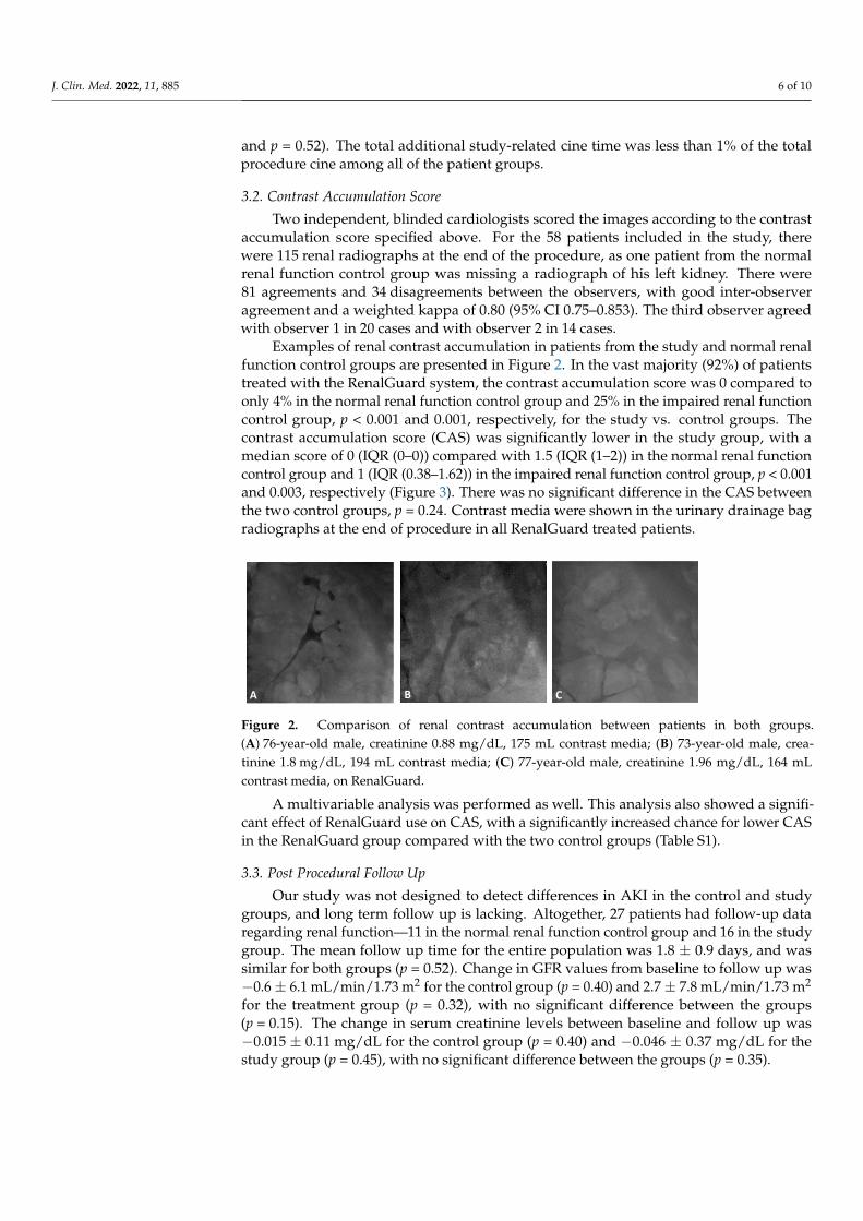
Examples of renal contrast accumulation in patients from the study and normal renalfunction control groups are presented in Figure 2. In the vast majority (92%) of patientstreated with the RenalGuard system, the contrast accumulation score was 0 compared toonly 4% in the normal renal function control group and 25% in the impaired renal functioncontrol group, p < 0.001 and 0.001, respectively, for the study vs. control groups. Thecontrast accumulation score (CAS) was significantly lower in the study group, with amedian score of 0 (IQR (0–0)) compared with 1.5 (IQR (1–2)) in the normal renal functioncontrol group and 1 (IQR (0.38–1.62)) in the impaired renal function control group, p < 0.001and 0.003, respectively (Figure 3). There was no significant difference in the CAS betweenthe two control groups, p = 0.24. Contrast media were shown in the urinary drainage bagradiographs at the end of procedure in all RenalGuard treated patients.
J. Clin. Med. 2021, 10, x FOR PEER REVIEW 6 of 10
The volume of contrast media administrated was lower in the study group without reaching statistical significance (134.8 ± 44.04 mL vs. 152.52 ± 54.18 mL for study vs. con-trol, respectively, p = 0.25, and 152.86 ± 69.49 mL for the impaired renal function control group, p = 0.41) mean procedure time was also similar for the three groups (43.00 ± 26.51 min, 35.94 ± 21.35 min, and 32.75 ± 37.05 min for the study, normal renal function control group, and impaired renal function control group, respectively, p = 0.46 and p = 0.52). The total additional study-related cine time was less than 1% of the total procedure cine among all of the patient groups.
3.2. Contrast Accumulation Score Two independent, blinded cardiologists scored the images according to the contrast
accumulation score specified above. For the 58 patients included in the study, there were 115 renal radiographs at the end of the procedure, as one patient from the normal renal function control group was missing a radiograph of his left kidney. There were 81 agree-ments and 34 disagreements between the observers, with good inter-observer agreement and a weighted kappa of 0.80 (95% CI 0.75–0.853). The third observer agreed with observer 1 in 20 cases and with observer 2 in 14 cases.
Examples of renal contrast accumulation in patients from the study and normal renal function control groups are presented in Figure 2. In the vast majority (92%) of patients treated with the RenalGuard system, the contrast accumulation score was 0 compared to only 4% in the normal renal function control group and 25% in the impaired renal function control group, p < 0.001 and 0.001, respectively, for the study vs. control groups. The con-trast accumulation score (CAS) was significantly lower in the study group, with a median score of 0 (IQR 0–0) compared with 1.5 (IQR 1–2) in the normal renal function control group and 1 (IQR 0.38–1.62) in the impaired renal function control group, p < 0.001 and 0.003, respectively (Figure 3). There was no significant difference in the CAS between the two control groups, p = 0.24. Contrast media were shown in the urinary drainage bag ra-diographs at the end of procedure in all RenalGuard treated patients.
Figure 2. Comparison of renal contrast accumulation between patients in both groups. (A) 76-year-old male, creatinine 0.88 mg/dL, 175 mL contrast media; (B) 73-year-old male, creatinine 1.8 mg/dL, 194 mL contrast media; (C) 77-year-old male, creatinine 1.96 mg/dL, 164 mL contrast media, on Re-nalGuard.
A B C
Figure 2. Comparison of renal contrast accumulation between patients in both groups.(A) 76-year-old male, creatinine 0.88 mg/dL, 175 mL contrast media; (B) 73-year-old male, crea-tinine 1.8 mg/dL, 194 mL contrast media; (C) 77-year-old male, creatinine 1.96 mg/dL, 164 mLcontrast media, on RenalGuard.
A multivariable analysis was performed as well. This analysis also showed a signifi-cant effect of RenalGuard use on CAS, with a significantly increased chance for lower CASin the RenalGuard group compared with the two control groups (Table S1).
3.3. Post Procedural Follow Up
Our study was not designed to detect differences in AKI in the control and studygroups, and long term follow up is lacking. Altogether, 27 patients had follow-up dataregarding renal function—11 in the normal renal function control group and 16 in the studygroup. The mean follow up time for the entire population was 1.8 ± 0.9 days, and wassimilar for both groups (p = 0.52). Change in GFR values from baseline to follow up was−0.6 ± 6.1 mL/min/1.73 m2 for the control group (p = 0.40) and 2.7 ± 7.8 mL/min/1.73 m2
for the treatment group (p = 0.32), with no significant difference between the groups(p = 0.15). The change in serum creatinine levels between baseline and follow up was−0.015 ± 0.11 mg/dL for the control group (p = 0.40) and −0.046 ± 0.37 mg/dL for thestudy group (p = 0.45), with no significant difference between the groups (p = 0.35).

J. Clin. Med. 2022, 11, 885 7 of 10J. Clin. Med. 2021, 10, x FOR PEER REVIEW 7 of 10
Figure 3. Contrast accumulation score (CAS) in study and control groups. CAS was significantly lower in the study group with a median score of 0 compared with 1.5 in the normal renal function control group and 1 in the impaired renal function control group, p < 0.001 and 0.003, respectively.
A multivariable analysis was performed as well. This analysis also showed a signifi-cant effect of RenalGuard use on CAS, with a significantly increased chance for lower CAS in the RenalGuard group compared with the two control groups (Table S1).
3.3. Post Procedural Follow up Our study was not designed to detect differences in AKI in the control and study
groups, and long term follow up is lacking. Altogether, 27 patients had follow-up data regarding renal function—11 in the normal renal function control group and 16 in the study group. The mean follow up time for the entire population was 1.8 ± 0.9 days, and was similar for both groups (p = 0.52). Change in GFR values from baseline to follow up was -0.6 ± 6.1 mL/min/1.73 m2 for the control group (p = 0.40) and 2.7 ± 7.8 mL/min/1.73 m2
for the treatment group (p = 0.32), with no significant difference between the groups (p = 0.15). The change in serum creatinine levels between baseline and follow up was −0.015 ± 0.11 mg/dL for the control group (p = 0.40) and −0.046 ± 0.37 mg/dL for the study group (p = 0.45), with no significant difference between the groups (p = 0.35).
4. Discussion In this study, we showed that peri-procedural controlled renal flushing using forced
diuresis with matched isotonic intravenous hydration, almost completely prevents con-trast media accumulation in the kidneys of patients who underwent coronary angi-ography, angioplasty, or TAVR, and were considered at increased risk to develop CI-AKI. In 92% of the patients treated with controlled renal flushing, no contrast could be detected in the post procedural renal radiography. In contrast, among the control groups of pa-tients with normal and impaired renal function, contrast media accumulation was clearly observed in renal radiography in 96% and 75% of the patients at the end of the procedure, respectively. Peri-procedural controlled renal flushing was significantly correlated with CAS, regardless of renal function, while there was no correlation between the volume of
Figure 3. Contrast accumulation score (CAS) in study and control groups. CAS was significantlylower in the study group with a median score of 0 compared with 1.5 in the normal renal functioncontrol group and 1 in the impaired renal function control group, p < 0.001 and 0.003, respectively.
4. Discussion
In this study, we showed that peri-procedural controlled renal flushing using forceddiuresis with matched isotonic intravenous hydration, almost completely prevents contrastmedia accumulation in the kidneys of patients who underwent coronary angiography,angioplasty, or TAVR, and were considered at increased risk to develop CI-AKI. In 92%of the patients treated with controlled renal flushing, no contrast could be detected in thepost procedural renal radiography. In contrast, among the control groups of patients withnormal and impaired renal function, contrast media accumulation was clearly observed inrenal radiography in 96% and 75% of the patients at the end of the procedure, respectively.Peri-procedural controlled renal flushing was significantly correlated with CAS, regardlessof renal function, while there was no correlation between the volume of contrast mediadelivered and the degree of contrast accumulation, regardless of renal flushing.
Recently, McDonald et al. [8] demonstrated that persistent bilateral global nephro-grams (visible kidneys in non-contrast CT) 24 h following contrast administration weredetectable on non-contrast CT in patients who underwent contrast-enhanced CT or cardiaccatheterization. This finding correlates with a marked increase in the likelihood of AKI(7-fold increase), dialysis (13-fold increase), or mortality (11-fold increase). Significant riskfactors for nephrograms included factors leading to decreased renal perfusion, such ashypotension and shock.
4.1. Pathophysiology of Contrast Induced Acute Kidney Injury
The pathophysiology of CI-AKI is not completely understood, but the current un-derstanding highlights medullary ischemia as the pivotal mechanism [9]. Contrast mediaare cytotoxic and cause direct injury to the epithelial and tubular cells, which causes therelease of vasoactive mediators, causing renal vasoconstriction and medullary ischemia.Furthermore, the high viscosity of contrast media may lead to capillary obstruction and

J. Clin. Med. 2022, 11, 885 8 of 10
decreased perfusion to the kidneys. Once contrast media are filtered and concentrated inthe tubules, they may also obstruct them and cause further tubular damage [9,10].
Data regarding the role of contrast accumulation in the kidney, in the complex patho-physiology of CI-AKI, are lacking. It has been suggested in a rat model that contrast mediaadministration increases the viscosity of the tubular fluid, thus leading to increased tubularpressure and impaired glomerular filtration. This prolongs the intrarenal retention time,and the longer exposure time might cause greater tubular damage, associated with higherrenal toxicity [11].
4.2. Prophylactic Strategies for CI-AKI
Many preventive approaches for CI-AKI have been proposed and tested over theyears. These include massive hydration, the use of low and iso-osmolar contrast media,and the administration of statins, sodium bicarbonate, and N-acetylcysteine [12]. Themost widely used strategy is peri-procedural intravenous hydration with an isotonic salinesolution [12,13]. The results of the AMACING trial, however, have challenged this routineapproach by showing that a lack of prophylaxis was non-inferior for preventing CI-AKIcompared with intravenous hydration, in a group of high-risk patients receiving contrastfor radiological or cardiovascular procedures [14]. Moreover, hydration is often limitedby potential complications of over-hydration, such as pulmonary congestion, especiallyin patients with chronic kidney disease, aortic stenosis, and heart failure. Over-hydrationitself may increase the risk for AKI by increasing renal venous pressure and decreasingrenal perfusion [15]. The RenalGuard system maintains an euvolemic state by inducing ahigh urine flow rate with matched IV hydration. The high urine flow rate may decreasethe incidence of CI-AKI by reducing contrast media accumulation and by causing a rapidtransit of contrast media through the kidneys. These two effects lead to lower overallkidney exposure to the contrast media, and potentially less medullary ischemia and lesscontrast media precipitation in the tubuli. Our study supports this suggested mechanismby showing a lower contrast accumulation score at the end of the procedure in patientstreated with the RenalGuard system.
4.3. Forced Diuresis with Matched Hydration Decreases the Incidence of CI-AKI Regardless of theAmount of Contrast Media Used
The REMEDIAL II trial [4] that included patients at very high risk for developingCI-AKI (eGFR < 30 mL/min/1.73 m2 and/or a Mehran risk score ≥ 11) demonstrated thatRenalGuard therapy was superior to sodium bicarbonate and N-acetylcysteine adminis-tration in preventing CI-AKI. Moreover, the MYTHOS trial [3] demonstrated a significantreduction of CI-AKI rates, from 18% in the control group to 4.6% in the RenalGuard treatedgroup. Similarly, we recently found that in patients considered at high risk to developCI-AKI, undergoing coronary angiography, angioplasty, or TAVR, forced diuresis withmatched controlled hydration resulted in decreased incidence of contrast induced AKI [5].Conversely, in the REDUCE-AKI trial, the use of the RenalGuard during the TAVR pro-cedure did not reduce the incidence of AKI and was associated with increased long-termmortality [16].
Previous studies showed no correlation between the contrast media volume and thedevelopment of CI-AKI [5–7]. In our study, there was no correlation between the amount ofcontrast media delivered and the contrast accumulation score at the end of the procedure.The lack of correlation between the two may indicate that other factors play a role in thedevelopment of CI-AKI, like contrast media concentration and the duration of exposure ofthe kidneys to the contrast media. The results of our study suggest that a shorter transittime of contrast media through the kidneys may have a protective effect and may reducethe risk of CI-AKI. As stated above, a lower median CAS in RenalGuard treated patientsmay indicate a shorter exposure of the kidneys to more dilute contrast media, which inturn may translate to a lower incidence of contrast induced AKI.

J. Clin. Med. 2022, 11, 885 9 of 10
4.4. Limitations
Our study has several limitations. First, we did not have data regarding the amount ofIV fluids administered to the control group before or during the procedure. In addition,we did not systematically collect creatinine and electrolyte levels following the procedure,and therefore we could not provide data regarding the prevalence of CI-AKI amongthe three groups, nor data regarding the safety of forced diuresis, as our study was notdesigned to address these issues. Our second control group (of patients with impaired renalfunction undergoing the procedure without RenalGaurd system) included a small numberof patients. Finally, we did not have data regarding long term sequela and complications ofthe procedure and/or the RenalGuard system.
5. Conclusions
Peri-procedural controlled renal flushing using forced diuresis with matched intra-venous hydration in patients undergoing interventional cardiac procedures involvingintra-arterial contrast media administration is associated with decreased renal contrastmedia accumulation. Previous studies have shown that the use of forced diuresis withmatched intravenous hydration is a safe and effective way to prevent CI-AKI in patientsundergoing coronary angiography, angioplasty, or TAVR. The results of our study suggestthat a shorter exposure time of the kidneys to more dilute contrast media is a possiblemechanism of action.
Supplementary Materials: The following is available online at https://www.mdpi.com/article/10.3390/jcm11030885/s1, Supplementary file: Table S1: Multivariable analysis of mean contrastaccumulation score as a function of various parameters.
Author Contributions: Conceptualization, S.B.; methodology, S.B. and E.C.; formal analysis, A.H.(Aviram Hochstadt); investigation, Y.B.-H., M.K., S.K., Y.A., A.H. (Amir Halkin) and A.F.; datacuration, Y.B.-H.; writing—original draft preparation, Y.B.-H.; writing—review and editing, Y.B.-H.,M.K., M.I., Y.A., S.B. and E.C.; supervision, S.B. and M.K. All authors have read and agreed to thepublished version of the manuscript.
Funding: This research received no external funding.
Institutional Review Board Statement: The study was conducted according to the guidelines of theDeclaration of Helsinki, and was approved by the Institutional Review Board (or Ethics Committee)of Tel Aviv Sourasky Medical Center (protocol code 0544-17-TLV, date of approval 1 January 2018).
Informed Consent Statement: Informed consent was obtained from all subjects involved in the study.
Data Availability Statement: The data presented in this study are available upon request from thecorresponding author. The data are not publicly available due to privacy restrictions.
Conflicts of Interest: The authors declare no conflict of interest.
References1. Rihal, C.S.; Textor, S.C.; Grill, D.E.; Berger, P.B.; Ting, H.H.; Best, P.J.; Singh, M.; Bell, M.R.; Barsness, G.W.; Mathew, V.; et al.
Incidence and prognostic importance of acute renal failure after percutaneous coronary intervention. Circulation 2002, 105,2259–2264. [CrossRef] [PubMed]
2. Tsai, T.T.; Patel, U.D.; Chang, T.I.; Kennedy, K.F.; Masoudi, F.A.; Matheny, M.E.; Kosiborod, M.; Amin, A.P.; Messenger,J.C.; Rumsfeld, J.S.; et al. Contemporary incidence, predictors, and outcomes of acute kidney injury in patients undergoingpercutaneous coronary interventions: Insights from the NCDR Cath-PCI registry. JACC Cardiovasc. Interv. 2014, 7, 1–9. [CrossRef]
3. Marenzi, G.; Ferrari, C.; Marana, I.; Assanelli, E.; De Metrio, M.; Teruzzi, G.; Veglia, F.; Fabbiocchi, F.; Montorsi, P.; Bartorelli, A.L.Prevention of contrast nephropathy by furosemide with matched hydration: The MYTHOS (Induced Diuresis With MatchedHydration Compared to Standard Hydration for Contrast Induced Nephropathy Prevention) trial. JACC Cardiovasc. Interv. 2012,5, 90–97. [CrossRef] [PubMed]
4. Briguori, C.; Visconti, G.; Focaccio, A.; Airoldi, F.; Valgimigli, M.; Sangiorgi, G.M.; Golia, B.; Ricciardelli, B.; Condorelli, G.;Investigators, R.I. Renal Insufficiency After Contrast Media Administration Trial II (REMEDIAL II): RenalGuard System inhigh-risk patients for contrast-induced acute kidney injury. Circulation 2011, 124, 1260–1269. [CrossRef] [PubMed]

J. Clin. Med. 2022, 11, 885 10 of 10
5. Chorin, E.; Ben-Assa, E.; Konigstein, M.; Rofe, M.T.; Hochstadt, A.; Galli, N.; Schnapper, M.; Arbel, Y.; Rabey, I.; Shoshan, J.B.;et al. Prevention of post procedural acute kidney injury in the catheterization laboratory in a real-world population. Int. J. Cardiol.2017, 226, 42–47. [CrossRef] [PubMed]
6. Konigstein, M.; Ben-Assa, E.; Abramowitz, Y.; Steinvil, A.; Leshem Rubinow, E.; Havakuk, O.; Arbel, Y.; Halkin, A.; Keren,G.; Banai, S.; et al. Usefulness of updated valve academic research consortium-2 criteria for acute kidney injury followingtranscatheter aortic valve implantation. Am. J. Cardiol. 2013, 112, 1807–1811. [CrossRef] [PubMed]
7. Wald, D.S.; Morris, J.K.; Wald, N.J.; Chase, A.J.; Edwards, R.J.; Hughes, L.O.; Berry, C.; Oldroyd, K.G.; Investigators, P. Randomizedtrial of preventive angioplasty in myocardial infarction. N. Engl. J. Med. 2013, 369, 1115–1123. [CrossRef] [PubMed]
8. McDonald, J.S.; Steckler, E.M.; McDonald, R.J.; Katzberg, R.W.; Williamson, E.E.; Cernigliaro, J.G.; Hamadah, A.M.; Gharaibeh, K.;Kallmes, D.F.; Leung, N. Bilateral Sustained Nephrograms After Parenteral Administration of Iodinated Contrast Material: APotential Biomarker for Acute Kidney Injury, Dialysis, and Mortality. Mayo Clin. Proc. 2018, 93, 867–876. [CrossRef] [PubMed]
9. Rear, R.; Bell, R.M.; Hausenloy, D.J. Contrast-induced nephropathy following angiography and cardiac interventions. Heart 2016,102, 638–648. [CrossRef] [PubMed]
10. Seeliger, E.; Sendeski, M.; Rihal, C.S.; Persson, P.B. Contrast-induced kidney injury: Mechanisms, risk factors, and prevention.Eur. Heart J. 2012, 33, 2007–2015. [CrossRef] [PubMed]
11. Jost, G.; Pietsch, H.; Sommer, J.; Sandner, P.; Lengsfeld, P.; Seidensticker, P.; Lehr, S.; Hütter, J.; Sieber, M.A. Retention of iodineand expression of biomarkers for renal damage in the kidney after application of iodinated contrast media in rats. Investig. Radiol.2009, 44, 114–123. [CrossRef] [PubMed]
12. McCullough, P.A.; Choi, J.P.; Feghali, G.A.; Schussler, J.M.; Stoler, R.M.; Vallabahn, R.C.; Mehta, A. Contrast-Induced AcuteKidney Injury. J. Am. Coll. Cardiol. 2016, 68, 1465–1473. [CrossRef] [PubMed]
13. Trivedi, H.S.; Moore, H.; Nasr, S.; Aggarwal, K.; Agrawal, A.; Goel, P.; Hewett, J. A randomized prospective trial to assess the roleof saline hydration on the development of contrast nephrotoxicity. Nephron Clin. Pract. 2003, 93, C29–C34. [CrossRef] [PubMed]
14. Nijssen, E.C.; Rennenberg, R.J.; Nelemans, P.J.; Essers, B.A.; Janssen, M.M.; Vermeeren, M.A.; Ommen, V.V.; Wildberger, J.E.Prophylactic hydration to protect renal function from intravascular iodinated contrast material in patients at high risk of contrast-induced nephropathy (AMACING): A prospective, randomised, phase 3, controlled, open-label, non-inferiority trial. Lancet 2017,389, 1312–1322. [CrossRef]
15. Gupta, R.; Moza, A.; Cooper, C.J. Intravenous Hydration and Contrast-Induced Acute Kidney Injury: Too Much of a Good Thing?J. Am. Heart Assoc. 2016, 5, e003777. [CrossRef] [PubMed]
16. Arbel, Y.; Ben-Assa, E.; Puzhevsky, D.; Litmanowicz, B.; Galli, N.; Chorin, E.; Halkin, A.; Sadeh, B.; Konigstein, M.; Bassat, O.K.;et al. Forced diuresis with matched hydration during transcatheter aortic valve implantation for Reducing Acute Kidney Injury:A randomized, sham-controlled study (REDUCE-AKI). Eur. Heart J. 2019, 40, 3169–3178. [CrossRef] [PubMed]
Copyright © 2022 FDOKUMEN