Teaching Difficult Learners Science: Hands-On Experiments ...
Upload
khangminh22Category
view
0download
0
https://doi.org/10.1177/11297298211059648
The Journal of Vascular Access 1 –7© The Author(s) 2021Article reuse guidelines: sagepub.com/journals-permissionsDOI: 10.1177/11297298211059648journals.sagepub.com/home/jva
JVA The Journal of Vascular Access
Introduction
Peripheral intravenous catheters (PIVCs) are the most commonly used invasive medical device, with up to 70% of patients requiring a peripheral venous line during their hospital stay.1 They are particularly important in the emer-gency setting, where timely insertion may be crucial for the management of critically ill patients.2
The insertion of PIVCs can be time intensive, with multiple venipuncture attempts sometimes required for successful insertion.3 This can cause pain and anxiety for the patient, and increases the risk of healthcare worker exposure to needlestick injuries, blood splashes, and bloodborne pathogens.4,5 Furthermore, failed attempts may lead to catheters being inserted in less than optimal
locations (such as the feet, thumbs, and wrists) increasing the risk of complications.6 Delayed, partial, or total loss of the prescribed dose of medication may extend the length of hospital stay.6 Ultimately, detrimental clinical outcomes can result in increased costs and inefficient use of resources.7
Defining difficult intravenous access (DIVA): A systematic review
Amit Bahl1 , Steven Johnson1, Kimberly Alsbrooks2 , Alicia Mares2, Smeet Gala2 and Klaus Hoerauf2,3
AbstractBackground: The term “difficult intravenous access” (DIVA) is commonly used but not clearly defined. Repeated attempts at peripheral intravenous catheter (PIVC) insertion can be a traumatic experience for patients, leading to sub-optimal clinical and economic outcomes. We conducted a systematic literature review (SLR) to collate literature definitions of DIVA, with the aim of arriving at an evidence-driven definition.Methods: The SLR was designed to identify clinical, cost, and quality of life publications in patients requiring the insertion of a PIVC in any setting, including studies on US-guidance and/or guidewire, and studies with no specific intervention. The search was restricted to English language studies published between 1st January 2010 and 30th July 2020, and the Ovid platform was used to search several electronic databases, in addition to hand searching of clinical trial registries.Results: About 121 studies were included in the SLR, of which 64 reported on the objectives relevant to this manuscript. Prevalence estimates varied widely from 6% to 87.7% across 19 publications, reflecting differences in definitions used. Of 43 publications which provided a definition of DIVA, six key themes emerged. Of these, themes 1–3 (failed attempts at PIV access using traditional technique; based on physical examination findings for example no visible or palpable veins; and personal history of DIVA) were covered by all but one publication. Following a failed insertion attempt, the most common number of subsequent attempts was 3, and it was frequently reported that a more experienced clinician would attempt to gain access after multiple failed attempts.Conclusions: Considering the themes identified, an evidence-driven definition of DIVA is proposed: “when a clinician has two or more failed attempts at PIV access using traditional techniques, physical examination findings are suggestive of DIVA (e.g. no visible or palpable veins) or the patient has a stated or documented history of DIVA.”
KeywordsDIVA, DVA, difficult venous access, venous access, hard stick
Date received: 30 July 2021; accepted: 19 October 2021
1Emergency Medicine, Beaumont Hospital, Royal Oak, MI, USA2Becton Dickinson and Co, Franklin Lakes, NJ, USA3 Department of Anesthesiology and Intensive Care, Medical University of Vienna, Vienna, Austria
Corresponding author:Kimberly Alsbrooks, Becton Dickinson and Co., Franklin Lakes, NJ. USA.Email: [email protected]
1059648 JVA0010.1177/11297298211059648The Journal of Vascular AccessBahl et al.research-article2021
Original research article
2 The Journal of Vascular Access 00(0)
The term “difficult intravenous access” (DIVA) is used in the literature to describe this patient population, but there is inconsistency in how DIVA is defined, and to our knowledge no systematic search has been undertaken to evaluate these definitions.
The resulting uncertainty around identification and management of DIVA patients in clinical practice may lead to sub-optimal clinical outcomes and unnecessary patient burden and resource use. Therefore, a systematic review was conducted to collate literature definitions of DIVA with the aim of arriving at an evidence-driven definition.
Methods
A systematic literature review (SLR) was designed to iden-tify clinical, cost, and quality of life publications in patients requiring the insertion of a PIVC in any setting, including studies on US-guidance and/or guidewire, and studies with no specific intervention. The search was restricted to English language studies published between 1st January 2010 and 30th July 2020. Full details of study eligibility criteria are provided in Supplemental Table S1. The SLR had a broader scope than the objectives described in the introduction; this paper focuses on the publications rele-vant to these objectives. The methodology and results of the study are reported in accordance with PRISMA guide-lines (Supplemental Table S7).8 The protocol was not regis-tered with any protocol registry.
The Ovid platform was used to search the following elec-tronic databases on 30th July 2020: Embase; MEDLINE Daily, In-Process & Other Non-indexed citations, and e-pub ahead-of-print; Cochrane library—Cochrane Central Register of Controlled Trials (CENTRAL); Cochrane library—Cochrane Database of Systematic Reviews (DSR); Cochrane library—Database of Abstracts of Reviews of Effects (DARE); Cochrane library—Health Technology Assessment (HTA).
Hand searching was also performed as a supplementary measure. To obtain details of potentially relevant published and ongoing trials, the following clinical trial registry data-bases were accessed: clinicaltrials.gov (https://clinicaltrials.gov/); International Clinical Trials Registry platform (http://apps.who.int/trialsearch/); ISRCTN Register (https://www.isrctn.com/); UK Clinical Trials Gateway (https://www.ukctg.nihr.ac.uk/); EU Clinical Trials Register (https://www.clinicaltrialsregister.eu/ctr-search/search). Bibliographic ref-erence lists of included publications and of relevant SLRs were also screened.
The database search strings identified all relevant publi-cations indexed in Embase and were modified for perform-ing searches in Medline and the Cochrane Library to account for differences in syntax and thesaurus headings. The Embase, Medline, and Cochrane Library search strings can be found in Supplemental Tables S2–S4.
Two independent reviewers screened citations by title/abstract, with any conflicts resolved by a third, more sen-ior investigator. Full-text articles were then evaluated by a single reviewer, and final inclusion and exclusion of cita-tions were verified by a second reviewer. Disputes regard-ing eligibility were referred to a third, more senior investigator (KH and AB). A record was kept of papers excluded at this stage together with a clear justification for their exclusion. Data from the included publications were extracted by one reviewer into a data extraction sheet; this information was checked and validated by conducting an independent internal data check once all required data had been entered.
Results
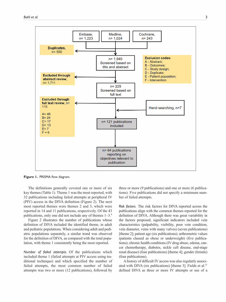
The electronic database search identified 2490 citations. On removal of 1711 citations at title and abstract screening stage, and 115 publications at full text screening stage, 114 publications from the electronic database searches were relevant for inclusion in the SLR. Hand-searching the ref-erence list of relevant SLRs identified a further 7 publica-tions, resulting in a total of 121 publications being included in the broader SLR. Those of relevance to the objectives described in the introduction are discussed in this manu-script (Figure 1).
Prevalence of DIVA
All prevalence data identified in the SLR were from the hospital setting. However, prevalence estimates varied depending on the definition used; when studies defined DIVA by a prior history of difficult access (which included multiple failed attempts to insert the IV line, no visible or palpable veins, or escalation to advanced techniques) prev-alence ranged from 45%9,10 to 59.3%.6 However, in studies assessing the prevalence of DIVA based on multiple failed attempts within the study (regardless of prior history), the prevalence ranged from 6% to 11.8%.11–14
Definition of DIVA
Forty-three publications provided a definition of DIVA in adult, pediatric, or mixed populations. Of the 43 publica-tions reporting a DIVA definition9–50, 35 publications reported a definition for adult patients only9–12,15,17–30,33–
38,40–43,45–49 5 publications reported a definition for pediat-ric patients only,13,14,31,44,50 and in 3 publications the age of the included patients was not clear.16,32,39 In terms of geo-graphic origin, 27 studies were from the US, 11 were European, 2 were from the Middle East, 2 were from East Asia, and 1 was from the Caribbean. Further study charac-teristics are reported in Supplemental Table S6.
Bahl et al. 3
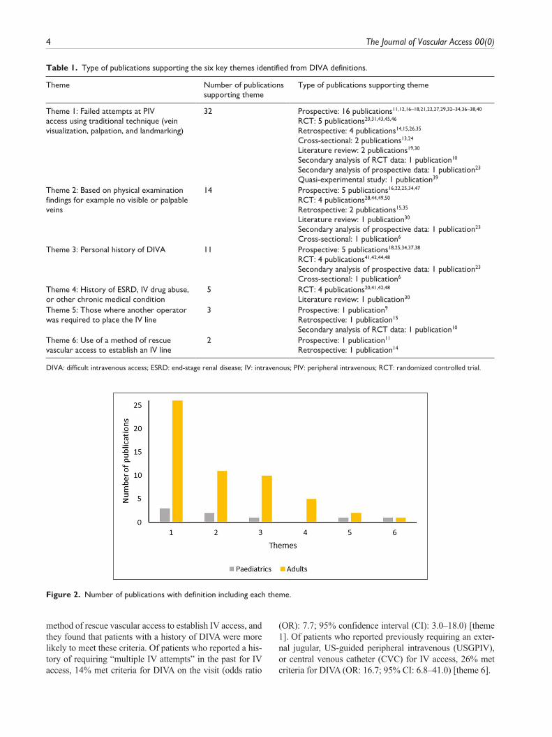
The definitions generally covered one or more of six key themes (Table 1). Theme 1 was the most reported, with 32 publications including failed attempts at peripheral IV (PIV) access in the DIVA definition (Figure 2). The next most reported themes were themes 2 and 3, which were reported in 14 and 11 publications, respectively. Of the 43 publications, only one did not include any of themes 1–3.9
Figure 2 illustrates the number of publications whose definition of DIVA included the identified theme, in adult and pediatric populations. When considering adult and pedi-atric populations separately, a similar trend was observed for the definition of DIVA, as compared with the total popu-lation, with theme 1 consistently being the most reported.
Number of failed attempts. Of the publications which included theme 1 (failed attempts at PIV access using tra-ditional technique) and which specified the number of failed attempts, the most common number of failed attempts was two or more (12 publications), followed by
three or more (9 publications) and one or more (6 publica-tions). Five publications did not specify a minimum num-ber of failed attempts.
Risk factors. The risk factors for DIVA reported across the publications align with the common themes reported for the definition of DIVA. Although there was great variability in the factors proposed, significant indicators included vein characteristics (palpability, visibility, poor vein condition, vein diameter, veins with many valves) (seven publications) [theme 2]; patient age (six publications); arthrometric values (patients classed as obese or underweight) (five publica-tions); chronic health conditions (IV drug abuse, edema, can-cer chemotherapy, diabetes, sickle cell disease, end-stage renal disease) (four publications) [theme 4]; gender (female) (four publications).
A history of difficult IV access was also regularly associ-ated with DIVA (six publications) [theme 3]. Fields et al.11 defined DIVA as three or more IV attempts or use of a
Figure 1. PRISMA flow diagram.
4 The Journal of Vascular Access 00(0)
Figure 2. Number of publications with definition including each theme.
Table 1. Type of publications supporting the six key themes identified from DIVA definitions.
Theme Number of publications supporting theme
Type of publications supporting theme
Theme 1: Failed attempts at PIV access using traditional technique (vein visualization, palpation, and landmarking)
32 Prospective: 16 publications11,12,16–18,21,22,27,29,32–34,36–38,40
RCT: 5 publications20,31,43,45,46
Retrospective: 4 publications14,15,26,35
Cross-sectional: 2 publications13,24
Literature review: 2 publications19,30
Secondary analysis of RCT data: 1 publication10
Secondary analysis of prospective data: 1 publication23
Quasi-experimental study: 1 publication39
Theme 2: Based on physical examination findings for example no visible or palpable veins
14 Prospective: 5 publications16,22,25,34,47
RCT: 4 publications28,44,49,50
Retrospective: 2 publications15,35
Literature review: 1 publication30
Secondary analysis of prospective data: 1 publication23
Cross-sectional: 1 publication6
Theme 3: Personal history of DIVA 11 Prospective: 5 publications18,25,34,37,38
RCT: 4 publications41,42,44,48
Secondary analysis of prospective data: 1 publication23
Cross-sectional: 1 publication6
Theme 4: History of ESRD, IV drug abuse, or other chronic medical condition
5 RCT: 4 publications20,41,42,48
Literature review: 1 publication30
Theme 5: Those where another operator was required to place the IV line
3 Prospective: 1 publication9
Retrospective: 1 publication15
Secondary analysis of RCT data: 1 publication10
Theme 6: Use of a method of rescue vascular access to establish an IV line
2 Prospective: 1 publication11
Retrospective: 1 publication14
DIVA: difficult intravenous access; ESRD: end-stage renal disease; IV: intravenous; PIV: peripheral intravenous; RCT: randomized controlled trial.
method of rescue vascular access to establish IV access, and they found that patients with a history of DIVA were more likely to meet these criteria. Of patients who reported a his-tory of requiring “multiple IV attempts” in the past for IV access, 14% met criteria for DIVA on the visit (odds ratio
(OR): 7.7; 95% confidence interval (CI): 3.0–18.0) [theme 1]. Of patients who reported previously requiring an exter-nal jugular, US-guided peripheral intravenous (USGPIV), or central venous catheter (CVC) for IV access, 26% met criteria for DIVA (OR: 16.7; 95% CI: 6.8–41.0) [theme 6].
Bahl et al. 5
Discussion
The key objective of this study was to review current defi-nitions of DIVA with the aim of arriving at an evidence-driven definition. There was a high level of variation in the prevalence of DIVA reported, in line with the definitions presented. In addition, the prevalence data captured in this SLR are all from the hospital setting, limiting generaliza-bility to other settings. However, the volume of DIVA prevalence estimates and definitions in the literature shows there is clearly a subgroup of patients in the healthcare sys-tem who are difficult to cannulate. The lack of a consistent definition across studies hinders estimation of the true prevalence; for example, studies which defined DIVA by a history of difficult access tended to provide higher preva-lence estimates than those with a definition that excluded patient history.
Of the 43 publications which provided a definition for DIVA in adult, pediatric, or mixed age populations, there was near-universal coverage of themes 1–3: (1) failed attempts at PIV access using traditional techniques; (2) based on physical examination findings for example no visible or palpable veins; (3) personal history of DIVA (Table 1). These themes were further supported by a review of the risk factors for DIVA, with significant indicators including chronic health conditions, vein characteristics, and a history of DIVA.
Considering themes 1–3 and the finding that most com-mon number of failed attempts was two or more, an evi-dence-driven definition of DIVA is proposed:
“A patient is considered to have DIVA if any of the following elements are present: a clinician has two or more failed attempts at PIV access using traditional techniques, physical examination findings are suggestive of DIVA (e.g. no visible or palpable veins) or the patient has a stated or documented history of DIVA”
The inclusion of patient history in the definition highlights the importance of documenting the assessment and listen-ing to patients when they say they have experience of dif-ficult access. In one study, 7 out of 10 patients described deficits in communication with the clinician placing the PIVC.1 Such a history can be used to guide the decision on whether to attempt traditional methods or to escalate immediately. However, it is important that patient history is considered alongside a clinical assessment of the vascu-lature; patients who were difficult to cannulate previously may no longer be. Consideration of both factors could avoid the unnecessary pain and discomfort associated with escalation pathways that rely on a specific number of tra-ditional attempts before escalation (even when clinicians know their attempts will be unsuccessful).
The evidence-driven definition also includes “two or more failed attempts.” Ideally, multiple failed attempts
would not form part of a formal DIVA definition or escala-tion pathway; this would be replaced by standardized clini-cal assessment of venous anatomy and patient history to ensure that DIVA patients are identified before the first cannulation attempt. This supports a patient-centric approach; by basing assessment on a known history of DIVA and clinical assessment of the vasculature and co-morbidities, unnecessary patient pain can be avoided, pro-moting escalation prior to attempting potentially unsuccessful IV access. Additional benefits may include minimizing healthcare worker exposure risk and costs and resource use associated with multiple failed attempts. Guidance from the Infusion Nurses Society (INS) also rec-ognizes the harms caused by multiple unsuccessful attempts; the INS recommends that PIVC insertion is restricted to no more than two attempts per clinician.51 Following two unsuccessful attempts, they recommend escalation to a clinician with a higher skill level, or use of alternative routes of administration.
Our definition broadly aligns with the definition used in the 2021 Infusion Therapy Standards of Practice, which also includes the themes of multiple attempts, physical assessment, and patient history.51
Conclusions
Failure to identify DIVA patients, and the use of informal or inappropriate escalation processes, may result in patients being given unnecessary and costly follow-up procedures when alternatives are available. Moreover, the lack of a clearly defined patient population hinders planning of resource allocation and training programs. The goal of future research endeavors should be to test the hypothesis that the proposed evidence-based definition can improve early identification of DIVA; it is hoped that this will pre-vent complications and additional patient discomfort.
There is also a need for future research to understand how evolving and novel medical technologies can be incor-porated into standard assessment algorithms for DIVA patients to improve clinical and economic outcomes. More research is also needed to better define the impact of escala-tion pathways; it is important that such studies consider the level of competence of the individuals inserting the PIVCs using US-guidance, as this has the potential to impact on outcomes. A suggestion would be the need to flag DIVA patients in every patient’s EMR to reduce needle-phobia especially in children requiring multiple hospitalizations. Given this, future research could also investigate the level of training (at an individual or vascular access team level) required for optimal outcomes.
Author’s note
All authors are members of INS, AVA. Externally collected con-tributor form attached.
6 The Journal of Vascular Access 00(0)
Acknowledgements
The authors recognize the valuable contributions of Neil Webb, Jenna Stephens, and Chris Hellmund, who are employees of Source Health Economics, the HEOR company that conducted the systematic literature review and provided writing services.
Author contributions
KA, AM, and SG designed the trial, conceived the study and obtained research funding. AB, SJ, KH, and SG supervised the conduct of the trial and reviewed data collection. KA and KH managed the data, provided statistical advice, including quality control. All authors reviewed and revised the manuscript.
Declaration of conflicting interests
The author(s) declared the following potential conflicts of inter-est with respect to the research, authorship, and/or publication of this article: Bahl: Research Grants from Access Vascular, Adhezion Biomedical, Becton Dickinson, B. Braun, Medline, Teleflex Consultancy: B. Braun, Teleflex, The Clinician Exchange, Lineus Medical, Interrad Medical. Johnson: no con-flicts of interest. Hoerauf, Alsbrooks, Gala, and Mares: employee of BD, owns stock/stock options.
Funding
The author(s) disclosed receipt of the following financial support for the research, authorship, and/or publication of this article: BD supported the funding for this project. No funding for the authors was provided.
Guarantor
Kimberly Alsbrooks, Becton Dickinson and Co., Franklin Lakes, NJ, USA. [email protected].
ORCID iDs
Amit Bahl https://orcid.org/0000-0001-9495-1293
Kimberly Alsbrooks https://orcid.org/0000-0002-8684-7100
Supplemental material
Supplemental material for this article is available online.
References
1. Plohal A. A qualitative study of adult hospitalized patients with difficult venous access experiencing short peripheral catheter insertion in a hospital setting. J Infus Nurs 2021; 44: 26–33.
2. Bridey C, Thilly N, Lefevre T, et al. Ultrasound-guided versus landmark approach for peripheral intravenous access by critical care nurses: a randomised controlled study. BMJ Open 2018; 8: e020220.
3. Goff DA, Larsen P, Brinkley J, et al. Resource utilization and cost of inserting peripheral intravenous catheters in hos-pitalized children. Hosp Pediatrics 2013; 3: 185–191.
4. Walsh G. Difficult peripheral venous access: recognizing and managing the patient at risk. J Assoc Vasc Access 2008; 13: 198–203.
5. Platt V and Osenkarski S. Improving vascular access outcomes and enhancing practice. J Infus Nurs 2018; 41: 375–382.
6. Armenteros-Yeguas V, Gárate-Echenique L, Tomás-López MA, et al. Prevalence of difficult venous access and associ-ated risk factors in highly complex hospitalised patients. J Clin Nurs 2017; 26: 4267–4275.
7. Hadaway L, Dalton L and Mercanti-Erieg L. Infusion teams in acute care hospitals: call for a business approach: an infusion nurses society white paper. J Infus Nurs 2013; 36: 356–360.
8. Moher D, Liberati A, Tetzlaff J, et al. Preferred report-ing items for systematic reviews and meta-analyses: the PRISMA statement. PLoS Med 2009; 6: e1000097.
9. Witting MD. IV access difficulty: incidence and delays in an urban emergency department. J Emerg Med 2012; 42: 483–487.
10. Witting MD, Moayedi S, Hirshon JM, et al. Predicting fail-ure of intravenous access in adults: the value of prior dif-ficulty. J Emerg Med 2019; 57: 1–5.
11. Fields JM, Piela NE, Au AK, et al. Risk factors associated with difficult venous access in adult ED patients. Am J Emerg Med 2014; 32: 1179–1182.
12. Feinsmith S, Huebinger R, Pitts M, et al. Outcomes of a simplified ultrasound-guided intravenous training course for emergency nurses. J Emerg Nurs 2018; 44: 169–175.e2.
13. Lee SU, Jung JY, Ham EM, et al. Factors associated with difficult intravenous access in the pediatric emergency department. J Vasc Access 2020; 21: 180–185.
14. Petroski A, Frisch A, Joseph N, et al. Predictors of diffi-cult pediatric intravenous access in a community emergency department. J Vasc Access 2015; 16: 521–526.
15. Acuña J, Sorenson J, Gades A, et al. Handheld ultrasound: overcoming the challenge of difficult peripheral intravenous access in the emergency department. J Ultrasound Med 2020; 39: 1985–1991.
16. Au AK, Rotte MJ, Grzybowski RJ, et al. Decrease in central venous catheter placement due to use of ultrasound guid-ance for peripheral intravenous catheters. Am J Emerg Med 2012; 30: 1950–1954.
17. Butterfield M, Abdelghani R, Mohamad M, et al. Using ultrasound-guided peripheral catheterization of the internal jugular vein in patients with difficult peripheral access. Am J Ther 2017; 24: e667–e669.
18. Chiricolo G, Balk A, Raio C, et al. Higher success rates and satisfaction in difficult venous access patients with a guide wire-associated peripheral venous catheter. Am J Emerg Med 2015; 33: 1742–1744.
19. Ehrhardt BS, Givens KEA and Lee RC. Making it stick: developing and testing the difficult intravenous access (DIVA) tool. Am J Nurs 2018; 118: 56–62.
20. EL-Shafey EM and Tammam TF. Ultrasonography-guided peripheral intravenous access: regular technique versus Seldinger technique in patients with difficult vascular access. Eur J Gen Med 2012; 9: 216–222.
21. Etezazian S, Tavakoli B, Hekmatnia A, et al. Evaluation of success rate of ultrasound-guided venous cannulation in patients with difficult venous access. Iran J Radiol 2010; 7: 61–65.
Bahl et al. 7
22. Fabiani A, Dreas L and Sanson G. Ultrasound-guided deep-arm veins insertion of long peripheral catheters in patients with difficult venous access after cardiac surgery. Heart Lung 2017; 46: 46–53.
23. Fields JM, Dean AJ, Todman RW, et al. The effect of vessel depth, diameter, and location on ultrasound-guided periph-eral intravenous catheter longevity. Am J Emerg Med 2012; 30: 1134–1140.
24. Fields JM, Piela NE and Ku BS. Association between multi-ple IV attempts and perceived pain levels in the emergency department. J Vasc Access 2014; 15: 514–518.
25. Gilardi E, Giannuzzi R, WoldeSellasie K, et al. Mini-midline in difficult intravenous access patients in emer-gency department: a prospective analysis. J Vasc Access 2020; 21: 449–455.
26. Gregg SC, Murthi SB, Sisley AC, et al. Ultrasound-guided peripheral intravenous access in the intensive care unit. J Crit Care 2010; 25: 514–519.
27. Mahler SA, Wang H, Lester C, et al. Ultrasound-guided peripheral intravenous access in the emergency department using a modified Seldinger technique. J Emerg Med 2010; 39: 325–329.
28. McCarthy ML, Shokoohi H, Boniface KS, et al. Ultrasonography versus landmark for peripheral intrave-nous cannulation: a randomized controlled trial. Ann Emerg Med 2016; 68: 10–18.
29. Meyer P, Cronier P, Rousseau H, et al. Difficult peripheral venous access: clinical evaluation of a catheter inserted with the Seldinger method under ultrasound guidance. J Crit Care 2014; 29: 823–827.
30. Morata L, Ogilvie C, Yon J, et al. Decreasing peripherally inserted central catheter use with ultrasound-guided periph-eral intravenous lines: a quality improvement project in the acute care setting. J Nurs Adm 2017; 47: 338–344.
31. Otani T, Morikawa Y, Hayakawa I, et al. Ultrasound-guided peripheral intravenous access placement for children in the emergency department. Eur J Pediatr 2018; 177: 1443–1449.
32. Partovi-Deilami K, Nielsen JK, Moller AM, et al. Effect of ultrasound-guided placement of difficult-to-place peripheral venous catheters: a prospective study of a training program for nurse anesthetists. AANA J 2016; 84: 86–92.
33. Piredda M, Biagioli V, Barrella B, et al. Factors affecting difficult peripheral intravenous cannulation in adults: a pro-spective observational study. J Clin Nurs 2017; 26: 1074–1084.
34. Raio C, Elspermann R, Kittisarapong N, et al. A prospective feasibility trial of a novel intravascular catheter system with retractable coiled tip guidewire placed in difficult intravas-cular access (DIVA) patients in the emergency department. Intern Emerg Med 2018; 13: 757–764.
35. Scoppettuolo G, Pittiruti M, Pitoni S, et al. Ultrasound-guided “short” midline catheters for difficult venous access in the emergency department: a retrospective analysis. Int J Emerg Med 2016; 9: 3.
36. Sebbane M, Claret PG, Lefebvre S, et al. Predicting periph-eral venous access difficulty in the emergency department
using body mass index and a clinical evaluation of venous accessibility. J Emerg Med 2013; 44: 299–305.
37. Stolz LA, Cappa AR, Minckler MR, et al. Prospective evalu-ation of the learning curve for ultrasound-guided peripheral intravenous catheter placement. J Vasc Access 2016; 17: 366–370.
38. Warrington WG, Penoyer DA, Kamps TA, et al. Outcomes of using a modified Seldinger technique for long term intra-venous therapy in hospitalized patients with difficult venous access. J Assoc Vasc Access 2012; 17: 24–30.
39. Whalen M, Maliszewski B, Sheinfeld R, et al. Outcomes of an innovative evidence-based practice project: building a difficult-access team in the Emergency Department. J Emerg Nurs 2018; 44: 478–482.
40. Zitek T, Busby E, Hudson H, et al. Ultrasound-guided place-ment of peripheral intravenous lines in the internal jugular vein. Acad Emerg Med 2018; 25: S217.
41. Bahl A, Pandurangadu AV, Tucker J, et al. A randomized controlled trial assessing the use of ultrasound for nurse-performed IV placement in difficult access ED patients. Am J Emerg Med 2016; 34: 1950–1954.
42. Bahl A, Hijazi M, Chen NW, et al. Ultralong versus stand-ard long peripheral intravenous catheters: a randomized controlled trial of ultrasonographically guided catheter sur-vival. Ann Emerg Med 2020; 76: 134–142.
43. Costantino TG, Kirtz JF and Satz WA. Ultrasound-guided peripheral venous access vs. the external jugular vein as the initial approach to the patient with difficult vascular access. J Emerg Med 2010; 39: 462–467.
44. de Graaff JC, Cuper NJ, van Dijk AT, et al. Evaluating NIR vascular imaging to support intravenous cannulation in awake children difficult to cannulate: a randomized clinical trial. Pediatr Anesth 2014; 24(11): 1174–1179.
45. Elia F, Ferrari G, Molino P, et al. Standard-length catheters vs long catheters in ultrasound-guided peripheral vein can-nulation. Am J Emerg Med 2012; 30: 712–716.
46. Mahler SA, Wang H, Lester C, et al. Short- vs long-axis approach to ultrasound-guided peripheral intravenous access: a prospective randomized study. Am J Emerg Med 2011; 29: 1194–1197.
47. Shokoohi H, Boniface KS, Kulie P, et al. The utility and survi-vorship of peripheral intravenous catheters inserted in the emer-gency department. Ann Emerg Med 2019; 74(3): 381–390.
48. Bahl A, Hang B, Brackney A, et al. Standard long IV cath-eters versus extended dwell catheters: a randomized com-parison of ultrasound-guided catheter survival. Am J Emerg Med 2019; 37: 715–721.
49. Kerforne T, Petitpas F, Frasca D, et al. Ultrasound-guided peripheral venous access in severely ill patients with sus-pected difficult vascular puncture. Chest 2012; 141: 279–280.
50. van der Woude OCP, Cuper NJ, Getrouw C, et al. The effectiveness of a near-infrared vascular imaging device to support intravenous cannulation in children with dark skin color. Anesth Analg 2013; 116: 1266–1271.
51. Gorski LA, Hadaway L, Hagle ME, et al. Infusion therapy standards of practice, 8th edition. J Infus Nurs 2021; 44: S1–S224.
Copyright © 2022 FDOKUMEN