Treatment of chronic hepatitis C in patients with HIV/HCV coinfection
Upload
independentCategory
view
1download
0
Protective KIR-HLA interactions for HCV infection in intravenousdrug users
Joaquin Zúñiga1,2,+, Viviana Romero2,*,+, José Azocar3,*,+, Daniel Terreros4, María InésVargas-Rojas1, Diana Torres-García1, Luis Jimenez-Alvarez1, Gilberto Vargas-Alarcón5,Julio Granados-Montiel2, Zaheed Husain6, Raymond T. Chung7, Chester A. Alper6, andEdmond J. Yunis2,8,*1Laboratory of Immunobiology and Genetics, Instituto Nacional de Enfermedades RespiratoriasIsmael Cosio Villegas, Tlalpan 4502, 14080, Mexico City, Mexico2Department of Cancer Immunology and AIDS, Dana-Farber Cancer Institute, 44 Binney Street,Boston, MA 021153Northgate Medical Center, 1985 Main Street, Springfield, MA 01103, USA4Texas Tech Health Sciences Center, El Paso, TX 799055Department of Molecular Biology, Instituto Nacional de Cardiología Ignacio Chávez, Juan Badiano1, Tlalpan 14050, Mexico City, Mexico6The Immune Disease Institute, 800 Huntington Avenue and Department of Pediatrics, HarvardMedical School, 77 Avenue Louis Pasteur, Boston, MA 021157Gastrointestinal Unit, GRJ 724, Massachusetts General Hospital, Harvard Medical School, 55 FruitSt, Boston, MA 02114, USA8Department of Pathology, Harvard Medical School, 77 Avenue Louis Pasteur, Boston, MA 02115
AbstractIntravenous drug use has become the principal route of hepatitis C virus (HCV) transmission due tothe sharing of infected needles. In this study, we analyzed the distribution of HLA-KIR genotypesamong 160 Puerto Rican intravenous drug users (IDUs) with HCV infection and 92 HCV-negativePuerto Rican IDUs. We found a significant association between the presence of differentcombinations of KIR inhibitory receptor genes (KIR2DL2 and/or KIR2DL3, pC = 0.01, OR = 0.07;KIR2DL2 and/or KIR2DL3+KIR2DS4, pC = 0.01, OR = 0.39) and HLA-C1 homozygous genotypes(HLA-C1+KIR2DS4, pC = 0.02, OR = 0.43; HLA-C1+KIR2DL2+KIR2DS4, pC = 0.02, OR = 0.40)together with the activating receptor KIR2DS4 (HLA-C1+KIR2DS4+KIR2DL3 and/or KIR2DL2,pC = 0.004, OR = 0.38) with protection from HCV infection. Our findings in HCV-infected and non-infected IDUs suggest an important role for KIRs (KIR2DL2 and KIR2DL3) with group HLA-C1molecules, in the presence of activating KIR2DS4, in protection from HCV infection. These results
*CORRESPONDENCE: Edmond J. Yunis, M.D., Department of Cancer Immunology and AIDS, Dana Farber Cancer Institute,Harvard, Medical School, 44 Binney Street, Boston, MA. 02115. Phone: 617-632-3347. [email protected].+These authors contributed equally in this study.Chester A. Alper M.D., The Immune Disease Institute, 800 Huntington Avenue and Department of Pediatrics, Harvard Medical School,77 Avenue Louis Pasteur, Boston, MA 02115. Phone: 617-278-3333. [email protected]'s Disclaimer: This is a PDF file of an unedited manuscript that has been accepted for publication. As a service to our customerswe are providing this early version of the manuscript. The manuscript will undergo copyediting, typesetting, and review of the resultingproof before it is published in its final citable form. Please note that during the production process errors may be discovered which couldaffect the content, and all legal disclaimers that apply to the journal pertain.
NIH Public AccessAuthor ManuscriptMol Immunol. Author manuscript; available in PMC 2010 August 1.
Published in final edited form as:Mol Immunol. 2009 August ; 46(13): 2723. doi:10.1016/j.molimm.2009.05.014.
NIH
-PA Author Manuscript
NIH
-PA Author Manuscript
NIH
-PA Author Manuscript
support the hypothesis that activator signaling, mediated by KIR2DS4, is a determinant in theregulation of NK cell antiviral-activity.
KeywordsKIR; HLA; NK cells; HCV
1. IntroductionHepatitis C virus (HCV) infection is a growing worldwide public health problem. For example,in the US, antibodies to HCV are found in 1.8% of blood donor volunteers and are present inmuch higher percentages in developing countries (Alter et al., 1999; Post et al., 2004). Afterinitial HCV infection, 70–80% of patients develop chronic persistent HCV viremia (CV), andat least 20% of these patients progress to liver cirrhosis and many of them develophepatocellular dysplasia and, eventually, hepatocellular carcinoma (Davila et al., 2004).
Interestingly, about 20–40% of HCV-infected patients spontaneously clear the virus bymechanisms not completely understood. Furthermore, despite HCV’s high infectivity andability to remain protected inside hepatocytes, some HCV-exposed individuals fail to developacute or chronic hepatitis C (Post et al., 2004; Davila et al., 2004). Previous studies reportedan association between the natural killer (NK) cells’ immunoglobulin-like receptors (KIR),MHC class I (Khakoo et al., 2004) and class II (Cramp et al., 1998; Fanning et al., 2000) loci,and the clinical outcome of HCV infection. Previously reported associations between HLAgenes, ethnicity, and clinical outcome of HCV infection (Alric et al., 1997; Cramp et al.,1998; Thurzs et al., 1999) have come under scrutiny as they may have resulted from eitherpopulation stratification or from different non-random associations among distinct MHCalleles in different ethnic groups (Pritchard and Rosenberg, 1999; Cardon and Palmer, 2003).
The KIRs are a family of cell surface receptors that recognize MHC class I molecules as ligandsand are predominantly expressed on NK cells (Hsu et al., 2002). KIR proteins react with oneof two epitopes at amino acid position 80 on HLA-Cw (and a few rare HLA-B) molecules.Those HLA-Cw molecules that have asparagine are group C1, those that have lysine are group2. In humans, KIRs are encoded by a cluster of genes located in the leukocyte receptor complexon chromosome 19q13.4. At least 14 functional KIR genes have been thus far characterized,of which 8 are NK cell inhibitory (KIR2DL1, 2DL2, 2DL3, 2DL4, 2DL5, 3DL1, 3DL2,3DL3), 6 are activating (KIR2DS1, 2DS2, 2DS3, 2DS4, 2DS5, 3DS1), and 2 are pseudogenes(KIR2DP1, 3DP1). There are a variable number of KIR inhibitory and activating genes amongindividuals and extensive allelic polymorphism of the known KIR genes has been described(Uhrberg et al., 1997; Hsu et al., 2002). NK cell activity is regulated by the expression arrayof inhibitory or activating receptors that recognize MHC class I molecules on target cells(Uhrberg et al., 1997; Hsu et al., 2002). Inhibitory KIR2DL3 genotypes and HLA-C1homozygotes are thought to be involved in HCV SC (Khakoo et al., 2004), possibly via lossof inhibition of NK cells (Parham, 2004; Romero V et al., 2008). However, it is calculated thatKIR gene activation conferring either SC or CV accounts for only some (~20%) of the SCoutcomes, suggesting that other genes are also involved.
The aim of this study was to establish the role of different KIR-HLA genetic interactions inprotection from or susceptibility to HCV infection in ethnically-matched intravenous drugusers (IDUs) with and without HCV infection.
Zúñiga et al. Page 2
Mol Immunol. Author manuscript; available in PMC 2010 August 1.
NIH
-PA Author Manuscript
NIH
-PA Author Manuscript
NIH
-PA Author Manuscript

2. Materials and Methods2.1 Subjects
In this case-control study, a group of 252 IDUs were studied, of which 160 had HCV infectionand 92 had no evidence of anti-HCV antibodies or infection. HCV infection was assessed bythe presence of anti-HCV IgG detected by both third-generation enzyme immunoassay (EIA)and recombinant immunoblot assay (RIBA). HCV RNA was determined by COBAS AmplicorHCV Kit (Roche Diagnostic Systems, Branchburg, NJ, USA). HCV genotypes weredetermined by restriction fragment length polymorphism (RFLP) analysis of the product ofthe reverse transcriptase-polymerase chain reaction (RT-PCR). The 160 HCV-infected patientsincluded in this study had anti-HCV antibodies and detectable HCV RNA in serum for morethan 6 months with or without persistently increased alanine aminotransferase levels. Thirty-nine HCV-infected patients (24.2%) were identified as having spontaneous clearance (SC) onthe basis of 1) the presence of anti- HCV IgG detected by both third-generation EIA and RIBAand 2) undetectable serum HCV RNA by qualitative assay in three successive samples collectedafter the first negative HCV RNA result over a minimum 6-month period. Patients with chronicHCV viremia (CV) (N = 121) (75.6%) were diagnosed on the basis of both anti-HCV positivityand persistently detectable serum HCV RNA over a period exceeding six months. All subjectswere of Puerto Rican ethnic background and were recruited from the HCV CommunityScreening and Counseling Programs at the Primary Care Clinic of the Northgate MedicalCenter in Springfield, Massachusetts, a clinic caring for active IDUs. A group of non-HCV-infected subjects (N = 92) was recruited from the same Hispanic community, had similarsocioeconomic background, and also reported by questionnaire that they were of Puerto Ricanancestry (4 grandparents originated from Puerto Rico and none were from Europe or othercontinents). This study was approved by the relevant Institutional Review Boards.
2.2 HLA class I typingGenomic DNA from peripheral blood was obtained using a QIAGEN-DNA Blood extractionkit (Qiagen, Leusden, The Netherlands). HLA typing was performed by PCR with publishedprimers to amplify the HLA class I DNA and sequence-specific oligonucleotide probes (SSOP,HLA quick-type kits, Lifecodes, Stamford, CT, USA) for allele detection. To resolveambiguous PCR-SSOP typing, sequence-specific primer amplification (PCR-SSP) was used(Unitray SSP Pel-Freez, Milwaukee, WI, USA) according to the manufacturer’s instructions.
2.3 KIR typingBriefly, the presence or absence of KIR genes was detected by PCR-SSP (Hsu et al., 2002;Gomez-Lozano and Vilches, 2002). The final concentration of each KIR-specific primer wasapproximately 1 µM. As an internal control, we amplified a 560 bp fragment of the HLA-DRA gene using the primers: forward: 5’-ACCTGTCACCACAGG-3’ and reverse: 5’-CAGACCCACAGTCAGGCCC-3’. Control primers were used at 0.5 µM final concentrationin all PCR-SSP reactions: 100 ng of genomic DNA were amplified in 10 µl of PCR buffer (67mM Tris–HCl, pH 8.8, 16 mM (NH4)2SO4, 2 mM MgCl2, 0.01% Tween-20, 100 µM dNTP’s),1 µM for each primer and 0.4 U of Taq DNA polymerase (Roche Applied Science, Indianapolis,IN, USA) in a PerkinElmer GeneAmp 9600 system with the following PCR conditions:denaturation for 2 min at 92 °C, then 30 cycles of 10 s at 92 °C, 30 s at 65 °C and 90 s at 68 °C; and final extension at 68 °C for 10 min. Annealing temperatures were modified for primersamplifying KIR2DL2 (63 °C), KIR2DS4D (KIR2DS4 deletion in exon 5) (63 °C), KIR2DS5(63 °C) and KIR2DS4 (61 °C). Amplification products were electrophoresed in 1.4% agarosegels stained with ethidium bromide.
Zúñiga et al. Page 3
Mol Immunol. Author manuscript; available in PMC 2010 August 1.
NIH
-PA Author Manuscript
NIH
-PA Author Manuscript
NIH
-PA Author Manuscript

2.4 Short tandem repeats typingFifteen autosomal short tandem repeats (STR) markers (CSF1PO, FGA, THO1, TPOX, VWA,D3S11358, D5S818, D7S820, D8S1179, D13S317, D16S539, D18S51, D21S11, D19S433,D2S1338 and amelogenin) were typed using the Applied Biosystems AmpFl STR IdentifilerKit (Applied Biosystems, Carlsbad, CA, USA). PCR amplification was carried out on a GeneAmp 9600 thermocycler using 1 ng of DNA according to the manufacturer’s protocol. ThePCR conditions were: 95 °C for 11 min followed by 28 cycles of 94 °C for 1 min, 59 °C for 1min, 72 °C for 1 min followed by a hold at 60 °C for 60 min. PCR products were diluted 1:15in Hi-Di formamide and GS500-LIZ internal size standard and analyzed on the ABIPrism 3100Genetic Analyzer (Applied Biosystems). Allele assignments were made using Genotype 3.7software by comparison with kit allelic ladders (Applied Biosystems). Gene frequencies,expected heterozygosity and fit to the Hardy–Weinberg (HW) equilibrium were calculated byArlequin population genetics software v2.1. p Values < 0.003 after Bonferroni correction wereconsidered significant. Previously published STR data on parental populations (Europeansfrom Spain (Sanz et al., 2003), West Africans from Angola (Goncalves et al., 2002) andAmerindian populations (Barrot et al., 2005) were used for the analyses of admixture.
2.5 Statistical MethodsThe differences in the distribution of KIR-HLA genetic interactions between groups wereanalyzed by chi-square test with Yates’ correction or Fisher’s exact test when necessary. Oddsratios (OR) and 95% CI were also calculated. The Bonferroni correction for multiplecomparisons was applied. Corrected p values (pC) < 0.05 were considered statisticallysignificant.
2.6 Population Genetics and Population Stratification AnalysisThe allelic frequencies of the STR markers were used to estimate the admixture of Puerto RicanAmericans by the maximum likelihood method and Arlequin v2.1 population geneticssoftware. Spaniards, Angolans and Amerindians were considered parental populations. Inaddition, individual admixture estimates were obtained using the Structure 2.0 program. Tofurther test for population stratification, we compared the frequencies of STR markers in thetest populations by chi-square analysis (Pritchard and Rosenberg, 1999) under the nullhypothesis that there were no differences in the frequency of the STR markers between HCV-infected patients and non-infected IDUs. p values < 0.05 were considered statisticallysignificant.
3. Results3.1 Admixture Estimation and Population Structure Analysis
No significant deviations from the Hardy-Weinberg equilibrium of the STR genotypes fromthose predicted by allele frequencies were detected (data not shown). Admixture estimations,using STR markers in a tri-hybrid model, demonstrated an important contribution of CaucasianEuropean (73.5%), African (16.0 %) and American Indian genes (10.4%) in Puerto Ricans. Inaddition, we did not detect significant differences in STR distribution between HCV-infectedand non-infected IDUs, suggesting that our studied groups were comparable. Bayesian analysiswith the software Structure 2.0 also confirmed the lack of population stratification in the studiedgroups. The individual admixture estimates of HCV-infected and non-HCV-infectedindividuals are shown in Figure 1.
Zúñiga et al. Page 4
Mol Immunol. Author manuscript; available in PMC 2010 August 1.
NIH
-PA Author Manuscript
NIH
-PA Author Manuscript
NIH
-PA Author Manuscript

3.2 HLA-C group and KIR inhibitory and activating genotypes associated with protectionfrom infection
HLA-C group, KIR inhibitory and KIR activating genotypes were determined in 160 IDUsinfected with HCV and a group of 92 ethnically matched IDUs with no markers of HCVinfection (Table 1). HLA-Cw alleles were classified as group C1 (encoding asparagine at aminoacid position 80 of the heavy chain of HLA-Cw or a few rare HLA-B alleles) or group C2(lysine).
The frequency of the combinations HLA-C1+KIR2DS4 (pC = 0.02, OR = 0.43); KIR2DL2 and/or KIR2DL3 (pC = 0.01, OR = 0.07); KIR2DL2 and/or KIR2DL3 together with KIR2DS4 (pC= 0.01, OR = 0.39) and HLA-C1+KIR2DL2+KIR2DS4 (pC = 0.02, OR = 0.40) weresignificantly increased in HCV non-infected compared with infected IDUs.
However, the presence of genes for the inhibitory receptors KIR2DL2 and/or KIR2DL3 andHLA-C1 together with the activating receptor KIR2DS4 (pC = 0.004, OR = 0.38) was thecombination most strongly associated with protection from HCV infection (frequency in HCV-infected IDUs (31.8% versus 55.48% in non-infected IDUs). Also, there was a trend in theinteraction of HLA-C2 and KIR2DS4/KIR2DL3 and/or KIR2DL2 (pC = 0.04) associated withprotection from HCV infection. No significant differences were detected in other combinationsthat included group HLA-C2 alleles.
4. DiscussionInjection drug users (IDUs) are at increased risk of acquiring HCV infection. In prevouosstudies we have reported (Romero et al., 2008) the association between KIR genotypes andHLA-class II alleles in the outcome of HCV infection and confirm the interaction between2DL3/2DL3, HLA-C1 as markers of spontaneous viral clearance. In the present study, wecompared the entire cohort of HCV infected individuals with a novel cohort of IDUs withoutevidence of HCV or HIV infection. A significant association was found between protectionfrom HCV infection and the presence of the inhibitory receptors KIR2DL2 and/or KIR2DL3and the HLA-Cw1 homozygous genotype together with the activating receptor KIR2DS4 inthe protection from HCV infection.
The interaction between KIRs and HLA-C ligands in the control of HCV infection suggeststhat these receptors play an important part in the regulation of the cytolytic activitycharacteristic of the antiviral innate immune response of NK cells (Lodoen and Lainer,2006). The repertoire of NK cells is extensive in the liver (Crispe and Mehal, 1996) andprevious functional and genetic studies (Husain et al., 2002a; Husain et al., 2002b) havedemonstrated that the repertoire of NK cells is influenced by the MHC class I phenotype inhumans (Parham, 2005).
Therefore, it is not clear whether KIR haplotypes that carry several activating KIRs wouldnecessarily favor a proactivating profile of NK cells. In this context, some authors havesuggested that the hepatic lymphocyte repertoire determines the immune responses againstHCV and the clinical outcome of HCV infection (Doherty and O’Farrelly, 2000; Khakoo etal., 2004, Romero et al., 2008).
Several population-based studies of viral infection have revealed an influence of specific HLA–KIR genetic interactions on the clinical outcomes. In HIV infection, a significant associationbetween the interaction of KIR3DS1 and HLA-Bw4 alleles (HLA alleles encoding Ile at position80 of the beta chain) with protection from AIDS progression was described recently (Martinet al., 2002). Interestingly, other studies have associated the coexpression of KIR3DL1 andHLA-B57 with slow progression to AIDS and with lower risk of HIV infection in multi-
Zúñiga et al. Page 5
Mol Immunol. Author manuscript; available in PMC 2010 August 1.
NIH
-PA Author Manuscript
NIH
-PA Author Manuscript
NIH
-PA Author Manuscript

exposed non-infected individuals (Boulet et al., 2008). Also, HLA–KIR interactions have beendescribed in the susceptibility to autoimmune disorders (van der Silk et al., 2003; Luszczek etal., 2004; Nelson et al., 2004).
As previously mentioned, HLA-C group and KIR2DL3 homozygosity have been associatedwith spontaneous clearance of HCV infection (Khakoo et al., 2004). The mechanism of viralcontrol proposed is that KIR2DL3 interacts with HLA-C molecules with lowerphysicochemical affinity than KIR2DL1 or KIR2DL2 receptors. The lower affinity of theKIR2DL3-HLA-C interaction causes reduced NK cell inhibition, promoting a greater capacityof NK cells to control the infection. In this regard, no differential affinity of different inhibitoryKIR2DL molecules for specific HLA-C ligands has been documented.
Our results indicate that inhibitory receptors, in the presence of HLA-C1 and KIR2SD4,interact in conferring protection from HCV infection. It is important to mention that theactivator KIR2DS4 is the ligand for HLA-Cw*04-encoded (group 2) molecules; however, theexistence of yet unidentified ligands for KIR2DS4 has been suggested (Katz et al., 2004). Inaddition, our results suggest that KIR2DS4 might be important in the stimulation of NK cellactivation together with the previously described phenomenon of KIR2DL3-HLA-C1-inducedloss of inhibition (Parham, 2004).
Population genetic studies have revealed that the distribution and frequency of KIR genes varieswith ethnicity. Our genetic admixture analysis showed that Puerto Ricans have 75% EuropeanCaucasian genes, which differs significantly from the estimates in the Mexican Mestizopopulation, in which genetic population studies showed only 40% of Caucasian genes (Liskeret al., 1990). It is noteworthy that IDU’s with and without HCV infection were ethnicallymatched since we did not detect significant population stratification using unlinked STRgenetic markers and chi-square association and model-based clustering analysis (Pritchard andRosenberg, 1999).
It is well-established that alterations in the NK cell subsets and cytolytic function contributeto the development of chronic viremia in HCV-infected patients (Nattermann et al., 2006;Morishima et al., 2006). Significant alterations in NK subsets in patients with chronic HCVinfection compared with individuals who resolved the infection and healthy controls have beendescribed (Golden-Mason et al., 2006). Phenotypic analysis of NK cells in HCV non-viremicand viremic groups suggests that elevated levels of CD56Bright NK cells, together with pre-NK cells and activated CD4+ T-cells in combination with CD4+CD25High T-cells, is associatedwith viremia control (Zarife et al., 2009).
Recent studies have explored the T cell responses in a cohort of long-term IDUs who remainuninfected by HCV, so-called "exposed uninfected." The production of IFN-γ by T cells inresponse to different structural HCV antigens (core, E1, NS3, NS4, and NS5) was found in58% of the exposed uninfected IDUs (Thurairajah et al., 2008). These results, together withthe findings described by us in this report, suggest that innate immunity via NK receptors andacquired immunity via T cell responses are both important in protection from HCV infection.
In summary, our findings in a group of HCV-infected and exposed non-infected IDUs of PuertoRican ancestry suggest an important role for the combination of inhibitory KIR genes(KIR2DL2 and KIR2DL3) with the genes that encode group HLA-C1 molecules in the presenceof activating KIR2DS4, in protection from HCV infection. We hypothesize that the activationsignaling mediated by KIR2DS4 regulates NK cell activity. Our findings in regard to thepotential role played by the NK cell-mediated responses in protection from HCV infection inexposed non-infected individuals offer clinically relevant opportunities for diseasemanagement. Functional studies are still needed to understand a) the full repertoire ofstochastically expressed KIR molecules in the subsets of NK cells in individuals with
Zúñiga et al. Page 6
Mol Immunol. Author manuscript; available in PMC 2010 August 1.
NIH
-PA Author Manuscript
NIH
-PA Author Manuscript
NIH
-PA Author Manuscript

haplotypes with different activating genes and b) functional differences in the NK cell subsetsin chronic HCV and exposed non-infected individuals and c) the role of T cell responses inprotection from HCV infection together with (or independent of) that produced by KIRs.
AcknowledgmentsThis work was supported by NIH grants AI49213 (EJY, VR), HL29583 (EJY, VR, CAA) and HL59838 (EJY, VR),and AI69939 (RTC). JA was supported by Northgate Medical Center, Springfield, MA.
ReferencesAlric L, Fort M, Izopet J, Vinel JP, Charlet JP, Selves J, et al. Genes of the major histocompatibility
complex class II influence the outcome of hepatitis C virus infection. Gastroenterology1997;113:1675–1681. [PubMed: 9352872]
Alter MJ, Kruszon-Moran D, Nainan OV, McQuillan GM, Gao F, Moyer, et al. The prevalence of hepatitisC virus infection in the United States, 1988 through 1994. N. Eng. J. Med 1999;341:556–562.
Barrot C, Sánchez C, Ortega M, Gonzalez-Martin A, Brand-Casadevall B, Gorostiza A, et al.Characterization of three Amerindian populations from Hidalgo State (Mexico) by 15 STR-PCRpolymorphisms. Int. J. Legal Med 2005;119:111–115. [PubMed: 15378309]
Boulet S, Kleyman M, Kim JY, Kamya P, Sharafi S, Simic N, et al. A combined genotype of KIR3DL1high expressing alleles and HLA-B*57 is associated with a reduced risk of HIV infection. AIDS2008;22:1487–1491. [PubMed: 18614872]
Cardon LR, Palmer LJ. Population stratification and spurious allelic association. Lancet 2003;361:598–604. [PubMed: 12598158]
Cramp ME, Carucci P, Underhill J, Naoumov NV, Williams R, Donaldson PT. Association of Class IIgenotype and spontaneous clearance of hepatitis C viremia. J. Hepatol 1998;29:207–213. [PubMed:9722201]
Crispe IN, Mehal WZ. Strange brew: T cells in the liver. Immunol. Today 1996;17:522–525. [PubMed:8961629]
Davila JA, Morgan RO, Shaib Y, McGlynn KA, El-Serag HB. Hepatitis C infection and the increasingincidence of hepatocellular carcinoma: a population-based study. Gastroenterology 2004;127:1372–1380. [PubMed: 15521006]
Doherty DG, O’Farrelly C. Innate and adaptive lymphoid cells in the human liver. Immunol. Rev2000;174:5–20. [PubMed: 10807503]
Fanning LJ, Levis J, Kenny-Walsh E, Wynne F, Whelton M, Shanahan F. Viral clearance in hepatitis C(1b) infection: Relationship with human leukocyte antigen class II in an homogenous population.Hepatology 2000;31:1334–1337. [PubMed: 10827160]
Gomez-Lozano N, Vilches C. Genotyping of human killer-cell immunoglobulin-like receptor genes bypolymerase chain reaction with sequence-specific primers: an update. Tissue Antigens 2002;59:184–193. [PubMed: 12074708]
Goncalves R, Jesus J, Fernández AT, Brehm A. Genetic profile of a multi-ethnic population from Guiné-Bissau (West African Coast) using the new PowerPlex1 16 system kit, Forensic. Sci. Int2002;129:78–80.
Golden-Mason L, Madrigal-Estebas L, McGrath E, Conroy MJ, Ryan EJ, Hegarty JE, et al. Altered naturalkiller cell subset distributions in resolved and persistent hepatitis C virus infection following singlesource exposure. Gut 2008;57:1121–1128. [PubMed: 18372499]
Husain Z, Levitan E, Larsen CE, Mirza NM, Younes S, Yunis EJ, et al. HLA-Cw7 zygosity affects thesize of a subset of CD158b+ natural killer cells. J. Clin. Immunol 2002a;22:28–36. [PubMed:11958591]
Husain Z, Alper CA, Yunis EJ, Dubey PJ. Complex expression of natural killer receptor genes in singlenatural killer cells. Immunology 2002b;106:373–380. [PubMed: 12100725]
Hsu KC, Liu XR, Selvakumar A, Mickelson E, O'Reilly RJ, Dupont B. Killer Ig-like receptor haplotypeanalysis by gene content: evidence for genomic diversity with a minimum of six basic frameworkhaplotypes, each with multiple subsets. J. Immunol 2002;169:5118–5129. [PubMed: 12391228]
Zúñiga et al. Page 7
Mol Immunol. Author manuscript; available in PMC 2010 August 1.
NIH
-PA Author Manuscript
NIH
-PA Author Manuscript
NIH
-PA Author Manuscript

Katz G, Gazit R, Arnon TI, Gonen-Gross T, Tarcic G, Markel G, et al. MHC class I-independentrecognition of NK-activating receptor KIR2DS4. J. Immunol 2004;173:1819. [PubMed: 15265913]
Khakoo SI, Thio CL, Martin MP, Brooks CR, Gao X, Astemborski J, et al. HLA and NK cell inhibitoryreceptor genes in resolving hepatitis C virus infection. Science 2004;305:872–874. [PubMed:15297676]
Lisker R, Ramirez E, Briceno RP, Granados J, Babinsky V. Gene frequencies and admixture estimatesin four Mexican urban centers. Hum. Biol 1990;62:791. [PubMed: 2262203]
Lodoen MB, Lanier LL. Natural killer cells as an initial defense against pathogens. Curr Opin Immunol2006;18:391–398. [PubMed: 16765573]
Luszczek W, Manczak M, Cislo M, Nockowski P, Wisniewski A, Jasek M, et al. Gene for the activatingnatural killer cell receptor, KIR2DS1, is associated with susceptibility to psoriasis vulgaris. Hum.Immunol 2004;65:758–766. [PubMed: 15310528]
Martin MP, Gao XJ, Lee JH, Nelson GW, Detels R, Goedert JJ, et al. Epistatic interaction betweenKIR3DS1 and HLA-B delays the progression to AIDS. Nat. Genet 2002;31:429–434. [PubMed:12134147]
Morishima C, Paschal DM, Wang CC, Yoshihara CS, Wood BL, Yeo AE, et al. Decreased NK cellfrequency in chronic hepatitis C does not affect ex vivo cytolytic killing. Hepatology 2006;43:573–580. [PubMed: 16496327]
Nelson GW, Martin MP, Gladman D, Wade J, Trowsdale J, Carrington M. Heterozygote advantage inautoimmune disease: hierarchy of protection/susceptibility conferred by HLA and killer Ig-likereceptor combinations in psoriatic arthritis. J. Immunol 2004;173:4273–4276. [PubMed: 15383555]
Nattermann J, Feldmann G, Ahlenstiel G, Langhans B, Sauerbruch T, Spengler U. Surface expressionand cytolytic function of natural killer cell receptors is altered in chronic hepatitis C. Gut2006;55:869–877. [PubMed: 16322112]
Parham P. Immunology. NK cells lose their inhibition. Science 2004;305:786–787. [PubMed: 15297654]Parham P. MHC class I molecules and KIRs in human history, health and survival. Nat. Rev. Immunol
2005;5:201–214. [PubMed: 15719024]Post JJ, Pan Y, Freeman AJ, Harvey CE, White PA, Palladinetti P, et al. Clearance of hepatitis C viremia
associated with cellular immunity in the absence of seroconversion in the hepatitis C incidence andtransmission in prisons study cohort. J. Infect. Dis 2004;189:1846–1855. [PubMed: 15122521]
Pritchard JK, Rosenberg NA. Use of unlinked genetic markers to detect population stratification inassociation studies. Am. J. Hum. Genet 1999;65:220–228. [PubMed: 10364535]
Romero V, Azocar J, Zúñiga J, Clavijo OP, Terreros D, Gu X, et al. Interaction of NK inhibitory receptorgenes with HLA-C and MHC class II alleles in Hepatitis C virus infection outcome. Mol. Immunol2008;45:2429–2436. [PubMed: 18289678]
Sanz P, Prieto V, Flores I, Torres Y, Lopez-Soto M, Farfan MJ. Population data of 13 STRS in SouthernSpain (Andalusia). Forensic. Sci. Int 2003;119:113–115. [PubMed: 11348802]
Thurairajah PH, Hegazy D, Chokshi S, Shaw S, Demaine A, Kaminski ER, et al. Hepatitis C virus (HCV)-specific T cell responses in injection drug users with apparent resistance to HCV infection. J. Infect.Dis 2008;198:1749–1755. [PubMed: 18959498]
Thursz M, Yallop R, Goldin R, Trepo C, Thomas HC. Influence of the MHC Class II genotype on outcomeof infection with hepatitis C. Lancet 1999;354:18–25.
Uhrberg M, Valiante NM, Shum BP, Shilling HG, Lienert-Weidenbach K, Corliss B, et al. Parham P.Human diversity in killer cell inhibitory receptor genes. Immunity 1997;7:753–763. [PubMed:9430221]
van der Slik AR, Koeleman BP, Verduijn W, Bruining GJ, Roep BO, Giphart MJ. KIR in type 1 diabetes:disparate distribution of activating and inhibitory natural killer cell receptors in patients versus HLA-matched control subjects. Diabetes 2003;52:2639–2642. [PubMed: 14514651]
Zarife MA, Reis EA, Carmo TM, Lopes GB, Brandão EC, Silva HR, et al. Increased frequency ofCD56Bright NK-cells, CD3+CD16+CD56+ NK-cells and activated CD4+T-cells or B-cells in parallelwith CD4+CDC25High T-cells control potentially viremia in blood donors with HCV. J. Med. Virol2009;81:49–59. [PubMed: 19031471]
Zúñiga et al. Page 8
Mol Immunol. Author manuscript; available in PMC 2010 August 1.
NIH
-PA Author Manuscript
NIH
-PA Author Manuscript
NIH
-PA Author Manuscript
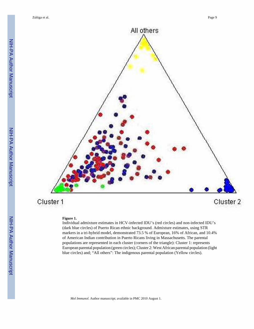
Figure 1.Individual admixture estimates in HCV-infected IDU’s (red circles) and non-infected IDU’s(dark blue circles) of Puerto Rican ethnic background. Admixture estimates, using STRmarkers in a tri-hybrid model, demonstrated 73.5 % of European, 16% of African, and 10.4%of American Indian contribution in Puerto Ricans living in Massachusetts. The parentalpopulations are represented in each cluster (corners of the triangle): Cluster 1: representsEuropean parental population (green circles); Cluster 2: West African parental population (lightblue circles) and; “All others”: The indigenous parental population (Yellow circles).
Zúñiga et al. Page 9
Mol Immunol. Author manuscript; available in PMC 2010 August 1.
NIH
-PA Author Manuscript
NIH
-PA Author Manuscript
NIH
-PA Author Manuscript

NIH
-PA Author Manuscript
NIH
-PA Author Manuscript
NIH
-PA Author Manuscript
Zúñiga et al. Page 10
Table 1
Frequency of group 1 and 2 HLA-C ligands and NK genes in HCV-infected and Non-infected IDU’s.
HLA, KIR genes
HCV+
IDUsN = 160
Non-HCV+
IDUsN = 92 pC OR (95% CI)
n (%) n (%)
C1 122 (76.2) 76 (82.6) ns
KIR2DS4 73 (45.6) 58 (63.0) ns
KIR2DL2 70 (43.7) 48 (52.2) ns
KIR2DL3 116 (72.2) 79 (85.9) ns
KIR2DL2 and/or KIR2DL3 137 (85.6) 91 (98.9) 0.01 0.07 (0–0.47)
KIR2DL2 and/or 68 (42.5) 58 (63.0) 0.01 0.39 (0.22-0.68)
KIR2DL3/KIR2DS4
C1+KIR2DL3 88 (55.0) 65 (70.6) ns
C1+KIR2DL2 52 (32.5) 40 (43.5) ns
C1/KIR2DL3 and/orKIR2DL2
103 (64.4) 75 (81.5) ns
C1+KIR2DS4 54 (33.7) 50 (53.3) 0.02 0.43 (0.24-0.75)
KIR2DL3+KIR2DS4 58 (36.2) 47 (51.1) ns
KIR2DL2+2DS4 37 (23.1) 36 (39.1) ns
C1+KIR2DL3+KIR2DS4 43 (26.7) 39 (42.4) ns
C1+KIR2DL2+KIR2DS4 29 (18.1) 33 (35.9) 0.02 0.40 (0.21–0.74)
C1/2;KIRDS4/KIR2DL3and/or KIR2DL2
51 (31.8) 51 (55.4) 0.004 0.38 (0.21–0.74)
C2 109 (68.1) 58 (63.0) ns
C2+KIR2DS4 44 (27.3) 35 (38.0) ns
C2+KIR2DL3 74 (46.2) 51 (55.4) ns
C2+KIR2DL2 49 (30.6) 29 (31.5) ns
C2+KIR2DS4+KIR2DL3 32 (20.0) 28 (30.4) ns
C2+KIR2DS4+KIR2DL2 23 (14.4) 21 (22.8) ns
C2/2;KIR2DS4/KIR2DL3and/or KIR2DL2
40 (25) 34 (36.9) ns*
HCV+ = Hepatitis C-infected; ns= not significant
pC: The p values were Bonferroni-corrected by multiplying by the number of comparisons (n = 21).
*non-corrected p value = 0.04.
Mol Immunol. Author manuscript; available in PMC 2010 August 1.
Copyright © 2022 FDOKUMEN