i DIFFERENTIATION OF Entamoeba histolytica AND ...
50
DIFFERENTIATION OF Entamoeba histolytica AND Entamoeba dispar COMPLEX BY MULTIPLEX POLYMERASE CHAIN REACTION GACHUHI SAMUEL GUTU A Thesis Submitted to the Graduate School in Partial Fulfilment for the Requirements of the Award of Master of Science Degree in Medical Parasitology of Egerton University EGERTON UNIVERSITY JULY, 2014
-
Upload
khangminh22 -
Category
Documents
-
view
1 -
download
0
Transcript of i DIFFERENTIATION OF Entamoeba histolytica AND ...

i
DIFFERENTIATION OF Entamoeba histolytica AND Entamoeba dispar
COMPLEX BY MULTIPLEX POLYMERASE CHAIN REACTION
GACHUHI SAMUEL GUTU
A Thesis Submitted to the Graduate School in Partial Fulfilment for the
Requirements of the Award of Master of Science Degree in Medical Parasitology of Egerton
University
EGERTON UNIVERSITY
JULY, 2014

ii
DECLARATION AND RECOMMENDATION
DECLARATION
This MSc thesis is my original work and has not been submitted or presented to any other
university for an award of a degree.
Mr. Gachuhi Samuel Gutu
SM17/2841/2010
Signature: ……………………….. Date……………………………….
RECOMMENDATION
This MSc thesis has been submitted to the graduate school of Egerton University with our
approval as University supervisors.
Dr. Meshack A. Obonyo, PhD
Senior lecturer
Department of Biochemistry
Egerton University
Signature: ……………………………… Date: ……………………………
Prof. Rose Odhiambo, PhD
Associate Proffesor
Department of Biological Sciences
Egerton University
Signature: ……………………………… Date: ……………………………

iii
COPYRIGHT
© 2014 Gachuhi Samuel Gutu
This document cannot be reproduced without the consent of the author or Egerton University
or on their behalf.

iv
DEDICATION
To my beloved wife of Jane and my children Caroline, Catherine and Chris who have
patiently moved with me through the journey of making this dream a reality.

v
ACKNOWLEDGEMENTS
I am grateful to my supervisors Dr. Meshack Obonyo, Prof. Rose Odhiambo, for
tirelessly encouraging me, through the rough times and giving me refined training and
knowledge while undertaking my research work. All reviewers from Department of
Biological Sciences, Faculty of Science and Graduate School who refined the earlier versions
of my proposal. Very special thank to Directors Microbiology HUB Kericho Dr. Brett and Dr.
Brook for financial support for the molecular analysis and entire staff lead by (Dr Brett) who
were always available to assist me with technical issues related to my studies.
I thank Dr. Graham Clark of London School of Hygiene and Tropical Medicine,
United Kingdom for providing me lyophilized DNA of standard cultures of E. histolytica
HM-I:IMSS and E.dispar SAW 760 for my molecular work.
I wish to thank all the participants from the following locations in Naivasha sub
County and its Environments, Karagita, Maela, Kihoto, County Council, Gilgil, Site Service,
Kabati, Longonot, Mai Mahiu, Kayole and North Kinangop for their commitment and
contribution in providing stool samples. Egerton Ethical Committee for approving my
research work, Dr Mburu the Medical Superintendent of Naivasha District hospital and
laboratory technical staff, who supported me by providing reagent and space for microscopy
analysis through the study period. Finally, I thank my family, for their unceasing support,
which carried me through to accomplishment of this thesis. Above all, I thank God for
enabling me to undertake and complete this thesis.
vi
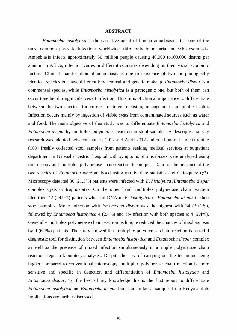
ABSTRACT
Entamoeba histolytica is the causative agent of human amoebiasis. It is one of the
most common parasitic infections worldwide, third only to malaria and schistosomiasis.
Amoebiasis infects approximately 50 million people causing 40,000 to100,000 deaths per
annum. In Africa, infection varies in different countries depending on their social economic
factors. Clinical manifestation of amoebiasis is due to existence of two morphologically
identical species but have different biochemical and genetic makeup. Entamoeba dispar is a
commensal species, while Entamoeba histolytica is a pathogenic one, but both of them can
occur together during incidences of infection. Thus, it is of clinical importance to differentiate
between the two species, for correct treatment decision, management and public health.
Infection occurs mainly by ingestion of viable cysts from contaminated sources such as water
and food. The main objective of this study was to differentiate Entamoeba histolytica and
Entamoeba dispar by multiplex polymerase reaction in stool samples. A descriptive survey
research was adopted between January 2012 and April 2012 and one hundred and sixty nine
(169) freshly collected stool samples from patients seeking medical services at outpatient
department in Naivasha District hospital with symptoms of amoebiasis were analyzed using
microscopy and multiplex polymerase chain reaction techniques. Data for the presence of the
two species of Entamoeba were analysed using multivariate statistics and Chi-square (χ2).
Microscopy detected 36 (21.3%) patients were infected with E. histolytica /Entamoeba dispar
complex cysts or trophozoites. On the other hand, multiplex polymerase chain reaction
identified 42 (24.9%) patients who had DNA of E. histolytica or Entamoeba dispar in their
stool samples. Mono infection with Entamoeba dispar was the highest with 34 (20.1%),
followed by Entamoeba histolytica 4 (2.4%) and co-infection with both species at 4 (2.4%).
Generally multiplex polymerase chain reaction technique reduced the chances of misdiagnosis
by 9 (6.7%) patients. The study showed that multiplex polymerase chain reaction is a useful
diagnostic tool for distinction between Entamoeba histolytica and Entamoeba dispar complex
as well as the presence of mixed infection simultaneously in a single polymerase chain
reaction steps in laboratory analyses. Despite the cost of carrying out the technique being
higher compared to conventional microscopy, multiplex polymerase chain reaction is more
sensitive and specific in detection and differentiation of Entamoeba histolytica and
Entamoeba dispar. To the best of my knowledge this is the first report to differentiate
Entamoeba histolytica and Entamoeba dispar from human faecal samples from Kenya and its
implications are further discussed.

vii
TABLE OF CONTENTS
DECLARATION AND RECOMMENDATION ......................................................................... ii
COPYRIGHT ................................................................................................................................. iii
DEDICATION ............................................................................................................................... iv
ACKNOWLEDGEMENTS ........................................................................................................... v
ABSTRACT .................................................................................................................................... vi
TABLE OF CONTENTS ............................................................................................................. vii
LIST OF TABLES .......................................................................................................................... x
LIST OF FIGURES ....................................................................................................................... xi
LIST OF ABBREVIATIONS ...................................................................................................... xii
CHAPTER ONE ............................................................................................................................. 1
INTRODUCTION .......................................................................................................................... 1
1.1 Background Information .......................................................................................................... 1
1.2 Statement of the Problem ......................................................................................................... 2
1.3 Objectives ................................................................................................................................ 3
1.4 Hypotheses ............................................................................................................................... 3
1.5 Justification .............................................................................................................................. 3
1.6 Expected Outputs ..................................................................................................................... 4
CHAPTER TWO ............................................................................................................................ 5
LITERATURE REVIEW .............................................................................................................. 5
2.1 Amoebiasis .............................................................................................................................. 5
2.1.1 Mode of infection ............................................................................................................ 6
2.1.2 Life cycle of Entamoeba histolytica ............................................................................... 6
2.2 Laboratory Diagnosis of Entamoeba histolytica ..................................................................... 9
2.3 Techniques for differentiation of Entamoeba species ........................................................... 10
2.3.1 Zymodemes ................................................................................................................... 10
2.3.2 Antigen–Antibody detection ......................................................................................... 11

viii
2.3.3 Molecular Approaches .................................................................................................. 11
2.4 Amoebiasis Management ....................................................................................................... 12
CHAPTER THREE ...................................................................................................................... 14
MATERIALS AND METHODS ................................................................................................. 14
3.1 Research Design .................................................................................................................... 14
3.2 Study Area ............................................................................................................................. 14
3.3 Sampling procedure ............................................................................................................... 15
3.4 Sample Analysis .................................................................................................................... 16
3.4.1 Microscopy ................................................................................................................... 16
3.4.2 Multiplex-Polymerase Chain Reaction ......................................................................... 16
3.4.3 Estimation of DNA Integrity ........................................................................................ 17
3.4.4 Amplification Reaction ................................................................................................. 17
3.4.5 Detection of PCR –Amplified DNA Products .............................................................. 18
3.5 Data Analysis ......................................................................................................................... 19
3.6 Logistical, Legal and Ethical Considerations ........................................................................ 19
CHAPTER FOUR......................................................................................................................... 20
RESULTS AND DISCUSSION ................................................................................................... 20
4.1 Results .................................................................................................................................... 20
4.1.1 Microscopy ................................................................................................................... 20
4.1.2 Infected Patients ............................................................................................................ 20
4.1.3 DNA Integrity ............................................................................................................... 21
4.1.4 Multiplex Polymerase Chain Reaction ......................................................................... 21
4.1.4 Incidence Rates ............................................................................................................. 23
4.1.5 Sensitivity and specificity of the two techniques .......................................................... 23
4.2 Discussion .............................................................................................................................. 25
CHAPTER FIVE .......................................................................................................................... 28
CONCLUSION AND RECOMMENDATIONS ........................................................................ 28
5.1 Conclusion ............................................................................................................................. 28

ix
5.2 Recommendations .............................................................................................................. 28
REFERENCES.............................................................................................................................. 30
APPENDICES ............................................................................................................................... 36
APPENDIX I: CONSENT FORM .............................................................................................. 36
APPENDIX II: EGERTON ETHICAL CLEARENCE .............................................................. 37
APPENDIX III: CLEARENCE LETTER FROM THE MINISTRY OF MEDICAL
SERVICES .................................................................................................................................. 38

x
LIST OF TABLES
Table 4.1: Detection rates by microscopy and multiplex –PCR ............................................ 22
Table 4.2: Relationship between age, gender and location in comparison to the incidences of
E.histolytica and E. dispar ............................................................................................... 23
Table 4.3: Multivariate tests for sensitivity between microscopy and multiplex PCR Tests 24

xi
LIST OF FIGURES
Figure 2.1: Life cycle of E. histolytica……………………………………………………...7
Figure 2.2: Stained trichrome cysts of E. histolytica ……………………….………...…….8
Figure 2.3: Stained trichrome Trophozoites of E. histolytica…….…………………… …..8
Figure 3.1: A map showing the location of Naivasha District hospital......……………….. 14
Figure 4.1: Microscopy positive samples in a wet mount stained with lugo,s iodine …..…20
Figure 4. 2: DNA amplification of total nucleic acid samples 1-13…………….………….21
Figure 4. 3: Agarose gel of PCR produc.ts amplified by E. histolytica/ dispar primers .…22

xii
LIST OF ABBREVIATIONS
ALA Amoebic liver abscess
bp Base pair
EDP1 Entamoeba dispar Primer 1
EDP2 Entamoeba dispar Primer 2
EHP1 Entamoeba histolytica Primer 1
EPH2 Entamoeba histolytica Primer 2
Gal/GalNAc Galactose / N-acetyl-galactosamine
MoH Ministry of Health
MPCR Multiplex Polymerase Chain Reaction
MUC2 Mucin 2
WHO World Health Organisation

1
CHAPTER ONE
INTRODUCTION
1.1 Background Information
Entamoeba histolytica belongs to the family Entamoebidae and is the primary
causative agent of human amoebiasis. It is one of the most common parasitic infections
worldwide, third only to malaria and schistosomiasis (Kurt et al., 2008). Entamoeba
histolytica infects approximately 50 million people worldwide causing 40,000 to 100,000
deaths per annum (Calderaro et al., 2006). There are cases where up to 50% of the population
may be affected in regions with poor sanitary conditions (Saeed and Manal, 2007). It has
been estimated that amoebiasis affects about 10% of the global population of which, 90%
will show no clinical symptoms (Gonin and Trudel, 2003). These figures appear to vary from
country to country and in different regions. For example, studies undertaken in different
countries showed variable infection rates as follows: in Pakistan 21.69% (Tasawar et al.,
2010), Egypt >21% (Stauffer et al., 2006), Côte d'Ivoire 18% (Quattara et al., 2010), Nigeria
14.3%, (Dawah et al., 2010), Brazil 21% (Santos et al., 2007), Kenya 12.6% (Nguhiu et al.,
2009). All these studies report different infection rates depending on the procedure that was
used therein.
Clinical manifestations of amoebiasis infections are due to the existence of two
morphologically identical species of Entamoeba, but with different biochemical and genetic
makeup. The two species of Entamoeba include the non pathogenic Entamoeba dispar and
pathogenic Entamoeba histolytica (Fotedar et al., 2007a). It has been stated in other studies
that the infection by E. histolytica may either be asymptomatic or symptomatic resulting in
dysentery or in extreme cases extra intestinal diseases involving other organs such as the
liver, lungs or brain (Salles et al., 2003; Fotedar et al., 2007a). The diagnosis of E. histolytica
infection in the laboratory has traditionally relied on microscopic examination of fresh or
fixed stool specimens (Stark et al., 2008). However, microscopy has several limitations, for
instance its inability to distinguish the pathogenic species from non pathogenic ones and high
chances of false-positive results due to misidentification of macrophages as trophozoites or
polymorphic nuclear leukocytes as cysts and other Entamoeba species (Rashed et al., 2011).
The concerns associated with the limitation of microscopy, necessitated the search for
more specific and sensitive alternative methods that are diagnostic nucleotides such as
polymerase chain reaction (PCR). These are in addition to enzyme based cultures and
antigen- antibody based enzyme linked immunosorbent assays (ELISA). All these have been
2
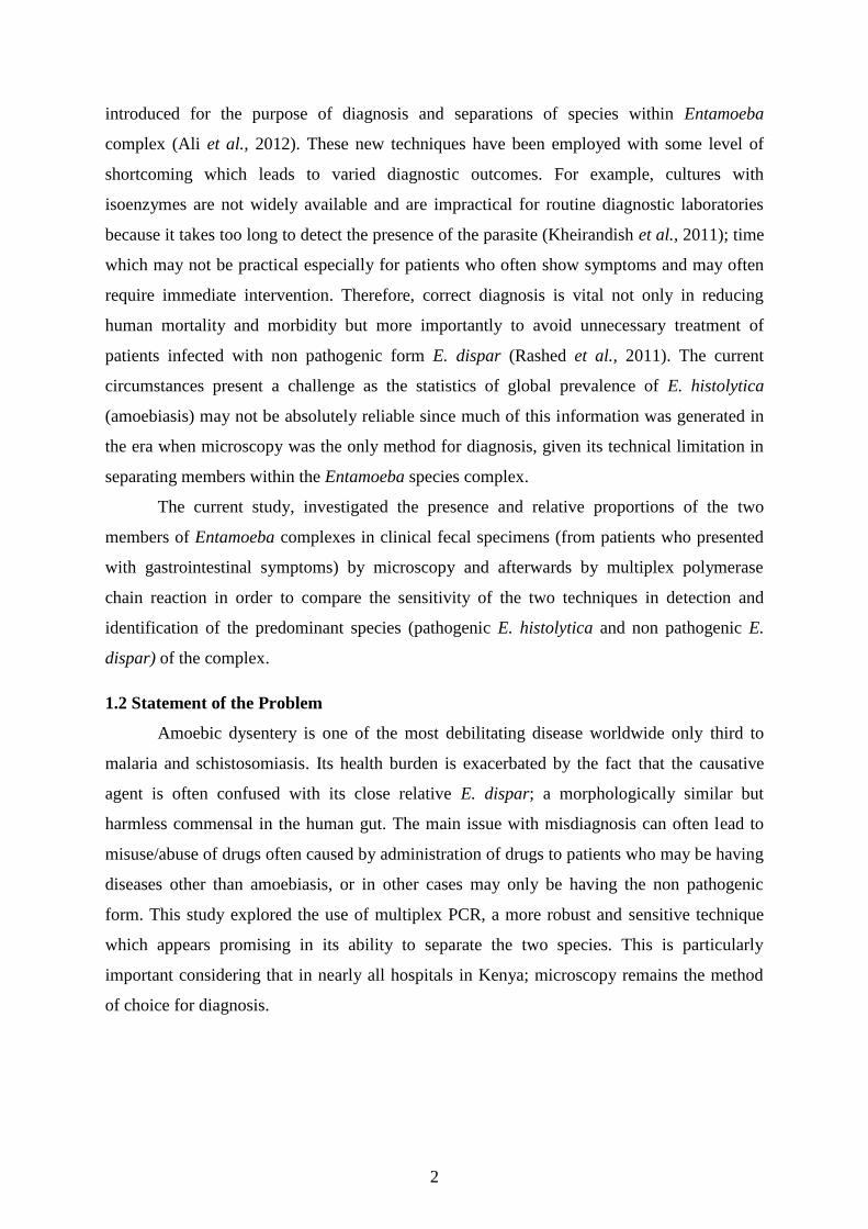
introduced for the purpose of diagnosis and separations of species within Entamoeba
complex (Ali et al., 2012). These new techniques have been employed with some level of
shortcoming which leads to varied diagnostic outcomes. For example, cultures with
isoenzymes are not widely available and are impractical for routine diagnostic laboratories
because it takes too long to detect the presence of the parasite (Kheirandish et al., 2011); time
which may not be practical especially for patients who often show symptoms and may often
require immediate intervention. Therefore, correct diagnosis is vital not only in reducing
human mortality and morbidity but more importantly to avoid unnecessary treatment of
patients infected with non pathogenic form E. dispar (Rashed et al., 2011). The current
circumstances present a challenge as the statistics of global prevalence of E. histolytica
(amoebiasis) may not be absolutely reliable since much of this information was generated in
the era when microscopy was the only method for diagnosis, given its technical limitation in
separating members within the Entamoeba species complex.
The current study, investigated the presence and relative proportions of the two
members of Entamoeba complexes in clinical fecal specimens (from patients who presented
with gastrointestinal symptoms) by microscopy and afterwards by multiplex polymerase
chain reaction in order to compare the sensitivity of the two techniques in detection and
identification of the predominant species (pathogenic E. histolytica and non pathogenic E.
dispar) of the complex.
1.2 Statement of the Problem
Amoebic dysentery is one of the most debilitating disease worldwide only third to
malaria and schistosomiasis. Its health burden is exacerbated by the fact that the causative
agent is often confused with its close relative E. dispar; a morphologically similar but
harmless commensal in the human gut. The main issue with misdiagnosis can often lead to
misuse/abuse of drugs often caused by administration of drugs to patients who may be having
diseases other than amoebiasis, or in other cases may only be having the non pathogenic
form. This study explored the use of multiplex PCR, a more robust and sensitive technique
which appears promising in its ability to separate the two species. This is particularly
important considering that in nearly all hospitals in Kenya; microscopy remains the method
of choice for diagnosis.

3
1.3 Objectives
1.3.1 General objective
To differentiate between E. histolytica and E. dispar complex in stool specimens
using multiplex polymerase chain reaction.
1.3.2 Specific objectives
1. To distinguish between the pathogenic E. histolytica from non pathogenic E.
dispar by multiplex polymerase chain reaction.
2. To determine the incidence of E. histolytica and E. dispar in Naivasha Sub County.
3. To compare the sensitivity between multiplex polymerase chain reaction and
microscopy methods in techniques of E. histolytica and E. dispar complex.
1.4 Hypotheses
1. There is no genetic difference between pathogenic E. histolytica and non pathogenic
E. dispar using multiplex polymerase chain reaction technique.
2. There is no difference in the incidences of E. histolytica and E. dispar in Naivasha
Sub County.
3. There is no difference in sensitivity between multiplex Polymerase chain reaction and
microscopy in diagnosis of E. histolytica and E. dispar complex.
1.5 Justification
Entamoeba species complex comprising among others species Entamoeba histolytica
and Entamoeba dispar are morphologically identical organisms but only differ biochemically
and genetically. Entamoeba histolytica is recognized as a pathogen while the status of
Entamoeba dispar remains unclear hence it is primarily considered a commensal. In Kenya,
microscopy remains the technique of choice for diagnosis of species within the Entamoeba
complex. In light of our present knowledge, microscopy must be considered as a screening
method for the E. histolytica and E. dispar complex and not as a technique to confirm their
diagnosis. However, majority of laboratories in health facilities lack the capacity to
differentiate between E. dispar and E. histolytica and often report the two species complex as
E. histolytica. Based on these facts the prevalence of E. histolytica and E. dispar is
questionable, because microscopically the two species are indistinguishable. Consequently, it
was important that this study was undertaken to differentiate E. histolytica and E. dispar
complex and determine their proper/actual incidences. In addition, the sensitivity and
specificity of the two techniques for accurate diagnosis was determined.

4
1.6 Expected Outputs
1. The pathogenic and non pathogenic Entamoeba species distinguished genetically and
their incidence in Naivasha Sub County established.
2. Publication in peer-reviewed journals.
3. Award of Master of Science degree in Medical Parasitology

5
CHAPTER TWO
LITERATURE REVIEW
2.1 Amoebiasis
Entamoeba histolytica is an entero-parasite and the primary cause of a disease called
amoebiasis in human, which is largely endemic in developing countries (Lejeune et al.,
2009). The first case of dysenteric disease in man, caused by amoeba was described in 1875
by the Russian physician Friedrich Losch. He described the motility of the amoeba and the
typical nucleus and ingested red blood cells reassures us that he was actually looking for the
trophozoite of what now is recognised as E. histolytica. He first named it Amoeba coli
because it appeared in the colon. In 1903, Fritz Schaudinn changed the name to E. histolytica
due its ability of the amoeba to cause tissue lysis (Pinilla et al., 2008). The distinction
between E. histolytica and E. dispar was first suspected by Brumpt in 1925 who then
suggested that the differences in symptoms and global distribution of invasive amoebiasis
were due to the presence of two morphologically identical species of amoebae, the
pathogenic and non-pathogenic forms (Kurt et al., 2008). The infection of E. histolytica
affects about 10% of the global population of which 90% are asymptomatic (Gonin and
Trudel, 2003).
In a study carried out in Kibwezi Sub County in Makueni County in Kenya, E.
histolytica and E. dispar complex infection is estimated to affect about 13% of population
(Nguhiu et al., 2009). However, the prevalence of the Entamoeba complex in these subs
Counties and generally in Kenya is unknown. In other countries, the prevalence of
Entamoeba complex is known. In a study undertaken on stool samples from 49 patients who
had been diagnosed with amoebiasis in Cuba; multiplex polymerase chain reaction showed
75.5% of the diagnostic fragments were characteristic of E. dispar (96bp) while the
remaining 24.5% showed both E. histolytica (132bp) and E. dispar (Nunez et al.,2001).
While in Brazil, eleven stool samples out of twenty seven were identified positively by
multiplex-PCR out of which nine (81.8%) presented the diagnostic fragment characteristic of
E. dispar (96 bp) and two (18.8%) had E. histolytica (132 bp). The remaining sixteen samples
(59.2%) had unknown DNAs. In addition, the samples were further examined
microscopically and among the negative samples detected by microscopic examination, three
were positive for E. dispar and one positive for E. histolytica (Santos et al., 2007). This is
likely to imply that: multiplex is superior and specific in detection and differentiation of the
two species. On the other hand, in Iraq out of one hundred stool samples processed 43% had

6
diagnostic fragment characteristic of E. dispar (96bp) while 26% had diagnostic fragment
characteristic of E. histolytica (132bp). The remaining 24 stool samples which were found
negative following microscopic examination were subjected to multiplex PCR. One stool
sample was found to have diagnostic fragment characteristic of E. histolytica while another
four had the diagnostic fragment characteristic of E. dispar, the remaining 19 were confirmed
negative (Aseel and Sarmad, 2010). In Egypt, multiplex PCR detected (36%) while
microscopy was (25%) in hundred stools samples analysed. Multiplex PCR identified (25%)
with diagnostic fragment were characteristic of E. histolytica while (41.7%) had the
fragment characteristic of E. dispar (96bp) and another (33.3%) had both E. dispar and E.
histolytica (Mona et al., 2011).
2.1.1 Mode of infection
There are several possible avenues of infection by Entamoeba species but the main
route is the fecal-oral route where edible materials contaminated with viable cysts are
ingested (Singh et al., 2009). For instance, direct contact with dirty hands, poor food
handling, use of untreated human faeces as fertilizers such as sewerage water for growing
vegetables which harbour the viable cysts (Ejaz et al., 2011). It has also been reported that
infection may also occur via oral-anal sexual practices or event direct rectal inoculation
through colonic irrigation devices (Sirikelum and Kodikara, 2011). Recently, Biswapriya et
al., (2011), presented evidence that infective cysts may be spread by arthropods such as
cockroaches and flies, thus suggesting that these insects are able to play a rare but important
role in physical transmission
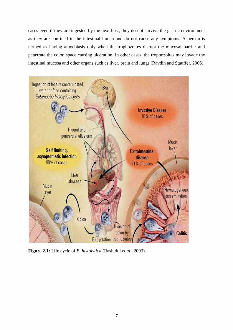
2.1.2 Life cycle of Entamoeba histolytica
Entamoeba histolytica has a simple life cycle (Figure 2.1) that comprises of an
infectious cyst form, an amoeboid trophozoite stage and a mature cyst (the infective stage)
(Figure 2.2). It is the mature cysts (the infective stage) which are ingested in fecal
contaminated material: food, water (Ejaz et al., 2011). Once ingested, encystation takes place
in the small intestines after which the trophozoites (Figure 2.3) are released and they migrate
into the large intestines. Once in the large intestines, the trophozoites multiply by binary
fission producing numerous cysts which are passed out in the faeces. The cysts have a thick
wall made partly of chitin which enables them to survive for days to weeks in the external
environment (Varki et al., 2009). In cases where the human patient has diarrhoea, the
trophozoites are passed out in stool but are rapidly destroyed once outside the body. In such
7
cases even if they are ingested by the next host, they do not survive the gastric environment
as they are confined in the intestinal lumen and do not cause any symptoms. A person is
termed as having amoebiasis only when the trophozoites disrupt the mucosal barrier and
penetrate the colon space causing ulceration. In other cases, the trophozoites may invade the
intestinal mucosa and other organs such as liver, brain and lungs (Ravdin and Stauffer, 2006).
Figure 2.1: Life cycle of E. histolytica (Rashidul et al., 2003).

8
Figure 2.2: Stained trichrome cysts of E. histolytica with three visible nuclei and a
Chromatoid body (Lebbad et al., 2005)
Figure 2.3: Trophozoites of E. histolytica with ingested erythrocytes. ((Lebbad et al., 2005)
Nuclei and Chromatoid
body
Ingested erythrocytes
Nuclei and Chromatoid bodied

9
2.1.3 Pathology of Entamoeba histolytica
Entamoeba histolytica trophozoite frequently lives within the large intestine with no
clinical overt symptoms. Infections of E. histolytica vary in intensity from asymptomatic to
severe or fatal invasions. The non-invasive trophozoites are often asymptomatic infections
and responsible for the spread of the parasite with numerous cysts being passed in normal
stools, diarrheic stools primarily contains trophozoites. Some of trophozoites invade the
colonic epithelium, leading to the formation of amoebic ulcers, while others invade the
intestinal mucosa affecting other organs thereby resulting into complications in the affected
organs and death (Levecke et al., 2010). The development of symptomatic disease results
from mucosal necrosis caused by liberated lysosomes enzymes. This results in the escape of
red blood cells which are ingested by the trophozoites. These results in severe diarrhea with
blood and mucus present and the patient will present symptoms such as abdominal pain or
cramps, tenesmus fever and vomiting (Dooron et al., 2005). The non-invasive infection can
persist or progress to an invasive disease in which trophozoites penetrate the intestinal
mucosa and kill the epithelial cells. The galactose and N-acetyl-D-galactosamine
(Gal/GalNAc)-specific lectin moieties mediate attachment to epithelial cells and mucin.
After substrate adherence, amoeba rapidly induces apoptosis and cytolysis of host cells
(Sateriale and Huston, 2011). Some of the outcomes of these penetrations include local
abscesses and peritonitis, secondary infections with bacteria and formation of ameboma. The
lesions observed during amoebiasis are caused by harmful products such as protein and or
oligosaccharide components of the mucin-2 (MUC2) polymer secreted by trophozoites and
possibly by host defence (Cassia et al., 2010). Also some trophozoites progress to a systemic
or extra intestinal infection via the blood stream (Stanley, 2003). These trophozoites affect
liver, lungs and brains.
2.2 Laboratory Diagnosis of Entamoeba histolytica
In past decades, microscopy has remained the preferred method of choice for
diagnosing intestinal E. histolytica infection. Despite its limitation in differentiating between
members of the Entamoeba complex, it has remained the technique of choice in many
parasitological laboratories worldwide (Fotedar et al., 2007a). In light of our present
knowledge, microscopy should be considered as a screening method for the E. histolytica and
E. dispar complex but not as a technique to confirm the diagnosis of E. histolytica (Santos et
al., 2007). Its sensitivity is about 60% compared to molecular, culture and immunological
techniques (Ali et al., 2008). The common concentration technique used in Kenyan

10
laboratories is formal-ether technique that was developed several decades ago by Ritchie,
1948. Although, slight modifications have been made since that time the principle remains
unchanged except that ether has been replaced by ethyl acetate (Young et al., 1979) and later
on sodium acetate-acetic acid-formalin (SAF) may be used instead of formalin as the fixative
(Ghazanchaei et al., 2012).
It is estimated that less than 1% of E. histolytica trophozoites invade the intestinal
mucosa affecting other organs leading to extra intestinal amoebiasis that results in
complications in the affected organs and death (Levecke et al., 2010). There exists a
challenge for diagnosis of patients with extra-intestinal amoebiasis using stool specimens
because they rarely have E. histolytica parasites in their stool samples (Fotedar et al., 2007b).
For this reason, more appropriate and sensitive techniques have been developed and include
molecular and antibody detection techniques, (e.g. enzyme-linked Immunosorbent assay).
Recently, in a study conducted in Bangladesh, it was indicated that a sensitivity of 96% for
amoebic liver abscess patients was achieved but with only 46% for amoebic colitis patients
(Haque et al., 2010). These means the antibody-antigen techniques are best techniques for
detecting extra-intestinal amoebiasis. Microscopy offers very low sensitivity (60%) whereas
molecular techniques have higher sensitivity and specificity of 94% and 100% respectively.
2.3 Techniques for differentiation of Entamoeba species
2.3.1 Zymodemes
This is a technique used to differentiate E. histolytica and E. dispar in stool or liver
abscess specimens. It was the first procedure to be used to characterize different E. histolytica
isolates. However, it did play a major role in the early differentiation of E. histolytica and E.
dispar (Sergeant et al., 1978). It involves culturing stool samples, rectal biopsy specimens or
liver abscess in axenic culture media such as TY1-S-33 in the absence of any other
metabolizing cells (Clark et al., 2002). If amoebic trophozoites are present, they would be
visualised on the wall of the test tube or in the debris. The indentified trophozoites are
differentiated using specific isoenzymes such as hexokinases by electrophoresis. The main
drawback of this procedure is that it is tedious and time consuming (Intarapuk et al., 2009).
Furthermore, the sensitivity and accuracy is low because many samples that are positive by
microscopy are culture negative (Fotedar et al., 2007a).

11
2.3.2 Antigen-Antibody detection
These are commercially available antigen detection kits such as Entamoeba CELISA
PATH and TechLab E. histolytica II both of which use monoclonal antibodies against
Gal/GalNAc-specific lectin an adhesin molecule of E. histolytica (Fotedar et al., 2007a).
Studies carried out in Bangladesh, an area with high E. histolytica transmission have shown
that antigen detection has a high sensitivity equal to PCR (Haque et al., 1998). However, in
investigations carried out in non-endemic areas demonstrated a poor sensitivity for antigen
detection compared to PCR (Stark et al., 2008). This is an indication that antigen testing has
the benefit of being simple, rapid and yielding good results in areas with high prevalence but
it is limited in settings where there are few cases of E. histolytica infections ( Visser et al.,
2006; Stark et al., 2008).
2.3.3 Molecular Approaches
These involve modern, powerful, highly sensitive and useful methods not only for
differentiation of E. histolytica and E. dispar but for genetic typing of isolates as well. There
are several PCR–based methods have that been developed and tested in diagnostics.
However, they are time-consuming, expensive and require specialized skills and practical
experience (Haque et al., 1998). PCR is used to amplify a specific region of DNA strand (the
DNA target). The amplification of the fragments varies with different isolates (Muller et al.,
1997). DNA extraction performed on cultured trophozoites has minor challenges compared to
the DNA extracted directly from stool samples. This is because feces contain several PCR
inhibitors (Abu and Radstrom, 2000) most of which require additional step of inhibitor
removal by an optimal extraction procedure. However, with current development and
advances in molecular biology, this step that requires inhibitors removal can be overcome by
using more sensitive commercial kits. Commercial spin columns (QIAampTM DNA mini kit
or QIAampTM DNA stool mini kit) are among the most widely used devices for extraction of
Entamoeba DNA directly from stool samples (Gonin and Trudel, 2003). The extraction
procedures involve cyst disrupting steps such as bead-beater, thawing, boiling, and freezing
treatment. However, manual extraction is time consuming and inconvenient when analyzing
a large number of samples. That notwithstanding, molecular biology techniques are now
becoming part of routine diagnostic procedures for detecting intestinal parasites. These
current techniques are amenable to automation especially as pertains to the DNA extraction
procedures (Calderaro et al., 2010).

12
Some of the earliest methods for differentiating E. histolytica from E. dispar included
two single PCRs that targeted either the small subunit ribosomal RNA (ssrRNA) gene (Clark
and Diamond, 1991) or the gene encoding peroxiredoxin (a 30-kDa protein) (Tachibana et
al.,1991). The single two single PCR techniques remain the most commonly used worldwide
(Fotedar et al., 2007a). Other more current protocols are available for detection and
differentiation of E. histolytica and E. dispar, which include duplex PCR, multiplex PCR
with sensitivity and specificity of 94% and 100% respectively (Khairnar and Parija,2007;
Nazemalhosseini et al., 2010 ). Nested PCR and real-time PCR are more sensitive, more
rapid than conventional PCR leading to shorter turnaround times with much reduced risk of
amplicon from laboratory environments. More importantly they have significant reduced
reagent costs (Qvarnstrom et al., 2005). The Principle of multiplex PCR is that more than one
target sequence can be amplified by including more than one pair of primers in the reaction.
It saves time and effort within the laboratory setup and without compromising test quality
(Elnifro et al., 2000). The conventional PCR basically tells whether or not a gene of interest
is in the sample. This is done semi-quantitavely if the PCR is done in a low number of cycles
it will indicate whether one sample expresses more of the gene of interest than another
sample. The amplified products can be visualized afterwards by agarose gel/ethedium
bromide electrophoresis.
2.4 Amoebiasis Management
There are several management options for amoebiasis including: pharmacological,
surgical intervention and preventive measures. According to the WHO recommendation the
administration of anti-amoeba drugs should be done only after differentiation of E. histolytica
and E. dispar WHO, (1997). It is further recommended that whenever possible no patient
should be treated on the basis of microscopic findings alone (Santos et al., 2013). In
addition, all cases identified as E. histolytica regardless of symptoms should be treated due to
the risk of the invasive disease further spreading. On the other hand, cases found involving
only E. dispar should not be treated (Stanley, 2003). If a patient with E. dispar has intestinal
symptoms other investigations should be carried out to find other possible causes of the
disease. Asymptomatic E. histolytica infection should be treated with a luminal amoebicide
such as diloxanide furoate or paromomycin while invasive intestinal or extra-intestinal
amoebiasis should be handled by administering a tissue amoebicide such as metronidazole
followed by luminal treatment (Blessmann et al., 2006). Surgical and percutaneous

13
intervention is required in cases of acute abdominal disturbances such as perforated amoebic
colitis, massive gastrointestinal bleeding and toxic megacolon (Gutierrez et al., 2010).

14
CHAPTER THREE
MATERIALS AND METHODS
3.1 Research Design
A survey was carried out in order to establish the correlations between age, gender,
locations, positive and negatives. Brief interviews were used in collecting data since they are
useful in investigating issues in an in depth way and also because they usually achieve a high
response rate.
3.2 Study Area
Samples were obtained between January 2012 and April 2012 at Naivasha District
hospital in Nakuru County about 90 km from Nairobi, the capital city of Kenya. Naivasha is
one of the sub counties in Nakuru County (Figure 3.1). It covers an area of 1707 Km2 with a
population density of approximately 350,000 people according to 2009 population census
(KNBS, 2009). It lies along the Trans Africa’s Great North Road which runs from Mombasa
through Nairobi, Uganda, Rwanda and Zaire. It is located on the shore of Lake Naivasha (0°
43' 0" South, 36° 26' 0" East).
Figure 3.1: A map showing the location of Naivasha District hospital in Naivasha sub
County, Nakuru County (Source: maps google.co.ke, 2011).
Naivasha District Hospital
N
E

15
3.3 Sampling procedure
In the current study, selection criteria focused on patients who had symptoms of colitis,
bloody or mucous diarrhoea or amoebic dysentery fever at the District hospital as participants
for the study. Sample size was determined following the method proposed by Cochran,
(1977). The number of patients to be sampled for the study was derived from the formula
shown below:
n = Z2pq (Cochran, 1977)
d2
Where n was the desired sample size, Z was the standard normal deviation at the required
confidence that have the characteristic of being measured, p was the target population
estimated to have characteristics being measured, d was the level of statistical significance
set, q was 1-p.
Where;
n = unknown
Z = 1.96
p = 12.6 %( 0.126)
d = 0.05
q = 1-p (1-0.126) = 0.874
n = 1.96 x 1.96 x 0.126 x 0.874 = 0.4231 = 169
0.05 x 0.05 0.0025
n = 169
Using the above formula, 169 patients were included in the study. Sampling was done from
patients seeking medical services at outpatient department in Naivasha District hospital.
After obtaining informed written consent from the patients, specimens were collected from
symptomatic patients presenting any of the following: colitis, bloody or mucoid diarrheal or
amoebic dysentery fever. Only male and female patients aged between 2 to 60 years who had
not used any anti-amoeba drugs within one week (prior to observation in the hospital) were
included in the study. Adults gave informed consent while for children between 2 and 18
years, their parents or guardians gave consent and signed. Clean dry faecal containers were
given to them or their parents or guardians and instructions given on how to collect the stool
specimen. Data concerning their gender and ages and location where the patients originally
came from were recorded on submission of specimen.

16
3.4 Sample Analysis
3.4.1 Microscopy
Two grams of each sample were immediately aliquoted into 1.5ml screw-cap tube and
stored in a freezer at -20°C. The samples were placed in cold chain boxes and transported to
U.S Army Medical Research Unit Microbiology-Hub Kenya-Kericho for PCR analysis.
Another two grams of the faecal samples were used for parasitological examinations at the
hospital diagnostic laboratory in Naivasha District hospital. Direct wet smear and formal-
ether concentration techniques were performed within two hours after collection. From each
sample, two wet smear and one from formal-ether concentration were prepared according to
the protocol previously described by the WHO (2006). The smears were then examined under
a light microscope for identification at 10X then for confirmation at 40X. Results were
recorded either as positive, if cysts or trophozoites of either species (E. histolytica/E. dispar)
were detected or negative if none were detected.
3.4.2 Multiplex-Polymerase Chain Reaction
DNA Extraction
DNA was extracted from 150 mgs of stool samples using ZR Faecal DNA
MiniPrep™
Catalogue No. D6010 kit, according to the manufacturer’s Protocol (ZYMO
RESEARCH CORP). Briefly, Zymo-spin™
IV-HRC Spin filter (green top) were prepared
prior to use by snapping off the base and inserted into a collection tube and centrifuged in a
micro centrifuge at 8,000 xg for 3 minutes. The faecal material was placed in a ZR Bashing
Bead™
Lysis and 750 µl lysis solutions added to the tube. The mixture was then vortexed at
high speed for 5 minutes. The ZR Bashing Bead™
was then centrifuged at 10,000 xg for 1
minute in a micro-centrifuge, 400 µl of collected supernatant was transferred into a Zymo-
spin™
IV spin filter (orange top) in a new collection tube and centrifuged in a micro-
centrifuge at 7,000 xg for 1 minute. Twelve hundred microliters of faecal DNA binding
buffer was added to the collected filtrate in the collection tube mixed and 800 µl of the
mixture was transferred into a new Zymo-spin™
IIC column in a new collection tube and
centrifuged in a micro-centrifuge at 10,000 xg for 1 minute. The filtrate in the collection tube
was discarded and the remaining 400 µl of the mixture was transferred to the same Zymo-
spin™
IIC column in a collection tube and centrifuged at 10,000 xg for 1 minute. Zymo-spin™
IIC column was placed in a new collecting tube and 200 µl DNA Pre–wash buffer added and
centrifuged in a micro-centrifuge at 10,000 xg for 1 minute. Five hundred microliters of
faecal DNA wash buffer was added into Zymo-spin™
IIC column and centrifuged at 10,000

17
xg for 1 minute. Zymo-spin™
IIC column was transferred into a clean micro-centrifuge tube
and 100 µl DNA elution buffer added directly to the column and centrifuged 10,000 xg for 30
seconds to elute the DNA. The eluted DNA was transferred into minute Zymo-spin™
IV-
HRC Spin filter in a clean 1.5 ml micro-centrifuge tube and centrifuged at 8,000 xg for 1
minute. The filtered DNA was stored at -20°C before PCR analysis.
3.4.3 Estimation of DNA Integrity
The purity of DNA was assessed using NanoDrop 2000 spectrophotometer (Thermo
Scientific). Then, 2 µl of the extracted DNA was loaded into the lower measurement pedestal
of the NanoDrop spectrophotometer and optical density measured. All the samples were
within the ratio 260/280 nm optical density (1.8-2.0). The concentration of the DNA was also
estimated using 1.0% w/v agarose gel electrophoresis (Sigma, UK). One gram of agarose
powder was weighed and put in a flask and 100ml of 1X Tris-Borate-EDTA (89Mm Tris-
HCL (pH 8.3), 89mM Boric acid and 2.5Mm EDTA), the mixture was boiled for quick
dissolution then allowed to cool to 50°C and stained with 2 µl Ethedium Bromide. The
solution was cast in tray with combs to form indentations (wells about 1mm depth) then
allowed to cool for 30 minutes. Afterwards, 2 µl of each sample of the extracted DNA were
mixed separately with 1µl of loading dye (50% glycerol, 250Mm EDTA (pH 8.0), 0.01
Bromphenol blue) and loaded into the wells and electrophoresed at 80V for 45 minutes. The
gel was visualized under an Alpha ImagerR Hp 3400 and the results printed using Mitsubishi
printer (Figure: 4.2), the extracted DNA were stored at -20°C prior to PCR analysis.
3.4.4 Amplification Reaction
The multiplex polymerase chain reaction was carried out according to the protocol
described by Nunez et al., (2001) with some modifications. In a 50 μl reaction contained
Dream Taq™
Green PCR Master Mix (2X) 25 μl, 21.25 μl nuclease free water; 0.75 μl of
40pmoles of each oligonucleotide primer and 3 μl of DNA template. Amplification was
carried out using a GenAmp PCR system 9700 (Applied Biosystems). Initial denaturation at
94°C for 5minutes followed by 35 cycles of 30 seconds at 94°C annealing at 55°C for 30
seconds and primer extension at 72°C for 40 seconds with final extension at 72°C for 7
minutes. The primers pair used in this study are as stated E. dispar (EDP1-5′-
ATGGTGAGGTTGTAGCAGAGA-3′and EDP2- 5′-CGATATTGACCTAGTACT-3′) and
E. histolytica (EHP1-5′ CGATTTTCCCAGTAGAAATTA-3′ and
EHP2-5′-CAAAATGGTCGTCTAGGC-3′) and were sourced from Bioneer, South Korea.
Dream Taq™
Green PCR Master Mix (2X) contained Green buffer, dNTPs and 4mM MgCl2

18
(Fermentas Life Sciences, USA) was used. Optimization of Multiplex PCR was done using
DNA from HMI-IMSS strain as a positive control for E. histolytica and SAW 760 strain as
positive control for E. dispar. Nuclease-free water was used as negative control. To rule out
amplification inhibitors DNAs of the negative samples were spiked with DNAs of positive
controls and all turned positive.
3.4.5 Detection of PCR -Amplified DNA Products
The PCR products were resolved in 2 % agarose gel and visualized under Alpha
ImagerR Hp 3400. Ten microliters of amplified PCR products of each sample were loaded
separately into 1mm depth wells and 5µl of a molecular marker 100bp (Fermentas Life
Sciences, USA) was loaded in one of the wells. The loaded gel was placed in an
electrophoresis tank and the left to run at 90V for 60 minutes. The gel was visualized under
Alpha ImagerR Hp 3400.

19
3.5 Data Analysis
Data of the presence of the two species of Entamoeba collected using microscopy and
PCR was analysed using multivariate statistics, while the efficiency of the two techniques in
detecting presence of the two species were compared using Chi-square (χ2). Test for
independence using SPSS version 20 to produce mean scores for incidences of E. histolytica
E. dispar. The positivity and negativity rates for both techniques were reported in terms of
frequencies and percentages. Percentage sensitivity and specificity of the two techniques was
determined as expressed below: (Hennekens and Buring, 1987)
Sensitivity =TP
TP +FN X 100%
Specificity =TN
TN +FP 𝑋 100%
Where;
TP = True Positive
FN =False Negative
TN= True Negative
FP= False Positive
3.6 Logistical, Legal and Ethical Considerations
In order to collect data from the field, a written research permit from the Ministry of
Health- Government of Kenya was sought together with prior written informed consent from
each subject according to the requirements of Egerton University Research Ethical
Committee. The clearance by the Ministry and Egerton University are attached in appendix 2
and 3 respectively. Other logistical concerns, like pre-field work logistics such as pre-testing
the instruments, including making an adjustment tour of the study area to strike rapport with
authorities and participants were done before commencement of research.

20
CHAPTER FOUR
RESULTS AND DISCUSSION
4.1 Results
4.1.1 Microscopy
One hundred and sixty nine stool samples were analysed by microscopy for the
presence of Entamoeba life stages. Thirty six (21.3%) samples were detected to have either
E. histolytica/E. dispar trophozoites or cysts or both and hence considered positive. One
hundred and thirty three (78.7%) samples were negative (Figure 4.1).
Figure 4.1: Microscopy positive samples in a wet mount stained with lugo,s iodine E.
histolytica/E. dispar trophozoites and E. histolytica/E. dispar cysts A and B respectively.
4.1.2 Infected Patients
The 36 (21.3%) patient who were infected with E. histolytica/dispar complex after
diagnosis with microscopy, were given the following medication; adults Tinidazole 2gm once
per oral doses daily for three days, followed by Paromomycin 25-35 mg/kg/day per oral in
three doses for seven days. For the children the same drugs were used but their doses were
determined after measuring their weight. These was done because E. histolytica/dispar
complex was not differentiated immediately, to avoid the risk of invasion by E. histolytica
trophozoites. Although, those drugs have minimal toxicity, the patients were adviced to see
the clinician in case they had any adverse reaction to the drugs.
A
Cyst of Entamoeba
histolytica/dispar
complex
E. histolytica/dispar
trophozoite
B

21
4.1.3 DNA Integrity
Figure 4. 2: DNA amplification of total Nucleic Acid samples 1-13, showed that the
extraction method was good but the samples had different concentrations
4.1.4 Multiplex Polymerase Chain Reaction
Detection and differentiation of E. histolytica and E. dispar in 169 stools samples
performed simultaneously in a PCR cycle. The multiplex PCR performed using the samples
initially preserved at -20°C was able to detect and differentiate the species at once rather
performing analysis twice (one for detection and another one for differentiation). The
presence of bands at 96bp indicated as positive for E. dispar and 132bp indicated as positive
for E. histolytica. Bands not within the 96bp and 132bp makers were indicated as unknown
DNA (Figure 4.3).
1 2 3 4 5 6 7 8 9 10 11 12 13
DNA

22
Figure 4. 3: Agarose gel of PCR products amplified by E. histolytica primers (EHP1 \ EHP2)
and E. dispar primers (EDP1 \ EDP2). Molecular ladder/marker size 100bp (M), negative
control (lane 1), E. dispar positive control (lane 2), E. dispar positive samples (lane 6, 7, 9,
11, 14), E. histolytica positive control (lane 12), mixed infection with histolytica and E.
dispar (lane17), negative or unknown DNA patient samples (lane 3, 4, 5, 8, 10, 1315, 16).
A comparison of microscopy and multiplex PCR in detection of E. histolytica and E.
dispar reveals that only 8 of the 169 (4.7%) stool samples contained DNA of E. histolytica
while 34 (20.1%) contained that of E. dispar. In addition, of the 36 samples that had been
positively identified for Entamoeba species by microscopy and subjected to multiplex PCR,
only 6 (16.7%) were found to contain E. histolytica DNA while 27 (75%) samples had DNA
of E. dispar and 3 (8.3%) had unknown DNA. On the other hand, of the 133 samples which
were found negative by microscopy, only 2 (1.5%) contained E. histolytica DNA while 7
(5.3%) contained DNA of E. dispar and 124 (93.2%) contained unknown DNA (Table 4.1).
Table 4.1: Detection rates by Microscopy and Multiplex–PCR
Microscopic examination Multiplex –PCR
E. histolytica E. dispar
Positive 36 6 27
Negative 133 2 7
Total 169 8 34
The Pearson Chi-Square was conducted to test the independence of the variables. There was a
significant relationship between microscopy test and multiple PCR test at χ2 (1df, n=169) =
132bp
96bp

23
14.444, p<0.05. On cross tabulation of tests by microscopy and results of tests by multiplex
PCR for both E. histolytica and E. dispar the following were observed. Out of 133 samples
found to be negative by microscopy 2 (1.5%) were positive by multiplex PCR for E.
histolytica and 7 (5.3) %) were positive with E. dispar.
4.1.4 Incidence Rates
Mono infection rate with E. dispar was demonstrated in 34 out of 169 stool samples
(20.1%), while co-infection rate with E. histolytica and E. dispar was demonstrated 4 out of
169 stool samples (2.4%). Similarly, mono-infection rate with E. histolytica was
demonstrated 4 out of 169 stool samples (2.4%). The relationship between age, gender and
location in comparison to the incidences of the two species showed that there was no
significant relationship (Table 4.2).
Table 4.2: Relationship between age, gender and location in comparison to the incidences of
E. Histolytica and E. dispar
Variables F-value p-value Sig.
Age vs. E. histolytica and
E. dispar
0.64 0.960 Ns
Gender vs. E .histolytica
and E .dispar
0.18 0.669 Ns
Location E .histolytica
and E. dispar
1.37 0.200 Ns
Ns= not significant at p<0.05, there was no significant relationship between the incidences of
E. histolytica and E. dispar across ages, gender and location.
4.1.5 Sensitivity and specificity of the two techniques
The sensitivity and specificity of microscopy was 73.3% and 98.2% respectively,
while for multiplex PCR the sensitivity and specificity was 93.3% and 100% respectively.
24
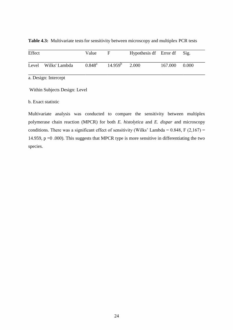
Table 4.3: Multivariate tests for sensitivity between microscopy and multiplex PCR tests
Effect Value F Hypothesis df Error df Sig.
Level Wilks' Lambda 0.848a
14.959b 2.000 167.000 0.000
a. Design: Intercept
Within Subjects Design: Level
b. Exact statistic
Multivariate analysis was conducted to compare the sensitivity between multiplex
polymerase chain reaction (MPCR) for both E. histolytica and E. dispar and microscopy
conditions. There was a significant effect of sensitivity (Wilks’ Lambda = 0.848, F (2,167) =
14.959, p =0 .000). This suggests that MPCR type is more sensitive in differentiating the two
species.

25
4.2 Discussion
Microscopy has remained the gold standard tool for diagnosis of intestinal amoebiasis
infection. The recognition of E. dispar as new non-pathogenic species which is
morphologically indistinguishable from the pathogenic (E. histolytica) has necessitated the
search for alternative methods for differentiating species within the Entamoeba complex
(WHO, 1997). The primary advantage of using PCR as a diagnostic tool is the possibility of
differentiating the species within the complex (E. histolytica and E. dispar) in an area where
other Entamoeba species are prevalent. In addition, PCR has the advantage of accuracy and
reliability especially when there is need to understand the epidemiology of E. histolytica and
E. dispar infections. Observations in the current study indicate that diarrhoea and other
gastrointestinal infection symptoms appeared to be associated with E. histolytica and E.
dispar infection. However, the cause-and- effect relationship of E. histolytica and E. dispar
with the clinical symptoms could not be determined in this present study due to the limitation
of the design given that there was no attempt to rule out other bacterial and/or viral
infections. Besides, it has been documented that not all E. histolytica infections lead to
clinical disease (Ali et al., 2008) which complicate the scenario for drawing conclusion. The
finding in this present study showed that all patients who were diagnosed with positive for E.
histolytica had clinical symptoms of amoebiasis.
The findings of the present study appeared to be consistent with other studies which
showed that multiplex PCR is superior than microscopy and the two species are genetically
different (Nunez et al., 2001; Santo et al., 2007; Aseel and Sarmad, 2010; Mona et al., 2011).
The mono infection rate with E. dispar was higher compared to E. histolytica while co-
infection rate with both species was low. These findings underscore the need for proper
diagnosis before administration of Entamoeba treatment as per the requirements by WHO,
(1997). Only few cases required anti amoeba treatment while those patients from whom the
stool contained DNA of E. dispar and unknown DNA required further investigation to rule
out other causes of gastrointestinal infections.
The present study demonstrated that the incidence of E. dispar was higher compared
to E. histolytica by multiplex PCR. These results are consistent with other studies which
reported such observations for example: Gonin and Trudel (2003) found the incidences of the
two species as follows: 2 E. histolytica and 66 E. dispar, Visser et al (2006) 6.7% E.
histolytica and 91.2% E. dispar, Fotedar et al., (2007b), 5 E. histolytica and 15 E. dispar and
Aseel and Sarmad, (2010), 26% E. histolytica and 43% E. dispar. All these studies reported
higher incidences of E. dispar than E. histolytica. These results suggest that other species of
4.7%

26
Entamoeba could be present, but in the current study, it was not possible to identify them
because the primers used were specific to amplify only DNAs of E. histolytica and E. dispar.
However, a follow up study could concentrate on separation of the Entamoeba species with
an aim of deciphering the identity of unknown E. histolytica species. However, those patients
presenting with gastrointestinal problem should also be investigated further for other causes
of gastrointestinal infections; this include bacterial and viral. The study, also demonstrated
that there was no relationship between the incidences of E. histolytica and E. dispar across
ages, gender and location (Table 4.2). This means the two species affect all patients
irrespective of their ages, gender or location. A differential characterization of E. histolytica
from other intestinal protozoa is essential because only E. histolytica infection requires a
specific drug treatment. The indiscriminate use of such drug can induce development of drug
resistance (Santo et al., 2007).
A comparison of the findings of the two techniques showed that there were some
variations: multiplex PCR detected more patients were infected with E. histolytica or E.
dispar compared to microscopy. Some samples which were positive by microscopy turned
negative when they were analysed by multiplex PCR. These variations in the results were
less, in consistent with other studies where by microscopy examination showed positive for E
.histolytica/E. dispar complex but negative by multiplex PCR. In Cuba the study showed that
49 out of 52 (94.2%) (Nunez et al., 2001), Brazil, 11 out of 27 (40.7%) (Santo et al., 2007),
Iraq, 69 out of 76 (90.8%) (Aseel and Sarmad, 2010), Malaysia, 63 out of 93 (67.7%) (Anuar
et al., 2013).
There was some discrepancy between microscopy and multiplex PCR results in 3
samples that had been positively identified by microscopy. However, these PCR did not
amplify DNA of the 3 samples, hence these were reported negative yet E. histolytica or E.
dispar with the primers used and there had no inhibition of PCR was observed in control
experiments during optimization of multiplex PCR. These negative results can potentially be
explained by the presence of other members of Entamoeba species complex such as:
(Entamoeba. moshkovskii, E. polecki, E. coli, and E. hartmanni) these discrepancies have
been observed by other scholars: in Brazil out 16 out of 27 (59.3%) (Santo et al., 2007), Iraq,
7 out of 76 (9.2%) (Aseel and Sarmad, 2010), Malaysia, 30 out of 93 (32.3%) (Anuar et al.,
2013). Another likely reason for this could be the fact that the three samples contained
trophozoites that could have degenerated with time during storage. However, in the current
study the storage was reduced in order to minimise chances of degradation. To rule out
whether lack of amplification was due to inhibitors the three samples were retested using

27
multiplex PCR and all were negative, Afterwards, the DNAs of the three negative samples
were spiked with 0.05μl DNA of the positive controls retested then they all became positive.
No evidence of inhibition was found in any of the multiplex PCR negatives. This suggested
that other species of Entamoeba (moshkovskii, E. polecki, E. coli, and E. hartmanni) are
present in Naivasha. These species were reported in areas of Ghana, Pondicherry and
Bangladesh (Santo et al., 2007). However, this supposition needs to be verified further in
subsequent studies and probably using a much larger sample size to confirm the presence of
other species commonly found in humans.
In addition, the current study further observed that 9 stool samples (8.8%) which were
negative by microscopy became positive by multiplex PCR and 2 (1.5%) were found to have
the DNA of E. histolytica and 7 (5.3%) had that of E. dispar. Generally multiplex PCR
technique reduced the chances of misdiagnosis by 9 (6.7%) patients. These findings were in
agreement with other studies which reported the use of multiplex PCR increases the
sensitivity of PCR techniques and allows for simultaneous differentiation between E.
histolytica and E .dispar in a single PCR step as well as the presence of mixed infections
(Mona et al., 2011). The Pearson Chi-Square was conducted to test the independence of
microscopy and multiplex PCR test. There was a significant relationship between microscopy
test and multiple PCR test at χ2 (1, N=169) = 14.444, p<0.05. On cross tabulation of tests by
microscopy and results of tests by multiplex PCR for both E. histolytica and E. dispar the
following were observed. Out of 133 samples found to be negative by microscopy 2 (1.5%)
were positive by multiplex PCR for E. histolytica and 7 (5.3) %) were positive with E. dispar
a total of 9 (6.8%) samples were positive by multiplex PCR (Table 4.1). These findings show
that the multiplex PCR is more sensitive and should be encouraged as technique for diagnosis
of amoebiasis. These will reduce the false negative results under microscopy and correct
diagnosis will be achieved. These findings provide important data for public health care
system in Kenya because this is the first time the species of the E. histolytica/E. dispar
complex circulating in this country have been differentiated at DNA level. This will reduce
the cost of treatments to patients who are diagnosed with non pathogenic species E. dispar.

28
CHAPTER FIVE
CONCLUSION AND RECOMMENDATIONS
5.1 Conclusion
The study showed that multiplex PCR is a robust procedure and easily adapted to
routine use and can serve as a tool for detection and confirmation of microscopy results. It is
also useful tool for distinction between E. histolytica and E. dispar as well as the presence of
mixed infection simultaneously in a single PCR round in laboratory analyses. To the best of
my knowledge, this is the first report that has differentiated E. histolytica and E. dispar from
human faecal samples from Kenya. The incidence of E. dispar was highest compared to E.
histolytica while there were only a few cases of mixed infection. The sensitivity and
specificity of multiplex PCR was 93.3% and 100% respectively compared to microscopy
73.3% and 98.2% respectively. Further investigations are essential for those patients
harbouring E. dispar to rule out other causes of gastrointestinal infections example bacterial
or viral infection. However, multiplex PCR technique does not substitute the microscopy
stool examination which widely screens for virtually intestinal parasite but may be a useful
tool for diagnosis and differentiation and epidemiological studies in areas where E.
histolytica is endemic. Despite cost of carrying out the technique being higher compared to
conventional microscopy, multiplex PCR is more sensitive and specific in detection and
differention E. histolytica and E. dispar. However, further epidemiological survey and
clinical studies are needed to determine the true pathogenic potential of the two species.
5.2 Recommendations
1. The government, through Ministry of Health in Kenya, should make a new policy in
Entamoeba diagnosis. No patient should be treated using microscopy results alone.
All positive Entamoeba cases must be confirmed using multiplex PCR before one
is given anti-amoeba drugs. Even though the cost of drugs ranges between Ksh250-
Ksh400, this will reduce the chances of resistance of currently recommended anti-
amoeba drugs.
2. More epidemiological surveys should be done in the whole country so that the actual
prevalence of Entamoeba species can be established.
3. The government should procure PCR equipment and reagent in each of county referral
hospital where specimens which are positive by microscopy can be referred for
differention of the two species at Ksh 400.
29
4. Microscopy should be used as screening technique for amoebiasis but multiplex PCR
techniques used for differentiation, so that one can confirm whether one has mono
infection or mixed infection at Ksh 250.
5. Those patients with gastrointestinal symptoms, but microscopy results negatives and
multiplex PCR doesn’t detect the amoeba DNA, further tests are required to rule out
other causes of gastrointestinal infections such as protozoan, helminthes, bacterial and
viral infections.
6. The government, through ministry of health should ensure all sewerage systems are
treated and water sources protected to avoid being contaminated with sewerage.
30
REFERENCES
Abu, S. W. and Radstrom, P. (2000). Effects of amplification facilitators on diagnostic PCR
in the presence of blood, feces and meat. Journal of Clinical Microbiology, 38: 4463-
4470.
Ali, I. K., Rashidul, H., Abdullah, S., Mamun, K., Nicholus, E. S., Sean, A. G., Gerard, A. C
and William, A. P. Junior. (2012). Proteomic Analysis of the Cyst Stage of
Entamoeba histolytica. Plos Neglected Tropical Diseases, 5: e1643.
Ali, I. K., Clark, C. G. and Petri Junior, W. A. (2008). Molecular epidemiology of
amebiasis. Infectious Genet Evolution, 8: 698-707.
Anuar, T. S., Hesham, M., Al-Mekhalifi, Mohamed, K. A., Gahani, S. N., Azreen, F., Salleh,
N., Ghazali, M. B and Norhayat, M. (2013). Different clinical outcomes of
Entamoeba histolytica in Malaysia: Does Genetic Diversity Exist. Korean Journal
Parasitol, 51: 231-236
Aseel, J. Y. and Sarmad, M. A. (2010). A comparison of Multiplex PCR and Sandwich
ELISA for the diagnosis of Entamoeba histolytica infection in Basrah Province.
Journal of Basrah Researches, 36: 5.
Biswapriya, D., Sudipta, T., Ashis, C. and Kalyan, C. (2011). Studies on Physico-Chemical
and microbiological parameters of water samples before and after jute retting.
Journal of Biological Sciences, 11: 210-215.
Blessmann, J., An Le Van and Egbert, T. (2006). Epidemiology and treatment of amebiasis
in Hue Vietnam. Archives of Medical Research, 37: 269-271.
Calderaro, A., Gorrini, C., Bommezzadri, S., Piccolo, G., Dettori, G. and Chezzi, C. (2006).
Entamoeba histolytica and Entamoeba dispar comparison of two PCR assays for
diagnosis in a non-endemic setting. Transactions of the Royal Society of Tropical
Medicine and Hygiene, 100: 450-457.
Calderaro, A., Gorrini, C., Montecchini, S., Peruzzi, S., Piccolo, G., Rossi, S., Gargiulo, F.,
Manca, N., Dettori, G. and Chezzi, C. (2010). Evaluation of a real-time polymerase
chain reaction assay for the laboratory diagnosis of giardiasis. Diagnosis Microbiol
Infectious Disease, 66: 261–267.
Cassia, A. X. C., Álvaro, C. N., Anderson, J. F., Maria, A. G. and Marcelo, V. C. (2010).
Entamoeba histolytica and E. dispar trophozoites in the liver of hamsters: In vivo
binding of antibodies and complement. Journal of Parasite Vectors, 3:23.

31
Clark, C. G. and Diamond, L. S. (1991). The Laredo strain and other Entamoeba histolytica
like amoebae are Entamoeba moshkovskii. Molecular Biochemistry Parasitology, 46:
11-18.
Clark, G. and Diamond, L. (2002). Methods for cultivation of luminal para-sitic protists of
clinical importance. Clinical Microbiology Review, 15:329–341.
Cochran, W. G. (1977). Sampling techniques, 3rd
edition, New York: john Wiley and Sons.
Dawah, I. S., Inabo, H. I. and Jatau, E. D. (2010). Comparative study of microscopy with
ELISA antibody based amoebiasis diagnosis in patients presenting with dysentery at
government hospitals in Kaduna metropolis. Continental Journal of Biomedical
Sciences, 4:43- 49.
Dooron, R. H., Henk, H., Rob, K., Henk, G., Ron, P., Haque, R., Mondal, D. and Duggal, P.
(2005). Entamoeba histolytica infection in children and protection from subsequent
amoebiasis. Infectious Immune, 74:904-909.
Ejaz, M., Ghulam, M. M. A., Shujaat, A. K., Qazi, N. S., Muhammad, H.H.A., Amir, W.,
Kalsoom, F. and Izhar, H. (2011). Determination of the prevalence of Entamoeba
histolytica in human at a private fertilizer company hospital in Pakistan using
microscopic technique. African Journal of Microbiology Research, 5:149-152.
Elfath, M., Elnifro, A., M., Ashshi, Robert, J., Cooper, Paul, E. and Klapper. (2000).
Multiplex PCR; Optimization and Application in Diagnostic Virology. Journal of
Clinical Microbiology Review, 13:559-570.
extra hepatic amoebiasis. Galle Medical Journal 16:1
Fotedar, R., Stark, D., Beebe, N., Marriott, D., Ellis, J. and Harkness, J. (2007a). Laboratory
diagnostic techniques for Entamoeba species. Clinical Microbiology Review, 20:511-
532.
Fotedar, R., Stark, D., Beebe, N., Marriott, D., Ellis, J. and Harkness, J. (2007b). PCR
detection of Entamoeba histolytica, Entamoeba dispar, and Entamoeba
moshkovskii in stool samples from Sydney, Australia. Journal Clinical
Microbiology, 45:1035-1037.
Ghazanchaei, A., Shohreh, S., Mohammad, S., Mohammad, N. and Seyed, M. N. (2012).
Detection of Dientamoeba fragilis in patients referred to Chaloos Medical Care
Centres by nested – polymerase chain reaction (PCR) method. African Journal of
Biotechnology, 11: 4079-4082.

32
Gonin, P. and Louise, T. (2003). Detection and Differentiation of Entamoeba histolytica and
Entamoeba dispar isolates in clinical samples by PCR and Enzyme-Linked
Immunosorbent Assay. Journal of clinical Microbiology, 41:237–241.
Gutiérrez-Cisneros, M. J., Cogollos, R., Lopez, V. R. (2010). Application of real-time
PCR for the differentiation of Entamoeba histolytica and E. dispar in cyst-positive
fecal samples from 130 immigrants living in Spain. Ann Tropical Medical
Parasitology, 104:145-149.
Haque, R., Ali, I. K., Akther, S. and Petri, W. A. (1998). Comparison of PCR, isoenzymes
analysis, and antigen detection for diagnosis of Entamoeba histolytica infection.
Journal of Clinical Microbiology, 36: 449-452.
Haque, R., Kabir, M., Noor, Z., Rahman, S.M., Mondal, D., Alam, F., Rahman, I., Al
Mahmood A, Ahmed, N. and Petri, W.A. (2010). Diagnosis of amebic liver abscess
and amebic colitis by detection of Entamoeba histolytica DNA in Blood, Urine and
Saliva by a Real-time PCR assay. Journal of Clinical Microbiology 8: 2798-2801
Hennekens, M.C.and Buring, J.E. (1987). Epidemiology in Medicine 1st edition, Lippincott
Williams and Wickins, Philadepia USA.
Intarapuk, A., Thareerat, K., Nitaya, T., Akpimol, M., Pradit, K., Adisak, B. H. and
Dechavudh, N. (2009). Identification of Entamoeba histolytica and Entamoeba
dispar by PCR assay of faecal. Southeast Asian Journal of Tropical Medicine;
Public Health, 40: 425.
Khairnar, K. and Parija, S. C. (2007). A novel nested multiplex polymerase chain reaction
(PCR) assay for differential detection of Entamoeba histolytica, E. moshkovskii and
E. dispar DNA in stool samples. Bacterial Micro compartment Microbial, 7: 47.
Kheirandish, F., Ebrahim, B., Ali, H., Ehsan, N.M. and Bahram, K.(2011). Differential
diagnosis of Entamoeba species in gastrointestinal disorder patients in
Khorramabad, Iran. African Journal of Microbiology Research 5: 2863-2866.
Kurt, O., Mete, D., Ipek, O., Naser, R. S., Aliye, M., Mehmet, T., Mucide, A. K. and Hande,
D. (2008). Investigation of the prevalence of amoebiasis in Izmir province and
determination of Entamoeba species using PCR and enzyme immunoassay. New
Microbiological, 31: 393-400.
Lebbad, M., and Svard, S. G. (2005). PCR differentiation of Entamoeba histolytica and
Entamoeba dispar from patients with amoeba infection initially diagnosed by
microscopy. Journal of Scandinavian Infectious Diseases. 37:680-685.
33
Lejeune, M., Joanna, M. R. and Kris, C. (2009). Recent discoveries in the pathogenesis and
immune response toward Entamoeba histolytica. Future Microbiology, 4:105-118.
Levecke, B., Leentje, D., Pierre, D., Jaco, J. V., Francis, V., Stijn, C., Jozef, V. and Peter, G.
(2010). Molecular identification of Entamoeba species in captive nonhuman primates.
Journal of Clinical Microbiology, 48: 2988–2990.
Mona, M., El Sobky, Manal, A., El Melegi, Noha, A. and Abo-Khalil. (2011). Use of
Multiplex PCR in the Differential Detection of Entamoeba histolytica and or
Entamoeba dispar in Comparison to Microscopic Examination. Parasitologist United
Journal, 4: 193-200.
Muller, M., Lothar, K., Alex, M., Tabrett, D. J., Barbara and Rohela, M. (1997). Detection
of a single base exchange in PCR-amplifiedDNA fragments using agarose gel
electrophoresis containing bisbenzimide–PEG. Oxford University Press Nucleic Acids
Research, 24:5125-5126.
Nazemalhosseini-Mojarad, E., Nochi, Z. and Sahebekhtiari, N. (2010). Discrimination of
Entamoeba moshkovskii in patients with gastrointestinal disorders by single-round
PCR. Journal of Japan Infectious Disease, 63:136-138.
Nguhiu, P. N., Kariuki, H. C., Magambo, J. K., Kimani, G., Mwatha, J. K., Muchiri, E.,
Dunne, D. W., Vennervald, B. J. and Mkoji, G. M. (2009). Intestinal polyparasitism
in a rural Kenyan community. East Africa Medical Journal, 86:272-278.
Nunez, Y. O., Maria, A. F. N., Diamela, T. N., Jose, A. S., Ivo, N. M., Jorge, l. M. and Luis,
F. (2001). Multiplex polymerase chain reaction amplification and differentiation of
Entamoeba histolytica and Entamoeba dispar DNA from stool samples. America
Journal of Tropical Medicine Hygiene, 64: 293–297.
Pinilla, A. E. López, M. C. and Viasus, D. F. (2008). History of the Entamoeba histolytica.
Journal of Review Medical Chil, 136: 118-124.
Quattara, M., Nicaise, A. N., Ahoua and Eliézer, K. N. M. (2010). Prevalence and Spatial
Distribution of Entamoeba histolytica/dispar and Giardia lamblia among
Schoolchildren in Agboville Area (Côte d’Ivoire). Plos Neglected Tropical
Diseases, 4: e 574.
Qvarnstrom, Y., James, C. and Xayavong, M. (2005). Comparison of real-time PCR
protocols for differential laboratory diagnosis of amoebiasis. Journal of Clinical
Microbiology, 43: 5491–5497.

34
Rashed, S. M., Nasr Mel, S., Abd-Allah, K. F., Eraky, M. A. and Nagieb, M. M. (2011).
Comparative study between PCR and microscopic examination in diagnosing
Entamoeba histolytica. Journal of Egyptian Society of Parasitology,1 41:89-98.
Rashidul, H., Christopher, D. H., Molly, H., Eric, H. and William, A. P (2003). Amoebiasis.
New England Journal of Medicine, 348:1565-1573.
Ravdin, J. I. and Stauffer, W. M. (2006). Entamoeba histolytica (amoebiasis) In: Mandell G.
L, Bennett JE, Dolin R, eds. Mandell, Douglas, and Bennett’s Principles and
Practice of Infectious Diseases, 2: 3097-3111.
Ritchie, L.S. (1948). An ether sedimentation technique for routine stool examinations. Bull
United State Army Medical Department 8: 326.
Saeed, A. A. and Manal, B. J. (2007). Diagnosis and differentiation of Entamoeba infection
in Makkah Al Mukarramah using microscopy and stool antigen detection kits.
World Journal of Medical Sciences, 2: 15-20.
Salles, J. M., Moraes, L. A. and Salles, M. C. (2003). Hepatic amoebiasis, Brazil Journal of
Infectious Diseases, 7: 96-110.
Santos, H. L. C., Bandyopadhyay, K., Bandea, R., Peralta, R. H. D., Peralta, J. M. and Silva,
A. (2013). LUMINEX new technology for the simultaneous identification of five
Entamoeba species commonly found in human stools. Journal of Parasites and
Vectors, 6: 69.
Santos, H. L. C., Peralta, R. H. S., Werneck de Macedo, H., Barreto, M. G. M. and Peraltra, J.
M. (2007). Comparison of Multiplex PCR and Antigen Detection Diagnosis of
Entamoeba histolytica. The Brazilian Journal of Infectious Diseases, 11:365-370.
Sargeaunt, P. G., Williams, J. E. and Grene, J. D. (1978). The differentiation of invasive and
non-invasive Entamoeba histolytica by isoenzymes electrophoresis. Transactions of
the Royal Society of Tropical Medicine and Hygiene, 72: 519-521.
Sateriale, A. and Christopher, D. H. (2011). A sequential model of host cell and phagocytosis
by Entamoeba histolytica. Journal of Parasitology Research, Article ID926706,
doi:10.1155/2011/926706.
Singh, A., Eric, H. and William, A. P. (2009). Rapid Diagnosis of Intestinal Parasitic
Protozoa, with a Focus on Entamoeba histolytica Review. Article Interdisciplinary
Perspectives on Infectious Diseases, ID 547090.
Sirikelum, K.G.K.P and Kodikara, A.R.W. (2011). Peripheral abscess; A rare manifestation
of extra hepatic amoebiasis. Galle Medical Journal 16:1.

35
Stanley, S. L. (2003). Amoebiasis. Lancet, 361: 1025-134.
Stark, D., Van Hal, S., Fotedar, R., Butcher, A., Marriott, D., Elis, J. and Harkness, J. (2008).
Comparison of stool antigen detection kits to PCR for diagnosis of amoebiasis.
Journal of Clinical Microbiology, 46:1678-1681.
Stauffer, W., Mohamed, A. and Jonathan, I. R. (2006). Prevalence and incidence of
Entamoeba histolytica infection in South Africa and Egypt. Review Article
Archives of Medical Research, 37: 266–269.
Tachibana, H., Kobayashi, S., Takekoshi, M. and Ihara, S. (1991). Distinguishing pathogenic
isolates of Entamoeba histolytica by polymerase chain reaction. Journal of Infectious
Diseases, 164: 825-826.
Tasawar, Z., Kausar, S. and Lashari, M. H. (2010). Prevalence of Entamoeba histolytica in
humans. Pakistan Journal Pharmaceutical Science, 23: 344-348.
Varki, A., Cummings, R. D., Esko, J. D. and Turco, S. (2009). Parasitic Infections, Essentials
of Glycobiology, 2nd edition. Cold Spring Harbor (NY): Chapter 40.
Visser, L. G., Verweij, J. J., Esbroeck, M., Edeling, W. M., Clerinx, J. and Polderman, A. M.
(2006). Diagnostic methods for differentiation of Entamoeba histolytica and
Entamoeba dispar in carriers: Performance and clinical implications in a non-endemic
setting. International Journal of Medical Microbiology, 296: 397-403.
World Health Organization, (1997). World Health Organisation /Pan American Health
Organization/UNESCO report of consultation of experts on amoebiasis weekly,
Epidemiol Rec, 72: 97-99.
World Health Organization, (2006). Guidelines on standard operating procedure for
microbiology chapter-20 parasitological examination of feces.
www.searo.who.int/en/section10/sectiom7/.../section482 1804htm Retrieved on
10/9/2011 at 10.00am.
Young, K. H., Bullock, S. L., Melvin, D. M. and Spruill, C. L. (1979). Ethyl acetate as a
substitute for diethyl ether in the formalin-ether sedimentation technique. Journal of
Clinical Microbiology, 10: 852-853.
Zymo Research Corp (2011). Fecal DNA extraction protocol retrieved from
http://www.zymoresearch.com/zrc/pdf/D6010i.pdf on 12/09/2011 at 1.00pm.

36
APPENDICES
APPENDIX I: CONSENT FORM
My name is Samuel Gutu Gachuhi an MSc Medical Parasitology student undertaking
Medical Parasitology course at Egerton University. I am carrying out my study in Naivasha
district hospital.
The purpose of this study is to identify and distinguish the pathogenic E, histolytica and non
pathogenic E.dispar species from patients seeking medical attention in outpatient department
with clinical symptoms of amoebiasis. Finding of this study will determine the incidences of
E. histolytica and E.dispar for proper case management of amoebiasis.
For the study to success, I will require stool specimen from you and the laboratory result will
be confidential, though it will be used anonymously to give other stakeholders. Result will
not be given to you but if you wish to know the finding you may contact the researcher on
cell phone 0723939765.
No pain or harm will be inflicted on you as the study is carried out. Participation is voluntary,
hence you have a right to decline and if decline this will have no negative effect on you.
There is no cost implication on your part and no incentive will be issued to you.
I...............................................................................agree to voluntarily take this part in this
study
Sign.........................................................................
Date..........................................................................
Witness sign.............................................................
Date...........................................................................
37
APPENDIX II: EGERTON ETHICAL CLEARENCE

38
APPENDIX III: CLEARENCE LETTER FROM THE MINISTRY OF MEDICAL
SERVICES