The Vacuolar ATPase from Entamoeba histolytica: Molecular cloning of the gene encoding for the B...
14
BioMed Central Page 1 of 14 (page number not for citation purposes) BMC Microbiology Open Access Research article The Vacuolar ATPase from Entamoeba histolytica: Molecular cloning of the gene encoding for the B subunit and subcellular localization of the protein Mayra Gisela Meléndez-Hernández 1 , María Luisa Labra Barrios 2 , Esther Orozco 1 and Juan Pedro Luna-Arias* 2 Address: 1 Departmento de Infectómica y Patogénesis Molecular, Cinvestav-IPN, Av. IPN 2508, Col. Zacatenco, CP07360, D. F. México, México and 2 Departmento de Biología Celular, Cinvestav-IPN, Av. IPN 2508, Col. Zacatenco, CP07360, D. F. México, México Email: Mayra Gisela Meléndez-Hernández - [email protected]; María Luisa Labra Barrios - [email protected]; Esther Orozco - [email protected]; Juan Pedro Luna-Arias* - [email protected] * Corresponding author Abstract Background: Entamoeba histolytica is a professional phagocytic cell where the vacuolar ATPase plays a key role. This enzyme is a multisubunit complex that regulates pH in many subcellular compartments, even in those that are not measurably acidic. It participates in a wide variety of cellular processes such as endocytosis, intracellular transport and membrane fusion. The presence of a vacuolar type H + -ATPase in E. histolytica trophozoites has been inferred previously from inhibition assays of its activity, the isolation of the Ehvma1 and Ehvma3 genes, and by proteomic analysis of purified phagosomes. Results: We report the isolation and characterization of the Ehvma2 gene, which encodes for the subunit B of the vacuolar ATPase. This polypeptide is a 55.3 kDa highly conserved protein with 34 to 80% identity to orthologous proteins from other species. Particularly, in silico studies showed that EhV-ATPase subunit B displays 78% identity and 90% similarity to its Dictyostelium ortholog. A 462 bp DNA fragment of the Ehvma2 gene was expressed in bacteria and recombinant polypeptide was used to raise mouse polyclonal antibodies. EhV-ATPase subunit B antibodies detected a 55 kDa band in whole cell extracts and in an enriched fraction of DNA-containing organelles named EhkOs. The V-ATPase subunit B was located by immunofluorescence and confocal microscopy in many vesicles, in phagosomes, plasma membrane and in EhkOs. We also identified the genes encoding for the majority of the V-ATPase subunits in the E. histolytica genome, and proposed a putative model for this proton pump. Conclusion: We have isolated the Ehvma2 gene which encodes for the V-ATPase subunit B from the E. histolytica clone A. This gene has a 154 bp intron and encodes for a highly conserved polypeptide. Specific antibodies localized EhV-ATPase subunit B in many vesicles, phagosomes, plasma membrane and in EhkOs. Most of the orthologous genes encoding for the EhV-ATPase subunits were found in the E. histolytica genome, indicating the conserved nature of V-ATPase in this parasite. Published: 23 December 2008 BMC Microbiology 2008, 8:235 doi:10.1186/1471-2180-8-235 Received: 3 September 2008 Accepted: 23 December 2008 This article is available from: http://www.biomedcentral.com/1471-2180/8/235 © 2008 Meléndez-Hernández et al; licensee BioMed Central Ltd. This is an Open Access article distributed under the terms of the Creative Commons Attribution License (http://creativecommons.org/licenses/by/2.0 ), which permits unrestricted use, distribution, and reproduction in any medium, provided the original work is properly cited.
Transcript of The Vacuolar ATPase from Entamoeba histolytica: Molecular cloning of the gene encoding for the B...

BioMed CentralBMC Microbiology
ss
Open AcceResearch articleThe Vacuolar ATPase from Entamoeba histolytica: Molecular cloning of the gene encoding for the B subunit and subcellular localization of the proteinMayra Gisela Meléndez-Hernández1, María Luisa Labra Barrios2, Esther Orozco1 and Juan Pedro Luna-Arias*2Address: 1Departmento de Infectómica y Patogénesis Molecular, Cinvestav-IPN, Av. IPN 2508, Col. Zacatenco, CP07360, D. F. México, México and 2Departmento de Biología Celular, Cinvestav-IPN, Av. IPN 2508, Col. Zacatenco, CP07360, D. F. México, México
Email: Mayra Gisela Meléndez-Hernández - [email protected]; María Luisa Labra Barrios - [email protected]; Esther Orozco - [email protected]; Juan Pedro Luna-Arias* - [email protected]
* Corresponding author
AbstractBackground: Entamoeba histolytica is a professional phagocytic cell where the vacuolar ATPaseplays a key role. This enzyme is a multisubunit complex that regulates pH in many subcellularcompartments, even in those that are not measurably acidic. It participates in a wide variety ofcellular processes such as endocytosis, intracellular transport and membrane fusion. The presenceof a vacuolar type H+-ATPase in E. histolytica trophozoites has been inferred previously frominhibition assays of its activity, the isolation of the Ehvma1 and Ehvma3 genes, and by proteomicanalysis of purified phagosomes.
Results: We report the isolation and characterization of the Ehvma2 gene, which encodes for thesubunit B of the vacuolar ATPase. This polypeptide is a 55.3 kDa highly conserved protein with 34to 80% identity to orthologous proteins from other species. Particularly, in silico studies showedthat EhV-ATPase subunit B displays 78% identity and 90% similarity to its Dictyostelium ortholog. A462 bp DNA fragment of the Ehvma2 gene was expressed in bacteria and recombinant polypeptidewas used to raise mouse polyclonal antibodies. EhV-ATPase subunit B antibodies detected a 55 kDaband in whole cell extracts and in an enriched fraction of DNA-containing organelles named EhkOs.The V-ATPase subunit B was located by immunofluorescence and confocal microscopy in manyvesicles, in phagosomes, plasma membrane and in EhkOs. We also identified the genes encodingfor the majority of the V-ATPase subunits in the E. histolytica genome, and proposed a putativemodel for this proton pump.
Conclusion: We have isolated the Ehvma2 gene which encodes for the V-ATPase subunit B fromthe E. histolytica clone A. This gene has a 154 bp intron and encodes for a highly conservedpolypeptide. Specific antibodies localized EhV-ATPase subunit B in many vesicles, phagosomes,plasma membrane and in EhkOs. Most of the orthologous genes encoding for the EhV-ATPasesubunits were found in the E. histolytica genome, indicating the conserved nature of V-ATPase inthis parasite.
Published: 23 December 2008
BMC Microbiology 2008, 8:235 doi:10.1186/1471-2180-8-235
Received: 3 September 2008Accepted: 23 December 2008
This article is available from: http://www.biomedcentral.com/1471-2180/8/235
© 2008 Meléndez-Hernández et al; licensee BioMed Central Ltd. This is an Open Access article distributed under the terms of the Creative Commons Attribution License (http://creativecommons.org/licenses/by/2.0), which permits unrestricted use, distribution, and reproduction in any medium, provided the original work is properly cited.
Page 1 of 14(page number not for citation purposes)

BMC Microbiology 2008, 8:235 http://www.biomedcentral.com/1471-2180/8/235
BackgroundEntamoeba histolytica is the protozoan parasite whichcauses human amebiasis. It is estimated that between40,000 and 100,000 people die annually worldwide fromthis condition [1]. Four sequential steps have beendescribed during the trophozoite-target cell interaction: 1)adherence, 2) extracellular cytolysis, 3) contact-depend-ent cytolysis and 4) phagocytosis [2]. Lysis of epithelialcells inside trophozoites requires specific and precise pHthat is provided in different vacuoles [2]. The vacuolar H+-ATPase (V-ATPase) is the key enzyme in many, if not all,acidification processes inside vacuoles. This enzyme is amultisubunit complex that translocates protons acrossmembranes against their electrochemical potentialthrough ATP hydrolysis. The V-ATPase is formed by the V0complex, corresponding to the integral membrane sector,and the V1 complex that constitutes the globular head-piece responsible for the catalytic activity [3-5]. The V-ATPase is located in endoplasmic reticulum, secretory ves-icles, Golgi vesicles, clathrin-coated vesicles, endosomes,lysosomes, storage vesicles, synaptic vesicles and the cen-tral vacuole (in plants and fungi), but it can also be foundin plasma membranes [3,4]. V-ATPase also participates inthe biosynthetic and endocytic pathways, transmembranetransport of viral contents and toxins, and in coupledtransport of small molecules [3-6]. Moreover, V-ATPase isinvolved in cytosolic pH regulation, in Na+, Ca2+ and Cd2+
uptake via H+-driven antiport, in H+-dependent transportof monoamines and γ-aminobutyrate neurotransmitterscarried out by the difference in H+ concentration, and inglutamate uptake driven by the membrane voltage [3-6].Additionally, it is thought that the V-ATPase is the pH sen-sor that regulates transport from early to late endosomes.This assumption is supported by the interaction betweenV-ATPase and the small GTP-binding protein ARF6 and itsGDP/GTP exchange factor ARNO in a pH-dependentmanner [7].
Several years ago acidification inhibition experiments ofpinocytic vesicles with bafilomycin A1 revealed the pres-ence of the vacuolar ATPase in E. histolytica [8]. However,only two genes encoding for E. histolytica ATPase subunitshave been cloned: Ehvma1 is an intron-less gene thatencodes for the 67 kDa subunit A of V1 complex [9].Ehvma3 encodes for an 18.1 kDa polypeptide correspond-ing to the c subunit of the V0 complex [10]. Recently, pro-teins related to V-ATPase have been identified byproteomic analysis of purified phagosomes in E. histolytica[11,12]. In order to continue with the study of subunitsforming the ATPase in this parasite and to investigate theirrole in phagocytosis, we report here the cloning and char-acterization of the Ehvma2 gene which encodes for the E.histolytica B subunit of the V1 complex. We also performedthe subcellular location of its encoded protein in tropho-zoites during phagocytosis.
ResultsCloning and characterization of the gene encoding for the subunit B of the vacuolar ATPase of E. histolyticaA 1,870 bp DNA fragment (amplified using S-Bvac andAS-Bvac primers) was cloned into the pGEM-T-Easy vec-tor. DNA sequencing revealed that cloned DNA containstwo open reading frames (ORFs) of 65 (E1, 1–64 nt) and1,427 bp (E2, 200–1626 nt), separated by a 135 bp non-coding region (I, 65–199 nt) (Fig. 1a). In region I we local-ized a splicing consensus sequence for nuclear-encodedgenes, suggesting that it could be an intron. RT-PCR assaysusing the primers Bvac-S-1 and Bvac-AS-833 gave a 700 bpproduct, whereas amplification with genomic DNA pro-duced an 833 bp fragment, indicating that Ehvma2 gene(UniProtKB/TrEMBL entry Q4VSM4) contains a 135 bpintron (Fig. 1a, b). Ehvma2 gene from clone A was 100%identical to the corresponding gene of E. histolyticaHM1:IMSS (locus EHI_189850) reported in the E. histolyt-ica genome data bank. It encodes for a predicted 496amino acid polypeptide of 55.3 kDa and a pI of 4.9. Blastsearch of the amino acid sequence encoded by Ehvma2revealed a high degree of conservation with other V-ATPase B subunits from a number of organisms with anidentity from 34% to 78% and a similarity varying from53% to 90%.
The EhV-ATPase B subunit (EhV-ATPaseB) has threedomains (Fig. 1c): Domain I, (aa 26–92) is the beta-barreldomain (Pfam domain PF02874). Domain II (aa 140–376) corresponds to the core region containing the highlyconserved nucleotide binding domain (PF00006).Domain III (aa 393–482) is the domain found in the ATPsynthase alpha/beta family (PF00306). The conservedcentral domain contains the P-loop structure, whichforms a flexible loop that interacts with one of the phos-phate groups of ATP in α and β subunits of F-ATPase, andin V-ATPase subunit A [13]. V-ATPase B subunit from Bostaurus brain cells has a region which works like a P-loopand binds BzATP, an ATP analog [14]. B subunits conserveall residues implicated in the interaction with actin, thenucleotide binding site and the ATP synthase alpha andbeta subunits signature. The actin-interaction domainfound in EhV-ATPase B subunit was located in the regionfrom K5 to V48, the BzATP consensus binding site ([IV]-Y-P- [EQ]-E-M-I- [QES]) was localized from V140 to Q147and the ATP synthase alpha and beta subunits signature(P- [SAP]- [LIV]- [DNH]-xxx-S-x-S) was found in theregion from P367 to S376 (Fig. 1c). Finally, the EhV-ATPaseB N- and C-terminal regions differed in size andsequence with respect to the corresponding ends of B sub-units from other organisms.
Page 2 of 14(page number not for citation purposes)

BMC Microbiology 2008, 8:235 http://www.biomedcentral.com/1471-2180/8/235
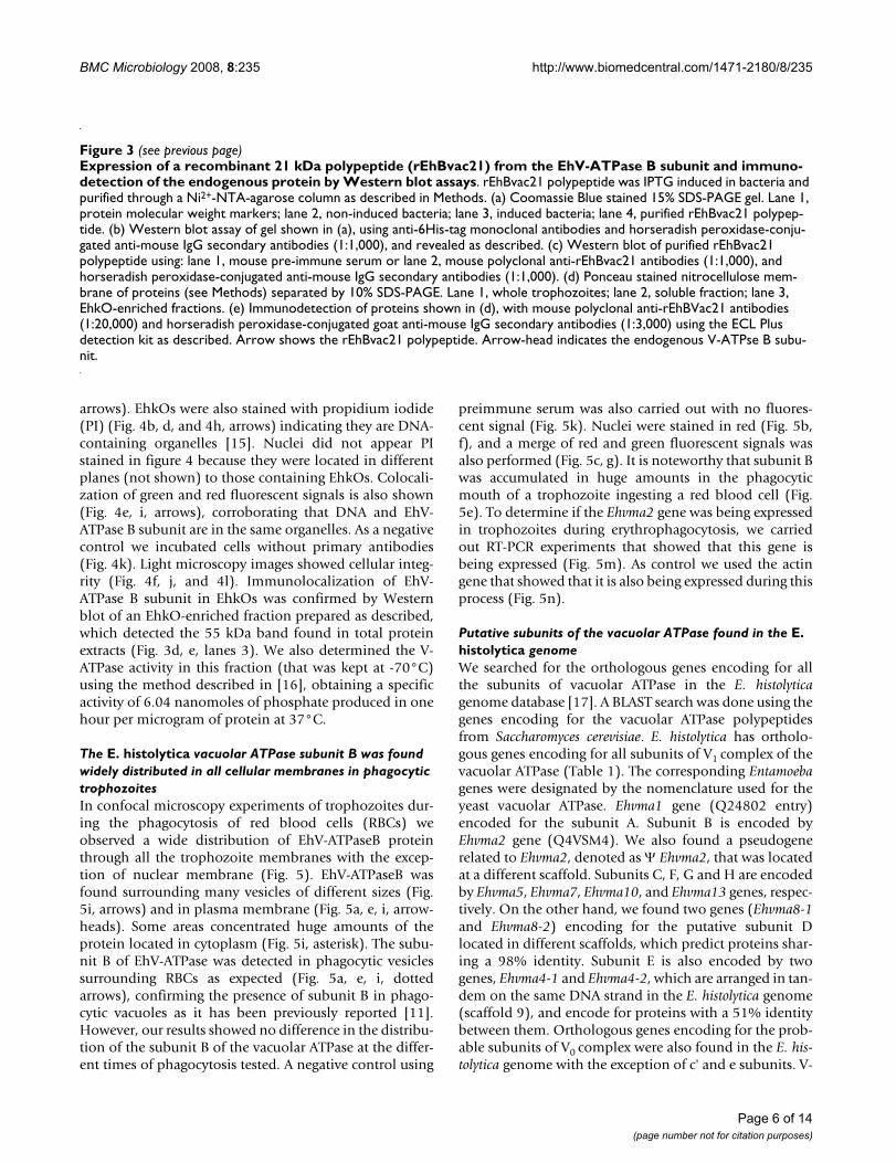
Expression of a specific polypeptide of the E. histolytica vacuolar ATPase subunit B in bacteriaTo determine if the E. histolytica genome containssequences related to the V-ATPase B subunit, we per-formed a BLAST search and found that the V-ATPase Asubunit was the only sequence related. It showed 23%identity and 38% similarity to EhV-ATPaseB (Fig. 2). The
most divergent region between these sequences resides intheir N-terminal ends. Thus, we selected a 462 bp region(I81 to V234) of the gene encoding for the V-ATPase sub-unit B to be cloned in frame into pRSET A vector to expressa 21 kDa recombinant His-tagged polypeptide(rEhBvac21) in E. coli (Fig. 3a, lane 3). This polypeptidewas purified by immobilized metal affinity chromatogra-
Organization of the Ehvma2 geneFigure 1Organization of the Ehvma2 gene. (a) Ehvma2 gene is structured by exons E1 (1–64 nt) and E2 (200–1,626 nt) separated by an intron (I) of 135 bp (65–199 nt). Arrows show position Bvac-S-1 (sense) and Bvac-AS-833 (antisense) primers used in PCR with genomic DNA (gDNA) and RT-PCR. Predicted lengths of DNA amplified from gDNA (833 bp) and from cDNA (700 bp) are also shown. (b) EtBr stained 4% PAGE gel. Lane 1, 100 bp DNA ladder; lane 2, DNA amplified by RT-PCR; lane 3, DNA amplified with gDNA; lane 4, negative control of RT-PCR where no reverse transcriptase was used. (c) Structure of EhV-ATPaseB showing the beta-barrel domain (D-I), the nucleotide-binding domain (D-II), the C-terminal domain found in the ATP synthase alpha/beta family (D-III), the putative actin-binding site, the BzATP-binding site sequence, and the signature of ATP synthase alpha and beta subunits.
Page 3 of 14(page number not for citation purposes)

BMC Microbiology 2008, 8:235 http://www.biomedcentral.com/1471-2180/8/235
phy (IMAC) through a Ni2+-NTA agarose column underdenaturing conditions (Fig. 3a, lane 4). In Western blotassays the anti-6His-Tag monoclonal antibodies recog-nized the induced rEhBvac21 polypeptide band in extractsof 1 mM IPTG induced bacteria (Fig. 3b, lane 3) and puri-fied rEhBvac21 (Fig. 3b, lane 4). This result confirms thenature of induced polypeptide. Therefore, rEhBvac21 pro-tein was electroeluted from 12% SDS-PAGE gels and usedto produce mouse polyclonal anti-rEhBvac21 antibodiesthat specifically identified rEhBvac21 in induced bacteria(Fig. 3c, lane 2), while pre-immune serum did not recog-nize any bacterial polypeptide (Fig. 3c, lane 1). Mousepolyclonal anti-rEhBvac21 antibodies reacted with a 55kDa band in trophozoite total extracts (Fig. 3d, e, lanes 1),
which corresponds to the predicted molecular weight ofthe full length EhV-ATPase B subunit.
Localization of the subunit B of V-ATPase in E. histolytica trophozoitesWe carried out immunodetection experiments of endog-enous EhV-ATPaseB protein using anti-rEhBvac21 anti-bodies and paraformaldehyde-fixed trophozoites.Through confocal microscopy, antibodies anti-rEhBvac21located the EhV-ATPase B polypeptide in many small ves-icles (Fig. 4a, c, g). The EhV-ATPase B subunit was alsoobserved in EhkOs, but not in nuclei (Fig. 4a, c, and 4g,arrows). Some cells presented the EhV-ATPase B subunitin two EhkOs located in different planes (Fig. 4a, c,
Alignment of polypeptide sequences of subunits A and B of the EhV-ATPaseFigure 2Alignment of polypeptide sequences of subunits A and B of the EhV-ATPase. Alignment was performed using the ClustalW program. Identical amino acids are shown in black boxes. Conserved changes are shown in gray boxes. Numbers correspond to amino acid positions in each polypeptide. Arrows indicate primers Bvac-S-373 and Bvac-AS-833 used to amplify by PCR a 465 bp DNA fragment containing a 154 aa region, which is a EhV-ATPaseB specific region.
Page 4 of 14(page number not for citation purposes)

BMC Microbiology 2008, 8:235 http://www.biomedcentral.com/1471-2180/8/235
Figure 3 (see legend on next page)
Page 5 of 14(page number not for citation purposes)
BMC Microbiology 2008, 8:235 http://www.biomedcentral.com/1471-2180/8/235
arrows). EhkOs were also stained with propidium iodide(PI) (Fig. 4b, d, and 4h, arrows) indicating they are DNA-containing organelles [15]. Nuclei did not appear PIstained in figure 4 because they were located in differentplanes (not shown) to those containing EhkOs. Colocali-zation of green and red fluorescent signals is also shown(Fig. 4e, i, arrows), corroborating that DNA and EhV-ATPase B subunit are in the same organelles. As a negativecontrol we incubated cells without primary antibodies(Fig. 4k). Light microscopy images showed cellular integ-rity (Fig. 4f, j, and 4l). Immunolocalization of EhV-ATPase B subunit in EhkOs was confirmed by Westernblot of an EhkO-enriched fraction prepared as described,which detected the 55 kDa band found in total proteinextracts (Fig. 3d, e, lanes 3). We also determined the V-ATPase activity in this fraction (that was kept at -70°C)using the method described in [16], obtaining a specificactivity of 6.04 nanomoles of phosphate produced in onehour per microgram of protein at 37°C.
The E. histolytica vacuolar ATPase subunit B was found widely distributed in all cellular membranes in phagocytic trophozoitesIn confocal microscopy experiments of trophozoites dur-ing the phagocytosis of red blood cells (RBCs) weobserved a wide distribution of EhV-ATPaseB proteinthrough all the trophozoite membranes with the excep-tion of nuclear membrane (Fig. 5). EhV-ATPaseB wasfound surrounding many vesicles of different sizes (Fig.5i, arrows) and in plasma membrane (Fig. 5a, e, i, arrow-heads). Some areas concentrated huge amounts of theprotein located in cytoplasm (Fig. 5i, asterisk). The subu-nit B of EhV-ATPase was detected in phagocytic vesiclessurrounding RBCs as expected (Fig. 5a, e, i, dottedarrows), confirming the presence of subunit B in phago-cytic vacuoles as it has been previously reported [11].However, our results showed no difference in the distribu-tion of the subunit B of the vacuolar ATPase at the differ-ent times of phagocytosis tested. A negative control using
preimmune serum was also carried out with no fluores-cent signal (Fig. 5k). Nuclei were stained in red (Fig. 5b,f), and a merge of red and green fluorescent signals wasalso performed (Fig. 5c, g). It is noteworthy that subunit Bwas accumulated in huge amounts in the phagocyticmouth of a trophozoite ingesting a red blood cell (Fig.5e). To determine if the Ehvma2 gene was being expressedin trophozoites during erythrophagocytosis, we carriedout RT-PCR experiments that showed that this gene isbeing expressed (Fig. 5m). As control we used the actingene that showed that it is also being expressed during thisprocess (Fig. 5n).
Putative subunits of the vacuolar ATPase found in the E. histolytica genomeWe searched for the orthologous genes encoding for allthe subunits of vacuolar ATPase in the E. histolyticagenome database [17]. A BLAST search was done using thegenes encoding for the vacuolar ATPase polypeptidesfrom Saccharomyces cerevisiae. E. histolytica has ortholo-gous genes encoding for all subunits of V1 complex of thevacuolar ATPase (Table 1). The corresponding Entamoebagenes were designated by the nomenclature used for theyeast vacuolar ATPase. Ehvma1 gene (Q24802 entry)encoded for the subunit A. Subunit B is encoded byEhvma2 gene (Q4VSM4). We also found a pseudogenerelated to Ehvma2, denoted as Ψ Ehvma2, that was locatedat a different scaffold. Subunits C, F, G and H are encodedby Ehvma5, Ehvma7, Ehvma10, and Ehvma13 genes, respec-tively. On the other hand, we found two genes (Ehvma8-1and Ehvma8-2) encoding for the putative subunit Dlocated in different scaffolds, which predict proteins shar-ing a 98% identity. Subunit E is also encoded by twogenes, Ehvma4-1 and Ehvma4-2, which are arranged in tan-dem on the same DNA strand in the E. histolytica genome(scaffold 9), and encode for proteins with a 51% identitybetween them. Orthologous genes encoding for the prob-able subunits of V0 complex were also found in the E. his-tolytica genome with the exception of c' and e subunits. V-
Expression of a recombinant 21 kDa polypeptide (rEhBvac21) from the EhV-ATPase B subunit and immunodetection of the endogenous protein by Western blot assaysFigure 3 (see previous page)Expression of a recombinant 21 kDa polypeptide (rEhBvac21) from the EhV-ATPase B subunit and immuno-detection of the endogenous protein by Western blot assays. rEhBvac21 polypeptide was IPTG induced in bacteria and purified through a Ni2+-NTA-agarose column as described in Methods. (a) Coomassie Blue stained 15% SDS-PAGE gel. Lane 1, protein molecular weight markers; lane 2, non-induced bacteria; lane 3, induced bacteria; lane 4, purified rEhBvac21 polypep-tide. (b) Western blot assay of gel shown in (a), using anti-6His-tag monoclonal antibodies and horseradish peroxidase-conju-gated anti-mouse IgG secondary antibodies (1:1,000), and revealed as described. (c) Western blot of purified rEhBvac21 polypeptide using: lane 1, mouse pre-immune serum or lane 2, mouse polyclonal anti-rEhBvac21 antibodies (1:1,000), and horseradish peroxidase-conjugated anti-mouse IgG secondary antibodies (1:1,000). (d) Ponceau stained nitrocellulose mem-brane of proteins (see Methods) separated by 10% SDS-PAGE. Lane 1, whole trophozoites; lane 2, soluble fraction; lane 3, EhkO-enriched fractions. (e) Immunodetection of proteins shown in (d), with mouse polyclonal anti-rEhBVac21 antibodies (1:20,000) and horseradish peroxidase-conjugated goat anti-mouse IgG secondary antibodies (1:3,000) using the ECL Plus detection kit as described. Arrow shows the rEhBvac21 polypeptide. Arrow-head indicates the endogenous V-ATPse B subu-nit.
Page 6 of 14(page number not for citation purposes)

BMC Microbiology 2008, 8:235 http://www.biomedcentral.com/1471-2180/8/235
ATPase a subunit is encoded by two genes, Ehstv1 andEhvph1. The former is encoded at a single locus. The latterhas two identical copies located at two different scaffolds.Ehstv1 and Ehvph1 predicted for putative proteins with51% identity. Subunits c and c" are putatively encoded byEhvma3 (Q24810 entry) and Ehvma16, respectively, andtheir polypeptides have 25% identity. A Blast search in theE. histolytica genome using the S. cerevisiae c' protein
sequence (P32842 entry) as query reported the samegenes mentioned above, but showing a lesser identitythan that shown when comparing with S. cerevisiae subu-nits c and c" (data not shown), suggesting that E. histolyt-ica genome lacks an orthologous c' subunit encodinggene. Finally, subunit d is encoded by a single copy of theEhvma6 gene. Noteworthy all the V-ATPase subunitencoding genes are located in different scaffolds.
The EhV-ATPase B subunit colocalized with the DNA-containing organelles (EhkOs)Figure 4The EhV-ATPase B subunit colocalized with the DNA-containing organelles (EhkOs). Trophozoites were fixed, permeabilized and incubated with anti-rEhBvac21 antibodies. Then, they were incubated with FITC-labeled goat anti-mouse antibodies and observed through a laser confocal microscope. (a, c) Immunodetection of EhV-ATPase B found within two dif-ferent planes of the same cell (green channel). (b, d) Sections observed in (a) and (c), respectively, showing the DNA staining (red channel). (e) Merge of green and red fluorescence emissions shown in (a) through (d) which correspond to the two cellu-lar planes mentioned before. (f) Cell from panels (a)-(e) observed by light microscopy. (g) EhV-ATPaseB localization in a sec-tion of another cell observed in green channel. (h) Same plane as (g), but observed in red channel. (i) Merge of fluorescence signals emitted in (g) and (h). (j) Cells of panels (g)-(i) observed by light microscopy. (k) Cells incubated only with secondary antibodies as a negative control and observed in green channel (maximal projection). (l) Cells of negative control observed by light microscopy. Arrows indicate EhkOs.
Page 7 of 14(page number not for citation purposes)

BMC Microbiology 2008, 8:235 http://www.biomedcentral.com/1471-2180/8/235
Page 8 of 14(page number not for citation purposes)
Localization of EhV-ATPase B subunit in trophozoites during RBC phagocytosisFigure 5Localization of EhV-ATPase B subunit in trophozoites during RBC phagocytosis. Trophozoites were incubated with diamine bencidine-stained RBCs for 5 min (a-j), washed with PBS and fixed with paraformaldehyde. Then, cells were treated with anti-rEhBvac21 antibodies and FITC-labeled goat anti-mouse antibodies, and observed through a laser confocal microscope as described. (a, e, i). Immunodetection of EhV-ATPaseB observed in green channel. (b, f) Cells observed in red channel. (c, g) Merge of fluorescent signals observed in (a, b) and (e, f), respectively. (d, h, j, l) Cells observed by light micros-copy. (k) Cells incubated only with secondary antibodies as negative control and observed in green channel. (m, n) Electro-phoresis in 1.0% agarose gels of RT-PCR gene products of Ehvma2 (m) and actin (n) in trophozoites at 0 min (lane 2) and 30 min (lane 3) of erythrophagocytosis. Lane 1, 100 bp size molecular ladder. Arrowheads, EhV-ATPaseB found in plasma mem-branes. Arrows, EhV-ATPaseB polypeptide found in cytoplasmic vesicles. Dotted arrows, EhV-ATPaseB polypeptide found in phagocytic vacuoles containing RBCs. Asterisks, huge concentrations of EhV-ATPaseB found within trophozoites.

BMC Microbiology 2008, 8:235 http://www.biomedcentral.com/1471-2180/8/235
DiscussionThe Ehvma2 gene encoding for the B subunit of V-ATPaseis highly conserved (Fig. 1c). The protein was located incytoplasm (showing punctate and diffuse patterns),plasma membrane, vacuolar membrane and in EhkOs(Fig. 4 and Fig. 5). Labeling in cytoplasm could corre-spond to free V1 sectors and to V-ATPase complexes foundin small fluorescent vesicles which might be lysosomes[18,19]. In yeast cells, the RAVE complex mediates thereversible dissociation between the V1 and V0 complexesby binding to peripheral stalk subunits (C or E and G)[20,21], that is considered as one of the mechanisms con-trolling V-ATPase activity [20-22]. This event seems to beregulated by the cellular environment as in yeast inresponse to glucose depletion [23]. The gene encoding forthe Skp1 subunit of the RAVE complex is found in the E.histolytica genome (there are four gene sequences encod-ing for this subunit in Pathema: EHI_118670,EHI_174130, EHI_066770 and EHI_174180), suggestinga similar V-ATPase regulation mechanism in this parasite.
On the other hand, the V-ATPase B subunit found in theE. histolytica plasma membrane (Fig. 5) could indicate thatthe V-ATPase complex is located there and has a role in tis-sue invasion in a similar process of metastasizing cells thatincrease the acidification of the extracellular fluid to pro-duce the destruction of normal tissue and facilitate inva-sion [3]. Moreover, it could be involved in the transport ofdiverse solutes in a similar way as it has been described forPlasmodium [24]. It is noteworthy the subunit B localiza-tion in EhkOs (Fig. 4), which are cytoplasmic DNA-con-taining organelles of unknown function [15,25]. EhkOs
carry EhTBP [26], EhC/EBP [27], and Ehp53 [28] tran-scription factors, and the EhPFO enzyme [29], but theirroles in EhkOs remain to be determined. Another cyto-plasmic DNA-containing organelle described in this para-site is the crypton, which harbors the chaperonin Hsp60[19,30].
Mitosomes, which are DNA-lacking organelles, also con-tain the chaperonin Cpn60 and are considered as mito-chondrial remnants [31]. Mitosomes have also beenfound in other organisms [32-34]. EhkOs and cryptonscould be similar organelles as they carry DNA. However,EhkOs and mitosomes are clearly different [35]. A possi-ble role for the V-ATPase located in EhkOs could be theATP synthesis using the membrane-bound H+- translocat-ing inorganic pyrophosphatase (H+-PPase) activity foundin some organelles in trophozoites [36]. Alternatively, V-ATPase could be pumping out H+ as a consequence of theB subunit localization towards the lumen of theseorganelles (Fig. 4), in order to keep pH regulated andavoid degradation of their content.
RBCs ingestion by trophozoites revealed a clustering proc-ess of EhV-ATPase B subunit in the vicinity of phagosomesafter 5 min of phagocytosis (Fig. 5). The principal route ofV-ATPase delivery to phagosomes in D. discoideum is byfusion with pre-existing acidic endosomal vesicles, priorto their content delivery into phagosomes [37]. A similarprocess might be occurring in Entamoeba, where lyso-somes could be stored in the cytosol prior to their fusionwith phagosomal vacuoles. During erythrophagocytosis apre-phagosomal vacuole (PPV) containing Rab5 and
Table 1: Genes encoding for putative subunits of the vacuolar ATPase of Entamoeba histolytica
Subunit Gene Locus Function#
A (Q24802)* Ehvma1 EHI_043010¶ Catalytic site and regulationB (Q4VSM4)* Ehvma2 EHI_189850¶ Non-catalytic site
ψEhvma2 122.t00032§ Involved in targeting?C Ehvma5 EHI_059840¶ Assembly and activityD Ehvma8-1 EHI_056390¶ Assembly and activity
Ehvma8-2 202.t00016§
E Ehvma4-1 9.t00012§ Assembly and activityEhvma4-2 9.t00013§
F Ehvma7 EHI_007940¶ Assembly and activityG Ehvma10 94.t00029§ Assembly and activityH Ehvma13 215.t00004§ Activity, but not assemblya Ehvph1 EHI_074020¶
417.t00001§H+ transport, assembly, and targeting
Ehstv1 EHI_107280¶
c (Q24810)* Ehvma3 EHI_059840¶ H+-transport; DCCD-binding sitec" Ehvma16 289.t00003§ H+ transportd Ehvma6 161.t00001§ Activity and assembly
*UniProtKB/TrEMBL Accession number.¶Locus name assigned at Pathema (Bioinformatics Resource Center; http://pathema.jcvi.org/cgi-bin/Entamoeba/PathemaHomePage.cgi).§Locus with its provisional names in the E. histolytica genome project at The J. Craig Venter Institute.#Function was assigned based on reference [5].
Page 9 of 14(page number not for citation purposes)

BMC Microbiology 2008, 8:235 http://www.biomedcentral.com/1471-2180/8/235
Rab7A proteins is gradually acidified by fusion with lyso-somes. During the first 5–10 min Rab5 is dissociated fromPPVs, and digestive proteins are transported from PPVs tophagosomes via Rab7A-mediated vesicular transport[38,39]. Although this process needs more studies to iden-tify all proteins involved, V-ATPase should be releasedduring PPV maturation. In addition, interactions of V-ATPase to cytoskeleton through subunits B and C, whichare able to bind actin, are essentials for its proper func-tion. For example, the osteoclast subunit B is responsiblefor microfilaments-V-ATPase interactions [40]. The puta-tive actin-binding domain that was found in EhV-ATPasesubunit B suggests that this process is conserved in thisparasite (Fig. 1).
In eukaryotic cells the V1 complex is formed by the subu-nits A3B3CDEFG2H1–2 and the V0 complex by subunitsac4c'c"de [3-5]. Based on the genes found in the E. histolyt-ica genome, we propose a putative model for the structure
of the vacuolar ATPase in this parasite (Fig. 6). The V0complex might be composed of a ring of five or six prote-olipid subunits (four c subunits and either one or two c"subunits, respectively) and one molecule of each a and dsubunits. Remarkably, E. histolytica V-ATPase lacks subu-nits e and c'. However, the existence of non-related pro-teins to e and c' subunits that could evolve separately veryearly in evolution in Entamoeba is possible. It is importantto note that the stoichiometry of the E and G strong inter-action is not well established yet. A relationship of two Gmolecules per one E molecule was assumed, howeverrecent data support that in V-ATPase, G and E subunits arepresent in more than one copy and thus two peripheralstalks connecting V1 to V0 could exist [41].
Most of the subunits of the E. histolytica V-ATPase areencoded by single genes, excepting for subunits D, E, anda, which are encoded by two genes in each case (Table 1).In the case of the subunit E, two E isoforms have been
Hypothetical model of the structure of the vacuolar ATPase of Entamoeba histolyticaFigure 6Hypothetical model of the structure of the vacuolar ATPase of Entamoeba histolytica. The Vacuolar ATPase of E. histolytica is a well conserved protein complex, with the exceptions of subunits c' and e, which have not conserved counter-parts in this parasite. The structure shown is according to that described in [3,5] and based on data from Table 1. Subunit e that was not found in the E. histolytica genome is surrounded by a dotted line.
Page 10 of 14(page number not for citation purposes)

BMC Microbiology 2008, 8:235 http://www.biomedcentral.com/1471-2180/8/235
found in mouse [6], and three in Arabidopsis [42]. Severalpea epicotyls V-ATPases isoenzymes exhibiting differentratios of E1 and E2 isoforms have been purified, exhibit-ing differences in their Km and Vmax of ATP hydrolysis,and different tissue distribution in the whole plant [43].Yeast has two subunit a isoforms: 1) Vph1p, which is tar-geted to the vacuolar membrane, and 2) Stv1p which istargeted to the late Golgi apparatus [44]. Mammals havefour genes encoding for different subunit a isoforms,often showing cell-type or tissue-specific locations [5]. Aspecial case is Paramecium, which contains 17 a subunitisoforms, which provide a specific location and functionfor V-ATPase in at least seven different compartments[45]. Thus, E. histolytica could have at least two V-ATPaseisoforms, one distributed in vacuoles, and the other in theGolgi apparatus that could be more primitive in tropho-zoites than those from other systems due to the lack of atypical Golgi structure [46-48]. More studies are needed toelucidate the V-ATPase role in the physiology of E. histolyt-ica trophozoites.
ConclusionWe cloned the Ehvma2 gene which encodes for the highlyconserved V-ATPase B subunit. This protein was immu-nolocated in EhkOs, in plasma membrane, vesicles,phagocytic vacuoles, and also in the cytoplasm. Most ofthe orthologous genes encoding for the V-ATPase subunitswere also identified in the E. histolytica genome showingthat this EhV-ATPase is a highly conserved complex. Thein silico analysis of the genes encoding for the V-ATPasesubunits in the E. histolytica genome revealed that the V1complex has all the eight subunits found in V-ATPases.However, the V0 complex lacks the subunits e and c'.
MethodsE. histolytica culturesTrophozoites of E. histolytica clone A were axenically cul-tured in TYI-S-33 medium at 37°C and harvested duringthe exponential growth phase as described [49]. ForEhkOs purification, the medium was supplemented with2 μCi/ml [methyl-3H]-Thymidine (Amersham) for 48 h[50].
Search of V-ATPase genes in the E. histolytica genome databaseA search of V-ATPase genes was performed in the E. histo-lytica genome databases at The Sanger Institute http://www.sanger.ac.uk and The J. Craig Venter Institute http://www.jcvi.org. As query, we used the subunit polypeptidesequences of the vacuolar ATPase of S. cerevisiae, and theWU-BLAST version 2.0 program and BLOSUM62 matrix.The UniProt Knowledgebase (UniProtKB)/TrEMBL entriesused were P17255 (subunit A), P16140 (subunit B),P31412 (subunit C), P32610 (subunit D), P22203 (subu-nit E), P39111 (subunit F), P48836 (subunit G), P41807
(subunit H), P37296 (subunit a), P25515 (subunit c),P32842 (subunit c'), P23968 (subunit c"), P32366 (subu-nit d), and Q3E7B6 (subunit e). Obtained DNAsequences were translated to proteins with the Translatetool at the ExPASy Proteomics Server http://www.expasy.org. Blast search for each of the Entamoebapolypeptide sequences was done with BLASTP 2.2.14algorithm in The UniProt Knowledgebase at the ExPASyProteomics server of the Swiss Institute of Bioinformaticsand BLOSUM62 matrix. Alignments were performed withClustalW version 1.83 algorithm at the European Bioin-formatics Institute (EBI, http://www.ebi.ac.uk/Tools/clustaw2/index.html).
Cloning of the Ehvma2 gene encoding for the subunit B of the E. histolytica vacuolar ATPaseBased on the nucleotide sequence of the putative Ehvma2gene found in the E. histolytica genome databases, wedesigned the primers S-Bvac (sense): 5'-ATGTTATTGTAT-TAAGACTTTTTAATT-3', and AS-Bvac (antisense): 5'-TATAAGGTTCCTTCTTTGTCGT-3' to amplify a 1.87 kbDNA fragment using 50 ng of total DNA as a template,and 400 μM of each dNTP, 2.5 mM MgCl2, 300 nM ofeach primer and 2 U of Taq DNA polymerase (Invitrogen).PCR conditions were 94°C for 5 min, 30 cycles of 94°Cfor 30 s, 57°C for 30 s and 72°C for 90 s, and 72°C for 10min. Amplified DNA was cloned into the pGEM-T-Easyvector (Promega) to generate pGEM-T-Easy-Bvac plasmid.Sequencing of cloned DNA was carried out using genespecific primers and the Big Dye Terminator kit version2.0 in an Automated DNA Sequencer (310 Genetic Ana-lyzer, Applied Biosystems) in the Sequencing Nucleic AcidCore Facility at Cinvestav-IPN.
RT-PCRTotal RNA was isolated from trophozoites using TRIzolreagent (Invitrogen) and reverse transcription was carriedout with 200 U of SuperScript II reverse transcriptase (Inv-itrogen) and 40 U of SUPERase-in RNAse inhibitor(Ambion) according to manufacturers' instructions and 1/6 of reaction volume was used for DNA amplification byPCR with Bvac-S-1 (5'-ATGGAAGCTTTCAAAATT-3')sense and Bvac-AS-833 (5'-CTTTCCATAGAACCATTTTC-3') antisense primers using the PCR conditions describedabove. RT-PCR products were separated on a 4% PAGE gelin 1× TBE (90 mM Tris, 90 mM H3BO3, and 2 mM EDTA,pH 8.3), stained with 0.05% (w/v) ethidium bromide(EtBr), visualized with a UV transilluminator, and imageswere processed with a Gel Documentation System (Bio-Rad). For RT-PCR analysis of Ehmav2 and actin genes introphozoites during RBCs erythrophagocytosis, we pro-ceeded as described bellow, but no fixative agents wereemployed. For actin gene we used primers actin-sense 5'-AGCTGTTCTTTCATTATATGC -3' and actin-antisense 5'-TCTCTTTCAGCACTAGTGGT-3', and used a Tm of 54°C.
Page 11 of 14(page number not for citation purposes)

BMC Microbiology 2008, 8:235 http://www.biomedcentral.com/1471-2180/8/235
Expression and purification of a recombinant fragment polypeptide of E. histolytica V-ATPase subunit B in Escherichia coliTo express a specific region of EhV-ATPaseB, we amplifiedby PCR (using the conditions mentioned above) a 478 bpDNA fragment (containing a 465 bp DNA encoding for a154 aa polypeptide of EhV-ATPaseB and the translationstop signal). The primers used were Bvac-S-373 (5'-CGGGATCCGGTATTGATGCACGTTATAC-3') sense andBvac-AS-833 (5'-CCCAAGCTTTTATCTTTCATAGAAC-CATTTTC-3') antisense, which contained the BamHI andHindIII restriction sites at their 5' ends, respectively. Then,the amplified DNA was cloned into pRSET A vector (Inv-itrogen) to generate the recombinant pRSET-EhBvac465plasmid. Recombinant protein expression was inducedwith 1 mM IPTG in E. coli BL21(DE3) pLysS (Invitrogen).N-terminal His-tagged recombinant EhBvac 21 kDa(rEhBvac21) polypeptide was purified under denaturingconditions by IMAC using a Ni2+-NTA agarose column(Qiagen), following the manufacturer's protocol. Purifica-tion of rEhBvac21 polypeptide was improved by electroe-lution from preparative 15% SDS-PAGE gels.
Generation of mouse polyclonal antibodies against the rEhBvac21 polypeptide and Western blot assaysSpecific antibodies against EhV-ATPaseB were generatedin BALB/c mice by immunization of 100 μg of purifiedrEhBvac21 polypeptide mixed with Freund's adjuvant.Three bursts were applied via intraperitoneal at ten dayintervals. Immune serum was collected seven days afterthe last immunization and used for Western blot assays.Mice were bleeding before immunization to use pre-immune sera as a control in all experiments. For Westernblot assays total protein extracts of induced bacteria orpurified rEhBvac21 polypeptide were separated by 15%SDS-PAGE, transferred to nitrocellulose membranes, andblocked. This was followed by incubation with mouseanti-His monoclonal antibodies (0.3 μg/ml) (Roche) for1 h at 37°C, or by incubation with mouse polyclonal anti-rEhBvac21 antibodies (1:1,000) overnight at 4°C. Then,membranes were washed and incubated with horseradishperoxidase-conjugated anti-mouse IgG secondary anti-bodies (Zymed) (1:1,000) for 3 h at 37°C. Immunoreac-tive bands were revealed with 4-chloro-1-naphthol and0.03% (v/v) H2O2. For Western blot analysis of total tro-phozoite extracts and EhkO-enriched fraction, proteinswere separated on 10% SDS-PAGE. Then, membraneswere incubated with anti-rEhBvac21 antibodies(1:20,000) for 3 h at 37°C, and secondary antibodies(1:3,000) at 37°C for 1 h. Positive bands were detectedusing the ECL Plus detection kit (Amersham) followed byexposure to X-ray films (Kodak).
Immunofluorescence and confocal microscopyTrophozoites on prewarmed cover slips were fixed with3% (w/v) paraformaldehyde for 1 h at 37°C, incubatedwith 50 mM NH4Cl for 1 h at 37°C, permeabilized with0.5% (v/v) Triton X-100 at 37°C for 30 min and blockedwith 0.1% (w/v) BSA in PBS at 37°C for 30 min. Then,cells were incubated with mouse polyclonal anti-rEhBvac21 antibodies (1:80) for 2 h. Subsequently, cellswere washed with PBS and incubated with FITC-labeledgoat anti-mouse secondary antibodies (1:200) in blockingsolution at 37°C for 2 h. Finally, cells were washed andcounterstained with 20 μg/ml PI (Fluka), and observedthrough a Leika NT-TCS confocal microscope. As a con-trol, cells were incubated without the anti-rEhBvac21 anti-bodies.
Phagocytosis assaysTrophozoites were incubated with human RBCs at 37°Cfor different times (2 to 120 min) as described [51]. At theend of the incubation times, the culture medium wasremoved and trophozoites were washed with PBS andfixed with 3% (w/v) paraformaldehyde. For immunofluo-rescence experiments cells were treated as describedbefore.
Isolation of an EhkO-enriched fraction and V-ATPase activity assayEhkO-enriched fraction purification steps were carried outat 4°C as described [50]. [3H]-Thymidine labeled tropho-zoites were washed and resuspended in eight volumes ofbuffer A (10 mM HEPES pH 7.9, 10 mM EDTA, and 10mM DTT) containing protease inhibitors and 250 mMsucrose. Cells were gently disrupted on ice using a Potterhomogenizer and centrifuged at 160 × g for 10 min. Then,the supernatant (nuclei-depleted fraction) was centri-fuged at 10,000 × g for 10 min at 4°C in a JA-20 rotor(Beckman) to obtain the soluble fraction and the pellet.The latter was resuspended in 15% (v/v) Nycodenz(Nycomed Pharma AS) in buffer A and top loaded on aNycodenz discontinuous gradient (30, 40 and 50%, v/v),which was centrifuged at 13,000 × g for 60 min at 4°C ina SW40Ti rotor (Beckman). 0.5 ml fractions were col-lected with a DensiFlow II C system (Buchler Instruments)and a RediFrac 1,000 fraction collector (Bio-Rad). 100 μlof each fraction were 10% (w/v) TCA precipitated andradioactivity content was determined in a LS6500 liquidscintillation counter (Beckman). EhkO enriched fractionswere identified by its [3H]-Thymidine incorporation.
The vacuolar ATPase activity was determined using a mod-ification of the method described by Conibear and Ste-vens [16]. The sample was incubated in 50 mM MES/Trisbuffer pH 6.9, 5 mM MgSO4 and 5 mM ATP at 37°C for16 h. Some reactions were performed incubating somesamples with 0.1 mM Na3VO4, or 300 nM concanamycin
Page 12 of 14(page number not for citation purposes)

BMC Microbiology 2008, 8:235 http://www.biomedcentral.com/1471-2180/8/235
A, or both. Released phosphate was determined byabsorbance at 630 nm after a 15 min incubation period atRT with a mixture of 1% (w/v) (NH4)6Mo7O24, 4% (w/v)H2SO4, 1% (w/v) SDS and 0.2% (w/v) ascorbic acid.
Authors' contributionsAll authors conceived the study. MGMH isolated andcharacterized the Ehvm2 gene, expressed the recombinantpolypeptide in bacteria, obtained the anti-EhV-ATPasesubunit B antibodies, purified EhkOs and performed theconfocal microscopy studies. MLLB helped in RT-PCRassays and measurement of vacuolar ATPase activity.MGMH and JPLA performed the in silico analysis. EO andJPLA drafted the manuscript. All authors read andapproved the final manuscript.
AcknowledgementsWe thank CONACyT from Mexico for the financial support for this project.
References1. Ackers JP, Mirelman D: Progress in research on Entamoeba his-
tolytica pathogenesis. Curr Opin Microbiol 2006, 9(4):367-373.2. Ravdin JI: Amebiasis: Human Infection by Entamoeba histolyt-
ica. New York: John Wiley & Sons Inc; 1988. 3. Beyenbach KW, Wieczorek H: The V-type H+ ATPase: molecu-
lar structure and function, physiological roles and regulation.J Exp Biol 2006, 209:577-589.
4. Kane P: The where, when, and how of organelle acidificationby the yeast vacuolar H+-ATPase. Microbiol Mol Biol Rev 2006,70:177-191.
5. Nishi T, Forgac M: The vacuolar (H+)-ATPases-natures' mostversatile proton pumps. Nat Rev Mol Cell Biol 2002, 3:94-103.
6. Sun-Wada GH, Wada Y, Futai M: Diverse and essential roles ofmammalian vacuolar-type proton pump ATPase: towardthe physiological understanding of inside acidic compart-ments. Biochim Biophys Acta 2004, 1658:106-114.
7. Hurtado-Lorenzo A, Skinner M, El Annan J, Futai M, Sun-Wada GH,Bourgoin S, Casanova J, Wildeman A, Bechoua S, Ausiello DA, BrownD, Marshansky V: V-ATPase interacts with ARNO and Arf6 inearly endosomes and regulates the protein degradativepathway. Nature Cell Biol 2006, 8:124-136.
8. Löhden-Bendinger U, Bakker-Grunwald T: Evidence for vacuolartype proton ATPase in Entamoeba histolytica. Z Naturforsch [C]1990, 45(3-4):229-232.
9. Yi Y, Samuelson J: Primary structure of the Entamoeba histolyt-ica gene (Ehvma1) encoding the catalytic peptide of a puta-tive vacuolar membrane proton-transporting ATPase (V-ATPase). Mol Biochem Parasitol 1994, 66:165-169.
10. Descoteaux S, Yu Y, Samuelson J: Cloning of Entamoeba genesencoding proteolipids of putative vacuolar proton-translo-cating ATPases. Infect Immun 1994, 62(8):3572-3575.
11. Okada M, Huston C, Mann B, Petri W, Kita K, Nozaki T: Proteomicanalysis of phagocytosis in the enteric protozoan parasiteEntamoeba histolytica. Eukaryotic Cell 2005, 4:827-831.
12. Okada M, Nozaki T: New insights into molecular mechanismsof phagocytosis in Entamoeba histolytica by proteomic analy-sis. Arch Med Res 2006, 37:244-252.
13. Futai M, Iwamoto A, Omote H, Maeda M: A glicine-rich sequencein the catalytic site of F-type ATPase. J Bioenerg Biomembr 1992,24:463-467.
14. Vasilyeva E, Forgac M: 3-O-(4-Benzoyl) benzoyladenosine 5-tri-phosphate inhibits activity of the vacuolar (H+)-ATPase frombovine brain clathrin-coated vesicles by modification of arapidly exchangeable, noncatalytic nucleotide binding site onthe B subunit. J Biol Chem 1996, 271:12775-12782.
15. Orozco E, Gharaibeh R, Riverón AM, Delgadillo DM, Mercado M,Sánchez T, Gómez Conde E, Vargas MA, López-Revilla R: A novelcytoplasmic structure containing DNA networks in Enta-
moeba histolytica trophozoites. Mol Gen Genet 1997,254:250-257.
16. Conibear E, Stevens TH: Studying yeast vacuoles. Methods Enzy-mol 2002, 351:408-432.
17. Loftus B, Anderson I, Davies R, Alsmark UC, Samuelson J, Amedeo P,Roncaglia P, Berriman M, Hirt RP, Mann BJ, et al.: The genome ofthe protist parasite Entamoeba histolytica. Nature 2005,433(7028):865-868.
18. Aley S, Cohn Z, Scott W: Endocytosis in Entamoeba histolytica.J Exp Med 1984, 160:724-737.
19. Ghosh S, Field J, Rogers R, Hickman M, Samuelson J: The Enta-moeba histolytica mitochondrion-derived organelle (crypton)contains double-stranded DNA and appears to be bound bya double membrane. Infect Immun 2000, 68:4319-4322.
20. Smardon A, Tarsio M, Kane P: The RAVE Complex is essentialfor stable assembly of the yeast V-ATPase. J Biol Chem 2002,277:13831-13839.
21. Smardon A, Kane P: RAVE is essential for the efficient assemblyof the C subunit with the vacuolar H+-ATPase. J Biol Chem2007, 282:26185-26194.
22. Peters C, Bayer MJ, Buhler S, Andersen JS, Mann M, Mayer A: Trans-complex formation by proteolipid channels in the terminalphase of membrane fusion. Nature 2001, 409:581-588.
23. Qi J, Forgac M: Cellular environment is important in control-ling V-ATPase dissociation and its dependence on activity. JBiol Chem 2007, 282:24743-24751.
24. Moriyama Y, Hayashi M, Yatsushiro S, Yamamoto A: Vacuolar pro-ton pumps in malaria parasite cells. J Bioenerg Biomembr 2003,35:367-375.
25. Solís F, Orozco E, Córdova L, Rivera B, Luna-Arias JP, Gómez-CondeE, Rodríguez MA: Entamoeba histolytica: DNA carrier vesiclesin nuclei and kinetoplast-like organelles (EhkOs). Mol GenetGenomics 2002, 267:622-628.
26. Luna-Arias JP, Hernández-Rivas R, de Dios-Bravo G, Garcia J, Men-doza L, Orozco E: The TATA-box binding protein of Enta-moeba histolytica: cloning of the gene and location of theprotein by immunofluorescence and confocal microscopy.Microbiology 1999, 145:33-40.
27. Marchat LA, Gómez C, Pérez DG, Paz F, Mendoza L, Orozco E: TwoCCAAT/enhancer binding protein sites are cis-activator ele-ments of the Entamoeba histolytica EhPgp1 (mdr -like) geneexpression. Cell Microbiol 2002, 4:725-737.
28. Mendoza L, Orozco E, Rodríguez MA, García-Rivera G, Sánchez T,García E, Gariglio P: Ehp53, an Entamoeba histolytica protein,ancestor of the mammalian tumour suppressor p53. Microbi-ology 2003, 149:885-893.
29. Rodriguez MA, Garcia-Perez RM, Mendoza L, Sanchez T, Guillen N,Orozco E: The pyruvate:ferredoxin oxidoreductase enzyme islocated in the plasma membrane and in a cytoplasmic struc-ture in Entamoeba. Microb Pathog 1998, 25:1-10.
30. Mai Z, Ghosh S, Frisardi M, Rosenthal B, Rogers R, Samuelson J:Hsp60 is targeted to a cryptic mitochondrion-derivedorganelle ("crypton") in the microaerophilic protozoan par-asite Entamoeba histolytica. Mol Cell Biol 1999, 19:2198-2205.
31. Tovar J, Fischer A, Clark CG: The mitosome, a novel organellerelated to mitochondria in the amitochondrial parasite Enta-moeba histolytica. Mol Microbiol 1999, 32:1013-1021.
32. Tovar J, León-Avila G, Sánchez L, Sutak R, Tachezy J, Giezen M vander, Hernández M, Müller M, Lucocq JM: Mitochondrial remnantorganelles of Giardia function in iron-sulphur protein matu-ration. Nature 2003, 426:172-176.
33. Giezen M van der, Tovar J, Clark CG: Mitochondrion-derivedorganelles in protists and fungi. Int Rev Cytol 2005, 244:175-225.
34. Giezen M van der, Tovar J: Degenerate mitochondria. EMBO Rep2005, 6:525-530.
35. Leon-Avila G, Tovar J: Mitosomes of Entamoeba histolytica areabundant mitochondrion-related remnant organelles thatlack a detectable organellar genome. Microbiology 2004,150:1245-1250.
36. McLaughlin J, Lindmark D, Müller M: Inorganic pyrophosphataseand nucleoside diphosphatase in the parasitic protozoon,Entamoeba histolytica. Biochem Biophys Res Comm 1978,82:913-920.
37. Clarke M, Köhler J, Arana Q, Liu T, Heuser J, Gerisch G: Dynamicsof the vacuolar H+-ATPase in the contractile vacuole com-
Page 13 of 14(page number not for citation purposes)
http://www.ncbi.nlm.nih.gov/entrez/query.fcgi?cmd=Retrieve&db=PubMed&dopt=Abstract&list_uids=2141984
http://www.ncbi.nlm.nih.gov/entrez/query.fcgi?cmd=Retrieve&db=PubMed&dopt=Abstract&list_uids=7984182
http://www.ncbi.nlm.nih.gov/entrez/query.fcgi?cmd=Retrieve&db=PubMed&dopt=Abstract&list_uids=7984182
http://www.ncbi.nlm.nih.gov/entrez/query.fcgi?cmd=Retrieve&db=PubMed&dopt=Abstract&list_uids=7984182
http://www.ncbi.nlm.nih.gov/entrez/query.fcgi?cmd=Retrieve&db=PubMed&dopt=Abstract&list_uids=8039932
http://www.ncbi.nlm.nih.gov/entrez/query.fcgi?cmd=Retrieve&db=PubMed&dopt=Abstract&list_uids=8039932
http://www.ncbi.nlm.nih.gov/entrez/query.fcgi?cmd=Retrieve&db=PubMed&dopt=Abstract&list_uids=8039932
http://www.ncbi.nlm.nih.gov/entrez/query.fcgi?cmd=Retrieve&db=PubMed&dopt=Abstract&list_uids=1429540
http://www.ncbi.nlm.nih.gov/entrez/query.fcgi?cmd=Retrieve&db=PubMed&dopt=Abstract&list_uids=1429540
http://www.ncbi.nlm.nih.gov/entrez/query.fcgi?cmd=Retrieve&db=PubMed&dopt=Abstract&list_uids=8662754
http://www.ncbi.nlm.nih.gov/entrez/query.fcgi?cmd=Retrieve&db=PubMed&dopt=Abstract&list_uids=8662754
http://www.ncbi.nlm.nih.gov/entrez/query.fcgi?cmd=Retrieve&db=PubMed&dopt=Abstract&list_uids=8662754
http://www.ncbi.nlm.nih.gov/entrez/query.fcgi?cmd=Retrieve&db=PubMed&dopt=Abstract&list_uids=9150258
http://www.ncbi.nlm.nih.gov/entrez/query.fcgi?cmd=Retrieve&db=PubMed&dopt=Abstract&list_uids=6206186

BMC Microbiology 2008, 8:235 http://www.biomedcentral.com/1471-2180/8/235
Publish with BioMed Central and every scientist can read your work free of charge
"BioMed Central will be the most significant development for disseminating the results of biomedical research in our lifetime."
Sir Paul Nurse, Cancer Research UK
Your research papers will be:
available free of charge to the entire biomedical community
peer reviewed and published immediately upon acceptance
cited in PubMed and archived on PubMed Central
yours — you keep the copyright
Submit your manuscript here:http://www.biomedcentral.com/info/publishing_adv.asp
BioMedcentral
plex and the endosomal pathway of Dictyostelium cells. J CellSci 2002, 115:2893-2905.
38. Saito-Nakano Y, Yasuda T, Nakada-Tsukui K, Leippe M, Nozaki T:Rab5-associated vacuoles play a unique role in phagocytosisof the enteric protozoan parasite Entamoeba histolytica. J BiolChem 2004, 279:49497-49507.
39. Nozaki T, Nakada-Tsukui K: Membrane trafficking as a virulencemechanism of the enteric protozoan parasite Entamoeba his-tolytica. Parasitol Res 2006, 98:179-183.
40. Holliday LS, Bubb MR, Jiang J, Hurst IR, Zuo J: Interactionsbetween vacuolar H+-ATPases and microfilaments in osteo-clasts. J Bioenerg Biomembr 2005, 37:419-423.
41. Ohira M, Smardon AM, Charsky CM, Liu J, Tarsio M, Kane PM: TheE and G subunits of the yeast V-ATPase interact tightly andare both present at more than one copy per V1 complex. JBiol Chem 2006, 281:22752-22760.
42. Strompen G, Dettmer J, Stierhof YD, Schumacher K, Jurgens G,Mayer U: Arabidopsis vacuolar H-ATPase subunit E isoform 1is required for Golgi organization and vacuole function inembryogenesis. Plant J 2005, 41:125-132.
43. Kawamura Y, Arakawa K, Maeshima M, Yoshida S: Tissue specificityof E subunit isoforms of plant vacuolar H+-ATPase and exist-ence of isotype enzymes. J Biol Chem 2000, 275:6515-6522.
44. Kawasaki-Nishi S, Bowers K, Nishi T, Forgac M, Stevens TH: Theamino-terminal domain of the vacuolar proton-translocat-ing ATPase a subunit controls targeting and in vivo dissocia-tion, and the carboxyl-terminal domain affects coupling ofproton transport and ATP hydrolysis. J Biol Chem 2001,276:47411-20.
45. Wassmer T, Kissmehl R, Cohen J, Plattner H: Seventeen a-subunitisoforms of Paramecium V-ATPase provide high specializa-tion in location and function. Mol Biol Cell 2006, 17:917-930.
46. Mazzuco A, Benchimol M, De Souza W: Endoplasmic reticulumand Golgi-like elements in Entamoeba. Micron 1997,28:241-247.
47. Dacks JB, Davis LA, Sjögren AM, Andersson JO, Roger AJ, DoolittleWF: Evidence for Golgi bodies in proposed 'Golgi-lacking' lin-eages. Proc Biol Sci 2003, 270(Suppl 2):S168-S171.
48. Bredeston LM, Caffaro CE, Samuelson J, Hirschberg CB: Golgi andendoplasmic reticulum functions take place in different sub-cellular compartments of Entamoeba histolytica. J Biol Chem2005, 280:32168-32176.
49. Diamond LS, Harlow R, Cunnick C: A new medium for the axeniccultivation of Entamoeba histolytica and other Entamoeba.Trans R Soc Trop Med Hyg 1978, 72:431-432.
50. Luna-Arias JP, Sanchez T, Herrera-Aguirre ME, Chavez P, Orozco E:Purification of Entamoeba histolytica DNA containingorganelles (EhkOs): a further characterization. J EukaryotMicrobiol 2003, 50(Suppl):706-708.
51. García-Rivera G, Rodríguez MA, Ocádiz R, Martínez-López MC,Arroyo R, González-Robles A, Orozco E: Entamoeba histolytica: anovel cysteine protease and an adhesin form the 112 kDasurface protein. Mol Microbiol 1999, 33:556-568.
Page 14 of 14(page number not for citation purposes)