Antiprotozoal Activity against Entamoeba histolytica of ... - MDPI
22
Molecules 2014, 19, 21044-21065; doi:10.3390/molecules191221044 molecules ISSN 1420-3049 www.mdpi.com/journal/molecules Article Antiprotozoal Activity against Entamoeba histolytica of Plants Used in Northeast Mexican Traditional Medicine. Bioactive Compounds from Lippia graveolens and Ruta chalepensis Ramiro Quintanilla-Licea 1, *, Benito David Mata-Cárdenas 2 , Javier Vargas-Villarreal 3 , Aldo Fabio Bazaldúa-Rodríguez 1 , Isvar Kavim Ángeles-Hernández 1 , Jesús Norberto Garza-González 3 and Magda Elizabeth Hernández-García 3 1 Universidad Autónoma de Nuevo León, UANL, Facultad de Ciencias Biológicas, Av. Universidad S/N, Cd. Universitaria, San Nicolás de los Garza, C.P. 66451 Nuevo León, Mexico; E-Mails: [email protected] (A.F.B.-R.); [email protected] (I.K.Á.-H) 2 Universidad Autónoma de Nuevo León, UANL, Facultad de Ciencias Químicas, Av. Universidad S/N, Cd. Universitaria, San Nicolás de los Garza, C.P. 66451 Nuevo León, Mexico; E-Mail: [email protected] 3 Laboratorio de Bioquímica y Biología Celular, Centro de Investigaciones Biomédicas del Noreste (CIBIN), Dos de abril esquina con San Luis Potosí, C.P. 64720 Monterrey, Mexico; E-Mails: [email protected] (J.V.-V.); [email protected] (J.N.G.-G.); [email protected] (M.E.H.-G.) * Author to whom correspondence should be addressed; E-Mail: [email protected]; Tel.: +52-81-8376-3668; Fax: +52-81-8352-5011. External Editor: Thomas J. Schmidt Received: 12 September 2014; in revised form: 10 December 2014 / Accepted: 11 December 2014 / Published: 15 December 2014 Abstract: Amoebiasis caused by Entamoeba histolytica is associated with high morbidity and mortality is becoming a major public health problem worldwide, especially in developing countries. Because of the side-effects and the resistance that pathogenic protozoa build against the standard antiparasitic drugs, e.g., metronidazole, much recent attention has been paid to plants used in traditional medicine around the world in order to find new antiprotozoal agents. We collected 32 plants used in Northeast Mexican traditional medicine and the methanolic extracts of these species were screened for antiprotozoal activity against E. histolytica trophozoites using in vitro tests. Only 18 extracts showed a significant inhibiting activity and among them six plant extracts showed more than 80% OPEN ACCESS
-
Upload
khangminh22 -
Category
Documents
-
view
0 -
download
0
Transcript of Antiprotozoal Activity against Entamoeba histolytica of ... - MDPI
Molecules 2014, 19, 21044-21065; doi:10.3390/molecules191221044
molecules ISSN 1420-3049
www.mdpi.com/journal/molecules
Article
Antiprotozoal Activity against Entamoeba histolytica of Plants Used in Northeast Mexican Traditional Medicine. Bioactive Compounds from Lippia graveolens and Ruta chalepensis
Ramiro Quintanilla-Licea 1,*, Benito David Mata-Cárdenas 2, Javier Vargas-Villarreal 3,
Aldo Fabio Bazaldúa-Rodríguez 1, Isvar Kavim Ángeles-Hernández 1,
Jesús Norberto Garza-González 3 and Magda Elizabeth Hernández-García 3
1 Universidad Autónoma de Nuevo León, UANL, Facultad de Ciencias Biológicas, Av. Universidad
S/N, Cd. Universitaria, San Nicolás de los Garza, C.P. 66451 Nuevo León, Mexico;
E-Mails: [email protected] (A.F.B.-R.); [email protected] (I.K.Á.-H) 2 Universidad Autónoma de Nuevo León, UANL, Facultad de Ciencias Químicas, Av. Universidad
S/N, Cd. Universitaria, San Nicolás de los Garza, C.P. 66451 Nuevo León, Mexico;
E-Mail: [email protected] 3 Laboratorio de Bioquímica y Biología Celular, Centro de Investigaciones Biomédicas del Noreste
(CIBIN), Dos de abril esquina con San Luis Potosí, C.P. 64720 Monterrey, Mexico;
E-Mails: [email protected] (J.V.-V.); [email protected] (J.N.G.-G.);
[email protected] (M.E.H.-G.)
* Author to whom correspondence should be addressed; E-Mail: [email protected];
Tel.: +52-81-8376-3668; Fax: +52-81-8352-5011.
External Editor: Thomas J. Schmidt
Received: 12 September 2014; in revised form: 10 December 2014 / Accepted: 11 December 2014 /
Published: 15 December 2014
Abstract: Amoebiasis caused by Entamoeba histolytica is associated with high morbidity
and mortality is becoming a major public health problem worldwide, especially in
developing countries. Because of the side-effects and the resistance that pathogenic protozoa
build against the standard antiparasitic drugs, e.g., metronidazole, much recent attention has
been paid to plants used in traditional medicine around the world in order to find new
antiprotozoal agents. We collected 32 plants used in Northeast Mexican traditional
medicine and the methanolic extracts of these species were screened for antiprotozoal
activity against E. histolytica trophozoites using in vitro tests. Only 18 extracts showed a
significant inhibiting activity and among them six plant extracts showed more than 80%
OPEN ACCESS

Molecules 2014, 19 21045
growth inhibition against E. histolytica at a concentration of 150 µg/mL and the IC50 values
of these extracts were determined. Lippia graveolens Kunth and Ruta chalepensis Pers.
showed the more significant antiprotozoal activity (91.54% and 90.50% growth inhibition
at a concentration of 150 µg/mL with IC50 values of 59.14 and 60.07 µg/mL, respectively).
Bioassay-guided fractionation of the methanolic extracts from these two plants
afforded carvacrol (1) and chalepensin (2), respectively, as bioactive compounds with
antiprotozoal activity.
Keywords: neglected diseases; amoebiasis; Mexican medicinal plants; bioguided isolation;
carvacrol; chalepensin; antiprotozoal agents; 1D- and 2D-NMR data
1. Introduction
An estimated billion people are infected with one or more neglected tropical diseases [1–4].
Amoebiasis caused by Entamoeba histolytica, a protozoan of the family Endomoebidae [5,6], is
associated with high morbidity and mortality and has become a major public health problem
worldwide [7] and is therefore considered as the third parasitosis of medical importance after malaria
and schistosomiasis [8]. E. histolytica is still endemic in tropical and sub-tropical regions, causing a
high incidence of infections in developing countries in Latin America, Asia and Africa [9], where poor
sanitary conditions, population explosion and inadequate control of reservoirs intensify the development
of these infections [10]. The amoebiasis is prevalent throughout the developing nations with tropical
ecosystems, at times reaching a prevalence of 50% of the general population and is estimated to cause
more than 100,000 deaths per year [11,12]. Symptomatic patients typically may develop abdominal
pain and tenderness, diarrhea, and bloody stools, but the disease may spread to the liver and other
organs resulting in death [13,14].
Currently metronidazole is the therapeutic drug of choice for the treatment of amoebiasis [15], but
is experiencing drug resistance by E. histolytica [16,17], resulting in the need for increased doses to
overcome the infection [18] and thus causing unpleasant side-effects, such as headache, nausea, dry
mouth, and a metallic taste, as well as neurotoxicity [19,20]. Owing to these undesired side effects and
taking into account the development of resistant strains of E. histolytica against metronidazole, new,
more effective and safer antiprotozoal agents are urgently required [20,21]. Natural products have
proved to be an important source of lead compounds in the development of new drugs and artemisinin,
quinine and licochalcone A are all examples of plant-derived products with antiparasitic activity [22,23].
Screening natural products provides the chance to discover new molecules of unique structure with
high activity and selectivity [24].
Thus, with the purpose of searching for new antiprotozoal agents, 32 medicinal plants used in
Northeast Mexican traditional medicine [25,26] were selected to evaluate the activity of their methanol
crude extracts against E. histolytica trophozoites. The selection of the species was mainly based on a
follow up of ethnobotanical uses for the treatment or relief of symptoms related with parasitic infections.

Molecules 2014, 19 21046
2. Results and Discussion
2.1. In Vitro Susceptibility Assays of Plant Extracts
In this work, we report the antiprotozoal activity of 32 crude methanolic extracts derived from plants
used in Northeast Mexico for the treatment of gastrointestinal disorders. The yields after Soxhlet extraction
of each plant are shown in Table 1.
Table 1. Soxhlet extraction of medicinal plants used in Northeast Mexico investigated for
antiprotozoal activity.
Scientific Name Family Voucher Specimen
Part Used a Yield
(%w/w) b
Agave lechugilla Torr Agavaceae 025529 L 16.5 Amphipterygium adstringens Standley Julianaceae 025530 B 18.8 Apium graveolens Linnaeus Apiaceae 025531 S 5.7 Arctostaphylos pungens Kunth Ericaceae 025532 FR 23.3 Artemisia mexicana Willd Asteraceae 025533 L 15.3 Bougainvillea spectabilis Willd Nyctaginaceae 025535 F 24.5 Capsicum annuum Linnaeus Solanaceae 025536 FR 37.9 Castela texana Torr & Grey Simaroubaceae 025538 L 14.0 Cecropia obtusifolia Bertol Cecropiaceae 025539 AP 12.6 Coriandrum sativum Linnaeus Apiaceae 025540 L 31.8 Cyclolepis genistoides Don Asteraceae 025541 L 16.5 Cymbopogon citrates Stapf Poaceae 025542 L 17.6 Eryngium heterophyllum Engelm Apiaceae 025544 A 15.3 Eucalyptus globulus Labill Myrtaceae 025545 L 10.8 Foeniculum vulgare Miller P. Apiaceae 025546 L 12.4 Gnaphalium oxyphyllum DC Asteraceae 025572 L 11.2 Gymnosperma glutinosum Spreng Asteraceae 025547 AP 39.9 Haematoxylon brasiletto Karsten Leguminosae 025548 L 18.8 Heterotheca inuloides Cass Asteraceae 025549 AP 16.3 Hibiscus sabdariffa Linnaeus Malvaceae 025550 F 44.0 Juglans mollis Engelm Juglandaceae 025551 L 6.3 Lippia graveolens Kunth Verbenaceae 025554 AP 41.0 Marrubium vulgare Linnaeus Lamiaceae 025555 AP 15.6 Melissa officinalis Linnaeus Lamiaceae 025557 L 17.9 Mentha spicata Crantz Lamiaceae 025558 L 18.9 Ocimum basilicum Linnaeus Lamiaceae 025559 L 18.5 Opuntia ficus-indica Linnaeus Cactaceae 025560 CL 17.0 Persea Americana Mill Lauraceae 025563 L 21.2 Ruta chalepensis Pers Rutaceae 025579 AP 12.7 Schinus molle Linnaeus Anacardiaceae 025567 AP 15.9 Syzygium aromaticum Linnaeus Myrtaceae 025569 F c 39.9 Tilia platyphyllos Scopoli Tiliaceae 025570 F 11.8
a Plant part extracted: AP, aerial parts; B, barks; CL, cladodes; F, flowers; FR, fruits; L, leaves; S, seeds; b Percentage based on dried plant material; c Not opened flower.

Molecules 2014, 19 21047
In vitro susceptibility assays were performed for each crude extract. Table 2 summarizes the
antiprotozoal activity on Entamoeba histolytica of the plant extracts and the control drug (metronidazole).
Extracts from 18 out of the 32 samples tested showed significant growth inhibition of E. histolytica
with percentage values ranging from 24.65 to 91.54 at a concentration of 150 µg/mL. The remaining
12 plants showed absolutely no activity. Lippia graveolens Kunth, Ruta chalepensis Pers,
Capsicum annuum Linnaeus, Opuntia ficus-indica Linnaeus, Haematoxylon brasiletto Karsten and
Schinus molle Linnaeus displayed more than 80% growth inhibition against E. histolytica at a
concentration of 150 µg/mL, with IC50 values ranging from 32.45 to 98.75 µg/mL, far less effective
than metronidazole (IC50 0.205 µg/mL), but these IC50 values are suitable as selection criterion for
further investigation of these plant extracts as source of potential antiprotozoal agents [10].
Table 2. Antiprotozoal activity against Entamoeba histolytica of methanolic extracts a
from selected plants.
Plant Specimen % Growth Inhibition b IC50 of Crude Extract (µg/mL) b
Lippia graveolens Kunth 91.54 59.14 Ruta chalepensis Pers 90.50 60.07 Capsicum annuum Linnaeus 87.87 98.75 Opuntia ficus-indica Linnaeus 87.47 70.33 Haematoxylon brasiletto Karsten 84.84 96.38 Schinus molle Linnaeus 81.79 32.45 Melissa officinalis Linnaeus 76.95 c
Castela texana Torr & Grey 73.82 c Cyclolepis genistoides Don 73.80 c Juglans mollis Engelm 71.87 c Agave lechugilla Torr 69.66 c Mentha spicata Crantz 65.72 c Tilia platyphyllos Scopoli 65.00 c Gymnosperma glutinosum Spreng 63.80 c Gnaphalium oxyphyllum DC 42.15 c Apium graveolens Linnaeus 29.03 c Cecropia obtusifolia Bertol 29.00 c Persea americana Mill 24.65 c a Adjusted to a concentration of 150 µg/mL; b Metronidazole 100% growth inhibition (IC50 = 0.205 µg/mL); c IC50 was not determined.
It is important to point out that the antiprotozoal activity of seven plants chosen for this work has been
previously reported. However, we decided to evaluate these species again because the antiprotozoal
activity was tested with different parasites or extracts. From Lippia graveolens the biological activity
mainly of its essential oils against Giardia lamblia [27–29] and Leishmania infantum [30] has been
reported. There have been previous reports describing the activity of methanolic extracts (macerated at
room temperature) of Artemisia mexicana, Ocimum basilicum, Ruta chalepensis and Schinus molle
against trophozoites of E. histolytica and G. lamblia [10], reporting IC50 values for these plants of
82.2, 41.7, 61.9 and 82.4 µg/mL, respectively, against E. histolytica. Although we observed a similar
IC50 to the one reported by Calzada [10] for R. chalepensis, we did not notice any activity for
Molecules 2014, 19 21048
A. mexicana and O. basilicum, but observed higher activity for S. molle, which could be explained by
the fact that the plant material was provided by different regional suppliers. In addition, R. chalepensis
also showed activity against L. infantum and L. major [31], and S. molle also showed activity against
Plasmodium falciparum, Trypanosoma brucei, T. cruzi, and L. infantum [32]. Melissa officinalis has
demonstrated biological activity reported against cysts and trophozoites of Acanthamoeba castellanii [33].
Some reports on Castela texana revealed that the ethanolic extract of aerial parts and the methanolic
extract from roots have relevant amebicide activity [34–36]. We also found good amebicide activity
for this plant (73.82% growth inhibition at a concentration of 150 µg/mL) by using leaves for the
preparation of a methanolic extract.
To our knowledge, this is the first report of antiprotozoal activity against Entamoeba histolytica
of extracts from Agave lechugilla Torr, Apium graveolens Linnaeus, Capsicum annuum Linnaeus,
Cecropia obtusifolia Bertol, Cyclolepis genistoides Don, Gnaphalium oxyphyllum DC,
Gymnosperma glutinosum Spreng, Haematoxylon brasiletto Karsten, Juglans mollis Engelm,
Melissa officinalis Linnaeus, Mentha spicata Crantz, Opuntia ficus-indica Linnaeus, Persea americana
Mill and Tilia platyphyllos Scopoli.
2.2. Phytochemistry of Bioactive Plants against Entamoeba histolytica
There are few reports relating to the antiprotozoal activity of pure compounds isolated from the plants
included in this study. Some flavonoids occurring in L. graveolens, e.g., apigenin, (−)-epigallocatechin,
galangin, kaempferol, narigenin, pinocembrin and quercetin [37,38] showed biological activity against
E. histolytica and G. lamblia [39]. Chaparrin (a simaroubolidane), presenting 100% growth inhibition
against E. histolytica at a concentration of 100 µg/mL, was isolated from the roots of Castela texana [34].
The isolated quassinoid from C. texana, 11-O-trans-p-coumaroyl amarolide also presented antimalarial
activity [40].
For the other bioactive plants no reports concerning antiprotozoal activity were found, but
remarkable phytochemical research has been done in order to isolate the bioactive compounds
responsible for different biological activities. Phytochemical studies showed the presence of steroidal
saponins with anti-inflammatory properties in Agave lechugilla [41–43]. From Apium graveolens
β-selinene and sedanolide with mosquitocidal, nematicidal and antifungal activity have been
isolated [44,45]. Furthermore, sesquiterpenoid, phtalide, aromatic and lignan glucosides were isolated
from A. graveolens [46,47]. Several capsaicinoids with antioxidant activity [48] as well as
sesquiterpenoids with cytotoxic activity [49] have been isolated from Capsicum annuum. In addition,
various phenylpropanoids with inhibitory action against Listeria monocytogenes were isolated from
C. annuum [50]. A phytochemical study of Andrade-Cetto et al. [51] confirmed the hypoglycemic
effect of chlorogenic acid and isoorientin, main components of Cecropia obtusifolia. Several triterpenes
and sesquiterpene lactones have been isolated from Cyclolepis genistoides [52]. Moreover, oleanolic
acid and deacylcynaropicrin, compounds with anti-inflammatory properties, were isolated from
C. genistoides [53]. From Gnaphalium oxyphyllum several diterpenoids, acetylenic compounds,
and carotenoids with antimicrobial activity have been isolated [54]. It has been reported that
Gymnosperma glutinosum contains flavonoids and diterpenes with antimicrobial, antifungal and cytotoxic
activities [25,55,56]. From Haematoxylon brasiletto the neoflavonoids hematoxylin and brazilin as well
Molecules 2014, 19 21049
as caffeic acid, gallic acid and 4-hydroxycinammic acid, all of them compounds with antimicrobial
activity, have been isolated [57]. Several monoterpene hydrocarbons, caffeate oligomers, flavonoids
and terpenoids have been identified in Melissa officinalis [58,59]. Many terpenes isolated from the
essential oils of Mentha spicata possess a wide spectrum of biological activity against many pathogenic
bacteria, fungi and some protozoa [60]. From Opuntia ficus-indica the alkaloids indicaxanthin and
neobetanin as well as various flavonoids have been isolated [61]. (E,Z,Z)-1-Acetoxy-2-hydroxy-4-oxo-
heneicosa-5,12,15-triene was isolated from avocado, Persea americana; it inhibited spore germination of
the fungal pathogen Colletotrichum gloeosporioides [62]. Furthermore, persenone A and B with good
activity as inhibitors of nitric oxide and superoxide generation were isolated from Persea Americana [63].
Several furanocoumarins and quinoline alkaloids with antimicrobial [64] and larvicidal [65] activities
have been isolated from Ruta chalepensis. Likewise several sesquiterpenoids, triterpenoids and flavonoids
have been isolated from Schinus molle [66]. From Tilia platyphyllos some monoterpenic hydrocarbons
and alcohols as well as flavonoids have been identified [67–69].
Neither chemical nor biological reports concerning antiprotozoal activity of Juglans mollis could be
found in the literature, but from the related plant Juglans regia oleic, linoleic, α-linoleic and ellagic
acid as well as the flavonoid juglamin have been isolated [70,71].
Bioassay-guided fractionation of the bioactive extracts from the plants included in this study will be
carried out in order to isolate pure compounds related to their antiprotozoal activity. Lippia graveolens
Kunth and Ruta chalepensis Pers showed the most significant antiprotozoal activity (91.50 and 90.50%
growth inhibition at a concentration of 150 µg/mL with IC50 values of 59.14 and 60.07 µg/mL,
respectively), therefore these plants were the first choice for subsequent work on the isolation of their
active constituents.
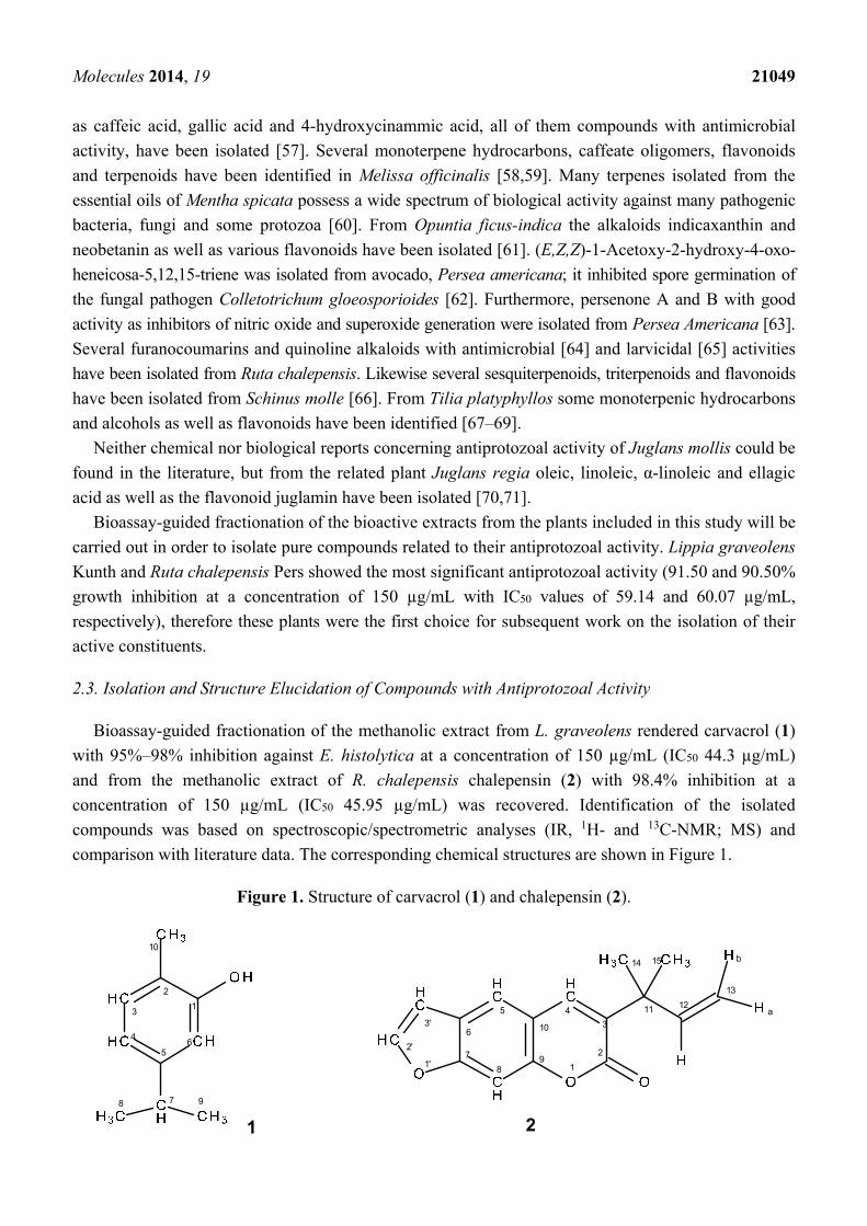
2.3. Isolation and Structure Elucidation of Compounds with Antiprotozoal Activity
Bioassay-guided fractionation of the methanolic extract from L. graveolens rendered carvacrol (1)
with 95%–98% inhibition against E. histolytica at a concentration of 150 µg/mL (IC50 44.3 µg/mL)
and from the methanolic extract of R. chalepensis chalepensin (2) with 98.4% inhibition at a
concentration of 150 µg/mL (IC50 45.95 µg/mL) was recovered. Identification of the isolated
compounds was based on spectroscopic/spectrometric analyses (IR, 1H- and 13C-NMR; MS) and
comparison with literature data. The corresponding chemical structures are shown in Figure 1.
Figure 1. Structure of carvacrol (1) and chalepensin (2).
8 9
1' 12
345
6
78
9
10
11 12
3'
2'
14 15
13
b
a
2
3
56
1
4
7
10
1 2
Molecules 2014, 19 21050
2.3.1. Carvacrol from Lippia graveolens
Preliminary fractionation of the methanolic extract of Lippia graveolens by extraction with n-hexane
led, after evaporation of solvent, to a residue with good activity against E. histolytica (90.9% growth
inhibition at a concentration of 150 µg/mL). Chromatography of this hexane residue over a silica gel
column led to the isolation of carvacrol (1) as a colorless oil. Spectroscopic data of 1 were in
concordance with literature values [72–74].
The essential oil of Lippia graveolens contains many monoterpenes, sesquiterpenes and phenolic
terpenes among which carvacrol and thymol are the most common components [75–77] and their
average abundance establishes the chemotype that can be assigned to L. graveolens varieties [78–80].
Due to its acidic and hydrophobic nature, carvacrol tends to damage biological systems and for that
reason is responsible for affecting a wide range of microorganisms, including bacteria, fungi, yeast and
parasites [81–85]. It also has been proposed as a therapeutic agent against some cancer cell lines due to
its activity as an antiproliferative compound [86,87], DNA synthesis inhibitor [88] and by triggering
apoptosis [89,90].
The monoterpenic phenol exerts a bactericidal effect against many foodborne bacteria responsible
for gastrointestinal disorders, such as Listeria monocytogenes, Escherichia coli [91], Salmonella enterica
ser. Enteritidis [92], Bacillus subtillis, Salmonella typhimurium, Escherichia coli [93,94], Shigella sonnei,
S. flexeri [95] and it has been considered useful to inhibit the growth of microorganisms responsible for
respiratory disorders like Staphyloccocus aureus, Staphyloccocus aureus MSRA, Streptococcus pneumonie,
Klebsiella pneumonie, Pseudomonas auroginosa [85]. Carvacrol is also able to inhibit the enterotoxin
production from Bacillus cereus, which generates abdominal pain and diarrhea [96].
Important toxigenic and pathogenic filamentous fungi, some causative of serious human mycoses,
have been subjected to carvacrol exposure and antifungal properties were found for this compound
against Aspergillus niger, A. flavus, A. fumigatus, Penicillum digitatum, P. brevicompactum,
P. expansum and Fusarium spp. [97–99]. Carvacrol has also shown strong fungicidal effects against
many clinical isolates from Candida spp. and Cryptococcus spp. [97,100].
Furthermore, carvacrol was tested against many tropical parasites responsible for serious human
diseases. Thus, the compound was evaluated for its trypanocidal activity against Trypanosoma cruzi and
T. brucei rhodesiense, being very effective in both cases, with IC50 values under 30 µg/mL [101–103],
but noteworthily an important inhibition effect was observed on the epimastigote form in T. cruzi
isolates (IC50 3.0 µg/mL). Following this topic, carvacrol was evaluated against visceral parasites of
the genus Leishmania and the results demonstrated an important effectiveness range over the
promastigote form of Leishmania chagasi with IC50 values between 2.3 to 28 µg/mL [103,104]. A
weak activity was observed on Leishmania donovani with an IC50 value of 17.8 mg/mL, compared
with the reference drug miltefosine (IC50 0.14 mg/mL). In addition, antimalarial activity against
Plasmodium falciparum was tested, obtaining a very significant IC50 value of 7.9 mg/mL [85].
Prior to our investigation, amebicide evaluation of carvacrol had not been carried out on
E. histolytica, but one clinical study was performed on 14 adult patients, whose stools tested positive
for enteric parasites such as Entamoeba hartmanni, Endolimax nana and Blastocystis hominis. The
patients were supplemented with an essential oil rich in carvacrol from Origanum vulgare and after
6 weeks of treatment total disappearance of E. hartmanni and E. nana was observed in all infected
Molecules 2014, 19 21051
patient cases. B. hominis was not detected in five cases [105]. Nevertheless this is the first report about
the antiprotozoal activity of carvacrol against Entamoeba histolytica.
2.3.2. Chalepensin from Ruta chalepensis
The bioguided fractionation of the methanolic extract of Ruta chalepensis by partition between
methanol and n-hexane followed by chromatography of the hexane residue (84.66% growth inhibition
against E. histolytica at a concentration of 150 µg/mL) over a silica gel column afforded chalepensin
(2) as colorless needles with a melting point of 75 °C. The spectroscopic data were identical with those
reported for chalepensin [106–113], but we now report the complete, unambiguous assignment of the 13C-NMR spectrum of chalepensin as the hydrogen and carbon connectivities in 2 were deduced from 1H-1H COSY, NOESY, HSQC and HMBC spectra.
Chalepensin has been isolated from several herbs, especially those of the Rutaceae family, including
Ruta chalepensis [114–120]. Chalepensin has strong allelochemical and phytotoxic activity [121–123].
Moreover, chalepensin has a variety of pharmacological effects, including a significant anti-fertility
activity [124], antiplatelet aggregation activity [112], cytotoxic effects on some human carcinoma cell lines
(breast MCF-7; colon HT-29; kidney A-498; lung A-549; pancreatic PACA-2; prostate PC-3) [122],
and further antiproliferative activity against human gastric adenocarcinoma (MK-1), human uterus
carcinoma (HeLa) and murine melanoma (B16F10) cells [125]. It has been also proved that
chalepensin is a mechanism-based inhibitor of cytochrome P450 (CYS) 2A6 [126–128]. This is the
first report on the antiprotozoal activity against Entamoeba histolytica of chalepensin.
2.4. Possible Antiprotozoal Mechanism of Action of Carvacrol and Chalepensin
Little knowledge exists about the antiprotozoal mechanism of action of carvacrol but the
antimicrobial mechanisms of action of carvacrol have been thoroughly investigated [81–85]. In the
following, we describe some facts of these antimicrobial mechanisms which future research might
reveal if these apply as well to protozoa. Carvacrol exhibits antimicrobial activity against the
biological membranes of bacteria. It exerts its action by rapidly depleting the intracellular ATP pool by
reducing ATP synthesis and increasing ATP hydrolysis. Reduction of transmembrane electric potential
which is the driving force of ATP synthesis enhances proton permeability of the membrane. At 1 mM
carvacrol lowers the internal pH of bacteria from 7.1 to 5.8 according to ion gradients of the cell
membrane. Carvacrol (1 mM) decreases cell protein content from 12 mmol/mg to 0.99 mmol/mg by
using potassium (K+) of bacterial cells in a short time (5 min). Potassium (K+) plays a role in the
activation of cytoplasmic enzymes, in maintaining osmotic pressure and in the regulation of
cytoplasmic pH. Leakage of K+ out of the cell is a clear indication of membrane damage. Ultee et al. [82]
hypothesized a scheme for the mechanism of action of carvacrol through the cytoplasmic membrane of
bacteria. According to this hypothesis undissociated carvacrol diffuses through the cytoplasmic
membrane and dissociates releasing its proton to the cytoplasm. It then returns undissociated through
the membrane into the external environment carrying a potassium ion. Outside the cell carvacrol
replaces potassium with a proton and reenters the cell the same way.
The mechanism of action of oregano oils has been shown to be related, especially, to the synthesis
of structural components and to the disruption of a series of energy systems. The leakage of ions, ATP
Molecules 2014, 19 21052
and amino acids from bacterial cells explains this phenomenon. Potassium and phosphate ion
concentrations were affected at a rate much lower than their MIC values [85].
Carvacrol increases overall permeability of the cytoplasmic membrane by disrupting the outer
membranes of Gram negative bacteria leading to the leakage of ATP from the cell. Carvacrol also
inhibits ATPase [85]. Similar alterations to those observed in bacteria [85] were also observed on
Giardia lamblia exposed to essential oils from different sources, especially those where carvacrol had a
dominant presence (over 70% of general composition) [28]. The main ultrastructural alterations
promoted by essential oils were deformations in typical trophozoite appearance, often roundly shaped,
irregular dorsal and ventral surface, presence of membrane blebs, electrodense precipitates in the
cytoplasm and nuclei and internalization of flagella and ventral disc. The data suggest that essential oils
probably induced cell death by processes associated to the loss of osmoregulation caused by plasmatic
membrane alterations [28].
To our knowledge there are no studies regarding the antiprotozoal mechanism of chalepensin but
chalepin, a furocoumarin structural related to chalepensin, exerts a potent inhibitory activity against the
recombinant enzyme TcGAPDH (glyceraldehyde-3-phosphate dehydrogenase) of Trypanosoma cruzi with
a strong IC50 of 64 µM [129,130]. Further studies are required to establish the antiprotozoal
mechanism of carvacrol and chalepensin against E. histolytica.
3. Experimental Section
3.1. General
Melting points were determined on an Electrothermal 9100 apparatus (Electrothermal Engineering Ltd.,
Southend on Sea, UK). IR spectra were recorded on a Frontier FT-IR spectrometer (PerkinElmer,
Waltham, MA, USA) using an ATR accessory. NMR spectra were measured on a Avance DPX 400
Spectrometer (Bruker, Billerica, MA, USA) operating at 400.13 MHz for 1H and 100.61 MHz for 13C.
ESI HR mass spectra were measured on a 4.7 T FT-ICR Mass spectrometer (Bruker, Bremen,
Germany). EI MS was recorded on a MAT 95 spectrometer (70 eV, Finnigan, San Jose, CA, USA).
TLC was carried out on pre-coated silica gel glass plates 5 cm × 10 cm (Merck silica gel 60 F254,
Darmstadt, Germany). Normal phase column chromatography was performed on silica gel (60–200 mesh)
purchased from J. T. Baker (Phillipsburg, NJ, USA).
3.2. Plant Material
The plants were obtained from the field or purchased from Pacalli® (pacalli.com.mx, Monterrey,
Mexico). Reference vouchers of the plant material were deposited at the herbarium UNL of the
Facultad de Ciencias Biológicas (Universidad Autónoma de Nuevo León). Plant species, botanical
name, family, voucher specimens and plant parts used to obtain the extracts are summarized in Table 1.
Vegetal material was dried and ground to powder.
3.3. Extraction and Isolation
Sixty grams of dried and powdered material from the respective plant was extracted with methanol
(MeOH, 600 mL) by using a Soxhlet system for continuous extraction. After filtration, the solvent was

Molecules 2014, 19 21053
evaporated under reduced pressure in a rotary evaporator [25]. The different extracts were conserved in
tightly sealed glass vials. The yields are shown in Table 1.
3.3.1. Bioguided Isolation of Carvacrol (1)
Ground and dried leaves of Lippia graveolens (600 g) were extracted with methanol in several
portions of 30 g in a Soxhlet apparatus for 40 h, each charged with 500 mL of CH3OH. After filtration
of the methanol solutions, the solvent was removed under reduced pressure to yield 260 g of combined
extract. The crude extract was analyzed for amebicide activity on trophozoites of E. histolytica
(HM1:IMSS strain), showing a very high inhibition percentage (89%) by standard concentration of
150 µg/mL. Afterwards, the extract was redissolved in 2 L methanol, being divided in 4 portions of
500 mL each and submitted to liquid-liquid partition with n-hexane (500 mL each portion) to yield,
after solvent evaporation, 19.9 g of a combined residue with high amebicidal activity (90.9% growth
inhibition). The n-hexane partition was divided into eighteen portions of ca. 1 g and each of them
chromatographed on a silica gel (20 g) column (39 cm × 2 cm) and eluted with stepwise gradients of
n-hexane–chloroform (100:0, 90:10, 80:20, 70:30, 60:40, 50:50, 40:60, 30:70, 20:80, 10:90, 0:100 v/v,
each 50 mL), chloroform–ethyl acetate (90:10, 80:20, 70:30, 60:40, 50:50, 40:60, 30:70, 20:80, 10:90,
0:100 v/v, each 50 mL), and finally with 50 mL methanol. A total of 110 subfractions (10 mL) were
collected for each column and combined on the basis of their TLC (CHCl3–EtOAc, 9:1) profiles into
six main fractions as follows: A (subfractions 1–30, 1.1 g), B (subfractions 31–42, 780 mg), C
(subfractions 43–63, 7.1 g), D (subfractions 64–72, 2.5 g), E (subfractions 73–75, 1.9 g) and F
(subfractions 76–110, 3.6 g). These main fractions, containing the non-polar to the more polar
compounds, were used for amebicide assays. Only fraction C showed amebicide activity (82.88%
growth inhibition) and was divided into seven portions of ca. 1 g and each of them chromatographed
on a silica gel (20 g) column (39 × 2 cm) and eluted with stepwise gradient solvent system consisting
of n-hexane–chloroform (100:0, 90:10, 80:20, 70:30, 60:40, 50:50, 40:60, 30:70, 20:80, 10:90, 0:100
v/v, each 50 mL), and finally with 50 mL methanol. A total of 60 subfractions (10 mL) were collected
and combined on the basis of their TLC (n-hexane–CHCl3, 2:8) profiles into three main fractions as
follows: G (subfractions 1–27, 1.0 g), H (subfractions 28–38, 4.2 g), I (subfractions 39–60, 677 mg).
The amebicide activity was detected on fraction G (44.24% growth inhibition) and very noticeable on
fraction H (94.63% growth inhibition); this last was submitted to additional fractionation. Fraction H
was divided into four portions of ca. 1 g and each of them later chromatographed on a silica gel
(20 g) column (39 cm × 2 cm) and eluted with stepwise gradient solvent system consisting of
n-hexane–chloroform (100:0, 90:10, 80:20, 70:30, 60:40, 50:50, 40:60, 30:70, 20:80, 10:90, 0:100 v/v,
each 50 mL), and finally with 50 mL methanol. A total of 120 subfractions (5 mL) were collected and
combined on the basis of their TLC (n-hexane–CHCl3, 2:8) profiles into three main fractions as
follows: J (subfractions 1–63, 514 mg), K (subfractions 64–90, 1.7 g) and L (subfractions 91–120, 1.0 g).
Fraction J (98.4% growth inhibition; IC50 44.30 µg/mL) and Fraction K (95.13% growth inhibition;
IC50 44.22 µg/mL) both provided an oil containing a compound with a very intense and characteristic
hydrocarbon smell (carvacrol, 1). Fraction L did not present amebicidal activity.
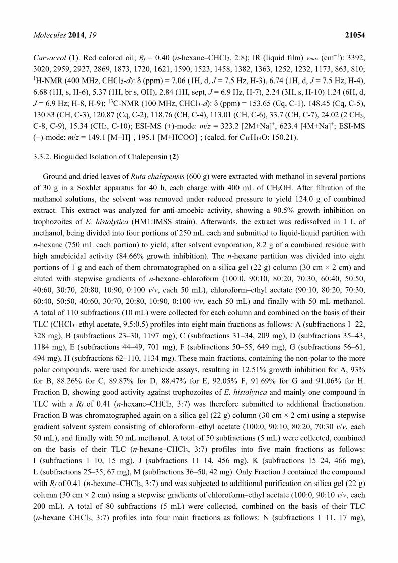
Molecules 2014, 19 21054
Carvacrol (1). Red colored oil; Rf = 0.40 (n-hexane–CHCl3, 2:8); IR (liquid film) vmax (cm−1): 3392,
3020, 2959, 2927, 2869, 1873, 1720, 1621, 1590, 1523, 1458, 1382, 1363, 1252, 1232, 1173, 863, 810; 1H-NMR (400 MHz, CHCl3-d): δ (ppm) = 7.06 (1H, d, J = 7.5 Hz, H-3), 6.74 (1H, d, J = 7.5 Hz, H-4),
6.68 (1H, s, H-6), 5.37 (1H, br s, OH), 2.84 (1H, sept, J = 6.9 Hz, H-7), 2.24 (3H, s, H-10) 1.24 (6H, d,
J = 6.9 Hz; H-8, H-9); 13C-NMR (100 MHz, CHCl3-d): δ (ppm) = 153.65 (Cq, C-1), 148.45 (Cq, C-5),
130.83 (CH, C-3), 120.87 (Cq, C-2), 118.76 (CH, C-4), 113.01 (CH, C-6), 33.7 (CH, C-7), 24.02 (2 CH3;
C-8, C-9), 15.34 (CH3, C-10); ESI-MS (+)-mode: m/z = 323.2 [2M+Na]+, 623.4 [4M+Na]+; ESI-MS
(−)-mode: m/z = 149.1 [M−H]−, 195.1 [M+HCOO]−; (calcd. for C10H14O: 150.21).
3.3.2. Bioguided Isolation of Chalepensin (2)
Ground and dried leaves of Ruta chalepensis (600 g) were extracted with methanol in several portions
of 30 g in a Soxhlet apparatus for 40 h, each charge with 400 mL of CH3OH. After filtration of the
methanol solutions, the solvent was removed under reduced pressure to yield 124.0 g of combined
extract. This extract was analyzed for anti-amoebic activity, showing a 90.5% growth inhibition on
trophozoites of E. histolytica (HM1:IMSS strain). Afterwards, the extract was redissolved in 1 L of
methanol, being divided into four portions of 250 mL each and submitted to liquid-liquid partition with
n-hexane (750 mL each portion) to yield, after solvent evaporation, 8.2 g of a combined residue with
high amebicidal activity (84.66% growth inhibition). The n-hexane partition was divided into eight
portions of 1 g and each of them chromatographed on a silica gel (22 g) column (30 cm × 2 cm) and
eluted with stepwise gradients of n-hexane–chloroform (100:0, 90:10, 80:20, 70:30, 60:40, 50:50,
40:60, 30:70, 20:80, 10:90, 0:100 v/v, each 50 mL), chloroform–ethyl acetate (90:10, 80:20, 70:30,
60:40, 50:50, 40:60, 30:70, 20:80, 10:90, 0:100 v/v, each 50 mL) and finally with 50 mL methanol.
A total of 110 subfractions (10 mL) were collected for each column and combined on the basis of their
TLC (CHCl3–ethyl acetate, 9.5:0.5) profiles into eight main fractions as follows: A (subfractions 1–22,
328 mg), B (subfractions 23–30, 1197 mg), C (subfractions 31–34, 209 mg), D (subfractions 35–43,
1184 mg), E (subfractions 44–49, 701 mg), F (subfractions 50–55, 649 mg), G (subfractions 56–61,
494 mg), H (subfractions 62–110, 1134 mg). These main fractions, containing the non-polar to the more
polar compounds, were used for amebicide assays, resulting in 12.51% growth inhibition for A, 93%
for B, 88.26% for C, 89.87% for D, 88.47% for E, 92.05% F, 91.69% for G and 91.06% for H.
Fraction B, showing good activity against trophozoites of E. histolytica and mainly one compound in
TLC with a Rf of 0.41 (n-hexane–CHCl3, 3:7) was therefore submitted to additional fractionation.
Fraction B was chromatographed again on a silica gel (22 g) column (30 cm × 2 cm) using a stepwise
gradient solvent system consisting of chloroform–ethyl acetate (100:0, 90:10, 80:20, 70:30 v/v, each
50 mL), and finally with 50 mL methanol. A total of 50 subfractions (5 mL) were collected, combined
on the basis of their TLC (n-hexane–CHCl3, 3:7) profiles into five main fractions as follows:
I (subfractions 1–10, 15 mg), J (subfractions 11–14, 456 mg), K (subfractions 15–24, 466 mg),
L (subfractions 25–35, 67 mg), M (subfractions 36–50, 42 mg). Only Fraction J contained the compound
with Rf of 0.41 (n-hexane–CHCl3, 3:7) and was subjected to additional purification on silica gel (22 g)
column (30 cm × 2 cm) using a stepwise gradients of chloroform–ethyl acetate (100:0, 90:10 v/v, each
200 mL). A total of 80 subfractions (5 mL) were collected, combined on the basis of their TLC
(n-hexane–CHCl3, 3:7) profiles into four main fractions as follows: N (subfractions 1–11, 17 mg),
Molecules 2014, 19 21055
O (subfractions 12–17, 243 mg), P (subfractions 18–44, 92 mg), Q (subfractions 45–80, 18 mg).
Fraction O produced pure chalepensin (2), showing 98.4% growth inhibition against E. histolytica and
an IC50 value of 45.95 µg/mL. Fraction P was subjected to additional purification on a silica gel (22 g)
column (30 cm × 2 cm) using stepwise gradients of chloroform–ethyl acetate (100:0, 95:05, v/v, each
150 mL) and finally 50 mL of methanol as eluent. A total of 70 subfractions (5 mL) were collected,
combined on the basis of their TLC (n-hexane–CHCl3, 3:7) profiles into three main fractions as
follows: R (subfractions 1–27, 37 mg), S (subfractions 28–47, 24 mg) and T (subfractions 48–70, 8 mg).
Fraction R rendered additional pure chalepensin.
Chalepensin (2). Colorless needles; M.p. 75 °C; Rf = 0.41 (n-hexane–CHCl3, 3:7); IR (powder) vmax
(cm−1): 3016, 2968, 2932,1715, 1629, 1583, 1542, 1451, 1024, 751; 1H-NMR (400 MHz, CDCl3-d): δ
(ppm) = 7.68 (1H, s, H-4), 7.67 (1H, d, J = 2.24 Hz, H-2’), 7.65 (1H, s, H-5), 7.43 (1H, s, H-8), 6.82
(1H, dd, J = 2.24, 0.96 Hz, H-3’), 6.21 (1H, dd, J = 17.2, 11.0 Hz, H-12), 5.10 (1H, s, H-13a-cis), 5.07
(1H, dd, J = 8.16, 0.97 Hz, H-13b-trans), 1.52 (6H, s, 14-CH3, 5-CH3); 13C-NMR (100 MHz, CDCl3-d):
δ (ppm) = 159.94 (Cq, C-2), 155.84 (Cq, C-7), 151.28 (Cq, C-9), 146.59 (CH, C-2’), 145.45 (CH, C-12),
138.34 (CH, C-4), 133.09 (Cq, C-3) 124.57 (Cq, C-6), 119.52 (CH, C-5), 115.91 (Cq, C-10), 112.33
(CH2, C-13), 106.35 (CH, C-1'), 98.95 (CH, C-8), 40.51 (Cq, C-11), 26.15 (2 CH3, C-14, C-15); ESI-MS
(+)-mode: m/z = 255.1 [M+H]+, 277.1 [M+Na]+, 531.2 [2M+Na]+; (+)-ESI HR MS: m/z = 255.1014
[M+H]+ (calcd. For C16H15O3: 255.1016), 277.0835 [M+Na]+ (calcd. for C16H14NaO3: 277.0835).
3.4. Antiprotozoal Assay
3.4.1. Test Microorganisms
Entamoeba histolytica strain HM-1:IMSS was obtained from the microorganism culture collection
of the Centro de Investigación Biomédica del Noreste (CIBIN-IMSS) in Nuevo León, Mexico.
The trophozoites were grown axenically and maintained in peptone, pancreas and liver extract plus
bovine serum [131]. The trophozoites were employed at log phase of growth (2 × 10⁴ cells/mL) by all
the performed bioassays [132,133].
3.4.2. In Vitro Assay for Entamoeba histolytica
The MeOH extract from each plant was dissolved in DMSO and adjusted to a concentration of
150 µg/mL in a suspension of E. histolytica trophozoites at logarithmic phase in PEHPS medium
containing 10% of bovine serum. Vials were incubated for 72 h, then chilled in iced water for 20 min
and the number of dead trophozoites per milliliter was counted by using a hemocytometer. Each
extract assay was performed by triplicate [132,133]. Each test included a positive control by using
metronidazole and a negative control by using E. histolytica suspension in PEHPS medium with no
extract added. The inhibition percentage was estimated as the number of dead cells compared with the
untreated controls.
The same procedure was performed with fractions or pure isolated compounds.

Molecules 2014, 19 21056
3.4.3. In Vitro IC50 Determination
The MeOH extract from each plant was dissolved in DMSO and adjusted to 150, 75, 32.5 and
16.25 µg/mL with a suspension of E. histolytica trophozoites at logarithmic phase in PEHPS medium
containing 10% of bovine serum. Vials were incubated for 72 h, then chilled in iced water for 20 min
and the number of dead trophozoites per milliliter was determined by using a hemocytometer.
Each extract assay was performed by triplicate. The 50% inhibitory (IC50) concentration of each
extract was determined by using a Probit analysis with a 95% confidence level. The same procedure
was performed with fractions or pure isolated compounds.
4. Conclusions
Entamoeba histolytica is the most common parasite to cause enteric protozoan infections. The drug
of choice used to treat amoebic dysentery is metronidazole, which has been associated with unpleasant
side effects [134–136], therefore alternative drugs are needed and medicinal plants may be an
important alternative source of new antiamoebic compounds. The results of the antiprotozoal screening
in this work support the popular uses of 18 of the studied species for the treatment of diarrhea and
dysentery in Mexican traditional medicine. The extracts from both Lippia graveolens Kunth and
Ruta chalepensis Pers showed the most significant antiprotozoal activity and were submitted to a
bioguided fractionation. Structure elucidation of the isolated compounds was accomplished by
spectroscopic and mass spectrometric data. The methanolic extract of L. graveolens rendered carvacrol
(1) with 95%–98% inhibition against E. histolytica at a concentration of 150 µg/mL (IC50 44.3 µg/mL)
and from the methanolic extract of R. chalepensis chalepensin (2) with 98.4% inhibition at a
concentration of 150 µg/mL (IC50 45.95 µg/mL) was recovered. To our knowledge, this is the first
report on the antiamoebic activity of carvacrol and chalepensin, both known compounds with other
notable pharmacological activities. These compounds may also offer new opportunities for treating
amoebiasis and other important and often neglected diseases [137] or be useful as lead compounds in
the development of new antiprotozoal agents. Further work for isolation of other active constituents
from these plants is under way.
Acknowledgments
The authors would like to thank the DAAD (Germany), who financed a stay of R.Q.L. at the
University of Göttingen, to CONACYT (Mexico) for doctoral fellowships awarded to A.F.B.R. and
I.K.A.H. and to José A. Almaguer-González (Secretaría de Salud, Mexico) for support given for the
realization of this research project. We also thank the Universidad Autónoma de Nuevo León (Mexico)
for PAICYT grants CN-422-10, and CN-662-11. For the spectroscopic measurements we thank
H. Frauendorf, R. Machinek and H. Laatsch of the University of Göttingen (Germany) and
Noemi Waksman of the College of Medicine from Universidad Autónoma de Nuevo León.
Supplementary Materials
Supplementary materials can be accessed at: http://www.mdpi.com/1420-3049/19/12/21044/s1.

Molecules 2014, 19 21057
Author Contributions
R.Q.L. initiated, conceived and coordinated the project. B.D.M.C. and J.V.V. supervised the
biological in vitro tests and analyzed the data. A.F.B.R and I.K.A.H. performed extraction of plants,
bioguided isolation of bioactive compounds and analyzed the spectroscopic data. J.N.G.G. and
M.E.H.G. performed the biological in vitro tests and analyzed the data. R.Q.L. wrote the paper with
help of A.F.B.R and I.K.A.H. All authors read and approved the final manuscript.
Conflicts of Interest
The authors declare no conflict of interest.
References
1. Kirmizibekmez, H.; Atay, I.; Kaiser, M.; Brun, R.; Cartagena, M.M.; Carballeira, N.M.; Yesilada, E.;
Tasdemir, D. Antiprotozoal activity of Melampyrum arvense and its metabolites. Phytother. Res.
2011, 25, 142–146.
2. Martinez, P.A.; Petersen, C.A. Chronic infection by Leishmania amazonensis mediated through
MAPK ERK mechanisms. Immunol. Res. 2014, 59, 153–165.
3. Sosoniuk, E.; Vallejos, G.; Kenawy, H.; Gaboriaud, C.; Thielens, N.; Fujita, T.; Schwaeble, W.;
Ferreira, A.; Valck, C. Trypanozoma cruzi calreticulin inhibits the complement lectin pathway
activation by direct interaction with L-Ficolin. Mol. Immunol. 2014, 60, 80–85.
4. Hotez, P.J.; Woc-Colburn, L.; Bottazzi, M.E. Neglected tropical diseases in Central America and
Panama: Review of their prevalence, populations at risk and impact on regional development.
Int. J. Parasitol. 2014, 44, 597–603.
5. Zerpa-Larrauri, R.; Náquira-Velarde, C.; Espinoza, Y. A new vision of Entamoeba histolytica.
Rev. Peru. Med. Exp. Salud Publica 2007, 24, 190–192.
6. López-Camarillo, C.; López-Rosas, I.; Ospina-Villa, J.D.; Marchat, L.A. Deciphering molecular
mechanisms of mRNA metabolism in the deep-branching eukaryote Entamoeba histolytica.
WIREs RNA 2014, 5, 247–262.
7. Bansal, D.; Sehgal, R.; Chawla, Y.; Malla, N.; Mahajan, R.C. Multidrug resistance in amoebiasis
patients. Indian J. Med. Res. 2006, 124, 189–194.
8. Pinilla, A.E.; López, M.C.; Viasus, D.F. History of the Entamoeba histolytica protozoan.
Rev. Med. Chile 2008, 136, 118–124.
9. Ximénez, C.; Morán, P.; Ramos, F.; Ramiro, M. Amibiasis intestinal: Estado actual del
conocimiento. Med. Interna Mex. 2007, 23, 398–407.
10. Calzada, F.; Yépez-Mulia, L.; Aguilar, A. In vitro susceptibility of Entamoeba histolytica and
Giardia lamblia to plants used in Mexican traditional medicine for the treatment of gastrointestinal
disorders. J. Ethnopharmacol. 2006, 108, 367–370.
11. Ximénez, C.; Morán, P.; Rojas, L.; Valadez, A.; Gómez, A. Reassessment of the epidemiology of
amebiasis: State of the art. Infect. Gen. Evol. 2009, 9, 1023–1032.

Molecules 2014, 19 21058
12. Abhyankar, M.M.; Shrimal, S.; Gilchrist, C.A.; Bhattacharya, A.; Petri, W.A., Jr. The Entamoeba
histolytica serum-inducible transmembrane kinase EhTMKB1–9 is involved in intestinal amebiasis.
Int. J. Parasitol. Drugs Drug Resist. 2012, 2, 243–248.
13. Phillipson, J.D.; Wright, C.W. Antiprotozoal agents from plant sources. Planta Med. 1991, 57,
S53–S59.
14. Somlata; Babuta, M.; Bhattacharya, S.; Bhattacharya, A. Protein kinases of the parasitic protist
Entamoeba histolytica. In Protein Phosphorylation in Parasites: Novel Targets for Antiparasitic
Intervention; Doerig, C., Spaeth, G., Wiese, M., Eds.; Wiley-VCH Verlag GmbH & Co. KGaA:
Weinheim, Germany, 2014; pp. 131–153.
15. Schlosser, A.; Leitsch, D.; Duchêne, M. Entamoeba histolytica: Identification of thioredoxin-targeted
proteins and analysis of serine acetyltransferase-1 as a prototype example. Biochem. J. 2013,
451, 277–288.
16. Samarawickrema, N.A.; Brown, D.M.; Upcroft, J.A.; Thammapalerd, N.; Upcroft, P. Involvement of
superoxide dismutase and pyruvate:ferredoxin oxidoreductase in mechanisms of metronidazole
resistance in Entamoeba histolytica. J. Antimicrob. Chemother. 1997, 40, 833–840.
17. Wassmann, C.; Hellberg, A.; Tannich, E.; Bruchhaus, I. Metronidazole resistance in the protozoan
parasite Entamoeba histolytica is associated with increased expression of iron-containing
superoxide dismutase and peroxiredoxin and decreased expression of ferredoxin 1 and flavin
reductase. J. Biol. Chem. 1999, 274, 26051–26056.
18. Conde-Bonfil, M.D.C.; Mora-Zerpa, C.D.L. Entamoeba histolytica: Un desafío vigente.
Salud Pub. Mex. 1992, 34, 335–341.
19. Bendesky, A.; Menéndez, D. Metronidazol: Una visión integral. Rev. Fac. Med. UNAM 2001, 44,
255–259.
20. Bautista, E.; Calzada, F.; Ortega, A.; Yépez-Mulia, L. Antiprotozoal activity of flavonoids isolated
from Mimosa tenuiflora (Fabaceae-Mimosoidae). J. Mex. Chem. Soc. 2011, 55, 251–253.
21. Singh, S.; Bharti, N.; Mohapatra, P.P. Chemistry and biology of synthetic and naturally occurring
antiamoebic agents. Chem. Rev. 2009, 109, 1900–1947.
22. Kayser, O.; Kiderlen, A.F.; Croft, S.L. Natural products as parasitic drugs. Parasitol. Res.
2003, 90, S55–S62.
23. Sülsen, V.; Güida, C.; Coussio, J.; Paveto, C. Muschietti, L.; Martino, V. In vitro evaluation of
trypanocidal activity in plants used in Argentine traditional medicine. Parasitol. Res. 2006, 98,
370–374.
24. Newman, D.J.; Cragg, G.M. Natural products as sources of new drugs over the 30 years from
1981 to 2010. J. Nat. Prod. 2012, 75, 311–335.
25. Quintanilla-Licea, R.; Morado-Castillo, R.; Gomez-Flores, R.; Laatsch, H.; Verde-Star, M.J.;
Hernández-Martínez, H.; Tamez-Guerra, P.; Tamez-Guerra, R.; Rodríguez-Padilla, C.
Bioassay-guided isolation and identification of cytotoxic compounds from Gymnosperma glutinosum
leaves. Molecules 2012, 17, 11229–11241.
26. Molina-Garza, Z.J.; Bazaldúa-Rodríguez, A.F.; Quintanilla-Licea, R.; Galaviz-Silva, L.
Anti-Trypanozoma cruzi activity of 10 medicinal plants used in northeast Mexico. Acta Trop.
2014, 136, 14–18.
Molecules 2014, 19 21059
27. Machado, M.; Sousa, M.C.; Salgueiro, L.; Cavaleiro, C. Effects of essential oils in growth of
Giardia lamblia trophozoites. Nat. Prod. Commun. 2010, 5, 137–141.
28. Machado, M.; Dinis, A.M.; Salgueiro, L.; Cavaleiro, C.; Custódio, J.B.A.; Sousa, M.C. Anti-Giardia
activity of phenolic-rich essential oils: Effects of Thymbra capitata, Origanum virens, Thymus zygis
subsp. sylvestris, and Lippia graveolens on trophozoites growth, viability, adherence, and
ultrastructure. Parasitol. Res. 2010, 106, 1205–1215.
29. Rufino-González, Y.; Ponce-Macotela, M.; González-Maciel, A.; Reynoso-Robles, R.;
Jiménez-Estrada, M.; Sánchez-Contreras, Á.; Martínez-Godillo, M.N. In vitro activity of the F-6
fraction of oregano against Giardia intestinalis. Parasitology 2012, 139, 434–440.
30. Machado, M.; Santoro, G.; Sousa, M.C.; Salgueiro, L.; Cavaleiro, C. Activity of essential oils on
the growth of Leishmania infantum promastigotes. Flavour Fragr. J. 2010, 25, 156–160.
31. Ahmed, S.B.H.; Sghaier, R.M.; Guesmi, F.; Kaabi, B.; Mejri, M.; Attia, H.; Laouini, D.; Smaali, I.
Evaluation of antileishmanial, cytotoxic and antioxidant activities of essential oils extracted from
plants issued from the leishmaniasis-endemic region of Sned (Tunisia). Nat. Prod. Res. 2011, 25,
1195–1201.
32. Abdel-Sattar, E.; Maes, L.; Salama, M.M. In vitro activities of plant extracts from Saudi Arabia
against Malaria, Leishmaniasis, Sleeping Sickness and Chagas Disease. Phytother. Res. 2010, 24,
1322–1328.
33. Malatyali, E.; Tepe, B.; Degerli, S.; Berk, S. In vitro amoebicidal activities of Satureja cuneifolia
and Melissa officinalis on Acanthamoeba castellanii cysts and trophozoites. Parasitol. Res.
2012, 110, 2175–2180.
34. Calzado-Flores, C.C.; Segura-Luna, J.J.; Domínguez, X.A.; García-González, S. Castela texana:
Cernimiento de su actividad antiamibiana. Arch. Investig. Med. (Mex.) 1986, 17, 127–134.
35. Calzado-Flores, C.; Verde-Star, J.; Lozano-Garza, G.; Segura-Luna, J.J. Preliminary acute
toxicological study of Castela texana. Proc. West. Pharamacol. Soc. 1998, 41, 77–78.
36. Reyes-López, M.; Villa-Treviño, S.; Arriaga-Alba, M.; Alemán-Lazarini, L.;
Rodríguez-Mendiola, M.; Arias-Castro, C.; Fattel-Fazenda, S.; Garza, M.D.L. The amoebicidal
aqueous extract from Castela texana possesses antigenotoxic and antimutagenic properties.
Toxicol. In Vitro 2005, 19, 91–97.
37. González-Güereca, M.C.; Soto-Hernández, M.; Kite, G.; Martínez-Vázquez, M. Antioxidant activity
of flavonoids from the stem of the Mexican oregano (Lippia graveolens HBK var. berlandieri
schauer). Rev. Fitotec. Mex. 2007, 30, 43–49.
38. Lin, L.Z.; Mukhopadhyay, S.; Robbins, R.J.; Harnly, J.M. Identification and quantification of
flavonoids of Mexican oregano (Lippia graveolens) by LC-DAD-ESI/MS analysis. J. Food
Comp. Anal. 2007, 20, 361–369.
39. Calzada, F.; Meckes, M.; Cedillo-Rivera, R. Antiamoebic and antigardial activity of plant
flavonoids. Planta Med. 1999, 65, 78–80.
40. Dou, J.; McChesney, J.D.; Sindelar, R.D.; Goins, D.K.; Walker, L.A. A new quassinoid from
Castela texana. J. Nat. Prod. 1996, 59, 73–76.
41. Hernández, R.; Lugo, E.C.; Díaz, L.; Villanueva, S. Extracción y cuantificación indirecta de las
saponinas de Agave lechuguilla Torrey. E-Gnosis 2005, 3, 1–9.

Molecules 2014, 19 21060
42. Romero-González, J.; Peralta-Videa, J.R.; Rodríguez, E.; Delgado, M.; Gardea-Torresdey, J.L.
Potential of Agave lechuguilla biomass for Cr(III) removal from aqueous solutions: Thermodynamic
studies. Bioresour. Technol. 2006, 97, 178–182.
43. Monterrosas-Brisson, N.; Arenas-Ocampo, M.L.; Jiménez-Ferrer, E.; Jiménez-Aparicio, A.R.;
Zamilpa, A.; Gonzalez-Cortazar, M.; Tortoriello, J.; Herrera-Ruiz, M. Anti-inflammatory activity
of different Agave plants and the compound Cantalasaponin-1. Molecules 2013, 18, 8136–8146.
44. Momin, R.A.; Ramsewak, R.S.; Nair, M.G. Bioactive compounds and 1,3-di[(cis)-9
octadecenoyl]-2-[(cis,cis)-9,12-octadecadienoyl]glicerol from Apium Graveolens L. Seeds. J. Agric.
Food Chem. 2000, 48, 3785–3788.
45. Momin, R.A.; Nair, M.G. Mosquitocidal, nematicidal, and antifungal compounds from
Apium graveolens L. Seeds. J. Agric. Food Chem. 2001, 49, 142–145.
46. Kitajima, J.; Ishikawa, T.; Satoh, M. Polar constituents of celery seed. Phytochemistry 2003, 64,
1003–1011.
47. Fazal, S.S.; Ansari, M.M.; Singla, R.K.; Khan, S. Isolation of 3-n-butyl phthalide & sedanenolide
from Apium graveolens Linn. Indo Glob. J. Pharm. Sci. 2012, 2, 258–261.
48. Ochi, T.; Takaishi, Y.; Kogure, K.; Yamauti, I. Antioxidant activity of a new capsaicin derivative
from Capsicum annuum. J. Nat. Prod. 2003, 66, 1094–1096.
49. Kawaguchi, Y.; Ochi, T.; Takaishi, Y.; Kawazoe, K.; Lee, K.H. New sesquiterpenes from
Capsicum annuum. J. Nat. Prod. 2004, 67, 1893–1896.
50. Acero-Ortega, C.; Dorantes-Alvarez, L.; Hernández-Sánchez, H.; Gutiérrez-López, G.; Aparicio, G.;
Jaramillo-Flores, M.E. Evaluation of phenylpropanoids in ten Capsicum annuum L. varieties and
their inhibitory effects on Listeria monocytogenes Murray, Webb and Swann Scott A. Food Sci.
Tech. Int. 2005, 11, 5–6.
51. Andrade-Cetto, A.; Vazquez, R.C. Gluconeogenesis inhibition and phytochemical composition
of two Cecropia species. J. Ethnopharm. 2010, 130, 93–97.
52. De Heluani, C.S.; de Boggiato, M.V.; Catalán, C.A.N.; Díaz, J.G.; Gédris, T.E.; Herz, W.
Triterpenes and sesquiterpene lactones from Cyclolepis genistoides. Phytochemistry 1997, 45,
801–805.
53. Sosa, A.; Fusco, M.R.; Rossomando, P.; Juárez, A.; Robles, S.; Petenatti, E.; Pelzer, L.
Anti-inflammatory properties from isolated compounds of Cyclolepis genistoides. Pharm. Biol.
2011, 49, 675–678.
54. Villagómez-Ibarra, J.R.; Sánchez, M.; Espejo, O.; Zúñiga-Estrada, A.; Torres-Valencia, J.M.;
Joseph-Nathan, P. Antimicrobial activity of three Mexican Gnaphalium species. Fitoterapia
2001, 72, 692–694.
55. Canales, M.; Hernández, T.; Serrano, R.; Hernández, L.B.; Duran, A.; Ríos, V.; Sigrist, S.;
Hernández, H.L.H.; Garcia, A.M.; Angeles-López, O.; et al. Antimicrobial and general toxicity
activities of Gymnosperma glutinosum: A comparative study. J. Ethnopharmacol. 2007, 110,
343–347.
56. Serrano, R.; Hernández, T.; Canales, M.; García-Bores, A.M.; Romo-De-Vivar, A.; Céspedes, C.L.;
Avila, J.G. Ent-labdane type diterpene with antifungal activity from Gymnosperma glutinosum
(Spreng.) Less. (Asteraceae). BLACPMA 2009, 8, 412–418.
Molecules 2014, 19 21061
57. Rivero-Cruz, J.F. Antimicrobial compounds isolated from Haematoxylon brasiletto.
J. Etnopharmacol. 2008, 119, 99–103.
58. Sarer, E.; Kökdil, G. Constituents of the essential oil from Melissa officinalis. Planta Med.
1991, 57, 89–90.
59. Fecka, I.; Turek, S. Determination of water-soluble polyphenolic compounds in commercial
herbal teas from Lamiaceae: Peppermint, Melissa, and Sage. J. Agric. Food Chem. 2007, 55,
10908–10917.
60. Lawrence, B.M. Mint: The Genus Mentha, 1st ed.; CRC Press, Taylor & Francis Group: Boca Raton,
FL, USA, 2007.
61. Saleem, M.; Kim, H.J.; Han, C.K.; Jin, C.; Lee, Y.S. Secondary metabolites from Opuntia
ficus-indica var. Saboten. Phytochemistry 2006, 67, 1390–1394.
62. Domergue, F.; Helms, G.L.; Prusky, D.; Browse, J. Antifungal compounds from idioblast cells
isolated from avocado fruits. Phytochemistry 2000, 54, 183–189.
63. Kim, O.K.; Murakami, A.; Nakamura, Y.; Takeda, N.; Yoshizumi, H.; Ohigashi, H. Novel nitric
oxide and superoxide generation inhibitors, Persenone A and B, from avocado fruit. J. Agric.
Food Chem. 2000, 48, 1557–1563.
64. Cho, J.H.; Lee, C.H.; Lee, H.S. Antimicrobial activity of quinoline derivatives isolated from
Ruta chalepensis toward human intestinal bacteria. J. Microbiol. Biotechnol. 2005, 15, 646–651.
65. Emam, A.M.; Swelam, E.S.; Megally, N.Y. Furocoumarin and quinolone alkaloid with larvicidal
and antifeedant activities isolated from Ruta chalepensis leaves. I. J. Nat. Prod. 2009, 2, 10–22.
66. Ono, M.; Yamashita, M.; Mori, K.; Masuoka, C.; Eto, M.; Kinjo, J.; Ikeda, T.; Yoshimitsu, H.;
Nohara, T. Sesquiterpenoids, triterpenoids, and flavonoids from the fruits of Schinus molle.
Food Sci. Technol. Res. 2008, 14, 499–508.
67. Rădulescu, V.; Oprea, E. Analysis of volatile compounds of officinal Tiliae flos by
gas–chromatography coupled with mass spectrometry. Farmacia 2008, 61, 129–138.
68. Toker, G.; Aslan, M.; Yeşilada, E.; Memişoğlu, M.; Ito, S. Comparative evaluation of the
flavonoid content in officinal Tiliae flos and Turkish lime species for quality assessment.
J. Pharm. Biomed. Anal. 2001, 26, 111–121.
69. Karioti, A.; Chiarabini, L.; Alachkar, A.; Chehna, M.F.; Vincieri, F.F.; Bilia, A.R. HPLC–DAD
and HPLC–ESI-MS analyses of Tiliae flos and its preparations. J. Pharm. Biomed. Anal.
2014, 100, 205–214.
70. Cruz-Vega, D.E.; Verde-Star, M.J.; Salinas-González, N.; Rosales-Hernández, B.; Estrada-García, I.;
Mendez-Aragón, P.; Carranza-Rosales, P.; González-Garza, M.T.; Castro-Garza, J.
Antimycobacterial activity of Juglans regia, Juglans mollis, Carya illinoensis and Bocconia
frutescens. Phytother. Res. 2008, 22, 557–559.
71. Shah, T.I.; Sharma, E.; Ahmad, G. Juglans regia Linn: A phytopharmacological review. World J.
Pharm. Sci. 2014, 2, 364–373.
72. Silverstein, R.M.; Webster, F.X.; Kiemle, D.J. Spectrometric Identification of Organic Compounds,
7th ed.; John Wiley & Sons, Inc.: Hoboken, NJ, USA, 2005.
73. Ahn, Y.J.; Lee, S.B.; Okubo, T.; Kim, M. Antignawing factor of crude oil derived from
Thjopsis dolabrata S. et Z. var. hondais sawdust against mice. J. Chem. Ecol. 1995, 21, 263–271.
Molecules 2014, 19 21062
74. Tang, X.; Chen, S.; Wang, L. Purification and identification of carvacrol from the root of Stellera
chamaejasme and research on its insecticidal activity. Nat. Prod. Res. 2011, 25, 320–325.
75. Vernin, G.; Lageot, C.; Gaydou, E.M.; Parkanyi, C. Analysis of the essential oil of Lippia graveolens
HBK from El Salvador. Flavour Fragr. J. 2001, 16, 21–226.
76. Rivero-Cruz, I.; Duarte, G.; Navarrete, A.; Bye, R.; Linares, E.; Mata, R. Chemical composition
and antimicrobial and spasmolytic properties of Poliomintha longiflora and Lippia graveolens
essential oils. J. Food Sci. 2011, 76, C309–C317.
77. Senatore, F.; Rigano, D. Essential oil of two Lippia spp. (Verbenaceae) growing wild in Guatemala.
Flavour Fragr. J. 2001, 16, 169–171.
78. Salgueiro, L.R.; Cavaleiro, C.; Gonçalves, M.J.; Cunha, A.P.D. Antimicrobial activity and chemical
composition of the essential oil of Lippia graveolens from Guatemala. Planta Med. 2003, 69, 80–83.
79. Hernández, T.; Canales, M.; Duran, A.; García, A.M.; Avila, J.G.; Hernández-Portilla, L.;
Alvarado, M.; Romero, M.; Terán, B.; Dávila, P.; et al. Variation in the hexanic extract
composition of Lippia graveolens in an arid zone from Mexico: Environmental influence or true
chemotypes? Open Plant Sci. J. 2009, 3, 29–34.
80. Hernández, T.; Canales, M.; Avila, J.G.; García, A.M.; Meraz, S.; Caballero, J.; Lira, R.
Composition and antibacterial activity of essential oil of Lippia graveolens H.B.K. (Verbenaceae).
BLACPMA 2009, 8, 295–300.
81. Ultee, A.; Kets, E.P.W.; Smid, E.J. Mechanisms of action of carvacrol on the food-borne
pathogen Bacillus cereus. Appl. Environ. Microbiol. 1999, 65, 4606–4610.
82. Ultee, A.; Bennik, M.H.J.; Moezelaar, R. The phenolic hydroxyl group of carvacrol is essential
for action against the food-borne pathogen bacillus cereus. Appl. Environ. Microbiol. 2002, 68,
1561–1568.
83. Arfa, A.B.; Combes, S.; Preziosi-Belloy, L.; Gontard, N.; Chalier, P. Antimicrobial activity of
carvacrol related to its chemical structure. Lett. Appl. Microbiol. 2006, 43, 14–154.
84. Xu, J.; Zhou, F.; Ji, B.P.; Pei, R.S.; Xu, N. The antibacterial mechanism of carvacrol and thymol
against Escherichia coli. Lett. Appl. Microbiol. 2008, 47, 174–179.
85. Baser, K.H.C. Biological and pharmacological activities of carvacrol and carvacrol bearing essential
oils. Curr. Pharm. Des. 2008, 14, 3106–3120.
86. Koparal, A.T.; Zeytinoglu, M. Effects of carvacrol on a human non-small cell lung cancer (NSCLC)
cell line, A549. Cytotechnology 2003, 43, 14–154.
87. Arunasree, K.M. Anti-proliferative effects of carvacrol on a human metastatic breast cancer cell
line, MDA-MB 231. Phytomedicine 2010, 17, 581–588.
88. Zeytinoglu, H.; Incesu, Z.; Baser, K.H.C. Inhibition of DNA synthesis by carvacrol in mouse
myoblast cells bearing a human N-RAS oncogene. Phytomedicine 2003, 10, 292–299.
89. Liang, W.Z.; Lu, C.H. Carvacrol-induced [Ca2+]i rise and apoptosis in human glioblastoma cells.
Life Sci. 2012, 90, 703–711.
90. Liang, W.Z.; Chou, C.T.; Lu, T.; Chi, C.C.; Tseng, L.L.; Pan, C.C.; Lin, K.L.; Kuo, C.C.; Jan, C.R.
The mechanism of carvacrol-evoked [Ca2+]i rises and non-Ca2+-triggered cell death in OC2
human oral cancer cells. Toxicology 2013, 303, 152–161.
91. Kim, J.; Marshall, M.R.; Wei, C.I. Antibacterial activity of some essential oil components against
five foodborne pathogens. J. Agric. Food Chem. 1995, 43, 2839–2845.
Molecules 2014, 19 21063
92. Burt, S.A.; Fleddermann, M.J.; Haagsman, H.P.; van Knapen, F.; Veldhuizen, E.J.A. Inhibition of
Salmonella enterica serotype Enteritidis on agar and raw chicken by carvacrol vapour. Int. J.
Food. Microbiol. 2007, 119, 346–350.
93. Sivropoulou, A.; Papanikolaou, E.; Nikolaou, C.; Kokkini, S.; Lanaras, T.; Arsehakis, M.
Antimicrobial and cytotoxic activities of Origanum essential oils. J. Agric. Food Chem. 1996, 44,
1202–1205.
94. Soković, M.; Glamočlija, J.; Marin, P.D.; Brkić, D.; van Griensven, L.J.L.D. Antibacterial effects
of the essential oils of commonly consumed medicinal herbs using an in vitro model. Molecules
2010, 15, 7532–7546.
95. Bagamboula, C.F.; Uyttendaele, M.; Debevere, J. Inhibitory effect of thyme and basil essential
oils, carvacrol, thymol, estragol, linalool and p-cymene towards Shigella sonnei and S. flexneri.
Food Microbiol. 2004, 21, 33–42.
96. Ultee, A.; Smid, E.J. Influence of carvacrol on growth and toxin production by Bacillus cereus.
Int. J. Food Microbiol. 2001, 64, 373–378.
97. Kordali, S.; Cakir, A.; Ozer, H.; Cakmakci, R.; Kesdek, M.; Mete, E. Antifungal, phytotoxic and
insecticidal properties of essential oil isolated from Turkish Origanum acutidens and its
three components, carvacrol, thymol and p-cymene. Bioresour. Technol. 2008, 99, 8788–8795.
98. Vale-Silva, L.A.; Gonçalves, M.J.; Cavaleiro, C.; Salgueiro, L.; Pinto, E. Antifungal activity of
the essential oil of Thymus x viciosoi against Candida, Cryptococcus, Aspergillus and dermatophyte
species. Planta Med. 2010, 76, 882–888.
99. Zabka, M.; Pavela, R. Antifungal efficacy of some natural phenolic compounds against significant
pathogenic and toxinogenic filamentous fungi. Chemosphere 2013, 93, 1051–1056.
100. Ahmad, A.; Kahn, A.; Akhtar, F.; Yousuf, S.; Xess, I. Fungicidal activity of thymol and carvacrol
by disrupting ergosterol biosynthesis and membrane integrity against Candida. Eur. J. Clin.
Microbiol. Infect. Dis. 2011, 30, 41–51.
101. Tasdemir, D.; Kaiser, M.; Demirci, F.; Baser, K.H.C. Essential oil of Turkish Origanum onites L.
and its main components, carvacrol and thymol show potent antiprotozoal activity without
cytotoxicity. Planta Med. 2006, 72, doi:10.1055/s-2006-949877.
102. Yazdanyar, A.; Zavareh, S.H.; Zangeneh, M. Antiparasitic activity of carvacrol obtained from
Thymus caramanicus Jalas. Planta Med. 2008, 74, doi:10.1055/s-0028-1084937.
103. Escobar, P.; Leal, S.M.; Herrera, L.V.; Martinez, J.R.; Stashenko, E. Chemical composition and
antiprotozoal activities of Colombian Lippia spp essential oils and their major components.
Mem. Inst. Oswaldo Cruz 2010, 105, 184–190.
104. Oliveira-de-Melo, J.; Aparecida-Bitencourt, T.; Fachin, A.L.; Oliveira-Cruz, E.M.;
Ramos-de-Jesus, H.C.; Barreto-Alves, P.; Arrigoni-Blank, M.D.F.; de Castro-Franca, S.;
Oiveira-Beleboni, R.; Miranda-Fernandes, R.P.; et al. Antidermatophytic and antileishmanial
activities of essential oils from Lippia gracilis Schauer genotypes. Acta Trop. 2013, 128, 110–115.
105. Force, M.; Sparks, W.S.; Ronzio, R.A. Inhibition of enteric parasites by emulsified oil of
Oregano in vivo. Phytother. Res. 2000, 14, 213–214.
106. Elgemal, M.H.A.; Elewa, N.H.; Elkhisy, E.A.M.; Duddeck, H.; 13C-NMR chemical shifts
and carbon-proton coupling constants of some furocoumarins and furochromones. Phytochemistry
1979, 18, 139–143.

Molecules 2014, 19 21064
107. Malikov, V.M.; Saidkhodzhaev, A.I. Coumarins: Plants, structure, properties. Chem. Nat. Comp.
1998, 34, 202–264.
108. Malikov, V.M.; Saidkhodzhaev, A.I. Coumarins: Plants, structure, properties. Chem. Nat. Comp.
1998, 34, 345–409.
109. Friebolin, H. Basic One- and Two-Dimensional NMR Spectroscopy, 2nd ed.; VCH
Verlagsgesellschaft mbH: Weinheim, Germany, 1993.
110. Kuffner, F.; Nikiforov, A.; Schulz, G. Über das Rutolid. Monatsh. Chem. 1973, 104, 911–915.
111. Malikov, V.M.; Saidkhodzhaev, A.I. Coumarins: Plants, structure, properties. Chem. Nat. Comp.
1998, 34, 517–548.
112. Wu, T.S.; Shi, L.S.; Wang, J.J.; Iou, S.C.; Chang, H.C.; Chen, Y.P.; Kuo, Y.H.; Chang, Y.L.;
Teng, C.M. Cytotoxic and antiplatelet aggregation principles of Ruta graveolens. J. Chin.
Chem. Soc. 2003, 50, 171–178.
113. Yang, Q.Y.; Tian, X.Y.; Fang, W.S. Bioactive coumarins from Boenninghausenia sessilicarpa.
J. Asian Nat. Prod. Res. 2007, 9, 59–65.
114. Brooker, R.M.; Eble, J.N.; Starkovsky, N.A. Chalepensin, chalepin, and chalepin acetate, three
novel furocoumarins from Ruta chalepensis. Lloydia 1967, 30, 73–77.
115. Ezmirly, S.T.; Wilson, S.R. Saudi Arabian medicinal plants I: Ruta chalepensis. J. Chem. Soc. Pak.
1980, 2, 55–57.
116. El-Beih, F.K.; El-Tawil, B.A.H.; Baghlaf, A.O. Constituents of local plants. Part 12. Coumarin
and chalepensin, further constituents of Ruta chalepensis L. J. Chin. Chem. Soc. 1981, 28, 237–238.
117. Ulubelen, A.; Güner, H. Isolation of dehydromoskachan C from Ruta chalepensis var. latifolia.
J. Nat. Prod. 1988, 51, 1012–1013.
118. Ulubelen, A.; Güner, H.; Çetindağ, M. Alkaloids and coumarins from the roots of Ruta chalepensis
var. latifolia. Planta Med. 1988, 54, 551–552.
119. El-Sayed, K.; Al-Said, M.S.; El-Feraly, F.S.; Ross, S.A. New quinolone alkaloids from Ruta
chalepensis. J. Nat. Prod. 2000, 63, 995–997.
120. Afifi-Yazar, F.U.; Shahadeh, M. Antiplatelet activity of Ruta chalepensis L. (Rutaceae) grown in
Jordan. Planta Med. 2006, 72, doi:10.1055/s-2006-949919.
121. Macias, M.L.; Rojas, I.S.; Mata, R.; Lotina-Hennsen, B. Effect of selected coumarins on spinach
chloroplast photosynthesis. J. Agric. Food Chem. 1999, 47, 2137–2140.
122. Anaya, A.L.; Macías-Rubalcava, M.; Cruz-Ortega, R.; García-Santana, C.;
Sánchez-Monterrubio, P.N.; Hernández-Bautista, B.E.; Mata, R. Allelochemicals from Stauranthus
perforatus, a Rutaceous tree of the Yucatan Peninsula, Mexico. Phythochemistry 2005, 66, 487–494.
123. Nebo, L.; Varela, R.M.; Molinillo, J.M.G.; Sampaio, O.M.; Severino, V.G.P.; Cazal, C.M.;
Fernandes, M.F.D.G.; Fernandes, J.B.; Macías, F.A. Phytotoxicity of alkaloids, coumarins
and flavonoids isolated from 11 species belonging to the Rutaceae and Meliaceae families.
Phytochem. Lett. 2014, 8, 226–232.
124. Kong, Y.C.; Lau, C.P.; Wat, K.H.; Ng, K.H.; But, P.P.H.; Cheng, K.F.; Waterman, P.G.
Antifertility principle of Ruta graveolens. Planta Med. 1989, 55, 176–178.
125. Chaya, N.; Terauchi, K.; Yamagata, Y.; Kinjo, J.; Okabe, H. Antiproliferative constituents in plants
14. coumarins and acridone alkaloids from Boenninghausenia japonica Nakai. Biol. Pharm. Bull.
2004, 27, 1312–1316.

Molecules 2014, 19 21065
126. Ueng, Y.F.; Chen, C.C.; Chung, Y.T.; Liu, T.Y.; Chang, Y.P.; Lo, W.S.; Murayama, N.;
Yamazaki, H.; Souček, P.; Chau, G.Y.; et al. Mechanism-based inhibition of cytochrome P450
(CYP)2A6 by chalepensin in recombinant systems, in human liver microsomes and in mice
in vivo. Br. J. Pharmacol. 2011, 163, 1250–1262.
127. Lo, W.S.; Lim, Y.P.; Chen, C.C.; Hsu, C.C.; Souček, P.; Yun, C.H.; Xie, W.; Ueng, Y.F. A dual
function of the furanocoumarin chalepensin in inhibiting Cyp2a and inducing Cyp2b in mice:
The protein stabilization and receptor-mediated activation. Arch. Toxicol. 2012, 86, 1927–1938.
128. Ueng, Y.F.; Chen, C.C.; Yamazaki, H.; Kiyotani, K.; Chang, Y.P.; Lo, W.S.; Li, D.T.; Tsai, P.L.
Mechanism based inhibition of CYP1A1 and CYP3A4 by the furanocoumarin chalepensin.
Drug Metab. Pharmacokinet. 2013, 28, 229–238.
129. Vieira, P.C.; Mafezoli, J.; Pupo, M.T.; Fernandes, J.B.; da Silva, M.F.G.F.; de Albuquerque, S.;
Oliva, G.; Pavão, F. Strategies for the isolation and identification of trypanocidal compounds
from the Rutales. Pure Appl. Chem. 2001, 73, 617–622.
130. Pavão, F.; Castilho, M.S.; Pupo, M.T.; Dias, R.L.A.; Correa, A.G.; Fernandes, J.B.;
da Silva, M.F.G.F.; Mafezoli, J.; Vieira, P.C.; Oliva, G. Structure of Trypanosoma cruzi
glycosomal glyceraldehyde-3-phosphate dehydrogenase complexed with chalepin, a natural
product inhibitor, at 1.95 Å resolution. FEBS Lett. 2002, 520, 13–17.
131. Said-Fernández, S.; Vargas-Villarreal, J.; Castro-Garza, J.; Mata-Cárdenas, B.D.; Navarro-Marmolejo, L.;
Lozano-Garza, G.; Martínez-Rodríguez, H. PEHPS medium: An alternative for axenic cultivation of
Entamoeba histolytica and E. invadens. Trans. R. Soc. Trop. Med. Hyg. 1988, 82, 249–253.
132. Mata-Cárdenas, B.D.; Vargas-Villarreal, J.; González-Salazar, F.; Palacios-Corona, R.;
Said-Fernández, S. A new vial microassay to screen antiprotozoal drugs. Pharamacologyonline
2008, 1, 529–537.
133. González-Salazar, F.; Mata-Cárdenas, B.D.; Vargas-Villarreal, J. Sensibilidad de trofozoitos de
Entamoeba histolytica a ivermectina. Medicina (Buenos Aires) 2009, 69, 318–320.
134. Chen, T.; Chen, L.; Li, H.; Chen, Y.; Guo, H.; Shu, Y.; Chen, Z.; Cai, C.; Guo, L.; Zhang, X.; et al.
Design and in vitro evaluation of a novel poly(methacrylicacid)/metronidazole antibacterial
nanogel as an oral dosage form. Colloids Surf. B 2014, 118, 65–71.
135. Sarker, M.M.A.; Rizwan, F.; Haque, R.; Siddique, A.; Parveen, S.; Islam, S. In vitro sensitivity
of different brands of antiamoebic drugs (metronidazole tablets) against clinical isolates of
Entamoeba histolytica in Bangladesh. J. Biol. Sci. 2008, 8, 925–929.
136. Miljkovic, V.; Arsic, B.; Bojanic, Z.; Nikolic, G.; Nikolic, L.J.; Kalicanin, B.; Savic, V.
Interactions of metronidazole with other medicines: A brief review. Pharmazie 2014, 69, 571–577.
137. Liu, C.T.; Tomsho, J.W.; Benkovic, S.J. The unique chemistry of benzoxaboroles: Current and
emerging applications in biotechnology and therapeutic treatments. Bioorg. Med. Chem. 2014,
22, 4462–4473.
Sample Availability: Samples of the compounds 1 and 2 are available from the authors.
© 2014 by the authors; licensee MDPI, Basel, Switzerland. This article is an open access article
distributed under the terms and conditions of the Creative Commons Attribution license
(http://creativecommons.org/licenses/by/4.0/).