Evidence for a “Wattle and Daub” Model of the Cyst Wall of Entamoeba
11
Evidence for a ‘‘Wattle and Daub’’ Model of the Cyst Wall of Entamoeba Anirban Chatterjee 1 , Sudip K. Ghosh 1,2 , Ken Jang 3 , Esther Bullitt 3 , Landon Moore 4 , Phillips W. Robbins 1 , John Samuelson 1 * 1 Department of Molecular and Cell Biology, Boston University Goldman School of Dental Medicine, Boston, Massachusetts, United States of America, 2 Department of Biotechnology, Indian Institute of Technology, Kharagpur, West Bengal, India, 3 Department of Biophysics and Physiology, Boston University School of Medicine, Boston, Massachusetts, United States of America, 4 Department of Genetics and Genomics, Boston University School of Medicine, Boston, Massachusetts, United States of America Abstract The cyst wall of Entamoeba invadens (Ei), a model for the human pathogen Entamoeba histolytica, is composed of fibrils of chitin and three chitin-binding lectins called Jacob, Jessie3, and chitinase. Here we show chitin, which was detected with wheat germ agglutinin, is made in secretory vesicles prior to its deposition on the surface of encysting Ei. Jacob lectins, which have tandemly arrayed chitin-binding domains (CBDs), and chitinase, which has an N-terminal CBD, were each made early during encystation. These results are consistent with their hypothesized roles in cross-linking chitin fibrils (Jacob lectins) and remodeling the cyst wall (chitinase). Jessie3 lectins likely form the mortar or daub of the cyst wall, because 1) Jessie lectins were made late during encystation; 2) the addition to Jessie lectins to the cyst wall correlated with a marked decrease in the permeability of cysts to nucleic acid stains (DAPI) and actin-binding heptapeptide (phalloidin); and 3) recombinant Jessie lectins, expressed as a maltose-binding proteins in the periplasm of Escherichia coli, caused transformed bacteria to agglutinate in suspension and form a hard pellet that did not dissociate after centrifugation. Jessie3 appeared as linear forms and rosettes by negative staining of secreted recombinant proteins. These findings provide evidence for a ‘‘wattle and daub’’ model of the Entamoeba cyst wall, where the wattle or sticks (chitin fibrils likely cross-linked by Jacob lectins) is constructed prior to the addition of the mortar or daub (Jessie3 lectins). Citation: Chatterjee A, Ghosh SK, Jang K, Bullitt E, Moore L, et al. (2009) Evidence for a ‘‘Wattle and Daub’’ Model of the Cyst Wall of Entamoeba. PLoS Pathog 5(7): e1000498. doi:10.1371/journal.ppat.1000498 Editor: Patricia J. Johnson, University of California Los Angeles, United States of America Received July 1, 2008; Accepted June 3, 2009; Published July 3, 2009 Copyright: ß 2009 Chatterjee et al. This is an open-access article distributed under the terms of the Creative Commons Attribution License, which permits unrestricted use, distribution, and reproduction in any medium, provided the original author and source are credited. Funding: This work was supported in part by NIH grants AI44070 (J.S.) and GM31318 (P.W.R.) and by a grant from CSIR, Government of India (S.K.G.). Competing Interests: The authors have declared that no competing interests exist. * E-mail: [email protected] Introduction The infectious and diagnostic form of Entamoeba histolytica (Eh), the trophozoites of which cause amebic dysentery and liver abscess, is the cyst, which contains four nuclei surrounded by a chitin-containing wall [1–4]. Entamoeba invadens (Ei), which infects reptiles, is a model for encystation by Eh, because Ei readily form cysts when deprived of serum and other nutrients and salts in axenic culture without bacteria [5,6]. In contrast, Eh encysts in an asynchronous manner in xenic cultures, which contain large numbers of bacteria and relatively few amebae [7]. In addition to chitin that is partially deacetylated to form chitosan, mass spectroscopy showed the Ei cyst wall contains three lectin families, members of which contain one or more Cys-rich chitin-binding domains (CBDs) that are unique to Entamoeba [8–11]. Seven Ei Jacob lectins (,30% of cyst wall protein) are composed of three to seven CBDs, each of which contains six Cys and conserved aromatic residues [9,11]. We hypothesize that Jacob lectins cross-link chitin fibrils, because all of the Jacob lectins have tandemly arranged CBDs, as do peritrophins, which are the major protein in chitin-based walls around the insect blood meal [12]. The CBDs of Jacob lectins are separated by Ser- and Thr-rich domains, which are modified by O-phosphodiester- linked glycans and contain conserved Cys-protease cleavage sites [11]. The Ei chitinase (,20% of cyst wall protein) is composed of a single N-terminal CBD containing eight Cys residues, a low complexity spacer, and a C-terminal enzymatic domain that resembles those of yeast and fungi [10,13]. The low complexity spacer of Eh chitinase contains heptapeptide repeats, which are polymorphic from isolate to isolate [14,15]. The Ei genome also predicts two chitinases, which lack CBDs and are not present within the cyst wall [11,16]. Two Jessie3 lectins, which are a focus of the present study, compose ,50% of the protein mass of the Ei cyst wall [11]. Each Jessie3 lectin contains a single N-terminal 8-Cys CBD like that of Ei chitinase, a low complexity spacer, and a unique C-terminal domain of unknown function [10]. In encysting Ei, Jacob lectins and chitinase are present in large and small vesicles, respectively [9]. So-called ‘‘wall-less cysts’’ are formed in the presence of excess Gal, which inhibits binding of the plasma membrane Gal/GalNAc lectin to cyst wall glycoproteins including Jacob lectins and may also interfere with signaling in encysting Ei [9,17–19]. Microarray analyses of Eh cysts formed in xenic cultures of recent isolates confirm that Jacob and Jessie3 lectins, as well as chitinases, are also encystation-specific proteins in the human pathogen Eh [7]. In the present study, we asked four questions concerning the assembly of the Entamoeba cyst wall. First, what is the order of addition of the various components of the cyst wall (chitin, PLoS Pathogens | www.plospathogens.org 1 July 2009 | Volume 5 | Issue 7 | e1000498
Transcript of Evidence for a “Wattle and Daub” Model of the Cyst Wall of Entamoeba

Evidence for a ‘‘Wattle and Daub’’ Model of the Cyst Wallof EntamoebaAnirban Chatterjee1, Sudip K. Ghosh1,2, Ken Jang3, Esther Bullitt3, Landon Moore4, Phillips W. Robbins1,
John Samuelson1*
1 Department of Molecular and Cell Biology, Boston University Goldman School of Dental Medicine, Boston, Massachusetts, United States of America, 2 Department of
Biotechnology, Indian Institute of Technology, Kharagpur, West Bengal, India, 3 Department of Biophysics and Physiology, Boston University School of Medicine, Boston,
Massachusetts, United States of America, 4 Department of Genetics and Genomics, Boston University School of Medicine, Boston, Massachusetts, United States of America
Abstract
The cyst wall of Entamoeba invadens (Ei), a model for the human pathogen Entamoeba histolytica, is composed of fibrils ofchitin and three chitin-binding lectins called Jacob, Jessie3, and chitinase. Here we show chitin, which was detected withwheat germ agglutinin, is made in secretory vesicles prior to its deposition on the surface of encysting Ei. Jacob lectins,which have tandemly arrayed chitin-binding domains (CBDs), and chitinase, which has an N-terminal CBD, were each madeearly during encystation. These results are consistent with their hypothesized roles in cross-linking chitin fibrils (Jacoblectins) and remodeling the cyst wall (chitinase). Jessie3 lectins likely form the mortar or daub of the cyst wall, because 1)Jessie lectins were made late during encystation; 2) the addition to Jessie lectins to the cyst wall correlated with a markeddecrease in the permeability of cysts to nucleic acid stains (DAPI) and actin-binding heptapeptide (phalloidin); and 3)recombinant Jessie lectins, expressed as a maltose-binding proteins in the periplasm of Escherichia coli, caused transformedbacteria to agglutinate in suspension and form a hard pellet that did not dissociate after centrifugation. Jessie3 appeared aslinear forms and rosettes by negative staining of secreted recombinant proteins. These findings provide evidence for a‘‘wattle and daub’’ model of the Entamoeba cyst wall, where the wattle or sticks (chitin fibrils likely cross-linked by Jacoblectins) is constructed prior to the addition of the mortar or daub (Jessie3 lectins).
Citation: Chatterjee A, Ghosh SK, Jang K, Bullitt E, Moore L, et al. (2009) Evidence for a ‘‘Wattle and Daub’’ Model of the Cyst Wall of Entamoeba. PLoS Pathog 5(7):e1000498. doi:10.1371/journal.ppat.1000498
Editor: Patricia J. Johnson, University of California Los Angeles, United States of America
Received July 1, 2008; Accepted June 3, 2009; Published July 3, 2009
Copyright: � 2009 Chatterjee et al. This is an open-access article distributed under the terms of the Creative Commons Attribution License, which permitsunrestricted use, distribution, and reproduction in any medium, provided the original author and source are credited.
Funding: This work was supported in part by NIH grants AI44070 (J.S.) and GM31318 (P.W.R.) and by a grant from CSIR, Government of India (S.K.G.).
Competing Interests: The authors have declared that no competing interests exist.
* E-mail: [email protected]
Introduction
The infectious and diagnostic form of Entamoeba histolytica (Eh),
the trophozoites of which cause amebic dysentery and liver
abscess, is the cyst, which contains four nuclei surrounded by a
chitin-containing wall [1–4]. Entamoeba invadens (Ei), which infects
reptiles, is a model for encystation by Eh, because Ei readily form
cysts when deprived of serum and other nutrients and salts in
axenic culture without bacteria [5,6]. In contrast, Eh encysts in an
asynchronous manner in xenic cultures, which contain large
numbers of bacteria and relatively few amebae [7]. In addition to
chitin that is partially deacetylated to form chitosan, mass
spectroscopy showed the Ei cyst wall contains three lectin families,
members of which contain one or more Cys-rich chitin-binding
domains (CBDs) that are unique to Entamoeba [8–11].
Seven Ei Jacob lectins (,30% of cyst wall protein) are
composed of three to seven CBDs, each of which contains six
Cys and conserved aromatic residues [9,11]. We hypothesize that
Jacob lectins cross-link chitin fibrils, because all of the Jacob lectins
have tandemly arranged CBDs, as do peritrophins, which are the
major protein in chitin-based walls around the insect blood
meal [12]. The CBDs of Jacob lectins are separated by Ser- and
Thr-rich domains, which are modified by O-phosphodiester-
linked glycans and contain conserved Cys-protease cleavage
sites [11].
The Ei chitinase (,20% of cyst wall protein) is composed of a
single N-terminal CBD containing eight Cys residues, a low
complexity spacer, and a C-terminal enzymatic domain that
resembles those of yeast and fungi [10,13]. The low complexity
spacer of Eh chitinase contains heptapeptide repeats, which are
polymorphic from isolate to isolate [14,15]. The Ei genome also
predicts two chitinases, which lack CBDs and are not present
within the cyst wall [11,16].
Two Jessie3 lectins, which are a focus of the present study,
compose ,50% of the protein mass of the Ei cyst wall [11]. Each
Jessie3 lectin contains a single N-terminal 8-Cys CBD like that of
Ei chitinase, a low complexity spacer, and a unique C-terminal
domain of unknown function [10].
In encysting Ei, Jacob lectins and chitinase are present in large
and small vesicles, respectively [9]. So-called ‘‘wall-less cysts’’ are
formed in the presence of excess Gal, which inhibits binding of the
plasma membrane Gal/GalNAc lectin to cyst wall glycoproteins
including Jacob lectins and may also interfere with signaling in
encysting Ei [9,17–19]. Microarray analyses of Eh cysts formed in
xenic cultures of recent isolates confirm that Jacob and Jessie3
lectins, as well as chitinases, are also encystation-specific proteins
in the human pathogen Eh [7].
In the present study, we asked four questions concerning the
assembly of the Entamoeba cyst wall. First, what is the order of
addition of the various components of the cyst wall (chitin,
PLoS Pathogens | www.plospathogens.org 1 July 2009 | Volume 5 | Issue 7 | e1000498

chitinase, Jacob lectins, and Jessie lectins)? Second, are the lectins
synthesized in the same or in different secretory vesicles? Are
chitin fibrils made at the plasma membrane, as is the case in fungi
[20,21]? Third, is the unique C-terminal domain of the Jessie3
lectins an enzyme (e.g. chitinase or chitin deacetylase)? Alterna-
tively, does the unique C-terminal domain of the Jessie3 lectin
contribute to protein self-aggregation to form the mortar or daub
in the cyst wall? Fourth, how do we synthesize the answers to these
questions into a more complete model of the Entamoeba cyst wall?
Results/Discussion
Previously we used confocal microscopy to show the Jacob lectin
is present in large vesicles in encysting Ei, which are distinct from
the small vesicles that contain chitinase [9]. Here we used 3D
high-resolution microscopy to observe encysting Ei, which were
fixed, permeabilized with non-ionic detergent, and then directly
labeled with antibodies to Jessie3 lectins, Jacob lectins, and
chitinase [9,22]. In addition, the plant lectin wheat germ
agglutinin, which binds to chito-oligosaccharides, was used to
detect chitin fibrils. Because Ei trophozoites encyst in an
asynchronous fashion, all time points are approximate in
descriptions of the encysting process. In contrast, the order of
appearance of the major cyst wall components is not approximate,
as it was determined with double or triple labels for chitinase,
Jacob, Jessie3, and/or chitin. Descriptions of various lectins in
‘‘small’’ or ‘‘large’’ vesicles are just that and do not imply an
interpretation as to the nature or function of these vesicles (e.g.
ER, Golgi, secretory vesicles, lysosomes, or chitosomes). Indeed
the size of the vesicles appeared to simply reflect the relative
abundance of the proteins present within them.
Chitin is made in secretory vesicles prior to its depositioninto the cyst wall
Chitin and the chitin-binding lectins present in the cyst wall
(Jacob, Jessie, and chitinase) were all absent from Ei trophozoites
(data not shown). Early during encystation (for example, in the
organism encysting for 12 hrs in Fig. 1A), chitin and Jacob lectins
were present in separate vesicles. The presence of chitin in
secretory vesicles of Ei, which was confirmed by examining serial
optical sections of stained organisms, has also been observed using
the chitin-binding stain calcoflour [23]. In contrast, chitin
synthases of Saccharomyces are stored in chitosomes, but chitin is
synthesized only at the plasma membrane [20,21]. By 24 and
36 hrs, both chitin and Jacob lectin were rapidly accumulating in
the Ei cyst wall, which became oval-shaped as in mature cysts
(Figs. 1B and 1C). In contrast, there was little or no Jessie3 lectin in
the Ei cyst wall at 48 hrs.
Jacob lectins and chitinase are synthesized early duringencystation
At early time points of encystation (12 to 24 hrs), Jacob lectins
were present in large vesicles and began to appear on the surface
of encysting Ei at the same time that chitin was present (Figs. 1A,
1B, and 2A to 2H). At the approximate midpoint of encystation
Author Summary
Parasitic protists, which are spread by the fecal-oral route,have cyst walls that resist environmental insults (e.g.desiccation, stomach acids, bile, etc.). Entamoeba histoly-tica, the cause of amebic dysentery and liver abscess, is theonly protist characterized to date that has chitin in its cystwall. We have previously characterized Entamoeba chitinsynthases, chitinases, and multivalent chitin-binding lec-tins called Jacob. Here we present evidence that theEntamoeba Jessie3 lectin contributes to the mortar ordaub, which makes the cyst wall impenetrable to smallmolecules. First, the Jessie3 lectin was made after chitinand Jacob lectins had already been deposited onto thesurface of encysting Entamoeba. Second, cysts becameimpenetrable to small molecules at the same time thatJessie3 was deposited into the wall. Third, recombinantJessie3 lectins self-aggregated and caused transfectedbacteria to agglutinate. These results suggest a ‘‘wattleand daub’’ model of the Ei cyst wall, where the wattle orsticks (chitin fibrils likely cross-linked by Jacob lectins) isconstructed prior to the addition of the mortar or daub(Jessie3 lectins).
Figure 1. Three-dimensional high-resolution fluorescence microscopy shows that Jacob lectins and chitin are each synthesized indiscrete vesicles and deposited onto the parasite surface early during Ei encystation. Parasites were fixed and permeabilized withnonionic detergent prior to labeling with antibodies to Jacob lectins (green), wheat germ agglutinin (red) that binds chitin, and DAPI (blue) thatstains nuclei. Because encystation is not well-synchronized, in each case multiple labels were used on the same set of organisms in order todetermine the order of events during encystation. Jacob lectins and chitin, which are absent in trophozoites (not shown), are each present in separatevesicles after 12 hrs of encystation. At 24 hrs there is substantially more Jacob lectin than chitin on the protist surface, while both Jacob and chitinare present in the wall of Ei encysting for 36 hrs. Bar is 10 microns. (A) is a composite of multiple optical sections, while (B and C) are each a singleoptical section.doi:10.1371/journal.ppat.1000498.g001
A ‘‘Wattle and Daub’’ Model of the Entamoeba Cyst Wall
PLoS Pathogens | www.plospathogens.org 2 July 2009 | Volume 5 | Issue 7 | e1000498

Figure 2. Three-dimensional high-resolution fluorescence microscopy shows Jacob lectins and chitinase are expressed early duringencystation of Ei in vitro, while Jessie lectins are expressed late during encystation. Ei were allowed to encyst for 12 hrs (A to D), 24 hrs (Eto H), 36 hrs (I to L), 48 hrs (M to P), or 72 hrs (Q to T) before they were fixed, permeabilized with non-ionic detergent, and then directly labeled withred anti-Jacob antibodies, green anti-chitinase antibodies, and blue anti-Jessie3 antibodies. At each time point, the same organism is shown with thethree labels, and the merged three-color images are shown in the right hand-column. Not shown are control Ei trophozoites, which do not bindantibodies to Jacob, chitinase, or Jessie3. Also not shown are negative controls with non-immune rabbit antibodies, which did not bind to encystingorganisms. Jacob lectins form large vesicles early during encystation; Jacob is the first lectin to appear on the surface of encysting Ei; and Jacob is amajor component of the mature cyst wall. Chitinase appears next and is present in large vesicles that rarely overlap with those of Jacob lectins. Incontrast to Jacob, most of the chitinase does not remain on the surface of mature cysts. Vesicles containing Jessie3 lectins appear late, and for themost part Jessie3 lectins are targeted to the cyst wall. Bar is 10 microns. Each image is a composite of multiple optical sections.doi:10.1371/journal.ppat.1000498.g002
A ‘‘Wattle and Daub’’ Model of the Entamoeba Cyst Wall
PLoS Pathogens | www.plospathogens.org 3 July 2009 | Volume 5 | Issue 7 | e1000498

(36 to 48 hrs), Jacob lectins were the major protein component of
the Ei cyst wall (Figs. 1C and 2I to 2P). Later when cysts walls were
completed at 72 hrs, both Jacob lectins and Jessie lectins were
major components (Figs. 2Q to 2T). These kinetics are consistent
with the idea that Jacob lectins, which have multiple tandemly
arranged CBDs like those of peritrophins, are involved in cross-
linking chitin fibrils [9–12].
While chitinase was also made early during encystation,
chitinase was present in vesicles that were distinct from those of
Jacob lectins (Figs. 2A to 2L). For the most part, chitinase was
difficult to detect in Ei cyst walls and was nearly absent in secretory
vesicles of fully developed cysts (Figs. 2M to 2T). These
morphological results are slightly in conflict with mass spectros-
copy of purified cyst walls, which suggested chitinase is relatively
abundant [11]. These results are consistent with previous inhibitor
studies, which show Ei chitinases are involved in remodeling the
cyst wall as it is formed, as described for chitinases of fungi
[12,24,25]. These results suggest the possibility that chitinases are
not involved in excystation. Consistent with this idea, we were
unable to inhibit excystation with the chitinase inhibitor
allosamadin (data not shown). As shown below, it is also possible
that allosamidin did not penetrate the wall of fully formed cysts.
The late appearance of Jessie3 lectins in cyst wallscorrelates with a reduced permeability of Ei cysts to smallmolecules
The synthesis of Jessie3 lectins was delayed in encysting Ei, so at
36 to 48 hrs, when Jacob lectins and chitin were already in the Ei
cyst wall, Jessie3 lectins were restricted for the most part to
secretory vesicles (Figs. 1A to 1C and 2A to 2P). In order to better
visualize Jessie3 as it was added to the cyst wall, anti-Jessie3
antibodies were incubated with encysting Ei prior to fixation or
permeabilization. These experiments confirmed the near absence
of Jessie3 on the surface of encysting Ei at 36 hrs (Fig. 3A). At 48
and 60 hrs, Jessie3 lectins appeared as punctate spots on cyst wall,
sometimes in linear arrays, which increased in density with time
(Figs. 3B and 3C). At 72 hrs when encystation was complete and
both Jacob and Jessie3 lectins stained the cyst wall (Figs. 2Q to
2T), Jessie3 lectins changed from a punctate to a continuous
pattern and had a whorl-like appearance on the surface of cysts.
By negative staining, Jessie3 formed linear arrays on the surface of
cysts (data not shown). Because chitin fibrils were not well-
visualized by these methods, we assume but cannot prove that
Jessie3 is binding to individual chitin fibrils.
Ameboid forms and early cysts, which contain small amounts of
Jessie3 lectins in their walls, label with DAPI and phalloidin, which
bind to nuclei and actin fibrils, respectively, after fixation and
permeabilization with non-ionic detergent (Figs. 4A to 4C). In
contrast, late cysts, which contain abundant Jessie3 and Jacob in
the cyst wall, do not label with DAPI or phalloidin until the cysts
have been frozen and thawed to introduce an ice artifact that
allows the dyes to penetrate the cyst wall (Fig. 4D to 4F).
Examined from another point of view, ameboid forms are lysed by
0.1% SDS or sarcosyl, while the spherical cyst-like forms are
resistant to ionic detergents (data not shown). However, treatment
of early cysts with ionic detergents greatly reduces their capacity to
excyst, while late cysts are more able to excyst after treatment with
detergent.
Because both Jacob and Jessie3 lectins are each abundant in the
walls of mature cysts, we cannot determine which abundant
protein is responsible for the impenetrability of Ei cysts to small
molecules. However, experiments with recombinant Jacob and
Jessie3 lectins (next section) show that the latter self-aggregates,
consistent with a possible role as the mortar or daub that seals the
wall.
Maltose-binding protein-EhJessie3 fusion-proteins self-aggregate and cause transformed bacteria to agglutinate
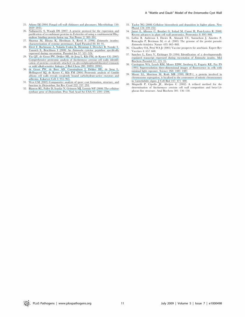
To better understand the domain structure of the Jessie3 lectin,
we expressed full-length Eh Jessie3, the putative N-terminal CBD
domain, and the unique C-terminal domain as maltose-binding
protein (MBP)-fusion proteins in the periplasm of Escherichia coli
[26]. The MBP-fusions with the N-terminal CBD or the full-length
Jessie3 each bound chitin beads (Fig. 5), while the MBP-fusion
with the unique C-terminal domain of Jessie3 did not bind chitin.
These results are consistent with our previous demonstration that
the N-terminal domain of the Eh Jessie3 lectin, when expressed as
an epitope-tagged protein in transfected Eh, is sufficient for chitin-
binding [10].
Neither the full-length Jessie3 lectin nor the Jessie3 unique C-
terminal domain had chitinase or chitin deacetylase activity, using
assays that had previously demonstrated such activity from lysed
Eh or recombinant Eh enzymes [8,13]. Although the N-terminal
CBD of full-length Jessie3 has the expected lectin activity, we
cannot rule out the possibilities that 1) the unique C-terminal
domain is not properly folded and so lacks its usual enzymatic
activity or 2) the unique C-terminal domain contains an enzymatic
activity not tested here.
Figure 3. Three-dimensional high-resolution fluorescence microscopy shows that Jessie lectins (red) arrive late but eventuallycover the surface of cysts. Unlike encysting Ei in Figs. 1, 2, and 4, protists here were labeled with antibodies to Jessie3 prior to fixation, so that onlythe surfaces of the encysting parasites are labeled. While there is very little Jessie3 lectin on the parasite surface after 36 hrs (A), Jessie3 appears inincreasing number of punctate spots, sometimes in linear arrays (arrows), in the cyst wall at 48 hrs (B) and 60 hrs (C). In contrast, at 72 hrs (D), Jessie3has a swirling appearance in the Ei cyst wall. After Jessie3 is fully incorporated into the wall (D), the cyst becomes impermeable to DAPI, so that nucleiare no longer visible or are weakly visible. Bar is 10 microns. Each image is the composite of multiple optical sections.doi:10.1371/journal.ppat.1000498.g003
A ‘‘Wattle and Daub’’ Model of the Entamoeba Cyst Wall
PLoS Pathogens | www.plospathogens.org 4 July 2009 | Volume 5 | Issue 7 | e1000498
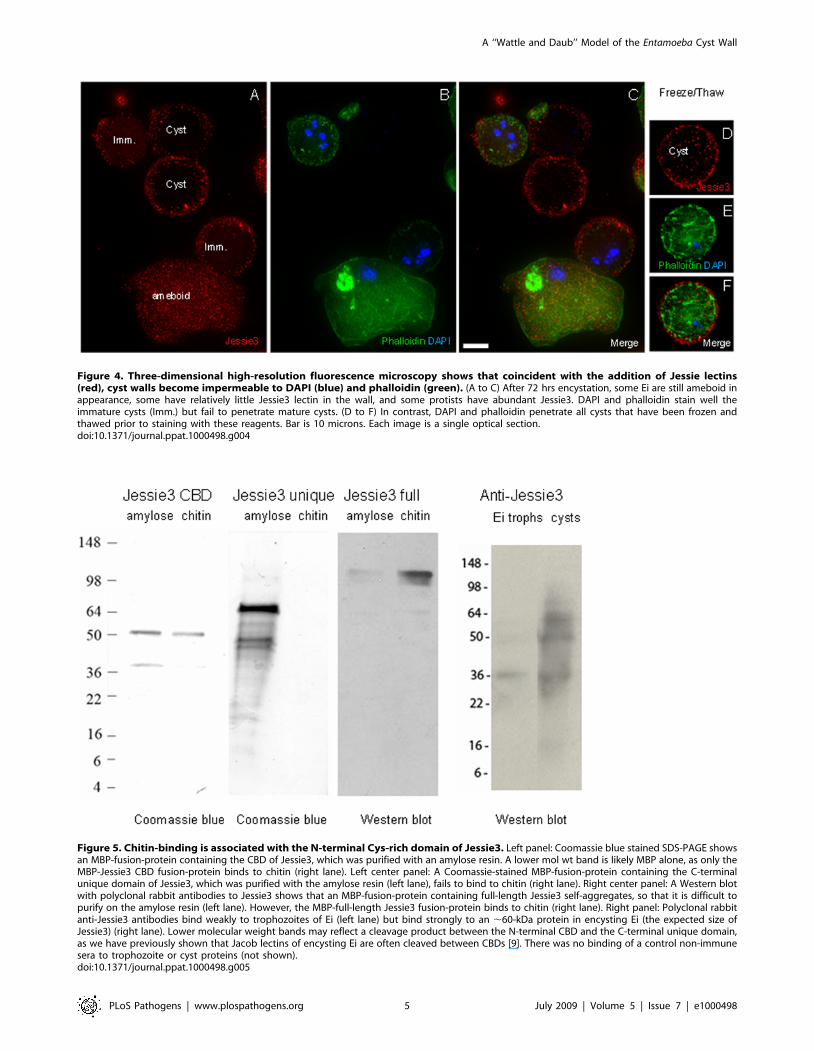
Figure 4. Three-dimensional high-resolution fluorescence microscopy shows that coincident with the addition of Jessie lectins(red), cyst walls become impermeable to DAPI (blue) and phalloidin (green). (A to C) After 72 hrs encystation, some Ei are still ameboid inappearance, some have relatively little Jessie3 lectin in the wall, and some protists have abundant Jessie3. DAPI and phalloidin stain well theimmature cysts (Imm.) but fail to penetrate mature cysts. (D to F) In contrast, DAPI and phalloidin penetrate all cysts that have been frozen andthawed prior to staining with these reagents. Bar is 10 microns. Each image is a single optical section.doi:10.1371/journal.ppat.1000498.g004
Figure 5. Chitin-binding is associated with the N-terminal Cys-rich domain of Jessie3. Left panel: Coomassie blue stained SDS-PAGE showsan MBP-fusion-protein containing the CBD of Jessie3, which was purified with an amylose resin. A lower mol wt band is likely MBP alone, as only theMBP-Jessie3 CBD fusion-protein binds to chitin (right lane). Left center panel: A Coomassie-stained MBP-fusion-protein containing the C-terminalunique domain of Jessie3, which was purified with the amylose resin (left lane), fails to bind to chitin (right lane). Right center panel: A Western blotwith polyclonal rabbit antibodies to Jessie3 shows that an MBP-fusion-protein containing full-length Jessie3 self-aggregates, so that it is difficult topurify on the amylose resin (left lane). However, the MBP-full-length Jessie3 fusion-protein binds to chitin (right lane). Right panel: Polyclonal rabbitanti-Jessie3 antibodies bind weakly to trophozoites of Ei (left lane) but bind strongly to an ,60-kDa protein in encysting Ei (the expected size ofJessie3) (right lane). Lower molecular weight bands may reflect a cleavage product between the N-terminal CBD and the C-terminal unique domain,as we have previously shown that Jacob lectins of encysting Ei are often cleaved between CBDs [9]. There was no binding of a control non-immunesera to trophozoite or cyst proteins (not shown).doi:10.1371/journal.ppat.1000498.g005
A ‘‘Wattle and Daub’’ Model of the Entamoeba Cyst Wall
PLoS Pathogens | www.plospathogens.org 5 July 2009 | Volume 5 | Issue 7 | e1000498

However, an alternative function for the unique C-terminal
domain of Jessie3 was suggested by the finding that transformed
bacteria expressing full-length Eh Jessie3 agglutinate in solution.
After centrifugation in a microfuge at high speed, these bacteria
aggregated into a solid pellet, which did not dissociate with
vortexing or pipetting. Transformed bacteria expressing MBP-
fusions containing only the unique C-terminal domain of Jessie3
also agglutinated in solution and aggregated into an insoluble
pellet after centrifugation. Anti-Jessie3 antibodies and fluorescence
microscopy showed agglutinated E. coli secrete large amounts of
the MBP-Jessie3 fusion-proteins, which accumulated in sheets or
biofilms on the surface of the slide or cover slip (Figs. 6A, 6B, and
6D). Negative staining showed secreted MBP-full-length Jessie3
fusion-proteins also formed large planar aggregates that labeled
with gold-conjugated secondary antibodies after primary antibody
labeling of the Jessie3 lectin (Fig. 6E). In contrast, bacteria
expressing MBP only, MBP fused to the N-terminal CBD of
Jessie3, or MBP fused to multiple CBDs of the Jacob lectin did not
agglutinate bacteria in solution, did not form a solid pellet after
centrifugation, and failed to make biofilms on slide surfaces
(Fig. 6C and data not shown). These results strongly suggest that
the unique C-terminal domain of Eh Jessie3 is an important
contributor to bacterial agglutination in solution and insoluble
pellet-formation after centrifugation.
We used negative staining to get a better view of the aggregated
MBP-Jessie3 fusions. The full-length Jessie3 and the unique C-
terminal domain of Jessie3 formed dense aggregates on the surface
of transformed E. coli and often disrupted the outer bacterial
membrane (Figs. 7D and 7E). While these aggregates did not form
a higher order crystal structure, MBP-full-length Jessie3 fusion-
proteins made linear structures that aggregated into larger
branched structures (Fig. 7I). Similar linear and branched
structures were formed by MBP-Jessie3 unique C-terminal domain
fusion-proteins, although the branches were not so long (Fig. 7H).
In contrast, control MBP alone and MBP-Jacob2 lectin fusion-
proteins stayed in solution and did not self-aggregate (Figs. 7A, 7B,
and 7F). Interestingly, MBP fusion-proteins containing the N-
terminal CBD of Jessie3 often formed cone-shaped aggregates, as
they were released from the periplasm of E. coli (Fig. 7C). These
MBP-Jessie3 CBD fusion-proteins formed smaller self-aggregates
and thinner linear forms than the MBP-full length Jessie3 fusion-
proteins (Fig. 7G).
We conclude that the C-terminal unique domain of Jessie3
appears to be responsible for most protein self-aggregation and
integrate these findings into a revised model of the Ei cyst wall
(next section).
A ‘‘wattle’’ (chitin fibrils and Jacob lectins) and ‘‘daub’’(Jessie3 lectins) model of the Entamoeba cyst wall
The results here and elsewhere suggest that the cyst wall of Ei
may be made in three phases (Fig. 8). During the first
‘‘foundation’’ phase, Jacob lectins, which are encystation-specific
glycoproteins that contain Gal, are bound to the surface of
encysting amebae by Gal/GalNAc lectins that are constitutively
expressed [9,16–18]. This idea is supported by our previous
demonstration that Ei form ‘‘wall-less cysts’’ in the presence of
Figure 6. Recombinant Jessie 3 lectins self-aggregate. Fluorescence microscopy (A to D) and negative staining (E) of bacteria transformed withMBP-fusion-proteins expressing full-length Jessie3 (green in A) or the unique C-terminal unique domain of Jessie3 (green in B) agglutinate to formlarge clumps of bacteria. In addition, biofilms containing MBP-full length Jessie3 are formed (green in D and immunostained with gold in E).Antibodies to the unique C-terminal domain of Jessie 3 are conjugated to Alexafluor in (A, B, and D) or are detected with immunogold in (E).Antibodies to the N-terminal CBD of Jessie3 (green in C) show that MBP-fusion-proteins containing this domain do not self-aggregate on a macro-scale and do not agglutinate bacteria. Bar (A to D) is 2 microns. Bar (E) is 500 nm.doi:10.1371/journal.ppat.1000498.g006
A ‘‘Wattle and Daub’’ Model of the Entamoeba Cyst Wall
PLoS Pathogens | www.plospathogens.org 6 July 2009 | Volume 5 | Issue 7 | e1000498

Figure 7. Negative stains of bacteria transformed with MBP constructs targeted to the periplasm suggest the unique C-terminaldomain of Jessie3 is an important contributor to self-aggregation. Bacteria (B) expressing MBP alone (low magnification in A and highmagnification in F) or an MBP-Jacob2 fusion-protein (jac in low magnification in B) release dense quantities of proteins, which do not self-aggregate.In contrast, bacteria expressing an MBP-Jessie3 CBD fusion-protein (J3 CBD in low magnification in C and high magnification in G) release proteins inconical shaped eruptions from the periplasm, and the MBP-Jessie3 CBD fusion-proteins form short, thin linear arrays. Bacteria expressing MBP-Jessie3unique C-terminal domain fusion-proteins (J3 sad (self-adhering domain) in low magnification in D and high magnification in H) or MBP-full-lengthJessie3 (J3 ful in low magnification in E and high magnification in I) release dense aggregates of proteins, and sheets of these MBP-Jessie3 fusion-proteins contain branched aggregates of multiple-stranded linear forms that are thicker than those formed by Jessie3 CBD. Bar (A to E) is 250 nm. Bar(F to I) is 100 nm. Circles and ovals are added to figures to highlight structures of secreted proteins.doi:10.1371/journal.ppat.1000498.g007
A ‘‘Wattle and Daub’’ Model of the Entamoeba Cyst Wall
PLoS Pathogens | www.plospathogens.org 7 July 2009 | Volume 5 | Issue 7 | e1000498

excess galactose and by demonstration that the Gal/GalNAc lectin
binds Jacob lectins on Western blots [9]. In the second ‘‘wattle’’
phase, chitin is synthesized, secreted, and is bound the surface of
encysting amebae. There chitin fibrils are likely cross-linked by
Jacob lectins, which contain multiple tandemly arranged CBDs,
although cross-linking has not been proven [9,10]. During the
third ‘‘daub’’ phase, the cyst wall is solidified and may be made
impermeable to small molecules by the addition of the Jessie3
lectin, which has a single CBD that binds chitin fibrils and a
unique C-terminal domain that appears to promote self-aggrega-
tion. As already mentioned, we cannot rule out a role for the Jacob
lectin in the formation of the mortar or daub in the Ei cyst wall.
We cannot rule out some other biochemical events (e.g. chemical
cross-linking), which might affect cyst wall permeability.
Other biochemical events, which we have not assigned to
particular stages in the model presented here include: 1) Jacob
lectins are cleaved into smaller repeats by the action of proteases
that recognize conserved sequences between CBDs [11,27,28],
and 2) chitin is trimmed by chitinase and deacetylated by chitin
deacetylase [8,13]. Although the ‘‘wattle and daub’’ model is
supported by numerous observations and experiments, we do not
presently have the technology to knock out or knock down gene
expression in Ei, which might more conclusively prove the
essential role of each component of the Ei cyst wall.
Figure 8. ‘‘Wattle and daub’’ model of the Ei cyst wall (a hypothesis). In the first ‘‘foundation’’ phase of encystation, Jacob lectins, which arethemselves glycoproteins that contain Gal, are bound to the surface of encysting amebae by constitutively expressed plasma membrane Gal/GalNAclectins [9,16]. In the second ‘‘wattle’’ phase, Jacob lectins, which contain tandemly arranged CBDs, appear to cross-link chitin fibrils that are depositedon the surface of encysting amebae. In the third ‘‘daub’’ phase, the cyst wall is solidified and made impermeable to small molecules by the addition ofthe Jessie3 lectin, which has an N-terminal CBD that binds chitin and a unique C-terminal unique domain that appears to cause self-aggregation.doi:10.1371/journal.ppat.1000498.g008
A ‘‘Wattle and Daub’’ Model of the Entamoeba Cyst Wall
PLoS Pathogens | www.plospathogens.org 8 July 2009 | Volume 5 | Issue 7 | e1000498

The ‘‘wattle and daub’’ model for the Entamoeba cyst wall shows
little resemblance to models of the walls of fungi, plants, or slime
molds [29–34]. This is likely the case because the Entamoeba cyst
wall, which appears to be homogenous rather than multi-layered,
contains a single sugar homopolymer (chitin) and a small set of
chitin-binding lectins (Jacob, Jessie, and chitinase). In contrast, the
walls of fungi, plants, and slime molds have multiple layers,
numerous sugar homopolymers (chitin, cellulose, glucans, and/or
GalNAc polymers), and numerous enzymes, structural proteins,
and large oligosaccharides (e.g. mannans). As noted above,
Entamoeba appears to make chitin within secretory vesicles rather
than at the plasma membrane, as described in fungi [20,21,23].
This model has been developed using encysting Ei and
bacterially expressed Eh Jessie3 and Jacob lectins. It is likely that
numerous aspects apply to Eh, the human pathogen that does not
readily encyst in culture, for the following reasons. First, Eh has
the same parts list of chitin synthase, chitinase, chitin deacetylase,
Jacob and Jessie3 lectins, and chitinase as Ei [4,8–11,13,16,35].
Second, Eh cysts from clinical samples stain with antibodies to
Jacob lectins and Jessie3 lectins (our unpublished data and [9]).
Third, Eh encysting in xenic cultures (filled with bacteria) express
numerous mRNAs for Jacob and Jessie lectins, and chitinase [7].
In human studies and in mouse models, antibodies to the Gal/
GalNAc lectin appear to protect the host from amebic infection
[3,17,36]. Whether antibodies to the Gal/GalNAc lectin or
antibodies to cyst wall lectins (Jacobs, Jessie3, or chitinase) interfere
with cyst wall formation during clinical infection is an important
unanswered question.
Materials and Methods
Deconvolving microscopy of Jessie3, Jacob, chitinase,and chitin in encysting Entamoeba
Trophozoites of the IP-1 strain of Ei were grown at 25uC in
axenic culture in TYI-SS medium. Ei encystation was induced by
placing parasites for 12–72 h in low-glucose (LG) medium, which
has reduced osmolarity, glucose, and serum levels with respect to
TYI-SS medium [37]. To test for excystation, cysts were washed
with sterile water to lyse trophozoites, and then cysts were placed
in TYI-SS medium for 2 hrs at 37uC. In addition, cysts were
treated with 0.1% SDS and then washed and excysted, as
described above.
Ei trophozoites and cysts, which contain four nuclei and have a
chitin wall at maturity, were incubated with antibodies or lectins
after washing in phosphate-buffered saline (PBS) or after fixation
for 10 min in 2% paraformaldehyde at 4uC in the presence of
0.1% Triton X-100 to permeabilize membranes [9].
Mono-specific polyclonal antibodies to Ei Jacob were made
previously by immunizing rabbits with Jacob lectin purified from a
two-dimensional protein gel of cyst walls [9]. Mono-specific rabbit
antibodies were made previously to a multi-antigenic peptide
containing chitinase repeats [22].
Here rabbits were immunized with either the N-terminal CBD
of Eh Jessie3 or its unique C-terminal domain, each of which was
expressed as a fusion-protein with the maltose-binding protein
(MBP) of Escherichia coli at the N-terminus (see below) [26]. MBP-
Jessie3 fusion-proteins were purified on amylose resins, checked by
SDS-PAGE, and sent to Strategic Biosolutions for mono-specific
polyclonal rabbit antibody production.
Prior to their use in microscopy, rabbit antibodies were purified
using MBP-Jessie3 fusion-proteins chemically coupled to agarose.
Western blots showed binding of the anti-Jessie3 antibody to an
MBP-full-length Jessie3 fusion-protein and to trophozoites and
cysts of Ei (Fig. 5). Rabbit antibodies were then directly labeled
red, green, or blue with Alexafluor dyes, diluted to a concentration
of 1 mg/ml, and incubated with trophozoites or cysts for 60 mins
at room temperature in PBS+2% bovine serum albumin (BSA).
Parasites were washed four times in PBS prior to staining of the
walls, nuclei, or cytoskeleton.
Chitin in encysting Ei and in cyst walls was stained with
Alexafluor-conjugated wheat germ agglutinin at a concentration of
20 mg per ml for 60 min at 4uC in PBS-BSA. Cyst nuclei were
visualized with 49,6-diamidino-2-phenylindole (DAPI), while actin
filaments were visualized with 1 mg per ml fluorescein-isothiocy-
anate-conjugated phalloidin. To determine whether the Ei cyst
wall causes a permeability barrier to small molecules, Ei cysts were
stained as described above, or Ei cysts were fixed and then exposed
to three cycles of freezing and thawing to create an ice artifact
prior to labeling with DAPI (molecular weight (MW 277) or
phalloidin (MW 3945 of pentamer).
Slides were examined by three-dimensional multiple wavelength
fluorescence microscopy using an Olympus IX70 microscope
equipped for Deltavision deconvolution (Applied Precision). This
system employs restorative as well as deconvolution techniques to
provide resolutions up to four times greater than conventional light
microscopes and is used to study the ultrastructure of intracellular
structures such as the kinetochore [38,39]. Images were collected
at 0.2 mm optical sections for the indicated wavelengths and were
subsequently deconvolved using SoftWoRx (Applied Precision).
Data were examined as either optical sections or as a projection of
the entire stack.
Negative staining of Ei cystsEi cysts were sonicated, and cyst walls were purified on sucrose
gradients, as described [9]. Purified cyst walls were then washed
and resuspended in Tris-EDTA (TE) buffer, pH 7.5, and applied
to carbon-coated, copper grids. The sample on the grid was
allowed to adsorb for 5 minutes, washed with TE, and blocked
with 2% BSA solution in TE. Grids were exposed to rabbit anti-
Jessie3 antibodies, which were diluted 1:20 dilution in TE, for 1 hr
at RT, and then grids were washed six times in TE for 10 min.
Grids were incubated with a secondary goat-anti rabbit antibody
conjugated to a 5 nm gold particle for 1 hr at RT, followed by 6
washes with TE. The grids were stained with 0.1% uranyl acetate
for 15 sec and visualized on a Phillips CM-12 microscope at 120
KV, 25,000 to 60,000 times magnification. Images were recorded
on Kodak 50–63 film and digitized on a Nikon 9000 scanner at
2000 dpi.
Expression of Jessie3 lectins in transformed bacteriaThe Eh Jessie3 gene was PCR amplified using three sets of
primers to amplify the N-terminal CBD (Fwd: CGGGATCCT-
TAAATATCACATTTAGTCAACG and Rev: GGGAAGCTT-
TTACTGATTGACTTGGTCTTC), the unique C-terminal
domain (Fwd: TCTAGACTTCCATTAGTACTAAAATTTGA
and Rev: AAGCTTTTAAGAAGAACATAAATTAGATCTTC),
and the full length Eh Jessie3 sequence (Fwd: GCTCTAGAT-
TAAATATCACATTTAAGTCAACGAACC and Rev: GGATCC-
TTAGTGGTGGTGGTGGTGGTGTTTTGAATAATGTTCTT-
GTTTGT). The three Eh Jessie3 PCR products were cloned into
pMAL-p2E vector (New England Biolabs (NEB), Beverley, MA), which
makes an IPTG-inducible, periplasm-targetted fusion-protein with
MBP at the N-terminus and Jessie3 at the C-terminus. Bl21-DE3 cells
from Invitrogen were transformed with the pMAL-p2E-Jessie3
constructs, and recombinant proteins were induced with 0.1 mM
IPTG. Fusion-proteins were extracted by sonication (5–10 pulses) of
bacteria in phosphate-buffer (PB) and 4 mM PMSF on ice. Fusion-
proteins were purified by 1) anion exchange chromatography through
A ‘‘Wattle and Daub’’ Model of the Entamoeba Cyst Wall
PLoS Pathogens | www.plospathogens.org 9 July 2009 | Volume 5 | Issue 7 | e1000498

DEAE-Sepharose resin using 200 mM KCl in PB as an eluent and 2)
amylose resin (NEB) using 100 mM maltose as an eluent. Enriched
proteins were checked for purity on SDS-PAGE stained with
Coomassie brilliant blue and by Western blot using horse-radish
peroxidase- (HRP-) tagged anti-MBP antibody from NEB.
Indirect immunoflourescence and negative staining ofbacteria expressing Jessie3 lectin
E. coli Bl21-DE3 cells expressing Eh Jessie3 proteins were
harvested and washed in PBS. For indirect immunofluorescence,
bacteria were fixed with 2% paraformaldehyde in PBS on ice for
5 min. The fixed cells were incubated with polyclonal mono-
specific rabbit antibody to MBP-Jessie3 CBD fusion-proteins at a
dilution of 1:100 for 1 hr at RT and washed three times in PBS.
For secondary antibody, the cells were incubated with goat anti-
rabbit antibody conjugated to Alexafluor for 1 hr at RT and
washed three times with PBS.
For negative staining, unfixed E. coli cells expressing Jessie3
protein were resuspended in TE buffer, applied to grids, and
incubated with anti-Jessie antibodies, as described for Ei cyst walls
(above). Alternatively, E. coli and Jessie3 proteins secreted into the
medium were visualized by negative staining in the absence of
anti-Jacob antibodies.
Methods for assaying for chitin-binding, chitinase, andchitin deacetylase activities
Chitin-binding assays for fusion-proteins containing MBP-full
length Jessie3, MBP-Jessie3 unique C-terminal domain, MBP-
Jessie3 N-terminal CBD, and MBP only (negative control) were
performed using methods similar to those described in refs. 9 and
10. Briefly, recombinant proteins, which were purified from
bacterial lysates on an amylose resin, were incubated with chitin
beads (NEB), washed, and then eluted by boiling in SDS. Eluted
proteins were separated on SDS-PAGE and identified by
Coomassie blue staining (MBP-Jessie3, MBP-Jessie N-terminal
CBD, and MBP only). Because of problems of self-aggregation,
MBP-full- length Jessie3, which was never very abundant after
release from the amylose resin, was detected with mono-specific
polyclonal antibodies to Eh Jessie3.
Recombinant MBP-Jessie3 unique domain fusion-proteins were
assayed for probable chitinase activity using the method to
determine cellular chitin in yeast [40]. Briefly, 5 mg of purified
Jessie3 protein was incubated with 10 mg of chitin or hexa-N-
acetyl-glucosamine (GlcNAc6) in presence of 50 ml of Mcllavaine’s
buffer (18 mM citric acid, 64 mM dibasic sodium phosphate,
pH 6) at 220uC for 10 min and subsequently incubated for 4 hrs
at 37uC. Fifty ml of water was added to the sample and centrifuged
at 120006g to produce a clear supernatant. The release of
GlcNAc was measured by the Morgan-Elson method of
incubating the supernatant with 10 ml Borax (2.7 M sodium
borate) at 99uC for 10 min, followed by incubation with 100 ml of
p-dimethylaminobenzaldehyde (DMAB) for 20 min at 37uC.
Resulting samples were measured by a colorimetric assay at
585 nm. Chitinase C (Sigma) was used as a positive control.
For chitin deacetylase assay, 5 mg of purified Jessie3 protein was
incubated with 25 mM chitin or GlcNAc6 in 50 mM Tris, pH 8
and 1 mM cobalt chloride buffer at 50uC for 16 hrs [8]. The
sample was separated on a thin layer chromatography (TLC) plate
with an n-butanol:ethanol:water:acetic acid (5:4:3:1) buffer system
and stained with diphenylamine or ninhydrin.
Author Contributions
Conceived and designed the experiments: AC SKG EB PR JS. Performed
the experiments: AC SKG KJ. Analyzed the data: AC SKG EB PR JS.
Contributed reagents/materials/analysis tools: LM. Wrote the paper: EB
JS.
References
1. Arroyo-Begovich A, Carabez-Trejo A, Ruiz-Herrera J (1980) Identification of
the structural component in the cyst wall of Entamoeba invadens. J Parasitol 66:
735–741.
2. Van Dellen KL, Bulik D, Specht C, Robbins PW, Samuelson J (2006)
Heterologous expression of an Entamoeba histolytica chitin synthase in Saccharomyces
cerevisiae. Eukaryotic Cell 5: 203–206.
3. Haque R, Huston CD, Hughes M, Houpt E, Petri WA Jr (2003) Amebiasis.
N Engl J Med 348: 1565–1573.
4. Clark CG, Alsmark UC, Tazreiter M, Saito-Nakano Y, Ali V, Marion S,
Weber C, Mukherjee C, et al. (2007) Structure and content of the Entamoeba
histolytica genome. Adv Parasitol 65: 51–190.
5. Avron B, Stolarsky T, Chayen A, Mirelman D (1986) Encystation of Entamoeba
invadens IP-1 is induced by lowering the osmotic pressure and depletion of
nutrients from the medium. J Protozool 33: 522–525.
6. Eichinger D (2001) Encystation in parasitic protozoa. Curr Opin Microbiol 4:
421–426.
7. Ehrenkaufer GM, Haque R, Hackney JA, Eichinger DJ, Singh U (2007)
Identification of developmentally regulated genes in Entamoeba histolytica: insights
into mechanisms of stage conversion in a protozoan parasite. Cell Microbiol 9:
1426–1444.
8. Das S, Van Dellen K, Bulik D, Magnelli P, Cui J, Head J, Robbins PW,
Samuelson J (2006) The cyst wall of Entamoeba invadens contains chitosan
(deacetylated chitin). Mol Biochem Parasitol 48: 86–92.
9. Frisardi M, Ghosh SK, Field J, Van Dellen K, Rogers R, Robbins PW,
Samuelson J (2000) The most abundant glycoprotein of amebic cyst walls (Jacob)
is a lectin with five Cys-rich, chitin-binding domains. Infect Immun 68:
4217–4224.
10. Van Dellen K, Ghosh SK, Robbins PW, Loftus B, Samuelson J (2002) Entamoeba
histolytica lectins contain unique 6-Cys or 8-Cys chitin-binding domains. Infect
Immun 70: 3259–3263.
11. Van Dellen KL, Chatterjee A, Ratner DM, Magnelli PE, Cipollo J, Steffen M,
Robbins PW, Samuelson J (2006) Unique posttranslational modifications
of chitin-binding lectins of Entamoeba invadens cyst walls. Eukaryotic Cell 5:
836–848.
12. Shao L, Devenport M, Jacobs-Lorena M (2001) The peritrophic matrix of
hematophagous insects. Arch Insect Biochem Physiol 47: 119–125.
13. de la Vega H, Specht CA, Semino CE, Robbins PW, Eichinger D, Caplivski D,Ghosh S, Samuelson J (1997) Cloning and expression of chitinases of Entamoebae.
Mol Biochem Parasitol 85: 139–147.14. Ghosh SK, Frisardi M, Ramierez-Avila L, Descoteaux S, Sturm-Ramirez K,
Newton-Sanchez OA, Santos-Preciado J, Ganguly C, Lohia A, Reed S,Samuelson J (2000) Molecular epidemiology of Entamoebae: Evidence of a
bottleneck (demographic sweep) and transcontinental spread of diploid parasites.
J Clin Microbiol 38: 3815–3821.15. Haghighi A, Kobayashi S, Takeuchi T, Masuda G, Nozaki T (2002)
Remarkable genetic polymorphism among Entamoeba histolytica isolates from alimited geographic area. J Clin Microbiol 40: 4081–4090.
16. Wang Z, Samuelson J, Clark CG, Eichinger D, Paul J, Van Dellen K, Hall N,
Anderson I, Loftus B (2003) Gene discovery in the Entamoeba invadens genome.Mol Biochem Parasitol 129: 23–31.
17. Mann BJ, Torian BE, Vedvick TS, Petri WA Jr (1991) Sequence of a cysteine-rich galactose-specific lectin of Entamoeba histolytica. Proc Natl Acad Sci USA 88:
3248–3252.18. Eichinger D (2001) A role for a galactose lectin and its ligands during encystment
of Entamoeba. J Eukaryot Microbiol 48: 17–21.
19. Turner NA, Eichinger D (2007) Entamoeba invadens: the requirement for galactoseligands during encystment. Exptl Parasitol 116: 467–474.
20. Cabib E, Roh DH, Schmidt M, Crotti LB, Varma A (2001) The yeast cell walland septum as paradigms of cell growth and morphogenesis. J Biol Chem 276:
19679–19682.
21. Ziman M, Chuang JS, Schekman RW (1996) Chs1p and Chs3p, two proteinsinvolved in chitin synthesis, populate a compartment of the Saccharomyces cerevisiae
endocytic pathway. Mol Biol Cell 7: 1909–1919.22. Ghosh S, Field J, Frisardi M, Rosenthal B, Mai Z, Rogers R, Samuelson J (1999)
Chitinase secretion by encysting Entamoeba invadens and transfected E. histolytica
trophozoites: Localization of secretory vesicles, ER, and Golgi apparatus. Infect
Immun 67: 3073–3081.
23. Chavez-Munguıa B, Cristobal-Ramos AR, Gonzalez-Robles A, Tsutsumi V,Martınez-Palomo A (2003) Ultrastructural study of Entamoeba invadens encystation
and excystation. J Submicrosc Cytol Pathol 35: 235–243.24. Villagomez-Castro JC, Calvo-Mendez C, Lopez-Romero E (1992) Chitinase
activity in encysting Entamoeba invadens and its inhibition by allosamidin. Mol
Biochem Parasitol 52: 53–62.
A ‘‘Wattle and Daub’’ Model of the Entamoeba Cyst Wall
PLoS Pathogens | www.plospathogens.org 10 July 2009 | Volume 5 | Issue 7 | e1000498
25. Adams DJ (2004) Fungal cell wall chitinases and glucanases. Microbiology 150:
2029–2035.26. Nallamsetty S, Waugh DS (2007) A generic protocol for the expression and
purification of recombinant proteins in Escherichia coli using a combinatorial His6-
maltose binding protein fusion tag. Nat Protoc 2: 383–391.27. Sharma M, Hirata K, Herdman S, Reed S (1996) Entamoeba invadens:
characterization of cysteine proteinases. Exptl Parasitol 84: 84–91.28. Ebert F, Bachmann A, Nakada-Tsukui K, Hennings I, Drescher B, Nozaki T,
Tannich E, Bruchhaus I (2008) An Entamoeba cysteine peptidase specifically
expressed during encystation. Parasitol Int 57: 521–524.29. Yin QY, de Groot PW, Dekker HL, de Jong L, Klis FM, de Koster CG (2005)
Comprehensive proteomic analysis of Saccharomyces cerevisiae cell walls: identifi-cation of proteins covalently attached via glycosylphosphatidylinositol remnants
or mild alkali-sensitive linkages. J Biol Chem 280: 20894–20901.30. de Groot PW, de Boer AD, Cunningham J, Dekker HL, de Jong L,
Hellingwerf KJ, de Koster C, Klis FM (2004) Proteomic analysis of Candida
albicans cell walls reveals covalently bound carbohydrate-active enzymes andadhesins. Eukaryot Cell 3: 955–965.
31. West CM (2003) Comparative analysis of spore coat formation, structure, andfunction in Dictyostelium. Int Rev Cytol 222: 237–293.
32. Blanton RL, Fuller D, Iranfar N, Grimson MJ, Loomis WF (2000) The cellulose
synthase gene of Dictyostelium. Proc Natl Acad Sci USA 97: 2391–2396.
33. Taylor NG (2008) Cellulose biosynthesis and deposition in higher plants. New
Phytol 178: 239–252.
34. Jamet E, Albenne C, Boudart G, Irshad M, Canut H, Pont-Lezica R (2008)
Recent advances in plant cell wall proteomics. Proteomics 8: 893–908.
35. Loftus B, Anderson I, Davies R, Alsmark UC, Samuelson J, Amedeo P,
Roncaglia P, Berriman M, et al. (2005) The genome of the protist parasite
Entamoeba histolytica. Nature 433: 865–868.
36. Chaudhry OA, Petri WA Jr (2005) Vaccine prospects for amebiasis. Expert Rev
Vaccines 4: 657–668.
37. Sanchez L, Enea V, Eichinger D (1994) Identification of a developmentally
regulated transcript expressed during encystation of Entamoeba invadens. Mol
Biochem Parasitol 67: 125–35.
38. Carrington WA, Lynch RM, Moore EDW, Isenberg G, Fogarty KE, Fay FS
(1995) Superresolution three-dimensional images of fluorescence in cells with
minimal light exposure. Science 268: 1483–1487.
39. Moore LL, Morrison M, Roth MB (1999) HCP-1, a protein involved in
chromosome segregation, is localized to the centromere of mitotic chromosomes
in Caenorhabditis elegans. J Cell Biol 147: 471–480.
40. Magnelli P, Cipollo JF, Abeijon C (2002) A refined method for the
determination of Saccharomyces cerevisiae cell wall composition and beta-1,6-
glucan fine structure. Anal Biochem 301: 136–150.
A ‘‘Wattle and Daub’’ Model of the Entamoeba Cyst Wall
PLoS Pathogens | www.plospathogens.org 11 July 2009 | Volume 5 | Issue 7 | e1000498