Genetic diversity of glucose phosphate isomerase from Entamoeba histolytica
Upload
independentCategory
view
0download
0
Eur. J. Biochem. 247, 348-355 (1997) 0 FEBS 1997
Sequencing, expression and properties of triosephosphate isomerase from Entamoeba histolytica Abraham LANDA I , Arturo ROJO-DOMiNGUEZ’, Lucia JIMENEZ ’ and D. Alejandro FERNANDEZ-VELASCO’
’ Departamento de Microbiologia y Parasitologia, Facultad de Medicina, Universidad Nacional Autdnoma de MCxico, MCxico ’ Departamento de Quimica, Universidad Aut6noma Metropolitana-Iztapalapa, MCxico ’ Departamento de Bioquimica, Facultad de Medicina, Universidad Nacional Autdnoma de MCxico, MCxico
(Received 17 February/l4 April 1997) ~ EJB 97 0248/3
We have isolated a cDNA clone of the glycolytic enzyme, triosephosphate isomerase (TPI) from Entamoeba histolytica. Degenerate oligonucleotides obtained by reverse translation of conserved poly- peptide sequences, derived from TPIs of other organisms, were used to amplify a 450-bp fragment using E. histolytica cDNA as a template. The fragment was used to screen a cDNA library. The isolated cDNA, encoding a protein of 261 amino acids, shares 43-52.6% positional identity with other known protozoan TPIs. The catalytic residues were conserved ; nevertheless, several indels occurred at other regions in the protein sequence. The complete coding sequence of the E. histolytica TPI gene was cloned into the expression vector pRSET and expressed as a wild-type TPI enzyme (E. histolytica TPI) and as a fusion protein with an N-terminal tail of six histidine residues E. histolytica TPI-His,) ; both recombinant proteins were purified.
Molecular modeling of E. histolytica TPI showed an identical topology to the known structures of other TPI molecules, but with a remarkable feature; more than 10 inserted residues are located in the same region of the molecular surface. Studies were performed to detect possible changes that might be caused by the inserted amino acids. The catalytic activity and oligomeric state of the purified protein were similar to that reported for TPI from other sources. In contrast, stability towards dilution, as well as thermal inactivation and unfolding assays, showed that E. histolytica TPI is significantly more stable towards denaturation than Trypanosoma brucei TPI.
Keywords: triosephosphate isomerase ; Entamoeba histolytica ; TIM barrel ; dimer stability.
Entamoeba histolyticu is a worldwide distributed protozoan causative of human amebiasis ; this disease produces annually more than 40 000 deaths (Martinez-Palomo, 1990).
Knowledge of the three-dimensional structure and catalytic properties of the proteins from parasites should help in the design of inhibitors and drugs (Kuntz, 1992; Verlinde et al., 1994; Blundell, 1996). The catalytic mechanism of the glyco- lytic enzyme triosephosphate isomerase (TPI) has been thor- oughly studied (Knowles, 1991 ; Lodi et al., 1994). The TPI se- quences of 38 species, including several parasites (Lambeir et al., 1987; Kohl et al., 1994; Ostoa et al., 1997) have been re- ported. The three-dimensional folding of six of them, show simi- lar homodimeric structures, each monomer being folded into a @/a)8 barrel composed of about 250 amino acids (Banner et al., 1975; Lolis et al., 1990; Wierenga et al., 1991; Noble et al., 1993; Mande et al., 1994; Delboni et al., 1995). Despite con- siderable variation in the amino acid sequences, the structure of
the barrel and the orientation of the monomers in the quaternary structure are conserved (Wierenga et al., 1992; Delboni et al., 1995). Recently, TPI has been used as a model for the design of inhibitors that interact with the intersubunit contacts (Kuntz et al., 1992), or with the non-conserved amino acids of the mol- ecule (Gbmez-Puyou et al., 1995; Garza-Ramos et al., 1996). In this work, we report the initial characterization of the triose- phosphate isomerase from Entamoeba histolytica ; the cDNA encoding for this enzyme was isolated and cloned. The re- combinant enzyme was expressed and purified, thereafter its catalytic and stability properties were determined. The three-di- mensional structure of E. histolytica TPI was modeled based on yeast and Trypanosoma brucei TPT crystallographic atomic coordinates. The possible role of a set of insertions oriented in the same region of the molecular surface of E. histolytica TPI is discussed.
Correspondence to D. Alejandro Fernindez-Velasco, Departamento de Bioquimica, Facultad de Medicina, Universidad Nacional Aut6noma de MCxico, Mexico, D. F. Mexico 04510
Fax: +525 616 24 19. E-mud: [email protected] Abbreviation. TPI, triosephosphate isomerase. Enzymes. Triosephosphate isomerase (EC 5.3.1.1); glycerol-3-phos-
phate dehydrogenase (EC 1.1.1.8). Note. The novel nucleotide sequence mentioned in this article has
been deposited with the EMBL data bank and is available under acces- sion number Y13387.
MATERIALS AND METHODS
Collection of parasites. Trophozoites of E. histolytica were cultivated in TYI-S33 medium at 363°C and harvested after 72 h culture. Trophozoites were washed in 15 mM sodium phosphate, 15 mM NaCI, pH 7.2 (NaCUP,), and centrifuged at 250 g for 5 min at 4°C; the pellet was frozen immediately at -70°C.
Isolation and sequence analysis of E. histolyticu TPI cDNA clone. The E. histolytica poly(A)-rich RNA was obtained

Landa et al. ( E m .I. Biochem. 247) 349
by chromatography of total RNA through oligo(dT)-cellulose (Sigma Chemical Co.) (Aviv and Leder, 1972). The cDNA library was prepared using the cDNA synthesis system from Gibco BRL, based in the method described by Gluber and Hoff- man (1983). The cDNA was ligated into an EcoRI site of the i g t l l vector, yielding about 4X lo7 independent clones. Two degenerated oligonucleotides designed from conserved polypep- tide sequences, derived from several TPIs, were used by PCR to prime on E. histolytica cDNA and obtain a 45-bp fragment. Approximately 50 000 phage plaques were screened overnight at 50°C under aqueous conditions with a 450-bp probe and washed in a 30 mM sodium citrate, 300 mM NaCl pH 7.0 (standard saline citrate buffer) with 0.1 % SDS at 55°C (Benton and Davis, 1977). After the secondary and tertiary screenings, several posi- tive phage clones were obtained. The two largest cDNA inserts were subcloned into an M13 vector and sequenced according to the dideoxy chain termination method (Sanger et al., 1977) using the sequenase kit (United States Biochemical Corporation) and ["S]dATP[aS] (Amersham International). Either the M13 uni- versal primers or various E. histolytica TPI-specific oligonucleo- tide primers were used. Computer analysis of the sequence was carried out with the freeware available in the World Wide Web.
Construction of the recombinant expression vectors. The cDNA encoding E. histolytica TPI was amplified by PCR to engineer NheI and NdeI sites upstream of the initiation codon using the oligonucleotide primers 5' GAA-TTC-CAT-ATG- GGG-GCG-GGT-AAA-TTT-G and 5' CTA-GCT-AGC-ATG- GGG-GCG-GGT-AAA-TTT-G, respectively ; a Hind111 site was also engineered immediately downstream of the termination codon using the oligonucleotide primer 3' GGG-AAG-CTT- ACA-ATT-TCT-CAG-AAA-CTG. The complete E. histolytica TPI coding sequence between the unique sites NheI-Hind111 (E. histolytica TPI-His, construction) or NdeI-Hind111 sites (E. histolyticu TPI construction) was introduced in to pRSET (InVitrogen Co.). In this plasmid vector, DNA inserts are under the control of a T7 promotor and in-frame with a sequence that encodes an N-terminal fusion peptide of six histidine residues. This peptide tail functions as a metal-binding site on the fusion protein, and allows the one-step purification of recombinant pro- teins by immobilized metal-affinity chromatography (Le Grice et al., 1990; Lilius et al., 1991 ; Muller and Skerra, 1993). E. histolytica TPI-His, contains the histidine tail whereas E. histo- lytica TPI contains only the complete TPI-coding sequence, both under the control of the T7 promotor. Escherichia coli TGI strain cells were transformed with the plasmid constructions and induced as recommended by the vector supplier (Invitrogene).
Expression and purification of E. histolytica TPI and TPI-His,. All the steps in the purification of E. histolytica TPI- His, and E. histolytica TPI were monitored by catalytic activity and by 12% polyacrylamide gel electrophoresis in the presence of SDS (Laemmli, 1970).
E. histolytica TPI-His, was purified as follows. The trans- formed cells were grown under vigorous agitation at 37 "C, until an A,,,,, of approximately 0.3 was reached. Then, cells were induced with isopropyl P-D-thiogalactopyranoside at a final concentration of 1 mM for 1 h. Thereafter, cells were infected with phage M13IT7; growth was continued for 5 h. The cell pellet from a 500-mL culture was resuspended in 40 mL buffer containing 100 mM triethanolaminehydrochloride, 50 mM NaCl 1 mM dithiothreitol, 1 mM sodium azide and 0.5 mM phenylmethylsulfonyl fluoride, pH 8.0. Cells were lysed by three passages through a french press (SLM Instruments Inc.) at 758 kPa and 4°C. Cell debris was eliminated by centrifugation at 65000 g for 1 h. The supernatant was loaded into a Hitrapp chelating column (Pharmacia Biotech). After extensive washing, bound E. histolytica TPI-His, was eluted with 100 mM tri-
ethanolamine and 50 mM EDTA, pH 8.0, and stored at 4°C until use.
E. histolytica TPI was purified as follows. Bacterial growth, induction, lysis and centrifugation were performed as described for E. histolytica TPI-His,, except that the cell pellet was resus- pended in 20 mM Tris/HCl, pH 8.0, and 0.2 mM phenylmethyl- sulfonyl fluoride. The supernatant obtained after centrifugation at 65000 g was precipitated with ammonium sulfate. The 60- 90% fraction was centrifuged (17500 g, 15 min), the pellet dis- solved in 2 mL 10 mM triethanolamine, 1 mM EDTA and 1 mM dithiothreitol, pH 7.4 (solution A), and dialyzed overnight against the same buffer. The protein was then loaded onto a Sephacryl S-300 column (Pharmacia Biotech) previously equili- brated in solution A. Fractions with high specific activity were pooled. Aliquots from the pool (500 pL) were loaded onto a Mono Q 515 column equilibrated in solution A, and eluted with a gradient of 0-100 mM NaCl of the same buffer. Fractions were collected, assayed for enzymatic activity, and further ana- lysed by SDSPAGE.
7: brucei TPI was purified (Borchert et al., 1993) from trans- formed E. coli cells kindly donated by R. PCrez-Montfort.
Protein determination was carried out by the Lowry method, using bovine serum albumin as a standard (Hartree, 1972), or by absortion at 280nm using an estimated molar absorption coefficient ~ (280) = 36440 M-' cm-' (Pace et al., 1995).
Molecular modeling. All energy calculations and molecular edition were performed on a Silicon Graphics Indigo2 system with an R4000 processor using the BIOGRAF molecular mechanics program (Molecular Simulations, Inc.) and the Drei- ding-I1 generic forcefield (Mayo et al., 1990). Energy minimiz- ations were performed until a 4 kJ mol-' nm-' or lower force was reached. All molecular dynamics simulations were calcu- lated by numeric integration of Newton's equations of motion for all atoms using the Verlet algorithm (Verlet, 1967), a step size of 1 fs, and updating velocities every 0.1 ps. Non-bonded interactions were considered within a distance of 0.9 nm with a smooth switch-off function 0.8-0.85 nm to avoid discontinuous forces. During the modeling procedure, all hydrogen atoms were explicitly included and, for computational convenience, calcula- tions were done in vacuo. The abscence of explicit solvent mole- cules decreases the accuracy of the calculations, however, solvent effects were introduced via a distance-dependent dielec- tric constant which simulates water screening of electrostatic interactions.
The starting model of E. histolytica TPI was constructed from yeast TPI (PDB file 1YPI) by the replacement of the neces- sary 137 single-point mutations according to multiple alignment of known TPI sequences. The automatic alignment procedure with Maxhom and ClustalW programs included a set of 38 se- quences; in order to obtain an alignment with topological sense, further refinement was done based on the superimposed crystal- lographic structures of the TPI family. This hand manipulation was necessary in loop regions where sequence divergence is higher and the positions of indels are not optimal. Every replace- ment was carried out by the following procedure. The side chain of a residue was first changed; in order to avoid the propagation of spurious distortions an attempt was made to adjust the posi- tion of the new atoms submitting them to three series of minimi- zation and a 3-ps molecular dynamics simulation with all sur- rounding atoms fixed (including the main chain atoms of the replaced residue). The region of mobile atoms was gradually increased so the same cycles of minimization dynamics were applied to a segment of 5 residues centered in the replaced one, then to the neighbor side chains in a 0.5-nm radius, and finally to all the atoms inside the 0.5-nm sphere.

350
1 E.histoly S. cervisi E. coli T. brucei H. sapiens G. lamblia
2 E.histoly S. cervisi E.coli T.brucei H. sapiens G. lamblia
3 E.histoly S. cervisi E . coli T . brucei H. sapiens G. lamblia
4 E.histoly S. cervisi E. coli T-brucei H. sapiens G. lamblia
5 E.histoly S . cervisi E.coli T-brucei H. sapiens G. lamblia
6 E.histoly S. cervisi E. coli T. brucei H. sapiens G. lamblia
7 E.histoly S.cervisi E. coli T.brucei H. sapiens G.lamblia
0 E.histoly S . cervisi E . coli T. brucei H.sapiens G . lamblia
Landa et al. ( E M J. Biochern. 247)
BOTTOM
-MGAGKF - -MARTF - - -MRHP -MSKPQP MAPSRKF -MPARRP
SVDAELAKKV ASIPE- - -NV ELAGV-A-GC TSINH---DV AKVPA---DT HKIPD---SV
LAGEANGAN V- - - -KKPQ A----EGSH L----SHPK L- ~ - - -DPK N- - - -TSKQ
CQVP VGAK IGAQ FGVN CGAT MGLK
AGL QGV QGL SGF EGL KGM
AI--SKE AWKN EV----K DWTN TQG--AA AFEG KL--KKA DWAK NV----K DWSK ELGESKM LWKE
IS KEVAEA T LG DKAASE L DA -NIAEQ V IG ADVRGE L VS DAVAQS T VC AEGAQH I
KKADID DKADVD AQPDID QQRDVN SQPDVD QCPNID
BETA
WGGN FVGGN LVMGN IAAAN FVGGN F I GGN
EVIVGV EWICP AVAIAP QCWAS EWCAP DWIAP
ILVSA VTVGA IMLGA FVIAA IAVAA LRIAA
YVIL WVIL YIII WIVL W L HVIV
KVIACI GVILCI TPVLCI MVIACI GVIACI TVIFCV
I ILAY W A Y AVIAY W I A Y W L A Y W I A Y
RIQY RILY I IQY RILY RIIY RIIY
GFLV GFLV GALV GFLV GFLV GFLV
ACTIVE-LOOP
WKCNGT FKLNGS WKLNGS WKCNGS WKMNGR FKCNGS
PFIY PATY PEMY TFVH PTAY SAVH
ENAW-TKSGAYTGEVH QNAYLKASGAFTGENS QNVDLNLSGAFTGETS QNAI-AKSGAFTGEVS QNCYKVTNGAFTGEIS QNVYLEGNGAWTGETS
GH SERRQI FHES GH SERRSY FHED GH SERRTY HKES GH SERRAY YGET GH SERRHV FGES GH SERRRI MGET
GET EAQRI ANQ GET LEEKK AGK GET EAENE AGK GET LQERE SGR GEK LDERE AGI GET LDERK ANR
EPVWAIGTGKTAT EPVWAIGTGLAAT EPVWAIGTGKSAT EPVWAIGTGKVAT EPVWAIGTGKTAT EPVWSJGTGWAT
GGSVN GGSAN GGSVN GGSVN GGSVT GGSAN
G GASL DAA G GASL KP- G GASL KA- G GASL KP- G GASL KP- G GASL KP-
HELIX
LASIETLTKGVAA KQSIKEIVERLNT RHMVHELVSNLRK QQSLSELIDLFNS KQSLGELIGTLNA LDFIKSHVAAIAA
IPKVQQI LDYSVSL IDMAKRE LAMTKER IDFARQK LSTAIAA
VGMLVD VDQIKD AAML KD LPILKD PGMIKD VEMLQD
NEQVAEKVKVAID DKFIADKTKFALG DELIAKKFAVLKE NE IVAD KVAAAVA DELIGQKVAHALA DEQSAKKAKRALE
TEEWAAQLKAINN TLDWERQLNAVLE TEEVCARQIDAVLK TAVWLTQIAAIAK TEKVVFEQTKVIAD TMEVNIAQLEALGK
PDQAQEVHQYIRKWMTEN PEDAQDIHASIRKFLASK PAQAQAVHKFIRDHIAKV PQQAQEAHALIRSWVSSK PQQAQEVHEKLRGWLKSN PEQAEEVHVGLRKWFAEK
PANCNELA GSNAVT F K ASNAAELF GKNARTLY GATCKELA GSNCEKLG
BOTTOM SVSEKF- - ~ KFKTIIN
EFVDIIN SRN- - - - - ~
DAFAVIV KAAEAAKQA EFVDIIK ATQ. ___. ~
EFVDI IN AKQ--. __.
EFMTMID ILTKTRT- -
Fig. 1. Multiple alignment of the amino acid sequences of TPI from E. histolytica, S. cerevisiae, E. coli, Z brucei, H. sapiens and G. Zambia. Sequences are arranged according to the secondary structure. -, absence of amino acids at the corresponding position. Inserted residues are shown in italics.
E. histolyticn TPI contains 14 additional residues and one deletion with respect to the yeast TPI template. Inserted resi- dues, located in 6 zones of the sequence, were added by con- structing the appropriate segment in extended conformation and slowly docking it to the correspondent peptide bond previously broken. Docking approximation was performed adding a har- monic distance constrain from the ends of the new segment to the ends of the broken peptide bond, and slowly decreasing the equilibrium distance to 0.2 nm. During the docking procedure, all atoms of the new segment and of six residues close to the insertion point were declared mobile in minimization steps.
The active loop three of E. hisrolytica TPI is one residue shorter than the equivalent region in yeast TPI (Fig. 1). In order to incorporate this deletion in the E. histolytica TPI model, the equivalent loop of T. brucei TPI (PDB file 2TIM), which is simi- lar in size to that of E. histolytica TPI (Fig. I), was used as a guide (Wierenga et al., 1992). This deletion was performed taking out the corresponding atoms, and minimizing the local region, first with a distance constrain similar to the docking pro- cedure described above, then with the new peptide bond in- cluded. At the end, the initial model was obtained by a global minimization of the structure.
Landa et al. (Eul: J. Biochem. 247) 35 1
Atomic coordinates of the molecular model are available upon request.
Determination of kinetic parameters. TPI catalysis was assayed with glyceraldehyde 3-phosphate as substrate. As pre- viously reported (Rozacky et al., 1971), dihydroxyacetone phos- phate production was coupled to NADH oxidation via a-glycerol phosphate dehydrogenase. The assay conditions were 100 mM triethanolamine hydrochloride, 10 mM EDTA, 1 mM dithio- threitol, pH 7.4, containing 0.2 mM NADH, 20 pg a-glycerol phosphate dehydrogenase and 0.1 -2 mM D-glyceraldehyde 3- phosphate. Ammonium sulfate in a-glycerol phosphate dehydro- genase was removed by the centrifugatiodfiltration technique (Penefsky, 1977). Glyceraldehyde 3-phosphate was prepared from glyceraldehyde-3-phosphate diethyl acetal, as recom- mended by the supplier (Sigma). In all cases, activity was linear with time, i.e. no inactivation occurred during the 2-5 min of measurement.
Molecular-sieve chromatography. Molecular-sieve chro- matography was performed on a Superose 12 HR 10/30 column (Pharmacia Biotech) eluted at 25°C with a flow rate of 0.2 ml/ min. The buffer used was 20 mM triethanolamine hydrochloride, 1 mM EDTA and 1 mM dithiothreitol, pH 7.4. Calibration of the column was performed with a set of eight proteins of known molecular masses.
Dilution curves. Different concentrations of TPI were incubated for 1 h and 24 h in buffer containing 100 mM tri- ethanolamine hydrochloride, 10 mM EDTA, 1 mM dithiothrei- tol, pH 7.4. The catalytic activity was measured in two different ways, depending on the protein concentration. Concentrated samples were assayed by adding an adequate aliquot of sample to the reaction cuvette, in order to obtain a constant enzyme concentration of 2 ng/mL during catalytic assays in all cases. In contrast, samples incubated at low concentrations of TPI were assayed by the addition of 80 pl reaction mixture to 920 p1 incu- bated TPI solutions ; these latter measurements were, therefore, carried out at variable TPI concentrations.
Thermal unfolding and inactivation. The denaturation of E. histolytica and 7: brucei TPIs was studied at a protein concen- tration of 0.12 mg/ml in solution A. Circular dichroism spectra were recorded in the far-ultraviolet region on a JASCO J . SOOA spectropolarimeter equipped with a water-jacketed cell holder. The temperature of the sample was increased at a rate of one degree/min, and measured during the experiment with a thermis- tor probe inside the cell. From the obtained spectra, ellipticity at 220 nm (y) was used to calculate the apparent fraction of dena- tured subunits cfu), using the equation fD = Cy-y,)/(y,-y,), where y N and y,, are the ellipticity values characteristic of the native and unfolded subunits. Both parameters were linear ex- trapolations from the initial and final portions of the curve of y versus temperature.
Irreversible loss of catalytic activity due to thermal in- activation was assayed as follows. TPI, at a concentration of 240pg/ml in solution A, was incubated at 50°C; at different times, aliquots were withdrawn to ice and left for 30 min, there- after the samples were assayed for catalytic activity.
RESULTS
Cloning and sequence analysis. The amplified TPI fragment of 450 bp prepared by PCR was used as a probe to screen an E. histolytica cDNA library. Eight positive clones were obtained from the tertiary screening. Two of them containing a 0.9-kb insert were chosen for characterization. The sequences of the two clones were the same. The predicted translation product of the E. histolytica TPI cDNA is a protein of 261 amino acids
1 2 3 4 5
28kDa
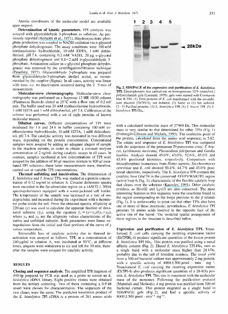
Fig. 2. SDSPAGE of the expression and purification of E. histolytica TPI. Electrophoresis was carried out on homogeneous 12% (masdvol.) polyacrylamide gels (Laemmli, 1970) ; gels were stained with Coomassie blue R-350. (1) Total proteins of E. coli transformed with the recombi- nant plasinid (EhTPI-4), not induced. (2) Same as (1) but induced. (3-5) Purified proteins. (3) E. histolytica TPI. (4) 7: brucei TPI. ( 5 ) E. histolytica TPI-His, ,
with a calculated molecular mass of 27969 Da. This molecular mass is very similar to that determined for other TPIs (Fig. 1) (Fothergill-Gilmore and Michels, 1993). The isoelectric point of the protein, calculated from the amino acid sequence, is 5.82. The amino acid sequence of E. histolyticu TPI was compared with the sequences of the protozoan Trypanosoma cvuzi, 7: bru- cei, Leishmania mexicana, Plusmodium jalciparum and Gardia lamblia. Analysis showed 45.0%, 43.0%, S2.6%, 45.0% and 45.4 % positional identities, respectively. Comparison with triosephosphate isomerases from Homo sapiens, Saccharomyces cerevisiue and E. coli showed 50.4%, 44.7% and 43.0% posi- tional identities, respectively. The E. histolytica TPI contains the catalytic base Glul74 in the conserved AYEPVWAIGTG region (active loop 6, Fig. I), characteristic of the flexible catalytic loop that closes over the substrate (Knowles, 1991). Other catalytic residues, as His102 and Lysl3 are also conserved. The most interesting features in this sequence were the insertions found in the region corresponding to the loops previous to the strands (Fig. 1). It is notheworthy to point out that other TPIs also have one or more of these insertions; nevertheless, E. histolyticu TPI presents 16 amino acids inserted in the opposite face of the active site of the barrel. The modeled spatial arrangement of these regions in the structure is described below.
Expression and purification of E. histolytica TPI. Trans- formed E. coli cells carrying the resulting expression vector (EhTPIH,-4) produce significant quantities of the fusion protein E. histolytica TPI-His,. This protein was purified using a metal affinity column (Fig. 2) . Eluted E. histolytica TPI-His, runs as a single band with a molecular mass higher than 28 kDa, probably due to the tail of histidine residues. The usual yield from a 100-ml bacterial culture was approximately 2 mg protein, with a specific activity of 4000?500 pmol . min-' mg-'. Transformed E. coli carrying the resulting expression vector (EhTPI-4) also produces significant quantities of a 28-kDa pro- tein, E. histolytica TPI. This size is consistent with the molecular mass of the monomer. Following the purification protocol (Materials and Methods), 4 mg protein was purified from 100 ml bacterial culture. This protein migrated as a single band in SDS/PAGE gels (Fig. 2), and had a specific activity of 8000 2 500 pmol . min-l mg-'.

352 Landa et al. (EUK J. Biochem. 247)
Table 1. Kinetic parameters for the isomerisation of D-glyceralde- hyde 3-phosphate.
~~~ ~~
mM min-'
E. histolytica TPI 0.61 +- 0.46 2.41 X 10' E. histdytica TPI-His, 1.05 ? 0.23 2.20 x 10' Yeast TPI 1.27 1.0 X10"
1.27 2.64 X 10' 0.62' 1.41 X 10'
L. rnexicnna TPI 0.30" 2.5 X10' 7: brucei TPI 0.25 3.7 XlO'
" Krietsch et al., 1970. Nickbarg and Knowles, 1988. Sun et al., 1992. Kohl et al., 1994. Lambeir et al., 1987.
Kinetic parameters. Kinetic parameters for the isomerisation of glyceraldehyde 3-phosphate were carried out for the purified proteins. Michaelis-Menten behaviour was observed (Table 1). The kinetic constants of E. histolyticu TPI and E. histolytica TPI-His, are similar to those reported for TPI from other sources.
Stability to dilution. E. histolytica TPI eluted from the molecu- lar-sieve chromatography column as a single symmetrical peak, with an estimated apparent molecular mass of 53.3 kDa (data not shown). The estimated molecular mass for a dimeric protein, calculated from the amino acid sequence, is 55.94 kDa. There- fore, E. histolytica is a dimer, and has the same quaternary structure as reported for other TPI molecules from mesophiles (Rozacky et al., 1971 ; Wierenga et al., 1992; Mainfroid et al., 1996).
It has been observed that unstable mutants as well as wild- type TPI loose activity when incubated at low concentrations; inactivation by dilution is due to the dissociation of the active dimers into inactive monomers (Borchert et al., 1993, 1995; Mainfroid et al., 1996). To estimate the value of the dissociation constant of the dimer, E. histolytica TPI was incubated at different concentrations. Thereafter, aliquots were withdrawn to measure catalytic activity at 2 ng/ml. A constant catalytic activ- ity of 8928 pmol . min-' . mg-' (2910) and 3078 (2310) pmol . min-' mg-' was observed for E. histolytica TPI and E. histolyt- icu TPI-His,, respectively, i.e. no dilution-dependent inactivation was found. Catalytic activity was again determined after a 24-h incubation. E. histolytica TPI-His, lost activity in a concentra- tion-dependent way (Fig. 3); only less than 10% of the original activity remained when the enzyme was incubated at concentra- tions lower than 0.3 pg/ml. In contrast, E. histolytica TPI is sta- ble for 24 h at concentrations as low as 2 ng/mL (Fig. 3). In order to explore a lower concentration range, E. histolytica TPI was incubated for 24 h at concentrations of 0.4-5 ng/mL. No significant decrease in activity was observed (Fig. 3). As the introduction of the poly(histidine) tail produced a 200-fold decrease in the stability towards dilution, further experiments were carried out only with E. histolytica TPI.
Sequence alignment and molecular modeling. Modeled E. his- tolytica TPI shows the same structural features of all crystallo- graphic TPI molecules (Fig. 1). An rms of 0.153 nm is obtained when comparing the 246 topologically equivalent a carbon atoms between the model and the template. Nevertheless, there is a remarkable new feature in this TPI not present in any known
80
60
40
20
0 1 10 100 1000 10000
Fig. 3. Stability to dilution. TPI was incubated for 24 h at the indicated concentrations ; thereafter the catalytic activities of E. histolytica TPI- His, (m), E. hisrolytica TPI incubated at high concentrations (0) and E. histolytica TPI incubated at low concentrations (0), were determined as described in Materials and Methods.
structure or sequence of TPI; 13 of the 14 inserted residues are located in the same region of the molecular surface, at the bot- tom of the (pa)8 barrel, in the region of loops opposite to the active site. The set of 13 inserted residues contains five electri- cally charged residues, three basic and two acidic. Since model- ing of inserted segments yields uncertain geometries, an attempt was made to sample the conformational landscape of this region of the structure. The final model was constructed submitting the initial one to a local molecular dynamics simulation (lSOps), declaring mobile the inserted atoms and all neighbors closer than 0.5 nm Convergence of energy was reached at 50 ps simulation with a small rearrangement around 90 ps (Fig. 4). The molecular simulation permitted the arrangement of the inserted amino acids into a better packed and more globular conformation. The final model possesses satisfactory sterochemical quality, when ana- lyzed by Procheck (Morris et al., 1992) with 71% and 27% of the residues in the most favoured and allowed regions of the Ramachandran plot, respectively. All other geometric character- istics regarding side and main chain geometry place the model quality as equivalent to a crystallographic structure of around 0.27 nm resolution.
Comparative thermal unfolding and inactivation. Compara- tive thermal denaturation experiments were carried out with E. histolytica TPI and another protozoan TPI that from 7: brucei. The transition curves for both enzymes give t,, values of 58.6"C and 44.7"C E. histolyfica TPI and 7: brucei TPI, respectively. This result indicates that the enzyme from E. histolytica has a significantly higher resistance to thermal unfolding. Since both proteins aggregate upon unfolding, the use of a reversible two- state model on the transition data yields only apparent values for the enthalpy changes, being 197 kJ/mol and 732 kJ/mol for E. histolytica TPI and 7: brucei TPI, respectively. The difference in apparent enthalpy changes arises from the distinct cooperativ- ity of the processes. Thermal inactivation experiments were car- ried out to confirm the differences observed in the thermal un- folding experiments. After incubation at 50.0"C for different times, 7: brucei TPI lost activity irreversibly, whereas E. histo- lytica TPI maintained its catalytic activity (Fig. 5 , inset).

Landa et al. ( E m J. Biochern. 247) 353
Fig.4. C, trace of the E. histolytica TPI model. The C,x trace of one subunit of the E. histolyrica TPI model (gray) and the S. c e r e v i ~ i w crystallographic structure (black) are shown. The second subunit position is depicted by thin lines. Spheres represent the C,L of the inserted residues. The active site is located behind the structure in the left figure.
- Y
Time (rnin) / 1 0.4
0.0
20 30 40 50 60 70 80 Temperature (OC)
Fig. 5. Comparative effect of temperature on unfolding and inactiva- tion of protozoan TPI. Transition curve of the thermal unfolding of E. hisfolyticn TPI (0) and 7: brucei TPI (0) followed by changes in ellip- ticity at 220 nm. The inset shows thermoinactivation of E. histolytica TPI (0) and 7: brucei TPZ (0) at 50°C.
DISCUSSION
In this work we describe the cloning, sequencing and func- tional expression of E. histolytica TPI cDNA. The recombinant enzyme presented similar properties including amino acid simi- larity, kinetic parameters, oligomeric state and overall three-di- mensional structure, to those reported for the TPI form other sources. Comparison of E. histolytica TPI with other TPI mole- cules shows a singular feature, the presence of a set of inser- tions. The obtained model locates these insertions i n the bottom of the (pa), barrel, on the opposite face of the catalytic site. Due to the spatial distribution of the insertions, it is very likely that they play a role either in catalysis or in the folding or stability
of the enzyme. Scanning of E. histolytica TPI against the Prosite database did not show any known fingerprint in the sequence different from the TPI active site and some potential postraduc- tional modification zones. No cooperativity or significant differ- ences in the catalytic properties were observed between E. histo- lytica TPI and those reported (Lambeir et al., 1987) for other TPI molecules. In contrast, E. histolytica TPI is resistant to dilu- tion, and more stable towards thermal denaturation than 7: bru- cei TPI. It has been reported that rabbit TPI is stable towards dilution after a 30-min incubation at 2 ng/mL (Zabori et al., 1980), whereas human TPI loses 50% of its catalytic activity when incubated for the same period at a concentration of 0.5 ng/ ml (Mainfroid et al., 1996). In this work, it was found that activ- ity was not lost after a 24-h incubation at concentrations as low as 0.3 ng/ml. From these observations, it follows that the interac- tion between E. histolytica TPI monomers is the strongest so far detected. The interface residues of TPI show extensive variabil- ity (active loops 1-3, Fig. 3) (Wierenga et al., 1992), therefore, with the available data, it is not possible to ascribe the resistance towards dilution to a particular interface residue.
An N-terminal fusion peptide with six histidine residues was introduced in E. histolytica TPI. This procedure has been suc- cessfully used for the single-step purification of recombinant proteins (Le Grice et al., 1990; Lilius et al., 1991; Muller and Skerra, 1993). The purified protein, E. histolytica TPI-His,, showed similar kinetic properties to those determined for TPI, however, the histidine tail affects the dissociation constant of the dimer. This N-terminal tail is located in the same spatial region of the inserted residues, suggesting a stability relationship be- tween the interface and the bottom of the (/la), barrel.
The present results do not reveal the molecular basis of the observed properties of E. histolytica TPI ; further studies on the thermodynamics of the folding pathway of this enzyme and a comparison with the data obtained for the TPI of other sources (Mainfroid et al., 1996) would permit the elucidation of the role of the inserted amino acid residues in the stability properties of this enzyme. It is tempting to speculate that the observed partic-

354 Landa et al. (Eul: J. Biochem. 247)
ular characteristics of E. histolytica TPI might play a role in the survival of the cyst form of the parasite, exposed to extreme environmental conditions. Furthermore, the new surface region in E. histolytica TPI, not present in the homologous human en- zyme, is a potential site for drug design.
The generous gifts of transformed E. coli cells overexpressing Z brucei TPI (Dr R. Perez-Montfort and M. IBB E. Saavedra, Universi- clad Nacional Autdnoma de Mhico) and the 7: brucei TPI gene (Dr P. A. M. Michels, International Institute of Cellular and Molecular Pathology, Brussels, Belgium) are gratefully acknowledged. A. L. thanks Dr C. B. Shoemaker (Harvard School of Public Health, Boston, USA) for help in cloning the E. histolytica TPI gene. The authors would like to acknowl- edge Dr G. Mendoza for performing the molecular-sieve chromatogra- phy experiments and Dr A. G6mez-Puyou for kind help and valuable suggestions and discussion. This work was partially supported from grants IN 201795 and IN 103596 from the Programa de Apoyo a Proy- ectm de fnvestigacidn e Innovucidn Tecnolrigica of the Direccidn General de Asuntos del Personal Acadimico. UNAM and grants 473100-5-3771N and 0150P-N from the Consejo Nacional de Ciencia y Tecnologia.
REFERENCES Aviv, H. & Leder, P. (1972) Purification of biologically active globin
messenger RNA by chromatography on oligothymidylic acid cellu- lose, Proc. Natl Acud. Sci. USA 69, 1408-1412.
Banner, D. W., Bloomer, A. C., Petsko, G. A,, Phillips, D. C., Pogson, C. I., Wilson, I. A., Corran, P. H., Furth, A. J., Milman, J. D., Offord, R. E., Priddle, J. D. & Waley, S. G. (1975) Structure of chicken muscle triosephosphate isomerase determined crystallographically at 2.5 A resolution using amino acid sequence data, Nature 255, 609 - 614.
Benton, W. D. & Davis, R. W. (1977) Screening Agtll recombinant clones by hybridization to single plaques in situ, Science 196, 180- 182.
Blundell, T. L. (1996) Structure-based drug design, Nature 384, Suppl. 23 - 26.
Borchert, T. V., Pratt, K., Zeelen, J. Ph., Callens, M., Noble, M. E. M., Opperdoes, F. K., Michels, P. A. M. & Wierenga, R. K. (1993) Over- expression of trypanosomal triosephosphate isomerase in Escheri- chia coli and characterisation of a dimer-interface mutant, Eur: J Biochem. 211, 703-710.
Borchert, T. V., Zeelen, J. Ph., Schliebs, W., Callens, M., Minke, W., Jaenicke, R. & Wierenga, R. K. (1995) An interface point-mutation variant of triosephosphate isomerase is compactly folded and mono- meric at low protein concentrations, FEBS Lett. 367, 315-318.
Delboni, L. F., Mande, S. C., Rentier-Delrue, F., Mainfroid, V., Turley, S., Vellieux, F. M. D., Martial, J. A. & Hol, W. G. J. (1995) Crystal structure of recombinant triosephosphate isomerase from Bacillus stearothermophilus. An analysis of potential thermostability factors in six isomerases with known three-dimensional structures points to the importance of hydrophobic interactions, Protein Sci. 4 , 2594- 2604.
Feinberg, A. P. & Vogelstein, B. (1983) A technique for radiolabeling DNA restriction endonuclease fragments to high specificity, Anal. Biochem. 132, 6-13.
Fothergill-Gilmore, L. A. & Michels, P. A. (1993) Evolution of glycoly- sis, Prog. Biophys. Mol. Biol. 59, 105-235.
Garza-Ramos, G., Perez-Montfort, R., Rojo-Doniinguez, A., Tuena de G6mez-Puyou, M. & G6mez-Puyou, A. (1996) Species-specific inhi- bition of homologous enzymes by modification of nonconserved amino acid residues. The cysteine residues of triosephosphate iso- merase, Eur: J. Biochem. 241, 114- 120.
G6mez-Puyou, A,, Saavedra-Lira, E., Becker, I., Zubillaga, R. A,, Rojo-Dominguez, A. & Perez-Montfoi-t, R. (1 995) Using evolution- ary changes to achieve species-specific inhibition of enzyme action-studies with triosephosphate isomerase, Chem. & Biol. 2,
Gluber. J. & Hoffman, G. R. (1983) A simple and very efficient method 847 -855.
for generating cDNA libraries, Gene (Amst.) 25, 263-269.
Hartree, E. F. (1972) Determination of protein: A modification of the Lowry method that gives a linear photometric response, Anal. Bin- chem. 48, 422-427.
Knowles, J. R. (1991) Enzyme catalysis: not different, just better, Nature
Kohl, L., Callens, M., Wierenga, R. K., Opperdoes, F. R. & Michels, P. A. M. (1994) Triose phosphate isomerase of Leishmania mexicana mexicana. Cloning and characterization of the gene, overexpression in Escherichia coli and analysis of the protein, Eul: J. Biochem. 220,
Krietsch, W. K. G., Pentchev, P. G., Klingenburg, H., Hofstatter, T. & Bucher, T. (1970) The isolation and crystallization of yeast and rabbit liver triosephosphate isomerase and a comparative characterization with the rabbit muscle enzyme, Eur: J. Biochem. 14, 289-300.
Kuntz, D. A,, Osowski, R., Scudok, M., Wierenga, R. K., Miiller, K., Kessler, H. & Opperdoes, E. R. (1992) Inhibition of triosephosphate isomerase from 7: brucei with cyclic hexapeptides, Eul: J. Biochem.
Kuntz, I. D. (1992) Structure-based strategies for drug design and dis- covery, Science 257, 1078- 1082.
Laemmli, U. K. (1970) Cleavage of structural proteins during the assem- bly of the head of bacteriophage T4, Nature 227, 680-685.
Lambeir, A. M., Opperdoes, F. R. & Wierenga, R. K. (1987) Kinetic properties of triose-phosphate isomerase from Ttypano~oma brucei Drucei, Eur: J. Biochem. 168, 69-74.
Le Grice, S. F. & Gruninger-Leitch, F. (1990) Rapid purification of homodimer and heterodimer HIV-1 reverse transcriptase by metal chelate affinity chromatography, Eur: J. Biochem. 187, 307-314.
Lilius, G., Persson, M., Bulow, L. & Mosbach, K. (1991) Metal affinity precipitation of proteins carrying genetically attached polyhistidine affinity tails, Eur: J. Biochem. 198, 499-504.
Lodi, P. J., Chang, C. L., Knowles, J. R. & Komives, E. A. (1994) Triosephosphate isomerase requires a positively charged active site: The role of lysine 12, Biochemistry 33, 2809-2814.
Lolis, E., Alber, T., Davenport, R. C., Rose, D., Hartman, F. C. & Petskq, G. A. (1990) Structure of yeast triosephosphate isomerase at 1.9 A resolution, Biochemistry 29, 6609-6618.
Mande, S. C., Mainfroid, V., Kalk, K. H., Goraj, K., Martial, J. A. & HOI,~W. G. J . (1994) Crystal structure of triosephosphate isomerase at 2.8 A resolution. Triosephosphate isomerase-related human genetic disorders and comparison with the trypanosomal enzyme, Protein Sci. 3 , 810-821.
Martinez-Palomo, A. & Ruiz-Palacios, G . (1 990) Amebiasis, in Tropical geogruphicul medicine (Warren, K. S . & Mahmoud, A. A. F., eds) pp. 327-344, McGraw-Hill, New York.
Mayo, S. L., Olafson, B. D. & Goddard, W. A. 111 (1990) Dreiding: A generic forcefield for molecular simulations, J. Phys. Chem. 94,
Morris, A. L., McArthur, M. W., Hutchinson, E. G. & Thornton, J. M. (1992) Stereochemical quality of protein structure coordinates, Pro- teins: Struct. Funct. Genet. 12, 345-364.
Muller, H. N. & Skerra, A. (1993) Functional expression of the uncom- plexed serum retinol-binding protein in Escherichia coli. Ligand binding and reversible unfolding characterists, J. Mol. Bid. 230,
Nickbarg, E. B. & Knowles, J. R. (1988) Triosephosphate isomerase: Energetics of the reaction catalyzed by the yeast enzyme expressed in Escherichia coli, Biochemistry 27, 5939 -5947.
Noble, M. E. M., Zeelen, J. Ph., Wierenga, R. K., Mainfroid, V., Goraj, K., Gohimont, A. C. & Martial, J. A. (1993) Structure of tripephos- phate isomerase from Escherichia coli determined at 2.6 A resolu- tion, Acta Ctystallogl: D 49, 403-417.
Ostoa-Saloma, P., Garza-Ramos, G., Ramirez, J., Becker, I., Berzunza, M., Landa, A., Gomez-Puyou, A,, Tuena de Gomez-Puyou, M. & PCrez-Montfort, R. (1997) Cloning, expression, purification and characterisation of the triosephosphate isomerase from Trypanosoma cruzi, Eul: J . Biochem., in the press.
Pace, C. N., Vajdos, F., Fee, L., Grimsley, G. & Gray, T. (1995) How to measure and predict the molar absorption coefficient of a protein, Protein Sci. 4, 2411 -2423.
Penefsky, H. S. (1977) Reversible binding of Pi by beef heart mito- chondrial adenosine triphosphatase, J. Biol. Chem. 252, 2891 -2899.
Rozacky, E. E., Sawyer, T. H., Barton, R. A. & Gracy, R. W. (1971) Studies of human triosephosphate isomerase. Isolation and properties
350, 121-124.
331-338.
207, 441 -447.
8897 -8909.
725-732.

Landa et al. ( E M J . Biochem. 247) 355
of the enzyme from erytrocytes, Arch. Biochem. Biophys. 146, 312- 320.
Sanger, F., Nicklen, S. & Coulson, A. R. (1977) DNA sequencing with chain terminating inhibitors, Proc. Nut1 Acud. Sci. USA 74, 5463- 5467.
Sun, A. Q., Yuskel, K. U. & Gracy, R. W. (1992) Interactions between the catalytic centers and subunit interface of triosephosphate iso- merase probed by refolding, active site modification, and subunit exchange, J. Biol. Chem. 267, 20 168-20 174.
Verlet, L. (1967) Computer “experiments” on classical fluids. I Thermodynamical properties of Lennard-Jones molecules, Phys. Rev.
Verlinde, C. L., Memtt, E. A., Van den Akker, F., Kim, H., Feil, I., Delboni, L. F., Mande, S. C., Sarfaty, S., Petra, P. H. & Hol, W. G. (1994) Protein crystallography and infectious diseases, Protein Sci.
159, 98-103.
3, 1670-1686.
Wierenga, R. K., Noble, M.oE. M., Vriend, G., Nauche, S. & Hol, W. G. J. (1991) Refined 1.83 A structure of trypanosomal triosephosphate isomerase crystallized in the presence of 2.4 M ammonium sulphate. A comparison with the structure of the trypanosomal triosephosphate isomerase-glycerol-3 -phosphate complex, J. Mol. B id . 220, 995- 1015.
Wierenga, R. K., Noble, M. E. M. & Davenport, R. C. (1992) Compari- son of the refined structures of liganded and unfiganded chicken, yeast and trypanosomal triosephosphate isomerase, J. Mol. Biol. 224,
Zabori, S., Rudolph, R. & Jaenicke, R. (1980) Folding and association of triosephosphate isomerase from rabbit muscle, 2. Nuturforsch 35c,
1115- 1126.
999- 1004.
Copyright © 2022 FDOKUMEN