Stature Estimation from Length of Fingers in Gujarati Population
251
-
Upload
independent -
Category
Documents
-
view
2 -
download
0
Transcript of Stature Estimation from Length of Fingers in Gujarati Population


EDITORProf. R K Sharma
Dean (R&D), Saraswathi Institute of Medical Sciences, Hapur, UP, IndiaFormerly at All India Institute of Medical Sciences, New Delhi
E-mail: [email protected]
SCIENTIFIC COMMITTEE
1. Pradeep Bokariya (Assistant Professor) Anatomy Dept.Mahatma Gandhi Institute of Medical Sciences, Wardha, Maharashtra
2. Dr Anil Rahule (Associate Professor) Dept of Anatomy, Govt medicalcollege Nagpur
3. Dr Yadaiah Alugonda (Assistant Professor) Forensic Medicine,MNR Medical college, Hyderabad
4. Dr Vandana Mudda (Awati) (Associate Prof) Dept of FMT,M.R.Medical College,Gulbarga,Karnataka,
5. Dr.Lav Kesharwani (Asst.Prof.) School of Forensic Science,SamHigginbottom Institute of Agriculture Technology & Sciences,Allahabad U.P,
6. Dr. NIshat Ahmed Sheikh (Associate Professor) Forensic Medicine,KIMS Narketpally, Andhra Pradesh
7. Dr K.Srinivasulu (Associate Professor) Dept of Forensic Medicine &Toxicology, Mediciti Institute of Medical sciences,Ghanpur, MEDCHALRanga Reddy. Dist.AP_501401.
8. Dr. Mukesh Sharma (Senior Scientific Officer) Physics Division,State Forensic Science Laboratory, Jaipur, Rajasthan
9. Dr Amarantha Donna Ropmay (Associate Professor) NEIGRIHMS,Shillong
10. Dr Basappa S. Hugar (Associate Professor) Forensic Medicine, M.S.Ramaiah Medical College, Bangalore
11. Dr.Anu Sharma (Associate Prof) Dept of Anatomy, DMCH,Ludhiana (PB)
Website: www.ijfmt.com
NATIONAL EDITORIAL ADVISORY BOARD
1. Prof. Shashidhar.C.Mestri (Professor) Forensic Medicine &Toxicology, Karpaga Vinayaga Institute of Medical Sciences,Palayanoor Kanchipuram Distric, Tamil Nadu
2. Dr. Madhuri Gawande (Professor) Department of Oral Pathologyand Microbiology, Sharad Pawar Dental College, Sawangi, Wardha.
3. Dr.T.K.K. Naidu (Prof & Head) Dept of Forensic Medicine, PrathimaInstitute of Medical Sciences, Karimnagar, A.P.
4. Dr. Shalini Gupta (Head) Faculty of Dental Sciences, King GeorgeMedical University, Lucknow, Uttar Pradesh
5. Dr. Pratik Patel (Professor & Head) Forensic Medicine Dept, Smt NHLMun Med College,Ahmedabad
6. Professor Devinder Singh Department of Zoology & EnvironmentalSciences, Punjabi University, Patiala
7. Dr. Pankaj Datta (Principal & Head) Department of prosthodontics,Inderprasth Dental college & Hospital, Ghaziabad
8. Dr. Mahindra Nagar (Head) Department of Anatomy, UniversityCollege of Medical Science & Guru Teg Bahadur Hospital, Delhi
9. Dr. D Harish (Professor & Head) Dept. Forensic Medicine & Toxicology,Government Medical College & Hospital, Sector 32, Chandigarh
10. Dr. Dayanand G Gannur (Professor) Department of ForensicMedicine & Toxicology, Shri B M Patil Medical College, Hospital &Research centre, Bijapur-586101, Karnataka
11. Dr. Alok Kumar (Additional Professor & Head) Department ofForensic Medicine & Toxicology, UP Rural Institute of MedicalSciences and Research, Saifai, Etawah. -206130 (U.P.), India.
12. Prof. SK Dhattarwal, Forensic Medicine, PGIMS, Rohtak, Haryana
13. Prof. N K Aggrawal (Head) Forensic Medicine, UCMS, Delhi
14. Dr. Virender Kumar Chhoker (Professor)Forensic Medicine andToxicology,Santosh Medical College,Ghaziabad, UP.
INTERNATIONAL EDITORIAL ADVISORY BOARD
1. Dr Nuwadatta Subedi (In Charge) Dept of Forensic Med andToxicology College of Medical Sciences, Bharatpur, Nepal
2. Dr. Birendra Kumar Mandal (In charge) Forensic medicine andToxicology, Chitwan Medical College, Bharatpur, Nepal
3. Dr. Sarathchandra Kodikara (Senior Lecturer) Forensic Medicine,Department of Forensic Medicine, Faculty of Medicine, University ofPeradeniya,Sri Lanka
4. Prof. Elisabetta Bertol (Full Professor) Forensic Toxicology at theUniversity of Florence, Italy
5. Babak Mostafazadeh (Associate Professor) Department of ForensicMedicine & Toxicology, Shahid Beheshti University of MedicalSciences, Tehran-Iran
6. Dr. Mokhtar Ahmed Alhrani (Specialist) Forensic Medicine & Clinical Toxicology,Director of Forensic Medicine Unit, Attorney General's Office, Sana'a, Yemen
7. Dr. Rahul Pathak (Lecturer) Forensic Science, Dept of Life SciencesAnglia Ruskin University, Cambridge, United Kingdom
8. Dr. Hareesh (Professor & Head) Forensic Medicine,Ayder ReferralHospital,College of Health Sciences,Mekelle University, MekelleEthiopia East Africa
Indian Journal of Forensic Medicine & Toxicology
“Indian Journal of Forensic Medicine & Toxicology “ is peer reviewed sixmonthly journal. It deals with Forensic Medicine, Forensic Science,Toxicology, DNA fingerprinting, sexual medicine and environment medicine.It has been assigned International standard serial No. p-0973-9122 and e-0973-9130. The Journal has been assigned RNI No. DELENG/2008/21789.
The journal is indexed with Index Copernicus (Poland) and is covered byEMBASE (Excerpta Medica Database). The journal is also abstracted inChemical Abstracts (CAS) database (USA. The journal is also covered byEBSCO (USA) database.
The Journal is now part of UGC, DST and CSIR Consortia. It is now officalpublication of Indian Association of Medico-Legal Experts (Regd.).
Print-ISSN:0973-9122 Electronic - ISSn: 0973-9130 Frequency: Six Montlhly
© All Rights reserved The views and opinions expressed are of the authorsand not of the Indian Journal of Forensic Medicine & Toxicology. IndianJournal of Forensic Medicine & Toxicology does not guarantee directly orindirectly the quality or efficacy of any products or service featured in theadvertisement in the journal, which are purely commercial.
Editor
Dr. R.K. Sharma
Institute of Medico-legal Publications4th Floor, Statesman House Building, Barakhamba Road,
Connaught Place, New Delhi-110 001
Design & Printed at
M/s Vineeta Graphics, Mobile: 9990005742, 9990005734
Printed, Published and owned by
Dr. R.K. Sharma
Institute of Medico-legal Publications4th Floor, Statesman House Building, Barakhamba Road,
Connaught Place, New Delhi-110 001
Published at
Institute of Medico-legal Publications4th Floor, Statesman House Building, Barakhamba Road,
Connaught Place, New Delhi-110 001
IFC PAGE FINAL.pmd 1/20/2014, 12:14 AM1

I
Indian Journal of Forensic Medicine& Toxicology
www.ijfmt.com
Volume 08 Number 02 July-December 2014
1. A Retrospective Study of Pregnancy Related Deaths ................................................................................................................ 01Shrikant Shinge
2. Significance of Maintaining Dental Records ............................................................................................................................... 04Sanchana Balakrishnan, Nithya Jagannathan MDS
3. Stature Estimation from Length of Fingers in Gujarati Population ....................................................................................... 08Senthil Kumaran M, Binaya Kumar Bastia, Lavlesh Kumar, Sweta H Patel, Keyur K Nimavat
4. Forensic Entomology Solves the Mysteries ................................................................................................................................. 12Dattatray Ghodake, Datta Pawale, T V Sathe
5. Autopsy Study of Unnatural Deaths among Young People of 13-21 Years .......................................................................... 16Age Group Conducted at Kims Hospital, BengaluruC Ramesh, Hanumantha, Ananda K, Devidas Tondare
6. Analysis of Tissue Samples from Unknown to Known ............................................................................................................ 21V Dekal, J Magendran, N Srinivasa Ragavan
7. Desperate Need of Modernized Mortuaries in Hospitals of India ......................................................................................... 25Shilekh Mittal
8. Estimation of Stature from Middle Finger Length-in Salem Region ...................................................................................... 30S Sasi Kumar, Pavanchand Shetty, Shankar
9. Thermal Burn: An Epidemiological Retrospective Study ........................................................................................................ 34S K Pandey, Neha Chaurasia, Awdhesh Kumar
10. Fusion of Epiphyses of Ischial Tuberosity in Relation with Age: A Cross Sectional Study ............................................... 38Anita Verma, Kanan Shah
11. Role of Dna in Forensic Identification .......................................................................................................................................... 43Karthavya Sampath, Nithya Jagannathan
12. Morphometric Analysis of Foramen Magnum for Sex Determination in Karnataka ......................................................... 48Kazi, S A K, Aadamali A Nadaf, Pramod B Gai
13. Analysis of Fatal Railway Accidents in District Hospital, South India: A Retrospective Study ....................................... 52Chandru K, Rajendra Kumar R, Rudramurthy S
14. An Epidemiological Study of Fatal Firearm Cases in Varanasi, UP ....................................................................................... 55Kaulaskar Shashikant V, S K Pandey, Pravin N Yerpude, Manoj Pathak, Keerti S Jogdand
15. Sexual Dimorphism of Cranial Sutures in Maharashtra and North-Karnataka Region ..................................................... 59Makandar U K, Kaushal Vishuddha M, Rajendra R, Sharieff J H
16. Post Mortem and the Risk of HIV Infection ................................................................................................................................ 63Mukesh Kumar Bansal, SK Naik, Poonam Gupta, Yashoda Rani, BL Sherwal
Contents
Content Final.pmd 7/24/2014, 11:23 AM1

II
17. Fetal Head Circumference: An Important Criterion for Gestational Age Estimation ......................................................... 68Sachin Sudarshan Patil, Ravindra Baliram Deokar, Jyoti Rahul Sul,Sandeep Shivaji Gosavi, Varsha Pratik Mahajan
18. A Study on Pattern of Injuries Due to Railway Accidents Occuring at Khammam Region - Andhra Pradesh ............. 72Roop Kumar KM, Udaypal Singh, Satya Sai Panda, Shaikh Khaja
19. Pattern of Railway Fatalities in Western Vidarbha Region of India ....................................................................................... 77SN Hussaini, A A Mukherjee, PA Wankhede, AS Rahule, SV Tambe, MSM Bashir
20. A Study on Estimation of Stature from Foot Measurements ................................................................................................... 81Kazi S A K, Aadamali A Nadaf, Vijayachandra, B G Shalawadid
21. A Study of Ligature Mark in Deaths Due to Hanging in Warangal Area, Andhra Pradesh .............................................. 85Bharathi Rama Rao, V Chand Basha, K Sudhakar Reddy
22. A Five Year Retrospective Study of Victims of Sexual Offences in Jaipur Region ............................................................... 89Yadav A, Meena R L, Pathak D, Kothari N S
23. A Study of Different Position of Mastoid Foramen Related of Skull Bone ........................................................................... 94Dimple S Patel, Rohit C Zariwala, Bharat D Trivedi
24. Estimation of Stature from Index Fingers Length in Davangere District .............................................................................. 97Shahina, Vijayakumar BJ, Nagesh Kuppast, Dileep Kumar, Shobha
25. Crime Investigation by Forensic Expert Based on Teeth ........................................................................................................ 101Seetha Ramaiah, Sanjay Punuri, V P Kumar Sriperumbuduru
26. Postmortem Autolytic Changes in Oral Mucosa: Histological Review ............................................................................... 105Charan Gowda BK, C V Mohan, Hemavathi
27. A Socio-Demographic Profile of Fatal Burn Deaths in Jaipur City, India ............................................................................ 109Punia R K, Yadav A, Meena D K
28. Troponin: the Valuable Assay in Post Mortem Investigation of Ischemic Heart Disease ................................................ 115Anita Verma, Shaista Saiyad, Pratik Patel
29. An Insight Into Additional Factors in Drug Abuse ................................................................................................................. 120Seema S Sutay, Nishat Ahmed Sheikh
30. Hidden Facts of Peri-Operative Deaths in ASA-Class I and ASA- Class II Patients - A Cross-Sectional Study .......... 125N Vijayakumari, K Meiyazhagan
31. Clinical Pattern and Outcome in Patients of Organophosphorus Poisonings .................................................................. 129in the Rural Area Around Talegaon - DabhadeMaya Pai Dhungat, Pradip Pai Dhungat, Dilip P Bhoge
32. Age Related Changes In Human Kidneys- An Autopsy Study ............................................................................................ 134S R Jagannatha, Mouna Subbaramaiah
33. Morbidity and Mortality among the RTA Casualties Attending in a Tertiary Care Hospital, ........................................ 138Belgaum - A Retrospective StudyBimlesh Kumar Sah, Ashwini Narasannavar, M D Mallapur, Sanjay Kumar Yadav
34. The Suicidal Cowdung Powders of Kongunadu ..................................................................................................................... 142Tarun K George, Anugrah Chrispal, Ramya I, Joe Flemming, Pradeep Kumar, Sneha Anna Joy
35. Juvenile Delinquency: Retribution / Reform?=Futuristic Vision vis a vis Changing Scenario ...................................... 145Shrikant Asawa, Sandeep Bhowate, Pardeep Singh
36. Study of Ossification of Carotido-Clenoid Ligament and Interclenoid Ligament in ........................................................ 151Skulls of Central India and its Clinical ImplicationsRashmi Deopujari, Ashutosh S Mangalgiri, Dibya Kishore Satpathi, Lata S Khanzode
Content Final.pmd 7/24/2014, 11:23 AM2
III
37. Haematemesis and Malaena - Rare Presentations of Methotrexate Toxicity - A Case Report ......................................... 154Aundhakar S C, Bhalla G B, Mhaskar D M, Mane M B, Panpaliya N, Mahajan S K
38. Psychopathology and other Contributing Stressful Factors in Female Offenders: An Exploratory Study .................. 157Pallavi Joshi, Sanjay Kukreja, Avinash Desousa, Nilesh Shah, Amresh Shrivastava
39. Stature and Percutaneous Tibial Length - A Correlational and Comparative Study in Sex of Gujarat ......................... 164Dimple S Patel, Rohit C Zariwala, Bharat D Trivedi
40. Study of Origin of Left Vertebral Artery from the Arch of Aorta in Central India and its Clinical Relevance ............ 169Ashutosh S Mangalgiri, Akash Deep Suri, Dibya Kishore Satpathi, Lata S Khanzode
41. An Analytical Study of 818 Poisoning Cases Autopsied at Osmania Medical College, Hyderabad, AP ...................... 173Sanjay Punuri, Seetharamaiah Mynedi, VP Kumar Sriperumbuduru
42. Myositis due to Consumption of Swarmer Termites: A Case Report .................................................................................. 177Agrawal A, Venkatrathamma P N, Srinivas S V
43. Histopathological Spectrum in Lung Autopsies- A 50 Case Study ...................................................................................... 180Kanwardeep Kaur Tiwana, Sarita Nibhoria, Manvi Gupta, Ashish Yadav
44. A Topographic Study of Wormian (Sutural) Bones and their Clinical Significance .......................................................... 184in the Region of Parietal Bone in Human Skulls of Central IndiaDibya Kishore Satpathi, Ashutosh S Mangalgiri
45. Comparative Study of Incidence of Inca Bones in North and South Indian Human Crania .......................................... 188Makandar U K, Kaushal Vishudda M, Rajendra R, Patil B G
46. Non Parasitic Liver Cyst-Incidental Diagnosis Made on Autopsy ....................................................................................... 193Kanwardeep Kaur Tiwana, Sarita Nibhoria, Preeti Padda, Varinder Nibhoria
47. Spontaneous Rupture of Abdominal Aorta due to Penetrating Atheromatous Ulcer: ..................................................... 195A Rare Cause of Sudden Death in a Young IndividualPradeep K Saralaya, Y P Girish Chandra, Clement Wilfred D, Amoolya Bhat
48. Elastofibroma Dorsi: A Rare Soft Tissue Tumor Detected Incidentally on Autopsy ......................................................... 200Vardendra Kulkarni, Seema Bijjaragi
49. Neurological Manifestation's of Lead Toxicity in a Battery Worker-A Case Report ......................................................... 204Varma Sanjay, Gupta G B, Malhotra Yogendra, Khare R L, Singh C, Agrawal Shweta, Mandavi Sanjay
50. Fatal Case of Hexaconazole Poisoning: A Rare Case Report ................................................................................................. 209Tanmay Sardar, Saswata Biswas, Aniruddha Das, Nirmalya Chakrabarti,Partha Pratim Mukhopadhyay, Subodh Bhattacharyya
51. A Rare Case of Fatal Cardiotoxicity and Methemoglobinemia Following Nitrobenzene Poisoning ............................ 212B J Kiran, P N Venkatarathnamma, S V Srinivas, K Sabari Girish
52. A Medicolegal Study to Evaluate Relationship Between Fingerprint Pattern and Abo Blood Groups ........................ 216Md Ziya Ahmad, R N Karmakar
53. Estimation of Stature & Sexual Dimorphism from Dimensions of Palm in Living and in Cadavers ............................ 220More Raghunath S, R J Patil, G V Kesari, B N Umarji
54. A Study of Ossification of Capitate, Hamate, Triquetral & Lunate in Forensic Age Estimation .................................... 226Ajay Balachandran, Moumitha Kartha, Anooj Krishna, Thomas Jerry, Prathilash K, Prem T N,Libu G K, Krishnan B, Liza John
55. Correlative Study of Gall Bladder with Liver in South Indian Cadavers ........................................................................... 233Rajendra R, Makandar Uk, Tejaswi H L, Ajay N
56. Death due to Consumption of Calcium Hydroxide or "Choona" Presumed to Be A Slimming ..................................... 238Agent: An Unusual Case ReportMohit Chauhan, Monisha Pradhan, Jatin Bodwal, C Beherea
Content Final.pmd 7/24/2014, 11:23 AM3

DOI Number: 10.5958/j.0973-9130.8.1.001
A Retrospective Study of Pregnancy Related Deaths
Shrikant ShingeAssistant Professor, Dept. of Forensic Medicine, Govt. Medical College, Miraj, Dist.- Sangli, Maharashtra
ABSTRACT
The death of a woman in childbirth is a tragedy, an unacceptable and wasteful event that carries witha huge burden of grief and pain. It is recognized as a social indicator, and there is a large gap betweenthe maternal mortality rate in developed countries and that in developing nations. The lifetime riskof dying from pregnancy related complication in developed country is 1 in 11 compared to 1 in 5000in developing countries. Information provided by medical autopsies has played an important role inincreasing the accuracy of cause-of-death reports and improving clinical practice in the developedworld. We carried out this study to understand the magnitude of maternal mortality in this regionand also to know the cause of death and to find out preventive factor. We found that most of thewomen were from age group 21 - 25 years (45.45%) with mean age of 23.72 years. All the femaleswere of primipara except one who delivered in second para. Hemorrhage remained the main causeof death followed by septicemia.
Keywords: Pregnancy, Maternal Deaths, Hemorrhage, Septicemia
INTRODUCTION
The death of a woman in childbirth is a tragedy, anunacceptable and wasteful event that carries with ahuge burden of grief and pain. Pregnancy is not adisease and pregnancy related morbidity and mortalityare preventable1. According to the World HealthOrganization (WHO), 55% of maternal deaths occurin Asia, 40% occur in Africa, and only 1% occurs indeveloped countries2. With 16% world’s populationIndia accounts for over 20% of maternal deaths. Thelifetime risk of dying from pregnancy relatedcomplication in developed country is 1 in 11 comparedto 1 in 5000 in developing countries3. One of theMillennium development goals (MDG) set by WHOwas to reduce maternal mortality ratio by three quarterbetween 1990 and 2015, and achieving universal accessto reproductive health by 2015. But an importantchallenge is that a majority of countries still lack a
Corresponding author:Shrikant ShingeBlock No 3. Bulding No- 2, Residential campus,Govt. Medical College, Miraj, Dist.- Sangli,Maharashtra, Pin - 416410Phone No - +919860151299Email id - [email protected]
complete civil registration system with goodattribution of cause of death, making it challenging toassess accurately the extent of progress towards MDG4.
Maintenance of data on maternal deaths is crucialto the implementation of maternal health programs inthe country5. Information provided by medicalautopsies has played an important role in increasingthe accuracy of cause-of-death reports and improvingclinical practice in the developed world6. We carriedout this study to understand the magnitude ofmaternal mortality in this region and also to know thecause of death and to find out preventive factor.
MATERIAL & METHOD
We carried out the retrospective review of all themedico-legal cases referred to Govt. Medical College,Miraj, Dist – Sangli (Maharashtra) during the periodof January 2011 to December 2012. All the cases ofdeath of a woman while pregnant or within 42 days ofpregnancy irrespective of duration and site ofpregnancy from any cause related to or aggravated bypregnancy or its management were included in thestudy. These cases were revived with respect tomaternal demographic profile, autopsy and histo-pathological findings. Performa for study was
1. aastha pandey--1-5.pmd 7/24/2014, 10:04 AM1

2 Indian Journal of Forensic Medicine & Toxicology. July-December 2014, Vol. 8, No. 2
prepared and all collected data were put into themaster-chart, which was prepared and then feed intothe computer in Excel worksheet and then analyzed.
RESULTS
During January 2011 to December 2012 total11 pregnancy associated deaths were recorded in thisinstitute. Their age ranged from 17 years to 35 yearsand most of the women were from age group 21 – 25years (45.45%) with mean age of 23.72 years. (Table no1) All the females were of primipara except one whodelivered as second para. Considering the pregnancyoutcome 5 were undelivered and brought dead tocasualty while in 4 cases baby were live born and inone cases stillbirth was recorded. The cases intrauterinefetal age varied from 24 to 28 weeks. Hemorrhageremained the main cause of death followed bysepticemia. (Table no 2) One maternal death wasrecorded due to indirect cause who suffered from lungabscess associated with liver cirrhosis. One patientdied due intra-cerebral hemorrhage which cannot becounted as direct or indirect cause of death.
Table No. 1 Age wise distribution
Sr. No Age No of cases Percentage
1 15 – 20 04 36.37
2 21 – 25 05 45.45
3 26 – 30 1 09.09
4 31 – 35 1 09.09
5 More than 35 0 00
Table No. 2 Distribution of cases according to cause ofdeath
Sr. No Cause of death No of cases Percentage
1 Post partum 04 36.37hemorrhage
2 Septicemia 02 18.18
3 Disseminatedintravascularcoagulation 02 18.18
4 Pulmonary thromboe 01 09.09mbolism
5 Lung abscess associated 01 09.09with liver cirrhosis
6 Other 01 09.09
DISCUSSION
Death during pregnancy is dependent upon thegeneral socioeconomic status, nutrition level and thelevel of maternal healthcare in the community. It isrecognized as a social indicator, and there is a large
gap between the maternal mortality rate in developedcountries and that in developing nations7.
As per ICD 10 (international classification ofdisease) Pregnancy-related death is that whichoccurred during pregnancy, or within 1 year afterdelivery and resulting from pregnancy-specificcomplications2. The causes can be classified as directand indirect. Direct maternal deaths are those resultingfrom obstetric complications of the pregnancy, deliveryor their management. Indirect maternal deathsincludes conditions present before or developedduring pregnancy but aggravated by physiologicaleffects of pregnancy and strain of labour3.
In the present study 21-25 years age group was thesufferer of this tragedy which is similar to studiesconducted by other authors in developed countries1, 6,
8, 9, and 10. All the women who died during pregnancywere married while one female was unmarried and of17 years old who died due to septicemia of unskillfulabortion. Among all the pregnancy related deaths,primiparas contributed to the majority of maternaldeaths. In primiparas the major cause of death washemorrhage followed by sepsis. DIC is a consumptioncoagulopathy and is a key contributor to primarypostpartum hemorrhage8 other causes includedrupture horn of bicornuate uterus.
Limitation: Main limitation of a retrospective studyis incompleteness of data due to incomplete recordingsof case notes. Using death certificate as sole sourcesuffers from drawback because many times cause ofdeath is not mentioned, especially if death incurredmedico-legal autopsy. Often only cardio-respiratoryarrest was furnished as a cause of death.
ACKNOWLEDGEMENT
I gratefully acknowledge to Dr. M. B. Shrigiriwar,Prof & head, Dept of FMT, VNGMC, Yavatmal for hisvaluable support and constant encouragement. I amalso thankful to the Dr. V. D. Sonar, Assistant professorand Dr. R.V. Bardale, Associate Professor, Dept of FMT,GMC, Miraj, for their help and support. I expresssincere gratitude to the authors & writers of varioustextbook and journals whose references have beencited.
Conflict of Interest: None
Source of Funding: Self
Ethical Clearance: Not required
1. aastha pandey--1-5.pmd 7/24/2014, 10:04 AM2

Indian Journal of Forensic Medicine & Toxicology. July-December 2014, Vol. 8, No. 2 3
REFERENCES
1. Herpassa Garomssa, AD Dwiwedi; Maternalmortality in Ambo hospital a five yearretrospective review; Ethiopian journal ofreproductive health; May 2008; 2(1); p 2-13
2. Linda A Goodrum; Maternal Mortality –strategies in prevention and care; Hospitalphysician; January 2001; p 44-51
3. D C Dutta; Textbook of obstretics oncludingprenatalogy and contraception; New central bookagency; Kolkata; 6th edition; 2006; p – 599 to 602
4. Trends in maternal mortality 1990 to 2010; WHOlibrary cataloguing in publication, WHOdocument production services, Geneva,Switzerland, 2012, p 3
5. Chrisitan C Ibeh, Patricia U. Okpala; Maternaldeaths where do they occur? A survey of healthfacilities in Abia state, south east Nigeria; Journalof nursing education and practice; 2013; vol 3 (3),p 139 -146
6. Clara Menendez, Cloafe Romagosa, Mamudo RIsmail et al; An Autopsy Study of MaternalMortality in Mozambique: The Contribution ofInfectious Diseases; PLoS Med. 2008February; 5(2): e44
7. T S Panchbhai, P D Patil, D R Shah, A S Joshi; Anautopsy study of maternal mortality – A tertiaryhealthcare perspective; journal of postgraduatemedicine; 2009; vol 55 (1); p 8-11
8. R V Bardale, P G Dixit; Pregnacy –related deathsa three year retrospective study; Journal of Indianacademy of forensic medicine; vol 32 (1);p 15 – 18
9. Shinagawa S, Katagiri S, Noro S, Nishihira M; Anautopsy study of 306 cases of maternal deaths inJapan; Nihon sanka fujinka gakkai zashi;February1983; Vol 35(2); p 194-200.
10. Wandira, Richard; Autopsy study of maternaldeaths in Mulago hospital; June 2007;Dessertation submitted to the MekerereUniversity, Kampala
1. aastha pandey--1-5.pmd 7/24/2014, 10:04 AM3

4 Indian Journal of Forensic Medicine & Toxicology. July-December 2014, Vol. 8, No. 2DOI Number: 10.5958/j.0973-9130.8.1.001
Significance of Maintaining Dental Records
Sanchana Balakrishnan1, Nithya Jagannathan MDS2
1BDS Student, 2Senior Lecturer, Department of Oral Pathology, Saveetha Dental College, Saveetha University,No 162, Poonamalle High Road, Vellapanchavadi, Chennai, India
ABSTRACT
Dental record is essential in forensic analysis as it serves as a major tool in providing information forantemortem source. The dental patient record provides a history of care delivered to the patient, theresults of examination and diagnostic tests, imaging studies, a plan for addressing conditions thatwarrant treatment and other information that assists those who provide care to the patient. It is alsoa legal record that documents professional care provided to the patient. A thorough knowledge ofdental records is essential for the practicing dentist, as it not only has forensic application, but also alegal implication with respect to insurance and consumerism. As the dental remains are usually thelast to get destroyed among the various body parts after death and hence it has a great importance inidentification of a person. So a proper maintenance of dental records is essential so that forensicodontologist can compare the antemortem and postmortem records of the suspect and arrive at a aconclusion. They may also be useful for personal identification in cases of mass disasters anddecomposed unidentified bodies. This article enumerates on the methods of collecting dental recordsand their documentation and their implication in medicolegal issues.
Keywords: Records, Personal Identification, Recovery, Identification
INTRODUCTION
Forensic dentistry or forensic odontology is theapplication of dental knowledge to those criminal andcivil laws that are enforced by police agencies in acriminal justice system. Forensic dentists assistinvestigative agencies to identify recovered humanremains in addition to the identification of whole orfragmented bodies. Identification is done by thecomparison of antemortem and postmortem dentalrecords and using the unique features visible on dentalradiographs1. A dental record is the detailed document
Corresponding author:Nithya JagannathanDepartment of Oral Pathology,Saveetha Dental College, Saveetha university,No 162, Poonamale High road, Vellapanchavadi,Chennai, India - 600077Mail id: [email protected] no : 9884754910
of the history of illness, physical examination,diagnosis, treatment and management of a patient.Dental records serve as an information source fordentists and the patients, in medico-legal,administrative financial function within generalpractice for quality assurance and audit.
Every practicing dentist has a legal duty as inkeeping some sort of record of each patient for whomthey are providing dental care2. The ability of a dentalpractitioner to produce and maintain accurate dentalrecords is essential for good quality patient and follow-up. With increasing awareness among the generalpublic of legal issues surrounding healthcare, inforensic purposes and with the worrying arise inmalpractice of insurance claim cases, a thoroughknowledge of dental records issues is essential for anypractitioner3. Dental records can be used for teachingand also for research purposes. Record maintenanceis legally mandatory in the American and European
2. Nithya--4--.pmd 7/24/2014, 10:04 AM4

Indian Journal of Forensic Medicine & Toxicology. July-December 2014, Vol. 8, No. 2 5
countries4, but the rules are not clear in India and thereis ignorance regarding the same among dentists in ourcountry. The review aims to create awareness aboutthe maintenance of dental records and its importancein forensics or medico-legal cases.
General Principles To Be Applied In CompletingPatients Records
Dentists should take reasonable steps to ensure thatthe information in dental records in accurate, completeand up to date. Clinically relevant, accurate,contemporaneous records are essential to providedental care and for forensic purposes. Records mustbe sufficiently comprehensible in each entry so thatanother practitioner, relying on the record canundertake the patient’s ongoing care. Entries shouldbe made in chronological order. Dental records mustbe understandable by third parties, particularly otherhealth care providers. Records should be legible andabbreviations standard ones. Dental records must beable to be retrieved when required. All comments mustbe provided based on the facts, do not includeemotional language or make defamatory statements.Dentists should protect the privacy and confidentialityof dental records and comply with all relevant privacylaws5.
Contents and Standards For Record Keeping
Patient details
Sufficient information to identify and communicatewith the patient should be recordedincluding, identifying details of the patient (includingfull name, sex, date of birth and address, includingemail and telephone number) and current medicalhistory of the patient, including any adverse drugreactions4.
Substitute decision maker
If the patient is a child or under the care of a legalguardian or substitute decision maker, the dentalrecord should contain the name, address and contactdetails of the parent, guardian or substitute decisionmaker and the relationship of the substitute decisionmaker to the patient4,5.
Practitioner details
It is essential to clearly identify the name andprofessional role of the practitioner providing theservices and making the record4.
Consents and restrictions
Constrictions and restrictions include record ofconsents provided by the patient. If a written consentis provided, the signed consent form and if writtenconsent is not provided, then a description of thetreatment as explained to the patient and the consentsprovided by the patient, including consent totreatment, privacy consents and financial consent aredocumented. The patient is then adviced on thetreatment options, the relevant material risks andbenefits of those patients pre and post-treatmentinstructions and likely outcomes. Documentation ofany treatment advice that is denied by the patient, anycomments or complains by patients about treatmentprovided and if the patient has made a direction tocare, such as a restriction on blood transfusions arealso essential5.
Clinical details
For every appointment a clear documentationdescribing, the date of visit the identifying details ofthe practitioner providing the treatment. Informationabout the type of examination conducted, thepresenting complaint, relevant history, clinical findingsand observations, diagnosis, treatment plans andalternatives are recorded. An informed consent of thepatient, client or consumer, all procedures conducted,instrument batch (tracking) control identification,where relevant , a medicine/drug prescribed,administered or supplied or any other therapeuticagent used (name, quantity, dose instructions), detailsof advice provided, unusual sequelae of treatment,significant events or adverse events, radiographs andother relevant diagnostic data, digital radiographsmust be readily transferable and available in highdefinition digital , other digital information includeCAD-CAM restoration files, instructions to andcommunications with laboratories , other details, allreferrals to and from other practitioners, any relevantcommunications with or about the patient, client or
2. Nithya--4--.pmd 7/24/2014, 10:04 AM5

6 Indian Journal of Forensic Medicine & Toxicology. July-December 2014, Vol. 8, No. 2
consumer, details of anyone contributing or the dentalrecord, estimates or quotations of fees records shouldalso indicate when the patient failed to attend andprovide for adequate follow up6.
Importance of Maintaining Dental Records
Protecting health information and diligent andcomplete record keeping is extremely important formany reasons
Care of the patient: Patient records document thecourse of treatment and may provide data that can beused in evaluating the quality of care that has beenprovided to the patient7.
Means of communication: Records also provide acommunication between the treating dentist and anyother doctor who will care for the patient. Completeand accurate records provide enough information toallow another provider who has no prior knowledgeof the patient to know the patient’s dental experience6,7.
Defence allegations of malpractice: Besides, thedental record may be in a court of law to establish thediagnostic information that was obtained and thetreatment that was rendered to the patient7.
Identification of a dead or missing person:Another way the dental record may be used is to helpprovide information to appropriate legal authoritiesthat will aid in the identification of dead or missingperson. The most common element of forensicdentistry that a general practitioner is likely toencounter is to supply antemortem records to forensicodontologists7.
Forensic Uses of Patient Records
As mentioned above the most common element inforensic dentistry that a general practitioner is likelyto encounter is to supply antemortem records to aidin personal identification. Forensic dentists arefrequently called upon to identify the remains ofindividuals who cannot be identified visually. Thisencompasses a large number of situations such asburnt, grossly decomposed or mutilated remains. Theidentification is normally carried out by thecomparison of antemortem and postmortem records8.
The identification of the deceased individuals is anessential element in the process of death certificationand is a crucial component in the investigation ofhomicides or suspicious death. The police officers in-
charge of the case will normally call upon the dentistto provide details of dental records. When a requestfor records is received the entire record is useful,including such items as laboratory prescriptions andstudy models. Many documented cases have used theunique pattern of the palatial rugae recorded on anorthodontic study model to identify young individualswith no dental restorations9.
CONCLUSION
Thus maintenance of dental record is very essentialfor a good dental practitioner for helping in medicolegal cases and in the identification of a person in anymass fatality disorders etc . An awareness must becreated among the dentists for documenting goodpatient record an fits maintenance is very essential. Toachieve positive changes in maintaining the standardof keeping records, proper education is to be givenamong the undergraduates and postgraduates10.
ACKNOWLEDGEMENT
The authors thank the Department of OralPathology, Saveetha University for the technicalassistance and guidance in preparation of themanuscript.
Declaration of Interest: The authors report no conflictsof interest. The authors alone are responsible for thecontent and writing of the paper.
Source of Funding: The paper is a review paper andrequires no source of funding.
Ethical Clearance: The article does not involve withpatients/ animals and is a review paper. Hence it isnot relevant.
REFERENCES
1. Verma K , Joshi B, Joshi CH, Reject Paul MP. Bitemarks as physical evidence fro the crime scene -An overview. Open Acc Scient Reports. 2013;2:605
2. Charangowda BK. Dental records: An overviewJ.Forensic Dent.Sci 2010; 2:5-10
3. Lawney M. For the record understanding patientrecord keeping. NY state Dent.J. 1998;64:34-43
4. American Academy of Pediatric Dentistry.Clinical guideline on record keeping. PediatrDent 2004;26:134-9
2. Nithya--4--.pmd 7/24/2014, 10:04 AM6

Indian Journal of Forensic Medicine & Toxicology. July-December 2014, Vol. 8, No. 2 7
5. Drafted taking into considerations “guidelineson dental records” Dental Board of AustraliaVersion 25 September 2012
6. Madhusudan A, Swati S, Gayathri R, NisheethS. Dental Records: Are we ready for the forensicneeds?J Forensic Dent Sci. 2011; 3(2): 52–57.Maintaining dental records: Are we ready forforensic needs?Maintaining dental records: Arewe ready for forensic needs? Maintaining dentalrecords: Are we ready for forensic needs?
7. Platt M, Yewe-Dyer M. How accurate is yourcharting? Dent. Update. 1995;22;37
8. American Board of Forensic Odontology. Bodyidentification guidelines. J Am DentAssoc.1994;125:1244-54
9. Bormann H, Dahlbom U, Loyola E, Rene N.Quality evaluation of 10 years patient records inforensic odontology. Int J Legal Med.1995;108:100-4
10. Pessian F, Beckett HA. Record keeping byundergraduate dental students : A clinical audit.Br. Dent J . 2004;197:703-5.
2. Nithya--4--.pmd 7/24/2014, 10:04 AM7

8 Indian Journal of Forensic Medicine & Toxicology. July-December 2014, Vol. 8, No. 2DOI Number: 10.5958/j.0973-9130.8.1.001
Stature Estimation from Length of Fingers in GujaratiPopulation
Senthil Kumaran M1, Binaya Kumar Bastia2, Lavlesh Kumar2, Sweta H Patel3, Keyur K Nimavat4
1Senior Resident, 2Professor, 3Junior Resident, Department of Forensic Medicine, SBKS Medical Institute & ResearchCentre, Vadodara, Gujarat, India, 4Junior Resident, Department of Forensic Medicine, B.J. Medical College,
Ahmedabad, Gujarat, India
ABSTRACT
Background: Anthropometric characteristics have direct relationship with sex, shape and form of anindividual. These are closely linked with each other in estimating height of an individual from availabledata, which form the basis of forensic anthropology.
Aim and Objective: To derive a regression equation to estimate the height of an individual usingfinger lengths in Gujarati population.
Material and Method: A cross-sectional study was conducted among two hundred Gujarati studentsin the age group of 20-24 years. Standing height of the individual and finger lengths of ten digitswere measured to derive a regression formula using SPSS software version 17.
Results: Stature estimation from finger lengths using different regression equation was obtained.The length of left middle finger was specific for males and left index finger for females.
Conclusion: This study will be helpful for forensic experts, anthropologists, anatomists in estimatingthe stature of an individual from fragmentary remnants.
Keywords: Anthropometry, Stature, Finger Length, Regression Equation, Gujarati Population
INTRODUCTION
Establishing the identity of an individual hasalways been a necessity due to natural disasters likeearthquakes, tsunamis, cyclones, floods and man-made disasters like terror attacks, wars, plane crashesetc. It is often required in medico-legal practice andthe problem mainly arises when the body is recoveredin decomposed, mutilated or skeletonized state.Sometimes, fragments of soft tissues are found
Corresponding author:Senthil Kumaran MSenior ResidentDepartment of Forensic Medicine, JIPMER,Puducherry, INDIAContact no : +91 8940355577Email: [email protected]
disposed off in rubbish dumps, ditches or open etc.,and this material is brought to forensic experts foranthropological evaluation. Stature is an importantparameter for identification of an individual and hasa notable importance in forensic anthropometry innarrowing down the pool of possible victim matchesin cases of identification from dismembered remains.1
So there exists a need to estimate stature from variousbody parts in different population groups.
Trotter and Glesser formulae are used mostfrequently in many parts of the world for statureestimation from long bone lengths, however there areexpressed concerns regarding the use of populationspecific formulae.2 Krogman and Iscan has stressedupon that regression formula for stature estimationshould be population specific.3
3. Senthil kumaran--8--.pmd 7/24/2014, 10:04 AM8

Indian Journal of Forensic Medicine & Toxicology. July-December 2014, Vol. 8, No. 2 9
There are various ways to estimate stature frombones but the easiest and most reliable method is byregression analysis.4 Many studies are available toestimate height of an individual using various bonelengths but literature regarding estimation of statureusing finger length are very limited.5-8 The diversityof Indian population provides an unique opportunityto study the morphogenetic variations. The aim of thisstudy is to derive different regression equations tocalculate the height of an individual usingpercutaneous measurement of finger lengths of all theten digits.
MATERIAL AND METHOD
This cross-sectional study was conducted at atertiary care teaching hospital, Vadodara, Gujarat.Study population consisted of 200 healthy medicalstudents (100 males and 100 females) from differentparts of the Gujarat in the age group of 20 to 24 years.Those with congenital or acquired skeletal abnormalitywere excluded. The procedure and aim of the studywere explained in a group and an informed writtenconsent was taken from each participant prior to takingmeasurements. Measurements were made at a fixedtime of each day to avoid diurnal variation and by thesame person to avoid personal errors in methodology.
An anthropometer was used to measure height.Height was measured from crown to heel on standingerect in anatomical position with bare foot against awall. The feet were kept parallel to each other withheels, buttocks and back touching the wall. The head
was kept in the eye-ear-eye plane and then height wasmeasured to the nearest 0.1 cm.
A sliding caliper was used to measure the fingerlength. The subject was asked to place the hand on aflat table, with palms facing downwards. The proximalpoint (phallangion) was noted by palpating the jointspace between head of metacarpal and base ofproximal phalange. Then sliding caliper was used tomeasure the distance between phallangion and thedistal most point of the finger (dactylion). Sameprocedure was repeated for all the digits respectively.
The data were entered in MS Excel 2007 andanalysed using SPSS version 17 for windows.Independent linear regression equations to calculatethe height were obtained for individual finger for bothmales and females. P value for <0.05 was consideredstatistically significant.
RESULTS AND OBSERVATION
Linear regression analysis was done by using fingerlengths as independent variables and height asdependant variable. For males the derived regressionequations were significant for all the ten digits, andmost specific for left middle finger (p<0.001; r2=0.138)followed by right ring (p<0.001; r2= 0.123) and left ringfingers (p<0.001; r2= 0.121). (Table-1) Similarly, infemales it was significant for all the digits, being mostspecific for left index finger (p<0.001; r2=0.170)followed by left middle (p<0.001; r2= 0.139) and leftring finger (p<0.001; r2=0.133). (Table-2)
Table 1 Results obtained for male subjects
Finger Regression equationfor Percentage p valueheight explained (r2)
Right Thumb (RT) 150.275+3.792x(RT) 0.068 0.002
Right Index (RI) 139.319+3.728x(RI) 0.087 0.001
Right Middle (RM) 132.914+4.076x(RM) 0.117 <0.001
Right Ring (RR) 132.699+4.397x(RR) 0.123 <0.001
Right Little (RL) 145.195+3.658x(RL) 0.072 0.002
Left Thumb (LT) 152.016+3.499x(LT) 0.058 0.007
Left Index (LI) 137.267+3.960x(LI) 0.100 <0.001
Left Middle (LM) 128.084+4.604x(LM) 0.138 <0.001
Left Ring (LR) 132.637+4.398x(LR) 0.121 <0.001
Left Little (LL) 143.076+3.955x(LL) 0.081 0.001
3. Senthil kumaran--8--.pmd 7/24/2014, 10:04 AM9

10 Indian Journal of Forensic Medicine & Toxicology. July-December 2014, Vol. 8, No. 2
Table 2 Results obtained for female subjects
Finger Regression equationfor Percentage p valueheight explained (r2)
Right Thumb (RT) 138.450+3.608x(RT) 0.046 0.016
Right Index (RI) 121.631+4.442x(RI) 0.095 <0.001
Right Middle (RM) 115.596+4.760x(RM) 0.121 <0.001
Right Ring (RR) 117.164+4.995x(RR) 0.119 <0.001
Right Little (RL) 126.567+4.649x(RL) 0.082 0.001
Left Thumb (LT) 135.439+4.191x(LT) 0.071 0.003
Left Index (LI) 111.653+5.712x(LI) 0.170 <0.001
Left Middle (LM) 108.056+5.635x(LM) 0.139 <0.001
Left Ring (LR) 116.550+5.055x(LR) 0.133 <0.001
Left Little (LL) 120.931+5.514x(LL) 0.121 <0.001
DISCUSSION
The regression formula devised by Trotter andGlesser is still used throughout the world to estimatethe height of an individual from long bones. However,it is specific to Europeans, Africans, Mongolians andAmericans, and not reliable for Asians.9It wasconstantly stressed that all these equations should bepopulation specific. Literature reveals that proportionsof the length of fingers vary according to sexes andthat females had longer second digits than fourthdigits, while males had longer four digits than seconddigits.10 In Nigereia, a study was conducted to estimateheight using 2D and 4D finger length on both hands;the result showed that height could be predicted fromthe lengths of right and left 2D and 4D significantly (P< 0.001).6
To estimate the height from finger lengths in boththe genders among Gujarati population we deriveddifferent linear regression equations from individualfingers. We found that in both the sexes, all the tenfingers are statistically significant (p value <0.05) andwith high degree of correlation. Therefore any of thefinger length can be used to calculate the height of anindividual. The best among them is left middle finger(r2=0.138) for males and left index finger (r2=0.170) forfemales.
A study done in Karnataka, India showed thatmiddle finger length bears a significant relation tostature and could be an important tool for statureestimation.7 Another study from the same geographicalpopulation showed that in case of males left thumband in females right thumb was more significantcompared to other fingers.8 However in our study, weobserved that for estimation of stature, left middle
finger is most suitable for males and left index fingerfor females.
CONCLUSION
The results show that there is strong positivecorrelation between stature and finger length inGujarati population. We also found that estimatingstature from fragmentary remains like any of theavailable fingers using this method is reliable andaccurate for forensic anthropological evaluation.
REFERENCES
1. Mant Keith A. Identification of the living anddead. In: Taylor ’s Principles and Practice ofMedical Jurisprudence. 13th ed. New Delhi: B.I.Churchill Livingstone Pvt. Ltd; 1995. p. 156-82.
2. Trotter M, Glesser G. Estimation of stature fromlong bones of American Whites and Negroes. AmJ Phys Anthropol 1952; 10(4):463-514.
3. Krogman WM, Iscan MY. The Human Skeletonin Forensic Medicine.2nd ed. Springfield: CharlesC Thomas; 1986. p. 302-51.
4. Jantz LM, Jantz RL. Secular changes in long bonelength and proportion in the United States, 1800-1970. Am J Phys Anthropol 1999; 110(1):57-67.
5. Krishnan K, Kanchan T, Asha N. Estimation ofstature from index and ring finger length in aNorth Indian adolescent population. J ForensicLeg Med 2012; 19(5):285-90.
6. Barnabas D, Samuel SA, Alexander BA, SamuelAO. Estimation of height and weight from thelengths of second and fourth digits in Nigerians.The Internet Journal of Forensic Science. 3(2)[Internet]. [cited 2012 Dec 21].
3. Senthil kumaran--8--.pmd 7/24/2014, 10:04 AM10

Indian Journal of Forensic Medicine & Toxicology. July-December 2014, Vol. 8, No. 2 11
7. Rastogi P, Kanchan T, Menezes RG,Yoganarasimha K. Middle finger length-apredictor of stature in the Indian population. MedSci Law 2009; 49(2):123-6.
8. Verghese AJ, Balraj BM, Pramod KGN. A studyof estimation of stature from length of fingers inMysore. Indian Journal of Forensic Medicine andToxicology 2010; 4(2):12-4.
9. Trotter M, Gleser GC. A re-evaluation ofestimation of stature based on measurements ofstature taken during life and of long bones afterdeath. Am J Phys Anthropol 1958; 16(1):79-123.
10. Manning JT, Barley L, Walton J. The 2nd:4th digitratio, sexual dimorphism, population differencesand reproductive success: Evidence for sexuallyantagonistic genes?. Evol Hum Behav 2000;21(3):163-183.
3. Senthil kumaran--8--.pmd 7/24/2014, 10:04 AM11

12 Indian Journal of Forensic Medicine & Toxicology. July-December 2014, Vol. 8, No. 2DOI Number: 10.5958/j.0973-9130.8.1.001
Forensic Entomology Solves the Mysteries
Dattatray Ghodake1, Datta Pawale2, T V Sathe3
1Assistant Professor, 2Professor and Head, Dept. of Forensic Medicine, RCSM, Govt. Medical College & CPR Hospital,Kolhapur, 3Professor, Dept. of Zoology, Shivaji University, Kolhapur
ABSTRACT
Insects have important role in forensic science for solving problems related to murder and mysteries.Forensic Entomology is the use of the insects and other arthropods that feed on decaying remains toaid legal investigations. Therefore in the present paper history of Forensic Entomology, why insectsare used in Forensic Science, major groups of insects associated with cadavers, insect life cycle andmedico legal aspects, role of autopsy surgeon and challenges in Forensic Entomology were discussed.
Keywords: Forensic Entomology, Insect, Autopsy
INTRODUCTION
Insects have important role in forensic science forsolving problems related to murder and mysteries.Forensic Entomology is the use of the insects and otherarthropods that feed on decaying remains to aid legalinvestigations. They provide important information ontime since death, the road used by the culprit, theregion or place of death etc. However, early historicalaccount of forensic insects is almost non-existent.Review of literature indicates that very little attentionis paid in India on forensic insects and medicolegalaspects. Hence the present work will add greatrelevance to the field of medico-legal forensicentomology.1
History
In the 13th century, a death investigator namedSung T’zu wrote the first book “The Washing Away ofWrongs” about forensic entomology. In which he tellsof a murder in a Chinese village in which the victimwas repeatedly slashed with a sickle. The magistrate
Corresponding author:Dattatray G GhodakeAssistant ProfessorDept. of Forensic Medicine, RCSM, Govt. MedicalCollege & CPR Hospital, Kolhapur-416002Maharashtra State (India)Phone No. 09867850167Email: [email protected]
ordered all the men in the village to assemble withtheir sickles, and in the heat, the flies were attracted toone sickle, because of the blood residue and smalltissue fragments clinging to it. They determined thathe was indeed the murderer. After confrontation, theowner of the sickle confessed to the crime.
In 1850 the application of entomology to forensicscience happened when Dr Bergeret d’ Arborisdetermined the date of death of a child.2 LaterEuropeans used insects in forensic cases (Meganin1887).3 In late 19th century Glaister & Brash in 1937 inBritain in the notorious Buxton case determined timesince death from the appearance of maggots. In pastAruntjunov (1963), Leclercq (1969), Nuorteva (1947,1977), Friedrich (2003), Amendt et al (2004), Williams& Villet (2006), Sukontason et al. (2007), Sathe (2011),etc worked on forensic insects. Later the evidences ofForensic insects have been used commonly in Europethroughout the 20th century. In western country todayForensic Entomology used quite extensively andcommonly in homicide investigations.4
Why are insects used in Forensic Science?
A cadaver is a very rich but short-lived resource.In most seasons and environments, insects colonize adead body almost immediately after death. There istremendous competition among organisms, especiallyin the early stages of decomposition. Insectcolonization of a corpse occurs in a series of stages.Different groups are adapted to different
4. Dattaray--12--.pmd 7/24/2014, 10:04 AM12

Indian Journal of Forensic Medicine & Toxicology. July-December 2014, Vol. 8, No. 2 13
decomposition stages of a corpse. Thus, there is a fairlypredictable sequence of colonization. Their rate ofdevelopment and species dynamics over time can beused to accurately determine time since death.Entomological evidences after 72 hours are the mostaccurately determine the elapsed time since death.4
Major groups of insects associated with cadavers
Flies (Diptera): Blowflies, House Flies, CheeseSkippers, Flesh Flies.
Blowflies: Blowflies are an attractive blue-green,metallic colored, leading to the common Englishnames, blue-bottles and green-bottles. They also comein a non-metallic, brown form, but all blowflies usuallyrelatively large flies. Blowflies can pick up faint tracesof the odor of decay from up to 20 km away and laytheir eggs in a suitable corpse. Blow flies are one ofthe first insects to arrive at a cadaver – they prefer freshand moist flesh. A particularly maggots efficientlyexploit semi-liquid material of dead body.
House Flies: Adult flies are most common atcorpses in the early stages of decomposition when thecorpse is moist. The larvae are usually dung feeders.The most common house flies found in India are Muscadomestica and Musca nebulo.
Cheese Skipper: Cheese flies are attracted to thecheesy odors which come from a corpse during thelater stages of decomposition, particularly when thebody is undergoing butyric fermentation. They are alsocommon pests of cheeses and hams.4
Beetles (Coleoptera): Carrion Beetles, Histeridbeetles, Rove Beetles, Hide Beetles, Ham beetles,Scarab Beetles
Carrion Beetle: The beetle arrives first at a corpsesoon after the body begins to putrefy. The beetles havechewing mouthparts and can manage solid foods ofdead body. Several beetle types make their living outof corpses. Some of these species lay their eggs in thecorpse, and the emerging larvae, which share theirparent’s powerful jaws, also feed on fly larvae. Thereare over 200 species in this family, but the ones thateat dead flesh are those that belong to the subfamilyNecrophorinae.
Histerid beetles: They are among the first beetlesto arrive at carrion. They generally hide under a corpse
during the daylight, and only become active at nightwhen they enter the maggot-infested part of the corpseto capture and devour maggots. The adults feed onboth the larvae and pupae of all species of blowfly.The adults lay their eggs in the corpse, and the larvaefeed on blowfly pupae when they emerge.
Rove Beetles: They eat the fauna residing on andin a corpse. Adults are early visitors to a corpse andthey feed on larvae and eggs of all species of fly,including predatory fly larvae. They lay their eggs inthe corpse, and the emerging larvae are also predator.
Hide Beetles: They are late-arriving species tendto be specialist scavengers which feed on tougher partslike skin and tendons as the body dries out. Thedominant late stage scavengers include the larvae ofhide beetles.
Ham beetles: They are elongate beetles that oftenhave a metallic sheen or are colored red or yellow. Boththe larvae and the adults are predatory, feeding onother insects. The Ham beetle is also common in thelater stages of decomposition of a corpse. The larvaefeed on dried fat and pupate inside the empty pupalcases of flies.
Scarab Beetles: Like the ham beetle, scarab beetlesarrive when the body is completely dry.4
Insect life cycle and medico legal aspects
Flies and beetles have four (Egg, Larva, Pupa andAdult) distinct life stages and are associated withspecific stages of cadaver decomposition.
Adult flies lay eggs on the cadaver especially atwound areas or around the openings in the body suchas the nose, eyes, ears, anus, etc. Eggs hatch into larva(maggot) in 12-24 hours. Larva grows and molt intovarious stages. 1st stage measures about 5 mm longand lasts for 1.8 days, 2nd stage measures 10 mm longand lasts for 2.5 days and 3rd stage lasts for 4-5 daysand measures about 14-16 mm. long. While 4th stagelarva measures about 17 mm and develop into pupaand descend down in soil for further development.After 8 days Adult fly emerge from pupal case.
Beetles lay eggs either singly or in clusters oncadaver decomposition. Larvae feed voraciously oncadaver or fly larvae. All beetle larvae go throughseveral instars. In many species the larvae simply
4. Dattaray--12--.pmd 7/24/2014, 10:04 AM13

14 Indian Journal of Forensic Medicine & Toxicology. July-December 2014, Vol. 8, No. 2
increase in size with each successive instar as morefood is consumed. Beetle larvae pupate, and from thispupa emerges a fully formed, sexually mature adultbeetle, or imago. Adults have an extremely variablelifespan, from weeks to years, depending on thespecies.9
Applications of forensic Entomology
Insect developmental stages tells more regarding
1. Post mortem interval
2. Place of death
3. Whether the body was moved after death
4. Presence and position of wound sites
5. Detection of toxins or drugs through analysis ofinsect larvae
6. Association of suspects with the death scene
7. Length of time of abuse or neglect in livingvictims.1,2,4,6
Role of autopsy surgeon
For autopsy examination of decomposed body,autopsy surgeon should collect the necessary samplesof maggots and related other samples. Insects can becollected from corpse or their surrounding in threegroups namely maggots, insects from soil and otherinsects
1. Maggots: Maggots are immersed and killed in veryhot (almost boiling) water and transferred topreservative (solution of acetic alcohol that is 3parts 70% alcohol and 1 part glacial acetic acid).Killing of maggots in hot water over immediateimmersion in preservative have advantages.Rearing of maggots can be done on some meat orliver and reared to adult stage. All containerscarrying live specimen must be perforated forgaseous exchange.
2. Soil insects: The soil samples should be collectedin specimen bags from which insects can later beextracted in the laboratory. Soil should not becompacted.
3. Other insects: Other insects are collected by insectnet for comparison.4,9
Challenges in Forensic Entomology
1. Temperature: Temperature affect rate of maggotdevelopment in different species. Hence, needtemperature data to get a precise idea of insectdevelopment. Flies lay eggs at a particulartemperature and in the dark also.
2. Variations in larval development: Insects from thesame species but different localities develop atdifferent rate.
3. Season: Forensic entomology is only valuableduring certain times of the year, when insects arepresent.
4. Exclusion of Insects: Freezing, burying orwrapping a body can prevent insects fromcolonizing it.
5. Myiasis: Invasion of living tissues by maggots willcreate uncertainty in estimation of time sincedeath.4,9
Acknowledgment: Nil
Conflict of Interest: Nil
Source of Funding: Self
Ethical Clearance: Nil
REFERENCES
1. Sathe T.V, Sathe Asawari, Sathe NT. Diversity ofdipterous forensic insect from westernMaharashtra, India. Int J Pharm Bio Sci2013;4(2):173-89.
2. Bergeret M. Infanticide, momification ducadaver.Decouverte du cadaver dum enfant nouveau-nedans une dheminee ou il setait mopal l’mifie.Determination de l’epoque de la naissance par laprescence et pal l’etude de leurs metamorphoses.1855.
3. Megnin J.P. La faune des cadues: application del’entomologie a la medicine legale. Masson etGanthiers-Villars, Paris. 1894:214.
4. Dattaray--12--.pmd 7/24/2014, 10:04 AM14

Indian Journal of Forensic Medicine & Toxicology. July-December 2014, Vol. 8, No. 2 15
4. Dorothy E. Gennard. Forensic Entomology- Anintroduction. John Wiley & Sons Ltd 2007:1-199.
5. Arutjunov, A.M. Sud Med Ekspert. 1963;6:51-52.6. Nuorteva P. Age determination of a blood stain
in a decaying shirt by entomological means.Forensic Sci 1974;3:89-94
7. Nuorteva P. Sarcosaprophagous insect as forensicindicators. In forensic Medicine, ed. C.G.Tedeschi, W.G. Eckert, and L.G. Tedeschi.1977:1072-1095.
8. Sathe T.V, M.R. Awate. Crikets and householdpests. Daya Publishing House. 2009:1-179.
9. Leclercq M. entomological parasitology. Therelation between entomology and the medicalsciences. Oxford, pergamon. 1969:159.
10. Amendt J. Roman Krettek and R. Zehuer. ForensicEntomology. Natuewissenschaften. 2004;91:5165.
11. Sukontason et al. forensic entomology cases inThailand: a review of cases from 2000 to 2006.Parasitol. Res. 2007;101:1417-1423.
4. Dattaray--12--.pmd 7/24/2014, 10:04 AM15

16 Indian Journal of Forensic Medicine & Toxicology. July-December 2014, Vol. 8, No. 2DOI Number: 10.5958/j.0973-9130.8.1.001
Autopsy Study of Unnatural Deaths among Young Peopleof 13-21 Years Age Group Conducted at KIMS Hospital,
Bengaluru
C Ramesh1, Hanumantha2, Ananda K3, Devidas Tondare4
1Assistant Professor, Department of Forensic Medicine, Kempegowda Institute of Medical Sciences (KIMS) Bengaluru,2Assistant Professor, Department of Forensic Medicine, ESIC PGIMSR & Medical College, K K Nagar,
Chennai,3Professor and Head, Department of Forensic Medicine, Kempegowda Institute of Medical Sciences (KIMS)Bengaluru, 4Associate Professor, Department of Community Medicine, ESIC PGIMSR & Medical College,
K K Nagar, Chennai
ABSTRACT
Unnatural deaths are the leading killer of today's younger generation, as the patterns have changedfrom infections towards social etiologies during the last decades. This particular section of the societyis very much vulnerable due to tough competition in every field. There is limited data on youngpeople mortality particularly from developing countries, which is much needed for designing schemesdirecting towards healthy development and preventing unnatural deaths. Descriptive study ofunnatural deaths of young people of the age group 13-21 years was conducted for 2 years from June2009 to May 2011. Total 114 (14.2%) cases of unnatural deaths were autopsied of this age group. 20(17.5%) were in the age group of 13-15 years, 33 (30%) 16-18 years and 61(53.5%) 19-21 years. 55(48.2%) were males and 59 (51.8%) were females. Maximum victims were from social class III i.e., 51cases. 91(79.8%) were intentional and 23 (20.2%) were unintentional. Hanging is preferred methodused to commit suicide with 68 (76.4%) out of 89 suicidal cases, followed by 10 (11.2%) of poisoning,7 (7.9%) of burns and 4 (4.5%) of fall from height. Major cause for suicide was Insanity/Mental illness28 cases (31.5%), 25 (28.1%) were due to family problems. Out of 23 unintentional deaths 18 (78.3%)were due to road traffic accidents. Majority deaths occurred among the age group of 18 to 21 yrs asthis age group is facing more psychological stress related problems. Female outnumbered male dueto lack of support from family and society. Road traffic accidents are major cause for unintentionaldeaths among young males.
Keywords: Unnatural Deaths, Young People, Suicide, Road Traffic Accidents
INTRODUCTION
A significant number of young people die each yeardue to unnatural causes. Unnatural deaths are theleading killer of today’s young generation1, as thepatterns have changed from infections towards socialetiologies during the last decades. The unnatural
Corresponding author:HanumanthaAssistant Professor,Department of Forensic Medicine, ESIC PGIMSR &Medical College, K K Nagar, Chennai-78Cell no.9489885901e-mail:[email protected]
deaths may be due to unintentional or intentionalinjuries. Unintentional injuries are mainly accidentsand homicides. Intentional injuries are mainly suicides.Road traffic accidents and poisoning are a majorproblem all over the world. The last quarter of thecentury has seen tremendous advances in the fields ofagriculture, industrial technologies and medicalpharmacology. These advances have been paralleledwith remarkable changes in the trends of acutepoisoning in developing countries, including India 2.The burn fatalities are not just related with accidentsbut also become a social calamity in India 3. Theprevailing system of dowry, is a product of emergingcapitalist ethos - the offshoot of an unequal society, a
5. Hanumantha--16--.pmd 7/24/2014, 10:04 AM16

Indian Journal of Forensic Medicine & Toxicology. July-December 2014, Vol. 8, No. 2 17
result of rampant consumerism, aided and abetted bythe black market economy. Its increasing incidence issymbolic of continuing erosion and devaluation ofwomen’s status in independent India.4 The othermeans of unnatural deaths - include hanging,drowning, jumping from height, etc for suicidalpurposes. This is so because methods used byindividuals bent on self- destruction depend upon theavailability of the lethal instruments.5 Snake bites,electrocution, anaphylactic deaths, etc categorizedunder “others” constitute a substantial number ofunnatural deaths in this part of the world because ofthe lack of infrastructural facilities for timelymanagement of such patients. Undiagnosed andsudden deaths are registered to be under suspiciouscircumstances and inquest proceedings initiated by thepolice, only to find on postmortem examination, thatin most of such cases a disease process was responsiblefor the death. Crime rate in a community is directlylinked with the rate of poverty and illiteracy.6 India ispassing through a major socio-demographic,epidemiological, and technological and mediatransition. In the past two decades, India has witnessedrapid urbanization, motorization, industrialization andmigration of people resulting from socioeconomicgrowth and development. Many cultural and socio-economic factors of a country are usually related tothe causation of unnatural deaths. The aim of this studyis to investigate young deaths due to unnatural causein the city of Bangalore. Unintentional injury andsuicides are leading causes of death among youngstersand reducing this is an important health priority asthe people of this age are characterized by hope, driveand ambition. This particular section of the society isvery much needed for the building the nation in future.There is limited data on young people mortalityparticularly from developing countries with unreliabledeath registration systems. This calls for the use ofother sources of data to ascertain cause of teenager andadolescent mortality. Police investigation recordsprovide a valuable source of information on the eventsleading up to the death of an individual, and analysisof these records along with postmortem analysis mayhelp us understanding of the causal pathway, elucidatepotential areas for intervention and socio-politicalsystem to investigate and develop preventivemeasures.
AIMS AND OBJECTIVES
1. To study the incidence of unnatural deaths in agegroup 13 to 21 years.
2. To assess the type of unnatural death, age, sex,diurnal variations and socioeconomic statusamong these victims.
METHODOLOGY SOURCE OF DATA
Department of Forensic Medicine, KIMS Hospitaland Research Centre, Bengaluru is a post graduateInstitute which conducts autopsies of sudden,suspicious, unnatural deaths which occur in andaround south Bangalore. The present study is adescriptive study of unnatural death cases which wasautopsied at KIMS hospital, Bengaluru for a period of2 years from June 2009 to May 2011 which form thematerial of the study.
METHOD OF COLLECTION OF DATA
All unnatural death cases of the age group 13 to 21years autopsied at Kempegowda Institute of MedicalSciences and Research Centre, Bengaluru wereincluded in the study.
Relevant autopsy findings related to each of thesecases were taken for analysis. Further the details ofclinical data of the victim including the investigationsand procedure done, survival period, time and causeof death were ascertained from hospital records.Information pertaining to the time and manner ofdeath was sought from the police personnelinvestigating the case. Some of the particulars likereasons for the death were also obtained from relatives,friends and others. The various epidemiological factorsinvolved such as age, sex, socioeconomic status andothers were noted down. These were then correlatedwith the post- mortem findings to conclude theanalysis of each case.
Inclusion Criteria
All autopsied cases of unnatural deaths in the agegroup of 13 to 21 years.
5. Hanumantha--16--.pmd 7/24/2014, 10:04 AM17

18 Indian Journal of Forensic Medicine & Toxicology. July-December 2014, Vol. 8, No. 2
Exclusion Criteria
a. Decomposed dead bodies of the age group 13 to21 years.
b. Unknown dead bodies where exact age is notclearly established.
RESULTS AND DISCUSSION
Table 1: Incidence Unnatural Deaths
Incidence No. Percent
Natural Deaths 114 12.4
Unnatural Deaths 804 87.6
Total* 918 100.0
*Postmortems Conducted During Study Period
During two years of study period 918 cases wereautopsied out which 804 (87.6%) cases were unnaturaldeaths and only 114 (12.4%) cases were natural deaths.
As per the National Crime Records Bureau (NCRB)report of 2001 total 2,710,019 accidental deaths, 108,506suicidal deaths and 44,394 violence-related deathswere reported in India.7 During the year 2009 inBangalore 2466 people died due to unintentionalinjuries and 2314 people died due to intentionalinjuries.8
Table 2: Incidence Of Unnatural Deaths Of Age Group13-21 Years
Incidence No. Percent
Unnatural Deathsamong Young 114 14.2
Total 804 100.0
Out of 804 unnatural deaths 114 (14.2%) cases werefrom the age group of 13-21 years which wereautopsied at above centre in study period. This showssignificant numbers of young people are becomingvictim of unnatural death.
Table 3: Distribution Of Victims According Age And Sex
Age (yrs) Male Female Total
No. Percentage No. Percentage No. Percentage
13-15 7 12.7 13 22.0 20 17.5
16-18 11 20.0 22 37.3 33 30.0
19-21 37 67.3 24 40.7 61 53.5
Total 55 100.0 59 100.0 114 100.0
Table shows the distribution of victims accordingto age and sex. In this most common affected age groupis 19 to 21 years (53.5%) cases and femalesoutnumbered males. Out 114 cases 59 (51.8%) arefemales and 55 (48.2%) are males.
Table 4: Socio Economic Status (B. G. PrasadClassification)
Social Class No. of cases Percentage
I 12 10%
II 21 18%
III 51 45%
IV 20 18%
V 10 9%
Total 114 100
Majority of the victims 51 (45%) were from social class III. Socioeconomic classification gives the background ofvictim which is important in day to day struggle of life.
Table 5: Methods Employed To Commit Suicide
Method Male Female Total
No. Percent No. Percent No. Percent
Hanging 28 82.4 40 72.7 68 76.4
Poisoning 3 8.8 7 12.7 10 11.2
Burns 1 2.9 6 10.9 7 7.9
Jumping from height 2 5.9 2 3.6 4 4.5
Total 34 100.0 55 100.0 89 100.0
5. Hanumantha--16--.pmd 7/24/2014, 10:04 AM18

Indian Journal of Forensic Medicine & Toxicology. July-December 2014, Vol. 8, No. 2 19
Suicide is most common in unnatural deaths andmost common method used to commit suicides is byhanging, in this study majority of participants 68(76.4%) used this method by readily available ligaturematerial. According to various studies method ofsuicide is dictated by what is convenient and readilyavailable, though the acceptance of various suicidemethods can change over time.9
Table 6: Causes For Suicide
Causes for suicide No. Percent
Love affair 9 10.1
Failure in examination 9 10.1
Insanity/ Mental illness/ 28 31.5Depression
Family problems 25 28.1
Illness 14 15.7
Intentional excessive 1 1.1consumption of drugs
Dowry related 3 3.4
Total 89 100.0
Most common cause for committing suicide wasinsanity/mental illness with 28 (31.5%) cases wasreported with this cause. Next cause is familyproblems.
The suicide rate among teenage and pre-teenchildren ages 10 to 14 was 1.3/100,000 or 272 deathsamong 20,910,440 children in this age group. Four outof five teenage people who try to kill themselves gaveclear signals of their intent.10 While females attemptsuicide more often than males, at a rate of 4:1, males“succeed” more often, at the same rate.11
According to NCRB the student suicides areincreased by 26% in the last 5 years (from 2006 to2010).12
Since the origin of the mankind in this world,poisoning always remained associated with it; thoughit was mostly accidental in nature in the earlier times.13
In spite of advanced medical treatment and awareness,the fatal outcome from exposure (inhalation, skincontacts and ingestion) to the chemicals of agriculturaland domestic use is increasing day by day.14Easyavailability, extensive use and low cost of thechemicals, all make the population more vulnerablefor suicidal poisoning.15
Poisoning is one of the preferred means ofcommitting suicide among males and females inIndia.16 The relatively high ratio of teenage poisoning
deaths to hospital admissions and the recent increasesin teenage death rates from suicide’3 underscore theimportance of identifying causal factors anddeveloping preventive measures that will reduce notonly the morbidity from poisoning but the fataloutcomes for several hundred teenagers each year.17
The commonly used poisons to commit suicide in Indiaare Organophosphorus compounds, Opium,Barbiturates, Oleander, Oxalic acid, Copper sulphate,Combination of two or more. These poisons areconsidered as ideal suicidal poisons because it is easilyavailable, cheap, either tasteless or agreeable taste, caneasily be consumed with food, drinks, highly toxic andwill be certain in action, and will cause painless death.18
Cluster suicides occur in the young and are ofteninitiated by direct communication. As it is possible thatInternet-based social sites may facilitate thisphenomenon, investigations should include anevaluation of the victim‘s Internet access given thepotential risk of similar actions by peers.19 The methodof suicide is dictated by what is convenient and readilyavailable, though the acceptance of various suicidemethods can change over time.9Drowning remains asignificant public health concern, as it is a major causeof disability and death, particularly in children.20
Table 7: Types Of Unintentional Deaths
Unintentional Deaths No. Percentage
Road Traffic Accidents 18 78.3
Electric Shock 2 8.7
Drowning 3 13
Total 23 100
Out of 23 cases of unintentional deaths 18 (78.3%)cases are due to road traffic accidents. RTA is importantcause of unnatural deaths particularly in country likeIndia where traffic rules, road conditions and vehicleconditions are responsible directly or indirectly. Apartfrom this at personal level high risk behavior likespeeding, drug abuse, inadequate training anddisregard for traffic rule also contribute for unnaturaldeaths.
CONCLUSION
From this study it can be concluded that unnaturaldeath trend is increasing and that to in the youngerage group, which is draining major economic powerby loosing productive population. These deaths canbe prevented if proper measures are taken at everylevel and it can be advised to come up with newnational level health scheme to curb unnatural deaths.
5. Hanumantha--16--.pmd 7/24/2014, 10:04 AM19

20 Indian Journal of Forensic Medicine & Toxicology. July-December 2014, Vol. 8, No. 2
Acknowledgement: Authors are thankful toDepartment of Forensic Medicine KIMS Bengaluru,relatives of victims and Bengaluru Police.
Conflict of Interest: Nil
Ethical Clearance: Cleared by Ethical Committee ofInstitution.
Source of Funding: Self (no external funding)
REFERENCES
1. Blum RW, Nelson-Mmari K: The health of youngpeople in a global context. J Adolesc. Health 2004,35(5):402-418.
2. Singh D, Jit I. Changing Trends of acute Poisoningin Chandigarh Zone. Am J Forensic Med. Patho.1999; 20 (2): 203-210.
3. Sharma BR, Naik RS, Anjankar AJ. Epidemiologyof burnt females. The Antiseptic 1991; 88(11):570-572.
4. Sharma BR, Harish D, Sharma V et al. KitchenAccidents Vis-à-vis Dowry deaths. Burns 2002;28 (3): 250 - 253.
5. Avis SP, Archibald JF. Asphyxial Suicide byPropane inhalation & plastic Bag suffocation.Journal of Forensic Sciences JFSCA 1994; 39 (1):253-256.
6. Aryappan A and Jayadev CJ. Society in India;Social Science Publication; 1985.
7. National Crime Records Bureau.8. Injury and Violence in India: Facts and figures;
NIMHANS: 2011.9. Shaw. Suicide in children and adolescents: A 10-
Year retrospective review; American Journal of
Forensic Medicine & Pathology; 2005 Vol.26 issue4: pp 309-315.
10. http://www.add-adhd-help-center.com/depression/adolescent_suicide.htm.
11. Peters, Lori J. Teenage Suicide: Identification,Intervention and Prevention. Highlights: AnERIC/CAPS Fact Sheet; 1985.
12. Forensic Pathology: Bernard Knight, 2nd Ed.1996; pg 231.
13. Gorea RK, Dalal JS, Gargi J, Rai H. Pattern ofPoisoning in Punjab. J Punjab Acad. Forensic MedToxicol. 2001; 1: 6-8.
14. Gupta BD and Vaghela PC. Profile of FatalPoisoning in and around Jamnagar. Journal ofIndian Academy of Forensic Medicine. 2005;27(3): 145-148.
15. Unnikrishnan B, Singh B and Rajeev A. Trends ofAcute Poisoning in South Karnataka. KathmanduUniv Med J. 2005 Apr-Jun; 3(2):149-54.
16. Kanchan T, Meneze RG. “Suicidal poisoning inSouthern India: Gender differences”: Journal ofForensic and Legal Medicine, Volume 16, Issue6, August 2009, Pg 365.
17. Baker SP, O’Neill B, Karpf RS: The Injury FactBook. Lexington, MA: Lexington Books, 1984.
18. Karmakar. Text book of Forensic Medicine andToxicology, 2010; pg 234.
19. Meel BL. A study on the incidence of suicide byhanging in the sub-region of Transkei: Journal ofClinical Forensic Medicine (2003); Volume: 10,Issue: 3, Pages: 153-157.
20. Wirthwin DP, Barnard JJ, Prahlow JA; Suicide bydrawing; 20- year review. Journal of ForensicScience 2012 Jan; 47 (1): 131-6.
5. Hanumantha--16--.pmd 7/24/2014, 10:04 AM20
Indian Journal of Forensic Medicine & Toxicology. July-December 2014, Vol. 8, No. 2 21DOI Number: 10.5958/j.0973-9130.8.1.001
Analysis of Tissue Samples from Unknown to Known
V Dekal1, J Magendran1, N Srinivasa Ragavan2
1Assistant Professor, 2Professor, Department of Forensic Medicine, Saveetha medical College, Thandalam,Chennai, India
ABSTRACT
Death due to poisoning accounts to a large number of fatalities throughout the world and India.
Irrespective of the mode of exposure of a poison, the confirmation of the cause of death is always bysubjecting the viscera and blood for chemical analysis. The methods of analysis will be selected onthe view of Drugs involved, Resources available and Standard of proof acceptable by the prosecution.Each laboratory would have formulated a protocol for the analysis of tissue specimens, based on theincidences of poisonings in the region. The forensic science laboratory of Bangalore has got a schemefor detection of poisons based on various studies.
When certain compounds are suggested by the history or clinical findings, simple tests like colourtests may be performed. However, in the absence of clinical or other evidences to indicate the poisonsinvolved a defined series of tests is needed. In the analysis of an unknown poison, various steps areinvolved and tests are performed sequentially. The importance of better communication between theanalytical toxicologist, clinician, autopsy surgeon is emphasized in this paper as well.
Keywords: Analytical Toxicology, Unknown Poison, Detection Methods, Procedures Involved
INTRODUCTION
Death due to poisoning accounts to a large numberof fatalities throughout the world and India is not anexception to it. Cases of acute pesticide poisoning(APP) account for significant morbidity and mortalityworldwide, especially in developing countries.1,2 Thepercentage of deaths due to poisoning would be muchhigher in India, compared to the global average.According to WHO (1999)3 more than three millionpoisoning cases with 251,881 deaths occur worldwideannually, of which, 99% of fatal poisonings occur indeveloping countries, particularly among agriculturalworkers. Poisoning in children is usually accidentaland suicide by poisons is more common in people ofage above 15 years4.
Corresponding author:V DekalAssistant ProfessorDepartment of Forensic medicineSaveetha Medical College, Thandalam,Chennai, IndiaTel: +91-9940376579 (M)E. mail - [email protected]
Irrespective of the mode of exposure of a poison(accidental / occupational, suicidal or homicidal)many toxic substances do not leave any specificpostmortem findings. The confirmation of the causeof death is always by subjecting the viscera and bloodfor chemical analysis. Almost all the cases of death dueto poisoning are medico-legal cases and the proof ofdeath due to a particular poison is accepted by the lawenforcing agencies only by a positive chemical analysisreport.
There are various methods available in the analysisof poisons and drugs with varying sensitivity andspecificity. The particular method applied on a samplewill be chosen based on many criteria. Owing to thelarge number of poisons available in medicolegalpractice, the methods of analysis will be selected onthe view of Drugs involved, Resources available andStandard of proof acceptable by the prosecution.
In this practice, more complicated and highlysensitive techniques have limited application owingto availability of resources, high cost and non-availability of technical expertise and hence arerestricted to only those toxins where the routine
6. Dekal--21--.pmd 7/24/2014, 10:04 AM21

22 Indian Journal of Forensic Medicine & Toxicology. July-December 2014, Vol. 8, No. 2
methods fail to detect a poison or when the quantityof the poison is very low in the organic material makingidentification difficult.
Each laboratory would have formulated a protocolfor the analysis of tissue specimens, based on theincidences of poisonings in the region. The forensicscience laboratory of Bangalore has got a scheme fordetection of poisons based on various studies.
The objectives of this paper are
1. To highlight the basic steps involved in unknownpoisons detection for medical officers
2. To discuss importance of the clinical findings andpostmortem findings in screening procedures ofunknown poison samples.
3. Precautions to be taken by the doctors whileforwarding the samples to forensic sciencelaboratory.
MATERIALS & METHOD
The study was carried out in the central forensicscience laboratory, Bangalore from January to April2006. The steps involved in the processing anddetermination of any unknown poisons are discussed& monitored. There was a detailed interaction betweenthe analytical toxicologists & various staffs done.Standard methods involved in the qualitative andquantitative analysis of different group of poisons werestudied.
Procedure of analysis
In the analysis of a poison, the following detailsare essential in selecting a particular a group of tests.
1. History
2. Information furnished by the police and relatives
3. Postmortem findings
4. Nature of the poisons available in a particular area,and
5. Access of the person to the poison.
The basic steps involved in the poison detection fromthe given sample are
1. Extraction of the active principle
2. Stripling or purification of the toxic substances andits metabolites
3. Identification or rapid screening
4. Confirmation of the identity
5. Quantitative assay
When certain compounds are suggested by thehistory or clinical findings, simple tests like colour testsmay be performed. However, in the absence of clinicalor other evidences to indicate the poisons involved adefined series of tests is needed.
Generally unknown Poisons are gases, volatilecompound, drug, metals, pesticide or anion. Hence,three entirely different analytical techniques areperformed5
1. Color test
2. Chromatography
3. Spectroscopy
Extraction of the active principle
A simple direct solvent extraction scheme isgenerally employed to eliminate endogenoussubstances which might reduce the efficacy of thesystem.
Isolation and purification of poison
This process is vital to purify the toxic substancefrom other metabolites. For the purpose of chemicalanalysis poison are grouped, according to the methodsused for the isolation of a group substance from thetissues.
Table 1. Isolation methods for different poison groups
Group Isolation method
Noxious gases Diffusion and distillation
Volatile poisons Stem distillation from ether in an(organic/inorganic) acidic or basic medium
Non volatile metallic Dry ashing process (450 degreepoisons C),Wet digestion process
Non volatile organic Solvent extraction under etherpoisons
Toxic anions Dialysis
Miscellaneous Special extraction techniqueslikeIon exchange columns,Freezedrying andContinuous extractionwith a polar solvent
Identification or rapid screening
Screening tests are carried out presumptively toidentify the poison or the group to which it belongsto. These tests usually have more sensitivity. A broadspectrum screening capable of diagnosing or
6. Dekal--21--.pmd 7/24/2014, 10:04 AM22
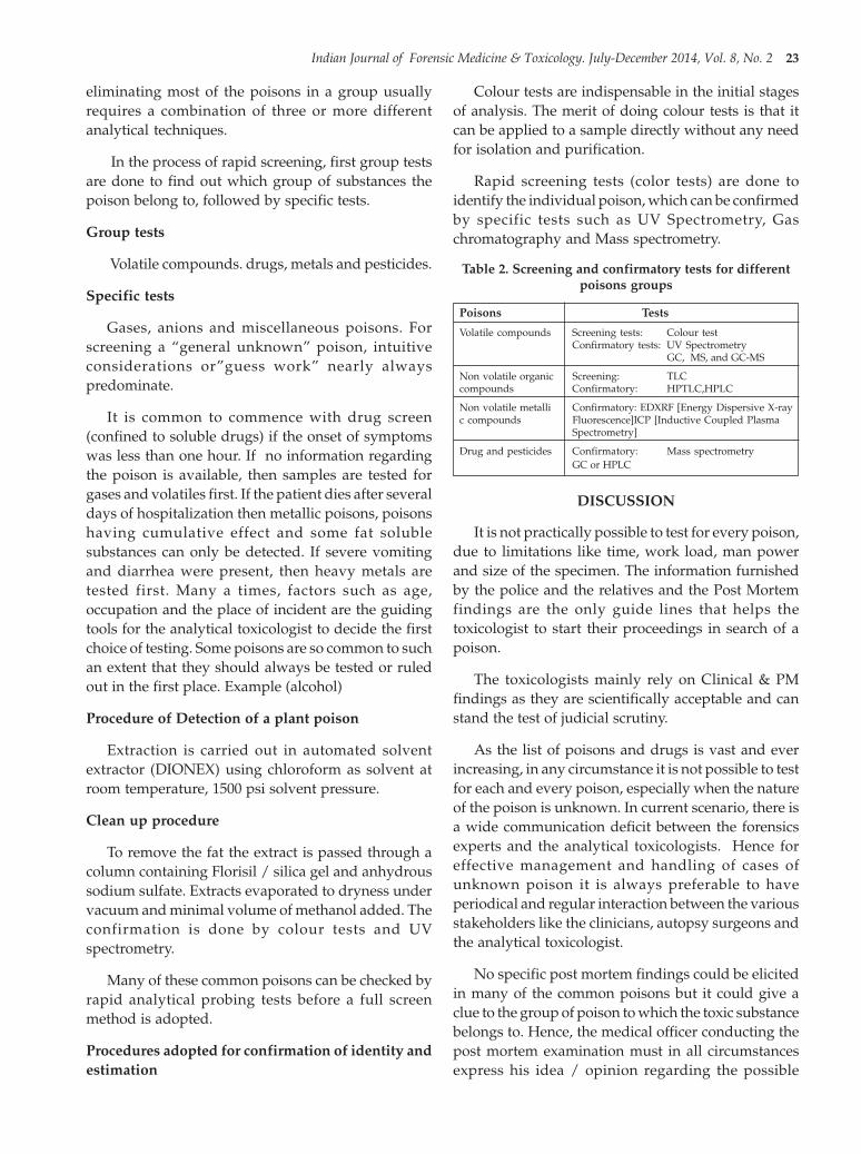
Indian Journal of Forensic Medicine & Toxicology. July-December 2014, Vol. 8, No. 2 23
eliminating most of the poisons in a group usuallyrequires a combination of three or more differentanalytical techniques.
In the process of rapid screening, first group testsare done to find out which group of substances thepoison belong to, followed by specific tests.
Group tests
Volatile compounds. drugs, metals and pesticides.
Specific tests
Gases, anions and miscellaneous poisons. Forscreening a “general unknown” poison, intuitiveconsiderations or”guess work” nearly alwayspredominate.
It is common to commence with drug screen(confined to soluble drugs) if the onset of symptomswas less than one hour. If no information regardingthe poison is available, then samples are tested forgases and volatiles first. If the patient dies after severaldays of hospitalization then metallic poisons, poisonshaving cumulative effect and some fat solublesubstances can only be detected. If severe vomitingand diarrhea were present, then heavy metals aretested first. Many a times, factors such as age,occupation and the place of incident are the guidingtools for the analytical toxicologist to decide the firstchoice of testing. Some poisons are so common to suchan extent that they should always be tested or ruledout in the first place. Example (alcohol)
Procedure of Detection of a plant poison
Extraction is carried out in automated solventextractor (DIONEX) using chloroform as solvent atroom temperature, 1500 psi solvent pressure.
Clean up procedure
To remove the fat the extract is passed through acolumn containing Florisil / silica gel and anhydroussodium sulfate. Extracts evaporated to dryness undervacuum and minimal volume of methanol added. Theconfirmation is done by colour tests and UVspectrometry.
Many of these common poisons can be checked byrapid analytical probing tests before a full screenmethod is adopted.
Procedures adopted for confirmation of identity andestimation
Colour tests are indispensable in the initial stagesof analysis. The merit of doing colour tests is that itcan be applied to a sample directly without any needfor isolation and purification.
Rapid screening tests (color tests) are done toidentify the individual poison, which can be confirmedby specific tests such as UV Spectrometry, Gaschromatography and Mass spectrometry.
Table 2. Screening and confirmatory tests for differentpoisons groups
Poisons Tests
Volatile compounds Screening tests: Colour testConfirmatory tests: UV Spectrometry
GC, MS, and GC-MS
Non volatile organic Screening: TLCcompounds Confirmatory: HPTLC,HPLC
Non volatile metalli Confirmatory: EDXRF [Energy Dispersive X-rayc compounds Fluorescence]ICP [Inductive Coupled Plasma
Spectrometry]
Drug and pesticides Confirmatory: Mass spectrometryGC or HPLC
DISCUSSION
It is not practically possible to test for every poison,due to limitations like time, work load, man powerand size of the specimen. The information furnishedby the police and the relatives and the Post Mortemfindings are the only guide lines that helps thetoxicologist to start their proceedings in search of apoison.
The toxicologists mainly rely on Clinical & PMfindings as they are scientifically acceptable and canstand the test of judicial scrutiny.
As the list of poisons and drugs is vast and everincreasing, in any circumstance it is not possible to testfor each and every poison, especially when the natureof the poison is unknown. In current scenario, there isa wide communication deficit between the forensicsexperts and the analytical toxicologists. Hence foreffective management and handling of cases ofunknown poison it is always preferable to haveperiodical and regular interaction between the variousstakeholders like the clinicians, autopsy surgeons andthe analytical toxicologist.
No specific post mortem findings could be elicitedin many of the common poisons but it could give aclue to the group of poison to which the toxic substancebelongs to. Hence, the medical officer conducting thepost mortem examination must in all circumstancesexpress his idea / opinion regarding the possible
6. Dekal--21--.pmd 7/24/2014, 10:04 AM23

24 Indian Journal of Forensic Medicine & Toxicology. July-December 2014, Vol. 8, No. 2
poison which is suspected to be involved in thatparticular case, which will definitely help thetoxicologist to test for that toxic substance first.
CONCLUSION
To conclude, I wish to highlight yet another aspectin the sample storage and forwarding of tissue samplesby the medical officers for chemical analysis. Duringthe period of time when I was with the analyticaltoxicologists at CFSL, Bangalore to study andunderstand their design protocol of testing a sampleof unknown poison, they requested me to express theirviews in sending samples for analysis.
1) Any particulate matter present in the stomachduring autopsy should be isolated and sent to theFSL separately labeled “stomach contents” withoutpreservatives. Toxicologists by their knowledgemay easily suspect or exclude a particular poisonand proceed with the analysis.
2) Preserve water soluble compounds is alcohol.
3) Try to collect or arrange to send the stomach washcontents to FSL, especially if the patient dies afterprolonged hospitalization.
4) Quantization is done only on blood and hencepreservation of blood is very important (sufficientsample with appropriate preservative in rightquantity)
5) The post mortem doctor should make an attemptto identify the nature of the poison on the basis ofpostmortem findings as observed by him and itshould always be mentioned in the PM report.
Ethical Clearance: Nil, as the study does not involveany human participation.
ACKNOWLEDGEMENTS
• My sincere thanks to the Director, Central ForensicScience laboratory, Bangalore &
• Dr. Anandha, Professor, Kempagowda Institute OfMedical Sciencs, Bangalore
Conflict of Interest: Nil
Source of Funding: Nil
REFERENCES
1. Kishi M, Ladou J. International pesticide use. IntJ Occup Environ Health 2001; 7: 259-65.
2. Jeyaratnam J. Acute pesticide poisoning: a majorglobal health problem. World Health Stat Q 1990;43: 139-44.
3. World Health Organisation. Guidelines forpoison control. Bulletin 1999; Geneva, WorldHealth Org.
4. Taft C et al. Childhood unintentional injuryworldwide: meeting the challenge. Washington,DC, Safekids Worldwide, 2002 (http://www.safekids.org/pdf/WWStudy- Ltr.pdf,accessed 6 April 2008).
5. Basic analytical toxicology. RJ Flanagan - ý1995Geneva, World Health Org.
6. Dekal--21--.pmd 7/24/2014, 10:04 AM24

Indian Journal of Forensic Medicine & Toxicology. July-December 2014, Vol. 8, No. 2 25DOI Number: 10.5958/j.0973-9130.8.1.001
Desperate Need of Modernized Mortuaries in Hospitals ofIndia
Shilekh MittalAssistant Professor, Department of Forensic Medicine, GGS Medical College, Faridkot
ABSTRACT
In majority of the Mortuaries there are no proper rooms for basic necessities and staff. Some of themortuaries have attained the status of historical monument, which have no caretaker and present adeserted look. At many places, the mortuaries are in the dirtiest corner and neglected part of thehospital. Facilities for autopsy like dissection table, instruments, and water supply, lighting andsterilization facilities are lacking at many places and if present, are in worst and rusted condition, inspite of the guidelines of Medical Council of India and judiciary. In this paper, some problems arehighlighted and their planed solutions for modernized mortuaries are discussed.
Keywords: Modernized Mortuaries, Hospitals, Judiciary, Police
INTRODUCTION
Presently, mortuaries dispel the sense of dislike, fearand hatred prevailing in majority of the so-calledpostmortem buildings in major part of the country. Themortuary is the most neglected, ignored place in allhospitals including medical colleges and is generallylocated in a far-off isolated corner of the hospital withStone Age facilities for body preservation and autopsy.From the overall expenditure of any hospital, a veryminute share is spent on the autopsy facility; it beingconsidered an unnecessary evil & the overallenvironment in a mortuary is depressing and gloomy.This situation is further compounded byadministrative apathy towards medico-legal work asa whole. Working in a mortuary is extremely stressfulexperience, which is made worse in India due to thelarge number of people dying sudden violent deathsand the pitiable conditions of mortuaries throughoutthe country. New concepts regarding mortuariesinclude the need for a total change in the way the deadbodies are being managed as well as changed workingconditions for the staff working in mortuaries.Facilities for safe custody of dead bodies before andafter post-mortem are to be provided accordingly. Theconcept of health of the population from “womb totomb” in the community health care clearly indicatesthat, a doctor’s duty is not only caring for the livingbut also in helping to arrange for the disposal of thosepatients who die, to keep unclaimed bodies until
disposal or identification by relatives, police and otherpeople. To receive dead bodies brought to the hospitalfor medico legal post-mortem work and store in themortuary pending further disposal and for teachingthe undergraduates as well as post-graduates.
The services provided by a Forensic facility, i.e.comprehensive morgue and postmortem comprising:- Specimen handling area, Administrative functions(documentation of incoming or outgoing bodies reportpreparation), preparation and temporary storage ofcadavers. Determining the cause of death, byperforming post mortem examination on the deadbody. The demonstration of post mortem findings incases of clinical interest, for teaching or forensicpurposes, mobile radiography, photography, family/police viewing and/or identification of the body.
Recommendations as Per Various Govt.Organizations and Judiciary
According to Bureau of Police Research &Development sub-committee report 1975, no teachinghospital should have more than 500 autopsies in a year,otherwise teaching activities suffer. In criminal caseno. 214 DB of 1997, Justice Amar Dutt & Justice KiranAnand Lall (Punjab and. Haryana High Court) on23.10.2005 have passed orders for ways and means tofacilitate better conduction of post-mortems plusmedico legal work. The division Bench has observed
7. Shilekh Mittal --25--.pmd 7/24/2014, 10:04 AM25

26 Indian Journal of Forensic Medicine & Toxicology. July-December 2014, Vol. 8, No. 2
that orders be served to the States of Punjab andHaryana and Union Territory of Chandigarh. The courthas also observed, “It may be appropriate if theconcerned state communicates the steps taken by it tothe sister States so that they do not lag behind.”
The Sub-Committee Report (Bureau of PoliceResearch and Development of 1975) laid down thestaffing pattern and this has been accepted in principleby the government.
(A) For initial 100 autopsies per year
• Specialists: Two (as one specialist is likely to bebusy in other important work, teaching work, incourt attendance, or if he falls sick, it is necessaryto have two specialists).
• Post mortem technician: One.
• Post Mortem Assistant: One.
• Clerk/Steno: One.
• Chowkidar: One.
• Peon: One
• Sweeper/Morgue attendants: [4] (Three sweepersfor shift duty round the clock and one as a reliever).
(B) For every additional 100 autopsies per year,following additional staff is required
• Specialist: One.
• Post mortem assistant: One.
• Technician: One (for teaching institutions).
• Technical assistant: (300-500 Autopsies/yr): One,(>500 autopsies / yr): Two.
• Photographer: One.
• Dark Room Attendant: One (on big centers,personnel for photographic work)
The Sub-Committee noted that in teachinginstitutions, teaching staff prescribed by the MedicalCouncil of India should not be counted while assessingthe total number of staff required as they will be busyin discharging their administrative, teaching andresearch guidance work.
Minimum Facilities Needed Arrangements forreceiving the dead bodies from the hospital or fromoutside, with separate arrangements for keeping
decomposed and infectious bodies (known HIV/hepatitis death cases) etc. Arrangements forperforming autopsies, handing over the dead bodiesafter postmortem examination to the relatives/undertakers through police, postmortem viewinggallery for the students /IO /nominees as per courtorders etc. Other basic essential requirements likeoffices and related rooms should have basic facilitieslike furniture, telephone and other infrastructures.
The design of the teaching mortuary was preparedkeeping in view the intake of students in medicalcollege, workload, condition of the various bodiesbeing brought for postmortem examination and tohave workable atmosphere as to cleanliness &breathing with fresh air and natural light to beavailable in each room of the mortuary. It should haveadequate parking space. It should be preferablycentrally air conditioned.
The mortuary should be located in a separatebuilding near the pathology laboratory on the groundfloor, easily accessible from the wards, accident andemergency departments and operation theatres, in anarea with ample natural light through windows; thewidows of the principal rooms should preferably beon the northern side. It should be located in one wingof the hospital near the main road, preferably awayfrom major traffic junctions. It must have a separateentrance and exist for relatives.
Proposed Plan For A Mortuary Complex at TertiaryLevel Hospitals
1. Veranda should be in front of the faculty office,autopsy surgeon room in which these should openand working windows for the reception room onboth sides.
2. Faculty office with an attached toilet.
3. Autopsy surgeon’s room (size 14x20 sq. ft.): wherethe Autopsy surgeon /medical officer can discussdetails of the case with police and relatives andwrite reports peacefully without any disturbance.
4. Computer Room & Office (12x10 sq. ft): withfurniture, computer, printer cum scanner,photography instruments.
5. Stores-1 (12x10 sq. ft) for clean gowns, aprons,rubber gloves, gumboots, towels etc.
6. Reception and waiting area (Size 240 sq. ft.). Itshould be easily approachable and due care should
7. Shilekh Mittal --25--.pmd 7/24/2014, 10:04 AM26

Indian Journal of Forensic Medicine & Toxicology. July-December 2014, Vol. 8, No. 2 27
be taken to shield it from OPD/ward block. Thisarea should be gently illuminated, warm and havecomfortable chairs. It should be pleasantly andsoberly furnished and decorated with plants andpictures, which would create a pleasantatmosphere, as the last impression of the relativesreceiving the deceased is one of quiet dignity indeath. This area can also be used as prayer areawhere relatives and friends of all religion may liketo offer prayer. A lavatory also must be provided.Waiting hall for attendants, police with provisionsfor a central platform.
7. Investigating officer /Police Room (10x10 sq. ft.)for the police personnel accompanying the bodywho has to watch and take care of the dead bodiesin the mortuary complex. Veranda/shade fortrolley etc.
8. Class IV Rooms (8x10 sq. ft.); it has lockers etc.and used as changing place for them. Toilet forstaff. Walled enclosure with gate.
9. Pre autopsy room (16x20sq. ft) the number ofbodies to be accommodated will be taken as threepercent of the hospital beds. Body racks should berefrigerated, with a temperature maintainedbetween 4-6.5°C. Body in the mortuary should bekept with complete identity
10. A/c plant room, where more than twelve bodiesare to be stored, a separate plant room 25-30 sq. ft.may be required, immediately adjacent to the bodystoreroom. Access for maintenance should bearranged externally so that unnecessary entrantsare stopped.
11. Post Autopsy Room (14x18 sq. ft.): should have acentral platform (4x8 sq. ft.) for handing over thebody to the investigating officer who subsequentlyhandover the body to the relatives for finaldisposal after completion of the post-mortemexamination.
12. Teaching autopsy room of the size should be atleast 400 Sq. Meters or more. This room, like anoperation theatre must be kept clean to protect thedoctors & staff from bacterial contamination. Itshould have at least two mortuary tables ofstainless steel with arrangements for free drainageof a constant flow of water (hot and natural) fromtop to bottom. A proper vent and duct system forexit of foul smelling gases and entry of fresh air.Room should have mortuary work station. There
should have big windows up to the level of roofwith glasses facing the east, south and west, so thatthe maximum sun light may remain in the rooms.Fluorescent lighting/good concentrated lightingover tables with at least one having tiltingmechanism.
13. There should be large charts depicting weights andmeasurements of viscera, bones etc. for quickreference. There should be X-ray view boxes forseeing x-ray film etc. Portable x-ray machineshould be available. Built in cupboards for keepinginstruments and equipments. Water-imperviousfloors sloping to a drain. Laminated walls so thatthe whole room can be easily washed. Suitablycovered junctions between the walls and floors.Two sinks for clean and dirty work separately.Writing desk and chairs, shelves for jars (and tanksunder) for disposing the specimens immediate.
14. Trolleys for shifting dead bodies and sufficientfurniture. Testing table, accommodating scales,gas, and light with blackboard on behind wall, fansif the room is not air conditioned. The viewinggallery for the students shall be constructed aroundthe autopsy tables depending upon their strengthin teaching autopsy rooms. Staired benches at thesides of the room for observers to visualize andavoid interference. The doors should be fly proof.
15. Laboratory Facilities: - Biochemistry andMicrobiology laboratories should be present insidethe mortuary complex preferably near the teachingautopsy room. Tests for infectious diseases shouldbe must before every autopsy. Chemicalexamination laboratory for quick examination ofblood, viscera and urine is very useful.
16. Space for open mortuary /maceration tanks/openresearch lab; (with roof covered with net) for thedecomposed bodies and other ancillary work. Thisspace should be all around both the teachingautopsy rooms so that public cannot have an accessto the ongoing postmortem.
17. Instrument Room (8x10 sq. ft.): instrumentcleaning room to be provided for the thoroughcleansing of all numbered instruments /equipments in the autopsy room.
18. Viscera Preparation Room (12x10 sq. ft.) where thetechnician / mortuary attendant can do theassigned job under the supervision of the medicalofficer.
7. Shilekh Mittal --25--.pmd 7/24/2014, 10:04 AM27

28 Indian Journal of Forensic Medicine & Toxicology. July-December 2014, Vol. 8, No. 2
19. Stores-II (12x10 sq. ft.) for reserve stocks andchemical solutions for preserving the viscera andpacking materials etc.
20. Officer’s toilet; (8x12 sq. Ft.) One separate maleand female W.C. lavatory, basin and a showercubical are needed.
21. Doctors changing room: there must be twochanging rooms (12x10 sq. ft. each) having bathwith shower for autopsy surgeons and with lockersfor male and female officers.
22. Green belt: this is required to make mortuarycomplex eco-friendly.
Corridors & Verandas’ all around: these will helpnot only in free movement of the functioningofficials in the premises of mortuary, but also incases of emergency of handling of mass disastercases and will provide more space for decomposedgases to spread and exit.
23. Main Gate; preferably it should be sliding type.
24. Open space for parking for vehicles bringing andcarrying the dead bodies.
25. Miscellaneous Requirements
a) Floors: should be hard and durable, moistureresistant and can be easily cleaned. Floor ducts andtrenches should be avoided. The junctions betweenthe walls and floors should be suitably covered.
b) Walls: the walls of the mortuary should be thick,durable and permanent. The walls should be fittedwith pale blue color tiles up to the ceiling so thatnatural colors of the dead body can be appreciateas in day light.
c) All doors in the mortuary complex should be widesliding type and fly proof.
d) Windows: the mortuary should have sufficientnatural light. The windows should be preferablyon the east, south and west sides, wheneverpossible for receiving maximum sunlight. Theyshould have glass. Windows slits should be at least5 ft. above the floor and go up to near the ceiling.
e) Corridors: the corridors of the mortuary limitshould be wide to allow passage of trolleys. (Notless than 8 ft.)
f) Air conditioning: the entire complex should be air
conditioned with a separate system for the autopsyrooms to prevent foul air permeating the rest ofthe area. No air should be re-circulated in themortuary in order to ensure a clean airenvironment.
g) Proper disposal of waste: the mortuary complexshould have all the arrangements for disposal ofdifferent types of waste products. Cleanliness andsanitation in and around the mortuary should bemaintained properly.
h) Measurements of the space may be adjusteddepending upon the requirements and workload.The mortuary complex must have different signboards like “No Admission”, “Prohibited Area’ etc.
i) The light fitting should be designed to avoid glare,and should be easy to clean and maintain. In themortuary either tungsten or florescent lighting canbe used. Switches in wet area should be hose proof.Special lighting should be provided in the post-mortem room to ensure adequate illumination ofpost-mortem tables and dissection tables. Theheight of the room should be optimum preferablytwenty feet.
j) Hot and cold water supply: hot and cold water berequired in the sinks, wash-basins and showers.The post-mortem tables should be fitted withindividual /water hoses. Water suction pumpsshould not be used. Floor service ducts should beavoided. All the taps in the mortuary complexshould be of elbow operated type particularly inthe working area.
k) Safety: the complex should be fitted withemergency lighting, fire sprinklers and smoke/thermal detector in all rooms. A fire alarm systemwith blue /red beacon light with hooter should beinstalled. Fire exit routes should be clearlyidentifiable, well illuminated and earmarked withbold red arrows.
l) Refrigeration: the temperature of cold rooms is tobe maintained between 4°C to 6.5°C, thermostatcontrol will be required for each cold chamber.Facilities should be provided to enable thechambers which are not in use to be switched off.
The design of the ventilation system should provideair movement which is generally from clean to lessclean areas. To satisfy exhaust needs, replacement airfrom the outside is necessary. Number of air changes
7. Shilekh Mittal --25--.pmd 7/24/2014, 10:04 AM28

Indian Journal of Forensic Medicine & Toxicology. July-December 2014, Vol. 8, No. 2 29
may be reduced when the room is unoccupied ifprovisions are made to ensure that the number of airchanges indicated is re-established any time the spaceis being utilized. Adjustments shall include provisionsso that the direction of air movement shall remain thesame when the number of air changes is reduced. Airfrom areas with contamination and/or odourproblems shall be exhausted to the outside and not re-circulated to other areas.
Conclusion and Future Advancements: Safety andsecurity are integral components of a forensic facility.Defining public areas from secure zones to ensure thesecure custody of sensitive items such as case files,evidence, and human remains is critical. The conceptof a modern and ideal mortuary will protect theoccupants against diseases emanating from filthy andunhealthy environment and the mortuary contentsagainst destruction from decay of unrepaired andneglected mortuary buildings. Tele-medicine facilitiescan be envisaged in the modern mortuary complexusing large training areas and various meeting roomswith video conferencing capability
CT, MRI, and digital x-ray technology should bean integral component available to the mortuary staff– to limit invasive procedures for operational orreligious reasons. An ideal mortuary will undoubtedlycontribute to the advancement and evolution offorensic science and medicine, strengthening therelationship between service, teaching, and research.The public safety network and criminal justice systemwill benefit tremendously and will now be supportedby a forensic facility built to match the level ofexcellence required.
Acknowledgements: Nil
Conflict of Interest: Nil
Source of Funding: Nil
Ethical Clearance: Nil
REFERENCES
1. Singh S, Sinha US, Kapoor AK, Verma SK, SinghD, Sharma S; Planning And Designing Of ModernMortuary Complex In Tertiary Care; IIJFMT 4(1)2006
2. Sirohiwal BL, Paliwal PK, Sharma L, Chawala HDesign and Layout of Mortuary Complex for aMedical College and Peripheral Hospitals. (2011)J Forensic Res 2:102e. (cited 2012 August 30): 1(1):(4 Screen). doi:10.4172/2157-7145.1000102e.
3. Mittal S, Yadav M, Singh H, Sharma G, ChawalaR ; Current Scenario of Forensic Medicine in India;JIAFM, 2007 29 (2). p. 59-60.
4. Hon’ble J. Amar Dutt & J. Kiran Anand Lall(Punjab and Haryana High Court): Judgmentdelivered in Criminal Law, No. 214-DB of 1997dt. 23.10.2005).
5. The Committee on Plan Projects. Report onGeneral Hospitals, 1964.
6. Jinesh P. S Modern Mortuary: 20/06/2012 (cited2012 August 29): 1(1): (4 Screen). http://www.authorstream.com/Presentat ion/dreamydews-1451189-modern-mortuary
7. Shilekh Mittal --25--.pmd 7/24/2014, 10:04 AM29

30 Indian Journal of Forensic Medicine & Toxicology. July-December 2014, Vol. 8, No. 2DOI Number: 10.5958/j.0973-9130.8.1.001
Estimation of Stature from Middle Finger Length-inSalem Region
S Sasi Kumar1, Pavanchand Shetty2, Shankar3
1Assistant professor, Department of Forensic Medicine, Vinayaka Mission Kirupanda Variyar Medical College, Salem,Tamil Nadu, India, 2Assistant Professor, Department of Forensic Medicine, Kasturba Medical College, Manipal,
3Associate Professor, Department of Community Medicine, Vinayaka Mission Kirupanda Variyar Medical College,Salem, Tamil Nadu, India
ABSTRACT
Estimation of stature is considered to be important in those cases like only fragmented and mutilatedremains of unknown person are recovered. Many previous studies have been done to estimate thestature using length of different long bones. The present study is an effort to establish correlationbetween middle finger length and stature. The study was conducted on students from the VinayakaMission Kirupanada Variyar Medical College aged between 16-22 years. Standing height and Middlefinger length of 150 subjects were measured. Measurements were analysed statistically to establishrelationship between Middle finger lengths with stature. Correlation co efficient between statureand middle finger length found to be positive. Regression equation was derived from using this We derived the formula for middle finger length for male as Y=3.70X+135.39and for female as Y=4.33X+129.26. Y stands for standing height and x for middle finger length. Byputting value of x in different situations stature are calculated and compared with correspondingreal standing height.
Keywords: Middle Finger Length, Stature, Regression Equation
INTRODUCTION
One of the most important criteria for establishingidentification is stature. Increasing frequency of massdisasters like plane crashes, earthquakes, genocide etc,has created problems in the determination of statureand identification of victims when only fragmentedor dismembered humane remains are available forinvestigation. Those cases forensic expert has no optionbut to use other method of reconstruction if part ofthe body is available i.e mathematical method whichis workable even part of the body parts areavailable.1,2,3. Most of the workers used intact longbones like femur, tibia, humerus and radius for
Corresponding author:S Sasi KumarAssistant Professor,Dept of Forensic Medicine, Vinayaka MissionKirupanda Variyar Medical College, Salem, TamilNadu, IndiaE mail; [email protected] no; 91-8220496604
´estimation of stature which is natural heights of aperson in upright position and various formula,regression equations are devised for various groups4,5,6. In the world various regression equation andmultiplication factors are developed to estimate thestature using different body parts like length of hand,foot length, arm span and head length.
The present is an effort to establish correlationbetween middle finger length and stature. This willhelp the autopsy surgeon to determine the stature inmutilated bodies when the hand is intact most otherparts are damaged.
MATERIALS AND METHOD
150 healthy Students of Vinyaka MissionKirupanada Variyar Medical College aged between16—22 years were selected for this study. Height wasmeasured without wearing shoes, from vertex to floorusing caliber. Middle finger length was measured fromtip of middle finger to most proximal flexion of skin
8. Sasi kumar--30--.pmd 7/24/2014, 10:04 AM30

Indian Journal of Forensic Medicine & Toxicology. July-December 2014, Vol. 8, No. 2 31
crease. These measurements were compiled on masterchart and also on excel. Dietary habits, Caste, Religionand Socio economic status were not considered.Students having fractured forearm and arm, growthdisorder, deformities were excluded from the study.We derived mean, standard deviation of standingheight and middle finger length. From whichCorrelation coefficient with standing height wascalculated. 7
Regression equation was derived from middlefinger length for estimation of stature separately formale and female using this formula
correlation co efficient between middle finger lengthand height
Y is standing height (stature)
mean of standing height
X middle finger length
mean of middle finger length
standard deviation of standing height
standard deviation of middle finger length
RESULTS
Standing height of the males varied from 190 cmto 150 cm with standard deviation (S.D) of 9.08 andmean value of 167.06 cm. For females it varied from179 cm to 144 cm with standard deviation of 9.46 andmean value of 166.06 cm. (Table no 1).
Table no 1. Stature of study group
Height Male Female
Maximum 190 179
Minimum 150 144
S.D 9.08 9.46
Mean 167.06 166.06
Middle finger length
The length of middle finger varied from 12.5 cm to6.5 cm with mean value of 8.56 cm, standard deviation1.076 and correlation co efficient with standing height0.44. The length of middle finger for females variedfrom 9.5 cm to 6.5 cm with mean value of 8.50 cm,standard deviation 1.09 and correlation co efficientwith standing height 0.50.( Table no. 2)
Table No 2. Middle finger length for females andmales
Middle finger length Male Female
Maximum 12.5 9.5
Minimum 6.5 6.5
Mean 8.56 8.50
S.D 1.076 1.09
Correlation co efficient for stature 0.44 0.50
Regression equation was derived from middlefinger length for estimation of stature separately formale and female using this formula
Middle finger
For male: Y= 0.44 * 9.08 / 1.076 (X-8.56)-167.06
Y=3.70X+135.39
For Female: Y=0.50x 9.46 / 1.09 (X-8.50)-166.06
Y=4.33X+129.26
For males regression equation for stature frommiddle finger length is 3.70X+135.39 and for females4.33X+129.26. Here X is middle finger length. Byputting X value the stature can be calculated and itcan be compared with corresponding real standingheight.
Table No.3: Regression Equations and variations in calculated stature
Middle finger length Subject Regression Length of Standing Calculated Variationequation middle finger height stature in cm
Male 3.70X+135.39 Max 12.5 cm 190 cm 181 cm -9
Min 6.5 cm 150 cm 159.4 cm 9.4
Female 4.33X+129.26 Max 9.5 179 cm 170.39 cm -9.39
Min 6.5 144 cm 157.40 cm 13.40
8. Sasi kumar--30--.pmd 7/24/2014, 10:04 AM31

32 Indian Journal of Forensic Medicine & Toxicology. July-December 2014, Vol. 8, No. 2
DISCUSSION
Studies have been conducted in the recent past onheight determination from length of hand, feet, upperextremity and facial measurement in the recent past.The present study documents relationship betweenmiddle finger length and stature. The present studyshows the correlation co efficient for male is 0.44 andfor female is 0.50 which is significant. Correlation coefficient between stature and middle finger lengthfound to be statistically significant and positiveindicating strong relationship between twoparameters. Prateek rastogi (8) also reported staticallysignificant correlation between middle finger lengthand stature and significant differences in mean statureof both males and females in north and south Indianpopulation. Mean height for male is 167.06 cm and forfemales mean height is 166.06 cm. Mean middle fingerlength for male is 8.56 cm and for female is 8.50 cm.Mean height for male is higher as compared to femaleswhich is similar to Thakur (1985)9, Sharma (2001)10.
Regression equation for stature estimation wasformulated using middle finger lengths and checkedfor their accuracy by comparing the estimated statureand the actual stature. The results indicate that middle
finger length provides an accurate and reliable meansin reconstructing the stature of an unknown individual.
Regression equation for stature estimation wasformulated using middle finger length for male
Y= 0.44 * 9.08 / 1.076 (X-8.56)-167.06
Y=3.70X+135.39
For Female regression equation is: Y=0.50x 9.46 / 1.09(X-8.50)-166.06
Y=4.33X+129.26
Verghese AJ et al 11 found significant correlationbetween middle finger length of both the hands andstature in males and females and recommended thatthose equations should be used for estimation staturein Mysore and surrounding region of the Karnataka.Tyagi AK et al 12 also found significant correlationbetween finger length and stature and they alsosuggested use of regression equation for statureestimation. Rahule et al14 also found good correlationand statistically highly significant correlation coefficient between right middle finger length and staturein the tribal district of Andhra Pradesh.
Table No. 4. Regression equation for various regions:
Authors Region Sex middle finger Regression Equation
Present Study Tamil Nadu Male Right S=3.70X RMFL+135.39
Female Right S=4.33X RMFL+129.26
Rahule AS Andhra Pradesh Male Right S=120.74 +0.457 X RMFL
Females Right S=93.56 + 0.636 X RMFL
Verghese AJ Mysore Male Right S=120.20+4.95XRMFL
Karnataka Left S=117.11+5.27XLMFL
Female Right S=117.55+4.26XRMFL
Left S=115.77+4.43XLMFL
Sivkumar AH Karnataka Male Right S=152.02+1.47XRMFL
Female Right S=120.20+4.95XRMFL
CONCLUSION
Mean height for male is higher as compared tofemales. Mean middle finger length for male is higherthan males. There is positive correlation betweenstature and middle finger length. Simple linearregression equation derived can be used for estimation
of height. This study would be useful foranthropologist and Forensic Medicine experts.
Acknowledgement: Nil
Ethical Clearance Taken From: Institutional Ethicalcommittee of Vinayaka Mission Kirupanada VariyarMedical College
8. Sasi kumar--30--.pmd 7/24/2014, 10:04 AM32

Indian Journal of Forensic Medicine & Toxicology. July-December 2014, Vol. 8, No. 2 33
Source of Funding: Self
Conflict of Interest: Nil
REFERENCES
1. Jasuja OP and Singh G. Estimation of stature fromhand and phalange length. J Ind Asso ForensicMed. 2004; 26(3):100-106
2. Shahnaz C, Harbans S, Neena G. Estimation ofstature from combined length of forearm andhand in Jammu region of India. InternationalJournal of basic and Applied Sciences. 2014;3(1):8-10
3. Vij K , Forensic Medicine and Toxicology, 4 thedition, Elsevier India Private Ltd. 2008: 64
4. Trotter M & Gleser GC. Estimation of stature fromlong bones of America whites and nigros. Am JPhys Anthropol. 1952; 10:463-514
5. Pan N. Lengths of long bones and theirproportions to body height in Hindus.J Ana 1924;58: 374-378
6. Siddqui M A H & Shah M A. Estimation of staturefrom long bones of Punjabis. Indian J Med Res1944; 31:105-108
7. Kumar Amit, Srivastava A.K. Verma A.K.Estimation of stature by Percutaneousmeasurements of Distal half of upper limb.Journal of Indian Academy of Forensic Medicine.2010; 32 (4): 325-328
8. Prateek rastogi, Tanuj Kanchan, Ritesh G mensesand Yoganarasimha- middle ginger length – apredictor of stature in Indian population. Journalof medicine and science and law.2009; 49 (2):126-129
9. Thakur SD and Rai KS. Determination of Staturefrom hand measurements. Medicine, Science andLaw 1987:78: 25-28
10. Sharma pk and Kapoor AK. Estimation staturefrom finger tip length and finger tip length amongcriminals. Recent advances in forensic biology(edited)—Kamla-Raj publication; 2007:117-127
11. Verghese AJ. Balraj BM. Pramod Kumar GN.Astudy of estimation of stature from length offingers in Mysore. Indian Journal of ForensicMedicine Toxicology 2010:4 (2):12-13
12. Tyagi AK. Kohli A. Verma SK. AggarwalBBL.Correlation between stature and fingerslength. International Journal of MedicalToxicology and Legal Medicine. 1999:1(2):20-22
13. Shivkumar AH. Raju GM. Prediction of statureby right middle finger length of males amongsouth Indian population. Journal ofPharmaceutical and scientific Innovation 2013;2(1):31-33
14. Rahule AS et al. A Study of Correlation betweenMiddle Finger Length and Height in a TribalDistrict Population of India. J Cont Med A DentSept-Dec 2013;1(1):8-12
8. Sasi kumar--30--.pmd 7/24/2014, 10:04 AM33

34 Indian Journal of Forensic Medicine & Toxicology. July-December 2014, Vol. 8, No. 2DOI Number: 10.5958/j.0973-9130.8.1.001
Thermal Burn: An Epidemiological Retrospective Study
S K Pandey1, Neha Chaurasia2, Awdhesh Kumar3
1Assistant Professor, 2Senior Research Fellow, 3Junior Resident. Forensic Medicine, Department of Forensic Medicine,Institute of Medical Sciences, Banaras Hindu University, Varanasi, India
ABSTRACT
The present retrospective study has been conducted in Varanasi area, cases brought to the Departmentof Forensic Medicine, IMS, BHU, Varanasi for the period of two years i.e. 2009 and 2010.Of the totalunnatural deaths reported during the study period deaths due to burn injuries were 17.98 % and17.07% respectively, showing the more or less steady trend. Burn deaths are usually associated withfemale and Dowry where female dies in her in-laws house within 7 years of her marriage. Femaleburn deaths dominated over male in the ratio of 1:4. Most of the deceased were from the marriedgroup (73.19%) followed by unmarried (23.55%). Predominant age group found to be 21-30years(45.13%) followed by age group 11-20 and 31-40 years showing almost same rate around 20% reflectingthat young adults were more involved in such type of deaths. As regards to place of death only 9.72%died on the spot or on the way to Hospital while 90.28% died in Hospital reflecting the prompt andproper health care services. Whatever the manner of death Female burn deaths are investigated bymagistrate as per provision of 176 Cr.P.C.and case registered under 304B IPC (Dowry Death) andMedico legal postmortem examination conducted by panel of Two Doctors, whereas male burn deathsare investigated by police as per provisions of 174 Cr.P.C. as routine case and medico-legal postmortemexamination conducted by single doctor.
Keywords: Dowry Death; Female Burn; Magistrate; Cr.P.C
INTRODUCTION
Fire was perhaps man’s first double-edged sword,evidenced throughout history, it has served as well asdestroyed mankind1.Burning or burn injuries arecaused due to contact with dry heat. Thus burning mayoccur due to contact with hot metal or any other hotsolid or it may be caused due to contact with flame.The effect of contact very extremely depending on thedegree of heat and period of exposure2.Burn deathshave tremendous medico-legal importance as theymay be considered to be the commonest cause ofunnatural deaths in India. Often, the circumstances ofburns are enveloped in mystery, obscurity andunreliable statements. The reason behind this actionmay be personal, domestic, occupational or socialtragedy and more recently dowry deaths3.
Married female burn death where death of femaleoccurs within 7 years of her marriage such death casesinvestigated by Magistrate under Cr.P.C 176( Dowrydeath) and other burn deaths as routinely investigated
´by police as per section 174 of Cr.P.C. In India all newlymarried female burn deaths are linked with Dowrydeath, where a young married women commits suicidein consequent to their being subjected to cruelty orharassment by their husband or in-laws or his relativeconstitute the offence of Dowry death, a monstroussocial evil is widely prevalent and deep rooted insociety inspite of stringent Legal system andadministration. A burn injuries death is very painfulbut what compels or in what circumstances womenor men commits suicide or those accidentally burnedbut most heinous is burning of newly married womeni.e. homicidal burning. In this respect it is very difficultor next to impossible to find out the manner (Suicidal,Accidental and Homicidal) of burn injuries that inwhat circumstances the burn injuries took place it canonly be possible up to some extent by meticulousinvestigation of scene of crime and interrogation ofperson concerned. In most of the cases of female burndeaths as per interrogation of her parents are homicidalin manner i.e. their daughter was alleged to be killed
9. Neha Chaurasia--34--.pmd 7/24/2014, 10:04 AM34

Indian Journal of Forensic Medicine & Toxicology. July-December 2014, Vol. 8, No. 2 35
by burning by in-laws and their relative but at the sametime as per in-laws she committed suicide or burnedaccidently while cooking, in very few cases both thesides are agreed on the same theory.
MATERIAL AND METHOD
This retrospective study was carried out on theburn death cases brought to the Department ofForensic Medicine, Institute of Medical Sciences,Banaras Hindu University, from Varanasi itself andnearby districts and western part of Bihar for medico-legal autopsy examination.
Data was collected from postmortem record registerfor the period from 1st January 2009 to 31st December2010.During this period total of 706 burn deaths caseswere recorded out of 4031 medico-legal postmortemconducted. Data was analyzed retrospectively inrespect of incidence of burn deaths, age, sex, cause ofdeath, place of death and other relevant data.
RESULTS AND OBSERVATION
Out of 4031 medico-legal autopsy cases conductedduring the study period from 1stJanuary 2009 to 31st
December 2010, total of 706 cases (17.51%) of deathfrom fatal burn injuries were recorded. If we split thefinding year wise 357 cases (17.98%) and 349 Cases(17.07%) in year 2009 and 2010 respectively (Table-1),which is almost static. Male comprised of 20.25% oftotal burn death. Female (79.75%) preponderance wasseen in burning with male female ratio equal to 1:4(Table2 & Table6). Maximum of the victims of burndeaths were in the age group 21-30 year followed by31-40 years in the year 2009 but 11-20 age group in theyear 2010 with slight difference (Table 3 & 7). Most ofthe victims of burn deaths were recorded between 21-40 year (which is more than half of the total burn death)with peak incidence at 21-30 year (Table3 &7).Extremesof ages are least involved as compared to adult agegroup as seen in tables for age and sex incidence.
If we consider Season wise distribution of burnvictims maximum death were reported in summerseason in 2009 (43.98%) and 2010( 47.27%) irrespectiveof sex followed by winter in the year 2009 and equalincidence in year 2010 (Table2&6).Married person(68.63%) in year 2009 and (77.75%) in 2010outnumbered the unmarried(27.73%) in 2009 and(19.36%) in 2010 (and widow and widower Table4&8).Regarding cause of death most of the victims diedof septicaemia followed by primary shock ( including
spot deaths or death on the way to hospital andhospital death within 48 hours of burn infliction)(Table5 &9).
Table1: Incidence of burn deaths in Medico-legalautopsy
Year Total Thermal %cases Burn Death
2009 1986 357 17.98
2010 2045 349 17.07
Table2: Season wise incidence of Thermal burn deathin year (2009)
Season Male Female Total %
Summer 46 111 157 43.98
Rainy 12 75 87 24.37
Winter 22 91 113 31.65
Total 80(22.41%) 277(77.59%) 357 100
Table 3: Age and Sex wise incidence of burn death inyear (2009)
Age in years Male Female Total (%)
0-10 5 6 11(3.08%)
11-20 16 54 70(19.61%)
21-30 23 130 153(42.86%)
31-40 20 56 76(21.29%)
41-50 6 15 21(5.89%)
51-60 4 5 9(2.52%)
61-70 5 8 13(3.64%)
71- above 1 3 4(1.12%)
Table 4: Marital status of burn death cases in year(2009)
Marital Status Male Female Total (%)
Married 48 197 245(68.63%)
Unmarried 27 72 99(27.73%)
Widow/Widower 5 8 13(3.64%)
Total 80 277 357(100%)
Table 5: Cause of death of burn death cases in year(2009)
Cause of death Male Female Total (%)
Primary shock 32 111 143(40.06%)
Septicaemic Shock 48 166 214(59.94%)
Total 80 277 357(100%)
Table 6: Season wise incidence of burn death in Year(2010)
Season Male Female Total(%)
Summer 32 135 167(47.27%)
Rainy 15 76 91(26.30%)
Winter 16 75 91(26.30%)
Total 63(18.05%) 286(81.95%) 349(100%)
9. Neha Chaurasia--34--.pmd 7/24/2014, 10:04 AM35

36 Indian Journal of Forensic Medicine & Toxicology. July-December 2014, Vol. 8, No. 2
Table 7: Age and sex wise incidence of burn deathcases in year (2010)
Age in years Male Female Total (%)
0-10 5 4 9(2.60%)
11-20 13 60 73(21.10%)
21-30 19 145 164(47.40%)
31-40 13 49 62(17.92%)
41-50 6 12 18(5.20%)
51-60 3 7 10(2.89%)
61-70 4 3 7(2.02%)
71-Above Nil 3 3(0.87%)
Total 63 286 349(100%)
Table 8: Cause of Death in burn death cases in year(2010)
Cause of death Male Female Total (%)
Primary Shock 30 127 157(44.5%)
Septicaemic Shock 33 159 192(55.49%)
Total 63 286 349(100%)
Table 9: Marital status of burn death victims in year(2010)
Marital status Male Female Total(%)
Married 40 230 270(77.75%)
Unmarried 21 48 69(19.36%)
Widow/Widower 2 8 10(2.89%)
Total 63 286 349(100%)
Table 10: Cause of Death in burn victims in year (2009)
Cause of death Male Female Total %
Primary shock 32 111 143 40.06
Septicaemic shock 48 166 214 59.94
Total 80 277 357 100
Table-11: Cause of death in burn victims in year (2010)
Cause of death Male Female Total (%)
Primary shock 30 124 154(44.13)
Septicemia shock 33 162 195(55.87)
Total 63 286 349(100)
DISCUSSION
Human society ever since its inception has neverbeen free from criminal activity. Burn injuries occurworldwide in all society irrespective of its developed,developing or poorly developed condition. Theseinjuries constitute a medical and psychologicalproblem but also have severe economic and socialconsequences not only to the victim but also to theirfamily and society in general4.
Analysis of sex and age record in our study showedthat females (77.59%) in 2009 & (81.95%) in 2010
superseded males with percentage of 22.41 and 18.05in 2009 and 2010 respectively. The overall femalepreponderance in the present study confirms withsome previous study5, 6, 7,8,9..Married females 563(79.75%) most common victim of present studyfollowed by married male 143 (20.25%) which isconsistent with the study ofUsama B Ghaffar et al &H.M.Mangal,Akhilesh Pathak5,9.In this study most ofthe patients have died due to septicemia in both theyears 59.94% and 55.49% (Table-5&9) and is consistentwith study done by Prabhsharan singh & DasariHarish.This shows the prompt and proper healthservices provided to people suffering from burninjuries. In the present study most of the victim weredied in Summer season followed by winter and rainythis for 2009 and in 2010 Summer season accounts formost burn deaths followed by same in both winter andrainy season, reason behind this is during summerresults of various schools and colleges declared, and aperson looses temperament easily or failure in loveand also this is the season for marriages. I did not finda single study where this parameter was studied.
As the female burn deaths reported to Policeirrespective of its manner and registered under304BIPC (Dowry death) all the family members of in-laws side alleged in causing death of female arearrested and send to jail.
ACKNOWLEDGEMENT
Author would like to thank of Department ofForensic Medicine for their support and help in datacollection.
Source of Funding: This research was not financiallysupported by any funding agencies.
Ethics statement: The study was approved by“Institutional Ethics Committee” of Institute ofMedical Sciences, Banaras Hindu University. All theinformation has been taken under consideration ofmedical ethical committee.
Conflict of Interest Nil
REFERENCES
1. Rong Xiang Xu, Karger. Brief introduction tohistory of burn medical Science.Burnregenerative medical and therapy, 2004; 1-3.
2. Apurba Nandy. Principles of ForensicMedicine.Thermal injuries.Reprint. New centralBook agency (P) Limited.1998.pp-269.
9. Neha Chaurasia--34--.pmd 7/24/2014, 10:04 AM36

Indian Journal of Forensic Medicine & Toxicology. July-December 2014, Vol. 8, No. 2 37
3. Mohanty et.al.Self inflicted burn fatalities inManipal.Medicine science and law: 2005; 45:27-30.
4. Xiao Jie,Cai Baoren. Mortality rates and among5321 patients with burn admitted to a burn unitin China:1980-1988. Burn 2003; 29:239-45.
5. Usama B Ghaffar et al.Thermal burns:Anepidemiological prospective study.journal ofIndian Academy of ForensicMedicineVol.30(1);2008pp10-14.
6. R o o h - U l - M u q u i m , M o h a m m e dZareen,Dilbag,Muhammad Hayat.Epidemiology and outcome of burns at Khyberteaching hospital Peshawar.Pak J Med Sci, 2007;23(3):420-24.
7. Dhiraj Buchade,Hemant Kukde,RajeshDere,Ramesh Savardekar.Pattern of burn casesbrought to Morgue,of sion Hospital,Mumbai: Atwo year study. Journal of Indian academy ofForensic Medicine. Oct-Dec 2011, Vol.33,No.4.pp309-310.
8. Bangal RS., Thermal injuries- A study of mortalitypattern .JFMT:1995, XII (1&2), 1-4.
9. H.M.Mangal,Akhilesh Pathak.,The Fire is both ‘ABlessing and Scourge to theMankind’JIAFM,2007-29(4)75-77.
10. Prabhsharan Singh,Dasari Harish.Incidence ofpost burn septicemia at a tertiary carehospital.JIAFM.Oct-Dec2011,Vol.33,No.4,pp317-321.
9. Neha Chaurasia--34--.pmd 7/24/2014, 10:04 AM37

38 Indian Journal of Forensic Medicine & Toxicology. July-December 2014, Vol. 8, No. 2DOI Number: 10.5958/j.0973-9130.8.1.001
Fusion of Epiphyses of Ischial Tuberosity in Relation withAge: A Cross Sectional Study
Anita Verma1, Kanan Shah2
1Associate Professor, Department of Physiology, Associate Professor, Department of Anatomy, Smt. NHL MuniMedical College
ABSTRACT
Introduction: Fixing a person's individuality has got its own importance. Age being one of the cardinalparameter for establishing the identity, its estimation is of paramount importance and requires specialattention. It is important when the liability and punishment are dependent on maturity of theindividuals. It is important in developing countries, where many births take place in rural settingsand poorly recorded or more often not recorded.
Aims & Objective: Study the progress of union of epiphyses of ischial tuberosity in natives of Gujaratstate, in relation to age & sex.
Material & Method: In the present study radiological examination (digital X-ray) was done, forfusion of epiphyses of ischial tuberosity, in 300 individuals of both the sexes of 17-22yrs.
Observation: 100% union of ischial tuberosity seen at age of 21-22yrs.Fusion of epiphyses occur atsame age group in both gender.
Discussion: The observations in this study are comparable to previous studies done by McKern &Stewart, Dr Gaurang & Dr Alok.
Conclusion: Among many factors used for age estimation, none has withstood the test of time whichnecessitates the continuous work on this vital issue by the medico legalists. Radiological examinationof bones is quick, precise & reliable method for estimation of age.
Keywords: Epiphyseal Union, Ischial Tuberosity, Radiological Examination, Age Estimation
INTRODUCTION
Fixing a person’s individuality has got its ownimportance. Age being one of the cardinal parameterfor establishing the identity, its estimation is ofparamount importance and requires specialattention1,2. It is important when the liability andpunishment are dependent on maturity of theindividuals3. It is important in developing countries,where many births take place in rural settings andpoorly recorded or more often not recorded. In manyother cases, records are falsely projected for some gain,e.g. to get government jobs or pensions.4
The assessment of age can be done by anatomist,physiologists and persons engaged in medico legalworks. And it should be done with great attention asconclusions are debated in court of law. A wide variety
´
of methods are used. Though the general developmentincluding height, weight, secondary sexual character,are less reliable data for estimation of age, changes inbones specially time related fusion of epiphyses ingrowing periods are valuable indices . These changescan be studied by means of X-rays and these changesare age related. It is therefore possible to determinethe approximate age of an individual by radiologicalexamination of bones till ossification is complete5.
Epiphyseal union of long bones & eruption of teethare reliable data till 25yrs of age +/- 6months. Andfusion of sutures of skull is reliable data after 25 yearsof age +/- 5 years6. Work on the determination of ageof epiphyseal union has been carried out in differentstates of India as well as abroad and from the findingof various workers; it is evident that there is difference
10. Anita Verma--38.pmd 7/24/2014, 10:04 AM38

Indian Journal of Forensic Medicine & Toxicology. July-December 2014, Vol. 8, No. 2 39
in the age of epiphyseal union in the different states ofIndia. These differences may be on account of varyinghereditary factors, climate, and diet7. Among manyfactors used for age estimation, none has withstoodthe test of time which necessitates the continuous workon this vital issue by the medico legalists. Very littlework in this regard has been carried out in Gujaratstate. The present study is an attempt to add to theknowledge in this field by studying the epiphysealunion of ischial tuberosity among the natives of Gujaratstate.
AIMS & OBJECTIVE
Study the progress of union of epiphyses of ischialtuberosity in natives of Gujarat state, in relation to age& sex.
MATERIAL & METHOD
This descriptive cross sectional study was carriedout in Smt NHL MMC in 2011-2012.For the study 300students of both sexes, aged 17-22 years were selected.Only natives of Gujarat having sound health anddefinite birth records were included for this study &as study was aimed to establish the age of epiphysealunion in persons from Gujarat state only those residingin Gujarat state since birth were selected. These cases
were selected after ruling out the nutritional,developmental, and endocrinal abnormality which canaffect the skeletal development & growth. Participantsselected were X-rayed (digital X ray) &theirradiographs were studied for progress of epiphysealunion of ischial tuberosity. Data was recorded &analyzed to draw conclusions and compared withavailable results of previous studies. Evaluation ofepiphyseal union was carried out as it was analyzedby McKern and Stewart in 1957 i.e. as follows.
0 = nonunion
1 = 1/4 union
2 = 1/2union
3 = 3/4union
4 = complete union
OBSERVATIONS
In the present study radiological examination wasdone, for fusion of epiphyses of ischial tuberosity, in300 individuals of both the sexes of 17-22yrs. Attemptis also made to analyze the stages of ossification andco-relate them with the age of the individuals
Table No 1. Shows numbers of students in different known age group.
Age group in years No of males No of females
17-18 18 12
18-19 36 6
19-20 60 24
20-21 72 36
21-22 24 12
Total 210 90
Table: 1 show, out of 300 subjects 210 belongs to male gender & 90 belong to female.
Table no 2: Showing degree of fusion of epiphyses of ischial tuberosity in cases studied under different age groups.Here % are calculated for specific age group
Age group No of cases No of cases showing % of cases % of casesin years studied various degrees of fusion showing showing
50%or completemore union fusion
0 1 2 3 4
17-18 30 12 12 0 6 0 20 0
18-19 42 12 12 12 6 0 42.86 0
19-20 84 0 18 18 36 12 78.57 14.29
20-21 108 0 0 18 42 48 100 44.44
21-22 36 0 0 0 0 36 100 100
Table: 2 Shows that fusion of ischial tuberosity has not yet started in 40% of cases in age group 17-18 yearsand 28.57% of 18-19 years age group. 8 100% union of ischial tuberosity seen at age of 21-22yrs.
10. Anita Verma--38.pmd 7/24/2014, 10:04 AM39

40 Indian Journal of Forensic Medicine & Toxicology. July-December 2014, Vol. 8, No. 2
Table No 3a & 3b: % of cases showing complete fusion in both sexes separately at different age group in both sexes separately.
Table No 3a: % of fusion of ischial tuberosity in relation to age in males
17-18 yrs 18-19yrs 19-20yrs 20-21yrs 21-22yrs
No of cases 18 36 60 72 24
% of cases with 50% or more union 0.00 33.33 70 100 100
%of cases with 100% union 0.00 0.00 10 50 100
Table No 3b: % of fusion of ischial tuberosity in relation to age in females
17-18 yrs 18-19yrs 19-20yrs 20-21yrs 21-22yrs
No of cases 12 6 24 36 12
% of cases with 50% or more union 50 100 100 100 100
% of cases with 100% union 0.00 0.00 25 33.33 100
In table 3a & 3b, percentages are calculated for specific age group in relation to sex. In age group 17-18 and 18-19 years among both sexes, none of the subjects show complete union of the epiphyses. In age group 17-18years not a single boy’s x-ray show 50% or more fusion. All the cases of age group 20-21 and 21-22 years show50% or more union in x- ray of boys.50% cases of girls in age group 17-18 years show 50% or more union. Allcases of 18-22 years among the girls show 50% or more union of epiphyses of ischial tuberosity. In age group21-22 years fusion of ischial tuberosity is found to be 100%.
Table No 4:-Relation of complete fusion of ischial tuberosity with complete fusion of iliac crest at different agegroups.
Age groups Total cases [A]no of cases [B]no. of cases [C]% of casesstudied showing showing with complete
complete complete fusion of ischialfusion of fusion tuberosity
ischial of iliac showing completetuberosity crest fusion of iliac crest
17-18 30 0 6 -
18-19 42 0 6 -
19-20 84 12 48 50%
20-21 108 48 78 100%
21-22 36 36 36 100%
Total 300 96 174
According to present study table no 4 showscomplete fusion of iliac crest in age group 20-21yrs &thereafter. In age group 20-21 yrs all cases show fusionat iliac crest while 44.44% cases show fusion in ischialtuberosity. This suggests that union of iliac crest occurearlier than ischial tuberosity.
DISCUSSION
As per krogman’s finding difference between bonefinding & x ray finding are not too great,usually it isplus or minus 6 months11.In this study 100% of thecases show complete fusion of ischial tuberosity at theage of 21-22 years age group. In this study 50% unionor more union found in 33.33% cases, at age of 18-19years age group, 70% cases at 19-20 years age group &100% cases thereafter in males; in females 50% or moreunion is found in all cases after age of 18yrs, 100%
union found in male in 10% cases of 19-20 yrs agegroup, 50% cases of 20-21 yrs age group & 100% casesof 21-22years age group.100% union found in femalein 25% cases in 19-20 yrs age group, in 33.33% casesage of 20-21 yrs and 100% cases in 21-22yrs age group.However study carried out by McKern & Stewart in50% of the cases union has not yet started in age group17-18years & in 52% of the cases in age group 18-19years6. ,% of the cases showing 50% or more union were,40% at the age of 17-18yrs, 35% at the age of 18-19 yrs,62% at the age of 19-20yrs, 76% at the age of 20-21 yrs,84% at the age of 21-22 yrs, 96 % at the age of 22-23 yrs,100% in age of >23 yrs .
Findings of our study are different from findingsobserved by McKern & Stewart. However findings ofour study are comparable to the findings in the studydone by Dr William who observed fusion in 100% cases
10. Anita Verma--38.pmd 7/24/2014, 10:04 AM40
Indian Journal of Forensic Medicine & Toxicology. July-December 2014, Vol. 8, No. 2 41
by 21 years age9. As per study carried out by Dr PratikPatel12 fusion in 100% cases observed at 21-22 yrs agegroup and Dr Gaurang patel4 observed completefusion of ischial tuberosity after 21 yrs in males & at19-20 yrs age group in females in state of Gujarat.Findings of present study are consistent with findingsof Dr Pratik, however findings differs from the findingsof Dr Gaurang. In present study 100% union is foundin same age group as far as gender is concerned :however as per study carried by Dr Gaurang completeunion is found one year earlier in female sex thatmeans 19-20 years, 20-21 yrs in female & male sexrespectively. And finding of Flecker10 is different inwhich fusion of ischial tuberosity occurs earlier in malethan in female.
As per study done by Dr Alok average age of fusionwas found 20.5years in males & 19.5 yrs in females.Dr Yatiraj Singh who also observed fusion of theepiphyses occur earlier in females compared tomales.7This finding is contrasting in our study. Studydone by S.S.Bhise showed 50% union in 14.3% casesat 17-18yrs,64.3% cases at 18-19yrs & 21.4% cases at19-20yrs age group and ; complete union seen in 14.3%cases at 20-21 yrs age group, 23.8% cases at 22-23 yrs& 19% cases at 23-24yrs & 9.5% cases at 24-25yrs agegroup. These findings are different from the findingsobserved in our study. This difference could be due tovarious factors like climate, diet & familial.
CONCLUSION
The pelvis of three hundred boys and girls areradiologically examined and the results wereconcluded, the epiphyses of ischial tuberosity showedcomplete fusion in cases of 21-22 years. The femalesshow beginning of fusion of ischial tuberosity earlierthan those of males. The epiphyses of iliac crest showfusion at an earlier stage than that of ischial tuberosity.Age estimation from morphological changes in bonehad always been a matter of debate as it was veryerratic and affected by various factors such as climatic,dietetic, hereditary, sociological, racial, environmental,geographical etc. Among many factors used for ageestimation, none has withstood the test of time whichnecessitates the continuous work on this vital issue bythe medico legalists. Radiological examination of bonesis quick, precise & reliable method for estimation ofage before 25 yrs. However this study was carried outon relatively smaller sample size & no statisticalformula or test was used. We recommend further study
on larger sample size with appropriate statisticalmethod & test.
ACKNOWLEDGEMENT
The authors sincerely thank to the Students whoconsented for radiological examination in research.The authors also thank to the entire staff of theradiology department and the forensic medicinedepartment of Smt NHL municipal medical college fortheir support and guidance rendered.
Conflict of Interest: None declared.
Source of Funding: None
Ethical Clearance: Informed written consent of eachsubject was taken.
REFERENCES
1. Singh Pradeep, Gorea R.k., Oberoi S. S., KapilaA.K.: Age estimation from medial end of clavicleby X-ray examination, J Indian Acad ForensicMed, 32(1)28
2. Singh Pradeep, Gorea R.k., Oberoi S. S., KapilaA.K.: Estimation of age from epiphyseal fusionin iliac cre, J Indian Acad Forensic Med, 33(1)24-6
3. Kumar Alok, Srivastava A.K., Yadav Mukesh, Kumar Virendra, Bhagoliwal A.,Mathur S.P. Arora S.K. Gupta :R.K. Estimation ofage from pelvis – A radiological study
4. Gaurang Patel, Pankaj Prajapati, DharmendraDodiya, Bhavik Doshi: Study of Fusion of IschialTuberosity in Gujarati Population:-By usingDigital X-Ray Method Indian Acad Forensic Med.April-june 2012, Vol.34, No.2 0971-0973
5. Bhise, S S., chavan, G S, chikhalkar, BGNanandkar, S D: Age determination from claviclea radiological study in Mumbai region.j of Indianacademy of forensic medicine 2012 Jan: 34(1):7-9
6. McKern T.W.and Stewart T.D.: skeletal agechanges in young American males, analysed fromthe standpoint of identification. Head quarter QMResidence and development command, Tech RepEP-45, Natick, Mass, 1957
7. Singh Yatiraj, Kiran Deshmukh, Nagaraj BijapurAssesment: of age in adolescents by radiologicalexamination of pelvis & hip joint: international Jof Medical & Health Sciences, April 2012, vol-1:issue-2, p10-15
10. Anita Verma--38.pmd 7/24/2014, 10:04 AM41

42 Indian Journal of Forensic Medicine & Toxicology. July-December 2014, Vol. 8, No. 2
8. Bhise S.S, S.D.Nanandkar: Age determinationfrom pelvis: A radiological study in Mumbairegion Indian Acad Forensic Med.April-june2012, Vol.34, No.2, p104-7
9. Bilkey William, Sangma Ch., FermingstonK.Marak, Singh Shyamao M., Kharrubon Biona:Age determination in girls of north-Easternregion of India: JIAFM, 2007-29(4)0971-3
10. Flecker, H: Time of appearance & fusion ofossification centers as observed by
roengenographic methods. J Roentgenol & RadTher.47 (1):97-159, 1942
11. Krogman W.M., Iscan M.Y.: The human skeletonin Forensic Medicine.2nd edn, 1986, p 76
12. Patel Pratik: A study of fusion of epiphyses ofischial tuberosity in relation with age,A.J.B.A.M.S. Sept 2003, 101-2
10. Anita Verma--38.pmd 7/24/2014, 10:04 AM42

Indian Journal of Forensic Medicine & Toxicology. July-December 2014, Vol. 8, No. 2 43DOI Number: 10.5958/j.0973-9130.8.1.001
Role of DNA in Forensic Identification
Karthavya Sampath1, Nithya Jagannathan2
1BDS Undergraduate Student, 2Senior Lecturer, Department of Oral Pathology, Saveetha Dental College, SaveethaUniversity, No 162, Poonamale High Road, Vellapanchavadi, Chennai, India
ABSTRACT
Forensic identification of dead and decayed had always posed a problem, as there is any evidentiarymaterial available for forensic analysis. In such cases DNA analysis plays a role in identification ofthe victims. DNA plays a significant role at various points through the criminal case from the initiationof any investigation till the confirmation of a death. DNA is available in two forms namely thechromosomal DNA present in the nucleus and mitochondrial DNA present in the cytoplasm of a cell.DNA is available from numerous sources in a human body. However, when there is completedecomposition of the body, DNA from dental tissues helps in revealing the identity. DNA from dentaltissues can be extracted from dental pulp, cementum and dentin. DNA analysis also forms a primerole in Bite mark analysis when traces of saliva are left behind as DNA is rich in saliva and it helps inidentification. This review serves as a primer on the basics of DNA, its application in dentistry andthe methods of extraction.
Keywords: Identification, Analysis, Bite Marks, Saliva, Mitochondria, Chromosome
INTRODUCTION
Forensic odontology is a branch of dentistry whichinvolves the proper handling, examination andevaluation of dental evidence which along with dentalrecords and antemoretem records interest the justice1.Advances in technology have made DNA testing a vitaltool in sexual assault investigation and prosecution,primarily were the detection of primary issue is ofconcern. Further, they also serve as a tool in serologyto establish identity using blood, saliva, and semen2.Over the past decade, forensic analysis has madetremendous contributions in producing valuableevidence with the use of DNA technology which hasbeen an additional potential to identify criminals.
DNA is present in saliva, oral tissues and teeth andthey serve as an aid in forensic analysis which is oftenextracted and typed. Hence it is important that forensic
´
Corresponding author:Nithya JagannathanDepartment of Oral Pathology, Saveetha DentalCollege, Saveetha University, No 162, Poonamale HighRoad, Vellapanchavadi, Chennai, India - 600077Mail id: [email protected] no : 9884754910
odontologist get familiar with these for the extractionand analysis of DNA from oral tissues3,4 . The followingreview discusses in detail the DNA structure, theanalysis, extraction and analysis and its potential roleof DNA as an investigative tool.
Structure and role of DNA
DNA is a molecule of genetic materials thatencoding the hereditary information of an individual.The DNA strand has a double helix composed of threefundamental macromolecules namely thepolysaccharides, proteins and nucleic acids. The stepsconnecting either side of the helical structure are pairsof molecules called ìbases. Genomic DNA is a polymercomposed of four bases in the DNA strand namelypurines (adenine (A), guanine (G)) and pyrimidines(cytosine (C) and thymine (T)). The nucleotides fromseparate DNA strands bond are specific for eachindividual and this accounts for the genetic diversityof all humans 3,5,6. DNA which is a source for forensicapplications is usually found in the nuclei.
DNA proteins perform diverse functions in thebody serving as components of organs. They transportproteins and bind certain substances; they also serveas protective proteins enabling the defense
11. Nithya--43--.pmd 7/24/2014, 10:04 AM43

44 Indian Journal of Forensic Medicine & Toxicology. July-December 2014, Vol. 8, No. 2
mechanisms in the body. They act as regulatoryproteins and control the biochemical process in thebody through enzymatic activity6.
Cellular and Mitochondrial DNA
The DNA material is contained in the nucleus aschromosomes and a small fraction is also present inmitochondria6. Apart from the genomic DNA, a cellalso contains mitochondrial DNA which differs fromnuclear DNA also making it an alternative tool forforensic analysis7. Unlike nuclear DNA, which is agenomic material containing the paternal and maternalcomponents, mitochondrial DNA is distributedthrough out the cytoplasm and contains only maternalcomponents8. It is responsible for the synthesis ofAdenosine triphosphatase3. The mitochondrial DNAis usually double stranded structure with light andheavy strands with a circular structure containing16569 base pairs coding for 2 r RNA, 22 tRNA3,9. Thebase pairs of mt DNA is usually numbered in aclockwise direction beginning at the origin ofreplication. The area of forensic investigation iscentered on the control region which a highly diversesegment3.
The advantage of mtDNA is that it is easilyavailable as they are present in almost all the cellsresulting in more than hundreds of copies. It is alsopresent in blood cells where the chromosomal DNA isabsent due to absence of nuclei. It also serves as amethod of identifying the maternal family membersas it is strictly inherited from mothers. It is also besuccessful when there is a failure in nuclear DNA testidentification when the DNA is too degraded. Mt DNAis haploid and thus makes it easier to handle and work.DNA also contains high polymorph regions, and itaugments the value of the test as they are containnumerous variations within the DNA sequence. Thisdecrease the possibility of resemblance between thedonor and volunteer6,10
Basis of DNA analysis
The DNA testing provides information in thecource of biological soecimen found at the crime scene.The type of test to be performed depends on theamount of DNA obtained. The more than 50ngs of highmolecular weight DNA is obtained, RFLP can beperformed. If degradation has occurred, Ampli-type
RHLA DQ alpha and polymarker tests can beperformed. In case of extensive degradation wherehardly any DNA is recovered, mitochondrial DNA canbe performed. Nuclear DNA can be used indetermination of an individual when immediaterelatives are available. However when the DNAextracts is insufficient, alternative methods likemtDNA come into role3.
DNA can be split by restriction enzymes whichcleaves the DNA at certain points calld loci. The splitparts can be joined together by DN ligase. Followingthe extraction of DNA, the fragments are usuallyseparated by gel electrophoresis. The electrophoreticprocess separates the DNA into short and long piecesand based on the charges carries on them. A DNA orRNA marked probe with complimentary segments aresubjected on them though a process called blotting andmade visible by radioactive marking6.
Source of DNA from Orodental tissues
The most common dental identification to proveidentity of an individual is based on conventionalmethods. However when they are not sufficient or fail,the role of DNA comes into place to prove identity.DNA material from teeth serves as an excellent tooldue to their resistant nature to incineration,immersion, trauma, mutilation and decomposition4,7.The dentin polymerase chain reaction.and enamelpresent in the tooth provide the genomic andmitochondrial DNA for basic analysis. The hard tissuesshould be intact and initial documentation should bedone using conservative techniques includingradiographic, ultrastructural and biochemical analysis.After the documentation is complete, the DNAinvestigations can be performed with a moreaggressive technique using polymerase chainreaction,7,11,12.
The DNA rich site in dental tissues is the dentalpulp including the pulp chamber in the coronal region,root canals and accessory canals. DNA can also beisolated from dentin or cementum in small quantities.The amounts of DNA from molar teeth are easy toextract due to the presence of large pulp chamber. Thusthe basic targets for DNA sampling are the grossvolume of the dental pulp followed by theodontoblastic process, accessory canals and cellularcementum. In mass fatality, when the body tissues are
11. Nithya--43--.pmd 7/24/2014, 10:04 AM44
Indian Journal of Forensic Medicine & Toxicology. July-December 2014, Vol. 8, No. 2 45
decomposed, there is persistence of dental structuresfrom which DNA can be extracted. The most commonmethod of obtaining DNA from teeth is by a cryogenicgrinding12.
Principles of DNA extraction
The DNA sample obtained from a tooth should beexamined before further sampling is being done. Thetooth should be deprived of any soft tissue or bloodbefore the sampling. A complete debridement ofplaque and calculus from the tooth should be donemechanically ( curettage) and chemically (hydrogenperoxide and ethanol). If the tooth is untreated andintact with a normal extraction done recently, aconventional endodontic removal of pulpal tissue canbe performed. However a greater access can beobtained by sectioning a tooth vertically exposing thelarge pulp chamber12.
Following the exposure of the pulp chamber, thewalls of the pulp should be curetted and the pulptissue should be extirpated and dispensed into a widemouth sterile container. When the specimen is old, thepulp gets dried and there exists only parched and flakystrand of tissue attached to the walls of the pulpchamber and root canals giving a mummifiedappearance. The root canal is then irrigated with TEbuffer and the ultrafiltrate is usually rich in cellularmaterial needed for forensic analysis12.
The DNA can extracted by a tooth through a crushof the entire tooth and then picking the DNA’s frompulp, dentin and cementum or by a conventionalendodontiuc access removing on ly the pulpal tissue.However other methods can also be used likehorizontal sectioning of the tooth with either partialextirpation of coronal and radicular half of tooth, orwith any aggressive extirpation or aggressivepulpectomy and crushing the radicular half of thetooth1,11.
Cryogenic grinding
The procedure is performed in the freezer millwhere in the ferromagnetic plunger is oscillated backan forth in an alternating current. The tooth is subjectedto liquid nitrogen, to freeze the sample to resist fromheat degradation. Following this the tooth is groundedto powder to increase the surface area and expose thecells for the DNA to be released into the solution. The
procedures are conducted in a sterile environment toprevent contamination from infection. The mostfrequent contaminants are blood or saliva includingbacterial contaminants like feces, decaying tissue,vomit or animal hair which restricts the enzymaticactivity during the procedural steps of PCR 13, 14,15,.
DNA ANALYSIS
Restriction fragment length polymorphism
RFLP is a method wherein the restriction enzymecuts the DNA into fragments. They are laterelecrtophoresed using an agarose gel and incubatedwith a radioactive probe on a nylon membrane. Thecomplementary pieces of polynecleotides on theradioactive probe hybridize with the fragmentsresulting in marking of various parts of the DNA. RFLPdetects genetic variations in the DNA. It helps indetection of mutation and insertion, deletion orvariations in number of repeating units11.
Polymerase chain reaction
PCR enables the DNA analysis by amplifying thepolymorph regions and thereby determine thesequence of an allele by a specific primer. The PCRproducts can be investigated in three ways. (i)hybridization with marked sequence specific probes.(ii) electrophoretic analysis of PCR products (iii)determination of DNA sequence of PCR product. PCRis sensitive and specific and so PCR based DNAanalysis is an advantageous method. It is also less timeconsuming and less extensive with no radioactivemarkers16,17,18.
DNA extraction from human bite marks
Traces of saliva are usually left behind on a humanskin following a contact of the mouth and oralstructures on a skin19. The extraction of DNA from thesesources on the skin has a potential role in identifyingthe suspect20. The DNA is usually collected from suchareas by swabbing the skin. Double swabbing can alsobe done and tested for anzymes like amylase. Apositive result confirms that the observed mark is abite mark. Several techniques have been employed forobtaining the saliva from bite marks. One method is aclassical stain recovery technique which uses a wetcotton swab against one utilizing a wet filter paper.Another common method is a double swab technique,
11. Nithya--43--.pmd 7/24/2014, 10:04 AM45
46 Indian Journal of Forensic Medicine & Toxicology. July-December 2014, Vol. 8, No. 2
which uses wet cotton first followed by a dry cottonswab which was later analyzed by Chelex method andquantified with a slot- blot procedure19,20.
Salivary DNA
Human saliva contains high molecular weightDNA and the collection of saliva from the crime sceneserves as a vital tool in forensic evidence and inidentification14. The DNA obtained from a site can berecovered up to 48 hours after its deposition on theskin In case the skin is unwashed, DNA may beretrievable for up to 72 hours. However contamination,degradation and putrefaction makes it difficult toisolate and give positive results posing potentialproblem19,20.
DNA analysis is used in routine criminalinvestigations, family investigations and massdisasters and serves as a powerful method of humanidentification. DNA has a pivotal role in establishingthe identity of a suspect, victim, parent or a historicperson21. The DNA technology in forensic science notjust serves as a tool in identification but also toreassociate the body part in mass disasters proving apowerful method of identification.
ACKNOWLEDGEMENT
The authors thank the Department of OralPathology, Saveetha University for the technicalassistance and guidance in preparation of themanuscript.
Declaration of Interest: The authors report no conflictsof interest.
Source of Funding: The paper is a review paper andrequires no source of funding.
Ethical Clearance: The article does not involve withpatients/ animals and is a review paper. Hence it isnot relevant.
REFERENCES
1. C Stavrianos, A Eliades, A Kokkas. The role ofDNA in forensic Odontology (Part I) DNAAnalysis methods. Res J Med Sci. 2010; 4:334-339.
2. Travis C. Pratt, Michael J. Gaffney, Nicholas P.Lovrich and Charles L. Johnson, This Isn’t CSI :
Estimating the National Backlog of Forensic DNACases and the Barriers Associated With Criminalprocessing. Criminal Justice Policy Review 2006;17: 32
3. Smith BS, MM Holland, DL Sweet, JA Dizinno.DNA and the forensic odontologist in: Themanual of forensic odontology. Amer Soc ForenOdontol. 1995.. 283-300
4. Sweet DJ, CH Sweet, DNA analysis of dental pulpto ink incinerated remains of homicide victim tocrime scene. J Forensic Sci. 1995;40:310-314
5. K Gehring, JL Leroy, M Gueron. A tetramericDNA structure with protonated cytosine cytosinebase pairs. Nature. 1993; 363: 561-565
6. I Lijnen, G. Willems. DNA Research in ForensicDentistry. Methods Find Exp Clin Pharmacol.2001; 23(9): 1-7
7. Pretty IA, D . Sweet . A look at the forensicdentistry Part 1. The role od teeth in thedetermination of human identity. Br Dental J.2001; 190: 359 – 366
8. Cann RL, M Stoneking, AC Wilson.Mitochondrial DNA and human evolution.Nature. 1987; 325: 31-36
9. Hutchinson CA, JE Newbold, SS Potter, MHEdgell. Metarnal inheritance of mammalianmitochondrial DNA. Nature. 1974; 251:536-538.
10. Ginther C, Issel Tarver L, King M. Identifyingindividuals by sequencing mitochondrial DNAfrom teeth. Nat Genet 1992;2:135-8
11. Smith BC, DL Fischer, VW Weedn, GR Warnock,MM Holland. A systematic approach to thesampling of dental DNA. J Forensic Sci. 1993;38:1194-1209
12. Stavrinos C.Forensic Dentistry. University Studioexpress.. 2009; 45-98.
13. Ginther C, Issel Tarver L, King M. Identifyingindividuals by sequencing mitochondrial DNAfrom teeth. Nat Genet 1992;2:135-8
14. Stevens A, Lowe J. Human Histology, Mosby;1995; 182
15. Walsh DJ, Corey C, Cotton R et al. Isolation ofdeoxyribonucleic acid from saliva and forensicscience samples containing saliva. J Forensic Sci.1992; 37: 387-95
16. Reynolds R, Sensabaugh G, Blake E. Analysis ofgenetic markers in inforensic DNA samples usingthe polymerase chain reaction. Anal Chem1991;63:2-15
11. Nithya--43--.pmd 7/24/2014, 10:04 AM46

Indian Journal of Forensic Medicine & Toxicology. July-December 2014, Vol. 8, No. 2 47
17. Lee HC, Ladd C, Scherczinger CA, Bourke MT.forensic application in DNA typing. Am JForensic Med Pathol 1998;19:10-18
18. Hochmeister MN, B Budowle, UV Jung Borer, CTComey, N Dirnhoffer. PCR based typing of DNAextracted from cigarette butts. Int J Legal Med.1991;104:229-233.
19. Sweet D, M Lorente,JA Lorente, A Valenzuela, EVillanueva. An improved method to recoversaliva from human skin: The double swabtechnique. J Forensic Sci. 1997a; 42:320-322
20. Sweet D, M Lorente,JA Lorente, A Valenzuela, EVillanueva. PCR based DNA typing of salivastains recovered from human skin. J Forensic Sci.1997b;42: 447-451
21. Vernesi C, G Di Benedetto, D Caramelli, ESecchieri, L Simoi et al. genetic characterizationof the body attributed to the evangelist. Luke ProcNatl. 2001; 98: 13460-13463.
11. Nithya--43--.pmd 7/24/2014, 10:04 AM47

48 Indian Journal of Forensic Medicine & Toxicology. July-December 2014, Vol. 8, No. 2DOI Number: 10.5958/j.0973-9130.8.1.001
Morphometric Analysis of Foramen Magnum for SexDetermination in Karnataka
Kazi, S A K1, Aadamali A Nadaf2, Pramod B Gai3
1Associate Professor, Dept. of Anatomy, 2Assistant Professor, Dept. of Forensic Medicine, Karnataka Institute ofMedical Sciences (KIMS) Hubli, Karnataka, India, 3Professor of Anthropology and Director, Karnataka Institute for
DNA Research, Dharwad, Karnataka. India
ABSTRACT
In most of the day today forensic practice the skeleton will be incomplete and makes genderidentification difficult. Many of craniofacial skeletal structures are damaged in mass disasters; naturalor manmade, making it difficult to determine the gender. In such cases the skull base and the occipitalbone protected by virtue of its anatomical position and packed with large amount of soft tissue,which makes an access for sex assessment. The most conspicuous feature of the cranial base is thelarge foramen magnum (FM). Length and breadth of the FM was measured using Vernier calipersand the area of FM was calculated. The length, breadth, and area of FM were found to be larger inmales than females and were statistically significant. Using discriminant function accuracy of sexingusing FM parameters analyzed and found that Morphometric analysis of FM for sex determinationshould be used only as a supportive finding in estimation of sex of skeletal remains of skull.
Keywords: Forensic Anthropology, Skeletal Remains, Identification, Sex Estimation, Foramen Magnum
INTRODUCTION
Sex determination is one of the first step in theidentification of any human skeletal remainsdiscovered in a forensic or archaeological context. Inmost of the forensic studies the skeleton will beincomplete and makes gender identification difficult.The determination of gender in human skulls is basedon morphological differences, mainly on size andprominences of certain structures, which arecharacteristic to particular ethnic groups and areinfluenced by genetic, environmental and nutritionalfactors1. Many of craniofacial skeletal structures aredamaged in mass disasters; natural or manmade,making it difficult to determine the gender. In suchcases the skull base and the occipital bone protectedby virtue of its anatomical position and packed withlarge amount of soft tissue, which makes an access forsex assessment. In such cases identification may notbe complete, but partial identification by means of sexdetermination would be of vital importance, whichhelps in further investigation proceedings. The mostconspicuous feature of the cranial base is the large FM(FM); through which cranial cavity communicate with
´vertebral canal. Characteristics of FM and cranial basehave identifying features for sexing2. Significantcraniometry differences exist in the FM between twosexes with in a restricted geographical region andhistorical period.3 As studies of FM dimensions are notcommon and there is a paucity of literatures availablein sexual dimorphism based on FM in Indianpopulations. Hence the present study is beenattempted to determine the presence of sexualdimorphism among Karnataka skulls using thediscriminant function analysis.
MATERIALS AND METHOD
The study was carried out in Department ofAnthropology, Karnataka University, Dharwad,Karnataka, India. An approval was obtained from theInstitutional Ethical Committee of KarnatakaUniversity, Dharwad, before conducting this study.The study sample included 264 dry skulls (137 male,127 female) available in the Department of Anatomyand as well as from the personal bone set collection(Osteological Purpose) of I Year MBBS students, of the
12. Aadamali ---48--.pmd 7/24/2014, 10:04 AM48

Indian Journal of Forensic Medicine & Toxicology. July-December 2014, Vol. 8, No. 2 49
various Medical and Dental Colleges of Karnatakawhich invariably belong to the same and nearby areas.The data collected after obtaining permission of headof the department of Anatomy of concerned colleges.The sex of the skulls was assigned based onmorphological examination of the sexually dimorphictraits described in literature4. The adult age of thecrania was confirmed based on the closure ofsphenooccipital synchondrosis and dentition. TheSkulls of dimorphic sex and mutilated or fracturedwere excluded from this study. The data obtained fromthe crania were selected on the basis of systematicsampling technique. The following parameters were
studied using the Vernier calliper to the nearestmillimetre.
Length or Antero posterior diameter of FM (LFM):Maximum length of the FM as measured from basionto opisthion along the mid-saggital plane.
Width or Transverse Diameter of FM (WFM):Maximum width of the FM as measured fromperpendicular to the mid-sagital plane.
The data were incorporated in SPSS 17.0 softwareand analyzed for descriptive statistics and discriminantfunction.
RESULTS
Table 1: Descriptive statistics: dimensions, area and index of the FM
Males (n=137) Female (n=127) t-value p-value
Mean SD Range Mean SD Range
LFM 34.91 3.32 28-48 33.75 3.70 24-48 1.562 0.008
WFM 28.18 2.90 20-38 27.29 3.03 20-35 1.486 0.015
Area(R) 777.36 135.50 471.43-1244.57 728.27 139.84 449.43-1206.86 0.710 0.004
FMI 81.01 7.63 59.46-102.7 81.27 8.42 44.44-108.33 0.373 0.794
LFM :Length or Antero posterior diameter of FM (mm), WFM:Width or Transverse Diameter of FM (mm), FMI–FM Index=LFM/WFMx100, Area(R)–Area from Roual et al(1984) formula =LFMxWFMxð/4, S.D. – Standard Deviation
Table 2: Classification of skulls using discriminant function Resultsa
Sex Predicted Group Membership Total
Female Male
Count Female 63 64 127
Male 44 93 137
% Female 49.6 50.4 100.0
Male 32.1 67.9 100.0
a. 59.1% of original grouped cases correctly classified.
Table I shows the descriptive statistics for thelength, breadth, area and the index of the foramen inmales and females. The length, breadth, and the areaof FM are found to be larger in males than females.However, FM index is marginally higher for femalesthan males. Statistically significant sex differences wereobserved for the length, breadth, and area of the FM.However sex difference with FM index is statisticallyinsignificant. Overlapping of the male-female valuesis apparent for the different variables analyzed in thestudy. The percentage of correctly classified skullsusing discriminant function is 59.1% as shown intable 2.
DISCUSSION
The determination of the morphological differencesbetween genders and ethnic groups requires practiceand precision. Many a times forensic anthropologistface problem to estimate sex from cranial fragmentswhen only a part of skull is brought for identification.The adult human occipital bone tends to surviveinhumation and physical insults more readily thanother bones of the cranium. It is therefore logical toexamine the occipital region of the human skull forsex and ethnicity differences. Some observers maintainthat sexual differences in the occiput are better
12. Aadamali ---48--.pmd 7/24/2014, 10:04 AM49

50 Indian Journal of Forensic Medicine & Toxicology. July-December 2014, Vol. 8, No. 2
described than measured5, while others believe thatmeasurements and discriminant functions are usefulto a certain degree6.
In current study it is evident from the results thatmales displayed larger mean values than femalesexcept for FM index, which is marginally higher infemales. Excluding FMI all the variables exhibitedstatistically significant difference between the sexes.Our findings are consistent with the results reportedon British sample7 and UNIFESP sample8, which showstatistically significant differences between males andfemales for length and breadth. However, in Frenchsample9 the length of FM did not reveal significantdifferences but width showed the significant results.Our findings are in contrast with results reported bythem10, as they did not find statistical significance withlength and breadth of FM, However statisticalsignificance was observed with area of the FM andsame observation also observed in our study. Ourfindings are inconsistent with results observed bythem11, as they did not find any statistical significancewith length, breadth and area of FM. We found thataccuracy of sexing from FM in our study is 59.1%,hence sexing of human skull based only FMparameters is low and similar findings notated in thestudies from different parts of India12-15 have not foundthe FM measurements to be a reliable in sexing ofskulls. These studies however, do not mention thedetails of analysis for determining the sexing accuracyof the FM measurements.
In contrast to our study, Gapert et al7 showedsignificant differences between men and womenclassifying 70% of male skulls and 69.7% of femaleskulls by discriminant function. A recent study fromthis region15 has reported a low predictive accuracy ofFM length in sex estimation based on BLR analysiswhile FM breadth was not found to be a useful criterionfor sex estimation.
Though we observed significant differences invarious parameters of FM in our sample but theaccuracy in the classification is lower and most of theresearchers are of the opinion that the dimensions ofthe FM and its area are not a very reliable indicator inestimation of sex of an unknown skull and thus, theseshould only be used as a corroborative finding alongwith other qualitative features of the occipital bone,such as the roughness of the nuchal lines, the shape ofthe occipital condyles, and the characteristics of theexternal occipital protuberance.
CONCLUSIONS
The length, breadth and area of the FM were foundto be significant in assessing sex of human skull butaccuracy in estimation of the sex of the skulls usingFM parameters is lower. Hence the present studyobserves that the Morphometric analysis of FM for sexdetermination should be used only as a supportivefinding in estimation of sex of skeletal remains of skull.
ACKNOWLEDGEMENT
We would like to thank all the head of thedepartments of Anatomy of all medical colleges whohave given permission to utilize their museum skullsfor our study. We also would like to thank all thestudents for providing their skull samples for ourstudy.
There were no source of funding for this researchstudy and there is no conflict of interest among authorsand the institute.
REFERENCES
1. Saunders, Sr and Yang, D. sex determination: XXor XY from the human skeleton, in Fairgrieve, SI(Ed) Forensic osteological analysis, Springfield:Charles C Thomas, 1999 p, 36-59.
2. DURIC, M., RAKOCEVIC, Z. and DONIC, D.“The reliability of sex determination of skeletonsfrom forensic context in Balkans”. ForensicScience International. 2005, p. 159-164.
3. Joy O, Ahemed E, Gabriel O, Ezon-ebidor E,“Anthropometric study of the facial and nasallength of adult Igbo ethnic group in Nigeria”,Internet J Biol Anthropol 2009;2(2)
4. Krogman WM, Iscan MY. The human skeleton inforensic medicine, 2nd ed. Springfield Illinois:Charles Thomas Publisher,1986;189–243.
5. Olivier G, Biometry of the human occipital bone.Journal of Anatomy.1975:120: 507-518.
6. Holland TD. Sex determination of fragmentarycrania by analysis of the cranial base. AmericanJournalof Physical Anthropology 1986:70; 203-208.
7. Gapert R, Black S, Last J. Sex determination fromthe FM: discriminant function analysis in aneighteenth and nineteenth century British sample.Int J Legal Med 2009; 123:25–33.
8. Sauzo GIC, Russo PP, Zavando MDA, Smitl. RL.Sexual dimorphism in FM dimensions. Int J.Morphol.2009; 27(1):21-23.
12. Aadamali ---48--.pmd 7/24/2014, 10:04 AM50

Indian Journal of Forensic Medicine & Toxicology. July-December 2014, Vol. 8, No. 2 51
9. Macaluso Jr PJ. Metric sex determination frombasal region of occipital bone in a documentedFrench sample. Bull Mem.Soc. Anthropol.Paris.2011;23:19-26
10. Tanuj Kanchan, Anadi Gupta, and KewalKrishan. Craniometric Analysis of FM forEstimation of Sex World Academy of Science,Engineering and Technology 79 2013
11. Gagandeep Singh and Indu Talwar,Morphometric analysis of FM in human skull forsex determination. Human Biology ReviewDec:2012; 29-41
12. Routal RR, Pal GP, Bhagawat SS, Tamankar BP.Metrical studies with sexual dimorphism in FMof human crania. J Anat Soc India 1984;33:85–89.
13. Sayee R, Janakiram S, Thomas IM. FMmeasurements of Crania from Karnataka. J. AnatSoc India 1987;36:87–89.
14. Deshmukh AG, Devershi DB. Comparison ofCranial Sex Determination by Univariate andMultivariate Analysis. J Anat Soc India2006;55:48–51.
15. Raghavendra Babu YP, Kanchan T, Attiku Y, DixitPN, Kotian MS. Sex estimation from FMdimensions in an Indian population. J ForensicLeg Med 2012;19(3):162-7.
12. Aadamali ---48--.pmd 7/24/2014, 10:04 AM51
52 Indian Journal of Forensic Medicine & Toxicology. July-December 2014, Vol. 8, No. 2DOI Number: 10.5958/j.0973-9130.8.1.001
Analysis of Fatal Railway Accidents in District Hospital,South India: A Retrospective Study
Chandru K1, Rajendra Kumar R2, Rudramurthy S3
1Associate Professor, 2Associate Professor, Dept of Forensic Medicine, Shree Siddhartha Medical College, Tumkur,3RMO and senior specialist (forensic medicine), District hospital, Tumkur
ABSTRACT
A three year retrospective study of railway related fatal cases was carried out, in district hospital,Tumkur, Karnataka, South India. Fatal railway cases form 4.65 %( total 65 cases) of all autopsies(1398 cases). Victims were predominantly males (46 cases), maximum were in age group of 21-40years i.e. 38 cases. More number of railway fatalities were accidental (86.15 %) followed by thesuicidal (13.85 %) in nature. Crush injury involving extremities followed by injury to thorax andabdomen were the most common injuries, shock and hemorrhage being the commonest cause ofdeath.
Keywords: Fatal Railway Accidents, Crush Injury, Decapitation, Public Awareness
INTRODUCTION
A good railway network is an essential indicatorfor economic development of the country. Withincreased rail network the deaths associated with ithas become inevitable. Indian railways are consideredas one of the world’s largest railway networkcontaining 1, 15,000 km of track with more than 20million passengers travelling daily. About 15,000people die every year trying to cross the tracks ofIndia’s mammoth rail network1. Most of the deathoccurs at unmanned rail road crossings. Many livesare lost trying to catch running train or fall fromrailway coaches and lastly due to derailment of railwaycoaches. Increase in population of the country has ledto increase in railway traffic which in turn hascontributed to increase in unnatural deaths due torailway mishap2.Railway injuries and illness cause anaverage 3 billion dollars in fatality costs and 650 milliondollars in nonfatal incident costs anually11.
Railway fatalities do not raise many medico legalquestions, but medico legal expert opinion is soughtfor logical and fair conclusion as to manner of deathand nature of injuries3.
A train accident is defined as “a collision,derailment or any other event involving the operation
´
of on-track equipment”. Fatal railway injury ischaracterized by extensive disruption of more than onebody region4.
Railway accidents may be broadly classified into thefollowing groups
• Accidents where the casualties are actually to thepeople on-board the train.
• Accidents where the people other than on-boardare involved.
• Accidents where people on railway premises areinvolved.
• Collision between train and another vehicle oranother train5.
Railway injuries are studied under primary impactinjuries, secondary injuries and run-overs.Primaryimpact injuries may be sustained over the front, backor one or the other side of the body. After being hit, ifhe is thrown clear, he can sustain secondary injuriesdue to impact against the rail tracks, the ground, rocksor boulders, posts or trees.
If there is run over by train, the injuries areunusually in the form of decapitation, amputations at
13. Chandru K--52--.pmd 7/24/2014, 10:04 AM52
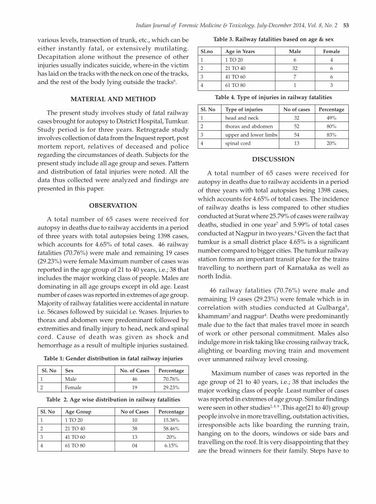
Indian Journal of Forensic Medicine & Toxicology. July-December 2014, Vol. 8, No. 2 53
various levels, transection of trunk, etc., which can beeither instantly fatal, or extensively mutilating.Decapitation alone without the presence of otherinjuries usually indicates suicide, where-in the victimhas laid on the tracks with the neck on one of the tracks,and the rest of the body lying outside the tracks6.
MATERIAL AND METHOD
The present study involves study of fatal railwaycases brought for autopsy to District Hospital, Tumkur.Study period is for three years. Retrograde studyinvolves collection of data from the Inquest report, postmortem report, relatives of deceased and policeregarding the circumstances of death. Subjects for thepresent study include all age group and sexes. Patternand distribution of fatal injuries were noted. All thedata thus collected were analyzed and findings arepresented in this paper.
OBSERVATION
A total number of 65 cases were received forautopsy in deaths due to railway accidents in a periodof three years with total autopsies being 1398 cases,which accounts for 4.65% of total cases. 46 railwayfatalities (70.76%) were male and remaining 19 cases(29.23%) were female Maximum number of cases wasreported in the age group of 21 to 40 years, i.e.; 38 thatincludes the major working class of people. Males aredominating in all age groups except in old age. Leastnumber of cases was reported in extremes of age group.Majority of railway fatalities were accidental in naturei.e. 56cases followed by suicidal i.e. 9cases. Injuries tothorax and abdomen were predominant followed byextremities and finally injury to head, neck and spinalcord. Cause of death was given as shock andhemorrhage as a result of multiple injuries sustained.
Table 1: Gender distribution in fatal railway injuries
Sl. No Sex No. of Cases Percentage
1 Male 46 70.76%
2 Female 19 29.23%
Table 2. Age wise distribution in railway fatalities
Sl. No Age Group No of Cases Percentage
1 1 TO 20 10 15.38%
2 21 TO 40 38 58.46%
3 41 TO 60 13 20%
4 61 TO 80 04 6.15%
Table 3. Railway fatalities based on age & sex
Sl.no Age in Years Male Female
1 1 TO 20 6 4
2 21 TO 40 32 6
3 41 TO 60 7 6
4 61 TO 80 1 3
Table 4. Type of injuries in railway fatalities
Sl. No Type of injuries No of cases Percentage
1 head and neck 32 49%
2 thorax and abdomen 52 80%
3 upper and lower limbs 54 83%
4 spinal cord 13 20%
DISCUSSION
A total number of 65 cases were received forautopsy in deaths due to railway accidents in a periodof three years with total autopsies being 1398 cases,which accounts for 4.65% of total cases. The incidenceof railway deaths is less compared to other studiesconducted at Surat where 25.79% of cases were railwaydeaths, studied in one year7 and 5.99% of total casesconducted at Nagpur in two years.4 Given the fact thattumkur is a small district place 4.65% is a significantnumber compared to bigger cities. The tumkur railwaystation forms an important transit place for the trainstravelling to northern part of Karnataka as well asnorth India.
46 railway fatalities (70.76%) were male andremaining 19 cases (29.23%) were female which is incorrelation with studies conducted at Gulbarga8,khammam2 and nagpur4. Deaths were predominantlymale due to the fact that males travel more in searchof work or other personal commitment. Males alsoindulge more in risk taking like crossing railway track,alighting or boarding moving train and movementover unmanned railway level crossing.
Maximum number of cases was reported in theage group of 21 to 40 years, i.e.; 38 that includes themajor working class of people .Least number of caseswas reported in extremes of age group. Similar findingswere seen in other studies2, 8, 9 .This age(21 to 40) grouppeople involve in more travelling, outstation activities,irresponsible acts like boarding the running train,hanging on to the doors, windows or side bars andtravelling on the roof. It is very disappointing that theyare the bread winners for their family. Steps have to
13. Chandru K--52--.pmd 7/24/2014, 10:04 AM53

54 Indian Journal of Forensic Medicine & Toxicology. July-December 2014, Vol. 8, No. 2
be taken to educate frequent travelers about theimportance of safe travelling.
Study clearly shows that majority of railway fataldeaths were accidental in nature, i.e. 56cases followedby suicidal i.e. 9cases.These findings were in consistentwith studies done at Gulbarga8, Nagpur4,Khammam2.Accidental railway deaths were due toinjuries sustained while crossing railway tracks,unmanned level crossing accidents, while travellingon footboard near door and boarding running trains.
The study revealed that majority of victimssustained crush injuries to extremities, followed bythorax and abdomen injuries and lastly to head andspinal cord, which is in agreement with studiesconducted at Nagpur3. Decapitation and head injurywere common among suicidal deaths similar to theearlier study9. Crush injuries to the extremities wasobserved in 54 cases which was due to railway run-over caused by the rotating effect of train wheels.Pedestrians are more prone to traumatic amputationof lower extremities in consistent with other study10.Injury involving thorax and abdomen (52 cases) werethe next commonest findings among the victims. Theseinjuries may be due to primary impact by portions ofthe engine or bogies. Head injuries are seen in almosthalf of the cases. These injuries are result of primaryimpact as well as secondary injuries.
CONCLUSION
India carries one of the largest railway networks inthe world and accidents from rail operations may notbe unexpected. From the present study we canconclude that maximum victims were males in theworking age group who died in accidental manner.Hence these untimely deaths caused mainly bycarelessness can be prevented by creating awarenessamong the people. Public should be educated aboutthe dangers of travelling footboard, boarding andalighting moving train, observing caution whilecrossing unmanned level crossing. Railway authoritiesshould take strict measures to prevent trespassing byawarding fine or punishment. Pedestrians, passengerover bridges, manned railway crossing, barricading/fencing to avoid trespassing are urgently needed.Public on their part should follow the rules andregulations regarding safety to prevent railwayfatalities.
ACKNOWLEDGEMENT
I would like to thank Dr Shivakumar.S, Head ofthe department, Forensic Medicine, Sri Siddhartha
medical college, Tumkur, for providing valuableguidance during the entire study. I also thank Mrs.Shwetha for providing technical support in bringingout the article.
Source of Funding and Ethical Clearance: Ethicalclearance and funding was not necessary as it was aretrospective study which included only collection ofdata.
Conflict of Interest Statement: The authors declarethat there is no conflict of interest.
REFERENCES
1. Nirmala George. Indian Rail Track deaths,www.Huffingtonpost.com; 2012/02/21.
2. Kumar K.M., Roop, Khaja Shaikh. DemographicProfile of Railway Accidents Occurring InKhammam Region- Indian Journal of ForensicMedicine and Toxicology, 2013; 7(1): 254-287.
3. K.R.Nagesh, Y.P.Raghavendra Babu. Excavationof Chest due to railway Mishap, AustralianJournal of Forensic Sciences, 2011; 1-6.
4. Ramesh Manaji Wasnik. Analysis of RailwayFatalities in Central India, Journal Of IndianAcademy Of Forensic Medicine,2012;32(4) : 311
5. Krishan Vij. Textbook Of Forensic Medicine andToxicology, Third Edition; TransportationInjuries: 455-456.
6. V.V.Pillay. Textbook Of Forensic medicine AndToxicology, 14th Edition; Medico legal Aspects OfInjuries and death: 210-211.
7. Sheikh.M, Sha.J.V. Study Of Death due ToRailway Accident, Journal Of Indian AcademyOf Forensic Medicine, 2008; 30 ( 3): 122-127.
8. Patel Basavraj. A study On Pattern of Injuries inRailway Deaths, Indian Journal of ForensicMedicine and Toxicology. 2011; 5 ( 1): 20-22.
9. Manoj Kumar Mohanty. Death due to TraumaticRailway Injury, Medicine, Science and the law,Royal Society Of Medicine Journals; April 2007;47(2): 156-160.
10. Marc.J.Shapiro, William.B.Luchtefeld. TraumaticTrain Injuries, The American Journal OfEmergency Medicine, Jan 1994; 12(1): 92-93.
11. Ted.R.Miller, John.B.Douglass, Nancy.M.Pindus.Railroad Injury: Causes, Cost and Comparisonswith other transport modes, Journal Of SafetyResearch; 1994; 25(4): 183-195.
13. Chandru K--52--.pmd 7/24/2014, 10:04 AM54

Indian Journal of Forensic Medicine & Toxicology. July-December 2014, Vol. 8, No. 2 55DOI Number: 10.5958/j.0973-9130.8.1.001
An Epidemiological Study of Fatal Firearm Cases inVaranasi, UP
Kaulaskar Shashikant V1, S K Pandey2, Pravin N Yerpude3, Manoj Pathak4, Keerti S Jogdand5
1Assistant Professor, Dept of Forensic Medicine, Gujarat Adani Institute of Medical Sciences, Bhuj, Gujarat,2Asso. Prof., Dept of Forensic Medicine, IMS, Banaras Hindu University, 3Asso.Prof,Dept of Community
Medicine,GAIMS , Bhuj, Gujarat, 4Asst. Prof. IMS, Dept of Forensic Medicine Banaras Hindu University,5Asso.Prof,Dept of Community Medicine,GAIMS , Bhuj, Gujarat
ABSTRACT
Introduction: Use of firearm is a worldwide increasing and Varanasi is no exception to this. This isdue to increasing number of hijacking, urban terrorism, dacoity, rioting, robberies, smuggling ofdrugs, political motivation, personal and group rivalry, quarrel over land, Property, caste feuds andthe like.
Materials and Method: Fifty four cases of fatal firearm injury has been drawn from the medicolegalautopsies brought to the mortuary of Department of Forensic Medicine, Institute of Medical Sciences,Banaras Hindu University, Varanasi, U.P. India, during the period from 1 June,2009 to 30th March2011 which were studied for a detailed epidemiological and medicolegal analysis.
Results: Out of total 3534 autopsies conducted during the above period, 54 (1.52%) victims had dieddue to fatal firearm injury. Most of the victims (98.15%) were male. Majority (66.65%) were youngadults in the age group of 21-40 years. Most of the victims, (83.33%) of fatal firearm injury wereHindus followed by Muslims (12.96%). The maximum number, 46.3% of victims came from ruralareas. The occupational status of most of the victims was agriculture (40.74%) followed by business(31.48%).
Conclusion: Overall incidence of fatal firearm injury is reduced because of legal restriction on licenseof personal firearms weapon while the incidence of firearm injury due to country made guns areincreased which are illegal, easily available and cheap.
Keywords: Epidemiology, Fatal Firearm Cases
INTRODUCTION
Invention of fire was the greatest invention for thehuman civilization but the invention of firearm hasproved to be a curse to this world, it has become themost dreaded killing tool used by human beings tokill them. Though, in western countries, suicidal fatalfirearm injury is very common, in region of the world,it is mostly used in the homicide cases only [1]. Firearms
´
Corresponding author:Kaulaskar ShashikantFlat No. G-1, BRJ Tower,Sarpanch Nagar, MalegaonRoad, Nanded-431605, MaharashtraMobile No: 09423802898Email id: [email protected]
have passed through continuous evolutionary changesand have been established as the most popularinstrument for committing homicide, whether duringpeace or in war time.
Since guns are recognized as being highly lethal,all assailants who use such weapons were believed byWolfgang to have been highly determined to kill.Wolfgang proposed the ‘weapon substitutionhypotheses (1986). This hypothesis posits that theintentions of an assailant, whether they are to kill orinjure, determines the weapon selected. For example,if an assailant has a single minded, thoroughly‘determined’ intention to kill one’s victim, one will seekout the kind of weapon that is most likely to ensurethe desired outcome. Because a gun is well recognized
14. shashi kant--55-.pmd 7/24/2014, 10:04 AM55

56 Indian Journal of Forensic Medicine & Toxicology. July-December 2014, Vol. 8, No. 2
as being a highly lethal weapon, that intent of killingwill, if it is available, seek out such a weapon. If afirearm is not available, then this effective weapon willbe substituted for the next most available and lethalweapon. This hypothesis suggests that if an assailantdid not intend to kill but only harm one’s victim, thenone would have selected some other less lethalweapon [2].
MATERIALS AND METHOD
The present study comprised of Fatal Firearminjuries drawn from the Medico-legal autopsies heldin mortuary of the department of Forensic Medicine,Institute of Medical Sciences, Banaras HinduUniversity, Varanasi, U.P., India, during period of 1stJune 2009 to 30th March 2011, accompanied by sufficientnumber of relevant persons who were thoroughlyinterviewed at the time of autopsy on the body ofdeceased victim of fatal firearm injury. Before the startof the study ,ethical clearance was taken from EthicalCommittee of IMS,BHU. For the study, relevantquestionnaires schedule were prepared to collectvarious information related to, socioeconomic factors,about incidence of fatal firearm injuries, medico legalcrime investigation and evidential data etc. These caseswere studied for the history of the cases, theirepidemiological characteristics e.g. Age, sex,community character etc. nature, distribution andtypes of injuries including their medico legal aspect.
The various data relating to the cases were collectedfrom sources as under
A. Examination of inquest reports and connectedpapers
B. Interviewing the police personnel accompanyingthe cases
C. Interviewing the relatives, friends and neighborsof the deceased
D. The autopsy examination paper
RESULTS AND DISCUSSION
Out of total number of 3534 cases which werebrought for postmortem examination during theperiod of study i.e. 22 months from 1.6.2009 to30.03.2011, 54 (1.52%) cases were declared to be due tofatal firearm injuries (Table 1).
Age and sex distribution of victims of fatal firearminjuries observed that most of the victims (98.15%)
were male and only one victim (1.85%) was female.Over majority (66.65%) were young adults in the agegroup of 21-40 years. 16.66 per cent victims were ofmiddle age group of 41-50 years (Table 2).
In other studies, autopsies of 89 cases of firearmdeaths were conducted in Varanasi area which foundthat 33.67% cases were from the age group of 31 to 40years, followed by (28.08%) the cases who came under21 -30 years [3]. On the basis of religion, most of thevictims (83.33%) of fatal firearm injury were Hindusfollowed by Muslims (12.96%) (Table 3).
As per educational status maximum (27.77%)victims were of secondary school education, followedby graduate (25.92%), illiterate (16.66%), intermediate(12.96%) and post-graduation (9.25%) ( Table 4).
The community character of victims of fatal firearminjury reveals that the maximum number, 46.3% ofvictims came from rural areas followed by suburbanareas (31.48% )and urban areas (20.37% ) (Table 5). Thisis more or less similar to the study of Gupta et al whohave observed the percentage distribution ofcommunity character of the firearm victims andrecorded 43.82% rural, 21.95% urban and 17.17%suburban of Varanasi area. The other study showedpreponderance of rural (65.86%) over the urban(17.17%) victims in firearm fatalities [4]. However, asmall fraction (10.2%) belonged to sub-urban area ofVaranasi.
One of the most important parameter is maritalstatus of victims which shows, that majority (64.1%)were married followed by unmarried (18.51%),widower (5.55%), separated(3.7%) (Table 6). Similarstudy done by Gupta, in their study also found a highrate of married cases amongst the victims of fatalfirearm injuries (80.91) [3] , In other study, it wasrevealed that 62% victims of fatal firearm injuries, weremarried and 37% victims were unmarried in Delhi[5].The occupational status of most of the victims wasagriculture (40.74%) followed by business (31.48%),labourers (9.25%), services (7.4%) and unemployed(1.85%) (Table 7).
The incidence of firearm casualty indicates thatmajority (77.77%) of victims came from joint familiesfollowed by those from nuclear family type (12.96%)(Table 8).
In case of manner of death in fatal firearm injury,homicide accounted for maximum percentage(81.48%), followed by accident (9.25%), suicide (5.55%)(Table 9).
14. shashi kant--55-.pmd 7/24/2014, 10:04 AM56

Indian Journal of Forensic Medicine & Toxicology. July-December 2014, Vol. 8, No. 2 57
In case of motive in fatal firearm injury enmityaccounted for maximum percentage (38.88%) followedby Property dispute (16.66%), Robbery (12.96%)(Table 10).
Conclusion and recommendations
The overall incidence of fatal firearm injury isreduced as compared to previous studies conductedin this area. Though overall incidence of fatal firearminjury is reduced because of legal restriction on licenseof personal firearms weapon, the incidence of firearminjury due to country made guns are increased whichare illegal, easy available and cheap. In our study weobserve enmity was the major motive behind the fatalfirearm injury as compared to previous studiesconducted in this area in which dispute over landedproperty was major motive. Police patrolling for earlydetection of crime and shifting of injured to thehospital/critical care center and provision of promptambulance service by state/private hospital or NGOsfor medical care is another aspect, which may declinemortality due to such crimes.
To prevent killings, there should be combined effortfrom all sections of the society.
Following steps should be of help in its prevention
A) Proper employment facility for the youth.
B) Social stability and creation of proper politicalenvironment.
C) Strong and effective measures to control theunlicensed arms.
D) Need to eliminate illegal gun making units in ourregion in order to decrease the rate of firearmfatalities.
E) Various law enforcing agencies have to makeconcerted efforts and be more vigilant on thisaccount in order to accomplish these goals.
Table1: Incidence of fatal firearm injuries in medico
legal autopsies (22 months from 1.6.2009 to 30.03.2011)
Total number of autopsies Fatal firearm injuries
Total number Percentage
3534 54 1.52
Table 2: Age and sex wise distribution of victims offatal firearm injury
(n=54)
Age group (year) Male Female Number Percentage
11 to 20 2 - 2 3.7
21 to 30 21 - 21 38.88
31 to 40 15 - 15 27.77
41 to 50 8 1 9 16.66
51 to 60 5 - 5 9.25
61 to 70 1 - 1 1.85
71 to 80 1 - 1 1.85
Table 3:Religion wise distribution of victims of fatalfirearm injury.
(n=54)
Religion Percentage
Hindu 83.33
Muslim 12.96%
Other Nil
Not known 3.7%
Table 4: Distribution of victims of fatal firearm injuryaccording to their educational status(
n=54)
Educational status Number Percentage
Illiterate 9 16.66
Primary 2 3.7
Secondary 15 27.77
Intermediate 7 12.96
Graduate 14 25.92
Post graduate 5 9.25
Unknown 2 3.7
Table 5: Area wise distribution of victims of fatalfirearm injury
(n=54)
Area Number Percentage
Rural 25 46.3
Urban 11 20.37
Suburban 16 29.62
Unknown 2 3.7
Table 6: Distribution of victims of fatal firearm injuryaccording to their marital status
(n=54)
Marital status Number PercentageMarried 35 64.81Unmarried 10 18.51Widow 1 1.85Widower 3 5.55Divorcee 1 1.85Separated 2 3.7Unknown 2 3.7
14. shashi kant--55-.pmd 7/24/2014, 10:04 AM57

58 Indian Journal of Forensic Medicine & Toxicology. July-December 2014, Vol. 8, No. 2
Table 7: Distribution of victims of fatal firearm injuryaccording to their occupation
(n=54)
Occupation Number Percentage
Agriculture 22 40.74
Business 17 31.48
Labourer 5 9.25
Service 4 7.4
Unstable job 3 5.55
Unemployed 1 1.85
Not known 2 3.7
Table 8: Distribution of victims of fatal firearm injuryaccording to their family type
(n=54)
Family type Number percentage
Alone 3 5.55
Joint 42 77.77
Nuclear 7 12.96
Unknown 2 3.7
Table 9: Distribution of victims of fatal firearm injuryaccording to their manner of death
(n=54)
Manner Number Percentage
Accident 5 9.25
Homicide 44 81.48
Suicide 3 5.55
Not known 2 3.7
Table 10: Distribution of victims of fatal firearm injuryaccording to their motive of death
(n=54)
Motive Number Percentage
No 5 9.25
Enmity 21 38.88
Property dispute 9 16.66
Robbery 7 12.96
Damage to prestige 4 7.4
Encounter 3 5.55
Depression 2 3.7
Loan 1 1.85
Unknown 2 3.7
ACKNOWLEDGEMENT
I would like to thanks for the valuable support fromHead of Department Dr.(Prof) C.B.TRIPATHI forallows me this study in Department of ForensicMedicine and Thanks to Dr. S.K Tripathi, Prof. Dept.of Forensic Medicine, IMS, Banaras Hindu University,for kind support.
Ehical Clearance: From Ethical Committee ofIMS,BHU
Conflict of Interest: None declared
Financial Support: None declared
REFERENCES
1. Patowary AJ. Study of pattern of injuries inhomicidal firearm injury cases. JIAFM 2005;27(2):92-93.
2. Wright JD, Rossi PH. Armed and considereddangerous: a survey of felons and their firearms.New York: Aldine.1986; 12:332-34.
3. Gupta AK. Das Gupta SM, Rastogi BL. Study ofInjuries from Firearm in Medico legal Autopsies.Thesis Submitted for the Degree of Doctor ofMedicine (Forensic Medicine), Institute ofMedical Science, B.H.U., Varanasi, 1979.
4. Das Gupta SM ,Tripathi CB. A Study of theHomicide Cases Occurring in Varanasi Area.Indian Medical Gazette. CXVII (9) 1983; 285-288.
5. Krishna KP. Victims of Homicide: A Study inPersonal and Sociocultural Antecedents. PoliceResearch and Development. 1981:11.
14. shashi kant--55-.pmd 7/24/2014, 10:04 AM58

Indian Journal of Forensic Medicine & Toxicology. July-December 2014, Vol. 8, No. 2 59DOI Number: 10.5958/j.0973-9130.8.1.001
Sexual Dimorphism of Cranial Sutures in Maharashtraand North-Karnataka Region
Makandar U K1, Kaushal Vishuddha M2, Rajendra R3, Sharieff J H4
1Associate Professor of Anatomy, A.I.M.S. B.G.Nagar, Belur, Nagamangala, (Tq) Mandya, Dist, 2Assistant Professor ofCommunity Medicine, Geetanjali Medical College Udaipur, Rajasthan, 3Professor and HOD of Anatomy A.I.M.S
B.G.Nagar, Belur, Nagamangala, (Tq) Mandya, Dist, 4Professor of Anatomy D.M.Wynad Institute of MedicalSciences, Meppadi, Kerala
ABSTRACT
112 dried non-pathological adult crania of known sex were studied from Maharashtra and NorthKarnataka cranial sutures were traced by sketch pen then transparent butter paper was fixed andsuture was traced by micro tipped pen. Each suture was studied in three equal segments and classifiedinto closely widely and straight type of sutures.
Widely serrated sutures were common in male crania of coronal suture and in female crania closelyand straight type of suture was common while sagital suture of male crania also had widely serratedsuture but female crania of sagital suture had only closely serrated type of sutures. Lambdoid sutureof male crania had widely and straight type of suture and female crania had closely serrated sutures.The angle between two halves of lambdoid was highly significant (P<0.01) in Female crania. Thisstudy will certainly help the Anatomist, Anthropologist and Medico Legal Expert becauseMorphometric Values of Dermal Bones are uncertain.
Keywords: (1) Closely Serrated, (2) Widely serrated, (3) Straight Type, (4) Butter Paper
INTRODUCTION
“Suture” is a Latin word (Sutura=Seam) which isrestricted to skull it has mainly three functions Unitethe bones while still allowing slight moments. Act asgrowth areas to absorb mechanical stress. Duringgrowing period these sutures contain connectivetissues, blood vessels, osteogenic cells to -facilitate theoppositional growth of the bone margins to meet atthe suture(1) and obliterate at certain age.
Many Criteria are available to differentiate sex ofthe crania after puberty like length of mastoid process,prominence of glabella, superciliaryarches, width of
Corresponding author:Makandar U KC/o. A.M Pathan,Behind S.P Office, Kumbar Galli, Jorapur Peth,Bijapur - 586101 (Karnataka).E - Mail ID - [email protected] No. : +91-9341610428
zygomatic arches, diameter of foramen magnum,depth of digastric groove but least data is availableregarding sexual dimorphism of cranial sutures.Moreover evolutionary point of view also suturesdeveloped in the armored jawless fishes since then nomammals show evolutionary progress of sutures(2)Asour ancestors were cannibolic they approached thetasty brain through normaverticalis. (3) Hence attemptwas made to study the sexual dimorphism of thesutures. Medico-legally serrations of the sutures areanomalous to estimate the age of the subject but inour study serrations of the sutures have greatsignificance for sexual Dimorphism.
MATERIAL AND METHOD
112 adult, non-pathological crania of known sexwere studied from medical colleges of Sholapur andBijapur. Out of 112, 67 crania were male and 45 werefemale. Each cranium was put in anatomical position
15. Makander--59--.pmd 7/24/2014, 10:04 AM59
60 Indian Journal of Forensic Medicine & Toxicology. July-December 2014, Vol. 8, No. 2
the coronal, sagital and Lambdoid sutures were tracedby color sketch pen then transparent butter paper wasfixed on the crania with the help of rubber band andsutures were traced by micro tipped pen. Each cranialsuture was studied in three equal segments from rightto left but in sagital suture anterior to posterior toobserve the maximum serrations morphologically andmetrically. Sutures were classified under threeheadings closely serrated, widely serrated and straighttype. Breadth of the suture was measured by thevernier caliper. Length and Breadth of the crania wasmeasured by measuring (tailors tape).
OBSERVATIONS AND RESULTS
a=widely serrated, b=closely serrated, c=straighttype
Table No. 1 Comparison of coronal suture in bothsexes in all three segments.
Segment-I
a= in Males 25 (37.5%) in Females 17 (37.70%)
b=in Males 34 (50.7%) in Female 24 (53.42%)
c=in Males 8 (11.8%) in Female 4 (8.88%)
Segment-II
a= in Males 15 (22.4%) in Females 6 (13.4%)
b=in Males 10 (14.9%) in Female 8 (17.8%)
c=in Males 42 (62.7%) in Female 31 (68.88%)
Segment-III
a= in Males 32 (47.7%) in Females 15 (33.3%)
b=in Males 26 (38.8%) in Female 20 (44.4%)
c=in Males 9 (13.5%) in Female 10 (22.3%)
Table No. 2 – Comparison of Sagital Suture in bothsexes
Segment-I
a= In Males 28 (41.7%) and Female 15 (33.3%)
b= In Males 21 (31.5%) and Female 14 (31.2%)
c= In Males 15 (22.4%) and Female 16 (35.5%)
Segment-II
a= In Males 31 (46.3%) and Female 16 (35.5%)
b= In Males 21 (31.5%) and Female 14 (31.2%)
c= In Males 15 (22.4%) and Female 15 (33.3%)
Segment-III
a= In Males 45 (67.6%) and Female 27 (60%)
b= In Males 12 (17.9%) and Female 8 (17.7%)
c= In Males 10 (14.5%) and Female 10 (22.3%)
Table No. 3. Comparison of lambdoid suture in bothsexes
Segment-I
a= In Males 35 (52.2%) and Female 21 (46.6%)
b= In Males 23 (34.4%) and Female 19 (42.2%)
c= In Males 9 (13.4%) and Female 5 (11.2%)
Segment-II
a= In Males 38 (58.7%) and Female 26 (57.7%)
b= In Males 23 (34.3%) and Female 13 (28.8%)
c= In Males 6 (9%) and Female 6 (13.5%)
Segment-III
a= In Males 23 (49.3%) and Female 22 (48.8%)
b= In Males 21 (31.3%) and Female 18 (40.6%)
c= In Males 13 (19.4%) and Female 5 (11%)
Table-4 Comparison between lengths of Nasion toinion which was insignificant (P>0.01)pterion topterion was insignificant (P>0.001) and length betweenbitemporal width was also insignificant (P>0.01).
Table-5 The angle between coronal and sagital,sagital and lambdoid sutures were insignificant(P>0.01) but angle between two limbs of lambdoidsutures was highly significant (P<0.01).
15. Makander--59--.pmd 7/24/2014, 10:04 AM60

Indian Journal of Forensic Medicine & Toxicology. July-December 2014, Vol. 8, No. 2 61
Table 1. Comparative study of coronal suture segment in both sexes with %
Segment Male Crania (67) Female Crania (45)
a b c a b c
I 25(37.5%) 34(50.7%) 8(11.8%) 17(37.7%) 24(54.42%) 4(8.88%)
II 15(22.4%) 10(14.9%) 42(62.7%) 6(13.4%) 8(17.8%) 31(68.8%)
III 32(47.7%) 26(38.8%) 9(13.5%) 15(33.3%) 20(44.4%) 10(22.3%)
a= widely serrated b= closely serrated c= straight type
Table 2. Comparative study of sagital sutural segment in both sexes with %
Segment Male Crania Female Crania
a b c a b c
I 28(41.7%) 21(31.5%) 18(26.8%) 15(33.3%) 14(33.3%) 16(33.5%)
II 31(46.3%) 21(31.3%) 15(22.4%) 16(33.5%) 14(31.2%) 15(33.3%)
III 45(67.6%) 12(17.9%) 10(14.9%) 27(60.0%) 8(60.0%) 10(22.3%)
Table 3. Comparative study of lambdoid suture segment in both sexes with %
Segment Male Crania (67) Female Crania (45)
a b c a b c
I 35(52.2%) 23(34.4%) 9(13.4%) 21(46.6%) 19(42.2%) 5(11.2%)
II 38(56.7%) 23(34.3%) 6(9.0%) 26(57.7%) 13(28.8%) 6(13.5%)
III 33(49.3%) 21(31.3%) 13(19.4%) 22(48.8%) 18(40.8%) 15(11.2%)
Table 4. Comparison between (a)length of Inion to Nasion. (b) Pterion to Pterion. (c) Length between bitemporalwidth in both sexes.
Sl No Observations Male crania (67) Female Crania (45)
Mean SD Mean SD ‘P’ value
a Inion to Nasion 317.16 20.6 316.55 18.46 P> 0.01
b Pterion to Pterion 182.01 17.5 182.33 18.7 P> 0.01
c Bitemporal width 221.41 18.4 224.44 20.9 P> 0.01
All the values are insignificant (P >0.01)
Table 5. Comparison of angle between two limbs of lambdoid suture in both sexes
Male Crania 67 Female Crania 45 T Test Value P Value Results
Mean Value S1 Mean Value S1
117.31 16.39 128.66 25.24 1.98 P<0.01Highlysignificant
DISCUSSION
In this present study of coronal suture in(Table-1) had more number of widely serrated suturesobserved in the male crania, closely serrated andstraight were common in female. In sagital suture(Table-2) widely serrated sutures were common inmale crania and closely serrated sutures were commonin female crania. In lambdoid suture (Table-3) widelyserrated and straight type of suture was common whileclosely serrated sutures were common in female crania.These findings are more or less in agreement withprevious studies but they classified the sutures of thecrania into nine types viz closely, more closely, widely,
more widely etc…(4)No English Literature is availableto compare the findings of present study. The probablereasons of these finding could be due to gravity ofmasticatory differences between both sexes becausemasticatory stress play vital role in the morphology ofsutures(5)The regional and genetic foctors, might beresponsible for such type of sutural development,TWIST and BMP genes enhances the serration ofsutures, F G F gene accelerates the closure and suture.(6)but sexual dimorphism and these genes is yet to beestablished, moreover earlier closure of the sutures infemale crania (7)Could be one of the factor for closelyserrated suture in female. Moreover growth of the
15. Makander--59--.pmd 7/24/2014, 10:04 AM61

62 Indian Journal of Forensic Medicine & Toxicology. July-December 2014, Vol. 8, No. 2
cranial suture is influenced by the muscularattachments independently.(8)Hence influence in theweaker muscles of female might have resulted intoclosely serrated suture. Intracranial contents,intracranial volume and intracranial pressure decidethe pattern of suture.(9)Pattern and position of sutureis determined before completion ofossification.(10)Regarding the pattern of suture it canbe hypothesized that, brain was not last organ toevolve, rather brain was clearly a focus for importantand stage setting selective postures early in hominoidevolution associated with changes in erect posture.
The angle between the two limb of lambdoid suturesignificant (Table-5) was highly significant (P<0.01)studied in 1900. (11)This significance value could be dueto migration of neutral crest between the formation ofcaudal part of sagital and whole part of lambdoidsuture in the Intra uterine life which juxtaposes thetwo limbs of lambdoid suture to facilitate the normalgrowth of the brain (12).
SUMMARY AND CONCLUSION
These significance incidences of widely serratedsuture common in male crania and closely serrated infemale crania and significant of lambdoid angle infemale crania will certainly help the anatomist,anthropologist and medico legal expert but theseinferences demand further genetic, Hormonal,Embryological, histological, environmental, nutritionalstudies because exact mechanism, factors of formationand closure of sutures in both sexes is yet to beestablished. Due to early obliteration of cranial suturesleast attention is paid in the literature, so far.
ACKNOWLEDGEMENT
Author is grateful to Prof. Gaikawad P.G ofSholapur and Prof. Patil B.G of Shri B.M Patil MedicalCollege Bijapur for their kind permission to carry outthis research work in their department above all to mybeloved Principal Dr Shivramu M.G for his constantencouragement.
Ethical Clearance: Taken from AIMS & RC B.GNagara, Belur. Nagamangala (TQ.) Mandya (Dist)
Source of Funding: Self
Conflict of Interest: Nil
REFERENCES
1. Dixon A.D sarnat B.G – factors and mechanisminfluencing bone growth, progress in clinical andbiological research 1982, Vol. 101,153-59,Newyork.
2. Hartwig W.C – Fractal analysis of sagital suturemorphology. Journal of morphology (1991), Vol.210, 289-298.
3. Garns and Block.W – The limited nutritionalvalues of cannibalism American Anthropologist(1970), Vol. 72 (1), 106-108.
4. Parez-Parez A, Espur.z – Nan-occlusal dentalmicroware variability in sample of middle andlate Pleistocene Human Population from Europeand near east. Journal of Human Evolution (2003),Vol. 44, 497-513.
5. Biswas U.N and Bhattacharya – SexualDimorphism of cranial sutures Ph.D. Thesissubmitted to Delhi University, (1986).
6. David P.C, Rice Thomas, Abergo Tchany Tangz –Integration of FGF and TWIST in calvarial boneand sutural development. Journal ofDevelopment (2000) Vol 127, 1845-1855.
7. Ridson Dz – study of crania and other excavatedat tell. Duweir (Lachish) by welcome – mortsonArcheological research expedition Biometrika(1902), Vol.1, 99-166.
8. Washburn.S - The study of human evolution. InDolhinow.P and serich Boston, (1971), 87-117Brown and company publication.
9. Babler. W.J – Relation of altered cranial suturegrown to cranial base and midface. In persing J.A.Edgerton M.R.Jane J.F. Scientific FoundationM.R.Jane Surgical treatment of craniosynostosis,IIIrd edition (1989), 87-95William and wilkinspublications Baltionmore publication.
10. Clerk Le gross W - Tissues of the body IInd edition1945,110, Oxford University Publication.
11. Tidsley M.L – Metrical Study of skull in differentraces. Biometrika (1900), 9, 176-262.
12. Jiang X, Isekis’Maxon RE, Kay GM – Tissueorigins and interactions in mammalian skull vaultDev. Bio, (2002), Vol. 24, 106-116.
15. Makander--59--.pmd 7/24/2014, 10:04 AM62
Indian Journal of Forensic Medicine & Toxicology. July-December 2014, Vol. 8, No. 2 63DOI Number: 10.5958/j.0973-9130.8.1.001
Post Mortem and the Risk of HIV Infection
Mukesh Kumar Bansal1, SK Naik2, Poonam Gupta3, Yashoda Rani4, BL Sherwal5
1Post Graduate Student, 2Associate Professor, Department of Forensic Medicine and Toxicology, 3Assistant Professor,Department of Microbiology, 4Assistant Professor, Department of Microbiology, 5Director Professor, Department of
Microbiology, Lady Hardinge Medical College, New Delhi
ABSTRACT
The present study "Post Mortem and the risk of HIV infection" was conducted upon 200 randomlyselected cases having known and unknown identities. (100 each) at the Department of ForensicMedicine and Toxicology and Department of Microbiology, Lady Hardinge Medical College fromNovember 2011-March 2013. Out of 200 cases, 10 (5%) were positive for HIV by ELISA test. Positivityin known bodies was 2% compared to 8% in unknown bodies. Males outnumbered, with maximumprevalence in the Age group of 31-40 years (9.52%). Drug abuse was found in 1(14.28%) case inknown bodies and in 8(10.12%) cases in unknown bodies. HIV infection could be detected by ELISAtest from 12 hours till 7 days post-mortem.
Keywords: Claimed, Unclaimed Bodies, HIV, ELISA test
INTRODUCTION
The common occupational hazards likely to beencountered in an autopsy room include infections,toxicity and radiation. However, there is more chanceof infections than any toxicity or radiation.1
Streptococcal sepsis, tuberculosis, blastomycosis,AIDS, hepatitis B and C, rabies, tularaemia, diphtheria,erysipeloid fever and certain viral hemorrhagic feversare some of the serious infections that can betransmitted through these routes.2-8
The major hazards HIV transmission topathologists and technical staff in performingautopsies come from skin injuries from sharpinstruments and bone spicules, and from inhalingvirulent pathogens such as Mycobacteria tuberculosis.Oral and conjunctival infection are also possible butcan be prevented by simple barriers. Even priorknowledge of the patients HIV status has not beenshown to reduce the rate of percutaneous exposure.
Corresponding author:BL SherwalDirector Professor, Department of MicrobiologyLady Hardinge Medical College, New Delhi, [email protected]+919868168400
The infectivity of HIV in samples decays slowly withtime. This decay in infectivity is variable, dependingon environmental and viral factors. In highconcentration, HIV may remain infectious upto threeweeks.9
Most of the unclaimed dead bodies brought to themortuary of Lady Hardinge Medical College and Smt.S.K. Hospital, New Delhi are either beggar or destitutecommonly involved in different kind of drug abusesand sexual activities. Post-mortem examinations oversuch dead bodies in the past have revealed tuberculosis(which is commonly associated with HIV infection)as a predominant cause of deaths.
The time for which a corpse remains potentiallycontagious with HIV is controversial. Infectious virushas been recovered from liquid blood held at roomtemperature for three weeks and virus in highconcentration has been found to remain viable for threeweeks.10,11
Considering the risk posed by post-mortemdissection of unclaimed bodies at autopsies, thepresent study is taken to weigh the actual risk pose byHIV during routine dissection of the unclaimed deadbodies compared to claimed dead bodies at medico-legal autopsies.
16. BL sherwal--63--.pmd 7/24/2014, 10:04 AM63
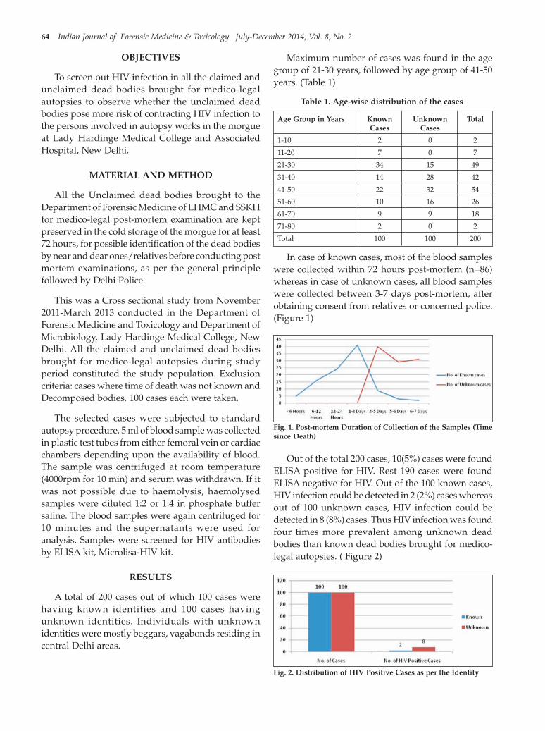
64 Indian Journal of Forensic Medicine & Toxicology. July-December 2014, Vol. 8, No. 2
OBJECTIVES
To screen out HIV infection in all the claimed andunclaimed dead bodies brought for medico-legalautopsies to observe whether the unclaimed deadbodies pose more risk of contracting HIV infection tothe persons involved in autopsy works in the morgueat Lady Hardinge Medical College and AssociatedHospital, New Delhi.
MATERIAL AND METHOD
All the Unclaimed dead bodies brought to theDepartment of Forensic Medicine of LHMC and SSKHfor medico-legal post-mortem examination are keptpreserved in the cold storage of the morgue for at least72 hours, for possible identification of the dead bodiesby near and dear ones/relatives before conducting postmortem examinations, as per the general principlefollowed by Delhi Police.
This was a Cross sectional study from November2011-March 2013 conducted in the Department ofForensic Medicine and Toxicology and Department ofMicrobiology, Lady Hardinge Medical College, NewDelhi. All the claimed and unclaimed dead bodiesbrought for medico-legal autopsies during studyperiod constituted the study population. Exclusioncriteria: cases where time of death was not known andDecomposed bodies. 100 cases each were taken.
The selected cases were subjected to standardautopsy procedure. 5 ml of blood sample was collectedin plastic test tubes from either femoral vein or cardiacchambers depending upon the availability of blood.The sample was centrifuged at room temperature(4000rpm for 10 min) and serum was withdrawn. If itwas not possible due to haemolysis, haemolysedsamples were diluted 1:2 or 1:4 in phosphate buffersaline. The blood samples were again centrifuged for10 minutes and the supernatants were used foranalysis. Samples were screened for HIV antibodiesby ELISA kit, Microlisa-HIV kit.
RESULTS
A total of 200 cases out of which 100 cases werehaving known identities and 100 cases havingunknown identities. Individuals with unknownidentities were mostly beggars, vagabonds residing incentral Delhi areas.
Maximum number of cases was found in the agegroup of 21-30 years, followed by age group of 41-50years. (Table 1)
Table 1. Age-wise distribution of the cases
Age Group in Years Known Unknown TotalCases Cases
1-10 2 0 2
11-20 7 0 7
21-30 34 15 49
31-40 14 28 42
41-50 22 32 54
51-60 10 16 26
61-70 9 9 18
71-80 2 0 2
Total 100 100 200
In case of known cases, most of the blood sampleswere collected within 72 hours post-mortem (n=86)whereas in case of unknown cases, all blood sampleswere collected between 3-7 days post-mortem, afterobtaining consent from relatives or concerned police.(Figure 1)
Fig. 1. Post-mortem Duration of Collection of the Samples (Timesince Death)
Out of the total 200 cases, 10(5%) cases were foundELISA positive for HIV. Rest 190 cases were foundELISA negative for HIV. Out of the 100 known cases,HIV infection could be detected in 2 (2%) cases whereasout of 100 unknown cases, HIV infection could bedetected in 8 (8%) cases. Thus HIV infection was foundfour times more prevalent among unknown deadbodies than known dead bodies brought for medico-legal autopsies. ( Figure 2)
Fig. 2. Distribution of HIV Positive Cases as per the Identity
16. BL sherwal--63--.pmd 7/24/2014, 10:04 AM64
Indian Journal of Forensic Medicine & Toxicology. July-December 2014, Vol. 8, No. 2 65
Prevalence of HIV infection observed in the studywas maximum in the age group of 31-40 years(n=4:9.52%) followed by 21-30 years (n=3: 6.12%) and51-60 years (n=1:3.84%). Age groups of 1-20 years and61-80 years did not show any HIV positive cases.(Table 2)
Table 2. Distribution of ELISA positive HIV cases asper Age
Age Group in year Total Positive PercentageAutopsy Cases Cases
1-10 2 0 0.00%
11-20 7 0 0.00%
21-30 49 3 6.12%
31-40 42 4 9.52%
41-50 54 2 3.70%
51-60 26 1 3.84%
61-70 18 0 0.00%
71-80 2 0 0.00%
HIV infection could be detected upto 7 days post-mortem that is the maximum period of preservationof dead bodies in the present study. (Figure 1)
In present study, ELISA positive for HIV werefound in 10 cases where the post-mortem interval wereranging from 12 hours to 7 days. (Figure 3)
infectious agents, especially during post-mortemexaminations.
Emphasis should be placed on the prevention ofinfection by establishing “universal precautions, butlater to screen out HIV in all claimed and unclaimeddead bodies brought for medicolegal autopsies. ELISAcan be the screening test of choice before conductingthe autopsy to prevent the exposure of HIV to personsinvolved in mortuary works. It is easy to perform,adaptable to large number of samples, is sensitive andcost effective. Spread of HIV infection from deadbodies occurs from contact with high risk fluid by aneedle or a sharp instrument injury. Limited data isavailable regarding these risks to forensic medicalpersonnel who are exposed daily to large number ofseverely traumatized bodies in India12
In the present study prevalence of HIV by usingELISA on post-mortem serum samples was found tobe 5% (10 positive cases out of 200 cases). A study byKarhumen et al. on prevalence of HIV by usingEnzyme immunoassay (ELISA) on post-mortem serafound HIV positive results in 0.12% cases (9 positiveout of 7305 cases).13 Zhang et al. found 5.6% wereseropositive for HIV.14 Christensen PB et al. in his studyon post-mortem blood samples of drug related deathsfor HIV, found the prevalence of 4% and similarlyAchusi IB et al found 5.8% positive cases (44 out of754 cases).15,16
The infectivity of HIV in samples decays slowlywith time. This decay in infectivity is variable,depending on viral and environmental factors. In highconcentration, HIV may remain infectious for uptothree weeks.9 Post mortem interval during which theblood sample could remain HIV positive had beenstudied by Nyberg M et al. and Edler C et al.17,18 NybergM et al. had studied 10 patients with HIV infection inwhich post-mortem intervals were 1 to 6 days andfound positive in 8 cases. Edler C et al. in his studytaking samples from 6 HIV positive deceased personshad detected HIV infection in all the samples 24 h postmortem. In the present study, HIV infection could bedetected till 7 days post-mortem that was maximumperiod of preservation of dead body before conductionof autopsy, supporting the study by Nyberg M et al.
HIV infection can reduce the survival period ofaffected individuals to great extent.In the present study,maximum prevalence of HIV positive cases wereobserved in the age group of 31-40 years (9.52%)followed by 21-30 years (6.12%) and 51-60 (3.84%). Age
Fig. 3. Distribution of HIV Positive Cases as per Time since Death
DISCUSSION
Hepatitis viruses usually died within 48 hours afterdeath of individual or cooling of the dead bodies.Therefore, possibility of transmission of Hepatitis Binfection to the autopsy surgeon or supporting staffsis expected to be about zero percent. Autopsy safetywas not much of consideration until 1980’s when HIVinfection appeared. The concern about contracting aninfectious disease during course of an autopsy is great;the concern is heightened when the devastatinginfections such as HIV that could be transmitted areconsidered. Mortuaries, dissecting rooms and forensiclaboratories are high risk areas for infection and thestaff have the risk of occupational exposure to
16. BL sherwal--63--.pmd 7/24/2014, 10:04 AM65

66 Indian Journal of Forensic Medicine & Toxicology. July-December 2014, Vol. 8, No. 2
groups of 1-20 years and 61-80 years did not show anyHIV positive cases. This could be because positivecases were mostly among unknown individuals whoindulged in drug abuse and different kind of sexualactivities after puberty as only sources of entertainmentand died early because of chronic malnutrition anddiseases.
Christensen PB et al. found that in drug relateddeaths 45% were intravenous drug abusers showingHIV positivity in 4% cases.15 NACO in 2008 reportshows that prevalence of HIV in injecting drug usersis 7.2%.19 Increased prevalence of HIV was seen in thepresent study is due to more number of drug abuserare either destitute or vagabond who have leastknowledge and information about transmission ofHIV. Besides they are crippled with poverty, chronicmalnutrition, chronic infection in the absence of propermedical attention etc.
Blood was taken as sample because it was easier toobtain and process and there is higher chance ofdetection of HIV virus as compared to other tissuesamples. Blood has maximum chances of detection ofHIV virus compared to other body fluids due to itsamount and persistence in the blood.
ELISA is the most commonly performed screeningtest, testing a large number of specimens a day. It iseasy to perform, adaptable to large number of samples,is sensitive and cost effective. ELISA has always beenthe method of choice for screening of HIV in bloodtissues during life time or after death.
CONCLUSION
Post mortem testing of HIV infection in Medicolegal autopsies can be useful in monitoring thesurveillance of HIV-infection in the population. It isconvenient and effective “back-up” forepidemiological studies and could be used instead ofunlinked anonymous tests from hospital and othersimilar patient materials. Screening of HIV infectionprior to autopsy especially in high risk cases such asunclaimed bodies, drug abusers etc. can alarm to takeutmost precautions while conducting autopsy in HIVpositive cases. ELISA being cost effective and easymethod to screen out HIV infection should be appliedon post-mortem blood samples to detect HIV infectionprior to autopsy. More studies should be conductedon post-mortem prevalence of HIV infection in relationto its detection with time since death and accidentaltransmission in health care workers along withviability of HIV virus in dead bodies in Indian scenario.
Acknowledgement: To forensic department and HIVstaff
Conflict of Interest: Nil
Source of Funding: Self
Ethical Clearance: Cleared from Ethical committee ofLady Hardinge Medical College
REFERENCES
1. Sharma BR and Reaader MD. Autopsy Room: Apotential Source of Infection at Work Place inDeveloping Countries. American Journal ofInfectious Diseases. 2005;1 (1):25-33.
2. Hawkey PM, Pedler SJ, Southall PJ. Streptococcuspyogenes: a forgotten occupational hazard in themortuary. BMJ. 1980; 281: 1058.
3. Goette DK, Jacobson KW, Dotty RD. Primaryinoculation tuberculosis of the skin (prosectorsparonychia). Arch. Dermatol. 1987; 114: 567-9
4. Johnson MD, Schaffner W, Atkinson J, Pierce MA.Autopsy risk and acquisition of human immunodeficiency virus infection: a case report and re-appraisal. Arch. Pathol. Lab. Med. 1997; 121: 64-6.
5. Collins CH, Kennedy DA. microbiologicalhazards of occupational needle stick and sharpinjuries. J. Appl. bacterial. 1987; 62: 385-40 ShapiroCN. Occupational risk of infection with hepatitisC virus. Surg.Clin. North Amr. 1995; 75: 1047-56.
6. Shapiro CN. Occupational risk of infection withhepatitis C virus. Surg.Clin. North Amr. 1995; 75:1047-56.
7. Little D, Ferris JAJ. Determination of humanimmunodeficiency virus antibody status inforensic autopsy cases in Vancouver using arecombinant immunoblast assay. J. For. Sci. 1990;35: 1029-34.
8. Sadler DW, Pounder DJ, Urquhart FED, Porter-Boveri M. Prevalence of HIV antibody in Forensiccases. BMJ. 1992; 304: 1027-8.
9. Draft DGAFMS Memorandum on Draft oncadavers infected with the HumanImmunodeficiencyVirus.www.cdapmerrut.nic.in/writereaddata/Documents/Drafts% 20DG%20Memo-HIV%20Autopsy accessed on20th April, 2013.
10. Knight B. Precautions regarding potentialinfective conditions. Forensic Pathology, 2nd
edition, Arnold publication, London, 1996:p. 10-11.
16. BL sherwal--63--.pmd 7/24/2014, 10:04 AM66

Indian Journal of Forensic Medicine & Toxicology. July-December 2014, Vol. 8, No. 2 67
11. Cao Y, Ngai H, Gu G, Ho DD. Decay of HIV-1infectivity in whole blood, plasma and peripheralblood mononuclear cell. 1993; AIDS S7: 596-7.
12. Mehta S, Singh V, Kaur B, Aggarwal OP.Pretesting screening for HIV before conductingPost –mortem examinations. April-June 2012;14:2, 70-73.
13. Karhunen PJ, Korvenkontio B, Laaksonen H etal. A Study -Surveillance of HumanImmunodefieiency Virus (HIV) Antibodies inMedico legal Autopsies in Finland—MonitoringEarly Changes in HIV-Seropositivity Among RiskGroups and Average Population,” Journal ofForensic Sciences, JFSCA, 1992:37(5):1261-1268.
14. Zhang Li L X, Constantine NT, Smialek JE.Seroprevalence of parentrally transmitted viruses(HIV-1, HBV, HCV and HTLV-I/II) in forensicautopsy cases. J. Forensic. Sci. 1993; 38:1075-83.
15. Christensen PB, Kringsholm B, Banner J, et al.study on Surveillance of HIV and viral hepatitisby analysis of samples from drug related deaths,European journal Of Epidemiology. 2006; 21;383-387.
16. Achusi IB, Obiajulu FJN, Abdulkareem FB, BanjoAAF et al. prospective study- Spectrum of hepatichistopathologic ûndings in HIV/AIDS patientsat autopsy in Lagos University Teaching Hospital(LUTH) ,Oct 2012.
17. Nyberg M, Sunni J, Haltia M. Isolation of HumanImmunodeficiency Virus at Autopsy One to SixDays Post-mortem. Am J Clin Patho 1990; 94422-425.
18. Elder C, Wulff B, Schroder AS, Wilkemeyer I,Polywka S, Meyer T et al. A Prospective time –course study on serological testing for humanimmunodeficiency virus,hep B virus and hep Cvirus with blood samples taken up to 48 h afterdeath. Journal of Medical Microbiology.2011; 60,920-926.
19. NACO (2008, October) ’HIV sentinel surveillanceand HIV estimation in India 2007: A technicalbrief.
16. BL sherwal--63--.pmd 7/24/2014, 10:04 AM67

68 Indian Journal of Forensic Medicine & Toxicology. July-December 2014, Vol. 8, No. 2DOI Number: 10.5958/j.0973-9130.8.1.001
Fetal Head Circumference: An ImportantCriterion for Gestational Age Estimation
Sachin Sudarshan Patil1, Ravindra Baliram Deokar1, Jyoti Rahul Sul2, Sandeep Shivaji Gosavi3,Varsha Pratik Mahajan4
1Assistant Professor, Department of Forensic Medicine and Toxicology, Seth G.S. Medical College & K.E.M. Hospital,Parel, Mumbai, 2Assistant Professor, Department of Obstetrics and Gynaecology, MIMER Medical College,
Latur, Maharashtra, 3Private Consultant in Obstetrics and Gynaecology, Navi Mumbai, Maharashtra,4Private Consultant in Obstetrics and Gynaecology, Indore, Madhya Pradesh
ABSTRACT
Background & Objective: Fetal age estimation is of considerable importance in civil and criminalcases. In addition, accurate fetal age estimation is cornerstone in management of any obstetric andpediatric case. The objectives of the current study were to estimate relation between gestational ageand head circumference and also in determining the accuracy of head circumference in estimatinggestational age. Another objective was to compare the values with that obtained from previous studies.
Material and method: The present study was conducted in the Department of Forensic Medicineand Toxicology, Indira Gandhi Government Medical College, Nagpur for the duration of two yearshaving sample size of 99 foetuses. Head circumference was measured as per standard method andregression analysis was used for analyzing the collected data.
Result: A statistically highly significant linear co-relationship between Head circumference (HC)and gestational age (r = 0.897, p < 0.0001) was observed.
Conclusion: Head circumference of the fetuses has shown a significant correlation with gestationalage, which is suggestive of accuracy of the present study. Thus it is highly dependable & reliablecriterion to estimate the gestational age of fetus.
Keywords: Fetus, Gestational age estimation, Head circumference
INTRODUCTION
Fetal age estimation is of considerable importancein civil and criminal cases01. It plays vital role tosupport a charge of infanticide to know that the babywas born alive and had a separate existence from themother and that a wilful act of commission or omissioncaused its death02. In addition, accurate fetal ageestimation is cornerstone in management of any
Corresponding author:Sachin Sudarshan PatilAssistant Professor, Department of Forensic Medicine,Seth G.S. Medical College & K.E.M. Hospital, Parel,Mumbai, Maharashtra.Contact No. 9757179483.E- mail: [email protected] mail: [email protected]
obstetric and pediatric case03.Utility of gestational ageestimation for variety of purposes includingmonitoring the incidence of preterm delivery and intrauterine growth retardation, investigating the potentialrisk factors associated with preterm birth, constructingprenatal care use indices and evaluating interventionsfocused on the prevention of preterm labour anddelivery04.
Fetal head circumference is trustworthy parameterused for fetal age estimation as well as fetal growthevaluation and type of growth restriction (symmetricalor asymmetrical)05.The fetal head was first body partused for determining fetal growth using ultrasound06-
08.The fetal fronto-occipital head circumference is anintegral component of the neonatal indices of maturity.Head circumference can be used as more reliableindicator in assessing fetal maturity in cases in which
17. Sachin Patil --68--.pmd 7/24/2014, 10:04 AM68

Indian Journal of Forensic Medicine & Toxicology. July-December 2014, Vol. 8, No. 2 69
variations in head shape like dolicocephaly,brachycephaly etc adversely affect the accuracy of thebiparietal diameter in predicting gestational age09. Theobjectives of the current study were to estimate relationbetween gestational age and head circumference andalso in determining the accuracy of head circumferencein estimating gestational age. Another objective wasto compare the values with that obtained fromprevious studies05.
MATERIAL AND METHOD
The present study was carried out in theDepartment of Forensic Medicine and Toxicology,Indira Gandhi Govt. Medical College, Nagpur,Maharashtra for the duration of two years. Presentstudy consisted of 99 fetuses which were obtained fromthe labor room, womb of deceased pregnant women& fetuses brought for medicolegal postmortemexamination. An inclusion criterion consists of fetusesmore than 10 weeks of gestation and fetuses withanencephaly excluded from the study. Last menstrualperiod and ultrasonography report were used forintrauterine Gestational age of foetus. The Fetal headcircumference was measured with the help ofmeasuring tape in centimeters. Fetal headcircumference was measured in horizontal plane atfronto-occipital level passing through glabella andabove supra orbital ridges anteriorly and externaloccipital protuberance posteriorly10.
The data obtained were analysed statistically usingSPSS (Statistical package for social sciences, Version8.0). Head circumference obtained with reference toeach case was statistically compared with thegestational age. Statistical significance was defined atthe 0.05 level.
OBSERVATIONS
The present study was carried out in Departmentof Forensic Medicine and Toxicology, in this MedicalCollege, for the duration of two years. Total 99 caseswere studied for estimation of fetal age out of which45 were males, 50 were females and 04 cases were ofunrecognizable sex.
In the present study, observations were made from11 weeks to 37 weeks (Mean ± SD, 24.67 ± 6.87) ofintrauterine life (IUL). Out of the measured headcircumference from 99 subjects the minimum value forhead circumference was 5.7 cm and the maximumvalue of 35.5 cm (Mean ± SD, 21.62 ± 7.48)
Table no 1. Distribution of cases according togestational age and Head circumference.
(n=100)
Gestational age Number Headof cases Circumference
(Weeks) Range Mean
11 04 5.7-7 6.13
12 00 — —
13 02 8.3-8.4 8.35
14 02 9-9.3 9.15
15 06 9-12.4 11.38
16 00 — —
17 02 9.5-13 11.25
18 00 — —
19 07 13-35.5 21.14
20 06 16-19 17.25
21 03 18-19 18.67
22 09 17-20.5 18.74
23 03 17-19.5 18.67
24 03 18.4-21 19.3
25 07 21-25.5 22.34
26 02 22-23 22.5
27 06 22-28.4 23.65
28 03 22.3-25.5 23.67
29 08 23.5-27.6 25.75
30 05 25.5-30 28.32
31 05 26.5-30.8 28.78
32 02 29.3-31 30.15
33 02 28.5-30.5 29.5
34 03 31-33.5 32.3
35 05 29-31.4 30.42
36 00 — —
37 04 30.5-32.4 31.38
38 00 — —
Maximum number of fetuses were present in 22weeks consisting of 9 cases followed by 8 cases whichwere present in 29 weeks and no cases were present in12, 16,18,36 and 38 weeks. The mean Headcircumference progressively increased with increasein gestational age.
Statistical analysis of the study
The head circumference values were used as thedependent variable to calculate the gestational age offetus. The resulting linear regression equation in theform of y = ax + b (where, ‘y’ is head circumference;‘x’ is actual gestational age in weeks; ‘a’ is the slope ofregression line and ‘b’ is the intercept of the regressionline) were:
y = 0.9049x - 1.084.
17. Sachin Patil --68--.pmd 7/24/2014, 10:04 AM69

70 Indian Journal of Forensic Medicine & Toxicology. July-December 2014, Vol. 8, No. 2
Table no 02: Model Summary of relationship between the Gestational age (Predictors) and Head circumference(Dependent Variable)
Regression Statistics
Multiple R 0.896999412
R Square (r2) 0.804607944
Adjusted R Square 0.802614148
Standard Error (S.E.) 3.307888508
Observations (n) 99
Table no 03: Coefficients of dependable variables of Head circumference
Coeffi-cients Standard Error t Stat P-value Lower 95% Upper 95%
Intercept -1.083956611 1.177647432 -0.92044 0.359601848 -3.420959554 1.253046331
X Variable 1 0.904940479 0.045047249 20.0887 1.60626E-36 0.815545682 0.994335275
r = Correlation coefficient,
T = Derived from Student’s T test.
Mathematical formula for calculating gestational age in weeks from Head Circumference.
1) G.A= (1.084 + HC) / 0.9049 Std. Error = 3.307
GA = Gestational age in weeks, HC= Head circumference in cm,
A statistically significant linear relationship was observed between Head circumference (HC) and Gestational age (r = 0.897, p < 0.0001).
DISCUSSION
The findings obtained in this study were lesscompared to the findings of Davidson S et al11, ArchieJ G et al12, Fok T F et al13, Hadlock F. P et al09, Aryal DRet al14, Ghosh S et al15, Usher R et al16, Campbell S17,Hoffbauer H et al18 and Quddus S. R05 with differenceof less or equal to 3 cm except at 31, 32, 33 and 35 weekswhich were consistent with the findings of otherstudies.
The head circumference to gestational age of fetusobserved in the present study are very similar to theresults obtained in the study of Archie J G et al12 for allthe weeks, from 22 to 31 weeks with Quddus S.R05.While findings obtained in 22 to 31 weeks resemblemore with Davidson S et al11 & from 31 to 35 weekswith Ghosh S et al15 Usher R et al16, Campbell S17, &Hoffbauer H et al18.
Most of the findings of this study were consistentwith the study of other authors, while some findingswere deviating owing to the difference in amount ofdata and population, samples of study; genetic andenvironmental factors who affects foetal developmentand interfere with the right age estimation related tothe particular studied area.
CONCLUSION
Head circumference of the fetuses has shown asignificant correlation with gestational age, which issuggestive of accuracy of the present study. Thus itcan serve as highly dependable parameter in fetal ageestimation. In addition this method is non invasive,no need for any special training to calculate gestationalage as it is simple to perform, less time consumingand more economical especially in rural areas. Alsoby determining exact gestational age, investigatingauthorities are in better position in solving the crimeagainst the fetuses. This study will be of great help asno study on fetal head circumference was done incentral part of India for estimating fetal age.
ACKNOWLEDGEMENT
We thank Dr. M.B. Shrigiriwar, Prof & Head, FMTDept, SVNGMC, Yavatmal, Dr.A.N. Keoliya, Prof &Head, FMT Dept, IGGMC, Nagpur, Dr.A.G.Wankhede,Prof, FMT Dept, SKNMC, Pune; Dr P.R. Tekade, Asstprof, GMC Jagdalpur, Chhattisgarh; Dr N.K.Tumram,Asst prof, GMC Nagpur and Dr.M.D.Dake, Asst Prof,GMC Nanded for their valuable guidance andconstant encouragement.
17. Sachin Patil --68--.pmd 7/24/2014, 10:04 AM70

Indian Journal of Forensic Medicine & Toxicology. July-December 2014, Vol. 8, No. 2 71
Conflict of Interest: None.
Source of Support: Self.
Ethical Clearance: Institutional ethical committeeclearance of Indira Gandhi Govt. Medical college,Nagpur, Maharashtra.
REFERENCES
1. Bardale R., Sonar V. Assessment of GestationalAge from Hand and Foot Length. Indian Journalof Forensic Medicine and Pathology. April -June,2008, Vol.1, No.2:47-51.
2. Kumar GP, Kumar UK. Estimation of gestationalage from hand and foot length. Medicine, scienceand law, 1993; Vol.33, No.4, 48-50.
3. Joshi K S, Marahatta S B, Karki S, Tamrakar S,Shrestha N C. Fetal foot length and femur/footratio: significance in Nepalese context. NJR July-Dec 2011, Vol no 1, Issue no 1, 15-22.
4. Alexander G. R., Tompkins M. E.,Cornely D. A.,Gestational Age Reporting and Preterm Delivery,Public Health Reports, 1990; Vol. 105, No. 3:269-275.
5. Quddus S. R., Correlation of Fetal HeadCircumference with Menstrual Age inBangladesh, Bangladesh journal ofUltrasonography 2004 Vol. 11 Np. 2.
6. Donald I, Brown T.G., Demonstration of tissueinterfaces within the body of ultrasonic echosounding. Br.J. Radiol, 1961; 34; 539-546.
7. Willocks J., Donald I., Duggan T.C., Day N. Feotalcephalometry by ultrasound. J. Obstet.Gynaecol.Br. Cwlth.1964; 71:11-20.
8. Campbell S. An improved method of feotalcephalometry by ultrasound. J. Obstet.Gynaecol.Br. Cwlth.1968; 75:568-576.
9. Hadlock F. P., Deter R. L., Harrist R. B., Park S.K., Fetal Head Circumference: Relation toMenstrual age, American Journal ofRoentgenology, 1982; 138:649-653.
10. Kurniawan Y S, Deter R L, Visser G H A.,Predicting head circumference at birth: a studyin Dutch population using the Rossavik growthmodel. Ultrasound obstet gynecol 1995; 5:123-128.
11. Davidson S., Sokolover N., Erlich A., Litwin A.,Linder N., Sirota L. New and Improved IsraeliReference of Birth Weight, Birth Length and HeadCircumference by Gestational Age: A Hospital-Based Study. IMAJ 2008; 10:130–134.
12. Archie J.G., Collins J.S, Lebel R.R. QuantitativeStandards for Fetal and Neonatal Autopsy. Am JClin Pathol 2006; 126: 256-265.
13. Fok T F, So H K, Wong E, Ng P C, Chang A, LauJ, Chow C B, Lee W H, Updated gestationalage specific birth weight, crown-heel length, andhead circumference of Chinese Newborns. ArchDis Child Fetal Neonatal 2003; 88:229–236.
14. Aryal DR, Gurung R, Misra S, Khanal P, PradhanA, Gurubacharya SM Intrauterine GrowthCurves for Singleton Live Babies in ParopakarMaternity and Women’s Hospital in Nepal. JNepal Health Res Counc 2012 Jan; 10(20):160-6.
15. Ghosh S et al,Intra-uterine growth of north Indianbabies, Pediatrics, 1971; vol.47, No. 5:826-830.
16. Usher R, Mclean R. Intrauterine growth of liveborn Caucasian infants at sea level: Standardsobtained from measurements in 7 dimensions ofinfants born between 25 and 44 weeks ofgestation. J Pediatr 1969; 74:901-910.
17. Campbell S. Fetal head circumference againstgestational age. In Sanders R, James AE eds. Theprinciples and practice of ultrasonography inObstetrics and gynecology. New York: Appleton-Century-Crofts, 1980: 454.
18. Hoffbauer H, Arabin PB, Baumann ML. Controlof fetal development with multiple ultrasonicbody measure. Contrib Gynecol Obstet 1979;6:147-156.
17. Sachin Patil --68--.pmd 7/24/2014, 10:04 AM71

72 Indian Journal of Forensic Medicine & Toxicology. July-December 2014, Vol. 8, No. 2DOI Number: 10.5958/j.0973-9130.8.1.001
A Study on Pattern of Injuries Due to Railway AccidentsOccuring at Khammam Region - Andhra Pradesh
Roop Kumar KM1, Udaypal Singh2, Satya Sai Panda3, Shaikh Khaja4
1Assistant professor, Department of Forensic Medicine, Sri Venkateswaraa Medical College and Research Center,Ariyur, Puducherry, India, 2Professor and Head, 3Professor and Head, Great Eastern Medical School and Hospital,Srikakulam, Andhra Pradesh, 4Professor, Department of Forensic Medicine, Mamata Medical College, Khammam,
Andhra Pradesh, India
ABSTRACT
Accident is an unexpected, unplanned occurrence which may involve injury or it may be defined asan unpremeditated event resulting in recognizable damage. Railway related injuries are not thoseuncommon occurrences in forensic practise. Among the varied presentation of injuries, superficialinjuries along with fractures were commonly observed. Proper history taking along with performanceof meticulous autopsy following standard protocols is mandatory to resolve the cases associatedwith railway traffic related deaths. Public awareness linked with strict legislative control is the keyfor the prevention of railway related injuries and deaths.
Keywords: Railway Accidents, Pattern of Injuries, Deaths, Preventive Measures
INTRODUCTION
The well-developed Indian Railway system isplaying a pivotal role in the development of thecountry’s economic as well as in the domestic transportsystem. The Indian Railways have been a greatintegrating force of transportation and freight carriagein the country for the past 15 decades. Subsequent tothe ever increasing railway traffic the accidents relatedto the railway transportation are also on the increasingfront, hitting hard on the victims physical,psychological and financial status, when they meetwith Railway injuries. Even though the Railway lineswere laid some distance away from the dwellingplaces, the principle reason for the Railway accidentsis fast urbanization, leading to enormous extension ofresidential as well as industrial infrastructure in thevicinity of Railway tracks. In a few cases a determinedsuicide victim will deliberately lie across the railwaytrack or even place his/her head so that self –destruction is inevitable1, other reasons for death maybe a train and automobile accident, a collision betweentrains or passengers hanging out of compartmentdoors who are hit by posts, trees or electrical poles
and outbreak of fire in a running train2,3,4 . The Railwayaccidents present with a vast variety of injuries whichare often very difficult to assess as to their patterns atthe postmortem examination. Certain features such aswheel marks on the body, dirt and greasecontamination and the manner of severance of tissuesdeserve special observation to rule out criminalviolence1, 5 . In this present study, an effort has beenmade to analyze various patterns of injuries due tothe railway fatalities occurring in Khammam regionof Andhra pradesh.
MATERIALS AND METHOD
The present study was undertaken during August2006 to July 2007 under the jurisdiction of KhammamRailway Police Station limits. A total number of 62cases were studied during the said period. The datawas collected from the visiting the scene of incidence,hospital case records, police records, post-mortemreports and by interviewing the victims and theirattendants. The data was tabulated in a pre-testedproforma and the results were analysed usingappropriate statistical methods.
18. Roop Kumar --72.pmd 7/24/2014, 10:04 AM72

Indian Journal of Forensic Medicine & Toxicology. July-December 2014, Vol. 8, No. 2 73
OBSERVATION
Railway related deaths accounted to 8.7% (62) oftotal 653 unnatural deaths where autopsies wereperformed during the period August 2006 to July 2007at Khammam town.
Of a total number of 62 cases, fatalities followingthe incident were resulted in 57 cases (91.9%) and onlyfive of the victims survived, amounting to (8.6%).Among the afore mentioned cases, 51 (82.3%) casesthe victims died on the spot, 3 (4.8%) cases withinone day, and in another 3 (4.8%) cases the victimssuccumbed to death within two days.
With regard to the history related to the sequenceof events occurred, only in 16 (25.8%) cases there arewitnesses, whereas the first hand informationregarding the incident was not available in 46(74.2%)cases.
The railway accidents presented with a bizarrerange of injuries ranging from simple abrasions to acomplex range of fractures and crush injuries ,a briefaccount of various patterns of injuries are highlightedin the following paragraphs
Abrasions, being the most common pattern ofinjuries were found in all the 62 (100%) cases, of whichGrazed abrasions were present in 49 (79%),whileImpact abrasions were seen in 13(20.9%) cases.
Lacerated injuries are found in 55(90.3%) cases, ofwhich 23(41.0%) cases have split lacerations whereas,in 16(28.5%) cases avulsions were noticed. 10(17.8%)cases showed tears and in 7 (12.5%) cases stretchlacerations were observed.
Among the other patterns observed, contusionswere found in 59(95.2%) cases, crush injuries werepresent in 36(58.1%) cases, and traumatic amputationswere seen in 21 (33.9%) cases. With regard to bonyinjuries, individual fractures as well as multiple bonefractures were observed. In 4(6.5%) cases only Skull isfractured. Skull, Ribs and Limbs fractures were seenin 14(22.6%) cases, of which 9(14.5%) cases showedfractures of Skull and Limbs ,while 7 (11.3%) casesrevealed Spine fractures, 6(9.7%) cases presented withisolated Limb fractures. 6 (9.7%) cases presented withcombined fractures of Skull, Ribs, Limbs and Pelvis.while another 6(9.7%) cases revealed mixed fracturesof Skull, Spine, Ribs, Limbs and Pelvis. Fracturesinvolving Skull, Spine, Ribs and limbs were found in3 (4.8%) cases and rib and limbs fractures were seen in
2(3.2%) cases, while 2(3.2%) cases revealed Skull, Spineand Limbs fractures. In one (1.6%) case Skull and Ribsfractures were noticed and only one (1.6%) casepresented with combined fractures involving Skull,Spine and Ribs. In another one (1.6%) case, Spine, Ribsand Limbs were fractured.
Visceral injuries contributed for the fatal outcomein 53(85.4%) cases. Among the notable visceral injuriesBrain injuries alone contributed for fatality in 12(19.4%)cases, followed by brain , lung , liver injuries in 8(12.90%) cases, isolated spinal cord injuries were notedin 5(8.1%) cases and combination of Brain, Lung, Liverand Kidney were injured in 4 (6.5%) cases, while 3(4.8%) cases revealed total visceral injuries. Othervisceral injuries noted include those of brain and lungsin 2 cases ,brain ,lung, heart , liver and kidneys in3cases,brain , spinal cord , lung and liver in 2 cases ,brain , lung , heart , liver spleen and kidneys in 1 case, brain spinal cord liver and kidneys in one case, brainand spinal cord in one case , liver , spleen and lung inone case , lung , liver , spleen and kidneys in one casewhile involvement of all organs except pancreas ,stomach and intestines was observed in one case. Onecase revealed injury of all visceral organs except spinalcord, stomach and intestines and involvement of allorgans except brain, involvement of all organs exceptstomach, intestines and heart were the other noticeablefindings in one case each.
Haemorrhage, both external and internal was aconsistent finding that was noticed in all most all cases.Internal bleeding was observed in 48(77.4%) cases,while intracranial, intrathoracic and intraperitonialwas noticed in 26 cases (41.9%), intra cranial bleedingin 14 cases (22.6%), intra thoracic and intraperitonialin 3 cases (4.8%), intracranial and intraperitonialbleeding in 2(3.2%) cases, intracranial and intrathoracic bleeding was noted in 2(3.2%) cases andisolated intra peritoneal bleeding was a conspicuousfinding in only one (1.6%) case.
The cause of death could be established with areasonable accuracy in 55 cases (88.7%).44(80.0%) casesappeared to have died due to injury to vital organs,7(12.7%) cases due to decapitation, 3(5.4%) cases dueto combination of both of the above factors and in one(1.8%) case, the cause of death was hemi section of thebody .5(8.0%) cases survived the death
Regarding the manner of death, most of the caseswere accidental amounting to 44(71.0%) cases and18(29.0%) cases were suicidal. A slight male
18. Roop Kumar --72.pmd 7/24/2014, 10:04 AM73

74 Indian Journal of Forensic Medicine & Toxicology. July-December 2014, Vol. 8, No. 2
preponderance was observed in the incidence, with amale to female ratio of 2.87:1.
As for as the position of the victim at the time ofincident to the direction of train is concerned, it hasbeen observed that 7(43.8%) cases were crossing therailway track while train approached them, in 6(37.5%)cases victims slipped from running train and while intwo (12.0%) cases victims were hit while walking alongthe side of the track. Position of the victim could notbe assessed in 46(74.1%) cases and one (6.3%) case wasfound lying over the railway track.
DISCUSSION
Accident is an unexpected, unplanned occurrencewhich may involve injury or it may be defined as anunpremeditated event resulting in recognizabledamage .Deaths have occurred in association withrailways since the inception of railway industry6 Thepresent study enlightens that the railway accidentsamounting to around 8.7% of the total unnatural deathsoccurring in the region which is quiet significant whencompared to the studies conducted by Gargi et al7
where the percentage was only 5.41. This finding isprobably because of the increased frequency of railwaytransport in the region .Only 8.6% victims survivedthe incidents indicate the threshold of the railwayinjuries which are mostly incompatible with life.
The sequence of events that occurred at the time ofincident was known only in 25.8% of cases whichspeaks about the array of problems that these casespose to forensic expert and indicates that these are thecases which requires lot of imagination and subjectback ground to solve them. To get a clear idea aboutthe mechanisms of wounding in these cases demanda good grip over the aspects of mechanical forces whichare possible in such incidents.
As with many other transportation injuries, theabrasions were the commonest injuries which werefound in all the cases because frictional forces play apivotal role in the wounding in railway injuries. Thisfinding is consistent with the opinion of many authorssuch as Mukherjee J.B8, Gradwohl etc9. Least numberof abrasions [9.18%] was found on the head, probablybecause this part of the body is the least accessiblestructure during these mishaps. However thecombination of abrasions and lacerations formed the
largest group of injuries irrespective of what so everthe manner of death amounting to 45.57%.This factindicates blunt force is the major element contributingto the injuries in the railway mishaps. Among thelacerations, split lacerations are occupying the top oftable amounting to 41%, followed by avulsions andstretch lacerations indicating the sustained blunt forcesacting over the affected regions. This fact is incompliance with the study conducted by Amarjithsingh et al10.
Traumatic Amputations involving the limbs was aconsistent finding manifested in 32.2% of cases. Crushinjuries were seen in 61.2% of cases which is again aquite significant finding of this study. These findingsindicate the combination of heavy mass and highvelocity which will be present in the railway accidents.
In the railway accidents, most of the victims hadfatal injuries over the head which correlates with thefindings of other authors11, 12, and 13. In majority of cases67.7% skull is fractured either singly or in combinationwith other bones, single bone / single region fracturesare not commonly seen in this study rather multiregional fractures were quite common .These findingsindicate that multiple wounding forces will actsimultaneously on different regions of the body at thegiven moment of time which is also seen in othertransportation injuries, similar findings were observedby Lerer et al12 in their study conducted in 1997.
As for as visceral injuries were concerned,individual organ injuries were found but rare. Multiorgan involvement was quite common. Isolated braininjuries were seen in 19.45 of cases while rest of thecases were related to multi organ involvement. Internalhaemorrhage was common and consistent findingnoticed in 77.4% cases of which combination ofintracranial , intra thoracic and intra peritonealhaemorrhages was seen in 41.9% of cases .Thesefindings indicate that shock following haemorrhageis an important cause of death among these cases .
Cause of death though could be established inmajority of cases still in 3.2% cases it was not possibleto establish the cause of death due to various factorslike lack of history , severe mutilation of the body ,large post-mortem interval etc.,. The rarest pattern ofinjury observed in the current study was hemi sectionof the body, seen in one case.
18. Roop Kumar --72.pmd 7/24/2014, 10:04 AM74

Indian Journal of Forensic Medicine & Toxicology. July-December 2014, Vol. 8, No. 2 75
Regarding to manner of death, most of the caseswere accidental in nature amounting to 71%. Thesefigures shows that the carelessness of the public ,ignorance , overcrowding of trains and disobedienceto the rules of the safety framed .Correction of thesefactors are expected to bring down the railway relatedfatalities to a maximum extent .Most of the accidentswere occurred due to fall of the victims from a runningtrain as observed in 37.5% of cases .This findingindicates the potential danger of foot board travel aswell as smoking, speaking on phone or standing nearthe door of a running train. These findings areconsistent with the study conducted by Lerer andMatzopolis11. Though suicidal deaths stood second toaccidental deaths and were reported in 29% cases. Inthis modern era where there is struggle in each andevery step of life and increased stress for earlysettlement little failures combined with other factorscompel the victim to take decision for ending his life,this corresponds with other studies undertaken byLerer et al11, Cina .S.j, et al14 ,Pelletier et al15. Thesefindings are consistent with the study conducted byAmarjith singh et al10. If the person lie down on thetrack then there will be extrusion of organs, traumaticamputation of the limbs or trunk or decapitation mayoccur, wheel marks, dirt and grease contamination maybe found on the body16.
In spite of various measures taken by the railwaydepartment to reduce the railway related deaths /injuries like displaying sign boards , construction ofoverhead pathways , manned crossing levels ,advertisement in electronic media , frequentannouncement of upcoming trains at railway stationsmost of the victims fail to comply17.
CONCLUSION
Though it is possible to prevent most of the railwayrelated injuries and deaths, unfortunately still they arequite common incidents in the modern days, A partfrom the measures taken by railways we suggest Publiceducation and awareness program directed at schoolage children and people are necessary ,improvedsurveillance system , sophisticated engineering,regarding safety like automatic doors with electronicsensor devices ,very strict vigilance regardingtrespassing over the tracks ,strict law enforcing withmore effectiveness and more research on variousaspects may help reduce the fatalities .
ACKNOWLEDGEMENT
I extend my due gratitude to the following withwhose help I completed this study
1. Almighty, 2.Entire Departmental staff, 3.Railwaypolice department [GRPS] Khammam, 4.Authors oftext books and journals, 5.Mortuary staff and 6.To thevictims of Railway fatalities.
Conflict of Interests: Nil
Source of Funding: Self
Ethical Clearance: Approved by the Ethical committee
REFERENCES1. Text book of Forensic medicine and Toxicology
Principles and practices -5th Edition, Krishnan Vij.Page no .301-303.
2. Principles of Forensic medicine includingToxicology, Apurba nandy -3rd Edition -2010,Pageno 473-475.
3. H.W.V Cox – Medical Jurisprudence andToxicology by Bernard Knight -5th Edition -1987Page no 309-311
4. Text book of Forensic Medicine and Toxicology –V.V.Pillay -14th Edition ,Page no 210
5. Sarvesh Tendon , Ajay Aggarwal , Sharma K l andDas gupta P S .-Suicide simulating homicide – ARail way Track mystery ,a case report journal ofKarnataka medicolegal society 2001 ;10(2):60-62
6. Davis G.G.,Alexander B and Brissie R M (1997)A 15 year review of railway –related deaths inJefferson County , Alabama . Am.J.ForensicMed.Pathol.18,363-8.
7. Gargi.J, GoreaRJK and Chanana A: Death due torailway injuries, a 5 years study JFMT Vol.,No.1and No.2 Jan .Jun 1990 pp 19 to 26
8. Mukherjee B9. Gradwohl’s Legal medicine –Wounds and
trauma –page no 28910. Amarjith singh,-Pattern of injuries and manner
of death in alleged Railway accident deaths.200111. Lerer LB. and Matzopoulos R.G. Fatal Railway
injuries in Cape Town, South Africa AMJ ForensicMed. Pathol Jan: 18(2) 144-7.
12. Strauch H .,Wirth I and Geserick G (1998) Fatalaccidents due to train surfing in Berlin . Forensicsci.int .94,119-27
18. Roop Kumar --72.pmd 7/24/2014, 10:04 AM75

76 Indian Journal of Forensic Medicine & Toxicology. July-December 2014, Vol. 8, No. 2
13. Kejlaa G (1990) Fatal accidents at trainintersections in Denmark .Ugeskr Laeger 152(1),22-5
14. Cina S.J .,Koeipin J.L Nichols C.A and Conradi SE (1994) A decade of train –pedestrian fatalities .
15. Pelletier A .(1990) Fatal death among rail roadtrespassers .The role of alcohol in fatal injuries.JAMA 277,1064-6
16. K.S.N Reddy (2008)Essentials of Forensicmedicine and Toxicology (27th edition)Hyderabad Page no –253
17. Basavaraj patil – A study on pattern of injuries inRailway deaths – Indian journal of forensicmedicine and Toxicology. Jan- June ., 2012 , vol .6,No.1. Page no .71-73
18. Roop Kumar --72.pmd 7/24/2014, 10:04 AM76
Indian Journal of Forensic Medicine & Toxicology. July-December 2014, Vol. 8, No. 2 77DOI Number: 10.5958/j.0973-9130.8.1.001
Pattern of Railway Fatalities in Western Vidarbha Regionof India
SN Hussaini1, AA Mukherjee2, PA Wankhede3, AS Rahule4, SV Tambe5, MSM Bashir6
1Assistant Professor, 2Associate Professor in Department of FMT GMC Akola, 3Assistant Professor in Department ofFMT MGIMS Sewagram, 4Associate Professor, 5Assistant Professor in Department of Anatomy GMC Nagpur,
6Associate Professor in Department of Pharmacology, RIMS Adilabad
ABSTRACT
The study was carried out to understand the epidemiology of railway fatalities and injuries whichmay help in reducing such incidences. It was carried out in Akola region from January 2011 toDecember 2012. The data was obtained from medico-legal post mortem reports, police inquest reportand other relevant documents. Among all the post mortems, 4.5% of cases belonged to railwayfatalities. Males (89.65%) were the major victims. The common age group was 21-40 years (51%).Majority of victims died on the spot (91.37%) and a large number of victims identity was not established(29.31%). Cause of death in most of the victims was shock due to hemorrhage as a result of multipleinjuries (67.24%). We conclude that most of the incidences are accidental in nature with young adultmale predominance. Negligence of safety norms, poor medical facilities, illegal vendors and beggarsare some of the factors for incidences.
Keywords: Postmortem Cases, Railway Fatalities, Unclaimed Bodies
INTRODUCTION
Railway since its inception in India in 1853 (1) hasspread its reach and network throughout countryconnecting all major cities, states, ports and also withneighboring countries. It has become largest publictransport system in the world, and in our country itserves as second largest transport system for public aswell as goods (2).
In modern way of life accidents and injuries areinescapable but most of the times we think aboutvehicular or road traffic accidents although fatalitiesdue to railway is also not negligible especially in thoseareas and cities where railway traffic is higher. Trainsare often involved in accidents which injure the
Corresponding author:Sayyed Numan HussainiAssistant ProfessorDept. of Forensic Medicine, Government MedicalCollege, Akola (MS)Email: [email protected]@yahoo.co.inMob: 9673899621
passengers, bystanders of railway tracks and platform.Accident prone spots, unmanned crossing and railwaytracks passing through the city limits which are usuallynot covered by fencing further worsens the situation.
Akola is one of the major and important city ofWestern Vidarbha regions of Maharashtra India, whichconnects western India to central as well as easternparts of country through railway. Because of this reasonrailway traffic is more in this region which results inmore incidences of railway mishaps and fatalities atthis junction. The study was carried out to focus andunderstand the epidemiology of railway fatalities andpattern of injuries so that suggestion can be drawn onthe basis of study which may help in reducingincidences as well as damages to human life.
MATERIAL AND METHOD
It was a retrospective study which was carried outin the department of Forensic Medicine and ToxicologyGovernment Medical College Akola. All the cases fromthe period; January 2011 to December 2012 (two years)were included in the study. Present study includes allcases of railway fatalities brought by railway police
19. Anil Rahule--77--.pmd 7/24/2014, 10:04 AM77

78 Indian Journal of Forensic Medicine & Toxicology. July-December 2014, Vol. 8, No. 2
and other police stations of Akola during the studyperiod.
The data for study was obtained from medico-legalpost mortem reports, police inquest report and otherrelevant documents of each individual case. Age, sex,type, pattern and distribution of fatal injuries werenoted. In unidentified bodies age and sex wasdetermined and other data are recorded with the helpof police records.
RESULTS
A total of 2570 autopsies carried out during thestudy period from Jan 2011 to December 2012. Amongall the cases, 116 cases (4.5%) were of railway fatalities.There were 104 males and 12 females. The male tofemale ratio was 8.6:1. It was observed that maximumnumber of victims were in age group of 31-40 years(26.72%) followed 21–30 years (25%) and the combinedfigure in age group 21-40 years was 51.72% of cases(Table-1).
Table 1: Age wise distribution of cases
Age Group Number of Cases Percentage
0 - 10 03 2.28
11 - 20 04 3.44
21 - 30 29 25.0
31 - 40 31 26.72
41 - 50 19 16.37
51 - 60 15 12.93
61 and above 15 12.93
We found fatalities due to accidental or suicidal innature. Most of the cases were of accidental deaths 109(93.96%) while least number of cases belonged tosuicidal deaths 7 (6.34%) and not a single cases ofhomicidal death was observed. Most of the victimswere unidentified 82 (70.69%) and only few victimscan be identified 34 (29.31%). We also observed thatmajority of victims died on the spot 106 (91.37%) andhospital deaths were only 10 (8.63%). Seasonalvariations were not seen in the pattern of death as inall the seasons significant differences were notobserved (Table-2). It was observed that commonestinvolved region was head (54.31%) followed by upperand lower limb (63.79%) thorax (43.9%) abdomen(38.7%) and spine (21.5%) table-3. We found majorityof victims died due to sustenance of multiple injuries(67.24%) followed by cranio-cerebral injury (22.41%)table-4.
Table 2: Seasonal variations
Season Number of cases Percentage
Summer 40 38.48
Rainy 41 35.34
Winter 35 30.17
Table 3: Major site of injuries
Body region No. of cases Percentage
Head 63 54.31
Upper and Lower limb 74 63.79
Thorax 51 43.9
Abdomen 45 38.7
Spine 25 21.5
Table 4: Cause of death
Type of Injury No. of cases Percentage
Multiple Injuries 78 67.24
Cranio-cerebral 26 22.41
Decapitation 7 6.03
Transection 5 4.31
Total 116 100
DISCUSSION
Out of 2570 autopsies carried out during studyperiod of which 4.51% of the cases were of railwayfatalities. Almost similar trends of railway fatalitieswere also observed in various parts of India. Wasnikin eastern Vidarbha (central India) region found 5.99%autopsies related to railway fatalities in two yearperiod from January 2001 to December 2002 (2).Mohanty MK et al (3) found 9.1% railway related deathsin two year study period from January 2000 toDecember 2001 in Berhampur region of eastern India.But substantially high numbers of cases were observedby Sheikh MI et al (1) in Surat region of western Indiaas during one year period 25.79% cases belonged torailway related injuries. We cannot pin point theunusually high number of cases observed by SheikhMI al but probably since it is one of the busiest railwaystation in India and about 200 trains pass per day fromthis station in which almost one lack people travel dailyfrom this station. Moreover, the station is hardly 1.5Km from the institute and situated in the centre of thecity (1). The present study also revealed that fatalitiesby railway occur throughout year without anyseasonal variations. As far as our study is concernedour both the findings are consistent with otherstudies (2, 3).
19. Anil Rahule--77--.pmd 7/24/2014, 10:04 AM78

Indian Journal of Forensic Medicine & Toxicology. July-December 2014, Vol. 8, No. 2 79
Railway fatalities are seen mainly in males (89.65%)as compared to females (10.34%). Wasnik found malepredominance as male to female ratio was 8.62:1 (2).Mohanty et al (3) also reported male predominance as79.5% were males and 20.5% were females in theirstudy. Similar trends were also seen in other countriesas Cina et al (4) and Schmidtke (5) found malepredominance. Male predominance over females isbecause of fact that males are the earning members ofmost of the families and they are involved in most ofthe out station activities which exposes them to risk ofan accident more than females similar findings arereported by other workers also.
Maximum numbers of victims of railway fatalitieswere in the age group of 21 to 40 years (51.72%).Similar findings were observed by other researchersShikh MI et al in Surat (1), Wasnik in Nagpur (2) Mohantyet al in Berhampur (3) and Pathak et al in Vadodra (6) .The reason for this is that individuals in this age groupare the young adults at the peak of their physical andmental capabilities which make them to takeunnecessary risk by boarding in running trainshanging on doors of trains when trains areovercrowded.
Present study shows that amongst all railwayfatalities the commonest manner was accidental(93.96%), followed by suicidal deaths. Similar findingsare also reported by other researchers (1, 2, 7). This maybe due to fact that most of the railway fatalities occursat railway tracks or railway stations seen amongst thepassengers or among the poor or homeless they stayseither at platforms or by the side of railway tracks.Often they are exposed to accidental railway injuries.
It was observed that majority of victims died onthe spot because of sustainance of severe type ofinjuries and also because of poor medical facilities atrailway stations, in trains and insufficient ambulanceservices at railways stations. Our findings aresupported by other studies (2, 3, 8, 9). It was also notedthat amongst victims of railway fatalities, a largenumber of victims’ identities were not established(29.31%). Our findings are in agreement with thefindings of others studies (1). This is because of reasonthat in our country most of the poor, beggars andhomeless having some chronic illness or handicapperswho do not have any family or relatives they spenttheir lives by begging in trains or platforms andsometimes they met with an accident. Since there nodata of such homeless people, establishment of identitybecomes difficult.
The study also shows that the most commonlyaffected body region was head (54.31%) followed bylower and upper limbs (63.79%). It was noted thatmaximum number of victims died due to hemorrhagicshock following severe injuries to vital or (67.24%)followed by cranio-cerebral injuries (22.41%). This maybe due to fact that fatal railway injuries are devastatingcharacterized by extensive damage to more than onebody region or organ. Findings of our study aresupported by the findings of other similarstudies (1, 2, 8).
CONCLUSION
We conclude that most of the incidences areaccidental in nature with young adult malepredominance. Negligence of safety norms, poormedical facilities, illegal vendors and beggars are someof the factors for incidences. The number of railwayfatalities can be brought down by combined efforts bypassengers and railway authorities. Passengers mustfollow some of the safety laws regarding boarding,getting down from trains, and use of foot over bridgesat railway stations etc. Railway authorities must takesome steps to prevent the accidents by taking of safetymeasures. Railway tracks within city limit should besurrounded by compound wall or fencings. Railwaystations and railway platforms should be kept free ofbeggars. The railway police must have adequatecommunications with all other police stations of stateand other state especially in cases of missing persons,so that it helps in establishing the identity ofunidentified victims.
Conflict of Interest: Nil
Source of Funding: Nil
Acknowledgement: Nil
Ethical Clearance: Obtained
REFERENCES
1. Sheikh MI, Shah JV, Patel R. Study of death dueto railway accident. Journal of Indian Academyof Forensic Medicine 2008; 30 (3): 122-127.
2. Wasnik RN. Analysis of railway fatalities incentral India. Journal of Indian academy ofForensic Medicine 2010;.32(4): 311-314.
3. Mohanty MK, Panigarhi MK, Mohanty S, PatnaikKK. Death due to traumatic railway injury.Medicine Science and Law 2007; 47: 156-160.
19. Anil Rahule--77--.pmd 7/24/2014, 10:04 AM79

80 Indian Journal of Forensic Medicine & Toxicology. July-December 2014, Vol. 8, No. 2
4. Cina SJ, Koelpin JL, Nichols CA, Conradi SE. Adecade of train-pedestrian fatalities: Charlestonexperience. J Forensic Sci 1994; 39: 668–73.
5. Schmidtke A. Suicidal behaviour on railways inFRG. Soc Sci Med 1994; 38: 419–26.
6. Pathak A, Barai P, Mahajan AK, Rathod B, DesaiKP, Basu S. Risking Limbs and Life– Railwayfatalities in Vadodara: (A Retrospective Study).Journal of Forensic Medicine and Toxicology2009; 26(1): 54-58.
7. Lerer LB, Matzopouosetal. Fatal Railway injuriesin Cape Town South Africa. American Journal ofForensic Medicine and Pathology 1991; 18(2):144-147.
8. Sabale PR, Mohite SC. Railway fatalities in Southwest Mumbai. Medico-legal update 2010; 10(I):36-39.
9. Rautji R, Dogra TD. Rail traffic accidents: Aretrospective study. Medicine Science and law2004; 44 (1): 67-70.
19. Anil Rahule--77--.pmd 7/24/2014, 10:04 AM80

Indian Journal of Forensic Medicine & Toxicology. July-December 2014, Vol. 8, No. 2 81DOI Number: 10.5958/j.0973-9130.8.1.001
A Study on Estimation of Stature from Foot Measurements
Kazi S A Ka1, Aadamali A Nadaf2, Vijayachandra3, B G Shalawadid4
1Associate Professor, Dept. of Anatomy, 2Assistant Professor, Dept. of Forensic Medicine, 3Prof. & Head, Dept. ofAnatomy, 4Prof. & Head, Dept. of Forensic Medicine, Karnataka Institute of Medical Sciences(KIMS) Hubli, Karnataka
ABSTRACT
Stature is an important measure of physical identity. The identification of an individual from mutilatedbody remains is a challenge in Forensic practice. It is of vital importance in estimating stature ofunknown individual, especially when portions of body parts are available like in disasters. It hasbeen established that the dimensions of lower extremities show greater association with the bodyheight than those of upper extremity. There is a scarcity of literature on the estimation of stature fromfoot length and foot breadth among various Indian populations. Hence this study was undertaken inthis part of Karnataka, study group comprised of 173 individuals between 17 to 22 completed years.There were 85 male individuals and 88 female individuals for the examination. The data is subjectedto stepwise linear regression and correlation statistical tools applied using SPSS Software. The presentstudy has established significant correlation between stature and foot length and also derivedregression equations. It is observed that higher strength of correlation between height and LFL inmales and combined RFL and LFL in females.
Keywords: Stature, Foot Length, Forensic Anthropology
INTRODUCTION
Stature is the height of person in upright posture.It is an important measure of physical identity. Theidentification of an individual from mutilated bodyremains is a challenge in Forensic practice. Most oftena foot is brought for identification of an individual inmass disasters; natural or manmade. In such casesidentification may not be complete, but partialidentification by means of estimation of stature wouldbe of vital importance, which helps in furtherinvestigation proceedings. For estimation of stature,more emphasis has so far been laid in the study of longbones which involves tedious and time consumingprocess of cleaning and preparing bones forexamination. Ossification and maturation of bones offoot occur earlier than the long bones and height wouldbe more accurately predicted from foot measurementas compared to that from long bones.1 It has beenestablished that the dimensions of lower extremitiesshow greater association with the body height thanthose of upper extremity2. There is a scarcity of
literature on the estimation of stature from foot lengthand foot breadth among various Indian populations3.Hence in this study an effort is made to find outcorrelation between foot dimensions with stature andto derive regression formulae to estimate height fromthe foot length in this part of Karnataka.
MATERIALS AND METHOD
The current study was conducted in the departmentof Anatomy, Karnataka Institute of Medical SciencesHubli during the period from September 2011 toNovember 2011. The ethical clearance was obtainedprior to the conduction of this study from ethicalcommittee of KIMS Hubli. The relevant data collectedfrom 173 medical students - 88 females and 85 males,after taking informed consent, whose age range isbetween 17 years and 22 years, and are born andresiding in Karnataka. The data regarding, the rightfoot length (RFL), left foot length (LFL), right footbreadth (RFB), left foot breadth (LFB), and the Heightof the individual students were collected.
20. Syed Abdul ---81--.pmd 7/24/2014, 10:04 AM81

82 Indian Journal of Forensic Medicine & Toxicology. July-December 2014, Vol. 8, No. 2
For the feet lengths before measuring the lengthit is ensured that both the feet are firmly placed on aflat surface, both feet bearing the body weight evenly.The length is measured between most backward andprominent part of the heel (pternion) and the mostdistal part of the longest toe of the foot (acropodion).For the feet breadths distance between the mostprominent point on the inner side of foot (metatarsal-tibiale) and the most prominent point on the outer sideof foot (metatarsal- fibulare). Foot dimensions aremeasured using sliding calipers.
Stature is measured as the distance between vertexand floor. It is measured by making the individual tostand erect in barefoot against the wall, both feet kept
close together and the hands hanging down onthe sides, and measured using standard measuringstands.
The individuals with abnormal heights and footdeformities were excluded from the study. Data wastabulated and analyzed as per standard statisticalmethods using SPSS software.
OBSERVATIONS AND RESULTS
The study group comprised of 173 individualsbetween 17 to 22 completed years. There were 85 maleindividuals and 88 female individuals for theexamination.
Table No.1: Height, foot length and breadth, correlation coefficient, regression coefficient and value of constant inmales and Females
SI No Statistical Data Male Female
1 Total Number (173) 85 88
2 Mean height(cm) 168.6094 154.7386
3 SD of Height 6.68753 5.47692
4 Mean of foot dimensions
a. Right foot length 25.5682 22.6568
b. Left foot length 25.4835 22.7341
c. Right foot Breadth 9.8306 8.6625
d. Left foot breadth 9.7259 8.5920
5 Correlation Coefficient (r)
a. Height and Right foot breadth 0.789 0.815
b. Height and Left foot Breadth 0.803 0.813
c. Height and Right foot breadth 0.312 0.314
d. Height and Left foot Breadth 0.317 0.324
6 Regression Coefficient(b)
a. LFL 3.69 and R square 0.645
b. RFL and LFL 2.341 and 2.098, R square 0 .683
7 Value of constant (a) 74.530 54.008
Table No. 2. Model Summary, Correlation between Stature and foot length in Males and females
Model R R Square Adjusted R Std. Error of Change StatisticsSquare the Estimate
R Square F Change df1 df2 Sig.Change F Change
Male 0.803a 0.645 0.641 4.00625 0.645 151.063 1 83 .000
Female 0.815b0.826c 0.6650.683 0.6610.675 3.189753.12171 0.6650.018 170.4964.790 11 8685 .000.031
a. Predictors: (Constant), LFL. b. Predictors:(Constant), RFL. c. Predictors:(Constant), RFL,LFL
After splitting sex groups, the stepwise linearregression and correlation statistical tools appliedusing SPSS Software. Table No1, Show a descriptiveanalysis. Among males, mean height was 168.6094, andmean values of RFL, LFL, RFB and LFB were 25.5682,25.4835, 9.8306 and 9.7259 respectively. Amongfemales, mean height was 154.7386, mean values of
RFL, LFL, RFB and LFB were 22.6568, 22.7341, 8.6625and 8.5920 respectively. The S.D for height was 6.68753and 5.47692 for males and females respectively.
After stepwise linear regression of height on RFL,LFL, RFB and LFB, the statistical analysis results inour study showed in Table No 1 &2, “LFL” showed
20. Syed Abdul ---81--.pmd 7/24/2014, 10:04 AM82

Indian Journal of Forensic Medicine & Toxicology. July-December 2014, Vol. 8, No. 2 83
significant correlation in stature estimation with Rsquare value 0.645, (r) value of 0.803, (a) value of 74.53,(b) value of 3.692 for male individuals. Among thefemales it was combined “RFL and LFL” whichshowed the higher correlation with R square value0.645, (r) value of 0.815 for RFL and 0.813 for LFL, (a)value of 54.008, (b) value of 2.341 for RFL and 2.098for LFL. As depicted in table No 2, R square coefficientof determination will tell us strength of relationshipbetween height and other variables. Thus higher theR square value is the better model. Thus based onabove statistical values following equations for malesand females are derived:
Y= a + bx for males and Y= a + (b1 x1+b2 x2) forfemales
Y- height of person, a-constant, b-Regressioncoefficient, x- LFL in males, b1- Regression coefficientfor RFL in females, b2- Regression coefficient for LFLin females, x1- RFL in females and x2- LFL in females.
Y = 74.53 + 3.692 * (X). {MALES}
Y = 54.008 + (2.341*X1+2.098* X2). {FEMALES}.
DISCUSSION
Various studies have been conducted on estimationof stature from the human skeleton and there arevarious methods to estimate stature from the bonesbut the easiest and reliable method is by regressionanalysis4. In our study we studied 173 sample size (85males and 88 females) from Karnataka. Table No 1shows the descriptive analysis along with correlationcoefficients between various parameters, betweenheight and foot dimensions. The correlationcoefficients between height and foot length indicatethe foot length provides highest reliability andaccuracy in estimating stature of a person. We foundthat in case of males there is higher strength ofcorrelation between height and LFL and combined RFLand LFL in females. Also significant correlation wasobtained for male individuals with 0.803 height andLFL, whereas in females it showed significance of 0.815and 0.813 for height with RFL and LFL respectively.For gender estimation our study did not show anysignificance with foot dimensions. Height estimationusing long bones has been attempted by manyresearchers and have derived own formula forcalculating height. However a foot measurement hasnot frequently been used for this. It was Rutishauserwho for the first time showed that reliability of
prediction of height from foot length was as high asthat from long bones and derived a regression equationby inserting the value of foot length in the equation y= a + bx5.
Study by Hilmi Ozden, Turkey, 20046, showedhighest correlation was found in length measurements,which corresponds to our study. Similar to our studya study by Sultan G. Sanli, Turkey, was carried out toestimate relationship between hand length, foot lengthand stature using multiple linear regressions based onsample of adult male and female residing in Adana,Turkey. Sample size was 80 males and 75 females aged17-23 years;. The all possible multiple linear regressionmodels for both genders were tested and were foundto the best model.7
Foot prints study involving 200 subjects (100 malesand 100 females), at Mangalore, Karnataka, India wasstudied and in this study, correlation coefficient (r) of0.698 in males, 0.738 in females and 0.848 in thecombined data was obtained between the height andfoot print length of the subjects. The standard foot printlength obtained was 23.55 Cms. The accuracy of sexdetermination by this method is reported to be 80%, 8
compared to our study it is 64.5% in male and 68.3%in females.
References and further reading may be availablefor this article. To view references and further readingyou must purchase this article.
Similar findings were observed in his study9 themeasurements of foot length and stature were takenfrom 250 medical students (125 males and 125 females)aged 18–30 years. General multiple linear regressionmodel was highly significant (P < 0.001) and validatedwith highest values for the coefficients ofdetermination R2 = 0.769 and multiple correlationcoefficient r = 0.877.
A study in Gujarath by them10, comprising of 278males, 224 females medical students the correlationcoefficient between height and foot length was +0.65in males and +0.80 in females which was mostsignificant and similar findings were observed in ourstudy. In their study11, the differences of the foot lengthbetween the genders were found to be highlysignificant. A positive correlation between height andfoot length was observed in both sexes and it wasstatistically significant. Regression equation for statureestimation was formulated using the foot lengths forboth sexes. The results indicated that foot length
20. Syed Abdul ---81--.pmd 7/24/2014, 10:04 AM83

84 Indian Journal of Forensic Medicine & Toxicology. July-December 2014, Vol. 8, No. 2
provides an accurate and reliable means in estimatingthe stature of an unknown individual and same kindof results are obtained in our study.
The present study supports the results of study bythem4 in that the correlation coefficient between heightand foot length is +0.688 in males and +0.587 infemales, which were highly significant.
From above facts, it is clear that if either of themeasurement (foot length or total height) is knownthe other can be calculated and this is of practical usein medicolegal investigations and in Anthropometry.
CONCLUSION
The present study has established significantcorrelation between stature and foot length and alsoderived regression equations. It is observed that higherstrength of correlation between height and LFL inmales and combined RFL and LFL in females. It is ofvital importance in estimating stature of unknownindividual, especially when portions of body parts areavailable like in disasters. In such cases either of footis available then using equation derived in this studyone can able to establish stature of person, whichwould be useful for Forensic experts andAnthropologists. There are lot of variations observedin different studies in establishing stature from footdimensions among different regions and race. Henceit is suggested to conduct more studies among peopleof different ages, regions and ethnicity so that statureestimation becomes more reliable and identity of anindividual is easily established.
Acknowledgements
We profusely thank Dr. M V Muddapur, professorand consultant statistician, S D M Dental CollegeDharwad, Dr. Raviraj. K G Assistant professor ofForensic medicine, Vydehi Institute of Medical SciencesBangalore, and Dr. Promd B Gai, Professor ofanthropology and Director of KIDNAR KarnatakaUniversity Dharwad, for providing their knowledgein this study. Our thanks also goes to the medicalstudents of our college who were part of this study assubjects and the valuable comments of anonymousreviewers and editors, contributed to this script.
No authority/agency has funded for this study andConflict of interest is nil.
REFERENCES
1. Tanuj Kanchan, Ritesh G. Menezes, RohanMoudgil, Remneet Kaur, M.S. Kotian, RakeshK.Garg. Stature estimation from foot dimensions.J Forensic Sci Int. 2008; 179: 241.e1-241.e5.
2. Ozaslan, A.et al. Estimation of stature from bodyparts. Forensic science International 2003; vol132,p 40-45.
3. Sen J, Ghosh S, Estimation of stature from footlength and foot breadth among the Rajbanshi: anindigenous population of North Bengal. ForensicScience International 2008, 181(1-3); 55el-6.
4. Mansur Dl et al, Estimation of Stature from FootLength in Adult Nepalese Population and itsClinical Relevance, Kathmandu Univ Med J2012;37(1):16-19
5. Ingrid H. E. Rutishauser. Prediction of heightfrom foot length: Use of measurement in fieldsurveys. Arch. Dis. Childh., 1968; 43, 310.
6. Hilmi Ozden, Yasemin Balci et al., Stature andsex estimate using foot and shoe dimensions.Forensic Science International. 147; 2004, 181-184.
7. Sultan G. Sanli, Ozkan Ogz, et al. Statureestimation based on hand length and foot length.Clin. Anat. 2005, 18:589-596.
8. Oberoi Devesh V, Kuruvilla Ajee PhD, SaralayaK M,M.D., et al.,. Estimation of stature and sexfrom foot print length using regression formulaeand standard foot print length formularespectively. Journal of Punjab Academy ofForensic Medicine & Toxicology; 2006, Volume:6; Print ISSN: 0972-5687.
9. Arun Kumar Agnihotri MD et al.,. Estimation ofstature by foot length. Journal of Forensic andLegal Medicine; Volume 14, Issue 5, July 2007,Pages 279-283
10. 12. Patel S.M., Shah G.V., Patel S.V. Estimation ofHeight from measurements of foot length inGujarat region. Journal of the Anatomical Societyof India. Vol. 56, No. 1 2007-01 – 2007-06
11. 17. I Ilayperuma1, BG Nanayakkara2, et al. Amodel for reconstruction of personal staturebased on the measurements of foot length. GalleMedical Journal, Vol 13: No.1, September ‘08,
20. Syed Abdul ---81--.pmd 7/24/2014, 10:04 AM84

Indian Journal of Forensic Medicine & Toxicology. July-December 2014, Vol. 8, No. 2 85DOI Number: 10.5958/j.0973-9130.8.1.001
A Study of Ligature Mark in Deaths Due to Hanging inWarangal Area, Andhra Pradesh
Bharathi Rama Rao1, V Chand Basha2, K Sudhakar Reddy3
1Assistant Professor, 2Professor, 3Professor & HOD, Department of Forensic Medicine and Toxicology, Bhaskar MedicalCollege, Yenkapally (V), Moinabad (M), R.R.Dist., Andhra Pradesh
ABSTRACT
Back ground: Death due to hanging is one of the most common forms of suicide in most parts of theworld. It is the commonest method of suicide in India, Canada, second most common method inUSA and most common method of suicide in men in UK.The incidence of suicide in all metropolitancities in India is on the rise. Their magnitude is also quite significant among the total number ofdeaths.
Aims & Objectives: To evaluate the types of hanging, the pattern of ligature mark and the type ofmaterial used as ligature in relation to the ligature mark.
Material and Method: The study is done on dead bodies that were subjected to post-mortemexamination. The study included 100 cases out of 108 cases selected from both the sexes for statistics.
Result: The total of hanging deaths is 6.9%. The male to female ratio is 2:1 remains unaltered fromprevious studies. Age group is 21 years to 40 years .rural area 54% outnumbered the sub-urban 24%and urban area 22%.Low socio economic status 65% ranged over middle class25% and higher income10%. Typical hanging is seen in 22 cases and atypical hanging in 88 cases. Partial hanging is seen in 68deaths where as complete hanging is seen in 32 deaths. The ratio of male to female 2:1.Ligaturemarkimpression prominent in 94 percent deaths. In 6 cases ligature mark was faint.
Conclusion: Suicidal atypical hanging deaths are common. Ligature mark is prominently seen andLigature Material was whatever available in the surroundings- rope, electric wires, loop of trousers,nawar patti, neck threads bed sheets, dupatta, sari etc.
Keywords: Suicides, Hanging, Ligature mark and Ligature material
INTRODUCTION
Hanging is the most common method of suicidebecause of painless death and easy availability of theligature material at the surroundings. Suicide is amajor socio-economic and public health issueworldwide. Hanging is one of the 10 leading causes ofdeath in the world, accounting for more than a million
Corresponding author:Bharathi Rama RaoDepartment of Forensic Medicine and Toxicology,Bhaskar Medical College, Yenkapally (V), Moinabad(M), R.R.Dist., Andhra Pradesh.E-mail: [email protected]
deaths annually 1, 2. In India, hanging is the secondcommon method of committing suicide afterpoisoning3, 4, and 5. Their magnitude is also on the rise inWarangal area of Andhra Pradesh. Hanging is thatform of asphyxia, which is caused by suspension ofthe body by a ligature around the neck, the constrictingforce being the weight of the body6. If the bodycompletely suspends from above and is calledComplete hanging. When some part of body touchesthe ground, it is called Incomplete or Partialhanging7.The presence of ligature mark is moreprominent in many cases depending upon the type ofmaterial used. Ligature mark is more prominent withplastic ropes, jute ropes and electric wires comparedto soft materials like chunni and sari. In hanging the
21. Bharthi Rama Rao--85--.pmd 7/24/2014, 10:04 AM85

86 Indian Journal of Forensic Medicine & Toxicology. July-December 2014, Vol. 8, No. 2
ability to appreciate the external signs, particularly theligature mark plays a vital role. Hence a properobservation and study of ligature mark is necessary,which is the characteristic hallmark of hanging8, 9. Anattempt is being made to study the correlation betweenthe ligature mark and the material producing it alongwith the external and internal features in the neck incases of hanging.
MATERIAL AND METHOD
The present study is done on those dead bodieswhich are subjected to post-mortem examination inthe mortuary of The Department of Forensic Medicineand Toxicology, Kakatiya Medical College, Warangal,Andhra Pradesh. In all 108 cases out of which 100 caseswere selected from both sexes. Inquest reports,statements made by the relatives at the scene of offencewere collected from the police.
RESULTS
The total numbers of autopsies done in themortuary of Kakatiya Medical College, Warangalbetween 2006 to 2009 were 4733, out of which the totalnumber of autopsies done on cases of hanging were306(6.46%). A detailed study of 100 cases of death due
to hanging were analysed with special reference toligature mark and material. Overall, the age group 21-40 years accounted for the maximum number of cases,the Male: Female ratio being 2:1. When the type ofhanging was analysed, typical hanging cases wereabout 22(22%) and atypical hanging cases were about78 (78%). When the posture of the body was analysedin partial hanging, cases with the feet touching theground were 46 (46%), cases in sitting posture were5(05%) ,cases in kneeling posture were 12 (12%) andcases in lying posture were 05 (05%). Analysis inrelation to the nature of the ligature material showed27 cases out of 100 cases(27%) where the ligaturematerial was soft, 63cases out of 100 cases (63 %), wherethe ligature material was firm and in 10 cases out of100 cases (10%), the nature of ligature material wasnot known. The Ligature Mark (Ligature impression)was prominent in many cases as seen in 94% deaths.In addition to ligature marks the presence of otherinjuries, out of 100 cases, in 56 cases (56%) contusedabrasions were present, out of which 20 cases hadcontused abrasions on platysma muscle and 11 caseshad contused abrasions on the strap muscles of theneck (sterno-mastoid, sterno-hyoid, sterno-thyroidetc.,). Fracture of Thyroid cartilage and Hyoid bonewere seen in 3cases.
Table No.1: Total No of Hanging Deaths
Year Total No of Total No of PercentageAutopsies Hanging Deaths
2006 1536 96 6.25%
2007 1596 102 6.39%
2008 1601 108 6.74%
Table No.2: Type of Hangings
Type of Hanging Sex Complete Hanging Footing Kneeling Sitting Prone Supine
Typical Male 4 8 1 1 1 0
Female 3 2 1 0 1 0
Atypical Male 20 24 5 2 1 0
Female 5 12 5 2 2 0
Table No.3 Ligature Materials
Ligature Material <10 y 11-20y 21-30y 31-40y 41-50y 51-60y 61-70 y > 71 y Total
Coir rope 0 2 3 3 2 1 0 1 12
Plastic rope 0 8 7 10 8 3 3 0 39
Sari / Chunni 0 6 3 3 3 2 0 0 17
Bed sheet 0 1 2 5 1 1 0 0 10
Electric wire 0 0 1 1 1 1 0 0 4
Waist / Sacred thread 0 0 1 3 1 2 0 0 7
Ladder rang 1 0 0 0 0 0 0 0 1
Others 00 10 21 2 2 2 00 00 10
21. Bharthi Rama Rao--85--.pmd 7/24/2014, 10:04 AM86
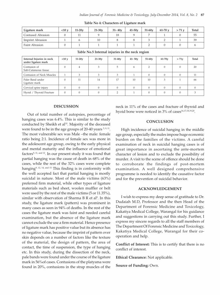
Indian Journal of Forensic Medicine & Toxicology. July-December 2014, Vol. 8, No. 2 87
Table No 4: Characters of Ligature mark
Ligature mark <10 y 11-20y 21-30y 31- 40y 41-50y 51-60y 61-70 y > 71 y Total
Contused Abrasion 0 11 9 18 9 7 1 0 55
Imprint Abrasion 1 6 10 8 8 3 2 1 39
Faint Abrasion 0 1 1 1 1 2 0 0 6
Table No.5 Internal injuries in the neck region
Internal Injuries in neck <10 y 11-20y 21-30y 31-40y 41- 50y 51-60y 61-70y > 71y Totalunder ligature mark
Contusion of 0 4 3 5 6 2 0 0 20Sub-Cutaneous tissue
Contusion of Neck Muscles 1 3 3 3 1 0 0 0 11
Paler Band under 0 11 14 17 10 10 3 1 66Ligature mark
Cervical spine injury 0 0 0 0 0 0 0 0 0
Hyoid / Thyroid Fracture 0 0 0 2 1 0 0 0 3
DISCUSSION
Out of total number of autopsies, percentage ofhanging cases was 6.4%. This is similar to the studyconducted by Sheikh et al10. Majority of the deceasedwere found to be in the age groups of 20-40 years 5, 9, 11.The most vulnerable sex was Male –the male: femaleratio being 2:1. Incidence of female sex was more inthe adolescent age group, owing to the early physicaland mental maturity and the influence of emotionalfactors9, 11, and 12. In our present study it was found thatpartial hanging was the cause of death in 68% of thecases, while the rest of the 32% cases were completehanging9, 12, 14, and 15. This finding is in conformity withthe well accepted fact that partial hanging is mostlysuicidal in nature. Most of the male victims (63%)preferred firm material, while other types of ligaturematerials such as bed sheet, woolen muffler or beltwere used by the rest of the male victims (5 or 11.35%),similar with observation of Sharma B R et al9. In thisstudy, the ligature mark (pattern) was prominent inmany cases as seen in 94% of deaths. In the rest of thecases the ligature mark was faint and needed carefulexamination, but the absence of the ligature markcannot exclude the use a firm material. Hence presenceof ligature mark has positive value but its absence hasno negative value, because the imprint of pattern overskin depends on a number of factors like the textureof the material, the design of pattern, the area ofcontact, the time of suspension, the type of hangingetc. In this study, during the dissection of the neck,pale bands were found under the course of the ligaturemark in 56%of cases. Contusions of the platysma werefound in 20%, contusions in the strap muscles of the
neck in 11% of the cases and fracture of thyroid andhyoid bone were noticed in 3% of cases16,17,18,19,20.
CONCLUSION
High incidence of suicidal hanging in the middleage group, especially the males impose huge economicburden on the families of the victims. A carefulexamination of neck in suicidal hanging cases is ofgreat importance in ascertaing the ante-mortemcharacter of lesion and to exclude the possibility ofmurder. A visit to the scene of offence should be doneto corroborate the findings of post-mortemexamination. A well designed comprehensiveprogramme is needed to identify the causative factorand for the prevention of suicidal behavior.
ACKNOWLEDGEMENT
I wish to express my deep sense of gratitude to Dr.Dudaiah M.D, Professor and the then Head of theDepartment of Forensic Medicine and Toxicology,Kakatiya Medical College, Warangal for his guidanceand suggestions in carrying out this study. Further, Iexpress my sincere regards to all the staff members ofThe Department Of Forensic Medicine and Toxicology,Kakatiya Medical College, Warangal for their co-operation and help.
Conflict of Interest: This is to certify that there is noconflict of interest.
Ethical Clearance: Not applicable.
Source of Funding: Own.
21. Bharthi Rama Rao--85--.pmd 7/24/2014, 10:04 AM87

88 Indian Journal of Forensic Medicine & Toxicology. July-December 2014, Vol. 8, No. 2
REFERENCES
1. Toro K, Krostof I, Keller E. Incompletedecapitation in suicidal hanging- report of a caseand review of the literature. J For Leg Med2008;15(3): 180-184.
2. Brock A, Griffiths C. Trends in Suicide by methodin England and Wales, 1979- 2001. Health StatQuart 2003; 20: 7-18.
3. Lalwani S, Sharma GS, Kabra SK, Girdhar S,Dogra TD. Suicide among children andadolescents in South Delhi (1991-2000). Indian JPediatrics 2004; 71:701-3.
4. Mohanty S, Sahu G, Mohanty MK, Patnaik M –‘Suicide in India: a four year retrospective study’–‘J Forensic Leg Med. 2007May; 14(4):185-9.
5. Bastia BK, Kar N. – ‘a psychological autopsystudy of suicidal hanging from Cuttack, India:focus on stressful life situations’ –‘Arch SuicideRes. 2009; 13(1):100-4.
6. Reddy KSN. The Essentials of Forensic Medicine& Toxicology. 29thed. India: Medical BookCompany; 2010. P.296-297.
7. Nandy A. Principle of Forensic Medicineincluding Toxicology. 3rd ed. India: Central BookAgency; 2010. P.517-518.
8. L Buris. Forensic Medicine, Springer, Budapest,1993. p 225.
9. Sharma B R, Harish D, Singh V P. Ligature Markon neck: How informative? A study paper, Journalof Indian Academy of Forensic Medicine (2005).Volume 27(1) pp 10 -15.
10. Sheikh MI and Agarwal SS. Medico-legalimplications of hyoid bone fracture- a studypaper. Journal of Indian Academy of ForensicMedicine 2001. Volume 23. Number 4. pp 61 – 63.
11. G.A. Sunil Kumar Sharma, O.P.Murthy,T.D.Dogra. “Study of ligature marks in asphyxialdeaths of hanging and strangulation”.International Journal of Medical Toxicology and LegalMedicine 2002; 4(2):21-24.
12. Uzün I, Büyük Y, Gürpinar K. – ‘Suicidal hanging:fatalities in Istanbul retrospective analysis of 761autopsy cases.’ – ‘J Forensiceg Med. 2007 Oct;14(7):406-9.
13. Kanchan T, Menezes RG – ‘Suicidal hanging inManipal, South India - victim profile and genderdifferences’ – ‘J Forensic Leg Med.2008 Nov; 15(8):493-6.
14. Green H, James RA, Gilbert JD, Byard RW. –‘Fractures of the hyoid bone and laryngealcartilages in suicidal hanging’ – ‘J ClinForensicMed. 2000 Sep; 7(3):123-6.
15. Khokhlov VD – ‘Calculation of tension exertedon a ligature in incomplete hanging’ – ‘ForensicSci Int. 2001 Dec 1; 123(2-3):172-7.
16. Nikolic S, Micic J, Atanasijevic T, Djokic V, DjonicD – ‘Analysis of neck injuries in hanging’ – Journalof Forensic Med Pathology. 2003Jun; 24(2):179-82’2008 Oct; 27(4):259-62.
17. Uzün I, Büyük Y, Gürpinar K. – ‘Suicidal hanging:fatalities in Istanbul retrospective analysis of 761autopsy cases.’ – ‘J Forensiceg Med. 2007 Oct;14(7):406-9.
18. Naik SK, Patil DY. Fracture of hyoid bone in casesof asphyxial deaths resulting from constrictingforces round the neck. J Indian Acad Forensic Med2005; 27: 971-3.
19. Joshi Rajeev, Chanana Ashok, Rai Hakumal.Incidence and Medico-legal importance ofAutopsy study of fracture of Neck structure inhanging and strangulation. Medico legal update(2007), volume 7(4) Pp 105-109.
20. Jani CB, Gupta BD. An autopsy study ofparameter influencing injury to osteo-cartilaginous structures of neck in hanging.International Journal of Medical Toxicology and LegalMedicine (2002). Volume No. 5(1). pp 4 – 7.
21. Bharthi Rama Rao--85--.pmd 7/24/2014, 10:04 AM88

Indian Journal of Forensic Medicine & Toxicology. July-December 2014, Vol. 8, No. 2 89DOI Number: 10.5958/j.0973-9130.8.1.001
A Five Year Retrospective Study of Victims of SexualOffences in Jaipur Region
Yadav A1, Meena R L2, Pathak D3, Kothari N S4
1Assistant Professor, Department of Forensic Medicine, NIMS Medical College, Jaipur, 2Senior Demonstrator,3Assistant Professor, Department of Forensic Medicine, SMS Medical College, Jaipur, 4Professor, Department of
Forensic Medicine, NIMS Medical College, Jaipur
ABSTRACT
This study aims to determine the magnitude and sociodemographic characteristics of victims ofsexual offences reported at state tertiary care and referral hospital, Jaipur. The present study wascarried out on 229 victims of sexual offences between the periods of 2008-2012. All the cases of sexualoffences reported during the study period were cases of natural sexual offences committed on femalevictims except three cases of unnatural sexual offences, committed on male victims. The ages ofvictims in our study ranged from three to 40 years. The most affected age group was victims between11-20 years (53.7%) followed by 21-30 years (25.7%). All the five admitted cases of sexual offenceswere children belonging to the age group 0-10 years. Majority of the victims were Hindu females(86.0%), unmarried (71.6%) educated up to secondary level (33.6%) and from middle socio economicstatus(52.8%). The majority of the victims were students (51%) followed by housewives (16.5%) anddomestic workers (16.1%). The assailant knew the vast majority of victims (90.4%) and remainingwere the strangers.
Keywords: Sexual Offences, Female Victims, Assailant, Sociodemographic Characteristics
INTRODUCTION
Of all the crimes, these crimes are the mostbarbarous and humiliating. 1Sexual assault includes allthose victimizations involving unwanted sexualcontact occurring between the victim and assailant.Sexual assault is one of the offenses mostunderreported to law enforcement. 2-4Sexual assaultfollows the iceberg phenomenon. The amount of itvisible to our eyes is much less than the amountbeneath the burden of social ethics. Most of the casesof crime related to sex go unannounced due to thesocial stigma related to such events. Rape is a seriousoffence, causing tremendous physical, mental andemotional trauma to the victim e.g. HIV infection,STD’s, mental depression, social isolation etc. It hasboth short and long-term effects leading to morbidityin the victims. 5 According to W.H.O., 1 out of 3 womenworldwide has experienced rape or sexual assault.6 It
is a global problem contributing towards increase inmorbidity in those who are at a higher risk.7
Securing evidence after sexual offence is animportant task for physicians.8 Poor medical evidenceis often responsible for low conviction rate.9 Thoroughand timely medical examination of the victim mayprovide extraordinary evidence to prove the event andestablish evidence against the accused. With the spreadof education and professionalism amongst thefeminine sector of our society, women are now movingout of the protected environment provided to themearlier making them more prone to such crimes.Therefore, there is a need for us to analyze the presentscenario of sex related crimes against women andchildren in our part of the world to look for ways tocheck the growing rate of such unlawful events. Thisstudy initiates to assess the magnitude of sexualoffences in this region and to study the profile of
22. Anil Yadav--89--.pmd 7/24/2014, 10:04 AM89

90 Indian Journal of Forensic Medicine & Toxicology. July-December 2014, Vol. 8, No. 2
victims as exact data of recent times for our region isunavailable. The role of a forensic expert in cases ofsexual offences can be remarkable to aid the victim inadministration of justice.
MATERIAL AND METHOD
This retrospective study was conducted in theDepartment of Forensic Medicine and Toxicology of astate tertiary care and referral hospital, Jaipur fromJanuary 2008 to December 2012. The inclusion criteriaconsisted of all cases of alleged history of sexualoffences brought to the Department of ForensicMedicine & Toxicology by competent investigationauthority and also all the cases with alleged history ofsexual offences admitted in state tertiary care andreferral hospital. All the cases, who (the victim/ herparent or guardian, when the victim was below 12years of age) did not consent for medico-legalexamination regarding sexual assault were excludedfrom the study. All the victims of sexual offences whofulfilled the inclusion criteria irrespective of their agewere taken for study. For indoor patients, the medico-legal examination of victim was conducted at bedsideafter taking valid consent of the patient and fulfillmentof all legal formalities. A total of 229 victims of sexualoffences fulfilled the inclusion criteria and their datawas analyzed.
OBSERVATION AND RESULTS
This study was carried out at the Department ofForensic Medicine and Toxicology, state tertiary careand referral hospital, Jaipur over a period of five yearsfrom 2008 to 2012. During the study period, 241 victimsof sexual offences were considered for medico-legalevaluation. Out of the victims of sexual assault broughtat our centre, twelve victims refused to consent for themedical examination for study purpose and hencewere not included in the study. A total of 229 caseswere included in the study and medico-legalexamination carried out for each case as per thestandard protocol after taking valid consent andfulfilling the legal formalities. The observations andresults for each case noted and the data was analyzed.Out of these victims of sexual offence, three were malesand rest were females. All the males were victims ofsodomy. The age of victims of study group ranged from3 year to 40 years. There were 18 (7.8%). victims whowere under 10 years of age Maximum number ofvictims fell in the range of 11-20 years i.e. the adolescentwho just attained majority; including a total of 123victims (53.7%). 59 (25.7%) victims were between 21-30 years of age and 29 (12.6%) victims were in 31-40years age group. No victim was more than 40 years ofage (Table-1).
Table 1. Age and year wise distribution of victims.
Year Age Group 2008(44) 2009(43) 2010(46) 2011(43) 2012(53) TOTAL
0-10 3 3 5 4 3 18
11 20 23 25 26 23 26 123
21-30 9 12 11 12 15 59
31-40 9 3 4 4 9 29
22. Anil Yadav--89--.pmd 7/24/2014, 10:04 AM90

Indian Journal of Forensic Medicine & Toxicology. July-December 2014, Vol. 8, No. 2 91
Maximum victims i.e. 140 (61.1%) were educatedupto primary and secondary levels, 43(18.7%) wereeducated above secondary level. Surprisingly, only 46
victims (20.0%) out of 229 were illiterate. Majority ofvictims (86%) were Hindu, unmarried (71.6%) andfrom a middle socioeconomic status (52.8%) (Table-2)
Table 2. Socio demographic profile of victims of sexual assault
YEAR 2008 2009 2010 2011 2012 TOTAL
Educational Status
Illiterate 11 8 9 6 12 46
Primary 9 7 12 17 18 63
Secondary 16 17 19 11 14 77
Above secondary 8 11 8 7 9 43
Religion
Hindu 36 40 39 38 44 197
Muslim 5 4 6 4 7 26
Sikh 1 0 2 1 0 4
Christian 0 0 1 0 1 2
Marital status
Unmarried 32 34 28 30 40 164
Married 11 10 13 12 15 61
Divorcee 1 0 0 1 2 4
Socio Economic status
Low 23 18 20 19 16 96
Middle 20 25 26 24 26 121
Higher 1 0 0 0 1 2
Majority of the victims i.e. 117 (51%) were students,38 (16.5%) were housewives, 37(16.1%) were domesticworkers; another twenty one (9.1%) were labourersand sixteen (7%) were employed in privateorganizations. Among them four victims were sexuallyassaulted by their employer (Table-3)
Table 3. Distribution of victims according toOccupation.
Occupation Total (%)
Student 117 (51.0)
Housewife 38(16.5)
Domestic Worker 37(16.1)
Labourer 21(9.1)
Private employee 16(6.9)
Grand Total 229 (100)
In only 22 (9.6%) cases the assailant was a strangerto the victim. In all the rest 207(90.4%) cases, thevictim knew the assailant. Out of them, theirneighbors sexually assaulted 101 (44.1%) victims.Among the 229 cases, where the victim knew theassailant; there were ten (4.3%) victims of incest(Table-4).
Table 4. Distribution of victims according to Relationto Assailant
Relation to Assailant Total (%)
Neighbor 101 (44.1)
Acquaintance 59(25.7)
Close friend 37(16..1)
Stranger 22(9.6)
Family members 10(4.36)
Grand Total 229 (100.00)
DISCUSSION
Sexual assault is a neglected public health issue inmost of the developing countries. Only 10-50 percentof female victims reports sexual assault.10It is a well-understood fact that quite a small percentage of sexualassaults are really reported. The underreporting ofcases of sexual assault is mainly due to social stigma,prejudice with regard to the chances of marriage, beingconsider promiscuous and responsible for incident,attendant humiliation and embarrassment caused byappearance and cross-examination in court, publicityimpress, risk of losing the love and respect of society,friends and that of her husband if married.11 This study
22. Anil Yadav--89--.pmd 7/24/2014, 10:04 AM91

92 Indian Journal of Forensic Medicine & Toxicology. July-December 2014, Vol. 8, No. 2
was carried out on 229 victims of sexual offences whowere brought to state tertiary care and referral hospital,Jaipur. Previously studies had been carried out on 38victims (Sagar et al,1992)12; 80 victims (Bhardwaj etal,1995)13; 90 victims (Sarkar S C et al 2001-02)1 and 32victims (Jain R et al 2006)14. In our study, only threemale victims of sexual offences reported at our hospitalduring the study period. Though in other studies also,they have reported that majority of the victims werefemales, still male victims have also been registered.In our study, 98.6% victims were females. The numberof female victims among study group population havebeen reported to be 86% (Grossin et al 2003)15; 88.9%(Sarkar S C et al), 100% (Jain R et al).All the malesreported were victims of sodomy. The incidence ofnatural sexual offence far exceeds the unnatural sexualoffence in our country.16
The ages of victims in our study ranged from threeto 40 years. No age is safe from rape. Cases are onrecords where the infants or elderly women above 70years of age have been the victims of rape.17 This ismore or less similar to between six and 25 years (JainR et al) and a little variable from between 4 and 66years (Sarkar S C et al). The most affected age groupin our study was between 11-20 years (53.7%) followedby 21-30 years age group which had 59 victims (25.7%).This is quite similar to that reported in other studies.Maximum victims had been reported to be in 16-20years (Sarkar SC et al, 2001-02); 21-25 years (Jain R etal,2006) and 15-20 years (Du Mont et al 2000) 18. Fromthis we can reach to a conclusion the females in thesexually promiscuous age group, i.e. 11 to 30 years arethe most prone age group for these crimes. Moreover,the adolescents and the just major females i.e. thosebetween 11 to 20 years are the most vulnerable victimsto this category of crime.
In our study, majority i.e. 33.6% victims were thosewho had finished or were about to finish educationupto secondary level. As compared with a study ofIndia we got similarity in the number of illiteratevictims being 28.9% (Sarkar S C et al 2001-02). Themajority of the victims in our study were studentscontributing to half of the whole population. The classof students covering the majority of victims in ourstudy indicates towards the changing values of oursociety, as many of these victims are those who hadsexual activities with their acquaintance afterabsconding together. However, the proportion ofdomestic workers who became victims of sexualassault is more or less same in the two studies, being16.1% (our study) and 15.75% (Jain R et al 2006).
However, we again vary in the proportion ofhousewives who became victims of these assaults, asit is 16.5% in our study being much lower than 43.5%(Jain R et al,2006). Probably this variation is due todifference of rural and urban proportions in the studygroup of both the studies. In our study, the majority ofvictims were unmarried being 71.6% which is similarto 81.1% (Sarkar SC et al 2001-02) and a little higherthan 65.2% (Dumont et al, 2000).
In our study, in 90.4% cases of sexual assault, thevictim knew the assailant and in only 9.6% cases, theassailant was a stranger. This is a little more than 87.5%known assailants (Jain R et al, 2006) and 81% (SarkarSC et al,2001-02); and quite high as compared to 69.7%acquaintances as assailants and 25.6% strangers(Fimate et al, 1998)19. Malhotra et al have reported thatrape by person acquainted with victim is common forgirls less than 10 years of age. Rape or assault bystrangers increases significantly with age. In addition,variation may result due to different lifestyle and socialcustoms.
In our study, 90.4% assailants knew the victims,44.1% were neighbours; 25.7% were acquaintances;16.1% were close friends, 4.36% were family members(in 7 cases assailant was a blood relative and in 3 casesthey were relatives of the in-laws side). This is quitesimilar to 46.75% neighbours and 15.75% friends (JainR et al, 2006). However our study differs greatly from44.1% acquaintances, 18.8% strangers, 30.8% closefriend and only 1.8% neighbours (Sarkar SC et al, 2001-02). This variation may have resulted due to thepopulation and time variation of victims due to thedifference in the regions of study.
CONCLUSION
Sexual assault is traumatic at the time it occurs,but it also may have long lasting negative effects onphysical health.14 The Indian scenario is even morealarming with reports of sex crimes making headlinesalmost every day.15 Majority of the victims were in theage group of 11 to 20 years, Hindu females belongingto urban regions. Most of the victims were unmarriedand students who were assaulted, known to theirassailants. In a maximum number of cases, theassailant had a friendly relationship with their victimand had sexual activities after absconding together.This study highlights the importance of addressingsexual offences as a public health issue and focuses onthe demographic profile of victims in an urban area.Stress should also be laid down on providing sex
22. Anil Yadav--89--.pmd 7/24/2014, 10:04 AM92

Indian Journal of Forensic Medicine & Toxicology. July-December 2014, Vol. 8, No. 2 93
education to school students of teenage to increaseawareness regarding medical and legal issues relatedto such events. Probably, spread of education andwisdom among adolescents may provide suppressionin vulnerability of these age groups to such torturingevents.
ACKNOWLEDGEMENT
The authors are thankful to Dr A.M. Dixit AssociateProfessor,Community Medicine and Dr. R.K GahlotPHOD,Forensic Medicine and Toxicology, NIMSMedical College, Jaipur for kindly help to carry outthis work.
No Conflicts of Interest.
No Source of Funding.
Ethical Clearance was sought from appropriateauthorities before the commencement of this work.
REFERENCES
1. Sarkar SC, Lalwani S, Rautji R., Bhardwaj DN,Dogra TD. A study on victims of sexual offencesin South Delhi URL: www.medind.nic.in(accessed on 13.01.2013).
2. Green, T., Adriana Ramelli and Mavis Mizumoto.2001. “Patterns Among Sexual AssaultVictimsSeeking Treatment Services.” Journal ofChild Sexual Abuse 10:89-108.
3. Greenfeld, L. Sex Offenses and Assailants: AnAnalysis of Data on Rape and Sexual Assault. BJSreport, NCJ 163392, February 1997
4. Maguire, K. and Ann L. Pastore, eds. Sourcebookof Criminal Justice Statistics 2000. U.S.Departmentof Justice, Bureau of Justice Statistics. Washington,DC. USGPO, 2001.
5. Jewes R, Sen P, Garcia MC. Sexual Violence. In :Krug EG, Dalberg LL, Mercy JA, Zwaid, OrzanoR eds. World Report on Violence and Health.Geneva: World Health Organization; 2002.147-82.
6. Statistics of Sexual assault. RAINN calculationbased on 2000 National crime VictimizationSurvey. Bureau of Justice Statistics, U Departmentof Justice.
7. Mukhopadhyay PP, Singh O. Psychiatricassessment of victims of sexual assault: Apreliminary study from Burdwan MedicalCollege, Burdwan, West Bengal. J Indian AcadForensic Med 2009; 31(3): 237-42
8. Crime in India, National Crime Record Bureau,Ministry of Home Affairs, New Delhi, 2002
9. Okonkwo JEN, Ibeh C. Female sexual assault inNigeria. Int Journ Gynaecol & Obstet 2002 Sep; 78(suppl) 1 : S 105- 10.
10. Malhotra N, Sood M. Sexual assault, a neglectedpublic health problem in the developed world,International Journal of Obstetrics andGynaecology,71;257-258,2000.
11. Beebe,D.K. Emergency management of the adultfemale rape victim. American family physician,43:2041-2043;1991.
12. Sagar MS. Sexual offences in South Delhi. JForensic Med Toxicol. 1992; 9: 8-11.
13. Bhardwaj DN, Sharma RK, Sagar MS, Murty OP.Study of sexual offences victims in South Delhi. JForensic Med Toxicol. 1995; 12 (3&4): 33-4.
14. Jain R, Mathur PN, Kothari NS, Mathur P.Medico-legal evaluation of Sex Assault casesadmitted at Sardar Patel Medical College & PBMHospital, Bikaner, India. Medico legal update 2006Jan-Jun; 8(1).
15. Grossin C, Sibille I, Grandmaison GLDI, BanasrA Brion F,Durigon M. Analysis of 418 cases ofsexual assault. Forensic Sci Int. 2003; 131: 125-30.
16. Vij K. Textbook of Forensic Medicine andToxicology : Principles and Practice. 3rd ed. NewDelhi (India) : Elsevier; 2005. 460-78.
17. Sexual Offences – I, II & III. In: Lyon’s MedicalJurisprudence and Toxicology. Dogra & Rudraeds. 11th ed. Delhi: Delhi Law House;2008. p 491-539.
18. Du Mont J and Parnis D. Sexual assault and legalresolution; Querying the medical collection ofForensic evidence. Med Sci Law. 2000; 19(4):779-92.
19. Fimate L, Devi M. An analytical study of rape inManipur. Int Journ Med Toxicol and legal Med. 1998;1:1-2.
20. Cloutier S, Martin SL, Poole C. Sexual assaultamong North Carolina women: prevalence andhealth risk factors J Epidemiol Community Health2002;56: 265–271.
21. Mukhopadhyay PP, Singh O. Psychiatricassessment of victims of sexual assault : Apreliminary study from Burdwan MedicalCollege, Burdwan, West Bengal. J Indian AcadForensic Med 2009; 31(3): 237-42.
22. Anil Yadav--89--.pmd 7/24/2014, 10:04 AM93

94 Indian Journal of Forensic Medicine & Toxicology. July-December 2014, Vol. 8, No. 2DOI Number: 10.5958/j.0973-9130.8.1.001
A Study of Different Position of Mastoid Foramen Relatedof Skull Bone
Dimple S Patel1, Rohit C Zariwala2, Bharat D Trivedi3
1Associate Professor, Department of Anatomy, 2Associate Professor, Department of Forensic Medicine, AMC METMedical College, Ahmedabad, 3Ex. Professor & Head, Department of anatomy, Smt. NHL Municipal Medical
College, Ahmedabad
ABSTRACT
The present study analyzed craniofacial bone variations in dried skulls in terms of gender and mastoidforamen (MF) were assessed dried skulls by direct observation and using a Mitutoyo caliper. MFoccurrence was higher in right male skulls, and multiple foramina were present in 60% of maleskulls (both sides), and in 36.4% of female skulls (both sides). Craniofacial bone variations, consideredto be radiological landmarks, have been studied since the beginning of the century using dried skullsand cadavers. These pitfalls are important bone landmarks used in the planning and execution ofanesthetic and surgical procedures. The identification and recording of craniofacial variations isimportant in the preparation of anesthetic blocks in surgical procedures and in the evaluation ofregional neurovascular anatomy, to avoid misinterpretations in planning. This study confirms theexistence of significant morphological variations in terms of gender and side in a given population.The anatomy of mastoid foramen is clinically important for surgeons1. In our present study I havefound structural variations of the mastoid foramen. This study will provide insight to our knowledgeof humanity.
Keywords: Mastoid Foramen (MF), Dried skull, Temporal bone, Occipital bone, Sigmoid sinus
INTRODUCTION
Craniofacial bone variations, considered to beradiological landmarks, have been the object of studysince the beginning of the century, using dried skullsand cadavers. They are important in planning andconducting anesthetic and surgical procedures.According to Hanihara and Ishida (2001), discretecranial characteristics may the consequence of anadaptation process to different environments andsurvival patterns that depend on aspects likepopulation size and isolation variables. It is as ofsurgical planning that surveying and recording thesebone variations become important. These efforts mayhelp prevent compromising and injuringneurovascular structures, as in the orbit area, whichhas connections with the central nervous system, nose,paranasal sinuses, face and structures associated to eyefunctions (KARAKAS, BOZKIR and OGUZ, 2002).Apart from this, the recording of bone variations inseveral population groups plays a relevant role in
anthropological and evolutionary studies (BERRY,1975; AGTHONG, HUANMANOP andCHENTANEZ, 2005).
MASTOID FORAMEN
This is a foramen of variable size and position.Usually it appears near the posterior margin of thebone, but it can occur in the suture between thetemporal and occipital bone or within the occipitalbone. It can range in size from quite large to very small.Sometimes it is absent. When this foramen is present,it transmits an emissary vein from outside the skull tothe sigmoid sinus within the skull. A small branch ofthe occipital artery also passes through this foramento vascularize the duramatter.
The mastoid foramen of variable size and positionis traversed by a vein from the sigmoid sinus and asmall dural branch of the occipital artery, frequentlylies near is posterior border2. 40% of mastoid foramenis seen on occipital occipito temporal suture.
23. Dimple 2--94--.pmd 7/24/2014, 10:04 AM94

Indian Journal of Forensic Medicine & Toxicology. July-December 2014, Vol. 8, No. 2 95
Etymology: Mastiod comes from the Greek mastosmeaning breast. Galen used this name to describe aprocess of the temporal bone because he thought itresembled a breast in appearance. Foramen is the Latinterm designating a hole-like opening. It derives fromthe Latin forare meaning to bore or perforate.
Latin: Foramen matoideum.
French: Foramen matiodein.
MATERIALS AND METHOD
In total 100 dried skulls were analyzed. Skulls wereobtained from the Laboratory of Human Anatomy;AMC MET Medical College, Ahmedabad. Skulls thatin one way or another did not afford perfectobservation and measurements, due to damage, wereexcluded. Records were carried out by directobservation and using a Mitutoyo caliper. In foramina,measurements were always carried out starting fromthe center. Data were analyzed by descriptive statistics,expressed as means ± standard deviation of mean. TheStudent’s t test and the Mann-Whitney test were usedto compare measurements of distances. Differencesbetween means were considered significant for P <0.05. The statistical analysis was carried out using the
Biostat 5.00™ software.
Location of MF was recorded based on its positionin the temporal bone, in the temporo-occipital sutureor on the occipital bone, in both sides of each skull.The existence of more than one smaller foramen nearMF was recorded as multiple mastoid foramen (mMF),as described in Berge and Bergman (2001).
The present study is carried out on 100 skulls(temporal bones) from the department of Anatomy atAMC MET Medical College, Ahmedabad, Gujarat byexamining the mastoid foramen of the right and theleft sides of both the sexes.
OBSERVATION AND RESULTS
It was observed that double mastoid foramen waspresent in 2% of the temporal bones on right side and16% on the left side. Single mastoid foramen ontemporal bone was seen in 26% on the right side and16%on the left side. In the occipital bones, singlemastoid process was seen in4% of occipitotemporalsuture on the right and 14% on the left. Mastoidforamen was also absent in 8% on the right and 10%on the left side of both the temporal as well as theoccipital bone. It is presented in table-1.
Table-1 Position of Mastoid Foramen
Serial No Position of Mastoid Foramen Side of Number % agethe Bone of Bones
1 Double mastoid foramen in temporal bone RightLeft 216 432
2 Single foramen on temporal bone RightLeft 2616 5232
3 Single foramen on occipital bone RightLeft 22 44
4 Foramen on occipito-temporal suture RightLeft 414 828
5 Absence of foramen RightLeft 810 1610
The results obtained concerning laterality of MFwere similar to those published by Berge and Bergman(2001), as well as the predominance of mMF on theright side3. Keskil, Gözil and Calgüner (2003) observedemissary mastoid foramina in 78% of skulls. Of thatpercentage, 88.5% were located in the temporo-occipital suture4.
DISCUSSION
1. The present study will be a useful guide whereinthe dural arteriovenous fistulas can be successfullytreated by surgical procedures or embolisation. Thegoal of embolisation is occlusion of the primarydraining vein.
2. Direct transcranial puncture of the meningealarteries through the parietal or mastoid foramencan be done in cases in which conventional arterialnavigation has failed especially when the occipitalartery is tortuous in its course. This method allowsclose access to the arteriovenous venous fistulas.In dural arterio venous fistulas, the arterialanastomosis is enlarged and supplied blood to theentire foramen. Therefore, puncture of the foramenallows access to the artery.
3. A through knowledge of mastoid foramen withdifferent variants is basic and very important tothe surgeons and radiologists before and duringsurgical interventions.
23. Dimple 2--94--.pmd 7/24/2014, 10:04 AM95

96 Indian Journal of Forensic Medicine & Toxicology. July-December 2014, Vol. 8, No. 2
4. Besides surgical importance this study will provideuseful information in human anthropometry.
CONCLUSION
The present study demonstrates the existence ofsignificant morphological variations in craniofacialstructures. The variations observed were related tolaterality and gender, though they also depend on thepopulational group studied. The identification andrecording of craniofacial variations is important in thepreparation of anesthetic blocks in surgical proceduresand in the evaluation of regional neurovascularanatomy, to avoid misinterpretations in planning. Thisstudy confirms the existence of significantmorphological variations in terms of gender and sidein a given population.
Acknowledgement: Nil.
Ethical Clearance: Informed consent was taken.
Source of Funding: Self.
Conflict of Interest: Nil.
REFERENCES
1. Standring S: Gray’s anatomy. ChurchillLivingstone 2005; 39: 468
2. Breathnach A. Frazers anatomy of humanskeleton, Churchill Livingstone 1965, 6:197
3. BERGE, JK. and BERGMAN, RA. Variations insize and in symmetry of foramina of the humanskull. Clinical Anatomy, 2001, vol. 14, n. 6,p. 406-413.
4. KESKIL, S., GÖZIL, R. and CALGÜNER, E.Common surgical pitfalls in the skull. SurgicalNeurology, 2003, vol. 59, n. 3, p. 228-231.
23. Dimple 2--94--.pmd 7/24/2014, 10:04 AM96

Indian Journal of Forensic Medicine & Toxicology. July-December 2014, Vol. 8, No. 2 97DOI Number: 10.5958/j.0973-9130.8.1.001
Estimation of Stature from Index Fingers Length inDavangere District
Shahina1, Vijayakumar BJ2, Nagesh Kuppast3, Dileep Kumar4, Shobha5
1Post Graduate Student; Dept. of Forensic Medicine, 2Prof. and Head, 3Asst. Prof., 4Post Graduate, 5Asst. Prof. Dept ofAnatomy, SSIMS & RC, Davangere
ABSTRACT
Estimation of stature holds a special place in the field of forensic medicine and forensic anthropology.This study was designed to investigate the ability of estimating stature from index finger length. Thestudy was carried out by taking the measurement of index fingers and body height of 250 medicalstudents (125 males and 125 females) of 18 to 25 years of age. The study was carried out in departmentof forensic medicine and toxicology at SSIMS & RC Davangere, Karnataka state, India. Obtaineddata was analysed statistically to establish the relationship between a person's index finger and stature.From given data mean, SEE, correlation coefficient(r). Regression equation and 'P' values wereobtained. A moderate correlation was observed between index finger length and height of a personwhich is statistically highly significant. The present study would be useful for anthropologists andforensic experts.
Keywords: Forensic Anthropology, Stature, Human Identification, Finger Length
INTRODUCTION
Identification of an individual has assumedimportance in almost all spheres of life. In certainsituations, such as putrefied, mutilated, or extensivelycharred body, conventional indicators and routinemethods of identification fail to yield results. Thesituation is worsened when only mutilated andfragmentary remains are available for examinationwhich is not uncommon in today’s world due to massdisasters both natural and manmade e.g. earth quakes,cyclones, tsunamis, flood, terror attacks, bomb blasts,accidents, wars and plane crashes etc. Estimating thestature of a person from such fragmented remainsforms an important tool of identification in suchcircumstances.
Kerley1 states that every body part bears a more orless constant relationship with stature. Simmons et al2
derived formulae to calculate stature with good results,
Corresponding author:ShahinaPost Graduate StudentDept. of Forensic Medicine, SSIMS & RC, DavangereE-mail: [email protected]: - 08197736303
even when only parts of the bone are available.Bhatnagar et al3 used different hand dimensions topredict the stature of an individual in differentpopulations. It is shown in earlier studies that varioushand measurements tend to differ in various ethnicgroups4. Consequently, the formulae designed toestimate stature from various anatomical dimensionsin one population do not apply to another5,6. Manystudies are done to calculate stature from foot length,limb lengths, long bones, but there are only few studieson hand and finger lengths, we are taking index fingerlength to calculate stature of a person. It will be helpfulin conditions in which only hand or part of hand isavailable for identification of a person.
India is a vast country with varied geographicalconditions and stature varies with race, sex, andgeographical locations as quoted above. Therefore, ourstudy examines the relationship of the index fingerlength with stature in and around Davangere district.This study may prove useful in conditions where onlypart of hand is brought for post-mortem examination.
MATERIAL AND METHOD
The present study was carried out in the
24. Shahina--97--.pmd 7/24/2014, 10:04 AM97

98 Indian Journal of Forensic Medicine & Toxicology. July-December 2014, Vol. 8, No. 2
department of forensic medicine and toxicology atSSIMS & RC Davangere. A total of 250 subjects wereincluded in the study, out of which 125 males and 125females within age group of 18 to 25 years. The subjectsincluded in study were healthy individuals free fromany apparent skeleton deformity.
Anthropometric measurements of the index fingerswere taken independently on left and right side of eachindividual. Besides the above measurements, statureof each subject was also recorded. All measurementswere taken in well lighted room. The measurementswere taken by using standard anthropometricinstruments in centimetres to the nearest millimetrein following manner.7
Anthropometric Measurement
Index finger length – It is the distance from the tipof index finger to the proximal crease of the indexfinger.
Instruments : Venire Calliper.
Technique: The measurement was taken instanding position with stabilization of hand on table.The calliper was horizontally placed along the ventralsurface of the left hand. The fixed part of the outer jaw
of the calliper was applied to the proximal crease ofindex finger and the mobile part of the calliper wasapproximated to the tip of the index finger andmeasurement was taken. In the same waymeasurement of the index finger of the right hand weretaken.
Statue: It is the vertical distance between the pointvertex and the heel touching the floor (ground surface).
Technique: The subject was made to stand in erectposture against the wall with the feet axis parallel orslightly divergent and the head balance on neck andthe measurement was taken.
The data was collected, analysed and subjected tostatistical analysis using statistical package for socialsciences (SPSS) to know the correlation of the staturewith the index finger length. The reliability ofestimation of stature from the lengths of index fingerwas determined with the help of ‘p’ value, SEE, r, rSquare and regression equations.
RESULTS
The present study focused on estimation of staturefrom the length of the index finger.
Table No. 1. Showing Mean, Standard Deviation and “P” value of both right and left index finger and height inmales and females.
Males Female
Parameters Mean SD P* Value, sig Mean SD P* Value, sig
RIFL 7.23 0.39 P<0.001 HS 6.68 0.43 P<0.001 HS
LIFL 7.28 0.43 P<0.001 HS 6.72 0.45 P<0.001 HS
Height 171.89 6.66 P<0.001 HS 158.73 5.56 P<0.001 HS
Table No. 2. Showing Correlation Coefficient and Standard Error of Estimation of right and left index finger inboth males and females.
Males Female
Parameter R R square SEE R R square SEE
RIFL 0.34 0.11 6.1 0.54 0.29 4.6
LIFL 0.36 0.13 6.0 0.50 0.25 4.8
Table No. 3: Depicting Regression Equation in both males and females from right and left index finger.
Males Females
Parameter Regression equation Regression equation
RIFL Height = 129.84+5.81 (RIFL) Height = 111.32+7.10 (RIFL)
LIFL Height =131.60+5.60 (LIFL) Height = 116.90+6.22 (LIFL)
Regression equation: stature= value of constant (a) + regression coefficient (b) x index finger length.
Males: Y1 = 129.84 + 5.81(RIFL). Y2 = 131.60 + 5.60(LIFL).
Females: X1 = 111.32 + 7.10(RIFL). X2 = 116.9 + 6.22(LIFL).
24. Shahina--97--.pmd 7/24/2014, 10:04 AM98

Indian Journal of Forensic Medicine & Toxicology. July-December 2014, Vol. 8, No. 2 99
Regression equation with range of height
Males: Y3 = 129.84 + 5.81(RIFL) +12.2 Y4 = 131.60 + 5.60 (LIFL) + 12.
Females: X3 = 111.32 + 7.10(RIFL) + 9.2. X 4 = 116.90 + 6.22(LIFL) + 9.6.
Where Y1 & Y2 = Height in males. Y3 & Y4 = Average height in males. X1 & X2 = Height in females.
X3 & X4 = Average height in females. RIFL = Right index finger length. LIFL = Left index finger length.
DISCUSSION
The identification of commingled mutilated remainis a challenge to forensic experts and hence, a need ofstudies on estimation of stature from various bodyparts in different population groups. Such studies canhelp in narrowing down the pool of possible victimmatches in cases of identification from dismemberedremains.
The present study show that estimation of staturefrom the index finger is highly significant (p<0.001) inboth males and females so it can be used for estimationof stature of a person. The values of ‘r’ Square in malesand females with r Square = 0.11 of RIF in males beingthe lowest and r Square = 0.29 for RIF in females beingthe highest value, depicts that estimation of heightfrom the RIF in females is more significant than males.Standard error of estimation SEE = 6.1 for RIF, SEE =6.0 for LIF in males, SEE= 4.6 RIF & SEE = 4.8 LIF incase of females. This indicates RIF in case of females isbetter predictor of height as compared to LIF and ishighly significant where as in case of males it is leftindex finger which gives better prediction of heightthan right index finger length. Considering allparameter it is in case of females where index fingerlength gives better prediction of stature estimation ascompared to males.
A study was done by Krishan K8 et al on staturefrom IFL and RFL which shows larger significance inmales than females, with higher significance for IFLthan RFL which is contradictory to our study showinghigher significance in females.
A study done by Rastogi9 et al on 500 subjects fromManipal North and South Indian population, showedhigher significance in males as compared to females,which is contradictory to our study showing highersignificance in females.
CONCLUSION
Estimation of stature forms an important parameterto reach to the partial identification of an unidentified
body and dismembered remains. The results of thepresent study indicate that the index finger length canbe efficiently used for estimation of stature. Mostauthors have underlined the need for population-specific stature estimation formulae. In this study wederived a separate regression equation to estimatestature from index finger length for Davangere region.
This study revealed that the IFL can be used withhigh significant values for estimation of stature inSouth Indian population even if only an amputatedhand is found and other body parts are unavailable.The results of this study are however, applicable onlywhen an intact index finger is examined. Such studiescan help in narrowing down the pool of possible victimmatches in cases of identification from dismemberedremains.
DIAGRAMS
24. Shahina--97--.pmd 7/24/2014, 10:04 AM99

100 Indian Journal of Forensic Medicine & Toxicology. July-December 2014, Vol. 8, No. 2
followed the instructions. The article, if published,shall be the property of the Journal and we surrenderall rights to the Editors. We agree to provide the latestfollow up of cases prior to the publication of casereports when requested”.
REFERENCES1. Kerley E R (1977) Forensic Anthropology. In
Tedeschi C.G, Eckert W.G. and Tedeschi LG (Eds)Forensic medicine. Philidelphia, WB SaundersCompany, 1101-15.
2. Simmons T, Jantz R C and Bass W. M. (1990)stature estimation from fragmentary femora: Arevision of the Steele mthod. J Forensic Sci. 35(5),628-36.
3. Bhatnagar DP, Thapar SP and Batish MK. (1984)identification of personal height from thesomatometry of the hand in Punjabi males.Forensic Sci. Int. 24(2), 137-41.
4. Davies BT, Benson AK, Courtney A, Minto I. Acomparison of hand anthropometry of femalesin the three ethnic groups. Ergonomics 1980;23:183–4.
5. Williams PL, Bannister LH, Berry MM, Collins P,Dyson M, Dussek JE. Gray’s Anatomy: Theanatomical basis of Medicine and Surgery. 38 Ed.Churchill Livingstone. New York, 2000. Pages425-436.
6. Athawale MC. Anthropological study of heightfrom length of forearm bones. A study of onehundred Maharashtrian male adults of agesbetween twenty five and thirty years. AmericanJournal of Physical Anthropology 1963;21:105–12.
7. Ghai OP, Singh G. estamation of stature fromhand & phalanges length. J of indian academayof forensic med 2004;26(3):100-106.
8. Krishan K, Kanchan T, Asha N. Estimation ofstature from index finger length in north Indianadolescents population. J of Forensic Med 2012July; 19(5): 285-90.
9. Rastogi P, Kanchan T, Menezes R G,Yoganarasimha. Middle finger length- a predictorof stature in the Indian population. Med Sci. law2009;49:2:123-26.
Acknowlegment: Department of Forensic Medicine,SSIMS & RC, Davangere.
Conflict of Interest: None
Ethical Clearance: Ethical clearance was obtained fromthe Institutional Ethical Committee.
Source of Support: Department of Forensic Medicine,S.S.Institute of Medical Sciences and Research Centre,Davangere. The corresponding author confirms thathe had full access to all the data in the study and hadfinal responsibility for the decision to submit forpublication.
WARRANTY : “The undersigned author / authorshereby declare that the article entitled as“ESTIMATION OF STATURE FROM INDEXFINGERS LENGTH IN DAVANGERE DISTRICT.” isoriginal, neither the article nor a part of it is underconsideration for publication anywhere else and hasnot been previously published anywhere. We havedeclared all vested interests. We have meticulously
24. Shahina--97--.pmd 7/24/2014, 10:04 AM100

Indian Journal of Forensic Medicine & Toxicology. July-December 2014, Vol. 8, No. 2 101DOI Number: 10.5958/j.0973-9130.8.1.001
Crime Investigation by Forensic Expert Based on Teeth
Seetha Ramaiah1, Sanjay Punuri2, V P Kumar Sriperumbuduru2
1Associate Professor, 2Assistant Professor, Department of Forensic Medicine & Toxicology, ASRAM Medical College,Eluru, West Godavari, Andhra Pradesh
ABSTRACT
Forensic odontology is branch of dentistry which deals with the proper handling and examination ofdental evidence with the proper evaluation and presentation of dental findings .The major arena ofactivity of forensic odontology is identification of human beings either living or dead. Areas ofapplication include mass disasters, Criminalities, Cases involving abuse of children elderly, Dentalmalpractice and archaeology. Dental identification1 report should consist of four basic sections likeIntroductory (administrative section), Postmortem evidence section, Ante mortem evidence, Opinion(conclusion section). Estimation of age and determination of sex of the victim or the remains are theimportant guides that help in process of identification. In this process teeth are the most reliabletools. Stages of development of teeth are the most dependable indicators in assessing the age ofvictim. Bite marks produced during assault of children or adults who are frequently associate withsex related crimes and child abuse are also important guides in process of identification.
Keywords: Forensic Odontology, Sex determination of teeth, Microscopic features of teeth in identification
INTRODUCTION
Dental age provides the most accurate indicationduring 18 -20 years of age. Age can be estimated inchildren and in adolescents by means of developmentand eruption of deciduous and permanent teeth up to14 years. For most age estimation methods, thedeveloping teeth are subjectively assessed onradiographs2. Maxillary central incisor proved to be asignificant indicator of chronological age3. After theage of 14, the third molar is the only remaining tooththat is still developing and consequently dental ageestimation methods have to rely on the developmentof this tooth until the age of 23. Strong relation of agewith stages of root development of 3rd molar4 teethhas been showed and suggested that age can bepredicted with a statistical significant result for agesbetween 13 and 23. The staging of third molar crownand root mineralization can be accomplished easilyand non-invasively through evaluation of dentalradiographs. After this period, age determination ismainly done by visual examination, radiographicmethods, and structural changes in teeth and by meansof chemical methods.
DISCUSSION
Various methods utilized for the determination ofage from the dentition are
1. Visual method
2. Radiological examination
3. Histological methods
4. Biochemical methods
Factors used for age determination
Harvey lists the following factors in dental ageestimation
1. Appearance of tooth germs
2. Earliest detectable trace of mineralization
3. Degree of completion unerupted teeth
4. Rate of formation of enamel
5. Clinical eruption of teeth
6. Degree of completion of roots
25. pawan kumar--101--.pmd 7/24/2014, 10:05 AM101

102 Indian Journal of Forensic Medicine & Toxicology. July-December 2014, Vol. 8, No. 2
7. Attrition of crowns
8. Formation of cementum
9. Formation physiologic secondary dentin
10. Transparency of root dentin
11. Gingival recession
12. Discoloration and staining of teeth
13. Influence of disease on eruption
14. Influence of sex on tooth eruption
Gustafson’s method of age determination5
This include six dental changes connected withageing
1. Attrition
2. Apical migration of periodontal ligament
3. Deposition of secondary dentin
4. Cemental opposition
5. Root resorption 6. Transparency of root dentin
Attrition
There are many scoring methods in assesing agingby attrition, but according to different researchers it isnot a reliable index as it is also affected by food habits,par functional habits, missing opposing tooth etc.
Deposition of secondary dentine
The size of the pulp chamber indicates the amountof secondary dentine formation. Moore used pulpdiameter to crown diameter ratio for calculating age6.Kvall SI et al7 have used pulp/root length, pulp/toothlength and pulp root width by periapical radiographsto estimate age. Both studies have not shown accurateage. Paewinsky , Pfeiffer and Brinkmann conducteda study of six teeth (including the maxillary centralincisor) on digital orthopantomograms to correlate themeasurements of pulp cavity with age8. It was foundthat the best correlations between the measurementsand age were found at the cementoenamel junction.
Cemental apposition
Cementum is continuously deposited at the rootend and seen as incremental lines. Many researchers
have used cemental annulations to determine age ofadults.
Dentinal root translucency
Apical zone of translucent dentine may be used assole indicator of age because it is less influenced bypathologic process and environmental factors. It showsasymmetric distribution teeth in the left and right sidesof the jaw. Causes of dentinal root translucency arefatty degeneration of the organic substances in dentine,increased intratrabecular mineralization.
Sex determination
Determination of sex is a great problem for forensicexperts when only fragments of body are recovered.Forensic dentist can determine sex of remains usingteeth in such conditions. Various features of teeth likemorphology, crown size, root lengths arecharacteristics for male and female.PCR amplificationwill assist in accurately determining the sex. Bar codingsystem is a way of transferring data to the computerand huge data can be stored as a record. Bar codingcan be easily incorporated during acrylization of thedenture and thus could be used in individualidentification9.
Measurements of the mandibular ramus tend toshow higher sexual dimorphism, and differencesbetween the sexes are generally more marked in themandibular ramus than in the mandibular body.10 Themean values of parameters like maximum ramusbreadth, minimum ramus breadth, Condylar heightDmaximum ramus height, Projective height of ramus,Coronoid height showed that all dimensions werehigher for males compared to females11.
Estimation of Sex by various methods
Visual or clinical method
Mandibular canines show greatest dimensionaldifference with larger teeth in males than in females.The mesiodistal width of canines of both the jaws issignificantly greater in males than females12.A studyby Kaushal et al. found a statistically significantdimorphism in the mandibular canines in 60 subjectsin a North Indian population, where the mandibularleft canine was seen to exhibit greater sexualdimorphism, 13,14. They also concluded that if the widthof the canine is greater than 7 mm, the probability ofthe sex of the person under consideration being malewas 100%.
25. pawan kumar--101--.pmd 7/24/2014, 10:05 AM102

Indian Journal of Forensic Medicine & Toxicology. July-December 2014, Vol. 8, No. 2 103
Microscopic method from pulp tissue.
Whittaker and co-workers determined sex fromnecrotic pulp tissue. Up to 5weeks sex determinationcan be done with high degree of accuracy. Duffy et alhave shown that Barr bodies and F bodies of Ychromosomes are preserved in dehydrated pulp tissueup to one year and pulp tissue retain sex diagnosticcharacteristics when heated up to 100o for one hour.The study showed that the presence of Barr body inbuccal mucosal cells can be demonstrated with a fairdegree of accuracy using acridine orange confocalmicroscopy. The sex of the individual can bedetermined accurately with other advantages offered,such as the rapidity of processing and screening aspecimen that results in saving of time. It was observedthat in the male samples, the percentage of Barr-body-positive cells ranged from 0-3%. In the female samples,the percentage of Barr-body-positive cells ranged from18-72%, and all the females showed the presence ofBarr bodies15.
ADVANCED METHOD
Sex determination from blood and teeth by PCRamplification reveals specific sequencesin males andfemales, Pulp tissue retain sex diagnosticcharacteristics. Amelogenin found in human enamelas different pattern of nucleotide sequence in malesand females.
Bite mark evidence
Bite marks are defined as marks made by teetheither alone or in combination with other mouth parts.Bite mark is a form of patterned injury, they are mostlyproduced during assault of children or adults who arefrequently associate with sex related crimes and childabuse16. Sexually oriented bites appear to have beeninflicted slowly and deliberately with suction appliedto a tissue by tongue and lips. Child abuse cases areeither anger bites or sexually oriented bites. Selfinflected bites mostly found on fore arms of childrencaused by themselves. Bitemarks are categoricalidentification evidence, the dynamics of biting, theanatomical location of the bite, and failures in woundrecords can introduce distorted images and misleadcrime investigation. The elasticity of polyether16
impression material, casts can compensate for primaryor secondary distortions, so that there is a better degreeof match when positioning the subject’s dental cast.Hence polyether is an alternative impression material
and is an excellent option for creating positive casts ofthe wound for physical dynamic comparison17.
In the presence of clinical signs, such as ecchymosis,lacerations, and abrasions, on the victims in ellipticalor ovoid pattern, a suspicion of bite marks should beconsidered. Bite marks may have a central area ofecchymoses (contusions) caused by 2 possiblephenomena: (1) positive pressure from the closing ofthe teeth with disruption of small vessels or (2)negative pressure caused by suction and tonguethrusting. Bites produced by dogs and othercarnivorous animals tend to tear flesh, whereas humanbites compress flesh and can cause abrasions,contusions, and lacerations but rarely avulsion oftissue18.
Mcdonald Classification of Bite Marks
• Tooth pressure marks are caused by incisal edgesof anterior teeth
• Tongue pressure marks, because of tonguepressure, impression of palatal surfaces of teethor palatal rugae are produced.
• Tooth scrape marks are produced due toirregularities of teeth fractures or restorations
• Complex marks are combination of above typesof marks.
CONCLUSION
There are so many recent advances in forensicodontology sound knowledge of modern techniquesin forensic odontology will enhance the efficacy ofcrime investigation
ACKNOWLEDGEMENTS
Dr.Ch.Lakshmi Kumar, professor, Department ofForensic Medicine, ASRAM medical college, Eluru,Andhra Pradesh.
Conflict of Interest Statement: We certify that thereis no conflict of interest with any financial organizationregarding the material discussed in the manuscript.
Source of Funding: self
Ethical Clearance: No human subjects are involvedin this as it is a review
25. pawan kumar--101--.pmd 7/24/2014, 10:05 AM103

104 Indian Journal of Forensic Medicine & Toxicology. July-December 2014, Vol. 8, No. 2
REFERENCES
1. Harvey W. Dental identification and forensicodontology, London, Henry Kimpton Publ. 1976
2. Ajmal M, Mody B, Kumar G. Age estimationusing three established methods: A study onIndian population. Forensic Sci Int 2001; 122:150-4.
3. Agarwal N, Ahuja P, Sinha A, Singh A. Ageestimation using maxillary central incisors: Aradiographic study. J Forensic Dent Sci 2012; 4:97-100
4. Ajmal M, Assiri KI, Al-Ameer KY, Assiri AM,Luqman M. Age estimation using third molarteeth: A study on southern Saudi population. JForensic Dent Sci 2012; 4:63-5
5. Gustafson G. Forensic Odontology, Staples press,London, P.P 118-39, 1966
6. Moore G.E. Age changes occuring in teeth J.Foren. Sci. Soc., 10: 179-178, 1970.
7. Kvall S.I., Kolltveit K. M., I.Thomsen et. al. Ageestimation of adults from dental radiographs.Foren. Sci.Int.., 74: 175-185, 1995.
8. Paewinsky E, Pfeiffer H, Brinkmann B.Quantification of secondary dentin formationfrom orthopantamograms - a contribution toforensic age estimation methods in adults. Int JLegal Med 2005; 119:27-30.
9. Nalawade SN, Lagdive SB, Gangadhar S A,Bhandari AJ. A simple and inexpensive bar-coding technique for denture identification. JForensic Dent Sci 2011; 3:92-4
10. Humphrey LT, Dean MC, Stringer CB.Morphological variation in great ape and modernhuman mandibles. J Anat 1999; 195:491-513
11. Indira AP, Markande A, David MP. Mandibularramus: An indicator for sex determination - Adigital radiographic study. J Forensic Dent Sci2012; 4:58-62
12. Yuwanati M, Karia A, Yuwanati M. Canine toothdimorphism: An adjunct for establishing sexidentity. J Forensic Dent Sci 2012; 4:80-3.
13. Rao NG, Rao NN, Pai ML, Kotian MS.Mandibular canine index: A clue for establishingsex identity. Forensic Sci Int 1989; 42:249-54.
14. Muller M, Lupipegurier L, Quatrehomme G,Bolla M. Odontometrical method useful indetermining gender and dental alignment.Forensic Sci Int 2001; 121:194-7
15. Reddy D S, Sherlin HJ, Ramani P, Prakash P A.Determination of sex by exfoliative cytologyusing acridine orange confocal microscopy: Ashort study. J Forensic Dent Sci 2012; 4:66-9.
16. Rajendran R, Sivapathasundharam B. Shafer’stext book of Oral Pathology. 6th Ed, New Delhi:Elsevier2009.
17. Fonseca GM, Farah MA, Orellano-Blaskovich SV.Bite mark analysis: Use of polyether in evidencecollection, conservation, and comparison. JForensic Dent Sci 2009; 1:66-72.
18. Kellogg N; American Academy of PediatricsCommittee on Child Abuse and Neglect. Oral anddental aspects of child abuse and neglect.Pediatrics 2005; 116:1565-8
25. pawan kumar--101--.pmd 7/24/2014, 10:05 AM104

Indian Journal of Forensic Medicine & Toxicology. July-December 2014, Vol. 8, No. 2 105DOI Number: 10.5958/j.0973-9130.8.1.001
Postmortem Autolytic Changes in Oral Mucosa:Histological Review
Charan Gowda BK1, C V Mohan2, Hemavathi3
1Reader, Dept Of Oral Pathology, Sri Siddhartha Dental College, Tumkur, Karnataka, 2Director , Dental care and,Research Center, Bengaluru, Karnataka, 3Former Prof. and HOD, Dept of Oral Pathology, Government Dental College,
Bengaluru, Karnataka
ABSTRACT
Following death various changes occur in the human body known as postmortem changes. Thesepostmortem changes begin at molecular level which can be noted microscopically and finally grossly.Postmortem interval (PMI) is the time elapsed since death. Estimation of time since death is still adifficult task even for an experienced forensic pathologist. Gross changes within human body whichoccur following death during the postmortem interval have been routinely used to estimate the timeof death considering different conditions. However, histological changes of skin and oral mucosawhich occur during the postmortem interval have occasionally been used to estimate the time ofdeath under different conditions.
Objective: This study is done to study the histological features of decomposition at cellular level inpostmortem oral mucosal tissue, skin and its appendages.
Method: Electronic and hard data is searched and studied to understand the postmortem changes oforal mucosal tissue and skin and its appendages. (regarding cases studied number have placed indiscussion).
Results: Studies conducted for morphological changes of skin and its appendages showed focaldermal-epidermal separation, eccrine duct necrosis, and dermal degeneration. Oral mucosal differentcellular changes like homogenization and eosinopilia, cytoplasmic and nuclear vacuolation, chromatinclumping, pyknosis of nuclei was noted at different time intervals.
Conclusion: PMI determination is essential for narrowing the fields of suspects or possible missingpersons. Histological examination of oral mucosa may be helpful in establishing the Postmorteminterval more precisely. PMI determination is essential for narrowing the fields of suspects or possiblemissing persons.
Keywords: Necrosis, Postmortem interval, Pyknosis, Decomposition, Homozenization
Corresponding author:Charan Gowda BKReaderDept of Oral Pathology, Sri Siddhartha Dental CollegeTumkur, KarnatakaEmail ID [email protected]@yahoo.co.in
INTRODUCTION
Cells are the structural and functional units of allliving organisms. They take in nutrients, convert thesenutrients into energy, carry out specialized functions,
and reproduce as necessary. These activities are carriedwithin the cell by instructions that are stored insidethe cell itself. Although human body composed ofmany different cells, certain basic characteristic arealike. They perform their function normally till theavailability of proper concentration of oxygen, glucose,different ions, amino acids, fatty substances, and otherconstituents available in environment.1,2 usually cellsconstantly participate in their environment where theyencounter physiologic stresses and also pathologicstimuli. To overcome these cells may undergoadaptation but they preserve their viability and
26. sriniwas reddy--105--.pmd 7/24/2014, 10:05 AM105

106 Indian Journal of Forensic Medicine & Toxicology. July-December 2014, Vol. 8, No. 2
function. When the stress is harmful it may lead toinjury of the cell. To certain extent cell can withstandinjury and return to normal functioning. When cell failsto withstand stress or when severity of stress is highcell fails to adapt which result in the cell death/necrosis. Necrosis usually results in degradative actionof enzymes within the cell. Series of change occursfollowing cell death which can be viewed undermicroscope.
Light microscope which magnifies objects remainsto be a basic tool for pathologist for visualization ofcell structure. Technical improvements in modern dayspermit the visualization of fine details of cell.3 Studiesthat have been conducted showed the changes thatoccur in cellular level following death progress withtime which can be observed under microscope.
Cell death
Necrosis is a Greek word which literally meansdeath or dead body. Necrosis and apoptosis are twodifferent entities which represent two different formof cell death. Apoptosis was the term coined by Kerret al which was accompanied by several morphologicalchanges in cell. 4 Various changes that were noted inapoptotic cell includes rounding-up of the cell,retraction of pseudopodes, reduction of cellularvolume (pyknosis), chromatin condensation, nuclearfragmentation (karyorrhexis), classically little or noultrastructural modifications of cytoplasmicorganelles, plasma membrane blebbing.5 TheNomenclature Committee on Cell Death (NCCD)proposes that in necrotic cell death morphologicalchanges like gain in cell volume, swelling of organelles,plasma membrane rupture and subsequent loss ofintracellular contents can be noted.4,5 Necrotic cells firstshow clumping of chromatin and swelling oforganelles. This is followed by swelling of the cell, andthe rupture of nuclei, mitochondria, and the plasmamembrane.6,7 Cell death due to lack of oxygen showmicroscopically eosinophilia, cell may show glassyappearance, vacoulation of cytoplasm. Nucleus mayshow karyolysis, pyknosis and karryorrhexis.5 Electronmicroscopically the necrosed cell may showdiscontinuities in plasma and organelle membranes,marked dilation of mitochondria with the appearanceof large amorphous densities, disruption of lysosomes,intracytoplasmic myelin figures, and profound nuclearchanges culminating in nuclear dissolution. 5
Postmortem interval
Postmortem interval is the time elapsed betweendeath and corpse discovery. Generally forensicpathologists and forensic entomologist’s determine thetime since death in cases of shorter postmortemintervals while forensic anthropologist determinestime since death in cases with longer postmortemintervals.8
Normally a series of changes occur in the humanbody following death. These changes vary in anorderly fashion along the different time interval.Various internal and external factors may accelerateor retard the decomposition rate. Understanding thepostmortem changes and confounding factors mayhelp a forensic pathologist to establish more accuratePMI and limit the time frame of death which may behelpful in the field of forensics. Generally the processof putrefaction and autolysis begins soon followingdeath. Putrefaction is associated with microbes andoccurs in moist climate. Presence of green discolorationof the body, gas production with associated bloating,skin slippage and a foul odor can be noted.9 Autolysisrefers to self digestion, in case of autolysis cell getsdestructed due to the action of its own enzyme.Common postmortem changes applied in this days forestablishing time of death are rigor mortis, livor mortis,and algor mortis, which progress on a relatively setschedule. Although various factors have to beconsidered during establishing time of death all bodiesdecompose to some extent which can be notedmicroscopically. Various bio-chemical andimmunological studies have been carried in moderndays to establish more precise PMI.
DISCUSSION
Post mortem interval or the time elapsed fromdeath until discovery and medical examination of thebody is one of the important tasks in the field offorensics. Till today unless death has been witnessedestablishing the time of death is extremely difficult.Various research studies have been conducted ondifferent tissues to establish the PMI but still accuracyin that particular issue is not possible. It is necessaryin the field of forensic to fix the time of death becauselonger the time interval between the death andexamination of body more the chance of losing thereliable data. Various physico-chemical changes occur
26. sriniwas reddy--105--.pmd 7/24/2014, 10:05 AM106

Indian Journal of Forensic Medicine & Toxicology. July-December 2014, Vol. 8, No. 2 107
in body following death. This changes progress in anorderly fashion at different time interval. Some of thecommon changes which have been used clinically inestablishing time of death are, algor mortis, rigormortis, livor mortis, putrefaction, adipocere,mummifaction and maceration.10
Additionally biochemical markers which includesprotein fractions, urea, creatinine, glucose, iron,potassium, calcium, enzymes, immunohistochemicaldetection of insulin in pancreatic â-cells, the myo-albumin fraction and the level of strontium-90 calciumanalogs were conducted none found to be fruitful.11
Morphological changes that a cell undergoesduring necrosis can be noted with the help ofmicroscope. Very little research work has been doneon the morphological changes of different cells withinbody. Kovarik C et al12 conducted study on threeindividuals for the postmortem gross and histologicchanges of the skin. In their study they showed focaldermal-epidermal separation, eccrine duct necrosis,and dermal degeneration. They conclude that studyon large scale sample may be useful in estimating thetime of death in the early postmortem interval.
Bardale R V et al13 studied the histologic changesof the skin and appendages in the early PMI. Theyshowed progressive morphological changes of skin inthe early postmortem period. The epidermis and thedermis appeared normal for six hours after death, andafter this period, degenerative changes began.Following six hours after death, degeneration beganin the dermis, and by the end of eighteen hours, thedermis began to disintegrate. They stress towards themorphological changes in the postmortem periodwhich can be observed under microscope andconcludes morphological changes of skin may assistin determining time of death in early postmortemperiod. sweat Cingolani M et al14 studied for post-mortem autolytic alterations in the skin at cellular andsubcellular levels.
The process of decomposition was noted in the oralmucosal tissue. Gururaj and sivapathsundaram15
conducted a study on postmortem gingival sample toassess the cytoplasmic and nuclear changes inpostmortem gingival sample which was fixed between10hrs-20hrs. postmortem gingival sample showednuclear vacoulation along the spinous and superficial
layers. Finally they concludes that cellulardecomposition appears within twenty four hours.
Pradeep G et al16 in their study collectedpostmortem gingival sample and showed cellulardecomposition at different time interval. Their studyshowed different cellular changes like homogenizationand eosinopilia, cytoplasmic and nuclear vacuolation,chromatin clumping, pyknosis of nuclei at differenttime intervals. They concludes that study on large scalemay help to establish time of death by examining thecellular decomposition.
From the histological studies conducted it wasnoted that various cellular alterations occurs followingdeath. The research work conducted showed asignificant association between time intervals anddegenerative changes. Within their study they foundvarious cytoplasmic and nuclear changes.Cytoplasmic changes include cytoplasmic vaculation,homogenization and eosinophilia. Nuclear changesinclude, pyknosis, nuclear clumpping, nuclearvacoulation.
Various research was conducted on variouschemical substance within human body forestablishing PMI none of them found to be useful inforensics. Morphological changes that were observedamong the different cell in the body as a result ofautolysis and putrefaction appears to be morepromising for establishing PMI. However postmortemhistologic changes within the cells of human ifanalyzed systemically may be helpful in the field offorensics where a pathologist can establish thepostmortem interval more accurately.
Establishing time of death or postmortem intervaldetermination is basic requirement in the field offorensics when death has not been witnessed. In deathscene investigation it is necessary to establish the cause,manner, and time since death. With progression of timesoft tissue decomposition begins and it will be difficultto establish the time of death/ less accuratepostmortem interval will be determined. Usually indeath investigations, discovery of corpse is oftendelayed because of intentional disguise by a suspector death occurring in a remote area to name few. Insuch cases obtaining tissue preferably oral mucosatissue and visualizing for autolytic changes of tissueunder microscope may become helpful for postmorteminterval determination.
26. sriniwas reddy--105--.pmd 7/24/2014, 10:05 AM107

108 Indian Journal of Forensic Medicine & Toxicology. July-December 2014, Vol. 8, No. 2
Necessary factors that a forensic pathologist to beconsidered during establishing PMI are,
1. Age and sex of the individual.
2. Temperature and humidity.
3. Cause of death.
CONCLUSION
In death investigations when foul play is suspectedor the identification of the unknown person a PMIdetermination is essential for narrowing the fields ofsuspects or possible missing persons.
Histological examination of oral mucosa which ismore preserved in case of fire, traumatic death whereexternal injuries are severe and in case of death underwater where tissue details have been lost may behelpful in establishing the Postmortem interval moreprecisely. A study on large scale samples consideringthe temperature, age and sex is necessary forestablishing time of death more precisely which maybe very helpful in the field of forensics. The forensicpathologist may obtain an approximate indication ofthe time of death with a simple, rapid and cheaptechnique.
Conflict of interest: This study is done to study thehistological features of decomposition at cellular levelin postmortem oral mucosal tissue, skin and itsappendages.
Source of Funding: Self funding
Ethical Clearance: Obtained
Acknowledgement: Dr. Rudramurthy RMO andSenior Consultant in Forensic Department DistrictGovernment General Hospital Tumkur.
REFERENCES
1. Cooper GM; Hausman RE; The Cell Molecularapproach; 5th edition;Sinauer Associates INC;2-22.
2. Guyton AC; Hall JE; Text book of MedicalPhysiology; 11th edition; Elsevier Saunders; 3-19.
3. Kumar; Abbas; Fausto; Robbins and CotranPathologic Basis of Disease; 7th edition; Elsevier;2004.
4. Kimberly McCall; Genetic control of necrosis –another type of programmed cell death; CurrOpin Cell Biol; 2010 December ; 22(6): 882–888.
5. Kroemer, G. et al; Classification of cell death:recommendations of the nomenclaturecommittee on cell death; 2005; Cell Death Differ;12; 1463–1467
6. BowenI, . D et al; Programmed cell death duringmetamorphosis in the blow-fly Calliphoravomitoria Microsc Res T ech; 1996; 3 4: 202-217.
7. Rochow VBM; Jarvilehto J; Cytologicalpostmortem changes in highly specialized cellssubjected to different causes of necrosis; 1997;Cytologia 62: 265-270.
8. Allaire M T; Postmortem interval (pmi)determination at three biogeoclimatic zones insouthwest Colorado; A thesis Submitted to theGraduate Faculty of the Louisiana StateUniversity and Agricultural and MechanicalCollege; 1997-2002.
9. Presnell E S; http://emedicine.medscape.com/article/1680032-overview.
10. http://www.dundee.ac.uk/forensicmedicine/notes/timedeath.pdf
11. Kikuchi K et al;HMGB1 A new marker forestimation of postmortem interval; Exp TherMed. 2010 Jan-Feb; 1(1): 109–111.
12. Kovarik C et al; Gross and Histologic PostmortemChanges of the Skin; The American Journal ofForensic Medicine and Pathology; 2005; 26 (4);305-308
13. Bardale R V et al; Evaluation of HistologicChanges of the Skin in Postmortem Period; Am JForensic Med Pathol; Mar 2001; 1-5
14. Cingolani M et al; morphology of sweat gland indetermining time of death; Int jour of leg med;1994; 107; 132-140i
15. Gururaj; Sivapathsundaram B; postmortemfindings in gingiva assessses by histology andexfoliative cytology; Journal of oral andmaxillofacial pathology; 2004; Vol 8 (1); 18-21.
16. Pradeep GL; Histological assessment of cellularchanges in gingival epithelium in ante-mortemand post-mortem specimens; Journal of ForensicDental Sciences / July-December 2009; Vol 1( 2);60-64.
26. sriniwas reddy--105--.pmd 7/24/2014, 10:05 AM108
Indian Journal of Forensic Medicine & Toxicology. July-December 2014, Vol. 8, No. 2 109DOI Number: 10.5958/j.0973-9130.8.1.001
A Socio-Demographic Profile of Fatal Burn Deaths inJaipur City, India
Punia R K1, Yadav A2, Meena D K3
1Professor and Head of the Department, 2Senior Demonstrator, 3Final Year Resident, Department of Forensic Medicine SMS Medical College, Jaipur
ABSTRACT
Deaths due to burn are some of the most challenging fatalities, both from the investigative and theautopsy aspect. The aim of this study was to record and evaluate the causes and the magnitude of thefatal burn injuries prospectively from 1st July 2012 to 30th June 2013.During this period, a total of3572 cases were autopsied, of which 425 (11.9%) cases of burn received in the mortuary of SMSHospital, Jaipur. The majority of deaths (82.3%) occurred between 11 and 40 years of age group witha preponderance of females (60.7%). The Flame burns were seen in 87.7% of the victims, followed byElectrical burns(7.7%). The maximum number of the burn victims 252(67.5%) died due to flameburns which were caused by kerosene oil. The majority of burn incidents were accidental (78%) innature followed by suicidal (19%) deaths. The percentages of burns (TBSA) over 40% were observedin most of the cases (90.6%). According to the size 38.8% patients had 61-80% of body surface burnsand 27.3% had more than 80% burns.The majority of deaths occurred within a week (53.4%) andmost of the victims died from septicaemic shock (55.3%) followed by hypovolumic shock (31.5%).
Keywords: Fatal burn, Flame burns , Electrical burn ,Septicaemic shock, TBSA
INTRODUCTION
A burn is an injury to the skin or other organic tissueprimarily caused by heat or due to radiation,radioactivity, electricity, friction or contact withchemicals. The injury represents an assault on allaspects of the patient, from the physical to thepsychological. It affects all ages, from babies to elderlypeople, and is a problem in both the developed anddeveloping world. Burn injuries are among the mostdevastating of all injuries and a major global publichealth crisis. 1,2 Burns are the fourth most common typeof trauma worldwide, following traffic accidents, falls,and interpersonal violence.3,4
Corresponding author:R K PuniaProfessor and HODDepartment of Forensic Medicine, SMS MedicalCollege, JaipurEmail id: [email protected]
About 45% of the mortality in burns patients iscaused by septicaemia.5 Burns are a major problem inthe developing world. Mortality in the developingworld is much higher than in the developed world.Most burns are due to flame injuries. The mostinfrequent burns are those caused by electrocution andchemical injuries.6 Burns and death due to burnscontinue to remain an important public health andsocial problem in India. The incidence is particularlyhigh among the ‘‘young married females”.7,8 belongingto lower socio-economic groups.9 Hence, this studywas done to know the magnitude and the socio-cultural factors of the problem of burns, so that aneffective prevention measures could be suggested forreducing the incidence of fatal burns.
MATERIAL AND METHOD
The present study is carried out in the departmentof Forensic Medicine, SMS Medical College & Hospital,Jaipur, from 1 July 20012 till 30 June 2013. 425 burncases were autopsied in the mortuary of the Forensic
27. Punia--109--.pmd 7/24/2014, 10:05 AM109

110 Indian Journal of Forensic Medicine & Toxicology. July-December 2014, Vol. 8, No. 2
Medicine Department. The various epidemiologicalcharacteristics of the cases were obtained and analyzedwith respect to the age-gender distribution, maritalstatus, socioeconomic status, place of occurrence,survival period, body surface area, cause of death frompanchayatnama papers, autopsy reports, theinvestigating officers and the relatives of the deceased.The data was recorded, compiled and analyzedstatistically. An informed consent was obtained fromrelatives of each victim prior to his/her inclusion inthe study.
OBSERVATION AND RESULTS
A total no of 3572 autopsies were done in theDepartment of Forensic Medicine during the studyperiod. Out of which 425 (11.9%) burn cases wereautopsied . The youngest patient was two and a halfyear and the oldest was 90 years of age. Maximumvictims (62.5%) were in the age group of 21-40 yearsage group in which females are more affected thanmales while in 41-60 age group, males are moreaffected than females. Females (60.7%) were morecommonly affected than males (39.3%) (Table-1).
Table 1. Demographic distribution of burn deaths.
Age group(in years) Male Female Total no. Percent %of cases. (Male : Female)
0-10 5 4 9 55.6:44.4
11-20 14 70 84 16.6:83.4
21-30 66 113 179 36.8:63.2
31-40 39 48 87 44.8:55.2
41-50 20 16 36 55.6:44.4
51-60 16 3 19 84.2:15.8
61-70 6 2 8 75:25
71-80 1 0 1 100:0
81-90 0 2 2 0:100
TOTAL 167 258 425 39.3:60.7
Table 2. Distribution of burn cases according to marital status.
Maximum numbers of burns victims were married (74%). Amongst those, maximum number of burns victims were females i.e. 197(63%). 112(26%) were unmarried burns victims and amongst those 61 (54.4%) were females (Table-2).
Marital status Male Female Total No.(%)
Married 116 197 313 (74)
Unmarried 51 61 112 (26)
Table 3. Area wise distribution of burn cases.
Out of 425 victims, 67% patients were from rural and 33% from urban dwellings ( Table 3) .
SEX Rural(%) Urban(%) Total (%)
Male 111(66.4) 56 (33.6) 167 (100)
Female 175 (67.8) 83 (32.2) 258 (100)
TOTAL 286 (67) 139(33) 425 (100%)
Table No 4. Distribution of patients with respect to manner of burn in relation to sex.
Accidental burning was observed in 78% followed by suicidal (19%) and homicidal burning only in 3% cases. Female preponderanceis observed in all manner of burn deaths.
Manner Male Female Total no. (%)
Accidental 134 198 332(78)
Suicidal 32 48 80(19)
Homicidal 1 12 13(3)
Total 167 258 425(100)
27. Punia--109--.pmd 7/24/2014, 10:05 AM110

Indian Journal of Forensic Medicine & Toxicology. July-December 2014, Vol. 8, No. 2 111
Table 5. Distribution of patients in relation to cause of burn.
The most common type of burn were flame burn in 87.8% of cases followed by electric burn in 7.7% cases (table 5). Flame burn wasmost common in all age groups (Table-5).
Types No. of cases.(%)
Flame burn 373 (87.8)
Electrocution 33 (7.7)
Chemical burn 12 (2.8)
Scald burn 7 (1.7)
Total 425( 100)
Table 6. Distribution of Burn cases in relation to Cause of Death.
The major cause of death was septicaemic shock (55.3%) followed by hypovolemic shock (31.5%), neurogenic shock(5.4%) andtoxaemia (5.3%).
Cause of death No. of cases Percentage(%)
Septicaemic shock 235 55.3
Hypovolemic shock 134 31.5
Neurogenic shock 23 5.4
Toxaemia 22 5.3
Acute renal failure 11 2.5
Total 425 100
Table No. 7. Distribution of victims according to Socioeconomic status.
Majority of victims (68.5%) belonged to low socioeconomic status in the present study(Table-7).
Socioeconomic status No. of cases Percentage (%)
Low 291 68.5
Middle 105 24.7
Higher 29 6.8
Total 425 100
Table 8. Distribution of victims in relation to place of occurrence.
The most common place of burn for female was in –laws home (78.3%) and for male is workplace (64%). (Table-8).
Place of occurence Male (%) Female(%) Total
In- laws home 1(0.6) 202(78.3) 203(47.7)
Own home 17(10.2) 14(5.4) 31(7.3)
Outdoor 42(25.2) 31 (12) 73(17.2)
Workplace 107(64) 11( 4.3) 118(27.8)
Total 167(100) 258(100) 425(100)
Table 9. Distribution of cases according to TBSA and duration of survival
The present study shows that 90.6% of the 425 victims sustained more than 40% of total body surface area (TBSA) burns. Maximumnumber of deaths (32%) occurs within a week of the incidence of burn, 15.8% of deaths occurred in more than 2 weeks post-burnincidence. There was a positive correlation between the TBSA and Duration of survival (Table-9).
%( Percentage of burn) Upto 6 h 7-24 h 1 week. 1-2 week >2 Week TotalNo(%) No(%) No.(%) No.(%) No.(%) No. %
0-20 0 0 1 2 1 4(0.9)
21-40 0 1 8 14 7 31(7.3)
41-60 2 11 24 42 25 104(24.5)
61-80 8 21 49 59 34 165(38.8)
81-100 13 35 54 14 0 116(27.3)
Total 23(5.4) 68(16) 136(32) 131(30.8) 67(15.8) 425(100)
27. Punia--109--.pmd 7/24/2014, 10:05 AM111

112 Indian Journal of Forensic Medicine & Toxicology. July-December 2014, Vol. 8, No. 2
DISCUSSION
Severe burn is a major public health issue indeveloping nations. This makes it the 4th leading causeof injuries after motor vehicle collisions, falls, andviolence. This has been attributed partly toovercrowding and an unsafe cooking situation.10
Overall, nearly 60% of fatal burns occur in SoutheastAsia with a rate of 11.6 per 100,000.11
Burn deaths are an important public health problemin a developing country like India. In India, about700,000 to 800,000 people per year sustain significantburns, though very few are looked after in specialistburn units. Part of this high rate is related to unsafekitchens and loose-fitting clothing typical to India.12 Itis estimated that one-third of all burns in India are dueto clothing catching fire from open flames.13Intentionalburns are also a common cause and occur at high ratesin young women, secondary to domestic violence andself-harm.14 Burning of married women in India is amajor concern for the Government, law enforcingauthorities, the judiciary, the police and forensicexperts all over the country.
In our study females 61% were more commonlyaffected than males 39%. This is in concordance withreports in the literature by other researchers 1-5-17. SRamcharan et al18 in their study had found females57.1% while as males 42.9%. Ashish K Jaiswal et al19 intheir study had found females 70.3% and males 29.7%,Anuradha Rajput et al20 in their study had foundfemales in 60% and males in 40%. This female sexpreponderance could be possibly because of their moredomestic involvement as they tend to spend most oftime near fire and also dowry deaths.
In our study majority of patients 67% were fromrural background while as only 33 from urbandwellings, Ashish K et al in their study had found67.3% of females and 58% of males from ruralbackground which are consistent with ourobservations.This may be explained on the basis ofpoverty, overcrowding and lack of proper safetymeasures in rural background.
In present study the most common cause of burnwas accidental (78%), the second common cause wassuicidal (19%) and homicidal (3%). Ashraf F et al21 intheir study had observed 89.1% of burn injury asunintentional, suicidal(4.3%) and homicidal(2.6%).This
could be attributed to the carelessness of victims whilehandling fire.
In our study, majority of females (78.3%) sufferedburn injuries at their in-law”s home. In contrast, 64%males suffered burn at work place. The high incidenceof burn in case of females (83.7%) inside the homebecause of their domestic activities and dowry deathsprevailing in our region. Our finding are consistentwith the studies of Ashraf et al who in their study hadfound home (91.4%) as the most common place ofburn, S Ramacharan et al who in their study hadobserved (62%) of burn occurring at home and AshishK Jaiswal et al6 who also had observed home (85%) asmost common place of burn. Other authors reportedsimilar observation-92% 22, 86% 8, 67% 23, 71% .24 It alsofocus on carefulness during their work in kitchen whilehandling the cooking equipment.
In our study, 373 patients (87.8%) had flame burns,34 (7.7%) had electric burn, 12 (2.8%) had chemicalburn followed by 7(1.7%) cases of scald burn. Flameburn accounts majority of case which is consistent withreports in literature by Subrahmanyam M , Ashish KJaiswal et al, Dhiraj buchade et al who had reportedthe similar findings in their studies. Electrical burns(7.7%) are second most common in our study whichhad male predominance which is not consistent withthe study of Gowri Shankar et al 25 where scald burns(9.3%) were the second most common.
Most of the victims (68.5%) in our study belongedto low socioeconomic group. This result is consistentwith the Subrahmanyam M and Gupta et al. Theincidence of burn injuries decreased with the uplift inthe economic status of the individual. This may be dueto the use of cheap and unsafe pressure stoves, openfires, adulterated kerosene, financial problems, floor-level cooking.In our study, septicaemic shock (55.3%)was major cause of death followed by hypovolemicshock (31.5%). Harish dasari et al26 also reportedsepticaemia (84%) as a major cause of death in theirstudy.
Married females 197(76.3%) most common victimsof present study followed by married males 116(69.4%)which was consistent with studies of H.M. Mangal etal, Usama b Ghaffar et al 27 and Dhiraj buchade etal.28Kerosene (67.5%) is main inflammable agentamong different sources of fire in the present study.
27. Punia--109--.pmd 7/24/2014, 10:05 AM112
Indian Journal of Forensic Medicine & Toxicology. July-December 2014, Vol. 8, No. 2 113
This may be explained on the basis that kerosene ischeap, easily available and widely used as a cookingfuel by people of the low socioeconomic group in India.Similar reports had been reported in studies fromIndia. 27,29
In the present study, the majority (90.6%) of thevictims had more than 40% of total body surface area(TBSA) burns indicating the incompatibility with lifeeven at a tertiary care centre. In the current study, 53.4%cases died within a week, 30.8% cases between 1-2week signifying that the burns are rapidly fatal.Similarly Virendra et al30also reported death fromburns within a week in 60.8% victims. Vilasco andBondurand31 in their study reported 40% burn deathsbetween 3 and 7 days of the incident. These findingshighlight that burn injuries are a serious public healthproblem with alarmingly high mortality andmorbidity.
CONCLUSION
Burn profile closely reflects the socioeconomicstatus of a country. Burn injuries are a major publichealth concern and are associated with significantlyhigh mortality in India. Majority of the burns werecaused by domestic accidents and are thereforepreventable by adequate safety measures and healtheducation especially for flame injuries occurring in thehome environment.
Acknowledgement: Nil
Conflicts of Interest: None declared.
Source of Funding: Nil
Ethical clearance: Not required.
REFERENCES
1. Forjuoh SN. Burns in low- and middle-incomecountries: a review of available literature ondescriptive epidemiology, risk factors, treatment,and prevention. Burns 2006; 32:529.
2. Peck MD, Kruger GE, van der Merwe AE, et al.Burns and fires from non-electric domesticappliances in low and middle income countriesPart I. The scope of the problem. Burns 2008;34:303.
3. World Health Organization. The Global Burdenof Disease: 2004 Update. World HealthOrganization,Geneva,2008,www.who.int/h e a l t h i n f o / g l o b a l _ b u r d e n _ d i s e a s e /GBD_report_2004update_full.pdf (Accessed onApril 02, 2010).
4. Institute for Health Metrics and Evaluation. TheGlobal Burden of Disease: 2010 Update. IHME,Seattle, 2012 (Accessed on July 01, 2013).
5. Dokter J. Boxma H, Oen IM. The mortality andthe causes of death at a burn centre. Burns 2008;34: 1103–07.
6. Shehan Hettiaratchy and Peter Dziewulski, ABCof burns BMJ 2004; 328; 1366.
7. Jayaraman V, Ramakrishnan KM, Davies MR.Burns in Madras, India: an analysis of 1368patients in 1 year. Burns 1993; 19: 334–9.
8. Chaurasia AR. Mortality from burns indeveloping countries. Burns 1982; 9: 184.
9. Sharma BR, Harish D, Sharma V. Kitchenaccidents viz-a-viz dowry deaths. Burns 2002; 28:250–3.
10. Peck, MD (2011 Nov). “Epidemiology of burnsthroughout the world. Part I: Distribution andrisk factors”. Burns : journal of the InternationalSociety for Burn Injuries 37 (7): 1087–100.
11. Herndon D (ed.). “Chapter 3: Epidemiological,Demographic, and Outcome Characteristics ofBurn Injury”. Total burn care (4th ed.). Edinburgh:Saunders. p. 23.
12. Ahuja, RB; Bhattacharya, S (2004 Aug 21). “Burnsin the developing world and burn disasters.”.BMJ (Clinical research ed.) 329 (7463): 447–9.
13. Gupta (2003). Textbook of Surgery. JaypeeBrothers Publishers. p. 42. ISBN 978-81-7179-965-7.
14. Peck, MD (2012 Aug). “Epidemiology of burnsthroughout the World. Part II: intentional burnsin adults.”. Burns : journal of the InternationalSociety for Burn Injuries 38 (5): 630–7.
15. H.M. Mangal, Akhilesh Pathak, J.S.Rathod: TheFire is Both “A Blessing and Scourge to themankind” JIAFM, 2007 October-December-29(4):75-76.
16. Memchoubi, H.Nabachandra: A study of Burndeaths in Imphal. JIAFM, 2007-29(4)131-134.
27. Punia--109--.pmd 7/24/2014, 10:05 AM113
114 Indian Journal of Forensic Medicine & Toxicology. July-December 2014, Vol. 8, No. 2
17. Sharma BR, Sharma V and Harish D: Sucides inNorthern India-causes, methods Used andprevention thereof, Med.Sci.Law, 2003, 43(3); 221-229.
18. S. Ramcharan, S. Dass, S. Romany, F. Mohammed,T. Ali, M. Ragbir Epidemiology Of Adult BurnsIn North Trinidad . The Internet Journal of ThirdWorld Medicine 2003; 1(1).
19. Jaiswal AK, Aggarwal H,Solanki P,LubanaPS,Mathur RK, Odiya S. Epidemiological andsocio-cultural study of burn patients in M. Y.Hospital, Indore, India. Indian Journal of PlasticSurgery, 2007; 40(2): 158-163.
20. Anuradha Rajput K.P.Singh, Vijay Kumar, RishiSexena, R.K.Singh. Antibacterial resistance ofaerobic bacteria isolates from burn patients intertiary care hospital. Biomedical Research 2008;19 (1):1-4.
21. Ashraf F. Attia, Aida A. Sherif, Ahmed M. Mandil,M. Nabil Massoud, Mervat W. Abou-Nazel andMoustafa A. Arafa. Epidemiological and socio-cultural study of burn patients in Alexandria,Egypt. Eastern Mediterranean Health Journal,1997,3(3): 452-461.
22. Subrahmanyam M. Epidemiology of burns in adistrict hospital in Western India. Burns.1996;22:439–42.
23. Gupta M, Guta OK, Yaduwanshi RK, UpadhyayaJ. Burn epidemiology: The pink city scene. Burns.1993;19:47–51.
24.. Dhillon K, Shekon HS, Mishra VK. Study of burndeaths in and around Shimla District.J PunjabAcad Forensic Medicine and Toxicol 2005;46(2)105-10.
25. Shankar G, Kalburgi B.E, Hunshikutti S. Analysisof burn injuries admitted in a tertiary careteaching hospital in Bagalkot, Karnataka, India.Medicolegal Update 2012, Vol 12 (2):62-65.
26. Dasari H, Kumar A, Sharma BR.BurnSepticaemia- The leading cause of BurnMortality.JPAFMAT 2008; 8(2):10-16.
27. Ghaffar UB, Hussain M and Rizvi SJ:ThermalBurn: An Epidemiological Prospective Study. JIndian Acad Forensic Med,2008,30(1) 10-14.
28. Buchade D, Kukde H, Dere R, Savardekar R:Pattern of burn cases brought to morgue, SionHospital Mumbai: A Two Year Study. J IndianAcad Forensic Med,2011,33(4)309-10.
29. Kumar V, Tripathi CB. Fatal accidental burns inmarried women. Legal Medicine 2003; 5: 139–145.
30. Virendra K, Manoj KM, Sarita K. Fatal burns inManipal area: a10 year study.J Forensic LegMed 2007;14:3–6.
31. Vilasco B, Bondurand A. Burns in Abidjan CoteD’Ivoire.Burns1995;21:291–6.
27. Punia--109--.pmd 7/24/2014, 10:05 AM114

Indian Journal of Forensic Medicine & Toxicology. July-December 2014, Vol. 8, No. 2 115DOI Number: 10.5958/j.0973-9130.8.1.001
Troponin: the Valuable Assay in Post MortemInvestigation of Ischemic Heart Disease
Anita Verma1, Shaista Saiyad2, Pratik Patel3
1Associate Professor, Dept. of Physiology, 2Asst. Professor Dept. of Physiology, 3Professor and Head Forensic MedicineDept., Smt NHL Municipal Medical College, Ellisbridge, Ahmedabad Gujara
ABSTRACT
Sudden, unexplained and unexpected but natural deaths are more common in adults than it can bepresumed with no age or sex exemption. It is found more common in younger one. History orcircumstances are nonspecific to give cause of death. In such type of incidences meticulous postmortemexamination is necessary with various laboratory investigations like histopathology, bacteriology,biochemistry, serology and toxicology. Amongst natural sudden deaths, coronary atherosclerosisand/or its effect(s) is the most common cause. In such cases of sudden death Meticulous & detailedAutopsy is necessary with all above cited laboratory investigations. In cardiac death, Troponin is thebest biomarker to detect cardiac damage as there is increase in Troponin in blood after cardiac damage& Troponin levels if raised are highly specific for cardiac damage or injury. In our study, we aimed tofind out the role of Troponin assays to establish the cardiac damage at the time of autopsy.
Keywords: Autopsy, Cardiac Injury, Coronary Atherosclerosis, Sudden Death, Troponin
INTRODUCTION
One of the objectives of the postmortemexamination is to know the cause of death and itsmanner, whether natural or unnatural. However inmany instances it is too difficult to find out the causeof death particularly in sudden unexpected andunexplained death. Here it requires detailed andmeticulous examination involving several disciplineslike microbiology-serology, biochemistry, toxicology,histopathology, radiology etc. It also requires analysisand interpretation of all the findings collectivelyconsidering gross external as well as internal findingson the dead body during post mortem examination.There are various causes for sudden natural deathsinvolving different vital system or organ. Amongstthese various causes for natural deaths, greaterpercentage is related to cardiac disease. The coronary
Corresponding author:Anita VermaAssociate ProfessorDept. of Physiology, 2nd Floor Smt NHL MunicipalMedical College, Ellisbridge, AhmedabadGujarat-380006Phone: ((M)9824061850, (R)079-26760202,(O)079-26576275
atherosclerosis and its effects play major role in suddendeath due to cardiac disease. Myocardial infarction, ismyocardial cell death due to cardiac ischemia resultingafter the rupture of an atherosclerotic plaque incoronary resulting in platelet aggregation or clottingor emboli, leading to complete blockage. Sometimesthere is too small interval between occlusion & deathto develop macroscopic or microscopic changes unlessalternative cardiac circulation is established with theeffective collaterals. Here no evidence of myocardialinfarction is found. Conversely, no recent block is seenin coronary in a person suddenly died of stenosis as aresult of atherosclerosis. 1 Even one of the earliesthistopathological finding of myocardial infarction dueto coronary atherosclerosis i.e. eosinophilia of musclecytoplasm is clear at about 6 hours of onset of infarctionin living. However we can see changes for myocardialischemic damage on heart only after 24 hours providedthe person is alive. So in almost all cases of suddencardiac death due to coronary block or stenosis /narrowing, it is not possible to get any evidences ofmyocardial infarction. In such type of cases ,particularly in death preceded by complain of chestpain, a rapid assay for cardiac Troponin I [proteinreleased due to damage to cardiac muscle] often servesas a valuable marker to give important useful data
28. Anita Verma--115--.pmd 7/24/2014, 10:05 AM115

116 Indian Journal of Forensic Medicine & Toxicology. July-December 2014, Vol. 8, No. 2
supportive of the cardiac cause of death in suspectedsudden cardiac related deaths. Measurement of thisbiomarker is useful in the autopsy where we suspectcardiac lesion but that cannot be established by routinemeans of histopathological study.
There are three isoforms for Troponin I (TnI), outof which, two are present in skeletal muscle and theother is present only in cardiac muscle. The cardiacisoform (cardiac Troponin), with a molecular weightof 24,000 Da, is larger than the other isoforms as itcontains an extra 32 amino acid N-terminal peptide.The rest of the protein has greater than 40%dissimilarity in its amino-acid sequence comparedwith skeletal muscle TnI.2-3-4
European Society of Cardiology and AmericanSociety of cardiology acknowledge that cardiacTroponin I and T has supplanted CK-MB as theanalytes of choice for diagnosis of cardiac damage.5
Troponin C does not have cardiac specificity and isnot used in assays for determining cardiac damage6.Since last 5 decades it is known that there is increasein transaminase activity with myocardial infarction.Hence Creatinine, one of the biomarkers for cardiacmuscle damage played a major role in such patientstill last few years. It has also issued new criteria thatelevation in biomarkers are fundamental to thediagnosis of acute myocardial infarction2,4 becausesymptoms are atypical or nonexistent and even ECGchanges are either absent or nonspecific5,8. Guidelinerecommends a level above the 99th percentile in areference population as the discriminatory value buta new generation of sensitive assays for cardiacTroponin with a 10% coefficient of variation for thelevels below the 99th percentile has been introduced inrecent past. Again using Troponin I assay, diagnosisof cardiac damage is evident within 3 hours of onset.Cardiac Troponin I is found very low or is undetectedin otherwise normal person. However Troponinelevation may help to make an early diagnosis but isnot always prognostic9-10.
In a case of sudden death or “found dead” personin hospital or autopsy Troponin assay could serve as alaboratory confirmation of myocardial damagewithout gross naked eye changes of cardiac damageor positive histopathological changes. CardiacTroponin I, if raised, can’t be taken alone for thediagnosis of myocardial infarction as cardiac Troponinelevations suggest cardiac damage but can’t be
conclusive of the mechanism, cause or reason.12
However high sensitivity and speciûcity of thisbiomarker makes it still valuable.
Various studies have proved the importance ofTroponin assay to form evidence of sudden death dueto cardiac damage, at postmortem examination ascause of death, without undergoing unnecessarypostmortem examination. We aimed our study todetermine the level of Troponin I cardiac protein, insuspected cases of cardiac death as compared to controlcases. After cardiac injury cardio-Troponin,myofibrillary protein, is released into the blood and isdetected in blood even though nothing positive orsignificant is found of cardiac damage atmorphological or microscopic examination atpostmortem or in ECG during life time prior to death.
MATERIAL & METHOD
In this study 32 consecutive cases brought topostmortem room for autopsy at Smt NHL municipalmedical college,V S. Hospital, Ahmedabad with thehistory of chest pain, typical of cardiac pain, followedby sudden death were included . 32 other cases, ascontrol, were also studied in database where cause ofdeath was known and was other than cardiac [non-cardiac].
After external examination of dead body was over,about 10 ml of blood was collected from heart duringinternal examination of thoracic cavity. Blood wasimmediately placed in airtight bulb and sent to Bio-chemistry department. Care was taken to avoidfreezing and to prevent the haemolysis of blood. Inbiochemistry department blood was immediatelywithdrawn from the bulb and blood sample wassubjected for cardiac Troponin I test. [AxSYMTroponin-I ADV, Abbott diagnostics: Lower Limit ofdetection -0.02/ 99th percentile-0.04/10% coefficientof variation=0.16]11. Here we considered a value of0.04ng/ml in 99 percentile of the population. Value ofmore than 0.04 ng/ml was taken as diagnostic ofcardiac muscle damage. A meticulous postmortemexamination was carried out in each with biochemical,radiological, serological, microbiological,histopathological examinations and chemical analysisfor poisons. In all cases heart was subjected toradiological examination i.e. coronary angiography byinjecting barium sulphate in coronaries to see anycoronary narrowing.
28. Anita Verma--115--.pmd 7/24/2014, 10:05 AM116

Indian Journal of Forensic Medicine & Toxicology. July-December 2014, Vol. 8, No. 2 117
Troponin level may be found increased in someconditions though there is no ischemic heart disease.Following cases are the examples in which it may befound to be increased and were not included in thestudy.
1. Congestive cardiac failure.
2. Trauma to heart.
3. Hypothyroidism.
4. Hypertension.
5. Gross infection.
6. Renal failure.
7. Cardiac surgery.
8. Burns more than one third of body surface area
9. CPR resulting into cardiac contusion.
10. Mitral valve disease.
11. Pericardial effusion.
Cases were designated as cardiac related death(IHD) and control (non-cardiac) cases, from theautopsy finding including ante mortem complain/history of cardiac pain, histopathological examination(included macro as well as microscopic cardiac studyin histopathology department), serologicalexamination, microbiological examination,biochemical examination , toxicology assay andradiological study of coronaries etc.
RESULTS
Table 1. Cases showing positive assay result [i.e.0.04ng/ml].
Cardiac related death Positive Non-cardiac Positiveresult cases result
32 30 32 0
Table 2. Cases showing coronary narrowing in relationto positive Troponin I value, histopathology and
chemical analysis reports.
No. of cases No. of cases No. of cases No. of caseswith coronary showing positive with gross showingnarrowing Troponin I value histopathology poison
changes in otherorgans responsiblefor sudden death
32 30 00 00
From table no: 1 it is observed that among controlcases, where cause of death was known frompostmortem findings & investigations , not a singlecase showed cardiac Troponin I above the upperreference limit. Table no: 2 shows that all 32 cases,where cause of death was cardiac, has obviouscoronary narrowing observed during radiologicalstudy of heart after infusing barium sulphate incoronaries, macroscopic and microscopic examinationof heart and coronaries. Out of all 32 cases withcoronary narrowing 30 cases were positive forTroponin I and any of 32 cases showed no grosspathology in any other vital organs or presence of anypoison responsible for sudden death. Out of 32 cases,studied of deaths related to cardiac damage, positiveresults were observed in 30 (93.75%) cases. The resultsFig. 1. Patent coronaries at post mortem radiology.
Fig. 2. Complete block of right coronary artery at its origin inradiological study.
28. Anita Verma--115--.pmd 7/24/2014, 10:05 AM117

118 Indian Journal of Forensic Medicine & Toxicology. July-December 2014, Vol. 8, No. 2
are more suggestive as significant difference can beseen statistically. [P < 0.0001, chi2 = 20.570]
Table 3. Distribution of cases indicating relationbetween age group and sex [in cardiac death].
Age group Sex Total
Male Female
21-30 yrs 0 (0%) 2 (6.25%) 2 (6.25%)
31-40 yrs 2 (6.25%) 4 (12.5%) 6 (18.75%)
41-50 yrs 4 (12.5%) 4 (12.5%) 8 (25%)
51-60 yrs 4 (12.5%) 0 (0%) 4 (12.5%)
> 61 yrs 10 (31.25%) 2 (6.25%) 12 (37.5%)
Table number 3 shows no significant relation ofTroponin I positive test and sex. Negative results forcardiac Troponin found amongst 2 cases of female sex.It could be due to sudden death within minutes beforegross cardiac muscle damage could have occurred asit takes time to increase the cardiac Troponin bloodlevel.
Our study showed that with the age Troponin Ivalue was found decreasing. On applying Wilcoxonmatched test [4.93] it is observed that person withhigher age group had significantly lower Troponin Ilevel (P < 0.0001). But the strength of relationship isnot significant after Spearman non parametric test (P> 0.05). This may be due to reason that Troponin Ilevel starts increasing after cardiac ischemic damageafter varying interval in different individuals. Severityof the cardiac damage is also related to sudden deathand Troponin level. Troponin I am usually foundpositive at about 3 hours of the injury. Its valueincreases gradually with peak value at about 18-24hours after onset of symptoms. Increase in its level hasrelation with the extent or severity of the damage tocardiac muscles. All these above cited are the reasonsto explain the non-significant relation of Troponin I toage.
DISCUSSION AND CONCLUSION
In our study 32 cases of cardiac related suddendeaths and 32 cases of sudden death due to knowncause but non-cardiac death were studied. They werecategorized as cardiac and non-cardiac from the historyof cardiac pain, gross findings at autopsy with variousinvestigations. The cardiac related deaths with historyof chest pain and obvious narrowing of coronaryvessels, confirmed at radiological as well asmacroscopic study, as a result of atherosclerosis
showed the positive Troponin I value (more than 0.04ng/ml) suggested cardiac muscle injury, and no othernon-cardiac pathology, trauma or poisoning was foundin any of these cases which could had caused suddendeath. Our study showed no significant relation ofcardiac Troponin I , with the age or sex of the deceased.Amongst non-cardiac deaths none of the 32 casesshowed the positive cardiac Troponin I value. So whena patient is brought with history of sudden deathwithout clinical findings of cardiac ischemia, anelevated level of Troponin I above cut off value withconsiderable coronary narrowing due toatherosclerosis, without other non-cardiac disease/pathology, injury and poisoning, the opinion can begiven as a cardiac related death.
When the cross sectional area of coronary arterylumen is reduced by more than 75% as a result ofdiffuse coronary atherosclerotic changes involving theepicardial arteries i.e. the proximal part and narrowingis much more severe, sometimes unexpected suddendeath occur with or without the symptoms. In majorityof such atherosclerotic narrowing, sudden unexplaineddeath occurs as a result of thrombosis, embolism,spasm or inflammation etc, where mechanism of deathis an arrhythmia and the gross morphological changesof myocardial infarction are absent. Though ECG isthe one of the best means of diagnosing acutemyocardial infarction, it may be normal in about 33 %of cases when recorded early enough. In such casesTroponin I, if used, in correlation with symptoms andgross morphological changes in form of coronarynarrowing it is possible to diagnose the cardiac relateddeath due to myocardial ischemic injury. In such typesof death, after ruling out other causes for death,increased cardiac Troponin with evidence of markednarrowing of coronaries at autopsy can be consideredto give opinion about cardiac death.
Troponin I test can be valuable supportive measurein the emergency department or in the postmortemroom where dead body is brought to confirm cardiacdeath. Study of Troponin I assay helps the diagnosisof myocardial infarction resulting in death as a resultof cardiogenic shock.
Acknowledgement: We would like to thank, DeanSmt. NHL Muni. Medical College, Prof & Head, Dept.of Radiology & Prof & Head Dept. of Biochemistry,for their valuable support.
28. Anita Verma--115--.pmd 7/24/2014, 10:05 AM118

Indian Journal of Forensic Medicine & Toxicology. July-December 2014, Vol. 8, No. 2 119
Conflict of Interest: Nil
Source of Funding: Nil
Ethical Clearance: NA, as we have collected data fromDept. of Forensic Medicine, Smt NHL Muni. medicalcollege
REFERENCES
1. Krishan Vij, Textbook of forensic medicine andtoxicology, 5th edition, Elsevier; 100
2. Filatov VL, Katrukha AG, Bulargina TV, et al.Troponin: structure, properties, and mechanismof functioning. Biochemistry (Moscow) 1999;64:1155–1174.
3. Schreier T, Kedes L, Gahlmann R. Cloning,structural analysis, and expression of the humanslow twitch skeletal muscle/cardiac Troponin Cgene. J Biol Chem. 1990; 265(212):47–53.
4. Goldmann BU, Christenson RH, Hamm CW, etal. Implications of Troponin testing in clinicalmedicine. Curr Control Trials CardiovascularMed 2001; 2:75–84
5. Luciano Babuin, Allan S. Jaffe, Troponin: thebiomarker of choice for the detection of cardiacinjury, CMAJ, November 8; 2005, 173(10), 1191
6. Schreier T, Keded L, Gahlmann, Cloning,structural analysis and expression of the humanslow twitch skeletal muscle/cardiac Troponin Cgene.J Biol Chem. 1990; 265:21247-53
7. Alpert JS, Thygesen K, Antaman E, et al.Myocardial infarction redefined- a consensusdocument of the joint European Society ofCardiology/American College of CardiologyCommittee for the redefinition of MyocardialInfarction. J Am Coll Cardio 2000; 36; 959-69
8. Lee TH, Rouan GW, Weisberg MC et al. Sensivityof routine clinical criteria for diagnosingmyocardial infarction within 24 hours ofhospitalization. Ann Intern Med 1987; 106:181-6
9. Wiviott SD, Cannon CP, Morrow DA et al.differential expression of cardiac biomarkers bygender in patients with unstable angina/non STelevation myocardial infarction: a TACTIS-TIMII8, Circulation 2004;109;580-6
10. Venge P, Johnston N, Lagerqvist B et al.Clinicaland analytical performance of the liaison cardiacTroponin I assay in unstable coronary arterydisease, and the impact of age on the definitionof reference limits. A FRISC-II study, clin chem2003; 49; 880-6
11. Luciano Babuin, Allan S. Jaffe, Troponin: thebiomarker of choice for the detection of cardiacinjury, CMAJ, November 8; 2005, 173(10), 1191
12. Scott M. Wells, DVM, DACVECC and MegSleeper, VMD, DACVIM (Cardiology): cardiacTroponin, Journal of Veterinary Emergency andCritical Care 18(3) 2008, pp 235–245
28. Anita Verma--115--.pmd 7/24/2014, 10:05 AM119

120 Indian Journal of Forensic Medicine & Toxicology. July-December 2014, Vol. 8, No. 2DOI Number: 10.5958/j.0973-9130.8.1.001
An Insight Into Additional Factors in Drug Abuse
Seema S Sutay1, Nishat Ahmed Sheikh2
1Associate Professor of Forensic Medicine, RD Gardi Medical College, Surasa, Ujjain, MP, 2Associate Professor ofForensic Medicine, Kamineni Institute of Medical Sciences, Narketpally District Nalgonda. State Andhra Pradesh
ABSTRACT
Drug abuse is a serious public health issue that is trending upward and shows no signs of slowing.The war on drugs is no longer confined to those "illegal" substances such as cocaine, heroin, andecstasy, but now includes legal prescription medications. The abuse of prescription drugs is a seriouspublic health issue and the increasing diversion of prescription drugs for illegal use is causing concernamong government officials. Doctor shopping is a term generated by the addiction and reliance onprescription medications as patients actively seeks doctors who will prescribe certain types ofmedications, usually opiates, depressants, and stimulants. Prescription monitoring programs shallprove to be an effective system in identifying and preventing the diversion and abuse of prescriptiondrugs at both the pharmacy and patient levels.
Keywords: Drug Abuse, Over the Counter Drugs, Prescription Monitoring Programme
INTRODUCTION
Over the counter medication in India is not a newproblem, but has been a concern since antibioticresistance and other health complications are noticed.Patient approach pharmacies without prescription toget these medications, is known as Pharmacyshopping. Over the counter drugs means drugs legallyallowed to be sold over the counter by pharmacist i.e.without prescription of Registered Medical Practioner.Prescription only drugs are those medicines that arelisted in schedules H & X of the drugs and cosmeticrules. Drugs listed in schedule G (Mostlyantihistamines) do not need prescription to purchasebut require the following mandatory text on the label“caution: it’s dangerous to take this preparation exceptunder medical supervision.” Currently non-druglicensed stores( E.G Non- pharmacists) can sell a fewmedicines classified as ‘ Household Remedies’ listedin schedule K of D & C rules in villages whosepopulation is below 1000 subject to certain othercondition8.
Corresponding author:Nishat Ahmed SheikhAssociate Professor of Forensic MedicineKamineni Institute of Medical Sciences, Narketpally,District Nalgonda, State Andhra PradeshEmail ID: [email protected] No: + 91 009390058109
‘Ayurvedic Medicines’ are also regulated by Drugcontrol Authority and Drugs and Cosmetic Rules. Theyare manufactured under Ayurvedic State LicensingAuthority, but do not require license for sale. OTCbrands in India are Vicks, Amrutanjan, Zandu, Halls,Dabur, Calcium Sandoz, etc.
The Drugs and Magic Remedies (ObjectionableAdvertisement) Act 1954 and Rules 1955, this ActControls the advertisements for certain category ofdrugs with a view to prevent people from selfmedication under the influence of misleading andexaggerated advertisements. There are 54 ailmentscovered under this section of which fever is one ofthem8.
Today’s man is more informed, use more gadgetslead a fast life and thereby face higher stress changinglifestyles, less exercise, junk food, pollutedenvironment and consuming addictive substances toremain alert at work and night . These negligentattitude towards health leads to psychosomaticdisorders e.g. frequent headache, allergy, commoncold, constipation, backache, acidity, fatigue, etc. whichleads them to over the counter drugs to manageproblems . With the help of available knowledge fromgadgets, pharmacists, etc., they medicate themselves.Because of Increase in defensive practice of doctors,people have lost of confidence in them and are
29. Nishat--120--.pmd 7/24/2014, 10:05 AM120

Indian Journal of Forensic Medicine & Toxicology. July-December 2014, Vol. 8, No. 2 121
dependent on Ayurvedic or other alternativemedicines for common ailments. They go topharmacist and take symptomatic medicines whichactually don’t treat the disease. Misdiagnosis, wrongdoses may sometimes worsen the condition10.
Popular drugs in India are- Diphenhydramine,Dimenhydrinate, Antispasmodic-oxybutynin/glycopyrolate, anticholinergic like Amantadine.Explosion of illegal drugs hit the ground quietly andmany of us didn’t realize its dangers its devastatingeffects- it would take on India. Most common Drugsof abuse are – cannabis/ Merijuana, Heroine/ BrownSugar, Tobacco. The worrisome fact is that we know
the drugs of abuse in addicted persons whom we calldrug addicts. But the persons who by virtue of buyingover the counter drugs on large account, have becomeaddicts, go on missing. The states like US, UK,Australia, etc. have drug monitoring program andcertain legal provisions enacted and some provisionsunder approval to reduce prescription abuse, misuseand overuse29. In India it’s under development.
A study of pharmacist show following trend ofproviding over the counter drugs without prescriptionbased on their knowledge11 in all age groups especially20-30 years
Table No: 1 showing issue of drugs by pharmacist without prescription.
Sr No. Ailment
1 Back and Aceclofenac Diclofenac Nimesulide Ibuprofen Indomethacin Acetaminophen Prednisolonejoint pain +Aceclofenac
2 Runny nose Cetrizine, Chlorphenaramine Diphenhydramine Levocitrizine Fexofenadine Ebastine
3 Headache Acetaminophen Ibuprofen aspirin Naproxen Mefanamicacid
4 Cough Amoxicillin Ciprofloxacin Doxycyclin Diphenhy- Ampicillin Roxithromycin Ofloxacin Cefpodoxime Ambroxol,dramine Dextrome-
thorphan,CodeinPhosphate
5 Fever Acetaminophen Ibuprofen Ketoprofen Naproxen Mefenamicacid
6 Wound care Ciprofloxacin Amoxicillin ofloxacin Penicillin Chloram- Ampicillin Amoxicillinphenicol
7 Throat Erythromycin Roxithromycin Azithromycin Clarithro- Tetracyclininfection/ mycinsorethroat
8 Diarrhoea Loperamide Tinidazole Diphenoxylate Metronidazole Bismuth Attapulgite Ciprofloxacin+Norfloxazine Subsalicylate
9 Vomitting Domperidone Ondansetron Doxylamine Promethazine Metoch- Chropromazinesuccinate lorpramide
10 Mouth ulcers Ciprofloxacin Ofloxacin Erythromycin Thiamine Niacinamide Clotrimazole gel Chlrohexidinehydrochloride gluconate
mouthwash
This revealed irrational use of antibiotics in mostof the ailments which in turn spreads to people whoasks for some drugs over the counter again when theyhave similar complaints11. Recommendations to curbon Antibiotics have been given in bill listed- waitingto be approved12. Contribution of advertising is muchon over the counter. Emphasis is always given onemotions rather than usefulness, as perceptual processdiffers from reality, we feel decepted in the sense ofconsumer’s right to information. e.g. Complexioncreams, Antidandruff, Weight loss, etc. .Also we findadvertisers exploit naïve and gullible attitude ofchildren in case of food and nutritional supplements13.
We have reviewed literature on abuse of thesedrugs which show worrisome facts to think about.
Steroids
Steroids are on top of the list among irrational userafter Antibiotics study released at Dermacon2012revealed that 15 lakh people are suffering from TopicalSteroid Damaged Face (TSDF). Patient had usedointments like Betnovate C &N, Tenovate, Lobate orQudriderm without prescription over the counter.These medicines have a high steroid content that mayturn the skin thinner, give rise to steroid dependenceand enhance pimples or rashes on skin. Othercomplications Hirsutism, Perioral dermatitis or rosaceacan also prop up15. Burning Skin syndrome is anotherproblem due to overuse of topical corticosteroids 17.
Over the counter topical steroid are prohibitedinternationally but not in India.
29. Nishat--120--.pmd 7/24/2014, 10:05 AM121

122 Indian Journal of Forensic Medicine & Toxicology. July-December 2014, Vol. 8, No. 2
Another steroid i.e. anabolic steroids which convertto Testosterone e.g. ANDRO used amongst youths andathletes, are available at Gyms. The risks associatedwith these are clotting disorders, liver damage,premature heart attacks, stroke, weakened tendons,and increased LDL, Decreased HDL levels,Gynaecomastia. These bottles are not labeled assteroids, but sold as muscle activators 16.
Another side effects of these steroids are Sexualdysfunction, depression, anxiety, hallucination,uncontrollable feelings, sleep disturbances, stuntedgrowth, oily skin, acne, hair loss and insomnia,addiction, kidney damage, edema, dysuria etc19.
Currently Assay that detect the presence ofTestosterone, DHE and other anabolic agents do notuniversally test for all possible forms of doping,making it difficult to detect steroid abuse amongathletes. Efforts to thwart steroid abuse in sport arefurther hindered by the continual development of socalled “designer drugs”, which are anabolic agents thatquickly break down in blood or during testingprocedure and therefore more difficult to detect 20 hencetotal ban on availability could be the solution.
Many Australians who want steroids go to Thailandand get these anabolic steroids what they call as steroidvacations because these are strictly prohibited inAustralia and hence not at all available for use18.
Ophthalmic drugs
Study at Mangalore observed that people use; 96%Antibiotics, 55% steroids, 65% Decongestants, and 16%of them use Lubricants over the counter withoutprescription, with pharmacist recommendations.Antibiotics used are ofloxacin, moxifloxacin,Gatifloxacin21. Steroidal eye drops on long term usemay cause ocular- conjunctival and corneal toxicity,Steroid induced glaucoma leading to loss of vision,herpetic Keratitis, amoeboid ulcers & Blindness. Overthe counter lubricants on long term use may producecorneal epithelial toxicity due to preservative presentin the drops e.g. Polysorbate and benzalkonium arehighly cytotoxic. Decongestant may induceconjunctivitis. Deeper diseases of eyes may goundiagnosed / misdiagnosed by pharmacist21.
Cough and cold
Codein containing cough syrups were banned andwere replaced with DXM (Dextrometharphan) over thecounter in 1970 by FDA due to abuse potential of
codeine. But today DXM is also abused by parents fortheir children and also by teenager’s reason beingeasily available over the counter22. Overuse of DXMmay cause gastrointestinal disturbances, Confusion,dizziness, blurred vision, rapid heartbeat, anddrowsiness; in large quantity it can cause similar effectsto those of Ketamine and Phencyclidine23.Diphenhydramine; another antihistamine used incough syrup and also used to tackle urticaria has apowerful Hypnotic effects and euphoria like action forwhich it is abused24.
Analgesics
Though opioid drugs are banned over the counter;severe pain is managed by Dihydrocodein (e.g DF118),Oxycodon (e.g. Oxyconin), Methadone andBuprenorphine25.
Psychotropic substances
Stimulants like Amphetamines, Methylphenidateand DXM. Depressants like Benzodiazepines e.g.Diazepam, Non-diazepam drugs like Zolpidem,Antipsychotics e.g. Clozapine, Resperidon, etc.,Imipramine, Fluxetine, Setraline. These drugs areobtained by abusing prescription26, 27.
Abortificient
Drugs like Mifepristone, Misoprostol are availableover the counter after MTP act 1971 to stop unsafeIllegal abortion. Now a day’s drugs like I-pillcontraceptive are available without prescription. It’srisky without blood transfusion facility and OperationTheater to tackle complications like incompleteabortion. It may cause congenital anomaly or otherbirth defects as a study conducted at Ludhiana thefailure rate is 34%. In this study they observedincomplete abortions in 75%, Severe anaemia requiringblood transfusion 33%, ruptured fallopian tube,hemoperitonium, Shock16% and previous LSCSrupture 8.3% of the patients who took medicationswithout prescription either by own, husband or withhelp of internet28.
DISCUSSION
Doctor Shopping
‘Doctor-shopping’ refers to a pattern of behaviorwhereby a patient fraudulently presents symptoms tomultiple doctors in order to obtain prescriptionmedication beyond his or her actual medical need, it
29. Nishat--120--.pmd 7/24/2014, 10:05 AM122
Indian Journal of Forensic Medicine & Toxicology. July-December 2014, Vol. 8, No. 2 123
is also being referred as ‘prescription shoppers’,identified on the basis that they have attended on sixor more registered Medical Practioner prescribers in athree month period 2.
Doctor-shopping is one of the most common andeasiest methods for the diversion of prescriptionmedication for non-medical use. The misuse ofprescription medication arises when an individualadministers medications in a manner or in dosagesunintended by their prescriber, for instance to achievean intoxicated state.
Patients who are unable to obtain a prescriptionfrom one physician will seek out another physician inan effort to obtain the amount and type of medicationsthey want. Doctor shopping ranges anywhere from thevery aggressive shopper, who frequents many differentdoctors, to the casual shopper, who visits differentdoctors but does so infrequently, once patients receivedrugs, they will seek out other physicians to prescribethe same pills, often exaggerating their symptoms oreven inventing new ailments. The goal in doctorshopping is to obtain the maximum amount of pillswithout the medical community becoming wise to thescheme. In order for the system to work, the addictmust frequent different pharmacies becausepharmacists are made to keep records of the controlledsubstances they dispense.
Doctors and pharmacies to get medications are atgreater risk of dying from a prescription drugoverdose1. Prescription abuse; CDC found that in 2010alone, there were enough painkillers prescribed tosupply every adult with one month supply2.
Commonly abused prescription drugs are:Sedatives e.g. Zolpidem (ambient), Alprazolam,Diazepam or opioid painkiller e.g. Oxycodon(Percocet/oxycontin) Hydrocodon (vicodin),Hydromorphone (palladone), Oxymorphone. Lesspowerful opioid e.g.codein, propoxyphen andtramadol and newer analgesic like fentanyl1
(Duragesic Patch).
Rochelle Clark one of the person netting acceptedthat he takes 5 prescriptions a week acquiring morethan 2800 tablets of hydrocodon alone3. Those peopleare very good actors, they give history of pain in somearea and may ouch if pressed upon and thus receivepainkiller prescription from doctor.
Prescription drug monitoring programme havebeen designed in US to collect prescription data onmedications classified as federal substances. Theinformation is stored in central database and can beaccessed by authorized users.
Centre for disease control and prevention found40Americans die per day from overdose of painkillerssuch as vicodin and oxycontin2.
The study examined information from westVirginia, controlled substance monitoring programme(WVCSMP) and drug related death data compiled byforensic drug data database from July 2005 to Dec. 2007in which 17.5% of diseased were pharmacy shoppersand 20% were doctor shoppers. The ways of tacklingthe problem at a broader level might be to hold periodicjoint meetings of both local physician and localpharmacy organization to discuss strategies to reducethe problem6. 49 states have laws establishingprescription drug monitoring programme and 43 ofthem have running that involve a database of patientwho have been prescribed certain controlledsubstances. Despite these efforts prescription abusecontinues4. Another debatable issue is; is databasemonitoring not invading privacy of patient. But it’sfor the benefit of patient and thus society at large,therefore legally should be acceptable.
Suggestion
India needs to create prescription monitoringprograms and make it statutorily illegal to engage inthe practice of doctor shopping. Prescriptionmonitoring programs shall prove to be an effectivesystem in identifying and preventing the diversion andabuse of prescription drugs at both the pharmacy andpatient levels. There is a disturbing trend in India; drugusers are attracted to doctors and pharmacy shoppingbecause they provide greater availability of drugs, easyaccess, and convenience. Traditionally, addicts relyupon doctor shopping to obtain prescription drugs.Present laws are not designed to address the specificsof doctors and pharmacy shopping and are ineffectiveat curbing the problems raised by them.
Acknowledgement: The authors would like to expresssincere thanks to Dr. T. Venkat Ramanaiah Prof & HODof Forensic Medicine KIMS Narketpally.
Source of Funding: Nil
29. Nishat--120--.pmd 7/24/2014, 10:05 AM123

124 Indian Journal of Forensic Medicine & Toxicology. July-December 2014, Vol. 8, No. 2
Ethical Clearance: Institutional Ethical clearance andProject Approval, No ethical involvement and noInterventional procedure.
Conflicts of Interest: Nil.
REFERENCES
1. Doctor shopping, http://en.wikipedia.org/w/doctor_shopping
2. Kim carollo, Deaths from painkiller overdose onthe rise, say CDC, Nov1, 2011.
3. New law puts heat on doctor shoppers, Americanmedical News.com, Sunday, March 18, 2012.
4. Pamela Lewis Dolan, More states make doctorsconsult database before prescribing; AmendNews staff- June 28, 2012.
5. Pierce G.L., Smith MJ, Abate MA, Halversan J;‘Doctor and Pharmacy shopping for controlledsubstances’ med care: 2012 Jun;50(6): 494-500.
6. WVU study finds doctor and pharmacy shoppingare linked to prescription drug overdose deaths;Morgan Town, Health News: 09/06/12.
7. Your pharmacy records and Doctor shopping;Ron Chapman, Justice Florida.com, March12,2010.
8. Organization of Pharmaceutical producers ofIndia; www.indiaoppi.com.
9. Most popular drugs in India: Introduction. De-addiction staff Sept 1, 2012.
10. Ajit Paninchukunnath, OTC drug marketing-Global trends and Indian Experiences; Healthcare & marketing- Part V.
11. C. Moothi, Rachael Paul, Rajesh Grandhi,Gundapalli, Dhananjay Rao; Irrational use ofOTC drugs: A Pilot study at communitypharmacies, International Journal of Pharmacyand Pharmaceutical Sciences, Vol.4, issue1, 2012.
12. Kounteya Sinha, TNN Sep 4, 2012, 12.30 AmIST.Curb on Over the Counter sale of 92 Antibioticsoon.
13. Legal and Ethical aspect of Advertising- DrSheetal Kapoor
14. http://wwww.medicareaustralia.gov.au/prescriber/pbs/prescription-shopping/index.jsp#N1002A
15. Yogesh Kant; without prescription- steroids area bane, beware, Aug 12, 2012; 17.48pmIST
1. www.nida.nih.gov/drug pages/steroids.html.17. Betsy Bates, Steroid overuse may ignite burning
skin syndrome. Internal Medicine News, Jan1.2003.
18. Tim Sharky Steroid vacation, http://w w w. s m h . c o m . a u / n a t i o n a l / h e a l t h /steroidvacation-20120526.
19. Steve Richards, Negative effects of anabolicsteroids, steroid abuse and side effects; Winstrol& Winstrol abuse, april 8, 2012.
20. Position statement-Steroid abuse. The Endocrinesociety; June 2008,
21. Rajani kadri, Sudhir Hegde, Ajay Kudva,’Overthe counter Ophthalmic drug Misuse, are weaware? Online journal of Health and AlliedSciences. Volm 9, issue2, April-june2010.
22. Steven Dowshen, cough and Cold Abuse, Oct2010.
23. Romanelli F & smith KM, Dextrometharphanabuse- Clinical Effects & Management. Journalof Pharmacist association 2009:49: 20-27.
24. Feldman MD & Behar M. A Case of Massive,Diphenhydramine Abuse withdrawal from useof drugs. JAMA, 255, 3119-3120.
25. Dasgupta N, bailey, EJ ,Cicero T, Inciardi J, ArrinoM, Rosenblum A, Dart R. Post marketingsurveillance of methadone and Buprenosrphinein United States. Pain Medicine, 2010: 11,1078-1091.
26. Owens DGC, A Guide to Extra pyramidal side-effects of antipsychotics drugs. Cambridge UnivPess1999.
27. Brenda Goodman, Study of Newer Antipsychoticdrug overuse. WEBMD Health News. Jna 7, 2011.
28. Seema Grover, Kumkum Awasthi, KanupriyaJain, over the counter sale of MTP Pills-Endangering lives of women. The IndianPractitioner, vol 65, No9, Sept 2012.
29. Prevention of prescription drug abuse. NationalConference of State Legislature, updatedSept2012
29. Nishat--120--.pmd 7/24/2014, 10:05 AM124

Indian Journal of Forensic Medicine & Toxicology. July-December 2014, Vol. 8, No. 2 125DOI Number: 10.5958/j.0973-9130.8.1.001
Hidden Facts of Peri-Operative Deaths in ASA-Class I andASA- Class II Patients - A Cross-Sectional Study
N Vijayakumari1, K Meiyazhagan2
1Assistant Professor, 2Prof. and HOD, Department of Forensic Medicine, Annapoorana Medical College and Hospitals,Salem, Tamilnadu, India
ABSTRACT
This study was aimed to determine the various factors contributing to peri-operative deaths in ASA-Class I and ASA-Class II patients. Using confidential structured questionnaire, data was collectedfrom 100 anaesthesiologists practicing in Tamilnadu. Among the 48 deaths, 32 patients (66.7%) wereadults, 16 patients (33.3%) were OBG cases, followed by general surgery cases 15 patients (31.3%), 30cases (62.5%) had underwent surgery under General Anaesthesia, 32 cases (66.7%) were emergencysurgeries, 25 deaths (52.08%) were encountered during post-operative period, 17 cases (35.41%) wasdue to patient factors and16 cases (33.34%) was anaesthesia related. Deficiency of equipments, drugsand lack of trained technicians to manage emergency situations especially in post-operative wardwas seen in 17 cases (39.5%). We conclude that, these findings can be used to improve the quality ofperi-operative care, prepare strategies and protocol to prevent such deaths.
Keywords: Peri-operative, Mortality, Anaesthesia, Developing Countries
INTRODUCTION
Peri-operative mortality studies, with adequateconfidentiality can provide valuable information onthe standards of health care and contribute to animprovement in the standards of surgical outcome.1
The knowledge obtained from these studies help usto identify our shortcomings in the management of asurgical case, avoiding mistakes which may haveoccurred in the past, identify the areas needingimprovements and developing strategies to preventsuch deaths.2
Peri-operative deaths in ASA-Class I and ASA-Class II patients have always remained controversial,as they are healthy patients coming to the hospital onlyfor surgical problem. As death of such a patient isunexpected it leads to various legal and socialproblems. The cause of death in such cases is alwaysthought to be either due to negligence by theanaesthesiologists or surgeon by the public. The realfact may not be clear unless an autopsy is conductedby competent persons, death audit is conducted or acase is filed in the court of law. In India, there arelimited studies done on this particular topic. Most ofthe studies are done by collecting data from the
hospital records.1,2,3,4,5 Though the hospital records area good source of patient information, certaininformation regarding inadequate facility, insufficientman power, equipment failure, human error, etc., maynot be available. To get the real facts, reviewinganaesthesiologists who came across such deaths willbe one of the valuable method of study. Hence thepresent study is planned to collect informationcovering all such factors by giving a confidentialstructured questionnaire to the Anaesthesiologists.
MATERIAL AND METHOD
After obtaining approval from the hospital ethicscommittee, data was collected using a structuredquestionnaire during the period of 2012-13.Questionnaire was given to 100 anaesthesiologistspracticing in Tamilnadu either by personal contact orthrough post or by e-mail. Adequate confidentiality ismaintained regarding the identity of theanaesthesiologists and the hospital in which they areworking. Data is obtained regarding .
1. Pre-op condition & pre-op preparation of thepatient- Age, ASA grading, type of the surgery,severity of surgical condition, co-morbidities, were
30. Vijay Kumari--125--.pmd 7/24/2014, 10:05 AM125

126 Indian Journal of Forensic Medicine & Toxicology. July-December 2014, Vol. 8, No. 2
the co-morbidities adequately managed prior tothe surgery, was sufficient time available for pre-op preparation of the case, etc,
2. Per-operative information- Type of anaesthesia,difficult airway, equipment malfunction,inadequate facilities like equipment, personnel,drugs, etc, and details about the complicationsoccurred during anaesthetic management orsurgery
3. Postoperative complications (during first 48hrs)and the probable cause of death.
At the end of the study, data was compiled andanalyzed using suitable statistical methods.
RESULTS
In the present study, the response rate was 87% andamong those 49.4% of the respondents had been inpractice for more than five years. 48 respondents(55.2%) had experienced atleast one death of ASA ClassI or ASA Class II patients during their practice. Amongthe 48 deaths, 32 patients (66.7%) were adults and 9patients (18.8%) were paediatric patients (Table 1)
Table 1. Age group of patients
Most of the deaths, 30 cases (62.5%) were seen inpatients who underwent surgery under GeneralAnaesthesia and 32 cases (66.7%) were emergencycases.
In the present study, higher mortality was reportedin patients with no pre-existing systemic involvement32 cases (66.7%). In 10 cases (45.45%) the co-morbidcondition was diagnosed during the administrationof anaesthesia or in post-op condition. (Table.3)
Table 3. Co-morbid conditions
Co-morbidity Frequency Percent
None 32 66.7
Hypertension 2 4.2
Bronchial asthma 1 2.1
Mild respiratory diseases 6 12.5
Endocrinal disease 2 4.2
Others 5 10.4
Total 48 100.0
In 23 cases (47.92%) sufficient time was notavailable for pre-operative management of the cases.Most of the deaths 25 cases (52.08%) were encounteredduring post-operative period. The commonest causeof death as per the respondents opinion is patientfactors in 17 cases (35.41) followed by anaesthesiarelated factors in 16 cases (33.34) and in 6 cases (12.50%)was due to surgical complications. The most commonfactor was airway or ventilatory problems 42 casesfollowed by cardiac causes 26 cases, technical problemsin 3 cases and drug related in 4 cases. More than onefactor was involved in most of the cases. Deficiency ormalfunctioning of equipments, drugs and lack oftrained technicians to manage emergency situationsespecially in post-operative ward was seen in 17 cases(39.5%).
DISCUSSION
Anaesthesia is considered as high risk activity as ithas the potential to induce physiological changeswhich may lead to morbidity and mortality. Peri-operative mortality is the one of the fearedcomplication of any surgery and it has remained acontroversial issue. Preoperative assessment of riskstatus is done by ASA grading as proposed byAmerican Society of Anesthesiology based on thephysical status of the patient. 6 The ASA score is asubjective assessment of a patient’s overall health thatis based on five classes (I to V).
Regarding the type of surgery majority were OBGcases 16 patients (33.3%) followed by general surgerycases 15 patients (31.3%) (Table 2)
Table 2. Nature of Surgery
Nature of surgery Frequency Percent
OBG 16 33.3
General surgery 15 31.3
No Answer 11 22.9
ENT 2 4.2
Ortho 2 4.2
Plastic surgery 1 2.1
Urology 1 2.1
Total 48 100.0
30. Vijay Kumari--125--.pmd 7/24/2014, 10:05 AM126
Indian Journal of Forensic Medicine & Toxicology. July-December 2014, Vol. 8, No. 2 127
I. Patient is a completely healthy fit patient.
II. Patient has mild systemic disease.
III. Patient has severe systemic disease that is notincapacitating.
IV. Patient has incapacitating disease that is a constantthreat to life.
V. A moribund patient who is not expected to live 24hours with or without surgery.
E. Emergency surgery, E is placed after the Romannumeral.
The rate of postoperative complications andmortality was found to be closely related to the ASAclass. The mortality rate of ASA class I and ASA classII is less then 0.2% and it increases with emergencysurgeries.7 Unexpected death of healthy patients withlow risk factors, i.e., ASA class I and ASA class IIpatients is always been one of the major public healthproblems as it creates lots of social, legal and financialproblems. Anaesthesia has been implicated as the maincontributory factor in these deaths.
In the present study, out of 87 respondents 49.4%of the respondents had been in practice for more thanfive years and 48 respondents (55.2%) had experiencedatleast one death of ASA class I or ASA class II casesduring their practice. We found a significant correlationbetween mortality and the experience level of theanaesthesiologists (p-value <0.0001). Among the 48deaths, the death of adult patients was higher 32patients (66.7%) when compared to paediatric patients9 patients (18.8%). This is because; paediatric patientsare associated with minimal co-morbid conditions.Whereas other studies have reported increasedmortality in paediatric patients.8, 9 In the present study,there has been higher mortality reporting in patientswho underwent emergency surgeries 32 cases (66.7%)when compared to elective surgeries 16 cases (33.3%).Similar findings have been reported in otherstudies, 8, 9, 10
With respect to type of surgery, OBG patients werefound to be more vulnerable 16 cases (33.3%) followedby general surgery 15 cases (31.3%). Most of the deaths30 cases (62.5%) were seen in patients who underwentsurgeries under General Anaesthesia when comparedto spinal anaesthesia 17 cases (35.4%). Similar findingshave been reported by various other studies.8, 9, 11 The
reason for this low mortality in spinal anaesthesiacould be due to usage of newer local anaesthetics withfewer side effects, fewer drugs used, minimalphysiological changes occurring when compared togeneral anaesthesia, possibility of better and easymonitoring of these patients.
In the present study, higher mortality was reportedin patients with no pre-existing systemic involvement32 cases (66.7%). Similar findings were reported inother studies.8 Mild respiratory problem was presentin 7 cases (14.6%) which was optimized before takingup for surgery. The respondents have reported that in23 cases (47.92%) sufficient time was not available forpre-operative management of the cases. This could beone of the major contributing factors in such deaths.
Intra-operative period has been considered as“incident rich phase” 12, 13 but in the present study themortality has been reported to be more in post-operative phase 25 cases (52.08%) Deficiency ormalfunctioning equipments and monitors, emergencydrugs and trained technicians to manage emergencysituations especially in post-operative period has beenreported in 17 cases (39.5%) which has directcorrelation with the high incidence of deaths duringpostoperative period. This could be attributed toinadequate post-operative management available in adeveloping country like India. Similar findings werereported in other studies.14
The commonest cause of death as per therespondents opinion is patient factors in 17 cases(35.41) followed by anaesthesia related factors in 16cases (33.34), in 6 cases (12.50%) due to surgicalcomplications and in 9 cases (18.75%) the exact causecould not be determined. Similar findings have beenreported in other studies.8, 15, 16, 17 Airway or ventilatoryproblem was the major factor involved in the presentstudy 42 cases (87.5%) followed by cardiac problem.Few studies have reported cardiovascular events asthe common factor where the majority of cases donewere cardiac surgeries.4, 18.
25 respondents have given suggestions that ifimprovements are done with respect to equipments(well maintained equipments), adequate monitoringcare especially in post-operative period, availabilityof ICU in every hospital, appointing trained assistantsin OT as well as post-op ward, complete pre-operativeevaluation and preparation of the patient will preventmost of the peri-operative deaths.
30. Vijay Kumari--125--.pmd 7/24/2014, 10:05 AM127

128 Indian Journal of Forensic Medicine & Toxicology. July-December 2014, Vol. 8, No. 2
CONCLUSION
Unexpected peri-operative deaths of healthypatients continue to be controversial despiteimprovements in drugs and equipments. Most of thedeaths are preventable if more importance is given topre-operative evaluation and preparation of thepatients, being vigilant, timely intervention, adequatepost-operative care, appointing trained assistants andalso developing strategies and protocols to avoid errorof judgement.
Source of Funding and Conflict of Interest: Nil
REFERENCES
1. Tan, AE. Delilkan. Anaesthetic Contribution toDeaths in the Operating Theatre at the UniversityHospital Kuala Lumpur -A Retrospective Survey.Med J Malaysia (1993); 48 (4): 397-402.
2. Y Kawashima, S Takahashi, M Suzuki, K Morita,K Irita. Anesthesia-related mortality andmorbidity over a 5-year period in 2,363,038patients in Japan. Acta AnaesthesiologicaScandinavica (2003); 47 ( 7): 809-817
3. Lienhart, André, Auroy Yves, et al. Survey ofAnesthesia-related Mortality in France.Anesthesiology (2006); 105(6):1087-1097
4. M. S. Arbous, J. W. Van Kleef , et al. Mortalityassociated with anaesthesia: a qualitative analysisto identify risk factors. Anaesthesia (2001); 56(12):1141-1153.
5. Daniel Bainbridge, Janet martin, et al.Perioperative and anaesthetic-related mortalityin developed and developing countries: asystematic review and meta-analysis. The Lancet(2012);380: 1075-1081
6. Dripps RD. New classification of physical status.Anesthesiol. 1963;24:111
7. Tiret L, Hatton F, Desmonts JM, Vourc’h G.Prediction of outcome of anaesthesia in patientsover 40 years: A multifactorial risk index. StatMed. 1988;7:947–54
8. SunandaGupta, UditaNaithani, SarojKumarBrajesh,VikrantSingh Pathania, ApoorvaGupta.Critical Incident Reporting in Anaesthesia: AProspective Internal Audit. Indian Journal ofAnaesthesia 2009; 53 (4):425-433
9. Leandro Gobbo Braz, Danilo Gobbo Braz, andJosé Reinaldo Cerqueira Braz. Mortality inAnesthesia: A Systematic Review. Clinics (SaoPaulo) 2009 october;69(10): 999-1006
10. McKenzie AG. Mortality associated withanaesthesia at Zimbabwean teaching hospitals.S Afr Med J 1996; 86: 338-342
11. Clergue F, Auroy Y, Pequignot F, Jougla E,Lienhart A, Laxenaire MC. French survey ofanesthesia in 1996. Anesthesiology 1999) 91: 1509–20
12. Khan PA and Hoda MQ. A prospective survey ofintra-operative critical incidents in a teachinghospital in a developing country. Anaesth2001;50:171-182
13. Webb RK, Currie M and Morgan CA. TheAustralian Incident monitoring study: AnAnalysis of 2000 incident reports. Anaesth IntensCare 1993;21:520-528
14. T. Pedersen, K. Eliasen, E. Henriksen. Aprospective study of mortality associated withanaesthesia and surgery: risk indicators ofmortality in hospital. Acta AnaesthesiologicaScandinavica. April 1990; Volume 34, Issue 3,pages 176–182,
15. Irita K, Kawashima Y, Iwao Y, Seo N, Tsuzaki K,Morita K, Obara H. Annual mortality andmorbidity in operating rooms during 2002 andsummary of morbidity and mortality between1999 and 2002 in Japan: a brief review. Masui. 2004Mar; 53(3):320-35
16. L. G. Braz1, N. S. P. Mo´dolo1, P. do NascimentoJr1, B. A. M. Bruschi1, Y. M. M. Castiglia1,E. M.Ganem1, L. R. de Carvalho and J. R. C. Braz1.Perioperative cardiac arrest: a study of 53 718anaesthetics over 9 yr from a Brazilian teachinghospital. British Journal of Anaesthesia 2006; 96(5): 569–75
17. Ce Ohanaka . Perioperative Deaths in a NigerianTertiary Teaching Hospital. Turk J Med Sci. 2007;37 (4): 219-222
18. Juraj Sprung, Randall P. Flick, Stephen J. Gleich,Toby N. Weingarten. Perioperative CardiacArrests. Review . SIGNA VITAE 2008; 3(2): 8 – 12
30. Vijay Kumari--125--.pmd 7/24/2014, 10:05 AM128

Indian Journal of Forensic Medicine & Toxicology. July-December 2014, Vol. 8, No. 2 129DOI Number: 10.5958/j.0973-9130.8.1.001
Clinical Pattern and Outcome in Patients ofOrganophosphorus Poisonings in the Rural Area Around
Talegaon - Dabhade
Maya Pai Dhungat11, Pradip Pai Dhungat1, Dilip P Bhoge2
1Associate Professor, 2Prof & Head, Department of Medicine , M.I.M.E.R .Medical College, Talegaon- Dabhade, Taluka-Maval, District- Pune, Maharashtra, India
ABSTRACT
The aim of this study was to analyse the patterns, clinical profile and the outcome in the patients oforgano-phosphorus poisoning in a rural population of Maharashtra.
Introduction: OP compounds are common household insecticides used extensively in agriculturalcommunities all over Maharashtra and Maval Taluka is not an exception for the same. The diagnosisof Organo-Phosphorus Poisoning (OPP) is established by the characteristic constellation of clinicalfeatures and depression of cholinesterase activity. There is variability of symptoms and signsdepending on the nature of the compound, amount consumed , absorption, time gap between exposureand presentation to the hospital.
Material & Methods: We carried out a retrospective analysis of the admissions of the patients ofOrgano Phosphorus Poisoning for the period from May 2010 to April 2012, at Dr. Bhausaheb SardessaiTalegaon Rural Hospital(bstrh) and M.I.M.E.R. Medical College Talegaon - Dabhade ,Maval, Pune,Maharashtra.
Results: Poisoning cases presenting to the Emergency department of our hospital amount to around12-15% of the total admissions. Out of 126 patients of poisoning admitted in the time period fromMay 2010 to April 2012 in BSTRH, 90 patients had Organo- phosphorus poisoning. It was noted that60 patients (66.67%) were in the age group of 14-30 years with a male preponderance.
Miosis was the most common clinical feature found in 93% of the patients followed by increasedlacrimation in 90% , and increased salivation in 84%. Most of the patients had sweating, bronchospasmand vomiting episodes. Intermediate syndrome (IMS) was diagnosed in 8 patients after 48 hours ofadmission. The overall mortality rate was 14.4%. All our results were similar to most of the studies.
Conclusion: Despite being a hospital-based study, we believe that these data provide importantpreliminary information on the pattern of symptoms of poisoning in our area. Most of the poisoningdeaths can be and should be prevented or at least reduced by drastic and combined efforts of allconcerned.
Keywords: Poisoning, Organo-Phosphorus, Cholinesterase, IMS, Miosis
INTRODUCTION
Deliberate self harm by Poisoning is a majorproblem throughout the world. Organo Phosphorus(OP) poisoning is a common cause of acute intentionalpoisoning in India with high mortality(1)The exactincidence of poisoning is not known in India due to
Corresponding author:Maya Pai DhungatAssociate ProfessorDepartment of Medicine, M.I.M.E.R . Medical college.Talegaon - Dabhade, Maval, Pune, MaharashtraEmail: [email protected] : 9860073364
31. Maya--129--.pmd 7/24/2014, 10:05 AM129

130 Indian Journal of Forensic Medicine & Toxicology. July-December 2014, Vol. 8, No. 2
the lack of central registry but approximately accountfor 12-15% of admissions in the emergency departmentof Medicine.1 Suicide is ranked as the third leadingcause of death in the age group of 15 to 44 years withan increasing frequency every year.
ORGANOPHOSPHATE poisoning (OPP) fromoccupational ,accidental and intentional exposurecontinues to be one of the most important causes ofpoisoning in India. The OP insecticides are of choiceas a poisoning agent in the agricultural world as theseare easily available in retail market. The mode ofexposure and absorption of organophosphates is fromdermal, gastrointestinal and inhalational routes.
Organophosphate compounds inhibitAcetylcholine esterase resulting in the accumulationof Acetylcholine and overstimulation of cholinergicreceptors. Patients die mostly of the Respiratory failurewhich can occur due to acute cholinergic crisis,intermediate syndrome (IMS) and central nervoussystem depression.4 The diagnosis of OPP isestablished by the characteristic constellation of clinicalfeatures and depression of cholinesterase activity. Agood history, high index of suspicion and detailedclinical examination are essential. The clinical featurescan be classified into 1.Muscarinic 2.nicotinic and3.Central nervous system features.6 There is variabilityof symptoms and signs depending on the nature ofcompound, amount consumed , severity, time gapbetween exposure and presentation to the hospital.However most of the patients who develop severetoxicity usually have symptoms within 6 hours.
Several studies have focussed on clinical features,treatment and prognostic factors in OPP.6-10
The aim
of this study was to analyse the patterns, clinicalprofile and the outcome in the patients oforganophosphorus poisoning
MATERIAL AND METHOD
Dr.BHAUSAHEB SARDESSAI TALEGAONRURAL HOSPITAL(BSTRH) is a tertiary care centrein Maval Taluka catering to the rural poor patients.
We carried out a retrospective analysis of theadmissions of the patients with ORGANOPHOSPHORUS POISONING(OPP) for the periodfrom May 2010 to April 2012, at B.S.T.R.H. and
M.I.M.E.R. MEDICAL COLLEGE Talegaon –Dabhade, Maval, Pune, Maharashtra.
Poisoning cases presenting to the Emergencydepartment of our hospital amount to 12-15% of thetotal admissions. The OP compounds were the mostcommonly responsible agents in poisoning cases as isseen in the other studies from rural parts of Asia.6,7
Inclusion Criteria : All those patients above 14years of age with Organo phosphorus poisoningattending our emergency department were includedin the study.
Exclusion Criteria : Patients below 14 years of age,those with unknown poisoning and the patients withassociated serious comorbidities were excluded fromthe study.
Outcome measured according to Peradeniyaorganophosphorous Poisoning (PoP) scale -clinicalcriteria score.
A score of 0 to 3 is considered as mild poisoning, 4to 7 as moderate poisoning and 8 to 11 as severepoisoning .16
Serum Cholinesterase levels were done on the 1st
day of admission, for the diagnostic and referencepurpose. The test was repeated on 2nd day if theprevious reports were normal. All patients includedin the study had > 50% reduction of normal S.cholinesterase value.
Descriptive statistics (frequency and percentage)were computed for categorical variable like sex, agegroup, clinical presentation and outcome.
RESULTS
Out of total 126 poisoning cases admitted in thetime period from May 2010 to April 2012 in BSTRH ,90 patients (67.44%) were of organo-phosphoruscompound poisoning.
The literacy status revealed that 8 patients weregraduates ,13 had studied till 12 standard ,25 uptoprimary and 44 were illiterate.
Financial problem was a common in married males– 25 cases (46.3%) and Domestic issues were mostfrequent in married females – 19 cases (52.8 %).
31. Maya--129--.pmd 7/24/2014, 10:05 AM130

Indian Journal of Forensic Medicine & Toxicology. July-December 2014, Vol. 8, No. 2 131
Table 1. Age and sex distribution of the patients
(n=90)
Age in years Male Female Total
14-30 31 29 60
31-45 19 4 23
>46 4 3 7
Total 54(60%) 36(40%) 90
It was noted that 60 patients (66.67%) were in theage group of 14-30 yrs.
Sex distribution in this study showed a malepreponderance. There were 54 (60%) males and36(40%) females.
About 58% belonged to lower socioeconomic strata.38% were agriculturist followed by housewives (22%),students (16%), labourers (10%) and others (14%).
About 40% consumed less than 60 ml of poisonand 10% consumed more than 120 ml of poison.
The time which elapsed between the poison intakeand initiation of therapy, varied from 30 minutes to 16hours. Majority of the cases i.e.54 (60%), reached thehospital within 6 hours ,25(27.8%) within 6-8hrs,6(6.67%) within 8-12 hrs whereas 5(5.56%)presented after 12 hours of consumption ofpoison to the hospital.
The mean hospital stay was 5.98 days, with amajority of the cases staying for 3 to 9 days in thehospital .
Table 2: Clinical features of organophosphoruspoisoning
(n=90)
Clinical features No of Clinical No ofpatients features patients
Pulse rate Salivation 76
45-65 56 Lacrimation 81
66-75 24 ↑ Urination 68
>75 10 Sweating 73
Miosis 84 Vomiting 70
Bronchospasm 67 Fasciculations 44
Anxiety /restlessness 73 Fits 2
↓Response to verbal 46 coma 5commands
Respiratory distress 32 Arrhythmia 4
Miosis was the most common clinical feature foundin 93% of the patients followed by increasedlacrimation in 90% .Out of 90 patients, 84% hadincreased salivation , 81% of the patients had anxiety
and restlessness ,81% had sweating,74% patients hadbronchospasm and 77% had vomiting episodes. In ourstudy, 5.5% of the patients were comatose on arrivalto the casualty. We had 8 (8.9%) patients whodeveloped intermediate syndrome during the hospitalstay. Delayed CNS manifestations could not bedocumented as they were beyond the scope of ourstudy.
On presentation to the emergency department ,allpatients were evaluated for Airway, Breathing &Circulation (ABC) . A thorough gastric lavage withdiluted KMNO4 solution was carried out till thereturning fluid was clear, clothes worn by the patientwere removed and a body wash with soap and waterwas given. All patients were evaluated for requirementof intubation and mechanical ventilation, bloodsamples were collected for routine investigations andserum cholinesterase levels. Arterial Blood Gases weresent in the patients of severe poisoning . All patientswere admitted in the medical I.C.U and monitoredcontinuously for Blood pressure, Heart rate, Pupillarysize, Respiratory rate and SpO2.The standard protocolfor treatment of OPP was followed for every patient.
Table 3: The mean age, stay and atropine used
(N=90)
Mean Std. StdDeviation Error
Mean
Age in years 29.9 12.242 1.395
Hospital stay in days 5.9870 2.65317 0.30236
Atropine used in ml 56.2987 80.18664 9.13812
The Peradenya poisoning scale was applied to theclinical signs of the patients at the time of admissionand patients were divided into mild, moderate andsevere grades.16
Table 4: Comparison of stay, mechanical ventilationand mortality in the different grades
(n=90)
Grades no stay ventilation mortality
Severe 26 7.2+ 3.4 16 (61.5 %) 10 (38.5%)
Moderate 51 6.3 ± 2.0 7 (13.7%) 3 (5.9%)
Mild 13 4.2+ 1.8 0 0
Overall mortality rate was 14.4% (13 out of 90patients) Among 26 patients who presented withsevere symptoms, 16 patients recovered completelyand were discharged; however 10 patients expired. Outof 51 patients with moderate severity, 48 patients
31. Maya--129--.pmd 7/24/2014, 10:05 AM131
132 Indian Journal of Forensic Medicine & Toxicology. July-December 2014, Vol. 8, No. 2
survived while 3 patients expired. Patients with mildsymptoms had uneventful recovery.
The incidence of respiratory failure in acute OPP ishighly variable and various reports have estimated anincidence of 34.95% to 56%. In our study 25.6%( 23 outof 90 patients) required ventilation.
DISCUSSION
Organo Phosphorus compounds accounted for 85%of all poisoning cases in our study in comparison tothe other studies, where the incidence ranged from 70% - 89.75%.2,3 In our study 97% of patients consumedOP compounds with suicidal intent and only 3% wereaccidental ingestion or occupational exposure.Homicidal poisoning was not reported in our study.Our data revealed that 60% of patients with OPpoisoning presented to hospital within 6 hours, 34%between 6-24 hrs and 5.5% after 24 hrs. This trend wassimilar to the ones shown in some other studies.9,10
Age/sex wise distribution (Table 1) shows apredilection for the group between 14-30 years withmale preponderance which is same as in otherstudies.4,5 Regarding marital status, 54(60%) weremarried and 36 (40%) unmarried. Perhaps the malesin this age group have a tendency to take more risksin pursuit of success and in the acts of passion beingin the most active period of life. Frustrations anddepressions are more common in the personal as wellas professional life of this age group.
In 21st century we live in so much of competitiveatmosphere which increasing the stress day by day.The factors may be related to finance, domestic issues,emotional problem like failure in love or exams phobia.In present study we found that amongst these factorsfinancial problems was commonest in married male– 25 cases (46.3%) and Domestic issues was mostfrequent in married females – 19 cases (52.8 %).Thiswas similar to other studies.8,9
Miosis was the most common clinical feature foundin 93% of the patients followed by increasedlacrimation in 90%. Out of 90 patients, 84% hadincreased salivation , 81% of the patients had anxietyand restlessness ,81% had sweating,74% patients hadbronchospasm and 77% had vomiting episodes.
Overall mortality rate was 14.4% (13 out of 90patients). Among 26 patients who presented withsevere symptoms, 16 patients recovered completelyand were discharged; however 10 patients expired.
Delay in hospitalisation, more amount of poisonconsumed, lack of first aid and severe symptoms onadmission seem to be the causative factors. Pattern ofpoisoning in present study is more or less similar tothe pattern found in most of the other studies.3.6.9.10 Thissimilarity is there in almost all parameters used instudy. Most poisoning is by Pesticide agents.
The clinical course and the outcome wascomparable with other studies .9,10
CONCLUSION
Despite being a hospital-based study, we believethat these data provide important preliminaryinformation on the pattern symptomatic poisoningin our area.
Pesticides are the major chemical agents which posea health threat particularly to young people, depressedand stressed individuals and farm workers. Thisserious condition needs rapid diagnosis, early andeffective treatment so that the morbidity and mortalitycan be reduced in the affected group. Poisoninformation centres should be set up apart from firstaid facilities and manpower provided at PHC level,as immediate treatment can help in saving the lives ofmany patients. Apart from medical efforts, socialsupport A sincere effort by N.G.Os and other socialgroups towards counselling of vulnerable populationcould be of great help.
The fundamental dictum being “prevention isbetter than cure”, most of the poisoning deaths can beand should be prevented or at least reduced by drasticand combined efforts of all concerned.
Acknowledgement: We acknowledge the assistanceof the staff of Medical Records Department ofM.I.M.E.R. Medical college.
Ethical clearance: Taken from M.I.M.E.R. Medicalcollege / BSTRH Hospital ethical committee.
Source of funding: Self
Conflict of Interest: Nil
REFERENCES
1. Falguni Parekh. Changing trends in Poisoning.JAPI Nov 2009; 57 :739
2. Jayaratnam.J. Acute pesticide poisoning: A majorglobal health problem wld. Hlth statistics 1990;43:139-144
31. Maya--129--.pmd 7/24/2014, 10:05 AM132
Indian Journal of Forensic Medicine & Toxicology. July-December 2014, Vol. 8, No. 2 133
3. Eddleston M., Sheriff MHR, Hawton K.Deliberate self harm in Sri Lanka :an overlookedtragedy in the developing world .BMJ1998;317:133-35
4. M.A.Cherian,C roshni, Jvisalaksi , L.Jeyaseelan,AM Cherian .Biochemical and clinical profileafter organophosphorus poisoning –a placebocontrolled trial using Pralidoxime. JAPI May2005;53:427-431
5. 7th congress meeting Asia Pacific abstract .journalof medical toxicology sept 2009; 5 (3):165-173
6. S.Shivkumar, KRaghavan, R.M Ishaq, S Geetha.JAPI March 2006; 54:250-251
7. Namba T,Nolte CT,Jackrel J, et al. Poisoning dueto organophosphate insecticide. Acute andchronic manifestations. Am J Med 1971;50:475-92
8. Bawaskar HS,Joshi SR.Organophosphruspoisoning in agricultural India –status in2005.JAPI 2005 ;53:422-4
9. Pyar Ali,Asma Anwer,Babar Bashir et al.Clinicalpattern and outcome of organophosphoruspoisoning .JLUMHS.Jan-April 2012;11(01):15-17
10. V.Chand Basha ,S.siraj Mohiyuddin,S.rajesewaraReddy,L.Ananda Kumar.A Retrospective studyof organophosphorus compound poisoning intertiary hospital in Hyderabad region. Indianjournal of Forensic Medicine & Toxicology2010;4:6-8 J Indian Acad Forensic Med. April-June 2011,Vol. 33, No. 2 ISSN 0971-0973
11. Dhaval J. Patel, Pawan R. Tekade. Profile ofOrganophosphorus Poisoning at Ma haraniHospital, Jagdalpur, Chhattisgarh: A Three YearsStudy J Indian Acad Forensic Med. April-June2011; 33( 2):102-105 Sociodemographic
12. Kora S.A.., Doddamani G.B., Halagali G.R.,Vijayamahantesh S.N., Boke Umakanth .Profileof the Organophosphorus Poisoning Cases inSouthern India Journal of Clinical and DiagnosticResearch. 2011 October, Vol-5(5): 953-956
13. Indranil Bannerjee, Santanu K. Tripathi, A.SinhaRoy. A study on comparative evaluation of add-on Pralidoxime therapy over Atropine in themanagement of organophosphorus poisoning ina tertiary care hospital .JK Science April-June2011; 13 No 2 :65-69
14. M. Eddleston ,L.Szinicz,P.Eyer , BuckleyN.Buckley .Oximes in acute organophosphoruspesticide poisoning :a systematic review inclinical trials .Q.J.Med 2002;95:275-283
15. S.N.Chugh, Navneet Aggarwal, Surekha Dabla,B Chhabra. Comparative evaluation of “Atropinealone” and “Atropine with Pralidoxime (PAM)”in the management of organophosphoruspoisoning. JIACM 2005;6(1):33-7
16. Senanayake N,de silva HJ, Karalliedde L.Scale toassess Severity in organo-phosphate intoxication(POP) scale .Hum Exp Toxicol 1993 July12(4)297-9
31. Maya--129--.pmd 7/24/2014, 10:05 AM133

134 Indian Journal of Forensic Medicine & Toxicology. July-December 2014, Vol. 8, No. 2DOI Number: 10.5958/j.0973-9130.8.1.001
Age Related Changes In Human Kidneys- An AutopsyStudy
S R Jagannatha1, Mouna Subbaramaiah2
1Associate Professor, 2Assistant Professor, Department of Anatomy, Kempegowda Institute of Medical Sciences, Banashankari II stage, Bangalore
ABSTRACT
Aging is a biological process from which no living being is exempt and a universal effect of aging isthe gradual loss of functioning cells from many organs and tissues. Like most other organs kidneyalso undergoes various structural changes with advancing age. The present study was carried out toassess the ageing process in kidneys belonging to South Indian masses. The study was carried out on60 human kidneys obtained from post mortem bodies of South Indian people aged from 14 years to80 years. The collected sample was divided into four age groups. They are as follows, group A (0-20years), group B (21-40 years) group C (41-60 years) and group D (61-80). Morph metric parameterslike weight, length, breadth and thickness were recorded. Results were analyzed statistically. Randomslides from each group were studied for histological changes. From the present study it can beconcluded that a number of structural changes occur in the kidney with aging. The aging kidney ischaracterized by loss of renal weight, breadth and thickness associated with arterial sclerosis, anincreased number of sclerotic glomeruli, loss of tubules and interstitial fibrosis.
Keywords: Kidneys, Age, Histological Changes, Nephrons
INTRODUCTION
The kidneys are a pair of essential organs, whichexcretes the final product of metabolic activities andexcess water, both of these actions being essential forthe control of concentration of various substances inthe body fluid.1 Normal kidney measures about 12cm in length, 6cm in breadth, 3 cm in thickness andweighs about130 gm.2 The kidney is composed ofmany tortuous, closely packed uriniferous tubules,bound by a little connective tissue in which run bloodvessels, lymphatics and nerves1. Each tubule consistsof two embryologically distinct parts: the secreting
Corresponding author:S R JagannathaAssociate ProfessorDepartment of Forensic Medicine, KempegowdaInstitute of Medical Sciences, Banashankari II Stage,Bangalore-560070, Karnataka State, IndiaPhone: (0)91-080-26715790-ext-143Fax: 91-080-26712798Mobile Phone No: 0-9886334494Email: [email protected]
nephron and a collecting tubule.3 The renal corpuscle(or malpighian corpuscle) serves as a passive filter,allowing substances up to a particular molecularweight to pass but preventing substances of a highermolecular weight from passing through and the renaltubules are concerned with selective reabsorption andexcretion of substances.4 The renal corpusclerepresents the beginning of the nephron. These arespheroidal, slightly flattened bodies that occur in largenumbers in the cortical labyrinths and their diameterin the adult is about 200 gm.5 Each corpuscle consistsof : i) Glomerulus - A tuft of capillaries and ii)Bowman’s capsule - A double layered epithelial cupwhich is invaginated by the glomerulus.3 Aging is abiologic process from which no living being is exemptand a universal effect of aging is the gradual loss offunctioning cells from many organs and tissues.Kidney loses its functioning cells with age that do nothave the ability to divide6 and from 4th decade up to8th decade of life the human kidneys loseapproximately a fifth of their weight.7 Kidneys withlength less than 8 cm are regarded as contraindicationfor intervention of renal arterial disease.8 The kidney
32. S R jagannatha--134--.pmd 7/24/2014, 10:05 AM134

Indian Journal of Forensic Medicine & Toxicology. July-December 2014, Vol. 8, No. 2 135
cannot regenerate new nephrons. Therefore, with renalinjury, disease or normal aging there is gradualdecrease in nephron number. After 40 years of age, thenumber of functioning nephrons usually decreasesabout 10% every 10 years.9 In this study we dependon the data published in textbooks and literatures,which come from the subjects of different races, fromthe individuals under different geographic andclimatic conditions. Hence the present study wasundertaken to assess the ageing process in kidneysbelonging to South Indian masses.
MATERIALS AND METHOD
The present study was carried out on 60 humankidneys of South Indian people aged from 14 years to80 years. Among them 48 were from male bodies and12 from female bodies. The kidneys were collectedfrom apparently fresh dead bodies that underwentmedicolegal examination in the Department ofForensic Medicine, KIMS, Bangalore. Written consentwas obtained from the victim’s relatives for the same.The specimens were washed thoroughly with tapwater and gently squeezed to remove the blood clotsfrom the lumina of blood vessels. Associated fat, fascia,nerves and other unwanted tissues were removed.Then the weight of the kidney was measured by meansof a electronic balance and results were recorded ingrams. The length was measured from upper pole tolower pole. The breadth was measured at the level ofthe hilum. The thickness was measured at the regionof maximum antero-posterior diameter. Length,breadth and thickness were measured with the helpof slide calipers. The collected sample was divided intothree age groups. They are as follows, group A (0-20years), group B (21-40 years), group C (41- 60 years)and group D (61-80). Results were analyzedstatistically. Five samples from each group wererandomly chosen for histological study. Small piecesof tissue measuring about 2cm sq were taken fromcortical regions of relatively fresh kidneys. Tissueswere processed using an automatic processor andembedded in paraffin blocks. Sections made wereprocessed following standard histological procedureand were stained with Hematoxylin and Eosin stain.One good slide prepared from each tissue block waschosen and histological details were noted.
RESULTS
The present study was done on 60 human kidneyswhich included 48 males and 12 females of SouthIndian origin. There were 12 kidneys in group A (0-20
years), 26 in group B (21-40 years), 15 in group C (41-60 years) and 7 in group D (61-80). The mean valuesof all parameters like weight, length, breadth andthickness in different age groups were tabulated(Table-1). From the present study it is evident thatweight of kidneys increased with age up to 50 thenstarted to regress. The length increased proportionatelywith age up to 50 while breadth and thicknessincreased up to 40 and then gradually decreased. Mostvalues were significantly higher on the right sideexcept for renal breadth which was higher on the leftside (Figure 1 and 2). The values varied considerablywhen sex was taken into account. Renal weight washigher in females compared to males in younger agegroup. As age advanced male kidneys weighed more(Figure-3). Renal length in very young and old agefemales was significantly higher (Figure-4), otherwiseit was higher in males. Histologically from the presentstudy it was found that the average number of renalcorpuscles per sq. mm area gradually decreased withthe increase of age. The size of renal corpusclesincreases but glomeruli size appeared to graduallydecrease with advancing age. All changes wereindicative of arteriolar sclerosis associated withinterstitial fibrosis.
Fig. 1. Graph showing weight changes on right and left sides indifferent age groups
Fig. 2. Showing changes in length, breadth and thickness on rightand left sides in different age group
32. S R jagannatha--134--.pmd 7/24/2014, 10:05 AM135

136 Indian Journal of Forensic Medicine & Toxicology. July-December 2014, Vol. 8, No. 2
Table 1: Showing mean values with standard error in different age groups
Age Weight in grams Length in cm Breadth in cm Thickness in cm
00-20(n=12) 105.83±3.12 08.66±01.90 05.16±0.90 02.77±0.63
21-40(n=26) 141.92±2.67 10.05±00.85 05.36±0.46 02.85±0.58
41-60(n=16) 142.15±2.32 10.25±00.35 05.26±0.98 03.03±0.34
61-80(n=07) 113.92±2.08 08.78±00.91 05.28±0.80 02.80±0.76
Fig. 3. Graph showing weight changes in males and females ofdifferent age groups
Fig. 4. Showing renal length, breadth and thickness in males andfemales of different age groups
DISCUSSION
From the present study it was found that the sizeof the kidney increases to its maximum level in andaround 45-50 years of age. Thereafter it decreasesslightly with increasing of age. The study wasconsistent with other previous studies. Anderson andBrenner reported that renal mass increases from about60 gm at birth to more than 400 gm during 3rd and 4thdecades. By the 9th decade it declines to less than 300gms.6 Mullick worked on 21 pairs of human kidney inBangladeshi population and found the average weightof the kidneys was 113 gms.10 It is evident from thepresent study that the mean weight of the kidneys inSouth Indian population is slightly variable from otherstudies. This might be because of their difference inbody weight as well as body surface area. Built is themajor determinant of kidney weight and nephron sizein normal humans. Multivariate, stepwise linear-regression analysis demonstrated that kidney weightwas best predicted by body surface area.11 It isobviously clear that the kidneys of South Indianpopulation are shorter, narrower and thinner incomparison with those of western population.Moreover the values might be marginally altered dueto formalin fixation.
Renal weight was higher in females compared tomales in younger age group. As age advanced malerenal values became higher. Studies suggest that the
hormone E2 (Estrogen) is responsible for the resistanceof kidneys to the progression of renal disease inwomen. In men, the progression of chronic renalfailure occurs at a faster rate than it does in womenindicating that gender can be considered one of thedeterminants of the progression of the age-relateddecline in renal function. The sexual dimorphismdeeply reflected on renal morphology and physiologyis most likely due to specific genes, to the actions ofgonadal steroids and to the endocrine paracrinepathways of the kidney. Moreover, it has also beenshown that aging exerts different effects on malescompared to females.12
From the present study it was found that theaverage number of renal corpuscles per sq. mm areawas gradually decreased along with the increase ofage. The size of renal corpuscles appeared to increasebut glomeruli appeared smaller with advancing ageindicating arterial sclerosis. The present study wasconsistent with most of the previous studies likeMoore13 and Darmady14 Anderson and Brennercommented that, aging is a biologic process, the effectof which is the gradual loss of functioning cells frommany organs and tissues. So, nephrons as well as nerveand muscle cells are gradually lost during a normallifetime. The kidney cannot regenerate new nephrons.They suggested that the number of functioningglomeruli declines roughly with the changes in therenal weight with age.6
32. S R jagannatha--134--.pmd 7/24/2014, 10:05 AM136

Indian Journal of Forensic Medicine & Toxicology. July-December 2014, Vol. 8, No. 2 137
CONCLUSION
A number of structural changes occur in the kidneywith aging. The aging kidney is characterized by lossof renal weight, breadth and thickness associated witharterial sclerosis, an increased number of scleroticglomeruli, loss of tubules and interstitial fibrosis. Thepathogenesis of aging-associated structural changesis not completely understood. Both geneticbackground and hemodynamic factors have beenassociated with progression of age-relatedmorphological changes. Due to limitation of time allage groups were not covered. Sample size was smalland unequal. The results might have been morespecific with a larger sample size covering wider agegroup including fetuses, covering both sexes of sameage group. Further studies with more histologicaldetails are recommended.
ACKNOWLEDGEMENT
I thank Dr. Ananda, Professor and HOD,Department of Forensic Medicine and MemberSecretary, Institutional Ethics Committee,Kempegowda Institute of Medical Sciences, Bangalorefor his help to conduct the study.
Conflict of Interest: Nil
Source of Funding: Nil
Ethical Clearance: Obtained
REFERENCES
1. Williams PL, Bannister LH, Berry MM, Collins P,Dyson M, Dussek JE, Ferguson MW. Gray’sAnatomy. 39th ed. London: Churchil Livingston;2005: 1269-84.
2. Sinnatamby CS. Last’s anatomy regional andapplied. 10th ed. Edinburg: Churchill Livingstone1999 : 267-280.
3. Ross MH, Romrell LJ, Kaye GL. Histology: a textand atlas. 4th ed. Baltimore: Williams andWilkins; 2003: 602-26.
4. Hamilton WJ. The text book of human anatomy2nd ed. London: Macmillan Publishers Ltd; 1958:542-67.
5. Junqueira LC, Cameiro J, Kelley RO. BasicHistology. 11th ed. New York: McGraw Hillcompanies Inc; 2006 : 373-88.
6. Anderson S, Brenner BM. Effect of aging on renalglomerulus. American Journal of Medicine ; 1986Mar ; 80 (3): 435-42.
7. MacLachlan MSF. The aging kidney. Lancet 1978;11:143-6.
8. Mikael, Mark. Renal artery intervention – aninvasive radiologist’s approach. Saudi Journal ofkidney diseases and transplantation 2005;16(2):181-192.
9. Guyton AC, Hall JE. Text book of medicalphysiology. 11th ed. India: V.B SaundersCompany; 2006: 291-401 .
10. Mullick MH. The arterial pattern of humankidney. Journal of Pakistan medical association1970 ; XX(3):77-83.
11. Kasiske BL, Umen AJ.The influence of age, sex,race, and body habitus on kidney weight inhumans. Arch Pathol Lab Med 1986 Jan; 110(1):55-60.
12. A.L. Gava, F.P.S. Freitas, S.S. Meyrelles, et. al.Gender-dependent effects of aging on the kidney.Braz J Med Biol Res, September 2011, Volume44(9) 905-913
13. Moore KL, Dalley AF, editors. Clinically orientedanatomy. 4th ed. Philadephia: Lipincott Williamasand Wilkins; 1999: 279-89.
14. Darmady EM, Offer J and Woodhouse MA. Theparameters of the aging kidney. J Path 1973; 109:195-207.
32. S R jagannatha--134--.pmd 7/24/2014, 10:05 AM137

138 Indian Journal of Forensic Medicine & Toxicology. July-December 2014, Vol. 8, No. 2DOI Number: 10.5958/j.0973-9130.8.1.001
Morbidity and Mortality among the RTA CasualtiesAttending in a Tertiary Care Hospital, Belgaum - A
Retrospective Study
Bimlesh Kumar Sah1, Ashwini Narasannavar2, M D Mallapur3, Sanjay Kumar Yadav4
1MPH (Master of Public Health, 2BDS, MPH, Department of Public Health, 3MSc, Statistics, Department ofCommunity Medicine, 4MPH, Department of Public Health, J.N. Medical College, KLE University,
Belgaum, Karnataka
ABSTRACT
Introduction: Accident is an "unpremeditated event resulting in recognizable damage, which usuallyproduces unintended injury, death or property damage". Globally, an estimated 1.2 million peopleare killed in road crashes each year and as many as 50 million are injured. WHO predicts that in thelist of leading public health problems by 2030 Road Traffic injuries (RTIs) will move to 5th positionfrom 9th position. Worldwide everyday as many as 140, 000 people are injured on roads. More than15,000 are disabled and 3,000 died.
Method: This was a Retrospective descriptive study conducted at a tertiary care Hospital of KLEUniversity, Belgaum. All the cases of Road traffic injuries and accidents admitted in this hospitalduring the period of Jan-Dec 2011 were taken.
Results: Out of 590 cases of road traffic injuries and accidents 453 (76.8%) were male and 137 (23.2%)female casualties. The highest number 247 (41.9%) victims were between 21 to 30 years of age. Mostcommon time of occurrence of accidents was between 12 noon to 6 pm in the evening and a greaternumber of accident cases were seen in summer season. Majority of them were simple types of injuries218 (36.9%) followed by Head and lower limb injuries. Two wheelers were the cause of maximumaccidents. In types of accidents collision was seen in more than half of the vehicles followed by falland overturning. Out of all the accidents 465 (78.8%) were cured, 83 (14.1%) died and 42 (7.1%) gotdisabled.
Conclusion: Most of the victims were of young age groups and out of all the accidents cases 78.8%were cured, 14.1% died and 7.1% got disabled. Maximum accident was seen between 12 noon to 6pm. Two wheelers were involved in more accidents and head injuries were the most common typesof injuries seen.
Keywords: Road Traffic Accidents, Morbidity & Mortality, Tertiary Hospital, Injuries, Belgaum
INTRODUCTION
An accident has been defined as an unexpected,unplanned occurrence which may involve injury. AWHO (World Health Organization) advisory group in1956 defined accident as an “unpremeditated eventresulting in recognizable damage”. In the presentcentury accidents represent a major epidemic of noncommunicable disease and they are no longerconsidered as accidental, instead are part of the price
we pay for technological progress.1 According toWorldwide, an estimated 1.2 million people are killedin road crashes each year and as many as 50 millionare injured. WHO predicts that in the list of leadingpublic health problems by 2030 Road Traffic injuries(RTIs) will move to 5th position from 9th position.2 Everyday nearly 140, 000 people are injured on the world‘sroads. Similarly more than 3,000 die and some 15,000are disabled.3 In India, Road Traffic Injuries (RTIs) arethe public health problem where still the society and
33. Ashwani--138--.pmd 7/24/2014, 10:05 AM138

Indian Journal of Forensic Medicine & Toxicology. July-December 2014, Vol. 8, No. 2 139
decision makers accept the death and disability on alarge scale that to among young people.4 In India, thetotal number of vehicles increased from 37 million in1997 to 73 million in 2004. This represents an annualaverage growth rate of about 11 percent for cars andmotorized two-wheelers and 7 percent for trucks andbuses. The total number of fatalities increased at anaverage rate of about 4 percent per year in the period1997-2003 and the rate has increased to 8 percent peryear since then.5 In 2010, an estimated 1, 60,000 personsdied due to road crashes in India. Millions werehospitalized and thousands ended up with disabilities.The heterogeneous traffic environment of India posesunique challenges for both mobility and safety. OnIndian roads, nearly 30 – 40 types of vehicles ofdifferent sizes, shapes and speeds compete foravailable the space and are in a rush to reach theirdestination.6 Major objectives of the study include theassessment of the quantum of Road Traffic Injuries anddeaths and to analyze its morbidity & mortalitypattern.
MATERIALS AND METHOD
A Retrospective descriptive study was conductedat KLE’s Dr. Prabhakar Kore Hospital and Researchcenter, Belgaum. All the cases of Road traffic injuriesand accidents registered during the period of Jan-Dec2011 in KLE hospital were taken. Universal samplecoverage was followed, i.e. all the cases of RTAregistered during the period of 2011 in KLE hospitalwere taken. Secondary data collection and recordreview was done using predesigned, pre-tested andstructured proforma. Ethical clearance fromInstitutional Ethics Committee of J.N.M.C, KLEUniversity was obtained for the study. Data analysiswas done by using Statistical Package for SocialSciences (16.0 Version) and the results were presentedin tables and percentage.
RESULTS
A total of 590 cases of road traffic injuries andaccidents were reported at KLE’s Dr. Prabhakar KoreHospital, Belgaum during the study period. Therewere 453 (76.8%) male and 137 (23.2%) femalecasualties. The average age of victims was 31.5 years.The highest number 247 (41.9%) of victims werebetween 21 to 30 years of age. (Table 1)
Table no. 1: Demographic Characteristics of theparticipants
(n=590)
Respondents by age ( in years)Age groups Frequency Percent≤ 10 02 0.311-20 83 14.121-30 247 41.931-40 152 25.841-50 76 12.951-60 22 3.7≥ 61 08 1.4Respondents by SexGender Frequency PercentMale 453 76.8Female 137 23.2Total 590 100.0
A greater number of accident cases 225 (38.13%)were registered in summer season and less cases wereseen in winter season whereas maximum accidentsoccurred between 12 noon to 6 pm in the evening 206(34.9%) and followed by 6 am to 12 noon 188 (31.9%)and during midnight very few accidents had occurred(Table no.2). Two wheelers 342 (58.0%) were themajority type of vehicles involved in RTA followed bylight motor vehicles (car, jeep, van, etc) 130 (22.0%),and heavy motor vehicle (bus, truck, tractor, etc) were99 (16.80%).
Table 2. Distribution of cases according to Season,Time & Types of vehicle involved
Season wise distribution of cases
Season No. of Percentaccident case
Rainy (June-Sept) 187 31.69
Winter (Oct-Jan) 178 30.16
Summer (Feb- may) 225 38.13
Time wise distribution of cases
Time of accident Frequency Percent
6 am-12 noon 188 31.9
12 noon-6 pm 206 34.9
6 pm- midnight 185 31.4
Midnight-6 am 11 1.9
Types of vehicle involved in accident
Types of vehicle Frequency Percent
2 wheeler 342 58.00
LMV 130 22.00
HMV 99 16.80
Others 19 3.20
33. Ashwani--138--.pmd 7/24/2014, 10:05 AM139

140 Indian Journal of Forensic Medicine & Toxicology. July-December 2014, Vol. 8, No. 2
297 (50.3%) of the accidents were due to collisionof two vehicles followed by fall 209 (35.4%),overturning of the vehicles 72 (12.2%) where ascollision with pedestrian were very few that is 12(2.0%). Out of all the types of injury occurred majoritywere simple types 218 (36.9%) followed by headinjuries 114 (19.3%), lower limb injuries 97 (16.4%) &upper limb injuries 79 (13.4%).
Table 3. Distribution of different Types of accidentand Injury
Distribution of different types of accident
Types of accident Frequency Percent
Collision 297 50.3
Fall 209 35.4
Overturning 72 12.2
Collision with pedestrian 12 2.0
Distribution of different types of injury
Types of injury Frequency Percent
Simple injury 218 36.9
Head injury 114 19.3
Lower limb 97 16.4
Upper limb 79 13.4
Abdominal injury 37 6.3
Chest injury 36 6.1
Others 9 1.5
DISCUSSIONS
A total of 590 cases of road traffic injuries andaccidents were reported at KLE’s Dr. Prabhakar KoreHospital, Belgaum during one year of period. Ourstudy shows the overwhelming majority of the cases453 (76.8%) were males and only 137 (23.2%) females.According to a study done in Bhopal,1047 (82.5%) ofthe victims were males and 221 (17.5%)females.1
Another Study from Western Maharashtra showedsimilar observations.7 It is due to greater male exposureon urban streets. The average age of victim was 31.5years and the highest number 247 (41.9%) victims werebetween 21 to 30 years of age groups. Similarobservations were also made in other study done fromeastern Nepal showed that the highest (28.6%)percentage of the cases were in the age group of 20-29years and less in age groups of 20 and 49 years.8 Similarother studies have shown the same results.1,7 Thepresent study shows that maximum number of roadtraffic accidents (38.13%) took place in summer seasonand more cases (80) in month of May and less winterseason. This is in consistent with the findings of studyconducted at Faisalabad, Pakistan where maximumnumber of fatal road traffic accidents (47.05%) took
place in summer season (May to August) and thehighest incidence (14.47%) was noted in the month ofJuly.9 The higher incidence of RTAs during months ofMay to August is attributed to the reason that in thesedays it is very hot outside and everyone is in hurry.8
In the present study the peak time for accidents wasbetween 12 noon to 6 pm in the evening 206 (34.9%)followed by 6 am to 12 noon 188 (31.9%) and 185(31.4%) occurred from evening to midnight. Almostsimilar peak times for accidents were also reported inBhopal, Pondicherry, Assam and Western Nepalstudies.1, 10, 11, 12 But a study conducted in Haryana is incontrast with the present study findings that is the peaktime for accident was evening (6 pm-12 midnight)(40.15%), with other time remaining almost similar.13
In the types of vehicle involved in accident, Motorizedtwo-wheeler occupants were involved in highestnumber 342 (58.0%) followed by occupant of lightmotor vehicles (Car, Jeep, Van) 130 (22.0%) & heavyvehicles 99 (16.80%). A similar results were found instudy done by Patil SS, et al, a study from WesternMaharashtra in which out of 174 occupants, motorizedtwo-wheeler occupants were highest in number, i.e.61 (35%) followed by occupants of four wheelers 45(25.9%), other occupants were truck 27 (15.5%), threewheelers 19 (10.9%), tractors 10 (5.7%) and tempos 4(2.3%).7 In this study it was observed that, out of allthe types of accidents cases occurred more than halfwere due to collision of two vehicles 297 (50.3%)followed by fall 209 (35.4%), overturning of the vehicles72 (12.2%). Similar results were observed in thePondicherry study done by Jha N et al, where thecommon mode of sustaining injury was collision inwhich 37% of victims were injured followed by beingknocked down by a vehicle and fall.10 Similar resultswere also observed in Delhi. Falling from a movingvehicle and collision between two vehicles wasresponsible for 20% and 19% respectively.15 But this isin contrast to the study done from eastern Nepal byJha et al where the common (37%) mode of sustainingaccidents was by falling down from a moving vehicle.8
In this study it was observed that, out of all the typesof injury occurred majority were simple types of injury218 (36.9%) followed by head injuries, lower limbinjuries & upper limb injuries. Whereas the study doneat Bhopal has almost similar trends of injury regardingthe involvement of body parts, head injury being themost common (59.3%) injury among RTA victimsfollowed by fracture of upper limb (21.3%) & lowerlimb 174(13.7%).1 Also the similar trend was seen inthe study done by Mishra B et al at western part ofNepal where, 156 (43.32%) had head injury followed
33. Ashwani--138--.pmd 7/24/2014, 10:05 AM140

Indian Journal of Forensic Medicine & Toxicology. July-December 2014, Vol. 8, No. 2 141
by limb injury and others.12 But this is in contrast tothe study done by Singh A et al in Haryana whereabdominal injury were maximum with 495 (40.0%) andrest were found similar.13
CONCLUSION
The present study revealed that most victims wereof young age groups and out of all the accidents casesmost of them were cured, few died and got disabled.Maximum accidents were of two wheelers and wereseen between 12 noon to 6 pm. Head injuries were themost common types of injuries seen. The studyhighlights the need of compulsory implementation ofhelmet wearing for motorcyclist and need for takingurgent steps for establishing ambulance services andprovision of pre-hospital care & trauma services. Alsoa nationwide computerized trauma registry is urgentrequired to bring out the risk factors, chain of eventsleading to the accidents and will be extremely helpfulin policy making and health management at thenational level in India and worldwide.
Conflict of Interest: Nil
ACKNOWLEDGEMENT
We wish to acknowledge the staff of theDepartment of Public Health, KLE University for theirsupport in conducting the study.
Funding: Nil
REFERENCES
1. Khare N, Gupta S, Varshney A, Athavale A.Epidemiological study of road traffic accidentcases attending tertiary care hospital, in BhopalMadhya Pradesh; National Journal ofCommunity Medicine, July – Sept 2012, 3(3):395-399.
2. N Mittal. Policies and programs for road safetyin developing India: J Emerg Trauma Shock. 2008.Jan-Jun; 1(1): 42–49.
3. Dr. LEE Jong-wook, WHO Director-General.Road Safety Is No Accident: A Brochure for WorldHealth Day 7 April 2004. 1-20. 2004. Geneva,World Health Organization.
4. D. Mohan. The road ahead: Traffic injuries andfatalities in India. Transportation Research andInjury Prevention Programme. Indian Institute ofTechnology, Delhi. 2004.
5. City Development Plan For Bangalore. JawaharlalNehru National Urban Renewal Mission. 2006;Vol 1: Pg 65.
6. G. Gururaj. Road Safety in India: A Frameworkfor Action, National Institute of Mental Healthand Neuro Sciences, Bangalore. 2011; Publicationno 83.
7. Patil S, Kakade RV, Durgawale PM, Kakade SV.Pattern of Road Traffic Injuries: A Study FromWestern Maharashtra; Indian Journal ofCommunity Medicine, January 2008 33(1): 56-57.
8. Jha N, Agrawal CS, Epidemiological Study ofRoad Traffic Accident Cases: A Study fromEastern Nepal. Regional Health Forum WHOSouth-East Asia Region, 2004; 8(1): 15-22.
9. Sulehri M, Qasim A. Road Traffic accidents;fatalities in Faisalabad; Professional Med J Mar-Apr 2012; 19 (2): 180-186.
10. Jha N, Srinivasa DK, Roy G, Jagdish S.Epidemiological study of road traffic accidentcases: a study from south india; Indian Journalof Community Medicine, Jan.-Mar., 2004, 29 (1):20-24.
11. Singh YN, Bairagi K, Das K. An epidemiologicalstudy of road traffic accident victims inmedicolegal autopsies; ISSN 0971 -0973, JIAFM,2005; 27 (3): 166-169.
12. Mishra B, Sinha N, Sukhla SK, SinhaAK.Epidemiological study of road traffic accidentcases from Western Nepal; Indian J CommunityMed, 2010; 35 (1): 15-21.
13. Singh A, Bhardwaj A, Pathak R, Ahluwalia SK.An epidemiological study of road traffic accidentcases at a tertiary care hospital in rural Haryana;Indian Journal of Community Health, July - Dec.2011, 23(2): 53-55.
14. Sharma D, Singh U, Mukherjee S. A study on roadtraffic accidents in Anand-Gujarat; HealthlineISSN 2229-337X, July-December 2011, 2 (2):12-15.
15. Kumar A, Lalwani S, Agrawal D, Rautji R, DograTD. Fatal road traffic accidents and theirrelationship with head injuries: Anepidemiological survey of five years; IndianJournal of Neurotrauma (IJNT) 2008, 5 (2): 63-67.
33. Ashwani--138--.pmd 7/24/2014, 10:05 AM141

142 Indian Journal of Forensic Medicine & Toxicology. July-December 2014, Vol. 8, No. 2DOI Number: 10.5958/j.0973-9130.8.1.001
The Suicidal Cowdung Powders of Kongunadu
Tarun K George1, Anugrah Chrispal2, Ramya I2, Joe Flemming3, Pradeep Kumar4, Sneha Anna Joy4
1Post Graduate, 2Assistant Professors, Department of Medicine, 3Professor, Department of Clinical Biochemistry,Christian Medical College, Vellore, 4Physicians, Bishop Walsh Memorial hospital, Thadagam Coimbatore
ABSTRACT
Suicide in India is a common cause of death. Common means employed are consumption of pesticidesand chemical compounds. Newer compounds are frequently consumed and often the composition isunknown. Here we describe a prevalent ' Cow Dung power ' poisoning. It is available in green andyellow variety and can cause gastrointestinal symptoms and sometimes persistent seizures. Thecompound was analyzed and found to be Malachite green and Auromine O respectively. Thesecompounds and the clinical profile require to be studied in more detail and steps taken to curb its useand availability.
Keywords: Malachite green, Auromine O, Cow dung powder, suicide, poisoning, South India
INTRODUCTION
Suicide rates in India are among the highest in theworld and in South India they account for upto 9% ofall deaths(1). About half of these are due to ingestionof poisons most of which are pesticides (2). Here wedescribe an unusual compound consumed as poisonwhich we encountered in our region.
The yellow and green cow dung powders are afrequently consumed for suicidal poisonings
in the region of central Tamil Nadu. These aretraditionally inorganic dyes which are used forcolouring the courtyards of houses. It is a modernsubstitute for the fresh cowdung solution (green) andcow dung and turmeric( yellow) solution , that is usedfor religuous, sanitary and aesthetic purposes. Thesepowders are sold in paper covers with no cdetails ofcontents . There is no literature available on itscomposition, complications of consumption ormanagement. A high incidence rate of suicidalpoisonings at a secondary hospital on the outskirts ofCoimbatore, prompted a study of the scenario.
MATERIAL AND METHOD
Aim
To provide a descriptive study of the clinical profileof patients presenting with suicidal yellow and greencow dung poisonings to a secondary level hospital.
Inclusion Criteria
All patients above the age of 18.
Intentional consumption of the yellow or greencowdung powder.
Exclusion Criteria
Failure to give consent .
Suspicious consumption of poison.
Consumption of multiple compounds.
METHODOLOGY
All patients having intentionally consumed apoison were included in the study.
At admission to the emergency services initialstabilization was done. All the patients were treatedwith Gastric lavage till the lavage fluid was clear. Thiswas followed by a single dose activated charcoal. Acareful history was taken to ascertain the symptoms,the quantity of poison consumed and time ofconsumption of the poison was determined.
Concurrently the signs and symptoms weredocumented in a Performa . Consent was taken fromthe relatives. Creatinine and electrolytes were sent forthe patients. All patients who had taken the yellow
34. Tarun K George--142--.pmd 7/24/2014, 10:05 AM142

Indian Journal of Forensic Medicine & Toxicology. July-December 2014, Vol. 8, No. 2 143
Cowdung were given Inj. Phenytoin 15mg/Kgprophylactically as to prevent seizures. Since it hasbeen noticed in prior patients that once patientsdevelop seizures it most often progressed to a statusepilepticus.
The clinical data during the course of admissionwas monitored and the outcomes were followed. Thedata was collected for a six month period.
FINDINGS
Over a period of Six months 20 cases of thecowdung poisonings were encountered.
(12 yellow and 8 green)
The gender distribution was 11 males and 9females.
Most patients presented within half an hour ofconsumption of the poison. 2 presented within 1 hourand one 2 hours later.
Among those who consumed green cowdung - 6were asymptomatic and 2 had symptoms of nauseaand abdominal discomfort.
Table 1. Clinical features of Malachite green poisoning
Symptoms Number of Dosepatients
Asymptomatic 6 < 20g
GI disturbances nausea and vomiting 2 > 30 g
Of those consumed yellow cowdung –nausea,vomiting and abdominal pain were seen in 4 patients,giddiness and drowsiness in 4, incontinence in 2 andseizures in 2. Of the patients who developed seizuresone died in within half an hour and the other wasreferred to a higher centre.
Table 2. Clinical features of Auromine yellowpoisoning
Clinical features Number of Dosepatients
Asymptomatic 4 < 10 grams
Gastrointestinal 6 < 20 grams
CNS (drowsiness) 4 > 20grams
Incontinence 2 > 20grams
Seizures 2 > 20grams
It was observed that chances of developingsymptoms were greater with more quantity of powderconsumed(>15g) . But few with consumption of smallquantity also developed symptoms.
All the asymptomatic patients of the cowdungpoisoning recovered steadily and were discharged wellafter one day
Information on the compound was scarce.
Attempting to detect the toxin we found that onspectrometric analysis :
(Sample sent to reference laboratory at ChristianMedical College Vellore Tamil nadu India for analysis.)
Yellow powder - absorbance spectrum consistent with Auramine O.
(Using a 0.1mg/ml solution in water, absorbance peaks at 370nm and 430nm).
The green powder - absorbance spectrum consistent with Malachite green
(Using a 0.1mg/ml solution in water, absorbance peaks at 620m and 420nm and 320nm).
34. Tarun K George--142--.pmd 7/24/2014, 10:05 AM143

144 Indian Journal of Forensic Medicine & Toxicology. July-December 2014, Vol. 8, No. 2
DISCUSSION
Malachite green is a coloured cation. It has beentraditionally used as a dye. The toxidrome of malachitegreen poisoning was generally benign in our cohortof patients. The outcomes were good and the toxicitywas also mild. In our cohort 75% of the patients wereasymptomatic and the rest had only mildgastrointestinal symptoms.
Auramine is a part of the Auramine Rhodaminedye used to stain Mycobacterium tuberculosis for rapiddiagnosis and also in the dying industry as a cloth dye.The chemical formula is C17 H21 N3 HCl. Auramine Ois a basic dye and it can stain skin and nail beds.Auramine is a powerful neurotoxin; it causes CNSdepression and irritation of the cerebral hemispheresas evidenced by drowsiness and seizures in our patientwhen consumed in excess of 20 grams. It is a powerfulgastrointestinal irritant also as 50% of our patientsadmitted had gastrointestinal symptoms.
There have been anecdotal case reports of a rarefatal auromine consumption (3) andmethemoglobinemia as a result of accidental malachitegreen consumption(4).
CONCLUSIONS
From our experience the cowdung powders arecommon source for deliberate self harm in this region.The lethality is variable, being benign in Malachitegreen and potentially lethal in Auromine consumption.Steps to educate the public and restriction ofdistribution of this compound should be carried out.In patients presenting after consumption of thecompound we recommend the following protocol.
Our current protocol for management includes
After preliminary assessment and stabilization,nasogastric stomach wash until the contents are clear.Instillation of activated charcoal. Close monitoring ofvitals.
In the case of yellow cowdung poisoning, wedilantinize with Phenytoin 15mg/Kg prophylactically.Symptoms of increasing giddiness, drowsiness andincontinence can be signs of an impending seizure. Oneshould be prepared to treat the onset of a statusepilepticus.
Considering the high frequency of thesepoisonings, there is not much data on its toxicityprofile. Further research regarding the cellularmechanism of action of these substances, treatmentregimes and prevention also needs to be addressed.
Conflict of interest: Nil
Acknowledgement: Dr K Kuruvilla, PSG CollegeCoimbatore and the staff of Bishop Walsh hospital,Coimbatore.
Source of funding: Internal.
Ethical Clearance: Cleared by Fellowship inSecondary Hospital Medicine, Course Committee,Christian Medical College, Vellore.
REFERENCES
1. Gajalakshmi V, Peto R. Suicide rates in rural TamilNadu, South India: verbal autopsy of 39 000deaths in 1997-98. Int J Epidemiol. 2007Feb;36(1):203–7.
2. Patel V, Ramasundarahettige C, Vijayakumar L,Thakur JS, Gajalakshmi V, Gururaj G, et al.Suicide mortality in India: a nationallyrepresentative survey. Lancet. 2012 Jun23;379(9834):2343–51.
3. Bastia BK, Nagesh KR, Kuruvilla A, Saralaya KM.A Rare Case of Fatality due to AuraminePoisoning. Journal of the Indian Society ofToxicology. 2005;1(1):25–7.
4. Spiller H, Rodgers G, Willias D, Bosse G, SullivanJ. Methemoglobinemia due to malachite greeningestion in a child. Clin Toxicol (Phila). 2008Apr;46(4):320–1.
34. Tarun K George--142--.pmd 7/24/2014, 10:05 AM144

Indian Journal of Forensic Medicine & Toxicology. July-December 2014, Vol. 8, No. 2 145DOI Number: 10.5958/j.0973-9130.8.1.001
Juvenile Delinquency: Retribution / Reform?=FuturisticVision VIS a VIS Changing Scenario
Shrikant Asawa1, Sandeep Bhowate2, Pardeep Singh3
1Professor & Head, 2Assistant Professor, 3Associate Professor PCMS & RC Bhopal
ABSTRACT
The present study attempts to have a insight in to a burning issue which has caused uproar ingeneral public, media , various state governments and central government and judiciary regardingjuvenile offences, the age of juvenile and retribution / reforms required to be in pace with the changingscenario. It also attempts to reviews statistical trends of juvenile delinquency in India and aroundthe globe with objective to plan preventive strategies for avoiding such crime. A detailed analysis ofreported cases indicates that despite the reduction in total juvenile crimes, the cases involvingmurder, rapes, robbery, theft , hurt are found to be increasing. The impact of globalization, industrialgrowth, changing socio economic dynamics, increasing cost of living and expectations, escape routesand short cuts for success , degradation and disintegration of family and cultural values, substanceabuse, child psychology and their overall growth as responsible law abiding citizens of the countryare thought to be some of the important factors responsible for such crimes. This paper stresses that,inculcation of social and moral values, better living conditions, better job opportunities, fast judicialtrials, good rehabilitation programs and a forward looking law to be some solutions to this presentand clear danger.
Keywords: Juvenile Delinquency / Offenses, Preventive Strategies
INTRODUCTION
The Juveniles or under-aged are characterized bylow level of maturity, in physical and mentalcapabilities which distinguishes them from adults.Forty-two per cent of our populations in India arechildren.1 The Juvenile Justice Act 1986' defines aJuvenile as a male below 16 years of age and femalebelow 18 years of age. The Act provides foradjudication and rehabilitation of Juvenile delinquents.They are highly vulnerable groups to fall prey totemptations, inducement and mechanizations by
Corresponding author:Shrikant AsawaProfessor & HeadDept of FMT, PCMS & RC , BhopalEmail:[email protected] No. 09921612456
vested groups to embark on the path of criminality.Crimes committed by Juveniles may range from pettyones to heinous ones. Now Juvenile Justice (Care andProtection of children) Act, 2000 brought both boysand girls on a par, treating anyone under 18 as juvenile.
Juvenile delinquency, also known as juvenileoffending, or youth crime, is participation in illegalbehavior by juveniles (individuals younger thanthe statutory age of majority.2 In recent years, theaverage age for first arrest has dropped significantly,and younger boys and girls are committing crimes.Between 60-80% percent of adolescents, and pre-adolescents engage in some form of juvenileoffense.3 These can range from status offenses (such asunderage smoking), to property crimes and violentcrimes. The percent of teens who offend is so high thatit surely seems to be a cause for worry.
35. Shrikant asawa--145--.pmd 7/24/2014, 10:05 AM145

146 Indian Journal of Forensic Medicine & Toxicology. July-December 2014, Vol. 8, No. 2
Table No. 1: Incidence of Juvenile Delinquency under IPC in India
Sr.no. Year Incidence of Juvenile No. of rapesCrimes (%) to by juvenileTotal crimes males
Juvenile TotalCrimes Cognizable
Crimes
1 2001 16509 1769308 0.9 399
2 2002 18560 1780330 1.0 485
3 2003 17819 1716120 1.0 466
4 2004 19229 1832015 1.0 568
5 2005 18939 1822602 1.0 588
6 2006 21088 1878293 1.1 656
7 2007 22865 1989673 1.1 746
8 2008 24535 2093379 1.2 776
9 2009 23926 2121345 1.1 798
10 2010 22740 2224831 1.0 858
11 2011 25125 2325575 1.1 1149
The share of IPC crimes committed by juveniles tototal IPC crimes reported in the country during 1999and 2000 was same at 0.5%. Since the beginning of thiscentury i. e. 21st century there is a constant, steadyincrease in the Juvenile offences which is reflected inthe above table no 14. The considerable increase in 2001may be partly attributed to increase in age ofdelinquent boys from 16 to 18 years as per the newdefinition of juveniles given by Hon. Supreme courtof India but one cannot rule out the increasing
incidence of juvenile offences due to changing scenarioin the country, over all development, high cost ofliving, globalization and raised expectations.
Crimes committed by juveniles have risen steadilyover a decade. There is rise in juvenile crimes in adecade from 16,509 in 2001 to 25,125 in 2011 i. e. 65.7%.However, arrests haven’t gone up proportionally. Thefigure was 33,628 in 2001 and 33,887 in 2011 i. e. is -0.77%.5
Table No.2 : State-wise Distribution of Juvenile Delinquency (IPC) Under Different Crime Heads (IPC) During2011 in India
Sr. No. Crime Head State
MP MS CG AP Raj Guj TN UP Bih
1 Murder (Sec.302 IPC ) 112 145 57 64 71 15 45 81 46
2 Attempt to commit murder 127 135 39 34 65 02 33 24 76(Sec.307 IPC )
3 Culpable Homicide not 4 05 0 05 03 42 01 23 01amounting to murder(Sec.304,308 IPC)
4 Rape (Sec.376 IPC) 271 125 80 59 79 206 14 146 17
5 Kidnapping and abduction 106 50 14 55 59 343 9 74 51(Sec. 363-369,371-373 IPC)
6 Of woman 105 36 14 25 49 117 8 66 29
7 Of others 01 14 0 30 10 01 1 08 22
8 Dacoity(Sec. 395-398 IPC) 08 48 12 03 01 15 03 03 09
9 Preparation and assembly for 04 14 0 0 03 02 0 0 02dacoity (Sec.399-402 IPC)
10 Robbery (Sec.392-394, 59 176 25 22 46 42 30 32 49397,398 IPC)
11 Burglary (Sec. 449-452, 287 571 384 229 205 206 154 51 18454,455,457-460 IPC)
12 Theft (Sec.397-382) 427 1256 311 646 349 343 499 223 109
13 Auto Theft 99 378 55 152 154 118 188 16 37
35. Shrikant asawa--145--.pmd 7/24/2014, 10:05 AM146

Indian Journal of Forensic Medicine & Toxicology. July-December 2014, Vol. 8, No. 2 147
Table No.2 : State-wise Distribution of Juvenile Delinquency (IPC) Under Different Crime Heads (IPC) During2011 in India (Contd.)
Sr. No. Crime Head State
MP MS CG AP Raj Guj TN UP Bih
14 Other Theft 328 878 256 494 195 225 311 207 72
15 Riots (Sec. 143-145, 147-151,153, 108 442 85 23 18 117 40 23 248153A, 153 B, 157,158,160 IPC
16 Criminal breach of trust 01 08 01 01 01 01 0 04 0(Sec.406-409 IPC)
17 Cheating (Sec.419 ,420 IPC) 04 25 05 16 21 05 08 17 7
18 Counterfeiting (Sec 231-254, 01 08 02 03 0 0 0 0 0489A-489D IPC)
19 Arson (Sec.435,436,438 IPC) 10 12 12 21 11 04 03 0 06
20 Hurt (Sec. 323-333, 827 1020 524 271 268 278 140 126 133335-338 IPC)
21 Dowry deaths (Sec.304 B IPC) 51 07 03 03 04 0 0 14 05
22 Molestation (Sec.354 IPC) 168 108 66 68 40 16 12 17 08
23 Sexual Harassment 24 28 14 88 0 01 0 0 01(Sec.509 IPC)
24 Cruelty by Husband & 77 105 10 05 06 58 0 6 02Relatives (Sec.498A IPC)
25 Importation of Girls 0 0 0 0 0 0 0 0 0(Sec.366B IPC)
26 Causing death by Negligence 16 29 10 37 16 09 20 01 0(Sec 304A IPC)
27 Other IPC crimes 2305 458 524 184 570 391 222 94 176
28 Total cognizable crimes 4997 4775 2178 1837 1836 1618 1233 959 964under IPC
It can be observed from the table no 3 that amongtotal crimes committed by juveniles in 2011 i.e. 25125,Madhya Pradesh (4,997), Maharashtra (4,775),Chhattisgarh (2,178), Andhra Pradesh (1,837),Rajasthan (1,836) and Gujarat (1,618) have reportedhigh incidence of juvenile crimes under IPC. These sixStates taken together have accounted for 69.6% of totaljuvenile delinquency cases under IPC reported in thecountry. Maharashtra and Madhya Pradesh reported145 and 112 cases of Murder respectively during 2011.The highest incidence of the juvenile rape cases in thecountry was reported from Madhya Pradesh (271)
followed by Uttar Pradesh (146) and Maharashtra (125)which was 24.6%, 13.3% and 11.4% of total juvenilerape cases respectively in the country. The highestincidence of the juvenile theft in the country wasreported from Maharashtra (1,256 i.e., 25.0%).4
In India Madhya Pradesh is leading in juvenilecrime rates which can be attributed to the fact that theliteracy rate in Madhya Pradesh is much lower thanthe other states. Other than this the standard of living,poor socio-economic status, unemployment, healthstatus are much inferior as compared to other states.
Table No.3: Classification of Juveniles Arrested (Under IPC & SLL) By Attributes During 2011 (State Wise)
Sr.No. State Education Family background
Illiterate Primary Above Matric / Total Living Living Homeless TotalPrimary \ H. Sec. with withBut Below & Above parents guardian
Matric / H. Sec.
1 MP 1445 2075 1396 878 5794 4892 606 296 5794
2 MS 691 2861 2215 1003 6770 5818 763 189 6770
3 CG 536 1111 861 184 2692 1893 661 138 2692
4 AP 911 969 402 192 2474 1831 192 451 2474
5 RAJ 432 814 978 318 2542 2392 132 18 2542
6 GUJ 441 1154 676 239 2510 2042 287 181 2510
7 TN 240 1170 617 56 2083 1715 238 130 2083
8 UP 123 353 471 257 1204 1090 96 18 1204
9 BIHAR 234 332 419 141 1126 795 261 70 1126
35. Shrikant asawa--145--.pmd 7/24/2014, 10:05 AM147

148 Indian Journal of Forensic Medicine & Toxicology. July-December 2014, Vol. 8, No. 2
While taking into account the psychosocialbehavior, 30-35% of all juvenile delinquents arepsychopaths.5 The above Table No 4 clearly shows thatin addition to psychological behavior, the educationstatus, family background and other important socioeconomic factors also play a major role in theoccurrence of juvenile offences. Madhya Pradesh isleading state in the illiterate juveniles arrested whileMaharashtra leads in juvenile arrested having primary,above primary & below Matric and Matric, H. Sec. &above education under various IPC and SLL crimes.Maharashtra also leads in juveniles arrested living withparents, living with guardians while Andhra Pradeshtops in juveniles arrested under various IPC and SLLcrimes.
The issue needs urgent attention and needs to beaddressed at the global level seriously. Question ishow?
Juvenile Punishments in different countries
France:Up to 10 years:-No criminal charges.10-13years: Education penalties (placing in a home orspecialized centre).13-16 years:-Minor (Will get onlyhalf the adult sentence).16-18 years:-Can be remandedand plea of juvenility can be set aside.8
United Kingdom : <10 years:- can’t be charged withcrime (In England and Wales).10-18:-Juveniles tried inyouth court without jury.(Youth justice and CriminalEvidence Act, 1999 says children between ages 10 and18 are capable of committing a crime and will be triedin a separate court for youth.). For serious crimes, likemurder or rape, case starts in youth courts but will bepassed to adult courts.5
United states: Juveniles can be tried as adults incriminal court by being transferred to adult court fromjuvenile justice system. The age at which this can bedone differs from state to state. Once transferred toadult court, juvenile defendants lose status as minor.5
Australia : <10 years :- No criminal charges.10-12years :-Can be criminally prosecuted if proved childunderstood that what he’s done was wrong.16years :-Age of juveniles in Queensland.17 years :- Age ofjuveniles in most of the states of Australia.8
Saudi Arabia : No codified penal law. Judgesinterpret and apply Sharia Law in criminal cases at theirdiscretion.5
Nether land : 12-15:-One year of detention.16-18:-Juvenile court may apply adult criminal law. For
heinous crimes 2 years of detention.19-21:-Sometimesaccused treated under juvenile law.5
India: A person below 16 will be treated as ‘juvenile’and if he commits some offence. Trial will be conductedin juvenile court and if convicted, he will be sent to “Borstal school.”An youthful offender can be detainedin “ Borstal School “ upto 21 years.
Death Penalty
Since 1990, nine countries have given capitalpunishment to juvenile offenders-Iran (46) , US (19),Saudi Arabia (5), Pakistan (4), Yemen (2), Sudan (2),China (2), Congo (1), Nigeria (1)
Indian Scenario
The Juvenile Justice Act allows the police not toregister an FIR against a juvenile if the punishmentfor the alleged crime is less than seven years –whichwould include practically all crimes barring murder,attempted murder and rape. The police refer not toregister an FIR in case of petty crimes, emboldeningthese gangs to commit bigger crimes?5
As per recent Hon’ble Supreme Court Judgment,Juvenile law cannot lessen punishment to ‘minor’offenders.12
The serious question that needs attention not onlyfrom Indian point or scenario but globally is can onlypunitive (suppressive) methods are sufficient toreduce Juvenile Delinquency as it is not the only resultof one’s psycho- social development but in mass theproduct of family environment, faulty parenting styles,socio-economic strata, education and peer influenceand many factors as clearly indicated anddemonstrated by recent gruesome and famous casethat occurred in India raising a lot of hue and cry inthe general public and protest and demonstrations allover the country. Similar such incidences were alsoreported all over the globe including Europe andunited states of America.
At least 30-35 % juvenile delinquents are psycho-paths including those as young as 10 years. They knowhow to hook an un-suspecting victim .They even lullthe counselor into believing that they are reformed asper psychologist.5
Is Law the only solution to deal with juveniledelinquency or it is going to require a multi prongattack and strategy at the global level in addition tosincere effort by each Nation. Can it be prevented like
35. Shrikant asawa--145--.pmd 7/24/2014, 10:05 AM148

Indian Journal of Forensic Medicine & Toxicology. July-December 2014, Vol. 8, No. 2 149
any disease before manifestation .After all they sayprevention is better than cure.
Preventing juvenile delinquency
The two largest predictors of juvenile delinquencyare, parenting style and peer group associationparticularly with antisocial peer groups mostly whenadolescents are left unsupervised.
Other factors which predominately contribute inJuvenile Delinquency are
Poor socio-economic status, poor school readiness/performance and / or failure, peer rejection,hyperactivity, or attention deficit disorder (ADHD),some biological factors such as high level of serotonin,giving them a difficult temper and poor self- regulation, and lower resting heart rate which may lead tofearlessness. Individual risk factors such as low intelligence, impulsiveness or the inability todelay gratification ,aggression , empathy ,and restlessness.13 aggressive or troublesome behavior,language delays or impairments, lack of emotionalcontrol (learning to control one’s anger), and crueltyto animals.9
Family environment and peer influence :- Familyfactors which may have an influence on offendinginclude: the level of parental supervision, the wayparents discipline a child, particularly harshpunishment, parental conflict or separation, criminalparents or siblings, parental abuse or neglect, and thequality of the parent-child relationship including peerrejection.10
CONCLUSION
It is clearly reflected that purely punitive (orsuppressive) efforts are not very effective for youth asmajority of crimes are committed by a relative handfulof repeat offenders who typically display seriousbehavioral problems in childhood. Professionaldevelopment programs , educational programs,recreational and youth developmental activitiesjuvenile delinquency can be prevented while to theorphaned, rehabilitation programs should beinstituted.
Prevention services may include activities such assubstance abuse education and treatment, familycounseling, youth mentoring, parenting education,educational support, and youth sheltering.
Acknowledgement: The authors sincerelyacknowledge the help provided by Central Library ofPeoples College of Medical Sciences and ResearchCentre, Bhopal.
Conflict of Interest: This study is insight and reviewof scenario in India and globally with regards tojuvenile delinquency and there is no conflict of interestinvolved so ever.
Source For Funding: The article does not have anyfunding issues involved in its generation.
Ethical clearance: The article do not violate any ethical, moral or legal guidelines pertaining to originalscientific work and juvenile rights.
REFERENCES
1. More needs to be done to protect women: PM.The Morung Express [Internet]. 2013 April [cited2013 May 15; [About 1p.]. Available from: http://www.morungexpress .com/frontpage/93742.html
2. Siegel, Larry J., and Brandon Welsh. JuvenileDelinquency: The Core. 4th ed. Belmont, CA:Wadsworth / cengage Learning, 2011.
3. Steinberg, L. (2008). Adolescence, 8th ed. NewYork, NY: McGraw-Hill
4. New Delhi. National Crime Records Bureau.Ministry of Home Affairs. Government of India.(New Delhi): Crime In India; 2011Statistics
5. Bhavna Vij-Aurora, Amitabh Srivastava. Youngand Dangerous. India Today.2013 January 21;15-24
6. Cahall A, Cohall R and Bannister H: ‘Adolescentsand violent crime’, Curr opin Pediatr. 1998 Aug;10(4): 356-62
7. Park K: ‘Preventive medicine in obstetrics,pediatrics and geriatrics’, Park’s text book ofPreventive & Social Medicine, 6th Edition2000: 397.
35. Shrikant asawa--145--.pmd 7/24/2014, 10:05 AM149

150 Indian Journal of Forensic Medicine & Toxicology. July-December 2014, Vol. 8, No. 2
8. Tougher penalty for heinous crimes by juveniles?SC agrees to examine. How others deal withjuveniles. Times Of India 2013 February 5:1 .
9. Bartol, Curt & Bartol, Anne (2009). JuvenileDelinquency and Antisocial Behavior: ADevelopmental Perspective, 3rd ed. UpperSaddle River, NJ: Pearson Prentice Hall.
10. Graham, J. & Bowling, B. (1995). Young Peopleand Crime, Home Office Research Study No. 145,London: Home Office.
11. Dishion & McCord (1999). When interventions
harm : Peer groups and problembehavior. American Psychologist, 54, 755-764
12. Juvenile law cannot lessen punishment to ‘minor’terrorists, Supreme Court rules. Times Of India2013 March 22:5
13. Farrington, D.P. (2002). ╘Developmentalcriminology and risk-focused prevention╙ inM. Maguire et al. (eds) The Oxford Handbook ofCriminology (3rd edn.). Oxford: OxfordUniversity Press
35. Shrikant asawa--145--.pmd 7/24/2014, 10:05 AM150

Indian Journal of Forensic Medicine & Toxicology. July-December 2014, Vol. 8, No. 2 151DOI Number: 10.5958/j.0973-9130.8.1.001
Study of Ossification of Carotido-Clenoid Ligament andInterclenoid Ligament in Skulls of Central India and its
Clinical Implications
Rashmi Deopujari1, Ashutosh S Mangalgiri2, Dibya Kishore Satpathi3, Lata S Khanzode4
1Associate Professor, People's College of Medical Sciences and RC, Bhopal, 2Professor, Anatomy, Chirayu MedicalCollege & Hospital, Bhopal, 3Professor, Forensic Medicine & Toxicology, L. N. Medical College, Bhopal, 4Professor,
Anatomy, Chirayu Medical College & Hospital, Bhopal
ABSTRACT
Ligament connecting anterior and middle clenoid processes, carotid-clenoid ligament ossifies to formCarotido clenoid foramen (CCF). Similarly ligament extending between anterior and posterior clenoidprocess, ossifies to form interclenoid bar. Sellar region of 54 dry human skulls (108 sides) of centralIndia were examined for ossification between anterior, middle and posterior clenoid process.Ossification between clenoid processes were observed in sixteen skulls. Complete Carotido-clenoidforamen was seen in 15.74 % and incomplete in10.18 %. Complete ossification of interclenoid barwas observed in 4.6 % and incomplete in 5.55 % skulls.
Bilateral carotido-clenoid foramen was observed in 5 skulls (9.2 %) and bilateral presence of osseousinterclenoid ligament was seen in single skull only.
Presence of bony CCF may cause compression, tightening or stretching of Internal carotid artery.Surgical procedures involving cavernous sinus, ACP and optic canal become difficult in presence ofCCF. Hence anatomical knowledge about the carotidoclenoid foramen and interclenoid bar isimportant for radiologist and surgeon as well as forensic experts.
Keywords: Anterior clenoid process (ACP), Posterior clenoid process (PCP), Middle clenoid process(MCP),Internal carotid artery, Carotido-clenoid ligament (CCL), Interclenoid bar
INTRODUCTION
The middle cranial fossa is butterfly shaped formedcentrally by the body of sphenoid and the wings ofsphenoid laterally. The medial end of posterior freeborder of lesser wing of sphenoid overhangs themiddle cranial fossa when traced medially shows aprominent anterior clinoid process (ACP). The ACPprovides attachment to the free margin of thetentorium cerebelli and is grooved medially by theinternal carotid artery. Sella turcica consists oftuberculum sellae, hypophysial fossa and dorsumsellae. On each side the tuberculum sellae presents asmall projection, the middle clenoid process (MCP)1.
Corresponding author:Ashutosh S. MangalgiriProfessor, AnatomyChirayu Medical College & Hospital, BhopalEmail: [email protected]: +91 9993366621
In the middle cranial fossa, fibrous ligamentsconnect the clenoid processes of sphenoid bone.Ossification spreads along these ligaments to formbony bars. Fibrous ligament connecting anterior andmiddle clenoid processes, the carotid-clenoid ligamentossifies to form Carotido clenoid foramen. Similarlyfibrous ligament extending between anterior andposterior clenoid process, ossifies to form interclenoidbar. The term interclenoid bar is also referred as sellabridge, interclenoid osseous bridge, or interclenoidtinea2. Such ossifications of fibrous ligaments in middlecranial fossa may interrupt surgical procedures3.Objective of the present study is to identifyossifications of carotid clenoid ligament andinterclenoid ligament and to study their clinicalimplications.
MATERIAL AND METHOD
The study was carried out at Chirayu medicalcollege & hospital and People’s college of medical
36. ashutosh mangalgiri--151--.pmd 7/24/2014, 10:05 AM151

152 Indian Journal of Forensic Medicine & Toxicology. July-December 2014, Vol. 8, No. 2
sciences, Bhopal on fifty four dry human skulls fromcentral India, irrespective of age and sex. Seller regionwas examined for ossification between anterior clenoidprocess, middle clenoid process and posterior clenoidprocess. Presence of carotido clenoid foramen andinterclenoid bony bar were noted.
OBSERVATIONS
Out of 54 skulls (108 sides) ossification betweenclenoid processes were observed in sixteen skulls. Outof this complete Carotido-clenoid foramen was seenon 17 sides (15.74 %), incomplete CCF on 11 sides (10.18%). Complete ossification of interclenoid bar was seenon 5 sides (4.6 %) and incomplete on 6 sides (5.55 %).
Bilateral carotido-clenoid foramen was observed in5 skulls (9.2 %) (Figure 1) and bilateral presence ofosseous interclenoid ligament was seen in single skullonly (1.8 %) (Figure 2).
DISCUSSION
The ACP is joined to the middle clinoid process(MCP) by the caroticoclinoid ligament (CCL).Ossification of the CCL results in the formation of thecaroticoclinoid foramen (CCF) which transmitsupturned course of internal carotid artery (ICA).
Dorsum sellae presents a posterior clinoid process(PCP) on each side. Fibrous ligament extendingbetween ACP & PCP ossifies to form interclenoid bar.
In present study complete CCF was observed on17 (15.74%) sides and incomplete on 11 (10.18%) sides.Lee et al 4 studied seventy three dry adult Korean skullsand observed complete CCF in 4.1% and incompleteCCF in 11.6% skulls.
Ozdogmus et al 5 studied 50 autopsy cases. On 27sides caroticoclinoid ligament was completely ossified,on 18 sides it was incompletely ossified. Patnaik et al6
also reported two skulls, one with a bilateral CCF andin the other one with incomplete CCF bilaterally alongwith bilateral interclinoid bars. In present studybilateral carotido-clenoid foramen was observed in 5skulls (9.2 %)
Erturk et al 7 studied one hundred nineteen adultdry skulls and 52 adult cadaveric heads and observedCCF in 35.67% of the specimens, unilaterally in 23.98%,and bilaterally in 11.69%. The complete-typecaroticoclinoid foramen was observed in 4.09% of thespecimens, the contact type in 4.68%, and theincomplete type in 14.91%.
Huynh – La et al 8 dissected 35 cadavers and theACP regions were examined in 55 skull sides. CCF wasobserved in only 8 sides. Das et al 9 also reported acase of CCF in a Malyasian skull while Freire et al 3
reported presence of bilateral CCF in Brazilian skull.
Internal carotid artery grooves anterior clenoidprocess. Due to ossification of carotidclenoid ligamentthis part of internal carotid artery may get compressed5and may also cause change in transverse diameter ofICA7. It is difficult to mobilize or retract the arteryduring cavernous sinus surgery in presence ofcarotidoclenoid foramen4. It is in close proximity tothe optic nerve, therefore it is important forneurosurgeon to have detailed knowledge for planningsafe and effective surgery 4,7
In present study complete ossification ofinterclenoid bar was seen on 5 sides (4.6 %) andincomplete on 6 sides (5.55 %). Bilateral osseous
Fig. 1. Illustration showing bilateral caoticoclenoid foramen. 1 –Anterior Clenoid Process (ACP); 2 – Middle Clenoid Process(MCP); 3 – ossified carotico clenoid ligament; 4 – Hypophysealfossa; 5 – Dorsum sellae.
Fig. 2. Illustration showing bilateral Interclenoid Bar. 1 – AnteriorClenoid Process (ACP); 2 – Middle Clenoid Process (MCP); 3 –Interclenoid Bar; 4 – Hypophyseal fossa; 5 – Dorsum sellae.
36. ashutosh mangalgiri--151--.pmd 7/24/2014, 10:05 AM152

Indian Journal of Forensic Medicine & Toxicology. July-December 2014, Vol. 8, No. 2 153
interclenoid ligament was seen in single skull only(1.8 %).
Ozdogmus et al 1 reported bilateral presence ofossified interclenoid ligaments in 3 skulls out of 50autopsy specimens. Suazo et al 10 reported a case of anextensive ossification of sella turcica forming CCF andinterclenoid bar in dry skull of 40 year old Brazilianskull. Ossification of sella turcica may be associatedwith craniofacial developmental abnormalities 11 andalso in subjects with dental abnormalities 12.
In optic nerve mobilization for aneurysms in duralfold 13, paraclenoid aneurysms 3 requires removal ofanterior clenoid process, transsphenoidal approach fortuberculum sellae maningiomas 14 is difficult inpresence of ossification of interclenoid ligament.Cavernous sinus is related to important structures like,occulomotar nerve, trochlear nerve, divisions oftrigeminal nerve and exposure of these structuresrequire removal of anterior clenoid process 3.
CONCLUSION
Surgical procedure on cavernous sinus becomedifficult in presence of CCF and involves the risk ofdamage to extraocular nerves. Existence of bony CCFmay cause compression, tightening or stretching ofICA. Injury to the Cavernous segment of ICA may leadto bleeding causing fatal cerebral infarction. Hence pre-operative knowledge of status of ossification ofcaroticoclinoid ligament is required. CCF and itsdifferentiation from the optic canal is clinicallyimportant for radiologists and neurosurgeons.Anatomical knowledge about the carotidoclenoidforamen and interclenoid bar may be useful in casesof surgeries involving removal of the ACP and alsofor neurosurgeons and forensic experts.
Acknowledgements: Nil
Conflict of Interest: None
Source of Funding: Self
Ethical Clearance: No ethical issue as study was ondried human skulls.
REFERENCES
1. Standring S. Overview of the Development of theHead and Neck Head: Skull and Mandible. In:Standring S, editor. Gray’s anatomy: theanatomical basis of clinical practice. NewYork:Elsevier; 2005. p. 462-3 & 465-7.
2. Ozdogmus O, Ssaka E, Tulay C, Gürdal E, UzünI, CavdarS. Ossification of interclenoid ligamentand its clinical significance. Neuroanatomy 2003(B); 2: 25 – 27.
3. Freire AR,Rossi AC,Prado FB,Caria PHF,BotacinPB.The caroticoclinoid foramen formation in thehuman skull and its clinical correlationsInternational Journal of Anatomical Variations(2010) 3: 149–150
4. Lee HY,Chung HI,Choi,Lee KS;Anterior Clinoidprocess and optic strut in Koreans.Yonsei medicalJournal 38(3);151-154,1997 .
5. Ozdogmus O, Saka E, Tulay C, Gürdal E, Uzün I,CavdarS. The anatomy of the carotico-clinoidforamen and its relation with the internal carotidartery. Surg Radiol Anat.2003(A);25(3-4):241-6.
6. Patnaik, V.V.G; Rajan S,Bala Sanju K. BilateralForamen Clinoideo-Caroticum & InterclinoidBars-AReport of 2 Cases J Anat. Soc. India 52 (1)6D-70 (20)
7. Erturk M, Kayalioglu G, Govsa F Anatomy ofthe clinoidal region with special emphasis on thecaroticoclinoid foramen and interclinoid osseousbridge in a recent Turkish populationNeurosurgical review 27(1)22-26,2004
8. Huynh-Le P; Natori Y; Sasaki T Surgical anatomyof the anterior clinoid process J Clin Neurosci;11(3):283-7, 2004
9 Das S, Suri R, Kapur V Ossification ofcaroticoclinoid ligament and its clinicalimportance in skull-based surgery. Sao PauloMed J. 2007 Nov 1; 125(6):351-3.
10 Suazo GIC, Zavando MDA, Smith RL.Ossification of the Sella Turcica and clinoidligaments, case report, morphological study andliterature review. International journal ofmorphology 2008; 26 (4): 799 – 801.
11 Becktor, J. P.; Einersen, S. & Kjær, I. A sella turcicabridge in subjects with severe craniofacialdeviations. Eur. J. Orthod., (22):69-74, 2000.
12 Leonardi, R.; Barbato, E.; Vichi, M. & Caltabiano,M. A sella turcica bridge in subjects with dentalanomalies. Eur. J. Orthod., (28):580-5, 2006.
13 Inoue T, Rhoton AL Jr, Theele D, Barry ME.Surgical approach to the cavernous sinus: amicrosurgical study.Neurosurgery.1990;26(6):903-32.
14 Arai H, Sato K, Okuda O, Miyajima M, Hishii M,Nakanishi H, Ishii H. Transcranialtranssphenoidal approach for tuberculum sellaemeningiomas. Acta Neurochir. 2000; 142:751 – 757.
36. ashutosh mangalgiri--151--.pmd 7/24/2014, 10:05 AM153

154 Indian Journal of Forensic Medicine & Toxicology. July-December 2014, Vol. 8, No. 2DOI Number: 10.5958/j.0973-9130.8.1.001
Haematemesis and Malaena - Rare Presentations ofMethotrexate Toxicity - A Case Report
Aundhakar S C1, Bhalla G B2, Mhaskar D M2, Mane M B3, Panpaliya N2, Mahajan S K2
1Professor and Head, 2Resident Doctor, 3Lecturer, Department of Medicine, Krishna Institute of Medical SciencesUniversity, Karad, Maharashtra
ABSTRACT
Methotrexate is a mysterious compound with unknown pharmacokinetics. It is primarily used inmany conditions like cancers, psoriasis, rheumatoid arthritis, lupus, Crohn's disease, etc. Many of itsadverse effects are known but there are many more adverse effects for which have there is nopathophysiological understanding till date. Haematological adverse effects of methotrexate are notuncommon but symptoms of haematemesis and malaena after 20 days of accidental intake ofmethotrexate is very very uncommon, especially when patient was only on methotrexate and noother drugs like NSAIDs to precipitate the upper GI-bleed. We hereby report such a case ofmethotrexate induced haematemesis and malena who came to us for treatment. After immediateadministration of essential drugs, there was rapid recovery. The patient was discharged from thehospital symptom free.
Keywords: Methotrexate, Haematemesis, Malaena
INTRODUCTION
Methotrexate is a potentially toxic antimetaboliteanticancer drug, which, in low doses, remains aneffective and safe therapy for pustular psoriasis & asdisease modifying antirheumatic drug (DMARD) inthe treatment of rheumatoid arthritis (RA)9 if carefulmonitoring is done according to the recommendedguidelines.5 Failure to adhere to the guidelines can leadto methotrexate toxicity. In MTX-treated RA patients,the prevalence of haematological toxicity, includingleucopenia, thrombocytopenia, megaloblastic anaemiaand pancytopenia, is estimated to be 3%.10 Well-knownsign of methotrexate toxicity include bone marrowsuppression.6 Oral & gastrointestinal ulcerations arevery rare complications.11
CASE HISTORY
A 75 yr old male had come with complaints of bloodin vomitus and loose motions with difficulty inswallowing since 4 days before admission to ourhospital. Patient was alright four days back when hesuddenly developed tarry black loose motions, 4-6episodes a day. He also complained of low grade feverwhich was intermittent in nature and was notassociated with chills/rigors/rash/sweating/burning
micturition. He also had difficulty in swallowingwhich was more for solids than liquids. Patient wasdiagnosed by a dermatologist as having psoriasis 20days back for which he was advised to take tabletmethotrexate 5mg once a week but, by mistake, patientconsumed it on a daily basis for 20 days. Onexamination, he was afebrile, Pulse-90/minute, BP-130/80mmHg; pallor was present. Icterus, clubbing,cyanosis and lymphadenopathy were absent. Systemicexamination was within normal limits. On localexamination, multiple pigmented patches over skinwith silvery scaling were present over the ankle andknee joints. On oral examination oral thrush andstomatitis were noted. His investigations were asfollows:
Haemoglobin- 9.2G/dl, Total Leukocyte Count-500/cmm with neutrophils-14, eosinophils-5,lymphocytes-10; platelet count- 75000/cmm.Liver andRenal function testes were within normal limits, HIV-ELISA-negative, Stool culture-sterile, Urine culture-sterile, Blood culture-sterile, Stool routine andmicroscopic examination - occult blood was present.Peripheral blood smear- normocytic normochromic,leucopenia, thrombocytopaenia, eosinophilia; ESR-125mm at the end of 1 hour.
37. Sanket Mahajan--154--.pmd 7/24/2014, 10:05 AM154
Indian Journal of Forensic Medicine & Toxicology. July-December 2014, Vol. 8, No. 2 155
Methotrexate was stopped immediately and thepatient was put on Inj. Meropenem 1gm IV 8th hrlyand Inj. Filgrastin 5 mcg/kg SC (gcsf) & Leucovorin15 mg 6th hrly for 3 days. Meanwhile, upper GI-scopywas done which showed multiple small ulcers in theantral region. Colonoscopy was done which turnedout to be normal. Patient showed rapid clinicalimprovement. He was soon fit enough to bedischarged. His lab reports at the time of dischargewere as follows- Hb-9.3G/dl,TLC-9500/cmm withneutrophils 64, eosinophil-0, lymphocytes-36 plateletcount-2.4 lac.
DISCUSSION
Methotrexate was originally developed andcontinues to be used for chemotherapy either alone orin combination with other agents. It is effective for thetreatment of a number of cancers. It is also used as atreatment for some autoimmune diseases includingrheumatoid arthritis, psoriasis, psoriatic arthritis,lupus and Crohn’s disease,1 to name a few.
Although methotrexate was originally designed asa chemotherapy drug (in high doses), in low dosesmethotrexate is a generally safe and well tolerated drugin the treatment of certain autoimmune diseases.Because of its effectiveness, low-dose methotrexate isnow first-line therapy for the treatment of rheumatoidarthritis.3 Though methotrexate for autoimmunediseases is taken in lower doses than it is for cancer,side effects such as hair loss, nausea, headaches, andskin pigmentation are still common. The most commonadverse effects include ulcerative stomatitis, low whiteblood cell count and thus predisposition to infection,nausea, abdominal pain, fatigue, fever, dizziness ´ pneumonitis.1,2,4
To our knowledge, none of the reported cases havementioned about dysphagia & GI bleed as the sideeffect of methotrexate as the incidence of such sideeffects is very low. Stomatitis usually preceedspancytopenia and should be considered a warningsign. This was the reason for dysphagia in our patient.Patients with mucositis have a 4 fold higher risk ofsepticaemia than the individuals without mucositis.These ulcers serve as portal of entry for endogenousoral flora to produce infection. Also, the oral flora ofneutropenic patients differs from that of the healthypopulation as it is rich in Gram-negative organismsand typical á-haemolytic streptococci.
Methotrexate is a highly selective competitiveinhibitor of the enzyme dihydrofolatereductase andconsequently reduces the production of thymidylateand DNA synthesis. Tissues undergoing rapid cellularturnover with a high fraction of the cells in S phasecycle (oral mucosa, gastrointestinal tract, bone marrowcells and testicular tissue) are the most susceptible toits cytocidal effects.The mucosal cells are moresensitive to methotrexate than the precursor cells inthe bone marrow because of greater accumulation andpersistence of methotrexate in the intestinalepithelium. Mucositis usually appears 3-7 days afterthe drug administration and precedes the onset of fallof leucocyte and platelet counts by several days. Thiswas the reason for haematemesis and malaena in ourpatient. The most common risk factors formethotrexate toxicity are - alteration in methotrexatedosage and the use of non-steroidal anti-inflammatorydrugs6 along with methotrexate. Other possiblecontributing factors for methotrexate toxicity are renalinsufficiency (because methotrexate is excretedunchanged primarily by the kidneys), infection,pustular flare of psoriasis and older age (55 years ormore). The risk of toxicity secondary to methotrexateis even greater if additional methotrexate isadministered sooner than the usual scheduled weeklydose, because a new population of dividing cells inthe S phase will be targeted.7 Rapidly proliferating cellshave a greater susceptibility to methotrexate becausemore cells are in the S phase, where methotrexate exertsits effect.8
Treatment of choice for bone marrow suppressionsecondary to methotrexate toxicity is leucovorincalcium,5,6 which is administered orally orintravenously or subcutaneously every 6 hours afteran immediate loading dose of 20 mg(10 mg/m2) untilplasma methotrexate concentration is 5×10 -8 or less.6
However, leucovorin calcium must be administeredwithin 12-24 hours after the last dose of methotrexateto be effective.6
It’s gratifying to observe the worldwide interest inmethotrexate as there are still many unansweredquestions about this compound. So, it is mandatory tofollow the strict advice of the treating physician or adermatologist when a patient is on methotrexatetherapy as even a minimal amount of negligence canlead to serious consequencies in any form.
37. Sanket Mahajan--154--.pmd 7/24/2014, 10:05 AM155

156 Indian Journal of Forensic Medicine & Toxicology. July-December 2014, Vol. 8, No. 2
Acknowledgements: Nil
Source of Funding: Nil
Conflicting Interest: Nil
Ethical Clearance: Done
REFERENCES
1. Bookstaver PB, Norris L, Rudisill C, DeWitt T,Aziz S, Fant J. Am J Health Syst Pharm. 2008 Nov15;65(22):2117-21.
2. Methotrexate: The American Society of Health-System Pharmacists. http://www.drugs.com/monograph/methotrexate.html. Retrieved 3April 2011.
3. Scheinfeld, N (2006). “Three cases of toxic skineruptions associated with methotrexate and acompilation of methotrexate-induced skineruptions”. Dermatology online journal 12(7):15.
4. Haustein UF, Rytter M. Methotrexate in psoriasis:26 years’ experience with low-dose long-termtreatment. J EurAcadDermatolVenereol. 2000Sep;14(5):382-8.
5. Dollery C. Therapeutic drugs. 2nd ed. Edinburgh:Churchill-Livingstone; 1999. p. M90-6.
6. Roenigk HH Jr, Auerbach R, Maiback HI,Weinstein GD. Methotrexate in psoriasis: Revisedguidelines. J Am AcadDermatol 1988;19:145-56.
7. Pearce HP, Wilson BB. Erosion of psoriaticplaques: An early sign of methotrexate toxicity. JAm AcadDermatol 1996;35:835-8.
8. Bleyer WA. Methotrexate: Clinical pharmacology,current status and therapeutic guidelines. CancerTreat Rev 1977;4:87-101.
9. Hoffman TE, Watson W. Methotrexate toxicity inthe treatment of generalized pustular psoriasis.Cutis 1978;21:68-71.
10. Cronstein BN. Molecular therapeutics.Methotrexate and its mechanism of action.Arthritis Rheum 1996;39:1951–60.
11. Weinblatt ME. Toxicity of low dose methotrexatein rheumatoid arthritis. J Rheumatol1985;(Suppl12):35–9.
12. Haddad LM, Winchester JF. 1990. Clinicalmanagement of poisoning and drug overdose. 2nd
ed. Philadelphia, PA: W.B. Saunders Company.
37. Sanket Mahajan--154--.pmd 7/24/2014, 10:05 AM156

Indian Journal of Forensic Medicine & Toxicology. July-December 2014, Vol. 8, No. 2 157DOI Number: 10.5958/j.0973-9130.8.1.001
Psychopathology and other Contributing Stressful Factorsin Female Offenders: An Exploratory Study
Pallavi Joshi1, Sanjay Kukreja2, Avinash Desousa3, Nilesh Shah4, Amresh Shrivastava5
1Consultant Psychiatrist, Private Practice, Bangalore, India, 2Research Associate, Department of Neurosurgery,3Research Associate, Department of Psychiatry, 4Professor and Head, Department of Psychiatry, Lokmanya TilakMunicipal Medical College, Mumbai, 5Department of Psychiatry, University of Western Ontario, Lawson Health
Research Institute, London, Canada
ABSTRACT
Background: Psychopathology and stressful factors contributing to the same have been under studiedin the Indian context with few studies available in the area. The objective of the present study was todetermine the prevalence and type of psychopathology and history of other contributing stressfulfactors in female offenders in a state run urban prison.
Methodology: The study was carried out in a state run prison located in an urban area with a crosssectional design. A semi structured proforma, DSM-IV TR, and Modified Kuppuswamy'ssocioeconomic scale was used by a trained psychiatrist to interview the participants and diagnosepsychopathology. A total of 50 female offenders participated in the study. All statistics were assessedusing relevant tests by a qualified bio-statistician and using appropriate computer software.
Results: The prevalence of psychopathology was found to be 82%. The most commonpsychopathology was found to be substance related disorder followed by major depressive disorder,adjustment disorder, personality disorder and generalized anxiety disorder. Forty eight percent (n=24)of the participants had dual diagnosis. Family history of psychopathology was found in 12% of theparticipants, history of child abuse in 36%, history of parental Loss in 22%, history of domestic violencein 40%, history of marital disharmony in 62%, history of inadequate family support in 54%, historyof deliberate self-harm in 28% and history of substance abuse was found in 40%.
Conclusions: Thus, the psychiatric morbidity and history of contributing stressful factors in femaleprisoners is very high. There is an urgent need to recognize these psychopathologies and providehelp and treatment to improve the overall quality of life of these female prisoners.
Keywords: Prisoners, Psychopathology, Offenders, DSM-IV TR
INTRODUCTION
It is well known that psychological factors such asfrustration, hostility, and feelings of helplessness mightbe the cause or the consequence of criminal behaviorand in some cases, both1. Researchers argue thatmothers in prison face multiple problems in
Corresponding author:Avinash De SousaCarmel, 18, St. Francis Avenue, Off S.V. Road,Santacruz West, Mumbai 400-054.Tel - 91-22-26460002E-mail: [email protected]
maintaining relationships with their children andencounter obstacles created both by the correctionalsystem and child welfare agencies. Women who givebirth while incarcerated are rarely allowed to spendtime with their child after birth. Termination ofparental rights also affects prison mothers2-3.
Women inmates who are substance abusers aremore prone to intense emotional distress,psychosomatic symptoms, and low self-esteem thanmale inmates4. The impact of physical, sexual, andemotional abuse found in the experience of womenoffenders also creates a significant need for counseling
38. Avinash --157--.pmd 7/24/2014, 10:05 AM157
158 Indian Journal of Forensic Medicine & Toxicology. July-December 2014, Vol. 8, No. 2
and therapy5. Several studies have also found thatfemale prisons offered fewer vocational and educationprogram opportunities when compared to thoseoffered in male institutions6-7. Gender bias research hasfound that women prisoners were cited morefrequently and punished more severely than males8.Based on the characteristics of women offenders, theirpathways to crime, how they differ from maleoffenders, and how the system responds to themdifferently, the need for gender-responsive treatmentand services seems clear in case of female offenders9.
There is a consensus that mental health problemsare more common among female prisoners than theirmale counterparts10. Studies suggest that womeninmates are more likely to be conformists and malesare more likely be antiauthoritarian (rebellious). Astudy of the tendency to blame others found that bothsexes placed the blame for their actions on the others,only that males found blame with peers while femalesfound blame with their families10.
Studies on female prisoners have confirmed thatchild abuse (sexual, physical, emotional abuse) andneglect can lead to psychiatric morbidities11-12. Studieshave found that the loss of father before the age of 10years was more highly correlated with signs ofdepression in women prisoners than was loss ofmother although, loss of both parents can lead todepression in women prisoners13. Among the stressfullife events that affect female offenders, sexual abuse,domestic violence, family and social support are veryimportant and have been studied extensively14. Highprevalence of various psychiatric disorders like majordepressive disorder, posttraumatic stress disorder,eating disorders & personality disorder have beenreported in female prisoners who are substanceabusers15.
There are very few Indian studies conducted onprisoners to study the psychopathology in femaleprisoners. To address this knowledge gap, we studiedthe frequency and type of psychopathology, andhistory of other stressful factors contributing topsychopathology in female prisoners.
MATERIAL AND METHOD
The study population was composed of femaleprisoners, aged 18 to 60 years, from a state run urbanprison (female inmates section) in Mumbai. Female
prisoners with a major medical/surgical illness ormental retardation were not included in the study. Atotal of fifty female participants agreed to participatevoluntarily in the study.
A semi structured proforma was used to obtain thedetails about socio-demographic profile, childhoodhistory, family history, marital history, personal history,past history, substance use history, history ofpsychiatric illness & details pertaining to psychiatricsymptoms. The DSM-IV TR criteria were applied tostudy the psychopathology in female prisoners.
Socioeconomic status (SES) is one of the mostimportant social determinants of health and disease.Usually composite scales are used to measure SES,which has a combination of social and economicvariables. The most widely used scale for urbanpopulations is Kuppuswamy’s socioeconomic scale,which is a composite score of education andoccupation of the head of the family along withmonthly income of the family, which yields a score of3-29. This scale classifies the study populations intohigh, middle, and low SES16.
The study protocol was approved by theInstitutional Ethical Committee. All the femaleprisoners (18-60 years of age) from the female inmatessection of the state run urban prison were informedabout the nature of the study and asked to participate.Participants who could not understand the nature ofthe study either due to mental retardation or languageproblem were not included. Participants who had amajor medical or surgical illness were also not includedin the study. Permission was taken from appropriateauthorities including the Deputy Inspector General(DIG) of Police for interviewing the female prisonersin the prison. Each participant was interviewed by atrained psychiatrist to assess patients withpsychological disorders. All the participants wereinterviewed by the same psychiatrist. Each interviewwas preceded by a written informed consent form, inwhich participants were explicitly informed of thevoluntary and confidential nature of participation inthe study. The study participants were interviewed andasked questions about socio-demographic profile,childhood history, family history, marital history,personal history, past history, past history ofpsychiatric illness & details pertaining to psychiatricsymptoms. The diagnosis of psychopathologicalillnesses was made by the interviewing psychiatrist.
38. Avinash --157--.pmd 7/24/2014, 10:05 AM158

Indian Journal of Forensic Medicine & Toxicology. July-December 2014, Vol. 8, No. 2 159
RESULTS
The participants were mainly of Muslim (46%) andHindu (40%) religion while Christians constituted 14% of the study participants. Most of the studyparticipants were Indian nationals (84%) while 14 %of them were foreign nationals. More than half of theparticipants were married (58%), 18% were unmarried,20% were divorced or separated and 4% were widows.Three fourth of the study participants wereunemployed (72%), 14% were skilled workers and 6%were semi skilled professionals. One third of the studyparticipants were illiterate (34%), one third had studiedup to primary school (34%) and one third had studiedsecondary school and above (32%). Majority of thestudy participants were of lower socioeconomic status(68%).
Most of the study participants were arrested fornon-violent crimes (90%) and only 10% for violentcrimes. Half of the participants (52%) were in theprison for less than six months, 20% for 6-12 monthsand 28% for more than a year. Only 6% were sentenced(convicts) and 94% were under-trial.
82% of the participants in our study were found tohave some psychopathology on Axis I or Axis II ofDSM-IV TR. 40% of the study participants hadsubstance related disorders. 32% of the participantshad major depressive disorder. Adjustment disorderwas found in 22% of the study participants. 12% ofthe study participants were found to have personalitydisorder. Of these most were having borderlinepersonality disorder and antisocial personalitydisorder. (Table 1).
48% of the participants had dual diagnosis. Amongparticipants with major depressive disorder (32%), 8%had nicotine dependence, 4% had alcohol abuse and
2% had opioid dependence in early full remission. Allparticipants with personality disorder had somesubstance related disorder also. (Table 1).
One fourth of the participants (24%) were alreadydiagnosed as having some psychiatric illness. the mostcommon history of psychiatric illness was majordepressive disorder (12%) followed by psychosis NOS(4%) and borderline personality disorder (12%). Onefourth of the participants (24%) were already takingsome treatment for their psychiatric problems.
14% of the study participants had a history ofsignificant past medical/surgical illness and 8% had ahistory of epilepsy while 4% had a history of headinjury. 36% of the participants in our study perceivedchild abuse. 6% reported sexual abuse and an equalnumber reported emotional abuse and neglect. 16%participants had physical, sexual and emotional abuse.
22% had a history of loss of a parent before the ageof 10 years. Only 4% of the participants in our studygave history suggestive of conduct traits in thechildhood. 40% of the study participants gave a historyof domestic violence prior to imprisonment. 62% ofthe study participants perceived their maritalrelationship as disharmonious and only 16% perceivedtheir marital relationship as harmonious. 54%perceived their family support as inadequate.(Table 2).
40% participants in our study had a history ofsubstance abuse or dependence. Nicotine dependencewas the most common (16%) substance relateddisorder in our study participants. 14% had a historyof alcohol abuse while 28% gave a history of deliberateself-harm in their life time. Almost all of them had thisattempt in impulse, out of frustration and boredom inthe prison (Table 2).
Table 1: Frequency and type of psychopathology in the study participants
S.No. Axis I& Axis Ii Disorders Number (n) Percentage (%)
1. Adjustment Disorder with Anxiety 2 4.0
2. Adjustment Disorder with Anxiety with Nicotine Dependence 1 2.0
3. Adjustment Disorder with Depressed Mood 5 10.0
4. Adjustment Disorder with Depressed Mood with Nicotine Dependence 1 2.0
5. Adjustment Disorder with Mixed Anxiety & Depressed mood 2 4.0
6. Alcohol Abuse 3 6.0
7. Alcohol Abuse with Borderline Personality Disorder 2 4.0
8. Alcohol Dependence in early full remission with Antisocial Personality Disorder 1 2.0
9. Alcohol Dependence in early full remission with Borderline Personality Disorder 1 2.0
10. Generalized Anxiety Disorder 1 2.0
11. Major Depressive Disorder 9 18.0
38. Avinash --157--.pmd 7/24/2014, 10:05 AM159

160 Indian Journal of Forensic Medicine & Toxicology. July-December 2014, Vol. 8, No. 2
Table 1: Frequency and type of psychopathology in the study participants (Contd.)
S.No. Axis I& Axis Ii Disorders Number (n) Percentage (%)
12. Major Depressive Disorder with Alcohol Abuse 2 4.0
13. Major Depressive Disorder with Nicotine Dependence 4 8.0
14. Major Depressive Disorder with Opioid Dependence in early full remission 1 2.0
15. Opioid Dependence in early full remission with Borderline Personality Disorder 2 4.0
16. Psychosis NOS 2 4.0
17. Somatization Disorder with Nicotine Dependence 2 4.0
Total 41 82.0
Table 2: History of other stressful factors in the study participants
S.No. Number (n) Percentage (%)
1. History of psychiatric illness 12 24.0
- MDD 6 12.0
- Psychosis 2 4.0
- Borderline PD 2 4.0
- Antisocial 1 2.0
2. History of psychiatric treatment 12 24.0
3. History of medical/surgical illness 7 14.0
- Epilepsy 4 8.0
- Head injury 2 4.0
- Epilepsy as well as Head injury 1 2.0
4. Family history
- MDD in mother 4 8.0
- Alcoholism in father 2 4.0
5. History of child abuse 18 36.0
- Sexual abuse 3 6.0
- Emotional abuse and neglect 3 6.0
- Sexual and emotional abuse 1 2.0
- Physical, sexual and emotional abuse 8 16.0
6. History of parental Loss (<10yrs of age) 11 22.0
- Loss of mother 7 14.0
- Loss of father 3 6.0
- Both 1 2.0
7. History of conduct Traits 2 4.0
8. History of domestic violence 20 40.0
9. Marital relationship harmony
- Disharmonious 31 62.0
- Harmonious 8 16.0
10. History of family support
- Adequate 23 46.0
- Inadequate 27 54.0
11. History of deliberate self-harm 14 28.0
12. History of substance abuse and dependence 20 40.0
- Alcohol Abuse 07 14.0
- Alcohol Dependence in early full remission 02 4.0
- Nicotine Dependence 08 16.0
- Opioid Dependence in early full remission 03 6.0
38. Avinash --157--.pmd 7/24/2014, 10:05 AM160

Indian Journal of Forensic Medicine & Toxicology. July-December 2014, Vol. 8, No. 2 161
DISCUSSION
In our study, two third (68%) of the participantswere either illiterate or had studied up to primaryschool. This is similar to the observations by a studydone on female prisoners from Coimbatore Jail17,which found that 72% of the female prisoners wereeither illiterate or had education up to primary school.Majority of the participants in our study were of lowersocioeconomic status (68%). Studies of femaleprisoners in India have reported that 60% of theprisoners belong to lower socioeconomic class17.Whether deprived individuals, in terms of educationand money, are pushed for the crime to fulfill theirneeds is matter of question. Socioeconomic &educational background plays an important role in thepsychopathology of female prisoners. They havenegative correlation with each other18.
Most of our study participants were arrested fornon-violent crimes (90%). Literature also suggests thatfemales usually do not commit violent crimes possiblybecause of their biological differences and differencesin their needs. Arrest statistics certainly indicate thatwomen do not engage in assaultive behavior (or at leastnot arrested for such behavior) as frequently as men10.It is interesting to note that 94% were under trial.Studies have reported the percentage of convictedwomen prisoners to be only 16 percent17. The undertrial period being so long in India can have its owntoll on the prisoner. As per the report of Committee onempowerment of women19, over 80% of the womenprisoners are under trials who have been in prison foryears together. No one knows when the trial will takeplace or when they will be able to come out of the coldprison walls. Our study also found that almost 70-80percent was of the female prisoners were unawareabout their trial status and many of them had nolawyers to fight for them17. This prolongedincarceration for under trial patients can be a matterof stress to develop psychopathology.
82% in our study were found to have somepsychopathology on Axis I or Axis II of DSM-IV TR.Studies have reported the prevalence of overallpsychopathology in female prisoners to be between70-85 percent20-23. Various studies have reported theprevalence of Major Depressive disorder in femaleprisoners to be 10-15 percent and that of personalitydisorder to be 18-65 percent24. Dual diagnosis has beenreported to be very common in female prisoners21.
We required a lot of effort to make the participantsanswer questions in detail (voluntarily) during theinterview. We believe the stigma associated withpsychiatric illness could be a reason for hiding theirillnesses from regular prison psychiatrists. 36% of theparticipants in our study perceived child abuse (sexualabuse, physical abuse, emotional abuse and neglect).According to the National Child Abuse Statistics ofAmerica, thirty-six percent of all women in prison wereabused as children25. History of child abuse not onlyincreases the chances of incarceration but also is apredictor of psychiatric morbidity in femaleprisoners11-12. Studies on female prisoners havereported that child abuse (sexual, physical, emotionalabuse) & neglect can lead to various psychiatricmorbidities like depression, suicidal behavior,substance related disorders and anxiety disorders11.
Various studies have reported that 23-90% of femaleprisoners have experienced domestic violence26-27. Onestudy reported correlation between the victims ofdomestic violence & psychopathologies like posttraumatic stress disorder (PTSD) & depression amongfemale inmates28. The prevalence of drug abuse anddependence in female prisoners is reported to be 30-60% across studies29-30. Various studies worldwide havereported the prevalence of deliberate self-harm at somepoint in life in female prisoners to be 25-45%31-32.Psychological disorders also appear to be related toan increased risk of self-injury and suicide33-35.
The psychiatric morbidity in female prisoners isvery high. There is an urgent need to recognize thesepsychopathologies and provide help and treatment toimprove the overall quality of life of these femaleprisoners & society at large. The limitations of ourstudy include small sample size and no informant wasavailable to countercheck the information provided bythe female participants. We also used pure clinicaljudgment to diagnose via DSM criteria rather thanusing a structured interview tool. It is recommendedthat future research in this area should include largesample size from multiple prisons across the countryto note the psychopathology and status of femaleprisoners across India.
Acknowledgments: None
Financial Disclosures: Nil
Conflict of Interest: Nil
38. Avinash --157--.pmd 7/24/2014, 10:05 AM161
162 Indian Journal of Forensic Medicine & Toxicology. July-December 2014, Vol. 8, No. 2
REFERENCES
1. Khurana, A, Dhar P. Effect of VipassanaMeditation on Quality of life, Subjective well-being, and Criminal Propensity among inmatesof Tihar jail, Delhi. Retrieved from http://www.vridhamma.org/Research-on-inmates-of-Tihar-Jail-Delhi, 2000.
2. Bloom B, Chesney-Lind M. Women in Prison:Vengeful Equity. In Muraskin (Ed.), It’s a Crime:Women and Criminal Justice. (pp. 183–204). NewYork: Prentice Hall ; 2000.
3. Bloom B, Steinhart D. Why punish the children?:a reappraisal of the children of incarceratedmothers in America. San Francisco: NationalCouncil on Crime and Delinquincy ; 1993.
4. Wellisch J, Anglin MD, Prendergast M. Treatmentstrategies for drug-abusing women offenders. InJ. Inciardi (Ed.), Drug Treatment and the CriminalJustice System (pp. 5–25). California: SagePublications ; 1994.
5. Pollock JM. Women, Prison, & Crime. Belmont,CA: Wadsworth Thomson Learning ; 2002.
6. Morash M, Bynum TS, Koons-Witt B. WomenOffenders: Programming Needs and PromisingApproaches. U.S. Department of Justice: Officeof Justice Programs, National Institute of Justice; 1998.
7. Pollock JM. Counseling Women in Prison.Thousand Oaks, CA: Sage Publications ; 2002.
8. McClellan DS, Farabee D, Crouch BM. EarlyVictimization, Drug Use, and Criminality AComparison of Male and Female Prisoners. CrimJustice Behav 1997; 24(4): 455–76.
9. Covington S, Bloom B. Gender SpecificProgramming for Female Offenders: What is itand why is it Important ? Report of the AmericanSociety of Criminology ; 1998.
10. Macdonald M. Women prisoners, mental health,violence and abuse. Int J Law Psychiatry 2013;36(3-4): 293–303.
11. MacMillan HL, Fleming JE, Streiner DL, Lin E,Boyle MH, Jamieson E, Beardslee WR. Childhoodabuse and lifetime psychopathology in acommunity sample. Am J Psychiatry 2001;158(11): 1878–83.
12. Goyal SK, Singh P, Gargi PD, Goyal S, Garg A.Psychiatric morbidity in prisoners. Indian JPsychiatry 2011; 53(3): 253–7.
13. Climent CE, Plutchik R, Ervin FR, Rollins A.Parental loss, depression and violence. III.
Epidemiological studies of female prisoners. ActaPsych Scand 1977; 55(4): 261–8.
14. Wright B, Duffy DM, Curtin K, Linehan SA,Monks S, Kennedy HG. Psychiatric morbidityamong women prisoners newly committed. IrishJ Psychol Med 2006; 23(2): 47–53.
15. Zlotnick C, Clarke JG, Friedmann PD, RobertsMB, Sacks S, Melnick G. Gender differences incomorbid disorders among offenders in prisonsubstance abuse treatment programs. Behav SciLaw 2008; 26(4): 403–12.
16. Kumar N, Shekhar C, Kumar P, Kundu AS.Kuppuswamy’s socioeconomic status scale-updating for 2007. Indian J Pediatr 2007; 74(12):1131–3.
17. Kumari MN. Socio-economic Profile of WomenPrisoners. Report on A Minor Research Projectunder University Grants Commission ; 2009.Downloaded from www.languageinindia.com/feb2009/nageshkumari.pdf
18. Kennealy PJ, Hicks BM, Patrick CJ. Validity ofFactors of the Psychopathy Checklist-Revised inFemale Prisoners. Assessment 2007; 14(4): 323–40.
19. Committee on empowerment of women.(Thirteenth Lok Sabha) Women in Detention.Eleventh Report. 2002-2003. Retrieved fromhttp://164.100.24.208/ls/committeeR/Empowerment/3rd/ANNEXUREII.html
20. Birmingham L. Mental disorder and prisons.Psychiatr Bull 2004;, 28(11): 393–7.
21. Edwards D. Mental Health Diagnoses in thePrison Population. North Carolina Departmentof Public Safety ; 2007. Retrieved from https://w w w . n c d p s . g o v / d i v / R P /Aug2007StatsMemo.pdf
22. Maden T, Swinton M, Gunn J. Psychiatricdisorder in women serving a prison sentence. BrJ Psychiatry 1994; 164(1): 44–54.
23. Von Schönfeld CE. Schneider F, Schröder T,Widmann B, Botthof U, Driessen M. [Prevalenceof psychiatric disorders, psychopathology, andthe need for treatment in female and maleprisoners]. Der Nervenarzt 2006; 77(7): 830–41.
24. Fazel S, Danesh J. Serious mental disorder in23000 prisoners: a systematic review of 62surveys. Lancet 2002; 359(9306): 545–50.
25. National Child Abuse Statistics - 2007. Retrievedfrom www.childhelp.org/resources/learning-center/statistics.
38. Avinash --157--.pmd 7/24/2014, 10:05 AM162

Indian Journal of Forensic Medicine & Toxicology. July-December 2014, Vol. 8, No. 2 163
26. Browne A, Miller B, Maguin E. Prevalence andseverity of lifetime physical and sexualvictimization among incarcerated women. Int JLaw Psychiatry 1999; 22(3-4): 301–22.
27. Acoca L. Defusing the Time Bomb:Understanding and Meeting the Growing HealthCare Needs of Incarcerated Women in America.Crime Delinquency 1998; 44(1): 49–69.
28. Lang AJ, Stein MB, Kennedy CM, Foy DW. Adultpsychopathology and intimate partner violenceamong survivors of childhood maltreatment. JInterpers Viol 2004; 19(10): 1102–18.
29. Abram KM, Teplin LA, McClelland GM.Comorbidity of severe psychiatric disorders andsubstance use disorders among women in jail.Am J Psychiatry 2003; 160(5): 1007–10.
30. Hartwell SW. Comparison of offenders with
mental illness only and offenders with dualdiagnoses. Psychiatr Serv 2004; 55(2): 145–50.
31. Singleton N, Meltzer H, Gatward R, Coid J, DeasyD. Psychiatric morbidity among prisoners.Summary report. UK: Government StastisticalService; 2007. Retrieved from http://www.google.co.in/www.ons.gov.uk.
32. Völlm BA, Dolan MC. Self-harm among UKfemale prisoners: a cross-sectional study. J ForPsychiatr Psychol 2009; 20(5): 741–51.
33. Liebling A. Suicides in Prison. Routledge ; 1992.34. Liebling A. Vulnerability and Prison Suicide. Br J
Criminology 1995; 35(2): 173–87.35. Liebling A, Krarup H. Suicide Attempts and Self-
Injury in Male Prisons. UK: Home Office London;1993.
38. Avinash --157--.pmd 7/24/2014, 10:05 AM163

164 Indian Journal of Forensic Medicine & Toxicology. July-December 2014, Vol. 8, No. 2DOI Number: 10.5958/j.0973-9130.8.1.001
Stature and Percutaneous Tibial Length - A Correlationaland Comparative Study in Sex of Gujarat
Dimple S Patel1, Rohit C Zariwala2, Bharat D Trivedi3
1Associate Professor, Department of Anatomy, 2Associate Professor, Department of Forensic Medicine, AMC METMedical College, Ahmedabad, 3Ex. Professor & Head, Department of anatomy, Smt. NHL Municipal Medical
College, Ahmedabad
ABSTRACT
One of the primary characteristic for the identification of a person is the stature of the person. Afterthe age of 21 - 25 years, the dimensions of the skeleton remain unchanged and the ratio in size. Ofdifferent parts to one another is also considerably variable in different individuals. In case ofdismembered body parts, stature can be estimated on the basis of ratio of the different parts. In thispaper, attempts are made for the estimation of the stature of native of Gujarat state at AMC METMedical College, Ahmedabad using "Percutaneous tibial length" in year 2009-10. Tibial length wastaken from medial condyle to the tip of medial malleolus with the knee semi flexed and foot partlyinverted.
Keywords: Stature, PCTL
INTRODUCTION
Stature is one of the various parameters ofidentification of the individuality of a person. It iswell known that there is a definite relationship betweenthe height of the person and various par ts of thebody like head, trunk and lengths of the upper andlower limbs. Assessing the height of an individual,from measurements of different part s has alwaysbeen of immense interest to the anatomists,anthropologists and forensic medicine expert.
Identification is an individual’s bir th right. As perstandard dictionary, “Stature” is defined as the heightof a person in a natural standing position. Estimationof stature of an individual by any means is a par t ofscience called Anthropology is a Greek word where“anthropos” means human being and “logos” meansknowledge. It is a field of science that deals withthe study of humans from their earliest beginningson ear th up to the present time. Forensic anthropologyis the application of this anthropological knowledgeand techniques in a legal context. This involves details
knowledge of osteology to aid in the identification,to establish cause of death and time since deathetc1. From the skeletal remains reconstruction ofstature from human skeletal remains continues to beimportant aspects of forensic science5. Although avariety of bones have been used to estimate stature,the most reliable results are based on long bone lengthsand par ticularly the bones of the lower limbs.
One purpose of this study was to evaluate theaccuracy of equations reported in the literature inthe stature estimations for people in Gujarat region.
When estimating stature, the examiner must firstchoose the appropriate formula for the calculation.Thus for our tibia, there is the prospect that the statureestimate may be off by as many as 3...7 cm, or 1.3 inchestaller or 1.3 inches shor ter. This is called the StandardError and the values are different for the otherlong bones of the human skeleton. Stature estimationis an important par t of the job of the forensicanthropologist to make personal identification ofhuman skeletal remains.
39. Dimple Patel--164--.pmd 7/24/2014, 10:05 AM164

Indian Journal of Forensic Medicine & Toxicology. July-December 2014, Vol. 8, No. 2 165
Fur ther attempts were made to review the newtechniques from time to time.
The American Journal of Physical Anthropology setaside a section of the journal for publishing ar ticlesconcerning anthropometric techniques. In 1950 VikingFund organized a seminar of a group of physicalanthropologiests under the direction of Washburn tolearn about the technical innovations in physicalanthropology. They divided them into three sections.
(1) Those concerning historical or evolutionaryproblems.
(2) Those dealing with the collection andstandardization of material on the living and
(3) Those dealing with analysis of the data.
It was suggested that statistics should be adoptedfor the specific problems. The utility of making use ofcomputers and other statistical machines was alsodiscussed and emphasized.
The estimation of height of an individual is ofmore compelling concern to forensic andanthropology expert s. The or thodox method forstature estimation is correlation between livingstature and lengths of long bones.
Applications of Anthropometry
The measurement on the human body or its parts ispracticed for various purposes like
(1) Industrial Purpose.
(2) Military Selection and Spor ts.
(3) Medical, Surgical and Dental Purposes.
(4) Criminal and other Identification.
(5) Scientific Investigations.
Estimation of Stature
Stature is an impor tant factor that complementsother data such as age, population affinity and sexin the identification of an individual from skeletalremains.
Three different methods have been suggested and usedfor the estimation of stature. These are
(1) The anatomical method.
(2) The mathematical method.
(3) The femur/tibia stature ratio. (Sjovold, 2000)
Aims and Objectives
- To assess & to evaluate any relation betweenpercutaneous tibial length
& Stature of an individual for medico-legalimportance.
- To predict the stature of an individual bypercutaneous tibial length using regressionanalysis.
- To analyze the effect of age and sex on humanmorphological variation represented by tibiallength.
- To review & compare the findings of study inlight of available literature.
Review of Literature
(1) The anatomical method - estimated the totalskeletal height and was initially introduced byDwight in 1894.
(2) The mathematical method; makes use of one ormore bone lengths to estimate statute of theindividual. This method employs the bonelength and stature tables and regression formulato estimate total skeletal height or the livingstature. Karl Pearson developed the first formalstature regression formula. To use mathematicalmethod the bone length is substituted intoregression equation (Hens ET. al., 1998l;Konigsberg ET. Al. 1998).
The Mathematical method may be used in followingways
(1) By formulating prediction equation.
(2) By computing multiplication factors.
Our of these two methods, the prediction equationsprovide a more reliable estimate of stature ascompared to the one obtained using multiplicationfactor.
Work done by various authors can be summarized asfollows.
Stevenson (1929), Telkka (1950), Trotter and Gleser(1952), Athawale (1963), Arch. Dis. Childh (1968),Gentry Steele (1970), Snow and Williams (1971),Musgrave and Harneja (1978), DP Bhatnagar (1984),
39. Dimple Patel--164--.pmd 7/24/2014, 10:05 AM165

166 Indian Journal of Forensic Medicine & Toxicology. July-December 2014, Vol. 8, No. 2
Lundy (1988), Byers et. al. (1989), Sciulli et al. (1990),Sjovold (1990), Simmons et. at. (1990), Prabha Badkur(1990), Jantsz (1992), Holland (1992), Ousley (1995), DR Jason (1995), Holland (1995), J. Kozak (1996), VFormicola (1996), Giuseppe Vercellotti (1196), Chibaand Terazawa (1998), Rajendra Prasad (1998), N. K.Mohanty (1998), Munzo Ji (2001), J. Piontek, et. al(2001), Dr. Gunnel (2001), Zipfel (2003), Pelin IC (2003),Duyar and Pelin (2003), HR Jadhav (2004) O. P. Jasuja(2004), Dr. Sunil (2005), Can Pelin (2005), Hause et al.(2005), O Celbis (2005), Mubarak Bidmos (2005),Kamla-Raj (2006), Sagir M (2006), Keal Krishna (2007),Bhavna and Surinder Nath (2007), L. E. Ebite (2007),SM Patel (2007), SL Smith (2007), A L Agmohotri (2007),BRNO (2008), B. Danborno et. al. (2008), A. G. Karaman(2008), Krishan (2008), Shalini Kalia (2008), ManishaR. Dayal (2008), Bhavna S. Nath (2009), H. Maijanen(2009), Syeda Zamila Hasan Laila January (2009),Vercellotti et al (2009), Blessing C. (2010), AshokSagar (2010), CL Kieffer (2010), SK Chavan (2010), AmitKumar (2010), Ilayperuma (2010), TD Holland (2010),K. R. Nagesh (2011), Seema et.al (2011), Vercellotti etal. (2011), Vijay Kumar (2011), Amal Hayati ZainalAbidin (2011), Mukta Rani (2011), A. Egwuo (2012),Kaore Ashita (2012), Malay Kumar Mondal (2012),Savita Gadekar (2012), Swati Ramakant Pandhare(2012), Kaweesakl Chittawatanarat (2012),Kanchankumar P. Wankheda (2012), DI Mansur (2012),BB Mohanty (2012), Duski Bjelica (2012), SatputeCharulata Annaji (2012), Bikramjeet Singh (2013),Manpreet Kaur (2013), N. Laxmi (2013), Bansal Hansi(2013), Maheswar Chawla (2013).
MATERIAL AND METHOD
Source
The study is conducted on the medical students ofAMC Medical College, Maninagar, belonging toGujarat state aged 21 years or above are includedin the study. Age above 21 years is chosen for the reasonthat, by this age three is completion of skeletalgrowth by ossification of long bones.
Sample Size
100 subjects (50 males and 50 females) of above 21years of age belonging to Gujarat state are included inthe study.
Data Collected
Stature (Standing height of an individual in cm)Percutaneous Tibial length (from medical condyle tomedical malleolus of tibia in cm)
Methodology
By Regression Equation: It is a statistical measureof average relationship between two variables oneindependent (tibial length; (x) and other depedentheight of an individual (y) in this study).
In a study on 100 young and healthy studentsin the age group of 21-30 years (50 male and 50female) percutaneous length of tibia was measured andregression equation were formulated. Directrelationship was observed between stature andpercutanoues length of tibia and height of theindividual could be estimated with fair accuracy byusing equations derived.
Instruments to be used
In above methods all the measurements will be doneusing following materials
- Baseboard of a standard metric height measuringstand.
- Metric Tape.
Data Analysis
Data thus collected will be reloaded on a pre-designed Performa on MS Excel spreadsheet & isstatistically tested by using the computer softwarenamed as Med Calc version 12.7, SPS (Smith’s Packageof Statics) and Microsoft Excel.
Inclusion Criteria
- The medical students of A.M.C. Medical College,Maninagar, Ahmedabad, born and brought up inGujarat State origin from this region are the targetpopulation.
- Age group of students ranged from 21 to 29 years.
- Measurements were taken at fixed time b/w. 12 to2 p.m. To eliminate the discrepancies due todiurnal variation.
- Similar socio-economic status.
Exclusion Criteria
- Age above 30 years and below 20 years is excluded.
- Cases of Dwarfism, where skeletal growth is abnormally stunted.
- Cases of Gigantism, where skeletal growth isabnormally enhance.
39. Dimple Patel--164--.pmd 7/24/2014, 10:05 AM166

Indian Journal of Forensic Medicine & Toxicology. July-December 2014, Vol. 8, No. 2 167
- Subjects with skeletal anomaly especially ofspine and long bones.
- Time other than b/w. 12 to 2 p.m. excluded.
Measurement of percutaneous tibial length
It is measured with the help of metric tape incentimeters, by measuring the distance between themost prominent palpable por tion of the medicalcondyle of the tibia and tip of the medical malleous.For this, the subject is asked to sit with knee placed inthe semi flexed position and the foot par tly invertedto relax the soft tissues and render bony landmarksprominent. Then, the prominent landmarks aremeasured with the help of meteric tape on rightside. These measurements are taken in both males andfemales separately.
RESULT
Table 1: Age distribution
Age in years Number Male Female(n=100) (n=50) (n=50)
21 11 03 0822 15 08 0723 13 08 0524 13 07 0625 10 06 0426 09 04 0527 09 04 0528 09 03 0629 11 07 04
Table-1 shows age distribution of the candidatespar ticipated in the study. The candidates includedin the study are of age above 20 and below 30 years.Maximum number of candidates belongs to age of22 years. Minimum number of candidates is of age26, 27 and 28 years. Maximum number of malecandidates belongs to age of 22 and 23 years.Maximum number of female candidates belongs toage of 21 years.
Table 2: Comparison of Stature parameters betweenmale and female.
Results presented in Mean (+) (–) SD (Min-Max)
StudyParameters MaleN=50 FemaleN=50 P Value
Range (cm) 159.00 - 187.50 147.70 - 170 p<0.0001
Body height 174.91 157.53 p<0.0001(cm) Mean
Standard deviation 6.57 4.77 p<0.0001
Variance 43.17 22.81 p<0.0001
95% of CI 173.04 to 176.77 155.80 to 159.07 p<0.0001for mean
SE of mean 0.92 0.67 p<0.0001
Table 2 shows that range of mean of Stature inmales is from 159.00 to 187.50 and the resultedmean is 174.91. However, the range of stature infemales is from 147.70 to 170 and the resulted meanare 157.53.
Table-3: Showing PCTL parameters between male andfemale.
Results are presented in Mean (+) (–) SD (Min-Max)
Study Male Female Difference P ValueParameters N=50 N=50
Range 35.50 - 46.00 32.50 - 43.50 - p<0.0001
Mean 40.90 38.09 2.81 p<0.0001PCTL(cm)
SD 2.48 2.39 0.09 p<0.0001
Variance 6.18 5.74 0.44 p<0.0001
95% of 39.50 to 42.50 37.41 to 38.77 - p<0.0001CIformean
SE of mean 0.35 0.33 0.02 p<0.0001
Table-3 shows that range of mean of PCTL inmales is from 35.50 - 46.00 and the resulted meanis 40.90. However, the range of PCTL in females isfrom 32.50
- 43.50 and the resulted mean is 38.0
Table-4: Showing Stature parameters in different agegroups of males.
Parameters Age groups
Parameters 21-23 24-26 27-29
n 19 17 14
Range 164.50 - 187.50 166.50 - 186.00 159.00 - 187.50
Mean 175.64 174.08 174.92
SD 6.85 5.64 7.53
Variance 47.05 31.84 56.72
95% of 172.33 to 178.94 171.18 to 176.98 170.57 to 179.27CIformean
SE of mean 1.57 1.36 2.01
Table-4 shows the comparison of statureparameters in different age groups in male population.Maximum numbers of subjects are of age group 21-23years. The men of status is highest in age group of21-23 years.
DISCUSSION
Estimation of stature is affected by the age, race,sex and many other unknown variables. Populationvariations in anthropometric dimensions do exist andare attributed to genetic, dietary habits and factors.Gujarat’s population has many genetic trait commonto the population of West-Asia.
39. Dimple Patel--164--.pmd 7/24/2014, 10:05 AM167

168 Indian Journal of Forensic Medicine & Toxicology. July-December 2014, Vol. 8, No. 2
It is quite problem to establish the height from thelength of long limb bones, since the proportionalrelationship of the length of a long limb bone to thebody height is variable from individual to individual.So in the study of biological data one is forced to takeinto account, a mean height and a mean bone lengthin the present work the mean PCTL for the type ofwork. The maximum stature in males in this study is180.50 cm and in females is 170 cm whereas in a studyconducted by Momonchand. A, Meera Devi T, it is176.5 cm and 166.5 cm respectively. Siddiqui and Shah2
in their study conducted in Punjabi population usinglong bones in 1944 also observed - mean stature in cmis 163.6 with range was 148.7 to 168.7 in cm. Pan N 3intheir study conducted in Hindus in 1924 observed -mean stature in cm was 162.0 Stature of male is higherthan females in all studies.
CONCLUSION
With respect to age, sex and racial groups,dimensions and body propor tions are widelyvariable. The present study has shown theusefulness of PCTL measurement in the estimationof stature amongst medical students of age between20-30 years belonging to Gujarat. Regression formulaefor stature estimation from PCTL measurements werederived in both males and females.
The following conclusions are drawn from thepresent study of stature estimation from PCTL
(1) Stature forms an impor tant aspect of individual’sphysiognomy and can be used as a tool for partialidentification of an individual.
(2) In the present study, the regression equationdeveloped for stature estimation from the PCTLby regression equation formulae Y = 69.9514 +2.5902 X in males and Y = 85.1460 + 1.9005 X infemales cab be useful in estimating stature ofthe population of Gujarat.
(3) Correlation coefficient of PCTL & height is0.9806 in males and 0.9535 in females.
For PCTL & Stature : Males : Y = 68.9514 + 2.5902 X
Females: Y=85.1460 + 1.9005 X
Where Y = Total height and X = PCTL.
In either case, if one of the measurements isknown, the other can be calculated.
This fact will be of practical use in medicolegal investigations and in anthropometry.
This is comparable to other studies conducted inEastern and Northern India6,Germany7, Mauritius9,Turkey8and Korea6. Mohanty et.al (1998) found acorrelation coefficient (0.952 in males and 0.939 infemales, similarly Ozaslan et.al. (2003) derivedcorrelation coefficient (0.740 in males and 0.790 infemales). The highest correlations were observedbetween stature and tibial length, which explains thatthe weight bearing bones are better indicators of thestature.
Acknowledgement: Nil.
Ethical Clearance: Informed consent was taken.
Source of Funding: Self.
Conflict of Interest: Nil.
REFERENCES
1. Nath B. S. Estimation of stature from longbones in Indians of the United Provinces; amedico-legal inquiry in anthropometr y. Indian JMed Res 1931; 18: 1245-1263.
2. Siddique M A H & Shah M A. Estimation ofstature from long bones of Punjabis, Indian J MedRes 1944; 31: 105-108.
3. Pan N. Lengths of long bones and their proportions to body height in Hindus. J Anat 1924; 58:374-378.
4. Telkka A. On the prediction of human staturefrom long bones. Acta Anatomica 1950; 9,103-107.
5. Journal of Forensic Sciences 1995 Jan; 40(1):59-62.
6. Choi BY, Chae YM, Chung IH, Kang HS.Correlation between the postmortem stature anddried limb-bone lengths of Korean adult males.Yonsei Med J, 1997; 38: 79-85.
7. De Mendonça MC. Estimation of height from thelength of long bones in a Portuguese adultpopulation. Am. J. Phys. Anthropol, 2000;112,39–48.
8. Pelin IC and Duyar I. Estimation of stature fromtibia length: a comparison of methods. Journalof Forensic Sciences 2003 Jul; 48(4):708-12.
9. Patel SM, Shah GV, Patel SV. Estimation of heightfrom measurements of foot length in Gujaratregion. J Anat Soc India, 2007: 56:25.
39. Dimple Patel--164--.pmd 7/24/2014, 10:05 AM168

Indian Journal of Forensic Medicine & Toxicology. July-December 2014, Vol. 8, No. 2 169DOI Number: 10.5958/j.0973-9130.8.1.001
Study of Origin of Left Vertebral Artery from the Arch ofAorta in Central India and its Clinical Relevance
Ashutosh S Mangalgiri1, Akash Deep Suri2, Dibya Kishore Satpathi3, Lata S Khanzode4
1Professor, Anatomy, 2Assistant Professor, Medicine, Chirayu Medical College & Hospital, Bhopal, 3Professor, ForensicMedicine & Toxicology, L. N. Medical College, Bhopal, 4Professor, Anatomy, Chirayu Medical College &
Hospital, Bhopal
ABSTRACT
Vertebral artery is a branch from first part of subclavian artery. On left side it may take origin fromthe arch of aorta. In present study on twenty cadavers and two fetuses, vertebral artery on left sidewas arising from the arch of aorta in three cases (13.63%). Condition may be presented with dizziness.Anomalous presentation may interfere with haemodynamics of circulation of brain and also interferewith catheterization, surgery and mislead radiologically. Therefore this anomaly should be kept inmind by physician, surgeon and also forensic experts in establishing cause of death.
Keywords: Vertebral Artery, Arch of Aorta, Anomaly, Variations, Branches
INTRODUCTION
Aortic arch gives 3 branches brachiochephalicartery, left common carotid artery and left subclavianartery. The vertebral artery arises as first branch fromthe supero-posterior aspect of the subclavian artery.Vertebral artery may originate from common carotidartery1 or from arch of aorta2. Variability in origin maybe unilateral either on left side3 or on right side4 orbilateral5. Vertebral artery usually originates as a singlebranch but some time origin may be bifid as reportedby Eisenberg et al6.
Due to variation in the origin and course vertebralartery is liable to complicate the surgery. Therefore isimportant for surgeon and forensic expert to know thecause of death due to rupture of anomalous route ofvertebral artery. The artery supplies the cervical partof the spinal cord, spinal cord, spinal ganglions,meninges and dura mater of the posterior cranial
Corresponding author:Ashutosh S MangalgiriProfessorDept. of Anatomy, Chirayu Medical College &Hospital, BhopalEmail: [email protected]: +91 - 9993366621
fossa4. Compression of artery may causehaemodynamic changes in the cerebral vasculatureleading to insufficient blood supply this fact shouldbe kept in mind by physician and neurologist.
MATERIAL AND METHOD
The study was conducted in the department ofanatomy Chirayu Medical College & Hospital Bhopal.Twenty cadavers and two fetuses were dissected.Standard steps for dissection of thoracic cavity werefollowed as per the Cunningham’s Dissection manual.The structures in superior mediastinum weredissected. All the branches arising from arch of aortawere observed.
OBSERVATION
Out of 20 cadavers and two fetuses anomalousorigin of left vertebral artery from arch of aorta wasseen in two cadavers (Figure 1,2) and in one fetus(Figure 3) i. e. in 3 cases (13.63%). Left vertebral arterybranches off from the aortic arch between left commoncarotid artery and left subclavian artery in all threecases (Figure 1,2,3). Right vertebral artery was arisingfrom the right subclavian artery. Variation in originwas unilateral and on left side only.
40. ashutosh mangalgiri--169--.pmd 7/24/2014, 10:05 AM169

170 Indian Journal of Forensic Medicine & Toxicology. July-December 2014, Vol. 8, No. 2
DISCUSSION
Commonly three branches arise from the arch ofaorta namely brachiocephalic trunk, left commoncarotid artery and left subclavian artery. Other thanthese three common branches, sometimes a fourthbranch i.e. left vertebral artery may arise between leftcommon carotid artery and left subclavian artery. Theleft vertebral artery arising from the aortic arch is thethird most common branching pattern of the aorticarch. Incidence of origin of left vertebral artery fromaortic arch is variable starting from 0.79% cases7. Daslerand Anson8 reported incidence between 1-3% and 2.4to 5.8% reported by Ligege & Scholtz9 . Present studyreported the incidence anomalous origin of vertebralartery from arch of aorta in 13.63% cases. Koenigsberget al10 reported left vertebral artery from arch of Aortain 6% of the population. Frequency of origin of leftvertebral artery from aortic arch was found to be 5.6%in the Japanese study11.
Fig. 1. Illustration showing left vertebral artery originating fromarch of aorta (Marked by an arrow).
Fig. 2. Illustration showing left vertebral artery originating fromarch of aorta (Marked by arrows).
Fig. 3. Illustration showing left vertebral artery originating fromarch of aorta (Marked by an arrow).
40. ashutosh mangalgiri--169--.pmd 7/24/2014, 10:05 AM170

Indian Journal of Forensic Medicine & Toxicology. July-December 2014, Vol. 8, No. 2 171
Glunèiæ and Marušiæ12 reported association of leftvertebral artery from aorta with the rightretroesophageal subclavian artery. Natsis et al7
reported left vertebral artery from aortic arch withabnormal origin of inferior thyroid artery as a branchof left vertebral artery.
Vertebral artery develops in four parts. The firstpart of vertebral artery develops from the dorsal ramusof seventh cervical intersegment artery, second part isderived from longitudinal communications of the postcostal anastomsis, third part from the spinal branch offirst cervical intersegmental artery and the fourth partdevelops from pre neural division of the spinal branch.Persistence of the left sixth dorsal intersegmental arterypresents as the left vertebral artery is arising from archof aorta2.
Karcaaltincaba et al13 reported a risk of operatingaortic aneurysm in cases of anomalous vertebral artery.This variation plays an important role for vascularsurgeons, neurosurgeons and thorax surgeons becausevertebral artery injury is a known complication of theextended lateral decompression during anteriorcervical spine surgery, which can result inexsanguinations and permanent neurologic deficits14.
Knowledge of these variations is crucial indiagnostic interventions before vascular surgeries ofsupra-aortic arteries and for planning aortic archsurgeries and endovascular interventions15. Ligege &Scholtz9 reported difficulty in catheterizatioin of supra-aortic vessels in anomalous vertebral artery duringfour-vessel digital subtraction angiography. Theexistence of these anomalies also accounts for thepolicy in many practices of routinely performing anangiogram of aortic arch before attempting selectivecatheterization of the carotid and vertebral arteries.
Vertebral artery may be wrongly considered to beoccluded or diseased, either by eluding catheterizationduring angiography or by lying outside the region ofinterest during noninvasive studies such as CTangiography, MR angiography or Doppler sonography6,16.
Abnormal origin of vertebral artery may favourcerebral disorders due to alterations in cerebralhemodynamics. Usually it remains asymptomatic butsometimes it may present with dizziness and maypredispose atherosclerosis17. Knowledge of anomalousvertebral artery is essential for physician, surgeon andalso for forensic experts in establishing the cause ofdeath during autopsy.
Acknowledgements: Nil
Conflict of Interest: None
Source of Funding: Self
Ethical Clearance: No ethical issue as study was ondried human skulls.
REFERENCES
1. Chen C, Wang L, Wong Y. Abnormal origin ofvertebral artery from the common carotid artery.AJNR Am J Neuroradiol 1998; 19: 1414-1416.
2. Panicker, HK, Tarnekar A, Dhawane V, Ghosh SK.Anomalous Origin of Left Vertebral Artery —Embryological Basis and Applied Aspects – ACase Report. J Anat. Soc. India2002; 51(2): 234-235.
3. Patasi B, Yeung A, Goodwin S, Jalali A.Anatomical variation of the origin of the leftvertebral artery. International Journal ofAnatomical Variations 2009; 2: 83–85.
4. Kubikova E, Osvaldova M, Mizerakova P, ElFalougy H, Benuska J. A variable origin of thevertebral artery. Bratisl Lek Listy. 2008;109:28–30.
5. Sikka A, Jain A. Bilateral variation in the originand course of the vertebral artery. AnatomyResearch International. Volume 2012 (2012),Article ID 580765, 3 pages http://dx.doi.org/10.1155/2012/580765.
6. Eisenberg R, Vines F, Taylor S: Bifid origin of theleft vertebral artery. Radiology 1986, 159:429-430.
7. Natsis KI, Tsitouridis IA, Didagelos MV, FillipidisAA, Vlasis KG, Tsikaras PD: Anatomicalvariations in the branches of the human aorticarch in 633 angiographies: clinical significanceand literature review. SurgRadiolAnat 2009; 31:319-323.
8. Dasler, E.H. and Anson, B.J. (1959): Surgicalanatomy of the subclavian artery and itsbranches. Surgery Gynecology Obstetrics 108:149-174.
9. Ligege P, Scholtz L. Rare variation in the originof the right vertebral artery. SA journal ofRadiology, 2004:34-35.
10. Koenigsberg RA, Pereira L, Nair B, McCormickD, Schwartzman R. Unusual vertebral arteryorigins: examples and related pathology. CatheterCardiovasc Interv. 2003;59:244–250.
11. Yamaki K, Saga T, Hirata T, Sakaino M, NohnoM, Kobayashi S, Hirao T. Anatomical study ofthe vertebral artery in Japanese adults. Anat SciInt. 2006;81:100–106.
40. ashutosh mangalgiri--169--.pmd 7/24/2014, 10:05 AM171

172 Indian Journal of Forensic Medicine & Toxicology. July-December 2014, Vol. 8, No. 2
12. Glunèiæ V and Marušiæ A. Association of thetruncus bicaroticus, common trunk of the leftsubsclavian and vertebral arteries, andretroesophageal right subclavian artery. Casereport. Annals of Anatomy – AnatomischerAnzeiger 2000;182(3):281-283
13. Karcaaltincaba M, Haliloglu M, Ozkan E, KocakM, Akinci D, Ariyurek M. Non-invasive imagingof aberrant right subclavian artery pathologiesand aberrant right vertebral artery. Br J Radiol2009;82:73–9.
14. Lu J, Ebraheim NA. The vertebral artery: surgicalanatomy. Orthopedics1999; 22: 1081-1085.
15. References and further reading may be availablefor this article. To view references and further
reading you must purchase this article.Lemke AJ,Benndorf G, Liebig T, Felix R. Anomalous originof the right vertebral artery: review of theliterature and case report of the right vertebralartery origin distal to the left subclavian artery.AJNR Am J Neuroradiol 1999;20: 1318–21.
16. Goray VB, Joshi AR, Garg A, Merchant S, YadavB, Maheshwari P. Aortic arch variation: a uniquecase with anomalous origin of both vertebralarteries as additional branches of the aortic archdistal to left subclavian artery. AJNR Am JNeuroradiol. 2005; 26: 93–95
17. Vicko G, Goran I, Damjan M, Sanja P.Anomalousorigin of both vertebral arteries. Clin anat1999;12:281-284.
40. ashutosh mangalgiri--169--.pmd 7/24/2014, 10:05 AM172

Indian Journal of Forensic Medicine & Toxicology. July-December 2014, Vol. 8, No. 2 173DOI Number: 10.5958/j.0973-9130.8.1.001
An Analytical Study of 818 Poisoning Cases Autopsied atOsmania Medical College, Hyderabad, Ap
Sanjay Punuri1, Seetharamaiah Mynedi2, VP Kumar Sriperumbuduru1
1Assistant Professor, 2Associate professor, Department of forensic medicine, ASRAM medical college, Eluru, WestGodavari, Andhra Pradesh
ABSTRACT
Poisoning has become a major health hazard with advent of so many industrial, agro chemicals oftenresulting in fatalities in this present study we have analysed 818 cases of poisoning related deathsautopsies in various parameters.
Keywords: Deaths Due To Poisoning, Autopsy Findings in Death Due to Poisoning, Manner of Death Dueto Poisoning, Motive for Poisoning Deaths
INTRODUCTION
Almost anything is a poison and there is really noboundary between a medicine and poison. It isestimated that some form of poison is directly orindirectly responsible for more than 1 million illnessesworldwide. Poison being invariably medico legal innature among fatal cases, Postmortem examination isdone to establish the exact cause and manner of death.The incidence of poisoning is increasing day by daybecause of its low cost, easy availability.
OBJECTIVES OF STUDY
To study the commonly used poisons, to study theincidence of poisoning in all aspects and to correlatepostmortem findings with the type of poison detectedby Chemical Analysis.
MATERIALS AND METHOD
The present study has been carried out in theDepartment of forensic medicine; Osmania MedicalCollege and hospital Hyderabad, during the periodJune 2007 to June 2009.A sum total of 818 cases wereselected for this prospective study. Detailedinformation of the deceased pertaining to the caseswere collected from the concerned police and relativesof the deceased by a questionnaire, hospital records,post mortem findings were analyzed with the chemicalanalysis reports.
OBSERVATION AND DISCUSSION
Various parameters like age, sex, maritalstatus, Locality, family pattern, time of consumptionof poison, motive of ingestion of poison, odour ofmucosal contents, changes in the stomach mucosawere observed and recorded in the tables and graphs.
Distribution of Study population according to Sex
Distribution of Study population according to Sex isin total cases males were 569 (70%), and females 249(30%). Males outnumbered the females in the studypopulation. It is correlating with other study whichshowed male predominance.1 4
41. sanjay Punari--173--.pmd 7/24/2014, 10:05 AM173

174 Indian Journal of Forensic Medicine & Toxicology. July-December 2014, Vol. 8, No. 2
It is observed from the above table that the maximumnumber of poisoning in the study population areseen in the age group of 21 to 30 years (36.3%)followed by 31-40years (26.6%). Least number wasnoticed in the age groups of 0-10 years and > 80 years.This is in correlation with highest incidence in a 25year study.12
Distribution of the study population according to age
Distribution of Study population according toFamily Pattern
According to the study, incidence of poisoning inthe Nuclear family is 82% as compared to the Non-Nuclear families is 18% in the present studypopulation.
Distribution of Study population according toLocality
In the study population, it is observed thatpoisoning cases were predominantly seen in urban
areas (57%) as compared to rural areas (43%).It is notin accordance with other study which showed ruralpreponderance1 . Increase in stress associated withurbanization can be considered as reason for urbanpreponderance.
Distribution of Study population according to Socio-economic status
The maximum number of cases were noticedamong Upper lower class (46%), followed by middleclass (35%). Least is seen among the upper class group.
Distribution of Study population according to Manner of death
In the present study it is observed that, in (83.8%),of the victims the manner of death were concludedas Suicidal, (16%) Accidental and only 2% there wereallegations of homicide. Suicidal manner is morecommon as shown in other studies.4
41. sanjay Punari--173--.pmd 7/24/2014, 10:05 AM174

Indian Journal of Forensic Medicine & Toxicology. July-December 2014, Vol. 8, No. 2 175
Distribution of Study population according to Modeof consumption of Poison
According to this study the incidence of poisoningis more with the consumption by Oral route (94.37%).Because easy way of consumption, availability is themajor reason for higher incidence by Oral route.
Distribution of Study population according toOccupation
In the present study, it is observed that the incidenceof poisoning were seen more amongst the peopleengaged in Casual labor (34.84%)and agriculture(22%),followed by Office (11.9%), House wife(11.73%),unemployment(8.19%), students( 8.06% ),others ( 1.4%).
Distribution of Study population according toMotive
It is seen from the above table that ill health(39.48%), followed by Financial constraints, Addiction,Family problems, Dowry harassment, Accidental,Love failure.
Among the ill health, in majority of cases evidenceof chronic illness like gastrointestinal disorders,bronchial Asthma, Tuberculosis, Diabetes,Hypertension and gynecological problem as procuredthrough the history and hospital records, on autopsyfinding.
cases it was eroded and in 1.01% cases it was erodedand perforated.
Distribution of Study population according to Typeof Poison
After the cases were detected by chemical analysis,57.82% were positive for organophosphorus, 14.79%ethyl alcohol, 6.60% for Sulphuric acid, 4.76% Snakebite, 3.42% para phenyl diamine(Dye), 2.93%phosphides, 1.46% unknown, 2.56% carbamates, 0.9%benzodiazepines, 0.48% anti-depressants, 0.48%Gamaxene, 0.48% kerosene, 0.36% phenol, 0.24% Nitricacid, 0.12% of methyl alcohol, 0.12% of methyl andethyl alcohol.
Distribution of Study population according toMucosa of the Stomach
On postmortem examination it is seen that in 722cases (88.26% cases) of orally ingested poison, themucosa of the stomach is congested due to the factthat more number of victims received treatmentcausing the degradation of the poison. In 3.66% themucosa of the stomach is Hemorrhagic and in 1.34%
Distribution of Study population according to odourof the stomach contents
In the present study, it is seen that in263cases(32.15%) the odour of stomach contents werepungent, peculiar in 250 cases( 30.56%), non-specific119cases( 14.54%), alcoholic 110cases( 13.44%),kerosene 57 cases( 6.96%), and Garlic odour in 19cases( 2.32%)..
SUMMARY AND CONCLUSION
• A Study on the fatal cases of poisoning autopsiedat Osmania General Hospital, Hyderabad betweenJune 2007 to June 2009 was taken. The aim of thisstudy was to study the type of poisons, incidenceof poisoning amongst the age and the sexdistribution, correlation of the post mortemfindings with the type of poison detected bychemical analysis. Among all the postmortemexamination done during June 2007 to June 2009,all poisoning cases (818 cases) were selected forthis study.
41. sanjay Punari--173--.pmd 7/24/2014, 10:06 AM175

176 Indian Journal of Forensic Medicine & Toxicology. July-December 2014, Vol. 8, No. 2
• Incidence of poisoning cases was seen in the agegroup of 21-30years (297 cases), 36.3% in both sexesand was more in males 27.13% as compared tofemales 9.16%. Married males outnumbered theun-married in Rural and urban community in bothsexes. Poisoning was more in urban communityas compared to rural community and amongnuclear families. Majority of the victims were oflow socioeconomic group i.e., upper lower class(380cases) 46%. Poison. Home was the ideal placeto consume poison (527cases) 64.42%.
• Among 818 cases it is seen that Organo-phosphorus compound comprises of (473 cases)57.82%, followed by Alcohol (121cases) 14.79%.The odour of the stomach contents were, pungentsmell (263cases) 32.15%, peculiar (250cases)30.56%, Garlic smell (19cases) 2.32%, kerosenesmell (57cases) 6.96%, alcoholic smell (110cases)13.44% followed by no specific smell (119cases)14.54%.
• The total orally ingested cases of poisoning are 772,in which stomach mucosa is congested in 722case,30 cases hemorrhagic, 11 cases eroded and 9 caseseroded and perforated. These findings weresuggestive of a poisoning but the type of poisonwas known only after the perusal of chemicalanalysis report.
• The motive behind consumption of poisoning wasIll health 323cases (39.48%), followed by financialproblems i.e., 168cases (20.53%). Majority of caseswere spot dead 182cases (22.2%) followed bydeaths occurred after taking treatment 175cases(21.3%).
• The manner of death is mostly suicidal in nature692cases (83.8%) followed by accidental poisoning.In majority of cases, the route of administration ofpoison is through mouth.
Conflict of Interest Statement: We certify that thereis no conflict of interest with any financial organizationregarding the material discussed in the manuscript.
Source of Funding: Self
Ethical Clearance: No identifying details are reportedhere and all the data related to patient are collectedwith prior permission from the ethical committe.
Acknowledgement: Dr.J.Srinivas, Assiatant Professor,Forensic Medicine, Bhaskara Medical College,Hyderabad.
REFERENCES
1. Adarsh kumar ,Krishan vij. “Trends of poisoningin Chandigarh-A Six Year Autopsy Study.”Journal of Forensic Medicine and Toxicology,2001; 18(1):8-11.
2. Dalbir Singh , Seema.j ,Tyagi .I. “Changing Trendsin Acute poisoning in Chandigarh zone”.TheAmerican Journal Of Forensic Medicine andpathology,1999;20(2);203-210.
3. Gargi.J, Hakumat Rai, Ashok Chanana,Gurumanjit Rai, Bagga.I.J.S. “Current Trends ofPoisoning –A Hospital Profile”. Journal of PunjabForensic medicine And Toxicology, 2003; 3:41-45.
4. Murari Atul, Sharma G.K “A Comparative StudyOf Poisoning cases Autopsied In LHMC ,NewDelhi and JIPMER , Pondicherry”. Journal OfForensic Medicine and Toxicology, 2002; 19(1);18-20.
5. Chan T.Y, Chan A.Y, Pang C W: “Epidemiologyof poisoning in the new territories sound of HongKong”; Hum Exop Toxicol, 1997 April; Vol 16 (4);204-207.
6. Dhattarwal S K, Harnam singh: “profile of deathsdue to poisoning in Rhotak, Haryana”. Journalof Forensic Medicine & Toxicology 2001; 18(2):28-29.
41. sanjay Punari--173--.pmd 7/24/2014, 10:06 AM176
Indian Journal of Forensic Medicine & Toxicology. July-December 2014, Vol. 8, No. 2 177DOI Number: 10.5958/j.0973-9130.8.1.001
Myositis due to Consumption of Swarmer Termites: ACase Report
Agrawal A1, Venkatrathamma P N2, Srinivas S V3
1Post Graduate, Dept. of Medicine, 2Professor, Dept. of Medicine, 3Assistant Professor, Dept of Medicine, Sri DevrajUrs Medical College, Kolar, Karnataka
ABSTRACT
The practice of insect consumption, Entomophagy, is widely encountered in India due to significantnutritional and economic benefits to rural community. Local allergic reactions and systemic responseslike bronchospasm, angioedema, anaphylaxis, and rarely seizures have been reported. We report thefirst case of generalised Skeletal and ocular muscles involvement, with no involvement of respiratoryand gut muscles due to termite consumption with a favourable outcome.
Keywords: Entomophagy, Swarmer Termites, Skeletal and Ocular muscles involvement
INTRODUCTION
In spite of change in eating habits and preferences,the practice of insect consumption, Entomophagy, iswidely encountered in India .The traditional use ofinsect as food provides significant nutritional andeconomic benefits to rural community. Local allergicresponses to various insects are well known andsystemic reactions such as anaphylaxis, laryngealedema, bronchospasm, serum sickness, and Seizureshave been rarely reported.
In Karnataka, the winged termite is known asEechalu hula. It’s mentioned by Fobres1in 1813that,termites are eaten by local tribes of Mysore andCarnatic region. We hereby report of case ofconsumption of fried Swarmed Termites belonging tofamily Isoptera Termitidae, leading to generalisedmyositis like picture along with ocular muscleinvolvement.
Corresponding author:Ashish AgrawalPost Graduate, Dept. of MedicineR.L. Jalappa Hospital and Research Centre,Sri Devraj Urs Medical College, TamakaKolar, Karnataka, India-563101E-mail: [email protected] No. +91990028012
CASE REPORT
A 40 year old lady housewife from remote rural ofKolar presented to emergency department withcomplains of difficulty in swallowing, difficulty inbreathing, generalized body aches, pain in face, neckand scalp.Patient gave history of, consumption ofsalted, Fried Swarmer Termites on previous night.There was no history of severeexertionalwork previousday, no history of trauma, exposure to pesticide, andconcomitant insect sting or bite, there was no historyof fever, rash, diarrhoea, pain abdomen, cough,hematuria, no ill habit of alcohol intake, was not ondrugs for cardiac and other illness. There was nohistory of recent weight loss. Though other familymembers also consumed the Fried Swarmer Termitesnone of them developed similar symptoms. Previousyear also though she had consumed SwarmerTermites there was no allergic response. Family historywas insignificant otherwise.
On examination she was conscious, pulse:86b/min,regular, Blood pressure: 120/80mmHg, respiratoryrate: 18cycles/min, there was no rash, no angioedema,and no respiratory distress. Cardiovascular,respiratory system and abdomen examination wasinsignificant. Nervous system examination revealednormal voice, normal cognition, there was bilateraldrooping of eyelids, external ocular muscle
42. ashish agrawal--177--.pmd 7/24/2014, 10:06 AM177

178 Indian Journal of Forensic Medicine & Toxicology. July-December 2014, Vol. 8, No. 2
movements were restricted in all directions, pupil wasspared, all the facial and jaw muscles were tender totouch and movements were restrictedsignificantly(raising eyebrows, frowning, whistling,filling air, pouting lips, showing teeth, clenching teethand tongue protrusion),limb muscles were tender totouch, movements were restricted and reflexes wereintact. Sensory system was normal. Laboratoryinvestigations: haemogram, creatinine, creatinephosphokinase, ALT, LDH, APT, rheumatoid factor,ANA, thyroid function, urine examination, Chest X-ray and ECG were normal. HIV was negative
Fig. 1. Drooping of both eyelidsFig. 1. Drooping of both eyelids
Fig. 2. Sparing of pupil
Fig. 3. Involvement of facial muscles
Fig. 4. Swarmer Termite Isoptera Reticulitermes
Patient was admitted in Medical Intensive Unitstarted on steroids, antihistamines, antioxidants, smalldose of opioid based analgesic, and intra muscularinjections were avoided. 14 hours later patient noticedsignificant relief from body pains, was able to swallowwithout much pain, Urine output was adequate. Therewas no delayed renal failure. Drooping of eye lidsimproved on third day and resolved by more than 75% on fifth day. She was able to walk without discomfortand was discharged.
DISCUSSION
Termites of Family Isoptera are valuable source ofprotein fat and essential amino acids in diet of bothprimates and modern human 2. After the first showers
42. ashish agrawal--177--.pmd 7/24/2014, 10:06 AM178

Indian Journal of Forensic Medicine & Toxicology. July-December 2014, Vol. 8, No. 2 179
of the rainy season, the winged adult sexual forms oftermites attracted by the lights hover around the lights,lose their wings, drop to the floor, they are swept up,cleaned, fried and eaten in Tamilnadu, Mysore andCarnatic region3. It‘s also prevalent in places likeOrissa, Jharkhand, Arunachal Pradesh, Manipur 4. Itis estimated in few studies that termites are goodsource of protein, carbohydrate, essential and nonessential amino acids 2. They are better source of Iron, Magnesium , Zinc and Selenium than conventionalfood item like Turnip , cauliflower, Bitter Gourd , greengram etc 2. It‘s also used as supplement for increasinglactation and against Asthma , in some south Indiantribes 5.
Local allergic responses to Entomophagy are wellknown and systemic reactions such as anaphylaxis,laryngeal edema, bronchospasm, serum sickness,seizures and Rhabdomyolysis have been rarelyreported7.Similar case with angio edema in paediatricsage was reported by V. Nandha Kumar6 due Red FireAnts consumption of order hymenoptera.
We Report above case as it is a rare presentationand may be the first report due to consumption oftermite of order Isoptera, Reticulitermes species.Reactions to Insect of order hymenoptera aredocumented. It is attributed to formic acid and variouspeptide and protein components, which are capableof inducing vaso-reactive substances found in therevenom 7, 8. But there is no literature suggestingextensive involvement of skeletal and ocular musclesafter swarmed termite consumption.
We present above case to sensitize the physiciansabout atypical side effects of Entomophagy which isprevalent in large scale in India.
CONCLUSION
We report above case as the first case of generalisedSkeletal and ocular muscles involvement, with noinvolvement of respiratory and gut muscles due totermite consumption with a favourable outcome. Thecase report emphasises and creates awareness aboutatypical manifestations of Entomophagy, which aremanageable with careful monitoring and symptomatictherapy.
Conflict of Interest: None.
Ethical Clearance: Obtained/Yes.
Statement of Informed Consent - Obtained/Yes.
Acknowledgement: None.
REFERENCES
1. Forbes J, Oriental Memoirs : A Narrative ofseventeen years residence in India, London:Richard Bentley I, 2 Vol. 1813 , pp 305
2. D Paul, Sudipta Dey, Nutrient content of sexualand worker form of Subterranean TermiteReticulitermes sp , Indian Journal of TraditionalKnowledge Vol 10 (3), July 2011, pp 505-507.
3. Ranjan, BKC, Tiny wild fauna and food Isoptera,(Family Uncertain), My Forrest 23(3)(1987)177-180.
4. T. Shantibala, R.K. Lokeshwari, H. DebrajSharma, Entomophagy practices among theethnic communities of Manipur, north-east India,Int. J. Int sci. Inn. Tech. Sec. A, Dec. 2012, Vol. 1, Iss5, pg 13-20.
5. V Wilsanand, Utilization of termite,Odontotermes Formosanus by tribes of southIndia in medicine and food, Vol 4 (2) Mar-Apr2005.
6. Angioedema following ingestion of fried flyingred fire ants. Indian Pediatr. 2013 Apr 8;50(4):423-4.
7. Rhabdomyolysis and Acute Renal Failure AfterFire Ant Bites ,J Gen Intern Med. 2007 January;22(1): 145–147.Published online 2007 January 5.doi: 10.1007
8. DeShaze RD, Butcher BT, Banks WA. Reactionsto the stings of imported fire ant. N Engl J Med.1990;323:462-6
42. ashish agrawal--177--.pmd 7/24/2014, 10:06 AM179

180 Indian Journal of Forensic Medicine & Toxicology. July-December 2014, Vol. 8, No. 2DOI Number: 10.5958/j.0973-9130.8.1.001
Histopathological Spectrum in Lung Autopsies- A 50 CaseStudy
Kanwardeep Kaur Tiwana1, Sarita Nibhoria2, Manvi Gupta3, Ashish Yadav4
1Associate Professor, 2Associate Professor, Department of Pathology, G.G.S. Medical College & Hospital BFUHS,Faridkot, Punjab, 3Senior Resident, Department of Transfusion Medicine Dayanand Medical College & Hospital,
Ludhiana, 4Junior Resident Department of Pathology, GGS Medical College & Hospital BFHUS, Faridkot, Punjab
ABSTRACT
The lungs are one of the most vital organs of the body. They are affected by primary diseases rangingfrom inflammatory to neoplastic as well as are secondarily involved in almost all terminal diseases.Histopathological examination of lungs is vital in diagnostic work up of all lung diseases. The presentstudy was conducted in year 2012-2013 in which 50 cases were analyzed. Pulmonary congestion andedema was found as the most common diagnosis followed by pneumonia and tuberculosis.
Keywords: Pulmonary Congestion, Pneumonia, Histopathological Examination
INTRODUCTION
An autopsy is study of dead body which is carriedout for clinical as well as medicolegal purposes.1
Histopathological examination of viscera is veryimportant in ascertaining the cause of death but alsoas a learning tool enriching the medical knowledge.2
The present study on lung autopsies was conductedto evaluate the cause and mode of death and toacknowledge the present scenario of pulmonarydiseases. The lungs are involved in various kinds ofinflammatory, neoplastic diseases and also they aresecondarily involved in almost all form of terminaldiseases so that some degree of pulmonary edema,atelectasis or bronchopneumonia is seen in almostevery dying patient.3
MATERIALS AND METHOD
The present study is a prospective study donebetween period of 2012-2013 in which 50 cases receivedin the department of pathology were analyzed withreference to light microscopic changes. In each caseclinical findings were obtained for post mortem paperssend along with viscera. The lungs were received in10% formalin solution and then after tissue processingroutine staining using haematoxylin and eosin dyeswas done. Ancillary aids like special stain e.g. PAS(Periodic Acid Schiff) were used according to need ofcases. Gross and histopathological findings wererecorded in all cases.
RESULT
The present study is conducted in the Departmentof Pathology between period of 2012-2013 in which 50lung specimens received in autopsies were studied. Awide histopathological spectrum of pulmonary diseasewere diagnosed in the study consisting of maximumcases of pulmonary edema and congestion (30%)followed by pneumonia (14%), tuberculosis (10%),congestion (10%), carcinoma lung (4%), interstitial lungdisease (2%) along with rare diagnosis of pulmonarycandidiasis (2%) (table No. 1).
Age wise distribution showed highest incidence in4th -5th decade with mean age of presentation being49.5±2.0 year. Lungs of newborns were also consideredin the study as they were vital to rule out intrauterinedeath in still born babies. Male preponderance wasnoted with male to female ratio of 7:1.
Pulmonary edema and congestion was the mostcommon diagnosis and it was made onhistopathological examination of lungs. This findingwas considered as terminal event or changes due tocardiovascular causes. Also similar findings werenoted in ARDS (Acute respiratory distress syndrome)(Fig.-1).
Second most common diagnosis was pneumoniain which grossly lungs were consolidated and heavybut confirmation again was done on microscopy(Fig.-2). Tuberculosis was the next common diagnosis
43. kanwardeep--180--.pmd 7/24/2014, 10:06 AM180

Indian Journal of Forensic Medicine & Toxicology. July-December 2014, Vol. 8, No. 2 181
and in all cases the findings were incidental with noprior history. In one case miliary tuberculosis wasnoted while one case had presentation of acute abscess.
Carcinoma lung was seen in 4% of cases with onecase having metastasis in liver. In second case clinicaldiagnosis of lung abscess was made but on autopsyfoci of adenocarcinoma was found microscopically(Fig.-3). Rare diagnosis of pulmonary candidiasis (2%)was made in young female and it was incidentaldiagnosis which was confirmed using PAS (Fig.-4).
18% of cases had lungs exhibiting no pathology and8% of cases showed autolytic changes and the causewas lack of use of proper fixative.
Table 1. Distribution of cases according tohistopathological diagnosis
S.No. Diagnosis No. of Percentagecases of cases
1. Pulmonary Congestion 15 30%& edema
2. Pneumonia 7 14%
3. Tuberculosis 5 10%
4. Congestion 5 10%
5. Carcinoma 2 4%
6. Interstitial Lung Disease 1 2%
7. Pulmonary Candidiasis 1 2%
8. Allergic Bronchitis 1 2%
9. Normal Lungs 4 8%
10. Autolysed 9 18%
50 100
Fig. 1. Sections show pulmonary congestion (H&E X100)
Fig. 2. Sections show red hepatization stage of pneumonia (H&EX100)
Fig. 3. Adenocarcinoma lung (H&E X100)
Fig. 4. Pulmonary Candidiasis –sections show budding candidaspores (H&E X100)
43. kanwardeep--180--.pmd 7/24/2014, 10:06 AM181

182 Indian Journal of Forensic Medicine & Toxicology. July-December 2014, Vol. 8, No. 2
DISCUSSION
Histopathological evaluation of lungs particularlyas autopsy specimens is a great learning tool becausein routine practice we get small biopsies i.e. bronchialbiopsies that too in cases of suspected malignancies.In other lung pathologies radiological aids are takeninto account. So histopathological examination oflungs in autopsies help in predicting not only the causeof death but also is an excellent learning tool enrichingthe medical knowledge. The present study was donebased on objective of learning the importance of lungpathology as a cause of death.
The present study was conducted in betweenperiod of 2012-2013 and 50 cases were studied inDepartment of Pathology for histopathologicalexamination. Male preponderance was noted withmale to female ratio of 7:1 which is in concordance tovarious studies 1,4,5,6 and age wise distribution of casesshows that the incidence was higher in 4th and 5th
decade of life.
Pulmonary congestion and edema (30%) was themost common histopathological diagnosis and it is inconcordance with various studies done by A. Pathak& HM Mangal and Selvam V et al.1, 4 This commonfinding could be explained by the fact that lungs aresecondarily involved in almost all forms of terminaldiseases particularly when cause of death iscardiovascular.
Pneumonia was the second most commondiagnosis (14%) and the finding was in discordancewith various studies by Selvam V (24.44%) and byManjit S. et al (18%) and Hjorth et al (23%). 4,7,8 Nospecific reason was found for low number of cases inthe present study.
Selvam V et al and Manjit et al found tuberculosisin 8.89% and 8.6% cases respectively which was similarto findings in the present study where number of casesturned out to be 10%.4,7 Incidence of tuberculosis isvery high in India particularly with rise in AIDS(Acquired Immunodeficiency Syndrome). But in onestudy done by Hjorth et al only 1% cases of tuberculosiswas found.8
Malignancy was found in 4% of cases and thisfinding was quite high in comparison to study doneby Manjit et al where percentage was only 0.67%.7 All
the malignant tumours were carcinomas and in onecase the clinical suspicion was of lung abscess and onhistopathological examination it was found thatunderlying cause was adenocarcinoma.
A rare case of pulmonary candidiasis (2%) wasfound in this study which was an incidental findingand was confirmed using ancillary aids like PAS(Periodic acid Schiff) stain. Also important featurehighlighted in this study was 2% cases of interstitiallung disease and allergic bronchitis. These arerelatively common lung pathologies but we rarely getbiopsies for these entities as additional aids likeradiological are used to make the diagnosis.Histopathological examination of lungs for theseentities thus enrich medical knowledge.
CONCLUSION
Autopsy study and histopathological examinationof viscera particularly organs like lungs is of greatvalue in improving the vision and diagnostic set upfor better clinical assessment. They form best learningtools to detect any difference between clinical findingsand autopsy findings & also help in making rare andmany incidental diagnosis. The present studycomprises of 50 cases of lung autopsies highlightingthe wide range of histopathological diagnosis rangingfrom pulmonary congestion to rare diagnosis likepulmonary candidiasis.
ACKNOWLEDGMENT
The authors acknowledge Department of ForensicMedicine & Pathology for primary valuable methodfor study. We sincerely thank technical staff.
Source of Fund: None
Conflict of Interest: Non Declared
Ethical Clearance: Taken
REFERENCES
1. Pathak A & Mangal H.M. Original research paperHistopathological Examination in Medico-legalAutopsy Pros & Cons. J Indian Acad ForensicMed 2010; 32 [2]:128
2. Jhajj KK, Nibhoria S, Sandhu SK et al. A study ofHistopathological Examination in Medicolegal
43. kanwardeep--180--.pmd 7/24/2014, 10:06 AM182

Indian Journal of Forensic Medicine & Toxicology. July-December 2014, Vol. 8, No. 2 183
Autopsies in Faridkot, Punjab. Indian Journal ofForensic Medicine & Toxicology. January-June,2013, Vol.7, No.1
3. Lester Kobzik, the Lung. Robbins PathologicBasis of Disease;1999;6th edition 697-701
4. Selvam V, Thamil Selvi R, Subramaniam PM,Vijayanath V. Prevalence of Common Diseases inlungs and Liver ; A Histopathological study.JPBMS 2011;12(09)
5. Gupta BD & Jani CB. Stats of histopathologicalexamination in medicolegal post mortemexamination: Indian Scenario. J. Forensic Toxicol2003; 20(2):15-18
6. Jani CB, Gupta S, Gupta M, Patel K and Shah M.Forensic Histopathology: Bane or a Boon. J IndianAcad Forensic Med 2009;31 (3):222-29
7. Manjit S. Bal, P.S. Sethi, Anil K Suri, Vijay K.Bodal, G Kaur. Histopathological pattern in lungautopsies; Editorial JPAFMAT 2008 Volume 2.
8. Hjorth L, Jenser HS, Noer H: Acute pulmonaryinfections at autopsy. A study of clinical andmicroscopic autopsy findings. Vgesks Laeger1995;157 [49]:6873-6
43. kanwardeep--180--.pmd 7/24/2014, 10:06 AM183
184 Indian Journal of Forensic Medicine & Toxicology. July-December 2014, Vol. 8, No. 2DOI Number: 10.5958/j.0973-9130.8.1.001
A Topographic Study of Wormian (Sutural) Bones andtheir Clinical Significance in the Region of Parietal Bone
in Human Skulls of Central India
Dibya Kishore Satpathi1, Ashutosh S Mangalgiri2
1Associate Professor, Forensic Medicine & Toxicology, L. N. Medical College, Bhopal, 2Professor, AnatomyChirayu Medical College & Hospital, Bhopal
ABSTRACT
Wormian bones are extra pieces of bones observed along sutures. The study was conducted on 150human dry skulls for the presence of wormian bones. Wormian bones were observed in 65 skulls outof 150 i.e. 43.33%, along lambdoid suture in almost 37 skulls (24.66%), lymbda in 12 skulls (8%),sagittal suture in 8 skulls (5.33%), asterion in 2 skulls (1.33%), pteryon in 2 skulls (1.33%), coronalsuture in 2 skulls (1.33%) and at bregma in 2 skulls (1.33%). Wormian may be mistaken for the fractureskull. Hence it is important for forensic experts and neurosurgeons to keep its knowledge updated.
Keywords: Wormian Bones, Sutural Bones, Wormian, Bones
INTRODUCTION
Wormian or sutural bones are the extra pieces ofbone that occur within a suture in the cranium.Wormian bones are most commonly observed alonglambdoid suture1, 2. They may be seen along sagittalor coronal suture. Wormian bone may at times be seenat pteryon, asterion, bregma or lymbda. Wormianbones at lymbda are called as os inca bones.
Wormian bones are reported to be observed inassociation with genetic disease conditions orcongenital CNS diseases 3, but may also be observedwithout any underlying genetic diseases4.
Variability has been observed among various ethnicgroups regarding the incidence of wormian bone.Ethnic variation suggests the possible geneticinfluences. Clinically wormian bones may be mistakenfor the fracture skull on x-ray5. Therefore theknowledge of wormian bone is important forneurosurgeon, radiologists and forensic experts. Thepresent study aims at the presence of wormian bonesin skulls of Central India.
MATERIALS AND METHOD
The study was conducted on 150 dry human skullsfrom Chirayu medical college and L. N. medicalcollege Bhopal. Skulls were observed for presence ofwormian bones at the corners and margins of theparietal bone.
OBSERVATIONS
In present study 150 skulls were observed for thepresence of wormian bones. Wormian bones wereobserved in 65 skulls out of 150 i.e. 43.33%.
Wormian bones observed most frequently observedalong lambdoid suture in almost 37 skulls (24.66%)(Figure 1), lymbda in 12 skulls (8%) (Figure 2), sagittalsuture in 8 skulls (5.33%) (Figure 3), asterion in 2 skulls(1.33%)(Figure 4), pteryon in 2 skulls (1.33%) (Figure5), coronal suture in 2 skulls (1.33%) and at bregma in2 skulls (1.33%) (Figure 6).
44. ashutosh mangalgiri--184--.pmd 7/24/2014, 10:06 AM184

Indian Journal of Forensic Medicine & Toxicology. July-December 2014, Vol. 8, No. 2 185
Fig. 1. Illustration shows presence of wormian bones alonglambdoid suture.
Fig. 2. Illustration shows presence of wormian bone at Lymbda.
Fig. 3. Illustration shows presence of wormian bone along sagittalsuture (marked by arrows)
Fig. 4. Illustration shows presence of wormian bone at theAsterion.
Fig. 5. Illustration shows presence of wormian bone at the Pterion.
44. ashutosh mangalgiri--184--.pmd 7/24/2014, 10:06 AM185

186 Indian Journal of Forensic Medicine & Toxicology. July-December 2014, Vol. 8, No. 2
DISCUSSION
Wormian bones are accessory bones occur withinsuture of cranial vault. Wormian bones are smallseparate ossicles formed by detachment of ossifyingspicules at the margins of the growing bones6. Sutural,intrasutural, intercalary bones are the other alternativenames for the wormian bones. Wormian bones mayinvolve outer table, inner table or mostly wholethickness of the skull 7, 8.
Wormian bones are seen most commonly alonglambdoid suture. They may be seen along coronal andsagittal suture, also at pterion , asterion and rarely atbregma. In present study also we observed thewormian bones mostly along lambdoid suture in24.66% skulls. About half of the wormian bones arelocated in the lambdoid suture and fontanelles. Thesecond most common site of occurrence is in thecoronal suture. The rest occur in any remaining suturesand fontanelles1.
In present study we observed wormian bones in65 (43.33%) skulls out of 150. Murlimanju et al,observed the incidence of wormian bone in 73% ofcases out of 78 human skulls 2. Wormian bonesobserved along lambdoid suture in 56.4% cases, atasterion in 17.9% cases and at pterion in 11.5% cases,at coronal suture in 1.3% cases, at sagittal suture in1.3% cases. Wormian bones are not observed atbregma2, 9. Nayak S, reported wormian bone at bregmain one skull only 10.
Bergman et al (1988) reported presence of wormianbones at lambdoid suture in almost 40% of cases withwormian bones, next common at pteryon11.
Pterion is related to middle meningeal artery,Broca’s motor speech area on left side12. Asterion isthe surgical landmark to the transverse sinus locationwhich is of great importance in surgical approaches tothe posterior cranial fossa13.
Khan et al, reported wormian bones in 7 out of 25skulls at coronal, squamosal and sagittal sutures 5.Berry and Berry, reported presence of wormian bonesat coronal and squamosal sutures14, but Tewari et al,reported none at coronal, squamosal or sagittalsuture15.
The wormian bones, in themselves, do not carry anegative prognosis, and thus the prognosis willdepend on the type and severity of the associateddiseases. Higher incidence of wormian bones observedin diseases like osteogenesis imperfecta, cretinism,cleido-cranial dysostosis, progeria, rickets16. Presenceof multiple wormian bones in skull is major finding incases of congenital osteogenesis imperfecta 17. Jeantyet al, reported four cases of presence of wormian bonesby prenatal ultrasound diagnosis, but none wasreported with anomaly, concludes wormian bones arenot always associated with fetal anomalies4. Wormianbones are most commonly associated with thedisorders that have reduced cranial ossification,hypotonia18.
A careful search for other abnormalities should bemade. When wormian bones are found in isolation,standard obstetrical management is not altered.
Wormian bones may be the result of additionalossification centres in the fibrous tissue occurs duringlate foetal ages or postnatally and which remainseparated from the primary centres of ossification ofcranial bones.
Bennet KA in 1965 reported that the formation ofwormian bone is under direct genetic control but dueto stress19, but Pal et al reported in his study on 370adult human crania of Gujarati population that suturalbones are not formed under stress20.
CONCLUSION
Clinically wormian bones at corners of parietalbone may interfere in making burr hole. They may bemistaken for the fracture skull on x-ray. Priorconfirmation of wormian bone is important inposterior approach to the cranial cavity. Therefore theknowledge of wormian bone is important forneurosurgeon, radiologists and forensic experts.
Fig. 6. Illustration shows presence of wormian bone at Bregmaand along sagittal suture (marked by arrows).
44. ashutosh mangalgiri--184--.pmd 7/24/2014, 10:06 AM186

Indian Journal of Forensic Medicine & Toxicology. July-December 2014, Vol. 8, No. 2 187
REFERENCES
1. Chambellan V. Etude anatomique etanthropologique sur les os wormiens. InauguralThése, Paris, 1883; 66
2. Murlimanju BV, Prabhu LV, Ashraf CM, KumarCG, Rai R, Maheshwari C. Morphological andtopographical study of wormian bones in cadaverdry skulls. J morphological sci. 2011; 28(3): 176-179.
3. Pryles CV, Khan AJ. Wormian bones. A markerof CNS abnormality. Am J Dis Children.1979;133:380-382.
4. Jeanty P, Silva SR, Turner C. Prenatal diagnosisof Wormian bones. J Ultrasound Med. 2000; 19:863–869.
5. Khan AA, Asari MA, Hassan A. Unusualpresence of wormian (sutural)bones in humanskulls. Folia Morphol. 2011;70 (4): 291-294.
6. Le Gros Clark WE. Tissues of the body. 6th edn,Great Britain, Oxford university press. 1980: 167.
7. El-Najjar M, Dawson GL. The effect of artificialcranial deformation on the incidence of Wormianbones in the lambdoidal suture. Am J PhysAnthropol. 1977; 46: 155–160.
8. Barberini F, Bruner E, Crtolari R, Franchitto G,Heyn R, Ricci F, Manzi G. An unusually widehuman bregmatic wormian bone: anatomy,tomographic description, and possiblesignificance. Surg Radiol Anat. 2008; 30: 683-687.
9. O’Loughlin VD. EVects of diVerent kinds ofcranial deformation on the incidence of Wormianbones. Am J Phys Anthropol. 2004;123:146–155.
10. Nayak S. Presence of wormian bone at bregmaand paired frontal bone in an Indian skull.Neuroanatomy. 2006; 5: 42- 43.
11. Bergman RA, Afifi AK, Miyauchi R (1988) Skeletalsystems: Cranium. In: Compendium of humananatomical variations. Urban andSchwarzenberg, Baltimore,1988: 197–205.
12. Oguz O, Gurarslan Sanli S, Bozkir MG, SoamesRW. The pterion in Turkish male skulls. SurgRadiol Anat 2004; 26: 220-224.
13. Martinez F, Laxaque A, Vida L, Prinzo H, SqarbiN, Soria VR, Bianchi C. Topographic anatomy ofthe asterion. Neurocirugia (astur) 2005; 16 (5):441-446.
14. Berry CA, Berry RJ. Epigenetic variation in thehuman cranium. J Anat, 1967; 101 (Pt 2): 361–379.
15. Tewari PS, Malhotra VK, Agarwal SK, Tewari SP.Preinterparietal bone in man. Anat Anz,1982; 152:337–339.
16. Burgener FA & Kormano M. Bone and jointdisorders, conventional radiologic differentialdiagnosis. New York, Thieme medical publishers,1997:130.
17. Astley R. Metaphyseal fractures in osteogenesisimperfecta. Br J Radiol. 1979; 52:441-3.
18. Sanchez-Lara PA, Graham JM JR, Hing AV, Lee J,Cinningham M. The morphogenesis of wormianbones: A study of craniostosis and purposefulcranial deformation. Am J of Med Genet A. 2007;143A (24): 3243-51.
19. Bennett KA: The etiology and genetics ofwormian bones. Am J Phys Anthropol. 1965;23:255.
20. Pal GP, Bhagwat SS, Routal RV. A study of suturalbones in Gujarati (Indian) crania. Anthrop Anz.1986; (1): 67-76
44. ashutosh mangalgiri--184--.pmd 7/24/2014, 10:06 AM187

188 Indian Journal of Forensic Medicine & Toxicology. July-December 2014, Vol. 8, No. 2DOI Number: 10.5958/j.0973-9130.8.1.001
Comparative Study of Incidence of Inca Bones in Northand South Indian Human Crania
Makandar U K1, Kaushal Vishudda M2, Rajendra R3, Patil B G4
1Associate Professor Dept. of Anatomy, A.I.M.S & R.C,B.G Nagar, Belur, Nagamangala(Tq), Dist. Mandya, 2AssistantProfessor Dept. of Community Medicine, Geetanjali Medical College, Udaipur-313004 (Rajasthan), 3Professor &
HOD, Dept. of Anatomy A.I.M.S & R.C, B.G Nagar, Belur, Nagamangala(Tq), Dist. Mandya, 4Professor of Anatomy,B.M.Patil Medical College, (Deemed University) Bijapur-586101, (Karnataka)
ABSTRACT
1189 non-pathological dried crania of North and South India are studied for sexual dimorphism andregional (racial) study for inter parietal or Inca bones.observed that their incidence is insignificant inthe study of sexual dimorphism but regionally in North Indian Male crania had 1.96% and Femalecrania 1.99% of Inca bones are observed while in South Indian Male Crania had 2.80% and Femalecrania had 2.79 % of Inca bones were studied. These findings of incidence were useful for anatomist,anthropologist and medico-legal expert. Above all to the Radiologist to differentiate the Inca bonesfrom the fracture of the crania. This incidence of Inca Bones irrespective of sex and race (region)certainly represent that, the primates were our ancestors because evolution is not a harmoniousprogression of all parts of the organism. It is a jerky and asymmetrical transformation and a humancranium is late representatives of our ancestors in the form of interparietal bone. These findings aremore or less in agreement with previous workers but sexual dimorphism and studyof Inca bones insouth Indian population appears to be quite new study.
Keywords: (a) Evolution (b) Sexual (c) Regional (d) Primates (e) NI = North Indian & SI = South Indian
INTRODUCTION
Interparietal bones are also called “Inca bones”because of their common incidence in the children ofInca population who existed in 14th and middle of the15th Century, Near high lands of Peru (Western SouthAmerica). They were Pioneer stretch the earlobes forcosmetic reasons and nobles wore gold disk in them(1).In later life Inca bones are regularly united withoccipital bone. Hence inter parietal bones are alsocalled as Inca bones but this term “Inca” is disapprovedby anthropologist because these bones arecharacteristic of carnivorous animals but name stillpersist(2).
Inter parietal bones are also named as“GoethesOssicles” because Goethe the anatomist whoclaimed that inter-parietal bones represent additional
ossification centers near the sutures which are nothingbut isolated bones near the lambdoid sutures(3). Theseinter-parietal bones have evolutionary importancerather than sexual or regional importance. Henceattempt is made to study if these bones have sexual orregional (racial) importance.
MATERIAL AND METHOD
1189 crania are studied including NI and SI. It ispresumed that, NI is mainly occupied by Aryan raceand SI by Dravidian race. Hence four states from NIand four states from SI are selected for study. Thismethod as called SRS (Simple Random Sampling)method is lottery method. Four colleges are selectedfrom every states and totally 32 colleges are visitedfor study.
45. Makander--188--.pmd 7/24/2014, 10:06 AM188

Indian Journal of Forensic Medicine & Toxicology. July-December 2014, Vol. 8, No. 2 189
Table 1
Crania available No of Total Nocolleges of Crania
UP 389+75+22+40 526
Rajasthan 031+29+22+14 96
Haryana 021+14+09+08 52
Panjab 030+20+19+18 87
Andra Pradesh 036+29+20+18 103
Karnataka 047+39+24+24 134
Kerela 030+21+17+10 78
Tamil Nadu 048+41+17+07 113
Eleven Hundred 1189Eighty Nine Only
Z test applied to study the significance of difference between twosamples.
OBSERVATION AND RESULTS
Table No 2. Percentage incidence of inter partialbones in the crania of NI and SI in both sexes.
NIMale crania 510 had 1.96% and Female crania25% had 1.99% while SI crania Male 285 had 2.80%and Female crania 143 had 2.79% but sexualdimorphism study shown statistically insignificant(P>0.05).
Table No. 3. The Present study is compared withprevious workers the results are more or less inAgreement with previous studies but sexualdimorphism study is not carried out by any previousworkers.
DISCUSSION
In the present study SI male crania had highestincidence of interparietal bones 2.80%Femalesfollowed by SIFemale 2.79% and the difference wasstatistically insignificant (P>0.05) (Table No-2) and(Figures 1, 2, 3 and 4).
Similarly NIMale crania had incidence of 1.96% andfemale had 1.99% the difference was statisticallyinsignificant (P>0.05).
Incidence of interparietal bones reported bydifferent workers (Table-3) viz Jain and Kelkar (1969)repoted 2.83% Srivastav (1977) 3.7% Malhotra (1978)0.3% Jaysingh (1979) 2.4%Tewari (1982) 0.4% of NorthIndian crania but no such study was crania our inSIcrania. It is believed that, persistence of interparietalbones in mammals is a separate orunfused bone. Inprimates carnivorous animals like dogs, and cats inter
parietal bones with occipital bone just as human craniain the parietal bone(4).
Incidence of Interparietal bones was more in malecrania of SI 2.80% than Female crania 2.79%.WhileNIFemale crania 1.99% had more incidence ofinterparietal bones than male crania 1.96%. It is alsoobserved that, male crania had more incidence of interparietal bones than female crania(5).
It is also believed that, interparietal bones arenothing but Wormian bones migrated to parietal boneto provide space for developing brain (6). It canbehypnotized that, incidence of interparietal boneswas in response to functional demand, whenever thereis space required for cerebral hemisphere the spacecreated and provided by interparietal bones regardlessof any cause(7). It is also believed that inter parietalbones are due to non-fusion of mendosal suture(8).
When interparietal bones are nothing but Wormianbones their presence might be to fill the gap betweenmembranes of fontanalle(9), or it could be due to delayin ossification of dermal bones because of nutritionalstatus (10). Variation in the Nutritional leads to improperdivision of Morulla, Blastulla and Germ Layers.Moreover incidence of Inca bones irrespective ofsexesare the result of fluctuation of different climatesas bigger animals can easily overcome the colderclimate (11).Hence craniaMight have become larger toovercome colder climate by resulting into Inca bones.This pattern is known as Bergmanns Rule. It cannotbe denied that today’s homosapien once lived on atree (12) and undergone multiple stages of evolutionswith some remnants of primate’s probably Inca bones.
It can be hypothesized that inter parietal (Inca)bones were present in the most of the Inca children, asthey were stretching their earlobes to wear golden diskand putting heavy ornament in the neck. Hence theseInca bones might have resulted to overcome theintracranial pressure and antigravity pressure againsthead and neck. Moreover the incidence of Inca bonescould be the Genetic factor in particular race, that racemight have drifted to India for survival purpose inolden days because almost all the races have migratedto India and it could be one among them.
SUMMARY AND CONCLUSION
In the present study of incidence of inter parietalbones more in male crania of south India than female
45. Makander--188--.pmd 7/24/2014, 10:06 AM189

190 Indian Journal of Forensic Medicine & Toxicology. July-December 2014, Vol. 8, No. 2
crania while north Indian female crania had moreincidence of inter parietal bones than male crania.
This study of incidence of Inca bones demandsfurther Evolution, Genetic, Hormonal, Embryological,
Nutritional and Radiological study to throw more lightupon this study because morphometric study ofdermal bones are uncertain.
Table No 2. Percentage Incidence of inter Parietal Bones in the Crania of NI and SI in both Sexes with Graph
Name of the Place MALE FEMALE
Incidence of No. of Percentage Incidence No. of Percentageinter parietal crania of inter crania
Bones Studied parietal StudiedBones
North IndiaMale 10 510 1.96% 5 251 1.99%
South IndiaMale 8 285 2.80% 4 143 2.79%
Regional P value>0.05 P value>0.05
Significant Insignificant Insignificant
Sexual Dimorphism P> 0.05Insignificant P> 0.05Insignificant
Graph shows percentage incidence of Inca bones in North and South Indian crania of both sexes
Table No. 3. Incidence of inter parietal bones as reported by different workers
Sr.No. Worker Year No.of skulls Inter parietal bonestudied with place % of Inca bone
1 Jain SP & Kelkar G.N 1969 600 North India UP (Aryans) 2.83%
2 Brues 1977 -Peruvian population 3%
3 Srivatsav 1977 620 Skulls North India (UP)(Aryan race) 3.7%
4 Malhotra.etal 1978 1500 North Indian UP (Aryan) 0.3%
5 Jayasing.etal 1979 500skull North India (UP)(Aryan Population) 2.4%
6 Tewari.etal 1982 1500 North India UP (Aryans race) population 0.4%
7 Pal.etal 1984 348 skull Gujarati population 2.6%
8 Cirreli.etal 1985 150skull European population 4%
9 SaxenaChoudhary.etal 1986 40 skull Nigerian population 10%
10 Magden&Muftuoglu 1990 420 American population 1.6%
45. Makander--188--.pmd 7/24/2014, 10:06 AM190

Indian Journal of Forensic Medicine & Toxicology. July-December 2014, Vol. 8, No. 2 191
Table No. 3. Incidence of inter parietal bones as reported by different workers (Contd.)
Sr.No. Worker Year No.of skulls Inter parietal bonestudied with place % of Inca bone
11 Gopinathan 1992 125 skull North India (Haryana)Population 0.8%
12 Aycan 1993 91skull European 6.5%
13 Katikci.etal 1955 302 skull Turkish population 0.99%
14 Present Study 2008 North India510Male251 Female 1.96%1.99%
2009 South India285Male143Female 2.80%2.79%
Fig. 1 & 2
Fig. 3. Inca bone of the South Indian Female Crania
Fig. 4. Inca Bone of the North Indian Female Crania
ACKNOWLEDGEMENTS
We are grateful to all following dignitaries forpermitting to carry outthe study.
UTTAR PARDESH =Vinodkumar, DharamSingh,NafisFarooqui, Shishir Mohan.
PUNJAB=Mukarji.M.MThukral.V.K,SubhasKaushal,ManpreetKaur
HARYNA=Sudhachabra, Sing B.N, UshaDholl, DubeyK.N
RAJASTHAN=Bhusar, Gupta UK, HussainZafri,Agarwal SK
KARNATAKA=Gaikawad P.G, Pwakarslts, Nandyal.V.B, Ramesh CM
ANDRA PRADESH=Jayshree, Satyavati Devi P,RajiReddy,Venugopal.N
KERALA=Raju Jacob, Bhagya.M,K.Selvarajalithamy,Gopi Nathan.
TAMIL NADU= DeeptiShastri, S Senthilkumar, Ravikumar, G Subbulaxmi
Above all beloved principal of my collegeDr.shivaramu m.g for his constant encouragement.
Conflict of Interest: Nil
Finance: Self
Ethical Clearance: By A.I.M.S & R.C, B.G Nagar, Bellur,Nagamangala,(Tq) Madhya(Dist.)
REFERENCES
1. Keatinge.R and K.Day – Socio-Economicorganization of Moche Valley Peru during thechimu occupation of chan chan. Journal ofAnthropological Research (1974), Vol 29, 275-294.
2. Rivero.M.E, Von Tschudi J.J. – Peruvianantiquities (1851), Page 38-39.
45. Makander--188--.pmd 7/24/2014, 10:06 AM191

192 Indian Journal of Forensic Medicine & Toxicology. July-December 2014, Vol. 8, No. 2
3. Willium.P.L, Bannister L.G, Berry.M.M, Collins.P– Gray’s Anatomy 38th Edition, 1998, 583, 606.Churchil Livingstone Publication London.
4. Hamilton.W.J – Textbook of Human Anatomy 2nd
Edition (1976) Page 71. Macmillan CompanyPublication London.
5. Hanihara, Tsuneka.I – Frequency Variations ofdiscrete cranial traits in Major Human Population– Super numeroryossicles. Journal of AnatomyPart-1 (June 2001), 686, 137-152.
6. Srivastav.R.C – Development of Ossificationcenters in the squamous portion of the occipitalbones in man. Journal of Anatomy (1977), 24,643-9.
7. Malhotra.V.K, Tiwari.P.S, Pandey.S.N and TiwariS.P – Interparietal bone. ActaAnatomica (Basel).(1978), 101, (1), 94-96.
8. Coon.R.S - People and races New York MacmillanPublication London. (1977).
9. Ranke.J – Skull showing the absence of coronalsuture. Journal of Anatomy London, (1934), 69,109-112.
10. Seethalaxmi.S – Single state has 63000 under fedkids. Times of India Hubli – Dharwar, BelgaumEdition. Daily Newspaper. (2013), May 27th,Page-4.
11. SiddharthVaradarajan - Animals are in bigger incold climates. The Hindu, Karnataka EditionDaily Newspaper. (2012) Nov. 29th Page-15.
12. Jaideep Bose Executive Editor - Early man livedin tree Times of India Hubli – Dharwar,Belgaum Edition. Daily Newspaper. (2012), Oct.28th, Page-11.
45. Makander--188--.pmd 7/24/2014, 10:06 AM192
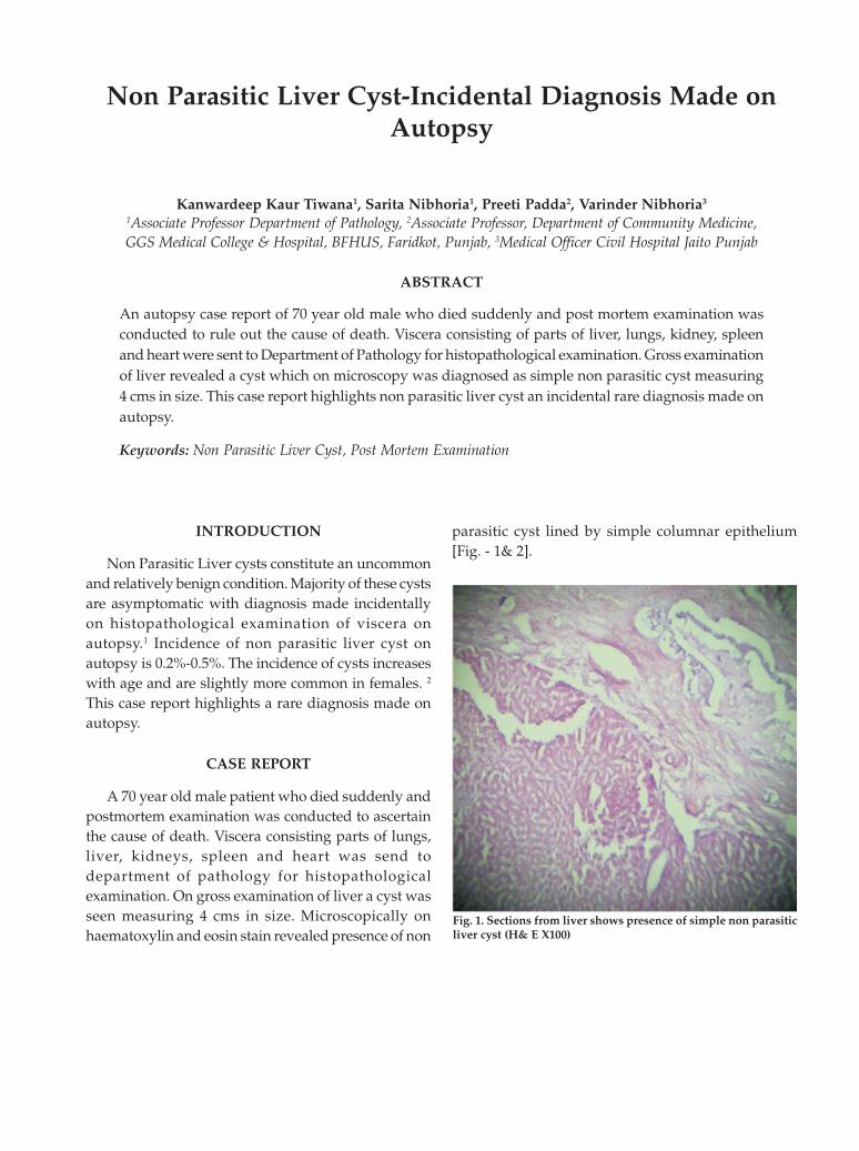
Indian Journal of Forensic Medicine & Toxicology. July-December 2014, Vol. 8, No. 2 193DOI Number: 10.5958/j.0973-9130.8.1.001
Non Parasitic Liver Cyst-Incidental Diagnosis Made onAutopsy
Kanwardeep Kaur Tiwana1, Sarita Nibhoria1, Preeti Padda2, Varinder Nibhoria3
1Associate Professor Department of Pathology, 2Associate Professor, Department of Community Medicine,GGS Medical College & Hospital, BFHUS, Faridkot, Punjab, 3Medical Officer Civil Hospital Jaito Punjab
ABSTRACT
An autopsy case report of 70 year old male who died suddenly and post mortem examination wasconducted to rule out the cause of death. Viscera consisting of parts of liver, lungs, kidney, spleenand heart were sent to Department of Pathology for histopathological examination. Gross examinationof liver revealed a cyst which on microscopy was diagnosed as simple non parasitic cyst measuring4 cms in size. This case report highlights non parasitic liver cyst an incidental rare diagnosis made onautopsy.
Keywords: Non Parasitic Liver Cyst, Post Mortem Examination
INTRODUCTION
Non Parasitic Liver cysts constitute an uncommonand relatively benign condition. Majority of these cystsare asymptomatic with diagnosis made incidentallyon histopathological examination of viscera onautopsy.1 Incidence of non parasitic liver cyst onautopsy is 0.2%-0.5%. The incidence of cysts increaseswith age and are slightly more common in females. 2
This case report highlights a rare diagnosis made onautopsy.
CASE REPORT
A 70 year old male patient who died suddenly andpostmortem examination was conducted to ascertainthe cause of death. Viscera consisting parts of lungs,liver, kidneys, spleen and heart was send todepartment of pathology for histopathologicalexamination. On gross examination of liver a cyst wasseen measuring 4 cms in size. Microscopically onhaematoxylin and eosin stain revealed presence of non
Fig. 1. Sections from liver shows presence of simple non parasiticliver cyst (H& E X100)
parasitic cyst lined by simple columnar epithelium[Fig. - 1& 2].
46. kanwardeep--193--.pmd 7/24/2014, 10:06 AM193

194 Indian Journal of Forensic Medicine & Toxicology. July-December 2014, Vol. 8, No. 2
DISCUSSION
Non parasitic cysts of liver constitute an uncommonand relatively benign condition which may assume animportant role in differential diagnosis of intra-abdominal mass lesion. Eliason and Smith found only28 cases of hepatic cysts in series of 28000, consecutiveautopsies and in no instance was the diagnosissuspected premortem.3 Stoesser and Wangensteenrecorded 104 cases in 1929 and after 30 yearsMorgenstern was able to increase the total number ofreported cases to 250. 4 There are various classificationsof liver cysts. Jones gave a widely acceptedclassification in which liver cysts were classified asproliferative cysts (Cystadenoma), degenerative cysts(pseudocyts), teratomatous cysts, lymphatic cysts,endothelial cysts, blood vessel cysts and retentioncysts(Bile duct cysts).5
Miliadis and Giannakopoulos classified liver cystsas true and false cysts depending on presence ofepithelial lining. True liver cysts include congenitalcysts (simple cysts and polycystic liver disease),parasitic cyst (caused by Echinococcus), neoplasticcysts (cystadenoma, cystadenocarcinoma) and biliaryduct related cysts (Caroli disease). False liver cyst arenot lined by epithelium.1
The etiological factors of simple liver cysts is notknown but are believed to be congenital in origin. Theyare lined by uniform cuboidal or columnar epitheliumand perhaps due to progressive dilatation of biliarymicrohamartomas that fail to develop normalconnection with biliary tree. 6
Incidence of liver cysts rises with age and they arefrequently an incidental finding on ultrasonography
and histopathological examination on autopsy.6 Theyare reported in all age groups with commonly affecting4th to 5th decade. This is in discordance with our casereport as patient was 70 years old. They are commonlyseen in females with male to female ratio being 3:1 7
which is in discordance with this case report asdeceased was a male.
The vast majority of simple hepatic cysts areasymptomatic and can produce symptoms due to theirsize and anatomical localization. Although quite rarecomplications of these cysts can develop due toinfection, torsion and spontaneous rupture intoperitoneal cavity. Although these complications arevery rare but they can significantly affect patient‘squality of life.
This case highlights non parasitic liver cysts areoften asymptomatic and a rare incidental diagnosismade on autopsy.
Acknowledgement: My sincerest thanks to mytechnical supporting staff for their constant help.
Conflict of Interest: None
Source of Funding: None
Ethical clearance: Yes
REFERENCES1. Miliadis L, Giannakopoulos T, Boutiskos G, Terzis
I, Kyriazanos I: Spontaneous rupture of a largenon-parasitic liver cyst: a case report. Journal ofMedical Case Reports 2010, 4:2 doi:10.1186/1752-1947-4-2.
2. Beecherl EE, Bigman DL, Langer B: Cystic diseaseof the liver. In;Zuidema GD, Yeo CJ eds.Shakelford’s Surgery of the Alimentary Tract, 5th
ed. Philadelphia, WB Saunders Co.pp 447-60,2002.
3. Eliason EL, Smith DC: As quoted by ManheimerLH.Solitary non parasitic cyst of liver. Ann Surg1953; 137:410-415.
4. Morgenstern L: Rupture of solitary non-parasiticcysts of liver. Ann Surg 1959;150:167-171
5. Jones JFX: Removal of retention cyst from liver.Ann Surg 1923;77:68-89
6. Ismail KA, Mousa GI, Elkhadraway OH,Mohamed HA: Symptomatic Non-parasiticbenign hepatic cyst: Evaluation of Managementby Deroofing in Ten Consecutive Cases. Annalsof Pediatric Surgery 2010 Vol 6 No.2:83-89
7. Regev A, Reddy KR et al: Large cystic lesions ofliver in adults: a 15 year old experience In atertiary center. J Am Coll Surg 2001, 193:36-45
Fig. 2. Sections show columnar lining of simple liver cyst (H& EX400)
46. kanwardeep--193--.pmd 7/24/2014, 10:06 AM194

Indian Journal of Forensic Medicine & Toxicology. July-December 2014, Vol. 8, No. 2 195DOI Number: 10.5958/j.0973-9130.8.1.001
Spontaneous Rupture of Abdominal Aorta due toPenetrating Atheromatous Ulcer: A Rare Cause of Sudden
Death in a Young Individual
Pradeep K Saralaya1, Y P Girish Chandra2, Clement Wilfred D3, Amoolya Bhat4
1Associate Professor, 2Professor and Head, Department of Forensic Medicine, 3Associate Professor, Department ofPathology, M S Ramaiah Institute of Medical Sciences and Research Centre, Bangalore, Karnataka, India,
4Assistant professor, Department of Pathology, Sapthagiri Institute of Medical Sciences andResearch Centre, Bangalore
ABSTRACT
Spontaneous rupture of aorta due to penetrating atheromatous ulcer is a rare cause of sudden deathin young individuals. Cystic medial necrosis of the aorta in the absence of any known predisposingfactors is also uncommon in young age. We present a rare case of penetrating atheromatous ulcerresulting in fatal rupture of aorta affected by extensive cystic medial necrosis in a young man.
Grade IV atheromatous plaques are prone to form ulcers. The resulting intimal tear causes haematomaformation in individuals with a normal tunica media with intact elastic fibers and smooth musclecoat. Coexistance of grade IV cystic medial necrosis had probably predisposed to extension ofhaematoma into the deeper layers of the wall of aorta and rupture at a young age even withoutaneurysm or dissection of the aorta. The knowledge of such entities contributes to determine thecause of sudden death and timely intervention prevents fatalities.
Keywords: Atheromatous Plaque, Cystic Medial Necrosis, Penetrating Atheromatous Ulcer, SpontaneousRupture of Aorta
INTRODUCTION
Rupture of the aorta secondary to aneurysm,dissection or trauma is a well known entity.Spontaneous rupture of nonaneurysmal, noninfectedaorta is extremely uncommon1 especially in youngindividuals. In most of the cases, it is due to penetratingatherosclerotic ulceration (PAU) of the aorta1 and isencountered in the thoracic aorta of elderly subjects.2
Abdominal aorta is an uncommon site of occurrence.1
The usual causes of cystic medial degeneration of aortaare advanced age and hypertension in elderly andMarfan syndrome or other connective tissue disordersin young individuals.3 The median age of presentationof cystic medial necrosis in non Marfan syndromepatients is fifty years.3 The present case report describessudden unexpected death in a healthy thirty year oldman due to spontaneous rupture infrarenal abdominalaorta affected by cystic medial degeneration throughatheromatous plaques without any predisposingfamilial disorders.
CASE HISTORY
A 30 years old man was brought dead to thehospital after he collapsed following a brief period ofdiffuse abdominal pain. The deceased was a labourerby profession and was resting at home when theincident took place. There was no history of ingestionof medications, trauma, surgery, hypertension, or anyother systemic illness. Personal history revealedcigarette smoking and occasional alcoholconsumption. There was no significant family history.
At autopsy the heart weighed 300grams. Theanterior descending branch of the coronary showedthickened wall. All the other coronaries, valves andthe cardiac chambers were unremarkable. The intimaof the ascending thoracic aorta showed brownishdiscolouration due to haematoma formation(Fig.1a).Abdominal aorta showed atheromatous plaques andrupture below the level of renal arteries. Peritonialcavity showed 1000ml of blood and blood clots in theretroperitoneal area at the site of rupture of aorta
47. Pradeep--195--.pmd 7/24/2014, 10:07 AM195

196 Indian Journal of Forensic Medicine & Toxicology. July-December 2014, Vol. 8, No. 2
reaching the pericolic fat around the caecum (Fig.1b).There was no evidence of aneurysm or dissection ofaorta. All the other organs were unremarkable.
The heart, thoracic and abdominal aorta along withcaecum and ascending colon were sent forhistopathological examination. Microscopically thethere was no infarct or inflammatory cell infiltrate inthe myocardium. The anterior descending branch ofcoronary artery showed American Heart Association(AHA)grade IV atheromatous plaque with grade Iluminal compromise. Right coronary and circumflexcoronary arteries were unremarkable.
The intima of the thoracic and abdominal aortashowed Grade IV atheromatous plaques(Fig.2). Tunicamedia of both abdominal and thoracic aorta showedfeatures of cystic medial necrosis characterized by lossof smooth muscle fibers, disarray and fragmentationof elastic lamellae with widening of inter lamellarspaces. The pseudo cystic spaces contained basophilicmucinous substance (Fig.3). The Van Gieson stainenhanced these findings (Fig.4).The infrarenal aortashowed rupture and retroperitoneal haemorrhage.There were no inflammatory infiltrates, bacterial orfungal invasion in the wall of the aorta.
Fig. 1: a) Ascending aorta showing brownish discolouration of tunica intima(arrow) due to intramural haemorrhage. b) Pericolichaemorrhage (arrows).
Fig. 2: a) Tunica intima showing atheromatous plaques and subintimal cystic medial necrosis(arrow). b) Atheromatous plaques showingfoamy histiocytes, inflammatory cells and extracellular pools of lipid (H&E x400).
47. Pradeep--195--.pmd 7/24/2014, 10:07 AM196

Indian Journal of Forensic Medicine & Toxicology. July-December 2014, Vol. 8, No. 2 197
Fig. 3: Tunica media showing widening of interlamellar spaces filled with blue mucinous ground substance (H&E x400).
Fig. 4: Tunica media showing disarray and fragmentation of elastic lamellae (Van Gieson (a) x100; (b) x400).
Grade IV atheromatous plaques containextracellular lipid and are prone to form ulcers. Theresulting intimal tear causes intramural haematomaformation individuals with a normal tunica media withintact elastic fibers and smooth muscle coat. Howeverthis man exhibited grade IV cystic medial necrosischaracterised by elastic fiber disarray and loss ofsmooth muscle cells in tunica media of aorta whichhad probably predisposed to extension of haematomainto the deeper layers of the wall of aorta and ruptureeven without aneurysm or dissection of the aorta. Thusin this patient, cystic medial degeneration might havefacilitated the formation of penetrating atherosclerotic
ulcer culminating in spontaneous rupture of aorta at ayoung age.
Thus based on the autopsy and histopathologyfindings, spontaneous rupture of the aorta due topenetrating atheromatous ulcer was concluded as thecause of massive haemorrhage and death.
DISCUSSION
Spontaneous rupture of abdominal aorta is a rareevent in young subjects and is an uncommon cause ofsudden unexpected death. Elderly individuals withhypertension and atherosclerotic plaques in the wall
47. Pradeep--195--.pmd 7/24/2014, 10:07 AM197

198 Indian Journal of Forensic Medicine & Toxicology. July-December 2014, Vol. 8, No. 2
of the aorta commonly suffer from this condition andtypically it involves the infrarenal aorta.4 Spontaneousrupture of aorta without hypertension has beenreported in the past.5
Cystic medial necrosis is a common cause ofthoracic aortic rupture in young individuals withMarfan syndrome, Loyes-Dietz syndrome, Ehlers-Danlos syndrome type IV and bicuspid aortic valve.Familial forms without associated syndromes andsporadic cases of cystic medial necrosis exhibitingcertain specific gene mutations are well known tooccur.3,4 The above described case did not exhibit anyfeatures of Marfan syndrome. The examination of theheart ruled out the presence of bicuspid aortic valveor other congenital anomalies. The cause for the cysticmedial degeneration aorta was unknown in this caseas the personal history did not reveal presence ofhypertension, autoimmune disease like arteritis orinfections like syphilis. Cystic medial necrosis candevelop in patients with aortopathies likeatherosclerotic disease and even healthy individuals.3
Rupture of aorta affected by cystic medial necrosis isusually secondary to aneurysm or dissection in youngsubjects. 4,5 The instant case lacked features ofaneurysm or dissection, and showed only cystic medialnecrosis and atheromatous plaques at the site ofrupture. Cases of sudden death due to spontaneousdissection7 and traumatic rupture 8 of aorta affectedby cystic medial necrosis have been reported in youngsubjects without any familial or genetic predisposingfactors in the past. A case of spontaneous rupture ofthoracic aorta secondary to cystic medial necrosisErdheim Gsell in a fifty eight year old lady has beenreported.6 Abdominal aorta is an uncommon site ofrupture in patients with cystic medial necrosis.4
Most spontaneous aortic ruptures are associatedwith perforation through the atheromatous plaque.4
Spontaneous rupture of aorta displaying grade fouratheromatous plaques in the intima and grade fourcystic medial necrosis of the tunica media, withoutaneurysm formation or dissection pointed towards thediagnosis of penetrating atheromatous ulcer in thedescribed case.
Penetrating atherosclerotic ulcer of the aorta hasbeen considered as an important cause of spontaneousrupture of aorta. About 2.3-7.6% of acute aorticsyndromes are caused by penetrating atheromatousulcer.2 It is typically seen in elderly individuals(seventh life decade) with hypertension and
atherosclerosis and usually involves the descendingthoracic aorta.2,4
It was first described by Shennan in 1934. Recentlyit is described as an atypical variant of aortic dissection.The condition is defined as the atheromatous ulcer thatpenetrates through the internal elastic lamina into thetunica media and is associated with hematoma withinthe aortic wall.2,9,10 Pseudoaneurysm formation,localized dissection, embolization or ruptures are theusual outcomes of penetrating atheromatous ulcer.2 9,10
The chances of rupture is reported to be up to 40% .11
Intimal defects resulting from atherosclerotic ulcersoccur in patients with advanced atherosclerosis(American heart association grade IV and above).Atheromatous ulcers are asymptomatic and areconfined to the tunica intima. Later the ulcer penetratesthrough the elastic lamina and enters the tunica media(ie, penetrating atherosclerotic ulcer).9,10 The stagnationand increased residence time of blood in the region ofthe penetrating atheromatous ulcer results in elevatedintraluminal pressure and a low flow velocity at thelevel of the aortic wall. The pulsatile arterial flow10 andexposure of the aortic wall to a relatively low wall shearstress increases the intercellular permeability andcontributes to penetrating atheromatous ulcerprogression. 10,11
CONCLUSIONS
We presented a unique case of sudden death dueto spontaneous rupture of abdominal aorta affectedby extensive cystic medial necrosis and atheromatousplaques in a young male without any predisposingfactors like systemic disease or trauma. In sporadiccases cystic medial necrosis is never diagnosed untilthe acute aortic syndrome has occurred and the aortictissue is examined after autopsy or surgery. Thefindings of autopsy and histopathology are useful toeducate the close relatives of the deceased, and tocreate awareness about the natural history of disease.
Acknowledgement: We would like to acknowledgethe Department of Pathology, MS Ramaiah Instituteof Medical sciences and Research Centre, Bangalorefor their valuable support in evaluating the case.
Conflicts of Interest: None
Permissions: Nil
Source of Funding and Ethical Clearance: Nil
47. Pradeep--195--.pmd 7/24/2014, 10:07 AM198

Indian Journal of Forensic Medicine & Toxicology. July-December 2014, Vol. 8, No. 2 199
REFERENCES
1. Chervenkov V, Tonev A, Maximov D, NikolovD, Stoinova V. Spontaneous rupture of anonaneurysmal infrarenal abdominal aorta dueto penetrating ulcer . EJVES Extra 2005; 10:101–3.
2. Eggebrect H, Plicht B, Kahlert P, Erbel R.Intramural hematoma and penetrating ulcers:Indications to endovascular treatment. Eur J VascEndovasc Surg 2009;38:659-65.
3. Yuan SM, Jing H. Cystic medial necrosis:pathological findings and clinical implications.Rev Bras Cir Cardiovasc 2011; 26.1:107-15.
4. LibbeyP, Bonow RO, Mann DL, Zipes DP, editors.Braunwald’s heart disease: A textbook ofcardiovascular medicine. 8th edition. SaundersElsevier 2007. p:1457-90.
5. Yada M, Maze Y, Tokui T, Shomura S.Asymptomatic spontaneous rupture of a nonaneurismal visceral aorta. Ann Thorac CardivascSurg 2008;14:336-8.
6. Gary T, Seinost G, Hafner F, Gorkiewicz G,Brodmann M. Cystic medial necrosis ErdheimGsell as a rare reason for spontaneous rupture ofthe ascending aorta. Vasa 2011; 40:147 –9.
7. Dermengiu S, Ceaus M, Hostiuc S, Curca G C,Dermengiu D, Turculet C. Spontaneous aortic
dissection due to cystic medial degeneration.Report of a sudden death case and literaturereview. Rom J Leg Med 2009;17:89 – 96.
8. Kumar PS, Sapeco SD, Pinto RGW, Couto F. Fataltraumatic rupture of ascending aortic aneurysmhaving idiopathic cystic medial necrosis: Anautopsy case. J Indian Acad Forensic Med2010;32:339-42.
9. Hayashi H, Matsuoka Y, Sakamoto I, Sueyoshi E,OkimotoT, Hayashi K, Matsunaga N. Penetratingatherosclerotic ulcer of the aorta: Imaging featuresand disease concept. Radiographics. 2000; 20:995–1005.
10. Macura KJ, Corl FM, Fishman EK, Bluemke DA.Pathogenesis in acute aortic syndromes: Aorticdissection, intramural hematoma, andpenetrating atherosclerotic aortic ulcer. AJR2003;181:309–16.
11. D’Ancona G, Amaducci A, Rinaudo A, Pasta S,Follis F, Pilato M and Baglini R. Haemodynamicpredictors of a penetrating atherosclerotic ulcerrupture using fluid–structure interaction analysis.Interactive CardioVascular and ThoracicSurgery.2013:1–3.
47. Pradeep--195--.pmd 7/24/2014, 10:07 AM199

200 Indian Journal of Forensic Medicine & Toxicology. July-December 2014, Vol. 8, No. 2DOI Number: 10.5958/j.0973-9130.8.1.001
Elastofibroma Dorsi: A Rare Soft Tissue Tumor DetectedIncidentally on Autopsy
Vardendra Kulkarni1, Seema Bijjaragi2
1Assistant Professor, 2Associate Professor, Department of Pathology, JJM Medical College, Davangere, Karnataka, India
ABSTRACT
Elastoûbroma dorsi is an uncommon benign soft tissue tumor usually located in the infra-scapularregion, deep to serratus anterior muscle, and often attached to the periosteum of the ribs, presentingwith long history of swelling and occasionally pain and discomfort. It is seen in elderly age groupand may be mistaken for a malignant soft tissue tumor due to its size and location. The pathogenesisof the lesion is still unclear, but repetitive microtrauma by friction between the scapula and the thoracicwall may cause the reactive hyperproliferation of fibroelastic tissue. Diagnosis is straight-forwardwhen the lesion is present in characterstic anatomic location and pathognomonic histopathologicalfeatures are seen. We describe a case of bilateral elastofibroma dorsi detected incidentally on autopsy.
Keywords: Elastofibroma, Autopsy, Histopathology, Subscapular Mass
INTRODUCTION
Elastofibroma dorsi (ED) is a benign, slow-growingfibro-elastic tumor that was first described by Jarvi andSaxen in 1961.1The typical presentation of ED involvesa subscapular mass associated with a long history ofswelling, discomfort, snapping of the scapula and, insome cases, pain. In 99% of cases, EDs are localized tothe infrascapular region between the thoracic wall,serratus anterior, lattisimus dorsi muscle, and are oftenattached to the periosteum of the thoracic wall.2 Veryrarely, elastofibroma is found in other sites, such asthe tip of the elbow, near the ischial tuberosities, thedeltoid muscle, foot, inguinal region, orbits, stomach,greater omentum and the intraspinal spaces. It isrelatively frequent among elderly women, with a meanage at onset of 70 years. It is bilateral in 10% of cases.3We report a case of bilateral elastofibroma dorsidetected incidentally on autopsy and review theliterature.
Corresponding author:Vardendra KulkarniAssistant professorDepartment of Pathology, JJM Medical College,Davangere, Karnataka, IndiaMobile :+ 919731501466E-mail : [email protected]
CASE REPORT
A 60 year old female agriculture worker wasbrought to casualty with an alleged history of snakebite. She was diagnosed to be in a state ofhemodynamic shock and succumbed to the conditionin few hours. A medico-legal autopsy was performedand shock due to snake bite was confirmed as the causeof death. Two soft tissue masses were detected onautopsy in bilateral subscapular area. Both the masseswere excised and sent for histopathologicalexamination. Grossly the masses were poorlydelineated from surrounding tissue, the cut surfaceshowed small areas of adipose tissue interposedbetween gray-white fibrous areas as shown in figure1. On microscopic examination the tumor like massconsisted of a mixture of intertwining swollen,eosinophilic collagen and elastic fibers in about equalproportions associated with occasional fibroblasts,small amounts of interstitial mucoid material, andvariably sized aggregates of mature fat cells. The elasticfibers appeared degenerated, beaded and fragmentedinto small flower-like, serrated disks or globules witha linear arrangement (Figure 2). Special stain (Verhoff’selastin) confirmed the elastic nature of the degeneratedfibres(Figure 3). The final histopathology report wassigned out as bilateral elastofibroma dorsi.
48. Verdendra--200--.pmd 7/24/2014, 10:07 AM200

Indian Journal of Forensic Medicine & Toxicology. July-December 2014, Vol. 8, No. 2 201
DISCUSSION
Elastoûbroma dorsi is an uncommon benign lesion.Negamine et al. 4have described a series of 170 patientsfrom Okinawa. Genetic predisposition was reportedwith 32% of the 170 patients having a family historyof elastoûbroma. All the larger series of elastoûbromareported in the literature showed elastoûbroma wascommoner in females however in the case seriesreported by Chandrasekar et al5 males were morecommonly (80%) involved than females. Case seriesreported by various authors has been shown intable 1.
Fig. 1. Gross specimen showing ill defined mass with alternatefibrous and adipose areas
Fig. 3. Photomicrograph showing elastic fibers (verhoff’s elastin,x100)
Fig. 2. Photomicrograph showing degenerated elastic fibers withfibrocollagenous tissue (H&E, x100)
Table 1. Showing elastofibroma case series published in literature
Sl. No. Study Year Number of Gender Bilateralitycases Distribution (%)
(F:M)
1 Mackenzie et al6 1968 03 1:2 33.3
2 Nagamine et al4 1982 170 13:1 66.7
3 Daigeler et al3 2007 07 5:2 14.2
4 Muratori et al7 2008 08 7:1 12.5
5 Chandrasekar et al 5 2008 15 1:4 13.3
6 Kastner et al8 2009 11 9:2 09.1
7 Cinar et al9 2009 13 11:2 15.4
The pathogenesis of elastofibroma dorsi is stillunclear, but repetitive microtrauma caused by frictionbetween the scapula and the thoracic wall may causereactive hyper-proliferation of fibroelastic tissue.3Thelocation of elastofibromas and its incidence amongolder individuals may be related to the natural
existence of fibro-elastic tissue in this region andsuggests a reactive process in response to friction ofthe scapula against the ribs. Alternatively, the genesisof elastofibroma is thought to involve anoverproduction of the collagenous connective tissuewith a degeneration of the collagen fibres and an
48. Verdendra--200--.pmd 7/24/2014, 10:07 AM201

202 Indian Journal of Forensic Medicine & Toxicology. July-December 2014, Vol. 8, No. 2
overproduction of immature elastic tissue, derivedfrom fibroblasts, alternating with deposition ofhyperplastic fat.6The nature of the altered elastic fibresis disputed and controversial. They may be caused byabnormal elastogenesis or by degenerating as asecondary process, or even by a combination of bothprocesses.3 Swelling, discomfort, snapping of thescapula, and occasionally pain are the most commonpresenting symptoms. However, it is not uncommonfor these tumors to be completely asymptomatic andto be found incidentally during physical examinationor during an investigation of a symptomatic mass onthe contra-lateral side.
MR is the imaging modality of choice in diagnosingED and typically demonstrates a well-defined,moderately inhomogeneous mass with no associatedsoft tissue edema. On T1-weighted MRI, EDs areisointense with skeletal muscle, which explains howthese tumors are often diagnostically overlooked. T1-and T2-weighted images both show interspersed linearand curvilinear, hyper intense areas representing fat.Fat saturation gradient technique applied post-gadolinium administration may show subtle areas ofheterogenous enhancement within the mass, makingit difficult to exclude a soft tissue sarcoma withoutbiopsy. Imaging with CT often reveals homogenousmasses with attenuation similar to that of surroundingskeletal muscle and a fat plane that is indistinct fromthe mass and adjacent skeletal muscle.2
Macroscopically, elastofibromas appear as anirregular, poorly defined, unencapsulated, fibroelasticmass with firm, rubbery consistency. Cut surfacereveals strands of white and yellow tissue representingadipose tissue intermingled with fibroelastic tissue ina “checkerboard” pattern. Histologically, fibrous,collagenous strands with eosinophilic, plump,elongated and round-shaped collections of elasticfibers are seen. These fibers may be difficult to visualizeon H and E, especially during frozen section diagnosis.An elastin stain (Elastic-van-Gieson) is necessary tohighlight these fibers and reveals deeply stainingbranched and unbranched fibers with central densecore and serrated margins. The lesions arepredominantly hypocellular, with benign fibrocyticand fibroblastic cells that lack atypia and mitoticfigures, and contain entrapped islands of adiposetissue. Fine needle aspiration is not a recommendeddiagnostic technique due to this hypocellular natureof ED.2
Diagnosis of elastofibroma was straight-forwardin the present case as the lesion was bilateral andlocated at the characterstic anatomical site withpathognomonic histological features of degeneratedelastic fibres interposed with fibrocollagenous andadipose tissue. In case of early lesions and lesions atunusual locations, the differential diagnoses shouldinclude nuchal fibroma, fibrolipoma, fibromatoses,sarcoma and subcutaneous metastasis. Under suchcircumstances,elastofibromas can be differentiatedfrom these lesions using an immunohistochemicalpanel of vimentin, smooth muscle actin, CD34, CD 99,S-100 and lysozyme.10
CONCLUSION
Elastofibroma is a rare tumor like lesion of softtissues occurring characteristically in infrascapularregion.Simple surgical excision is the treatment ofchoice and local recurrence is very rare.
Conflict of Interest: None
Source of Funding: None
Ethical Clearance: Not applicable
Acknowledgements: We thank the department ofForensic Medicine, JJM Medical College,Davangere forproviding the case details.
REFERENCES
1. Jarvi O Saxen E. Elastofibroma dorse. Acta PatholMicrobiol Scand Suppl. 1961; 51:83–4.
2. Pauline H. Go Meadows M C Essel Marie B.deLeon and Ronald S. Chamberlain.Elastofibroma dorsi: A soft tissue masquerade.Int J Shoulder Surg. 2010; 4(4): 97–101.
3. Daigeler A, Vogt PM, Busch K, Pennekamp W,Weyhe D, Lehnhardt M, et al. Elastofibromadorsi—differential diagnosis in chest walltumors. World J Surg Oncol. 2007; 5:15.
4. Nagamine N, Nohara Y, Ito E. Elastofibroma inOkinawa. A clinicopathologic study of 170 cases.Cancer. 1982; 50:1794–805.
5. Chandrasekar CR, Grimer RJ, Carter SR, TillmanRM, Abudu A, Davies AM, et al. Elastofibromadorsi: An uncommon benign pseudotumor.Sarcoma. 2008;2008:756565.
6. Mackenzie D H,Wilson J F and Cooke KB.Elastofibroma. J.Clin.Path.1968; 21:470-475
48. Verdendra--200--.pmd 7/24/2014, 10:07 AM202

Indian Journal of Forensic Medicine & Toxicology. July-December 2014, Vol. 8, No. 2 203
7. Muratori F, Esposito M, Rosa F, Liuzza F,Magarelli N, Rossi B, et al. Elastofibroma dorsi: 8case reports and a literature review. J OrthopTraumatol. 2008; 9:33–7.
8. Kastner M, Salai M, Fichman S, Heller S,Dudkiewicz I. Elastofibroma at the scapularregion. Isr Med Assoc J. 2009; 11:170–2.
9. Cinar BM, Akpinar S, Derincek A, Beyaz S, UysalM. Elastofibroma dorsi: An unusual cause ofshoulder pain. Acta Orthop Traumatol Turc. 2009;43:431–5.
10. Ramachandra S, Kuruvila S and Kindi M A. SoftTissue Swelling at the Subscapular Region. OmanMedical Journal, 2012; 27(2): 172-174.
48. Verdendra--200--.pmd 7/24/2014, 10:07 AM203

204 Indian Journal of Forensic Medicine & Toxicology. July-December 2014, Vol. 8, No. 2DOI Number: 10.5958/j.0973-9130.8.1.001
Neurological Manifestation's of Lead Toxicity in a BatteryWorker-A Case Report
Varma Sanjay1, Gupta G B2, Malhotra Yogendra1, Khare R L1, Singh C3, Agrawal Shweta4,Mandavi Sanjay4
1Assistant Professor, 2Professor & Head, 3Assistant Professor, 4Resident, Department of Medicine, Pt. J.N.M. MedicalCollege Raipur& Dr.BRAM Hospital, Chhattisgarh
ABSTRACT
Lead acts as cumulative metabolic poison. Occupational lead poisoning occurs frequently indeveloping country as it has many industrial uses like manufacture of pipes, storage batteries, shipbuilding. Toxic effect of lead on the body is known as Plumbism. It affects multiple organs, specificallybones, blood, brain, increased risk of cardiovascular disease, coronary artery disease and plateletdysfunction. Lead encephalopathy is rare in adults due to the capacity of the mature brain tosequestrate lead away from its mitochondrial site of action within the cerebral and cerebellar neurons.It is a life threatening condition and should be treated as an emergency. Here by, we present a case of18 year old young man with lead encephalopathy with brief review of literature.
Keywords: Lead Poisoning, Lead Encephalopathy
INTRODUCTION
Lead (Pb) has been known for centuries to be acumulative metabolic poison. [1] The toxicity of leadhas been known for more than a thousand year, butoccupational lead poisoning still occurs in developingcountry. Lead has many industrial uses, likemanufacture of pipes, cisterns, roof coverings; themanufacture of lead accumulator plates and storagebatteries, ship-building and ship-breaking, plumbing,painting and soldering.[2] Lead is ubiquitous in ourenvironment but has no physiologic role in biologicalsystems. Toxic effect of lead on the body is known asPlumbism and it is now well recognized that inorganiclead produces not only clinically definedencephalopathies and neuropathies, but also variousbehavioural changes indicative of cerebraldysfunction. Exposure of lead can take place eitherthrough inhalation of dust, fumes, vapours, oringestion of contaminated foods or drinks.[3] Leadpoisoning affects multiple organs, specifically bones(lead bands at the epiphyses of the long bones), blood(punctate basophilic stippling of erythrocytes andmicrocytic hypochromic haemolytic anaemia), brainedema, demyelination of the cerebral and cerebellarwhite matter, [4] Cerebrovascular diseases, stroke,cerebral haemorrhage, tremor, paresthesia, paralysis,[5]
psychomotor impairment, peripheral nervous system(demyelinating neuropathy), gingiva (lead lines), andkidneys (chronic tubulointerstitial disease). [4] Incardiovascular system lead toxicity may result inhypertension, increased risk of cardiovascular disease,coronary artery disease and platelet dysfunction. [5] Inthe central nervous system, the lesions are thought tobe the result of vascular injury. Lead acts as a cellulartoxin by inhibiting mitochondrial respiration. Leadencephalopathy is rare, [4] the increased resistance ofthe adult to encephalopathy and ataxia is believed tobe caused by the capacity of the mature brain tosequestrate lead away from its mitochondrial site ofaction within the cerebral and cerebellar neurons. Leadencephalopathy requires emergency admission to anintensive care unit with intubation and sedation. A longperiod of inpatient treatment usually followsadmission, although full recovery of cognitive, motor,and emotional function may not occur. [6]
CASE REPORT
An 18 year young man, employed in a batteryworkshop was admitted to hospital with complaintsof giddiness, weakness, headache, vomiting and oneepisode of convulsion. He had been in thisemployment for 2 years where he had to dismantle
49. Sanjay Varma--204--.pmd 7/24/2014, 10:07 AM204

Indian Journal of Forensic Medicine & Toxicology. July-December 2014, Vol. 8, No. 2 205
used batteries. Past history was negative for substanceabuse and any poisoning. On clinical examinationpatient was confused, disoriented and bilateral plantarwere absent. After 16 days of hospitalization hesuddenly developed bilateral sixth nerve palsy. Therewas no sensory deficit. On ophthalmic examinationbilateral esotropia of 15 degree (Figure.1) and fundusexamination showed bilateral papilloedema. Othersystemic examination was apparently normal.
134 µg/dl (higher). C.S.F. examination could not beperformed due to papilloedema. On neuroimaging,MRI brain documented infarction in the left basalganglia posteriorly (Figure.3) and CT Brain contrastdocumented bilateral mild diffuse cerebral oedema(Figure.4).
Fig. 1. Showing esotropia of Rt. eye
Fig. 2. Showing improvement in Ophthalmoplegia
On laboratory examination Hb was 7.8 gm/dl,documented neutrophilic leucocytosis 13400/mm3,neutrophils count 68, heamatocrit 25.7%, MCV-79 fl,MCH-23.9 pg, MCHC-30.3 g/dl and platelet count wasadequate. Peripheral smear showed microcytichypochromic anaemia without basophilic stippling.Urine analysis documented pus cells, Renal functiontest (RFT) was normal, Liver function test (LFT) wasnormal, Blood sugar level was normal. Serum Na+, K+,and Ca++ level was 131meq/L, 4.6 meq/L and 8.2mg/dl respectively. Serum level of Lead (plumbum) was
Fig. 3. MRI scan –infarction of Lt.basal ganglia
Fig. 4. CT scan-mild diffuse cerebral oedema
He was treated with Phenytoin, Mannitol,Dexamethasone, and D-penicillamine, he respondedby improvement in consciousness, plantar reflexesbecame flexor, improvement in ophthalmoplegia(Figure.2) and had no episode of convulsion. He wasdischarged in stable conditions.
49. Sanjay Varma--204--.pmd 7/24/2014, 10:07 AM205

206 Indian Journal of Forensic Medicine & Toxicology. July-December 2014, Vol. 8, No. 2
DISCUSSION
Lead encephalopathy was first documented in 1925.Normal lead levels should be below 10µg/dL. Leadpoisoning occurs with the blood level > 40µg/dL. Leadencephalopathy is almost always associated with ablood lead concentration of 100 µg/dL, but levels oflead as low as 70 µg/dL can also causeencephalopathy.[4] The Control of Lead at WorkRegulations had prescribed safety limits foroccupational lead exposure in 1998, according to whichblood lead values <1.45 micromol/l (30 microgm/100ml) represent reasonably well-controlled occupationalexposure provided there is 6-monthly monitoring, 1.45- 2.4 micromol/l (30 - 50 mg/100 ml) requireinvestigative action by the employer and 2.4 - 2.9micromol/l (50 – 60 mg/100 ml) call for suspension ofthe worker from exposure. However, US CDC andWHO recommends blood lead level > 0.5 micromol/l(10 microgm/100 ml) as cause for concern.
Encephalopathy is the most dramatic and life-threatening development of lead poisoning, with amortality rate of 25% or more. In adults the main formof exposure is to white lead oxide powder, which maybe inhaled, for example, during the manufacture ofbatteries. [2]
The adult form of lead encephalopathy is rarer thanthe paediatric form. [7] In adults, this encephalopathycan present with serious neurodeterioration and cansometimes be lethal. [8] Lead encephalopathy canpresent in acute and chronic forms. Acute leadencephalopathy presents pathologically as cerebraledema, [9] whereas in chronic lead encephalopathy,extensive tissue destruction with cavity formation andthickening of the veins with cellular disorganizationof their walls (suggesting vascular insult in cerebralinjury) is seen. [10] All this can lead to edema, gliosis,haemorrhage, neuronal loss, and perivascularproteinaceous exudates. [11] Lead acts as a cellular toxinby inhibiting mitochondrial respiration. [12] Clinically,patients present with headache, vomiting, ataxia,convulsions, paralysis, stupor, and coma in acuteforms. [7], [9], [13], [14]
Lead acts as a cellular toxin by inhibitingmitochondrial respiration. The increased resistance ofthe adult to encephalopathy and ataxia is believed tobe due to the capacity of the mature brain tosequestrate lead away from its mitochondrial site ofaction within the cerebral and cerebellar neurons. [1]
The onset of cerebral symptoms is usually acute, with
seizures (which may be focal or generalized), deliriumand coma, often associated with papilloedema. Anytype of neurological sign may develop, includingcerebellar ataxia, cranial nerve palsies, optic atrophy,hemiplegia, and decerebrate rigidity. Chronic casesmay show mental dullness, poor memory, headache,trembling, head retraction, deafness, hemianopia andamaurosis without fundal changes. Encephalopathyis not a common manifestation of plumbism in theadult. Abdominal colic and neuropathy involving themost frequently used muscles is more usual.[2]
Whitfield et al [15] have been suggested the followingcriteria for diagnosis of lead encephalopathy: 1)Documentation of diffuse encephalopathy with orwithout focal signs; 2) Diagnosis of lead toxicity usingclinical and laboratory aids; 3) Response to chelators;and 4) Exclusion of other causes of encephalopathy.
In our patient, a diagnosis was made on the basisof history of occupational exposure, higher serum levelof lead, feature of encephalopathy, and response tochelators. In patient, we found one episode of seizure,headache, vomiting and giddiness. Whitfield et al [15]
(1972) reported the largest series of 23 adults with leadencephalopathy; all of these followed consumption ofillicit liquor contaminated by lead (moonshine). Tenof these patients had altered sensorium, 18 of themhad seizures. Other symptoms included dizziness,syncope, disorientation, and blindness. One hadpapilloedema this could be secondary to raisedintracranial pressure or to direct involvement of thecerebellum. Severe medical illness, alcohol,dehydration, and emotional stress are known toprecipitate symptoms of lead poisoning, but we wereunable to identify any such factor in our patient.Basophilic stippling of erythrocytes is reported in 91%of patients. [10] Peripheral smear of our patient did nothave basophilic stippling of peripheral red blood cells.
Segal et al [2] reported diversity of neurologicalmanifestations in lead encephalopathy such as one casecomplaining of tremor of the arms and legs, andcramp-like abdominal pains, of 4 weeks’ duration,second case presented as one episode of seizure onlyand third case presented as four attack of seizure.
We found features of encephalopathy (bilateralmild diffuse cerebral oedema and infarction in the leftbasal ganglia posteriorly) in CT and MRI finding.Perelman et al [8] reported MR imaging findings suchas cerebellar edema, suggested by an enlarged
49. Sanjay Varma--204--.pmd 7/24/2014, 10:07 AM206

Indian Journal of Forensic Medicine & Toxicology. July-December 2014, Vol. 8, No. 2 207
cerebellum with the disappearance of the cerebellarfissure and protrusion. Lead intoxication can causeintracranial calcification but the pathogenesis ofintracranial calcification has not yet been provedsatisfactorily. Perelman et al [8] reported an incidenceof 84% cerebellar calcification at autopsy in a study of44 adults with a history of chronic lead poisoning.Schroter et al [16] reported bilateral symmetriccalcification in the subcortical area of the cerebralhemispheres and basal ganglia on CT. Tuzun et al [17]
reported bilateral symmetrical hypoattenuated lesionsin the thalamus, lateral putamen, claustrum, andinsula. Reyes et al [18] reported extensive intracranialcalcification on CT in 3 adults with chronic leadpoisoning. In our patient, CT and MR imaging did notshow any calcification.The extent of clinical recoverywas out of proportion to the decline in bloodconcentrations. Free erythrocyte protoporphyrin andthe urinary concentrations of á-aminolevulinic acid(ALA) or coproporphyrin are better clinical correlatesof lead toxicity, rather than estimations of blood leadconcentration. [10] In our patient only the serum leadcould be estimated.
Our patient who was a battery worker was foundto have lead encephalopathy. He presented withsymptoms of one episode of seizure, headache,vomiting, giddiness and on investigations was foundto have cerebral edema and infarction in the left basalganglia posteriorly which may be caused by vasculitisdue to lead toxicity. His diagnosis was confirmed byserum level of Lead which came out to be 134µg/dl(higher) and he had well responded to use of D-penicillamine, mannitol, and dexamethasone for raisedintracranial tension and anti-convulsants for seizures.
In conclusion, burden of lead toxicity may beincreasing in developing countries like ours wherestrict guidelines and screening facilities for workersemployed in potentially bio-hazardous occupationmay be lacking. So every worker employed in batteryworkshop and other lead related occupations shouldbe screened for symptoms and signs of lead toxicity,thereby preventing potentially life threatening anddisabling manifestations of occupational leadexposure.
Acknowledgement: Authors wish to acknowledge thecooperation of staff of Dr. BRAM Hospital ,Raipur,Chattisgarh.
Source of Gunding: As the study was done in theexisting facilities available in Dr BRAM Hospital
Raipur, no extra expenditure was incurred on thestudy.
Conflict of Interest: The authors don’t have anyconflict of interest.
Ethical Committee of: Pt.JNM Medical College, Raipurhave given Ethical clearance vide letter No.MCR/Ethics Comm./12/187. Dated 08/05/13.
REFERENCES
1. Ronald Eisler, Lead Hazards to Fish, Wildlife, andInvertebrates: A synoptic review biological report85(1.14) contaminant hazard reviews, report no.14, APRIL 1988.
2. I. Segal, D. Saffer, F. Segal, Diverse NeurologicalManifestations of Lead Encephalopathy, S. Afr.Med. J., 48, 1721-1722 (1974).
3. Sachdev Yadav, Veena Sharma Lead inducedencephalopathy: An Overview InternationalJournal of Pharma and Bio SciencesVol.2, Isssue1,70 – 86, Jan-March 2011.
4. A.L. Atre, P.R. Shinde, S.N. Shinde, R.S. Wadia,A.A. Nanivadekar, S.J. Vaid R.S. Shinde, Pre- andPost treatment MR Imaging Findings in LeadEncephalopathy, AJNR Am J Neuroradiol 27:902–03 Apr 2006.
5. Vance Vella, Elizabeth O’Brien, Elisa Idris Healthimpacts of lead poisoning a preliminary listingof the health effects & symptoms of leadpoisoning, Health Impacts of Lead PoisoningUpdate 23rd March 2011.
6. S Cairney, P Maruff, C B Burns, J Currie, B JCurrie, Saccade dysfunction associated withchronic petrol sniffing and lead encephalopathy,J Neurol Neurosurg Psychiatry 2004;75:472–476.
7. Bressler JP, Goldstein GW. Mechanisms of leadneurotoxicity. Biochem Pharmacol 1991; 41:479–84.
8. Perelman S, Hertz-Pannier L, Hassan M, et al.Lead encephalopathy mimicking a cerebellartumor. Acta Paediatr 1993; 82:423–25.
9. Saryan LA, Zenz C. Lead and its compounds. In:Saryan LA, Zenz C, eds. Occupational Medicine.3rd ed. St. Louis, Mo: Mosby; 1994:506–41.
10. Jayanti Mani, Nalin Chaudhary, MakarandKanjalkar, Pravina Ushah Cerebellar ataxia dueto lead encephalopathy in an adult, J NeurolNeurosurg Psychiatry 1998;65:797–806.
11. Teo JGC, Goh KYC, Ahuja A, et al. Intracranialvascular calcifications, glioblastoma multiforme,and lead poisoning. AJNR Am J Neuroradiol1997; 18: 576–79.
49. Sanjay Varma--204--.pmd 7/24/2014, 10:07 AM207

208 Indian Journal of Forensic Medicine & Toxicology. July-December 2014, Vol. 8, No. 2
12. Holtzman D, DeVries C, Nguyen H, et al.Maturation of resistance to encephalopathy:cellular and subcellular mechanism.Neurotoxicology 1984; 5: 97–124.
13. Landrigan PJ, Todd AC. Lead poisoning. West JMed 1994; 161:153–59.
14. Alkhayat A, Menon NS, Alidina MR. Acute leadencephalopathy in early infancy: clinicalpresentation and outcome. Ann Trop Paediatr1997; 17: 39–44.
15. Whitfield CL, Chien LT, Whitehead JD. Leadencephalopathy in adults. Am J Med 1972; 52: 289.
16. Schroter C, Schroter H, Huffmann G. Neurologicand psychiatric manifestations of lead poisoningin adults (case report and review of the literature)[in German]. Fortschr Neurol Psychiatr 1991;59:413–24.
17. Tuzun M, Tuzun D, Salan A, et al. Leadencephalopathy: CT and MR findings. J ComputAssist Tomogr 2002; 26:479–81.
18. Reyes PF, Gonzalez CF, Zalewska MK, et al.Intracranial calcification in adults with chroniclead exposure. AJR Am J Roentgenol 1986; 146:267–70.
49. Sanjay Varma--204--.pmd 7/24/2014, 10:07 AM208

Indian Journal of Forensic Medicine & Toxicology. July-December 2014, Vol. 8, No. 2 209DOI Number: 10.5958/j.0973-9130.8.1.001
Fatal Case of Hexaconazole Poisoning: A Rare Case Report
Tanmay Sardar1, Saswata Biswas2, Aniruddha Das2, Nirmalya Chakrabarti2, Partha PratimMukhopadhyay3, Subodh Bhattacharyya4
1Senior Resident, 2Junior Resident, Department of Pathology, 3Professor & Head, Department of Forensic & StateMedicine, 4Professor & Head, Department of Pathology, Burdwan Medical College, Burdwan
ABSTRACT
Hexaconazol (a systemic fungicide) due to its low toxicity, is very rarely reported in literature ofpesticide poisoning. With development of agricultural industries and change in the pattern ofcultivation, such poisoning may become more frequent in the future. There is no documented reportof death due to acute Hexaconazole poisoning till date. Liver is the primary target of organ damage.Severe involvement of other organs is also evident in this report. This fatal poisoning by Hexaconazole,perhaps the only case so far, is being reported for its rarity.
Keywords: Agrochemical Poisoning, Hexaconazole, Fatal poison, Pathology
INTRODUCTION
Hexaconazole, a systemic fungicide, is presentlygaining popularity in Indian agricultural industry. Inthis report, the autopsy findings (including histologyof vital organs) of a fatal case of acute Hexaconazolepoisoning are presented with an in-depth analysis ofits pathology. Considering all available resources anddatabase on toxicology to the best of our ability,thisperhaps is the only case report of fatal Hexaconazolepoisoning
The end organ damage noted at autopsy (supportedwith histopathology) are clearly indicative of multiorgan involvement. The documented changes areexplained on the basis of earlier animal studies andproposed mechanism of action hexaconazole and itsmetabolites
Poisoning with Hexaconazole is still rare, acutepoisoning is rarer and death due to acute poisoning iseven more rare. In this report, the autopsy findings ofa fatal case of acute Hexaconazole poisoning have beenpresented.
Hexaconazole: the compound
It is a systemic fungiside, widely used inagricultural fields, active against Ascomycetes andBaridiomyctes (1). In the Indian market it is availablein brand names like Hexon (Hexaconazole 5% EC),Conta Plus (Hexaconazole 5% SC), Hexastar(Hexaconazole 5.5% EC) (2) etc.
It is absorbed through oral, dermal and inhalationalroute; undergoes extensive glucoronidation andproduces metabolites like Hexaconazole acid, 5-Hydroxyhexaconazole, 5-Ketohexaconazole andHydroxyketohexaconazole. It inhibits cytochromeP450 monooxygenase and causes subsequenthydroxylation of steroids and fatty acids(1). It is excretedthrough urine, bile and faeces as well as undergoesenterohepatic circulation (3). Hexaconazole has lowtoxicity. It has been classified as a technical grade activeingredient of pesticide that is unlikely to present acutehazard in normal use(4). By oral route, LD50 forHexaconazole is 2180 mg /kg ( 5) The LD50 value is astatistical estimate of the number of mg of toxicant perkg of body weight required to kill 50% of a largepopulation of test animals(rats).
CASE HISTORY
On mid September 2013, in the police morgue ofBurdwan Medical College, Burdwan,West Bengalautopsy was performed on a 37 year old female froma nearby rural area. She was admitted for 3 days in aprivate hospital at Burdwan with acute Hexaconazolepoisoning. She died due to multi-organ failurefollowing the poisoning.
During autopsy examination, externally there wasno specific finding. On dissection, it was found thatgastric mucosa was acutely congested andhaemorrhagic. The liver was enlarged, hard in
50. Tanmay Sardar--209--.pmd 7/24/2014, 10:07 AM209

210 Indian Journal of Forensic Medicine & Toxicology. July-December 2014, Vol. 8, No. 2
consistency with few pale patches on the surfacewithout any distinct margins. Cut section of livershowed scattered areas of yellowish white pachesindicating fatty degeration. The kidneys swere grosslcongestion and cut section showed both cortical andmedullary haemorrhage. The lungs were congestedwith few whitish patches scattered on the surface. Thebrain and meninges were congested with no otherspecific findings.
On histopathological examination, the liver showeddegenerative changes in the hepatocytes with fattychanges and prominent sinusoidal spaces andperipheral infiltration of inflammatory cells (Figure 1).The kidneys showed normal looking glomeruli, acutetubular necrosis and wide spread inflammatorycellular infiltration (Figure 2). The lungs showedcongestion and hyperinflation with diffusehaemorrhage (Figure 3). CASE REVIEW
There is only one reported case of acuteHexaconazole poisoning in human, reported fromChristian Medical College, Vellore where the patienthad consumed 500 ml of Hexastar and presented withCNS depression and generalized trembling andrecovered after supportive treatment(2).
DISCUSSION
Suicidal pesticide consumption is very common inagrarian countries like ours Organophosphates andendosulfan are the leading causes of fatal agrochemicalpoisoning in the State of West Bengal (6). Apart fromstudies from the Northern Indian states whereAluminium Phosphide is a major hazard, the patternof fatal poisoning in almost similar ( 7,8,9).
Poisoning with Hexaconazole is rare anddocumentation of human poisoning withHexaconazole is inadequate. One case of Hexaconazolepoisoning from South India presented with CNSdepression and generalized trembling has beendocumented. The outcome was complete recoveryafter supportive treatment(2).
Earlier works and reports are primarily on animalstudies for chronic and subacute exposure. Thoseresearches have shown liver to be the primary targetorgan with increase of serum levels of SGPT andAlkaline phosphatase and decrease in plasma urealevel. Fatty infiltration of liver parenchyma has beendetected by Histopathology. Increase in the weight ofliver and kidneys have been reported. Studies havedemonstrated that Radio-labeled Hexaconazole is
Fig. 1. Microphotograph showing degenerative changes inhepatocytes with fatty changes and prominent sinusoidal spacesand peripheral infiltration of inflammatory cells. [Heamotoxylinand Eosin, 10X]
Fig. 2. Microphotograph showing normal looking glomeruli withacute tubular necrosis and wide spread inflammatory cellularinfiltration. [Heamotoxylin and Eosin, 10X]
Fig. 3. Microphotograph showing pulmonary congestion andhyperinflation with diffuse haemorrhage. [Heamotoxylin andEosin, 10X]
50. Tanmay Sardar--209--.pmd 7/24/2014, 10:07 AM210

Indian Journal of Forensic Medicine & Toxicology. July-December 2014, Vol. 8, No. 2 211
accumulated in liver, kidney and adrenals. Decreasedspermatogenesis and benign Ledig cell tumor werealso reported in animal studies. No neurotoxic effectwas observed in animals(3).
In the present reported case, microscopic changeswere found in liver, kidneys and lungs. The hepaticchanges were consistent with earlier animal studies.Renal and pulmonary changes is being reported forthe first time.
The end organ damage noted at autopsy (supportedwith histopathology) are clearly indicative of multiorgan involvement . The documented changes can beexplained on the basis of proposed mechanism ofaction of Hexaconazole and its metabolites. We alsohypothesize that there is a likely involvement of theimmune system in the pathology of end-organdamage. Further research with ultra-structural detailsof organs might be helpful in exploring the mechanism.
The changes in hepatic, renal and pulmonarytissues appeared to be consistent with free radicalinduced injuries. As there is no specific treatment ofhexaconazole, the primary effort should be to removethe poison and provide supportive therapy. Thepresent case report help establish that multi-organfailure, the leading mechanism of death inHexaconazole poisoning, can be mitigated by adequateprotective measure and close monitoring. AlsoHexaconazole due to its availability and use, needs tobe reckoned with as a formidable poison that mightbe encountered in clinical practice. Though a rare fatalpoisoning, we reiterate the need of adequate facilityto detect this poison in Forensic Science Laboratoriesfor benefit in medico-legal cases.
CONCLUSION
Hexaconazole has mild toxicity. Poisoning by thischemical is rare . Fatal case of hexaconazole poisoningis therefore a rarity. Removal from source of poisonand supportive therapy is the management forHexaconazole poisoning. This report shows thatthough liver is the primary target organ, acute renalfailure may also occur in acute poisoning. Cause ofdeath from Hexaconazole poisoning is mainly multi-organ failure. During treatment care should be takento reduce the free radical induced injury and toovercome the hepatic and renal damages.
ACKNOWLEDGEMENT
H.O.D. of Department of Forensic & State Medicine,Burdwan Medical College, Burdwan and H.O.D. foDepartment of Pathology, Burdwan Medical College,Burdwan for kind information.
Conflict of Interest: There is no conflict of interest,neither financial nor any other.
Source of Funding: There is no source of funding.
Ethical Clearance: Taken
REFERENCE
1. Hexaconazole Fungicide (Proseed). Canada: PestManagement Regulatory Agency; 2000 Sep 6. 13p.RDD2000-04.
2. David D, Prabhakar A, Peter JV, Pichamuthu K.Human poisoning with hexastar: a hexaconazolecontaining agrochemical fungicide. ClinicalToxicology. 2008, Aug; 46(7): 692-3.
3. Hexaconazole Pesticide Tolerance 6/99 [Internet].New York: Environmental Protection Agency;1999 Jun 30 [cited 2013 Nov 21]; 64(125): 35043-9.
4. WHO .The WHO recommended classification ofpesticides by hazard and guidelines toclassification 2004: 31-35
5. JMPR (1991) Pesticide residues in food – 1990evaluations. Part II – Toxicology. World HealthOrganization (WHO/PCS/91.47).
6. Mukhopadhyay Partha Pratim. Studies onAgrochemical Poisoning In North Bengal.Doctoral Dissertation (PhD) University of NorthBengal .WB, India.2005: 86-91
7. Mittal N , Shafiq N, Bhalla A, Pandhi P, MalhotraS . A prospective observational study on differentpoisoning cases and their outcomes in a tertiarycare hospital SAGE Open Medicine 2013 DOI:10.1177/2050312113504213
8. Garg R, Aggarwal S, Singh H, et al. Study of therelation of clin-ical and demographic factors withmorbidity in a tertiary care teaching hospital inIndia. Int J Crit Illn Inj Sci 2013; 3: 12–17.
9. Kanchan T, Menezes RG, Kumar TS, et al.Toxicoepidemiol-ogy of fatal poisonings inSouthern India. J Forensic Leg Med 2010; 17:344–347.
50. Tanmay Sardar--209--.pmd 7/24/2014, 10:07 AM211

212 Indian Journal of Forensic Medicine & Toxicology. July-December 2014, Vol. 8, No. 2DOI Number: 10.5958/j.0973-9130.8.1.001
A Rare Case of Fatal Cardiotoxicity andMethemoglobinemia Following Nitrobenzene Poisoning
B J Kiran1, P N Venkatarathnamma2, S V Srinivas3, K Sabari Girish4
1Professor, Department of Medicine, 2Assistant Professor, 3Post Graduate (First Year), Department of Medicine, SriDevaraj Urs Medical College, Tamaka, Kolar, Karnataka
ABSTRACT
A 36 year old rural lady presented with cyanosis and restlessness after ingestion of plant nutrientcontaining Nitrobenzene 20%. She was diagnosed to be having methemoglobinemia , though treatedwith methylene blue and symptomatically, she succumbed to death the next day due to severecardiotoxicity, acute pulmonary edema and cardiogenic shock.
Keywords: Cardiotoxicity, Nitrobenzene, Methemoglobinemia, Methylene Blue
INTRODUCTION
Nitrobenzene is an aromatic compound also knownas nitrobenzol or oil of mirbane, used in industries,paint, printing and as lubricant oil. It is a pale yellow,oily liquid, odour resembles that of bitter almonds. InIndia, 20% nitrobenzene is widely used as pesticidesand the lethal dose is reported to range from 1 to 10gm by different studies1-3. Nitrobenzene ingestionprimarily induces methemoglobinemia.
Methemoglobinamia is a condition in which theiron within hemoglobin is oxidized from the ferrous(Fe2+) state to the ferric (Fe3+) state, resulting in theinability to transport oxygen and carbon dioxide.Methemoglobinemia occurs when methemoglobinlevels is more than 2% (normal-0-2% of hemoglobin).Presentation can vary from cyanosis to frank seizures,coma and death reflecting the level ofmethemoglobinemia. 100% oxygen when fails tocorrect cyanosis it suggests methemoglobinemia.
Acquired methemoglobinemia is much morecommon than congenital form. Although exposure todrugs is the most common cause of acquiredmethemoglobinemia but accidental intake ofsubstances containing oxidizing agents should not beoverlooked. Methylene blue is treatment of choice foracquired methemoglobinemia4 .
We report here a fatal case of acute nitrobenzenepoisoning in a suicidal attempt by a 35 year old ladypresenting as methemoglobinemia and cardiotoxicity.
CASE REPORT
A 35 year old lady presented to our emergencyroom with h/o vomiting and breathelessness. She wasseen at a local hospital, and then referred to ourhospital after gastric lavage.7 hours prior topresentation, she had consumed around 200 ml of plantnutrient containing nitrobenzene 20% (Ranger is thetrade name), with suicidal purpose . There was no h/o convulsions, bleeding manifestations and loss ofconsciousness.
On examination: There was marked central andperipheral cyanosis present.
Pulse: 110/min, low volume , Blood Pressure: 80/60mmhg, SpO2:74% with 10 litres of oxygen.
No pallor and icterus, extremities were cold.
Systemic examination
Cardiac system: S1, S2 heard, tachycardia present, noS3 and murmurs.
Nervous system: conscious, restless, irritable, pupilsdilated and sluggishly reactive to light
Respiratory and abdominal examination was clinicallynormal.
Investigations: Hemoglobin :11.5 g% , white cellcount:15,600cells/mm3, N:90%, L:10%, platelet count:2.61 L/mm3. ABG: pH:7.06, HCO3:16, pO2:80mmHg,pCO2: 24mmHg, suggestive of severe metabolicacidosis
51. Kiran BJ--212--.pmd 7/24/2014, 10:07 AM212
Indian Journal of Forensic Medicine & Toxicology. July-December 2014, Vol. 8, No. 2 213
LFT: Normal
ECG : Showing sinus tachycardia, r SRlsl pattern inlead 3, deep ST depression and T wave inversion in allthe leads
ECHO: Resting tachycardia, no regional wall motionabnormalities, LVEF-50%
Pseudocholinesterase: 4416 U/L.
CK-MB: 36 U/L.
Gross appearance of blood was chocolate brown.Filter paper test for methemoglobinemia was done andfound to be positive.(that is dark colored blood ofpatient didn’t get converted into bright red onexposure to air.
Methemoglobin levels-16%.
Fig. 1(a). Showing Lead-1:
Fig. 1(b). Showing Lead-2:
Fig. 1(c). Showing lead 3:
Fig. 1(d). Showing lead aVR:
Fig. 1(e). Showing lead aVL:
Fig. 1(f). Showing lead aVF:
Fig. 1(g). Showing lead V1:
Fig. 1(h). Showing lead V2:
Fig. 1(i). Showing lead V3:
Fig. 1(j). Showing lead V4:
Fig. 1(k). Showing lead V5:
Fig. 1(l). Showing lead V6:
51. Kiran BJ--212--.pmd 7/24/2014, 10:08 AM213

214 Indian Journal of Forensic Medicine & Toxicology. July-December 2014, Vol. 8, No. 2
There was no improvement in cyanosis even withhigh flow oxygen.
Gastric lavage was given with activated charcoal,Methylene blue at the dose of 1 mg/kg was givenintravenously, there was no response. Dopamineinfusion was started . Repeat ABG showed : pH:7.02,HCO3:10, pO2:80mmHg, pCO2: 50mmHg, suggestiveof persistent metabolic acidosis, withoutimprovement.
6 hours later : A repeat dose of methylene blue wasadministered. Blood pressure and oxygen saturationdidn’t improve and tachycardia persisted.
12 hours later: Patient developed pulmonaryedema, cardiac monitor showed unifocal ventricularpremature beats, ventricular bigeminy, atrialpremature beats, sinus tachycardia and ST-T changespersisted. Since she was desaturating she was put onventilatory support,
24 hours later: patient had sinus bradycardia,cardiac arrest and succumbed to death inspite ofresuscitative measures.
DISCUSSION
Normal methemoglobin is less than 1% of the totalhemoglobin. Once formed, methemoglobin can bereduced enzymatically either via an Adeninedinucleotide (NADH)-dependent reaction, catalysedby cytochrome b5 reductase, or an alternative pathwayutilizing the nicotine adenine dinucleotide phosphate(NADPH)-dependent methemoglobin reductasesystem5.
Acute intoxication is usually asymptomatic up tothe level of 10- 15% of methemoglobin, showing onlycyanosis. Beyond 20%, headache, dyspnea, chest pain,tachypnea, and tachycardia develop. At 40 - 50%,confusion, lethargy, and metabolic acidosis occurleading to coma, seizures, bradycardia, ventriculardysrythmia, and hypertension. Fractions around 70%are fatal. Anemic or G6PD-deficient patients suffermore severe symptoms5,6 . Leukocytosis has beenreported, with relative lymphopenia1
Other effects include hepatosplenomegaly, alteredliver functions, and Heinz body haemolytic anaemia5,7
. Liver stomach, blood, and brain may act as storesand release it gradually7 .
Recommended treatment is based on the principlesof decontamination and supportive management.Methylene blue is the antidote of choice for theacquired (toxic) methemoglobinemia. It is anexogenous cofactor, which greatly accelerates theNADPH-dependant methemoglobin reductase systemand is indicated if the methemoglobin levels, whichare more than 30%6. Methylene blue is converted toleucomethylene blue which is the reducing agent.Leucomethylene blue increases the action ofdiaphorase-2 by more than five times though normallyit has a very low activity. Dose being 1-2 mg/kgfollowed by bolus of 25-30 ml of normal saline,repeated an hour later upto maximum dose of 7 mg/kg over 24 hours8.
Dose beyond 7 mg/kg/day will be harmful as theoxidizing action of methylene blue will become morethan the reducing action of leucomethylene blue.
It is contraindicated in patients with G6PDdeficiency, because it can lead to severe haemolysis.Ascorbic acid is an antioxidant that may also beadministered in patients with methemoglobin levelsof more than 30%9. Hyperbaric oxygen is reserved onlyfor those patients who have a methemoglobin level >50% or those who do not respond to standardtreatment5 .
CONCLUSION
We conclude that it is important to diagnosemethemoglobinemia at an early stage as it is potentiallytreatable, if delayed can be life threatening. This patienthad predominantly (rare and grave) cardiotoxicmanifestations (i.e. tachycardia, persistent hypotension
Fig. 2. Showing patient blood which is dark brown color andcontrol blood which is bright red color.
51. Kiran BJ--212--.pmd 7/24/2014, 10:08 AM214

Indian Journal of Forensic Medicine & Toxicology. July-December 2014, Vol. 8, No. 2 215
not responding to inotropes, pulmonary edema,ventricular and atrial arrhythmias, ECG changes,elevated CK-MB and reduced LVEF) which led to herdeath. As a treating physician one must be aware ofthese rare complication of nitrobenzene poisoning andmanage accordingly.
Acknowledgement: No.
Conflict of Interest: No.
Source of Funding: Self funding.
Ethical Clearance: Obtained.
REFERENCES
1. International Programme on Chemicalsafety(IPCS) 2003(8.11)Nitrobenzene.Environmental health criteria230,Geneva:WHO;2003.
2. Toxicological review of Nitrobenzene.IntegratedRisk Information system (IRIS);March 2007.
3. Chongtham DS,Phorailatpam,Singh MM,SinghTR.Methaemoglobinemia in nitrobenzenepoisoning. J Post grad Med.1997;43:73-4.
4. Verma S,Gomber S.Thinner intoxication
manifesting as methemoglobinemia.Indian JPediatr 2009;76:315-316.
5. Dutta R, Dube SK, Mishra LD, Singh AP. Acutemethemglobinemia. Internet J Emerg IntensiveCare Med 2008;11:1092-4051.
6. Chongtham DS, Phurailatpam, Singh MM, SinghTR. Methemglobinemia in nitrobenzenepoisoning. J Postgrad Med 1997;43:73-4.
7. Wakefield JC. Nitrobenzene toxicologicaloverview file format. PDF/AdobeAcrobat Version 1. Available formhttp://www.hpa.org.uk/web/HPAwebFile/H P A w e b _ C / 1 2 2 2 0 6 8 8 5 0 3 3 4 :page 1-10.
8. Prasanna L, Rao MS, Singh V, Kujur R,Gowrishankar. Indoxacarb Poisoning: Anunusual presentation as methemoglobinemia.Indian J Crit Care Med 2008;12:198-200.
9. Verive M, Kumar M. Methemglobinemia:Treatment and Medication, e medicine from webmed. Available from:http://www.emedicine.medscape.com/article/956528-treatment. [cited in 2010].
51. Kiran BJ--212--.pmd 7/24/2014, 10:08 AM215

216 Indian Journal of Forensic Medicine & Toxicology. July-December 2014, Vol. 8, No. 2DOI Number: 10.5958/j.0973-9130.8.1.001
A Medicolegal Study to Evaluate Relationship BetweenFingerprint Pattern and ABO Blood Groups
Md Ziya Ahmad1, R N Karmakar2
1Associate Professor, 2Professor, Department of Forensic Medicine, IIMSAR(Haldia), W.B
ABSTRACT
The science of identification of fingerprints is an exact science and does not admit any mistakes ordoubt. Fingerprint is undoubtedly the most reliable and acceptable evidence till date in the court oflaw. Due to the immense potential of fingerprint as an effective method of identification an attempthas been made in the present work to analyze their correlation with finger print pattern of right handthumb and blood group of an individual.
This study was conducted in the department of forensic medicine, IIMSAR, Haldia from January2013 to July 2013. 50 cases were studied during that period. Among 50 (40 males and 10 females)finger print samples of right hand thumb belonging to the age group of 25 to 40 yrs, the study showsthat highest frequency of loop pattern were seen in 'O' blood group 68.18%, followed by 'AB' 66.66%,'B' blood group 53.33% & 'A' 50% respectively. Whorls pattern were common in blood group A (40%)& B (33.33%).
Keywords: 1.Finger Print, 2. Right hand Thumb, 3. Blood Group
INTRODUCTION
A reliable personal identification is critical in thesubject of forensic medicine as it faced with manysituations like civil, criminal, commercial and financialtransaction frauds, where the question of identificationbecomes a matter of paramount importance.
Although fingerprints were used as a means ofidentification for a long time but in this study we havemade an effort to take steps further to “Study arelationship between Fingerprint pattern and ABOBlood group”, so that one can get an idea about theexpected blood group from the study of fingerprintpattern and vice-versa.
Corresponding author:Md Ziya AhmadAssociate professor,Department of Forensic Medicine, ICare Institute ofMedical Sciences, Haldia, Purba Midnapur, WestBengal, Pin- 721645Email: [email protected]. No. - 09931198245
The finger prints are capable of endless variation,so that it has been speculated that there is one choicein 64 millions of 2 persons of having identical fingerprints. The patterns are not inherited and is differenteven in identical twins.
Medicolegal Importance of Fingerprints2,3
• The recognition of impression left at a crime scene,e.g. weapons, furniture, doors, utensils, cloths etc.establish the identity of the criminal.
• Identification in case of accidental exchange ofnewborn infants.
• To maintain identity records.
• Cheques, bank notes and other legal documentscan bear a finger print.
Fingerprint as biometrics: Fingerprint reader is acomputerised automatic fingerprint reading systemwhich can record each fingerprint data in seconds.Pattern of 8 fingers are recorded excluding little fingers.The light reflection from a fingerprint can be measured
52. Zia--216--.pmd 7/24/2014, 10:08 AM216

Indian Journal of Forensic Medicine & Toxicology. July-December 2014, Vol. 8, No. 2 217
and converted to digital data which is classified,codified and stored in computers.
Blood
The evidence available through blood typing is notas convincing as genetic fingerprinting but it canreadily prove innocence or increase the probability ofa defendant being guilty.
Antigen of ABO system can be detected even priorto birth. These antigens are detected by testing red cellswith specific AntiA, AntiB and AntiO sera. Thecorresponding antibodies are determined by usingknown red cells of A, B and O groups.
MATERIALS AND METHOD
The present study was conducted in theDepartment of Forensic Medicine from January 2013to July 2013. Total 50 (40 males and 10 females) caseswere included in this study. Those persons who weresuffering from infectious or contagious diseases werenot included in present study. Before taking theimpression each subject was asked to wash their handsand fingers and dry them. Plain impression is takenby lightly pressing the inked bulb surface of rightthumb on the paper without any turning movement.
Rolled impression of the subject was taken by firstinking the bulb surface of the thumb between the nailboundaries and then rolling the inked finger on the
white paper from one side to another. After taking thefingerprint, the details Name, Age, Address werenoted. The next step was to determine the blood groupof the subject.
The presence of blood group substance is shownby an agglutination of the red cells when these aremixed with appropriate antisera. The distributiondermatoglyphic fingerprint in right hand thumb ofindividuals and its relationship with different ABOblood groups was evaluated and analysed.
RESULTS AND OBSERVATIONS
In 7 months study total 50 cases were taken in theDepartment of Forensic Medicine to evaluate therelationship between right thumb fingerprint and ABOblood groups.
1. Total no. of male cases taken for the study were 40(80%) and female 10 (20%).
2. The distribution of finger print pattern of righthand thumb among 50 samples taken are:- highestfrequency were of Loops pattern with 30 (60%),while frequency of Whorls pattern were 17 (34%)and lowest frequency was of arch pattern with 3(6%).
3. The present study shows that loops pattern werehighest in “O” blood group 68.18%, followed by“AB” 66.66%, “B” group 53.33% and “A” bloodgroup 50% respectively.
Table 1: Distribution of Fingerprint Pattern in Cases:- N=50
Pattern of Finger print No. of Persons Percentage
Loops 30 60%
Whorls 17 34%
Arches 3 6%
Composites 0 0
Total 50 100%
Table 2: Distribution of Blood Groups in relation to Sex:- N=50
Sex BLOOD GROUP Total
A ‘+’ A ‘-’ B ‘+’ B ‘-’ O ‘+’ O ‘-’ AB
Male 6 2 9 2 17 2 2 40
Female 2 0 4 0 2 1 1 10
Total 8 2 13 2 19 3 3
Grand-total 10 15 22 3 50
52. Zia--216--.pmd 7/24/2014, 10:08 AM217

218 Indian Journal of Forensic Medicine & Toxicology. July-December 2014, Vol. 8, No. 2
Table 3: Distribution of Fingerprint Pattern in relation to sex:- N=50
Sex FINGER PRINT PATTERN Total
Loops Whorls Arches Composite
Male 24 14 2 0 40
Female 06 03 01 0 10
Total 30 17 3 0 50
Table 4: Distribution of various Fingerprint pattern in relation to Blood groups:- N=50
Fingerprint Pattern BLOOD GROUP Total
A B O AB
Loops 5(50%) 8(53.33%) 15(68.18%) 2(66.66%) 30
Whorls 4(40%) 5(33.33%) 7(31.81%) 1(33.33%) 17
Arches 1(10%) 2(13.33%) 0 0 3
Composites 0 0 0 0 0
Total 10 15 22 3 50
Few samples of Fingerprint pattern with Name, Sex and Blood Group
Figure 1:Gulam Sex M Figure 2:Lalita Sex F Figure 3:Shankar Sex M Figure 4:Abhay Sex MAB+Ve Loop B+Ve Arch O+Ve Loop B+Ve Loop
Figure 5:Roshan Sex M Figure 6: Sanjay Sex M Figure 7: Bikash Sex M Figure 8: Hemant Sex MB+Ve Loop B+Ve Loop B+Ve Whorl B+V Whorl
Figure 9:Yash Sex M Figure 10:Veto Sex M Figure 11: Puleno Sex F Figure 12:Aziz Sex FO+Ve Loop O+Ve Loop O+Ve Loop O+Ve Loop
52. Zia--216--.pmd 7/24/2014, 10:08 AM218

Indian Journal of Forensic Medicine & Toxicology. July-December 2014, Vol. 8, No. 2 219
DISCUSSION
The present study revealed that blood group O andAB had highest incidence of loops (68.18% and 66.66%respectively) followed by whorls (31.81% and 33.33%respectively). Similarly in blood groups A and B hasloop pattern 50% and 53.33% respectively. Finallyfrequency of arch pattern in blood group A and B were10% and 13.33% respectively. 1Bharadwaja etal(2004)reported that loop pattern were highest in blood groupAB and O and common in Blood group in A & B. 4Nooretal(2010) & 5Rastogi etal(2010) shows in their studiesthat whorl type is common in A & B blood groups. Sothis study is consistent with other studies madepreviously.
Summary and Conclusion
The present study shows that loops pattern werecommon in blood group O & AB 68.18%, 66.66%respectively; whereas in blood groups A & B were 50%& 53.33%. In this study there is a strong correlationbetween blood group & fingerprint pattern. Thescience of fingerprint was known in ancient Asyria andwas used for purpose of identification in 700 A.D. In1899, an act was passed by Indian Council that theevidence given by experts to decipher fingerprints wasrelevant in any case.
Now-a-days fingerprint is the ideal tool in both civiland criminal cases for the purpose of identification.
ACKNOWLEDGEMENT
We sincerely thanks to Principal IIMSAR(Haldia)and tutors of the Department of Forensic Medicine fortheir cooperation, support and constructivesuggestion.
Conflict of Interest: Nil
Sourse of Funding: Internal Source and self sponsored.
Ethical Clearance: Taken
REFERENCES
1. Bharadwaja, A.Saraswat, P.K. Agarwal,Bharadwaj (2004) – Pattern of Fingerprints indifferent ABO blood groups.
2. Dr. K.S. Narayan Reddy – The Essentials ofForensic Medicine and Toxicology 24th Edition2005 (46-48 p).
3. Modi’s Medical Jurisprudence and Toxicology –33rd Edition (514-520 p).
4. Noor, F.I. Farida N. and Irshad, A.H. (2010) -Relation between Fingerprints and differentblood groups’ Forensic Medicine 2012 June 19(1)18-21.
5. Rastogi, Prateek, Ms Pillai, Keerthi, R. (2010) – AStudy of fingerprints in relation to gender andblood group. Journal of Indian Academy ofForensic Medicine 2010, vol. 32 issue 1.
52. Zia--216--.pmd 7/24/2014, 10:08 AM219

220 Indian Journal of Forensic Medicine & Toxicology. July-December 2014, Vol. 8, No. 2DOI Number: 10.5958/j.0973-9130.8.1.001
Estimation of Stature & Sexual Dimorphism fromDimensions of Palm in Living and in Cadavers
More Raghunath S1, R J Patil2, G V Kesari3, B N Umarji2
1Assistant Professor, Dept of Anatomy, Gujarat Adani Institute of Medical Sciences, Bhuj, Gujarat, 2Prof. Dept ofAnatomy, KIMS Karad, 3Prof. Dept of Anatomy, Gujarat Adani Institute of Medical Sciences, Bhuj, Gujarat
ABSTRACT
Introduction: Estimation of stature of individual from measurement of different parts of body hasalways been of particular interest to the anthropologist for long time. In forensic medicine also theestimation of stature from the hand length, foot length, forms an important piece of evidence in courtof law. 9
Materials and Method: Present study was done with 200 subjects (100 males and 100 females) andalso 50 cadavers (25 Males and 25 Females) age above 25 years, in the Department of Anatomy,Krishna Institute of Medical Sciences, Karad and different Medical colleges in western part ofMaharashtra. Measurements like Hand Length, Hand Breadth, were taken independently on theright and left side of each individual. Beside these, stature of each individual was also recorded.These parameters were analyzed using a standard computer programme. Duration of the study wasone year.
Results: In present study, males show higher mean values in each anthropometric dimension thanamong females. Concerning bilateral asymmetry, only the hand breadth in both the sexes showedstatistically significant asymmetry and the right side shows preponderance over the left side in thisright handed sample. Correlation coefficients of the length measurements are higher than that ofbreadth measurements in both the sexes. It is observed that in males, the highest correlation is exhibitedby right hand length (r = 0.723).There is no difference in the values of hand dimensions in living maleand male cadaver, as well as in living female and female cadaver .We can consider either of the valuein the medico legal cases for estimation of stature for identification of person in suspected cases.Regression equations were derived for the stature estimation.
Conclusion: The bilateral variation which is insignificant for all the measurements except hand breadthin both the sexes (P < 0.01). Sex differences were found to be highly significant for all the measurements(P < 0.01). Linear regression equations for stature estimation were calculated using the all thementioned variables. These regression equations were checked for their accuracy by comparing theestimated stature and actual stature.
Keywords: Stature Estimation, Hand Length, Hand Breadth, Sexual Dimorphism
INTRODUCTION
All the human being occupies this globe to the samespecies i.e. Homosapiens.1No two individuals are alike
Corresponding author:Raghunath Shahji MoreDoctors Quarters, Block 'O'Room No. 01, Shrmati Kashibai Navale MedicalCollege And General HospitalNarhe (Ambegaon) Pune -411041Mobile No: 09601060934, 09922403479Email id: [email protected]
in all their measurable traits; even genetically identicaltwins (Monozygotic) differ in some respects. Thesetraits tend to undergo change in varying degrees frombirth to death, and skeletal development is influencedby number of factors producing differences in skeletalproportions between different geographical areas.Anthropometry is a series of systematized measuringtechniques that express quantitatively the dimensionsof human body and skeleton. The ultimate aim of usinganthropometry in forensic science is to help the lawenforcement agencies in achieving ‘personal identity’in case of unknown human remains.2 Anthropometric
53. raghunath more--220--.pmd 7/24/2014, 10:08 AM220

Indian Journal of Forensic Medicine & Toxicology. July-December 2014, Vol. 8, No. 2 221
characteristics have direct relationship with sex, shape,and from an individual and these factors are intimatelylinked with each other.5Relationship between thedimensions of individuals body segment and whilebody have been of interest to artists, anthropologists,scientists, for many years.7Artist use dimensionalrelationships in depicting the ideas of beauty, and thisresulted in creation of rules of body proportions. Theearliest evidence of use such rules come from theancient Egyptians. Applications of anthropologicalstudy are very vast anthropological measurements areutilized for designing proper equipments forindustries, defense forces.9
In management of patients of burns body surfacearea is calculated from height and weight where hand-length helps in estimation of height. This study can beuseful in artificial limb centers in calculatingappropriate length of prosthesis.9 This study also beuseful in corrective surgeries for leprosy patients,amputation of limb surgeries for accidental injuries.The derived formulae can help to calculate stature incase of patients suffering from spine disorders likekiphoscoliosis. The data collected can be useful infurther anthropological studies also. This study is alsohelpful in forensic medicine for if a hand specimen ofmedico legal case is found we can estimate theprobable stature of that person. So an attempt is madeto establish a relation between hand length, handbreadth and stature of an individual.
MATERIALS AND METHOD
The present study consists of a cross- sectionalsample of 200 male and females and 50 cadavers fromwestern part of Maharashtra age above 25 years.
The sample for the present study taken fromdifferent medical colleges in western part ofMaharashtra such as
I) Krishna Institute Of and Medical SciencesUniversity Karad.
II) B.J Medical College Pune.
III) Bharati Vidypeeth Pune.
IV) Smt. Kashibai Navale Medical College and GeneralHospital Pune.
Ehical clearance was given by ethical committeeKIMS karad.Anthropometric measurements viz. HandLength, Hand Breadth, were taken independently on
the right and left side of each individual with verniercaliper. Stature of each individual was also recorded.
• Only right handed subjects were considered for inthe present study.
• All the measurements were taken in a well lightedroom.
• Before taking the measurements, each subject wasasked to remove the shoes.
• The measurements were taken by one observer(AS) in order to avoid inter-observer error.
• The measurements were taken using standardanthropometric instruments in centimeters to thenearest millimeter according to the techniquesdescribed by Vallois12.
• The subjects included in the study were healthyand free from any apparent symptomaticdeformity.
• All the measurements were taken at fixed timebetween 2.00 to 4.30 pm.
• The data were subjected to statistical analysis usingstatistical package for social sciences (SPSS) andregression formulae were calculated for variouscombinations to reach the best estimate possible.
Measurements are taken as shown in Photo no. 1,2, 3 stature, hand length, hand breadth respectively.
STATURE : It is the vertical distance between the pointvertex and the ûoor.
Technique: The subject is made to stand in an erectposture and measurement is taken without any wearon head and foot. The subject should stand up againstthe wall, feet axis parallel or slightly divergent withhead balanced on neck in F.H. plane. Hands shouldhang down. If a wall is not available the subject shouldstand in an erect posture on a leveled ûoor. Held theanthrop meter vertically in front of the subject exactlyin mid-sagittal plane and by the right hand, movementof cross rod is controlled. No pressure should beexerted since this is a contact measurement.
HAND LENGTH: Anterior aspect of the hand is calledas palm so palmar length is taken as hand length. It isprojected distance between the point’s inter-stylionand the tip of the third ûnger.
53. raghunath more--220--.pmd 7/24/2014, 10:08 AM221

222 Indian Journal of Forensic Medicine & Toxicology. July-December 2014, Vol. 8, No. 2
HAND BREADTH
Anterior aspect of the hand is called as palm sopalmar breadth is taken as hand breadth.
It is the distance between the most prominent point,out-side of the lower epiphyses of the 2nd metacarpal(metacar-pal radiale) to the most prominent insidepoint of the lower epiphyses (metacarpal ulnare) ofthe 5th metacarpal.
RESULTS AND DISCUSSION
Stature, hand length, hand breadth, of living maleand female, cadaver of both sexes were measured.Parameters are analyzed using a standard computerprogramme and comparison was done as shown in(Table No. 1, 2, 3, 4). Linear regression equations werederived for the stature estimation by hand length, handbreadth of male and female .It was compared withactual stature and estimated stature as shown in (TableNo.5).
There is difference in mean values of right and lefthand length of male and female but there was nostatistically significant difference.
In both sexes hand breadth of right and left handshows statistically significant difference. (Table No.2)
As shown Table No.1 there was a statistically highlysignificant difference in both the sexes between rightand left hand length, and breadth.i.e. both theparameters shows clear cut sexual dimorphism.
In our study we compared the parameters live maleand female with male and female cadaver in (TableNo .3) and (Table. No.4) respectively, but there was nostatistically significant difference in hand length andhand breadth between right and left hands. It showsthat we can consider either of the value in the medicolegal cases for estimation of stature for identificationof person in suspected cases.
As shown in (Table No.5).The correlation coefficientbetween stature and Right hand length was + 0.7231in male and + 0.6521 in female which was mostsignificant. It means there is a strong bond betweenstature and Right hand length and if either of themeasurement (Right hand length or stature) is known,the other can be calculated and this would be usefulfor Anthropologists and Forensic Medicine experts.Linear regression equations are given in (Table No.5).
It is also noticed that females of western part ofMaharashtra exhibit a low SEE (±4 –5.56 cm.) and arelatively higher correlation coefficient between stature
and all the dimensions of hands than those observedin their male counterparts SEE( ± 5 –6 cm.). It suggeststhat the accuracy in predicted stature would be greateramong females than that of males.
Comparison of actual stature and stature estimatedfrom measurements of hands using linear
Regression equations. Minimum, maximum andmean values of the measurements were substituted intheir respective regression equation and the estimatedstature is calculated. Correlation coefficients of thelength measurements are higher than that of breadthmeasurements in both the sexes.
CONCLUSION AND RECOMMENDATIONS
The results of the present study show that thedimensions of hands and feet can successfully be usedfor estimation of stature by law enforcement agenciesand forensic scientists. The only precaution whichmust be taken into consideration is that these formulaeare applicable to the population from which the datahave been collected due to inherent populationvariations in these dimensions which may beattributed to genetic and environmental factors likeclimate, nutrition etc.
• Regression equation derived can be of help inartificial limb centers for construction of prosthesisrequired in cases of amputations followinggangrene, trauma, frostbite etc.
• Regression equation derived for stature from otherparameters can be utilized to calculate stature inpatients suffering from spine disorders likekyphoscoliosis.
• The different formulae derived can be useful forcorrective surgeries for leprosy patients.
• Regression equation derived for the stature ofindividuals can be applied to calculate stature andthen body surface are in patients of burns.
• Correlation between various parameters can helpin medico- legal cases for identification of bodyparts as well as for identification of in warcasualties.
• This study is help to provide database forbiometrics.
• The data collected can be utilized for futureanthropological studies.
• Present study helpful for the sexual dimorphismin medico legal cases.
53. raghunath more--220--.pmd 7/24/2014, 10:08 AM222

Indian Journal of Forensic Medicine & Toxicology. July-December 2014, Vol. 8, No. 2 223
Table 1. Male to female comparision
Sr. No Hand Length Hand Breadth
1 Sex Male Female Male Female Male Female Male Female
2 Side Right Right Left Left Right Right Left Left
3 Sample Size 100 100 100 100 100 100 100 100
4 Minimum 15.3 14.7 15.1 14.7 7.3 6.7 7 6.5
5 Maximum 21 18.3 21 18.2 9.4 8.3 9.2 8.3
6 Mean 18.3 16.64 18.17 16.54 8.194 7.43 7.99 7.292
7 [Sd] 0.953 0.838 0.970 0.806 0.461 0.319 0.498 0.313
8 (Sem) 0.095 0.084 0.097 0.081 0.046 0.032 0.050 0.031
9 Degree of Freedom 198 198 198 198
10 ‘T’ Value 12.98 12.88 13.63 11.83
11 Statistically Significant Yes Yes Yes YesOr Not [Yes Or No]
12 Two Tailed P Value < 0.0001 < 0.0001 < 0.0001 < 0.0001
Table 2. Male to male and female to female comparisionn
Sr. No Male Female
1 Parameter Hand Length Hand Breadth Hand Length Hand Breadth
2 Side Right Right Left Left Right Right Left Left
3 Sample Size 100 100 100 100 100 100 100 100
4 Minimum 15.3 15.1 7.3 7 14.7 14.7 6.7 6.5
5 Maximum 21 21 9.4 9.2 18.3 18.2 8.3 8.3
6 Mean 18.3 18.17 8.194 7.988 16.64 16.54 7.43 7.292
7 [SD] 0.953 0.970 0.461 0.498 0.838 0.806 0.319 0.313
8 (SEM) 0.095 0.097 0.046 0.050 0.084 0.081 0.032 0.031
9 Degree of Freedom 198 198 198 198
10 ‘T’ Value 0.8675 3.036 0.8257 3.089
11 Statistically Significant No Yes No Yesor Not [Yes or No]
12 Two Tailed P Value 0.3867 0.0027 0.41 0.0023
Table 3. Male to male cadaver comparision
Sr. No Hand Length Hand Breadth
1 SEX Male Male Male Male Male Male Male MaleCadaver Cadaver Cadaver Cadaver
2 Side Right Right Left Left Right Right Left Left
3 Sample Size 100 25 100 25 100 25 100 25
4 Minimum 15.3 16.4 15.1 16.4 7.3 7.4 7 7.3
5 Maximum 21 20.2 21 20.2 9.4 9 9.2 8.9
6 Mean 18.3 18.23 18.17 18.07 8.194 8.2 7.99 7.984
7 [Sd] 0.953 0.960 0.970 0.969 0.461 0.451 0.498 0.424
8 (Sem) 0.095 0.192 0.097 0.194 0.046 0.090 0.050 0.085
9 Degree of Freedom 123 123 123 123
10 ‘T’ Value 0.262 0.452 0.058 0.037
11 Statistically Significant No No No Noor Not [Yes or No]
12 P’ Value 0.794 0.652 0.954 0.971
53. raghunath more--220--.pmd 7/24/2014, 10:08 AM223

224 Indian Journal of Forensic Medicine & Toxicology. July-December 2014, Vol. 8, No. 2
Table 4. Female to female cadaver comparision
Sr. No Hand Length Hand Breadth
1 SEX Female Female Female Female Female Female Female FemaleCadaver Cadaver Cadaver Cadaver
2 Side Right Right Left Left Right Right Left Left
3 Sample Size 100 25 100 25 100 25 100 25
4 Minimum 14.7 14.7 14.7 14.7 6.7 7 6.5 6.8
5 Maximum 18.3 18.3 18.2 18 8.3 7.9 8.3 7.6
6 Mean 16.64 16.95 16.54 16.79 7.43 7.42 7.29 7.21
7 [Sd] 0.838 0.780 0.806 0.720 0.319 0.269 0.313 0.222
8 (Sem) 0.084 0.156 0.081 0.144 0.032 0.054 0.031 0.044
9 Degree Of Freedom 123 123 123 123
10 ‘T’ Value 1.704 1.398 0.144 1.202
11 Statistically Significant No No No Noor Not [Yes or No]
12 P’ Value 0.091 0.165 0.885 0.232
Table 5. Regression equation, actual stature and estimated stature comparison
Sr. No. Parametres Sex Side Regression See Co-relation Mean Estimated StatureEquation Coefficient Actual
Stature
Min Max Mean
1 Hand Length Male Right S =62.74 + 5.62 RHL ± 5.14 0.723 165.55 148.77 148.81 165.55
Left S =66.351+ 5.461 LHL ± 5.20 0.715 180.82 148.81 165.55
Female Right S =75.66 + 4.56 RHL ± 4.47 0.652 151.64 142.79 181.03 151.64
Left S =74.03+ 4.692 LHL ± 4.50 0.645 143.00 159.42 151.64
2 Hand Breadth Male Right S =91.26+ 9.07 RHB ± 6.15 0.564 165.55 157.44 176.48 165.55
Left S =96.90 + 8.60 LHB ± 6.08 0.577 157.06 175.97 165.55
Female Right S =105.20+ 6.26 RHB ± 5.55 0.340 151.64 147.07 157.08 151.64
Left S =102.10 + 6.80 LHB ± 5.49 0.363 146.26 158.49 151.64
(RHL- right hand length, LHL-left hand length, RHB- right hand breadth, LHB- left hand breadth)
Fig. 1
Fig. 2. Photo Showing the Technique Used For the Hand LengthMeasurement
Fig .3 Photo Showing the Technique used For the Hand BreadthMeasurement.
53. raghunath more--220--.pmd 7/24/2014, 10:08 AM224

Indian Journal of Forensic Medicine & Toxicology. July-December 2014, Vol. 8, No. 2 225
Acknowledgement: I express my humble andprofound respect to DR. SURESH BACHUWAR myprofessor and head of the dept. of anatomy, GAIMSBHUJ, for his meticulous advice and appropriateguidance.
Ethical Clearance: From Ethical Committee of KIMS,KARAD
Conflict of Interest- None declared
Financial Support- None declared
REFERENCES
1. Krishan K: Anthropometry in Forensic Medicineand forensicscience- ‘Forensic Anthropometry’:The Internet journal of forensic science.2007;Volume-2, Number-1.
2. Montagu AA: Handbook of Anthropometry.Springfield, Illinois, U.S.A. Charles C. ThomasPub Ltd, 1960. Cited in 1.
3. Singh IP. Bhasin MK: Laboratory Manual onBiologicalAnthropology, Anthrapometry.Kamalaraj enterprise, New Delhi, 1989, 4-7.Citedin 9
4. Moenssens AA: Fingerprinting Techniques-InbauLaw Enforcement Series, Radnor, Pennsylvania:Chilton BookCompany, 1995.Cited in 1.
5. Adams BJ, Byrd JE: Interobserver variation ofselectedpostcranial skeletal remains. J forensicsciences 2002; 47(6):1193-1202. Cited in 1
6. Topinard P: L Anthropologic: Vol, 1,2nd edition,Paris 1877- quoted by Robins LM. J forensicsciences 1986, Volume-31,Number-1, 143-152.Cited in 9
7. Sultan GS, Emine D, Kizilkanat: StatureEstimation Based On Hand Length and FootLength. Clinical Anatomy (2005)18:589–596.
8. Chiba M, Terazawa K: Estimation of stature fromsomatometry of skull. Forensic Sci Int 97:87–92.Cited in 7.
9. Jason DR, Taylor K: Estimation of stature fromthe length of the cervical, thoracic and lumbarsegments of the spine in American Whites andBlacks. J Forensic Sci 1995;40:59–62.
10. Krishan K, Sharma JC: Bilateral upper limbasymmetry and Estimation of stature of a personfrom arm length and segments.In: Proceedingsof IXth all india forensic science conference, stateforensic science laboratory (HP), Shimla; 27th–29th April 1995.p.171–8.
11. Patel SM, Shah GV, Patel SV.: Estimation of heightfrom measurements of foot length in Gujaratregion. Journal of Anatomical Society of India2007. 56(1) 25-27.
12. Vallois HV. Anthropometric techniques. CurrAnthropol 1965; 6:127–44. Cited In 10.
13. Tanuj K, Ritesh G.Stature estimation from footdimensions.Forensic Science International 179(2008) 241.
53. raghunath more--220--.pmd 7/24/2014, 10:08 AM225

226 Indian Journal of Forensic Medicine & Toxicology. July-December 2014, Vol. 8, No. 2DOI Number: 10.5958/j.0973-9130.8.1.001
A Study of Ossification of Capitate, Hamate, Triquetral &Lunate in Forensic Age Estimation
Ajay Balachandran1, Moumitha Kartha2, Anooj Krishna2, Thomas Jerry2, Prathilash K2, Prem T N2,Libu G K3, Krishnan B4, Liza John4
1Associate Professor, Forensic Medicine, AIMS, Kochi, 2Resident, AIMS, 3Assistant Professor, Community Medicine,4Assistant Professor, Forensic Medicine, T.D. Medical College, Alappuzha
ABSTRACT
The scenario where skeletal age estimation has to be employed arises only very rarely in infancy, butwhen needed, accurate and specific determination of age becomes imperative. The present studyaims to evaluate the ossification of Capitate, Hamate, Triquetral and Lunate in the Kerala populationby analyzing wrist X-Rays of 88 children under the age of 5.5 years. ROC curve analysis and ANOVAwere the statistical tools employed.
Keywords: Age determination in the living, Capitate, Carpal Bone, Forensic Age Estimation, Hamate, KeralaData, Lower end of Radius, Lunate, Skeletal Change, Triquetral
INTRODUCTION
Along with secondary sexual characters, and teetheruption, ossification data is considered to be a reliableindicator of age estimation. Ethnic differences play arole in skeletal maturity.1 Comprehensive approachesof age estimation which use multiple indicators areinherently superior.2, 3
The data which is currently used for agedetermination from bone ossification in Kerala4 is notbased on any study published in peer reviewedscientific journals. A study conducted at Calicut (1977)5,has not yet been published in peer reviewed journalseven though it has found mention in an undergraduatetextbook6. The data collated in this study itself isincomplete since it does not include many importantossification events like those of carpal bones other thanthe pisiform. This indicates a large gap in the corpusof knowledge on skeletal ossification of the Keralapopulation. It is obvious that there is a dearth ofpublished scientific studies to verify the values ofossification followed in the state. The present studyattempts to partially fill the gap by collating the timeof appearance of Capitate, Hamate, Triquetral andLunate for use in forensic age estimation.
The legal standards of “preponderance of evidence”and “beyond reasonable doubt” require proof of near
100% probability7. So the tests used in forensic agedetermination should ideally predict the age with near100% accuracy. There is a need to assess the specificityand sensitivity of the tests used in practice.
MATERIALS AND METHOD
88 wrist X-Rays of children aged less than five andhalf years taken in the year 2010 and the first half of2011 (18 month period) collected from the digitalarchives of AIMS were used for the study. Two X-Raysof one individual taken 1 year and 16 days apart wereincluded in the study. Though the hospital data didnot have information on socioeconomic status, date ofbirth, residential address, complete clinical history,progress notes, investigation results and growth chartswere evaluated in all cases.
The collection included X-Rays of both hands.Differences in the ossification of centers between theright and left sides have been noted in literature8, butthe present study does analyze this difference. The(adjusted) age in years was calculated by counting thenumber of days between the date of birth and the dateof taking X-Ray and dividing it by 365.
Anyone diagnosed with nutritional deficiency;genetic abnormalities; endocrine diseases; global
54. Ajay Balachnadra--226--.pmd 7/24/2014, 10:08 AM226

Indian Journal of Forensic Medicine & Toxicology. July-December 2014, Vol. 8, No. 2 227
developmental delay and those cases where staturefell below the 3rd percentile or went above 97th
percentile were excluded. The study was confined toresidents of Kerala. One case each from Mahe andLakshadweep were included since these unionterritories are in linguistic, cultural and geographiccontiguity with Kerala.
The present study attempts to put forward sensitivelower cutoff values and specific upper cutoff valuesfor determining age from appearance of the centers inconsideration. Radiological evidence of appearance ofa particular ossification center is a test which‘diagnoses’ that the age of the individual is above aparticular cutoff value. This cutoff needs to have highsensitivity. When the ossification of one particularcenter has yet to commence, it can be ‘diagnosed’ thatthe age of the individual is below a particular cutoff.This cutoff needs to have high specificity. Since a singlecutoff cannot be expected to have high sensitivity andspecificity at the same time, use of two cutoffs for thelower and upper limits of an age range becomesnecessary.
To calculate optimum age range, ROC Curveanalysis was done on the data for each ossification
center. ANOVA was used to analyze the variancebetween different subgroups/categories divided onthe basis of the number of carpal bones present.
RESULTS
10 out of 14 districts in Kerala were represented inthe study. 62 out of 88 cases (70.5%) were fromErnakulam, Thrissur and Kottyam districts (Figure 1).Thiruvananthapuram, Malappuram, Wayanad andKannur were not represented in the present study. Thesex distribution was even with 43 girls and 45 boys inthe study group (Table 1).
Table 1: Age and sex wise breakdown
Age Groups Male Female Total
<0.5 5 4 9
0.6-1.0 9 9 18
1.1-1.5 5 10 15
1.6-2.0 8 9 17
2.1-2.5 5 2 7
2.6-3.0 4 4 8
3.1-3.5 2 3 5
3.6-4.0 1 1 2
4.1-4.5 4 0 4
4.6-5.0 1 0 1
5.0-5.5 1 1 2
45 43 88
Table 2: Comparison of Kerala Data with other studies.
Carpal Bones ‘Kerala Galstaun9 Lall & Nat Bajaj et al Present studyData’4 (Bengalis) (Males ofU.P.) 1967Delhi
M. Hassan& mixedpopulation6
D. Naraian9
Adjusted In months&years years*
Capitate 1 y 6m 6m 6m 1.01(+/-0.25) 1.01(+/-0.25) 0.150- 0.350 1m–5m
Hamate 2 y 8–14m 8–14m 6m 1.01(+/-0.25) 1.06(+/- 1.0) 0.150- 1.000 1m – 1y
Triquetral 3y 2–3y 3 –4y @& - 3yB& - 5y 3.2(+/-1.5) 3.7(+/-1.5) 0.750- 4.150 9m– 4y 2m
Lunate 4y 2 –3y 3 –4y 6y 4.8(+/-0.22) 6.5(+/-1.5) 1.500- 4.250 1y 6m– 4y 3m
Table 3: Co-ordinates of the ROC Curve for appearance of Capitate and Hamate. (The smallest cut of value is theminimum observed test value minus 1; the table is truncated at 1.150. The other cutoff values are the averages of
two consecutive ordered observed test values). The age range when ossification of capitates commences is between0.15 and 0.35 years while for hamate, it is between 0.15 and 1.000 year (highlighted).
Positive if e”Age Capitate Hamate
Sensi-tivity Speci-ficity Sensi-tivity Speci-ficity
-1.000 1.000 0.000 1.000 0.000
.150 1.000 0.667 1.000 0.250
.350 0.976 1.000 0.988 0.500
.450 0.953 1.000 0.975 0.625
.550 0.929 1.000 0.950 0.625
.650 0.882 1.000 0.925 0.875
.750 0.812 1.000 0.850 0.875
54. Ajay Balachnadra--226--.pmd 7/24/2014, 10:08 AM227

228 Indian Journal of Forensic Medicine & Toxicology. July-December 2014, Vol. 8, No. 2
Table 3: Co-ordinates of the ROC Curve for appearance of Capitate and Hamate. (The smallest cut of value is theminimum observed test value minus 1; the table is truncated at 1.150. The other cutoff values are the averages of
two consecutive ordered observed test values). The age range when ossification of capitates commences is between0.15 and 0.35 years while for hamate, it is between 0.15 and 1.000 year (highlighted). (Contd.)
Positive if e”Age Capitate Hamate
Sensi-tivity Speci-ficity Sensi-tivity Speci-ficity
.850 0.765 1.000 0.800 0.875
1.000 0.718 1.000 0.762 1.000
1.150 0.682 1.000 0.725 1.000
Table 4: Co-ordinates of the ROC Curve for appearance of Triquetral and Lunate. (The smallest cut of value istruncated at 0.650 and the largest cut off value at 4.550. The other cutoff values are the averages of two consecutiveordered observed test values). The age range when ossification of Triquetral commences is between 0.75 and 4.15
years whereas Lunate ossifies between 1.500 and 4.550 years (highlighted).
Positive if e”Age Triquetral Lunate
Sensi-tivity Specificity Sensi-tivity Speci-ficity
.650 1.000 0.241 1.000 0.120
.750 1.000 0.352 1.000 0.253
.850 .941 0.389 1.000 0.307
1.000 .941 0.463 1.000 0.360
1.150 .912 0.500 1.000 0.400
1.250 .794 0.556 1.000 0.493
1.350 .794 0.611 1.000 0.533
1.500 .794 0.648 1.000 0.560
1.650 .765 0.704 .923 0.600
1.750 .765 0.759 .923 0.640
1.850 .706 0.833 .923 0.720
1.950 .647 0.833 .846 0.733
2.050 .618 0.852 .846 0.760
2.150 .618 0.907 .769 0.787
2.300 .529 0.907 .692 0.813
2.550 .529 0.926 .692 0.827
2.750 .500 0.926 .692 0.840
2.850 .471 0.944 .692 0.867
2.950 .441 0.944 .692 0.880
3.050 .353 0.963 .615 0.920
3.200 .294 0.963 .462 0.920
3.400 .235 0.963 .385 0.933
3.550 .235 0.981 .385 0.947
3.650 .206 0.981 .385 0.960
3.900 .176 0.981 .308 0.960
4.150 .176 1.000 .308 0.973
4.250 .118 1.000 .308 1.000
4.550 .088 1.000 .231 1.000
Capitate
Different studies have suggested different cutoffsfor age determination using appearance of capitate4, 6,
9 (Table 2). In the present study, the youngest age atwhich capitate had appeared and the highest agewhere it was absent were both 0.3 years (Figure 2).
An ROC Curve (Figure 6) was plotted using theco-ordinates (Table 3). The area under the curve was0.996. The test result variable (age) was shown to haveat least one tie between the positive actual state group(capitates present) and the negative actual state group.
54. Ajay Balachnadra--226--.pmd 7/24/2014, 10:08 AM228

Indian Journal of Forensic Medicine & Toxicology. July-December 2014, Vol. 8, No. 2 229
Fig. 1. The distribution of cases
Fig. 2. The left image shows the X-Ray of the oldest subject (3m19d) in the sample where ossification of capitate had not started.The next image shows the youngest subjct where it was present(3m 9d)
Fig. 3. The left image shows wrist X-Ray of the oldest subject (10m22d) where hamate had not ossified. The next image shows theyoungest subject (3m 19d) whose X-Ray showed its ossification.
Fig. 4. The image on the left shows the earliest appearance oftriquetral (9m 12d). The image on the right shows the oldest subject(4y 1m 10d) whose wrist did not show the center.
Fig. 5. The image on the left shows the X-Ray of the oldest subject(4y 2m 2d) without ossification of lunate. The image on the rightshows the X-Ray of the youngest subject (1y 7m 17d) whose X-Ray showed the center.
54. Ajay Balachnadra--226--.pmd 7/24/2014, 10:08 AM229
230 Indian Journal of Forensic Medicine & Toxicology. July-December 2014, Vol. 8, No. 2
Fig. 6. ROC Curve for appearance of the four carpal bones. Diagonal segments are produced by ties.
Fig. 7. Box plot showing the differences between five categories.
54. Ajay Balachnadra--226--.pmd 7/24/2014, 10:08 AM230

Indian Journal of Forensic Medicine & Toxicology. July-December 2014, Vol. 8, No. 2 231
The data suggests that if 0.15 years (55 days) is takenas the lower limit of the age range and 0.35 years (128days) is taken as the upper limit, we will get maximumsensitivity and specificity. The age range of 1 – 5months can be used with accuracy in forensic ageexamination setting (Table 2). While converting theadjusted age in years to months, the lower cutoff limitwas adjusted down to the lower whole month topreserve 100% sensitivity. Likewise the upper cutoffwas adjusted up so that diagnosis of age would stillhave 100% specificity. The same method was used forthe other centers discussed below.
Hamate
The different cutoffs suggested for appearance ofhamate4, 6, 9 can be seen in Table 2. In the present study,the earliest age at which hamate was seen in X-Rayswas 0.3 yerars whereas the oldest subject whose wristX-Ray did not show the center (Figure 3) was aged 0.9years.
An ROC Curve (Figure 6) was plotted using theco-ordinates (Table 3). The area under the curve was0.953. The test result variable (Age) was shown to haveat least one tie between the positive actual state group(those with radiologically demonstrable ossificationof Hamate) and the negative actual state group.
The data suggests that if 0.15 years (55 days) is takenas the lower limit of the age range and 1 year is takenas the upper limit, we will get the maximum sensitivityand specificity. The age range of 1 – 12 months can beused forensic age examination in the practical setting(Table 2).
Triquetral
The different cutoffs suggested by different authorsfor appearance of triquetral4, 6, 9 can be seen in Table 2.In the present study, the earliest appearance of thecenter was in the X-Ray of a subject aged 0.8 years andthe other extreme where the center was absent at theage of 4.1 years (Figure 4).
An ROC Curve (Figure 6) was plotted using theco-ordinates (Table 4). The area under the curve was0.833. The test result variable (Age) was shown to haveat least one tie between the positive actual state group(those with radiologically demonstrable ossificationof Triquetral) and the negative actual state group.
The data suggests that if 0.750 years (9 months) istaken as the lower limit of the age range and 4.150 years
(4years and 2 months) is taken as the upper limit, wewill get the maximum sensitivity and specificity. Theage range of 9 – 50 months can be utilized forensic ageexamination (Table 2).
Lunate
The different cutoffs suggested for appearance oflunate4, 6, 9 is shown in Table 2. In the present study, theoldest subject whose X-Ray did not show theossification center of Lunate was aged 4.2 years. Atthe other extreme, the center had appeared in a subjectaged 1.6 years (Figure 5).
An ROC Curve (Figure 6) was plotted using theco-ordinates (Table 4). The area under the curve was0.885. The test result variable (age) was shown to haveat least one tie between the positive actual state group(those with radiologically demonstrable ossificationof Lunate) and the negative actual state group.
The data suggests that if 1.500 years (18 months) istaken as the lower limit of the age range and 4.250 years(4years and 3 months) is taken as the upper limit, wewill get the maximum sensitivity and specificity (Table2). The age range of 18 – 51 months can be utilized forforensic age examination.
Number of Carpal Bones and Age:
The mean age of the group was 659.05 days (1.806years) with standard deviation of 448.039 days(1.2284y). In the present study, the carpal bones wereossified in a constant sequential pattern (capitate first,hamate second, triquetral third and lunate fourth). Thegroup could be divided in to five catagories based onthe appearance of the individual centers (Figure 7).
For the first category (no carpal bones), the meanage was 43 days (0.1y) with SD of 59.195 days (0.1732y).The second category (capitates present) had a meanage of 200.2 days (0.56y) with SD of 86.326 days(0.2302y). The third category (capitate and hamatepresent) had mean age of 510.87 days (1.4y) with SD298.831 days (0.818y). The fourth category (capitate,hamate and triquetral present) had mean age of 815.35days (2.226y) with SD 378.098 days (1.0252y). In thelast category where capitate, hamate, triquetral andlunate had appeared, there had a mean age of 1260.33days (3.467y) with SD 454.613 days (1.2572y).
The variance in mean age between the abovecategories (Figure 7) is found to be statisticallysignificant (ANOVA: P < 0.001).
54. Ajay Balachnadra--226--.pmd 7/24/2014, 10:08 AM231

232 Indian Journal of Forensic Medicine & Toxicology. July-December 2014, Vol. 8, No. 2
Discussion and Conclusions
Like the other ossification centers, the ossificationof the bony centers10 of hands and wrist is also subjectto variation. The ossification of the four centersanalyzed in the present study confirmed this(Table 2).
The present study showed that the age ofossification of capitate, hamate and lunate were closerto the data obtained in Bengal (Galstaun) and UP (Lall& Nat, M. Hassan & D. Naraian)9 than the ‘KeralaData’4. It was seen that the ossification of triquetraloccurs over a wide age range similar to the range givenby the Bengal study. The ‘Kerala Data’ claims that if asingle carpal bone (capitates) alone is present, the ageis above 1; two carpal bones mean that the age is above2; three carpal bones suggest that the age is above 3and four bones mean that the age is above 4. Thepresent study proves that this is not true (Figure 7).
The ‘Kerala Data’4 as well as the present study doesnot take into account the sexual differences, which is adrawback11..
The authors feel that the results of the present studystrongly suggest that a series of major studies to collectand standardize data from all parts of the stateincluding all socio-economic sections12 of thepopulation is needed. Atlases like GOK, the Greulich-Pyle (GP), and the Tanner-Whitehouse (TW3) whichare widely used for forensic age estimation13 can alsobe adopted for our population.
Acknowledgement: None.
Conflict of Interest: Nil.
Ethics Clearance: Obtained from AIMS Kochi as perpolicy.
REFERENCES
1. Soegiharto BM, Cunningham SJ, Moles DRSkeletal maturation in Indonesian and whitechildren assessed with hand-wrist and cervicalvertebrae methods American Journal ofOrthodontics and Dentofacial Orthopedics. 2008Aug;134(2):217-26.
2. Introna F, Campobasso CP, Biological vs legal ageof living individuals, in: Schmitt A, Cunha E,Pinheiro J (Eds.), Forensic Anthropology and
Medicine: Complementary Sciences fromRecovery to Cause of Death, Humana Press, Inc.,Totowa,NJ, 2006;57–82.
3. Schmeling A, Geserick G, Reisinger W, Olze A,Age estimation, Forensic Sci. Int. 165(2–3)2007;178–181.
4. Umadethan B. Personal Identity. ForensicMedicine 1st ed., CBS Publishers New Delhi:2011;51-52
5. Balachandran K. Age Determination fromOssification Changes. Thesis for MD (ForensicMedicine), Calicut University. 1978
6. Guharaj PV, Personal Identity, Forensic Medicine,1st ed., Orient Longman, Hyderabad, 1982reprinted in 1999;27 – 32
7. Buchanan A, Norko MA (Eds.) The PsychiatricReport: Principles and Practice of ForensicWriting. Cambridge University Press.Cambridge, NY, 2011;222
8. Wankhade PA, Tirpude BH, Khandekar IL,Hussaini N, Wankhade SP A RoentgenographicStudy of Wrist Joint Ossification for AgeEstimation in the Male Population of CentralIndia. Journal of Forensic Medicine, Science andLaw Vol 22(1) Jan-Jun 2013
9. Mathiharan K, Patnaik AK (Eds) Modi’s MedicalJurisprudence and Toxicology. 23rd ed.,LexisNexis, New Delhi 2011;288-291
10. Garn SM, Rohman CG, Variablity in the order ofossification of the body centers of the hand andwrist. American Journal of PhysicalAnthropology. Vol 18(3);219-230.
11. Bala M, Pathak A, Jain RL Assessment of skeletalage using MP3 and hand-wrist radiographs andits correlation with dental and chronological agesin children. Journal of Indian Society ofPedodontics and Preventive Dentistry. 2010 Apr-Jun;28(2);95-9
12. Santoro V, Donno AD, Marrone M, CampobassoCP, Francesco Introna Forensic age estimation ofliving individuals: A retrospective analysisForensic Science International 193: 2009;129.e1–129.e4
13. Buken B, Erzengin OU, Buken E, Safak AA,Yazýcý B, Erkol Z Comparison of three ageestimation methods: Which is more reliable forTurkish children? Forensic Science International.183:2009;103.e1–103.e7
54. Ajay Balachnadra--226--.pmd 7/24/2014, 10:08 AM232

Indian Journal of Forensic Medicine & Toxicology. July-December 2014, Vol. 8, No. 2 233DOI Number: 10.5958/j.0973-9130.8.1.001
Correlative Study of Gall Bladder with Liver in SouthIndian Cadavers
Rajendra R1, Makandar UK2, Tejaswi H L3, Ajay N4
1Professor and Head, 2Associate Professor, 3Asst Professor, 4PG Dept. of Anatomy, Aims, B G Nagar, Belur,Nagamangala (Taluk), Mandya (Dist) Karnataka
ABSTRACT
78 non-pathological Gall Bladders and Livers were studied morphometrically .The length and breadthof GB was compared with that of the respective liver. The measurements were taken by measuringTape. Morphologically Normal (Pyriform) GBs were 53.2%, cylindrical 11.4%, oval shaped 11.4% ,partially intra hepatic 5.1 %, intra hepatic 3.8%, hour glass 6.3%, phrygian cap 3.8%, double GB 1.3%,left sided 2.5%. Metrically length and breadth of GB was highly significant (p<0.01). Correlativecoefficient values between length of GB and length of Liver was +0.356 and p value was highlysignificant (p<0.01). Similarly breadth of GB and breadth of Liver correlative coefficient was +0.242and p value was significant (p<0.05). By these values of GB we can explore the length and breadth ofliver. Hence physiological liver can be differentiated from pathological liver (hepatomegaly) especiallyduring the liver transplantation. This study will certainly help anatomists, anthropologists and medicolegal experts to compare the south Indian GB and Liver with that of other parts of the country andabroad. Moreover to radiologists and laparoscopic surgeons during cholecystectomy and livertransplantation.
Keywords: 1-GB= Gall Bladder, 2- South India, 3-Variations, 4-Coeffecient
INTRODUCTION
It was a belief that GB diseases were quite commonin the short statured, broad faced and asthenicpopulation. [1] Hence attempt is made to study themorphometry of GB because this institution is in SouthIndia and majority of the people are short statured withblack complexion and are presumed as of Dravidianrace. [2]
GB and liver developmentally belong to the sameentity, hence length and breadth of the GB is correlatedwith length and breadth of the respective liver.Moreover the parameters of the GB and liver given intext book belong to European population; hence an
Corresponding author:Rajendra RDoor No 2139, 2nd Cross, 2nd Phase Rajiv Nagar 2ndStage, Mysore-560019Ph. No: [email protected]
attempt is made here to measure the parameters ofGB and liver of South Indian population. The exactcapacity of the GB cannot be measured as it mayexpand 50 times its original volume. [3]
MATERIALS AND METHOD
A total of 78 GB and livers were studied. 38 werefrom the dissection theater of AIMS which waspreserved in the cooler. 40 were studied fromDepartment of Anatomy, Government MedicalCollege, Mysore. Photographs of the variations of GBwere taken. The length and breadth were measuredby measuring tape (tailor’s tape). Statistically, t-testwas applied for comparison and correlativecoefficient(r) was also studied.
OBSERVATION AND RESULTS
Table No1 - Morphological classification of GB,with percentage and graphs.
55. Rajendra--233--.pmd 7/24/2014, 10:08 AM233
Table No 2 – The present morphometric study wascompared with previous workers. of India andabroad.
Table No 3 – Comparative study of length andbreadth of GB by ANOVA (t) test which is highlysignificant (p< 0.01).
Table No 4a – Correlative coefficient betweenlength of GB and length of liver which is + 0.356, highlysignificant p value (p<0.01)
Table No 4b – Correlative coefficient betweenbreadth of GB and breadth of liver was + 0.242 whichis significant p value (p< 0.05)
Table No 1. Morphological classification of GB
Particulars Incidence Percentage
Normal 42 53.2
oval shaped 9 11.4
cylindrical 9 11.4
hour glass shaped 5 6.3
partially intrahepatic 4 5.1
intrahepatic 3 3.8
phrygian cap 3 3.8
left GB 2 2.5
double GB 1 1.3
78 98.8
Table 2. Comparison Of Present Morphometric Study Of GB With Previous Workers
Sl. No Name of Year Place Max Length Max.breadth Shape With %The Worker of Study of Study (cms) (cms) Percentage
1 Lee Mc Gregar Etal 1986 South Africa 7.5-10
2 Turner & Fulcher 2000 Usa 10 3.5 Elliptical
3 Moore & Dailey 2006 Usa 7.0-10 Pear Shaped
4 Cfhari & Shaw 2008 Usa 7.0-10 2.5 Pear Shaped
5 Vakil & Pomfret 2008 North America 7.0-10 4 Pirifom
6 Stranding 2008 London 7.0-10 Flask Shaped
7 Jaba Rajguru, 2012 North India 5.0-12 2.5-5 Normal Gb 85Satyam Khase et al
Flask Shaped 5
Cylindrical 3.33
Hour Glass 1.67
8 Present Study 2013 South India 4.0-11 2.5-5 Normal 53.2
Oval Gb 11.4
Cyllindrical 11.4
Hour Glass 6.3
Partially Intrahepatic 5.1
Intrahepatic 3.8
Phyrigean 3.8
Left Gb 2.5
Double Gb 1.3
Table 3. Comparison between length and breadth of GB
Particulars Sum of Squares df Mean Square t value p value
GBL * GBB
Between Groups (Combined) 10799.65 30 359.988 2.825 0.001
Within Groups 5989.338 47 127.433 Higly significant
Total 16788.99 77
GBB = GB breadth, GBL = GB length.
55. Rajendra--233--.pmd 7/24/2014, 10:08 AM234

Table 4(a). Correlation coefficient of length of GB andLiver Correlations
Particulars GBL LIVERL
GBL
Pearson Correlation 1 .356**
Sig. (2-tailed) .001
N 78 78
LIVERL
Pearson Correlation .356** 1
Sig. (2-tailed) .001
N 78 78
**. Correlation is significant at the 0.01 level (2-tailed).
Table 4(b). Correlation coefficient of breadth of GBand Liver
Particulars GBB LIVERB
GBB
Pearson Correlation 1 .242*
Sig. (2-tailed) .033
N 78 78
LIVER B
Pearson Correlation .242* 1
Sig. (2-tailed) .033
N 78 78
*. Correlation is significant at the 0.05 level (2-tailed).
Fig. 1. Specimen showing oval shaped gall bladder
Fig. 2. Specimen showing cylindrical gall bladderFig. 2. Specimen showing cylindrical gall bladder
Fig. 3. Specimen showing hourglass shaped gall bladder
Fig. 4. Specimen showing intrahepatic gall bladder
55. Rajendra--233--.pmd 7/24/2014, 10:08 AM235

236 Indian Journal of Forensic Medicine & Toxicology. July-December 2014, Vol. 8, No. 2
Fig. 5. Specimen showing Phrygian cap anomaly of gall bladder
Fig. 6. Specimen showing double gall bladder
Fig. 7. Specimen showing partially intrahepatic gall bladder
DISCUSSION
In the present study (Table No 1) the normal shapeof GB was 53.2% and oval GB was 11.4% (Figure 1),cylindrical GB 11.4% (Figure 2), Hourglass GB was6.3% (Figure 3), partially intra hepatic GB 5.1% (Fig–7), intrahepatic GB (Fig-4) 3.8 %, phrygian cap GB3.8%,(Fig-5) left GB 2.5%, double GB 1.3%. (Fig-6) Itwas also noted that during fetal life, GB is entirelyintrahepatic. [3] 2 % left GB was observed in NorthIndia.[4] 2% double GB and 3% intrahepatic GB wasalso observed in Western India. [5][6] 85% normal GBwas also reported in North India.[7] The present studywas compared with previous workers of India andabroad (Table No 2). There is no exact known cause todefine these variations but following are the probablereasons. A) As contraction of GB and secretions of bileare under activation of cholecystokinin and secretinhormones which are released from adreno-axis ofpituitary gland, maturations of functional activity ofliver depends on adreno-axis of pituitary. Hence roleof pituitary may be responsible for these variations. [8]
B) Microscopically, there is no muscularis mucosa inGB rather there is a muscularis lamina consisting ofirregular anastomosing bundles of smooth musclesrunning in longitudinal, circular and obliquedirections, moreover concentration of bile solelydepends on the ability of epithelium which withdraws water and inorganic ions from the bile. The height ofthe cells of the epithelium is quite variable to respondto the degree of contraction of bile. [8] Hence indefinitemuscular framework might have resulted intovariations of shape and size of the GB. C) The plasticityof hepatic parenchyma is observed in fetal life as itdoes not develop completely until several years afterbirth but with proliferation of hepatic parenchymathere is an increase in the size and shape of the GB,cystic duct and common bile duct hence there is amutual or reciprocal relation between GB andfunctional liver as pars cystica is a spurt of parshepatica (9). Hence delay in proliferation or plasticityof hepatic parenchyma might result in Variations ofmorpho metricity of GB. D). As there is an intimaterelation between germ layers, fate or destiny ofAnatomy of any gland or organ is difficult to predict,especially in the secondary mesoderm whichundergoes such an intimate differentiation that it ishardly possible to follow.(10) Hence it clearly indicatesthat these variations of the GB and length and breadth
55. Rajendra--233--.pmd 7/24/2014, 10:09 AM236

Indian Journal of Forensic Medicine & Toxicology. July-December 2014, Vol. 8, No. 2 237
of the liver are resulted in response to the functionalneed of the body. In the metrical study, length andbreadth of GB and correlative coefficient between thelength and breadth of GB with liver are highlysignificant (p<0.01) (Table No 3 & 4 a &b). These valuesare very much important to the clinician to explorethe normal liver and also during liver transplantation.As there is no literature available in English to correlatethese values, it can only be hypothesized that glandularcells don’t reach full differentiation until their secretoryfunctions are imminent; moreover proteolytic enzymesthat are present in the alimentary tract of human fetusare variable even at full term. [11] Hence there has to beanatomical variations to adapt or cope withphysiological functions. Apart from this, geneticsignaling requires proper nutrition because eachhormone or enzyme is regulated by each gene. [12]
Nutritional [13], ecological or environmental factors alsoplay a contributory role for deciding themorphometricity of any gland. Anthropologists oranatomists still have much to learn about the relativeroles of genetic and direct environmental influenceson the variations in the size and shape of glands inhumans because humans have developed from theinteraction between hereditary and environment.
CONCLUSION
These variations and significant values willcertainly help the clinician, anatomist, anthropologistand medico-legal expert to differentiate the SouthIndian findings with other parts of the country andabroad. Above all this study will help the laparoscopicsurgeon to take preventive measures beforecholecystectomy and transplantation of liver and theradiologist to differentiate the normal from anomalies.This study warrants for further genetic, embryologicaland nutritional studies because even though to someextent they are purely anatomical, glandular activitieslike secretion, concentration and contraction requirepharmacological agents or nervous stimulations andsecretory functions of the cells which are yet to beestablished. As GB is cut and discarded if pathologicalhence least attention is paid for the study.
ACKNOWLEDGEMENT
The author is thankful to Dr. Dakshayani K R,Professor and HOD, Mysore Medical college andResearch Institute, Mysore for her kind permission tostudy the liver and GB and Dr. Srinivasa B M, AsstProf of Community Medicine for his statisticalassistance. Above all I thank my beloved Principal Dr.Shivaramu M G for his constant encouragement.
No Conflict of Interest:
No Finance
This research work has been approved by the ethicalcommittee of the institution.
REFERENCES
1. Hooton. E L – Body built and diseases up fromape. 2nd edition 1931, 688. The macmillancompany New York publication.
2. Bhasin M K – Genetic of caste and tribes of Indianpopulation. Milieu. International journal ofGenetics 2006, vol6(3), 233-274.
3. Bailey and Love – Short practice of surgery 17th
edition, 1977, 878, HK Lewis and companyLondon publications.
4. Chrungoo R K, Kacchroo S L, Sharma A K, KhanA B, Nadeem A S – Left sided GB. Journal ofminimal access to surgery. July-sep 2007 vol3(3),108-110.
5. Gupta Indrajit, Mujumdar, Chakraborty, Ghosh– A study on anomalies of GB and associatedstructures – J.Anat.soc.India 2012, vol61(2)205-213.
6. Desolneux G, Mucci S, Lebigot J, Arnaud J P andHamy A – Duplication of GB – A case report.Hindawi publishing corporationgastroenterology research and practice 2009, 1-3.
7. Jaba Rajguru, Satyam Khare, Jain, Ghai, Singla,Goel – Variations in the external morphology ofGB. J.anat.soc.India. 2012, 61(1), 9-12.
8. Myrin Borysenko and Theodore Beringer.Functional histology – accessory digestive organs,3rd edition, 1989, 357-59, Little Brown andcompany Boston/Toronto/London publications.
9. Hamilton, Boyd and Mossman’s Humanembryology. Alimentary system chapter 11th 4th
edition, 1976, 343 – 349. The Macmillanpublications London and Basingstoke.
10. Gross Clark W.E.Le – tissues of the body 6th
edition, 1971, 24, oxford university presspublication.
11. Lucas Keene. M F and Hewer E E – Glandularactivity in the human fetus. Lancet, July 1924,37 – 46.
12. Newman M T – Nutritional and geneticadaptations in man. In a Daman (ed) 3rd edition,1975, 210 – 259, physiological anthropology. NewYorK Oxford University press publication.
13. Karnataka state has 63,000 under fed kids by SitaLaxmi – 27th may 2013, 1st page – Times of IndiaHubli edition (Daily News paper)
55. Rajendra--233--.pmd 7/24/2014, 10:09 AM237
DOI Number: 10.5958/j.0973-9130.8.1.001
Death due to Consumption of Calcium Hydroxide or"Choona" Presumed to Be A Slimming Agent: An Unusual
Case Report
Mohit Chauhan1, Monisha Pradhan2, Jatin Bodwal3, C Beherea4
1Junior Resident, 2Assistant Professor, 3Senior Resident, Department of Forensic Medicine and Toxicology, MaulanaAzad Medical College, New Delhi, India, 4Assistant Professor, Department of Forensic Medicine and Toxicology,
AIIMS, New Delhi, India
ABSTRACT
Calcium hydroxide is a strong alkali which may cause irreversible injury to tissues that comes incontact with this substance. It is commonly consumed as "choona" in paan or betel leaf preparationsin this part of the world. We present a rare case of death due to overdose of calcium hydroxideconsumed for a strange belief of being a slimming agent. The corrosive effects of calcium hydroxidecaused severe bleeding from the upper GI tract and proved to be fatal. The social myths and ignorancewith respect to such toxic compounds are resulting in morbidity and mortality even in this age. Herewe have discussed the incidence, symptomatology, pathology and manner of calcium hydroxidepoisoning.
Keywords: Caustic, Alkali, Calcium Hydroxide, Choona, Poisoning, Gastrointestinal Damage
INTRODUCTION
Calcium hydroxide, Ca(OH)2, traditionally called slaked lime, is a strong alkali having a pH of morethan 12 in a saturated state. It is a white amorphouspowder obtained when calcium oxide or quicklime ismixed or “slaked” with water. It is known by variousnames like hydrated lime, builders’ lime, picklinglime.1 It has various edible uses such as pickling andin some Asian countries it is known as “Chuna” andis popularly used as an additive to ‘paan’ or betel leafpreparations and tobacco. It is known to increase thestimulant effects of alkaloids present in the betel leafand tobacco.2 It is a constituent of many householdproducts like hair removers and hair relaxers and areused in whitewash, mortar and plaster. It is also widelyused in endodontic procedures as the initial filling.3
Its toxicity and pathology is similar to any otheralkali and causes liquefactive necrosis by denaturingof proteins as well as saponification of fats. It hasproperties to cause corrosive burns when it comes incontact with skin and mucus membrane.1,3 It can cause
severe ocular burns and can lead to permanent damageto the cornea.4-6 On ingestion the gastrointestinaldamage can be extensive. Alkaline liquids are oftenhighly viscous and thus persist for a longer durationin the esophageal mucosa causing more damage to theesophagus than stomach.3The incidence of poisoningand chemical burns by calcium hydroxide is lessfrequent than other alkalis more commonly availablein household products and detergents like hydroxidesand carbonates of sodium, potassium and ammonia.Here we report an unusual case of calcium hydroxidepoisoning resulting from intentional ingestion due tosome ignorant false beliefs.
CASE REPORT
A 32 year old man was brought to the emergencydepartment with history of multiple episodes ofhematemesis after intentional consumption of calciumhydroxide compound also known as “choona” in thelocal language. He had been consuming the compoundas slurry mixed with water since eight days believing
56. Monisha--238--.pmd 7/24/2014, 10:09 AM238

it to have weight reducing properties as advised byone of his relatives. He had an episode of blood stainedvomiting on the third day. However, he refusedtreatment despite repeated requests by his familymembers. The deceased was an alcoholic and an auto-rickshaw driver by profession.
On arrival the deceased was conscious, orientedbut drowsy. His BP was 170/100mmHg and pulse ratewas 84/minute. The respiratory and cardiac functionswere normal and clear. The ABG report on the day ofarrival was pH: 7.447, PO2: 107.4mm; PCO2:17.3mmHg; 1013; SO2: ustable, 1013; Hct: out of range1006; HCO3: 11.7 mmol/l; SO2 ©: 98.2 mmol/l; BE: -9.3 mmol/l; BE ecf: -12.4 mmol/l; temp: 37o C. Therandom blood sugar level was 110 g%. The ABG reporton the second day of hospital stay was pH: 7.o96; PO2:42.5 mmHg; PCO2:22.7mmHg; SO2: out of range; Hct:17.7%; HCO3: 6.8 mmol/l; SO2: 53.4%; BE: -21.1mmol/l; BE ecf: -22.9 mmol/l; temp: 37o C. Histreatment included a Ryle’s tube gastric lavage,intravenous antacids, antibiotics, fluids and an upperGIT endoscopy was planned. However, his conditiondeteriorated on the second day of admission and heexpired despite resuscitative efforts. The provisionalcause of death was given as metabolic acidosis andhyperkalemia following caustic ingestion. Autopsywas requested as death was due to unnatural cause.
AUTOPSY REPORT
At autopsy it was a dead body of a middle agedman of heavy built and good nutritional statusweighing 88 kg and of 164 cm height. (Fig 1) Thetongue was corroded and appeared brownish black incolor. (Fig 2) There were no injuries over the body onexternal examination. The esophageal mucosa wascongested and did not show any gross damage.(Fig 3)Stomach wall was intact with hemorrhagic mucosalwall and contained coffee colored altered blood about50 cc in volume. (Fig 4)There were areas of ulcerationin the gastric mucosa with the largest ulcer measuring3.0 cms × 1.5 cms × 0.2 cms.(Fig 5) The small intestinecontained dark brown color altered blood all along thetract and the mucosa appeared mildly congested withscattered superficial erosions. (Fig 6) Liver showedcirrhotic changes with multiple nodules over thesurface and on cut section of varying in size from 0.3mm to 12 mm. Other internal organs were congestedand appeared normal. The cause of death was
Fig. 1. Image of the deceased showing truncal obesity; the reasonfor consumption of calcium hydroxide
Fig. 2. Brownish black discoloration of the tongue due to corrosiveeffects of calcium hydroxide.
Fig. 3. The relatively spared esophageal mucosal wall.
hemorrhage and shock consequent upon massivegastric bleed following caustic ingestion and themanner as accidental overdose.
56. Monisha--238--.pmd 7/24/2014, 10:09 AM239

240 Indian Journal of Forensic Medicine & Toxicology. July-December 2014, Vol. 8, No. 2
cause significant proportion of such injuries. Calciumhydroxide, though a strong base, is not frequentlyfound in these products, thus poisoning is rare.However, it is widely used in “paan” preparations inIndia and the neighboring countries and is aconstituent of many products like hair removers andrelaxers, whitewash and dental fillings.
Caustic poisonings are usually accidental orsuicidal and rarely homicidal. 1,3,7-12 The manner ofpoisoning in this case was rather unusual as it wasneither suicidal nor accidental but there was anoverdose of calcium hydroxide without the deceasedhaving any intention to kill himself. He consumed apacket of “choona” containing about a 10 gm ofcalcium hydroxide mixed with water daily for eightdays. Its fatal dose being 5 -50mg/kg body weight, itwas sufficient to cause such damage. Collapse anddeath can occur within 24 hours in calcium hydroxidepoisoning if taken in saturated form.1Here death wasdelayed possibly due to the dilution of the compound.Symptoms and signs after ingestion of Ca(OH)2include severe burning pain in the throat and stomach,nausea and vomiting, thirst, and shock.
Corrosives are lethal to the GI tract on ingestionand cause severe damage, life threateningcomplications and death. The severity ofgastrointestinal damage depends upon the type,quantity and concentration of the corrosive substanceand the duration of exposure. Holinder and Fridmanhave classified endoscopic changes into three degrees.13
First degree includes superficial damage associatedwith hyperthermia, epithelial desquamation andmucous edema. Second degree includes transmucousdamage affecting all of the mucosal layers, followedby exudation, erosions and ulcerations. Third degreeincludes transmural damage associated with ulcer’spenetration in the deep layers of the tissue andneighboring organs. In our case, the deceasedconsumed the alkali over a period of time causing dailyaccumulation in the gastrointestinal tract. Theprolonged contact with the gastric mucosa and smallintestine caused third degree damage including severetransmural erosions and deep tissue ulceration. Alkalisusually cause more esophageal damage than gastricmucosa.1,3 In this case the esophagus was spared asthe “Choona” was consumed after diluting it withwater. This decreased the alkali’s viscosity andadherence to the esophageal mucosa causing lesscontact and minimal damage to it. The earlycomplications of corrosive ingestion are perforationand gastrointestinal bleeding. In this case there was
Fig. 4. The hemorrhagic stomach wall with altered blood in thecavity.
Fig. 5. Stomach wall with patchy areas of hemorrhage and ulcer.
Fig. 6. Intestinal cavity containing altered black colored blood withscattered erosions.
DISCUSSION
Caustic ingestion causes severe injury ofgastrointestinal tract and causes significant morbidityand mortality. Household products, containing alkalinecaustics, like bleach or sodium hypochlorite, causticsoda or sodium hydroxide, detergents and cleaningagents containing carbonates of sodium and potassium
56. Monisha--238--.pmd 7/24/2014, 10:09 AM240

Indian Journal of Forensic Medicine & Toxicology. July-December 2014, Vol. 8, No. 2 241
no perforation but extensive gastrointestinal bleedcaused the death of the patient. The metabolic acidosisresulted due to the bicarbonate loss from the mucosaldamage of the GI tract caused by the alkali.
CONCLUSION
Calcium hydroxide’s wide edible use in “paan”preparations in this region makes it easily accessibleand is socially acceptable without inhibition. However,unawareness of its toxicity may cause suchunwarranted deaths and morbidity. It may continueto be harmful to the societies where it is traditionallyconsumed considering its carcinogenic properties.Stricter laws are required to curb its use and awarenessof its potential toxicity should be spread among thegeneral public of this region.
Conflict of Interest: The authors declare no conflictof interest in publishing this article.
Source of Support: There is no funding andsponsorship from anywhere for reporting of this casereport.
REFERENCES
1. Modi. A Textbook of Medical Jurisprudence &Toxicology, 24th edition, Lexis Nexis ButterworthsWadhwa, Nagpur, 2011.
2. Thomas, S.J.; MacLennan, R. (1992). “Slaked limeand betel nut cancer in Papua New Guinea”. TheLancet 340 (8819): 577–8.
3. Goldfrank’s Toxicologic Emergencies, 9th Edition,McGraw Hill Professional, 2010.
4. Lipski M, Buczkowska Radliñska J, Góra M. Lossof sight caused by calcium hydroxide pasteaccidentally splashed into the eye duringendodontic treatment: case report. J Can DentAssoc 2012; 78:57.
5. Midelfart A, Hagen YC, Myhre GB. Chemicalburns to the eye. Tidsskr Nor Laegeforen. 2004;124(1):49-51.
6. Agarwal T, Vajpayee RB, Sharma N, Tandon R.Severe ocular injury resulting from chunapackets. Ophthalmology. 2006; 113(6):961
7. Schaffer SB, Hebert AF. Caustic ingestion. J LaState Med Soc. 2000 Dec; 152(12):590-6.
8. Eyer F, Zilker T. Caustic injuries of the eye, skinand the gastrointestinal tract. Ther Umsch. 2009May; 66(5):379-86.
9. Zarkoviæ S, Busiæ I, Voliæ A. Acute states inpoisoning with corrosive substances. Med Arh1997; 51:43-6.
10. Rodríguez MA, Meza Flores JL. Clinical-epidemiological characteristics in causticsingestion patients in the Hipólito UnanueNational Hospital. Rev Gastroenterol Peru 2003;23(2):115-25.
11. Arévalo-Silva C, Eliashar R, Wohlgelernter J,Elidan J, Gross M. Ingestion of caustic substances:a 15-year experience. Laryngoscope 2006;116(8):1422-6..
12. Nuutinen M, Uhari M, Karvali T, KouvalainenK. Consequences of caustic ingestions in children.Acta Paediatr 1994;83(11):1200-5..
13. Chibishev A, Simonovska N, Shikole A. Post-corrosive injuries of upper gastrointestinal tractPrilozi 2010; 31(1):297-316.
56. Monisha--238--.pmd 7/24/2014, 10:09 AM241

53. makandar--249--255.pmd 2/8/2014, 1:24 PM254
53. makandar--249--255.pmd 2/8/2014, 1:24 PM255

IFC PAGE FINAL.pmd 6/17/2013, 9:14 AM1


![Pre-Medical [NEET (UG)] - [For English/Hindi/Gujarati Medium ...](https://static.fdokumen.com/doc/165x107/631b4ebf7d4b3c24320cc20f/pre-medical-neet-ug-for-englishhindigujarati-medium-.jpg)