Homozygous Mutations in ADAMTS10 and ADAMTS17 Cause Lenticular Myopia, Ectopia Lentis, Glaucoma,...
11
ARTICLE Homozygous Mutations in ADAMTS10 and ADAMTS17 Cause Lenticular Myopia, Ectopia Lentis, Glaucoma, Spherophakia, and Short Stature Jose Morales, 1 Latifa Al-Sharif, 2 Dania S. Khalil, 2 Jameela M.A. Shinwari, 2 Prashant Bavi, 3 Rahima A. Al-Mahrouqi, 1,4 Ali Al-Rajhi, 1 Fowzan S. Alkuraya, 2,5,6,7 Brian F. Meyer, 2 and Nada Al Tassan 2, * Weill-Marchesani syndrome (WMS) is a well-characterized disorder in which patients develop eye and skeletal abnormalities. Auto- somal-recessive and autosomal-dominant forms of WMS are caused by mutations in ADAMTS10 and FBN1 genes, respectively. Here we report on 13 patients from seven unrelated families from the Arabian Peninsula. These patients have a constellation of features that fall within the WMS spectrum and follow an autosomal-recessive mode of inheritance. Individuals who came from two families and met the diagnostic criteria for WMS were each found to have a different homozygous missense mutation in ADAMTS10. Linkage analysis and direct sequencing of candidate genes in another two families and a sporadic case with phenotypes best described as WMS-like led to the identification of three homozygous mutations in the closely related ADAMTS17 gene. Our clinical and genetic find- ings suggest that ADAMTS17 plays a role in crystalline lens zonules and connective tissue formation and that mutations in ADAMTS17 are sufficient to produce some of the main features typically described in WMS. Introduction Weill-Marchesani syndrome (WMS; MIM 277600) is a rare connective-tissue disorder, mainly characterized in the eye by subluxation of microspherophakic lenses and, conse- quently, severe myopia and possible glaucoma. 1,2 Other symptoms include short stature, brachydactyly, joint stiff- ness, and heart defects. 1,3–6 Both autosomal-recessive (AR) and autosomal-dominant (AD) forms of the disease have been described. 7–10 Three mutations in ADAMTS10 (MIM *608990, ADAM metallopeptidase with thrombospondin type 1 motif, 10) were reported in two Middle Eastern families with an AR form of the disease and in a single sporadic case of WMS. These include a nonsense mutation (p.R237X) and two splice mutations (810þ1G > A and 1190þ1G > A). All three mutations were predicted to affect the catalytic domain of ADAMTS10, which plays a role in the remodeling of connective tissue. 8 Compound heterozygous p.A25T and p.E318X mutations in ADAMTS10 were also identified in a sporadic case of WMS. 8,11 In a large family with AD WMS, an in-frame (24 bp) deletion in FBN1 (MIM *134797, fibrillin1), which is implicated in Marfan syndrome 12,13 , was identified, con- firming the genetic heterogeneity of this condition. 7,12 The purpose of the present study was to describe the clinical and molecular characterization of 13 affected indi- viduals (twelve Saudi and one Yemeni) representing seven unrelated families who presented with varying manifesta- tions of AR WMS. Two homozygous missense mutations were identified in ADAMTS10 in four WMS patients from two different families. Additionally, we identified three different homozygous truncating mutations in ADAMTS17 (MIM *607511, ADAM metallopeptidase with thrombospon- din type 1 motif, 17), a gene that we mapped by using linkage analysis in four affected members of a consanguineous family. In contrast to the phenotype related to ADAMTS10, the ADAMTS17-related phenotype is best described as WMS-like because none of the patients harboring ADAMTS17 mutations met the full clinical definition of WMS. To the best of our knowledge, this is the first report of ADAMTS17 mutations in humans, and our characteriza- tion of this ADAMTS protein supports its role in the develop- ment of a connective-tissue disorder that resembles WMS. Material and Methods Subjects and Samples Blood samples for molecular analysis were obtained from partici- pants, including patients (13 individuals) and unaffected family members (seven participants), who provided approved written informed consent in adherence to institutional and international guidelines (RAC# 2070008). Clinical Assessment Medical and family history, particularly with regard to consan- guinity and ocular and cardiac involvement, was obtained. Ocular history specifically addressed the need for spectacle correction and the age at which any correction occurred, medical or surgical 1 King Khaled Eye Specialist Hospital, Riyadh 11462, Saudi Arabia; 2 Department of Genetics, 3 Biological Repository Section, King Faisal Specialist Hospital and Research Centre, Riyadh 11211, Saudi Arabia; 4 Ophthalmology Department, Al-Nahda Hospital, Muscat 134, Oman; 5 Division of Genetics and Metab- olism, Department of Medicine, Children’s Hospital Boston and Harvard Medical School, Boston, MA 02115, USA; 6 Department of Pediatrics, King Khalid University Hospital and College of Medicine, King Saud University, Riyadh 11461, Saudi Arabia; 7 Department of Anatomy and Cell Biology, College of Medicine, Alfaisal University, Riyadh 11533, Saudi Arabia *Correspondence: [email protected] DOI 10.1016/j.ajhg.2009.09.011. ª2009 by The American Society of Human Genetics. All rights reserved. 558 The American Journal of Human Genetics 85, 558–568, November 13, 2009
Transcript of Homozygous Mutations in ADAMTS10 and ADAMTS17 Cause Lenticular Myopia, Ectopia Lentis, Glaucoma,...
ARTICLE
Homozygous Mutations in ADAMTS10 and ADAMTS17Cause Lenticular Myopia, Ectopia Lentis,Glaucoma, Spherophakia, and Short Stature
Jose Morales,1 Latifa Al-Sharif,2 Dania S. Khalil,2 Jameela M.A. Shinwari,2 Prashant Bavi,3
Rahima A. Al-Mahrouqi,1,4 Ali Al-Rajhi,1 Fowzan S. Alkuraya,2,5,6,7 Brian F. Meyer,2
and Nada Al Tassan2,*
Weill-Marchesani syndrome (WMS) is a well-characterized disorder in which patients develop eye and skeletal abnormalities. Auto-
somal-recessive and autosomal-dominant forms of WMS are caused by mutations in ADAMTS10 and FBN1 genes, respectively. Here
we report on 13 patients from seven unrelated families from the Arabian Peninsula. These patients have a constellation of features
that fall within the WMS spectrum and follow an autosomal-recessive mode of inheritance. Individuals who came from two families
and met the diagnostic criteria for WMS were each found to have a different homozygous missense mutation in ADAMTS10. Linkage
analysis and direct sequencing of candidate genes in another two families and a sporadic case with phenotypes best described as
WMS-like led to the identification of three homozygous mutations in the closely related ADAMTS17 gene. Our clinical and genetic find-
ings suggest that ADAMTS17 plays a role in crystalline lens zonules and connective tissue formation and that mutations in ADAMTS17
are sufficient to produce some of the main features typically described in WMS.
Introduction
Weill-Marchesani syndrome (WMS; MIM 277600) is a rare
connective-tissue disorder, mainly characterized in the eye
by subluxation of microspherophakic lenses and, conse-
quently, severe myopia and possible glaucoma.1,2 Other
symptoms include short stature, brachydactyly, joint stiff-
ness, and heart defects.1,3–6 Both autosomal-recessive (AR)
and autosomal-dominant (AD) forms of the disease have
been described.7–10 Three mutations in ADAMTS10 (MIM
*608990, ADAM metallopeptidase with thrombospondin
type 1 motif, 10) were reported in two Middle Eastern
families with an AR form of the disease and in a single
sporadic case of WMS. These include a nonsense mutation
(p.R237X) and two splice mutations (810þ1 G > A and
1190þ1 G > A). All three mutations were predicted to
affect the catalytic domain of ADAMTS10, which plays
a role in the remodeling of connective tissue.8 Compound
heterozygous p.A25T and p.E318X mutations in
ADAMTS10 were also identified in a sporadic case of
WMS.8,11 In a large family with AD WMS, an in-frame
(24 bp) deletion in FBN1 (MIM *134797, fibrillin1), which
is implicated in Marfan syndrome12,13, was identified, con-
firming the genetic heterogeneity of this condition.7,12
The purpose of the present study was to describe the
clinical and molecular characterization of 13 affected indi-
viduals (twelve Saudi and one Yemeni) representing seven
unrelated families who presented with varying manifesta-
tions of AR WMS. Two homozygous missense mutations
558 The American Journal of Human Genetics 85, 558–568, Novemb
were identified in ADAMTS10 in four WMS patients from
two different families. Additionally, we identified three
different homozygous truncating mutations in ADAMTS17
(MIM *607511,ADAM metallopeptidasewith thrombospon-
din type 1 motif, 17), a gene that we mapped by using linkage
analysis in four affected members of a consanguineous
family. In contrast to the phenotype related to ADAMTS10,
the ADAMTS17-related phenotype is best described as
WMS-like because none of the patients harboring
ADAMTS17 mutations met the full clinical definition of
WMS. To the best of our knowledge, this is the first report
of ADAMTS17 mutations in humans, and our characteriza-
tion of this ADAMTS protein supports its role in the develop-
ment of a connective-tissue disorder that resembles WMS.
Material and Methods
Subjects and SamplesBlood samples for molecular analysis were obtained from partici-
pants, including patients (13 individuals) and unaffected family
members (seven participants), who provided approved written
informed consent in adherence to institutional and international
guidelines (RAC# 2070008).
Clinical AssessmentMedical and family history, particularly with regard to consan-
guinity and ocular and cardiac involvement, was obtained. Ocular
history specifically addressed the need for spectacle correction
and the age at which any correction occurred, medical or surgical
1King Khaled Eye Specialist Hospital, Riyadh 11462, Saudi Arabia; 2Department of Genetics, 3Biological Repository Section, King Faisal Specialist Hospital
and Research Centre, Riyadh 11211, Saudi Arabia; 4Ophthalmology Department, Al-Nahda Hospital, Muscat 134, Oman; 5Division of Genetics and Metab-
olism, Department of Medicine, Children’s Hospital Boston and Harvard Medical School, Boston, MA 02115, USA; 6Department of Pediatrics, King Khalid
University Hospital and College of Medicine, King Saud University, Riyadh 11461, Saudi Arabia; 7Department of Anatomy and Cell Biology, College of
Medicine, Alfaisal University, Riyadh 11533, Saudi Arabia
*Correspondence: [email protected]
DOI 10.1016/j.ajhg.2009.09.011. ª2009 by The American Society of Human Genetics. All rights reserved.
er 13, 2009
treatment of glaucoma, and crystalline lens extraction. Physical
examination of all affected individuals and available unaffected
family members included assessment of height, weight, joint flex-
ibility, and the presence of brachydactyly. Ophthalmologic evalu-
ation included assessment of visual acuity, ocular motility status,
slit lamp biomicroscopy, and gonioscopy, intraocular pressure
measurement by Goldmann applanation tonometry, and dilated
examination of the lens, retina, and optic nerve. Biometric ultra-
sound measurements of axial length, lens thickness, and depth
of the anterior chamber were obtained for affected individuals.
Goldmann manual kinetic perimetry (Haag Streit International,
Koniz-Bern, Switzerland) or automated Humphrey perimetry
(Humphrey Field Analyzer II; Carl Zeiss Meditec, Dublin, CA)
was obtained in patients with elevated intraocular pressure.
Shallow anterior chambers, microspherophakia, and lens subluxa-
tion were documented whenever possible by anterior segment
photography (Carl Seiss Meditec) and rotating Scheimpflug
camera measurements (Pentacam HR, Oculus, Wetzlar, Germany).
Definitions of Clinical AbnormalitiesLenticular Myopia
High myopia (spherical equivalent greater than or equal to �5.00
Diopters in both eyes) without significant (>24.5 mm) enlarge-
ment of globe axial length.
Ectopia Lentis
Presence of crystalline lens subluxation either axially or laterally as
determined by slit lamp biomicroscopy.
Glaucoma
Glaucomatous damage, as evidenced by characteristic glaucoma-
tous optic nerve cupping of more than 0.6 vertically and/or glau-
comatous changes in the visual field as determined by Humphrey
automated perimetry or Goldmann static manual perimetry.
Spherophakia
Rounded shape of crystalline lens as evidenced by slit-lamp
biomicroscopy exam and an increased antero-posterior diameter
(>4 mm) documented by ultrasound.
Short Stature
Abnormally short stature was defined as that in the third percen-
tile or less as determined by growth charts, recently studied and
validated, for average height in the region.14
Brachydactyly
Presence of short, stubby fingers in comparison to normal-appear-
ing hands in unaffected family members.
Stiffness of Hand Joints
Inability to flex hand completely, i.e., to make a full fist.
Congenital Heart Abnormalities
History of clinical heart problems detected since childhood.
Automated SequencingPurified PCR products covering genes of interest were sequenced
with an ABI Prism Big Dye Terminator v3.1 Cycle Sequencing
Kit as instructed by the manufacturer. Results were exported in
one of several formats for visualization, and sequences were
analyzed with SeqMan 6.1 (Lasergene 6 software package).
Screening for Variants in ADAMTS10 and FBN1PCR products covering all coding exons, along with primers as
previously described8, were used for sequencing the entire ORF of
ADAMTS10 (NM_030957.2) in patients. Primers covering the 66
coding exons of FBN1 (NM_000138.3) were designed, and ampli-
fied products from affected individuals were sequenced (Table S1).
The American
Linkage AnalysisSNP genotyping was performed with Affymetrix GeneChip
Human Mapping 10K Arrays. SNP genotypes were called with Affy-
metrix GCOS 1.4 software and an overall SNP call rate of 95%–99%.
Multipoint LOD score calculations were performed with the
Allegro module of the Easy Linkage software package15 under the
assumption of an autosomal-recessive mode of inheritance with
100% penetrance and disease allele frequency of 0.01%.
Mutation Detection in ADAMTS17Genomic DNA (10–50 ng) was amplified by PCR in 25 ml reactions
via intronic primers that flank each of the 22 exons of ADAMTS17
(NM_139057.2) (Table S2). Standard PCR conditions were as
follows: 94�C for 12 min, 30 cycles of 94�C for 45 s, 56�C for 45 s,
and 72�C for 45 s, followed by an extension step at 72�C for
10 min. Mutations and variants in ADAMTS17 were identified after
PCR products were sequenced. Sequencing new PCR products in
both directions confirmed mutations identified in each patient,
and the segregation pattern was checked whenever applicable.
Sequencing 300 ethnically matched normal controls further
confirmed the mutations.
Characterization of Splicing Pattern
for c.1721 þ1 G > A in ADAMTS17To ascertain the effect of the c.1721 þ1 G > A splice mutation in
lymphoblasts from patient WMS-D1, we amplified exons 11-13
and 11-14 from cDNA by using the common forward primer
ADAMTS17-x11Frna, 50-GACACATCCTGCAAGACCAA-30 and
reverse primers ADAMTS17-x13Rrna, 50-GAAGCTGGGCAGACCC
TTG-30 and ADAMTS17-x14Rrna, 50-AGGCTTATCGTCAACCAC-30
to generate wild-type amplicons of 300 bp and 384 bp, respectively.
Expression Analysis of ADAMTS17First-strand cDNA libraries from multiple human adult and fetal
tissues were obtained commercially (Genemed Synthesis, South
San Francisco, CA and Capital Biosciences, Rockville, MD, USA).
The following primers specific for the two reported ADAMTS17
transcripts were designed: ADAMTS17-x17Frna, 50-CCGCAGGA
CTCTTTGTTCAT-30 and ADAMTS17x19Rrna, 50-ACTCAGACGCC
TCCCAGAT-30 (used for generation of a 400 bp product specific for
transcript ENST00000268070); and ADAMTS17-x14F, 50-GCCATG
TGAACTCTACTGCTC-30 and ADAMTS17-x16Rrna, 50-TCCACCA
CACGGATCCTC-30 (used for generation of a 300 bp product
from transcript ENST00000378898). A 100 bp b-actin fragment
was used as a positive control for gene expression. Representative
bands were excised from the gel, purified, and sequenced so that
the origin of each isoform could be confirmed.
ImmunoblotADAMTS17 mouse monoclonal antibody that recognizes amino
acids 543–651 was obtained commercially (ab58099, Abcam,
Cambridge, MA, USA). To assess the ADAMTS17 antibody, we puri-
fied proteins from cultures of the following cell lines: normal
breast cell line MCF-10 and breast cancer cell lines MCF-7 and
T47D. In brief, cells were washed with PBS, then lysed in RIPA lysis
buffer (50 mM Tris-HCl [pH 7.5], 150 mM NaCl, 1% NP-40, 0.5%
deoxycholate, and 0.1% SDS) containing complete protease
inhibitor cocktail (Roche, Indianapolis, IN, USA). The protein
concentration was analyzed by Bradford assay. Subsequently, for
immunoblotting, 30 mg of each cell lysate was separated on an
8% SDS-PAGE gel and then transferred onto polyvinylidene
Journal of Human Genetics 85, 558–568, November 13, 2009 559
difluoride membrane (PVDF). The membrane was incubated over-
night with ADAMTS17 monoclonal antibody at a 1:500 dilution.
The secondary horseradish peroxidase-conjugated rabbit anti-
mouse antibody was visualized with a chemiluminescence detec-
tion procedure (Amersham Pharmacia Biotech [GE Healthcare
Life Science], Piscataway, NJ, USA) according to the manufacturer’s
protocol, and by exposure to Kodak biomax MR film (Sigma
Aldrich, St Louis, MO, USA).
Construction of Tissue MicroarrayTissue microarrays (TMA) were constructed as described previ-
ously.16 Two tissue cylinders with a diameter of 0.6 mm were
punched from archival paraffin blocks and transferred into a recip-
ient paraffin block via a semi-automatic precision instrument
(Beecher Instruments, Silver Spring, MD, USA).
ImmunohistochemistryTo validate the ADAMTS17 antibody, we used ADAMTS17 recombi-
nant protein (Q01) specific for ADAMTS17 (NP_620688.2, amino
acids 543–650) (Abnova, Taiwan) on MCF-7 breast cancer cell line
and a control tissue microarray comprising various adult human
normal tissues (colon, liver, kidney, pancreas, thyroid, lymph
node, spleen, heart, lung, smooth muscle, ovary, and testis).
Human TMA slides and mouse E14.5 embryo slides were incubated
overnight in a 1:6000 dilution of anti-ADAMTS17 (ab58099,
Abcam), and the Dako Envision Plus System kit was used as the
secondary detection system with DAB as chromogen. Endogenous
peroxidase activity was quenched with 3% hydrogen peroxidase in
methanol. Endogenous biotin was blocked, and all slides were
counterstained with hematoxylin, dehydrated, cleared, and cover-
slipped with premount. Cell line blocks of MCF-7 and T47D and
tumor tissue samples of ovarian and colon cancers served as a
positive control. Finally, two types of negative controls were used:
the first was the exclusion of the primary antibody, and the second
was a peptide competition assay. For the latter, ADAMTS17 anti-
body was preincubated with the ADAMTS17 recombinant protein
(Q01) at a 1:2 ratio (w/w) for 1 hr at room temperature prior to stain-
ing. Tissues were scored on a four-tier system: 0, absence of staining;
and 1, 2, and 3, weak, moderate, and strong staining, respectively.
Results
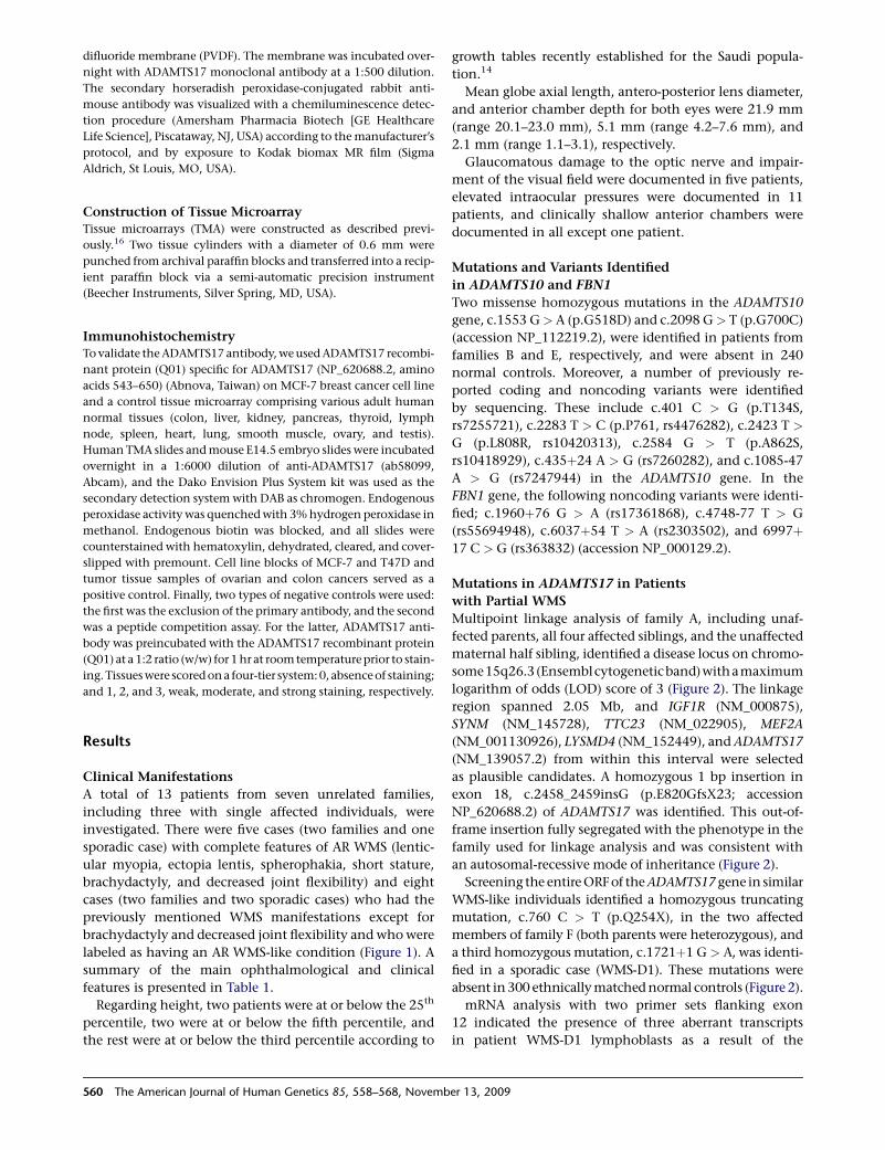
Clinical Manifestations
A total of 13 patients from seven unrelated families,
including three with single affected individuals, were
investigated. There were five cases (two families and one
sporadic case) with complete features of AR WMS (lentic-
ular myopia, ectopia lentis, spherophakia, short stature,
brachydactyly, and decreased joint flexibility) and eight
cases (two families and two sporadic cases) who had the
previously mentioned WMS manifestations except for
brachydactyly and decreased joint flexibility and who were
labeled as having an AR WMS-like condition (Figure 1). A
summary of the main ophthalmological and clinical
features is presented in Table 1.
Regarding height, two patients were at or below the 25th
percentile, two were at or below the fifth percentile, and
the rest were at or below the third percentile according to
560 The American Journal of Human Genetics 85, 558–568, Novem
growth tables recently established for the Saudi popula-
tion.14
Mean globe axial length, antero-posterior lens diameter,
and anterior chamber depth for both eyes were 21.9 mm
(range 20.1–23.0 mm), 5.1 mm (range 4.2–7.6 mm), and
2.1 mm (range 1.1–3.1), respectively.
Glaucomatous damage to the optic nerve and impair-
ment of the visual field were documented in five patients,
elevated intraocular pressures were documented in 11
patients, and clinically shallow anterior chambers were
documented in all except one patient.
Mutations and Variants Identified
in ADAMTS10 and FBN1
Two missense homozygous mutations in the ADAMTS10
gene, c.1553 G> A (p.G518D) and c.2098 G> T (p.G700C)
(accession NP_112219.2), were identified in patients from
families B and E, respectively, and were absent in 240
normal controls. Moreover, a number of previously re-
ported coding and noncoding variants were identified
by sequencing. These include c.401 C > G (p.T134S,
rs7255721), c.2283 T > C (p.P761, rs4476282), c.2423 T >
G (p.L808R, rs10420313), c.2584 G > T (p.A862S,
rs10418929), c.435þ24 A > G (rs7260282), and c.1085-47
A > G (rs7247944) in the ADAMTS10 gene. In the
FBN1 gene, the following noncoding variants were identi-
fied; c.1960þ76 G > A (rs17361868), c.4748-77 T > G
(rs55694948), c.6037þ54 T > A (rs2303502), and 6997þ17 C > G (rs363832) (accession NP_000129.2).
Mutations in ADAMTS17 in Patients
with Partial WMS
Multipoint linkage analysis of family A, including unaf-
fected parents, all four affected siblings, and the unaffected
maternal half sibling, identified a disease locus on chromo-
some15q26.3(Ensemblcytogeneticband)withamaximum
logarithm of odds (LOD) score of 3 (Figure 2). The linkage
region spanned 2.05 Mb, and IGF1R (NM_000875),
SYNM (NM_145728), TTC23 (NM_022905), MEF2A
(NM_001130926), LYSMD4 (NM_152449), and ADAMTS17
(NM_139057.2) from within this interval were selected
as plausible candidates. A homozygous 1 bp insertion in
exon 18, c.2458_2459insG (p.E820GfsX23; accession
NP_620688.2) of ADAMTS17 was identified. This out-of-
frame insertion fully segregated with the phenotype in the
family used for linkage analysis and was consistent with
an autosomal-recessive mode of inheritance (Figure 2).
Screening the entire ORF of the ADAMTS17 gene in similar
WMS-like individuals identified a homozygous truncating
mutation, c.760 C > T (p.Q254X), in the two affected
members of family F (both parents were heterozygous), and
a third homozygous mutation, c.1721þ1 G > A, was identi-
fied in a sporadic case (WMS-D1). These mutations were
absent in 300 ethnically matched normal controls (Figure 2).
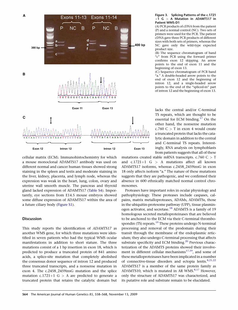
mRNA analysis with two primer sets flanking exon
12 indicated the presence of three aberrant transcripts
in patient WMS-D1 lymphoblasts as a result of the
ber 13, 2009
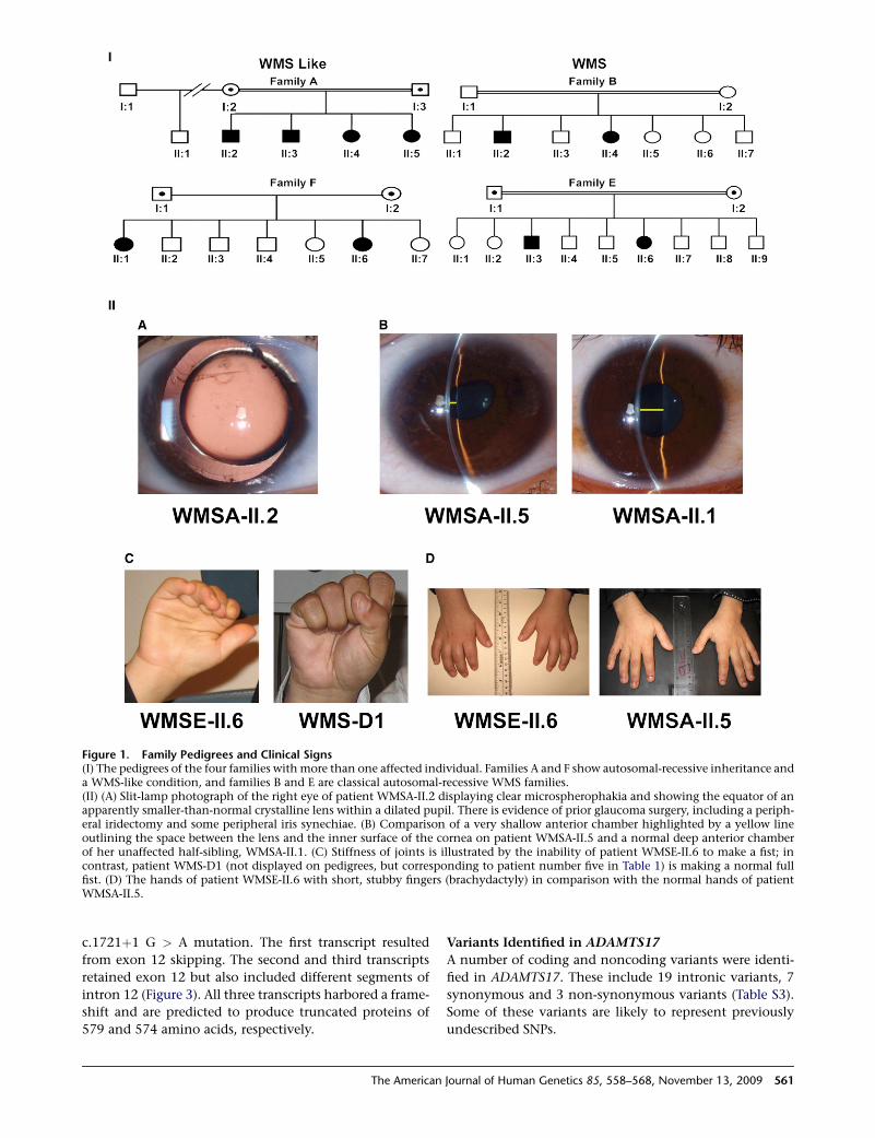
Figure 1. Family Pedigrees and Clinical Signs(I) The pedigrees of the four families with more than one affected individual. Families A and F show autosomal-recessive inheritance anda WMS-like condition, and families B and E are classical autosomal-recessive WMS families.(II) (A) Slit-lamp photograph of the right eye of patient WMSA-II.2 displaying clear microspherophakia and showing the equator of anapparently smaller-than-normal crystalline lens within a dilated pupil. There is evidence of prior glaucoma surgery, including a periph-eral iridectomy and some peripheral iris synechiae. (B) Comparison of a very shallow anterior chamber highlighted by a yellow lineoutlining the space between the lens and the inner surface of the cornea on patient WMSA-II.5 and a normal deep anterior chamberof her unaffected half-sibling, WMSA-II.1. (C) Stiffness of joints is illustrated by the inability of patient WMSE-II.6 to make a fist; incontrast, patient WMS-D1 (not displayed on pedigrees, but corresponding to patient number five in Table 1) is making a normal fullfist. (D) The hands of patient WMSE-II.6 with short, stubby fingers (brachydactyly) in comparison with the normal hands of patientWMSA-II.5.
c.1721þ1 G > A mutation. The first transcript resulted
from exon 12 skipping. The second and third transcripts
retained exon 12 but also included different segments of
intron 12 (Figure 3). All three transcripts harbored a frame-
shift and are predicted to produce truncated proteins of
579 and 574 amino acids, respectively.
The American
Variants Identified in ADAMTS17
A number of coding and noncoding variants were identi-
fied in ADAMTS17. These include 19 intronic variants, 7
synonymous and 3 non-synonymous variants (Table S3).
Some of these variants are likely to represent previously
undescribed SNPs.
Journal of Human Genetics 85, 558–568, November 13, 2009 561
iese
AverageAxialLengthf
ShallowAnteriorChamber
PeripheralIrisSynechiae Gene/Mutation
23.0 Yes Yes ADAMTS17/c.2458_2459insG
22.8 No No ADAMTS17/c.2458_2459insG
22.5 Yes Yes ADAMTS17/c.2458_2459insG
21.5 Yes Yes ADAMTS17/c.2458_2459insG
21.3 Yes No ADAMTS17/c.1721þ1G > A
21.7 Yes Yes ADAMTS17/c.760 C > T
20.7 Yes Yes ADAMTS17/c.760 C > T
21.9 Yes Yes No mutations inADAMTS10,ADAMTS17and FBN1
22.4 Yes No ADAMTS10/c.1553 G > A
21.4 Yes No ADAMTS10/c.1553 G > A
23.7 Yes No ADAMTS10/c.2098 G > T
20.1 Yes No ADAMTS10/c.2098 G > T
21.4 Yes No No mutations inADAMTS10,ADAMTS17and FBN1
and 6, respectively.
562
Th
eA
merica
nJo
urn
alof
Hum
an
Gen
etics
85,
558–568,
Nove
mb
er13,
2009
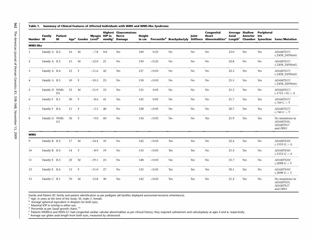
Table 1. Summary of Clinical Features of Affected Individuals with WMS and WMS-like Syndrome
NumberFamilyID
PatientID Agea Gender
MyopiaLevelb
HighestIOP inmmHgc
GlaucomatousNerveDamage
Heightin cm Percentiled Brachydactyly
JointStiffness
CongenitalHeartAbnormalit
WMS-like
1 Family A II.2 16 M �7.8 NA Yes 160 0.25 No No No
2 Family A II.3 15 M �12.0 21 No 150 <0.25 No No No
3 Family A II.4 12 F �11.6 42 Yes 127 <0.03 No No No
4 Family A II.5 10 F �10.5 23 No 118 <0.03 No No No
5 Family D WMS-D1
33 M �11.9 33 Yes 155 0.03 No No No
6 Family F II.1 30 F �8.6 41 Yes 145 0.05 No No No
7 Family F II.6 11 F �5.5 40 No 128 <0.05 No No No
8 Family G WMS-G1
36 F �9.0 60 No 134 <0.03 No No Yes
WMS
9 Family B II.2 17 M �14.4 35 No 142 <0.03 Yes Yes No
10 Family B II.4 14 F �8.9 19 No 133 <0.03 Yes Yes No
11 Family E II.3 22 M �19.1 23 No 148 <0.03 Yes Yes No
12 Family E II.6 13 F �11.0 27 No 132 <0.03 Yes Yes Yes
13 Family C II.1 70 M 13.8 30 Yes 142 <0.03 Yes Yes No
Family and Patient ID: family and patient identification as per pedigree (all families displayed autosomal-recessive inheritance).a Age: in years at the time of the study. M, male; F, female.b Average spherical equivalent in diopters for both eyes.c Maximal IOP in mmHg in either eye.d Percentile as per Saudi growth charts.14
e Patients WMEII.6 and WMS-G1 had congenital cardiac valvular abnormalities as per clinical history; they required catheterism and valvuloplasty at ages 4f Average eye globe axial length from both eyes, measured by ultrasound.
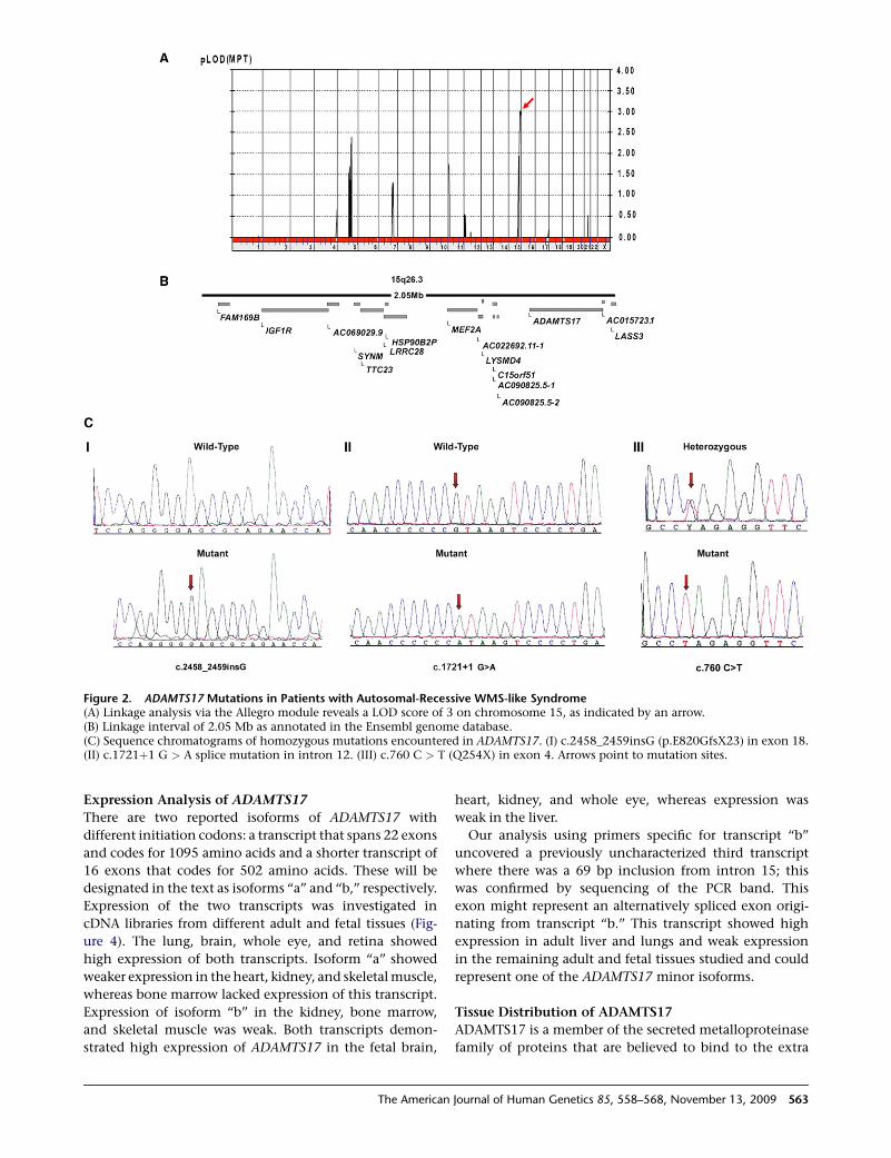
Figure 2. ADAMTS17 Mutations in Patients with Autosomal-Recessive WMS-like Syndrome(A) Linkage analysis via the Allegro module reveals a LOD score of 3 on chromosome 15, as indicated by an arrow.(B) Linkage interval of 2.05 Mb as annotated in the Ensembl genome database.(C) Sequence chromatograms of homozygous mutations encountered in ADAMTS17. (I) c.2458_2459insG (p.E820GfsX23) in exon 18.(II) c.1721þ1 G > A splice mutation in intron 12. (III) c.760 C > T (Q254X) in exon 4. Arrows point to mutation sites.
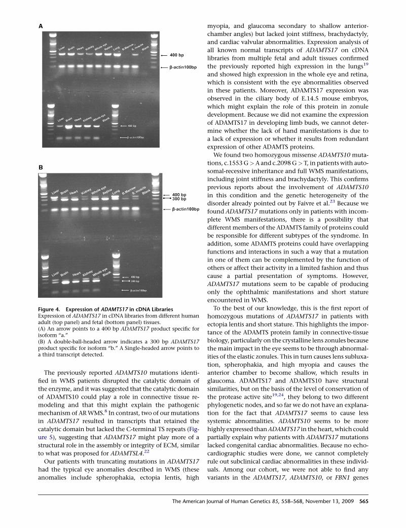
Expression Analysis of ADAMTS17
There are two reported isoforms of ADAMTS17 with
different initiation codons: a transcript that spans 22 exons
and codes for 1095 amino acids and a shorter transcript of
16 exons that codes for 502 amino acids. These will be
designated in the text as isoforms ‘‘a’’ and ‘‘b,’’ respectively.
Expression of the two transcripts was investigated in
cDNA libraries from different adult and fetal tissues (Fig-
ure 4). The lung, brain, whole eye, and retina showed
high expression of both transcripts. Isoform ‘‘a’’ showed
weaker expression in the heart, kidney, and skeletal muscle,
whereas bone marrow lacked expression of this transcript.
Expression of isoform ‘‘b’’ in the kidney, bone marrow,
and skeletal muscle was weak. Both transcripts demon-
strated high expression of ADAMTS17 in the fetal brain,
The American
heart, kidney, and whole eye, whereas expression was
weak in the liver.
Our analysis using primers specific for transcript ‘‘b’’
uncovered a previously uncharacterized third transcript
where there was a 69 bp inclusion from intron 15; this
was confirmed by sequencing of the PCR band. This
exon might represent an alternatively spliced exon origi-
nating from transcript ‘‘b.’’ This transcript showed high
expression in adult liver and lungs and weak expression
in the remaining adult and fetal tissues studied and could
represent one of the ADAMTS17 minor isoforms.
Tissue Distribution of ADAMTS17
ADAMTS17 is a member of the secreted metalloproteinase
family of proteins that are believed to bind to the extra
Journal of Human Genetics 85, 558–568, November 13, 2009 563
cellular matrix (ECM). Immunohistochemistry for which
a mouse monoclonal ADAMTS17 antibody was used on
different normal and cancer human tissues showed strong
staining in the spleen and testis and moderate staining in
the liver, kidney, placenta, and lymph node, whereas the
expression was weak in the heart, lung, colon, ovary and
uterine wall smooth muscle. The pancreas and thyroid
gland lacked expression of ADAMTS17 (Table S4). Impor-
tantly, eye sections from E14.5 mouse embryos showed
some diffuse expression of ADAMTS17 within the area of
a future ciliary body (Figure S1).
Discussion
This study reports the identification of ADAMTS17 as
another WMS gene, for which three mutations were iden-
tified in seven patients who had the typical WMS ocular
manifestations in addition to short stature. The three
mutations consist of a 1 bp insertion in exon 18, which is
predicted to produce a truncated protein of 841 amino
acids, a splice-site mutation that completely abolished
the consensus donor sequence of intron 12 and produced
three truncated transcripts, and a nonsense mutation in
exon 4. The c.2458_2459insG mutation and the splice
mutation c.1721þ1 G > A are predicted to generate a
truncated protein that retains the catalytic domain but
Figure 3. Splicing Patterns of the c.1721þ1 G > A Mutation in ADAMTS17 inPatient WMS-D1(A) PCR products of cDNA from the patient(P) and a normal control (NC). Two sets ofprimers were used for the PCR. The patientcDNA gave three PCR products of differentsizes with both sets of primers, whereas theNC gave only the wild-type expectedproduct size.(B) The sequence chromatogram of band‘‘c’’ from PCR using the forward primerconfirms exon 12 skipping. An arrowpoints to the end of exon 11 and thebeginning of exon 13.(C) Sequence chromatogram of PCR band‘‘a.’’ A double-headed arrow points to theend of exon 12 and the beginning ofintron 12, and a single-headed arrowpoints to the end of the ‘‘spliced-in’’ partof intron 12 and the beginning of exon 13.
lacks the central and/or C-terminal
TS repeats, which are thought to be
essential for ECM binding.17 On the
other hand, the nonsense mutation
c.760 C > T in exon 4 would create
a truncated protein that lacks the cata-
lytic domain in addition to the central
and C-terminal TS repeats. Interest-
ingly, RNA analysis on lymphoblasts
from patients suggests that all of these
mutations created stable mRNA transcripts. c.760 C > T
and c.1721þ1 G > A mutations affect all known
ADAMTS17 isoforms, whereas c.2458_2459insG in exon
18 only affects isoform ‘‘a.’’ The nature of these mutations
suggests that they are pathogenic, and we confirmed their
absence in 600 ethnically matched normal control chro-
mosomes.
Proteases have important roles in ocular physiology and
pathophysiology. These proteases include caspases, cal-
pains, matrix metalloproteases, ADAMs, ADAMTSs, those
in the ubiquitin-protesome pathway (UPP), tissue plasmin-
ogen activator, and secretase.18 ADAMTS is a family of 19
homologous secreted metalloproteinases that are believed
to be anchored to the ECM via their C-terminal thrombo-
spondin (TS) repeats.19 These proteins undergo N-terminal
processing and removal of the prodomain during their
transit through the membrane of the endoplasmic retic-
ulum; they also undergo C-terminal processing that affects
substrate specificity and ECM binding.20 Previous charac-
terization of the ADAMTS proteins showed their involve-
ment in different cellular mechanisms17,19, and some of
these metalloproteinases have been implicated in a number
of connective-tissue disorders and ectopia lentis.8,21,22
ADAMTS17 is a member of the same protein family as
ADAMTS10, which is mutated in AR WMS.8,17 However,
only the structure of ADAMTS17 was characterized, and
its putative role and substrate remain to be elucidated.
564 The American Journal of Human Genetics 85, 558–568, November 13, 2009
The previously reported ADAMTS10 mutations identi-
fied in WMS patients disrupted the catalytic domain of
the enzyme, and it was suggested that the catalytic domain
of ADAMTS10 could play a role in connective tissue re-
modeling and that this might explain the pathogenic
mechanism of AR WMS.8 In contrast, two of our mutations
in ADAMTS17 resulted in transcripts that retained the
catalytic domain but lacked the C-terminal TS repeats (Fig-
ure 5), suggesting that ADAMTS17 might play more of a
structural role in the assembly or integrity of ECM, similar
to what was proposed for ADAMTSL4.22
Our patients with truncating mutations in ADAMTS17
had the typical eye anomalies described in WMS (these
anomalies include spherophakia, ectopia lentis, high
Figure 4. Expression of ADAMTS17 in cDNA LibrariesExpression of ADAMTS17 in cDNA libraries from different humanadult (top panel) and fetal (bottom panel) tissues.(A) An arrow points to a 400 bp ADAMTS17 product specific forisoform ‘‘a.’’(B) A double-ball-headed arrow indicates a 300 bp ADAMTS17product specific for isoform ‘‘b.’’ A Single-headed arrow points toa third transcript detected.
The American
myopia, and glaucoma secondary to shallow anterior-
chamber angles) but lacked joint stiffness, brachydactyly,
and cardiac valvular abnormalities. Expression analysis of
all known normal transcripts of ADAMTS17 on cDNA
libraries from multiple fetal and adult tissues confirmed
the previously reported high expression in the lungs19
and showed high expression in the whole eye and retina,
which is consistent with the eye abnormalities observed
in these patients. Moreover, ADAMTS17 expression was
observed in the ciliary body of E.14.5 mouse embryos,
which might explain the role of this protein in zonule
development. Because we did not examine the expression
of ADAMTS17 in developing limb buds, we cannot deter-
mine whether the lack of hand manifestations is due to
a lack of expression or whether it results from redundant
expression of other ADAMTS proteins.
We found two homozygous missense ADAMTS10 muta-
tions, c.1553 G>A and c.2098 G> T, in patients with auto-
somal-recessive inheritance and full WMS manifestations,
including joint stiffness and brachydactyly. This confirms
previous reports about the involvement of ADAMTS10
in this condition and the genetic heterogeneity of the
disorder already pointed out by Faivre et al.23 Because we
found ADAMTS17 mutations only in patients with incom-
plete WMS manifestations, there is a possibility that
different members of the ADAMTS family of proteins could
be responsible for different subtypes of the syndrome. In
addition, some ADAMTS proteins could have overlapping
functions and interactions in such a way that a mutation
in one of them can be complemented by the function of
others or affect their activity in a limited fashion and thus
cause a partial presentation of symptoms. However,
ADAMTS17 mutations seem to be capable of producing
only the ophthalmic manifestations and short stature
encountered in WMS.
To the best of our knowledge, this is the first report of
homozygous mutations of ADAMTS17 in patients with
ectopia lentis and short stature. This highlights the impor-
tance of the ADAMTS protein family in connective-tissue
biology, particularly on the crystalline lens zonules because
the main impact in the eye seems to be through abnormal-
ities of the elastic zonules. This in turn causes lens subluxa-
tion, spherophakia, and high myopia and causes the
anterior chamber to become shallow, which results in
glaucoma. ADAMTS17 and ADAMTS10 have structural
similarities, but on the basis of the level of conservation of
the protease active site19,24, they belong to two different
phylogenetic nodes, and so far we do not have an explana-
tion for the fact that ADAMTS17 seems to cause less
systemic abnormalities. ADAMTS10 seems to be more
highly expressed than ADAMTS17 in the heart, which could
partially explain why patients with ADAMTS17 mutations
lacked congenital cardiac abnormalities. Because no echo-
cardiographic studies were done, we cannot completely
rule out subclinical cardiac abnormalities in these individ-
uals. Among our cohort, we were not able to find any
variants in the ADAMTS17, ADAMTS10, or FBN1 genes
Journal of Human Genetics 85, 558–568, November 13, 2009 565
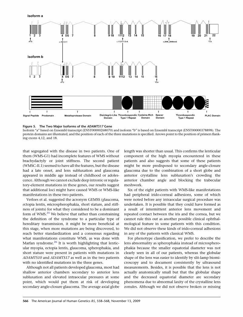
Figure 5. The Two Major Isoforms of the ADAMTS17 GeneIsoform ‘‘a’’ based on Ensembl transcript (ENST00000268070) and isoform ‘‘b’’ is based on Ensembl transcript (ENST00000378898). Theprotein domains are illustrated, and the position of each of the three mutations is specified. Arrows point to the position of primers flank-ing exons 4,12, and 18.
that segregated with the disease in two patients. One of
them (WMS-G1) had incomplete features of WMS without
brachydactyly or joint stiffness. The second patient
(WMSC-II.1) seemed to have all the features, but the disease
had a late onset, and lens subluxation and glaucoma
appeared in middle age instead of childhood or adoles-
cence. Although we cannot exclude deep intronic or regula-
tory-element mutations in these genes, our results suggest
that additional loci might have caused WMS or WMS-like
manifestations in these two patients.
Verloes et al. suggested the acronym GEMSS (glaucoma,
ectopia lentis, microspherophakia, short stature, and stiff-
ness of joints) for what they considered to be a dominant
form of WMS.25 We believe that rather than constraining
the definition of the syndrome to a particular type of
hereditary transmission, it might be more beneficial at
this stage, when more mutations are being discovered, to
reach better standardization and a consensus regarding
what manifestations constitute WMS, as was done with
Marfan syndrome.26 It is worth highlighting that lentic-
ular myopia, ectopia lentis, glaucoma, spherophakia, and
short stature were present in patients with mutations in
ADAMTS10 and ADAMTS17 as well as in the two patients
with no identified mutations in the three genes.
Although not all patients developed glaucoma, most had
shallow anterior chambers secondary to anterior lens
subluxation and elevated intraocular pressures at some
point, which would put them at risk of developing
secondary angle-closure glaucoma. The average axial globe
566 The American Journal of Human Genetics 85, 558–568, Novemb
length was shorter than usual. This confirms the lenticular
component of the high myopia encountered in these
patients and also suggests that some of these patients
might be more predisposed to secondary angle-closure
glaucoma due to the combination of a short globe and
anterior crystalline lens subluxation’s crowding the
anterior chamber angle and blocking the trabecular
meshwork.
Six of the eight patients with WMS-like manifestations
had peripheral irido-corneal adhesions, some of which
were noted before any intraocular surgical procedure was
undertaken. It is possible that they could have formed as
a result of intermittent anterior lens movement and
repeated contact between the iris and the cornea, but we
cannot rule this out as another possible clinical ophthal-
mological feature in some patients with this condition.
We did not observe these kinds of irido-corneal adhesions
in any of the patients with classical WMS.
For phenotype classification, we prefer to describe the
lens abnormality as spherophakia instead of microsphero-
phakia because the smaller equatorial diameter was not
clearly seen in all of our patients, whereas the globular
shape of the lens was easier to identify by slit-lamp biomi-
croscopy and to document consistently by ultrasound
measurements. Besides, it is possible that the lens is not
actually anatomically small but that the globular shape
and the decreased equatorial diameter are secondary
phenomena due to abnormal laxity of the crystalline lens
zonules. Although we did not observe broken or missing
er 13, 2009

zonules, we assume that there might have been significant
zonular elongation, particularly in those individuals with
significant lens displacement or clear visualization of the
whole equator of the lens after dilation.
In summary, we have identified null mutations in
ADAMTS17 as a cause of an autosomal-recessive WMS-
like syndrome. We have confirmed abnormalities in
ADAMTS10 in typical cases of WMS. Lenticular myopia, ec-
topia lentis, glaucoma (secondary to anterior lens subluxa-
tion and angle closure), spherophakia, and short stature
were common denominators in patients with complete
and incomplete manifestations of WMS. In our cohort of
patients with these manifestations, ADAMTS17 and
ADAMTS10 mutations were present in more than 80% of
cases. Further work involving other ethnic groups and ec-
topia lentis patients might clarify the role that different
mutations play in the pathogenesis of this condition.
Supplemental Data
Supplemental data include one figure and four tables and can be
found with this article online at http://www.cell.com/AJHG/.
Acknowledgments
The authors would like to thank Valorie Balde, Zakia Shinwari,
Ayodele. A. Alaiya, and Tarfa Al Sheddi for all their technical assis-
tance; Faisal Al Hamidi for his help in patient recruitment;
members of the photography department of the King Khaled
Eye Specialist Hospital (KKESH) for their detailed documentation
of the clinical signs; Mohamad Rajab, Shamsa Al Enazi, and Dorota
Moniez for their help with DNA Sequencing; Batoul Baz and Salma
Wakil of the genotyping core facility for their help in processing
the 10K affymetrix chip; and Futwan Al Mohanna for his help
and efforts. This work was approved and funded by King Faisal
Specialist Hospital and Research Centre (RAC# 2070008). and
KKESH (RP# 0664-P).
Received: July 22, 2009
Revised: September 7, 2009
Accepted: September 21, 2009
Published online: October 15, 2009
Web Resources
The URLs for data presented herein are as follows:
GenBank, http://www.ncbi.nlm.nih.gov/Genbank
Ensembl project genome database, http://www.ensembl.org/
index.html
Online Mendelian Inheritance in Man (OMIM), http://www.ncbi.
nlm.nih.gov/omim
Single Nucleotide Polymorphism database, http://www.ncbi.nlm.
nih.gov/SNP/
UCSC Genome Browser, http://genome.ucsc.edu/
Accession Numbers
Submitter SNP (ss) numbers for variants identified in this study are
ss161151830, ss161151831, ss161151832, ss161151833.
The American
References
1. Wright, K.W., and Chrousos, G.A. (1985). Weill-Marchesani
syndrome with bilateral angle-closure glaucoma. J. Pediatr.
Ophthalmol. Strabismus 22, 129–132.
2. Chu, B.S. (2006). Weill-Marchesani syndrome and secondary
glaucoma associated with ectopia lentis. Clin. Exp. Optom.
89, 95–99.
3. Giordano, N., Senesi, M., Battisti, E., Mattii, G., and Gennari,
C. (1997). Weill-Marchesani syndrome: Report of an unusual
case. Calcif. Tissue Int. 60, 358–360.
4. Rennert, O.M. (1969). The Marchesani syndrome. A brief
review. Am. J. Dis. Child. 117, 703–705.
5. Kojuri, J., Razeghinejad, M.R., and Aslani, A. (2007). Cardiac
findings in Weill-Marchesani syndrome. Am. J. Med. Genet.
A 143A, 2062–2064.
6. Herrera, J., andMorales,M. (1986).Weill-Marchesani syndrome.
Rev. Chil. Pediatr. 57, 571–572.
7. Faivre, L., Gorlin, R.J., Wirtz, M.K., Godfrey, M., Dagoneau, N.,
Samples, J.R., Le Merrer, M., Collod-Beroud, G., Boileau, C.,
Munnich, A., et al. (2003). In frame fibrillin-1 gene deletion
in autosomal dominant Weill-Marchesani syndrome. J. Med.
Genet. 40, 34–36.
8. Dagoneau, N., Benoist-Lasselin, C., Huber, C., Faivre, L.,
Megarbane, A., Alswaid, A., Dollfus, H., Alembik, Y., Munnich,
A., Legeai-Mallet, L., et al. (2004). ADAMTS10 mutations
in autosomal recessive Weill-Marchesani syndrome. Am.
J. Hum. Genet. 75, 801–806.
9. Young, I.D., Fielder, A.R., and Casey, T.A. (1986). Weill-
Marchesani syndrome in mother and son. Clin. Genet. 30,
475–480.
10. Evereklioglu, C., Hepsen, I.F., and Er, H. (1999). Weill-Marche-
sani syndrome in three generations. Eye 13, 773–777.
11. Kutz, W.E., Wang, L.W., Dagoneau, N., Odrcic, K.J., Cormier-
Daire, V., Traboulsi, E.I., and Apte, S.S. (2008). Functional
analysis of an ADAMTS10 signal peptide mutation in Weill-
Marchesani syndrome demonstrates a long-range effect on
secretion of the full-length enzyme. Hum. Mutat. 29,
1425–1434.
12. Dietz, H.C., Saraiva, J.M., Pyeritz, R.E., Cutting, G.R., and
Francomano, C.A. (1992). Clustering of fibrillin (FBN1)
missense mutations in Marfan syndrome patients at cysteine
residues in EGF-like domains. Hum. Mutat. 1, 366–374.
13. Dietz, H.C., and Pyeritz, R.E. (1995). Mutations in the human
gene for fibrillin-1 (FBN1) in the Marfan syndrome and related
disorders. Hum. Mol. Genet. 4, 1799–1809.
14. El Mouzan, M.I., Al Herbish, A.S., Al Salloum, A.A., Foster, P.J.,
Al Omar, A.A., Qurachi, M.M., and Kecojevic, T. (2008).
Comparison of the 2005 growth charts for Saudi children
and adolescents to the 2000 CDC growth charts. Ann. Saudi
Med. 28, 334–340.
15. Lindner, T.H., and Hoffmann, K. (2005). easyLINKAGE: A
PERL script for easy and automated two-/multi-point linkage
analyses. Bioinformatics 21, 405–407.
16. Bavi, P., Abubaker, J., Hussain, A., Sultana, M., Al-Dayel, F.,
Uddin, S., and Al-Kuraya, K.S. (2008). Reduced or absent
cyclin H expression is an independent prognostic marker for
poor outcome in diffuse large B-cell lymphoma. Hum. Pathol.
39, 885–894.
17. Porter, S., Clark, I.M., Kevorkian, L., and Edwards, D.R. (2005).
The ADAMTS metalloproteinases. Biochem. J. 386, 15–27.
Journal of Human Genetics 85, 558–568, November 13, 2009 567
18. Wride, M.A., Geatrell, J., and Guggenheim, J.A. (2006). Prote-
ases in eye development and disease. Birth Defects Res. C
Embryo Today 78, 90–105.
19. Cal, S., Obaya, A.J., Llamazares, M., Garabaya, C., Quesada, V.,
and Lopez-Otin, C. (2002). Cloning, expression analysis, and
structural characterization of seven novel human ADAMTSs,
a family of metalloproteinases with disintegrin and thrombo-
spondin-1 domains. Gene 283, 49–62.
20. Gao, G., Westling, J., Thompson, V.P., Howell, T.D., Gott-
schall, P.E., and Sandy, J.D. (2002). Activation of the proteo-
lytic activity of ADAMTS4 (aggrecanase-1) by C-terminal
truncation. J. Biol. Chem. 277, 11034–11041.
21. Colige, A., Sieron, A.L., Li, S.W., Schwarze, U., Petty, E., Werte-
lecki, W., Wilcox, W., Krakow, D., Cohn, D.H., Reardon, W.,
et al. (1999). Human Ehlers-Danlos syndrome type VII C
and bovine dermatosparaxis are caused by mutations in the
procollagen I N-proteinase gene. Am. J. Hum. Genet. 65,
308–317.
22. Ahram, D., Sato, T.S., Kohilan, A., Tayeh, M., Chen, S., Leal, S.,
Al-Salem, M., and El-Shanti, H. (2009). A homozygous muta-
568 The American Journal of Human Genetics 85, 558–568, Novemb
tion in ADAMTSL4 causes autosomal-recessive isolated
ectopia lentis. Am. J. Hum. Genet. 84, 274–278.
23. Faivre, L., Dollfus, H., Lyonnet, S., Alembik, Y., Megarbane, A.,
Samples, J., Gorlin, R.J., Alswaid, A., Feingold, J., Le Merrer,
M., et al. (2003). Clinical homogeneity and genetic heteroge-
neity in Weill-Marchesani syndrome. Am. J. Med. Genet. A
123A, 204–207.
24. Huxley-Jones, J., Apte, S.S., Robertson, D.L., and Boot-Hand-
ford, R.P. (2005). The characterisation of six ADAMTS prote-
ases in the basal chordate Ciona intestinalis provides new
insights into the vertebrate ADAMTS family. Int. J. Biochem.
Cell Biol. 37, 1838–1845.
25. Verloes, A., Hermia, J.P., Galand, A., Koulischer, L., and
Dodinval, P. (1992). Glaucoma-lens ectopia-microspheropha-
kia-stiffness-shortness (GEMSS) syndrome: A dominant
disease with manifestations of Weill-Marchesani syndromes.
Am. J. Med. Genet. 44, 48–51.
26. De Paepe, A., Devereux, R.B., Dietz, H.C., Hennekam, R.C.,
and Pyeritz, R.E. (1996). Revised diagnostic criteria for the
Marfan syndrome. Am. J. Med. Genet. 62, 417–426.
er 13, 2009