Management of Nonsustained Ventricular Tachycardia Guided By Electrophysiological Testing
BioMed CentralBMC Neuroscience
ss
Open AcceResearch articleHomozygous mutation of focal adhesion kinase in embryonic stem cell derived neurons: normal electrophysiological and morphological properties in vitroP Charlesworth2, NH Komiyama2 and SGN Grant*1Address: 1Wellcome Trust Sanger Institute, Hinxton, Cambridge, CB10 1SA, UK and 2Centre for Neuroscience Research, University of Edinburgh, Edinburgh, UK
Email: P Charlesworth - [email protected]; NH Komiyama - [email protected]; SGN Grant* - [email protected]
* Corresponding author
AbstractBackground: Genetically manipulated embryonic stem (ES) cell derived neurons (ESNs) providea powerful system with which to study the consequences of gene manipulation in mature,synaptically connected neurons in vitro. Here we report a study of focal adhesion kinase (FAK),which has been implicated in synapse formation and regulation of ion channels, using the ESNsystem to circumvent the embryonic lethality of homozygous FAK mutant mice.
Results: Mouse ES cells carrying homozygous null mutations (FAK-/-) were generated anddifferentiated in vitro into neurons. FAK-/- ESNs extended axons and dendrites and formedmorphologically and electrophysiologically intact synapses. A detailed study of NMDA receptorgated currents and voltage sensitive calcium currents revealed no difference in their magnitude, ormodulation by tyrosine kinases.
Conclusion: FAK does not have an obligatory role in neuronal differentiation, synapse formationor the expression of NMDA receptor or voltage-gated calcium currents under the conditions usedin this study. The use of genetically modified ESNs has great potential for rapidly and effectivelyexamining the consequences of neuronal gene manipulation and is complementary to mousestudies.
BackgroundA major goal in post-genomic neuroscience is to elucidatethe function of individual genes in neurons. Gene target-ing is one of the most widely used methods to study thefunction of genes in the vertebrate nervous system and inrecent years the need for conditional mutation strategieshas been emphasised. For example, the tetracycline sys-tem has been used to transiently express genes in the brain[1] and the Cre/LoxP system used to delete genes inselected neuronal populations [2]. One disadvantage to
these techniques is that it can take 2 or more years forresults, in part because of the breeding of mice. Thereforeany method that allows for a shorter duration and reducesthe need for animals is potentially useful. Toward thisend, we have adopted an experimental strategy usinggenetically engineered embryonic stem (ES) cell derivedneurons (ESNs). This system has the potential to greatlyspeed the functional analysis of genetic manipulationsand importantly also allows access to lethal mouse phe-notypes.
Published: 12 June 2006
BMC Neuroscience 2006, 7:47 doi:10.1186/1471-2202-7-47
Received: 27 February 2006Accepted: 12 June 2006
This article is available from: http://www.biomedcentral.com/1471-2202/7/47
© 2006 Charlesworth et al; licensee BioMed Central Ltd.This is an Open Access article distributed under the terms of the Creative Commons Attribution License (http://creativecommons.org/licenses/by/2.0), which permits unrestricted use, distribution, and reproduction in any medium, provided the original work is properly cited.
Page 1 of 11(page number not for citation purposes)

BMC Neuroscience 2006, 7:47 http://www.biomedcentral.com/1471-2202/7/47
Totipotent murine ES cells can be induced to differentiatein vitro to form mature post-mitotic neurons, which inaddition to being synaptically connected, show manyproperties comparable to primary cultures of embryonicneonatal cortex [3-5]. The use of genetically manipulatedES cells to study knockout of gene function in neurons hashowever thus far not been extensively exploited [6-8]. Inthese studies, the spontaneous conversion of a hetero-zygous to a homozygous knockout ES cell has beenselected using high concentrations of neomycin. An alter-native and more reliable strategy, used in this study,involves the use of Cre/loxP to create the homozygousmutation in sequential rounds of gene targeting (see Fig.1, and additional file 1). This has particular advantages,allowing precise targeting of both alleles using the samevector and the ES cell line carrying mutations in which agene was flanked by two loxP sites may be used to gener-ate a conditional, tissue specific, knock-out mouse (see[9]).
Genetically modified mice have become a standard toolto examine the role of synaptic proteins in the biology ofneurotransmission, synaptic plasticity and behaviour. Thefirst studies of tyrosine kinases in synaptic plasticity[10,11] and learning [10] revealed an important role forfyn tyrosine kinase. The lack of Fyn resulted in reductionin glutamate receptor signaling and the impaired induc-tion of long-term potentiation. In addition, there wereobserved changes in the morphology of the dendritic treeof hippocampus pyramidal neurons and learning deficits.Since Fyn was a Src family kinase, subsequent studies onknown Src substrates lead to the finding that FAK washypophosphorylated in fyn mutant mice and this wasassociated with reduced kinase activity [12]. Moreover,studies in non-neuronal cells showed that FAK wasresponsible for phosphorylation of several proteinsinvolved in cell-cell contacts (see below) and thus a likelymediator of the synaptic phenotype of fyn mutant mice. Inaddition, Rico and colleagues showed that in vivo the dele-tion of FAK resulted in abnormal architecuture of neurons[13]. Thus FAK may play an important role in the forma-tion and function of synapses and neurotransmission.
Unfortunately, mice lacking FAK die in early embryogen-esis before neurons can be cultured (Mice carryinghomozygous FAK null mutation die at an early stage inembryonic development (e 8.5) [15], although use hasbeen made recently of conditional FAK knockout mice[13,16,17] to circumvent this problem. An alternativeapproach is the creation of neurons derived directly fromhomozygous mutant embryonic stem cells.
FAK is a 125 KDa non-receptor protein-tyrosine kinaselocalised at focal adhesion plaques where cells attach tothe extracellular matrix and much work has focused on
the role of FAK in cell adhesion, specifically as a key medi-ator of signalling events initiated by integrin binding andactivation of FAK by autophosphorylation [18]. FAK pos-sesses binding sites for a number of proteins, most impor-tantly, Src family kinases [19]. These link FAK to a varietyof signaling pathways, including the MAPK pathway. FAKis highly expressed in the brain and is activated by a vari-ety of stimuli, such as neurotransmitters (including gluta-mate and acetylcholine), lipid messengers (such asanandamide, arachidonic acid and lysophatidic acid),growth factors (insulin, NGF, endothelin) and NCAM[20,21]). The identification of neuronal and brain specificsplice isoforms of FAK generated by alternative RNA splic-ing [22] also suggested that FAK may have roles in neu-rons beyond, or independent of, cell attachment.Furthermore, unlike the restricted localisation of FAK tofocal contacts in non-neuronal cells, FAK is expressedthroughout neurons, suggesting FAK has different mecha-nisms for subcellular localisation in neurons, and, poten-tially, has novel targets [23]. Since neuronal FAK appearsto have functions other than adhesion/motility in neu-rons, we explored the possibilities, principally using thewhole cell patch clamp technique, that FAK is insteadinvolved in ion channel modulation and synapse forma-tion. We focused our attention on two channel types withpivotal roles in neuronal signaling pathways: the NMDAreceptor and voltage-gated calcium channels.
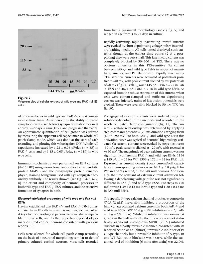
ResultsFAK expression in wild type and mutant cellsWestern blot analyses using FAK monoclonal antibodywere performed on cellular extracts to examine FAKexpression in wild type (WT) and FAK null ES cells.Results showed a single band whose molecular weightwas approximately 125 KDa in E14TG2a lines. No signalwas detected in FAK null homozygous lines (see Fig. 2).Additionally, McLean et al [9] found no expression of atruncated product using N-terminal FAK antibodies. PCRanalysis of RNA extracted from WT ESNs (10 days in vitro)was performed using primers hybridising either side ofthe FAK neuronal specific exons [22]. The resulting prod-uct contained bands corresponding to neuron-specificsplice isoforms of FAK, in addition to non-neuronalspliced FAK, the latter presumably due to the presence ofnon-neuronal cells in the differentiated ES cell culture.
FAK null ESNs display normal growth and morphologyAfter plating the cell suspension created by enzymatic dis-persal of the trans-retinoic acid (RA) treated embryoidbodies, cells rapidly adhered to the substrate (laminin). Inboth wild type and FAK -/- cultures cells with rudimentaryprocesses were evident within 24–48 hrs. With increasingtime in culture, cells with progressively longer processesand more complex branching were observed. Again, noclear difference was observed in the length or complexity
Page 2 of 11(page number not for citation purposes)

BMC Neuroscience 2006, 7:47 http://www.biomedcentral.com/1471-2202/7/47
Page 3 of 11(page number not for citation purposes)
FAK locus targetingFigure 1FAK locus targeting. Simplified diagram of FAK locus targeting strategy. The top of the panel shows the targeted allele con-taining 3 loxP sites. Below is shown one of the deletion products following cre recombinase expression, with ATP binding loops deleted. This was used in the present study to generate homozygous FAK null ES cells after a second round of targeting and deletion, depicted schematically in the lower panel. For further details see additional file [1]1and [9].
ATP binding loopSelection cassette
Lox P Lox P Lox P
Targeted Allele
Cre expression
Y397
Y397
Knockout
allele
Repeat targeting
& Cre expression
FAK null
ES cells
Targeted allele
Heterozygous knockout
Homozygous knockout
Targeting of 2nd allele
BMC Neuroscience 2006, 7:47 http://www.biomedcentral.com/1471-2202/7/47
of processes between wild type and FAK -/- cells at compa-rable culture times. As evidenced by the ability to recordsynaptic currents (see below) synapse formation began atapprox. 5–7 days in vitro (DIV), and progressed thereafter.An approximate quantitation of cell growth was derivedby measuring the apparent cell capacitance in whole cellpatch clamp mode, which was done at the start of eachrecording, and plotting this value against DIV. Whole cellcapacitance increased by 1.22 ± 0.06 pF/day (n = 85) inFAK -/- cells, and by 1.15 ± 0.05 pF/day (n = 119) in wildtype cells.
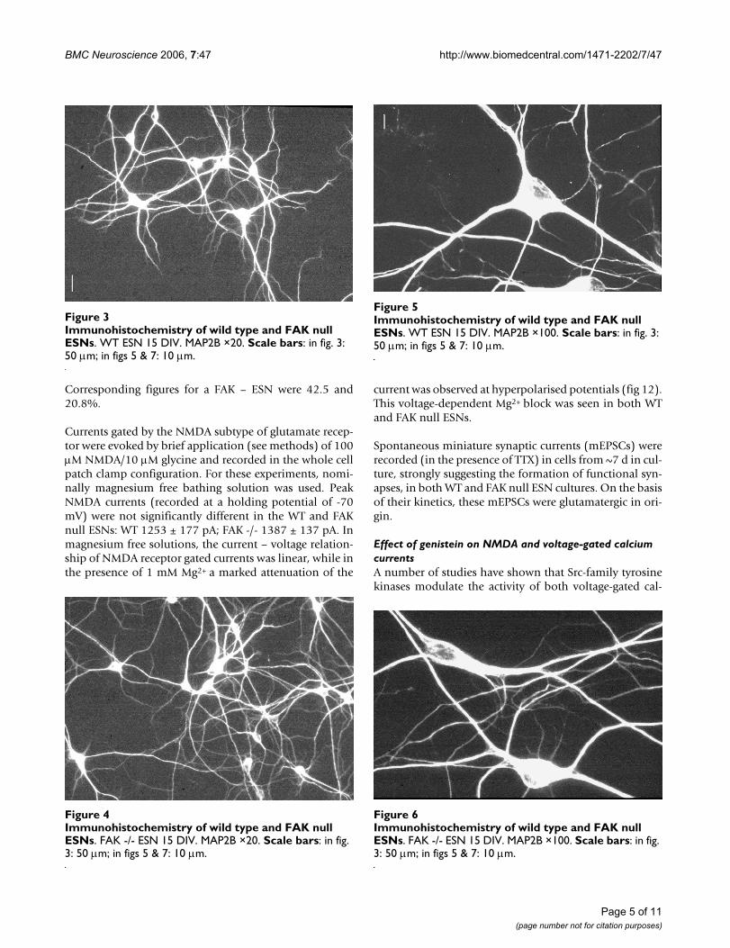
Immunohistochemistry was performed on ESN cultures(8–15 DIV) using monoclonal antibodies to the dendriticprotein MAP2B and the pre-synaptic protein synapto-physin, staining being visualised with Cy3 conjugated sec-ondary antibody. The results showed (see Fig 3, 4, 5, 6, 7,8) the extent and complexity of neuronal processes inboth wild type and FAK -/- ESN cultures, and the extensiveformation of synapses in both.
Electrophysiological properties of wild type and Fak null ESNsHaving established that FAK +/+ and FAK -/- ESNs differ-entiated from ES cells in a similar manner, we next askedif key electrophysiological parameters were also compara-ble in these cells, and to the properties expected of pri-mary cultured cortical neurons consistent with previousreports [3-5].
Cells were selected for whole cell patch clamp recordingon the basis of a neuronal morphology similar to that ofprimary cultured cortical neurons. Most cells recorded
from had a pyramidal morphology (see e.g fig. 5) andranged in age from 3 to 21 days in culture.
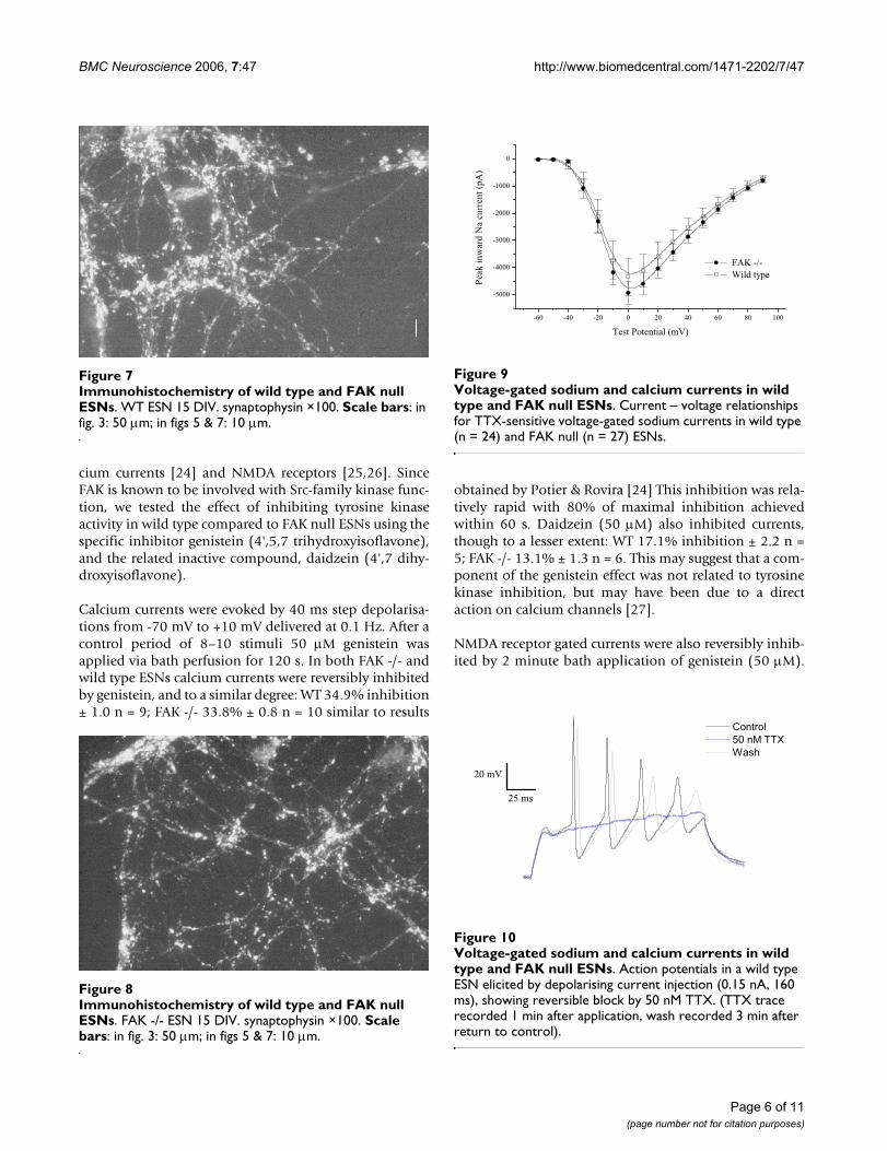
Rapidly activating, rapidly inactivating inward currentswere evoked by short depolarising voltage pulses in stand-ard bathing medium. All cells tested displayed such cur-rents, though at the earliest time points (2–3 d post-plating) they were very small. This fast inward current wascompletely blocked by 50–200 nM TTX. There was noobvious difference in this TTX-sensitive Na currentbetween FAK -/- and wild type ESNs in respect of magni-tude, kinetics, and IV relationship. Rapidly inactivatingTTX- sensitive currents were activated at potentials posi-tive to -40 mV, with peak current elicited by test potentialsof ~0 mV (fig 9). Peak INa was 5145 pA ± 496 n = 25 in FAK-/- ESN and 4671 pA ± 861 n = 18 in wild type ESNs. Asexpected from the robust expression of this current, whencells were current-clamped and sufficient depolarisingcurrent was injected, trains of fast action potentials wereevoked. These were reversibly blocked by 50 nM TTX (seefig 10).
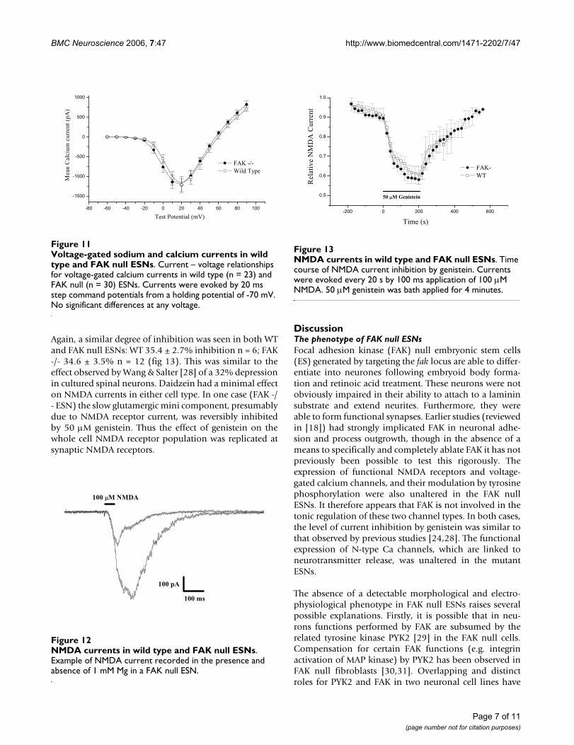
Voltage-gated calcium currents were isolated using thesolutions described in the methods and recorded in thewhole cell patch clamp configuration (fig. 11). The cur-rent – voltage relationship was determined by applyingstep command potentials (20 ms duration) ranging from-60 to +90 mV. For both FAK -/- and wild type ESNs thisactivation curve was typical of neuronal high voltage acti-vated Ca current: currents were evoked by steps positive to-30 mV, peak currents elicited at +20 mV, with reversal at~+60 mV. The magnitude of peak calcium current was notsignificantly different in FAK -/- and wild type ESNs: 1166± 189 pA, n = 25 for WT; 1393 ± 172 n = 32 for FAK null.Expressed as current density (peak current/cell capaci-tance), corresponding values were 69.2 ± 5.8 pA/pF forWT and 68.9 ± 6.8 pA/pF for FAK null neurons. Addition-ally, the time constant of calcium current activation fol-lowing a depolarising voltage pulse was not significantlydifferent in FAK -/- and wild type ESNs. For steps to +20mV, τ were 1.39 ± 0.13 ms in wild type and 1.20 ± 0.13 msin FAK null ESNs.
The specific N-type calcium channel blocker, ω-conotoxinGVIA (2 µM) irreversibly inhibited a proportion of thehigh voltage activated calcium current in both FAK -/- andwild type ESNs (WT 40.4 ± 4.8% inhibition n = 5; FAK-49.1 ± 4.6% n = 6). While the inhibition was somewhatgreater in the FAK null cells, the difference was not statis-tically significant. ω-conotoxin MVIIC (2 µM) inhibitedcurrents in a partly reversible manner, consistent with itsreported action as an (almost) irreversible inhibitor of P/Q type channels, but a reversible inhibitor of N-type. Inone WT ESN acute blockade was 43.0%, while the sus-tained level of inhibition (6 mins after toxin) was 22.0%.
Western blot of cellular extract of wild type and FAK null ES cellsFigure 2Western blot of cellular extract of wild type and FAK null ES cells.
Page 4 of 11(page number not for citation purposes)
BMC Neuroscience 2006, 7:47 http://www.biomedcentral.com/1471-2202/7/47
Corresponding figures for a FAK – ESN were 42.5 and20.8%.
Currents gated by the NMDA subtype of glutamate recep-tor were evoked by brief application (see methods) of 100µM NMDA/10 µM glycine and recorded in the whole cellpatch clamp configuration. For these experiments, nomi-nally magnesium free bathing solution was used. PeakNMDA currents (recorded at a holding potential of -70mV) were not significantly different in the WT and FAKnull ESNs: WT 1253 ± 177 pA; FAK -/- 1387 ± 137 pA. Inmagnesium free solutions, the current – voltage relation-ship of NMDA receptor gated currents was linear, while inthe presence of 1 mM Mg2+ a marked attenuation of the
current was observed at hyperpolarised potentials (fig 12).This voltage-dependent Mg2+ block was seen in both WTand FAK null ESNs.
Spontaneous miniature synaptic currents (mEPSCs) wererecorded (in the presence of TTX) in cells from ~7 d in cul-ture, strongly suggesting the formation of functional syn-apses, in both WT and FAK null ESN cultures. On the basisof their kinetics, these mEPSCs were glutamatergic in ori-gin.
Effect of genistein on NMDA and voltage-gated calcium currentsA number of studies have shown that Src-family tyrosinekinases modulate the activity of both voltage-gated cal-
Immunohistochemistry of wild type and FAK null ESNsFigure 6Immunohistochemistry of wild type and FAK null ESNs. FAK -/- ESN 15 DIV. MAP2B ×100. Scale bars: in fig. 3: 50 µm; in figs 5 & 7: 10 µm.
Immunohistochemistry of wild type and FAK null ESNsFigure 4Immunohistochemistry of wild type and FAK null ESNs. FAK -/- ESN 15 DIV. MAP2B ×20. Scale bars: in fig. 3: 50 µm; in figs 5 & 7: 10 µm.
Immunohistochemistry of wild type and FAK null ESNsFigure 3Immunohistochemistry of wild type and FAK null ESNs. WT ESN 15 DIV. MAP2B ×20. Scale bars: in fig. 3: 50 µm; in figs 5 & 7: 10 µm.
Immunohistochemistry of wild type and FAK null ESNsFigure 5Immunohistochemistry of wild type and FAK null ESNs. WT ESN 15 DIV. MAP2B ×100. Scale bars: in fig. 3: 50 µm; in figs 5 & 7: 10 µm.
Page 5 of 11(page number not for citation purposes)
BMC Neuroscience 2006, 7:47 http://www.biomedcentral.com/1471-2202/7/47
cium currents [24] and NMDA receptors [25,26]. SinceFAK is known to be involved with Src-family kinase func-tion, we tested the effect of inhibiting tyrosine kinaseactivity in wild type compared to FAK null ESNs using thespecific inhibitor genistein (4',5,7 trihydroxyisoflavone),and the related inactive compound, daidzein (4',7 dihy-droxyisoflavone).
Calcium currents were evoked by 40 ms step depolarisa-tions from -70 mV to +10 mV delivered at 0.1 Hz. After acontrol period of 8–10 stimuli 50 µM genistein wasapplied via bath perfusion for 120 s. In both FAK -/- andwild type ESNs calcium currents were reversibly inhibitedby genistein, and to a similar degree: WT 34.9% inhibition± 1.0 n = 9; FAK -/- 33.8% ± 0.8 n = 10 similar to results
obtained by Potier & Rovira [24] This inhibition was rela-tively rapid with 80% of maximal inhibition achievedwithin 60 s. Daidzein (50 µM) also inhibited currents,though to a lesser extent: WT 17.1% inhibition ± 2.2 n =5; FAK -/- 13.1% ± 1.3 n = 6. This may suggest that a com-ponent of the genistein effect was not related to tyrosinekinase inhibition, but may have been due to a directaction on calcium channels [27].
NMDA receptor gated currents were also reversibly inhib-ited by 2 minute bath application of genistein (50 µM).
Voltage-gated sodium and calcium currents in wild type and FAK null ESNsFigure 10Voltage-gated sodium and calcium currents in wild type and FAK null ESNs. Action potentials in a wild type ESN elicited by depolarising current injection (0.15 nA, 160 ms), showing reversible block by 50 nM TTX. (TTX trace recorded 1 min after application, wash recorded 3 min after return to control).
25 ms
20 mV
Control 50 nM TTX Wash
Immunohistochemistry of wild type and FAK null ESNsFigure 8Immunohistochemistry of wild type and FAK null ESNs. FAK -/- ESN 15 DIV. synaptophysin ×100. Scale bars: in fig. 3: 50 µm; in figs 5 & 7: 10 µm.
Immunohistochemistry of wild type and FAK null ESNsFigure 7Immunohistochemistry of wild type and FAK null ESNs. WT ESN 15 DIV. synaptophysin ×100. Scale bars: in fig. 3: 50 µm; in figs 5 & 7: 10 µm.
Voltage-gated sodium and calcium currents in wild type and FAK null ESNsFigure 9Voltage-gated sodium and calcium currents in wild type and FAK null ESNs. Current – voltage relationships for TTX-sensitive voltage-gated sodium currents in wild type (n = 24) and FAK null (n = 27) ESNs.
-60 -40 -20 0 20 40 60 80 100
-5000
-4000
-3000
-2000
-1000
0
FAK -/- Wild type
Pea
k in
war
d N
a cu
rren
t (pA
)
Test Potential (mV)
Page 6 of 11(page number not for citation purposes)
BMC Neuroscience 2006, 7:47 http://www.biomedcentral.com/1471-2202/7/47
Again, a similar degree of inhibition was seen in both WTand FAK null ESNs: WT 35.4 ± 2.7% inhibition n = 6; FAK-/- 34.6 ± 3.5% n = 12 (fig 13). This was similar to theeffect observed by Wang & Salter [28] of a 32% depressionin cultured spinal neurons. Daidzein had a minimal effecton NMDA currents in either cell type. In one case (FAK -/- ESN) the slow glutamergic mini component, presumablydue to NMDA receptor current, was reversibly inhibitedby 50 µM genistein. Thus the effect of genistein on thewhole cell NMDA receptor population was replicated atsynaptic NMDA receptors.
DiscussionThe phenotype of FAK null ESNsFocal adhesion kinase (FAK) null embryonic stem cells(ES) generated by targeting the fak locus are able to differ-entiate into neurones following embryoid body forma-tion and retinoic acid treatment. These neurons were notobviously impaired in their ability to attach to a lamininsubstrate and extend neurites. Furthermore, they wereable to form functional synapses. Earlier studies (reviewedin [18]) had strongly implicated FAK in neuronal adhe-sion and process outgrowth, though in the absence of ameans to specifically and completely ablate FAK it has notpreviously been possible to test this rigorously. Theexpression of functional NMDA receptors and voltage-gated calcium channels, and their modulation by tyrosinephosphorylation were also unaltered in the FAK nullESNs. It therefore appears that FAK is not involved in thetonic regulation of these two channel types. In both cases,the level of current inhibition by genistein was similar tothat observed by previous studies [24,28]. The functionalexpression of N-type Ca channels, which are linked toneurotransmitter release, was unaltered in the mutantESNs.
The absence of a detectable morphological and electro-physiological phenotype in FAK null ESNs raises severalpossible explanations. Firstly, it is possible that in neu-rons functions performed by FAK are subsumed by therelated tyrosine kinase PYK2 [29] in the FAK null cells.Compensation for certain FAK functions (e.g. integrinactivation of MAP kinase) by PYK2 has been observed inFAK null fibroblasts [30,31]. Overlapping and distinctroles for PYK2 and FAK in two neuronal cell lines have
NMDA currents in wild type and FAK null ESNsFigure 12NMDA currents in wild type and FAK null ESNs. Example of NMDA current recorded in the presence and absence of 1 mM Mg in a FAK null ESN.
100 ms
100 pA
100 µM NMDA
Voltage-gated sodium and calcium currents in wild type and FAK null ESNsFigure 11Voltage-gated sodium and calcium currents in wild type and FAK null ESNs. Current – voltage relationships for voltage-gated calcium currents in wild type (n = 23) and FAK null (n = 30) ESNs. Currents were evoked by 20 ms step command potentials from a holding potential of -70 mV. No significant differences at any voltage.
-80 -60 -40 -20 0 20 40 60 80 100
-1500
-1000
-500
0
500
1000
Mea
n C
alci
um c
urre
nt (
pA)
Test Potential (mV)
FAK -/- Wild Type
NMDA currents in wild type and FAK null ESNsFigure 13NMDA currents in wild type and FAK null ESNs. Time course of NMDA current inhibition by genistein. Currents were evoked every 20 s by 100 ms application of 100 µM NMDA. 50 µM genistein was bath applied for 4 minutes.
-200 0 200 400 600
0.5
0.6
0.7
0.8
0.9
1.0
50 µM Genistein
FAK- WT
Rel
ativ
e N
MD
A C
urre
nt
Time (s)
Page 7 of 11(page number not for citation purposes)

BMC Neuroscience 2006, 7:47 http://www.biomedcentral.com/1471-2202/7/47
been described [32] and also that PYK2 alone is sufficientto enable neurite outgrowth in PC12 cells. This is sup-ported by Park et al [33] who found that PYK2, rather thanFAK, was primary involved in NGF-induced neuronal dif-ferentiation in PC12 cells. These issues may be addressedin the future with PYK2/FAK double mutant ESNs.
Secondly, the possibility remains that intracellular signal-ling pathways in ESNs are fundamentally different fromthose in the neuronal tissues used in previous studies. Theavailable evidence would suggest that this is not the case.For example, β1 integrin null ES cells display severelyretarded neuronal differentiation, with severely limitedneurite outgrowth [6,34]. Thus far, no important physio-logical or biochemical differences have been observedbetween ESNs and primary cultured neurons. It is alsopossible, albeit unlikely, that during in vitro differentia-tion we are selecting for FAK independent subpopulationof neurons and/or FAK is required in neurons in vivo butnot in cultured neurons.
Finally, it is of course also possible that FAK is involved inprocesses beyond the focus of this study. FAK may partic-ipate in pathways leading to modulation of other channeltypes, or (more complex) physiological phenomena, suchas apoptosis [35] or synaptic plasticity, [12,14] the latterpossibly mediated by binding to SAPAP3 and PSD-95[36]. In relation to the present study, it is interesting tonote that while synaptic plasticity is altered in PSD-95mutants, NMDA receptor currents are not [37].
Work using mice with a targeted deletion of fak in devel-oping forebrain has shown that, in vivo, FAK may influ-ence neuronal function indirectly as a result of itsexpression in non-neuronal cell types resulting in corticaldysplasia due to aberrant neuronal migration[16]. Wewould not expect to observe such cell autonomous effectsof FAK in our ESN system. Using cortical cultures fromthese conditional knock out mice [17] it was shown thatFAK ablation did not alter axon outgrowth in vitro, inagreement with our findings. Netrin stimulated attractionwas however altered, but this was not examined in ourwork. Similarly, we did not analyse dendritic spines(which are not visualised by MAP2B staining), and havebeen shown to be morphologically altered by FAK dele-tion in an in vitro study by Moeller et al [38].
ESNs as a model system to study neuronal genesIn a more general context, we believe that ES cell derivedneurons, with properties comparable to primary culturesof embryonic or neonatal cortex, provide a powerful sys-tem in which to study the consequences of neural genemanipulation. There are a number of significant advan-tages afforded by this methodology. ES cells are readilytransfected and appropriate clones selected to yield stable
mutant cell lines. Furthermore, since ES cells have aeuploid karyotype it is possible to produce precise tar-geted disruptions of a gene. Both these features affordconsiderable advantages over strategies involving thetransfection of primary cultures, organotypic slices, ortransformed cell lines, for example low and variable trans-fection rate or the existence of endogenous gene product.The study of neuronal gene alterations are now com-monly studied in mutant mice, generated from the intro-duction of engineered ES cells into blastocysts. Theproduction of ESNs by in vitro differentiation has theadvantage, pertinent to this study, of allowing access tothe neuronal phenotype for mutations which give rise toembryonic lethality in mutant mice. In certain cases itmay still be possible to obtain mutant neurons by prepar-ing cultures from embryos, however for FAK, lethality is soearly as to preclude this and the only cell type which hasbeen derived from the embryos of FAK +/- crosses arefibroblasts [15]. The in vitro derivation of ESNs effectivelyallows the neuronal phenotype of a given mutation to bestudied in isolation of more widespread developmentalconsequences. This technology may of course be used in acomplementary manner to mouse studies, since engi-neered ES cells may be introduced into blastocysts, and,since they can contribute to the germline of chimaeras,mutant mouse lines may be established. Large scale func-tional studies should now be possible using high through-put gene targeting and trapping techniques and offerunprecedented access to molecular genetic studies ofmammalian neurons.
ConclusionFocal adhesion kinase (FAK) null embryonic stem cells(ES) may be differentiated in vitro into neurones whichextend neurites and form functional synapses. NMDAreceptor gated currents and voltage sensitive calcium cur-rents were unaltered in FAK null ESNs, both in the level oftheir expression and modulation by tyrosine kinases.Embryonic stem cell derived neurons provide a powerfulsystem in which to study the effect of gene manipulationin cells with properties very similar to primary corticalneurones.
MethodsGene targeting and ES cell cultureIn order to generate a fak null allele as well as a condi-tional (loxP flanked) allele, a targeting vector carryingthree loxP sites in an intronic region of fak gene (LP3) wasconstructed see Fig 1, additional file 1 and [9].
E14 TG2a embryonic stem cells were maintained in feederfree condition on tissue culture plates coated with 0.1%gelatin using the following medium: Glasgow's modifiedEagles medium (GMEM) supplemented with 0.1 mM
Page 8 of 11(page number not for citation purposes)
BMC Neuroscience 2006, 7:47 http://www.biomedcentral.com/1471-2202/7/47
mercaptoethanol, 10% fetal calf serum (FCS) and LIF(100 u/ml).
Not I digested DNA (150 µg) was electroporated into EScells (1 × 10⁸ cells) using a Bio-Rad Gene Pulser (0.8 kV, 3µF, in a cuvette with 0.4 cm electrode gap). G418 (200 µg/ml, Gibco) was added to the media 24 hours after plating.8–12 days after selection, resistant ES colonies from theplates were picked using 200 µl pipette tips, dispersedwith trypsin and transferred into wells of a 24 well plate.Each plate was duplicated, with one plate being frozen asa stock and the other plate used to prepare genomic DNA.G418 resistant ES clones were screened by Southern blotanalysis. Genomic DNA from each clone was digestedwith Bam HI and 500 bp Eco RI ~ Bam HI fragment (3' tothe end of homology arm) was used as a probe. Integra-tion of the third loxP site was screened by PCR using prim-ers flanking the two Hind III sites (which are absent when3rd loxP present) and products were digested by Hind III.
To excise the drug marker cassette (neomycin phospho-transferase and thymidine kinase), Cre recombinase wastransiently expressed from pMC1-Cre plasmid, electropo-rated into the homologous recombinant clone cells. Gan-cyclovir (200 µM) was added to the media 5 days afterplating. Resistant clones (those with the drug marker cas-sette deleted) were analysed by Southern blot using thesame probe as the initial screening (see above). Cell lineswere chosen in which the exon is deleted together with thedrug marker cassette (fak LP1 allele). These were subjectedto a second round of targeting, using the same targetingvector. Clones containing one knockout allele (fak LP1)and one allele targeted with the LP3 vector were selected.Further cre-mediated deletion of these cells generatedhomozyygous FAK null (LP1/LP1) ES cells, which wereselected by gancyclovir resistance as before. (see Fig 1 and[9]).
Neuronal differentiation of embryonic stem cells and electrophysiological recordingConfluent ES cells (grown as above) were trypsinised,diluted and replated and grown to confluence once more.These confluent ES cells were then treated briefly (~60 s)with trypsin, such that cells mostly detached from theflask in clumps of ~100 cells. These were then grown insuspension culture in bacterial grade Petri dishes, inGMEM/10% FCS, without LIF. Medium was changedevery 48 hrs. From days 4–9, 1 µM all trans retinoic acidwas added to the suspension medium. On day 9 (after'4+5' RA+/-) the embryoid bodies were collected, washedtwice with phosphate buffered saline (PBS), treated withtrypsin for 10 minutes and disaggregated by drawing upand down a flamed Pasteur pipette to yield a single cellsuspension. This was centrifuged and the pellet resus-pended in GMEM/10% FCS and cells plated at densities of
0.5 – 2.0 × 10⁵ cells/ml on laminin coated glass coverslips.After 48 hrs the medium was replaced by Neurobasal©
medium supplemented with B27, 1% FCS and 0.5 mMglutamine.
Extracellular recording solution: NaCl 140, KCl 3, CaCl22.5, HEPES 15, glucose 10, MgCl2 0 or 1 mM, TTX 0 or 50nM, pH 7.4 with NaOH. For calcium current recordings:NaCl 100, TEA-acetate 20, CsCl 5, KCl 3, CaCl2 10, HEPES15, glucose 15, MgCl2 1 mM, TTX 200 nM, pH 7.4 withNaOH. Drugs/toxins were applied by bath perfusion.NMDA was microperfused via a solenoid valve operatedU-tube. The composition of the recording electrode solu-tion was (mM): K-gluconate 117.5, K-MeSO4, NaCl 8,HEPES 10, Mg-ATP 4, Na2GTP 0.3, EGTA 0.5, pH 7.25with KOH. For calcium current recordings: CsMeSO3 100,CsBAPTA 5, HEPES 15, MgATP 4, NaGTP 0.4, sucrose 40;pH 7.2 with CsOH. Recording electrodes were pulledfrom borosilicate glass (Clark GC-150TF) on a Sutter P-87.
Currents were recorded and analysed using an Axopatch1D amplifier and pClamp6 software (Axon Instruments).
Immunocytochemistry and Western blottingFor immunofluorescent staining of ESN cultures, cells(grown on glass coverslips) were first fixed with coldmethanol (5 mins) and washed (PBS with 5% horseserum, 0.1% Triton X-100 and 0.02% NaN3) and treatedwith blocking solution (wash solution plus 5% goatserum). Primary antibody (MAP2B; mouse monoclonal,clone 18, Transduction Laboratories or synaptophysin;mouse monoclonal, clone SY 38, Boehringer Mannheim)was then added (1:200 dilution in wash solution) andincubated for 2 hrs after which coverslips were washed 4times and secondary antibody (Cy3 conjugated goat anti-mouse, Jackson Laboratories) was added for 1 hr. After afurther 4 washes and a final rinse in distilled water, cover-slips were mounted on slides using Fluoromount andviewed on an Olympus Vannox microscope. For Westernblotting, cells were lysed on ice in RIPA buffer (150 mMNaCl, 1.0% NP-40, 0.5% DOC, 0.1% SDS, 50 mM Tris/ClpH 8.0 contains 50 mM NaF, 20 µM ZnCl2, 1 mM Sodiumorthovanadate, 0.5 mM PMSF and protease inhibitorcocktail (cϕmplete™, Boehringer Mannheim). Lysateswere quantified by BCA™ Protein Assay Reagent Kit(Pierce).
Samples were run on 8% SDS polyacrylamide gel andblotted on PVDF membrane. Anti-FAK monoclonal anti-body and anti-phosphotyrosine-RC20 antibody werefrom Transduction Laboratory. Horseradish peroxidaselinked anti-mouse antibody (sheep) and ECL™ westernblotting detection reagents were from Amersham Pharma-cia Biotech.
Page 9 of 11(page number not for citation purposes)
BMC Neuroscience 2006, 7:47 http://www.biomedcentral.com/1471-2202/7/47
Total cellular RNA was extracted from ESNs using Rneasyfrom Qiagen. Single strand cDNA was synthesised fromthis RNA using oligo(dT) primers, Superscript II anddNTP mix (Gibco). The polymerase chain reaction, usingprimers hybridising either side of the neuronal specificexons [22] was performed on this cDNA.
Authors' contributionsPC carried out the electrophysiological and immunocyto-chemical studies and drafted the manuscript. NHKdesigned and constructed the targeting vector generatedFAK null ES cells and performed Western blots. PC andNHK prepared ESNs by in vitro differentiation of ES cells.PC, NHK & SGNG conceived and designed the study, readand approved the final manuscript.
Additional material
AcknowledgementsThis work was supported by the Wellcome Trust and EU.
References1. Mansuy IM, Bujard H: Tetracycline-regulated gene expression
in the brain. Curr Opin Neurobiol 2000, 10:593-596.2. Tsien JZ, Chen DF, Gerber D, Tom C, Mercer EH, Anderson DJ, May-
ford M, Kandel ER, Tonegawa S: Subregion- and cell type-restricted gene knockout in mouse brain. Cell 1996,87:1317-1326.
3. Bain G, Kitchens D, Yao M, Huettner JE, Gottlieb DI: Embryonicstem cells express neuronal properties in vitro. DevelopmentalBiology 1995, 168:342-357.
4. Okabe S, Forsberg-Nilsson K, Spiro A, Segal M, McKay R: Develop-ment of neuronal precursor cells and functional postmitoticneurons from embryonic stem cells in vitro. Mech Dev 1996,59:89-102.
5. Strubing C, Ahnert-Hilger G, Shan J, Wiedenmann B, Hescheler J,Wobus AM: Differentiation of pluripotent embryonic stemcells into the neuronal lineage in vitro gives rise to matureinhibitory and excitatory neurons. Mechanisms of Development1995, 53:275-287.
6. Andressen C, Arnhold S, Puschmann M, Bloch W, Hescheler J, FässlerR, Addicks K: Beta1 integrin deficiency impairs migration anddifferentiation of mouse embryonic stem cell derived neu-rons. Neuroscience Letters 1998, 251:165-168.
7. Metzler M, Chen N, Helgason CD, Graham RK, Nichol K, McCutch-eon K, Nasir J, Humphries RK, Raymond LA, Hayden MR: Life with-out huntingtin: normal differentiation into functionalneurons. Journal Of Neurochemistry 1999, 72:1009-1018.
8. Strittmatter SM, Fankhauser C, Huang PL, Mashimo H, Fishman MC:Neuronal pathfinding is abnormal in mice lacking the neuro-nal growth cone protein GAP-43. Cell 1995, 80:445-452.
9. McLean GW, Komiyama NH, Serrels B, Asano H, Reynolds L, ContiF, Hodivala-Dilke K, Metzger D, Chambon P, Grant SG, Frame MC:Specific deletion of focal adhesion kinase suppresses tumorformation and blocks malignant progression. Genes Dev 2004,18:2998-3003.
10. Grant SG, O'Dell TJ, Karl KA, Stein PL, Soriano P, Kandel ER:Impaired long-term potentiation, spatial learning, and hip-pocampal development in fyn mutant mice. Science 1992,258:1903-1910.
11. O'Dell TJ, Kandel ER, Grant SG: Long-term potentiation in thehippocampus is blocked by tyrosine kinase inhibitors. Nature1991, 353:558-560.
12. Grant SG, Karl KA, Kiebler MA, Kandel ER: Focal adhesion kinasein the brain: novel subcellular localization and specific regu-lation by Fyn tyrosine kinase in mutant mice. genes and devel-opment 1995, 9:1909-1921.
13. Rico B, Beggs HE, Schahin-Reed D, Kimes N, Schmidt A, Reichardt LF:Control of axonal branching and synapse formation by focaladhesion kinase. Nat Neurosci 2004, 7:1059-1069.
14. Yang YC, Ma YL, Chen SK, Wang CW, Lee EH: Focal adhesionkinase is required, but not sufficient, for the induction oflong-term potentiation in dentate gyrus neurons in vivo. JNeurosci 2003, 23:4072-4080.
15. Ilic D, Furuta Y, Suda T, Atsumi T, Fujimoto J, Ikawa Y, Yamamoto T,Aizawa S: Focal adhesion kinase is not essential for in vitro andin vivo differentiation of ES cells. biochemical and biophysicalresearch communications 1995, 209:300-309.
16. Beggs HE, Schahin-Reed D, Zang K, Goebbels S, Nave KA, Gorski J,Jones KR, Sretavan D, Reichardt LF: FAK deficiency in cells con-tributing to the basal lamina results in cortical abnormalitiesresembling congenital muscular dystrophies. Neuron 2003,40:501-514.
17. Liu G, Beggs H, Jurgensen C, Park HT, Tang H, Gorski J, Jones KR,Reichardt LF, Wu J, Rao Y: Netrin requires focal adhesion kinaseand Src family kinases for axon outgrowth and attraction.Nat Neurosci 2004, 7:1222-1232.
18. Mitra SK, Hanson DA, Schlaepfer DD: Focal adhesion kinase: incommand and control of cell motility. Nat Rev Mol Cell Biol2005, 6:56-68.
19. Schaller MD, Hildebrand JD, Parsons JT: Complex formation withfocal adhesion kinase: A mechanism to regulate activity andsubcellular localization of Src kinases. Mol Biol Cell 1999,10:3489-3505.
20. Girault JA, Costa A, Derkinderen P, Studler JM, Toutant M: FAK andPYK2/CAKbeta in the nervous system: a link between neu-ronal activity, plasticity and survival? trends in neurosciences1999, 22:257-263.
21. Parsons JT: Focal adhesion kinase: the first ten years. J Cell Sci2003, 116:1409-1416.
22. Burgaya F, Toutant M, Studler JM, Costa A, Le Bert M, Gelman M,Girault JA: Alternatively spliced focal adhesion kinase in ratbrain with increased autophosphorylation activity. J Biol Chem1997, 272:28720-28725.
23. Messina S, Onofri F, Bongiorno-Borbone L, Giovedi S, Valtorta F,Girault JA, Benfenati F: Specific interactions of neuronal focaladhesion kinase isoforms with Src kinases and amphiphysin.J Neurochem 2003, 84:253-265.
24. Potier B, Rovira C: Protein tyrosine kinase inhibitors reducehigh-voltage activating calcium currents in CA1 pyramidalneurones from rat hippocampal slices. Brain Research 1999,816:587-597.
25. Bernard-Trifilo JA, Kramar EA, Torp R, Lin CY, Pineda EA, Lynch G,Gall CM: Integrin signaling cascades are operational in adulthippocampal synapses and modulate NMDA receptor physi-ology. J Neurochem 2005, 93:834-849.
26. Yu XM, Askalan R, Keil GJn, Salter MW: NMDA channel regula-tion by channel-associated protein tyrosine kinase Src. Sci-ence 1997, 275:674-678.
27. Paillart C, Carlier E, Guedin D, Dargent B, Couraud F: Direct blockof voltage-sensitive sodium channels by genistein, a tyrosinekinase inhibitor. J Pharmacol Exp Ther 1997, 280:521-526.
28. Wang YT, Salter MW: Regulation of NMDA receptors by tyro-sine kinases and phosphatases. Nature 1994, 369:233-235.
29. Lev S, Moreno H, Martinez R, Canoll P, Peles E, Musacchio JM, Plow-man GD, Rudy B, Schlessinger J: Protein tyrosine kinase PYK2involved in Ca(2+)-induced regulation of ion channel andMAP kinase functions. Nature 1995, 376:737-745.
30. Sieg DJ, Ilic D, Jones KC, Damsky CH, Hunter T, Schlaepfer DD:Pyk2 and Src-family protein-tyrosine kinases compensate forthe loss of FAK in fibronectin-stimulated signaling events but
Additional File 1Figure showing in detail the construction of the targeting vector, together with legend.Click here for file[http://www.biomedcentral.com/content/supplementary/1471-2202-7-47-S1.ppt]
Page 10 of 11(page number not for citation purposes)

BMC Neuroscience 2006, 7:47 http://www.biomedcentral.com/1471-2202/7/47
Publish with BioMed Central and every scientist can read your work free of charge
"BioMed Central will be the most significant development for disseminating the results of biomedical research in our lifetime."
Sir Paul Nurse, Cancer Research UK
Your research papers will be:
available free of charge to the entire biomedical community
peer reviewed and published immediately upon acceptance
cited in PubMed and archived on PubMed Central
yours — you keep the copyright
Submit your manuscript here:http://www.biomedcentral.com/info/publishing_adv.asp
BioMedcentral
Pyk2 does not fully function to enhance FAK- cell migration.Embo Journal 1998, 17:5933-5947.
31. Ueki K, Mimura T, Nakamoto T, Sasaki T, Aizawa S, Hirai H, Yano S,Naruse T, Nojima Y: Integrin-mediated signal transduction incells lacking focal adhesion kinase p125FAK. Febs Letters 1998,432:197-201.
32. Ivankovic-Dikic I, Gronroos E, Blaukat A, Barth BU, Dikic I: Pyk2 andFAK regulate neurite outgrowth induced by growth factorsand integrins. Nat Cell Biol 2000, 2:574-581.
33. Park SY, Avraham H, Avraham S: Characterization of the tyro-sine kinases RAFTK/Pyk2 and FAK in nerve growth factor-induced neuronal differentiation. journal of biological chemistry2000, 275:19768-19777.
34. Strubing C, Rohwedel J, Ahnert-Hilger G, Wiedenmann B, HeschelerJ, Wobus A: Development of G protein-mediated Ca2+ chan-nel regulation in mouse embryonic stem cell-derived neu-rons. Eur J Neurosci 1997, 9:824-832.
35. Ilic D, Almeida EA, Schlaepfer DD, Dazin P, Aizawa S, Damsky CH:Extracellular matrix survival signals transduced by focaladhesion kinase suppress p53-mediated apoptosis. Journal OfCell Biology 1998, 143:547-560.
36. Bongiorno-Borbone L, Kadare G, Benfenati F, Girault JA, Messina S,Onofri F, Giovedi S, Valtorta F: FAK and PYK2 interact withSAP90/PSD-95-Associated Protein-3 Specific interactions ofneuronal focal adhesion kinase isoforms with Src kinases andamphiphysin. Biochem Biophys Res Commun 2005, 337:641-646.
37. Migaud M, Charlesworth P, Dempster M, Webster LC, Watabe AM,Makhinson M, He Y, Ramsay MF, Morris RG, Morrison JH, et al.:Enhanced long-term potentiation and impaired learning inmice with mutant postsynaptic density-95 protein. Nature1998, 396:433-439.
38. Moeller ML, Shi Y, Reichardt LF, Ethell IM: EphB receptors regu-late dendritic spine morphogenesis through the recruit-ment/phosphorylation of focal adhesion kinase and RhoAactivation. J Biol Chem 2006, 281:1587-1598.
Page 11 of 11(page number not for citation purposes)
Copyright © 2022 FDOKUMEN