
Mutations in Zebrafish lrp2 Result in Adult-Onset Ocular Pathogenesis That Models Myopia and Other...
12
Mutations in Zebrafish lrp2 Result in Adult-Onset Ocular Pathogenesis That Models Myopia and Other Risk Factors for Glaucoma Kerry N. Veth 1 , Jason R. Willer 2 , Ross F. Collery 1 , Matthew P. Gray 1 , Gregory B. Willer 2 , Daniel S. Wagner 3 , Mary C. Mullins 4 , Ava J. Udvadia 5 , Richard S. Smith 6 , Simon W. M. John 6 , Ronald G. Gregg 2 , Brian A. Link 1 * 1 Department of Cell Biology, Neurobiology, and Anatomy, Medical College of Wisconsin, Milwaukee, Wisconsin, United States of America, 2 Department of Biochemistry and Molecular Biology, University of Louisville, Louisville, Kentucky, United States of America, 3 Department of Biochemistry and Cell Biology, Rice University, Houston, Texas, United States of America, 4 Department of Cell and Developmental Biology, University of Pennsylvania Medical School, Philadelphia, Pennsylvania, United States of America, 5 Department of Biological Sciences, University of Wisconsin–Milwaukee, Milwaukee, Wisconsin, United States of America, 6 Howard Hughes Medical Institute, The Jackson Laboratory, Bar Harbor, Maine, United States of America Abstract The glaucomas comprise a genetically complex group of retinal neuropathies that typically occur late in life and are characterized by progressive pathology of the optic nerve head and degeneration of retinal ganglion cells. In addition to age and family history, other significant risk factors for glaucoma include elevated intraocular pressure (IOP) and myopia. The complexity of glaucoma has made it difficult to model in animals, but also challenging to identify responsible genes. We have used zebrafish to identify a genetically complex, recessive mutant that shows risk factors for glaucoma including adult onset severe myopia, elevated IOP, and progressive retinal ganglion cell pathology. Positional cloning and analysis of a non- complementing allele indicated that non-sense mutations in low density lipoprotein receptor-related protein 2 (lrp2) underlie the mutant phenotype. Lrp2, previously named Megalin, functions as an endocytic receptor for a wide-variety of bioactive molecules including Sonic hedgehog, Bone morphogenic protein 4, retinol-binding protein, vitamin D-binding protein, and apolipoprotein E, among others. Detailed phenotype analyses indicated that as lrp2 mutant fish age, many individuals—but not all—develop high IOP and severe myopia with obviously enlarged eye globes. This results in retinal stretch and prolonged stress to retinal ganglion cells, which ultimately show signs of pathogenesis. Our studies implicate altered Lrp2- mediated homeostasis as important for myopia and other risk factors for glaucoma in humans and establish a new genetic model for further study of phenotypes associated with this disease. Citation: Veth KN, Willer JR, Collery RF, Gray MP, Willer GB, et al. (2011) Mutations in Zebrafish lrp2 Result in Adult-Onset Ocular Pathogenesis That Models Myopia and Other Risk Factors for Glaucoma. PLoS Genet 7(2): e1001310. doi:10.1371/journal.pgen.1001310 Editor: Janey Wiggs, Harvard University, United States of America Received September 24, 2010; Accepted January 13, 2011; Published February 17, 2011 Copyright: ß 2011 Veth et al. This is an open-access article distributed under the terms of the Creative Commons Attribution License, which permits unrestricted use, distribution, and reproduction in any medium, provided the original author and source are credited. Funding: This work was supported by National Institutes of Heath (NIH) grant R01EY016060 (BAL). SWMJ is an Investigator of the Howard Hughes Medical Institute. The funders had no role in study design, data collection and analysis, decision to publish, or preparation of the manuscript. Competing Interests: The authors have declared that no competing interests exist. * E-mail: [email protected] Introduction The multi-factorial nature of many ocular diseases poses a major challenge in understanding their molecular etiology and in engineering animal models to study mechanisms of pathology. Macular degeneration, myopia, and glaucoma are examples of prevalent and disruptive complex ocular diseases. While charac- terization of complement factor genes has provided insight into most cases of macular degeneration [1], no major genetic pathway has been found to underlie myopia or glaucoma. Myopia is the most common human ocular disorder worldwide and is caused by abnormal growth of the eye resulting in refractive error [2,3]. Myopia also increases risk for other visual impairing diseases including glaucoma [4]. The glaucomas are a heterogeneous group of progressive blinding disorders that result from damage to retinal ganglion cells and their axons [5]. Important risk factors for glaucoma include elevated intraocular pressure (IOP), age, family history, and myopia [6]. Although traditional human genetic analysis has been limited in identifying causative genes for complex disorders, mutational screens in animals can provide insights into disease etiology. Recently, progress has been made on establishing the zebrafish model to study phenotypes associated with glaucoma. From a forward-genetic perspective, zebrafish offer a major advantage in studying complex disease, in that large pedigrees can be efficiently generated with moderate space and time requirements. Through a mutational screen for adult ocular defects, we identified a complex mutant, bugeye, that manifests multiple adult- onset phenotypes associated with glaucoma including enlarged eyes with myopia, elevated IOP, and damage to retinal ganglion cells. Using linkage analysis we discovered non-sense mutations in low density lipoprotein receptor-related protein 2 (lrp2) for bugeye, as well as within a non-complementing allele. Lrp2 is a large transmem- brane protein of the LDL-receptor related protein (Lrp) family [7]. Lrp2 participates in receptor-mediated endocytosis and has a host of identified ligands including signaling molecules like Sonic PLoS Genetics | www.plosgenetics.org 1 February 2011 | Volume 7 | Issue 2 | e1001310
Transcript of Mutations in Zebrafish lrp2 Result in Adult-Onset Ocular Pathogenesis That Models Myopia and Other...

Mutations in Zebrafish lrp2 Result in Adult-Onset OcularPathogenesis That Models Myopia and Other RiskFactors for GlaucomaKerry N. Veth1, Jason R. Willer2, Ross F. Collery1, Matthew P. Gray1, Gregory B. Willer2, Daniel S.
Wagner3, Mary C. Mullins4, Ava J. Udvadia5, Richard S. Smith6, Simon W. M. John6, Ronald G. Gregg2,
Brian A. Link1*
1 Department of Cell Biology, Neurobiology, and Anatomy, Medical College of Wisconsin, Milwaukee, Wisconsin, United States of America, 2 Department of Biochemistry
and Molecular Biology, University of Louisville, Louisville, Kentucky, United States of America, 3 Department of Biochemistry and Cell Biology, Rice University, Houston,
Texas, United States of America, 4 Department of Cell and Developmental Biology, University of Pennsylvania Medical School, Philadelphia, Pennsylvania, United States of
America, 5 Department of Biological Sciences, University of Wisconsin–Milwaukee, Milwaukee, Wisconsin, United States of America, 6 Howard Hughes Medical Institute,
The Jackson Laboratory, Bar Harbor, Maine, United States of America
Abstract
The glaucomas comprise a genetically complex group of retinal neuropathies that typically occur late in life and arecharacterized by progressive pathology of the optic nerve head and degeneration of retinal ganglion cells. In addition toage and family history, other significant risk factors for glaucoma include elevated intraocular pressure (IOP) and myopia.The complexity of glaucoma has made it difficult to model in animals, but also challenging to identify responsible genes. Wehave used zebrafish to identify a genetically complex, recessive mutant that shows risk factors for glaucoma including adultonset severe myopia, elevated IOP, and progressive retinal ganglion cell pathology. Positional cloning and analysis of a non-complementing allele indicated that non-sense mutations in low density lipoprotein receptor-related protein 2 (lrp2) underliethe mutant phenotype. Lrp2, previously named Megalin, functions as an endocytic receptor for a wide-variety of bioactivemolecules including Sonic hedgehog, Bone morphogenic protein 4, retinol-binding protein, vitamin D-binding protein, andapolipoprotein E, among others. Detailed phenotype analyses indicated that as lrp2 mutant fish age, many individuals—butnot all—develop high IOP and severe myopia with obviously enlarged eye globes. This results in retinal stretch andprolonged stress to retinal ganglion cells, which ultimately show signs of pathogenesis. Our studies implicate altered Lrp2-mediated homeostasis as important for myopia and other risk factors for glaucoma in humans and establish a new geneticmodel for further study of phenotypes associated with this disease.
Citation: Veth KN, Willer JR, Collery RF, Gray MP, Willer GB, et al. (2011) Mutations in Zebrafish lrp2 Result in Adult-Onset Ocular Pathogenesis That ModelsMyopia and Other Risk Factors for Glaucoma. PLoS Genet 7(2): e1001310. doi:10.1371/journal.pgen.1001310
Editor: Janey Wiggs, Harvard University, United States of America
Received September 24, 2010; Accepted January 13, 2011; Published February 17, 2011
Copyright: � 2011 Veth et al. This is an open-access article distributed under the terms of the Creative Commons Attribution License, which permits unrestricteduse, distribution, and reproduction in any medium, provided the original author and source are credited.
Funding: This work was supported by National Institutes of Heath (NIH) grant R01EY016060 (BAL). SWMJ is an Investigator of the Howard Hughes MedicalInstitute. The funders had no role in study design, data collection and analysis, decision to publish, or preparation of the manuscript.
Competing Interests: The authors have declared that no competing interests exist.
* E-mail: [email protected]
Introduction
The multi-factorial nature of many ocular diseases poses a
major challenge in understanding their molecular etiology and in
engineering animal models to study mechanisms of pathology.
Macular degeneration, myopia, and glaucoma are examples of
prevalent and disruptive complex ocular diseases. While charac-
terization of complement factor genes has provided insight into
most cases of macular degeneration [1], no major genetic pathway
has been found to underlie myopia or glaucoma. Myopia is the
most common human ocular disorder worldwide and is caused by
abnormal growth of the eye resulting in refractive error [2,3].
Myopia also increases risk for other visual impairing diseases
including glaucoma [4]. The glaucomas are a heterogeneous
group of progressive blinding disorders that result from damage to
retinal ganglion cells and their axons [5]. Important risk factors for
glaucoma include elevated intraocular pressure (IOP), age, family
history, and myopia [6]. Although traditional human genetic
analysis has been limited in identifying causative genes for
complex disorders, mutational screens in animals can provide
insights into disease etiology. Recently, progress has been made on
establishing the zebrafish model to study phenotypes associated
with glaucoma. From a forward-genetic perspective, zebrafish
offer a major advantage in studying complex disease, in that large
pedigrees can be efficiently generated with moderate space and
time requirements.
Through a mutational screen for adult ocular defects, we
identified a complex mutant, bugeye, that manifests multiple adult-
onset phenotypes associated with glaucoma including enlarged
eyes with myopia, elevated IOP, and damage to retinal ganglion
cells. Using linkage analysis we discovered non-sense mutations in
low density lipoprotein receptor-related protein 2 (lrp2) for bugeye, as well as
within a non-complementing allele. Lrp2 is a large transmem-
brane protein of the LDL-receptor related protein (Lrp) family
[7]. Lrp2 participates in receptor-mediated endocytosis and has a
host of identified ligands including signaling molecules like Sonic
PLoS Genetics | www.plosgenetics.org 1 February 2011 | Volume 7 | Issue 2 | e1001310

hedgehog and Bone morphogenetic protein 4, vitamin and
hormone binding proteins, apolipoproteins, among others [8].
Lrp2 is expressed on cells of the renal proximal tubule, choroid
plexus, developing neural tube, intestine, thyroid, and inner ear.
Within the eye, Lrp2 is expressed on retinal pigment epithelial
cells as well as ciliary epithelial cells [7–9]. In humans, mutations
in LRP2 result in Donnai-Barrow syndrome [10], a rare disease
characterized by a spectrum of phenotypes including agenesis of
the corpus collosum, diaphragmatic hernia, sensonurial deafness,
hypertelorism, buphthalmia (enlarged eye globes) and high
myopia [11,12]. As the eyes of bugeye zebrafish are also highly
myopic, Lrp2 may be critical in regulating emmetropic eye
growth across species. The strong association of myopia with
glaucoma [13] makes bugeye an attractive model to study the
genetic and molecular pathways involved in these ocular
diseases.
Results
Identification of an enlarged eye mutant with elevatedintraocular pressure
The bugeye zebrafish mutant was identified in a three-generation
forward-genetic screen for adult ocular abnormalities. Mutants
were easily identified by 6 months as their eyes were visibly
enlarged (Figure 1A, 1C). Interestingly, the degree of eye
enlargement often varied between the two eyes of a single fish
(Figure S1A–S1G). Occasionally the phenotype presented only in
one eye, and the other eye remained normal in size (Figure S1B).
To address whether ocular enlargement in mutants might
represent a retinoblastoma phenotype, we analyzed eyes by
histology. Instead of obvious cellular overgrowth we found that
the retina was notably thinner in all layers (Figure 1B, 1D). As
buphthalmia is often associated with elevated IOP, we used servo-
null electrophysiology to measure the eye pressures in mutants and
wild-type siblings [14]. Compared to wild-type fish, bugeye mutants
consistently showed elevated IOPs (Figure 1I). In addition, the rare
fish that presented the phenotype in a unilateral manner had
normal pressure in the wild-type sized eye and elevated pressure in
the enlarged eye (Figure S1H). IOP is maintained by the balance
of aqueous humor production and drainage. Like mammals,
aqueous humor in zebrafish is produced in the ciliary epithelium
and drained at the iridocorneal angle. However, unlike mammals
where drainage occurs circumferentially throughout the angle
region, aqueous outflow for zebrafish is facilitated through a
discrete ventrally localized canalicular network [15]. Histology did
not reveal obvious disorganization in either the dorsal ciliary
epithelium (Figure 1E, 1G) or in the ventral canalicular outflow
network (Figure 1F, 1H). However, the ciliary epithelium
occasionally appeared mildly hypertrophied (Figure 1G, arrow)
and the angle region of mutants was more prone than wild-type
specimens to separation between the iris and corneal tissues during
histological preparation (Figure 1H, asterisk). Additional charac-
terization of these regions at the time of phenotype onset
confirmed these observations (Figure S2).
Mutations in lrp2 underlie the complex bugeyephenotype
The original bugeye mutants presented in the third generation of
a three-generation screen, suggesting the mutation was recessive.
However, only 3 fish out of a family of 28 showed the phenotype
and therefore the penetrance was lower than predicted for a
Author Summary
Complex genetic inheritance, including variable pene-trance and severity, underlies many common eye diseases.In this study, we present analysis of a zebrafish mutant,bugeye, which shows complex inheritance of multipleocular phenotypes that are known risk factors forglaucoma, including high myopia, elevated intraocularpressure, and up-regulation of stress-response genes inretinal ganglion cells. Molecular genetic analysis revealedthat mutations in low density lipoprotein receptor-relatedprotein 2 (lrp2) underlie the mutant phenotypes. Lrp2 is alarge transmembrane protein expressed in epithelia of theeye. It facilitates transport and clearance of multiplesecreted bioactive factors through receptor-mediatedendocytosis. Glaucoma, a progressive blinding disorder,usually presents in adulthood and is characterized by opticnerve damage followed by ganglion cell death. In bugeye/lrp2 mutants, ganglion cell death was significantlyelevated, but surprisingly moderate, and therefore theydo not model this endpoint of glaucoma. As such, bugeye/lrp2 mutants should be considered valuable as a geneticmodel (A) for buphthalmia, myopia, and regulated eyegrowth; (B) for identifying genes and pathways thatmodify the observed ocular phenotypes; and (C) forstudying the initiation of retinal ganglion cell pathologyin the context of high myopia and elevated intraocularpressure.
Figure 1. Adult bugeye zebrafish have enlarged eye globes, thinned retinas, and elevated intraocular pressure without iridocornealangle obstruction or malformation. A,C Dorsal views of adult wild-type (A) and bugeye (C) zebrafish. B,D Histology of central retina sections at 6months in wild-type (B) and mutant (D) eyes. E-H Histology of wild-type (E,F) and bugeye mutant (G,H) iridocorneal angles in the dorsal region (E,G) orat the ventral canalicular aqueous humor drainage region (F,H). I Intraocular pressures (IOP) in adult wild-type and bugeye zebrafish. IOPs in bugeyefish were elevated compared to age and size matched fish from TL wild-type stain (p,0.0001, t-test). Scale bars: A,C = 4 mm; B,D = 50 mm; E-H = 40 mm.doi:10.1371/journal.pgen.1001310.g001
Lrp2 Mutations Model Glaucoma Risk Factors
PLoS Genetics | www.plosgenetics.org 2 February 2011 | Volume 7 | Issue 2 | e1001310

simple recessive mutation (,9% vs. 25% predicted). Moreover,
incrossing 2 of those original mutant fish resulted in 25 progeny
that showed large eyes and 18 that never developed the
phenotype. Again, if the mutation was a simple recessive mutation,
incrossing should have resulted in all progeny showing the
phenotype. To better characterize inheritance and establish
recombinant mapping panels to genetically position the mutant
locus, we set up a series of test-crosses. Table 1 summarizes the
results of incross, outcross and backcross matings over multiple
generations and genetic backgrounds (Table 1). The data indicate
that the bugeye phenotype is most likely caused by a single recessive
mutation, but like many multi-factorial complex diseases, the
penetrance was modified by common wild-type backgrounds and/
or by non-genetic factors.
To map the mutant locus, progeny from single pair backcross
matings were used for whole-genome linkage analysis. Co-
segregation for markers on chromosome 9 and the mutant
phenotype was found (Figure 2A). Informatively, no other linkage
in the genome was noted, consistent with the single recessive
causative mutation hypothesis. Public databases revealed that the
lrp2 gene was within the critical recombinant interval. Given the
similarity of the bugeye phenotype to those caused by LRP2
mutations in humans, we sequenced this candidate gene. Analysis
of lrp2 cDNA from bugeyemw1 mutants revealed a T to A conversion
that changes a cysteine to a stop codon at predicted amino acid
position 23 (C23X) (Figure 2B, 2C). Through an independent
genetic screen we identified a second large eye mutant that like the
bugeyemw1 allele, presented in adulthood and showed reduced
penetrance. Intercrosses between this mutant (allele p5bnc) and
bugeyemw1 were non-complementing and suggested that lrp2 may
also be affected in the p5bnc mutant. Indeed, sequencing of p5bnc
cDNA revealed a separate non-sense mutation, also very early in
the coding region of lrp2 (bugeyep5bnc, Q413X) (Figure 2B, 2C). To
test whether somatic reversion or alternate splicing around the
non-sense mutations might underlie the reduced penetrance or
variability often observed between the left and right eyes, we
sequenced ocular cDNA in affected and unaffected eyes. However,
we did not find evidence of mosaicism or alternate splicing
surrounding the mutations, suggesting the penetrance and
phenotype variability is influenced by other genes, epigenetics,
and/or unpredictable changes in physiology which affects the
phenotypes.
In mammalian eyes, the multi-ligand receptor Lrp2 is known to
be expressed in the developing and adult retinal pigment
epithelium (RPE) and ciliary epithelium. We therefore analyzed
Lrp2 expression in wild-type, bugeye mutant larvae treated with
phenyl-thio-urea (PTU), which blocks pigmentation and allows
visualization of potential RPE immunoreactivity. As predicted,
strong immunoreactivity was found in wild-type RPE and ciliary
epithelium. Other regions of expression noted in wild-type fish
included forebrain ventricles, regions of the inner ear, proximal
pronephros, and gut epithelium (data not shown). All Lrp2
immunoreactivity was completely absent in mutant larvae for both
bugeye alleles (Figure 2D, 2E and data not shown). We next
developed genotyping protocols for both mutant alleles and
confirmed that large-eyed fish never showed wild-type lrp2
genotypes (Figure 2F, 2G). We also used this assay to test whether
the reduced penetrance of the ocular phenotype could be
explained by increased larval lethality of lrp2 mutants. However,
we found that all genotypes were represented in Mendelian ratios
in the adult progeny of either heterozygous or backcross pairwise
matings, despite the fact that some homozygous mutants never
developed the enlarged eye phenotype. Cumulatively, these data
indicate that lrp2 mutations are responsible for the large-eyed
phenotype in bugeye and that the reduced penetrance and
variability in eye enlargement are due to either common (yet
unknown) genetic background differences and/or non-genetic
factors such as physiological modifiers of the mutation.
Lrp2 mutants show adult onset buphthalmia andprogressive myopia
Having established the causative gene for bugeye, we next
investigated the onset of the ocular phenotype and quantified the
pathology. To characterize the development of enlarged eyes in
bugeye/lrp2 mutants we performed longitudinal studies tracking
wild-type and mutant fish from 1-12 months. The zebrafish eye
reaches its final adult anatomy by approximately 1 month of age
[16]. Because overall growth rates can vary between equally
aged fish — even within the same tank — we used the ratio of
eye size to body length (E:B) to determine the relative size of the
eye. This ratio remained constant in wild-type fish, allowing
comparison of relative eye size between individuals regardless of
the overall growth of the fish. Although this ratio remained flat as
wild-type fish grew, the E:B ratio increased over time for most
Table 1. Inheritance and penetrance of large eye phenotype.
Mutation Type of test cross Progeny Analyzed (n) Large eye Phenotype (%) Expected for Recessive (%)
lrp2 C23X Incross a 528 66 d 100
(bugmw1) Outcross b 856 0 0
Backcross c 2094 11 50
lrp2 Q413X Incross a 213 87 d 100
(bugp5bnc) Outcross b 158 0 0
Backcross c 487 35 50
aIncross: Each parent was homozygous for the lrp2 mutation and showed the large eye phenotype.bOutcross: One parent was homozygous for the lrp2 mutation and showed the eye phenotype. The other parent was wild-type. Five wild-type strains were used for the
lrp2C23X test crosses, while three wild-type strains were used for the lrp2Q413X test crosses.cBackcross: One parent was homozygous for the lrp2 mutation and showed the large eye phenotype. The other parent was an adult progeny from an Outcross andtherefore heterozygous for the lrp2 mutation.
dSome families after repeated incrossing showed 100% penetrance of the large eye phenotype.doi:10.1371/journal.pgen.1001310.t001
Lrp2 Mutations Model Glaucoma Risk Factors
PLoS Genetics | www.plosgenetics.org 3 February 2011 | Volume 7 | Issue 2 | e1001310
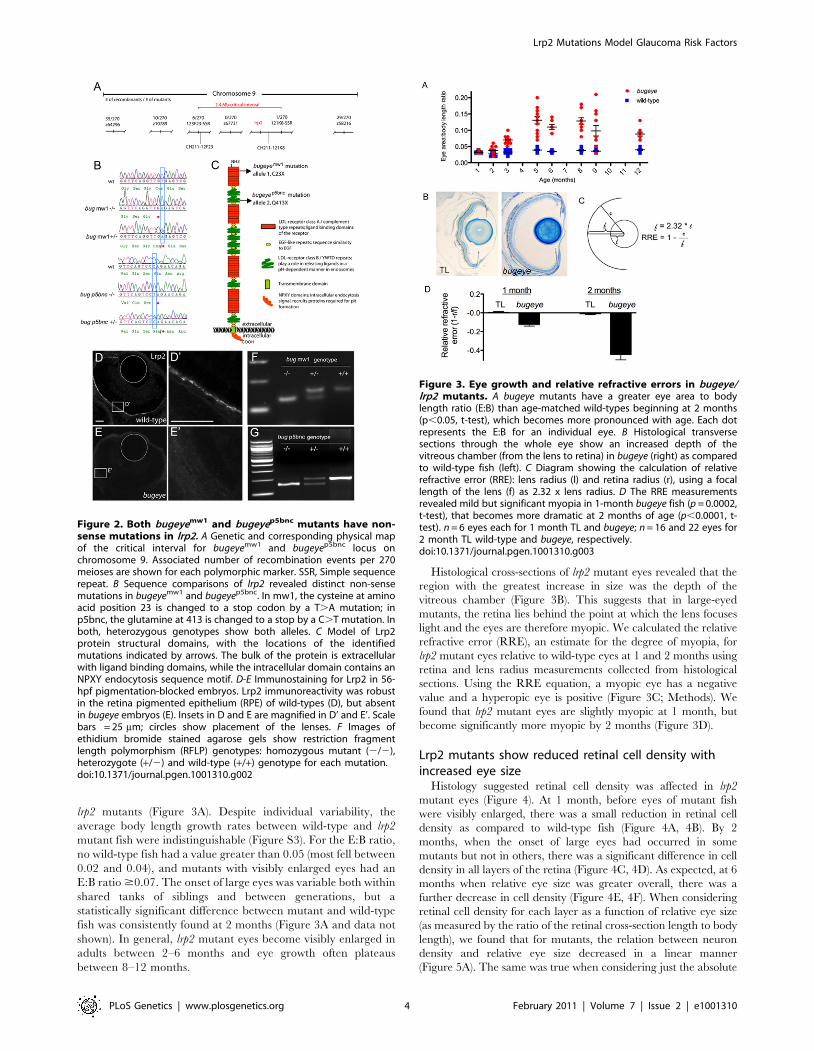
lrp2 mutants (Figure 3A). Despite individual variability, the
average body length growth rates between wild-type and lrp2
mutant fish were indistinguishable (Figure S3). For the E:B ratio,
no wild-type fish had a value greater than 0.05 (most fell between
0.02 and 0.04), and mutants with visibly enlarged eyes had an
E:B ratio $0.07. The onset of large eyes was variable both within
shared tanks of siblings and between generations, but a
statistically significant difference between mutant and wild-type
fish was consistently found at 2 months (Figure 3A and data not
shown). In general, lrp2 mutant eyes become visibly enlarged in
adults between 2–6 months and eye growth often plateaus
between 8–12 months.
Histological cross-sections of lrp2 mutant eyes revealed that the
region with the greatest increase in size was the depth of the
vitreous chamber (Figure 3B). This suggests that in large-eyed
mutants, the retina lies behind the point at which the lens focuses
light and the eyes are therefore myopic. We calculated the relative
refractive error (RRE), an estimate for the degree of myopia, for
lrp2 mutant eyes relative to wild-type eyes at 1 and 2 months using
retina and lens radius measurements collected from histological
sections. Using the RRE equation, a myopic eye has a negative
value and a hyperopic eye is positive (Figure 3C; Methods). We
found that lrp2 mutant eyes are slightly myopic at 1 month, but
become significantly more myopic by 2 months (Figure 3D).
Lrp2 mutants show reduced retinal cell density withincreased eye size
Histology suggested retinal cell density was affected in lrp2
mutant eyes (Figure 4). At 1 month, before eyes of mutant fish
were visibly enlarged, there was a small reduction in retinal cell
density as compared to wild-type fish (Figure 4A, 4B). By 2
months, when the onset of large eyes had occurred in some
mutants but not in others, there was a significant difference in cell
density in all layers of the retina (Figure 4C, 4D). As expected, at 6
months when relative eye size was greater overall, there was a
further decrease in cell density (Figure 4E, 4F). When considering
retinal cell density for each layer as a function of relative eye size
(as measured by the ratio of the retinal cross-section length to body
length), we found that for mutants, the relation between neuron
density and relative eye size decreased in a linear manner
(Figure 5A). The same was true when considering just the absolute
Figure 2. Both bugeyemw1 and bugeyep5bnc mutants have non-sense mutations in lrp2. A Genetic and corresponding physical mapof the critical interval for bugeyemw1 and bugeyep5bnc locus onchromosome 9. Associated number of recombination events per 270meioses are shown for each polymorphic marker. SSR, Simple sequencerepeat. B Sequence comparisons of lrp2 revealed distinct non-sensemutations in bugeyemw1 and bugeyep5bnc. In mw1, the cysteine at aminoacid position 23 is changed to a stop codon by a T.A mutation; inp5bnc, the glutamine at 413 is changed to a stop by a C.T mutation. Inboth, heterozygous genotypes show both alleles. C Model of Lrp2protein structural domains, with the locations of the identifiedmutations indicated by arrows. The bulk of the protein is extracellularwith ligand binding domains, while the intracellular domain contains anNPXY endocytosis sequence motif. D-E Immunostaining for Lrp2 in 56-hpf pigmentation-blocked embryos. Lrp2 immunoreactivity was robustin the retina pigmented epithelium (RPE) of wild-types (D), but absentin bugeye embryos (E). Insets in D and E are magnified in D’ and E’. Scalebars = 25 mm; circles show placement of the lenses. F Images ofethidium bromide stained agarose gels show restriction fragmentlength polymorphism (RFLP) genotypes: homozygous mutant (2/2),heterozygote (+/2) and wild-type (+/+) genotype for each mutation.doi:10.1371/journal.pgen.1001310.g002
Figure 3. Eye growth and relative refractive errors in bugeye/lrp2 mutants. A bugeye mutants have a greater eye area to bodylength ratio (E:B) than age-matched wild-types beginning at 2 months(p,0.05, t-test), which becomes more pronounced with age. Each dotrepresents the E:B for an individual eye. B Histological transversesections through the whole eye show an increased depth of thevitreous chamber (from the lens to retina) in bugeye (right) as comparedto wild-type fish (left). C Diagram showing the calculation of relativerefractive error (RRE): lens radius (l) and retina radius (r), using a focallength of the lens (f) as 2.32 x lens radius. D The RRE measurementsrevealed mild but significant myopia in 1-month bugeye fish (p = 0.0002,t-test), that becomes more dramatic at 2 months of age (p,0.0001, t-test). n = 6 eyes each for 1 month TL and bugeye; n = 16 and 22 eyes for2 month TL wild-type and bugeye, respectively.doi:10.1371/journal.pgen.1001310.g003
Lrp2 Mutations Model Glaucoma Risk Factors
PLoS Genetics | www.plosgenetics.org 4 February 2011 | Volume 7 | Issue 2 | e1001310

size of eye (as measured by retinal cross-section length, Figure 5B).
Interestingly, there was an increase in photoreceptor density in
larger eyes for wild-type fish (Figure 5B). When considering cell
density for wild-type and mutant eyes of the same absolute size,
but of different ages in order to match size, density was still
reduced in lrp2 mutant fish (Figure 5C). For this comparison we
evaluated retinal cell density of 6-month old wild-type fish and 2-
month old lrp2 mutant fish, each that had retinal lengths that fell
between 2–3 mm. Importantly, there was no significant change in
cell density for the retinal ganglion cells layer between 2–6 months
in wild-type fish. For the inner nuclear and photoreceptor layers,
there was a small, but significant change (ANOVA, p,0.001),
where the cellular densities increased with age. Together, these
data suggest that the reduced neuron density seen in lrp2 mutant
retinas is not simply due to an acceleration of normal ocular
growth.
Despite the reduced cell density in mutant eyes, total retinal cell
number was estimated to be greater than wild-type, owing to the
much larger eye size overall. We estimated total retinal cell
numbers by considering the retina area as that of the surface area
for half a sphere and extrapolated total cell numbers using density
data. These calculations showed that mutant eyes with E:B ratios
.0.07 had significantly increased numbers of total neurons. More
directly, analysis of DNA content, which is proportional to total
cell number, confirmed that large-eyed mutant fish (EB ratio
.0.07) had more cells, even though retinal cell density was much
lower (data not shown).
The altered retinal cell density in lrp2 mutants could be due to
either insufficient cell generation to match scleral growth and
remodeling, or through increased cell death. To address these
possibilities we analyzed by immunofluorescence the number of
proliferating cells within the ciliary margin zone (using Mini-
chromosome maintenance homolog 5, Mcm5 antibodies) and the
number of apoptotic cells across the retina (using activated-
Caspase3 antibodies). Mcm5 is required for DNA replication and
is expressed throughout the cell cycle in all proliferating cells, but
the protein is rapidly lost in post-mitotic cells. Proteolytic cleavage
of Caspase3, recognized by the activated-Caspase3 antibody, is
one of the last steps in the apoptosis cascade and marks cells
committed to die in a number of contexts, including glaucoma. At
1 month, proliferation in both wild-type and lrp2 mutant retinas
was primarily confined to the ciliary marginal zone, a stem cell
niche where ongoing proliferation from multipotent elongated
neuroepithelial cells is known to occur in fish [17] (Figure 6A–6C).
For each genotype, occasional Mcm5-positive cells were also
located in the inner nuclear layer, which have previously been
shown to be rod progenitor cells in teleost fish [18–20]. At 2
months, cell counts indicated a reduction in Mcm5-positive cells
per CMZ niche in bugeye fish, suggesting maintenance of stem cells
was inadequate to match eye globe growth (Figure 6D–6F).
Consistent with this observation, a role for Lrp2 in maintaining
neuronal stem cells of the adult mouse forebrain has been recently
described [21].
Similar to analysis of proliferation, cryosections of wild-type and
lrp2 mutant retinas were used to investigate cell death. However,
very few dying cells were noted in sections of retina from either
condition. Similar results were obtained using the TUNEL assay
to characterize dying cells. We therefore used activated-Caspase3
immunoreactivity on control and lrp2 mutant flat-mounted retinas
Figure 4. Retinal cell density. Semi-thin plastic sections of the central retina, with associated quantification of cell density in each neural layer at 1(A–B), 2 (C–D), and 6 (E–F) months in TL and bugeye. Scale bar A–E = 50 mm. *p,0.05, ***p,0.001, t-test; FOV, field of view.doi:10.1371/journal.pgen.1001310.g004
Figure 5. Retinal neuron density in relation to relative andabsolute eye size. A,B Cell density in 1 and 2-month central retinasversus relative eye size(A), as measured by the retinal cross-sectionlength to body length ratio, or versus absolute eye size (B), as measuredby only retinal cross-section length. C Cell density in central retinas ofsimilarly sized (2–3 mm retinal cross-section length) TL and lrp2 mutanteyes. Reported p-values (lower left of each graph) are for a Hotelling-Lawley multivariate test comparing the differences between TL and lrp2mutants for both cell density and relative eye size (A) or absolute eyesize (B, C). In addition, analysis indicated that for TL wild-type fish,retinal length and photoreceptor density (B) approximated a linearrelationship (Pearson Correlation Coefficient = 0.77; p,0.001).doi:10.1371/journal.pgen.1001310.g005
Lrp2 Mutations Model Glaucoma Risk Factors
PLoS Genetics | www.plosgenetics.org 5 February 2011 | Volume 7 | Issue 2 | e1001310

to observe all neurons from individual samples. Even by flat-
mount analysis, there was little apoptosis up to 6 months of age
(Figure 6G), although at these times bugeye mutants showed trends
towards increased numbers of activated-Caspase3-positive cells. By
12 months, apoptosis in bugeye retinas was significantly elevated.
We also noted that activated-Caspase3 immunoreactivity from all
ages was restricted to the retinal ganglion cell layer (Figure 6H). It
is possible, however, that some cells, including those outside of the
ganglion cell layer die by Caspase3- and TUNEL-independent
mechanisms. Overall, these data indicate that initially, as lrp2
mutant eyes expand, proliferation is not sufficient to maintain
proper cell density and later, perhaps following mechanical stress
imposed by retinal stretch, retinal ganglion cells begin to die.
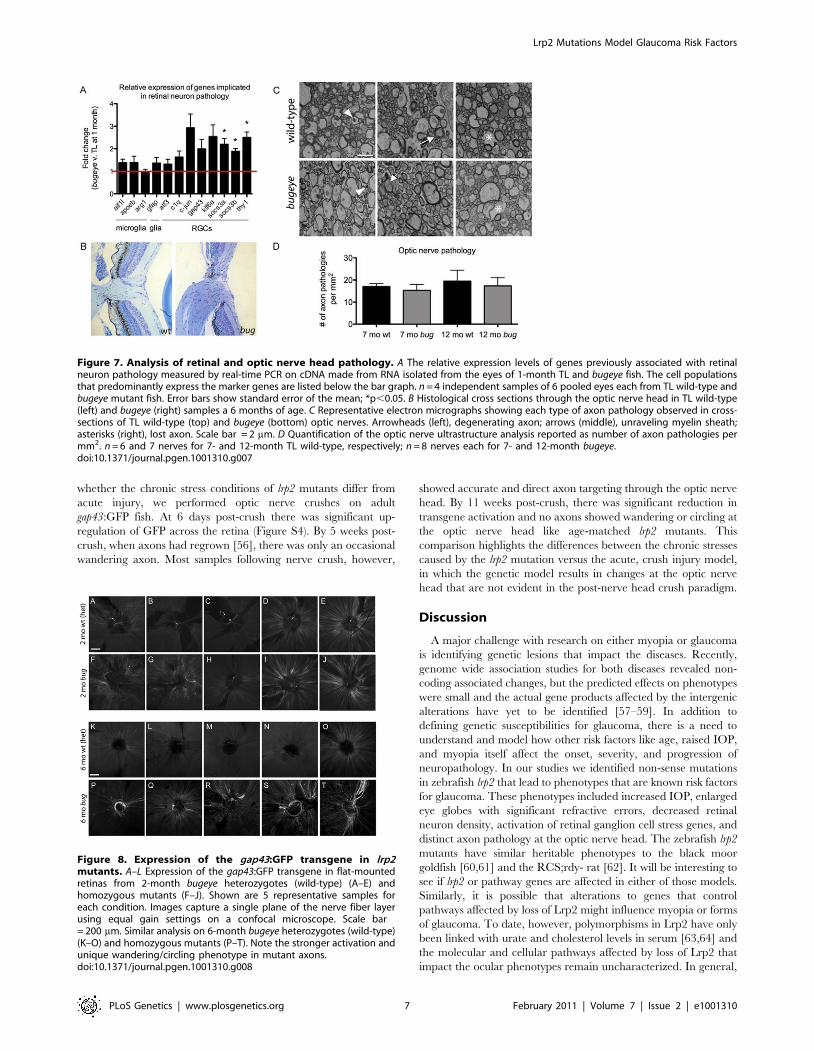
Genes associated with retinal ganglion cell stress andaxon pathology are upregulated in lrp2 mutants
In the following studies we evaluated the onset of retinal
ganglion cell stress and pathology. Relative expression levels of
twelve genes known to be up-regulated in animal models of
retinal ganglion cell injury was surveyed by quantitative RT-
PCR. This panel of markers included three transcripts expressed
in microglia (aif1l, [22,23]; apoeb, [24,25]; arg1, [26,27]), one
expressed in Muller glia and astrocytes (gfap, [28,29]), and eight
expressed in retinal ganglion cells (atf3, [30,31]; c1q, [32–34]; c-
jun, [31,35,36]; gap43, [37,38]; klf6a, [31,39]; socs3a and socs3b,
[31,40,41]; thy1, [42,43]). Analysis was conducted on cDNA
isolated from pooled 1-month-old retinas, a time just prior to
when mutant eyes were measurably enlarged. We chose this early
time-point to avoid measuring changes that might simply reflect
significant alterations in cell proportions and density. With this
assay, we found induction primarily of transcripts associated with
retinal ganglion cells, but not for the glia-associated genes
(Figure 7A).
To investigate whether the markers of retinal ganglion cell stress
correlated with optic nerve pathology, we first compared sagittal
sections of wild-type and lrp2 mutant optic nerve heads from 6-
month-old fish by light microscopy. We then analyzed cross-
sections of wild-type and lrp2 mutant optic nerves, just posterior to
the optic nerve head from 7- and 12-month-old fish by
transmission electron microscopy (TEM). In zebrafish, like other
teleost fish as well as some rodents, the optic nerve head is
comprised of an astroglial lamina without obvious elastin-collagen
rich laminar plates as observed in primates [44–46]. In addition, as
the optic nerve exits the fish eye, it is initially unmyelinated, like
that in humans and most mammals [47–50]. Histology of the optic
nerve head did not reveal excavation or cupping in lrp2 mutants,
but did indicate mutant nerves were larger, consistent with
increased total numbers of retinal ganglion cells in the large-eyed
fish (Figure 7B). Optic nerve cross-sections for TEM were collected
distal to the exit point from the eye within the myelinated region of
the optic nerve, which is adjacent to the site of axonal injury in
glaucoma [51–53]. Nerve damage was scored as 1) degenerating
axons, as noted by electron-dense appearance, 2) axons having an
unraveled myelinated sheath, or 3) space left behind by a shrunken
and degenerating axon. At both ages, examples for each type of
pathology were found in wild-type and lrp2 mutant optic nerves
(Figure 7C). Surprisingly, when total counts were normalized to
area (mm2) there were no differences between genotypes or ages
(Figure 7D).
Because the ultrastructural signature of degenerating axons
following a crush injury is relatively short-lived in the optic nerve
tract of teleost fish as compared to mammals [54], we utilized a
genetic tool to label damaged and regenerating axons over a
longer period of time [55]. We crossed Tg(3.6Frgap43:GFP)mil1
transgenic fish with lrp2 homozygous mutants and then used the
resulting progeny to backcross with non-transgenic lrp2 mutant
fish. This breeding scheme resulted in families with equal
proportions of lrp2 heterozygous and homozygous mutant fish
carrying single insertions of the 3.6Frgap43:GFP transgene. This
transgene contains 3.6 kb of regulatory sequence (59 flanking
region and first intron) from theTakifugu rubripes gap43 locus driving
GFP. Importantly, in these transgenic fish, GFP is expressed in
axons following injury [55]. For our analysis, we compared large-
eyed lrp2 homozygous mutant fish (.0.07 E:B ratio) to normal-
eyed heterozygous siblings (Figure 8K–8T). In all large-eyed
mutant fish we observed strong activation of GFP in a sub-set of
retinal ganglion cells. In the majority of mutant retinas examined
(6 of 6 at 6 months, Figure 8P–8T; and 10 of 12 at 12 months, data
not shown), there was a characteristic axon ‘wandering’ and
‘circling’ around the optic nerve head. This axon phenotype,
where GFP-positive axons approached the optic nerve head in a
disorganized and circuitous fashion, was never observed in retinas
from age matched lrp2 heterozygotes (Figure 8K–8O) or from 12-
month wild-type fish that carried the 3.6Frgap43:GFP transgene
(data not shown). The transgene was activated with variability at 2
months in both wild-type or lrp2 mutant fish (Figure 8A–8J), but
the wandering axon phenotype was only rarely observed in
mutants at this early timepoint. Weak expression of the transgene
was noted in the nerve fiber layer of non-mutant retinas, consistent
with the ongoing neurogenesis of zebrafish. In addition, older wild-
type fish occasionally showed stronger GFP-positive axons,
suggesting sporadic age-related degeneration. In wild-type eyes,
all of the low-GFP expressing axons, as well as the occasional high-
GFP expressing axons, exited the eye directly without wandering
or circling the optic nerve head like those of mutants. To address
Figure 6. Retinal proliferation and apoptosis. A–F Mcm5expression in 1 (A–C) and 2-month (D–F) cryosections. Dashed whitelines denote proliferative ciliary marginal zone (CMZ) of the retina;asterisks indicate autofluorescent blood vessels. G Quantitation ofapoptotic cells identified on whole retina flat-mounts by activatedcaspase-3 (aCasp3) immunofluorescence. H Confocal images of aCasp3-positive cell in 1 year old bugeye mutant. Upper shows compressed z-stacks, lower shows 90urotation to reveal z location of positive cell(arrow). The flat mounted retinas were orientated with retinal ganglioncell layer up. n = 15 eyes for each condition; ***p,0.001, **p,0.01, n.s.,not significant (t-test).doi:10.1371/journal.pgen.1001310.g006
Lrp2 Mutations Model Glaucoma Risk Factors
PLoS Genetics | www.plosgenetics.org 6 February 2011 | Volume 7 | Issue 2 | e1001310
whether the chronic stress conditions of lrp2 mutants differ from
acute injury, we performed optic nerve crushes on adult
gap43:GFP fish. At 6 days post-crush there was significant up-
regulation of GFP across the retina (Figure S4). By 5 weeks post-
crush, when axons had regrown [56], there was only an occasional
wandering axon. Most samples following nerve crush, however,
showed accurate and direct axon targeting through the optic nerve
head. By 11 weeks post-crush, there was significant reduction in
transgene activation and no axons showed wandering or circling at
the optic nerve head like age-matched lrp2 mutants. This
comparison highlights the differences between the chronic stresses
caused by the lrp2 mutation versus the acute, crush injury model,
in which the genetic model results in changes at the optic nerve
head that are not evident in the post-nerve head crush paradigm.
Discussion
A major challenge with research on either myopia or glaucoma
is identifying genetic lesions that impact the diseases. Recently,
genome wide association studies for both diseases revealed non-
coding associated changes, but the predicted effects on phenotypes
were small and the actual gene products affected by the intergenic
alterations have yet to be identified [57–59]. In addition to
defining genetic susceptibilities for glaucoma, there is a need to
understand and model how other risk factors like age, raised IOP,
and myopia itself affect the onset, severity, and progression of
neuropathology. In our studies we identified non-sense mutations
in zebrafish lrp2 that lead to phenotypes that are known risk factors
for glaucoma. These phenotypes included increased IOP, enlarged
eye globes with significant refractive errors, decreased retinal
neuron density, activation of retinal ganglion cell stress genes, and
distinct axon pathology at the optic nerve head. The zebrafish lrp2
mutants have similar heritable phenotypes to the black moor
goldfish [60,61] and the RCS;rdy- rat [62]. It will be interesting to
see if lrp2 or pathway genes are affected in either of those models.
Similarly, it is possible that alterations to genes that control
pathways affected by loss of Lrp2 might influence myopia or forms
of glaucoma. To date, however, polymorphisms in Lrp2 have only
been linked with urate and cholesterol levels in serum [63,64] and
the molecular and cellular pathways affected by loss of Lrp2 that
impact the ocular phenotypes remain uncharacterized. In general,
Figure 7. Analysis of retinal and optic nerve head pathology. A The relative expression levels of genes previously associated with retinalneuron pathology measured by real-time PCR on cDNA made from RNA isolated from the eyes of 1-month TL and bugeye fish. The cell populationsthat predominantly express the marker genes are listed below the bar graph. n = 4 independent samples of 6 pooled eyes each from TL wild-type andbugeye mutant fish. Error bars show standard error of the mean; *p,0.05. B Histological cross sections through the optic nerve head in TL wild-type(left) and bugeye (right) samples a 6 months of age. C Representative electron micrographs showing each type of axon pathology observed in cross-sections of TL wild-type (top) and bugeye (bottom) optic nerves. Arrowheads (left), degenerating axon; arrows (middle), unraveling myelin sheath;asterisks (right), lost axon. Scale bar = 2 mm. D Quantification of the optic nerve ultrastructure analysis reported as number of axon pathologies permm2. n = 6 and 7 nerves for 7- and 12-month TL wild-type, respectively; n = 8 nerves each for 7- and 12-month bugeye.doi:10.1371/journal.pgen.1001310.g007
Figure 8. Expression of the gap43:GFP transgene in lrp2mutants. A–L Expression of the gap43:GFP transgene in flat-mountedretinas from 2-month bugeye heterozygotes (wild-type) (A–E) andhomozygous mutants (F–J). Shown are 5 representative samples foreach condition. Images capture a single plane of the nerve fiber layerusing equal gain settings on a confocal microscope. Scale bar= 200 mm. Similar analysis on 6-month bugeye heterozygotes (wild-type)(K–O) and homozygous mutants (P–T). Note the stronger activation andunique wandering/circling phenotype in mutant axons.doi:10.1371/journal.pgen.1001310.g008
Lrp2 Mutations Model Glaucoma Risk Factors
PLoS Genetics | www.plosgenetics.org 7 February 2011 | Volume 7 | Issue 2 | e1001310

Lrp2 functions in regulation and homeostasis of multiple bioactive
molecules including vitamins, hormones, nutrients, and growth
factors through localized tissue delivery or reuptake by epithelia.
In knowing the affected gene, the zebrafish mutants hold
promise in shedding light on how de-regulated signaling and
homeostasis affect phenotypes such as elevated IOP or excessive
eye growth.
While it is tempting to speculate that the excessive eye growth in
lrp2 mutants is due to the elevated IOP, our studies do not rule out
the possibility that these two phenotypes are distinct. In fact, the
only two Donnai-Barrow patients who have had their IOPs
reported (each with non-sense mutations in LRP2), showed values
in the normal range [65]. Despite normal IOPs, the eyes of the two
young siblings were enlarged and showed high myopia. Further-
more, as an endocytic receptor found on the RPE, Lrp2 is an
interesting candidate as a direct regulator of emmetropization
[66]. Potentially, Lrp2 mediates the availability or transport of
signaling molecules from the retina to affect remodeling within the
sclera. In this context Lrp2 might be key in facilitating the
matching of visual input with axial length of the eye. Nonetheless,
relationships between eye pressure and size are established and the
elevated IOP in zebrafish lrp2 mutants is likely to be at least
contributory to the observed buphthalmia. Consistent with this
possibility, in the few mutant fish where the eye phenotype
presented in a unilateral manner, IOPs were normal in unaffected
eyes, yet elevated in enlarged ones. Indeed, expression of Lrp2 on
the ciliary epithelium suggests a direct role in IOP regulation,
particularly considering the function of Lrp2 at other sites of fluid
regulation. For example in mice, Lrp2 has been shown to regulate
glomerular filtration in the proximal tubule of the kidney and in
the choroid plexus the receptor modulates homeostasis of
cerebrospinal fluid [67–71].
A significant characteristic of lrp2 mutant fish is the strong
relationship between abnormal eye globe growth, retinal thinning,
and activation of retinal ganglion cell stress markers. In this
context, lrp2 mutants have value as a genetic model for studying
the effects of protracted mechanical stress on retinal ganglion cells,
their axons, and the associated glia. As this phenotype relates to
glaucoma, it was surprising that mutant fish did not show
significantly elevated optic nerve pathology with TEM analysis.
It is possible that the stresses induced by lrp2 mutations simply do
not reach a threshold to cause ultrastructural pathology.
Alternatively, low-grade stress may actually ‘‘pre-condition’’ and
promote protective mechanisms in the mutant neurons [72,73].
However, the lack of a difference in ultrastructure pathology
between mutant and wild-type siblings could also be explained by
the surprisingly high number of pathological events noted in the
wild-type fish. This perhaps relates to the regenerative capacity of
teleosts [56,74] and a relaxation of selective pressure to maintain
nerve health with normal aging. Through evolution, fish may have
lost highly-robust nerve protective mechanisms against age-related
stresses, and instead rely on ongoing growth and regeneration to
maintain vision, perhaps accounting for the unexpected pathology
scored in wild-type optic nerves. In addition, because a higher
proportion of the ganglion cell axons in lrp2 mutant fish are in fact
younger than those of wild-type siblings (due to the excessive
ongoing generation of neurons in their eyes), many of the optic
nerve profiles might be expected to in fact look healthier in a
relative manner.
The modest death of retinal ganglion cells in lrp2 mutants was
less surprising. First, extended retinal ganglion cell soma survival,
despite axonal damage and dysfunction, is known for the DBA/2
mouse glaucoma model. DBA/2 mice show a pigment dispersion-
related glaucoma with elevated IOP [75,76]. In young DBA/2
mice, axons at the nerve head often show focal insults with many
having dystrophic features [51]. In many aged animals, axons are
clearly degenerative [51]. Most retinal ganglion cells, however,
survive for extended periods of time and their disconnected
proximal (intra-retinal) axons take on reactive and stressed
characteristics [51,77,78]. Second, the resilient nature of retinal
ganglion cells in teleosts has been well characterized. In fact for
goldfish, experimental axotomy or optic nerve crush results in less
than 10% death of retinal ganglion cells [79], and in zebrafish
only 20% of the lesioned neurons are reported to die [80]. In
contrast, optic nerve axotomy in mammals results in apoptosis of
nearly all retinal ganglion cells [81–83]. The regrowth of axons in
teleosts occurs over a course of weeks and results in correct axon
pathfinding and appropriate tectal innervation [84,85]. In
contrast, in lrp2 mutants, retinal ganglion cells appear to be
under prolonged mechanical stress from the stretching and
growth of the eye globe. This was evident from the changes in
retinal density with eye enlargement and the activation of retinal
ganglion cell stress markers. Of interest, axon regrowth through
the optic nerve was affected in lrp2 mutants. The wandering and
circling phenotype of the gap43:GFP axons in large-eyed mutants
is reminiscent of the EphB3-dependent ‘reactive plasticity’
following optic nerve injury in mice [86,87]. Regardless, of why
bugeye/lrp2 mutants do not show dramatic retinal ganglion cell
death, this fact emphasizes that while these fish model initiating
risk factors for glaucoma, they do not model the end stages of the
disease.
Lrp2 mutations in humans and mice are often lethal, but always
developmentally relevant, particularly within the nervous system
[71,88,89]. Our analyses of both bugeye alleles indicate Lrp2 is
dispensable for survival in zebrafish. Furthermore, we did not
detect morphological phenotypes in mutant embryos, similar to
the observations following oligonucleotide knock-down of zebra-
fish lrp2 [67]. The total lack of lethality in zebrafish lrp2 mutants
may be due to species differences in respiration, as mice mutants
often die from respiratory failure at birth. Alternatively, there may
be compensation from other Lrp family members in zebrafish.
Compensation from Lrp family members may also explain the lack
of obvious developmental defects. More detailed studies of the
zebrafish mutant embryos and larvae are warranted to assess
whether subtle defects exist.
In summary, we have identified mutations in lrp2 that cause
adult-onset ocular pathogenesis in zebrafish. While mutants
appear normal during larval stages of development, as young
adults they develop enlarged eyes with elevated IOP. Over time,
retinal cell density becomes significantly reduced due to insuffi-
cient proliferation of marginal zone stem cells and increased
neuronal cell death. Markers of retinal ganglion cell stress become
elevated and damaged and/or regenerating axons at the optic
nerve head show a characteristic wandering and circling
phenotype. These fish will be valuable for future studies on the
signaling and cellular mechanism of myopia and other risk factors
for glaucoma.
Materials and Methods
Fish maintenanceWild-type and mutant zebrafish (Dano rerio) were maintained at
28uC with a 14 on/10 off light cycle and were feed a standard diet
[90]. All animal husbandry and experiments were approved and
conducted in accordance with the guidelines set forth by the
Institutional Animal Care and Use Committee of the Medical
College of Wisconsin.
Lrp2 Mutations Model Glaucoma Risk Factors
PLoS Genetics | www.plosgenetics.org 8 February 2011 | Volume 7 | Issue 2 | e1001310

Mutant and transgenic allelesbugeye; lrp2mw1 (this study)
bugeye; lrp2p5bnc (this study)
Tg(3.6Frgap43:GFP)mil1 [55]
Accession numberslrp2, HM_754616
aif1l, NM_198870
apoeb, NM_131098
arg1, XM_001922563
gfap, NM_131373
atf3, NM_200964
c1q, NM_001005976
c-jun, NM_199987
gap43, NM_131341
klf6a, NM_201461
socs3a, NM_199950
socs3b, NM_213304
thy1, NM_198065
Measurement of intraocular pressuresServo-null electrophysiology was used to measure IOPs as
described previously [14].
Recombinant linkage mappingMapping panels of 6 month adult mutant fish (obviously
enlarged eyes) were collected from backcross pedigrees. Bulked
segregant analysis, using pooled samples of mutant genomic DNA
and individual parental DNA, was conducted with simple
sequence repeat (SSR) markers to establish linkage to Chromo-
some 9. For higher-resolution mapping, sequencing of parental
genomic DNA in regions associated with the closest linked
microsatellite markers was done to find additional SSRs. These
new SSRs were then used to refine the critical interval by
analyzing single mutant fish.
GenotypingPCR was performed on DNA isolated using the Puregene kit
(Qiagen, Germantown, MD) from tailfin-clips, using primers
designed to amplify the allele specific mutations in lrp2:
bug mw1 F: CGTTATTTTCTGTCTAGGTTCAGGTTA,
bug mw1 R: GAAAAGAAAAGATTGATACATACGG
bug p5bnc F: GTGTGTTTTCTGAAAACTGTCAAGC,
bug p5bnc R: CTTTGCAGCTGGTAATGAAAATCCACAC-
CAACAGCGGCTCCTCTGTCCTA. Underlined letter in prim-
er denotes mutant nucleotide, bolded letter denotes a single
nucleotide change in the primer to generate a novel restriction site
for each allele (bug mw1: MseI; bug p5bnc AvrII).
Eye size and body length measurementsFish were anesthetized with 0.05% Tricaine and body lengths
were measured in side-view from the tip of the head to the end of
the trunk (before the caudal fin). To measure eye size, anesthetized
fish were imaged at a fixed magnification from a dorsal perspective
using a Nikon CoolPix995 camera attached to a Leica MZFLIII
microscope. These images were imported into Metamorph
software (Universal Imaging Corp, Philadelphia, PA), and the
area of each eye from the dorsal view was traced using the Region
Measurements function.
Relative refractive errorLens radius (L) was measured from histological cross sections;
retina radius (R) was back-calculated by assuming the retina to be
a semi-circle, measuring the length of the retina, and taking that
measurement as half the circumference of a circle (so R = length
of retina/p). Sections with minimal distortion from processing
were used and no attempts to correct for distortions were made. A
focal length (F) of 2.32 x L for the lens was used as in studies with
goldfish [91]. RRE was calculated as 1- (R/F). By this calculation,
all wild type fish were predicted to be slightly hyperopic (RRE
.1), likely due to fixation artifact. To adjust this, the ratio of (R/F)
was multiplied by a constant factor for both genotypes at each age
(1 month, 1.15; 2 months, 1.18), so that on average, the wild type
fish were emmetropic (RRE = 0).
HistologyHeads were removed from terminally anesthetized fish and
fixed overnight in gluteraldehyde/paraformaldehyde at 4uC,
washed three times in PBS, and dehydrated in increasing ethanol
solutions (50%, 70%, 80%, 90%, 95%, 100%, 100%, 100%) for
10 minutes each, all at RT. The heads were then infiltrated with
propylene oxide for 15 minutes twice, then a 1:1 mix propylene
oxide:epon for 2 hours at RT. An additional equal volume of epon
was added to the samples and these were incubated overnight with
culture tube caps off so that the propylene oxide would evaporate.
Heads were bisected when necessary to fit in block-molds,
embedded in epon, and baked for at least 24 hours at 65uC.
Semi-thin sections were cut on a Leica RM2255 microtome and
stained with 1% Toluidine, 1% Borax.
Cell countsFor each eye, 5 non-consecutive sections were imaged from
the central retina (sections with the largest lens diameter) with a
40X objective on a Nikon E600FN microscope with a
Photometrics CoolSnap camera attached. Each image was
printed and the nuclei in each layer of the retina were counted.
The average of the 5 sections was calculated and represented 1
data point. For sample condition, between 6–12 eyes were scored
in this manner.
ImmunostainingZebrafish embryos or isolated eyes were fixed overnight at 4uC
in 4% PFA (pH 7.4, in PBS), washed three times for 10 minutes in
PBS, then infiltrated with increasing concentrations of sucrose
(15%, 30%) for 2 hours each at 4uC, followed by overnight
incubation in HistoPrep freezing media (Fischer Scientific,
Pittsburgh, PA). Cryoprotected embryos were embedded in
HistoPrep and flash frozen, sectioned at 10–12 mM and collected
on Supercharge Plus slides (Fischer Scientific). Cryosections were
allowed to dry on the slide for 1hr at RT, and the edge of the slide
was traced with a PAP pen. Slides were rinsed briefly with PBTD
(PBS +1% DMSO +1% Tween-20) to rehydrate the tissue, and
then incubated in block (5% donkey serum in PBTD) for 2 hours
at RT. Primary antibody was diluted in block (Sheep-anti-Lrp2
1:1000, gift from Dr. Thomas Willnow (Max Delbruck Center,
Berlin, Germany)) and incubated on slides overnight at 4uC.
Antibody was removed and slides were washed three times with
PBTD rinses, and secondary antibody diluted in block (Cy3-
Donkey anti-Sheep 1:250, Jackson ImmunoResearch, West-
grove, PA) was incubated at RT for 1.5 hours. Secondary
antibody was removed with three washes of PBTD, and slides
were mounted in 1:1 PBS to glycerol with 0.1% Hoechst nuclear
stain (cryosections). Images were collected using a Nikon C1
confocal microscope. The same procedure was followed for
dissected whole adult retinas prior to flat-mount analysis, using
anti-cleaved caspase-3 primary antibody (1:500, Cell Signaling
Lrp2 Mutations Model Glaucoma Risk Factors
PLoS Genetics | www.plosgenetics.org 9 February 2011 | Volume 7 | Issue 2 | e1001310

Technology, Danvers, MA) and DyLight 488 secondary (1:1000,
Jackson ImmunoResearch).
Real-time PCR1 month fish measuring between 10–12mm were anesthetized
in Tricaine, and both eyes were removed and placed immediately
in TRIzol (Invitrogen). Each sample was a pool of 3 pairs of eyes (6
eyes per sample), and 4 samples were used for each genotype.
RNA was isolated following the Invitrogen protocol. Reverse-
transcription PCR was carried out following the protocol for
SuperScript III First Strand Synthesis (Invitrogen). Gene specific
primers were used as follows to amplify the genes of interest: Aif1l
(F: CAACATGGACTTACAAGGCG, R: TCCTCTTCGTC-
TCTGTACTTCTG); ApoEb (F: GTGCAAAACATCAAGG-
GCTC, R: GGGTCATCTGGGTTTGGAG); Arg1 (F: TGG-
GCATCAAAACCTTCTCC, R: AAACTCAGATGGATCGG-
CTTC); Atf3 (F: AGCCTGCATGAACACTGAG, R: TTTT-
CCTTCGGTCGTTCTCC); C1q (F: CTCTGCTGACACCT-
GTCCTG, R: GGTGGTCCTTTCAGACCAAA); c-Jun (F:
ACGTGGGACTTCTCAAACTG, R: TCTTGGGACACAGA-
AACTGG); Gap43 (F: GAAGGCAATGCACAGAAAGAG, R:
TGCTGGTTTGGATTCCTCAG); Gfap (F: AAGCTCTGC-
AAGACGAGATC, R: GCTTAGACACATCCAGATCCAC);
Klf6 (F: CACTTAAAAGCACATCAGCGG, R: GAAGTGT-
CGGGTTAGCTCATC); Socs3a (F: CATTCAACAAAAGA-
GACTCATAGGC, R: TGTGGGTTATCATGGCGATAC);
Socs3b (F: CCCAAGATTGAGTCGGATAACG, R: ACCAA-
CACAAAGCCCAGAG); Thy-1 (F: CCGGTGTCAATCATT-
CAAACTG, R: CAGTGGGAAAGTGAGGAAGG). Initially,
PCR products were amplified with Accuprime Taq HighFidelity
(Invitrogen), and sequenced to verify specificity. Real-time analysis
was performed on a Bio-Rad iCycler using iQ SYBR Green
SuperMix (Bio-Rad). 3-step PCR with a 57uC annealing
temperature was used for all primer sets except Arg1, Atf3, and
Thy1, which used a 2-step PCR with a 54uC annealing
temperature to eliminate a non-specific product. All samples were
run in triplicate, and fold change was calculated using the DDCt
method, with Ef1a as the housekeeping gene for all primer sets.
TEM of optic nervesHeads were removed from terminally anesthetized fish. In a
Petri dish filled with buffer, the optic nerves were dissected from
the heads first by removing the skin, skeleton, and connective
tissue, leaving the eyes and attached nerves and tectum intact. The
tectum was cut from the nerves, leaving the nerves intertwined at
the chiasm. The nerves were separated by gently pulling on the
eye globes with forceps, and making a cut with an 8 mm Spring
Scissors (Fine Science Tools) when necessary. Dissected nerves
with attached eyes were then incubated overnight at 4uC in
gluteraldehyde/paraformaldehyde fixative. Heads were washed
three times in 0.1M PO4 buffer, and then most of the eye globe
removed by using the 8 mm scissors to make a circumferential cut
around the optic nerve head, leaving a small portion of the
posterior eye attached to the dissected nerve. The nerves were
post-fixed in gluteraldehyde/paraformaldehyde for 1 hr at room
temperature, washed 3X in 0.1M PO4 buffer, fixed in 1% buffered
Osmuium for 1 hr on ice, and washed 3X with ice cold water. The
following steps were all done at room temperature: nerves were
dehydrated in an increasing series of MeOH (30%, 50%, 70%,
95%, 100%, 100%, 100%), then infiltrated with acetonitrile, 2X
for 15minutes each, followed by 2 hours in a 1:1 mix of
acetonitrile and EM Epon, and finally incubated in 100% EM
Epon overnight, embedded in molds, and baked for at least
24 hours at 65uC. The blocks were trimmed to between 100–200
microns past the optic nerve head on a Leica RM2255 microtome,
and ultra-thin sections were cut and plated on a grid, and imaged
using a Hitachi H600 transmission electron microscope. The
entire nerve cross-section was canvassed at 8000X, and 10–16
representative images were collected from each nerve at this
magnification. Quantitative assessment of nerve pathology was
conducted in a double-blinded manner in which both the TEM
microscopist and the individual scoring pathology for the samples
was unaware of the sample genotype.
Retinal flat-mount analysisEyes were dissected from terminally anesthetized adult fish and
fixed overnight at 4uC in 4% PFA (pH 7.4, in PBS), then washed
three times in PBS. In a Petri dish filled with PBS, a
circumferential cut was made at front of the eye with a scalpel,
near the border of the anterior and posterior segments. The
anterior segment was discarded, followed by removal of the sclera
from the posterior segment. The remaining retina with RPE was
post-fixed 1-2 hrs with 4% PFA (pH 7.4, in PBS), washed in PBS,
and then laid flat on a slide by making incisions through the retina
so that it would lay flat. Whole retinas were mounted on the slides
with 20 ml of Vectashield Mounting Medium (Vector Labs,
Burlingame, CA), and coverslipped. For retinas used for anti-
activated-caspase-3 immunofluorescence, antibody incubations
were done after removal of the anterior segment and sclera, but
prior to flat-mount analysis.
Supporting Information
Figure S1 A-G bugeye mutants show varying degrees of
asymmetry in eye size (B-G), with the smaller of the two eyes
only occasionally falling within the range of the wild-type eye size
to body length ratio (E:B; wild-type E:B , 0.05, bugeye E:B .0.06).
For each eye, the E:B is indicated. H In asymmetric eyes of bugeye
fish, intraocular pressure (IOP) was elevated only in the enlarged
eyes. In TL wild-type, the eye measured for IOP first had a higher
value than the second eye measured in the same fish. Black
symbols represent the first eye of a fish that was measured for IOP
and gray symbols represent the second eye. The vertical bar
connecting symbols shows eyes from the same fish. Circles
represent normal sized eyes; squares represent enlarged eyes.
Found at: doi:10.1371/journal.pgen.1001310.s001 (0.81 MB TIF)
Figure S2 Iridocorneal angle histology of 2.5 month old wild-
type (A-,D), normal eyed bugeye mutants (B-E), and large-eyed
bugeye mutants (C-F). A-C. Dorsal ciliary epithium. D-F, ventral
canalicular outflow pathway. Red boxes show from where high
magnification isets were derived. Scale bar = 50 mm. Insets are
magnified 2.5X. Note the elongated and dysplasic ciliary epithelial
cells in B and C insets.
Found at: doi:10.1371/journal.pgen.1001310.s002 (1.18 MB TIF)
Figure S3 Body length measurements over time. Five TL wild-
type and bugeye fish were measured weekly from 5 to 26 weeks, and
again at 30, 35, and 40 weeks. Mean and standard error of the
mean (error bars) are plotted for each genotype at all timepoints.
Bonferonni tests at each timepoint show that there is no statistical
difference between genotypes at any time.
Found at: doi:10.1371/journal.pgen.1001310.s003 (0.13 MB TIF)
Figure S4 Expression of the gap43:GFP transgene following
optic nerve crush in adult (6 month old) wild-type fish. A-F
Expression of the gap43:GFP transgene in experimental eyes 6
days after optic nerve crush (A-D) or in the contralateral
uncrushed control eye (E-F). G-L Expression of the gap43:GFP
transgene in experimental eyes 5 weeks after optic nerve crush (I-
Lrp2 Mutations Model Glaucoma Risk Factors
PLoS Genetics | www.plosgenetics.org 10 February 2011 | Volume 7 | Issue 2 | e1001310

L) or in the contralateral uncrushed control eyes (M-P). M-R
Expression of the gap43:GFP transgene in experimental eyes 11
weeks after optic nerve crush (Q-T) or in the contralateral
uncrushed control eyes (E-F). Shown are 3 representative samples
for each condition. Images capture a single plane of the nerve fiber
layer using equal gain settings on a confocal microscope. Scale bar
= 200mm.
Found at: doi:10.1371/journal.pgen.1001310.s004 (1.16 MB TIF)
Acknowledgments
The bugeye mw1 allele was originally isolated is a genetic screen conducted
in John E. Dowling’s laboratory (Harvard University). We thank Clive
Wells (MCW) and Michael Cliff for technical assistance with electron
microscopy and animal husbandry, respectively. We gratefully acknowl-
edge Drs. Qun Xiang and Sergey Tarima for statistical consultation. We
also thank Drs. Thomas Willnow and Salim Abdelilah-Seyfried (Max
Delbrueck Center, Berlin) for sharing reagents and for helpful discussions.
Author Contributions
Conceived and designed the experiments: KNV RFC SWMJ RGG BAL.
Performed the experiments: KNV JRW RFC MPG GBW RSS SWMJ
BAL. Analyzed the data: KNV JRW RFC GBW RSS SWMJ RGG BAL.
Contributed reagents/materials/analysis tools: KNV JRW RFC DSW
MCM AJU RGG BAL. Wrote the paper: KNV BAL.
References
1. Gehrs KM, Jackson JR, Brown EN, Allikmets R, Hageman GS (2010)
Complement, age-related macular degeneration and a vision of the future.Arch Ophthalmol 128: 349–358.
2. Bloom RI, Friedman IB, Chuck RS (2010) Increasing rates of myopia: the long
view. Curr Opin Ophthalmol 21: 247–248.
3. Young TL (2009) Molecular genetics of human myopia: an update. Optom Vis
Sci 86: E8–E22.
4. Loyo-Berrios NI, Blustein JN (2007) Primary-open glaucoma and myopia: anarrative review. Wmj 106: 85–89, 95.
5. Libby RT, Gould DB, Anderson MG, John SW (2005) Complex genetics ofglaucoma susceptibility. Annu Rev Genomics Hum Genet 6: 15–44.
6. Boland MV, Quigley HA (2007) Risk factors and open-angle glaucoma:
classification and application. J Glaucoma 16: 406–418.
7. May P, Woldt E, Matz RL, Boucher P (2007) The LDL receptor-related protein
(LRP) family: an old family of proteins with new physiological functions. Ann
Med 39: 219–228.
8. Fisher CE, Howie SE (2006) The role of megalin (LRP-2/Gp330) during
development. Dev Biol 296: 279–297.
9. Christensen EI, Birn H (2002) Megalin and cubilin: multifunctional endocyticreceptors. Nat Rev Mol Cell Biol 3: 256–266.
10. Kantarci S, Al-Gazali L, Hill RS, Donnai D, Black GC, et al. (2007) Mutationsin LRP2, which encodes the multiligand receptor megalin, cause Donnai-
Barrow and facio-oculo-acoustico-renal syndromes. Nat Genet 39: 957–959.
11. Donnai D, Barrow M (1993) Diaphragmatic hernia, exomphalos, absent corpuscallosum, hypertelorism, myopia, and sensorineural deafness: a newly recognized
autosomal recessive disorder? Am J Med Genet 47: 679–682.
12. Pober BR, Longoni M, Noonan KM (2009) A review of Donnai-Barrow andfacio-oculo-acoustico-renal (DB/FOAR) syndrome: clinical features and differ-
ential diagnosis. Birth Defects Res A Clin Mol Teratol 85: 76–81.
13. Xu L, Wang Y, Wang S, Wang Y, Jonas JB (2007) High myopia and glaucoma
susceptibility the Beijing Eye Study. Ophthalmology 114: 216–220.
14. Link BA, Gray MP, Smith RS, John SW (2004) Intraocular pressure inzebrafish: comparison of inbred strains and identification of a reduced melanin
mutant with raised IOP. Invest Ophthalmol Vis Sci 45: 4415–4422.
15. Gray MP, Smith RS, Soules KA, John SW, Link B (2009) The aqueous humoroutflow pathway of zebrafish. Invest Ophthalmol Vis Sci.
16. Soules KA, Link BA (2005) Morphogenesis of the anterior segment in thezebrafish eye. BMC Dev Biol 5: 12.
17. Raymond PA, Barthel LK, Bernardos RL, Perkowski JJ (2006) Molecular
characterization of retinal stem cells and their niches in adult zebrafish. BMCDev Biol 6: 36.
18. Johns PR, Fernald RD (1981) Genesis of rods in teleost fish retina. Nature 293:
141–142.
19. Morris AC, Scholz TL, Brockerhoff SE, Fadool JM (2008) Genetic dissection
reveals two separate pathways for rod and cone regeneration in the teleost retina.Dev Neurobiol 68: 605–619.
20. Otteson DC, D’Costa AR, Hitchcock PF (2001) Putative stem cells and the
lineage of rod photoreceptors in the mature retina of the goldfish. Dev Biol 232:62–76.
21. Gajera CR, Emich H, Lioubinski O, Christ A, Beckervordersandforth-Bonk R,
et al. (2010) LRP2 in ependymal cells regulates BMP signaling in the adultneurogenic niche. J Cell Sci 123: 1922–1930.
22. Bosco A, Inman DM, Steele MR, Wu G, Soto I, et al. (2008) Reduced retinamicroglial activation and improved optic nerve integrity with minocycline
treatment in the DBA/2J mouse model of glaucoma. Invest Ophthalmol Vis Sci
49: 1437–1446.
23. Schluesener HJ, Seid K, Meyermann R (1999) Effects of autoantigen and
dexamethasone treatment on expression of endothelial-monocyte activating
polypeptide II and allograft-inflammatory factor-1 by activated macrophagesand microglial cells in lesions of experimental autoimmune encephalomyelitis,
neuritis and uveitis. Acta Neuropathol 97: 119–126.
24. Ignatius MJ, Gebicke-Harter PJ, Skene JH, Schilling JW, Weisgraber KH, et al.
(1986) Expression of apolipoprotein E during nerve degeneration and
regeneration. Proc Natl Acad Sci U S A 83: 1125–1129.
25. Kuhrt H, Hartig W, Grimm D, Faude F, Kasper M, et al. (1997) Changes in
CD44 and ApoE immunoreactivities due to retinal pathology of man and rat.
J Hirnforsch 38: 223–229.
26. Pernet V, Bourgeois P, Di Polo A (2007) A role for polyamines in retinal
ganglion cell excitotoxic death. J Neurochem 103: 1481–1490.
27. Zhang W, Baban B, Rojas M, Tofigh S, Virmani SK, et al. (2009) Arginase
activity mediates retinal inflammation in endotoxin-induced uveitis. Am J Pathol
175: 891–902.
28. Osborne NN, Block F, Sontag KH (1991) Reduction of ocular blood flow results
in glial fibrillary acidic protein (GFAP) expression in rat retinal Muller cells. Vis
Neurosci 7: 637–639.
29. Tanihara H, Hangai M, Sawaguchi S, Abe H, Kageyama M, et al. (1997) Up-
regulation of glial fibrillary acidic protein in the retina of primate eyes with
experimental glaucoma. Arch Ophthalmol 115: 752–756.
30. Takeda M, Kato H, Takamiya A, Yoshida A, Kiyama H (2000) Injury-specific
expression of activating transcription factor-3 in retinal ganglion cells and its
colocalized expression with phosphorylated c-Jun. Invest Ophthalmol Vis Sci 41:
2412–2421.
31. Veldman MB, Bemben MA, Thompson RC, Goldman D (2007) Gene
expression analysis of zebrafish retinal ganglion cells during optic nerve
regeneration identifies KLF6a and KLF7a as important regulators of axon
regeneration. Dev Biol 312: 596–612.
32. Kuehn MH, Kim CY, Ostojic J, Bellin M, Alward WL, et al. (2006) Retinal
synthesis and deposition of complement components induced by ocular
hypertension. Exp Eye Res 83: 620–628.
33. Stasi K, Nagel D, Yang X, Wang RF, Ren L, et al. (2006) Complement
component 1Q (C1Q) upregulation in retina of murine, primate, and human
glaucomatous eyes. Invest Ophthalmol Vis Sci 47: 1024–1029.
34. Stevens B, Allen NJ, Vazquez LE, Howell GR, Christopherson KS, et al. (2007)
The classical complement cascade mediates CNS synapse elimination. Cell 131:
1164–1178.
35. Herdegen T, Bastmeyer M, Bahr M, Stuermer C, Bravo R, et al. (1993)
Expression of JUN, KROX, and CREB transcription factors in goldfish and rat
retinal ganglion cells following optic nerve lesion is related to axonal sprouting.
J Neurobiol 24: 528–543.
36. Koistinaho J, Hokfelt T (1997) Altered gene expression in brain ischemia.
Neuroreport 8: i- viii.
37. Bormann P, Zumsteg VM, Roth LW, Reinhard E (1998) Target contact
regulates GAP-43 and alpha-tubulin mRNA levels in regenerating retinal
ganglion cells. J Neurosci Res 52: 405–419.
38. Doster SK, Lozano AM, Aguayo AJ, Willard MB (1991) Expression of the
growth-associated protein GAP-43 in adult rat retinal ganglion cells following
axon injury. Neuron 6: 635–647.
39. Moore DL, Blackmore MG, Hu Y, Kaestner KH, Bixby JL, et al. (2009) KLF
family members regulate intrinsic axon regeneration ability. Science 326:
298–301.
40. Fischer D, Petkova V, Thanos S, Benowitz LI (2004) Switching mature retinal
ganglion cells to a robust growth state in vivo: gene expression and synergy with
RhoA inactivation. J Neurosci 24: 8726–8740.
41. Rolls A, Cahalon L, Bakalash S, Avidan H, Lider O, et al. (2006) A sulfated
disaccharide derived from chondroitin sulfate proteoglycan protects against
inflammation-associated neurodegeneration. Faseb J 20: 547–549.
42. Dabin I, Barnstable CJ (1995) Rat retinal Muller cells express Thy-1 following
neuronal cell death. Glia 14: 23–32.
43. Schlamp CL, Johnson EC, Li Y, Morrison JC, Nickells RW (2001) Changes in
Thy1 gene expression associated with damaged retinal ganglion cells. Mol Vis 7:
192–201.
44. Koke JR, Mosier AL, Garcia DM (2010) Intermediate filaments of zebrafish
retinal and optic nerve astrocytes and Muller glia: differential distribution of
cytokeratin and GFAP. BMC Res Notes 3: 50.
45. Levine RL (1989) Organization of astrocytes in the visual pathways of
the goldfish: an immunohistochemical study. J Comp Neurol 285: 231–
245.
Lrp2 Mutations Model Glaucoma Risk Factors
PLoS Genetics | www.plosgenetics.org 11 February 2011 | Volume 7 | Issue 2 | e1001310

46. Lillo C, Velasco A, Jimeno D, Lara JM, Aijon J (1998) Ultrastructural
organization of the optic nerve of the tench (Cyprinidae, Teleostei). J Neurocytol27: 593–604.
47. Downs JC, Roberts MD, Burgoyne CF (2008) Mechanical environment of the
optic nerve head in glaucoma. Optom Vis Sci 85: 425–435.48. Easter SS, Jr., Rusoff AC, Kish PE (1981) The growth and organization of the
optic nerve and tract in juvenile and adult goldfish. J Neurosci 1: 793–811.49. Maggs A, Scholes J (1986) Glial domains and nerve fiber patterns in the fish
retinotectal pathway. J Neurosci 6: 424–438.
50. Sun D, Lye-Barthel M, Masland RH, Jakobs TC (2009) The morphology andspatial arrangement of astrocytes in the optic nerve head of the mouse. J Comp
Neurol 516: 1–19.51. Howell GR, Libby RT, Jakobs TC, Smith RS, Phalan FC, et al. (2007) Axons of
retinal ganglion cells are insulted in the optic nerve early in DBA/2J glaucoma.J Cell Biol 179: 1523–1537.
52. Nickells RW (2007) From ocular hypertension to ganglion cell death: a
theoretical sequence of events leading to glaucoma. Can J Ophthalmol 42:278–287.
53. Quigley HA (2005) Glaucoma: macrocosm to microcosm the Friedenwaldlecture. Invest Ophthalmol Vis Sci 46: 2662–2670.
54. Colavincenzo J, Levine RL (2000) Myelin debris clearance during Wallerian
degeneration in the goldfish visual system. J Neurosci Res 59: 47–62.55. Udvadia AJ (2008) 3.6 kb genomic sequence from Takifugu capable of
promoting axon growth-associated gene expression in developing and regener-ating zebrafish neurons. Gene Expr Patterns 8: 382–388.
56. Becker CG, Becker T (2007) Growth and pathfinding of regenerating axons inthe optic projection of adult fish. J Neurosci Res 85: 2793–2799.
57. Hysi PG, Young TL, Mackey DA, Andrew T, Fernandez-Medarde A, et al.
(2010) A genome-wide association study for myopia and refractive erroridentifies a susceptibility locus at 15q25. Nat Genet 42: 902–905.
58. Solouki AM, Verhoeven VJ, van Duijn CM, Verkerk AJ, Ikram MK, et al.(2010) A genome-wide association study identifies a susceptibility locus for
refractive errors and myopia at 15q14. Nat Genet 42: 897–901.
59. Thorleifsson G, Walters GB, Hewitt AW, Masson G, Helgason A, et al. (2010)Common variants near CAV1 and CAV2 are associated with primary open-
angle glaucoma. Nat Genet 42: 906–909.60. Easter SS, Jr., Hitchcock PF (1986) The myopic eye of the Black Moor goldfish.
Vision Res 26: 1831–1833.61. Raymond PA, Hitchcock PF, Palopoli MF (1988) Neuronal cell proliferation and
ocular enlargement in Black Moor goldfish. J Comp Neurol 276: 231–238.
62. Thanos S, Naskar R (2004) Correlation between retinal ganglion cell death andchronically developing inherited glaucoma in a new rat mutant. Exp Eye Res 79:
119–129.63. Mii A, Nakajima T, Fujita Y, Iino Y, Kamimura K, et al. (2007) Genetic
association of low-density lipoprotein receptor-related protein 2 (LRP2) with
plasma lipid levels. J Atheroscler Thromb 14: 310–316.64. Kamatani Y, Matsuda K, Okada Y, Kubo M, Hosono N, et al. (2010) Genome-
wide association study of hematological and biochemical traits in a Japanesepopulation. Nat Genet 42: 210–215.
65. Patel N, Hejkal T, Katz A, Margalit E (2007) Ocular manifestations of Donnai-Barrow syndrome. J Child Neurol 22: 462–464.
66. Rymer J, Wildsoet CF (2005) The role of the retinal pigment epithelium in eye
growth regulation and myopia: a review. Vis Neurosci 22: 251–261.67. Anzenberger U, Bit-Avragim N, Rohr S, Rudolph F, Dehmel B, et al. (2006)
Elucidation of megalin/LRP2-dependent endocytic transport processes in thelarval zebrafish pronephros. J Cell Sci 119: 2127–2137.
68. Carro E, Spuch C, Trejo JL, Antequera D, Torres-Aleman I (2005) Choroid
plexus megalin is involved in neuroprotection by serum insulin-like growth factorI. J Neurosci 25: 10884–10893.
69. Christensen EI, Willnow TE (1999) Essential role of megalin in renal proximaltubule for vitamin homeostasis. J Am Soc Nephrol 10: 2224–2236.
70. Hammad SM, Ranganathan S, Loukinova E, Twal WO, Argraves WS (1997)Interaction of apolipoprotein J-amyloid beta-peptide complex with low density
lipoprotein receptor-related protein-2/megalin. A mechanism to preventpathological accumulation of amyloid beta-peptide. J Biol Chem 272:
18644–18649.
71. Zarbalis K, May SR, Shen Y, Ekker M, Rubenstein JL, et al. (2004) A focused
and efficient genetic screening strategy in the mouse: identification of mutationsthat disrupt cortical development. PLoS Biol 2: e219. doi:10.1371/journal.
pbio.0020219.
72. Dirnagl U, Simon RP, Hallenbeck JM (2003) Ischemic tolerance andendogenous neuroprotection. Trends Neurosci 26: 248–254.
73. Roth S (2004) Endogenous neuroprotection in the retina. Brain Res Bull 62:461–466.
74. Hitchcock PF, Raymond PA (2004) The teleost retina as a model for
developmental and regeneration biology. Zebrafish 1: 257–271.
75. John SW, Smith RS, Savinova OV, Hawes NL, Chang B, et al. (1998) Essential
iris atrophy, pigment dispersion, and glaucoma in DBA/2J mice. InvestOphthalmol Vis Sci 39: 951–962.
76. Anderson MG, Smith RS, Hawes NL, Zabaleta A, Chang B, et al. (2002)
Mutations in genes encoding melanosomal proteins cause pigmentary glaucomain DBA/2J mice. Nat Genet 30: 81–85.
77. Buckingham BP, Inman DM, Lambert W, Oglesby E, Calkins DJ, et al. (2008)Progressive ganglion cell degeneration precedes neuronal loss in a mouse model
of glaucoma. J Neurosci 28: 2735–2744.
78. Soto I, Oglesby E, Buckingham BP, Son JL, Roberson ED, et al. (2008) Retinalganglion cells downregulate gene expression and lose their axons within the optic
nerve head in a mouse glaucoma model. J Neurosci 28: 548–561.
79. Murray M (1982) A quantitative study of regenerative sprouting by optic axons
in goldfish. J Comp Neurol 209: 352–362.
80. Zhou LX, Wang ZR (2002) [Changes in number and distribution of retinalganglion cells after optic nerve crush in zebrafish]. Shi Yan Sheng Wu Xue Bao
35: 159–162.
81. Berkelaar M, Clarke DB, Wang YC, Bray GM, Aguayo AJ (1994) Axotomy
results in delayed death and apoptosis of retinal ganglion cells in adult rats.J Neurosci 14: 4368–4374.
82. Quigley HA (1985) Early detection of glaucomatous damage. II. Changes in the
appearance of the optic disk. Surv Ophthalmol 30: 111, 117-126.
83. Villegas-Perez MP, Vidal-Sanz M, Bray GM, Aguayo AJ (1988) Influences of
peripheral nerve grafts on the survival and regrowth of axotomized retinalganglion cells in adult rats. J Neurosci 8: 265–280.
84. Murray M (1976) Regeneration of retinal axons into the goldfish optic tectum.
J Comp Neurol 168: 175–195.
85. Sperry RW (1948) Patterning of central synapses in regeneration of the optic
nerve in teleosts. Physiol Zool 21: 351–361.
86. Allcutt D, Berry M, Sievers J (1984) A qualitative comparison of the reactions ofretinal ganglion cell axons to optic nerve crush in neonatal and adult mice. Brain
Res 318: 231–240.
87. Liu X, Hawkes E, Ishimaru T, Tran T, Sretavan DW (2006) EphB3: an
endogenous mediator of adult axonal plasticity and regrowth after CNS injury.J Neurosci 26: 3087–3101.
88. Spoelgen R, Hammes A, Anzenberger U, Zechner D, Andersen OM, et al.
(2005) LRP2/megalin is required for patterning of the ventral telencephalon.Development 132: 405–414.
89. Willnow TE, Hilpert J, Armstrong SA, Rohlmann A, Hammer RE, et al. (1996)Defective forebrain development in mice lacking gp330/megalin. Proc Natl
Acad Sci U S A 93: 8460–8464.
90. Westerfield M. EugeneOregon: The Zebrafish Book: University of OregonPress,.
91. Charman WN, Tucker J (1973) The opitcal system of the goldfish eye. VisionRes 13: 1–8.
Lrp2 Mutations Model Glaucoma Risk Factors
PLoS Genetics | www.plosgenetics.org 12 February 2011 | Volume 7 | Issue 2 | e1001310