Enhancing Ocular Bioavailability of Ciprofloxacin Using ...
12
Citation: Al-Joufi, F.A.; Salem-Bekhit, M.M.; Taha, E.I.; Ibrahim, M.A.; Muharram, M.M.; Alshehri, S.; Ghoneim, M.M.; Shakeel, F. Enhancing Ocular Bioavailability of Ciprofloxacin Using Colloidal Lipid-Based Carrier for the Management of Post-Surgical Infection. Molecules 2022, 27, 733. https://doi.org/10.3390/ molecules27030733 Academic Editor: Jessica Rosenholm Received: 24 December 2021 Accepted: 22 January 2022 Published: 23 January 2022 Publisher’s Note: MDPI stays neutral with regard to jurisdictional claims in published maps and institutional affil- iations. Copyright: © 2022 by the authors. Licensee MDPI, Basel, Switzerland. This article is an open access article distributed under the terms and conditions of the Creative Commons Attribution (CC BY) license (https:// creativecommons.org/licenses/by/ 4.0/). molecules Article Enhancing Ocular Bioavailability of Ciprofloxacin Using Colloidal Lipid-Based Carrier for the Management of Post-Surgical Infection Fakhria A. Al-Joufi 1 , Mounir M. Salem-Bekhit 2,3, *, Ehab I. Taha 2 , Mohamed A. Ibrahim 2 , Magdy M. Muharram 4,5 , Sultan Alshehri 2 , Mohammed M. Ghoneim 6 and Faiyaz Shakeel 2 1 Department of Pharmacology, College of Pharmacy, Jouf University, Al-Jouf 72341, Saudi Arabia; faaljoufi@ju.edu.sa 2 Department of Pharmaceutics, College of Pharmacy, King Saud University, Riyadh 11451, Saudi Arabia; [email protected] (E.I.T.); [email protected] (M.A.I.); [email protected] (S.A.); [email protected] (F.S.) 3 Microbiology and Immunology Department, Faculty of Pharmacy, Al-Azhar University, Cairo 11651, Egypt 4 Department of Pharmaceutics, College of Pharmacy, Prince Sattam Bin Abdulaziz University, Al-Kharj 11942, Saudi Arabia; [email protected] 5 Department of Microbiology, College of Science, Al-Azhar University, Nasr City, Cairo 11884, Egypt 6 Department of Pharmacy Practice, College of Pharmacy, AlMaarefa University, Ad Diriyah 13713, Saudi Arabia; [email protected] * Correspondence: [email protected] Abstract: Conjunctivitis and endogenous bacterial endophthalmitis mostly occurred after ophthalmic surgery. Therefore, the present study aimed to maximize the ocular delivery of ciprofloxacin (CPX) using colloidal lipid-based carrier to control the post-surgical infection. In this study, CPX was formulated as ophthalmic liposomal drops. Two different phospholipids in different ratios were utilized, including phosphatidylcholine (PC) and dimyrestoyl phosphatidylcholine (DMPC). The physiochemical properties of the prepared ophthalmic liposomes were evaluated in terms of particle size, entrapment efficiency, polydispersity index, zeta potential, and cumulative CPX in-vitro release. In addition, the effect of sonication time on particle size and entrapment efficiency of CPX ophthalmic drops was also evaluated. The results revealed that most of the prepared formulations showed particle size in nanometer size range (460–1047 nm) and entrapment efficiency ranging from 36.4–44.7%. The antibacterial activity and minimum inhibitory concentration (MIC) were investigated. Ex vivo antimicrobial effect of promising formulations was carried out against the most common causes of endophthalmitis microorganisms. The pharmacokinetics of the prepared ophthalmic drops were tested in rabbit aqueous humor and compared with commercial CPX ophthalmic drops (Ciloxan ® ). Observed bacterial suppression was detected in rabbit’s eyes conjunctivitis with an optimized formulation A3 compared with the commercial ophthalmic drops. CPX concentration in the aqueous humor was above MIC against tested bacterial strains. The in vivo data revealed that the tested CPX drops showed superiority over the commercial ones with respect to peak aqueous humor concentration, time to reach peak aqueous humor concentration, elimination rate constant, half-life, and relative bioavailability. Based on these results, it was concluded that the prepared ophthalmic formulations significantly enhanced CPX bioavailability compared with the commercial one. Keywords: bioavailability; ciprofloxacin; lipid-based colloidal carriers; ocular delivery 1. Introduction Ciprofloxacin (CPX) is one of the most effective antibiotics, active against the vast range of infection causing ophthalmic pathogens. It blocks the bacterial deoxyribonucleic acid (DNA) synthesis via inhibition of DNA gyrase [1,2]. CPX lipophilicity is high enough to permeate via ocular humors and it is the preferred therapy for intraocular infections [3]. Molecules 2022, 27, 733. https://doi.org/10.3390/molecules27030733 https://www.mdpi.com/journal/molecules
-
Upload
khangminh22 -
Category
Documents
-
view
1 -
download
0
Transcript of Enhancing Ocular Bioavailability of Ciprofloxacin Using ...
�����������������
Citation: Al-Joufi, F.A.; Salem-Bekhit,
M.M.; Taha, E.I.; Ibrahim, M.A.;
Muharram, M.M.; Alshehri, S.;
Ghoneim, M.M.; Shakeel, F.
Enhancing Ocular Bioavailability of
Ciprofloxacin Using Colloidal
Lipid-Based Carrier for the
Management of Post-Surgical
Infection. Molecules 2022, 27, 733.
https://doi.org/10.3390/
molecules27030733
Academic Editor: Jessica
Rosenholm
Received: 24 December 2021
Accepted: 22 January 2022
Published: 23 January 2022
Publisher’s Note: MDPI stays neutral
with regard to jurisdictional claims in
published maps and institutional affil-
iations.
Copyright: © 2022 by the authors.
Licensee MDPI, Basel, Switzerland.
This article is an open access article
distributed under the terms and
conditions of the Creative Commons
Attribution (CC BY) license (https://
creativecommons.org/licenses/by/
4.0/).
molecules
Article
Enhancing Ocular Bioavailability of Ciprofloxacin UsingColloidal Lipid-Based Carrier for the Management ofPost-Surgical InfectionFakhria A. Al-Joufi 1 , Mounir M. Salem-Bekhit 2,3,*, Ehab I. Taha 2, Mohamed A. Ibrahim 2,Magdy M. Muharram 4,5, Sultan Alshehri 2 , Mohammed M. Ghoneim 6 and Faiyaz Shakeel 2
1 Department of Pharmacology, College of Pharmacy, Jouf University, Al-Jouf 72341, Saudi Arabia;[email protected]
2 Department of Pharmaceutics, College of Pharmacy, King Saud University, Riyadh 11451, Saudi Arabia;[email protected] (E.I.T.); [email protected] (M.A.I.); [email protected] (S.A.);[email protected] (F.S.)
3 Microbiology and Immunology Department, Faculty of Pharmacy, Al-Azhar University, Cairo 11651, Egypt4 Department of Pharmaceutics, College of Pharmacy, Prince Sattam Bin Abdulaziz University,
Al-Kharj 11942, Saudi Arabia; [email protected] Department of Microbiology, College of Science, Al-Azhar University, Nasr City, Cairo 11884, Egypt6 Department of Pharmacy Practice, College of Pharmacy, AlMaarefa University,
Ad Diriyah 13713, Saudi Arabia; [email protected]* Correspondence: [email protected]
Abstract: Conjunctivitis and endogenous bacterial endophthalmitis mostly occurred after ophthalmicsurgery. Therefore, the present study aimed to maximize the ocular delivery of ciprofloxacin (CPX)using colloidal lipid-based carrier to control the post-surgical infection. In this study, CPX wasformulated as ophthalmic liposomal drops. Two different phospholipids in different ratios wereutilized, including phosphatidylcholine (PC) and dimyrestoyl phosphatidylcholine (DMPC). Thephysiochemical properties of the prepared ophthalmic liposomes were evaluated in terms of particlesize, entrapment efficiency, polydispersity index, zeta potential, and cumulative CPX in-vitro release.In addition, the effect of sonication time on particle size and entrapment efficiency of CPX ophthalmicdrops was also evaluated. The results revealed that most of the prepared formulations showed particlesize in nanometer size range (460–1047 nm) and entrapment efficiency ranging from 36.4–44.7%.The antibacterial activity and minimum inhibitory concentration (MIC) were investigated. Ex vivoantimicrobial effect of promising formulations was carried out against the most common causes ofendophthalmitis microorganisms. The pharmacokinetics of the prepared ophthalmic drops weretested in rabbit aqueous humor and compared with commercial CPX ophthalmic drops (Ciloxan®).Observed bacterial suppression was detected in rabbit’s eyes conjunctivitis with an optimizedformulation A3 compared with the commercial ophthalmic drops. CPX concentration in the aqueoushumor was above MIC against tested bacterial strains. The in vivo data revealed that the testedCPX drops showed superiority over the commercial ones with respect to peak aqueous humorconcentration, time to reach peak aqueous humor concentration, elimination rate constant, half-life,and relative bioavailability. Based on these results, it was concluded that the prepared ophthalmicformulations significantly enhanced CPX bioavailability compared with the commercial one.
Keywords: bioavailability; ciprofloxacin; lipid-based colloidal carriers; ocular delivery
1. Introduction
Ciprofloxacin (CPX) is one of the most effective antibiotics, active against the vastrange of infection causing ophthalmic pathogens. It blocks the bacterial deoxyribonucleicacid (DNA) synthesis via inhibition of DNA gyrase [1,2]. CPX lipophilicity is high enoughto permeate via ocular humors and it is the preferred therapy for intraocular infections [3].
Molecules 2022, 27, 733. https://doi.org/10.3390/molecules27030733 https://www.mdpi.com/journal/molecules
Molecules 2022, 27, 733 2 of 12
When CPX is dispensed locally into the conjunctival sac at specific concentration, it in-filtrates into the aqueous humor and its concentration is reliant on the dose number [4].Therefore, it has been verified to be an effective local antimicrobial for the applicationas a sole drug for conjunctivitis and keratitis treatment [5]. CPX has also been proposedfor prophylactic use in cases of endogenous bacterial endophthalmitis. Different studiesreported the most common causes of endophthalmitis include Staphylococcus aureus, Bacillusspp., Escherichia coli, Pseudomonas spp., and Klebsiella spp. [6]. CPX interacts with infectiousbacteria at the site of infection, not in the blood stream. Therefore, in order to preventbacterial growth, CPX must be delivered at a high concentration to the infected area [7].
For marketed CPX ophthalmic drops to be effective, frequent dosing to eye sac mustbe applied because all marketed CPX ophthalmic drops utilize aqueous solutions [8,9]. Thesolubility of CPX is optimum in acidic pH (around 4.5); however, the pH of the tears is in theneutral range (almost 7) [10]. This way of administering the commercial CPX ophthalmicdrops is always accompanied by burning sensation and itching [11]. In addition, becauseof the negative charge in the corneal surface (due to presence of a thin layer of negativelycharged mucin), a positively charged carrier would result in increasing the drug residenttime in the eye [12]. Stearylamine (STA) is a well-known positive charge-inducing agent.Thus, it was utilized in the formulations to increase the contact time, prolong the drugrelease, and improve CPX bioavailability.
Liposomes are vesicular systems (usually within the size range of 10 nm to 1 µmor greater) composed of an aqueous core enclosed by phospholipid bilayers of naturalor synthetic origin. Liposomes are advantageous in encapsulating both lipophilic andhydrophilic molecules. Hydrophilic drugs are entrapped in the aqueous layer, whilehydrophobic drugs are stuck in the lipid bilayers [13]. Liposomes have been considered forocular administration because they pose ophthalmic drug delivery advantages. They arebiocompatible and biodegradable nanocarriers and can enhance the permeation of poorlyabsorbed drug molecules by binding to the corneal surface and prolonging residencetime [14].
This study aims to design CPX loaded colloidal lipid-based carriers (ophthalmicliposomal drops) to enhance its ocular bioavailability in order to reduce and control surgicalsite infections after ophthalmic operation. The pharmacokinetics parameters such as peakaqueous humor concentration (Cmax), time to reach peak aqueous humor concentration(Tmax), half-life (t1/2), and area under curve from time zero to t (AUC0–t) were used toevaluate the prepared CPX drops in comparison with the marketed one in the rabbit model.In order to achieve this goal, several vesicular formulations utilizing phospholipids suchas phosphatidylcholine (PC) and dimyrestoyl phosphatidylcholine (DMPC) were used.The prepared CPX ophthalmic drops were evaluated in terms of particle size, entrapmentefficiency, polydispersity index, zeta potential, and in vitro CPX release rate.
2. Results and Discussion2.1. Polydispersity Index (PDI) and Zeta Potential of CPX Colloidal Lipid-Based Formulations(CPX-CLBFs)
The PDI and zeta potential values of different CPX-CLBFs formulations were de-termined as described in the experimental section. The PDIs of different formulationswere found to be 0.128–0.767. The minimum PDI was recorded for formulation B3 (0.128),indicating the maximum uniformity in particle size distribution compared to other liposo-mal formulations evaluated. The maximum PDI was recorded for formulation A0 (0.767),indicating broad size distribution in formulation A0. The zeta potential of different formula-tions was obtained as 3.6–25.7 mV. The minimum and maximum zeta potential values wererecorded for formulations A0 (3.6 mV) and A3 (25.7 mV), respectively. The zeta potentialvalue in the range of ±30 mV indicated the maximum stability of formulations [15]. Thezeta potential of liposomal formulation A3 was found to be much closed with ±30 mV,indicating the maximum stability of liposomal formulation A3 compared to the otherformulations studied.
Molecules 2022, 27, 733 3 of 12
2.2. Influence of Phospholipids Type on Ophthalmic CPX-CLBFs
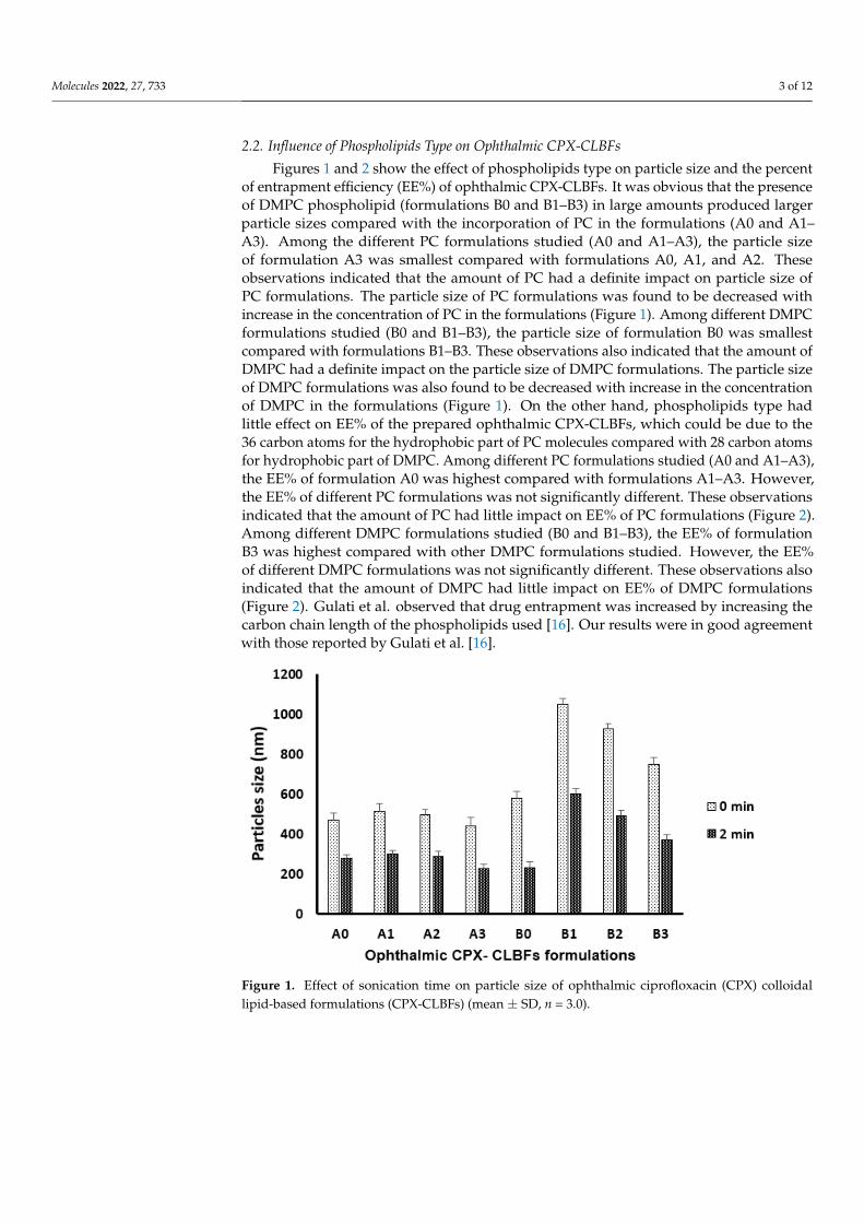
Figures 1 and 2 show the effect of phospholipids type on particle size and the percentof entrapment efficiency (EE%) of ophthalmic CPX-CLBFs. It was obvious that the presenceof DMPC phospholipid (formulations B0 and B1–B3) in large amounts produced largerparticle sizes compared with the incorporation of PC in the formulations (A0 and A1–A3). Among the different PC formulations studied (A0 and A1–A3), the particle sizeof formulation A3 was smallest compared with formulations A0, A1, and A2. Theseobservations indicated that the amount of PC had a definite impact on particle size ofPC formulations. The particle size of PC formulations was found to be decreased withincrease in the concentration of PC in the formulations (Figure 1). Among different DMPCformulations studied (B0 and B1–B3), the particle size of formulation B0 was smallestcompared with formulations B1–B3. These observations also indicated that the amount ofDMPC had a definite impact on the particle size of DMPC formulations. The particle sizeof DMPC formulations was also found to be decreased with increase in the concentrationof DMPC in the formulations (Figure 1). On the other hand, phospholipids type hadlittle effect on EE% of the prepared ophthalmic CPX-CLBFs, which could be due to the36 carbon atoms for the hydrophobic part of PC molecules compared with 28 carbon atomsfor hydrophobic part of DMPC. Among different PC formulations studied (A0 and A1–A3),the EE% of formulation A0 was highest compared with formulations A1–A3. However,the EE% of different PC formulations was not significantly different. These observationsindicated that the amount of PC had little impact on EE% of PC formulations (Figure 2).Among different DMPC formulations studied (B0 and B1–B3), the EE% of formulationB3 was highest compared with other DMPC formulations studied. However, the EE%of different DMPC formulations was not significantly different. These observations alsoindicated that the amount of DMPC had little impact on EE% of DMPC formulations(Figure 2). Gulati et al. observed that drug entrapment was increased by increasing thecarbon chain length of the phospholipids used [16]. Our results were in good agreementwith those reported by Gulati et al. [16].
Molecules 2022, 27, x FOR PEER REVIEW 3 of 13
were recorded for formulations A0 (3.6 mV) and A3 (25.7 mV), respectively. The zeta po-
tential value in the range of ±30 mV indicated the maximum stability of formulations [15].
The zeta potential of liposomal formulation A3 was found to be much closed with ±30
mV, indicating the maximum stability of liposomal formulation A3 compared to the other
formulations studied.
2.2. Influence of Phospholipids Type on Ophthalmic CPX-CLBFs
Figures 1 and 2 show the effect of phospholipids type on particle size and the percent
of entrapment efficiency (EE%) of ophthalmic CPX-CLBFs. It was obvious that the pres-
ence of DMPC phospholipid (formulations B0 and B1–B3) in large amounts produced
larger particle sizes compared with the incorporation of PC in the formulations (A0 and
A1–A3). Among the different PC formulations studied (A0 and A1–A3), the particle size
of formulation A3 was smallest compared with formulations A0, A1, and A2. These ob-
servations indicated that the amount of PC had a definite impact on particle size of PC
formulations. The particle size of PC formulations was found to be decreased with in-
crease in the concentration of PC in the formulations (Figure 1). Among different DMPC
formulations studied (B0 and B1–B3), the particle size of formulation B0 was smallest com-
pared with formulations B1–B3. These observations also indicated that the amount of
DMPC had a definite impact on the particle size of DMPC formulations. The particle size
of DMPC formulations was also found to be decreased with increase in the concentration
of DMPC in the formulations (Figure 1). On the other hand, phospholipids type had little
effect on EE% of the prepared ophthalmic CPX-CLBFs, which could be due to the 36 car-
bon atoms for the hydrophobic part of PC molecules compared with 28 carbon atoms for
hydrophobic part of DMPC. Among different PC formulations studied (A0 and A1–A3),
the EE% of formulation A0 was highest compared with formulations A1–A3. However,
the EE% of different PC formulations was not significantly different. These observations
indicated that the amount of PC had little impact on EE% of PC formulations (Figure 2).
Among different DMPC formulations studied (B0 and B1–B3), the EE% of formulation B3
was highest compared with other DMPC formulations studied. However, the EE% of dif-
ferent DMPC formulations was not significantly different. These observations also indi-
cated that the amount of DMPC had little impact on EE% of DMPC formulations (Figure
2). Gulati et al. observed that drug entrapment was increased by increasing the carbon
chain length of the phospholipids used [16]. Our results were in good agreement with
those reported by Gulati et al. [16].
Figure 1. Effect of sonication time on particle size of ophthalmic ciprofloxacin (CPX) colloidal lipid-
based formulations (CPX-CLBFs) (mean ± SD, n = 3.0).
Figure 1. Effect of sonication time on particle size of ophthalmic ciprofloxacin (CPX) colloidallipid-based formulations (CPX-CLBFs) (mean ± SD, n = 3.0).

Molecules 2022, 27, 733 4 of 12Molecules 2022, 27, x FOR PEER REVIEW 4 of 13
Figure 2. Effect of sonication time on percent entrapment efficiency (EE%) of ophthalmic CPX-
CLBFs (mean ± SD, n = 3.0).
2.3. Impact of Sonication on Ophthalmic CPX-CLBFs
Figures 1 and 2 show the impact of sonication time on ophthalmic CPX-CLBFs parti-
cle size and EE%. It was clear that 2 min of sonication time decreases the particle size of
all formulations. In addition, it was observed that CPX-CLBFs containing cholesterol (CL)
are not able to increase EE% due to high membrane rigidity of CL. As a result, the values
of EE% for formulations containing CL (A1–A3 and B1–B3) was decreased after sonication
for 2 min (Figure 2) due to the presence of CL which minimized CPX leakage from CLBFs
upon sonication.
2.4. In Vitro Release Study from Ophthalmic CPX-CLBFs
Figure 3 shows the in vitro release profile of CPX form ophthalmic CPX-CLBFs. All
formulations showed small initial gradual CPX release. Both B0 and A3 showed a faster
release compared with other formulations with cumulative CPX percent release of 73%
and 87%, respectively. CPX release was found to depend mainly on formulations particle
size. Formulations with small particle size showed high cumulative drug percent release.
Figure 3. In vitro release profile of CPX from different ophthalmic CPX-CLBFs (mean ± SD, n =
3.0).
Figure 2. Effect of sonication time on percent entrapment efficiency (EE%) of ophthalmic CPX-CLBFs(mean ± SD, n = 3.0).
2.3. Impact of Sonication on Ophthalmic CPX-CLBFs
Figures 1 and 2 show the impact of sonication time on ophthalmic CPX-CLBFs particlesize and EE%. It was clear that 2 min of sonication time decreases the particle size of allformulations. In addition, it was observed that CPX-CLBFs containing cholesterol (CL) arenot able to increase EE% due to high membrane rigidity of CL. As a result, the values ofEE% for formulations containing CL (A1–A3 and B1–B3) was decreased after sonicationfor 2 min (Figure 2) due to the presence of CL which minimized CPX leakage from CLBFsupon sonication.
2.4. In Vitro Release Study from Ophthalmic CPX-CLBFs
Figure 3 shows the in vitro release profile of CPX form ophthalmic CPX-CLBFs. Allformulations showed small initial gradual CPX release. Both B0 and A3 showed a fasterrelease compared with other formulations with cumulative CPX percent release of 73% and87%, respectively. CPX release was found to depend mainly on formulations particle size.Formulations with small particle size showed high cumulative drug percent release.
Molecules 2022, 27, x FOR PEER REVIEW 4 of 13
Figure 2. Effect of sonication time on percent entrapment efficiency (EE%) of ophthalmic CPX-
CLBFs (mean ± SD, n = 3.0).
2.3. Impact of Sonication on Ophthalmic CPX-CLBFs
Figures 1 and 2 show the impact of sonication time on ophthalmic CPX-CLBFs parti-
cle size and EE%. It was clear that 2 min of sonication time decreases the particle size of
all formulations. In addition, it was observed that CPX-CLBFs containing cholesterol (CL)
are not able to increase EE% due to high membrane rigidity of CL. As a result, the values
of EE% for formulations containing CL (A1–A3 and B1–B3) was decreased after sonication
for 2 min (Figure 2) due to the presence of CL which minimized CPX leakage from CLBFs
upon sonication.
2.4. In Vitro Release Study from Ophthalmic CPX-CLBFs
Figure 3 shows the in vitro release profile of CPX form ophthalmic CPX-CLBFs. All
formulations showed small initial gradual CPX release. Both B0 and A3 showed a faster
release compared with other formulations with cumulative CPX percent release of 73%
and 87%, respectively. CPX release was found to depend mainly on formulations particle
size. Formulations with small particle size showed high cumulative drug percent release.
Figure 3. In vitro release profile of CPX from different ophthalmic CPX-CLBFs (mean ± SD, n =
3.0).
Figure 3. In vitro release profile of CPX from different ophthalmic CPX-CLBFs (mean ± SD, n = 3.0).

Molecules 2022, 27, 733 5 of 12
2.5. Antibacterial Activity of Different CPX-CLBFs Formulations
The antibacterial activity and the minimum inhibitory concentration (MIC) wereevaluated using the cup plate method and microdilution method, respectively, againstbacterial strains frequently responsible for endophthalmitis. The MIC values of liposo-mal formulation samples containing CPX were investigated and the most effective arepresented in Table 1. All formulations showed weak antibacterial activity (MIC rangedfrom 64–128 µg/mL) against all the tested strains except for formulations A3 and B0, whichdisplayed a potent antibacterial activity (MIC ranged from 16–32 µg/mL) against all thetested strains. These formulations showed obvious activities in comparison with the otherformulations against Gram-positive and -negative strains. For the S. aureus, B. subtilis, andP. aeruginosa bacterial strains, A3 revealed the best strength with MIC values of 16 µg/mLand exhibited minimum bactericidal concentration (MBC) values lower than that of thereference CPX. The higher in vitro antibacterial activity of formulations A3 and B0 werepossibly due to the faster release of CPX from formulations A3 and B0 compared with otherformulations studied.
Table 1. Antibacterial activity of selected ophthalmic ciprofloxacin (CPX) colloidal lipid-basedformulations (CPX-CLBFs) and the commercial one.
Gram-Positive Bacteria
FormulationCode
S. aureusATCC 29213
B. subtilisATCC 10400
E. faecalisATCC 29212
Sensitivity MIC Sensitivity MIC Sensitivity MIC
B0 +++ 0.5 ++++ 0.25 +++ 0.125A3 ++++ 0.25 ++++ 0.125 ++++ 0.125
CPX ++++ 0.5 ++++ 0.25 ++++ 0.125
Gram-Negative Bacteria
E. coliATCC 9637
P. aeruginosaATCC 27953
K. pneumoniaATCC 10031
Sensitivity MIC Sensitivity MIC Sensitivity MIC
B0 ++++ 0.5 +++ 1.0 +++ 0.5A3 ++++ 0.5 ++++ 0.25 ++++ 0.25
CPX ++++ 0.5 ++++ 0.5 ++++ 0.5
The microbiological susceptibility testing of released CPX from the lipid-based en-trapped CPX formulations was tested against different strains of Gram-negative (E. coli,P. aeruginosa, and K. pneumonia) and Gram-positive (S. aureus, E. faecalis, and B. subtilis)bacteria. These organisms are implicated in endophthalmitis [17]. Donnenfeld et al. al-leged that the most common bacteria accountable for endophthalmitis were S. aureus,S. epidermidis, and Streptococcus sp. (50%, 30%, and 10%, respectively) [18]. The meanconcentrations of antibiotic achieved in the current study were less than 1 µg/mL. Thereported MIC of CPX required to inhibit the growth of S. aureus and P. aeruginosa rangedfrom 0.125–1 µg/mL [19]. This result was in agreement with Cutarelli et al. who reporteda MIC90 of 1 µg/mL for CPX against tested strains [20]. Lesk et al. also estimated aslow as 0.4 µg/mL for S. epidermidis and up to 1 µg/mL for B. cereus [21]. Therefore, themost effective two lipid-based entrapped CPX formulations were selected for the in vivoevaluation (B0 and A3). Both CPX-CLBF and Ciloxan® eye drop improved MIC and MBCof P. aeruginosa and S. aureus. Jain and Shastri reported a comparable result of liposomalformulation on MIC and MBC of CPX [22].
The results of the antimicrobial effectiveness and the level of CPX in the conjunctivalsac of tested rabbits following the topical application of B0 and A3 formulations andCiloxan® eye drops are shown in Figure 4. The reduction in bacterial count was detectedand better improvement was observed in rabbit’s eyes conjunctivitis. The ex vivo resultswere consistent with the results of the in vitro release, where the lipid-based entrapped CPX

Molecules 2022, 27, 733 6 of 12
formulations A3 showed the uppermost CPX levels in the conjunctival sac in comparison toretailed Ciloxan® drops. B0, when compared to A3 and commercial eye drops, significantlyfailed to reduce the bacterial growth, which could be due to lack of positively chargingagent. Effectually, cationic molecules such as STA are deemed to be notable bio-adhesivesowing to an ability to produce forces of molecular attraction through the electrostaticinteractions with the negative charges of mucin coating corneal surface as well as thesecretions at the site of infection.
Molecules 2022, 27, x FOR PEER REVIEW 6 of 13
consistent with the results of the in vitro release, where the lipid-based entrapped CPX
formulations A3 showed the uppermost CPX levels in the conjunctival sac in comparison
to retailed Ciloxan® drops. B0, when compared to A3 and commercial eye drops, signifi-
cantly failed to reduce the bacterial growth, which could be due to lack of positively charg-
ing agent. Effectually, cationic molecules such as STA are deemed to be notable bio-adhe-
sives owing to an ability to produce forces of molecular attraction through the electrostatic
interactions with the negative charges of mucin coating corneal surface as well as the se-
cretions at the site of infection.
Figure 4. Colony count of the infected aqueous humor after the topical application of B0 and A3
formulations and Ciloxan® eye drops against S. aureus (A), K. pneumonia (B), and P. aeruginosa (C),
(mean ± SD, n = 3.0).
2.6. Pharmacokinetic Study
Figure 5 shows CPX concentration in aqueous humor following application of CPX-
CLBF A3 compared with Ciloxan® . Table 2 shows the pharmacokinetic parameters of A3
and Ciloxan® . One–way ANOVA was utilized to analyze the calculated pharmacokinetic
parameters after application of CPX-CLBF A3 and Ciloxan® . The Cmax of 4.2 μg/mL was
obtained from A3 compared with 2.7 μg/mL for Ciloxan® . There were no significant dif-
ferences in values of Tmax between A3 and Ciloxan® . The values of AUC0-t for A3 (34.9 μg
min/mL) were significantly greater than those obtained from Ciloxan® (12 μg min/mL).
These results confirmed a greater ocular bioavailability of CPX-CLBF A3. High values of
AUC0–t indicated high extent of drug absorption. Regarding t1/2, A3 shows increased t1/2
value compared with that of the commercial formulation which indicated the presence of
CPX for longer time in aqueous humor. The relative bioavailability valued significantly
emphasize that A3 increased drug bioavailability by 2.9-folds compared with the com-
mercial formulation. Based on the bioavailability results, the CLBF delivered extra CPX
into the aqueous humor and enhanced therapeutic effects in comparison with Ciloxan®
drop.
Figure 4. Colony count of the infected aqueous humor after the topical application of B0 and A3formulations and Ciloxan® eye drops against S. aureus (A), K. pneumonia (B), and P. aeruginosa (C),(mean ± SD, n = 3.0).
2.6. Pharmacokinetic Study
Figure 5 shows CPX concentration in aqueous humor following application of CPX-CLBF A3 compared with Ciloxan®. Table 2 shows the pharmacokinetic parameters ofA3 and Ciloxan®. One–way ANOVA was utilized to analyze the calculated pharmacoki-netic parameters after application of CPX-CLBF A3 and Ciloxan®. The Cmax of 4.2 µg/mLwas obtained from A3 compared with 2.7 µg/mL for Ciloxan®. There were no signif-icant differences in values of Tmax between A3 and Ciloxan®. The values of AUC0-tfor A3 (34.9 µg min/mL) were significantly greater than those obtained from Ciloxan®
(12 µg min/mL). These results confirmed a greater ocular bioavailability of CPX-CLBF A3.High values of AUC0–t indicated high extent of drug absorption. Regarding t1/2, A3 showsincreased t1/2 value compared with that of the commercial formulation which indicatedthe presence of CPX for longer time in aqueous humor. The relative bioavailability val-ued significantly emphasize that A3 increased drug bioavailability by 2.9-folds comparedwith the commercial formulation. Based on the bioavailability results, the CLBF deliveredextra CPX into the aqueous humor and enhanced therapeutic effects in comparison withCiloxan® drop.
Table 2. Pharmacokinetics parameters of ophthalmic CPX- CLBF (A3) and commercial one(mean ± SD, n = 3.0).
Parameters A3 Commercial CPX
Dose (µg) 115 150Cmax (µg/mL) 4.2 ± 0.1 2.7 ± 0.1
Tmax (h) 2 2K (h−1) 0.2 ± 0.03 0.4 ± 0.01t1/2 (h) 3.5 ± 0.2 1.7 ± 0.2
AUC 0–t (µg.h/mL) 34.9 ± 2.1 12 ± 0.5Relative bioavailability 2.9 -
Cmax: maximum plasma concentration; Tmax: time to reach Cmax; K: elimination rate constant; t1/2: half-life;AUC0–t: area under curve from time 0 to t.

Molecules 2022, 27, 733 7 of 12Molecules 2022, 27, x FOR PEER REVIEW 7 of 13
Figure 5. CPX concentrations in rabbit aqueous humor for ophthalmic CPX-CLBF (A3) compared
with commercial one (mean ± SD, n = 6.0).
Table 2. Pharmacokinetics parameters of ophthalmic CPX- CLBF (A3) and commercial one (mean ±
SD, n = 3.0).
Parameters A3 Commercial CPX
Dose (µg) 115 150
Cmax (µg/mL) 4.2 ± 0.1 2.7 ± 0.1
Tmax (h) 2 2
K (h−1) 0.2 ± 0.03 0.4 ± 0.01
t1/2 (h) 3.5 ± 0.2 1.7 ± 0.2
AUC 0–t (µg.h/mL) 34.9 ± 2.1 12 ± 0.5
Relative bioavailability 2.9 -
Cmax: maximum plasma concentration; Tmax: time to reach Cmax; K: elimination rate constant; t1/2:
half-life; AUC0–t: area under curve from time 0 to t.
3. Materials and Methods
3.1. Materials
CPX was obtained from Fluka Biochemika (Busch, Switzerland). PC, CL, DMPC, and
STA were obtained from Avanti Polar Lipids Inc. (Alabaster, AL, USA). Chloroform was
obtained from BDH Laboratory Ltd. (Poole, England, UK). The solvents used in this study
were of chromatography grade.
3.2. Bacterial Culturing
The growing bacterial cultures were determined by controlling the turbidity changes
with the measurements of absorbance at 600 nm. Applying a titration curve, the changes
in the absorbance were then turned correspondingly into CFU/mL (colony forming units).
The titration curve was created by considering the bacterial samples with optical density
of 0.05–2.0 at 600 nm. Serial dilution for the cells was performed in trypticase soy broth
(Oxoid, Lenexa, KS, USA) and then culturing the diluted cells on Mueller–Hinton agar
(MHA) (Oxoid, Lenexa, KS, USA). The plates were incubated for overnight at 35 ± 1 °C.
Bacterial colonies were calculated, and result obtained from triplicate assays were applied
to generate a titration curve that compared optical density measurements with CFU/mL
for the bacterial strains growth.
3.3. Preparation of CPX-CLBFs
Figure 5. CPX concentrations in rabbit aqueous humor for ophthalmic CPX-CLBF (A3) comparedwith commercial one (mean ± SD, n = 6.0).
3. Materials and Methods3.1. Materials
CPX was obtained from Fluka Biochemika (Busch, Switzerland). PC, CL, DMPC, andSTA were obtained from Avanti Polar Lipids Inc. (Alabaster, AL, USA). Chloroform wasobtained from BDH Laboratory Ltd. (Poole, England, UK). The solvents used in this studywere of chromatography grade.
3.2. Bacterial Culturing
The growing bacterial cultures were determined by controlling the turbidity changeswith the measurements of absorbance at 600 nm. Applying a titration curve, the changes inthe absorbance were then turned correspondingly into CFU/mL (colony forming units).The titration curve was created by considering the bacterial samples with optical densityof 0.05–2.0 at 600 nm. Serial dilution for the cells was performed in trypticase soy broth(Oxoid, Lenexa, KS, USA) and then culturing the diluted cells on Mueller–Hinton agar(MHA) (Oxoid, Lenexa, KS, USA). The plates were incubated for overnight at 35 ± 1 ◦C.Bacterial colonies were calculated, and result obtained from triplicate assays were appliedto generate a titration curve that compared optical density measurements with CFU/mLfor the bacterial strains growth.
3.3. Preparation of CPX-CLBFs
CPX-CLBFs were prepared utilizing reverse evaporation technique [23]. The molarratios of CPX-CLBFs are shown in Table 3. The exact amount of lipids was dissolved in13 mL of chloroform. Five mL of CPX was dissolved in phosphate buffer (pH 4.5) and addedto the mixture. Chloroform was evaporated from the mixture by rotary evaporation at 40 ◦C.The mixture was centrifuged for 40 min at 30,000 rpm. Free CPX content in supernatantwas detected and the lipid capsules were dispersed in phosphate buffer (pH 7.4) [24]. Eachformulation was subjected to sonication for 2 min right after preparation.
3.4. Characterization of CPX-CLBFs3.4.1. Particle Size, PDI, and Zeta Potential Measurements
The mean particle size and PDI for each CPX-CLBFs were determined at ambient tem-perature using laser diffraction analysis (NiComp Particle Size system ZW380 Santa Barbara,CA, USA). Each CPX-CLBF formulation was diluted with phosphate buffer before analysis.

Molecules 2022, 27, 733 8 of 12
Table 3. Composition of ophthalmic CPX-CLBFs in molar ratios.
FormulationCode
Composition (Molar Ratios)PC DMPC CL STA
A0 1 - - -B0 - 1 - -A1 1 - 1 2B1 - 1 1 2A2 3 - 1 2B2 - 3 1 2A3 6 - 1 2B3 - 6 1 2
PC: phosphatidylcholine; DMPC: dimyrestoyl phosphatidylcholine; CL: cholesterol; STA: stearylamine.
3.4.2. Evaluation of CPX EE%
Each CPX-CLBF formulation was centrifuged for 40 min at 30,000 rpm to get rid offree CPX content and EE% was calculated using the following equation [25,26]:
EE% =(Wt − Wf)
Wt× 100 (1)
where, Wt and Wf are the total CPX and the free CPX content, respectively.
3.4.3. Effect of Sonication on CPX-CLBFs Particle Size and EE%
To study the impact of sonication on particle size and EE% of CLBFs, all formulationswere sonicated for 2 min. The data were recorded before and after sonication.
3.5. CPX Assay Method
Reversed-phase high performance liquid chromatographic (HPLC) method was usedfor the quantification of CPX. A mobile phase was a mixture of acetonitrile: methanol:0.05 acetate buffer (10:20:70, v/v/v) and 1% (v/v) of acetic acid. The acetate buffer wasat pH 3.6. The mobile phase was degassed using sonication and filtered through 0.45 µmMillipore filter immediately after preparation. Samples of 20 µL were injected in Shi-madzu HPLC system consists of Fluorescence detector, pump, Kromasil 100, C18, 5 µm(250 × 4.6 mm) column, and Rheodyne sample injector were used in the assay method. CPXand anthranilic acid (internal standard) was detected at λexc = 300 nm and λemi = 458 nm,respectively, using mobile flow rate of 0.8 mL/min at room temperature [27].
3.6. In Vitro CPX Release Study from CLBFs
Franz diffusion cells were used to carry out this experiment. The in vitro release of CPXwas performed in release medium of artificial tears (pH 7.4) placed in receptor cells [28].A semipermeable membrane with a molecular weight cut-off range of 12,000–14,000 Dawas used [29]. The membrane was soaked in the dissolution medium for about 12 h beforeconducting the experiment. The utilized membrane was able to permeate CPX moleculesand hold CLBF vesicles. The temperature was kept at 34 ± 0.5 ◦C and the mixture wasstirred at 300 rpm. A specific amount of each CPX ophthalmic formulation was placed inthe dialysis donor cell. Samples of 1 mL each were periodically collected at time intervalsof 0, 0.25, 0.5, 1, 2, 3, 4, and 6 h and replaced by same volume of artificial tears to maintainconstant volume and sink condition. Then, the amount of CPX was determined in eachsample using HPLC method described above [27]. All tests were repeated in triplicate.Based on the characterizations of ophthalmic CPX-CLBFs formulations, A3 (after sonication)and B0 (without sonication) were selected for further investigation.
3.7. Evaluation of the In Vitro Antibacterial Activity of CPX-CLBFs
Antibiogram was carried out for the prepared 8 colloidal lipid-based entrapped CPXformulations, as well as the standard CPX-hydrochloride, by the agar well diffusion

Molecules 2022, 27, 733 9 of 12
method [30,31]. CLBF vesicles were separately investigated against a set of Gram-positiveand -negative bacterial strains as proposed by the Clinical Laboratory Standard Institute(CLSI) guidelines [32]. For this purpose, different strains of Gram-negative bacteria (E. coliATCC 9637, P. aeruginosa ATCC 27953, and K. pneumonia ATCC 10031) as well as threedifferent strains of Gram-positive bacteria (S. aureus ATCC 29213, E. faecalis ATCC 29212,and B. subtilis ATCC 10400) were selected. Bacterial suspension (100 µL) comprising1 × 106 CFU/mL of bacterial strain were mixed with MHA medium. After waiting forthe media to solidified, wells of 6.0 mm diameter were made through the solidified MHAusing a cork borer and burdened equal quantities of each tested liposomal formulationsamples. The implanted plates were then overnight incubated at 35 ± 1 ◦C. Negativecontrol was prepared using free MHA. The zone of inhibition against the tested organismswas measured after incubation time for evaluating the antimicrobial activity. Each test wasperformed in triplicate and inhibition zone average was calculated. The average diametersof the inhibition zones were recorded and expressed according to the following score guidein mm as follows: + = ≥ 6 mm; ++ = ≥ 10 mm (moderate); +++ = ≥ 15 mm (strong); and++++ = ≥20 mm (very strong).
3.8. Determination of In Vitro MIC and MBC
The MIC of the most effective CPX-CLBF formulations (3 efficient formulae) andcommercial ophthalmic formulation containing 0.3% CPX-hydrochloride (Ciloxan® drop,Alcon Laboratories Inc., Fort Worth, TX, USA) were assayed against mentioned bacteriathat are frequently responsible for endophthalmitis [33]. The test was performed by brothmicrodilution method [34] in conformity with the European Committee on AntimicrobialSusceptibility Testing (EUCAST) [35]. Overnight bacterial suspensions were adjusted to1 × 106 CFU/mL as described above. The CLBF samples and CPX commercial formulawere adjusted to give appropriate concentration by sequential dilutions in 2X MH broth(Merck, Darmstadt, Germany). Equal bacterial inoculums (5 µL) were dispensed into wellsand the microtiter plate was incubated overnight at 35 ± 1 ◦C. Subsequently, the MICresults were listed and explicated consistent with the EUCAST guidelines. Positive control(inoculum and MH media, devoid of liposomal entrapped CPX) was used. Free culturemedium was used as a negative control. To ensure reproducibility, each assessment wascompleted in triplicates on three different days. Wells displaying no bacterial growth werestreaked on MH agar and incubated overnight for MBC. Tested MBC was considered asthe lowest concentration of either free or liposomal CPX that able to cause reduction (morethan 99.9%) of the initial inoculum.
3.9. Ex Vivo Antibacterial Effect of CPX-CLBFs and Ciloxan®
Male New Zealand white Albino rabbits (weighed 2–2.3 kg) were obtained from theExperimental Animal Care Center (EACC) at Prince Sattam bin Abdulaziz University,Al-Kharj, Saudi Arabia and utilized to conduct the in vivo/ex vivo antibacterial study. Allanimals were provided free access to water but were starved for 24 h before the experiment.The in vivo antibacterial studies were approved by the Animal Ethics Committee of theEACC Board (Prince Sattam bin Abdulaziz University, Al-Kharj, Saudi Arabia with ap-proval number: BERC-005-04-21). The animal use and experimental procedures followedEU directive 2010/63/EU.
The bacterial suspensions were prepared by overnight growing at 35 ± 1 ◦C in trypti-case soy broth and adjusted to achieve a concentration of 1 × 10³ CFU/30 µL, proper forocular application. Rabbits were randomly selected and divided into three equal groupsfor each bacterial strain: group A for S. aureus, group B for K. pneumonia, and group Cfor P. aeruginosa. Each group contained 9 rabbits with the same race, gender and averageweight of 2.0–2.3 kg. To induce intraocular infections in the rabbit eyes, 5 µL of each bacte-rial suspension was inoculated into the conjunctival sac through sterile dropper for eachgroup. After infection verification, the right eyes were treated by CLBF and Ciloxan® dropsfor the assessment of the in vivo antibacterial efficacy of different CPX-CLBF formulations.
Molecules 2022, 27, 733 10 of 12
On the other hand, the left eyes were kept un-treated. From anterior chamber paracentesis,the aqueous humor specimens were withdrawn via the translimbal pathway by a 27-gaugeneedle mounted on a tuberculin syringe at a dose of 0.1 mL, at zero (before pouring the eyedrops), 0.5, 1, 2, 3, 4, 5, and 6 h after application of the formulated drug. The CPX-CLBF inaqueous humor specimens were measured by culturing on TSA media for the microbialcount and estimation of MIC and MBC. Antibacterial activities were expressed as inhibitiondiameter zones in mm. MIC was expressed in µg/mL and evaluated by microbroth dilutionmethod according to EUCAST.
3.10. Ex Vivo Antibacterial Effect of Selected CPX-CLBFs and Ciloxan® Drop
Both MIC and MBC of the prepared CLBF formulations in ex vivo media (mediaenriched by infected aqueous humor specimens) against S. aureus, K. pneumonia, andP. aeruginosa were examined (data not presented). CPX entrapped in A3 formulationdecreased MIC and MBC against K. pneumonia and S. aurous more than B0 and Ciloxan®.A3 significantly enhanced the susceptibility of K. pneumonia and S. aurous to CPX comparedwith B0 and Ciloxan®. On the other hand, only A3 decreased MIC and MBS for P. aeruginosaand no significant difference was noticed between B0 and Ciloxan®.
3.11. Study of CPX-CLBFs Treatment Effect
For the evaluation of the treatment outcome, colony count was determined in treatedand un-treated rabbit’s eyes. Selected rabbits infected by S. aureus, K. pneumonia, andP. aeruginosa were treated by B0, A3, and Ciloxan®. After 6 h of treatment, the sampleswithdrawn from the infected aqueous humor and streaked for colony count. Accordingto these results, A3 displayed significantly reduced colony counts for all tested organismsafter 6 h. Although Ciloxan® showed reducing effect in colony count, but it is insignificantcompared to A3 effect. Blank CLBF control did not show any effect on demonstratedbacterial growth. On the other hand, bacterial count was significantly higher than thatobserved with A3 and commercial ophthalmic drops, which means B0 failed to reduce thebacterial infection.
3.12. Pharmacokinetic Studies
Based on the data obtained from the physiochemical and antimicrobial evaluationsof the prepared CPX ophthalmic formulations, formulation A3 was selected for in vivopharmacokinetic experiment along with the commercial CPX eye drops, Ciloxan®. FiftyµL dose of CPX ophthalmic formulation was applied in the lower conjunctival sac of therabbit’s right eye; however, Ciloxan® (Riyadh Pharma, Riyadh, Saudi Arabia) was appliedin the left one. The animals were euthanized after withdrawing the aqueous humor atspecific time intervals. The samples were centrifuged for 20 min at 4 ◦C and 20,000 rpmand supernatant were frozen at –20 ◦C until assay. The amount of CPX in each sample wasanalyzed using the HPLC method described above [27].
3.13. Pharmacokinetic Parameters
The Cmax and Tmax values were obtained directly from rabbit aqueous humor concentration-time plot. The AUC0–t (µg min/mL) was calculated using the trapezoidal method. Theelimination rate constant (k) was determined from the final segment of the curve by fittingthe data in first order elimination graph. Relative bioavailability of CPX-CLBF was obtainedin comparison to that of the commercial product.
3.14. Statistical Analysis
All experiments in this study were subjected to one way analysis of variance withTukey’s multiple comparisons test at a 5% level of significance (p ≤ 0.05).

Molecules 2022, 27, 733 11 of 12
4. Conclusions
The MIC for CPX lipid-based formulations was noticeably lower than that of theretailed and free drug, indicating that CLBF systems may participate in the increasedantimicrobial activity of CPX. The pharmacokinetic parameters of the selected ophthalmicformulation showed significant improvement in ocular bioavailability of CPX in compari-son with Ciloxan® ophthalmic drops. This could be due to several factors that characterizethe tested CPX formulation, including that it produces low lacrimation due to its almostneutral pH value of 7.4. In addition, the presence of a positive charge inducing agent whichincreases the drug contact time with corneal surface. Finally, the liposomal formulationimproves drug permeability to the cornea which maximizes drug concentration in theaffected area. Based on these results, the prepared CPX formulation could provide a betteralternative to the marketed one with respect to enhancing ocular drug activity, especiallyin the management of post-surgical infections.
Author Contributions: Conceptualization, M.M.S.-B. and F.S.; methodology, F.A.A.-J., E.I.T., M.A.I.,M.M.M., F.A.A.-J. and S.A.; software, M.M.G.; validation, F.S., M.M.G. and S.A.; formal analysis,M.M.G.; investigation, M.M.M., E.I.T., F.A.A.-J. and F.S.; resources, M.A.I.; data curation, M.M.G.;writing—original draft preparation, E.I.T.; writing—review and editing, M.M.S.-B., F.S. and M.A.I.;visualization, M.A.I.; supervision, M.M.S.-B. and F.S.; project administration, M.M.S.-B.; fundingacquisition, M.A.I. All authors have read and agreed to the published version of the manuscript.
Funding: This research was funded by the Researcher Supporting Project (Number RSP-2021/171) atKing Saud University, Riyadh, Saudi Arabia. The APC was funded by RSP.
Institutional Review Board Statement: The study was conducted according to the guidelines of theDeclaration of Helsinki, and approved by the Animal Ethics Committee of the EACC Board at PrinceSattam bin Abdulaziz University, Al-Kharj, Saudi Arabia (approval number: BERC-005-04-21).
Data Availability Statement: This study did not report any data.
Acknowledgments: Authors are thankful to the Researcher Supporting Project (Number RSP-2021/171) at King Saud University, Riyadh, Saudi Arabia for supporting this work.
Conflicts of Interest: The authors declare no conflict of interest.
Sample Availability: Samples of the CPX compounds are available from the authors.
References1. Hooper, D.; Wolfson, J.; Ng, E.; Swartz, M.J.T. Mechanisms of action of and resistance to ciprofloxacin. Am. J. Med. 1987, 82, 12–20.
[PubMed]2. Ting, D.S.J.; Ho, C.S.; Deshmukh, R.; Said, R.D.G.; Dua, H.S. Infectious keratitis: An update on epidemiology, causative
microorganisms, risk factors, and antimicrobial resistance. Eye 2021, 35, 1084–1101. [CrossRef] [PubMed]3. Subrizi, A.; del Amo, E.M.; Korzhikov-Vlakh, V.; Tennikova, T.; Ruponen, M.; Urtti, A. Design principles of ocular drug delivery
systems: Importance of drug payload, release rate, and material properties. Drug Discov. Today 2019, 24, 1446–1457. [CrossRef][PubMed]
4. Mamah, C.C.; Anyalebechi, O.C.; Onwubiko, S.N.; Okoloagu, M.N.; Maduka-Okafor, F.C.; Ebede, S.O.; Umeh, R.E. Conjunctivalbacterial flora and their antibiotic sensitivity among patients scheduled for cataract surgery in a tertiary hospital in south-eastNigeria. Graefes Arch. Clin. Exp. Ophthalmol. 2021, 259, 443–448. [CrossRef] [PubMed]
5. Bhattacharyya, A.; Sarma, P.; Sarma, B.; Kumar, S.; Gogoi, T.; Kaur, H.; Prajapat, M. Bacteriological pattern and their correlationwith complications in culture positive cases of acute bacterial conjunctivitis in a tertiary care hospital of upper Assam: A crosssectional study. Medicine 2020, 99, E18570. [CrossRef]
6. Jackson, T.L.; Eykyn, S.J.; Graham, E.M.; Stanford, M.R. Endogenous bacterial endophthalmitis: A 17-year prospective series andreview of 267 reported cases. Surv. Ophthalmol. 2003, 48, 403–423. [CrossRef]
7. Gieling, E.M.; Wallenburg, E.; Frenzel, T.; Dylan, W.L.; Schouten, J.A.; Oever, J.; Kolwijck, E.; Burger, D.M.; Pickkers, P.; Heine, R.;et al. Higher dosage of ciprofloxacin necessary in critically ill patients: A new dosing algorithm based on renal function andpathogen susceptibility. Clin. Pharmacol. Ther. 2020, 108, 770–774. [CrossRef]
8. Mundada, A.S.; Shrikhande, B.K. Formulation and evaluation of ciprofloxacin hydrochloride soluble ocular drug insert.Curr. Eye Res. 2008, 33, 469–475. [CrossRef]
9. Ludwig, A. The use of mucoadhesive polymers in ocular drug delivery. Adv. Drug Deliv. Rev. 2005, 57, 1595–1639. [CrossRef]

Molecules 2022, 27, 733 12 of 12
10. Atugoda, T.; Wijesekara, H.; Werellagama, D.R.I.B.; Jinadasa, K.B.S.N.; Bolan, N.S.; Vithanage, M. Adsorptive interaction ofantibiotic ciprofloxacin on polyethylene microplastics: Implications for vector transport in water. Environ. Technol. Innov. 2020, 19,E100971. [CrossRef]
11. Wilhelmus, K.R.; Abshire, R.L. Corneal ciprofloxacin precipitation during bacterial keratitis. Am. Ophthalmol. 2003, 136, 1032–1037.[CrossRef]
12. Law, S.L.; Huang, K.J.; Chiang, C.H. Acyclovir-containing liposomes for potential ocular delivery: Corneal penetration andabsorption. J. Control. Rel. 2000, 63, 135–140. [CrossRef]
13. Lila, A.S.A.; Ishida, T. Liposomal delivery systems: Design optimization and current applications. Biol. Pharm. Bull. 2017, 40,1–10. [CrossRef] [PubMed]
14. Danion, A.; Arsenault, I.; Vermette, P. Antibacterial activity of contact lenses bearing surface-immobilized layers of intactliposomes loaded with levofloxacin. J. Pharm. Sci. 2007, 96, 2350–2363. [CrossRef]
15. Perween, N.; Alshehri, S.; Easwari, T.S.; Verma, V.; Faiyazuddin, M.; Alanazi, A.; Shakeel, F. Investigating the feasibility ofmefenamic acid nanosuspension for pediatric delivery: Preparation, characterization, and role of excipients. Processes 2021, 9,E574. [CrossRef]
16. Gulati, M.; Grover, M.; Singh, M.; Singh, S. Study of azathioprine encapsulation into liposomes. J. Microencapsul. 1998, 15, 485–494.[CrossRef]
17. Keynan, Y.; Finkelman, Y.; Lagacé-Wiens, P. The microbiology of endophthalmitis: Global trends and a local perspective.Eur. J. Clin. Microbiol. Infect. Dis. 2012, 31, 2879–2886. [CrossRef]
18. Donnenfeld, E.D.; Perry, H.D.; Snyder, R.W.; Elsky, M.; Jones, H. Intercorneal, aqueous Humor, and vitreous humor penetrationof topical and oral ofloxacin. Arch. Ophthalmol. 1997, 115, 173–176. [CrossRef]
19. Leigue, L.; Montiani-Ferreira, F.; Moore, B.A. Antimicrobial susceptibility and minimal inhibitory concentration of Pseudomonasaeruginosa isolated from septic ocular surface disease in different animal species. Open Vet. J. 2016, 6, 215–222. [CrossRef]
20. Cutarelli, P.E.; Lass, J.H.; Lazarus, H.M.; Putman, S.C.; Jacobs, M.R. Topical fluoroquinolones: Antimicrobial activity and in vitrocorneal epithelial toxicity. Curr. Eye Res. 1991, 10, 557–563. [CrossRef]
21. Lesk, M.R.; Ammann, H.; Marcil, G.; Vinet, B.; Lamer, L.; Sebag, M. The penetration of oral ciprofloxacin into the aqueous humor,vitreous, and subretinal fluid of humans. Am. J. Ophthalmol. 1993, 115, 623–628. [CrossRef]
22. Jain, R.L.; Shastri, J.P. Study of ocular drug delivery system using drug-loaded liposomes. Int. J. Pharm. Investig. 2011, 1, 35–41.[CrossRef] [PubMed]
23. Szoka, F.; Papahadjopoulos, D. Procedure for preparation of liposomes with large internal aqueous space and high capture byreverse-phase evaporation. Proc. Nat. Acad. Sci. USA 1978, 75, 4194–4198. [CrossRef] [PubMed]
24. Gürsoy, A.; Senyücel, B. Characterization of ciprofloxacin liposomes: Derivative ultraviolet spectrophotometric determinations.J. Microencapsul. 1997, 14, 769–776. [CrossRef] [PubMed]
25. Akanksha, G.; Navneet, G.; Vivek, T.; Mayank, M. Topical liposomal gel with aceclofenac: Characterization and in vitro evaluation.Pharmacist 2007, 2, 41–44.
26. Budai, L.; Hajdú, M.; Budai, M.; Gróf, P.; Béni, S.; Noszál, B.; Klebovich, I.; Antal, I. Gels and liposomes in optimized ocular drugdelivery: Studies on ciprofloxacin formulations. Int. J. Pharm. 2007, 343, 34–40. [CrossRef]
27. Zotou, A.; Miltiadou, N. Sensitive LC determination of ciprofloxacin in pharmaceutical preparations and biological fluids withfluorescence detection. J. Pharm. Biomed. Anal. 2002, 28, 559–568. [CrossRef]
28. Paulsson, M.; Hägerström, H.; Edsman, K. Rheological studies of the gelation of deacetylated gellan gum (Gelrite) in physiologicalconditions. Eur. J. Pharm. Sci. 1999, 9, 99–105. [CrossRef]
29. Avgoustakis, K.; Beletsi, A.; Panagi, Z.; Klepetsanis, P.; Karydas, A.G.; Ithakissios, D.S. PLGA-mPEG nanoparticles of cisplatin:In vitro nanoparticle degradation, in vitro drug release and in vivo drug residence in blood properties. J. Control Rel. 2002, 19,123–135. [CrossRef]
30. Balouiri, M.; Sadiki, M.; Ibnsouda, S.K. Methods for in vitro evaluating antimicrobial activity: A review. J. Pharm. Anal. 2016, 6,71–79. [CrossRef]
31. Wiegand, I.; Hilpert, K.; Hancock, R.E.W. Agar and broth dilution methods to determine the minimal inhibitory concentration(MIC) of antimicrobial substances. Nat. Protoc. 2008, 3, 163–175. [CrossRef] [PubMed]
32. CLSI WL: M02-A11. Performance Standards for Antimicrobial Disk Susceptibility Tests; Approved Standard; CLSI (Clinical andLaboratory Standards Institute): Wayne, PA, USA, 2015; Volume 32.
33. Callegan, M.C.; Gilmore, M.S.; Gregory, M.; Ramadan, R.T.; Wiskur, B.J.; Moyer, A.L.; Hunt, J.J.; Novosad, B.D. Bacterialendophthalmitis: Therapeutic challenges and host–pathogen interactions. Prog. Retin. Eye Res. 2007, 26, 189–203. [CrossRef][PubMed]
34. Otvos, L.; Cudic, M. Broth microdilution antibacterial assay of peptides. Methods Mol. Biol. 2007, 386, 309–320. [PubMed]35. EUCAST SOP 1.1. Testing ECoAS: Setting Breakpoints for New Antimicrobial Agents; EUCAST: Växjö, Sweden, 2013.