Clinical presentation and visual outcome of broomstick ocular ...
15
Page 1/15 Clinical presentation and visual outcome of broomstick ocular injury in 120 eyes of children at a tertiary eye care in India Abhishek Gupta ( [email protected] ) Indira Gandhi Institute of Medical Sciences https://orcid.org/0000-0001-6141-7887 Prabhakar Singh Nirwana Netralaya Richa Gupta Indira Gandhi Institute of Medical Sciences Vidya Bhushan Indira Gandhi Institute of Medical Sciences Shivani Sinha Indira Gandhi Institute of Medical Sciences Bibhuti Prasann Sinha Indira Gandhi Institute of Medical Sciences Research article Keywords: Broomstick, bow and arrow, ocular injury, endophthalmitis Posted Date: May 13th, 2020 DOI: https://doi.org/10.21203/rs.3.rs-27005/v1 License: This work is licensed under a Creative Commons Attribution 4.0 International License. Read Full License
-
Upload
khangminh22 -
Category
Documents
-
view
3 -
download
0
Transcript of Clinical presentation and visual outcome of broomstick ocular ...
Page 1/15
Clinical presentation and visual outcome ofbroomstick ocular injury in 120 eyes of children at atertiary eye care in IndiaAbhishek Gupta ( [email protected] )
Indira Gandhi Institute of Medical Sciences https://orcid.org/0000-0001-6141-7887Prabhakar Singh
Nirwana NetralayaRicha Gupta
Indira Gandhi Institute of Medical SciencesVidya Bhushan
Indira Gandhi Institute of Medical SciencesShivani Sinha
Indira Gandhi Institute of Medical SciencesBibhuti Prasann Sinha
Indira Gandhi Institute of Medical Sciences
Research article
Keywords: Broomstick, bow and arrow, ocular injury, endophthalmitis
Posted Date: May 13th, 2020
DOI: https://doi.org/10.21203/rs.3.rs-27005/v1
License: This work is licensed under a Creative Commons Attribution 4.0 International License. Read Full License
Page 2/15
Abstract
BackgroundEye injuries are a serious health problem globally. Ocular trauma accounts for 5% of blindness cases. InIndia broomstick injury is very common. But only few studies are published regarding the nature andoutcome of broomstick ocular injuries. The aim of this study is to determine the frequency, mode ofpresentation, complications and surgical results with a view to offering solutions to reduce this trend.
MethodsThis retrospective study was conducted at Regional Institute of Ophthalmology, Patna. The records of allpatients presenting to the Eye OPD and Emergency clinic with ocular trauma from broomstick injurybetween March2017 and April 2020 were reviewed. A total of 120 cases were identi�ed. Patient’s age,gender, interval between injury and presentation to eye OPD, mechanism of injury, activity at time of injury,visual acuity at presentation, anterior and posterior segment �ndings, diagnosis, complications,treatments offered and follow-up events were documented. Data were analysed statistically.
ResultsThe mean age of presentation was 8.10 ± 4.93 years. All were children < 15 years old. 80% patientssustained trauma from broomstick shot as an arrow. 70% had presenting vision < Hand movement. 90%of the cases were open globe injuries. Most of them had multiple complications such as cornealperforation (80%), traumatic cataract (27%), endophthalmitis (68%), retinal detachment (12.5%),panophthalmitis 8 (7%) and orbital cellulitis (6%). Culture was positive in 20%. Pseudomonasaeuroginosa was the most common organism isolated. Therapeutic vitrectomy was performed in 67%eyes. Only 12% eyes gained ambulatory vision (VA > 3/60) after vitrectomy.
ConclusionBroomstick shot as an arrow causes devastating and multiple complications resulting in rapid andimmediate loss of vision. Overall prognosis is bad and early presentation to the hospital does not appearto improve the prognosis. Such injuries often affect younger, male children. Primary prevention is the onlyway to control blindness occurring from such injuries. Primary health education should be given in schoolto highlight these risk factors.
BackgroundEye injuries are a serious health problem globally. Ocular trauma accounts for 5% of blindness cases1.Certain occupations and cultural practices are more prone. Therefore, the type and prognosis of injuries
Page 3/15
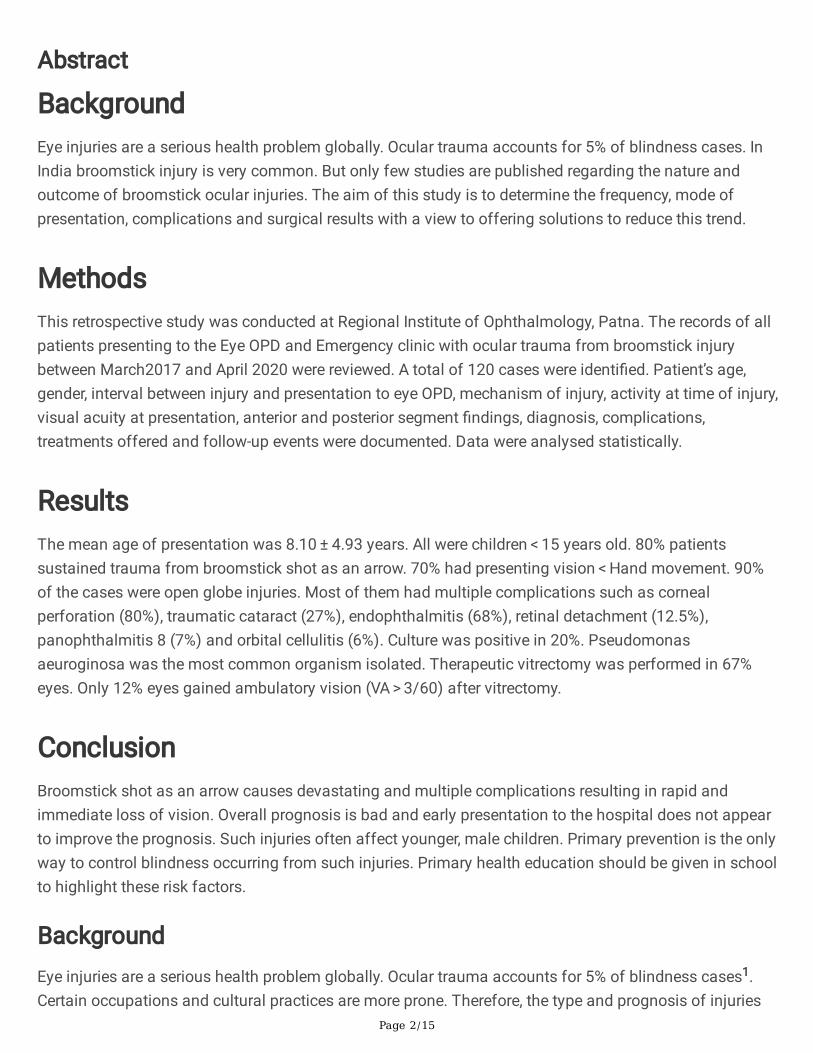
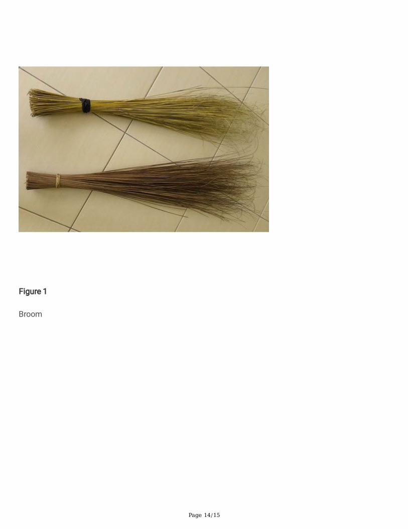
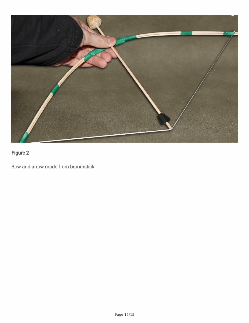
differ between developed and developing countries2. Ocular trauma among children is a serious healthconcern. It is one of the major cause of monocular blindness.3–5. Vision loss in childhood retards themental and physical development of the child. Thus it has signi�cant psychosocial impact. Consideringhigh frequency and severe vision loss it is a subject of major concern in children6–9. Many objects cancause penetrating ocular trauma. They are sharp objects such as pencil tips, knife, needles, sticks, twigs,toys, pellets, stone, metal rods and bricks. In India broomstick injury is very common. A broom is acommon household agent used for sweeping �oors and roasting grains (Fig. 1). It is made from a bundleof broomsticks, derived from a variety of materials like ra�a palm, grasses, reeds, date palm and coconutleaves. In India children frequently play from bow and arrow made of broomsticks (Fig. 2). It is a populargame inspired from the mythological Gods depicted in the television serials and movies. Brooms arepresent in every household. Children often take them and cause accidental ocular injuries to their siblingsor friends while playing. It can also become an intraocular foreign body (IOFB). The injury causesfulminant endophthalmitis because they are a reservoir of bacteria and fungi10. Broomstick injuries arevery common but very few studies are published in literature. Few case reports have been published fromIndia in 1968, Ghana and South Africa in 2005.11,12. Sharma et al described the results of surgicaltreatment of 100 such cases in 199410. Ukponmwan et al have described a case series of 20 patientsfrom Nigeria13.
Broomstick ocular injury presents at our hospital throughout the year. But literature is quite limited. Theaim of this study is to determine the frequency, mode of presentation, complications and surgical resultsof broomstick eye injury with a view to offering solutions to reduce this trend.
MethodsThis retrospective study was conducted at Regional Institute of Ophthalmology, Patna. The records of allpatients presenting to the Eye Out patient department (OPD) and Emergency clinic with ocular traumafrom broomstick injury between Sep 2017 and March 2020 were reviewed. A total of 120 cases wereidenti�ed. Relevant information was retrieved that formed the database for analysis.
Patient’s age, gender, interval between injury and presentation to eye OPD, mechanism of injury, activity attime of injury, visual acuity at presentation, anterior and posterior segment �ndings, diagnosis,complications, treatments offered and follow-up events were documented.
Ocular trauma from broomstick was classi�ed using the Birmingham Eye Trauma Terminology system(BETTS)14. The children were treated promptly with systemic and topical antibiotics and antifungal.Surgical repair of corneal or scleral wound and removal of retained IOFB was carried out when indicated.In case of endophthalmitis or retinal detachment (RD) pars plana vitrectomy (PPV) and intravitrealantibiotics were injected. Vitreous biopsy was taken and sent for cytology and culture and sensitivity.
Data were analysed using SPSS, IBM, Chicago, USA. Ethics approval for this study was obtained from theInstitutional Research Board of our hospital.
Page 4/15
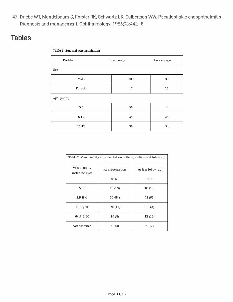
ResultsA total of 120 eyes in 120 patients were seen. All were children < 15 years old. The mean age was 8.10 ± 4.93 years with a range of 2 to 14 years. Males were 6 times more affected than females (Table 1)
96 (80%) patients sustained trauma from broomstick shot as an arrow, often by a friend or sibling. 11(9%) sustained injury from beating with a broom by their teacher or parent. 7 (6%) children were hit bytheir siblings with the broom while playing at home. 3 got injured from poor handling of the broom atchores and 3 children accidentally fell upon the broom. 50% of cases presented within 24 h of injury, 40%presented within 4 days and 10% presented after 1‐week.
15 (13%) children presented with no light perception (NLP), 70 (58%) patients had visual acuities rangingfrom hand movement (HM) to light perception (LP). Vision could not be assessed in 5 children who werepreverbal. Table 2 shows the visual acuities of the eyes at presentation and last follow‐up visit. The lastfollow‐up visit ranged from 3 weeks to 13 months with a mean follow‐up of 21 weeks. 60% children wereadmitted through the emergency clinic, rest through OPD. Under the BETTS classi�cation, 90% (n = 108)of the cases were open globe injuries (OGI) ranging from penetrating (70%, n = 84), perforating (15%, n = 18) and retained IOFB (15%, n = 18) injuries. Only 10% (n = 12) were closed (lamellar) injuries (Table 3).Most of them had multiple complications such as corneal perforation (80%), traumatic cataract (27%),endophthalmitis (68%), RD (12.5%), panophthalmitis 8 (7%) and orbital cellulitis 7 (6%). Othercomplications are shown in Table 4. Vitreous biopsy was taken in 81 patients sample sent for staining,culture and sensitivity. Culture was positive in 20%. Pseudomonas aeuroginosa was the most commonorganism isolated (Table 5). All patients were admitted. All received topical antibiotics moxi�oxacin,tobramycin and natamycin. Injectable antibiotics vancomycin and ceftriaxone were started from day 1. Ifsuspected inj Metronidazole was added to cover anaerobic organisms in cases of panophthalmitis ororbital cellulitis. Inj Tetanus toxoid was given. Thorough slit lamp and indirect ophthalmoscopicevaluation was done. Ultrasound B scan was gently performed to evaluate the lens status and posteriorsegment. Patients were immediately taken for globe exploration and repair under general anaesthesia.Corneal and scleral repair was done with 10/0 nylon and 8/0 polyglactin sutures respectively. IOFB, ifpresent was removed. In cases of endophthalmitis core PPV was performed provided posterior segmentwas adequately visualised. If view was hazy then patients were taken up for surgery once the anteriorsegment cleared. RD was repaired either in same setting or later depending upon media clarity. Intravitrealvancomycin, ceftriaxone and voriconazole were injected in all cases of endophthalmitis. Therapeuticvitrectomy was performed in 80 (67%) eyes. Only 10 (12%) eyes gained ambulatory vision (VA > 3/60)after vitrectomy.
DiscussionBroomstick injury was 6 times more common in males than females. The mean age was 8.10 ± 4.93 years. Majority (70%) were less than 10 years. Other studies have shown males to be more prone toinjuries 7,11,15. Young children are more prone because they have immature motor skills and unable to

Page 5/15
protect themselves. They are unaware of the consequences of rough games. Previous studies reported adelay in presentation of more than 24 hours in 91% of cases 7,9,11,16. Ukponwman etal13 reported 50%cases presenting beyond 24 hours. In our study half of the cases presented within 24 hours, 40% within4 days and rest after 1 week. The patients came from far off areas so they reached the hospital late afterarranging travel and money logistics 17,18. In few cases the primary ophthalmologist referred them late.Those who presented within 24 hours either lived in the same city or nearby districts. They were highlyanxious as it involved young children and wanted immediate help.
Ukponwman etal13 presented a case series of 20 cases over 6 years. Essumann etal11 reported 2 cases in6 years and Omobolanle etal7 reported 9 cases in 1 year. We saw 120 broom stick injury cases in 3 years.Broomstick injury at our centre is prevalent throughout the year. It is peculiar to Indian subcontinent 19,20.A broom is a common household agent. Children frequently play from bow and arrow made ofbroomsticks. This game is quite popular in rural areas. But the incidence increased during the Covid19pandemic lockdown period. At this time popular shows were retelecast on television. In these shows Godswere shown using bow and arrow upon their enemies. The children started playing with homemadearrows, inspired by the television shows. Also more number of cases were referred to our centre during thelockdown period because of limited healthcare facilities. That might have also contributed towards theincreased incidence.
The most common mechanism (80%) of injury was broomstick used as an arrow with a rubber slingduring games. Often the children were from lower socioeconomic strata and there was no parentalsupervision while playing.
90% injuries were OGI from penetrating and retained IOFB. The arrow is a pointed sharp object whichtravels at high speed and can easily penetrate the eye. Visual acuity at initial presentation was very poor,80% being HM or worse. They did not improve even after adequate intervention till last follow up(Table 2). Their globe was salvaged though. 3 cases went into phthisis. This might be due to multipleinjuries like corneal perforation, iris prolapse, hypopyon, severe endophthalmitis and corneal scar 11.Those presenting with vision > 6/60 had better outcome. Their vision was salvaged. They had paracentralor limbal corneal perforation and mild endophthalmitis.
The initial presenting visual acuity was NLP in 15% and CF at 1 m to LP in 65%. Early presentation ofpatients to the hospital did not appear to improve visual outcome. This could be because of poorbaseline vision, rapid, fulminant Gram negative endophthalmitis and severity of complications like RD.Published studies have shown that clinical features associated with good visual outcome in OGI werebetter baseline vision, culture of nonvirulent organism, lack of RD, absence of clinical endophthalmitisand shorter wound length21,22.
In this study 81 patients presented with endophthalmitis. All cases vitreous biopsy was taken and sent forcytology and culture. Culture positivity was 20% (n = 16). 63% (n = 10) had Gram negative infections.Pseudomonas aeruoginosa was the most common organism isolated (n = 5), followed by Bacillus cereus
Page 6/15
(n = 3) and Enterococcus (n = 2). Among Gram positive, Staphylococcus and Streptococcus were isolatedin 2 eyes each. 2 cases had fungal growth, Aspergillus and Candida. Culture-positive rates have beenreported from 27–75%19,20,23,24. The lower culture-positivity rates in our study could be because of prioruse of antibiotics by the primary physician. Published reports on paediatric traumatic endophthalmitishave implicated Gram-positive organisms in 57–67% cases20,23,25, Staphylococcus aureus,Streptococcus epidermidis and Bacillus cereus being the most common organisms. Our study hadpredominantly Gram negative infections, Pseudomonas being the most common. This might be becausea broom is a dirty object and harbours many bacteria and fungi. We also analysed the risk factors ofpenetrating traumatic endophthalmitis. Factors associated with increased risk were young age, delayedpresentation, delayed primary repair, incomplete history regarding the exact nature of the injury, injurywith a contaminated object, di�culty in undertaking a thorough initial and follow-up examination, ruralsetting, and retained IOFB 19,26,27,28−38. Therapeutic vitrectomy was performed in 80 (67%) eyes. Theindication was either endophthalmitis or RD. Only10 out of 80 eyes (12%) gained ambulatory vision (VA > 3/60) after vitrectomy. Factors predictive of poor anatomical success were corneal abscess, injuriesinvolving both anterior and posterior segment (P < .02), endophthalmitis (P < .05), and presence of RDwith proliferative vitreoretinopathy (P < .05). Presence of either of these warrants an early and aggressivetreatment. We did not �nd any impact of age on functional or anatomical outcome following treatment.This is in contrast to Alfaro et al39 who reported poor visual outcomes in children younger than 10 years.This may be because potent antibiotics and advanced surgical instruments were not available in in 1995when their report came. Today many advances have been made in managing endophthalmitis cases interms of availability of potent antibiotics and improvements in surgical instrumentation. Lens rupture hasbeen reported to have poor outcomes40,41. We did not �nd any association between the two. Patientsundergoing vitrectomy were more likely to have good anatomical outcome as it decreased themicrobiological load and helped in diffusion of intravitreal and systemic antibiotics within the eye42–47.
ConclusionBroomstick shot as an arrow causes devastating and multiple complications resulting in rapid andimmediate loss of vision. Overall prognosis is bad and early presentation to the hospital does not appearto improve the prognosis. Such injuries often affect younger, male children during misguided andunsupervised play. Primary prevention is the only way to control blindness occurring from such injuries. Itis recommended that primary health education should be given in school to highlight these risk factors.School teachers should also educate the parents in parents teacher meeting about its danger and theneed to monitor their children while playing. Broom kept at home should be away from the reach ofchildren. Loose pieces should be immediately picked up and discarded safely. Bow and arrow gamesshould be prohibited. Governments can broadcast messages in the media to raise awareness about theserisks to reduce the incidence of injuries.
Abbreviations

Page 7/15
OPD- Out patient department
IOFB- Intraocular foreign body
BETTS- Birmingham Eye Trauma Terminology system
RD- Retinal detachment
PPV- Pars plana vitrectomy
OGI- Open globe injury
DeclarationsEthics declaration, Ethics approval and consent to participate
This study followed the tenets of the Declaration of Helsinki and was approved by the ethical committeeof the hospital, IGIMS Ethical Committee, Indira Gandhi Institute of Medical Sciences, Patna. Verbalinformed consent from each patient was taken before the study. Parental consent was obtained.
Consent to publish
Not applicable
Availability of data and materials
All data and materials generated or analysed during the study are submitted in the manuscript.
Competing interests
The authors declare that they have no competing interests.
Funding
No funding was received for this study.
Authors Contributions
Dr. Abhishek Gupta wrote the paper. Dr. Richa Gupta designed the research. Dr. Prabhakar Singh collectedthe data. Dr. Shivani Sinha analysed the data. Dr. Vidya Bhushan revised the paper. Dr. Bibhuti P Sinhamonitored and supported the research. All authors have read and approved the manuscript
Acknowledgement
Not applicable
Author information
Page 8/15
Dr. Abhishek, Dr. Prabhakar Singh , Dr. Richa, Dr. Vidya Bhusan, Dr.Shivani Sinha, Dr. Bibhuti PrasannSinha.
A�liations
Indira Gandhi Institute of Medical Sciences, Patna
Dr. Abhishek, Dr. Richa, Dr. Vidya Bhusan, Dr.Shivani Sinha, Dr. Bibhuti Prasann Sinha.
Nirwana Netralaya, Sasaram
Dr. Prabhakar Singh
References1. Thylefors B. Epidemiological patterns of ocular trauma. Aust N Z J Ophthalmol. 1992;20:95–8.
2. Négrel AD. Magnitude of eye injuries worldwide. Community Eye Health J. 1997;10:49–53.
3. Thylefors B, editor. Ocular trauma. In: Strategies for Prevention of Blindness in National Programmes– A Primary Health Care Approach. Geneva: World Health Organisation; 1997. pp. 74–80.
4. Otoibhi SC, Osahon AI. Perforating eye injuries in children in Benin City. Niger J Biomed Sci.2003;2:40–5.
5. Lithander J, Al Kindi H, Tönjum AM. Loss of visual acuity due to eye injuries among 6292 schoolchildren in the Sultanate of Oman. Acta Ophthalmol Scand. 1999;77:697–9.
�. Osahon AI, Dawodu OA. Pattern of eye diseases in childrenin Benin City, Nigeria: A hospital–basedstudy. Trop Doct. 2002;32:158–9.
7. Omobolanle AA, Henrietta N. Pattern of paediatric corneal laceration injuries in the University of PortHarcourt teaching hospital, Rivers state, Nigeria. BMC Res Notes. 2012;5:683.
�. Kyari F, Alhassan MB, Abiose A. Pattern and outcome of paediatric ocular trauma – A 3–year reviewat National Eye Centre, Kaduna. Niger J Ophthalmol. 2000;8:11–6.
9. Ashaye AO. Eye injuries in children and adolescents: A report of 205 cases. J Natl Med Assoc.2009;101:51–6.
10. Sharma T, Agarwal P, Gopal L, Badrinath SS. R Murugesan Penetrating Ocular Trauma in Children byBroomstick Bows and Arrows. Ophthalmic Surg. 1994 Mar;25(3):175–9.
11. Essuman VA, Ntim–Amponsah CT. Preventing broomstick eye injuries in children in Accra.Community Eye Health J. 2004;17:46.
12. Grieshaber MC, Stegmann R. Penetrating eye injuries in South African children: Aetiology and visualoutcome. Eye (Lond). 2006;20:789–95.
13. Ukponmwan CU, Momoh RO. Broomstick injuries to the eye; An emerging cause of blindness amongchildren in Nigeria. Niger J Surg. 2015;21:13–7.
Page 9/15
14. Kuhn F, Morris R, Witherspoon CD. Birmingham Eye Trauma Terminology (BETT): Terminology andclassi�cation of mechanical eye injuries. Ophthalmol Clin North Am. 2002;15:139–43, v.
15. Onwasigwe EN, Umeh RE, Onwasigwe CN. Ocular injury in children. Niger J Ophthalmol. 1994;2:9–17.
1�. Ukponmwan CU, Akpe AB. Aetiology and complications of ocular trauma. Niger J Surg Sci.2008;18:92–7.
17. Vasnaik A, Vasu U, Bath RR, Kurian M, George S. Mechanical eye (globe) injuries in children. JPediatric Ophthalmol Strabismus. 2002;39:5–10.
1�. Serrano JC, Chalela P, Arias JD. Epidemiology of childhood ocular trauma in a northeasternColombian region. Arch Ophthalmol. 2003;121:1439–45.
19. Narang S, Gupta V, Simalandhi P, Gupta A, Raj S, Dogra MR. Pediatric open globe injuries. Visualoutcome and risk factors for endophthalmitis. Indian J Ophthalmol. 2004;52:29–34.
20. Dasgupta S, Mukerjee R, Ladi DS, Gandhi VH. Pediatric ocular trau trauma. A clinical presentation. JPostgrad Med. 1990;36:20–2.
21. Lieb DF, Scott IU, Flynn HW Jr, Miller D, Feuer WJ. Open globe injuries with positive intraocularcultures: Factors in�uencing �nal visual acuity outcomes. Ophthalmology. 2003;110:1560–6.
22. Knyazer B, Bilenko N, Levy J, Lifshitz T, Belfair N, Klemperer I, et al. Open globe eye injurycharacteristics and prognostic factors in southern Israel: A retrospective epidemiologic review of 10years experience. Isr Med Assoc J. 2013;15:158–62.
23. Alfaro DV, Roth DB, Laughlin RM, Goyal M, Liggett PE. Pediatric post-traumatic endophthalmitis. Br JOphthalmol. 1995;79:888–91.
24. Chhabra S, Kunimoto DY, Kazi L, Regillo CD, Ho AC, Belmont J, et al. Endophthalmitis after openglobe injury. Microbiologic spectrum and susceptibilities of isolates. Am J Ophthalmol.2006;142:852–4.
25. Mieler WF, Ellis MK, Williams DF, Han DP. Retained intraocular foreign bodies and endophthalmitis.Ophthalmology. 1990;97:1532–8.
2�. Brinton GS, Topping TM, Hyndiuk RA, Aaberg TM, Reeser FH, Abrams GW. Post traumaticendophthalmitis. Arch Ophthalmol. 1984;102:547–50.
27. Boldt HC, Pulido JS, Blodi CF, Folk JC, Weingeist TA. Rural endophthalmitis. Ophthalmology.1989;96:1722–6.
2�. Thompson JT, Parver LM, Enger CL, Mieler WF, Liggett PE. Infectious endophthalmitis afterpenetrating injuries with retained intraocular foreign bodies: National Eye Trauma System.Ophthalmology. 1993;100(10):1468–74.
29. Zhang Y, Zhang MN, Jiang CH, Yao Y, Zhang K. Endophthalmitis following open globe injury. Br JOphthalmol. 2010;94(1):111–4.
30. Thompson WS, Rubsamen PE, Flynn HW, Schiffman J, Cousins SW. Endophthalmitis afterpenetrating trauma: Risk factors and visual acuity outcomes. Ophthalmology. 1995;102(11):1696–
Page 10/15
701.
31. Essex RW, Yi Q, Charles PG, Allen PJ. Post-traumatic endophthalmitis. Ophthalmology.2004;111(11):2015–22.
32. Farr AK, Hairston RJ, Humayun MU, et al. Open globe injuries in children: A retrospective analysis. JPediatr Ophthalmol Strabismus. 2001;38:72–7.
33. Jonas JB, Knorr HL, Budde WM. Prognostic factors in ocular injuries caused by intraocular orretrobulbar foreign bodies. Ophthalmology. 2000;107(5):823–8.
34. Soheilian M, Rafati N, Peyman GA. Prophylaxis of acute posttraumatic bacterial endophthalmitiswith or without combined intraocular antibiotics: A prospective, double masked randomized pilotstudy. Int Ophthalmol. 2001;24(6):323–30.
35. Jalali S, Das T, Majji AB. Hypodermic needles: A new source of penetrating ocular trauma in Indianchildren. Retina. 1999;19(3):213–7.
3�. Bhagat N, Nagori S, Zarbin M. Post-traumatic infectious endophthalmitis. Surv Ophthalmol.2011;56(3):214–51.
37. Khan S, Athwal L, Marco Z, Bhagat N. Pediatric infectious endophthalmitis: A review. J PediatrOphthalmol Strabismus. 2014;51(3):140–53.
3�. Jandeck C, Kellner U, Bornfeld N, Foerster MH. Open globe injuries in children. Graefes Arch Clin ExpOphthalmol. 2000;238:420–6.
39. Alfaro DV, Roth DB, Laughlin RM, Goyal M, Liggett PE. Pediatric post-traumatic endophthalmitis. Br JOphthalmol. 1995;79:888–91.
40. Mansouri M, Faghihi H, Hajizadeh F, Rasoulinejad SA, Rajabi MT, Tabatabaey A, et al. Epidemiologyof open-globe injuries in Iran: analysis of 2,340 cases in 5 years (report no. 1). Retina. 2009;29:1141–9.
41. Jonas JB, Knorr HL, Budde WM. Prognostic factors in ocular injuries caused by intraocular orretrobulbar foreign bodies. Ophthalmology. 2000;107:823–8.
42. Reynolds DS, Flynn HW Jr. Endophthalmitis after penetrating ocular trauma. Curr Opin Ophthalmol.1997;8:32–8.
43. Sternberg P Jr, Martin DF. Management of endophthalmitis in the post-endophthalmitis vitrectomystudy era. Arch Ophthalmol. 2001;119:754–5.
44. Thordsen JE, Harris L, Hubbard GB. Pediatric endophthalmitis: A 10 year consecutive series. Retina.2008;28:3–7.
45. Weinstein GS, Mondino BJ, Weinberg RJ, Biglan AW. Endophthalmitis in a pediatric population. AnnOphthalmol. 1979;11:935–43.
4�. Han DP, Wisniewski SR, Wilson LA, Barza M, Vine AK, Doft BH, et al. Spectrum and susceptibilityofmicrobiologic isolates in the endophthalmitis vitrectomy study. Am J Ophthalmol 1996: Jul;122(1): 1–17.
Page 11/15
47. Driebe WT, Mandelbaum S, Forster RK, Schwartz LK, Culbertson WW. Pseudophakic endophthalmitisDiagnosis and management. Ophthalmology. 1986;93:442–8.
Tables
Table 1. Sex and age distribution
Profile Frequency Percentage
Sex
Male 103 86
Female 17 14
Age (years)
0-5 50 42
6-10 34 28
11-15 36 30
Table 2: Visual acuity at presentation in the eye clinic and follow‐up
Visual acuity(affected eye)
At presentation
n (%)
At last follow up
n (%)
NLP 15 (13) 18 (15)
LP-HM 70 (58) 78 (65)
CF-5/60 20 (17) 10 (8)
6/18-6/60 10 (8) 12 (10)
Not assessed 5 (4) 2 (2)
Page 12/15
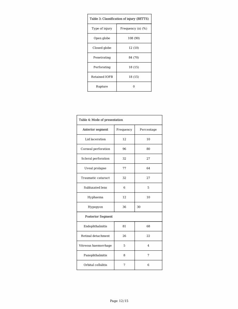
Table 3: Classification of injury (BETTS)
Type of injury Frequency (n) (%)
Open globe 108 (90)
Closed globe 12 (10)
Penetrating 84 (70)
Perforating 18 (15)
Retained IOFB 18 (15)
Rupture 0
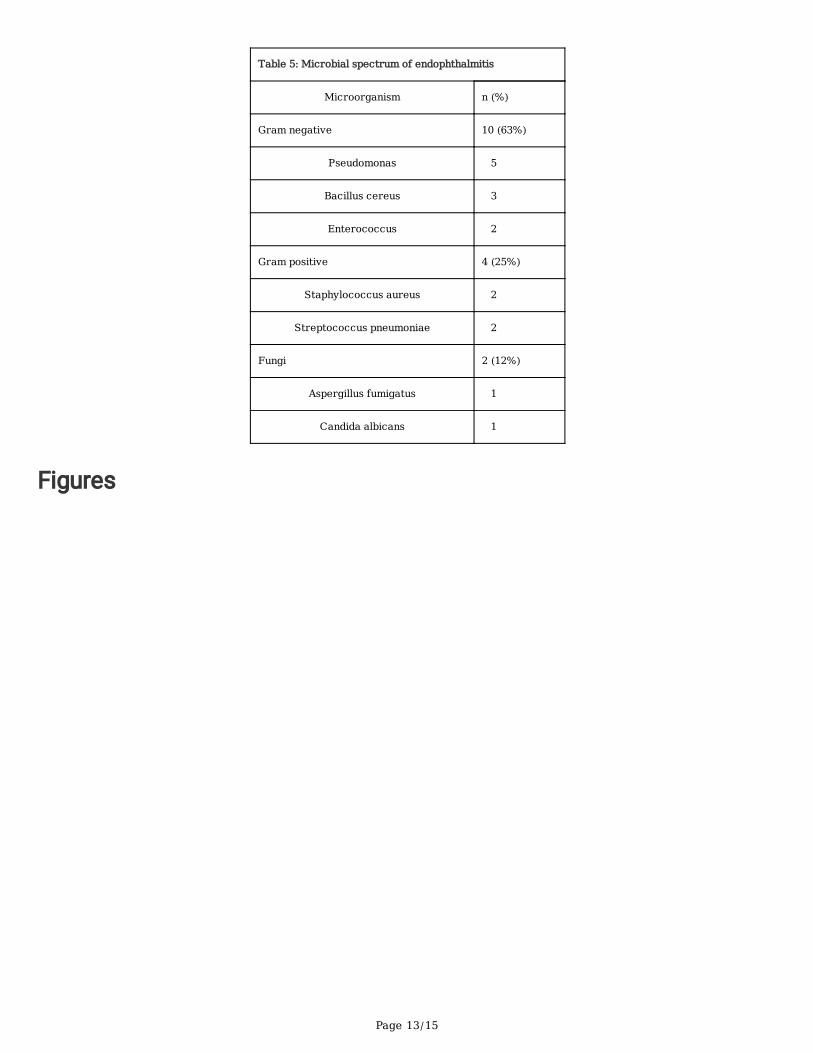
Table 4: Mode of presentation
Anterior segment Frequency Percentage
Lid laceration 12 10
Corneal perforation 96 80
Scleral perforation 32 27
Uveal prolapse 77 64
Traumatic cataract 32 27
Subluxated lens 6 5
Hyphaema 12 10
Hypopyon 36 30
Posterior Segment
Endophthalmitis 81 68
Retinal detachment 26 22
Vitreous haemorrhage 5 4
Panophthalmitis 8 7
Orbital cellulitis 7 6
Page 13/15
Table 5: Microbial spectrum of endophthalmitis
Microorganism n (%)
Gram negative 10 (63%)
Pseudomonas 5
Bacillus cereus 3
Enterococcus 2
Gram positive 4 (25%)
Staphylococcus aureus 2
Streptococcus pneumoniae 2
Fungi 2 (12%)
Aspergillus fumigatus 1
Candida albicans 1
Figures
Page 14/15
Figure 1
Broom
Page 15/15
Figure 2
Bow and arrow made from broomstick