The potential of chitosan in ocular drug delivery
13
1451 Review Article JPP 2003, 55: 1451–1463 ß 2003 The Authors Received August 29, 2003 Accepted September 22, 2003 DOI 10.1211/0022357022476 ISSN 0022-3573 Department of Pharmacy and Pharmaceutical Technology, Faculty of Pharmacy, University of Santiago de Compostela, 15782 Santiago de Compostela, Spain Marõ´a Jose ´ Alonso, Alejandro Sa ´ nchez Correspondence: M. J. Alonso, Department of Pharmacy and Pharmaceutical Technology, Faculty of Pharmacy, University of Santiago de Compostela, 15782 Santiago de Compostela, Spain. E-mail: [email protected] Acknowledgement and funding: Some of the work covered in this review was supported by a grant from the Spanish Ministry of Sciences and Technology (MCyT) (MAT 2000-0509-C02-01). The authors would like to thank Prof. Caruncho for his help in the interpretation of the confocal images. The potential of chitosan in ocular drug delivery Mar| ´a Jose ´ Alonso and Alejandro Sa ´ nchez Abstract This paper presents an overview of the potential of chitosan-based systems for improving the retention and biodistribution of drugs applied topically onto the eye. Besides its low toxicity and good ocular tolerance, chitosan exhibits favourable biological behaviour, such as bioadhesion- and permeability-enhancing properties, and also interesting physico-chemical characteristics, which make it a unique material for the design of ocular drug delivery vehicles. The review summarizes the techniques for the production of chitosan gels, chitosan-coated colloidal systems and chitosan nanoparticles, and describes their mechanism of action upon contact with the ocular mucosa. The results reported until now have provided evidence of the potential of chitosan gels for enhancing and prolonging the retention of drugs on the eye surface. On the other hand, chitosan-based colloidal systems were found to work as transmucosal drug carriers, either facilitating the transport of drugs to the inner eye (chitosan-coated colloidal systems containing indometacin) or their accumulation into the corneal/conjunctival epithelia (chitosan nanoparticles containing ciclosporin). Finally, the tolerance, toxicity and biodegradation of the carriers under evaluation were reviewed. Introduction Even though the history of the polysaccharide chitosan dates back from the 19th century, it has only been over the last couple of decades that this material has received attention in the biomedical and drug delivery fields. Chitosan is a deacetylated form of chitin, which is the second-most abundant polymer in nature after cellulose. The small difference in the chemical structure of chitin and chitosan has, however, extremely important consequences in terms of their utility for drug delivery. Chitin is insoluble in water or in the most common organic solvents used in pharmaceutical technology and, therefore, it is not useful in the development of drug delivery devices. In contrast, chitosan base is soluble in acidic solutions wherein it becomes protonized. This positive charge of the chitosan molecule enables its interaction with polyanions, a process that has been used to obtain complexes as well as micro and nanoparticulate drug delivery systems. Besides its polycationic nature, chitosan has shown excellent film-forming properties (RemunÄ an-Lopez & Bodmeier 1996). Consequently, its use for the prepara- tion of films, bandages and laminated devices has also opened new applications for this biopolymer. There are a few review articles regarding the utility of chitosan in drug delivery following different modalities of administration. These articles are a good illustration of how the number of applications of chitosan in the drug delivery field has increased over the last decade (Dodane & Vilivalam 1998; Felt etal 1998; Illum 1998; Paul & Sharma 2000; Janes etal 2001; Singla & Chawla 2001). Even though chitosan has been proposed for parenteral drug delivery, there is no doubt that the main interest in chitosan-based systems has shifted to mucosal drug delivery. This is due to chitosan’s unique biological properties such as mucoadhesiveness (Lehr et al 1992; Borchard et al 1996), its ability to enhance transiently the permeability of mucosal barriers (Artursson et al 1994; Borchard et al 1996) and its biodegradability in the rich lysozyme-containing mucus. As we will comment later, these properties are particularly important with respect to the use of chitosan in ophthalmology. Taking this information into account, the purpose of this review is to provide the reader with an overview of what is currently known about the use of chitosan for the treatment of eye disorders, with particular emphasis on its utility for the design of new
-
Upload
independent -
Category
Documents
-
view
3 -
download
0
Transcript of The potential of chitosan in ocular drug delivery

1451
Review Article
JPP 2003 55 1451ndash1463szlig 2003 The AuthorsReceived August 29 2003Accepted September 22 2003DOI 1012110022357022476ISSN 0022-3573
Department of Pharmacy andPharmaceutical TechnologyFaculty of Pharmacy Universityof Santiago de Compostela15782 Santiago de CompostelaSpain
Marotildeacutea Jose Alonso AlejandroSanchez
Correspondence M J AlonsoDepartment of Pharmacy andPharmaceutical TechnologyFaculty of Pharmacy Universityof Santiago de Compostela15782 Santiago de CompostelaSpain E-mail ffmjalonusces
Acknowledgement and fundingSome of the work covered in thisreview was supported by a grantfrom the Spanish Ministry ofSciences and Technology (MCyT)(MAT 2000-0509-C02-01) Theauthors would like to thank ProfCaruncho for his help in theinterpretation of the confocalimages
The potential of chitosan in ocular drug delivery
Mar|a Jose Alonso and Alejandro Sanchez
Abstract
This paper presents an overview of the potential of chitosan-based systems for improving the
retention and biodistribution of drugs applied topically onto the eye Besides its low toxicity and
good ocular tolerance chitosan exhibits favourable biological behaviour such as bioadhesion- and
permeability-enhancing properties and also interesting physico-chemical characteristics which
make it a unique material for the design of ocular drug delivery vehicles The review summarizes
the techniques for the production of chitosan gels chitosan-coated colloidal systems and chitosan
nanoparticles and describes their mechanism of action upon contact with the ocular mucosa The
results reported until now have provided evidence of the potential of chitosan gels for enhancing
and prolonging the retention of drugs on the eye surface On the other hand chitosan-based
colloidal systems were found to work as transmucosal drug carriers either facilitating the transport
of drugs to the inner eye (chitosan-coated colloidal systems containing indometacin) or their
accumulation into the cornealconjunctival epithelia (chitosan nanoparticles containing ciclosporin)
Finally the tolerance toxicity and biodegradation of the carriers under evaluation were reviewed
Introduction
Even though the history of the polysaccharide chitosan dates back from the 19thcentury it has only been over the last couple of decades that this material has receivedattention in the biomedical and drug delivery fields Chitosan is a deacetylated form ofchitin which is the second-most abundant polymer in nature after cellulose The smalldifference in the chemical structure of chitin and chitosan has however extremelyimportant consequences in terms of their utility for drug delivery Chitin is insoluble inwater or in the most common organic solvents used in pharmaceutical technology andtherefore it is not useful in the development of drug delivery devices In contrastchitosan base is soluble in acidic solutions wherein it becomes protonized This positivecharge of the chitosan molecule enables its interaction with polyanions a process thathas been used to obtain complexes as well as micro and nanoparticulate drug deliverysystems Besides its polycationic nature chitosan has shown excellent film-formingproperties (RemunAuml an-Lopez amp Bodmeier 1996) Consequently its use for the prepara-tion of films bandages and laminated devices has also opened new applications for thisbiopolymer
There are a few review articles regarding the utility of chitosan in drug deliveryfollowing different modalities of administration These articles are a good illustrationof how the number of applications of chitosan in the drug delivery field has increasedover the last decade (Dodane amp Vilivalam 1998 Felt et al 1998 Illum 1998 Paul ampSharma 2000 Janes et al 2001 Singla amp Chawla 2001) Even though chitosan has beenproposed for parenteral drug delivery there is no doubt that the main interest inchitosan-based systems has shifted to mucosal drug delivery This is due to chitosanrsquosunique biological properties such as mucoadhesiveness (Lehr et al 1992 Borchard et al1996) its ability to enhance transiently the permeability of mucosal barriers (Arturssonet al 1994 Borchard et al 1996) and its biodegradability in the rich lysozyme-containingmucus As we will comment later these properties are particularly important withrespect to the use of chitosan in ophthalmology
Taking this information into account the purpose of this review is to provide thereader with an overview of what is currently known about the use of chitosan for thetreatment of eye disorders with particular emphasis on its utility for the design of new
ocular drug delivery systems Accordingly we will firstcomment on the therapeutic role of chitosan in ophthal-mology and then following a brief presentation of thebarriers that need to be overcome in ocular drug deliverywe will illustrate with a number of examples the potentialthat chitosan offers to resolve such limitations Theseexamples will be classified into the different pharmaceuti-cal presentations of chitosan that are acceptable forophthalmic application gels microspheres and colloidalsystems (nanoparticles nanocapsules)
Chitosan as an active biomaterial inophthalmology
Chitosan is a very promising biomaterial in ophthalmologynot only because of the favourable biological propertiesindicated above but also because of its inherent biologicalactivity which may also have an impact in ocular therapeu-tics The various forms in which chitosan has been investi-gated in ophthalmology are indicated in Table 1 Besidesbeing a major component in drug delivery devices chitosanitself has been shown to have wound healing and antimicro-bial activity (Balassa amp Prudden 1978 Allan amp Hadwiger1979 Muzzarelli 1983) These effects could be very beneficialfor the treatment of a number of ocular diseases The idea ofusing chitosan in corneal wound healing came from therecognized acceleration of wound-healing activity of chito-san degradation products (Balassa amp Prudden 1978) and theobserved success of chitosan as a haemostatic agent forporous vascular grafts (Malette et al 1983) More preciselybased on this previous work it was hoped that chitosanwould play an active role increasing keratocyte migrationthereby leading to a more rapid production of collagen andimproved corneal healing Unfortunately histological eva-luation and measurement of the tensile strength of the rabbitcornea exposed to chitosan (1 solution) did not show animproved corneal wound healing (Sall et al 1987) It is pos-sible that the lack of effectiveness of chitosan in this pre-liminary work may rely on the type of chitosan selected (notindicated in the publication) or on the protocol of adminis-tration Nevertheless from our knowledge there has notbeen any further work attempting to verify the accelerationof wound healing by chitosan
Taking advantage of the film-forming propertiesof chitosan Markey et al (1989) developed contactlenses with excellent edges and optics By assuming thewound-healing acceleration (Balassa amp Prudden 1978)and antimicrobial activity of the N-acetyl-D-glucosamineoligomers (Allan amp Hadwiger 1979 Muzzarelli 1983) itwas thought that this kind of system could be applied as aprotective device for an acutely traumatized eye (egfollowing ocular surgery) or a chronically compromisedcornea Unfortunately despite the suggested acceptabilityof these contact lenses no evidence of their effectivenesshas been reported
It is surprising that this early work on ocular woundhealing acceleration has not been further corroborated Itis possible that the lack of success of these initial appliedexperiments has discouraged researchers from investingefforts in this direction However a very important aspectto keep in mind that stimulates this research field is theactual availability of rigorously characterized and ultra-purified chitosan In fact researchers who specialize inchitosan are well aware of the difficulties in extrapolatingor simply comparing early work on chitosan due to thelimited information regarding the source characteristicsand purity of this natural material
In contrast to the lack of confirmation of chitosanrsquoswound-healing activity its antimicrobial activity wasrecently corroborated by the findings of Felt et al (2000)who suggested the use of a chitosan solution as an artificialtear formulation Besides the bacteriostatic activity of chit-osan this proposal was based on its excellent tolerance aftertopical ocular administration and its ability to spreadwell over the entire cornea after topical instillation (Feltet al 1999a) Additionally the prolonged pre-corneal resi-dence time of chitosan solutions reinforced the utility ofchitosan for the treatment of dry eye and keratoconjuncti-vitis sicca
Barriers to overcome in topical ocular drugdelivery
To understand the potential that chitosan offers inimproving the treatment of ocular diseases it is important
Table 1 Forms of chitosan investigated in ophthalmology
Chitosan form Application Drug incorporated Reference
Contact lenses Corneal wound healing ETH Markey et al (1989)
Solution Corneal wound healing ETH Sall et al (1987)
Solution Dry eye ETH Felt et al (2000)
Solution Prolonged retention Tobramycin Felt et al (1999)
Coated liposomes Improved corneal retention Marker Henricksen et al (1996)
Coated nanocapsules Improved corneal penetration Indometacin Calvo et al (1997)
Coated nanocapsules Improvedprolonged retention Marker De Campos et al (2003)
Microspheres Improved corneal penetration Aciclovir Genta et al (1997)
Microspheres Improved corneal penetration Ofloxacin Di Colo et al (2002)
Nanoparticles Improvedprolonged retention Ciclosporin De Campos et al (2001)
Nanoparticles Improvedprolonged retention Marker De Campos (2002)
1452 Maria Jose Alonso and Alejandro Sanchez
to know first what the targets for the drugs are andsecond what barriers need to be overcome to reachthose targets
The topical ocular administration of drugs has twodifferent purposes to treat superficial eye diseases suchas infections (ie conjunctivitis blepharitis keratitissicca) and to provide intra-ocular treatment through thecornea for diseases such as glaucoma or uveitis Despitethe efforts dedicated in the 1980s to the design of solidocular drug delivery systems the most popular andaccepted forms of delivering drugs to the eye are stillliquid forms administered as eye-drops As shown inFigure 1 one of the major problems encountered withthe topical administration of liquid forms is the rapidand extensive pre-corneal loss caused by drainage andhigh tear turnover After instillation of an eye-drop amajor fraction of the instilled dose is lost due to thelimited capacity of liquid retention of the eye surface andalso as a consequence of the blinking process which isnormally stimulated after instillation Additionally a cer-tain amount of drug is often absorbed systemically via theconjunctiva and the nasolachrymal duct
In addition to these anatomical constraints to theretention of drugs at the eyersquos surface the second majorlimiting step for the transport of drugs to the inner eye is
diffusion across the cornea Indeed although the corneacovers only one-sixth of the total surface area of the eye-ball it is considered to be the main pathway for thepermeation of drugs into intra-ocular tissues Obviouslythe extent to which topically applied drugs remain on theeyersquos surface or penetrate into the inner eye is dependentnot only on the physiological characteristics of the cornealbarrier but also on the physico-chemical properties of thedrug and the specific behaviour of the vehicle The impor-tance of these limitations is well illustrated by the knownfact that with classical ophthalmic liquid forms typicallyless than 5 of the applied drug penetrates the cornea andreaches intra-ocular tissues (Lang 1995)
The cornea consists of three different sections theepithelium stroma and endothelium The most externalsection the epithelium is composed of a number of well-organized and tightly packed cell layers and represents aselective barrier for the penetration of hydrophilic andionized compounds Indeed the tight junctions betweenepithelial cells prevent the entrance of molecules by aparacellular route Contrarily the stroma beneath theepithelium is a hydrophilic space that represents 90 ofthe cornea and consequently is a selective barrier forthose lipophilic compounds that could have easily diffusedthrough the epithelium As a consequence of this perfect
Instilleddrug
Conjunctival systemicabsorption
Corneal penetration
Eye surface
Conjunctiva
Conjunctiva
Cornea
Mucus layer
Inner eye
Pre-corneal drug loss
Figure 1 Schematic view of the pre-corneal elimination and transport pathways of a drug applied topically onto the eye
Chitosan in ocular drug delivery 1453
organization the transport of hydrophilic compounds ishindered at the epithelial level whereas that of lipophiliccompounds is blocked at the hydrophilic gel-like stromalcompartment The stroma may act as a drug reservoirslowly releasing the drug into the aqueous humour Theendothelium is a well-organized monolayer of epithelialcells and therefore does not contribute as a barrier forthe penetration of drugs
A drug that is applied topically and retained on the eyesurface may easily reach an important competitive routecovering five-sixths of the total surface area of the eyeballplusmn the conjunctiva The conjunctiva is a thin vascularizedmucous membrane that lines the inner surface of the eyelidsand coves the anterior part of the sclera up to the corneaOwing to the relative surface area and rich blood flowconjunctival uptake of a topically applied drug from thetear fluid is typically greater than corneal uptake Ocularpenetration via the sclero-conjunctival route is more rapid(for a hydrophilic drug) than via the transcorneal route(Romanelli et al 1994) However transconjunctival pene-tration is generally undesirable because most of the drugthat crosses the conjunctiva reaches the blood circulationConsequently the drug will not only be unable to reachthe ocular target site but it might also be responsible forsevere systemic side effects
Covering the conjunctiva and corneal surfaces of theeye is the mucus layer The tear-film mucus layer is acomplex macromolecular structure consisting essentiallyof mucin proteins lipids and DNA Ocular mucus ismainly secreted by the conjunctival goblet cells (Mooreamp Tiffany 1981) although corneal and conjunctivalepithelium has also been reported to produce mucins(Greiner et al 1985) Mucin is a macromolecular glycopro-tein that gives mucus its specific properties such as visc-osity and lubricant and surfactant effects Mucins arethought to consist of hundreds of short polysaccharidechains attached to a central protein core having a variablemolecular weight and residual negative charges presum-ably from sialic acid residues
After topical instillation of an ophthalmic drug solu-tion the drug is firstly mixed with the lachrymal fluid andremains in contact with the ocular mucosa for a very shortperiod of time typically 1plusmn2 min because of the perma-nent production of lachrymal fluid Drainage of lachrymalfluid towards the nasolachrymal duct induces a rapidelimination of conventional dosage forms during blinking
Researchers have dedicated important efforts to designsystems intended to overcome the aforementioned bar-riers The specific details of these delivery systems includ-ing solid devices and liquid eye-drops have already beendescribed in a number of review articles (Sintzel et al 1996Le Bourlais et al 1998 Felt et al 1999b) Despite the vari-ety of systems designed at present it is accepted that anoptimum ocular drug delivery system would be one thatcan be delivered in eye-drop form and that would need nomore than one or two instillations a day Most of the newdelivery devices have been aimed at improving ocular drugpenetration through prolongation of the drug residencetime in the cornea and conjunctival sac as well as slowingdrug release from the delivery system and minimizing
pre-corneal drug loss Among the marketed systemspre-formed gels and in-situ gelling systems are probablythe closest to reaching these objectives However theperformance of these viscous vehicles is limited by severalconstraints they do not facilitate the internalization ofthe drug in the corneal or conjunctival epithelia they areaqueous systems in which hydrophobic drugs can notbe solubilized and they do not provide a controlled drugdelivery
Therefore the necessity of exploring novel liquid sys-tems specifically designed for ocular drug delivery stillremains Within this research context it is currentlyaccepted that the primary objectives indicated above (pro-longed retention enhanced penetration and controlleddelivery) can be attained though different strategies thatinclude the use of bioadhesive polymers penetrationenhancers and the advanced design of micro- and nano-particulate delivery systems These approaches have beenexplored independently over the last decade (Greaves ampWilson 1993 Zimmer amp Kreuter 1995 Kaur amp Smitha2002) and have led to significant advances in the fieldProbably an adequate combination of them will lead to anew generation of ocular drug delivery systems As justi-fied in the next section the position that chitosan is takingwithin this research field holds great promise The follow-ing sections will address the progress made so far on thedesign of chitosan-based delivery systems intended fortopical ocular administration References pertaining tothe ocular application of these systems are presented inTable 1
Chitosan a good candidate for ocular drugdelivery
The residence time of a topically applied ophthalmic drugrefers to the duration of its contact with the ocular sur-face This concept is of particular interest in the formula-tion of topical ocular drug vehicles where mucoadhesivepolymers are frequently used as an approach to prolongdrug residence times When using a mucoadhesive mate-rial the clearance of the drug is controlled by the mucusturnover rate which is much slower than the tear turnoverrate This prolonged retention of the drug formulationimplies for a drug with good permeability properties anenhanced ocular drug bioavailability Chitosan is in thiscategory of mucoadhesive polymers The mucoadhesivecharacter of chitosan relates to the attraction between itspositively charged amino groups and the negativelycharged residues of sialic acid in the mucus (Lehr et al1992) along with other forces such as hydrogen bonds(Hassan amp Gallo 1990) In addition to this special prop-erty chitosan exhibits other attractive features mentionedin the following paragraphs which make it a unique can-didate for ocular drug delivery
It has penetration-enhancing properties which wereinitially attributed to the modulation of the tight junctionbarrier between epithelial cells (Artursson et al 1994Schipper et al 1997 Koch et al 1998) and recently alsorelated to intracellular routes (Dodane et al 1999) More
1454 Maria Jose Alonso and Alejandro Sanchez
specifically using Caco-2 cells these authors found thatchitosan increases cell permeability by affecting bothparacellular and intracellular pathways of epithelial cellsin a reversible manner without affecting cell viability orcausing membrane wounds This permeability-enhancingproperty has been used to explain the increased cornealtransport of specific drugs (see Chitosan-based oculardrug delivery systems below)
Chitosan is biodegradable (Pangburn et al 1982Hirano et al 1989a 1990) which enables the safe admin-istration and degradation of topically applied ocularchitosan vehicles As mentioned before chitosan biode-gradation is mediated by the hydrolytic actions oflysozyme and other enzymes (ie human chitinase and N-acetyl-shy -D-glucosaminidases) which produce chito-oligo-mers and monomers (Muzzarelli 1993 1997 Nordtveitet al 1994) This susceptibility to enzymatic depolymeriza-tion is an exclusive characteristic of chitosan with respectto other polysaccharides It has been reported that thedegradation rate in the presence of lysozyme depends onthe degree of acetylation (Nordtveit et al 1994) Howeverthe study of the influence of this parameter has led tocontradictory results Hirano et al (1989b) found an opti-mum lysozyme degradation rate for 80 N-acetylatedchitosan whereas Sashiwa et al (1990) found the highestlysozyme susceptibility for 30 N-acetylation In addi-tion little is known about some enzymes of human origin(ie collagenases and heparinases) even when it is wellknown that chitosans are more vulnerable than expectedto the non-specific actions of a number of enzymes(Muzzarelli 1997) Therefore these are important issuesthat need to be further investigated
Chitosan has excellent ocular tolerance This has beenreported in a rabbit model following topical application ofchitosan solutions and using confocal laser scanningophthalmoscopy combined with corneal fluorescein staining(Felt et al 1999a) Other evidence of the low toxicity andtolerance of chitosan delivery systems will be described later
Chitosan has favourable rheological behaviour Chitosansolutions have shown pseudoplastic and viscoelastic prop-erties (Wang amp Xu 1994 Mucha 1997) These are veryimportant characteristics since the pre-corneal tear filmhas a pseudoplastic character that should not be disturbedby application of liquid formulations On the other handviscoelastic fluids exhibit high viscosity under low shearrate and low viscosity under high shear rate conditionsThis behaviour is particularly important in ophthalmicformulations since it facilitates the retention while it per-mits the easy spreading of the formulation due to theblinking of the eyelids
On the basis of these favourable biological propertiesand also because of its adaptability for designing differentdelivery systems chitosan has attracted great attention inthe pharmaceutical and biomedical fields It is howeversurprising that the number of reports on the potential ofthis cationic polysaccharide in the ophthalmic field is stilllimited (Felt et al 1998 1999a) In the following section wewill describe in detail the characteristics of chitosan andthe in-vivo behaviour of chitosan-based ocular drug deliv-ery systems reported so far
Chitosan-based ocular drug delivery systems
Chitosan solutionsAs previously described in this review the most acceptabledosage forms for topical ocular drug delivery are theliquid forms The simplest presentation of chitosan in aliquid formulation consists of a chitosan solution which issometimes referred to as a hydrogel Hydrogels are nor-mally defined as polymers that have the ability to swell inaqueous solvents and undergo a liquidplusmngel transitionHowever in ophthalmology there is not a clear distinc-tion between hydrogels and highly viscous solutionsChitosan solutions can be prepared in different concentra-tions and using different types of chitosan (different mole-cular weight different salts and different deacetylationdegree) Preferably highly deacetylated chitosan (gener-ally more than 60) is used since the water solubilitydecreases with reduction in deacetylation Chitosansolutions have been well characterized in terms of theirpseudoplastic and viscoelastic behaviour (Wang amp Xu1994 Mucha 1997) Furthermore a synergism betweenrheological behaviour and mucoadhesion has also beendescribed (Caramella et al 1999) These studies haveshown that the viscosity and mucoadhesive propertiescan be modulated by adjusting the chitosan molecularweight and concentration Most of the reports on thetopical ocular administration of chitosan solutions referto concentrations in the range 05plusmn5 and molecularweight higher than 70plusmn100 kDa (Felt et al 1999a b)However it is possible that other molecular weights andconcentrations could be used for this specific application
An alternative way to modulate the viscosity and vis-coelastic behaviour of chitosan solutions could be throughthe incorporation of other hydrophilic polymers that areknown to interact with chitosan (eg hyaluronic acid)Recently a number of chemical derivatives of chitosanhave been produced among them PEG-chitosan whichis already commercially available These approachesinvolving other polymers are valid insofar as the newpolymers are acceptable for ocular administration
The specific bioadhesive activity of chitosan towards theocular surface has been confirmed in an ex-vivo study per-formed using freshly excised cattle cornea and radiolabelledchitosan (Henriksen et al 1996) Some years later the capa-city of chitosan for increasing pre-corneal drug residencetimes was clearly shown in a rabbit model using gammascintigraphy For this purpose chitosan gels containingtobramycin and the commercial drug solution were labelledwith 99mTc-DTPA and instilled onto the cornea of consciousrabbits The pre-corneal retention time was assessed bydetermining the eye-associated radioactivity using agamma camera The results showed a 3-fold increase ofthe corneal residence time of the chitosan solution as com-pared with that of the commercial drug solution (Felt et al1999a) In addition at 10 min post-intillation of the com-mercial solution all the radioactivity was concentrated inthe lachrymal duct whereas in the case of chitosan formula-tions 25plusmn50 of the radioactivity remained associated withthe cornea The results of pre-corneal drainage were verysimilar irrespective of the chitosan concentration (05plusmn15)
Chitosan in ocular drug delivery 1455
or molecular weight (160plusmn1930 kDa) On the basis of theseresults the authors suggested that the improvement in reten-tion time using chitosan might be due to a saturable bioad-hesive mechanism Hence they concluded that the use of alow concentration of low-molecular-weight chitosan wouldbe sufficient to provide a significant enhancement of theresidence time However no comment was made on thedifferences in deacetylation degree (which was in the range59plusmn87) of the polymers tested a parameter that could alsohave some effect on the behaviour of chitosan in-vivo
As indicated above besides their utility as drug deliv-ery vehicles chitosan solutions have also been investigatedas tear substitutes in the management of dry eye disorders(Felt et al 2000) These studies revealed that chitosan solu-tions are easier to manipulate and provide a more accurateand reproducible administration as well as a lower inci-dence of blurred vision and discomfort than conventionalhydrogels used at the ocular level In addition the anti-bacterial activity of chitosan could be useful to prevent asmentioned above the frequent secondary infections asso-ciated with this disorder
Chitosan microspheresChitosan microspheres have mainly been prepared usingtwo basic methodologies the water-in-oil solvent evapora-tion technique and the spray-drying technique (Kas 1997)In both cases chitosan is dissolved in an aqueous medium(either acidic or neutral) and then emulsified in an oilyphase where the evaporation takes place or simplysprayed-dried to accelerate the evaporation processThese particles are finally collected and stored as powders
Genta et al (1997) developed aciclovir-loaded chitosanmicroparticles of a size below 25 middotm using the water-in-oil solvent evaporation technique In-vitro release profilesrevealed that these chitosan microparticles slightlyreduced the drug dissolution rate Following in-vivoadministration to conscious rabbits of the drug-containingmicroparticles it was found that the ocular drug bioavail-ability increased as compared with that of the drug sus-pension (control formulation) More specifically thesesystems provided a 4-fold increase in the AUC valuewith respect to that of the control drug suspension Theauthors explained these positive results in terms of themucoadhesive character of chitosan However no furthermechanistic details were explored
More recently chitosan microspheres were incorpo-rated within a poly(ethylene oxide) (PEO) ocular insertwith the idea of improving the release behaviour of thesystem while taking advantage of the known beneficialeffects of chitosan (Di Colo et al 2002) Chitosan micro-spheres containing the drug ofloxacin were prepared usingthe spray-drying technique and then added to the PEOgel The in-vivo results showed no clear benefit from theincorporation of the chitosan microspheres into the insertHowever it was found that the tmax of the aqueoushumour ofloxacin concentration-vs-time profile wasincreased for the formulations containing chitosan plusmn aresult that was attributed to the corneal-permeability-enhancing effect of chitosan
The scarce number of reports on the use of chitosanmicrospheres for ophthalmic purposes could be justifiedby the limited acceptability of large particles In fact it hasbeen indicated that the size of the particles is a criticalparameter in their ocular tolerance and acceptability Thismay be the reason why research in this field has basicallyshifted to the design of colloidal systems (less than 1 middotm)including liposomes submicron emulsions nanocapsulesand nanoparticles The preparation characteristics andpotential of chitosan-based colloidal drug carriers in ocu-lar drug delivery are discussed in the following section
Chitosan-based colloidal drug carriers
Introduction of colloidal carriers in ocular drugdelivery Overall the research carried out over the last15 years has led to the conclusion that colloidal drug car-riers such as liposomes submicron emulsions nanocap-sules and nanoparticles are promising systems forimproving the ocular retention of topically administereddrugs The first proof of efficacy of liposomes in ophthalmictherapy was shown for idoxuridine used in the treatment ofherpetic keratitis in a rabbit model in 1981 (Smolin et al1981) Some years later Fitgerald et al (1987a) demon-strated using gamma-scintigraphy that liposomes have areduced pre-corneal clearance In addition they found thatthis behaviour was positively affected by the small size and apositive surface charge Despite the number of articlesdirected to this end (Maurice 1993) some inherent disad-vantages of liposomes such as their low drug-loading capa-city and compromised stability have probably hinderedfurther development in this area In this sense it is impor-tant to keep in mind that due to the low retention of theeye-drops on the eye surface the drug loading of the colloi-dal vehicles is a crucial parameter An alternative colloidalvehicle plusmn nanoparticlesnanocapsules made of biodegrad-able polymer plusmn was initially proposed with the idea ofcontrolling the release and simultaneously facilitating theinteraction of the drug with the ocular mucosa (Wood et al1985 Fitgerald et al 1987b) Surprisingly using polyalkyl-cyanoacrylate nanoparticles it was soon observed thatthese colloidal carriers were able to enter the corneal epithe-lium via endocytosis (Zimmer et al 1991) Unfortunatelysome damage to the cell membrane was also observedwhich was attributed to the polymer degradation productsThe transport of the colloidal systems across the cornealepithelium was later corroborated by our research groupusing poly-E-caprolactone (PECL) nanocapsules (Calvoet al 1994) However in our study no evidence of mem-brane alteration was detected Furthermore in a later workwe observed that microparticles were not as efficient asnanoparticles or nanocapsules at increasing the cornealtransport of the model drug indometacin (Calvo et al1996) and thus concluded that size has a key role in theability of these particles to work as ocular drug carriers
Chitosan-coated colloidal drug carriers A second gen-eration of ocular colloidal drug carriers is represented bythose coated with mucoadhesive polymers Following the
1456 Maria Jose Alonso and Alejandro Sanchez
observation of the improvement in transport of drugsassociated with nanocapsules and nanoparticles wedecided to explore whether or not a chitosan coatingwould further improved their efficacy as ocular drug car-riers The rationale for designing chitosan-coated systemswas to combine the advantages of nanoparticles as oculardrug carriers with the mucoadhesive and permeability-enhancing properties of chitosan We first reported thepreparation of these novel systems in 1997 (Calvo et al1997a) The systems can be composed of a solid core(nanoparticles) which is made of a biodegradable poly-mer or an oily core (nanocapsules) These systems can beprepared using solvent-displacement or solvent-emulsionevaporation techniques (Alonso 1996) A negatively chargedphospholipid is introduced into the colloidal carrier tofacilitate the attachment of chitosan onto the surface ofthe particles The size of the particles is slightly increasedand their zeta potential shifts from negative to positivevalues due to the chitosan coating These chitosan-coatedsystems are very versatile in regard to their drug-loadingcapacity and release properties Indeed by varying thecore composition and judiciously adjusting the prepara-tion technique it is possible to load either hydrosoluble orhydrophobic compounds For example hydrosoluble pro-teins such as tetanus toxoid have been loaded into chito-san-coated poly(lactic acidglycolic acid) using the doubleemulsion-solvent evaporation technique (Vila et al 2002)On the other hand lipophilic drugs such as indometacinand diazepam have been very efficiently incorporatedinto chitosan-coated oily droplets otherwise called chito-san nanocapsules (Calvo et al 1996 1997a)
Despite the versatility of chitosan-coated nanostruc-tures and preparation techniques only those providing ahigh loading and a relatively rapid release (from minutesto a few hours) are expected to have a potential applica-tion in ophthalmology For example we have studied thein-vivo efficacy of these systems using indometacin as amodel drug The ocular drug disposition was determinedafter a single instillation of 14C-indometacin-loaded sys-tems to conscious rabbits and subsequent quantificationof the radioactivity levels in cornea and aqueous humour(Calvo et al 1997b) The results showed that chitosan-coated nanoparticles increased the drug levels in corneaand aqueous humour to a significantly greater extent thaneither the commercial drug preparation or drug-loadeduncoated systems This initial work led to the conclusionthat a chitosan coating adds a clear benefit to the potentialof colloidal systems as ocular drug carriers To investigatewhether this benefit was solely due to the positive chargeor could be additionally related to the inherent propertiesof chitosan we compared the behaviour of chitosan-coated formulations with that of poly(L-lysine)-coatedformulations The lack of effect of the poly(L-lysine)-coat-ing stood for the intrinsic beneficial effect of chitosanHence the mechanism that explains the increased oculardrug penetration achieved with the chitosan-coated sys-tems could be a combination of an improved interactionwith the corneal epithelium followed by the penetrationof the particles in the corneal epithelial cells (Calvo et al1994) This mechanism was later corroborated as dis-
cussed hereafter In addition to their efficacy we couldobserve that these chitosan-coated systems have a goodocular tolerance (low ocular lesion index)
The effect of a chitosan coating on the ocular retentionof liposomes has also been investigated in the anaesthe-tized rat model (Henriksen et al 1996) In this study theauthors chose to label the liposomes with 125I-bovineserum albumin (BSA) and monitored the eye-associatedradioactivity over time Even though the ocular retentionof 125I-BSA was apparently more pronounced for thechitosan-coated liposomes the difference from the non-coated liposomes was not statistically significant In theinterpretation of these results we should however takeinto account that the measurements were performed on125I-BSA and a certain amount of protein could have beenreleased from the system following in-vivo applicationMore studies need to be performed to verify the limitedsuccess of this experiment
The importance of the coating of nanoparticles with amucoadhesive has been noted with other polymersbesides chitosan For example in a study performed byZimmer et al (1995) it was observed that coating albuminnanoparticles with mucoadhesive polymers hyaluronicacid among others led to a prolongation of the responseof the drug associated with them (pilocarpine)
Chitosan nanoparticlesAs mentioned in the introductory section the cationicnature of chitosan has been conveniently exploited forthe development of particulate drug delivery systems Aninteresting property of chitosan is its ability to gel oncontact with specific polyanions Based upon this princi-ple a few years ago we developed chitosan nanoparticles(Calvo et al 1997c) This technique involves the additionat room temperature of an aqueous phase containingsodium tripolyphosphate into an aqueous phase contain-ing chitosan Nanoparticles are formed immediately uponmixing of the two phases through inter- and intramolecu-lar linkages created between the phosphate groups ofsodium tripolyphosphate and the amino groups of chitosanThe conditions for the formation of high-yield nanoparti-cles with a particular nanometric size may vary significantlydepending on the purity acid salt and molecular weight ofchitosan employed Consequently the formulation para-meters should be optimized for each individual chitosantype More recently we adapted this gelation technique toproduce depolymerized chitosan nanoparticles (Janes et al2003) This idea came from different studies suggesting thatthe molecular weight could affect a number of key proper-ties of chitosan such as biocompatibility (Richardson et al1999) permeability enhancement (Schipper et al 1996) andmucoadhesion (Henriksen et al 1996) The conclusion fromthis study was that depolymerized chitosan retains its abilityto form low-molecular-weight chitosan nanoparticles viaionotropic gelation
The described nanoparticles can be made entirely ofchitosan or a combination of chitosan with other hydro-philic polymers and macromolecules The introductionof a second ingredient in the nanoparticle formulation
Chitosan in ocular drug delivery 1457
increases their versatility in terms of the association anddelivery of drugs and their susceptibility to interact withbiological surfaces Interesting properties have beenobserved with nanoparticles made of chitosan and a diblockcopolymer of ethylene oxide and propylene oxide (PEO-PPO) (Calvo et al 1997d) An alternative strategy aimed atmodifying the surface properties of chitosan nanoparticleshas been based on the use of PEG-conjugated chitosanDespite the change in the solubility properties of the copo-lymer nanoparticles consisting of chitosan-PEG could alsobe produced by adjusting the gelation conditions
More recently we have adapted the ionic gelation tech-nique to produce particles consisting of two polysacchar-ides with potential interest for ocular drug delivery Usingan experimental design we were able to identify the for-mulation conditions for obtaining chitosanplusmnhyaluronicacid nanoparticles The interest in these new nanoparticlescomes from the fact that hyaluronic acid is already beingused in ocular drug delivery (Felt et al 1999b)
Because of the particularly mild conditions requiredfor the formation of these chitosan-based nanoparticles(chitosan alone or in combination with other hydrophilicpolymers) they are particularly attractive for the associa-tion and delivery of sensitive macromolecules Proteinssuch as tetanus toxoid and the peptide insulin are examplesof macromolecules that have been efficiently associatedwith these nanoparticles (Calvo et al 1997d Fernandez-Urrusuno et al 1999) Protein loading reached values ashigh as 50 (50 mg of protein per 100 mg of nanoparticles)which is to our knowledge the greatest loading capacityreported so far for a nanoparticulate protein carrier Morerecently we adapted the nanoparticle gelation procedure toencapsulate lipophilic compounds such as ciclosporin Inthis case it was necessary to incorporate a small amount ofa polar solvent such as acetone ethanol or acetonitrile todissolve the hydrophobic peptide This approach allowedus to precipitate the peptide in the form of nanocrystalswithin the gelled nanoparticles (De Campos et al 2001)
With respect to the in-vitro release behaviour of thesenanoparticles the results reported to date indicate that therelease rate is highly affected by whether the active ingre-dient is entrapped in a solid form or molecularly dispersedwithin the chitosan matrix and in the latter case on thenature of the interactive forces between the active mole-cule and chitosan Obviously where the active molecule isentrapped in a solid form its dissolution rate rather thanits interaction with chitosan is the factor that determinesthe release process (De Campos et al 2001) The in-vitrorelease behaviour of chitosan nanoparticles was alsofound to be affected by the introduction of auxiliaryingredients in the nanoparticles For example the incor-poration of PEOplusmnPPO into the nanoparticles led to asignificant increase in the release rate of BSA (Calvo et al1997d) Consequently these results show that it is possibleto modulate the drug release rate simply by adjusting thecomposition of the chitosan nanoparticles
Bearing in mind the interest of these nanoparticles formucosal drug delivery we have very recently investigatedtheir stability in simulated fluids containing mucus com-ponents (ie lysozyme and mucin) We found it important
to assess the stability of these nanoparticles given therecognized role of the particle size in their ability to inter-act with mucosal surfaces in general and in particularwith the ocular mucosa (De Campos et al 2002) Theresults showed there to be a slight size reduction andmaintenance of the zeta potential upon incubation ofchitosan nanoparticles in the presence of lysozyme Onthe other hand no noticeable change in the viscosity of amucin dispersion was seen after incubation with chitosannanoparticles Therefore the conclusions from this studywere that the stability of chitosan nanoparticles is notcompromised by the presence of lysozyme in the tearfluid and that the viscosity of the suspension of nanopar-ticles is not expected to change upon contact with theocular mucosa However these conclusions should betaken cautiously due to the absence of blinking tear turn-over and other physiological variables that are presentin-vivo
We have recently investigated the efficacy of chitosannanoparticles in prolonging the delivery of drugs to theeye surface We chose ciclosporin as a model drug thatcould benefit from a controlled release behaviour at theeye surface due to its potential indication for the treatmentof severe dry eye With this idea in mind we compared theocular disposition of three different formulations of3H-ciclosporin following topical instillation to consciousrabbits 3H-ciclosporin-loaded chitosan nanoparticles ananosuspension of 3H-ciclosporin in a chitosan solutionand an aqueous nanosuspension of 3H-ciclosporinInterestingly chitosan nanoparticles are able to providea selective and prolonged drug delivery to the ocularmucosa without compromising inner ocular tissues avoid-ing systemic absorption (De Campos et al 2001) Morespecifically following topical instillation of a suspensionof 3H-ciclosporin-loaded chitosan nanoparticles it waspossible to achieve significant levels of ciclosporin in cor-nea and conjunctiva for at least 48 h these levels were upto 2- to 10-fold higher than those provided by the chitosansolution containing 3H-ciclosporin and the aqueous3H-ciclosporin suspension (Figure 2) In addition it wasfound that the access of ciclosporin to the intraocularstructures and blood circulation was restricted by thenanoparticle formulation These results led us to concludethat chitosan nanoparticles may represent an interestingvehicle for drugs whose target is the ocular mucosa
The specific localization of ciclosporin in the externalocular structures could be related to the mechanism ofaction and biodistribution of chitosan nanoparticles (dis-cussed in next section) and also to the inherent propertiesof this peptide Indeed the high accumulation in thecornea must be certainly due to a facilitated interactionof the drug with the corneal epithelium However thisaccumulation may also be favoured by the hydrophobicnature of ciclosporin and hence its inability to overcomethe corneal stroma The most surprising result was theimportant accumulation of ciclosporin in conjunctivatogether with the lack of systemic absorption This specificdrug retention could be justified by the mechanism ofinteraction of the particles with the conjunctival epithe-lium described later
1458 Maria Jose Alonso and Alejandro Sanchez
Mechanism of action of chitosan-based systemsupon contact with the eye surface
Two types of study have been performed to elucidate themechanism of action of chitosan-based systems thosebased upon the use of radioactivity and further evaluationby gamma-scintigraphy and those based upon the use offluorescent markers and further evaluation by fluorimetry(quantitative analysis) or confocal laser scanning micro-scopy (CLSM) (qualitative analysis)
The work aimed at studying the mechanism of action ofchitosan solutions following topical ocular application hasused gamma emitters as markers (Henriksen et al 1996 Feltet al 1999a) These studies led to the conclusion that chit-osan solutions have a prolonged retention at the ocularsurface behaviour that has been attributed to the mucoad-hesive character of chitosan However this type of analysisdid not provide information about the possible interactionbetween the polymer and the biological surface
The ocular retention of chitosan nanoparticles comparedwith that of chitosan solution was very recently investi-gated in-vivo (De Campos et al 2002) For this purposechitosan was previously labelled with fluorescein acidwhich was covalently linked to chitosan These fluorescein-labelled chitosan nanoparticles were administered by topicalinstillation to rabbits and their interaction with the corneaand conjunctiva analysed quantitatively by spectrofluori-metry and qualitatively by confocal microscopy The firstgeneral observation was that chitosan nanoparticles had agreater corneal and conjunctival retention than chitosansolutions Moreover it was found that the retention of the
nanoparticles was more important in the conjunctival tissuethan in the cornea Overall these results indicated that theaffinity of chitosan for the ocular surface (either cornea orconjunctiva) is greater when it is in a particulate form
The more important retention of the chitosan nanopar-ticles as compared with the chitosan solution could bejustified by a different mechanism of interaction of thesoluble and particulate forms of chitosan with the ocularmucosa To elucidate this mechanism we examined crosssections of the corneal and conjunctival epithelia byCLSM The corneal epithelium exhibited a strong fluor-escent signal at the boundary region between cornealepithelial cells and a lower fluorescent signal inside thecells This observation suggests that the nanoparticlesmay enter the corneal epithelium by a paracellulartranscellular pathway This behaviour appears to be dif-ferent from that of other types of nanoparticles such aspoly(alkylcyanoacrylate) (Zimmer et al 1991) and PECLnanoparticles (Calvo et al 1994) which were found tocross the corneal epithelium by a transcellular pathwayNevertheless the paracellular transport of chitosan nano-particles could be explained by the presence of solublechitosan molecules in the formulation In fact observa-tion of the cornea exposed to chitosan solution alsoshowed there to be significant fluorescent signals sur-rounding the cells thus suggesting its paracellular trans-port Consequently these results represent preliminaryevidence of the mechanism of interaction of chitosannanoparticles and chitosan solutions with the cornealepithelium However more detailed studies are required
2 6 24 48
1000
2000
3000
4000
5000
6000
7000
8000
Time (h)
0
Ciclosporin-loaded chitosan nanoparticles
Ciclosporin suspension in a chitosan solution
Ciclosporin suspension in water
Co
ncn
(n
g g
ndash1)
Figure 2 Ciclosporin concentration in the cornea (expressed as ng ciclosporin per g cornea mean sect sd) after topical administration in
rabbits of ciclosporin-loaded chitosan nanoparticles and control formulations consisting of a ciclosporin suspension in a chitosan aqueous
solution and a ciclosporin suspension in water (P lt 005 vs controls) (from De Campos et al (2001) with permission)
Chitosan in ocular drug delivery 1459
Interestingly the confocal microscopy images of crosssections of the conjunctival epithelium exposed to chito-san nanoparticles were quite different to those of thecorneal epithelium The nanoparticles were localizedinside some specific cells This uneven distribution couldbe due to the heterogenous nature of this epitheliumIndeed this epithelium contains not only regular epithelialcells but also goblet cells and antigen-presenting cellsTherefore there is a possibility that chitosan nanoparti-cles are taken up differently by the various types of cellsThe affinity for some specific epithelial cells could explainthe more important retention of chitosan nanoparticles inthe conjunctiva as compared with the cornea
We have further investigated the mechanism of interac-tion of chitosan nanoparticles with conjunctival epithelialcells using normal human conjunctival cell (NHC) cultures(Enriquez de Salamanca et al 2002) The confocal images ofNHC cells exposed to FITC-BSA-labelled nanoparticlesshowed a great number of fluorescent particles inside thecells thus corroborating our previous in-vivo observationsAn additional interesting result from these studies was thatthe particles exhibit very low toxicity
The mechanism of interaction of chitosan-coated sys-tems more specifically chitosan-coated PECL nanocap-sules has also been investigated in-vitro and in-vivo (DeCampos et al 2003) For this purpose chitosan-coatedsystems were labelled with rhodamine B (Rd) a hydro-phobic probe that was introduced in the oily core of thesystem Then the quantitative analysis of the Rd trans-port across the cornea was performed using a diffusionchamber Rd transport across the cornea was significantlyincreased (P lt 005) by the physical presence of chitosan-coated nanocapsules (Figure 3) thus suggesting that these
systems have absorption-enhancing properties Howeverdespite the recognized effect of the physical presence ofthe nanocapsules a greater Rd transport was observedwhen the marker Rd was encapsulated into the nanocap-sules This observation led us to suggest that these systemswork as ocular carriers rather than as penetration-enhancement vehicles
The corneal disposition of chitosan-coated systems wasfurther investigated by examining cross sections of therabbit cornea previously incubated with the systems byCLSM The images of the cross-sections showed a greatamount of fluorescent spots that were uniformly distrib-uted inside the cells thus suggesting that the nanocapsulespenetrate the corneal epithelium through a transcellularpathway as previously reported for uncoated PECLnanocapsules (Calvo et al 1994) Furthermore the imagesof cross-sections at different depths of the corneal epithe-lium showed that even after prolonged exposure thesechitosan-coated systems were not able to reach more thana 20-middotm depth This penetration depth was much lowerthat previously observed for PECL and PEG-PECL nano-capsules (De Campos et al 2003) Systems coated withchitosan exhibited an important interaction but a lowpenetration depth into the corneal epithelium Thereforethese results suggest that both the extent of interactionand penetration depth of colloidal systems with the corneaare highly affected by the surface composition of thesystem
To verify whether or not the mechanism of transport ofchitosan-coated nanocapsules was affected by the extremeconditions of the ex-vivo study (corneal diffusion cham-ber) Rd-loaded chitosan-coated nanocapsules wereinstilled into the eye of conscious rabbits and cross sec-tions of the corneal epithelia were examined by CLSMFigure 4 compares the confocal microscopy images ofcross sections of the corneal epithelium previouslyexposed to the colloidal systems ex-vivo during a 4-hperiod and sections of the corneal epithelium at 4 h post-instillation of this formulation in-vivo Following in-vivoadministration the nanocapsules were found to cross thecorneal epithelium by a transcellular pathway as pre-viously observed in the ex-vivo studies
Overall the studies performed so far that have beenaimed at investigating the mechanism of action of chito-san-based systems led to the conclusion that irrespectiveof their presentation (gels and colloidal systems) they havean increased and prolonged residence time on the ocularmucosa Furthermore all of them were shown to penetratethe epithelium either by a cellular or paracellular mechan-ism of transport Whether this transport is affected by theway chitosan is presented (as a soluble coating or as a solidparticle) requires further investigation
Tolerance and toxicity of chitosan-basedsystems
Before any excipient is accepted for drug administration inman it is necessary to demonstrate its low toxicity andadequate biocompatibility Furthermore if the excipient
21 40
100
200
300
Time (h)
Rd
tra
nsp
ort
ed a
cro
ss t
he
corn
ea (
ng
)
Free Rd
Free Rd + blank chitosan-coated nanocapsules
Rd-loaded chitosan-coated nanocapsules
Figure 3 Amount of rhodamine (Rd) transported across the cornea
during the ex-vivo studies (mean sect sd n ˆ 4) P lt 005 vs free RdP lt 005 vs free Rd and free Rd plus blank chitosan-coated nano-
capsules (from De Campos et al (2003) with permission)
1460 Maria Jose Alonso and Alejandro Sanchez
is a polymer of a high molecular weight and is expected tointeract intimately with biological surfaces as in the caseof chitosan the proof of biodegradability becomes ofprimary importance A number of reports have claimedthe low toxicity and good biocompatibility of chitosanfollowing intravenous oral (Hirano et al 1989aKnapczyk et al 1989) and nasal (Aspden et al 1997) admin-istration Similarly the ocular tolerance and toxicity ofchitosan has been investigated following topical adminis-tration to rabbits and also in cell cultures More specifi-cally the ocular tolerance of chitosan gels was tested byFelt et al (1999a) using CLSM and corneal fluorescencestaining These studies provided evidence of the low irrita-tion caused by chitosan after repeated topical administra-tion to the corneal surface of rabbits (4 instillations a dayfor a period of 3 days) Similarly an acute ocular toler-ance test was also performed for chitosan-coated PECLnanoparticles (Calvo et al 1997b) After repeated admin-istration of these systems (2 instillations per hour for aperiod of 6 h) no irritation or appreciable disruptions inthe epithelial cells was observed using a slit-lamp and ahistological assay
In the previous section it was shown that chitosan-based systems are able to enter epithelia Given this inti-mate interaction it is important to assess whether or not
chitosan can be conveniently biodegraded and eliminatedfrom the organism With respect to the biodegradation ofchitosan it has been shown that chitosan is rapidlydegraded by lysozyme (Hirano et al 1989b Aiba 1993)This degradation mechanism could have a-priori a certainrelevance in ophthalmics since lysozyme is highly concen-trated in mucosal surfaces and in particular in the ocularmucosa (Rohen et al 1992) We have recently verified thesusceptibility of the chitosan coating of colloidal carriers tolysozyme (Vila et al 2002) More specifically after incuba-tion of chitosan-coated PLGA nanoparticles with lysozymewe observed a neutralization and further inversion of thezeta potential (from positive to negative values) This obser-vation indicated that lysozyme interacts with the chitosancoating However more detailed work is necessary to deter-mine the degradation rate of these carriers Nevertheless inthis regard it is also important to keep in mind that chitosancan also be depolymerized in-vitro to obtain the adequate-molecular-weight fractions (Janes amp Alonso 2003) and thusto reduce the necessity of being degraded following in-vivoapplication On the other hand it is known that the degra-dation by lysozyme increases with a reduction of the degreeof deacetylation (Hirano et al 1989b) Therefore it could beexpected that by adjusting the molecular weight and deace-tylation degree of the polymer a specific degradation ratewill be achieved
Finally preliminary studies performed in conjunctivalcell cultures have shown the low toxicity of chitosan nano-particles (De Campos et al 2002 Enriquez de Salamancaet al 2002) More detailed studies are underway to fullyunderstand the intracellular fate of these new drug deliverysystems
Concluding remarks
The information reported until now has provided evidenceof the ability of chitosan-based systems to improve thesurface ocular retention and to enhance the transport ofdrugs across the cornea Furthermore some studies indi-cated that the in-vivo ocular fate of these chitosan-basedsystems is dependent on the intrinsic characteristics of thesystem This is particularly appealing since chitosan canbe presented as a simple solution as a soluble coatingaround oily droplets or solid nanoparticles and also inthe form of solid chitosan nanoparticles Furthermoreinitial experiments have indicated the adequate toleranceand low toxicity of these new ocular drug carriersTherefore while more work is necessary to fully under-stand all factors that are determinant for the mechanismof action and efficacy of these carriers we can certainlyconclude that chitosan-based systems have a promisingfuture in ocular drug delivery
References
Aiba S I (1993) Studies on chitosan relationship between N-acetyl group distribution pattern and chitinase digestibility ofpartially N-acetylated chitosans Int J Biol Macromol 15241plusmn245
A
B
Figure 4 Confocal images of a cross section of the rabbit corneal
epithelium after 4 h of incubation ex-vivo (A) or at 4 h post-instilla-
tion in-vivo (B) with chitosan-coated poly-E-caprolactone nanocap-
sules (from De Campos et al (2003) with permission)
Chitosan in ocular drug delivery 1461
Allan C R Hadwiger L A (1979) The fungicidal effect ofchitosan on fungi of varying cell wall composition Exp Micol3 285plusmn287
Alonso M J (1996) Nanoparticulate drug carrier technologyIn Cohen S Bernstein H (eds) Microparticulate systems forthe delivery of proteins and vaccines Marcel Dekker Inc NewYork pp 203plusmn242
Artursson P Lindmark T Davis S S Illum L (1994) Effectof chitosan on the permeability of monolayers of intestinalepithelial cells (Caco-2) Pharm Res 11 1358plusmn1361
Aspden T J Mason J D T Jones N S Lowe J Skaugrud OIllum L (1997) Chitosan as a nasal delivery system the effectof chitosan solutions on in vitro and in vivo mucociliarytransport rates in human turbinates and volunteers JPharm Sci 86 509plusmn513
Balassa L L Prudden J F (1978) Application of chitin andchitosan in wound healing acceleration Proceedings of theFirst International Conference on ChitinChitosan 296plusmn305
Borchard G Lueben H L De Boer G A Coos Verhoef JLehr C M Junginger H E (1996) The potential of mucoad-hesive polymers in enhancing intestinal peptide drug absorptionIII effects of chitosan glutamate and carbomer on epithelialtight junctions in vitro J Control Release 39 131plusmn138
Calvo P Thomas C Alonso M J Vila Jato J L Robinson J(1994) Study of the mechanisms of interaction of poly-E-caprolactone nanocapsules with the cornea by confocal laserscanning microscopy Int J Pharm 103 283plusmn291
Calvo P Alonso M J Vila-Jato J L Robinson J R (1996)Improved ocular bioavailability of indomethacin by novelocular drug carriers J Pharm Pharmacol 48 1147plusmn1152
Calvo P RemunAuml an C Vila Jato J L Alonso M J (1997a)Development of positively charged colloidal drug carrierschitosan-coated polyester nanocapsules and submicron emul-sions Colloid Polym Sci 275 46plusmn53
Calvo P Vila-Jato J L Alonso M J (1997b) Evaluation ofcationic polymer-coated nanocapsules as ocular drug carriersInt J Pharm 153 41plusmn50
Calvo P RemunAuml aAcirc n-LoAcirc pez C Vila-Jato J L Alonso M J(1997c) Novel hydrophilic chitosan-polyethylene oxide nano-particles as protein carriers J Appl Polym Sci 63 125plusmn132
Calvo P RemunAuml an-LoAcirc pez C Vila-Jato J L Alonso MJ(1997d) Chitosan and chitosanethylene oxide-propylene oxideblock copolymer nanoparticles as novel carriers for proteinand vaccines Pharm Res 14 1431plusmn1436
Caramella C M Rossi S Bonferoni M C (1999) A rheolo-gical approach to explain the mucoadhesive behavior of poly-mer hydrogels In Mathiowitz E (ed) Encyclopedia ofcontrolled drug delivery Vol 1 John Wiley amp Sons Inc NewYork pp 25plusmn65
De Campos A Sanchez A Alonso M J (2001) Chitosannanoparticles a new vehicle for the improvement of the ocularretention of drugs Application to cyclosporin A Int JPharm 224 159plusmn168
De Campos A Diebold Y Carvalho E L S SaAcirc nchez AAlonso M J (2002) De Campos A (ed) EvaluacioAcircn biofar-maceAcircutica de sistemas coloidales para el transporte de medica-mentos por votildeAcirca ocular Doctoral Thesis pp 48plusmn76
De Campos A M SaAcirc nchez A Gref R Calvo P Alonso M J(2003) The effect of a PEG versus a chitosan coating on theinteraction of drug colloidal carriers with the ocular mucosaEur J Pharm Sci 20 73plusmn81
DiColoG ZambitoYBrugalassiSSerafininASaettone M F(2002) Effect of chitosan on in vitro release and ocular deliveryof ofloxacin from erodible inserts on poly(ethylene oxide) IntJ Pharm 248 115plusmn122
Dodane V Vilivalam V D (1998) Pharmaceutical applicationsof chitosan Pharma Sci Technol Today 1 246plusmn253
Dodane V Khan M A Merwin J R (1999) Effect of chit-osan on epithelial permeability and structure Int J Pharm182 21plusmn32
Enriquez de Salamanca A Biebold Y Callejo S Jarrin MVila A Alonso M J (2002) 4th Int Symposium on OcularPharmacology and Pharmaceutics p 38
Felt O Buri P Gurny R (1998) Chitosan a unique polysac-charide for drug delivery Drug Dev Ind Pharm 24 979plusmn993
Felt O Furrer P Mayer J M Plazonnet B Buri P Gurny R(1999a) Topical use of chitosan in ophthalmology toleranceassessment and evaluation of precorneal retention Int JPharm 180 185plusmn193
Felt O Baeyens V Zignani M Buri P Gurny R (1999b)Mucosal drug delivery ocular In Mathiowitz E (ed)Encyclopedia of controlled drug delivery Vol 1 John Wiley ampSons Inc New York pp 605plusmn626
Felt O Carrel A Baehni P Buri P Gurny R (2000)Chitosan as tear substitute a wetting agent endowed withantimicrobial efficacy J Ocular Pharmacol 16 261plusmn270
Fernandez-Urrusuno R Calvo P RemunAuml an-LoAcirc pez CVila-Jato J L Alonso M J (1999) Enhancement of nasalabsorption of insulin using chitosan nanoparticles PharmRes 16 1576plusmn1581
Fitzgerald P Handgraft J Wilson C G (1987a) A gammascintigraphic evaluation of the precorneal residence of liposo-mal formulations in rabbit J Pharm Pharmacol 39 487plusmn490
Fitzgerald P Handgraft J Kreuter J Wilson C G (1987b)A scintigraphic evaluation of microparticulate ophthalmicdelivery systems liposomes and nanoparticles Int J Pharm40 81plusmn84
Genta I Conti B Perugini P Pavaneto F Spadaro APuglisi G (1997) Bioadhesive microspheres for ophthalmicadministration of acyclovir J Pharm Pharmacol 49 737plusmn742
Greaves J L Olejnik O Wilson C G (1992) Polymers andthe precorneal tear film STP Pharma Sci 2 13plusmn33
Greaves J L Wilson C G (1993) Treatment of diseases of theeye with mucoadhesive delivery systems Adv Drug Del Rev11 349plusmn383
Greiner J V Weidman T A Korb D R Allansmith M R(1985) Histochemical analysis of secretory vesicles in nongob-let conjunctival epithelial cells Acta Ophthalmol (Copenh)63 89plusmn92
Hassan E E Gallo J M (1990) A simple rheological methodfor the in vivo assessment of mucin-polymer bioadhesive bondstrength Pharm Res 7 491plusmn495
Henriksen I Green K L Smart J D Smistad G Karlsen J(1996) Bioadhesion of hydrated chitosans An in vitro and invivo study Int J Pharm 145 231plusmn240
Hirano S Seino H Akiyama Y Nonaka I (1989a)Biocompatibility of chitosan by oral and intravenous admin-istration Polym Eng Sci 59 897plusmn901
Hirano S Tsuchida H Nagao N (1989b) N-acetylation inchitosan and the rate of its enzymic hydrolysis Biomaterials10 574plusmn576
Hirano S Seino H Akiyama I Nonaka I (1990) Chitosan abiocompatible material for oral and intravenous administra-tion In Gebelein C G Dunn R L (eds) Progress in biome-dical polymers Plenum Press New York pp 283plusmn289
Illum L (1998) Chitosan and its use as a pharmaceutical exci-pient Pharm Res 15 1326plusmn1331
Janes K A Alonso M J (2003) Depolymerized chitosannanoparticles for protein delivery preparation and character-ization J Appl Polym Sci 88 2769plusmn2776
1462 Maria Jose Alonso and Alejandro Sanchez
Janes K A Calvo P Alonso M J (2001) Polysaccharidecolloidal particles as delivery systems for macromoleculesAdv Drug Deliv Rev 47 83plusmn97
Kas H S (1997) Chitosan properties preparation and applica-tion to microparticulate systems J Microencapsul 14 659plusmn711
Kaur I P Smitha R (2002) Penetration enhancers and ocularbioadhesives two new avenues for ophthalmic drug deliveryDrug Dev Ind Pharm 28 353plusmn369
Knapczyk J KroAcirc wczynski L Krzck J Brzeski M Nirnberg ESchenk D Struszcyk H (1989) Requirements of chitosan forpharmaceutical and biomedical applications In Skak-Braek GAnthonsen T Sandford P (eds) Chitin and chitosan sourceschemistry biochemistry physical properties and applicationsElsevier London pp 657plusmn663
Koch M A Dodane V Khan M A Merwin J R (1998)Chitosan induced effects on epithelial morphology as seen byconfocal scanning microscopy Scanning 20 262plusmn263
Lang J C (1995) Ocular drug delivery conventional ocularformulations Adv Drug Deliv Rev 16 3943
Le Bourlais C Acar L Zia H Sado P A Needham TLeverge R (1998) Ophthalmic drug delivery systems plusmn recentadvances Prog Retin Eye Res 17 33plusmn58
Lehr C M Bowstra J A Schacht E H Junginger H E(1992) In vitro evaluation of mucoadhesive properties of chit-osan and some other natural polymers Int J Pharm 78 43plusmn48
Losa C Marchal-Heussler L Orallo F Vila-Jato J LAlonso M J (1993) Design of new formulations for topicalocular administration polymeric nanocapsules containingmetipranolol Pharm Res 10 80plusmn87
Malette W G Quigly H J Geines R D (1983) Chitosan anew hemostatic Ann Thorac Surg 36 1plusmn3
Markey M L Bowman L M Bergamini M V W (1989)Contact lenses made of chitosan In Skjak-Braek G AntonsenP Sanford P (eds) Chitin and chitosan sources chemistry andbiochemisty physical properties and applications Elsevier AppliedScience London pp 713plusmn717
Maurice D M (1993) Prolonged-action drops Int OphthalmolClin 33 81plusmn91
Moore J C Tiffany J M (1981) Human ocular mucusChemical studies Exp Eye Res 33 203plusmn212
Mucha M (1997) Rheological characteristics of semi-dilute chit-osan solutions Macromol Chem Phys 198 471plusmn484
Muzzarelli R A A (1983) Chitin and its derivatives new trendsof applied research Carbohydrate Polymers 3 53plusmn75
Muzzarelli R A A (1993) Biochemical significance of exogen-ous chitins and chitosans in animals and patientsCarbohydrate Polym 20 7plusmn16
Muzzarelli R A A (1997) Human enzymatic activities relatedto the therapeutic administration of chitin derivatives CellMol Life Sci 53 131plusmn140
Nordveit R J Varum K M Smidsrod O (1994) Degradationof fully water-soluble partially N-acetylated chitosans withlysozyme Carbohydrate Polym 24 253plusmn260
Pangburn S H Trescony P V Heller J (1982) Lysozymedegradation of partially deacetylated chitin its films andhydrogels Biomaterials 3 105plusmn108
Paul W Sharma C (2000) Chitosan a drug carrier for the 21stcentury STP Pharma Sci 10 5plusmn22
RemunAuml an-Lopez C Bodmeier R (1996) Mechanical and watervapor transmission properties of polysaccharide films DrugDev Ind Pharm 22 1201plusmn1209
Richardson S C W Kolbe H V J Duncan R (1999)Potential of low molecular weight chitosan as a DNA deliverysystem biocompatibility body distributionand ability to com-plex and protect DNA Int J Pharm 178 231plusmn243
Rohen J W LuEgrave tjen-Drecoll E (1992) Functional morphologyof the conjunctiva In Lemp M A Marquard R (eds)The dry eye a comprehensive guide Springer-Verlag Berlinpp 35plusmn63
Romanelli L Valcri P Mortone L A Pimpinella GGraziani G Tita B (1994) Ocular absorption and distribu-tion of bendazac after topical administration to rabbits withdifferent vehicles Life Sci 54 877plusmn885
Sall K N Kreter J K Keates R H (1987) The effect ofchitosan on corneal wound healing Ann Ophthalmol 19 31plusmn33
Sashiwa H Saimoto H Shigemasa Y Ogawa R Tokura S(1990) Lysozyme susceptibility of partially deacetylated chitinInt J Biol Macromol 12 295plusmn296
Schipper N G M VaEcirc rum K M Artursson P (1996)Chitosan as absorption enhancers for poorly absorbabledrugs 1 influence of molecular weight and degree of acetyla-tion on drug transport across human intestinal epithelia(Caco-29 cells) Pharm Res 13 1686plusmn1692
SchipperN G M Olsson S Hoostraate A J De Boer A GVaEcirc rum K M Artursson P (1997) Chitosan as absorptionenhancers for poorly absorbable drugs 2 mechanism ofabsorption enhancement Pharm Res 14 923plusmn929
Singla A K Chawla M (2001) Chitosan some pharmaceuticaland biological aspects plusmn an update J Pharm Pharmacol 531047plusmn1067
Sintzel M B Bernatchez S E Tabatabay C Gurny R(1996) Biomaterials in ophthalmic drug delivery Eur JPharm Biopharm 42 358plusmn374
Smolin G Okumoto M Feiler S Condon D (1981)Idoxuridine-liposome therapy for herpes simplex keratitisAm J Opthalmol 91 220plusmn225
Vila A SaAcirc nchez A TobotildeAcirc o M Calvo P Alonso M J (2002)Design of biodegradable particles for protein delivery JControl Release 78 15plusmn24
Wang W Xu D (1994) Viscosity and flow properties of con-centrated solutions of chitosan with different degrees of acet-ylation Int J Biol Macromol 16 149plusmn152
Wood R W Lee V H K Kreuter J Robinson J R (1985)Ocular disposition of poly-hexyl-2-cyano(3-14C)acrylatenanoparticles in the albino rabbit Int J Pharm 23 175plusmn183
Zimmer A Kreuter J (1995) Microspheres and nano-particles used in ocular drug delivery Adv Drug Del Rev16 61plusmn73
Zimmer A Kreuter J Robinson J K (1991) Studies on thetransport pathway of PBCA nanoparticles in ocular tissues JMicroencapsulation 8 497plusmn504
Zimmer A K Chetoni P Saettone M F Zerbe H Kreuter J(1995) Evaluation of pilocarpine-loaded albumin particles ascontrolled drug delivery systems for the eye II Co-adminis-tration with bioadhesive and viscous polymers J ControlRelease 33 31plusmn46
Chitosan in ocular drug delivery 1463
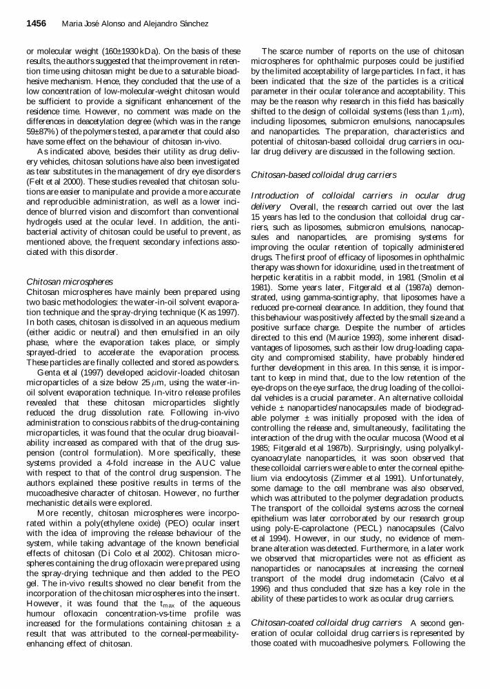
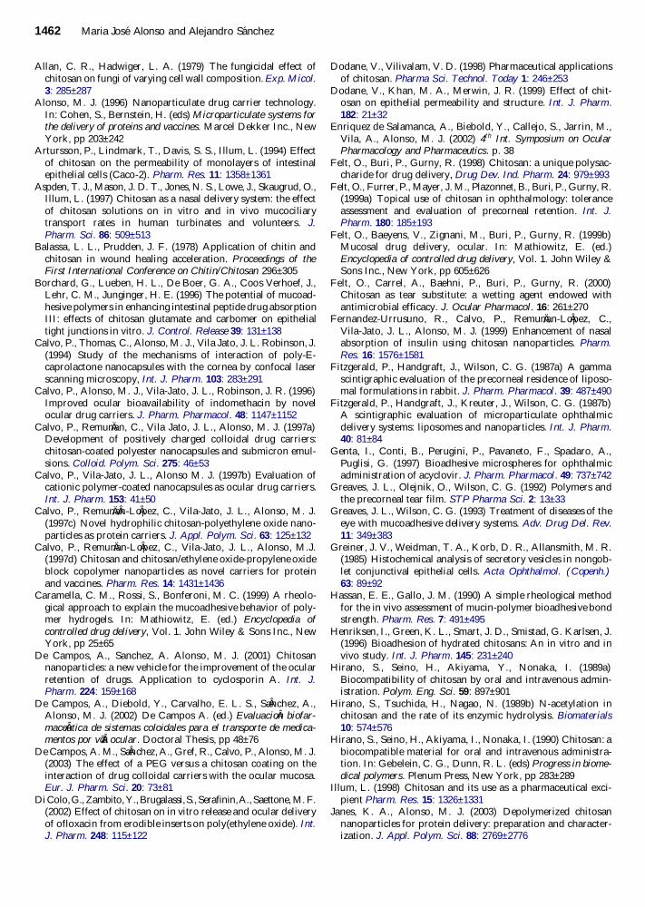