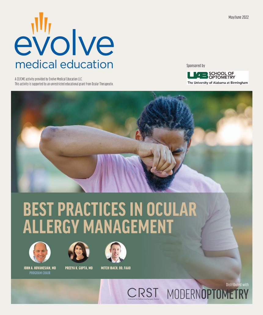
BEST PRACTICES IN OCULAR ALLERGY MANAGEMENT
16
JOHN A. HOVANESIAN, MD PROGRAM CHAIR PREEYA K. GUPTA, MD MITCH IBACH, OD, FAAO BEST PRACTICES IN OCULAR ALLERGY MANAGEMENT May/June 2022 Distributed with Sponsored by A CE/CME activity provided by Evolve Medical Education LLC. This activity is supported by an unrestricted educational grant from Ocular Therapeutix.
-
Upload
khangminh22 -
Category
Documents
-
view
5 -
download
0
Transcript of BEST PRACTICES IN OCULAR ALLERGY MANAGEMENT
JOHN A. HOVANESIAN, MD PROGRAM CHAIR
PREEYA K. GUPTA, MD MITCH IBACH, OD, FAAO
BEST PRACTICES IN OCULAR ALLERGY MANAGEMENT
May/June 2022
Distributed with
Sponsored by
A CE/CME activity provided by Evolve Medical Education LLC. This activity is supported by an unrestricted educational grant from Ocular Therapeutix.
2 SUPPLEMENT TO CATARACT & REFRACTIVE SURGERY TODAY AND MODERN OPTOMETRY | MAY/JUNE 2022
Faculty John A. Hovanesian, MD Program ChairClinical InstructorJules Stein Eye InstituteUniversity of California, Los AngelesOwnerHarvard Eye AssociatesLaguna Hills, CA
Preeya K. Gupta, MDManaging Director Triangle Eye ConsultantsCary, NC
Mitch Ibach, OD, FAAOCataract, Cornea, Refractive, and Glaucoma Surgery SpecialistVance Thompson VisionSioux Falls, SD
Content Source This continuing education (CE/CME) activity captures content
from a live virtual symposium.
Activity DescriptionThis supplement summarizes an expert panel discussion on ocular
allergy classification and diagnosis, a review of current and future treatments, as well as the correlation between dry eye disease and ocular allergy.
Target AudienceThis certified CE/CME activity is designed for optometrists and gen-
eral ophthalmologists.
Learning ObjectivesUpon completion of this activity, the participant should be able to:• Differentiate between the various types and stages of ocular
allergies based on presentation
• Create individualized management options for ocular allergy based on disease severity, patient factors, and associated risks and benefits
• Describe the limitations of current treatment options for ocular allergy
• Evaluate new management options for ocular allergy
Grantor StatementThis activity is supported by an unrestricted educational grant from
Ocular Therapeutix.
Accreditation StatementEvolve Medical Education LLC (Evolve) is accredited by the
Accreditation Council for Continuing Medical Education (ACCME) to provide continuing medical education for physicians.
Evolve is a COPE-accredited administrator.
Credit Designation StatementEvolve Medical Education designates this enduring material for a
maximum of 1 AMA PRA Category 1 Credit™. Physicians should claim only the credit commensurate with the extent of their participation in the activity.
This activity, COPE Activity Number 123744, is accredited by COPE for continuing education for optometrists. This course is approved for 1.0 hour CE.
Course #77999-TDActivity #123744
To Obtain CreditTo obtain credit for this activity, you must read the activity in
its entirety and complete the Pretest/Posttest/Activity Evaluation/Satisfaction Measures Form, which consists of a series of multiple-choice questions. To answer these questions online and receive real-time results, go to https://evolvemeded.com/course/2160-supp. Upon completing the activity and self-assessment test, your certifi-cate will be available. Alternatively, please complete the Posttest/Activity Evaluation/Satisfaction Form and mail or fax to Evolve
Release Date: April 27, 2022CME Expiration Date: July 1, 2023COPE Expiration Date: April 30, 2023
BEST PRACTICES IN OCULAR ALLERGY MANAGEMENT
MAY/JUNE 2022 | SUPPLEMENT TO CATARACT & REFRACTIVE SURGERY TODAY AND MODERN OPTOMETRY 3
Medical Education LLC, 353 West Lancaster Avenue, Second Floor, Wayne, PA 19087; Fax: (215) 933-3950.
Disclosure Policy It is the policy of Evolve that faculty and other individuals who are
in the position to control the content of this activity disclose any real or apparent financial relationships relating to the topics of this edu-cational activity. Evolve has full policies in place that will identify and mitigate all financial relationships prior to this educational activity.
The following faculty/staff members have the following financial relationships with ineligible companies:
John A. Hovanesian, MD, has had a financial agreement or affiliation with the following ineligible companies in the form of Consultant: 1-800-DOCTORS, Abbott Medical Optics, Acufocus, Aerie Pharmaceuticals, Alcon, Allegro Ophthalmics, Allergan, BlephEx, Eyedetec, Glaukos, Guardion Health Sciences, IOP/Katena, Ivantis, Kala Pharmaceuticals, Novartis, Ocular Therapeutix, Omeros, ReVision Optics, Sarentis Ophthalmics, Sensimed, Shire, Tear Film Innovations, TearLab, Valeant, Veracity, and Vindico Medical Education. Advisory Board: 1-800-DOCTORS, Abbott Medical Optics, Aerie Pharmaceutical, BlephEx, Cord LLC, Eyedetec, Glaukos, Guardion Health Sciences, Ingenoeye, IOP/Katena, Ivantis, Kala Pharmaceuticals, MDbackline, Ocular Therapeutix, Omeros, ReVision Optics, Shire, Sight Sciences, Tear Film Innovations, TearLab, Valeant, and Veracity. Grant/Research Support: Abbott Medical Optics, Acufocus, Aerie Pharmaceutical, Alcon, Cloudbreak Therapeutics, Cord, Eyedetec, Glaukos, Ingenoeye, IOP/Katena, Novartis, Ocular Therapeutix, Omeros, ReVision Optics, Shire, Tear Film Innovations, and Valeant. Stock/Shareholder: Alcon, Alicia Surgery Center, Allegro Ophthalmics, Allergan, BlephEx, Eyedetec, Glaukos, Guardion Health Sciences, Harvard Eye Associates, Harvard Hearing, Ingenoeye, MDbackline, Novartis, Ocular Therapeutix, Sarentis Ophthalmics, Sight Sciences, and Tear Film Innovations. Royalties: Slack Books and The Laser Center.
Preeya K. Gupta, MD, has had a financial agreement or affiliation
with the following ineligible companies in the form of Consultant: Alcon, Aldeyra, Allergan, Azura, Carl Zeiss Meditec, Expert Opinion, HanAll Biopharma, Johnson & Johnson Vision, Kala Pharmaceuticals, New World Medical, Novartis, Ocular Science, Ocular Therapeutix, Orasis, Oyster Point, Sight Sciences, Spyglass, Surface Ophthalmics, Sun Pharmaceutical Industries, Tear Clear, TearLab, Tissue Tech, and Visionology. Stock/Shareholder: Azura, Expert Opinion, Orasis, Oyster Point, Tarsus, Tear Clear, Surface, Spyglass, Visionology, and Visant.
Mitch Ibach, OD, FAAO, has had a financial agreement or affilia-tion with the following ineligible companies in the form of Consultant: Aerie Pharmaceuticals, Alcon, Allergan, Dompé, Gauch Health, Glaukos, Heru, Kala Pharmaceuticals, Ocular Therapeutix, and Oyster Point. Grant/Research Support: Ocular Therapeutix. Speakers’ Bureau: Aerie Pharmaceuticals, Dompé, Glaukos, Heru, Ocular Therapeutix, and Oyster Point. Stock/Shareholder: Equinox.
Editorial Support DisclosuresThe Evolve staff, planners, reviewer, and writers have no financial
relationships with ineligible companies.
Off-Label StatementThis educational activity may contain discussion of published and/
or investigational uses of agents that are not indicated by the FDA. The opinions expressed in the educational activity are those of the faculty. Please refer to the official prescribing information for each product for discussion of approved indications, contraindications, and warnings.
DisclaimerThe views and opinions expressed in this educational activity are
those of the faculty and do not necessarily represent the views of Evolve, Cataract & Refractive Surgery Today, Modern Optometry, or Ocular Therapeutix.
Digital EditionTo view the online version of the material, log in to your Evolve
account and go to https://evolvemeded.com/course/2160-supp or scan the QR code with your smartphone’s camera.
To view the webinar associated with this supplement, log in to your Evolve account and go to: https://evolvemeded.com/course/2160-ocular-allergy-enduring?page=1.
BEST PRACTICES IN OCULAR ALLERGY MANAGEMENT
1. Please rate your confidence in your ability to evaluate new management options for ocular allergy and create individualized management strategies (based on a scale of 1 to 5, with 1 being not at all confident and 5 being extremely confident).
a. 1b. 2c. 3d. 4e. 5
2. What percentage of US citizens test positive for at least one allergen?a. ~25%b. ~35%c. ~45%d. ~55%
3. A 45-year-old patient presents to your clinic with red, itchy, watery eyes. She notes she recently adopted a cat. All of the following are reasonable treatment options for this patient EXCEPT?
a. Allergy testing and avoidance of allergen b. Brimonidine eye drops c. Antihistamine eye dropsd. Lubricant eye drops
4. A 35-year-old man with seasonal allergies presents to your clinic with signs of allergic conjunctivitis. Which of the following treatments has been found to be clinically superior in relief of signs and symptom plus tolerability?
a. Topical corticosteroidsb. Topical NSAIDsc. Topical antihistaminesd. Topical dual-activity agents
5. A 76-year-old woman presents to your clinic with uncomfortable, burning, itching eyes. She has a history of chronic ocular surface disease with meibomian gland dysfunction. On exam, she has moderate inferior PEK OU, as well as 2-3+ NS OU. All of the following are reasonable treatment options for this patient except?
a. Aggressive lubrication with frequent artificial tears and ointmentb. Dexamethasone intracanalicular insertc. Schedule for cataract surgeryd. Short course of loteprednol
6. All of the following are cardinal symptoms that characterize allergic conjunctivitis, EXCEPT:
a. Rednessb. Itchingc. Tearingd. Corneal subepithelial infiltrates
7. An 8-year-old boy presents to your clinic with a history of seasonal allergies. He notes itchy, watery eyes. On exam, you note gelatinous-appearing raised bumps at the limbus. What is the most likely diagnosis?
a. Vernal keratoconjunctivitisb. Chemical conjunctivitisc. Viral conjunctivitisd. Bacterial conjunctivitis
8. The allergic cascade begins when ____ binds to mast cells, which launches a cascade of events.
a. IgDb. IgEc. IgGd. IgM
9. You are seeing a 76-year-old patient with severe allergic conjunctivitis. She has decreased manual dexterity and difficulty administering drops. You determine she needs therapy with corticosteroids. Which of the following is a reasonable treatment option for this patient?
a. Dexamethasone 0.4 mg intracanalicular insert b. Topical corticosteroid dropsc. Oral prednisoned. Intravitreal triamcinolone
10. You are seeing a 23-year-old white woman in your office who has chronic allergic conjunctivitis. She has a habit of frequently rubbing her eyes with her knuckles. She is currently on topical ketotifen 0.025%. What is this patient at risk for?
a. Shield ulcerb. Horner-Trantas Dots c. Keratoconusd. Recurrent corneal erosions
11. You are seeing a 55-year-old man with mild allergic conjunctivitis. You start him on ketotifen. What is the ideal dosing of this drug?
a. Once dailyb. Twice dailyc. Three times dailyd. Four times daily
12. You are seeing a 63-year-old woman who has seasonal allergic conjunctivitis with significant punctate epithelial erosions. You recommend steroid therapy and discuss loteprednol 0.2% suspension and the dexamethasone 0.4 mg intracanalicular insert as potential options. The patient asks you about the relative difference between the two, in terms of improving her corneal erosions. Which of the following statements is true?
a. Both the dexamethasone intracanalicular insert and loteprednol suspension significantly reduce corneal staining
b. Dexamethasone intracanalicular insert reduces corneal staining more than loteprednol suspension
c. Loteprednol suspension reduces corneal staining more than dexa-methasone intracanalicular insert
d. Neither dexamethasone intracanalicular insert nor loteprednol suspension had significant impact on corneal staining
PRETEST QUESTIONSPlease complete prior to accessing the material and submit with Posttest/Activity Evaluation/Satisfaction Measures for credit.
4 SUPPLEMENT TO CATARACT & REFRACTIVE SURGERY TODAY AND MODERN OPTOMETRY | MAY/JUNE 2022
BEST PRACTICES IN OCULAR ALLERGY MANAGEMENT
MAY/JUNE 2022 | SUPPLEMENT TO CATARACT & REFRACTIVE SURGERY TODAY AND MODERN OPTOMETRY 5
Ocular allergy is extremely common, affecting one in five people in the United States, yet it is often underdiagnosed and undertreated.1 Allergic diseases have increased in recent
years, particularly in the context of the COVID-19 pandemic where people were spending an inordinate amount of time at home, exposed to indoor allergic triggers.2,3 No two patients with ocular allergy are alike, therefore, diagnosis and treatment must be personalized. A robust understanding of the immunologic mechanisms at play, clinical features, and current and emerging treatments are critical to optimal patient care. In the following activity, experts in the ocular allergy space discuss its stages, keys to a differential diagnosis, the role of ocular surface disease, and new developments in sustained-release treatments.
—John A. Hovanesian, MD, Program Chair
OCULAR ALLERGY BASICSIncidence and Triggers
Dr. Hovanesian: Allergy is incredibly common. About 55% of all Americans test positive for at least one allergen, and 50 million Americans may have meaningful allergic disease.1 Most people will have an ocular component to their allergic disease because the eyes are exposed to environmental factors. Allergies are the sixth leading cause of chronic illness in the United States, with a total cost on the order of $18 billion, which is shocking.4 It’s also shocking that ocular allergy specifically represents a $4.3 billion industry.
Four cardinal symptoms characterize allergic conjunctivitis: redness, itching, swelling, and tearing, most of which overlap with dry eye disease (DED).5 Often the tearing is not just an overproduction of tears; you get such boggy chemosis that it actually blocks the tear punctum, so that you don’t get drain-age through the lacrimal system. Patients can also be sensitive to light. If accompanied by nasal allergies, symptoms include a stuffy or itchy nose, sneezing, headache, itchy or sore throat, and coughing.5 Dr. Ibach, is this representative of the kind of symp-toms you see with your patients?
Mitch Ibach, OD, FAAO: Yes, I think these are the big ones. For ocular allergy, I always think of itchy, watery eyes.
Dr. Hovanesian: Itch is such a prominent component that it is almost always indictive of allergy. Common indoor and outdoor allergy triggers include animal dander, cockroaches,
cosmetics/perfume, medications, mold, pollen, and smoke, among other things.4 I am surprised that cockroaches are so high on the list of triggers. Dr. Gupta, you’ve treated a lot of ocu-lar surface disease (OSD). What do you think about cosmetics and their role, particularly in women as they relate to allergy?
Preeya K. Gupta, MD: I think the most complicated part of cosmetics is that it’s not one product. Blush, eye shadow, eye liner, and mascara can include upwards of 40 ingredients. Patients with certain skin conditions like rosacea, for example, can be sensitive to certain metals like nickel. Both chemical and metallic components can trigger allergies. Because the cause is not always obvious, I’ve sent patients for allergy testing to try to determine their trigger.
Dr. Hovanesian: The incidence of allergies may be on the rise due to factors from the COVID-19 pandemic.2 Many of us were contained in our houses for months, and many people still work from home. Interestingly, time spent at home may exacerbate ocular allergy symptoms among patients affected by indoor air pollution from poor ventilation.2 Dust mites, for example, are dif-ficult to eradicate. Scented candles are another issue. A 2015 study found that certain scented candle products are potent sources of volatile organic compounds emission in an indoor environment, regardless of whether they are lit.6
Seasonal and Perennial Allergic Conjunctivitis Dr. Hovanesian: Let’s move on to the two main categories of
allergic conjunctivitis: seasonal and perennial.7 Seasonal allergic conjunctivitis (SAC) is the most common type of ocular allergy, occurring in the spring, summer, or fall in response to pollen or mold, each of which have their own seasons.7 Perennial allergic conjunctivitis (PAC) occurs year-round in response to household allergens such as dust mites, mold, and pet dander.7 In my expe-rience, the symptoms are very similar. To help determine if it’s SAC or PAC, it’s helpful to ask the patient when they have the symptoms. Chronic or recurrent varieties of PAC may progress and lead to serious ocular complications. Interestingly, seasonal spikes have been noted in up to 80% of patients with PAC.8 If I want to know whether it’s seasonal versus perennial, I ask the patient when they have their symptoms.
There are also other very distinct types of allergies that affect about 2% of patients with ocular allergies.1,9 Atopic keratoconjunc-tivitis can be blinding; it’s a much more complicated issue than a
BEST PRACTICES IN OCULAR ALLERGY MANAGEMENT
BEST PRACTICES IN OCULAR ALLERGY MANAGEMENT
6 SUPPLEMENT TO CATARACT & REFRACTIVE SURGERY TODAY AND MODERN OPTOMETRY | MAY/JUNE 2022
typical type I autoimmune response. It can cause permanent scar-ring of the cornea that is very difficult to treat. Even corneal trans-plants don’t work well in these vascularized, chronically inflamed eyes. Typically, these patients do have other atopy symptoms such as allergy-driving asthma or other skin disorders related to it.
Vernal keratoconjunctivitis occurs in children, typically males aged 8 to 10 years, sometimes a little older. It’s also typically seasonal, lasting from the beginning of spring until autumn.9 It is characterized by massive infiltration of T cells, macrophages, neutrophils, and especially eosinophils.10 The pathophysiology is poorly understood; it is considered a severe persistent reaction. These patients will have the classic Horner-Trantas dots at the limbus—tiny little white dots that can also accompany these limbal papillae that are very gelatinous, raised-looking bumps.
In the case of limbal vernal keratoconjunctivitis, a subtype, it’s around the limbus. In the case of palpebral vernal keratocon-junctivitis, they tend to be under the upper lid. The most aggres-sive form of this can be in the form of a shield ulcer, which can cause corneal erosions that are very difficult to treat. But this occurs in a small minority of allergy patients.
Dr. Gupta, when you see vernal or atopic keratoconjunctivitis, what’s the first treatment you reach for?
Dr. Gupta: I reach for a steroid. Anterior segment specialists are very comfortable using topical steroids. As you mentioned earlier, atopic and vernal keratoconjunctivitis can be intensely inflammatory and aggressive with long-term consequences. When I’m considering a steroid, I consider if the patient is at risk for meibomian gland damage or damage to the conjunctiva, the lid margin, or cornea. For example, limbal papillae can cause del-len and degradation of the cornea. Chronic inflammation can lead to meibomian gland atrophy as well. I take these things into consideration when I’m establishing the urgency. Steroids can be very safe molecules when used and monitored appropriately. It’s the go-to molecule when you need something to work quickly.
PHYSIOLOGIC RESPONSE TO OCULAR ALLERGY Dr. Hovanesian: The allergic cascade is so-called because it
starts, by definition, with an allergy to an antigen that creates the series of events that we see as clinical manifestation.11 It’s important to note that each patient will react differently to an allergen and to allergy treatment. Allergic tendency is genetically passed on.12 Dr. Ibach, please take us through the body’s physi-ologic response to an allergen and how we manage it.
Dr. Ibach: I like to think of the allergic cascade as a waterfall. There are three main phases: sensitization, early phase, and late phase.11,12 Sensitization is the time point of allergen exposure. This is when this unwanted allergen enters the body and we become sensitized to it.
That leads to the early phase, ie, the acute symptoms. This is when we have watery, itchy eyes; sneezing; and maybe bron-chial constriction, which makes it difficult to breathe. The last
phase, the late phase, is a more inflammatory cascade. It involves chronic inflammation and tissue damage.
The allergic cascade starts when an allergen comes into our body. We have the initial exposure, and then we have the forma-tion of immunoglobulin E (IgE). This is our body’s antibody that is produced in conjunction with and against allergies. Sensitization starts when IgE connects to and binds to mast cells, which launch-es a cascade of events. T helper 2 (Th2) cells release proinflam-matory cytokines (interleukin 3 [IL]-3, IL-4, IL-5, and IL-13) that stimulate IgE production by B cells. We have proinflammatory cytokines, and we have more IgE. We’re building a snowball of IgE that’s rolling down the hill. IgE further becomes bound to mast cells, and we have a process called degranulation.8,10,13,14
Simplified, degranulation is the breakdown of the mast cell, which results in the release of the contents inside that cell. Think of the mast cells in the allergic cascade as signalers; they’re send-ing out mediators and signals to our body to respond.14 Once degranulation happens, we have the release of histamine and tryptase. I think of histamine as our body’s defenses against the allergen; they’re the bouncers. They’re trying to get the allergen out of our body through coughing, sneezing, and itching.15
We also have newly formed inflammatory mediators, leukotri-enes and prostaglandins.14 The allergic cascade after sensitization starts to move into an inflammatory cascade as well.
The early phase of the allergic cascade occurs seconds to min-utes after allergen exposure and lasts 20 to 30 minutes.14 This is the acute symptomatic phase, which presents as watery, itchy eyes for ocular allergy. Itching is the hallmark. In the early phase, mast cells are extremely active.15 They become degranulated. They’re releasing all these signalers, and it’s probably an over-signaling. If our body would calm down a bit, we’d probably be okay. But the mast cell release of mediators cause symptoms such as itching, redness, tearing, chemosis, and a papillary reaction. As that system continues to run, the cycle becomes negative.
The late phase begins a few hours after allergen exposure and is characterized by epithelial infiltration of inflammatory cells like neutrophils, lymphocytes, basophils, and eosinophils.14,16 We’re transitioning from an allergic cascade to a chronic inflam-matory cascade, which will lead to long-term inflammation and tissue damage. As the reaction progresses, hypersecretion of tears increases drainage through the lacrimal ducts, carrying allergens directly into the nasal passage.15 We start making all of these tears, but the tears are full of allergens and inflammatory markers.
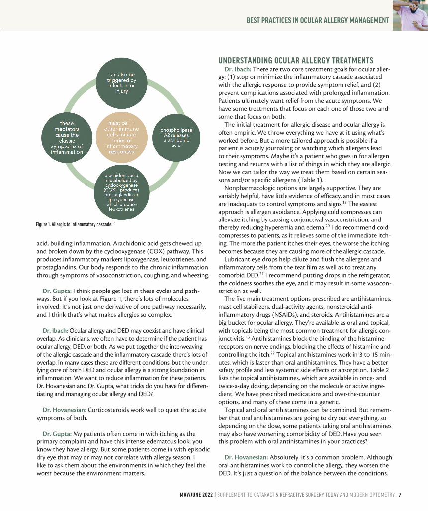
What do we want to do when we’re having that hypersecre-tion and acute symptom? We rub the eyes. That feels good tem-porarily, but it stirs up more inflammation and more allergens in the body. Figure 1 depicts the allergic cascade that then moves to the inflammatory cascade.17
The mast cell is the major signaling cell in the allergic path-way. When the mast cell becomes degranulated, it releases histamine, the bouncers, to get rid of the allergen. This starts the inflammatory cascade. Phospholipase A2, which is really just an immune inflammatory signaler, releases arachidonic
BEST PRACTICES IN OCULAR ALLERGY MANAGEMENT
MAY/JUNE 2022 | SUPPLEMENT TO CATARACT & REFRACTIVE SURGERY TODAY AND MODERN OPTOMETRY 7
acid, building inflammation. Arachidonic acid gets chewed up and broken down by the cyclooxygenase (COX) pathway. This produces inflammatory markers lipoxygenase, leukotrienes, and prostaglandins. Our body responds to the chronic inflammation through symptoms of vasoconstriction, coughing, and wheezing.
Dr. Gupta: I think people get lost in these cycles and path-ways. But if you look at Figure 1, there’s lots of molecules involved. It’s not just one derivative of one pathway necessarily, and I think that’s what makes allergies so complex.
Dr. Ibach: Ocular allergy and DED may coexist and have clinical overlap. As clinicians, we often have to determine if the patient has ocular allergy, DED, or both. As we put together the interweaving of the allergic cascade and the inflammatory cascade, there’s lots of overlap. In many cases these are different conditions, but the under-lying core of both DED and ocular allergy is a strong foundation in inflammation. We want to reduce inflammation for these patients. Dr. Hovanesian and Dr. Gupta, what tricks do you have for differen-tiating and managing ocular allergy and DED?
Dr. Hovanesian: Corticosteroids work well to quiet the acute symptoms of both.
Dr. Gupta: My patients often come in with itching as the
primary complaint and have this intense edematous look; you know they have allergy. But some patients come in with episodic dry eye that may or may not correlate with allergy season. I like to ask them about the environments in which they feel the worst because the environment matters.
UNDERSTANDING OCULAR ALLERGY TREATMENTS Dr. Ibach: There are two core treatment goals for ocular aller-
gy: (1) stop or minimize the inflammatory cascade associated with the allergic response to provide symptom relief, and (2) prevent complications associated with prolonged inflammation. Patients ultimately want relief from the acute symptoms. We have some treatments that focus on each one of those two and some that focus on both.
The initial treatment for allergic disease and ocular allergy is often empiric. We throw everything we have at it using what’s worked before. But a more tailored approach is possible if a patient is acutely journaling or watching which allergens lead to their symptoms. Maybe it’s a patient who goes in for allergen testing and returns with a list of things in which they are allergic. Now we can tailor the way we treat them based on certain sea-sons and/or specific allergens (Table 1).
Nonpharmacologic options are largely supportive. They are variably helpful, have little evidence of efficacy, and in most cases are inadequate to control symptoms and signs.13 The easiest approach is allergen avoidance. Applying cold compresses can alleviate itching by causing conjunctival vasoconstriction, and thereby reducing hyperemia and edema.20 I do recommend cold compresses to patients, as it relieves some of the immediate itch-ing. The more the patient itches their eyes, the worse the itching becomes because they are causing more of the allergic cascade.
Lubricant eye drops help dilute and flush the allergens and inflammatory cells from the tear film as well as to treat any comorbid DED.21 I recommend putting drops in the refrigerator; the coldness soothes the eye, and it may result in some vasocon-striction as well.
The five main treatment options prescribed are antihistamines, mast cell stabilizers, dual-activity agents, nonsteroidal anti-inflammatory drugs (NSAIDs), and steroids. Antihistamines are a big bucket for ocular allergy. They’re available as oral and topical, with topicals being the most common treatment for allergic con-junctivitis.13 Antihistamines block the binding of the histamine receptors on nerve endings, blocking the effects of histamine and controlling the itch.22 Topical antihistamines work in 3 to 15 min-utes, which is faster than oral antihistamines. They have a better safety profile and less systemic side effects or absorption. Table 2 lists the topical antihistamines, which are available in once- and twice-a-day dosing, depending on the molecule or active ingre-dient. We have prescribed medications and over-the-counter options, and many of these come in a generic.
Topical and oral antihistamines can be combined. But remem-ber that oral antihistamines are going to dry out everything, so depending on the dose, some patients taking oral antihistamines may also have worsening comorbidity of DED. Have you seen this problem with oral antihistamines in your practices?
Dr. Hovanesian: Absolutely. It’s a common problem. Although oral antihistamines work to control the allergy, they worsen the DED. It’s just a question of the balance between the conditions.
Figure 1. Allergic to inflammatory cascade.17
BEST PRACTICES IN OCULAR ALLERGY MANAGEMENT
8 SUPPLEMENT TO CATARACT & REFRACTIVE SURGERY TODAY AND MODERN OPTOMETRY | MAY/JUNE 2022
Dr. Gupta: I agree; I’m often negotiating the dosing of oral antihistamines. I have some patients report that certain oral antihistamines are less drying to them than others, so I recom-mend patients try different ones.
Dr. Ibach: Dual-activity agents combine a topical antihistamine with a topical mast cell stabilizer. Compared with either antihis-tamines or mast cell stabilizers alone, topical dual-activity agents are generally clinically superior due to both symptom/sign relief and tolerability.13,18 These work in different pathways or arenas, and they also take effect at different times. The topical antihista-mine treats the acute symptoms. The topical mast cell stabilizer takes longer to work because you’re trying to prevent degranula-tion of that mast cell. You’re trying to stop the allergic cascade really at the very top rather than just treating the symptoms.19
But remember, these agents are very commonly preserved with benzalkonium chloride, a surfactant that may cause ocular surface toxicity and worsen OSD and DED.
Table 3 lists topical combinations. All three of our current options have the same active ingredients but at different con-centrations. They are available as once- or twice-daily dosing. Olopatadine 0.7 mg used to be prescription only, but it recently became available over-the-counter.23
Dr. Hovanesian: These are inexpensive, highly effective anti-histamines. They used to be quite expensive when they were prescription only.
Dr. Gupta: The topical combinations with antihistamine and mast cell stabilizer are the same active ingredient with different doses, which can confuse patients. Navigating that with your patients can be helpful, so they know what to get when they go to the aisle and how to safely use the medication.
Dr. Ibach: Topical NSAIDs will not be your first-line of defense
for ocular allergy, and you’re not going to commonly use it as a standalone agent. NSAIDs specifically inhibit COX activity without suppressing the immune system. They are useful for
controlling pain and reducing inflammation. We’re trying to calm down inflammation when we go this route. Ketorolac trometh-amine can be given four times a day or twice a day. These are very different indications, so you must know what you’re dosing for. It’s only available as a prescription, and there is a generic.
WHEN TO USE OCULAR STEROIDS Dr. Gupta: Ocular steroids are an important part of treating the
inflammatory process. Allergic conjunctivitis and allergic disease as a whole require the use of topical steroids in most patients at some point in their disease course. Ocular steroids block the inflammatory pathways that lead to the release of those immune cells.18,24 It can be used in isolation or as an adjunct therapy.
Some clinicians save steroid use for severe cases. I agree that not every mild case needs steroids, but you really want to weigh the risks of not treating in certain types of allergic patients. I look at the chronicity of their disease. Is it seasonal? Is it episodic? If a patient has a high need for a rapid resolution of symptoms, then steroids are an appropriate first-line treatment course. However, we are hesitant to prescribe steroids to every patient in the clinic because of the potential side effects like elevated eye pressure and cataract formation.19
Table 4 shows the currently approved and available topical steroids. Regardless of the type of molecule, these agents are approved with different dosing. How we use these steroids in our own daily clinic is variable; we all use different prescribing profiles.
Dr. Hovanesian: These agents work well, but cost is often an issue. If you touch the eye with steroid, you get improvement with allergies regardless of the specific agent used.
Dr. Gupta: Do we undertreat patients with topical steroids? We all know that patients will feel better in a couple of days, but
TABLE 1. Treatment Approaches for Ocular Allergy Management13,18-21
NONPHARMACOLOGIC PHARMACOLOGIC
Allergen avoidance Antihistamines
Cold compresses Mast cell stabilizers
Lubricant eye drops Topical dual-activity agents
Wraparound sunglasses NSAIDs
Corticosteroids
Abbreviation: NSAIDs, nonsteroidal anti-inflammatory drugs.
TABLE 2. Topical Antihistamines
ACTIVE INGREDIENT
DOSING MANUFACTURER GENERIC RX OR OTC
Cetirizine BID Eyevance No Rx
Alcaftadine QD Allergan No OTC
Bepotastin BID Bausch Health No Rx
Epinastine BID Allergan Yes Rx
Zelastine BID Mylan Yes Rx
Ketotifen BID Alcon/Novartis Yes OTC
Ketotifen BID Bausch Health Yes OTC
Abbreviations: BID, twice a day; OTC, over the counter; QD, once a day; Rx, prescription.
BEST PRACTICES IN OCULAR ALLERGY MANAGEMENT
MAY/JUNE 2022 | SUPPLEMENT TO CATARACT & REFRACTIVE SURGERY TODAY AND MODERN OPTOMETRY 9
I often wonder if patients can have a prolonged, safe exposure to steroids, whether it’s 10 days, 2 weeks, or a month. The immune system stays heightened and slightly more activated for a prolonged time once exposed or re-exposed to an allergen. Sometimes, especially if a patient is relapsing every few weeks, I’ll prescribe a longer course of steroids because I do think it will improve their overall relapse situation.
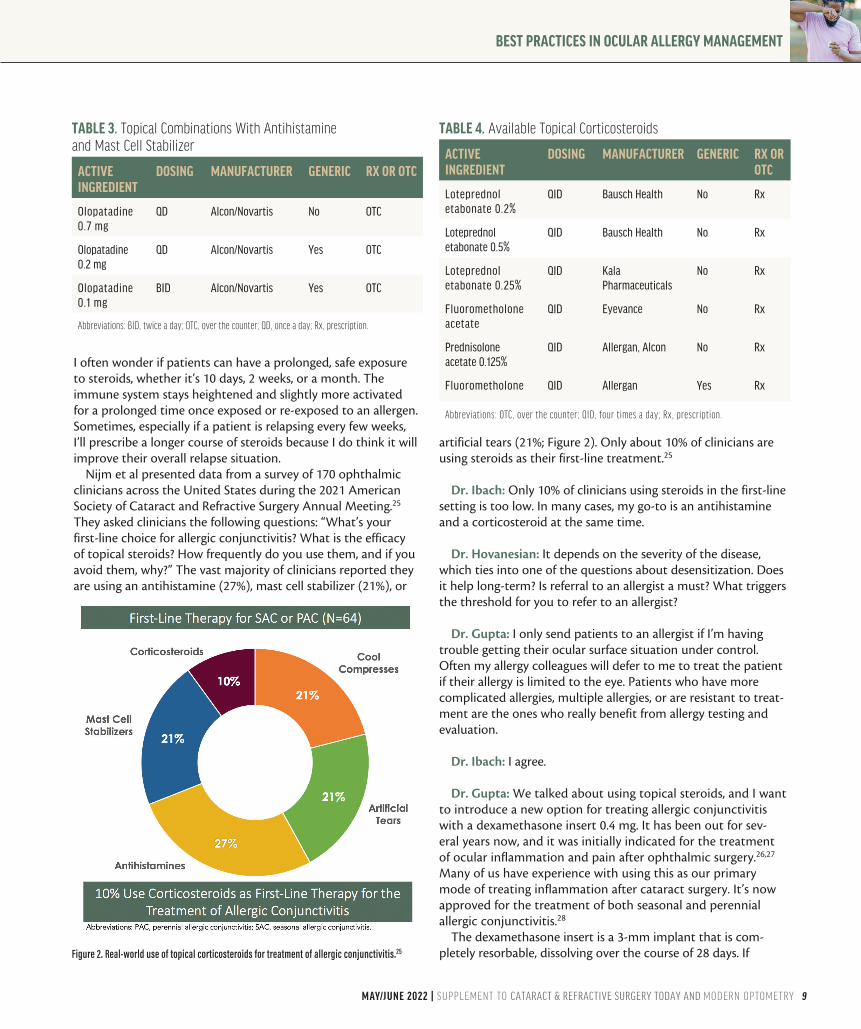
Nijm et al presented data from a survey of 170 ophthalmic clinicians across the United States during the 2021 American Society of Cataract and Refractive Surgery Annual Meeting.25 They asked clinicians the following questions: “What’s your first-line choice for allergic conjunctivitis? What is the efficacy of topical steroids? How frequently do you use them, and if you avoid them, why?” The vast majority of clinicians reported they are using an antihistamine (27%), mast cell stabilizer (21%), or
artificial tears (21%; Figure 2). Only about 10% of clinicians are using steroids as their first-line treatment.25
Dr. Ibach: Only 10% of clinicians using steroids in the first-line setting is too low. In many cases, my go-to is an antihistamine and a corticosteroid at the same time.
Dr. Hovanesian: It depends on the severity of the disease, which ties into one of the questions about desensitization. Does it help long-term? Is referral to an allergist a must? What triggers the threshold for you to refer to an allergist?
Dr. Gupta: I only send patients to an allergist if I’m having trouble getting their ocular surface situation under control. Often my allergy colleagues will defer to me to treat the patient if their allergy is limited to the eye. Patients who have more complicated allergies, multiple allergies, or are resistant to treat-ment are the ones who really benefit from allergy testing and evaluation.
Dr. Ibach: I agree.
Dr. Gupta: We talked about using topical steroids, and I want to introduce a new option for treating allergic conjunctivitis with a dexamethasone insert 0.4 mg. It has been out for sev-eral years now, and it was initially indicated for the treatment of ocular inflammation and pain after ophthalmic surgery.26,27 Many of us have experience with using this as our primary mode of treating inflammation after cataract surgery. It’s now approved for the treatment of both seasonal and perennial allergic conjunctivitis.28
The dexamethasone insert is a 3-mm implant that is com-pletely resorbable, dissolving over the course of 28 days. If
TABLE 3. Topical Combinations With Antihistamine and Mast Cell Stabilizer
ACTIVE INGREDIENT
DOSING MANUFACTURER GENERIC RX OR OTC
Olopatadine 0.7 mg
QD Alcon/Novartis No OTC
Olopatadine 0.2 mg
QD Alcon/Novartis Yes OTC
Olopatadine 0.1 mg
BID Alcon/Novartis Yes OTC
Abbreviations: BID, twice a day; OTC, over the counter; QD, once a day; Rx, prescription.
TABLE 4. Available Topical Corticosteroids
ACTIVE INGREDIENT
DOSING MANUFACTURER GENERIC RX OR OTC
Loteprednol etabonate 0.2%
QID Bausch Health No Rx
Loteprednol etabonate 0.5%
QID Bausch Health No Rx
Loteprednol etabonate 0.25%
QID Kala Pharmaceuticals
No Rx
Fluorometholone acetate
QID Eyevance No Rx
Prednisolone acetate 0.125%
QID Allergan, Alcon No Rx
Fluorometholone QID Allergan Yes Rx
Abbreviations: OTC, over the counter; QID, four times a day; Rx, prescription.
Figure 2. Real-world use of topical corticosteroids for treatment of allergic conjunctivitis.25
BEST PRACTICES IN OCULAR ALLERGY MANAGEMENT
10 SUPPLEMENT TO CATARACT & REFRACTIVE SURGERY TODAY AND MODERN OPTOMETRY | MAY/JUNE 2022
there is an issue, it can easily be removed with irrigation of the plug through the puncta or manual expression if necessary. It is designed to deliver a tapered dose, and contains fluorescein for visualization. It has no additional components or assembly required, and best of all, it is preservative-free. So many of our patients are sensitive to preservatives.29 Sparing that ocular sur-face, but still delivering that medication, has tremendous value in this patient population.
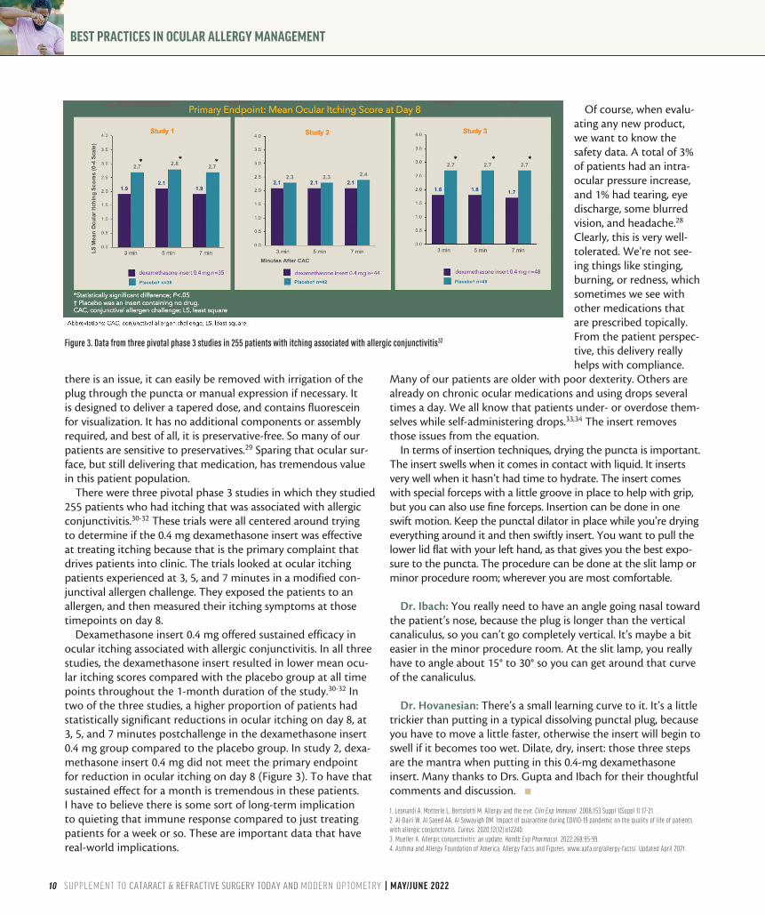
There were three pivotal phase 3 studies in which they studied 255 patients who had itching that was associated with allergic conjunctivitis.30-32 These trials were all centered around trying to determine if the 0.4 mg dexamethasone insert was effective at treating itching because that is the primary complaint that drives patients into clinic. The trials looked at ocular itching patients experienced at 3, 5, and 7 minutes in a modified con-junctival allergen challenge. They exposed the patients to an allergen, and then measured their itching symptoms at those timepoints on day 8.
Dexamethasone insert 0.4 mg offered sustained efficacy in ocular itching associated with allergic conjunctivitis. In all three studies, the dexamethasone insert resulted in lower mean ocu-lar itching scores compared with the placebo group at all time points throughout the 1-month duration of the study.30-32 In two of the three studies, a higher proportion of patients had statistically significant reductions in ocular itching on day 8, at 3, 5, and 7 minutes postchallenge in the dexamethasone insert 0.4 mg group compared to the placebo group. In study 2, dexa-methasone insert 0.4 mg did not meet the primary endpoint for reduction in ocular itching on day 8 (Figure 3). To have that sustained effect for a month is tremendous in these patients. I have to believe there is some sort of long-term implication to quieting that immune response compared to just treating patients for a week or so. These are important data that have real-world implications.
Of course, when evalu-ating any new product, we want to know the safety data. A total of 3% of patients had an intra-ocular pressure increase, and 1% had tearing, eye discharge, some blurred vision, and headache.28 Clearly, this is very well-tolerated. We’re not see-ing things like stinging, burning, or redness, which sometimes we see with other medications that are prescribed topically. From the patient perspec-tive, this delivery really helps with compliance.
Many of our patients are older with poor dexterity. Others are already on chronic ocular medications and using drops several times a day. We all know that patients under- or overdose them-selves while self-administering drops.33,34 The insert removes those issues from the equation.
In terms of insertion techniques, drying the puncta is important. The insert swells when it comes in contact with liquid. It inserts very well when it hasn’t had time to hydrate. The insert comes with special forceps with a little groove in place to help with grip, but you can also use fine forceps. Insertion can be done in one swift motion. Keep the punctal dilator in place while you’re drying everything around it and then swiftly insert. You want to pull the lower lid flat with your left hand, as that gives you the best expo-sure to the puncta. The procedure can be done at the slit lamp or minor procedure room; wherever you are most comfortable.
Dr. Ibach: You really need to have an angle going nasal toward the patient’s nose, because the plug is longer than the vertical canaliculus, so you can’t go completely vertical. It’s maybe a bit easier in the minor procedure room. At the slit lamp, you really have to angle about 15° to 30° so you can get around that curve of the canaliculus.
Dr. Hovanesian: There’s a small learning curve to it. It’s a little trickier than putting in a typical dissolving punctal plug, because you have to move a little faster, otherwise the insert will begin to swell if it becomes too wet. Dilate, dry, insert: those three steps are the mantra when putting in this 0.4-mg dexamethasone insert. Many thanks to Drs. Gupta and Ibach for their thoughtful comments and discussion. n
1. Leonardi A, Motterle L, Bortolotti M. Allergy and the eye. Clin Exp Immunol. 2008;153 Suppl 1(Suppl 1):17-21. 2. Al-Dairi W, Al Saeed AA, Al Sowayigh OM. Impact of quarantine during COVID-19 pandemic on the quality of life of patients with allergic conjunctivitis. Cureus. 2020;12(12):e12240. 3. Mueller A. Allergic conjunctivitis: an update. Handb Exp Pharmacol. 2022;268:95-99. 4. Asthma and Allergy Foundation of America. Allergy Facts and Figures. www.aafa.org/allergy-facts/. Updated April 2021.
Figure 3. Data from three pivotal phase 3 studies in 255 patients with itching associated with allergic conjunctivitis32
BEST PRACTICES IN OCULAR ALLERGY MANAGEMENT
MAY/JUNE 2022 | SUPPLEMENT TO CATARACT & REFRACTIVE SURGERY TODAY AND MODERN OPTOMETRY 11
Accessed March 20, 2022.5. Rodrigues J, Kuruvilla ME, Vanijcharoenkarn K, Patel N, Hom MM, Wallace DV. The spectrum of allergic ocular diseases. Ann Allergy Asthma Immunol. 2021;126(3):240-254. 6. Ahn JH, Kim KH, Kim YH, Kim BW. Characterization of hazardous and odorous volatiles emitted from scented candles before lighting and when lit. J Hazard Mater. 2015;286:242-251. 7. Varu DM, Rhee MK, Akpek EK, et al. Conjunctivitis Preferred Practice Pattern. Ophthalmology. 2019;126(1):P94-P169. 8. Bielory L. Allergic and immunologic disorders of the eye. Part II: ocular allergy. J Allergy Clin Immunol. 2000;106(6):1019-1032. 9. Ghiglioni DG, Zicari AM, Parisi GF, et al. Vernal keratoconjunctivitis: an update. Eur J Ophthalmol. 2021;31(6):2828-2842. 10. Roy N, Levanon S, Asbell PA. Potential Biomarkers for Allergic Conjunctival Diseases. Eye Contact Lens. 2020;46 Suppl 2(Suppl 2):S109-S121. 11. Bloemen K, Verstraelen S, Van Den Heuvel R, Witters H, Nelissen I, Schoeters G. The allergic cascade: review of the most important molecules in the asthmatic lung. Immunol Lett. 2007;113(1):6-18. 12. Galli SJ, Tsai M, Piliponsky AM. The development of allergic inflammation. Nature. 2008;454(7203):445-454. 13. Dupuis P, Prokopich CL, Hynes A, Kim H. A contemporary look at allergic conjunctivitis. Allergy Asthma Clin Immunol. 2020;16:5. 14. Leonardi A, De Dominicis C, Motterle L. Immunopathogenesis of ocular allergy: a schematic approach to different clinical entities. Curr Opin Allergy Clin Immunol. 2007;7(5):429-435. 15. Prokopich CL, Lee-Poyb M, Kimc H. Interprofessional management of allergic conjunctivitis. Canadian Journal of Optometry. 2018;80(3):11-27. 16. Small P, Kim H. Allergic rhinitis. Allergy Asthma Clin Immunol. 2011;7 Suppl 1(Suppl 1):S3. 17. AJMC. The emerging role of the type 2 inflammatory cascade in atopic diseases. www.ajmc.com/view/emerging-role-type-2-inflammatory-cascade-atopic-diseases. Published July 2, 2019. Accessed March 20, 2022.18. Leonardi A, Modugno RL, Salami E. Allergy and dry eye disease. Ocul Immunol Inflamm. 2021;29(6):1168-1176. 19. Bielory L, Duttachoudhury S, McMunn A. Bepotastine besilate for the treatment of pruritus. Expert Opin Pharmacother. 2013;14(18):2553-2569. 20. Sánchez-Hernández MC, Montero J, Rondon C, et al. Consensus document on allergic conjunctivitis (DECA). J Investig Allergol Clin Immunol. 2015;25(2):94-106.21. Bielory L, Meltzer EO, Nichols KK, Melton R, Thomas RK, Bartlett JD. An algorithm for the management of allergic conjuncti-vitis. Allergy Asthma Proc. 2013;34(5):408-420.
22. Simons FE, Simons KJ. Histamine and H1-antihistamines: celebrating a century of progress. J Allergy Clin Immunol. 2011;128(6):1139-1150.e4. 23. Alcon. Alcon announces FDA approval of the OTC switch of Pataday once daily relief extra strength. https://www.alcon.com/media-release/alcon-announces-fda-approval-otc-switch-pataday-once-daily-relief-extra-strength. Published July 14, 2020. Accessed March 20, 2022.24. Leonardi A, Silva D, Perez Formigo D, et al. Management of ocular allergy. Allergy. 2019;74(9):1611-1630. 25. Nijm L. Physician impressions on real world use of topical corticosteroids for the treatment of allergic conjunctivitis: need for a safer steroid. Presented at: 2021 American Society of Cataract and Refractive Surgery Annual Meeting. Las Vegas, NV.26. Walters T BS, Vold S, et al. Efficacy and safety of sustained release dexamethasone for the treatment of ocular pain and inflammation after cataract surgery: results from two phase 3 studies. J Clin Exp Ophthalmol. 2016;7(4). 27. Tyson SL, Bafna S, Gira JP, et al. Multicenter randomized phase 3 study of a sustained-release intracanalicular dexametha-sone insert for treatment of ocular inflammation and pain after cataract surgery [published correction appears in J Cataract Refract Surg. 2019 Jun;45(6):895]. J Cataract Refract Surg. 2019;45(2):204-212. 28. Ocular Therapeutix. Dextenza Prescribing Information. www.accessdata.fda.gov/drugsatfda_docs/label/2021/208742s007lbl.pdf. Updated October 2021. Accessed March 20, 2022. 29. Goldstein MH, Silva FQ, Blender N, Tran T, Vantipalli S. Ocular benzalkonium chloride exposure: problems and solutions. Eye (Lond). 2022;36(2):361-368. 30. Rubin JM SS, Kenyon KR, et al. Pooled analysis evaluating efficacy and safety of an intracanalicular dexamethasone insert for the treatment of allergic conjunctivitis. Presented at: 2021 American Society of Cataract and Refractive Surgery Annual Meeting. Las Vegas, NV.31. McLaurin EB, Evans D, Repke CS, et al. Phase 3 randomized study of efficacy and safety of a dexamethasone intracanalicular insert in patients with allergic conjunctivitis. Am J Ophthalmol. 2021;229:288-300. 32. Silverstein S. Efficacy and safety of an intracanalicular dexamethasone insert (0.4 mg) for the treatment of allergic conjunctivitis. Presented at: 2021 American Academy of Ophthalmology Annual Meeting. New Orleans, LA. 33. Matossian C. Noncompliance with prescribed eyedrop regimens among patients undergoing cataract surgery—prevalence, consequences, and solutions. US Ophthalmic Review. 2020;1:18-22.34. Wolfram C, Stahlberg E, Pfeiffer N. Patient-reported nonadherence with glaucoma therapy. J Ocul Pharmacol Ther. 2019;35(4):223-228.
Dr. Hovanesian: Our first case is a 67-year-old woman with uncomfortable burning and itching in both eyes. She has also had gradually declining vision for 6 months. She has a long history of OSD, meibomian gland disease, and aqueous deficiency. She has staining of her cornea and visually significant cataracts. What do you think about when you see a patient who needs cataract surgery but has meaningful OSD?
Dr. Gupta: To me, we can’t proceed with cataract surgery until the OSD is treated. There are countless papers showing that patients with OSD are more likely to have refractive miss, are more likely to be unhappy after surgery, and feel like you caused their problem.1 You must talk to your patient and help them understand there are two issues happening at once, so they don’t think everything is related to the cataract. Taking the time to treat the OSD before surgery is the right step.
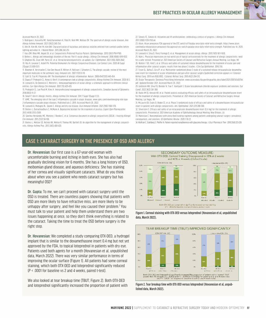
Dr. Hovanesian: We completed a study comparing OTX-DED, a hydrogel implant that is similar to the dexamethasone insert 0.4 mg but not yet approved by the FDA, to topical loteprednol in patients with dry eye. Patients used both agents for a month (Hovanesian et al, unpublished data, March 2022). There was very similar performance in terms of improving the ocular surface (Figure 1). All patients had some corneal staining, which both OTX-DED and loteprednol significantly reduced (P < .0001 for baseline vs 2 and 4 weeks, paired t-test).
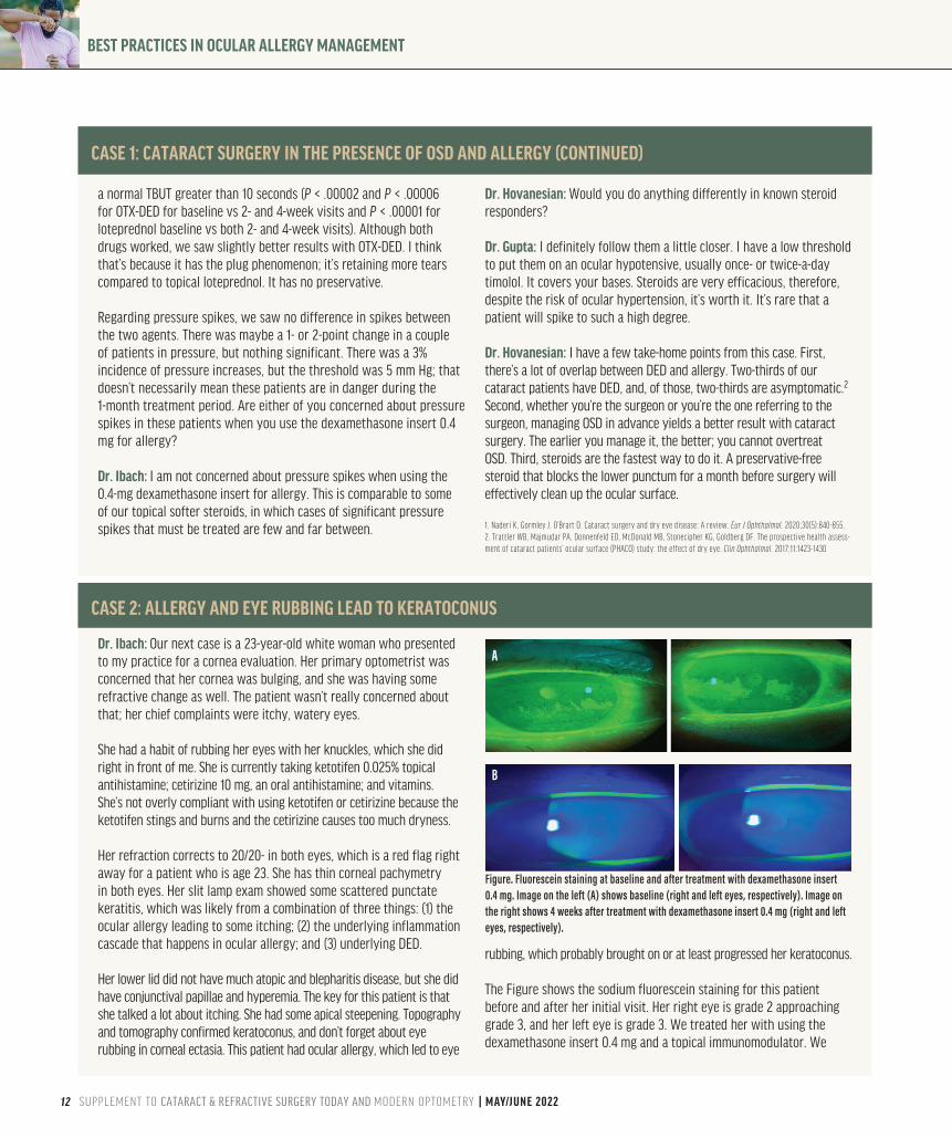
We also looked at tear breakup time (TBUT; Figure 2). Both OTX-DED and loteprednol significantly increased the proportion of patient with
CASE 1: CATARACT SURGERY IN THE PRESENCE OF OSD AND ALLERGY
Figure 1. Corneal staining with OTX-DED versus loteprednol (Hovanesian et al, unpublished data, March 2022).
Figure 2. Tear breakup time with OTX-DED versus loteprednol (Hovanesian et al, unpub-lished data, March 2022).
BEST PRACTICES IN OCULAR ALLERGY MANAGEMENT
12 SUPPLEMENT TO CATARACT & REFRACTIVE SURGERY TODAY AND MODERN OPTOMETRY | MAY/JUNE 2022
Dr. Ibach: Our next case is a 23-year-old white woman who presented to my practice for a cornea evaluation. Her primary optometrist was concerned that her cornea was bulging, and she was having some refractive change as well. The patient wasn’t really concerned about that; her chief complaints were itchy, watery eyes. She had a habit of rubbing her eyes with her knuckles, which she did right in front of me. She is currently taking ketotifen 0.025% topical antihistamine; cetirizine 10 mg, an oral antihistamine; and vitamins. She’s not overly compliant with using ketotifen or cetirizine because the ketotifen stings and burns and the cetirizine causes too much dryness.
Her refraction corrects to 20/20- in both eyes, which is a red flag right away for a patient who is age 23. She has thin corneal pachymetry in both eyes. Her slit lamp exam showed some scattered punctate keratitis, which was likely from a combination of three things: (1) the ocular allergy leading to some itching; (2) the underlying inflammation cascade that happens in ocular allergy; and (3) underlying DED.
Her lower lid did not have much atopic and blepharitis disease, but she did have conjunctival papillae and hyperemia. The key for this patient is that she talked a lot about itching. She had some apical steepening. Topography and tomography confirmed keratoconus, and don’t forget about eye rubbing in corneal ectasia. This patient had ocular allergy, which led to eye
rubbing, which probably brought on or at least progressed her keratoconus.
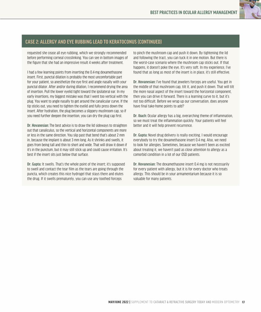
The Figure shows the sodium fluorescein staining for this patient before and after her initial visit. Her right eye is grade 2 approaching grade 3, and her left eye is grade 3. We treated her with using the dexamethasone insert 0.4 mg and a topical immunomodulator. We
CASE 2: ALLERGY AND EYE RUBBING LEAD TO KERATOCONUS
Figure. Fluorescein staining at baseline and after treatment with dexamethasone insert 0.4 mg. Image on the left (A) shows baseline (right and left eyes, respectively). Image on the right shows 4 weeks after treatment with dexamethasone insert 0.4 mg (right and left eyes, respectively).
A
B
a normal TBUT greater than 10 seconds (P < .00002 and P < .00006 for OTX-DED for baseline vs 2- and 4-week visits and P < .00001 for loteprednol baseline vs both 2- and 4-week visits). Although both drugs worked, we saw slightly better results with OTX-DED. I think that’s because it has the plug phenomenon; it’s retaining more tears compared to topical loteprednol. It has no preservative.
Regarding pressure spikes, we saw no difference in spikes between the two agents. There was maybe a 1- or 2-point change in a couple of patients in pressure, but nothing significant. There was a 3% incidence of pressure increases, but the threshold was 5 mm Hg; that doesn’t necessarily mean these patients are in danger during the 1-month treatment period. Are either of you concerned about pressure spikes in these patients when you use the dexamethasone insert 0.4 mg for allergy?
Dr. Ibach: I am not concerned about pressure spikes when using the 0.4-mg dexamethasone insert for allergy. This is comparable to some of our topical softer steroids, in which cases of significant pressure spikes that must be treated are few and far between.
Dr. Hovanesian: Would you do anything differently in known steroid responders?
Dr. Gupta: I definitely follow them a little closer. I have a low threshold to put them on an ocular hypotensive, usually once- or twice-a-day timolol. It covers your bases. Steroids are very efficacious, therefore, despite the risk of ocular hypertension, it’s worth it. It’s rare that a patient will spike to such a high degree.
Dr. Hovanesian: I have a few take-home points from this case. First, there’s a lot of overlap between DED and allergy. Two-thirds of our cataract patients have DED, and, of those, two-thirds are asymptomatic.2 Second, whether you’re the surgeon or you’re the one referring to the surgeon, managing OSD in advance yields a better result with cataract surgery. The earlier you manage it, the better; you cannot overtreat OSD. Third, steroids are the fastest way to do it. A preservative-free steroid that blocks the lower punctum for a month before surgery will effectively clean up the ocular surface.
1. Naderi K, Gormley J, O’Brart D. Cataract surgery and dry eye disease: A review. Eur J Ophthalmol. 2020;30(5):840-855. 2. Trattler WB, Majmudar PA, Donnenfeld ED, McDonald MB, Stonecipher KG, Goldberg DF. The prospective health assess-ment of cataract patients’ ocular surface (PHACO) study: the effect of dry eye. Clin Ophthalmol. 2017;11:1423-1430
CASE 1: CATARACT SURGERY IN THE PRESENCE OF OSD AND ALLERGY (CONTINUED)
BEST PRACTICES IN OCULAR ALLERGY MANAGEMENT
MAY/JUNE 2022 | SUPPLEMENT TO CATARACT & REFRACTIVE SURGERY TODAY AND MODERN OPTOMETRY 13
requested she cease all eye rubbing, which we strongly recommended before performing corneal crosslinking. You can see in bottom images of the figure that she had an impressive result 4 weeks after treatment.
I had a few learning points from inserting the 0.4-mg dexamethasone insert. First, punctal dilation is probably the most uncomfortable part for your patient, so anesthetize the eye first and angle nasally with your punctal dilator. After and/or during dilation, I recommend drying the area of insertion. Pull the lower eyelid tight toward the ipsilateral ear. In my early insertions, my biggest mistake was that I went too vertical with the plug. You want to angle nasally to get around the canalicular curve. If the tip sticks out, you need to tighten the eyelid and fully press down the insert. After hydration, the plug becomes a slippery mushroom cap, so if you need further deepen the insertion, you can dry the plug cap first.
Dr. Hovanesian: The best advice is to draw the lid sideways to straighten out that canaliculus, so the vertical and horizontal components are more or less in the same direction. You slip past that bend that’s about 2 mm in, because the implant is about 3 mm long. As it shrinks and swells, it goes from being tall and thin to short and wide. That will draw it down if it’s in the punctum, but it may still stick up and could cause irritation. It’s best if the insert sits just below that surface.
Dr. Gupta: It swells. That’s the whole point of the insert; it’s supposed to swell and contact the tear film as the tears are going through the puncta, which creates this nice hydrogel that stays there and elutes the drug. If it swells prematurely, you can use any toothed forceps
to pinch the mushroom cap and push it down. By tightening the lid and following the tract, you can tuck it in one motion. But there is the worst-case scenario where the mushroom cap sticks out. If that happens, it doesn’t poke the eye. It’s very soft. In my experience, I’ve found that as long as most of the insert is in place, it’s still effective.
Dr. Hovanesian: I’ve found that jewelers forceps are useful. You get in the middle of that mushroom cap, tilt it, and push it down. That will tilt the more nasal aspect of the insert toward the horizontal component, then you can drive it forward. There is a learning curve to it, but it’s not too difficult. Before we wrap up our conversation, does anyone have final take-home points to add? Dr. Ibach: Ocular allergy has a big, overarching theme of inflammation, so we must treat the inflammation quickly. Your patients will feel better and it will help prevent recurrence.
Dr. Gupta: Novel drug delivery is really exciting. I would encourage everybody to try the dexamethasone insert 0.4 mg. Also, we need to look for allergies. Sometimes, because we haven’t been as excited about treating it, we haven’t paid as close attention to allergy as a comorbid condition in a lot of our OSD patients.
Dr. Hovanesian: The dexamethasone insert 0.4 mg is not necessarily for every patient with allergy, but it is for every doctor who treats allergy. This should be in your armamentarium because it is so valuable for many patients.
CASE 2: ALLERGY AND EYE RUBBING LEAD TO KERATOCONUS (CONTINUED)
14 SUPPLEMENT TO CATARACT & REFRACTIVE SURGERY TODAY AND MODERN OPTOMETRY | MAY/JUNE 2022
INSTRUCTIONS FOR CREDITTo receive credit, you must complete the attached Pretest/Posttest/Activity Evaluation/Satisfaction Measures Form and mail or fax to Evolve Medical Education LLC; 353 West Lancaster Avenue, Second Floor, Wayne, PA 19087; Fax: (215) 933-3950. To answer these ques-tions online and receive real-time results, go to https://evolvemeded.com/course/2160-supp. If you experience problems with the online test, email us at [email protected]. NOTE: Certificates are issued electronically.
Please type or print clearly, or we will be unable to issue your certificate.
Full Name _________________________________________________________________________________ DOB (MM/DD): ____________
Phone (required) ____________________________ Email (required*) __________________________________________________________
Address/P.O. Box_____________________________________________________________________________________________________
City _________________________________________ State/Country _______________ Zip ____________________
License Number: _____________________ OE Tracker Number: ______________________ National Provider ID: __________________*Evolve does not share email addresses with third parties.
Did the program meet the following educational objectives? Agree Neutral Disagree
_____ _____ _____
_____ _____ _____
_____ _____ _____
_____ _____ _____
Differentiate between the various types and stages of ocular allergies based on presentation
Create individualized management options for ocular allergy based on disease severity, patient factors, and associated risks and benefits
Describe the limitations of current treatment options for ocular allergy
Evaluate new management options for ocular allergy
BEST PRACTICES IN OCULAR ALLERGY MANAGEMENTRelease Date: April 27, 2022CME Expiration Date: July 1, 2023COPE Expiration Date: April 30, 2023
DEMOGRAPHIC INFORMATION___ MD/DO___ OD___ NP___ Nurse/APN___ PA___ Other
Years in Practice___ >20___ 11-20___ 6-10___ 1-5___ <1
Patients Seen Per Week (with the disease targeted in this educational activity)___ 0___ 1-15___ 16-30___ 31-50____ >50
Region___ Midwest___ Northeast___ Northwest___ Southeast___ Southwest
LEARNING OBJECTIVES
MAY/JUNE 2022 | SUPPLEMENT TO CATARACT & REFRACTIVE SURGERY TODAY AND MODERN OPTOMETRY 15
POSTTEST QUESTIONS Please complete at the conclusion of the program.
1. Based on this activity, please rate your confidence in your ability to evaluate new management options for ocular allergy and create individualized management strategies (based on a scale of 1 to 5, with 1 being not at all confident and 5 being extremely confident).
a. 1b. 2c. 3d. 4e. 5
2. What percentage of US citizens test positive for at least one allergen?a. ~25%b. ~35%c. ~45%d. ~55%
3. A 45-year-old patient presents to your clinic with red, itchy, watery eyes. She notes she recently adopted a cat. All of the following are reasonable treatment options for this patient EXCEPT?
a. Allergy testing and avoidance of allergen b. Brimonidine eye drops c. Antihistamine eye dropsd. Lubricant eye drops
4. A 35-year-old man with seasonal allergies presents to your clinic with signs of allergic conjunctivitis. Which of the following treatments has been found to be clinically superior in relief of signs and symptom plus tolerability?
a. Topical corticosteroidsb. Topical NSAIDsc. Topical antihistaminesd. Topical dual-activity agents
5. A 76-year-old woman presents to your clinic with uncomfortable, burning, itching eyes. She has a history of chronic ocular surface disease with meibomian gland dysfunction. On exam, she has moderate inferior PEK OU, as well as 2-3+ NS OU. All of the following are reasonable treatment options for this patient except?
a. Aggressive lubrication with frequent artificial tears and ointmentb. Dexamethasone intracanalicular insertc. Schedule for cataract surgeryd. Short course of loteprednol
6. All of the following are cardinal symptoms that characterize allergic conjunctivitis, EXCEPT:
a. Rednessb. Itchingc. Tearingd. Corneal subepithelial infiltrates
7. An 8-year-old boy presents to your clinic with a history of seasonal allergies. He notes itchy, watery eyes. On exam, you note gelatinous-appearing raised bumps at the limbus. What is the most likely diagnosis?
a. Vernal keratoconjunctivitisb. Chemical conjunctivitisc. Viral conjunctivitisd. Bacterial conjunctivitis
8. The allergic cascade begins when ____ binds to mast cells, which launches a cascade of events.
a. IgDb. IgEc. IgGd. IgM
9. You are seeing a 76-year-old patient with severe allergic conjunctivitis. She has decreased manual dexterity and difficulty administering drops. You determine she needs therapy with corticosteroids. Which of the following is a reasonable treatment option for this patient?
a. Dexamethasone 0.4 mg intracanalicular insert b. Topical corticosteroid dropsc. Oral prednisoned. Intravitreal triamcinolone
10. You are seeing a 23-year-old white woman in your office who has chronic allergic conjunctivitis. She has a habit of frequently rubbing her eyes with her knuckles. She is currently on topical ketotifen 0.025%. What is this patient at risk for?
a. Shield ulcerb. Horner-Trantas Dots c. Keratoconusd. Recurrent corneal erosions
11. You are seeing a 55-year-old man with mild allergic conjunctivitis. You start him on ketotifen. What is the ideal dosing of this drug?
a. Once dailyb. Twice dailyc. Three times dailyd. Four times daily
12. You are seeing a 63-year-old woman who has seasonal allergic conjunctivitis with significant punctate epithelial erosions. You recommend steroid therapy and discuss loteprednol 0.2% suspension and the dexamethasone 0.4 mg intracanalicular insert as potential options. The patient asks you about the relative difference between the two, in terms of improving her corneal erosions. Which of the following statements is true?
a. Both the dexamethasone intracanalicular insert and loteprednol suspension significantly reduce corneal staining
b. Dexamethasone intracanalicular insert reduces corneal staining more than loteprednol suspension
c. Loteprednol suspension reduces corneal staining more than dexa-methasone intracanalicular insert
d. Neither dexamethasone intracanalicular insert nor loteprednol suspension had significant impact on corneal staining
Rate your knowledge/skill level prior to participating in this course: 5 = High, 1 = Low____
Rate your knowledge/skill level after participating in this course: 5 = High, 1 = Low____
This activity improved my competence in managing patients with this disease/condition/symptom. ____ Yes ____No
Probability of changing practice behavior based on this activity: ____High ____ Low ____No change needed
If you plan to change your practice behavior, what type of changes do you plan to implement? (check all that apply)
Change in pharmaceutical therapy ____ Change in nonpharmaceutical therapy ____
Change in diagnostic testing ____ Choice of treatment/management approach ____
Change in current practice for referral ____ Change in differential diagnosis ____
My practice has been reinforced ____ I do not plan to implement any new changes in practice ____
Please identify any barriers to change (check all that apply):
____ Cost ____ Lack of consensus or professional guidelines
____ Lack of administrative support ____Lack of experience
____ Lack of time to assess/counsel patients ____ Lack of opportunity (patients)
____ Reimbursement/insurance issues ____ Lack of resources (equipment)
____ Patient compliance issues ____ No barriers
____ Other. Please specify: ______________________________________________________________________________________________
The design of the program was effective for the content conveyed ___ Yes ___ No
The content supported the identified learning objectives ___ Yes ___ No
The content was free of commercial bias ___ Yes ___ No
The content was relative to your practice ___ Yes ___ No
The faculty was effective ___ Yes ___ No
You were satisfied overall with the activity ___ Yes ___ No
You would recommend this program to your colleagues ___ Yes ___ No
Please check the Core Competencies (as defined by the Accreditation Council for Graduate Medical Education) that were enhanced through your par-
ticipation in this activity:
____ Patient Care
____ Practice-Based Learning and Improvement
____ Professionalism
____ Medical Knowledge
____ Interpersonal and Communication Skills
____ System-Based Practice
Additional comments:
___________________________________________________________________________________________________________________
____ I certify that I have participated in this entire activity.
This information will help evaluate this activity; may we contact you by email in 3 months to inquire whether you have made changes to your practice based on this activity? If so, please provide your email address below.
___________________________________________________________________________________________________________________
___________________________________________________________________________________________________________________
ACTIVITY EVALUATIONYour responses to the questions below will help us evaluate this activity. They will provide us with evidence that improvements were made in patient care as a result of this activity.