Epidemiology and Management of Ocular Surface Squamous ...
303
Epidemiology and Management of Ocular Surface Squamous Neoplasia in Kenya Stephen Gichuhi Thesis submitted in accordance with the requirements for the degree of Doctor of Philosophy of the University of London 2016 International Centre for Eye Health Department of Clinical Research Faculty of Infectious and Tropical Diseases London School of Hygiene and Tropical Medicine Funded by: British Council for Prevention of Blindness – Sir John Wilson Fellowship
-
Upload
khangminh22 -
Category
Documents
-
view
1 -
download
0
Transcript of Epidemiology and Management of Ocular Surface Squamous ...

Epidemiology and Management of
Ocular Surface Squamous Neoplasia in
Kenya
Stephen Gichuhi
Thesis submitted in accordance with the requirements for the degree of
Doctor of Philosophy of the University of London
2016
International Centre for Eye Health
Department of Clinical Research
Faculty of Infectious and Tropical Diseases
London School of Hygiene and Tropical Medicine
Funded by: British Council for Prevention of Blindness – Sir John Wilson Fellowship

Declaration
I, Stephen Gichuhi, confirm that the work presented in this thesis is my own. Where
information has been derived from other sources, I confirm that this has been
indicated in the thesis.
Signature Date 27th February 2016
2
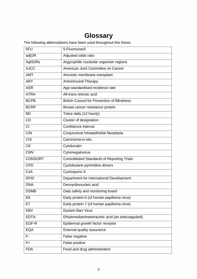
GlossaryThe following abbreviations have been used throughout this thesis
5FU 5-Fluorouracil
adjOR Adjusted odds ratio
AgNORs Argyrophilic nucleolar organizer regions
AJCC American Joint Committee on Cancer
AMT Amniotic membrane transplant
ART Antiretroviral Therapy
ASR Age-standardised incidence rate
ATRA All-trans retinoic acid
BCPB British Council for Prevention of Blindness
BCRP Breast cancer resistance protein
BD Twice daily (12 hourly)
CD Cluster of designation
CI Confidence interval
CIN Conjunctival Intraepithelial Neoplasia
CIS Carcinoma-in-situ
CK Cytokeratin
CMV Cytomegalovirus
CONSORT Consolidated Standards of Reporting Trials
CPD Cyclobutane pyrimidine dimers
CsA Cyclosporin A
DFID Department for International Development
DNA Deoxyribonucleic acid
DSMB Data safety and monitoring board
E6 Early protein 6 (of human papilloma virus)
E7 Early protein 7 (of human papilloma virus)
EBV Epstein Barr Virus
EDTA Ethylenediaminetetracetic acid (an anticoagulant)
EGF-R Epidermal growth factor receptor
EQA External quality assurance
F- False negative
F+ False positive
FDA Food and drug administration
3

FdUMP Fluorodeoxyuridine monophosphate
FdUTP Fluorodeoxyuridine triphosphate
FUTP Fluorouridine triphosphate
G1, G2 Growth phases of the cell cycle
GCLP Good clinical laboratory practice
GCP Good clinical practice
GDP Gross Domestic Product
Gy Grays (units of radiation)
H&E Haematoxylin and eosin
HAART Highly Active Antiretroviral Therapy
HACM HIV/AIDS Cancer Match study
HC-2 Hybrid Capture 2
HEED Health education and Early Detection study
HGF Hepatocyte growth factor
HIV Human Immunodeficiency Virus
HPLC High-performance liquid chromatography
HPSG Heparin sulphate proteoglycan
HPV Human Papilloma Virus
HR Hazard ratio
HSV Herpes Simplex Virus
IAPB International Agency for Prevention of Blindness
IARC International Agency for Research in Cancer
ICD International Classification of Diseases
ICMJE International Council of Medical Journal Editors
IFN Interferon
IQR Interquartile range
ISCO International Standard Classification of Occupations
IU International units
IVCM in vivo confocal microscopy
JAMA Journal of the American Medical Association
k Kappa statistic (for inter-observer agreement)
KAIS Kenya AIDS indicator survey
KAVI Kenya AIDS Vaccine Institute
KDH Kitale District Hospital
KDHS Kenya Demographic and Health Survey
4
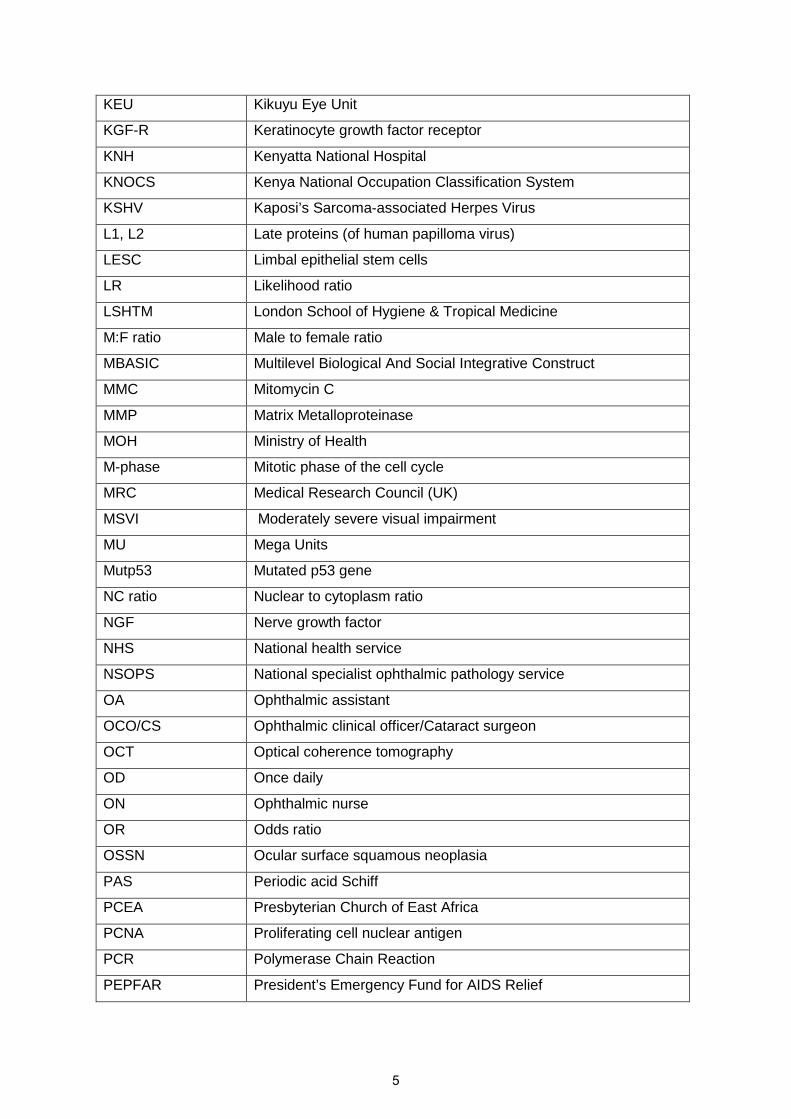
KEU Kikuyu Eye Unit
KGF-R Keratinocyte growth factor receptor
KNH Kenyatta National Hospital
KNOCS Kenya National Occupation Classification System
KSHV Kaposi’s Sarcoma-associated Herpes Virus
L1, L2 Late proteins (of human papilloma virus)
LESC Limbal epithelial stem cells
LR Likelihood ratio
LSHTM London School of Hygiene & Tropical Medicine
M:F ratio Male to female ratio
MBASIC Multilevel Biological And Social Integrative Construct
MMC Mitomycin C
MMP Matrix Metalloproteinase
MOH Ministry of Health
M-phase Mitotic phase of the cell cycle
MRC Medical Research Council (UK)
MSVI Moderately severe visual impairment
MU Mega Units
Mutp53 Mutated p53 gene
NC ratio Nuclear to cytoplasm ratio
NGF Nerve growth factor
NHS National health service
NSOPS National specialist ophthalmic pathology service
OA Ophthalmic assistant
OCO/CS Ophthalmic clinical officer/Cataract surgeon
OCT Optical coherence tomography
OD Once daily
ON Ophthalmic nurse
OR Odds ratio
OSSN Ocular surface squamous neoplasia
PAS Periodic acid Schiff
PCEA Presbyterian Church of East Africa
PCNA Proliferating cell nuclear antigen
PCR Polymerase Chain Reaction
PEPFAR President’s Emergency Fund for AIDS Relief
5

PMC Post-mitotic cells
pRB Retinoblastoma gene
PV- Predictive value of a negative test
PV+ Predictive value of a positive test
QID Four times daily (6 hourly)
qRT-PCR Quantitative Reverse Transcriptase Polymerase Chain Reaction
RCT Randomised controlled trial
RNA Ribonucleic acid
ROC curve Receiver operator characteristic curve
ROS Reactive oxygen species
SCC Squamous Cell Carcinoma
SCCC Squamous Cell Carcinoma of the Conjunctiva
SD Standard deviation
SE Standard error
SEH Sabatia Eye Hospital
SIR Standardised Incidence Rate
SOP Standard operating procedure
S-phase Synthesis phase of the cell cycle
SSA Sub-Saharan Africa
STI Sexually transmitted infections
TAC Transient amplifying cells
TDRC Tropical Diseases Research Centre
Th1 T-Helper 1 Lymphocytes
TID Three times daily (8 hourly)
TIL Tumour infiltrating lymphocytes
TIMP Transient Inhibitors of Matrix Metalloproteinase
ToB Toluidine Blue
UHR-OCT Ultra-high resolution Optical coherence tomography
UK United Kingdom
UON University of Nairobi
USA United States of America
UV Ultraviolet
UVR Ultraviolet radiation
WHO World Health Organization
XP Xeroderma pigmetosum
6

Abstract
Introduction
Ocular surface squamous neoplasia (OSSN) is a spectrum of disease that ranges from non-
invasive intra-epithelial dysplasia of the conjunctival and cornea (CCIN), through to invasive
squamous cell carcinoma (SCC). It often presents with unilateral tumours on the eyeball. The
tumours may cause blindness, disfigurement and even death. In East Africa, OSSN is
relatively common and aggressive, affecting younger adults and proportionally more women
than in other parts of the world. The management of OSSN is challenging for various reasons.
Its risk factors are not clearly understood. Studies have implicated HIV, human papilloma virus
(HPV) and solar radiation however about 30% of cases are HIV-negative while some studies
have implicated HPV and others found no association. The importance of vitamin A for a
healthy ocular surface is known, yet its role in OSSN has not been studied. Early diagnosis
relies on the clinical impression yet OSSN appears similar to other conjunctival tumours and
histopathology services are generally unavailable in Africa. Surgery is the mainstay of
treatment but recurrence is an issue. There is no trial evidence for the various treatments used
in HIV-infected persons. This project was an integrated set of studies to improve our
understanding of the epidemiology and management of OSSN in Kenya.
Methods
We conducted three systematic reviews on the epidemiology of OSSN in Africa, the
pathophysiology of OSSN and updated a Cochrane review on the interventions for OSSN in
HIV-infected individuals. Working in four eye care centres in Kenya between July 2012 and
July 2015, we conducted the following six studies: (i) clinical assessment of a series of patients
with conjunctival lesions to describe OSSN to determine how OSSN may differ clinically from
benign lesions, (ii) evaluated vital staining with a special dye called Toluidine Blue (ToB) for
making the diagnosis of OSSN, (iii) developed a diagnostic algorithm based on clinical
features and vital staining, (iv) conducted a large case-control study to investigate risk factors
that may contribute to the development of OSSN, (v) investigated the care-seeking journey of
OSSN patients to assess referral pathway and treatment delay, and finally, (vi) conducted a
randomised placebo-controlled trial of 5-Fluorouracil (5FU) chemotherapy eyedrops given
after surgery to investigate if this can reduce recurrence of the lesions.
7
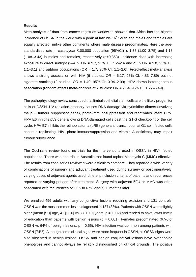
Results
Meta-analysis of data from cancer registries worldwide showed that Africa has the highest
incidence of OSSN in the world with a peak at latitude 160 South and males and females are
equally affected, unlike other continents where male disease predominates. Here the age-
standardized rate in cases/year /100,000 population (95%CI) is 1.38 (1.00–3.75) and 1.18
(1.08–3.43) in males and females, respectively (p=0.853). Incidence rises with increasing
exposure to direct sunlight (2–4 h, OR = 1.7, 95% CI: 1.2–2.4 and ≥5 h OR = 1.8, 95% CI:
1.1–3.1) and outdoor occupations (OR = 1.7, 95% CI: 1.1–2.6). Fixed-effect meta-analysis
shows a strong association with HIV (6 studies: OR = 6.17, 95% CI: 4.83–7.89) but not
cigarette smoking (2 studies: OR = 1.40, 95% CI: 0.94–2.09). HPV shows heterogeneous
association (random effects meta-analysis of 7 studies: OR = 2.64, 95% CI: 1.27–5.49).
The pathophysiology review concluded that limbal epithelial stem cells are the likely progenitor
cells of OSSN. UV radiation probably causes DNA damage via pyrimidine dimers (involving
the p53 tumour suppressor gene), photo-immunosuppression and reactivates latent HPV.
HPV E6 inhibits p53 gene allowing DNA-damaged cells past the G1-S checkpoint of the cell
cycle. HPV E7 inhibits the retinoblastoma (pRB) gene anti-transcription at G1 so infected cells
continue replicating. HIV, photo-immunosuppression and vitamin A deficiency may impair
tumour surveillance.
The Cochrane review found no trials for the interventions used in OSSN in HIV-infected
populations. There was one trial in Australia that found topical Mitomycin C (MMC) effective.
The results from case series reviewed were difficult to compare. They reported a wide variety
of combinations of surgery and adjuvant treatment used during surgery or post operatively;
varying doses of adjuvant agents used; different inclusion criteria of patients and recurrences
reported at varying periods after treatment. Surgery with adjuvant 5FU or MMC was often
associated with recurrences of 11% to 67% about 30 months later.
We enrolled 496 adults with any conjunctival lesions requiring excision and 131 controls.
OSSN was the most common lesion diagnosed in 187 (38%). Patients with OSSN were slightly
older (mean [SD] age, 41 [11.6] vs 38 [10.9] years; p =0.002) and tended to have lower levels
of education than patients with benign lesions (p = 0.001). Females predominated (67% of
OSSN vs 64% of benign lesions; p = 0.65). HIV infection was common among patients with
OSSN (74%). Although some clinical signs were more frequent in OSSN, all OSSN signs were
also observed in benign lesions. OSSN and benign conjunctival lesions have overlapping
phenotypes and cannot always be reliably distinguished on clinical grounds. The positive
8

predictive value of clinical appearance in identifying OSSN was 54%. Inter-observer
agreement was modest (κ= 0.1-0.4).
Any blue colour on vital staining with ToB 0.05% had a sensitivity of 92%, specificity of 31%,
positive predictive value of 41%, and negative predictive value of 88% for OSSN. Inter-
observer agreement was substantial for staining (k=0.8) and moderate for overall diagnosis
(OSSN or benign) (κ =0.4). Use of ToB caused mild discomfort in 88 (21%) patients; mild
superficial punctate keratopathy seen in 7 (1.7%) and no histological evidence of corneal
toxicity was observed. ToB had a high rate of false positives (69%).
We developed a simple probability-tree clinical algorithm that shows the probability of OSSN
with various combinations of clinical features. A multivariable regression model found 8
features strongly associated with OSSN; prior excision, corneal involvement, feeder vessels,
dark blue ToB staining, papillary or gelatinous tumour surface, severe inflammation, anti-
retroviral therapy and temporal or circumlimbal tumours. Using a cut-off of any 3 of these
features, the sensitivity was 89%, specificity 50%, and 65% of lesions were correctly classified.
This specificity was higher than any blue ToB staining (31%) but lower than clinical photo-
examination (60%).
A total of 131 cases were frequency-matched to 131 controls by age, sex and eye center. Risk
factors for OSSN were HIV infection without antiretroviral therapy (ART) use (OR=48.30;
95%CI 7.53-309.90) and with ART use (OR=19.02; 95%CI 6.55-55.26), longer duration of
exposure to the sun in the main occupation (6.9 hrs/day vs. 4.6 hrs/day, OR=1.23; 95%CI
1.08-1.39) and a history of allergic conjunctivitis (OR=80.20; 95%CI 8.62-746.29). Wearing
hats was protective (OR=0.21; 95%CI 0.07-0.63).
We studied the care-seeking journey followed by 158 new OSSN patients. About half (88/158,
[56%]) presented directly to the study centres while the rest were referred. Indirect presenters
sought care earlier than direct presenters (median 2.0 months vs 5.5 months) and travelled a
shorter distance to the first health facility (median 20km vs 30km) but had surgery later
(median 12.5 months vs 5.5 months). Visits beyond the first health facility for indirect
presenters markedly increased delay (median 7.3, 29.0, 37.9, and 32.0 months for 1-4
facilities, respectively). Delay was associated with number of health facilities visited (adjusted
ordered OR=9.12; 95%CI 2.83-29.4, p<0.001) and being female (adjusted ordered OR=2.42;
95%CI 1.32-4.44, p=0.004).
9
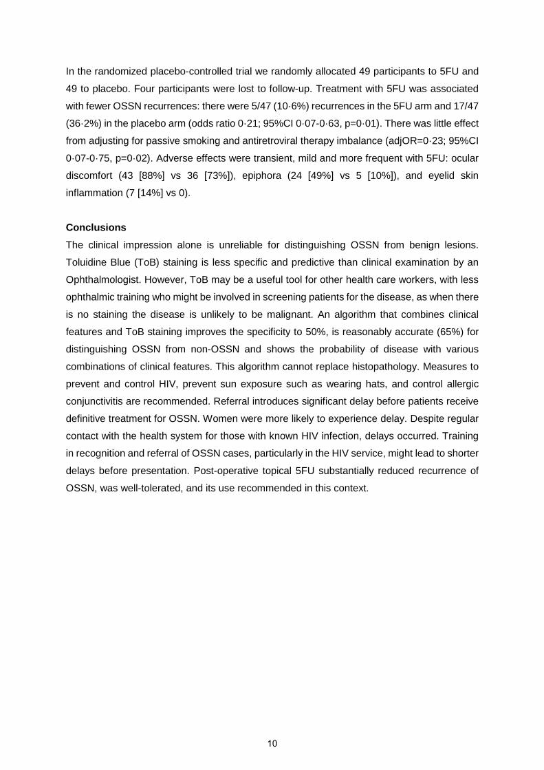
In the randomized placebo-controlled trial we randomly allocated 49 participants to 5FU and
49 to placebo. Four participants were lost to follow-up. Treatment with 5FU was associated
with fewer OSSN recurrences: there were 5/47 (10·6%) recurrences in the 5FU arm and 17/47
(36·2%) in the placebo arm (odds ratio 0·21; 95%CI 0·07-0·63, p=0·01). There was little effect
from adjusting for passive smoking and antiretroviral therapy imbalance (adjOR=0·23; 95%CI
0·07-0·75, p=0·02). Adverse effects were transient, mild and more frequent with 5FU: ocular
discomfort (43 [88%] vs 36 [73%]), epiphora (24 [49%] vs 5 [10%]), and eyelid skin
inflammation (7 [14%] vs 0).
Conclusions
The clinical impression alone is unreliable for distinguishing OSSN from benign lesions.
Toluidine Blue (ToB) staining is less specific and predictive than clinical examination by an
Ophthalmologist. However, ToB may be a useful tool for other health care workers, with less
ophthalmic training who might be involved in screening patients for the disease, as when there
is no staining the disease is unlikely to be malignant. An algorithm that combines clinical
features and ToB staining improves the specificity to 50%, is reasonably accurate (65%) for
distinguishing OSSN from non-OSSN and shows the probability of disease with various
combinations of clinical features. This algorithm cannot replace histopathology. Measures to
prevent and control HIV, prevent sun exposure such as wearing hats, and control allergic
conjunctivitis are recommended. Referral introduces significant delay before patients receive
definitive treatment for OSSN. Women were more likely to experience delay. Despite regular
contact with the health system for those with known HIV infection, delays occurred. Training
in recognition and referral of OSSN cases, particularly in the HIV service, might lead to shorter
delays before presentation. Post-operative topical 5FU substantially reduced recurrence of
OSSN, was well-tolerated, and its use recommended in this context.
10

Format of the thesis
The thesis for this PhD utilises the “research papers” format, recently introduced by the
London School of Hygiene and Tropical Medicine. It therefore includes a number of papers
which are either published, accepted or in submittable format for publication in peer-reviewed
journals. The chapters listed in italics in the Contents are in this research/review paper format,
and each chapter includes publication details in a cover sheet, including acknowledgement of
the contributions of other people.
The other chapters of the thesis are composed of “linking material” which includes
information/data not covered in the research papers and helps to make the thesis a coherent
body.
11
Contents
Declaration 2
Glossary 3
Abstract 7
Format of the thesis 11
Acknowledgements 13
List of contributors 14
Introduction 16
Chapter 1. Epidemiology of ocular surface squamous neoplasia in Africa 21
Chapter 2. Pathophysiology of ocular surface squamous neoplasia 44
Chapter 3. Diagnosis of ocular surface squamous neoplasia 58
Chapter 4. Interventions for squamous cell carcinoma of the conjunctiva in HIV-
infected individuals (Cochrane Review) 77
Chapter 5. Research setting 133
Chapter 6. Overview of project design 141
Data Chapters 147
Chapter 7. Clinical presentation of ocular surface squamous neoplasia in Kenya 148
Chapter 8. Toluidine Blue 0.05% vital staining for diagnosis of ocular surface
squamous neoplasia in Kenya 151
Chapter 9. Clinical algorithm for diagnosis and management of ocular surface
squamous neoplasia in East Africa 154
Chapter 10. Risk factors for ocular surface squamous neoplasia in Kenya 175
Chapter 11. Delay along the care-seeking pathway of patients with ocular surface
squamous neoplasia in Kenya 195
Chapter 12. Topical 5-Fluorouracil (5FU) following surgery for ocular surface
squamous neoplasia (OSSN) in Kenya: a randomised, double-blind,
placebo-controlled trial 218
General discussion and future work 242
Chapter 13. Discussion 243
Chapter 14. Future work 248
Appendices 249
12

Acknowledgements
I would like to thank the following organisations and individuals for the support they provided:
The British Council for Prevention of Blindness (BCPB) who awarded me £180,000 under the
Sir John Wilson Fellowship to conduct this research project.
To my supervisor Matthew Burton, and co-supervisors Helen Weiss and Mandeep Sagoo for
their patient guidance and very helpful insights.
To all my Kenya and UK based collaborators who gave a lot towards this work. Various
laboratories gave concessionary rates and IVEE Aqua in Kenya manufactured the trial
eyedrops at no cost to the study.
To all the study participants who volunteered for the sake of many others suffering from ocular
surface squamous neoplasia whose lives I hope will be improved by this work.
To my home institution, the University of Nairobi for giving me study leave.
The London School of Hygiene & Tropical Medicine experience shall remain a memorable
chapter in my life story.
To my wife Christine and sons Philip and David, who stood with me throughout and bore the
brunt of my ‘busyness’ with such grace and poise.
13

List of contributorsContributors to the research project in alphabetical order besides the listed authors in
manuscripts:
Person Position Contribution
David Essex Laboratory Manager, Eyepathology, Institute ofOphthalmology, Moorfields
Preparation ofimmunohistochemistry andhistopathology slides
Godfrey Nyaga Consultant Ophthalmologist,Kikuyu Eye Unit
Obtaining control conjunctivaltissue
Grace Muthoni Nurse, Kitale District Hospital Counselling and coordination offollow up of participants
Heidi Barnes Laboratory technologist, Instituteof Ophthalmology, Moorfield
Preparation ofimmunohistochemistry andhistopathology slides
Hodan Jama Laboratory technologist, Instituteof Ophthalmology, Moorfields
Slide photography
Irene Anne Mwangi Laboratory technologist, KAVI HIV and CD4 testing
Jane Nakhumicha Nurse, Kikuyu Eye Unit Coordination of sample collectionand counselling
John Kamonjo Maina Research assistant Data entry and assistance withstudy coordination. RCT manuscriptreview.
John Ndiritu Laboratory technologist, MPShah Hospital
Tissue preparation forhistopathology
John Njogu Pharmaceutical technician,eyedrop production unit, KikuyuEye Unit
Production of toluidine blue eyedrops
Justine Chileshe Scientific officer, TropicalDiseases Research Centre,Zambia
Vitamin A analysis
Leah Mwenda Nurse, Kikuyu Eye Unit Coordination of eye clinic reviewsof participants
Margaret W. Njoroge Counsellor, Kikuyu Eye Unit Participant counselling
Martin Hibberd Virologist, London School ofHygiene & Tropical Medicine
Viral assays
14

Merceline Mbayi Nurse, Sabatia Eye Hospital Counselling and coordination ofparticipant follow up
Mohammed Farah Laboratory manager, KAVI HIV and CD4 testing
Monica K. Ndungu Counsellor, Kikuyu Eye Unit Participant counselling
Phil Luthert Consultant Pathologist, Instituteof Ophthalmology, Moorfields
Reporting Immunohistochemistryslides
Ramadhani Athumani Research degree student,London School of Hygiene &Tropical Medicine & KCMC
DNA extraction from tumourspecimens
Ronald Mamboleo Ophthalmic Clinical Officer,Kitale District Hospital
Participant enrolment
Shadrack Chebet Ophthalmic Clinical Officer,Kitale District Hospital
Participant enrolment
Shaffiq Jafferjee Consultant Ophthalmologist,Kikuyu Eye Unit
Obtaining control conjunctivaltissue
Stephen Gathiga Production manager, Ivee AquaLtd, Kenya
Production of 5FU eye drops
Sunil Shah Proprietor, Ivee Aqua Ltd,Kenya
Production of 5FU eye drops
Warda Laboratory technologist, Instituteof Ophthalmology, Moorfields
Preparation ofimmunohistochemistry andhistopathology slides
William Kemei Ophthalmic Clinical Officer,Kitale District Hospital
Participant enrolment
Abbreviations: KAVI- Kenya Aids Vaccine Institute at University of Nairobi; KCMC – KilimanjaroChristian Medical Center, Moshi, Tanzania
15

Introduction
Ocular surface squamous neoplasia (OSSN) is a spectrum of disease that ranges from non-
invasive intra-epithelial dysplasia of the conjunctival and cornea (CCIN), through to invasive
squamous cell carcinoma (SCC).1 In recent decades OSSN has undergone an
epidemiological shift. In more temperate countries, it remains a rare, slow growing tumour of
elderly males.2 In contrast, in tropical countries, particularly in Eastern Africa, it is now more
common, more aggressive, affects younger people and with a higher incidence in women here
than in other parts of the world.3-6 It seems likely that much of this increased burden of disease
is attributable to the HIV/AIDS epidemic.7 Even though OSSN is not a target condition within
Vision 2020, it frequently leads to a poor quality of life, visual disability and death. In
September 2010 the “IAPB/Vision2020 Workshop on Research for Global Blindness
Prevention” identified specific research priorities.8 It was recognised that for Africa there was
a need for research on HIV-related conditions to better define the epidemiology and determine
context-specific management approaches.
Prevalence and Incidence
Reliable prevalence and incidence estimates of the numbers of individuals affected have been
difficult to ascertain and vary considerably. At one extreme, in one study from Kenya based in
a HIV testing clinic, 7.8% of HIV-infected adults were found to have conjunctival lesions which
were found to be OSSN on histopathology.5 The Kenyan national HIV prevalence is 6% (2.28
million out of 38 million) so it was suggested that over 170,000 Kenyans might have some
degree of OSSN. In contrast, a relatively low annual incidence estimate of 2.2/100,000 has
been suggested in a study from Tanzania, based on the number of cases being operated in
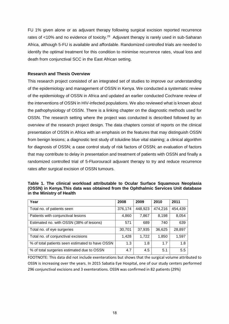
eye units.6 From the clinical perspective OSSN represents a significant component of the
ophthalmic “work-load” in East Africa. About 5% of all ophthalmic surgery in Kenya is for OSSN
(Table 1 on page 18) and is associated with a high level of morbidity for the patient, as
exenteration is often needed for those with late presentation or recurrent disease. However,
OSSN receives little or no attention from either ophthalmic or HIV care programs.
Risk factors and Aetiology
The risk factors and aetiology of OSSN in East Africa are not well understood. There is an
association with HIV; however, a significant proportion (~30%) of people with OSSN are not
infected with HIV, suggesting that other factors also contribute to the excess burden of disease
in this region.7, 9-11 Despite the association with HIV, a dose-response effect where lower
16
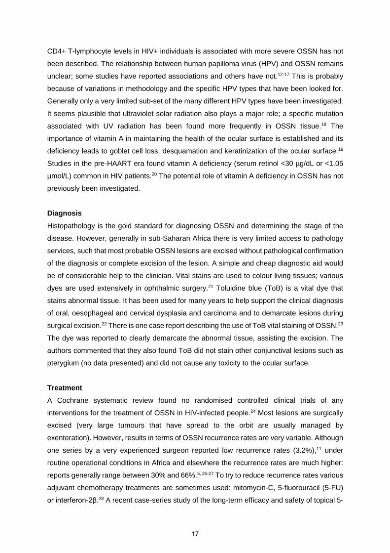
CD4+ T-lymphocyte levels in HIV+ individuals is associated with more severe OSSN has not
been described. The relationship between human papilloma virus (HPV) and OSSN remains
unclear; some studies have reported associations and others have not.12-17 This is probably
because of variations in methodology and the specific HPV types that have been looked for.
Generally only a very limited sub-set of the many different HPV types have been investigated.
It seems plausible that ultraviolet solar radiation also plays a major role; a specific mutation
associated with UV radiation has been found more frequently in OSSN tissue.18 The
importance of vitamin A in maintaining the health of the ocular surface is established and its
deficiency leads to goblet cell loss, desquamation and keratinization of the ocular surface.19
Studies in the pre-HAART era found vitamin A deficiency (serum retinol <30 µg/dL or <1.05
µmol/L) common in HIV patients.20 The potential role of vitamin A deficiency in OSSN has not
previously been investigated.
Diagnosis
Histopathology is the gold standard for diagnosing OSSN and determining the stage of the
disease. However, generally in sub-Saharan Africa there is very limited access to pathology
services, such that most probable OSSN lesions are excised without pathological confirmation
of the diagnosis or complete excision of the lesion. A simple and cheap diagnostic aid would
be of considerable help to the clinician. Vital stains are used to colour living tissues; various
dyes are used extensively in ophthalmic surgery.21 Toluidine blue (ToB) is a vital dye that
stains abnormal tissue. It has been used for many years to help support the clinical diagnosis
of oral, oesophageal and cervical dysplasia and carcinoma and to demarcate lesions during
surgical excision.22 There is one case report describing the use of ToB vital staining of OSSN.23
The dye was reported to clearly demarcate the abnormal tissue, assisting the excision. The
authors commented that they also found ToB did not stain other conjunctival lesions such as
pterygium (no data presented) and did not cause any toxicity to the ocular surface.
Treatment
A Cochrane systematic review found no randomised controlled clinical trials of any
interventions for the treatment of OSSN in HIV-infected people.24 Most lesions are surgically
excised (very large tumours that have spread to the orbit are usually managed by
exenteration). However, results in terms of OSSN recurrence rates are very variable. Although
one series by a very experienced surgeon reported low recurrence rates (3.2%),11 under
routine operational conditions in Africa and elsewhere the recurrence rates are much higher:
reports generally range between 30% and 66%.5, 25-27 To try to reduce recurrence rates various
adjuvant chemotherapy treatments are sometimes used: mitomycin-C, 5-fluorouracil (5-FU)
or interferon-2β.28 A recent case-series study of the long-term efficacy and safety of topical 5-
17
FU 1% given alone or as adjuvant therapy following surgical excision reported recurrence
rates of <10% and no evidence of toxicity.29 Adjuvant therapy is rarely used in sub-Saharan
Africa, although 5-FU is available and affordable. Randomized controlled trials are needed to
identify the optimal treatment for this condition to minimise recurrence rates, visual loss and
death from conjunctival SCC in the East African setting.
Research and Thesis Overview
This research project consisted of an integrated set of studies to improve our understanding
of the epidemiology and management of OSSN in Kenya. We conducted a systematic review
of the epidemiology of OSSN in Africa and updated an earlier conducted Cochrane review of
the interventions of OSSN in HIV-infected populations. We also reviewed what is known about
the pathophysiology of OSSN. There is a linking chapter on the diagnostic methods used for
OSSN. The research setting where the project was conducted is described followed by an
overview of the research project design. The data chapters consist of reports on the clinical
presentation of OSSN in Africa with an emphasis on the features that may distinguish OSSN
from benign lesions; a diagnostic test study of toluidine blue vital staining; a clinical algorithm
for diagnosis of OSSN; a case control study of risk factors of OSSN; an evaluation of factors
that may contribute to delay in presentation and treatment of patients with OSSN and finally a
randomized controlled trial of 5-Fluorouracil adjuvant therapy to try and reduce recurrence
rates after surgical excision of OSSN tumours.
Table 1. The clinical workload attributable to Ocular Surface Squamous Neoplasia(OSSN) in Kenya.This data was obtained from the Ophthalmic Services Unit databasein the Ministry of Health
Year 2008 2009 2010 2011
Total no. of patients seen 376,174 448,923 474,216 454,439
Patients with conjunctival lesions 4,860 7,867 8,198 8,054
Estimated no. with OSSN (38% of lesions) 571 689 740 639
Total no. of eye surgeries 30,701 37,935 36,625 28,897
Total no. of conjunctival excisions 1,428 1,722 1,850 1,597
% of total patients seen estimated to have OSSN 1.3 1.8 1.7 1.8
% of total surgeries estimated due to OSSN 4.7 4.5 5.1 5.5
FOOTNOTE: This data did not include exenterations but shows that the surgical volume attributed to
OSSN is increasing over the years. In 2015 Sabatia Eye Hospital, one of our study centers performed
296 conjunctival excisions and 3 exenterations. OSSN was confirmed in 82 patients (29%)
18
References
1. Lee GA, Hirst LW. Ocular surface squamous neoplasia. Survey of ophthalmology 1995; 39(6):429-50.
2. Lee GA, Hirst LW. Retrospective study of ocular surface squamous neoplasia. Australian andNew Zealand journal of ophthalmology 1997; 25(4): 269-76.
3. Poole TR. Conjunctival squamous cell carcinoma in Tanzania. The British journal ofophthalmology 1999; 83(2): 177-9.
4. Pola EC, Masanganise R, Rusakaniko S. The trend of ocular surface squamous neoplasia amongocular surface tumour biopsies submitted for histology from Sekuru Kaguvi Eye Unit, Hararebetween 1996 and 2000. The Central African journal of medicine 2003; 49(1-2): 1-4.
5. Chisi SK, Kollmann MK, Karimurio J. Conjunctival squamous cell carcinoma in patients withhuman immunodeficiency virus infection seen at two hospitals in Kenya. East African medicaljournal 2006; 83(5): 267-70.
6. Furahini G, Lewallen S. Epidemiology and management of ocular surface squamous neoplasia inTanzania. Ophthalmic epidemiology 2010; 17(3): 171-6.
7. Ateenyi-Agaba C. Conjunctival squamous-cell carcinoma associated with HIV infection inKampala, Uganda. Lancet 1995; 345(8951): 695-6.
8. International Agency for Prevention of Blindness. Workshop Report: A VISION 2020 Workshopon Research for Global Blindness Prevention International Center for Eye Health (ICEH), 2010.
9. Mbulaiteye SM, Katabira ET, Wabinga H, et al. Spectrum of cancers among HIV-infected personsin Africa: the Uganda AIDS-Cancer Registry Match Study. International journal of cancer Journalinternational du cancer 2006; 118(4): 985-90.
10. Waddell KM, Lewallen S, Lucas SB, Atenyi-Agaba C, Herrington CS, Liomba G. Carcinoma of theconjunctiva and HIV infection in Uganda and Malawi. The British journal of ophthalmology 1996;80(6): 503-8.
11. Waddell KM, Downing RG, Lucas SB, Newton R. Corneo-conjunctival carcinoma in Uganda. Eye(Lond) 2006; 20(8): 893-9.
12. Yu JJ, Fu P, Pink JJ, et al. HPV infection and EGFR activation/alteration in HIV-infected EastAfrican patients with conjunctival carcinoma. PloS one 2010; 5(5): e10477.
13. Moubayed P, Mwakyoma H, Schneider DT. High frequency of human papillomavirus 6/11, 16,and 18 infections in precancerous lesions and squamous cell carcinoma of the conjunctiva insubtropical Tanzania. Am J Clin Pathol 2004; 122(6): 938-43.
14. Guthoff R, Marx A, Stroebel P. No evidence for a pathogenic role of human papillomavirusinfection in ocular surface squamous neoplasia in Germany. Current eye research 2009; 34(8):666-71.
15. de Koning MN, Waddell K, Magyezi J, et al. Genital and cutaneous human papillomavirus (HPV)types in relation to conjunctival squamous cell neoplasia: a case-control study in Uganda.Infectious agents and cancer 2008; 3: 12.
16. Sen S, Sharma A, Panda A. Immunohistochemical localization of human papilloma virus inconjunctival neoplasias: a retrospective study. Indian journal of ophthalmology 2007; 55(5):361-3.
17. Simbiri KO, Murakami M, Feldman M, et al. Multiple oncogenic viruses identified in Ocularsurface squamous neoplasia in HIV-1 patients. Infectious agents and cancer 2010; 5: 6.
18. Ateenyi-Agaba C, Dai M, Le Calvez F, et al. TP53 mutations in squamous-cell carcinomas of theconjunctiva: evidence for UV-induced mutagenesis. Mutagenesis 2004; 19(5): 399-401.
19. Pfister RR, Renner ME. The corneal and conjunctival surface in vitamin A deficiency: a scanningelectron microscopy study. Investigative ophthalmology & visual science 1978; 17(9): 874-83.
20. Baeten JM, McClelland RS, Richardson BA, et al. Vitamin A deficiency and the acute phaseresponse among HIV-1-infected and -uninfected women in Kenya. Journal of acquired immunedeficiency syndromes (1999) 2002; 31(2): 243-9.
19
21. Rodrigues EB, Costa EF, Penha FM, et al. The use of vital dyes in ocular surgery. Survey ofophthalmology 2009; 54(5): 576-617.
22. Lingen MW, Kalmar JR, Karrison T, Speight PM. Critical evaluation of diagnostic aids for thedetection of oral cancer. Oral oncology 2008; 44(1): 10-22.
23. Kaji Y, Hiraoka T, Oshika T. Vital staining of squamous cell carcinoma of the conjunctiva usingtoluidine blue. Acta ophthalmologica Scandinavica 2006; 84(6): 825-6.
24. Gichuhi S, Irlam JJ. Interventions for squamous cell carcinoma of the conjunctiva in HIV-infectedindividuals. Cochrane database of systematic reviews (Online) 2007; (2): CD005643.
25. Tabin G, Levin S, Snibson G, Loughnan M, Taylor H. Late recurrences and the necessity for long-term follow-up in corneal and conjunctival intraepithelial neoplasia. Ophthalmology 1997;104(3): 485-92.
26. Birkholz ES, Goins KM, Sutphin JE, Kitzmann AS, Wagoner MD. Treatment of Ocular SurfaceSquamous Cell Intraepithelial Neoplasia With and Without Mitomycin C. Cornea 2010.
27. Yeatts RP, Engelbrecht NE, Curry CD, Ford JG, Walter KA. 5-Fluorouracil for the treatment ofintraepithelial neoplasia of the conjunctiva and cornea. Ophthalmology 2000; 107(12): 2190-5.
28. Sepulveda R, Pe'er J, Midena E, Seregard S, Dua HS, Singh AD. Topical chemotherapy for ocularsurface squamous neoplasia: current status. The British journal of ophthalmology 2010; 94(5):532-5.
29. Parrozzani R, Lazzarini D, Alemany-Rubio E, Urban F, Midena E. Topical 1% 5-fluorouracil inocular surface squamous neoplasia: a long-term safety study. Br J Ophthalmol 2010.
20
Chapter 1. Epidemiology of ocular surface
squamous neoplasia in Africa
21

RESEARCH PAPER COVER SHEET
PLEASE NOTE THAT A COVER SHEET MUST BE COMPLETED FOR EACH RESEARCH PAPER INCLUDEDIN A THESIS.
SECTION A – Student Details
Student
Principal Supervisor
Thesis Title
If the Research Paper has previously been published please complete Section B, if not please move toSection C
SECTION B – Paper already published
Where was the work published?
When was the work published?
If the work was published prior toregistration for your research degree,give a brief rationale for its inclusion
Have you retained the copyright for thework?* No
Was the work subject toacademic peer review? Yes
*If yes, please attach evidence of retention. If no, or if the work is being included in its published format, pleaseattach evidence of permission from the copyright holder (publisher or other author) to include this work.
SECTION C – Prepared for publication, but not yet published
Where is the work intended to bepublished?
Please list the paper’s authors in theintended authorship order:
Stage of publication Choose an item.
SECTION D – Multi-authored work
For multi-authored work, give full details of your role inthe research included in the paper and in the preparationof the paper. (Attach a further sheet if necessary)
Student Signature: Date:
Supervisor Signature: Date:
I searched the cancer registry databases and extracted the data; conducted the analysis with guidance from H.A. Weiss and M.J Burton; drafted and submitted the manuscript with consideration of comments from all the co-authors
Tropical Medicine and International Health
December 2013
22
Gichuhi
Typewritten text
Stephen Gichuhi
Gichuhi
Typewritten text
Matthew J. Burton
Gichuhi
Typewritten text
Epidemiology and management of ocular surface squamous neoplasia
Gichuhi
Typewritten text
4th February 2016
Gichuhi
Typewritten text
4th February 2016

Title: Epidemiology of ocular surfacesquamous neoplasia in Africa
Author: Stephen Gichuhi,Mandeep S.Sagoo,Helen A. Weiss,MatthewJ. Burton
Publication: Tropical Medicine & InternationalHealth
Publisher: John Wiley and SonsDate: Oct 30, 2013© 2013 The Authors. Tropical Medicine and InternationalHealth published by John Wiley & Sons Ltd.
Logged in as:
Stephen Gichuhi
Account #: 3000472881
Welcome to RightsLink
This article is available under the terms of the Creative Commons Attribution License (CCBY) (which may be updated from time to time) and permits use, distribution andreproduction in any medium, provided that the Contribution is properly cited.
For an understanding of what is meant by the terms of the Creative Commons License,please refer to Wiley’s Open Access Terms and Conditions.
Permission is not required for this type of reuse.
Wiley offers a professional reprint service for high quality reproduction of articles from over 1400scientific and medical journals. Wiley’s reprint service offers:
• Peer reviewed research or reviews• Tailored collections of articles• A professional high quality finish• Glossy journal style color covers• Company or brand customisation• Language translations• Prompt turnaround times and delivery directly to your office, warehouse or congress.
Please contact our Reprints department for a quotation. Email [email protected] [email protected] or [email protected] .
Copyright © 2016 Copyright Clearance Center, Inc. All Rights Reserved. Privacy statement. Terms and Conditions.Comments? We would like to hear from you. E-mail us at [email protected]
Rightslink® by Copyright Clearance Center https://s100.copyright.com/AppDispatchServlet?publisherName=Wil...
1 of 1 08/02/2016 20:40
23
Systematic Review
Epidemiology of ocular surface squamous neoplasia in Africa
Stephen Gichuhi1,2, Mandeep S. Sagoo3,4, Helen A. Weiss2 and Matthew J. Burton2,3
1 Department of Ophthalmology, University of Nairobi, Nairobi, Kenya2 London School of Hygiene and Tropical Medicine, London, UK3 Moorfields Eye Hospital, London, UK4 UCL Institute of Ophthalmology, University College London, UK
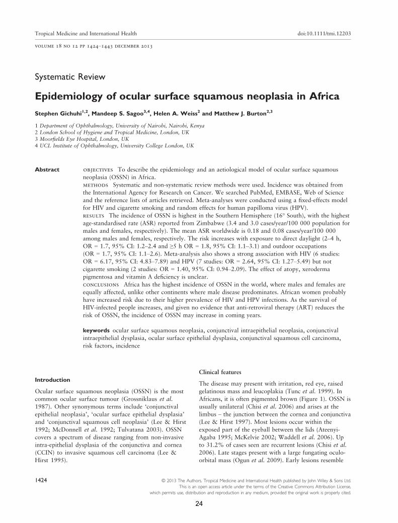
Abstract objectives To describe the epidemiology and an aetiological model of ocular surface squamous
neoplasia (OSSN) in Africa.
methods Systematic and non-systematic review methods were used. Incidence was obtained from
the International Agency for Research on Cancer. We searched PubMed, EMBASE, Web of Science
and the reference lists of articles retrieved. Meta-analyses were conducted using a fixed-effects model
for HIV and cigarette smoking and random effects for human papilloma virus (HPV).
results The incidence of OSSN is highest in the Southern Hemisphere (16� South), with the highest
age-standardised rate (ASR) reported from Zimbabwe (3.4 and 3.0 cases/year/100 000 population for
males and females, respectively). The mean ASR worldwide is 0.18 and 0.08 cases/year/100 000
among males and females, respectively. The risk increases with exposure to direct daylight (2–4 h,
OR = 1.7, 95% CI: 1.2–2.4 and ≥5 h OR = 1.8, 95% CI: 1.1–3.1) and outdoor occupations
(OR = 1.7, 95% CI: 1.1–2.6). Meta-analysis also shows a strong association with HIV (6 studies:
OR = 6.17, 95% CI: 4.83–7.89) and HPV (7 studies: OR = 2.64, 95% CI: 1.27–5.49) but notcigarette smoking (2 studies: OR = 1.40, 95% CI: 0.94–2.09). The effect of atopy, xeroderma
pigmentosa and vitamin A deficiency is unclear.
conclusions Africa has the highest incidence of OSSN in the world, where males and females are
equally affected, unlike other continents where male disease predominates. African women probably
have increased risk due to their higher prevalence of HIV and HPV infections. As the survival of
HIV-infected people increases, and given no evidence that anti-retroviral therapy (ART) reduces the
risk of OSSN, the incidence of OSSN may increase in coming years.
keywords ocular surface squamous neoplasia, conjunctival intraepithelial neoplasia, conjunctival
intraepithelial dysplasia, ocular surface epithelial dysplasia, conjunctival squamous cell carcinoma,
risk factors, incidence
Introduction
Ocular surface squamous neoplasia (OSSN) is the most
common ocular surface tumour (Grossniklaus et al.
1987). Other synonymous terms include ‘conjunctival
epithelial neoplasia’, ‘ocular surface epithelial dysplasia’
and ‘conjunctival squamous cell neoplasia’ (Lee & Hirst
1992; McDonnell et al. 1992; Tulvatana 2003). OSSN
covers a spectrum of disease ranging from non-invasive
intra-epithelial dysplasia of the conjunctiva and cornea
(CCIN) to invasive squamous cell carcinoma (Lee &
Hirst 1995).
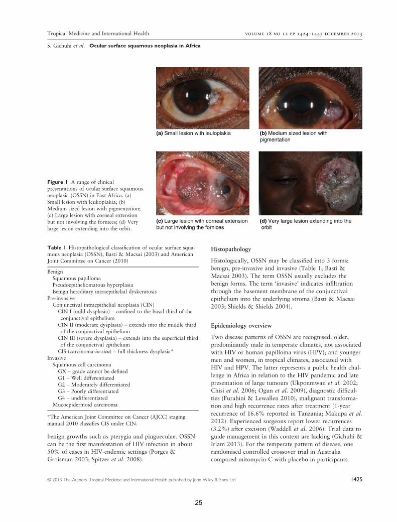
Clinical features
The disease may present with irritation, red eye, raised
gelatinous mass and leucoplakia (Tunc et al. 1999). In
Africans, it is often pigmented brown (Figure 1). OSSN is
usually unilateral (Chisi et al. 2006) and arises at the
limbus – the junction between the cornea and conjunctiva
(Lee & Hirst 1997). Most lesions occur within the
exposed part of the eyeball between the lids (Ateenyi-
Agaba 1995; McKelvie 2002; Waddell et al. 2006). Up
to 31.2% of cases seen are recurrent lesions (Chisi et al.
2006). Late stages present with a large fungating oculo-
orbital mass (Ogun et al. 2009). Early lesions resemble
1424 © 2013 The Authors. Tropical Medicine and International Health published by John Wiley & Sons Ltd.
This is an open access article under the terms of the Creative Commons Attribution License,
which permits use, distribution and reproduction in any medium, provided the original work is properly cited.
Tropical Medicine and International Health doi:10.1111/tmi.12203
volume 18 no 12 pp 1424–1443 december 2013
24
benign growths such as pterygia and pingueculae. OSSN
can be the first manifestation of HIV infection in about
50% of cases in HIV-endemic settings (Porges &
Groisman 2003; Spitzer et al. 2008).
Histopathology
Histologically, OSSN may be classified into 3 forms:
benign, pre-invasive and invasive (Table 1; Basti &
Macsai 2003). The term OSSN usually excludes the
benign forms. The term ‘invasive’ indicates infiltration
through the basement membrane of the conjunctival
epithelium into the underlying stroma (Basti & Macsai
2003; Shields & Shields 2004).
Epidemiology overview
Two disease patterns of OSSN are recognised: older,
predominantly male in temperate climates, not associated
with HIV or human papilloma virus (HPV); and younger
men and women, in tropical climates, associated with
HIV and HPV. The latter represents a public health chal-
lenge in Africa in relation to the HIV pandemic and late
presentation of large tumours (Ukponmwan et al. 2002;
Chisi et al. 2006; Ogun et al. 2009), diagnostic difficul-
ties (Furahini & Lewallen 2010), malignant transforma-
tion and high recurrence rates after treatment (1-year
recurrence of 16.6% reported in Tanzania; Makupa et al.
2012). Experienced surgeons report lower recurrences
(3.2%) after excision (Waddell et al. 2006). Trial data to
guide management in this context are lacking (Gichuhi &
Irlam 2013). For the temperate pattern of disease, one
randomised controlled crossover trial in Australia
compared mitomycin-C with placebo in participants
(a) Small lesion with leuloplakia (b) Medium sized lesion with pigmentation
(c) Large lesion with corneal extensionbut not involving the fornices
(d) Very large lesion extending into the orbit
Figure 1 A range of clinicalpresentations of ocular surface squamous
neoplasia (OSSN) in East Africa. (a)
Small lesion with leukoplakia; (b)
Medium sized lesion with pigmentation;(c) Large lesion with corneal extension
but not involving the fornices; (d) Very
large lesion extending into the orbit.
Table 1 Histopathological classification of ocular surface squa-mous neoplasia (OSSN), Basti & Macsai (2003) and American
Joint Committee on Cancer (2010)
Benign
Squamous papillomaPseudoepitheliomatous hyperplasia
Benign hereditary intraepithelial dyskeratosis
Pre-invasive
Conjunctival intraepithelial neoplasia (CIN)CIN I (mild dysplasia) – confined to the basal third of the
conjunctival epithelium
CIN II (moderate dysplasia) – extends into the middle thirdof the conjunctival epithelium
CIN III (severe dysplasia) – extends into the superficial third
of the conjunctival epithelium
CIS (carcinoma-in-situ) – full thickness dysplasia*Invasive
Squamous cell carcinoma
GX – grade cannot be defined
G1 – Well differentiatedG2 – Moderately differentiated
G3 – Poorly differentiated
G4 – undifferentiated
Mucoepidermoid carcinoma
*The American Joint Committee on Cancer (AJCC) staging
manual 2010 classifies CIS under CIN.
© 2013 The Authors. Tropical Medicine and International Health published by John Wiley & Sons Ltd. 1425
Tropical Medicine and International Health volume 18 no 12 pp 1424–1443 december 2013
S. Gichuhi et al. Ocular surface squamous neoplasia in Africa
25

whose average age was 67 years (Hirst 2007). There was
a significant treatment effect on clinically assessed com-
plete resolution of lesions (P = 0.0005), but no effect on
histologically assessed complete resolution (P = 0.49).
Incidence rates and geographical variation
Incidence estimates for OSSN are difficult to ascertain
and vary regionally (Table 2). The first paper to examine
this used cancer registry data from International Agency
for Research on Cancer (IARC; Newton et al. 1996). A
subset of these data were used in a subsequent publica-
tion looking at variation in incidence across the USA
(Emmanuel et al. 2012). However, published results need
to be interpreted with caution – firstly, all eye cancers are
classified together by the International Classification of
Diseases for Oncology (ICD-O-3 C.69) while other
databases classify squamous cell carcinoma of the
conjunctiva (SCCC) with head and neck cancers (Lee
et al. 2000; Curado et al. 2007; Parkin et al. 2010).
OSSN is not recognised as a separate entity. Squamous
cell carcinomas that are site-coded for the eye (C69)
probably include some cancers that originate in the eyelid
skin (WHO 2000, 2010; Curado et al. 2007). Secondly,
the availability of histopathology services to confirm
OSSN diagnosis is often limited in low- and middle-
income countries (Furahini & Lewallen 2010). Thirdly,
health information systems tend to capture invasive squa-
mous cell carcinoma (SCC) but not earlier stages. Coun-
tries reporting higher rates of SCC (mostly in Africa)
only started sending cancer registry data to IARC in the
mid-1980s (Curado et al. 2007). Completeness of the
current IARC database is hampered in that only data
from 80 countries were submitted, of which 75% was of
acceptable quality, and not all countries had data on
squamous cell carcinoma in the eye under code C69.
Africa had the lowest level of acceptable quality of data
(36%). Fourthly, crude incidence rates can be influenced
by population structure, a problem often addressed by
reporting age-standardised incidence rates. Finally, in
areas with limited health facilities for cancer treatment
where a large number of patients are treated outside the
reference area, incidence may be underestimated.
Moreover, in defining incidence from different sources, it
may be difficult to distinguish between recurrence or
extension of an existing cancer on one hand and the
development of a new primary on the other. Analysis of
incidence time trends is also difficult if geographical
coverage, ICD revisions and disease definitions in a
registry change.
Methods for this review
Systematic and non-systematic review methods were
used. No a priori systematic review protocol had been
published. Incidence data were obtained from the cur-
rent IARC report (9th Volume) covering the period
1998–2002. The IARC collates data from cancer regis-
tries worldwide. The report uses ICD codes to show the
age-standardised incidence per 100 000 population strat-
ified by sex and histological type. Under code C.69
where eye cancers are reported, the four main groups
are retinoblastoma, malignant melanoma, carcinomas
(11.4% of all eye cancers), sarcoma and other unspeci-
fied tumours. Under carcinomas, there are three sub-
groups – SCC (principally tumours of the conjunctiva
and cornea, comprising 70% of the carcinoma sub-
group), other specified carcinoma (adenocarcinomas of
the lacrimal gland and lacrimal duct) and unspecified
carcinomas. We extracted data from the SCC subgroup.
Table 2 Age-standardized incidence rates of squamous cell carcinoma in the eye (ICD-O-3 C.69) by continent for the period 1998–2002 (Curado et al. 2007)
Region
Age-standardized incidence rate (cases/year/100 000 pop)
P-valueMales mean (95% CI) Females mean (95% CI)
Africa 1.38 (�1.00 to 3.75) 1.18 (�1.08 to 3.43) 0.853
Central & South America 0.48 (0.33 to 0.62) 0.21 (0.10 to 0.33) 0.005Oceania 0.28 (0.14 to 0.41) 0.05 (0.01 to 0.10) 0.002
North America 0.08 (0.06 to 0.10) 0.00 (0.00 to 0.01) <0.001Asia 0.08 (0.01 to 0.14) 0.05 (0.00 to 0.09) 0.416
Europe 0.05 (0.02 to 0.08) 0.01 (0.00 to 0.03) 0.033Southern Hemisphere 0.61 (0.14 to 1.09) 0.33 (�0.12 to 0.78) 0.355
Northern Hemisphere 0.10 (0.06 to 0.14) 0.05 (0.00 to 0.08) 0.045
Worldwide estimate 0.18 (0.09 to 0.26) 0.08 (0.01 to 0.15) 0.091
CI = confidence interval.
1426 © 2013 The Authors. Tropical Medicine and International Health published by John Wiley & Sons Ltd.
Tropical Medicine and International Health volume 18 no 12 pp 1424–1443 december 2013
S. Gichuhi et al. Ocular surface squamous neoplasia in Africa
26
The coordinates locating each registry were obtained
from http://itouchmap.com/latlong.html.
We searched PubMed, EMBASE and Web of Science
for systematic reviews, meta-analysis and case–controlstudies using ‘OSSN’, ‘conjunctival squamous cell carci-
noma’, ‘risk factors’ and their synonyms as key words
with no language restrictions. Abstracts were assessed
and studies were selected if they reported analysis of
known or suspected risk factors. The search was con-
ducted on 2 January 2013 and updated on 31 May 2013.
Data were extracted from the full texts of articles and
additional articles obtained from their reference lists.
Meta-analyses were conducted where appropriate. A
fixed-effects model was used for HIV and cigarette
smoking. A random-effects model was chosen for HPV
after investigation of heterogeneity.
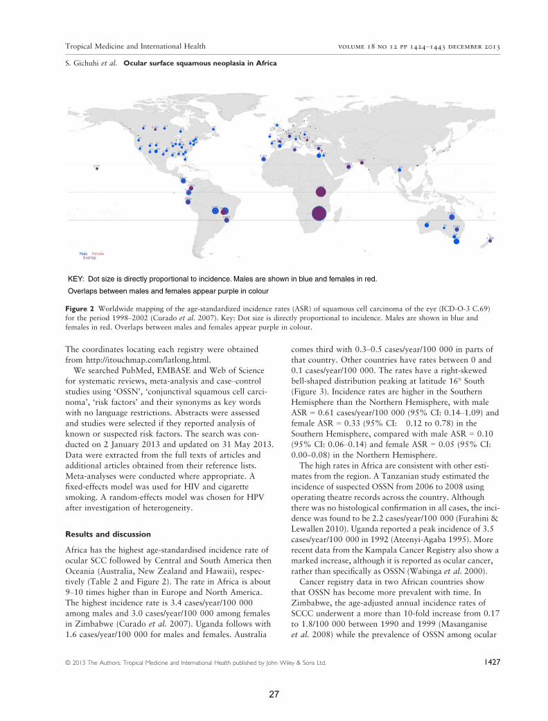
Results and discussion
Africa has the highest age-standardised incidence rate of
ocular SCC followed by Central and South America then
Oceania (Australia, New Zealand and Hawaii), respec-
tively (Table 2 and Figure 2). The rate in Africa is about
9–10 times higher than in Europe and North America.
The highest incidence rate is 3.4 cases/year/100 000
among males and 3.0 cases/year/100 000 among females
in Zimbabwe (Curado et al. 2007). Uganda follows with
1.6 cases/year/100 000 for males and females. Australia
comes third with 0.3–0.5 cases/year/100 000 in parts of
that country. Other countries have rates between 0 and
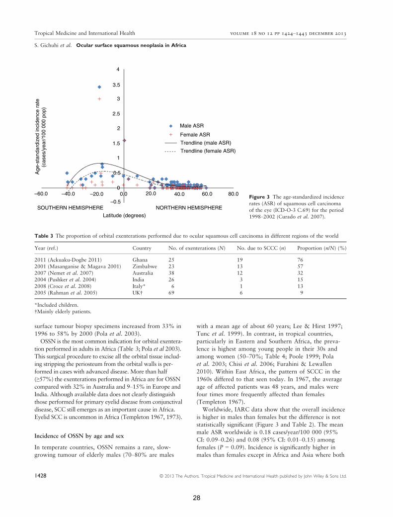
0.1 cases/year/100 000. The rates have a right-skewed
bell-shaped distribution peaking at latitude 16� South(Figure 3). Incidence rates are higher in the Southern
Hemisphere than the Northern Hemisphere, with male
ASR = 0.61 cases/year/100 000 (95% CI: 0.14–1.09) andfemale ASR = 0.33 (95% CI: �0.12 to 0.78) in the
Southern Hemisphere, compared with male ASR = 0.10
(95% CI: 0.06–0.14) and female ASR = 0.05 (95% CI:
0.00–0.08) in the Northern Hemisphere.
The high rates in Africa are consistent with other esti-
mates from the region. A Tanzanian study estimated the
incidence of suspected OSSN from 2006 to 2008 using
operating theatre records across the country. Although
there was no histological confirmation in all cases, the inci-
dence was found to be 2.2 cases/year/100 000 (Furahini &
Lewallen 2010). Uganda reported a peak incidence of 3.5
cases/year/100 000 in 1992 (Ateenyi-Agaba 1995). More
recent data from the Kampala Cancer Registry also show a
marked increase, although it is reported as ocular cancer,
rather than specifically as OSSN (Wabinga et al. 2000).
Cancer registry data in two African countries show
that OSSN has become more prevalent with time. In
Zimbabwe, the age-adjusted annual incidence rates of
SCCC underwent a more than 10-fold increase from 0.17
to 1.8/100 000 between 1990 and 1999 (Masanganise
et al. 2008) while the prevalence of OSSN among ocular
KEY: Dot size is directly proportional to incidence. Males are shown in blue and females in red.
Overlaps between males and females appear purple in colour
0.1 0.1 0.10.1
0.1
0.10.1
0.1 0.1
0.1
0.1 0.10.1
0.1
0.1
0.1
Male FemaleOverlap
0.1
0.3
0.1
0.2
0.1 0.5
0.2
0.10.1 0.1
0.1
0.1
0.1
0.1
0.4
0.3
0.4
0.4
0.7 0.4
0.4
0.1
0.1
0.1
0.3
0.1
0.10.1
0.1
0.1
0.20.2
0.10.2
0.10.2
0.3
1.6
0.2
0.2
0.5
0.3
0.2
0.20.5
0.30.1
0.2
1.6
3 3.4
0.2
0.1
0.1
0.1
0.3 0.1
0.10.1
0.1 0.2
0.1
Figure 2 Worldwide mapping of the age-standardized incidence rates (ASR) of squamous cell carcinoma of the eye (ICD-O-3 C.69)
for the period 1998–2002 (Curado et al. 2007). Key: Dot size is directly proportional to incidence. Males are shown in blue and
females in red. Overlaps between males and females appear purple in colour.
© 2013 The Authors. Tropical Medicine and International Health published by John Wiley & Sons Ltd. 1427
Tropical Medicine and International Health volume 18 no 12 pp 1424–1443 december 2013
S. Gichuhi et al. Ocular surface squamous neoplasia in Africa
27
surface tumour biopsy specimens increased from 33% in
1996 to 58% by 2000 (Pola et al. 2003).
OSSN is the most common indication for orbital exentera-
tion performed in adults in Africa (Table 3; Pola et al 2003).
This surgical procedure to excise all the orbital tissue includ-
ing stripping the periosteum from the orbital walls is per-
formed in cases with advanced disease. More than half
(≥57%) the exenterations performed in Africa are for OSSN
compared with 32% in Australia and 9–15% in Europe and
India. Although available data does not clearly distinguish
those performed for primary eyelid disease from conjunctival
disease, SCC still emerges as an important cause in Africa.
Eyelid SCC is uncommon in Africa (Templeton 1967, 1973).
Incidence of OSSN by age and sex
In temperate countries, OSSN remains a rare, slow-
growing tumour of elderly males (70–80% are males
with a mean age of about 60 years; Lee & Hirst 1997;
Tunc et al. 1999). In contrast, in tropical countries,
particularly in Eastern and Southern Africa, the preva-
lence is highest among young people in their 30s and
among women (50–70%; Table 4; Poole 1999; Pola
et al. 2003; Chisi et al. 2006; Furahini & Lewallen
2010). Within East Africa, the pattern of SCCC in the
1960s differed to that seen today. In 1967, the average
age of affected patients was 48 years, and males were
four times more frequently affected than females
(Templeton 1967).
Worldwide, IARC data show that the overall incidence
is higher in males than females but the difference is not
statistically significant (Figure 3 and Table 2). The mean
male ASR worldwide is 0.18 cases/year/100 000 (95%
CI: 0.09–0.26) and 0.08 (95% CI: 0.01–0.15) among
females (P = 0.09). Incidence is significantly higher in
males than females except in Africa and Asia where both
4
3.5
3
2.5
2
1.5
0.5
0–60.0 60.0 80.0–20.0
–0.5
–40.0 40.020.00.0
Male ASR
Trendline (male ASR)
Trendline (female ASR)
SOUTHERN HEMISPHERE NORTHERN HEMISPHERELatitude (degrees)
Age
-sta
ndar
dize
d in
cide
nce
rate
(ca
ses/
year
/100
000
pop
)
Female ASR
1
Figure 3 The age-standardized incidence
rates (ASR) of squamous cell carcinoma
of the eye (ICD-O-3 C.69) for the period
1998–2002 (Curado et al. 2007).
Table 3 The proportion of orbital exenterations performed due to ocular squamous cell carcinoma in different regions of the world
Year (ref.) Country No. of exenterations (N) No. due to SCCC (n) Proportion (n/N) (%)
2011 (Ackuaku-Dogbe 2011) Ghana 25 19 762001 (Masanganise & Magava 2001) Zimbabwe 23 13 57
2007 (Nemet et al. 2007) Australia 38 12 32
2004 (Pushker et al. 2004) India 26 3 15
2008 (Croce et al. 2008) Italy* 6 1 132005 (Rahman et al. 2005) UK† 69 6 9
*Included children.†Mainly elderly patients.
1428 © 2013 The Authors. Tropical Medicine and International Health published by John Wiley & Sons Ltd.
Tropical Medicine and International Health volume 18 no 12 pp 1424–1443 december 2013
S. Gichuhi et al. Ocular surface squamous neoplasia in Africa
28
sexes are equally affected (Table 2). Prevalence in Africa
is higher in females than males (Table 4). This may be
related to Africa having the highest prevalence of both
HIV and HPV, which may increase the risk of OSSN in
women and gender differences in mortality of HIV-
infected adults. In South Africa, HIV-infected females
have a longer life expectancy than HIV-infected males
(Cornell et al. 2012; Johnson et al. 2013; Maskew et al.
2013). Men present in later stages of HIV/AIDS for anti-
retroviral therapy (ART) and possibly have poorer adher-
ence to ART (Taylor-Smith et al. 2010). This has also
been observed in Latin America, China and Lao (Dou
et al. 2011; Gonzalez et al. 2011; Bastard et al. 2013). In
Europe, the response to ART and mortality is similar for
both sexes (Perez-Molina et al. 2012; Thorsteinsson et al.
2012).
Variation in disease severity
There may be variation in disease stage at presentation,
with more advanced disease present at time of surgery in
East Africa, compared with other regions (Table 5; Chisi
et al. 2006; Waddell et al. 2010; Kao et al. 2012;
Makupa et al. 2012). This may reflect delayed presenta-
tion to ophthalmic services in this region, leading to more
advanced pathology by the time of surgery. Histopatho-
logical reporting is also subjective, and pathologists may
not always grade tumours the same way (Margo et al.
2002). Alternatively, the disease may be intrinsically
more aggressive in the East African region or HIV
worsens disease progression.
Risk factors
Various factors are thought to influence the causation of
OSSN, but it is not clear how they interact or which is
the most potent. The rising incidence of OSSN in recent
decades may be driven by increased prevalence of these
factors. We found no systematic reviews of risk factors
for OSSN after the literature search. Of the case–controlstudies found, two in Uganda and Australia examined the
association with solar exposure; six in Africa examined
the association with HIV; sixteen examined the associa-
tion with HPV; seven in Africa, five in Asia, one in
Brazil, two in USA and one in Australia. Two studies
examined cigarette smoking in Uganda.
Ultraviolet solar radiation. Several cutaneous malignan-
cies, including melanoma and SCC, have a strong associ-
ation with solar radiation. It was first noted in the 1960s
that SCCC was relatively common in East Africa, and
this apparent excess risk was attributed to higher expo-
sure to sunlight (Templeton 1967). There is a strong rela-
tionship between the incidence of SCCC and increasing
Ultraviolet (UV) levels (Newton et al. 1996). Using IARC
data and published measurements of ambient solar ultra-
violet light, the incidence of SCCC was found to reduce
by 49% for every 10° increase in latitude from 1.2 cases/
year/100 000 (Table 7) in Uganda (latitude 0.3°) to<0.02/year/100 000 in the UK (latitude > 50°). More
recently, the National Institutes of Health/American
Association of Retired Persons (NIH-AARP) Diet and
Health Study in the USA found a slightly lower risk of
SCCC in those who lived >35° compared with ≤35° fromthe equator, although this was not statistically significant
(adjusted Hazard Ratio = 0.92, 95% CI: 0.49–1.71; Em-
manuel et al. 2012). The USA has comparatively lower
HIV prevalence, solar irradiance and incidence of OSSN
than Africa, which is bisected by the equator. The high
incidence of ocular SCC near the equator may be related
to high solar irradiance (the amount of solar radiant
energy incident on a surface per unit area and per unit
time) in the world (World Energy Council 2007).
A case–control study in Uganda adjusted for age, sex,
residential district, and HIV serostatus demonstrated that
the risk of OSSN was higher with increasing time spent
Table 4 The age and sex of patients affected by ocular surface squamous neoplasia (OSSN)
Year (ref.) Country Mean age (years) Male (%) Female (%) Male:Female ratio
1995 (Ateenyi-Agaba 1995) Uganda 33 52 48 1:2.3
2008 (Spitzer et al. 2008) Malawi 33 42 58 1:2.12010 (Simbiri et al. 2010) Botswana 39 39 61 1:1.6
2003 (Pola et al. 2003) Zimbabwe 35 30 70 1:1.4
2002 (Mahomed & Chetty 2002) S. Africa 37 50 50 1:1.32006 (Chisi et al. 2006) Kenya 38 50 50 1:1
2012 (Makupa et al. 2012) Tanzania 39 32 68 1:1
2009 (Ogun et al. 2009) Nigeria 54 43 57 1:0.9
1999 (Tunc et al. 1999) USA 64 70 30 1:0.42002 (McKelvie 2002) Australia 69 77 23 1:0.3
© 2013 The Authors. Tropical Medicine and International Health published by John Wiley & Sons Ltd. 1429
Tropical Medicine and International Health volume 18 no 12 pp 1424–1443 december 2013
S. Gichuhi et al. Ocular surface squamous neoplasia in Africa
29

in daylight (Waddell et al. 2010). Compared with those
who reported spending up to 1 h a day in direct sunlight,
the odds ratio (OR) for those who spent 2–4 h was 1.7
(95% CI: 1.2–2.4), and for those who spent 5 or more
hours a day, it was 1.8 (95% CI: 1.1–3.1). A case–con-trol study in Australia reported that the strongest risk
factor was a past history of skin cancer (OR = 15, 95%
CI: 2.0–113.6), although other factors, including outdoor
activity, pale skin and irides and propensity to burn, were
also important (Lee et al. 1994).
More direct evidence for UV radiation induced damage
in the pathophysiology of SCCC was described in another
case–control study in Uganda in which 52% of the cases
had mutations in the tumour suppressor gene TP53
compared with 14% of controls (Ateenyi-Agaba et al.
2004a). The mutations were mainly of the CC TT type,
consistent with UV-induced mutagenesis. This gene also
downregulates the replication of HPV type 16 via the
viral E2 protein, suggesting that its mutation may allow
replication of HPV particles (Brown et al. 2008). Further,
exposure to UV radiation is associated with altered
expression of matrix metalloproteinases (MMPs) and the
tissue inhibitors of these metalloproteinases (TIMPs),
molecules that may be responsible for tissue invasion and
metastasis of tumours (Ng et al. 2008).
In addition, OSSN lesions occur more often at the
limbus. A study in Uganda demonstrated that tumours
almost always occur in sun-exposed areas of the eye
(Waddell et al. 2006). It is thought that the human eye is
more exposed laterally, making this a large collecting
zone of peripheral sunlight, which, depending on the
incident angle and radius of curvature of the cornea, is
focused on the limbus, lens and lid margin, which are the
main foci of sun-related eye diseases such as pterygium,
OSSN, cataract and lid malignancies (Maloof et al.
1994). Low doses of ambient sunlight received on every
day exposure inhibit immunity in the skin and internal
organs (Halliday et al. 2012).
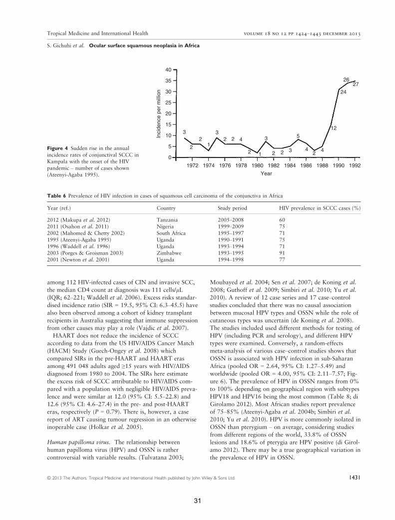
HIV. There is strong evidence that HIV is a major risk
factor for OSSN. Uganda, which had a cancer registry
since 1951, was the first country to report a dramatic
increase in the annual incidence of SCCC shortly after the
outbreak of HIV/AIDS. There was a sixfold increase from
0.6 cases/year/100 000 between 1970 and 1988 to 3.5/
year/100 000 by 1992 (Figure 4; Ateenyi-Agaba 1995). A
marked rise was also observed in the USA with the onset
of the HIV pandemic (Guech-Ongey et al. 2008). At the
same time, a US study observed a strong association in an
HIV-infected cohort (OR = 13.0, 95% CI: 4–34; Goedert
& Cote 1995). In Tanzania, regional incidence rates were
significantly correlated with regional HIV prevalence
(Pearson’s r = 0.53, P = 0.03; Furahini & Lewallen
2010). The majority of patients (60–77%) with OSSN
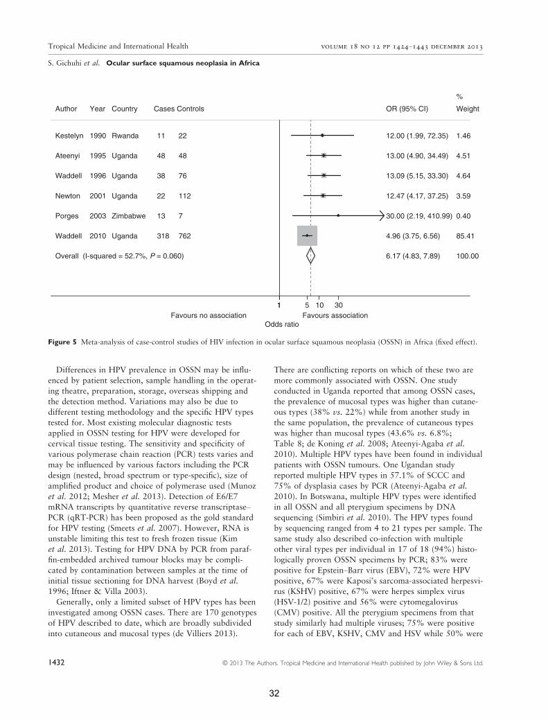
seen in Africa are HIV-infected (Table 6). A meta-analysis
of 6 case–control studies (Table 7) in Uganda, Rwanda
and Zimbabwe shows a strong association with HIV infec-
tion (pooled OR = 6.17, 95% CI: 4.83–7.89; Figure 5).
The association with HIV suggests that immunosup-
pression plays a role in OSSN; however, a linear associa-
tion between the CD4 lymphocyte count and OSSN has
not been confirmed. A cross-sectional study conducted in
Tanzania found a median CD4 cell count of 71 cells/llamong HIV-infected individuals with OSSN (Makupa
et al. 2012). HIV-infected cases tended to have larger
lesions: 71% had lesions >5 mm in diameter vs. 27%
among HIV-negative individuals (OR = 3.13, 95% CI:
1.5–6.5). HIV-infected cases were also more likely to
develop recurrent tumours within a year of excision (82%
vs. 18%; OR = 3.54, 95% CI: 1.12–11.2). However,
there was no significant trend found between CD4 count
and the grade of OSSN (P = 0.94). In a Ugandan study,
Table 5 Stages of ocular surface squamous neoplasia (OSSN) seen at presentation in Africa and USA
Country year (ref.)
Stage of OSSN, n (%)
Milddysplasia
(CIN I)
Moderatedysplasia
(CIN II)
Severedysplasia
(CIN III)
Carcinoma
in situ (CIS)
Welldifferentiated
SCC
Moderatelydifferentiated
SCC
Poorlydifferentiated
SCC
Kenya 2006 (Chisi et al. 2006) 7 (21.9) 1 (3.1) 9 (28.1) 15 (46.9)
Uganda 2008 (de Koning et al. 2008) 17 (21.0) 18 (22.2) 22 (27.2) 0 (0) 24 (29.6)
Uganda 2010
(Ateenyi-Agaba et al. 2010)39 (29.3) 94 (70.7)
Uganda 2010 (Waddell et al. 2010) 48 (15.1) 66 (20.8) 81 (25.5) 0 (0) 123 (38.7)
Tanzania 2012 (Makupa et al. 2012) 28 (21.2) 73 (55.3) 0 (0) 31 (23.5)
Malawi 2013 (Tiong et al. 2013) 1 (2.0) 5 (10.2) 9 (18.4) 17 (34.7) 17 (34.7)USA 2012 (Kao et al. 2012) 48 (8.1) 98 (16.4) 59 (9.9) 322 (54.0) 69 (11.6)
1430 © 2013 The Authors. Tropical Medicine and International Health published by John Wiley & Sons Ltd.
Tropical Medicine and International Health volume 18 no 12 pp 1424–1443 december 2013
S. Gichuhi et al. Ocular surface squamous neoplasia in Africa
30
among 112 HIV-infected cases of CIN and invasive SCC,
the median CD4 count at diagnosis was 111 cells/lL(IQR; 62–221; Waddell et al. 2006). Excess risks standar-
dised incidence ratio (SIR = 19.5, 95% CI: 6.3–45.5) havealso been observed among a cohort of kidney transplant
recipients in Australia suggesting that immune suppression
from other causes may play a role (Vajdic et al. 2007).
HAART does not reduce the incidence of SCCC
according to data from the US HIV/AIDS Cancer Match
(HACM) Study (Guech-Ongey et al. 2008) which
compared SIRs in the pre-HAART and HAART eras
among 491 048 adults aged ≥15 years with HIV/AIDS
diagnosed from 1980 to 2004. The SIRs here estimate
the excess risk of SCCC attributable to HIV/AIDS com-
pared with a population with negligible HIV/AIDS preva-
lence and were similar at 12.0 (95% CI: 5.5–22.8) and12.6 (95% CI: 4.6–27.4) in the pre- and post-HAART
eras, respectively (P = 0.79). There is, however, a case
report of ART causing tumour regression in an otherwise
inoperable case (Holkar et al. 2005).
Human papilloma virus. The relationship between
human papilloma virus (HPV) and OSSN is rather
controversial with variable results. (Tulvatana 2003;
Moubayed et al. 2004; Sen et al. 2007; de Koning et al.
2008; Guthoff et al. 2009; Simbiri et al. 2010; Yu et al.
2010). A review of 12 case series and 17 case–controlstudies concluded that there was no causal association
between mucosal HPV types and OSSN while the role of
cutaneous types was uncertain (de Koning et al. 2008).
The studies included used different methods for testing of
HPV (including PCR and serology), and different HPV
types were examined. Conversely, a random-effects
meta-analysis of various case–control studies shows that
OSSN is associated with HPV infection in sub-Saharan
Africa (pooled OR = 2.64, 95% CI: 1.27–5.49) andworldwide (pooled OR = 4.00, 95% CI: 2.11–7.57; Fig-ure 6). The prevalence of HPV in OSSN ranges from 0%
to 100% depending on geographical region with subtypes
HPV18 and HPV16 being the most common (Table 8; di
Girolamo 2012). Most African studies report prevalence
of 75–85% (Ateenyi-Agaba et al. 2004b; Simbiri et al.
2010; Yu et al. 2010). HPV is more commonly isolated in
OSSN than pterygium – on average, considering studies
from different regions of the world, 33.8% of OSSN
lesions and 18.6% of pterygia are HPV positive (di Girol-
amo 2012). There may be a true geographical variation in
the prevalence of HPV in OSSN.
1972
40
35
30
25
20
15
103 3
3
3
5
12
24
2627
2 2 2
2 2 2 22 1
1
4
4 45
0In
cide
nce
per
mill
ion
1974 1976 1978 1980 1982 1984 1986 1988 1990 1992
Year
Figure 4 Sudden rise in the annual
incidence rates of conjunctival SCCC inKampala with the onset of the HIV
pandemic – number of cases shown
(Ateenyi-Agaba 1995).
Table 6 Prevalence of HIV infection in cases of squamous cell carcinoma of the conjunctiva in Africa
Year (ref.) Country Study period HIV prevalence in SCCC cases (%)
2012 (Makupa et al. 2012) Tanzania 2005–2008 602011 (Osahon et al. 2011) Nigeria 1999–2009 75
2002 (Mahomed & Chetty 2002) South Africa 1995–1997 71
1995 (Ateenyi-Agaba 1995) Uganda 1990–1991 75
1996 (Waddell et al. 1996) Uganda 1993–1994 712003 (Porges & Groisman 2003) Zimbabwe 1993–1995 91
2001 (Newton et al. 2001) Uganda 1994–1998 77
© 2013 The Authors. Tropical Medicine and International Health published by John Wiley & Sons Ltd. 1431
Tropical Medicine and International Health volume 18 no 12 pp 1424–1443 december 2013
S. Gichuhi et al. Ocular surface squamous neoplasia in Africa
31
Differences in HPV prevalence in OSSN may be influ-
enced by patient selection, sample handling in the operat-
ing theatre, preparation, storage, overseas shipping and
the detection method. Variations may also be due to
different testing methodology and the specific HPV types
tested for. Most existing molecular diagnostic tests
applied in OSSN testing for HPV were developed for
cervical tissue testing. The sensitivity and specificity of
various polymerase chain reaction (PCR) tests varies and
may be influenced by various factors including the PCR
design (nested, broad spectrum or type-specific), size of
amplified product and choice of polymerase used (Munoz
et al. 2012; Mesher et al. 2013). Detection of E6/E7
mRNA transcripts by quantitative reverse transcriptase–PCR (qRT-PCR) has been proposed as the gold standard
for HPV testing (Smeets et al. 2007). However, RNA is
unstable limiting this test to fresh frozen tissue (Kim
et al. 2013). Testing for HPV DNA by PCR from paraf-
fin-embedded archived tumour blocks may be compli-
cated by contamination between samples at the time of
initial tissue sectioning for DNA harvest (Boyd et al.
1996; Iftner & Villa 2003).
Generally, only a limited subset of HPV types has been
investigated among OSSN cases. There are 170 genotypes
of HPV described to date, which are broadly subdivided
into cutaneous and mucosal types (de Villiers 2013).
There are conflicting reports on which of these two are
more commonly associated with OSSN. One study
conducted in Uganda reported that among OSSN cases,
the prevalence of mucosal types was higher than cutane-
ous types (38% vs. 22%) while from another study in
the same population, the prevalence of cutaneous types
was higher than mucosal types (43.6% vs. 6.8%;
Table 8; de Koning et al. 2008; Ateenyi-Agaba et al.
2010). Multiple HPV types have been found in individual
patients with OSSN tumours. One Ugandan study
reported multiple HPV types in 57.1% of SCCC and
75% of dysplasia cases by PCR (Ateenyi-Agaba et al.
2010). In Botswana, multiple HPV types were identified
in all OSSN and all pterygium specimens by DNA
sequencing (Simbiri et al. 2010). The HPV types found
by sequencing ranged from 4 to 21 types per sample. The
same study also described co-infection with multiple
other viral types per individual in 17 of 18 (94%) histo-
logically proven OSSN specimens by PCR; 83% were
positive for Epstein–Barr virus (EBV), 72% were HPV
positive, 67% were Kaposi’s sarcoma-associated herpesvi-
rus (KSHV) positive, 67% were herpes simplex virus
(HSV-1/2) positive and 56% were cytomegalovirus
(CMV) positive. All the pterygium specimens from that
study similarly had multiple viruses; 75% were positive
for each of EBV, KSHV, CMV and HSV while 50% were
Overall (I-squared = 52.7%, P = 0.060)
Waddell
Ateenyi
Newton
Author
Waddell
Kestelyn
Porges
2010
1995
2001
Year
1996
1990
2003
Uganda
Uganda
Uganda
Country
Uganda
Rwanda
Zimbabwe
318
48
22
Cases Controls
38
11
13
762
48
112
76
22
7
6.17 (4.83, 7.89)
4.96 (3.75, 6.56)
13.00 (4.90, 34.49)
12.47 (4.17, 37.25)
OR (95% CI)
13.09 (5.15, 33.30)
12.00 (1.99, 72.35)
30.00 (2.19, 410.99)
100.00
85.41
4.51
3.59
Weight
4.64
1.46
%
0.40
11 5 10 30
Odds ratioFavours no association Favours association
Figure 5 Meta-analysis of case-control studies of HIV infection in ocular surface squamous neoplasia (OSSN) in Africa (fixed effect).
1432 © 2013 The Authors. Tropical Medicine and International Health published by John Wiley & Sons Ltd.
Tropical Medicine and International Health volume 18 no 12 pp 1424–1443 december 2013
S. Gichuhi et al. Ocular surface squamous neoplasia in Africa
32

HPV positive. The proportion of HPV infection in this
series was much higher than any other studies in the
region have reported raising the question whether this
could be due to the methodology used.
The mechanism by which HPV is associated with
OSSN is unknown. HPV is associated with causation of
metaplasia in squamocolumnar epithelial transition zones
such as the corneoscleral limbus and eyelid skin of the
eye, the cervix and anus where there is active cell turn-
over and continuous cell division to replace desquamated
cells (Chow et al. 2010). HPV also promotes degradation
of the p53 gene (Scheffner et al. 1990).
The epidemiology of OSSN is closely related to that of
cervical cancer with respect to high incidence in Africa
and the association with HIV and HPV mainly types 18
and 16 (Sun et al. 1997; Clifford et al. 2003; Stanley
2010). A meta-analysis of HPV prevalence reports world-
wide shows that Africa has the highest adjusted preva-
lence (22.1%; 95% CI: 20.9–23.4%) among women with
cytologically normal cervical pap smears using
PCR-based or high-risk Hybrid Capture 2 (HC-2) tech-
nology to detect HPV DNA (de Sanjose et al. 2007).
Whether vaccination against HPV may help to reduce the
incidence of OSSN remains to be seen (Hughes et al.
2008).
Occupation. Outdoor occupations have been associated
with OSSN, probably related to UV solar radiation
exposure. In Uganda, those with outdoor occupations
had an OR of 1.7 (95% CI: 1.1–2.6) compared to those
with indoor occupations (Waddell et al. 2010). Another
in Uganda reported that 74% of 133 patients with
SCCC or dysplasia had outdoor occupations (Ateenyi-
Agaba et al. 2010). In Japan, exposure to petroleum
products was also described as a risk factor for conjunc-
tival intraepithelial neoplasia (synonym of OSSN) in a
Table 7 Characteristics of case–control studies included in the meta-analysis of HIV as a risk factor of ocular surface squamousneoplasia (OSSN)
Study period (ref.), Country Cases Controls
1989–1990(Kestelyn et al. 1990),Rwanda
11 patients with clinical evidence of conjunctivaldysplasia or malignancy seen at
Centre Hospitalier de Kigali
22 controls. 2 controls per casefrom the same area matched for age
and sex within 5 years. Referrals from
elsewhere were excluded1990–1991(Ateenyi-Agaba 1995),
Uganda
48 patients with conjunctival growths who presented
to the eye clinic at Mulago Hospital, Kampala
48 patients matched for age and sex
attending the same eye clinic with
other eye diseases
1993–1994(Waddell et al. 1996),Uganda
38 patients in seven countrywide eye clinics includingNew Mulago Hospital, Kampala who had suspicious
conjunctival lesions had excision biopsy of the lesion
76 controls. 2 controls per case matchedfor age and sex. 16 Controls were
patients in the eye clinic without
neoplasia or clinical features of HIV
disease; the remainder were general(non-eye clinic) anonymous outpatients
at the same health units
1993–1995(Porges & Groisman 2003),
Zimbabwe
13 cases from patients who underwent excisional biopsyfor conjunctival lesions at Bindura Provincial Hospital
(Mashonaland Central, Zimbabwe)
7 controls. Patients were from the samegroup as cases but had benign lesions
on histology
1994–1998(Newton et al. 2001),Uganda
22 cases. Patients aged >15 years with a provisional
diagnosis of cancer from all wards and out-patientclinics of the 4 main hospitals in Kampala: Mulago,
Nsambya, Mengo and Rubaga
112 controls. 93 patients with tumours
not suspected to be of infectiousaetiology and 19 with non-malignant
conditions
2001–2005(Waddell et al. 2010),Uganda
318 cases recruited from country-wide ophthalmology
clinics in Uganda. Anyone with a suspected OSSNwas offered surgical treatment and histology,
together with enrolment into a case-control study
762 controls were recruited from 2 sources.
The first group comprised patientsattending the ophthalmology clinics
with concerns or conditions other than
OSSN. This group also included thoseindividuals who were originally recruited
as cases, but where histology subsequently
revealed another diagnosis. The second
group comprised people who wererecruited through the voluntary HIV
counselling and testing (VCT) service
© 2013 The Authors. Tropical Medicine and International Health published by John Wiley & Sons Ltd. 1433
Tropical Medicine and International Health volume 18 no 12 pp 1424–1443 december 2013
S. Gichuhi et al. Ocular surface squamous neoplasia in Africa
33
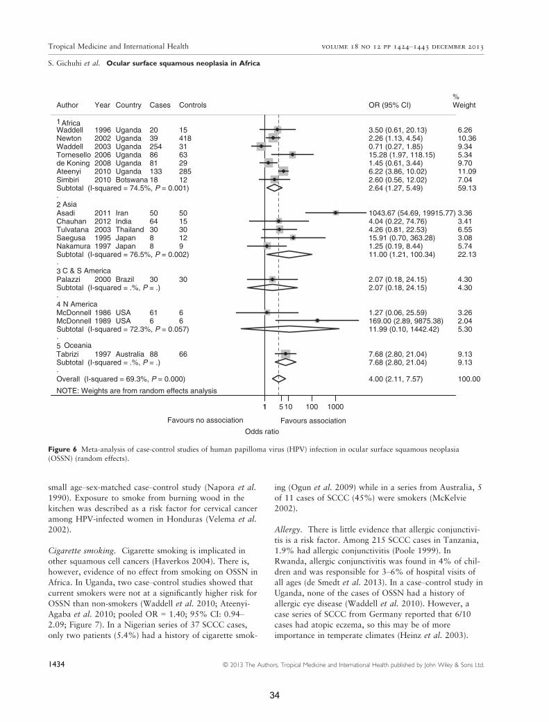
small age–sex-matched case–control study (Napora et al.
1990). Exposure to smoke from burning wood in the
kitchen was described as a risk factor for cervical cancer
among HPV-infected women in Honduras (Velema et al.
2002).
Cigarette smoking. Cigarette smoking is implicated in
other squamous cell cancers (Haverkos 2004). There is,
however, evidence of no effect from smoking on OSSN in
Africa. In Uganda, two case–control studies showed that
current smokers were not at a significantly higher risk for
OSSN than non-smokers (Waddell et al. 2010; Ateenyi-
Agaba et al. 2010; pooled OR = 1.40; 95% CI: 0.94–2.09; Figure 7). In a Nigerian series of 37 SCCC cases,
only two patients (5.4%) had a history of cigarette smok-
ing (Ogun et al. 2009) while in a series from Australia, 5
of 11 cases of SCCC (45%) were smokers (McKelvie
2002).
Allergy. There is little evidence that allergic conjunctivi-
tis is a risk factor. Among 215 SCCC cases in Tanzania,
1.9% had allergic conjunctivitis (Poole 1999). In
Rwanda, allergic conjunctivitis was found in 4% of chil-
dren and was responsible for 3–6% of hospital visits of
all ages (de Smedt et al. 2013). In a case–control study in
Uganda, none of the cases of OSSN had a history of
allergic eye disease (Waddell et al. 2010). However, a
case series of SCCC from Germany reported that 6/10
cases had atopic eczema, so this may be of more
importance in temperate climates (Heinz et al. 2003).
NOTE: Weights are from random effects analysis
.
.
.
.
.
Overall (I-squared = 69.3%, P = 0.000)
McDonnell
5
Subtotal (I-squared = 72.3%, P = 0.057)
Subtotal (I-squared = 74.5%, P = 0.001)
Author
Subtotal (I-squared = 76.5%, P = 0.002)
de Koning
McDonnell
Tabrizi
1
Palazzi
Subtotal (I-squared = .%, P = .)
Tornesello
TulvatanaSaegusa
Chauhan
Subtotal (I-squared = .%, P = .)
2
4
Waddell
SimbiriAteenyi
Asadi
3
Newton
Nakamura
Waddell
1989
Year
2008
1986
1997
2000
2006
20031995
2012
2003
20102010
2011
2002
1997
1996
USA
Country
Uganda
USA
Australia
Brazil
Uganda
ThailandJapan
India
Uganda
BotswanaUganda
Iran
Uganda
Japan
Uganda
6
Cases
81
61
88
30
86
308
64
254
18133
50
39
8
20
6
Controls
29
6
66
30
63
3012
15
31
12285
50
418
9
15
4.00 (2.11, 7.57)
169.00 (2.89, 9875.38)11.99 (0.10, 1442.42)
2.64 (1.27, 5.49)
OR (95% CI)
11.00 (1.21, 100.34)
1.45 (0.61, 3.44)
1.27 (0.06, 25.59)
7.68 (2.80, 21.04)
2.07 (0.18, 24.15)
7.68 (2.80, 21.04)
15.28 (1.97, 118.15)
4.26 (0.81, 22.53)15.91 (0.70, 363.28)
4.04 (0.22, 74.76)
2.07 (0.18, 24.15)
0.71 (0.27, 1.85)
2.60 (0.56, 12.02)6.22 (3.86, 10.02)
1043.67 (54.69, 19915.77)
2.26 (1.13, 4.54)
1.25 (0.19, 8.44)
3.50 (0.61, 20.13)
100.00
2.045.30
59.13
Weight
22.13
9.70
3.26
9.13
4.30
9.13
5.34
6.553.08
3.41
%
4.30
9.34
7.0411.09
3.36
10.36
5.74
6.26Africa
Asia
C & S America
N America
Oceania
11 5 10 100 1000
Odds ratio
Favours no association Favours association
Figure 6 Meta-analysis of case-control studies of human papilloma virus (HPV) infection in ocular surface squamous neoplasia(OSSN) (random effects).
1434 © 2013 The Authors. Tropical Medicine and International Health published by John Wiley & Sons Ltd.
Tropical Medicine and International Health volume 18 no 12 pp 1424–1443 december 2013
S. Gichuhi et al. Ocular surface squamous neoplasia in Africa
34
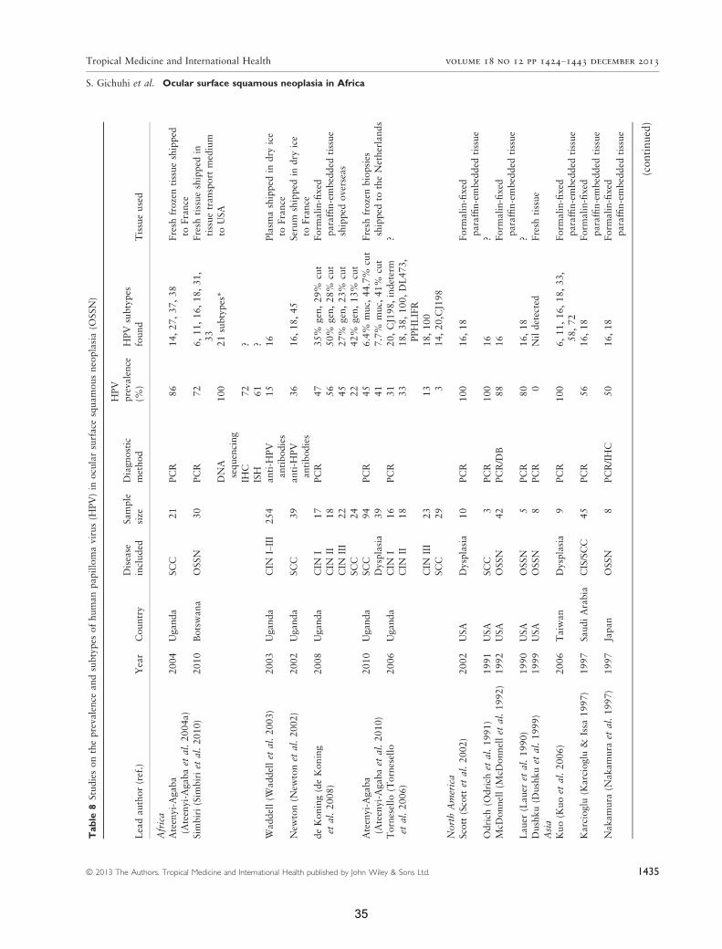
Table
8Studiesontheprevalence
andsubtypes
ofhumanpapillomavirus(H
PV)in
ocularsurfacesquamousneoplasia(O
SSN)
Leadauthor(ref.)
Year
Country
Disease
included
Sample
size
Diagnostic
method
HPV
prevalence
(%)
HPV
subtypes
found
Tissueused
Africa
Ateenyi-Agaba
(Ateenyi-Agabaet
al.2004a)
2004
Uganda
SCC
21
PCR
86
14,27,37,38
Fresh
frozentissueshipped
toFrance
Sim
biri(Sim
biriet
al.2010)
2010
Botswana
OSSN
30
PCR
72
6,11,16,18,31,
33
Fresh
tissueshipped
in
tissuetransport
medium
toUSA
DNA
sequencing
100
21subtypes*
IHC
72
?
ISH
61
?W
addell(W
addellet
al.2003)
2003
Uganda
CIN
I–III
254
anti-H
PV
antibodies
15
16
Plasm
ashipped
indry
ice
toFrance
New
ton(N
ewtonet
al.2002)
2002
Uganda
SCC
39
anti-H
PV
antibodies
36
16,18,45
Serum
shipped
indry
ice
toFrance
deKoning(deKoning
etal.2008)
2008
Uganda
CIN
I17
PCR
47
35%
gen,29%
cut
Form
alin-fixed
paraffin-embedded
tissue
shipped
overseas
CIN
II18
56
50%
gen,28%
cut
CIN
III
22
45
27%
gen,23%
cut
SCC
24
22
42%
gen,13%
cut
Ateenyi-Agaba
(Ateenyi-Agabaet
al.2010)
2010
Uganda
SCC
94
PCR
45
6.4%
muc,
44.7%
cut
Fresh
frozenbiopsies
shipped
totheNetherlands
Dysplasia
39
41
7.7%
muc,
41%
cut
Tornesello
(Tornesello
etal.2006)
2006
Uganda
CIN
I16
PCR
31
20,CJ198,indeterm
?CIN
II18
33
18,38,100,DL473,
PPHLIFR
CIN
III
23
13
18,100
SCC
29
314,20,CJ198
NorthAmerica
Scott(Scottet
al.2002)
2002
USA
Dysplasia
10
PCR
100
16,18
Form
alin-fixed
paraffin-embedded
tissue
Odrich
(Odrich
etal.1991)
1991
USA
SCC
3PCR
100
16
?
McD
onnell(M
cDonnellet
al.1992)
1992
USA
OSSN
42
PCR/D
B88
16
Form
alin-fixed
paraffin-embedded
tissue
Lauer
(Lauer
etal.1990)
1990
USA
OSSN
5PCR
80
16,18
?Dushku(D
ushkuet
al.1999)
1999
USA
OSSN
8PCR
0Nildetected
Fresh
tissue
Asia
Kuo(K
uoet
al.2006)
2006
Taiw
an
Dysplasia
9PCR
100
6,11,16,18,33,
58,72
Form
alin-fixed
paraffin-embedded
tissue
Karcioglu
(Karcioglu
&Issa
1997)
1997
SaudiArabia
CIS/SCC
45
PCR
56
16,18
Form
alin-fixed
paraffin-embedded
tissue
Nakamura
(Nakamura
etal.1997)
1997
Japan
OSSN
8PCR/IHC
50
16,18
Form
alin-fixed
paraffin-embedded
tissue
(continued)
© 2013 The Authors. Tropical Medicine and International Health published by John Wiley & Sons Ltd. 1435
Tropical Medicine and International Health volume 18 no 12 pp 1424–1443 december 2013
S. Gichuhi et al. Ocular surface squamous neoplasia in Africa
35
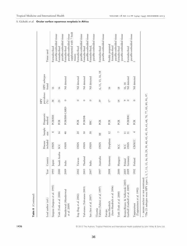
Table
8(C
ontinued)
Leadauthor(ref.)
Year
Country
Disease
included
Sample
size
Diagnostic
method
HPV
prevalence
(%)
HPV
subtypes
found
Tissueused
Saegusa
(Saegusa
etal.1995)
1995
Japan
OSSN
8PCR/ISH
38
16
Form
alin-fixed
paraffin-embedded
tissue
Toth
(Toth
etal.2000)
2000
SaudiArabia
SCC
16
PCR
25
16
Form
alin-fixed
paraffin-embedded
tissue
Manderwad
(Manderwad
etal.2009)
2009
India
OSSN
48
PCR/ISH-C
ARD
0Nildetected
Form
alin-fixed
paraffin-embedded
tissue
supplementedwith7fresh
tissues
Eng(Enget
al.2002)
2002
Taiwan
OSSN
20
PCR
0Nildetected
Form
alin-fixed
paraffin-embedded
tissue
Tulvatana(Tulvatana2003)
2003
Thailand
OSSN
30
PCR/D
B0
Nildetected
Form
alin-fixed
paraffin-embedded
tissue
Sen
(Sen
etal.2007)
2007
India
OSSN
30
IHC
0Nildetected
Form
alin-fixed,
paraffin-embedded
tissue
Ocean
iaTabrizi
(Tabrizi
etal.1997)
1997
Australia
OSSN
88
PCR
39
6,11,13,16,18
Form
alin-fixed
paraffin-embedded
tissue
Europe
Auw-H
aedrich
(Auw-H
aedrich
etal.2006)
2008
Germany
Dysplasia
12
PCR
17
16
Freshly
prepared
form
alin-fixed
paraffin-embedded
tissue
Toth
(Toth
etal.2000)
2000
Hungary
SCC
7PCR
14
18
Form
alin-fixed
paraffin-embedded
tissue
Reszec(Reszec&
Sulkowski2005)
2005
Poland
SCC
11
916,18
?
Guthoff
(Guthoffet
al.2009)
2009
Germany
OSSN
31
PCR/IHC
0Nildetected
Form
alin-fixed
paraffin-embedded
tissue
Tuppurainen
(Tuppurainen
etal.1992)
1992
Finland
CIS/SCC
4PCR
0Nildetected
?
?–meansunclearornotmentioned.
*The21subtypes
wereHPV
types
1,3,7,11,13,16,18,29,39,40,43,45,59,61,68,70,77,85,89,91,97.
1436 © 2013 The Authors. Tropical Medicine and International Health published by John Wiley & Sons Ltd.
Tropical Medicine and International Health volume 18 no 12 pp 1424–1443 december 2013
S. Gichuhi et al. Ocular surface squamous neoplasia in Africa
36
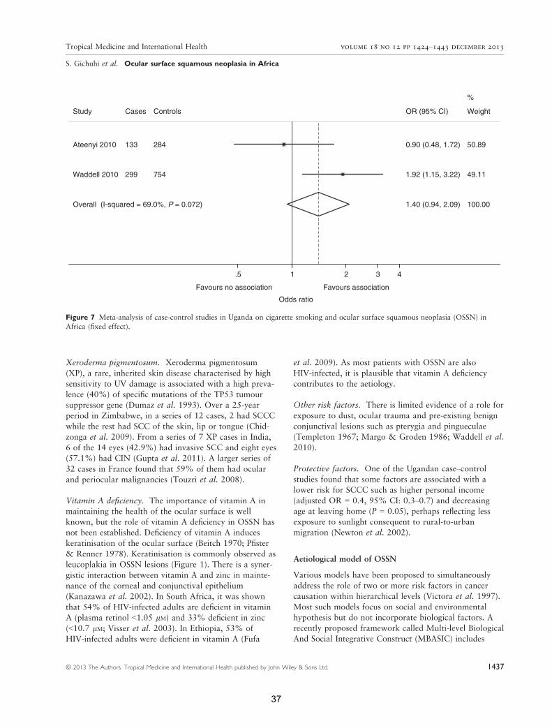
Xeroderma pigmentosum. Xeroderma pigmentosum
(XP), a rare, inherited skin disease characterised by high
sensitivity to UV damage is associated with a high preva-
lence (40%) of specific mutations of the TP53 tumour
suppressor gene (Dumaz et al. 1993). Over a 25-year
period in Zimbabwe, in a series of 12 cases, 2 had SCCC
while the rest had SCC of the skin, lip or tongue (Chid-
zonga et al. 2009). From a series of 7 XP cases in India,
6 of the 14 eyes (42.9%) had invasive SCC and eight eyes
(57.1%) had CIN (Gupta et al. 2011). A larger series of
32 cases in France found that 59% of them had ocular
and periocular malignancies (Touzri et al. 2008).
Vitamin A deficiency. The importance of vitamin A in
maintaining the health of the ocular surface is well
known, but the role of vitamin A deficiency in OSSN has
not been established. Deficiency of vitamin A induces
keratinisation of the ocular surface (Beitch 1970; Pfister
& Renner 1978). Keratinisation is commonly observed as
leucoplakia in OSSN lesions (Figure 1). There is a syner-
gistic interaction between vitamin A and zinc in mainte-
nance of the corneal and conjunctival epithelium
(Kanazawa et al. 2002). In South Africa, it was shown
that 54% of HIV-infected adults are deficient in vitamin
A (plasma retinol <1.05 lM) and 33% deficient in zinc
(<10.7 lM; Visser et al. 2003). In Ethiopia, 53% of
HIV-infected adults were deficient in vitamin A (Fufa
et al. 2009). As most patients with OSSN are also
HIV-infected, it is plausible that vitamin A deficiency
contributes to the aetiology.
Other risk factors. There is limited evidence of a role for
exposure to dust, ocular trauma and pre-existing benign
conjunctival lesions such as pterygia and pingueculae
(Templeton 1967; Margo & Groden 1986; Waddell et al.
2010).
Protective factors. One of the Ugandan case–controlstudies found that some factors are associated with a
lower risk for SCCC such as higher personal income
(adjusted OR = 0.4, 95% CI: 0.3–0.7) and decreasing
age at leaving home (P = 0.05), perhaps reflecting less
exposure to sunlight consequent to rural-to-urban
migration (Newton et al. 2002).
Aetiological model of OSSN
Various models have been proposed to simultaneously
address the role of two or more risk factors in cancer
causation within hierarchical levels (Victora et al. 1997).
Most such models focus on social and environmental
hypothesis but do not incorporate biological factors. A
recently proposed framework called Multi-level Biological
And Social Integrative Construct (MBASIC) includes
Overall (I-squared = 69.0%, P = 0.072)
Study
Ateenyi 2010
Waddell 2010
Cases
133
299
Controls
284
754
1.40 (0.94, 2.09)
OR (95% CI)
0.90 (0.48, 1.72)
1.92 (1.15, 3.22)
100.00
Weight
%
50.89
49.11
.5 1 2 3 4
Odds ratio
Favours no association Favours association
Figure 7 Meta-analysis of case-control studies in Uganda on cigarette smoking and ocular surface squamous neoplasia (OSSN) in
Africa (fixed effect).
© 2013 The Authors. Tropical Medicine and International Health published by John Wiley & Sons Ltd. 1437
Tropical Medicine and International Health volume 18 no 12 pp 1424–1443 december 2013
S. Gichuhi et al. Ocular surface squamous neoplasia in Africa
37
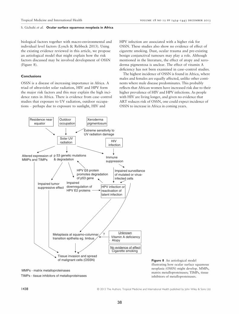
biological factors together with macro-environmental and
individual level factors (Lynch & Rebbeck 2013). Using
the existing evidence reviewed in this article, we propose
an aetiological model that might explain how the risk
factors discussed may be involved development of OSSN
(Figure 8).
Conclusions
OSSN is a disease of increasing importance in Africa. A
triad of ultraviolet solar radiation, HIV and HPV form
the major risk factors and this may explain the high inci-
dence rates in Africa. There is evidence from case–controlstudies that exposure to UV radiation, outdoor occupa-
tions – perhaps due to exposure to sunlight, HIV and
HPV infection are associated with a higher risk for
OSSN. These studies also show no evidence of effect of
cigarette smoking. Dust, ocular trauma and pre-existing
benign conjunctival tumours may play a role. Although
mentioned in the literature, the effect of atopy and xero-
derma pigmentosa is unclear. The effect of vitamin A
deficiency has not been examined in case–control studies.The highest incidence of OSSN is found in Africa, where
males and females are equally affected, unlike other conti-
nents where male disease predominates. This probably
reflects that African women have increased risk due to their
higher prevalence of HIV and HPV infections. As people
with HIV are living longer, and given no evidence that
ART reduces risk of OSSN, one could expect incidence of
OSSN to increase in Africa in coming years.
Residence near equator
Outdooroccupation
Solar UVradiation
Altered expression ofMMPs and TIMPs
p 53 genetic mutations& degradation
Impaired tumorsuppressive effect
Impaireddownregulation ofHPV E2 proteins
HPV infection orreactivation oflatent infection
Metaplasia at squamo-columnartransition epithelia eg. limbus
?Vitamin A deficiencyAtopy
Cigarette smoking
Unknown
No evidence of effect
Tissue invasion and spreadof malignant cells (OSSN)
MMPs - matrix metalloproteinases
TIMPs - tissue inhibitors of metalloproteinases
HPV E6 proteinpromotes degradationof p53 gene
Immunesuppression
Impaired surveillanceof mutated or virus-infected cells
HIVinfection
Xerodermapigmentosum
Extreme sensitivity toUV radiation damage
Figure 8 An aetiological model
illustrating how ocular surface squamous
neoplasia (OSSN) might develop. MMPs,matrix metalloproteinases; TIMPs, tissue
inhibitors of metalloproteinases.
1438 © 2013 The Authors. Tropical Medicine and International Health published by John Wiley & Sons Ltd.
Tropical Medicine and International Health volume 18 no 12 pp 1424–1443 december 2013
S. Gichuhi et al. Ocular surface squamous neoplasia in Africa
38
Currently, the best available options for OSSN control
remain early detection and effective treatment. However,
there are no early non-invasive diagnostic methods in use
and no trial evidence to guide treatment. OSSN is
currently largely neglected by both eye and HIV care
programmes. Eye care programmes prioritise preventable
blindness while OSSN often in early stages does not
affect vision. OSSN may, however, lead to facial disfig-
urement and death in late stages. In Africa, a key
research question is whether scale-up of ART and HPV
vaccination will impact on OSSN.
Acknowledgements
SG received funding from the British Council for
Prevention of Blindness (BCPB) to conduct this study.
MJB is supported by The Wellcome Trust (Grant
Number 098481/Z/12/Z). We acknowledge Benjamin D.
Hennig from the University of Oxford (http://www.view
softheworld.net) for help with preparing the incidence
map (Figure 2).
References
Ackuaku-Dogbe E (2011) Review of orbital exenterations in
Korle-Bu teaching hospital. Ghana Medical Journal 45, 45–49.
American Joint Committee on Cancer 2010. Carcinoma of the
conjunctiva. In: Cancer Staging Handbook, 7th edn (eds SB
Edge, DR Byrd, CC Compton, AG Fritz, FL Greene & A
Trotti) Springer, New York, 597.
Ateenyi-Agaba C (1995) Conjunctival squamous-cell carcinoma
associated with HIV infection in Kampala, Uganda. Lancet
345, 695–696.Ateenyi-Agaba C, Dai M, le Calvez F et al. (2004a) TP53 muta-
tions in squamous-cell carcinomas of the conjunctiva: evidence
for UV-induced mutagenesis. Mutagenesis 19, 399–401.
Ateenyi-Agaba C, Weiderpass E, Smet A et al. (2004b) Epiderm-
odysplasia verruciformis human papillomavirus types and
carcinoma of the conjunctiva: a pilot study. British Journal of
Cancer 90, 1777–1779.
Ateenyi-Agaba C, Franceschi S, Wabwire-Mangen F et al. (2010)
Human papillomavirus infection and squamous cell carci-
noma of the conjunctiva. British Journal of Cancer, 102,
262–267.
Auw-Haedrich C, Sundmacher R, Freudenberg N et al. (2006)
Expression of p63 in conjunctival intraepithelial neoplasia and
squamous cell carcinoma. Graefes Archive for Clinical and
Experimental Ophthalmology 244, 96–103.
Bastard M, Soulinphumy K, Phimmasone P et al. (2013) Women
experience a better long- term immune recovery and a better
survival on HAART in Lao People’s Democratic Republic.
BMC Infectious Diseases 13, 27.
Basti S & Macsai MS (2003) Ocular surface squamous
neoplasia: a review. Cornea 22, 687–704.
Beitch I (1970) The induction of keratinization in the corneal
epithelium. A comparison of the “dry” and vitamin A-deficient
eyes. Investigative Ophthalmology 9, 827–843.Boyd AS, Annarella M, Rapini RP, Adler-Storthz K & Duvic M
(1996) False-positive polymerase chain reaction results for
human papillomavirus in lichen planus. Potential laboratory
pitfalls of this procedure. Journal of the American Academy of
Dermatology 35, 42–46.
Brown C, Kowalczyk AM, Taylor ER, Morgan IM & Gaston K
(2008) P53 represses human papillomavirus type 16 DNA
replication via the viral E2 protein. Virology Journal 5, 5.
Chidzonga MM, Mahomva L, Makunike-Mutasa R & Masanga-
nise R (2009) Xeroderma pigmentosum: a retrospective case
series in Zimbabwe. Journal of Oral and Maxillofacial Surgery
67, 22–31.Chisi SK, Kollmann MK & Karimurio J (2006) Conjunctival
squamous cell carcinoma in patients with human immunodefi-
ciency virus infection seen at two hospitals in Kenya. East
African Medical Journal 83, 267–270.Chow LT, Broker TR & Steinberg BM (2010) The natural
history of human papillomavirus infections of the mucosal
epithelia. APMIS: Acta Pathologica, Microbiologica, et
Immunologica Scandinavica 118, 422–449.
Clifford GM, Smith JS, PlummerM,MunozN&Franceschi S
(2003) Human papillomavirus types in invasive cervical cancer
worldwide: a meta-analysis.British Journal of Cancer 88, 63–73.Cornell M, Schomaker M, Garone DB et al. (2012) Gender dif-
ferences in survival among adult patients starting antiretroviral
therapy in South Africa: a multicentre cohort study. PLoS
Medicine 9, e1001304.
Croce A, Moretti A, D’agostino L & Zingariello P (2008) Orbi-
tal exenteration in elderly patients: personal experience. Acta
Otorhinolaryngologica Italica 28, 193–199.
Curado MP, Edwards B, Shin HR et al. (eds.) (2007). Cancer
incidence in Five Continents. International Agency for
Research on Cancer, Lyon, France.
de Koning MN, Waddell K, Magyezi J et al. (2008) Genital and
cutaneous human papillomavirus (HPV) types in relation to
conjunctival squamous cell neoplasia: a case- control study in
Uganda. Infectious Agents and Cancer 3, 12.
de Sanjose S, Diaz M, Castellsague X et al. (2007) Worldwide
prevalence and genotype distribution of cervical human papil-
lomavirus DNA in women with normal cytology: a meta-
analysis. The Lancet Infectious Diseases 7, 453–459.de Smedt S, Wildner G & Kestelyn P (2013) Vernal keratoconjunc-
tivitis: an update. British Journal of Ophthalmology 97, 9–14.de Villiers EM (2013). Cross-roads in the classification of
papillomaviruses. Virology.
di Girolamo N (2012) Association of human papilloma virus
with pterygia and ocular-surface squamous neoplasia. Eye
(London, England) 26, 202–211.
Dou Z, Xu J, Jiao JH et al. (2011) Gender difference in 2-year
mortality and immunological response to ART in an HIV-
infected Chinese population 2006–2008. PLoS One 6, e22707.
Dumaz N, Drougard C, Sarasin A & Daya-Grosjean L (1993)
Specific UV-induced mutation spectrum in the p53 gene of
Tropical Medicine and International Health volume 18 no 12 pp 1424–1443 december 2013
S. Gichuhi et al. Ocular surface squamous neoplasia in Africa
© 2013 The Authors. Tropical Medicine and International Health published by John Wiley & Sons Ltd. 1439
39

skin tumors from DNA-repair-deficient xeroderma pigmento-
sum patients. Proceedings of the National Academy of
Sciences, USA 90, 10529–10533.Dushku N, Hatcher SL, Albert DM & Reid TW (1999) p53
expression and relation to human papillomavirus infection in
pingueculae, pterygia, and limbal tumors. Archives of
Ophthalmology 117, 1593–1599.Emmanuel B, Ruder E, Lin SW, Abnet C, Hollenbeck A & Mbu-
laiteye S (2012) Incidence of squamous-cell carcinoma of the
conjunctiva and other eye cancers in the NIH-AARP Diet and
Health Study. Ecancermedicalscience 6, 254.
Eng HL, Lin TM, Chen SY, Wu SM & Chen WJ (2002)
Failure to detect human papillomavirus DNA in malignant
epithelial neoplasms of conjunctiva by polymerase chain
reaction. American Journal of Clinical Pathology 117,
429–436.
Fufa H, Umeta M, Taffesse S, Mokhtar N & Aguenaou H
(2009) Nutritional and immunological status and their associa-
tions among HIV-infected adults in Addis Ababa, Ethiopia.
Food and Nutrition Bulletin 30, 227–232.
Furahini G & Lewallen S (2010) Epidemiology and management
of ocular surface squamous neoplasia in Tanzania. Ophthal-
mic Epidemiology 17, 171–176.
Gichuhi S & Irlam JH (2013) Interventions for squamous cell
carcinoma of the conjunctiva in HIV-infected individuals.
Cochrane Database Systematic Review, 2, CD005643.
Goedert JJ & Cote TR (1995) Conjunctival malignant disease
with AIDS in USA. Lancet 346, 257–258.Gonzalez MA, Martin L, Munoz S & Jacobson JO (2011)
Patterns, trends and sex differences in HIV/AIDS reported
mortality in Latin American countries: 1996–2007. BMC
Public Health 11, 605.
Grossniklaus HE, Green WR, Luckenbach M & Chan CC
(1987) Conjunctival lesions in adults. A clinical and histopath-
ologic review. Cornea 6, 78–116.
Guech-Ongey M, Engels EA, Goedert JJ, Biggar RJ & Mbulait-
eye SM (2008) Elevated risk for squamous cell carcinoma of
the conjunctiva among adults with AIDS in the United States.
International Journal of Cancer 122, 2590–2593.
Gupta N, Sachdev R & Tandon R (2011) Ocular surface squa-
mous neoplasia in xeroderma pigmentosum: clinical spectrum
and outcome. Graefes Archive for Clinical and Experimental
Ophthalmology 249, 1217–1221.
Guthoff R, Marx A & Stroebel P (2009) No evidence for a path-
ogenic role of human papillomavirus infection in ocular
surface squamous neoplasia in Germany. Current Eye
Research 34, 666–671.
Halliday GM, Damian DL, Rana S & Byrne SN (2012) The sup-
pressive effects of ultraviolet radiation on immunity in the skin
and internal organs: implications for autoimmunity. Journal of
Dermatological Science 66, 176–182.
Haverkos HW (2004) Viruses, chemicals and co-carcinogenesis.
Oncogene 23, 6492–6499.
Heinz C, Fanihagh F & Steuhl KP (2003) Squamous cell carci-
noma of the conjunctiva in patients with atopic eczema.
Cornea 22, 135–137.
Hirst LW (2007) Randomized controlled trial of topical mitomy-
cin C for ocular surface squamous neoplasia: early resolution.
Ophthalmology 114, 976–982.Holkar S, Mudhar HS, Jain A et al. (2005) Regression of inva-
sive conjunctival squamous carcinoma in an HIV-positive
patient on antiretroviral therapy. International Journal of STD
and AIDS 16, 782–783.Hughes DS, Powell N & Fiander AN (2008) Will vaccination
against human papillomavirus prevent eye disease? A
review of the evidence. British Journal of Ophthalmology 92,
460–465.Iftner T & Villa LL (2003) Chapter 12: Human papillomavirus
technologies. Journal of the National Cancer Institute, Mono-
graphs 31, 80–88.
Johnson LF, Mossong J, Dorrington RE et al. (2013) Life expec-
tancies of South African adults starting antiretroviral treat-
ment: collaborative analysis of cohort studies. PLoS Medicine
10, e1001418.
Kanazawa S, Kitaoka T, Ueda Y, Gong H & Amemiya T (2002)
Interaction of zinc and vitamin A on the ocular surface. Grae-
fes Archive for Clinical and Experimental Ophthalmology
240, 1011–1021.
Kao AA, Galor A, Karp CL, Abdelaziz A, Feuer WJ & Dubovy
SR (2012) Clinicopathologic correlation of ocular surface
squamous neoplasms at bascom palmer eye institute: 2001 to
2010. Ophthalmology 119, 1773–1776.Karcioglu ZA & Issa TM (1997) Human papilloma virus in
neoplastic and non-neoplastic conditions of the external eye.
British Journal of Ophthalmology 81, 595–598.
Kestelyn P, Stevens AM, Ndayambaje A, Hanssens M & van de
Perre P (1990) HIV and conjunctival malignancies. Lancet
336, 51–52.Kim Y, Choi KR, Chae MJ et al. (2013) Stability of DNA,
RNA, cytomorphology, and immunoantigenicity in Residual
ThinPrep Specimens. APMIS: Acta Pathologica, Microbiolog-
ica, et Immunologica Scandinavica doi: 10.1111/apm.12082
[Epub ahead of print].
Kuo KT, Chang HC, Hsiao CH & Lin MC (2006) Increased Ki-
67 proliferative index and absence of P16INK4 in CIN-HPV
related pathogenic pathways different from cervical squamous
intraepithelial lesion. British Journal of Ophthalmology 90,
894–899.Lauer SA, Malter JS & Meier JR (1990) Human papillomavirus
type 18 in conjunctival intraepithelial neoplasia. American
Journal of Ophthalmology 110, 23–27.
Lee GA & Hirst LW (1992) Incidence of ocular surface epithe-
lial dysplasia in metropolitan Brisbane. A 10-year survey.
Archives of Ophthalmology 110, 525–527.Lee GA & Hirst LW (1995) Ocular surface squamous neoplasia.
Survey of Ophthalmology 39, 429–450.Lee GA & Hirst LW (1997) Retrospective study of ocular sur-
face squamous neoplasia. Australian and New Zealand Journal
of Ophthalmology 25, 269–276.
Lee GA, Williams G, Hirst LW & Green AC (1994) Risk factors
in the development of ocular surface epithelial dysplasia.
Ophthalmology 101, 360–364.
Tropical Medicine and International Health volume 18 no 12 pp 1424–1443 december 2013
S. Gichuhi et al. Ocular surface squamous neoplasia in Africa
1440 © 2013 The Authors. Tropical Medicine and International Health published by John Wiley & Sons Ltd.
40
Lee SB, Au Eong KG, Saw SM, Chan TK & Lee HP (2000) Eye
cancer incidence in Singapore. British Journal of Ophthalmol-
ogy, 84, 767–770.Lynch SM & Rebbeck TR (2013) Bridging the gap between bio-
logic, individual, and macroenvironmental factors in cancer: a
multilevel approach. Cancer Epidemiology Biomarkers &
Prevention 22, 485–495.Mahomed A & Chetty R (2002) Human immunodeficiency virus
infection, Bcl-2, p53 protein, and Ki-67 analysis in ocular surface
squamous neoplasia. Archives of Ophthalmology 120, 554–558.
Makupa II, Swai B, Makupa WU, White VA & Lewallen S
(2012) Clinical factors associated with malignancy and HIV
status in patients with ocular surface squamous neoplasia at
Kilimanjaro Christian Medical Centre, Tanzania. British Jour-
nal of Ophthalmology, 96, 482–484.Maloof AJ, Ho A & Coroneo MT (1994) Influence of corneal
shape on limbal light focusing. Investigative Ophthalmology
& Visual Science 35, 2592–2598.
Manderwad GP, Kannabiran C, Honavar SG & Vemuganti GK
(2009) Lack of association of high-risk human papillomavirus
in ocular surface squamous neoplasia in India. Archives of
Pathology and Laboratory Medicine 133, 1246–1250.
Margo CE & Groden LR (1986) Squamous cell carcinoma of
the cornea and conjunctiva following a thermal burn of the
eye. Cornea 5, 185–188.
Margo CE, Harman LE & Mulla ZD (2002) The reliability of
clinical methods in ophthalmology. Survey of Ophthalmology
47, 375–386.Masanganise R & Magava A (2001) Orbital exenterations and
squamous cell carcinoma of the conjunctiva at Sekuru Kaguvi
Eye Unit, Zimbabwe. Central African Journal of Medicine 47,
196–199.Masanganise R, Rusakaniko S, Makunike R et al. (2008) A
historical perspective of registered cases of malignant ocular
tumors in Zimbabwe (1990 to 1999). Is HIV infection a
factor? Central African Journal of Medicine 54, 28–32.Maskew M, Brennan AT, Westreich D, McNamara L, Macphail
AP & Fox MP (2013) Gender differences in mortality and
CD4 count response among virally suppressed HIV- positive
patients. Journal of Women’s Health (2002) 22, 113–120.McDonnell JM, McDonnell PJ & Sun YY (1992) Human papil-
lomavirus DNA in tissues and ocular surface swabs of patients
with conjunctival epithelial neoplasia. Investigative Ophthal-
mology & Visual Science 33, 184–189.McKelvie PA (2002) Squamous cell carcinoma of the conjunc-
tiva: a series of 26 cases. British Journal of Ophthalmology
86, 168–173.
Mesher D, Szarewski A, Cadman L et al. (2013) Comparison of
human papillomavirus testing strategies for triage of women
referred with low-grade cytological abnormalities. European
Journal of Cancer 49, 2179–2186.
Moubayed P, Mwakyoma H & Schneider DT (2004) High Fre-
quency of Human Papillomavirus 6/11, 16, and 18 Infections
in Precancerous Lesions and Squamous Cell Carcinoma of the
Conjunctiva in Subtropical Tanzania. American Journal of
Clinical Pathology 122, 938–943.
Munoz M, Camargo M, Soto-De Leon SC et al. (2012) The
diagnostic performance of classical molecular tests used for
detecting human papillomavirus. Journal of Virological Meth-
ods, 185, 32–38.
Nakamura Y, Mashima Y, Kameyama K, Mukai M & Oguchi Y
(1997) Detection of human papillomavirus infection in squa-
mous tumours of the conjunctiva and lacrimal sac by immuno-
histochemistry, in situ hybridisation, and polymerase chain
reaction. British Journal of Ophthalmology 81, 308–313.Napora C, Cohen EJ, Genvert GI et al. (1990) Factors associated
with conjunctival intraepithelial neoplasia: a case control
study. Ophthalmic Surgery 21, 27–30.
Nemet AY, Martin P, Benger R et al. (2007) Orbital exentera-
tion: a 15-year study of 38 cases. Ophthalmic Plastic and
Reconstructive Surgery 23, 468–472.Newton R, Ferlay J, Reeves G, Beral V & Parkin DM (1996)
Effect of ambient solar ultraviolet radiation on incidence of
squamous-cell carcinoma of the eye. Lancet 347, 1450–1451.
Newton R, Ziegler J, Beral V et al. (2001) A case-control study
of human immunodeficiency virus infection and cancer in
adults and children residing in Kampala, Uganda. Interna-
tional Journal of Cancer 92, 622–627.
Newton R, Ziegler J, Ateenyi-Agaba C et al. (2002) The epide-
miology of conjunctival squamous cell carcinoma in Uganda.
British Journal of Cancer 87, 301–308.
Ng J, Coroneo MT, Wakefield D & di Girolamo N (2008)
Ultraviolet radiation and the role of matrix metalloproteinases
in the pathogenesis of ocular surface squamous neoplasia.
Investigative Ophthalmology & Visual Science 49, 5295–5306.
Odrich MG, Jakobiec FA, Lancaster WD et al. (1991) A spectrum
of bilateral squamous conjunctival tumors associated with
human papillomavirus type 16.Ophthalmology, 98, 628–635.Ogun GO, Ogun OA, Bekibele CO & Akang EE (2009) Intra-
epithelial and invasive squamous neoplasms of the conjunctiva
in Ibadan, Nigeria: a clinicopathological study of 46 cases.
International Ophthalmology 29, 401–409.Osahon AI, Ukponmwan CU & Uhunmwangho OM (2011)
Prevalence of HIV seropositivity among patients with squa-
mous cell carcinoma of the conjunctiva. Asian Pacific Journal
of Tropical Biomedicine 1, 150–153.Parkin DM, Nambooze S, Wabwire-Mangen F & Wabinga HR
(2010) Changing cancer incidence in Kampala, Uganda 1991–2006. International Journal of Cancer 126, 1187–1195.
Perez-Molina JA, Mora Rillo M, Suarez-Lozano I et al. (2012)
Response to combined antiretroviral therapy according to
gender and origin in a cohort of naive HIV-infected patients:
GESIDA- 5808 study. HIV Clinical Trials, 13, 131–141.
Pfister RR& Renner ME (1978) The corneal and conjunctival
surface in vitamin A deficiency: a scanning electron microscopy
study. Investigative Ophthalmology & Visual Science 17,
874–883.
Pola EC, Masanganise R & Rusakaniko S (2003) The trend of
ocular surface squamous neoplasia among ocular surface
tumour biopsies submitted for histology from Sekuru Kaguvi
Eye Unit, Harare between 1996 and 2000. Central African
Journal of Medicine 49, 1–4.
Tropical Medicine and International Health volume 18 no 12 pp 1424–1443 december 2013
S. Gichuhi et al. Ocular surface squamous neoplasia in Africa
© 2013 The Authors. Tropical Medicine and International Health published by John Wiley & Sons Ltd. 1441
41

Poole TR (1999) Conjunctival squamous cell carcinoma in
Tanzania. British Journal of Ophthalmology 83, 177–179.
Porges Y & Groisman GM (2003) Prevalence of HIV with
conjunctival squamous cell neoplasia in an African provincial
hospital. Cornea 22, 1–4.Pushker N, Kashyap S, Balasubramanya R et al. (2004) Pattern
of orbital exenteration in a tertiary eye care centre in India.
Clinical & Experimental Ophthalmology 32, 51–54.
Rahman I, Cook AE & Leatherbarrow B (2005) Orbital exenter-
ation: a 13 year Manchester experience. British Journal of
Ophthalmology 89, 1335–1340.Reszec J & Sulkowski S (2005) The expression of P53 protein
and infection of human papilloma virus in conjunctival and
eyelid neoplasms. International Journal of Molecular Medicine
16, 559–564.Saegusa M, Takano Y, Hashimura M, Okayasu I & Shiga J
(1995) HPV type 16 in conjunctival and junctional papilloma,
dysplasia, and squamous cell carcinoma. Journal of Clinical
Pathology 48, 1106–1110.Scheffner M, Werness BA, Huibregtse JM, Levine AJ & Howley
PM (1990) The E6 oncoprotein encoded by human papilloma-
virus types 16 and 18 promotes the degradation of p53. Cell
63, 1129–1136.
Scott IU, Karp CL & Nuovo GJ (2002) Human papillomavirus
16 and 18 expression in conjunctival intraepithelial neoplasia.
Ophthalmology 109(542–7), 39.Sen S, Sharma A & Panda A (2007) Immunohistochemical local-
ization of human papilloma virus in conjunctival neoplasias: a
retrospective study. Indian Journal of Ophthalmology 55,
361–363.Shields CL & Shields JA (2004) Tumors of the conjunctiva and
cornea. Survey of Ophthalmology 49, 3–24.Simbiri KO, Murakami M, Feldman M et al. (2010) Multiple
oncogenic viruses identified in Ocular surface squamous neo-
plasia in HIV-1 patients. Infectious Agents and Cancer 5, 6.
Smeets SJ, Hesselink AT, Speel EJ et al. (2007) A novel algo-
rithm for reliable detection of human papillomavirus in paraf-
fin embedded head and neck cancer specimen. International
Journal of Cancer 121, 2465–2472.
Spitzer MS, Batumba NH, Chirambo T et al. (2008) Ocular
surface squamous neoplasia as the first apparent manifestation
of HIV infection in Malawi. Clinical & Experimental Oph-
thalmology 36, 422–425.
Stanley M (2010) Pathology and epidemiology of HPV infection
in females. Gynecologic Oncology 117, S5–10.
Sun EC, Fears TR & Goedert JJ (1997) Epidemiology of squa-
mous cell conjunctival cancer. Cancer Epidemiology Biomar-
kers & Prevention 6, 73–77.Tabrizi SN, McCurrach FE, Drewe RH, Borg AJ, Garland SM
& Taylor HR (1997) Human papillomavirus in corneal and
conjunctival carcinoma. Australian and New Zealand Journal
of Ophthalmology 25, 211–215.Taylor-Smith K, Tweya H, Harries A, Schoutene E & Jahn A
(2010) Gender differences in retention and survival on antiret-
roviral therapy of HIV-1 infected adults in Malawi. Malawi
Medical Journal 22, 49–56.
Templeton AC (1967) Tumors of the eye and adnexa in Africans
of Uganda. Cancer 20, 1689–1698.
Templeton AC (1973) Tumours of the eye and adnexa. Recent
Results in Cancer Research, 41(20), 3–14.
Thorsteinsson K, Ladelund S, Jensen-Fangel S et al. (2012)
Impact of gender on the risk of AIDS-defining illnesses and
mortality in Danish HIV-1-infected patients: a nationwide
cohort study. Scandinavian Journal of Infectious Diseases 44,
766–775.Tiong T, Borooah S, Msosa J et al. (2013) Clinicopathological
review of ocular surface squamous neoplasia in Malawi. Brit-
ish Journal of Ophthalmology 97, 961–964.
Tornesello ML, Duraturo ML, Waddell KM et al. (2006) Evalu-
ating the role of human papillomaviruses in conjunctival
neoplasia. British Journal of Cancer 94, 446–449.Toth J, Karcioglu ZA, Moshfeghi AA, Issa TM, Al-Ma’ani JR &
Patel KV (2000) The relationship between human papillomavi-
rus and p53 gene in conjunctival squamous cell carcinoma.
Cornea 19, 159–162.Touzri RA, Mohamed Z, Khalil E et al. (2008) Ocular malig-
nancies of xeroderma pigmentosum: clinical and therapeutic
features. Annales de Dermatologie et de Venereologie 135,
99–104.
Tulvatana W (2003) Risk factors for conjunctival squamous cell
neoplasia: a matched case-control study. British Journal of
Ophthalmology 87, 396–398.Tunc M, Char DH, Crawford B & Miller T (1999) Intraepitheli-
al and invasive squamous cell carcinoma of the conjunctiva:
analysis of 60 cases. British Journal of Ophthalmology 83,
98–103.Tuppurainen K, Raninen A, Kosunen O et al. (1992) Squamous
cell carcinoma of the conjunctiva. Failure to demonstrate HPV
DNA by in situ hybridization and polymerase chain reaction.
Acta Ophthalmologica 70, 248–254.Ukponmwan CU, Igbokwe UO & Aligbe JU (2002) Squamous
cell carcinoma of the conjunctiva in Benin City Nigeria.
Nigerian Journal of Medical Practice 5, 143–147.
Vajdic CM, van Leeuwen MT, McDonald SP et al. (2007)
Increased incidence of squamous cell carcinoma of eye after
kidney transplantation. Journal of the National Cancer
Institute 99, 1340–1342.
Velema JP, Ferrera A, Figueroa M et al. (2002) Burning wood in
the kitchen increases the risk of cervical neoplasia in HPV-
infected women in Honduras. International Journal of Cancer
97, 536–541.
Victora CG, Huttly SR, Fuchs SC & Olinto MT (1997) The role
of conceptual frameworks in epidemiological analysis: a hier-
archical approach. International Journal of Epidemiology 26,
224–227.
Visser ME, Maartens G, Kossew G & Hussey GD (2003)
Plasma vitamin A and zinc levels in HIV-infected adults in
Cape Town, South Africa. British Journal of Nutrition 89,
475–482.
Wabinga HR, Parkin DM, Wabwire-Mangen F & Nambooze S
(2000) Trends in cancer incidence in Kyadondo County,
Uganda 1960–1997. British Journal of Cancer 82, 1585–1592.
Tropical Medicine and International Health volume 18 no 12 pp 1424–1443 december 2013
S. Gichuhi et al. Ocular surface squamous neoplasia in Africa
1442 © 2013 The Authors. Tropical Medicine and International Health published by John Wiley & Sons Ltd.
42
Waddell KM, Lewallen S, Lucas SB, Atenyi-Agaba C, Herrington
CS & Liomba G (1996) Carcinoma of the conjunctiva and
HIV infection in Uganda and Malawi. British Journal of
Ophthalmology 80, 503–508.
Waddell K, Magyezi J, Bousarghin L et al. (2003) Antibodies
against human papillomavirus type 16 (HPV-16) and conjunc-
tival squamous cell neoplasia in Uganda. British Journal of
Cancer 88, 2002–2003.
Waddell KM, Downing RG, Lucas SB & Newton R (2006) Cor-
neo-conjunctival carcinoma in Uganda. Eye (London,
England) 20, 893–899.Waddell K, Kwehangana J, Johnston WT, Lucas S & Newton R
(2010) A case-control study of ocular surface squamous neo-
plasia (OSSN) in Uganda. International Journal of Cancer
127, 427–432.
WHO (2000) International Classification of Diseases for Oncol-
ogy (ICD-O-3) [Online]. Available: http://www.who.int/classi
fications/icd/adaptations/oncology/en/index.html [Accessed
14th March 2013.
WHO (2010) International Statistical Classification of Diseases
and Related Health Problems 10th Revision (ICD-10)
[Online]. Available: http://apps.who.int/classifications/icd10/
browse/2010/en [Accessed 4th January 2013].
World Energy Council (2007) Survey of Energy Resources
[Online]. Available: http://www.worldenergy.org/documents/
fig_solar_10_2.gif [Accessed 4th March 2013].
Yu JJ, Fu P, Pink JJ et al. (2010) HPV infection and EGFR acti-
vation/alteration in HIV-infected East African patients with
conjunctival carcinoma. PLoS One 5, e10477.
Corresponding Author Stephen Gichuhi, Department of Ophthalmology, University of Nairobi, Nairobi, Kenya.
E-mail: [email protected]
Tropical Medicine and International Health volume 18 no 12 pp 1424–1443 december 2013
S. Gichuhi et al. Ocular surface squamous neoplasia in Africa
© 2013 The Authors. Tropical Medicine and International Health published by John Wiley & Sons Ltd. 1443
43

Chapter 2. Pathophysiology of ocular surface
squamous neoplasia
44

RESEARCH PAPER COVER SHEET
PLEASE NOTE THAT A COVER SHEET MUST BE COMPLETED FOR EACH RESEARCH PAPER INCLUDEDIN A THESIS.
SECTION A – Student Details
Student
Principal Supervisor
Thesis Title
If the Research Paper has previously been published please complete Section B, if not please move toSection C
SECTION B – Paper already published
Where was the work published?
When was the work published?
If the work was published prior toregistration for your research degree,give a brief rationale for its inclusion
Have you retained the copyright for thework?* No
Was the work subject toacademic peer review? Yes
*If yes, please attach evidence of retention. If no, or if the work is being included in its published format, pleaseattach evidence of permission from the copyright holder (publisher or other author) to include this work.
SECTION C – Prepared for publication, but not yet published
Where is the work intended to bepublished?
Please list the paper’s authors in theintended authorship order:
Stage of publication Choose an item.
SECTION D – Multi-authored work
For multi-authored work, give full details of your role inthe research included in the paper and in the preparationof the paper. (Attach a further sheet if necessary)
Student Signature: Date:
Supervisor Signature: Date:
45
Gichuhi
Typewritten text
Stephen Gichuhi
Gichuhi
Typewritten text
Matthew J. Burton
Gichuhi
Typewritten text
Epidemiology and management of ocular surface squamous neoplasia in Kenya
Gichuhi
Typewritten text
4th February 2016
Gichuhi
Typewritten text
4th February 2016
Gichuhi
Typewritten text
I conducted the literature searches, extracted the data, conducted the analysis with guidance from H.Weiss and MJ Burton, drafted and submitted the manuscript with consideration of comments from all the co-authors
Gichuhi
Typewritten text
December 2014
Gichuhi
Typewritten text
Experimental Eye Research

Creative Commons Attribution License (CC BY)This article is available under the terms of the Creative Commons Attribution License (CC BY).You may distribute and copy the article, create extracts, abstracts, and other revised versions,adaptations or derivative works of or from an article (such as a translation), to include in acollective work (such as an anthology), to text or data mine the article, including for commercialpurposes without permission from Elsevier. The original work must always be appropriatelycredited.
Permission is not required for this type of reuse.
Copyright © 2016 Copyright Clearance Center, Inc. All Rights Reserved.Comments? We would like to hear from you. E-mail us at [email protected]
Rightslink® by Copyright Clearance Center https://s100.copyright.com/AppDispatchServlet?publisherName=ELS...
1 of 1 09/02/2016 04:45
46
lable at ScienceDirect
Experimental Eye Research 129 (2014) 172e182
Contents lists avai
Experimental Eye Research
journal homepage: www.elsevier .com/locate/yexer
Review
Pathophysiology of ocular surface squamous neoplasia
Stephen Gichuhi a, b, *, Shin-ichi Ohnuma c, Mandeep S. Sagoo c, d, e, Matthew J. Burton a, d
a London School of Hygiene and Tropical Medicine, Keppel Street, London WC1E 7HT, UKb Department of Ophthalmology, University of Nairobi, P.O Box 19676-00202, Nairobi, Kenyac UCL Institute of Ophthalmology, 11-43 Bath Street, London EC1V 9EL, UKd Moorfields Eye Hospital, 162 City Road, London EC1V 2PD, UKe St. Bartholomew's Hospital, W Smithfield, London EC1A 7BE, UK
a r t i c l e i n f o
Article history:Received 29 August 2014Accepted in revised form 17 October 2014Available online 18 October 2014
Keywords:PathophysiologyOcular surface squamous neoplasia (OSSN)Limbal stem cellsCancer stem cellsUltraviolet radiationp53HPVHIV
* Corresponding author. International Center forSchool of Hygiene & Tropical Medicine, Keppel Street
E-mail addresses: [email protected] (Suk (S.-i. Ohnuma), [email protected] (M.S. Sagoo),(M.J. Burton).
http://dx.doi.org/10.1016/j.exer.2014.10.0150014-4835/© 2015 The Authors. Published by Elsevie
a b s t r a c t
The incidence of ocular surface squamous neoplasia (OSSN) is strongly associated with solar ultraviolet(UV) radiation, HIV and human papilloma virus (HPV). Africa has the highest incidence rates in the world.Most lesions occur at the limbus within the interpalpebral fissure particularly the nasal sector. The nasallimbus receives the highest intensity of sunlight. Limbal epithelial crypts are concentrated nasally andcontain niches of limbal epithelial stem cells in the basal layer. It is possible that these are the progenitorcells in OSSN. OSSN arises in the basal epithelial cells spreading towards the surface which resembles themovement of corneo-limbal stem cell progeny before it later invades through the basement membranebelow. UV radiation damages DNA producing pyrimidine dimers in the DNA chain. Specific CC / TT basepair dimer transformations of the p53 tumour-suppressor gene occur in OSSN allowing cells withdamaged DNA past the G1-S cell cycle checkpoint. UV radiation also causes local and systemic photo-immunosuppression and reactivates latent viruses such as HPV. The E7 proteins of HPV promote pro-liferation of infected epithelial cells via the retinoblastoma gene while E6 proteins prevent the p53tumour suppressor gene from effecting cell-cycle arrest of DNA-damaged and infected cells. Immuno-suppression from UV radiation, HIV and vitamin A deficiency impairs tumour immune surveillanceallowing survival of aberrant cells. Tumour growth and metastases are enhanced by; telomerase reac-tivation which increases the number of cell divisions a cell can undergo; vascular endothelial growthfactor for angiogenesis and matrix metalloproteinases (MMPs) that destroy the intercellular matrixbetween cells. Despite these potential triggers, the disease is usually unilateral. It is unclear how HPVreaches the conjunctiva.© 2015 The Authors. Published by Elsevier Ltd. This is an open access article under the CC BY license
(http://creativecommons.org/licenses/by/4.0/).
1. Introduction
Ocular surface squamous neoplasia (OSSN) comprises of aspectrum of tumours that affect the ocular surface ranging histo-logically from intraepithelial neoplasia to different grades of inva-sive squamous cell carcinoma (Lee and Hirst, 1995). Early lesions ofvarying size usually occur at the limbus, the area of transition be-tween the cornea and conjunctiva (Lee and Hirst, 1997; Waddellet al., 2006). Advanced stages may involve the eyelids and may
Eye Health (ICEH), London, WC1E 7HT London, UK.. Gichuhi), [email protected]@lshtm.ac.uk
r Ltd. This is an open access article
47
invade the orbit. Curiously OSSN usually affects only one eye (Chisiet al., 2006).
OSSN occurs worldwide but the peak incidence is found at alatitude of 16� South (Gichuhi et al., 2013). The mean age-standardised incidence rate worldwide is 0.18 and 0.08 cases/year/100,000 among males and females, respectively and thehighest incidence rate is found in Africa (1.38 and 1.18 cases/year/100,000 in males and females) (Gichuhi et al., 2013). In temperatecountries OSSN predominantly affects males while in Africa bothsexes are affected equally. Systematic reviews and meta-analysisshow that the main risk factors are solar ultraviolet (UV) radia-tion, HIV and human papilloma virus (HPV); while vitamin Adeficiency is a potential risk factor but has not been investigated(Gichuhi et al., 2013; Carreira et al., 2013). This paper reviews thepathophysiological mechanisms underlying the development ofOSSN.
under the CC BY license (http://creativecommons.org/licenses/by/4.0/).
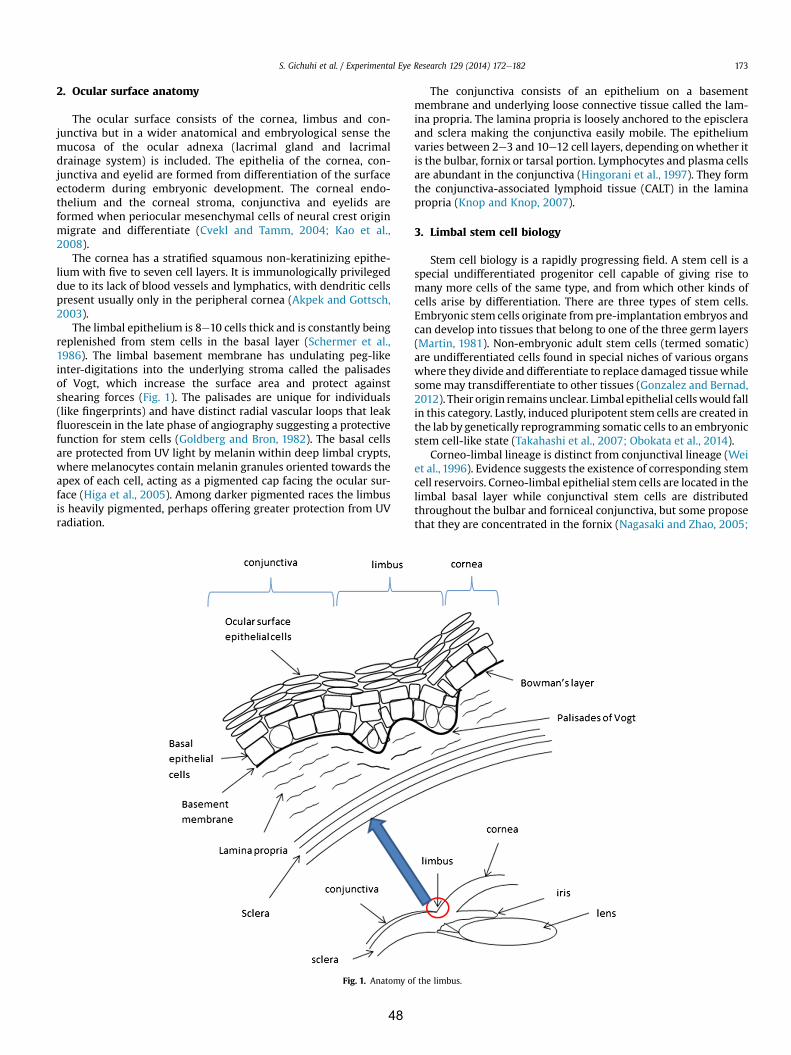
S. Gichuhi et al. / Experimental Eye Research 129 (2014) 172e182 173
2. Ocular surface anatomy
The ocular surface consists of the cornea, limbus and con-junctiva but in a wider anatomical and embryological sense themucosa of the ocular adnexa (lacrimal gland and lacrimaldrainage system) is included. The epithelia of the cornea, con-junctiva and eyelid are formed from differentiation of the surfaceectoderm during embryonic development. The corneal endo-thelium and the corneal stroma, conjunctiva and eyelids areformed when periocular mesenchymal cells of neural crest originmigrate and differentiate (Cvekl and Tamm, 2004; Kao et al.,2008).
The cornea has a stratified squamous non-keratinizing epithe-lium with five to seven cell layers. It is immunologically privilegeddue to its lack of blood vessels and lymphatics, with dendritic cellspresent usually only in the peripheral cornea (Akpek and Gottsch,2003).
The limbal epithelium is 8e10 cells thick and is constantly beingreplenished from stem cells in the basal layer (Schermer et al.,1986). The limbal basement membrane has undulating peg-likeinter-digitations into the underlying stroma called the palisadesof Vogt, which increase the surface area and protect againstshearing forces (Fig. 1). The palisades are unique for individuals(like fingerprints) and have distinct radial vascular loops that leakfluorescein in the late phase of angiography suggesting a protectivefunction for stem cells (Goldberg and Bron, 1982). The basal cellsare protected from UV light by melanin within deep limbal crypts,where melanocytes contain melanin granules oriented towards theapex of each cell, acting as a pigmented cap facing the ocular sur-face (Higa et al., 2005). Among darker pigmented races the limbusis heavily pigmented, perhaps offering greater protection from UVradiation.
Fig. 1. Anatomy o
48
The conjunctiva consists of an epithelium on a basementmembrane and underlying loose connective tissue called the lam-ina propria. The lamina propria is loosely anchored to the episcleraand sclera making the conjunctiva easily mobile. The epitheliumvaries between 2e3 and 10e12 cell layers, depending onwhether itis the bulbar, fornix or tarsal portion. Lymphocytes and plasma cellsare abundant in the conjunctiva (Hingorani et al., 1997). They formthe conjunctiva-associated lymphoid tissue (CALT) in the laminapropria (Knop and Knop, 2007).
3. Limbal stem cell biology
Stem cell biology is a rapidly progressing field. A stem cell is aspecial undifferentiated progenitor cell capable of giving rise tomany more cells of the same type, and from which other kinds ofcells arise by differentiation. There are three types of stem cells.Embryonic stem cells originate frompre-implantation embryos andcan develop into tissues that belong to one of the three germ layers(Martin, 1981). Non-embryonic adult stem cells (termed somatic)are undifferentiated cells found in special niches of various organswhere they divide and differentiate to replace damaged tissuewhilesome may transdifferentiate to other tissues (Gonzalez and Bernad,2012). Their origin remains unclear. Limbal epithelial cellswould fallin this category. Lastly, induced pluripotent stem cells are created inthe lab by genetically reprogramming somatic cells to an embryonicstem cell-like state (Takahashi et al., 2007; Obokata et al., 2014).
Corneo-limbal lineage is distinct from conjunctival lineage (Weiet al., 1996). Evidence suggests the existence of corresponding stemcell reservoirs. Corneo-limbal epithelial stem cells are located in thelimbal basal layer while conjunctival stem cells are distributedthroughout the bulbar and forniceal conjunctiva, but some proposethat they are concentrated in the fornix (Nagasaki and Zhao, 2005;
f the limbus.

S. Gichuhi et al. / Experimental Eye Research 129 (2014) 172e182174
Sun et al., 2010; Pellegrini et al., 1999; Wei et al., 1995). Stem cellsare also found in the corneal stroma and mucocutaneous junctionof the lid margin (Du et al., 2005; Wirtschafter et al., 1997). Themolecular structure of the basement membrane and extracellularmatrix at the cornea, limbus and conjunctiva differ from each otherand this is thought to play a role in regulation of epithelial differ-entiation (Kolega et al., 1989; Schlotzer-Schrehardt et al., 2007).Various markers have been studied in the ocular surface to deter-mine which are more concentrated at the limbus (Table 1)(Schlotzer-Schrehardt and Kruse, 2005). There are no specific lim-bal stem cell markers or any that distinguishes stem cells from theirearly progeny (Chee et al., 2006).
Limbal stem cells are found in special niches in the basal regioncalled limbal epithelial crypts which are cords of epithelial cellsextending from the palisades of Vogt into the underlying stroma
Table 1Molecular markers for limbal stem cells (Schlotzer-Schrehardt and Kruse, 2005).
Characterization Example
Cytoskeletal proteins- proteins that form intermediate filaments in epithelial cells
i) CytokIt is a
ii) Vimelimba
Cytosolic proteins- associated with cellular metabolic functions
a) enzymi) cii) ciii) a
biv) p
inv) avi) tr
b) Cell-ci) Cyii) Ki
c) Metad) Invol
kerate) calciuf) prote
proceNuclear proteins p63 is a
Althoughstem cel
Cell surface proteinsa) Cellecell and cellematrix interaction moleculesb) Growth factor receptorsc) Transporter moleculesd) Cell surface glycoconjugates
i) Conlowext
ii) Cadcell
iii) Inteconbas
iv) epiv) kervi) Trkvii) hepviii) Traix) tranx) ABC
proof twhdrulocaepi
xi) a-2Neuronal markers- human corneal and limbal cells may exhibit neuronalproperties characteristic of their neuroectodermal origin
i) Nestiexcep
ii) transHematopoietic stem cell markers i) CD34
ii) CD13
a In the original article cytokeratins were abbreviated as K. We have modified that to
49
(Dua et al., 2005). The crypts are most abundant nasally in the mid-or distal limbus (Shanmuganathan et al., 2007). Stem cells withinthe crypts have the followingmarker profile; CK3�/CK19þ/CD34�/Vimentinþ/p63þ/Connexin43þ/Ki67� (Shanmuganathan et al.,2007). Stem cells represent less than 10% of the limbal basal cellpopulation (Lavker et al., 1991). They are characterised by; low levelof differentiation, slow cell-cycle, long life-span, high proliferativepotential and self-renewal (ability to produce more stem cells)(Schlotzer-Schrehardt and Kruse, 2005; Dua and Azuara-Blanco,2000). The limbus creates a barrier to prevent extension ofconjunctival epithelium and blood vessels into the cornea (Osei-Bempong et al., 2013). Clinical features of limbal stem cell defi-ciency disorders thus include corneal epithelial defects, conjunc-tival epithelial migration onto the cornea and cornealneovascularization (Sejpal et al., 2013).
s
eratinsa (CK3, CK12, CK5, CK14, CK19) e CK3/CK12 pair is lacking in the limbus.marker of corneal phenotypentin fastens limbal stem cells to their local environment. It is localised inl basal cellses
ytochrome oxidase e Na/K-ATPasearbonic anhydrase-enolase (initially thought to be a glycolytic enzyme it acts as a plasminogeninding receptor)rotein kinase C (PKC), a key enzyme controlling signal transduction pathwaysgrowth and differentiation.
ldehyde dehydrogenase (ALDH)ansketolase (TKT)ycle associated proteinsclins67 acts as a marker for actively cycling cellsllothioneins, which are cysteine-rich metal-binding intracellular proteinsucrin, a structural protein found in the cytosol of differentiated humaninocytesm-linked protein (CLED), that is associated with early epithelial differentiationin S100A12, which is involved in Ca2 þ-dependent signal transductionsses in differentiated cellstranscription factor that regulates epithelial development and differentiation.concentrated at the limbus in stem cells, it is not exclusively expressed by
ls.nexins are transmembrane proteins in gap junctions that allow diffusion of ions,molecular weight metabolites, and second messengers thus determining the
ent of metabolic cooperation between cellsherins are a family of Ca2þ-dependent transmembrane receptors that mediateecell adhesiongrins are a large family of heterodimeric transmembrane glycoproteinssisting of a and b subunits, that play a role in attachment of cells to theement membrane, extracellular matrix proteins or to ligands on other cellsdermal growth factor receptor (EGF-R)atinocyte growth factor receptor (KGF-R)A, the high affinity receptor for nerve growth factor (NGF).atocyte growth factor (HGF)nsferrin receptor CD71sforming growth factor-beta (TGF-b) type I and IIG2, a member of the ATP binding cassette transporters. ABCG2 has beenposed as a universal and conserved marker for stem cells from a wide varietyissues. ABCG2 protein is also known as breast cancer resistant protein 1 (BCRP1),ich causes resistance to certain chemotherapeutic drugs. It is a multi-resistanceg protein that pumps drugs out of cells. This is protective to the cell. It islized to the cell membrane and cytoplasm of some human limbal basalthelial cells, but not in most limbal suprabasal cells and corneal epithelial cells.,3-sialyltransferasen is a neural stem cell marker. It is not normally expressed in limbal basal cellst when they are in an environment with mitogenscription factor Pax-6a sialomucin cell surface antigen3 a transmembrane glycoprotein
CK in keeping with more recent terminology.

S. Gichuhi et al. / Experimental Eye Research 129 (2014) 172e182 175
The ocular surface is self-renewing. Superficial cells areconstantly lost and are replaced by basal cells entering the dif-ferentiation pathway. To replenish the corneal epithelium, cornealepithelial stem cells undergo mitosis producing a progeny of fast-dividing transient amplifying cells (TAC) that make up the ma-jority of the proliferating cell population in the epithelium(Castro-Munozledo, 2013). TAC migrate superficially to thesuprabasal limbus and centripetally towards the centre of thecornea to form the basal layer of the corneal epithelium (Lehreret al., 1998). They undergo a limited number of divisions thendifferentiate into post-mitotic cells (PMC) and further intoterminally differentiated cells (TDC) that migrate superficially tothe corneal surface (Lehrer et al., 1998). It takes 14e21 days forcomplete renewal of the rabbit corneal epithelium (Haddad,2000). In humans it takes 5e7 days (Hanna and O'Brien, 1960).For conjunctival renewal, cells stream centrifugally (instead ofcentripetally as occurs in the cornea) at 10.5 ± 2.4 mm/day thensuperficially at 9.3 ± 5.4 mm/day with a cell-cycle time of 8.3 daysin rats (Zajicek et al., 1995). Stem cells have a slower passagethrough the cell cycle than the other basal cells of the cornea andconjunctiva (Lavker et al., 1991). In mice they take 4e8 weeks andare preferentially stimulated by wounding and tumour promotingcompounds (Cotsarelis et al., 1989). Intact innervation of theocular surface is needed to maintain the stem cell niche (Uenoet al., 2012).
4. Cancer stem cells
The term cancer stem cell is a relatively new one in cancerbiology, though this is a concept known for many years (Wichaet al., 2006). In malignant tumours there is frequently a subpopu-lation of cells that responds poorly to treatment such as chemo-therapy and radiotherapy, divides at a slower rate than other cancercells, and is less affected by hypoxia (Moore and Lyle, 2011; Lin andYun, 2010). This subpopulation is thought to drive tumour growthand is the subject of much debate and much investigation fordifferent tumour types. The origin of these cells is controversial andtheir interaction with non-stem cancer cells has been variouslystudied using mathematical models (Wang et al., 2014).
Cancer stem cells comprise less than 5% of the cell population inmost tumours (Yang and Chang, 2008). They are found in variouscancers including breast, brain, gastric, pancreatic and liver (Al-Hajjet al., 2003; Yuan et al., 2004; Takaishi et al., 2008; Lee et al., 2008;Sell and Leffert, 2008). In high-grade cervical intraepithelialneoplasia (CIN) associated with carcinogenic HPV types they arefound at the squamo-columnar junction but rarely in ectocervical/
Fig. 2. Location of 352 OSSN tumours in Uganda showing most lesions occurred within thReprinted by permission from Macmillan Publishers Ltd: Eye 20 (8):893e899 copyright 20
50
transformation zone CINs or those associated with non-carcinogenic HPVs (Herfs et al., 2012).
Their existence could help to explain the clinical observation ofthe inaction of conventional cancer chemotherapy and radio-therapy in some instances since these treatments target rapidlydividing cells yet stem cells by nature are slow-cycling. Forexample, treatment with fluorouracil (5-FU) selects and enrichesthe cancer stem cell population since the rapidly dividing cellswould be killed while slow-cycling ones incorporate the drug at alower rate (Shi et al., 2013; Wang et al., 2006). Potentially, therapiestargeting cancer stem cells could be more effective (Duggal et al.,2014).
5. Patterns of ocular surface dysplastic and neoplastic disease
The clinical presentation of ocular surface dysplastic andneoplastic diseases provides clues to the pathophysiology of OSSN.Firstly, OSSN, pterygium, pingueculae, climatic droplet keratopathyand actinic keratosis are usually located within the interpalpebralfissure, the space between the open upper and lower eyelid that isexposed to UV radiation (Waddell et al., 2006; Sudhalkar, 2012;Shields et al., 2004; Gray et al., 1992). Secondly, most OSSN le-sions arise from the limbus particularly the nasal quadrant (Fig. 2)(Waddell et al., 2006). A similar observation was made of pterygiain India where all the lesions in a study of 427 participants werenasal (Sudhalkar, 2012). This is the area with the highest concen-tration of limbal epithelial crypts (Shanmuganathan et al., 2007).Thirdly, the disease may involve the circumferential limbus withrelatively little involvement of the cornea or fornix (Fig. 3). Lastly,intraepithelial neoplasia begins in the basal cells and spreads up-wards, a pattern reflected in the histological grading (Basti andMacsai, 2003; American Joint Committee on Cancer, 2010). Thisresembles the spreading waves of limbal stem cells and theirprogeny described in biology above suggesting that the diseasemaybe of stem cell origin. It remains unclear why OSSN is often uni-lateral since exposure to UV radiation, HIV and HPV has nolaterality.
6. Vulnerability of the limbus
The limbus receives direct sunlight temporally which is focusednasally (Fig. 4). As the human eye is more exposed laterally, thelarge temporal visual field becomes a collecting zone of peripherallight, which, depending on the angle of incidence (q) and thecorneal central radius of curvature (r0), is intensely focused ontothe nasal limbus, lid margin or lens with up to a 20-fold increase in
e interpalpebral fissure with a higher concentration in the nasal sector.06
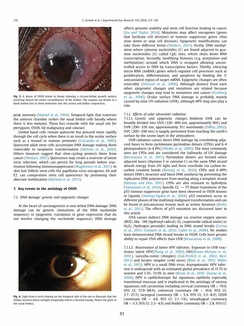
Fig. 3. A lesion of OSSN lesion in Kenya showing a circum-limbal growth patterninvolving almost the entire circumference of the limbus. The margins are drawn in ablack dotted line to show extension into the cornea and bulbar conjunctiva.
S. Gichuhi et al. / Experimental Eye Research 129 (2014) 172e182176
peak intensity (Maloof et al., 1994). Temporal light that traversesthe anterior chamber strikes the nasal limbal cells basally wherethere is less melanin. These foci coincide with the usual site forpterygium, OSSN, lid malignancy and cataract.
Limbal basal cells remain quiescent but proceed more rapidlythrough the cell cycle when there is an insult to the ocular surfacesuch as a wound or tumour promoter (Cotsarelis et al., 1989).Quiescent adult stem cells accumulate DNA damage making themvulnerable to neoplastic transformation (Mohrin et al., 2010).Others however suggest that slow-cycling protects them fromcancer (Wodarz, 2007). Quiescence may create a reservoir of latentvirus infection, which can persist for long periods before reac-tivation following immunosuppression (Maglennon et al., 2011). Inskin hair follicle stem cells the papilloma virus oncogenes, E6 andE7, can compromise stem cell quiescence by promoting theiraberrant mobilization (Michael et al., 2013).
7. Key events in the aetiology of OSSN
7.1. DNA damage: genetic and epigenetic changes
At the heart of carcinogenesis is non-lethal DNA damage. DNAdamage can be genetic (mutations of the DNA nucleotidesequence) or epigenetic (variations in gene expression that donot involve changing the nucleotide sequence). DNA damage
Fig. 4. Light from a torch shining on the temporal side of the eye to illustrate that thelimbus receives direct sunlight temporally which is focused nasally. Notice the glow inthe nasal limbus.
51
affects genome stability and stem cell function leading to cancer(Xu and Taylor, 2014). Mutations may affect oncogenes (genesthat facilitate cell division) or tumour suppressor genes (thatslow down or stop cell division). Epigenetic modifications cantake three different forms (Walters, 2013). Firstly, DNA methyl-ation where cytosine nucleotides (C) are found adjacent to gua-nine nucleotides (G) called CpG sites, which shuts down RNAtranscription. Secondly, modifying histones (e.g. acetylation andmethylation) around which DNA is wrapped allowing uncon-trolled access to DNA by transcription factors. Thirdly, silencingmicro RNA (miRNA) genes which regulate cell processes such asproliferation, differentiation, and apoptosis by binding the 30
untranslated region of target mRNA. Epigenetic changes are oftenreversible (Delcuve et al., 2009). Although distinct from eachother, epigenetic changes and mutations are related becauseepigenetic changes may lead to mutations and cancer (Feinberget al., 2006). Ocular surface DNA damage is probably mainlycaused by solar UV radiation (UVR), although HPV may also play arole.
7.1.1. Effects of solar ultraviolet radiation7.1.1.1. Genetic and epigenetic changes. Ambient UVR can bebroadly divided into UVA (320e400 nm, approximately 90%) andUVB (290e320 nm, approximately 5%) wavebands (Diffey, 2002).UVC (200e290 nm) is largely prevented from reaching the earth'ssurface by the ozone layer in the atmosphere.
UVB radiation causes direct DNA damage by crosslinking adja-cent bases to form cyclobutane pyrimidine dimers (CPDs) and 6-4photoproducts (6-4 PPs) (Pfeifer et al., 2005). The most commonlyseen are CPDs and are considered the hallmarks of UV damage(Besaratinia et al., 2011). Pyrimidine dimers are formed whenadjacent bases (thymine-T or cytosine-C) on the same DNA strandabsorb energy from UV light and form crosslinks via carbon-to-carbon covalent bonds (Rastogi et al., 2010). CPDs and 6-4PPsdistort DNA's structure and block DNA synthesis by preventing thereplicative DNA polymerases from moving along a template strand(Ikehata and Ono, 2011). CPDs are also resistant to hydrolysis(Yamamoto et al., 2014). Specific CC / TT dimer transitions of thep53 tumour-suppressor gene have been observed in OSSN lesionsin Uganda (Ateenyi-Agaba et al., 2004). p53 mutations occur indifferent phases of the multistepmalignant transformation and canbe found in precancerous lesions such as actinic keratosis (Rivlinet al., 2011). The effects of p53 mutation are discussed later inthis article.
UVA causes indirect DNA damage via reactive oxygen species(ROS), like �OH (hydroxyl radical), O2
� (superoxide radical anion) orH2O2 (hydrogen peroxide) leading to DNA strand breaks (Cortatet al., 2013; Greinert et al., 2012; Cadet et al., 2009). No studieshave demonstrated DNA strand breaks in OSSN. Cells have greaterability to repair UVA effects than UVB (Besaratinia et al., 2008).
7.1.1.2. Reactivation of latent HPV infection. Exposure to UVB reac-tivates latent HPV(Zhang et al., 1999), HIV(Breuer-Mcham et al.,2001), varicella-zoster (shingles) (Zak-Prelich et al., 2002; Rice,2011) and herpes simplex (cold sores) (Blatt et al., 1993; Milleret al., 1993). HPV is a small DNA virus. Asymptomatic HPV infec-tion is widespread with an estimated global prevalence of 11.7% inwomen and 1.3%e72.9% in men (Bruni et al., 2010; Dunne et al.,2006). HPV is epitheliotropic for squamous epithelia especiallytransitional mucosae and is implicated in the aetiology of varioussquamous cell carcinomas including cervical (summary OR ¼ 70.0,95% CI; 57.0e88.0), colorectal (summary OR ¼ 10.0, 95% CI;3.7e27.5), laryngeal (summary OR ¼ 5.4, 95% CI; 3.3e8.9), OSSN(summary OR ¼ 4.0, 95% CI; 2.1e7.6), oesophageal (summaryOR ¼ 3.3, 95% CI; 2.3e4.9) and bladder (summary OR ¼ 2.8, 95% CI;

Fig. 5. The cell division cycle.
S. Gichuhi et al. / Experimental Eye Research 129 (2014) 172e182 177
1.4e5.8) (Munoz, 2000; Damin et al., 2013; Li et al., 2013b, 2014,2011; Gichuhi et al., 2013; Li et al., 2014, Li et al., 2011).
Much of what is known about the pathophysiology of HPV incancer is derived from cervical studies. HPV invades the basementmembrane through micro abrasions where it initially binds toheparin sulphate proteoglycan (HSPG) (Johnson et al., 2009; Sappand Bienkowska-Haba, 2009). It does not bind intact epithelia(Johnson et al., 2009). The basement membrane is not merely apassive reservoir of virus but is involved in viral processing. Herethe viral capsids undergo a conformational change where the L2epitope is cleaved by a protease and exposure of its N-terminalleads to the transfer of capsids to the epithelial cell surface (Kineset al., 2009). After internalization, the virus is disassembled andthe DNA enters the nucleus by a mechanism that is still not wellunderstood where it replicates producing extra-chromosomalcopies of viral DNA (Sapp and Bienkowska-Haba, 2009). Howeverthe genome of high-risk HPV has been found incorporated intospecific preferential sites of the host DNA in cervical lesions (Liet al., 2013a). During differentiation of epithelial cells, virionsmature and are carried towards the surface (Doorbar, 2005). Innormal uninfected epithelia, as cells leave the basal layer, they exitthe cell cycle but infected cells remain active due to E7 (Longworthand Laimins, 2004). E7 inactivates the retinoblastoma gene (pRB)which usually acts in the G1 phase of the cell cycle where it bindstranscription factors, thus infected cells remain in a proliferativestate while E6 binds the p53 protein preventing it from suppressingreplication of such DNA-defective cells (Lehoux et al., 2009). Inhigh-risk HPV types, E6 and E7 also cause genomic instability; E7causes centriole over-duplication and disturbs mitotic fidelitywhile E6 causes structural chromosomal alterations and DNAbreakage (Korzeniewski et al., 2011). The effects on p53 and pRB areconsidered the molecular signatures of HPV-induced carcinogen-esis (Buitrago-Perez et al., 2009). Regression of HPV-induced le-sions is mediated by a T-helper 1 lymphocyte (Th1) cell mediatedimmune response (Stanley, 2012). HPV latency may arise in twoways; (i) low titre infection that is too low to complete the life cycleor (ii) clearance of lesions by the adaptive immune system followedby persistence of low-level viral gene expression, which is reac-tivated by immunosuppression (Doorbar, 2013).
How HPV initially reaches the conjunctiva is not clear. Theprevalence of HPV in OSSN tissue is heterogeneous, varying widelyfrom zero to 100% (Gichuhi et al., 2013).
HPV infection is associated with an increased incidence of HIVacquisition (summary OR¼ 1.96; 95% CI; 1.55e2.49) (Lissouba et al.,2013).
7.2. Failure of DNA repair mechanisms
Several mechanisms prevent UV-induced DNA mutations frombeing incorporated into the genome and UV-damaged cells fromestablishing themselves. Cells can correct carcinogen-induced DNAdamage; severely damaged cells are eliminated from healthy tis-sues by processes that trigger apoptosis; and abnormal cells arerecognized, targeted and destroyed by immune surveillance (DiGirolamo, 2010).
The cell-division cycle involves duplication of the genome andintracellular organelles (Imoto et al., 2011). The stages of the cyclecan be visualised directly by high-resolution imaging (Hesse et al.,2012). Nuclear DNA is synthesized during a stage of interphasecalled the S phase which is followed by a gap (G2), then mitosis (Mphase) in which nuclear and cell division occur and another gap(G1) before the next S phase (Fig. 5).
DNA damage activates checkpoint pathways that regulate spe-cific DNA repair mechanisms in the different phases of the cell cycle(Branzei and Foiani, 2008). There are three important cell-cycle
52
checkpoints (Sancar et al., 2004). The G1-S checkpoint preventscells with damaged DNA from entering the S phase by inhibitingthe initiation of replication. The intra-S-phase checkpoint dealswith DNA damage that may occur during S-phase or unrepaireddamage that escaped the G1-S checkpoint. The G2-M checkpointprevents cells with damaged DNA from undergoing mitosis.
The molecular mechanisms that repair UVR-induced DNAdamage include excision repair, mismatch repair, strand breakrepair, and cross-link repair (Rastogi et al., 2010). During excisionrepair sections of damaged DNA are replaced by a nucleotide orbase (Sinha and Hader, 2002). The nucleotide excision repair (NER)pathway is primarily responsible for repairing CPDs in humans(Sancar et al., 2004). There are two types of NER; general excisionrepair which removes lesions from the whole genome andtranscription-coupled repair which works on damage in tran-scribed DNA strands. The latter is not clearly understood but theformer is performed through a series of special proteins and pro-ceeds through four discrete steps; recognition of the damage;excision of the section of DNA that includes and surrounds the er-ror; filling in of the resulting gap by DNA polymerase; and sealing ofthe nick between the newly synthesized and older DNA by DNAligase (Hu et al., 2013; Reardon and Sancar, 2005).
Base excision repair (BER) is the predominant pathway againstlesions caused by ROS, ionizing radiation and strong alkylatingagents (Svilar et al., 2011). It proceeds through 5 steps as follows;DNA glycosylases remove damaged or modified bases; the apuri-mic/apyrimidinic site is removed by an endonuclease or lyase; theremaining phosphate residue is removed by a phosphodiesterase;the gap is filled by a DNA polymerase and the strand sealed by aDNA ligase (Hegde et al., 2008).
7.2.1. The p53 tumour-suppressor systemThe p53 tumour-suppressor gene is found on the short arm of
chromosome 17 (17p13.1) (Mcbride et al., 1986). P53 is a phos-phoprotein found in the nucleus which regulates the cell cycle toprotect cells from the effects of DNA damage (Ford, 2005; Jin andRobertson, 2013; Borras et al., 2011). It is thus described as the‘guardian of the genome’ (Lane, 1992). Once p53 is activated by astress signal it binds to specific DNA elements in the genome withvarious primary and secondary response effects (Levine et al.,2006). The primary responses include; cell cycle arrest at the G1-S checkpoint; irreversible withdrawal of cells from the cycle intoa terminal state of senescence or programmed cell death
S. Gichuhi et al. / Experimental Eye Research 129 (2014) 172e182178
(apoptosis) if the damage is irreparable. The secondary responsescome from p53-regulated gene products that prevent DNA damage(sestrins) or aid in DNA repair; mediate communication betweenthe cell and its neighbours, the extracellular matrix or more distantcells; or create intracellular or extracellular p53 feedback loops thatmodulate p53 activity. In addition, deacetylation of p53 facilitatesautophagy (autophagocytosis by controlled lysosomal degradation)(Contreras et al., 2013). Mutated p53 (Mutp53) leads to furthergenomic instability (Meek, 2009).
7.3. Reduced immunity
Tumour immunology presumes that tumour cells express anti-gens such as mutp53 and HPV proteins that distinguish them fromnon-transformed cells. The immune system prevents tumour for-mation in 3 ways (i) elimination or suppression of viral infection (ii)preventing establishment of a pro-inflammatory environment and(iii) specifically identifying and eliminating cells that expresstumour-specific antigens or molecular signals of cellular stressbefore they cause harm (Schreiber et al., 2011). The latter is part of amore general process called tumour immuno-editing. Immuno-editing is a dual process in which the immune system may eithersuppress tumour growth by destroying or inhibiting growth ofcancer cells or inadvertently promote tumour progression byselecting tumour cells that aremore likely to survive. It has 3phases:elimination, equilibrium and escape (Schreiber et al., 2011). In theelimination phase the immune system recognizes tumour cells andinitiates cell death eliminating themcompletely before theybecomeclinically apparent. If not fully eliminated the remaining tumourcells enter a state of temporary equilibrium between the immunesystem and the developing tumour in which the tumour cellsremain dormant or continue to accumulate further genetic andepigenetic changes. Finally if the immune system fails to contain thetumour at this phase, surviving tumour cell variants escape causinguncontrolled tumour expansion. The quantity, quality and distri-bution of tumour infiltrating lymphocytes (TILs) such as CD8þcytotoxic T lymphocytes (CTL), CD4þ T helper lymphocytes (Th),CD4þ regulatory T lymphocytes (Treg) and CD3þ lymphocytes in-fluence prognosis. A meta-analysis showed that improved survival,measured by the deathHazard ratio (HR),was associatedwith CD3þTIL infiltration (HR ¼ 0.58, 95% CI; 0.43e0.78) and CD8þ TIL(HR ¼ 0.71, 95% CI; 0.62e0.82) (Gooden et al., 2011). However TILcounts alone may overestimate this effect and ratios between TILsubsets CD8þ/FoxP3þ (effector:regulatory ratio) and CD8þ/CD4þ(effector:helper ratio) may be more informative. Natural killer cellsare another important component of cancer immunosurveillanceand immuno-editing (Gross et al., 2013).
7.3.1. PhotoimmunosuppressionAmbient UV radiation suppresses cell-mediated immunity
(Clydesdale et al., 2001). UVB is a more potent immunosuppressorthan UVA (Poon et al., 2005). This phenomenon referred to asphotoimmunosuppression is not limited to exposed cutaneoustissues but is also systemic, affecting internal organs (Gibbs andNorval, 2013). The immunosuppressive effect of UVB is used inphototherapy of skin conditions such as psoriasis (Chen et al.,2013). In the skin, UVB stimulates migration of epidermal Langer-hans cells, which present antigens to lymphocytes in the draininglymph nodes promoting a Th2 and regulatory T cells (Treg) domi-nated response that suppresses local immune responses (Taguchiet al., 2013; Schwarz and Schwarz, 2011; Norval et al., 2008).
7.3.2. HIVHIV preferentially infects helper T cells (CD4þ), inducing their
apoptosis (Lundin et al., 1987; Cloyd et al., 2001; Alimonti et al.,
53
2003). The virus establishes latency in resting memory T cells,which explains why combination antiretroviral therapy (ART) is notcurative; interruption of treatment inevitably results in reboundviraemia (Van Lint et al., 2013) (van Der Sluis et al., 2013). HIV mayweaken tumour immunosurveillance (Mbulaiteye et al., 2003). Ameta-analysis found an increased incidence of cancers among bothHIV/AIDS patients and immunosuppressed transplant recipients,however, therewas no significant difference in the risk between thetwo groups suggesting that it is primarily the immunosuppression,rather than another action of HIV that is responsible for theincreased risk of cancer (Grulich et al., 2007).
HIV potentiates the oncogenic action of other viruses such asHPV, Kaposi sarcoma-associated herpes virus (KSHV) and Eps-teineBarr virus (EBV) by enhancing their transmission to targetcells (Aoki and Tosato, 2004). A report from Botswana reportedmultiple oncogenic viruses (EBV, HPV, KSHV, HSV1/2 and CMV) incases of OSSN and pterygium (Simbiri et al., 2010). In rabbitsimmunosuppression induced by T-cell depletion facilitated reac-tivation of latent HPV infection leading to a 3 to 5 log increase in thenumber of viral copies to levels associated with productive infec-tion (Maglennon et al., 2014).
Lastly, HIV induces a state of persistent inflammation (Hunt,2012). Markers of inflammation such as C-reactive protein andinterleukin-6 are elevated in HIV patients particularly those on ARTeven when viral loads are undetectable (Neuhaus et al., 2010). In-flammatory microenvironments have tumour-promoting effects(Mantovani et al., 2008). One proposed mechanism is via over-expression of microRNA-155 (MiR155), which increases sponta-neous mutation rates (Tili et al., 2011; Valeri et al., 2010).
7.3.3. Vitamin A deficiencyVitamin A helps to maintain the integrity of the ocular surface
(Kanazawa et al., 2002). Its deficiency is associated with squamousmetaplasia of the conjunctiva (Mckelvie, 2003). Vitamin A also actsas a mucosal and systemic immune enhancer through immuno-homeostasis of CD4þ helper T cells and Treg cells (Hall et al.,2011; Pino-Lagos et al., 2011; Ross, 2012). These cells are part oftumour immuno-surveillance. Retinoids are reported to preventvarious cancers in the skin and liver (Alizadeh et al., 2014). Theypromote stem cell differentiation through epigenetic modificationsof histones or by altering chromatin structure to remove the stemcell from the self-renewing pluripotential state to a differentiatedone (Gudas, 2013). Loss of the differentiated phenotype can lead togeneration of cancer stem cells (Gudas, 2013). Retinoids activateDNA transcription in stem cells via retinoic acid receptors (RARa,b,g), retinoid X receptors (RXR a,b,g) and other transcriptionfactor regulatory proteins (Gudas and Wagner, 2011).
A study in Kenya found that HIV-positive women had a higherprevalence of vitamin A deficiency (<30 mg/dL) than HIV-negativewomen (59% vs 29%, p < 0.001) (Baeten et al., 2002).
We hypothesize that vitamin A deficiency has three effects; itcompromises the integrity of the surface epithelium creatingmicro-abrasions for HPV entry, it leads to cell-mediated immuno-deficiency, and dysregulation of stem cell differentiation.
8. Downstream events after initiation of neoplasia
8.1. Uncontrolled cell replication
Human somatic cells can only undergo a limited number of celldivisions (50e70) then arrest, an event related to shortening oftelomeres (Gomez et al., 2012). In comparison, epidermal stem cellscan divide for more than 150 generations in vitro (Mathor et al.,1996). Telomeres are a repetitive sequence of nucleotides rich inguanidine, synthesized by the enzyme telomerase, that cap the

S. Gichuhi et al. / Experimental Eye Research 129 (2014) 172e182 179
ends of chromosomes to prevent chromosomes from deterioration(Blackburn and Gall, 1978). Usually telomeres shorten during eachround of DNA replication but in advanced cancers telomerase isreactivated to maintain telomere length allowing many more celldivisions (Artandi and DePinho, 2010; Prescott et al., 2012).Downregulation of 14-3-3s protein in keratinocytes maintainstelomerase activity allowing them to escape replicative senescence(Dellambra et al., 2000).
8.2. Angiogenesis
Neovascularization occurs to meet the increased tumourmetabolic demand. Tumours overproduce vascular growth factorssuch as vascular endothelial growth factor (VEGF) (Aonuma et al.,1999). In conjunctival tumours this manifests clinically as feedervessels, enlarged blood vessels in the conjunctiva that perfuse thegrowth. In a Tanzanian study 88% of OSSN lesions and 61% of benigntumours had feeder vessels (Nguena et al., 2014).
8.3. Metastasis
Metastasis is considered a hallmark of malignancy. Cells looseadherence with each other and secrete proteolytic enzymes such asmatrix metalloproteinases (MMPs) that degrade the extracellularmatrix (Chiang and Massague, 2008). UVB radiation alters thebalance between MMPs and tissue inhibitors of matrix metal-loproteinases (TIMPs) (Ng et al., 2008). When exposed to UVB,cultured human dysplastic conjunctival epithelial cells showincreased expression of MMP-1 and MMP-3 with little change inTIMP-1 unlike normal conjunctival cells (Ng et al., 2008). IncreasedMMP activity upsets cell-to-cell adhesion and promotes carcino-genesis and tumour invasion into surrounding tissues (Johanssonet al., 2000). In lesions of squamous cell carcinoma of the con-junctiva MMPs and TIMP are overexpressed compared to normalconjunctiva and cornea (Di Girolamo et al., 2013).
9. Are cancer stem cells central to OSSN?
The short lifespan of ocular surface cells means that epithelialcells with DNA mutations do not last long enough to have an effect,as they are constantly being shed from the surface. The longevity ofstem cells however gives them enough time to accumulate muta-genic insults. Why tumours are heterogeneous yet originating fromthe same stem cells could partly be explained by ongoing muta-genesis (Pardal et al., 2003).
The location of stem cells on the ocular surface coincides withthe position of OSSN tumours while the growth pattern of lesions(from base upwards) is consistent with the stem cell theory. A studyin Australia described concurrent existence of features of OSSN andprimary acquired melanosis and stem cells arranged in micro-clusters in the basal epithelium in 12% of pterygiums (Chui et al.,2011). Focus would necessarily have to shift to ocular surface lim-bal epithelial stem cells (LESCs) as the potential progenitors ofOSSN to consider new explanations for tumour formation, newdiagnostic methods to detect the LESCs and new treatments thattarget LESCs.
10. Conclusion
The known risk factors of OSSN e solar UV radiation, HIV andHPV are implicated in the aetiology. The pattern of distribution ofOSSN lesions within the interpalpebral fissure of the ocular surfaceat the limbus, particularly the nasal side provides further clues. Thesite is highly vulnerable to solar UVR and has a high concentrationof stem cells in the basal epithelium. Stem cells in the limbal
54
epithelial crypts are the likely originators of this disease, and maytake on cancer stem cell properties.
Neoplasia is probably initiated when background solar UV ra-diation causes various forms of genetic and epigenetic DNA dam-age. UVBmainly creates pyrimidine dimers. Specific dimer CC/ TTtransformation, a signature UV mutation, occurs at the p53 gene, atumour suppressor that maintains cell-cycle arrest at the G1-Scheckpoint. UV radiation also reactivates latent viruses such asHPV. HPV's E7 protein keeps infected cells in a proliferative statewhile E6 inhibits cell cycle arrest of DNA-damaged cells. Immu-nosuppression caused by UV radiation, HIV and vitamin A defi-ciency weakens the tumour surveillance system and allows DNA-damaged cells to proliferate into tumours. Vitamin A deficiencyinterferes with ocular surface integrity creating micro-abrasionsthrough which HPV may invade the conjunctival basement mem-brane and epithelial cells. Cancer cells reactivate telomerase whichmaintains long telomeres increasing the number of cell divisions acell can undergo. Further tumour expansion and metastasis isenhanced by angiogenesis and increased matrix metal-loproteinases (MMPs) which destroy the intercellular matrix.
Despite these advances in our understanding there remain gaps,which are areas for further research. For example, there is noexplanation why the disease is mostly unilateral despite both eyesreceiving equal sunlight exposure. Equally the route of trans-mission of HPV to the conjunctiva is unknown. The drivers on amolecular level which convert intraepithelial neoplasia to squa-mous cell cancer are also out with our current understanding of thedisease.
Acknowledgements
SG received funding from the British Council for Prevention ofBlindness (BCPB) fellowship programme. MJB is supported by TheWellcome Trust (Grant Number 098481/Z/12/Z).
References
Akpek, E.K., Gottsch, J.D., 2003. Immune defense at the ocular surface. Eye (Lond.)17, 949e956.
Al-Hajj, M., Wicha, M.S., Benito-Hernandez, A., Morrison, S.J., Clarke, M.F., 2003.Prospective identification of tumorigenic breast cancer cells. Proc. Natl. Acad.Sci. U. S. A. 100, 3983e3988.
Alimonti, J.B., Ball, T.B., Fowke, K.R., 2003. Mechanisms of CD4þ T lymphocyte celldeath in human immunodeficiency virus infection and AIDS. J. Gen. Virol. 84,1649e1661.
Alizadeh, F., Bolhassani, A., Khavari, A., Bathaie, S.Z., Naji, T., Bidgoli, S.A., 2014.Retinoids and their biological effects against cancer. Int. Immunopharmacol. 18,43e49.
American Joint Committee On Cancer, 2010. Carcinoma of the conjunctiva. In:Edge, S.B., Byrd, D.R., Compton, C.C., Fritz, A.G., Greene, F.L., Trotti, A. (Eds.),Cancer Staging Handbook, seventh ed. Springer, New York.
Aoki, Y., Tosato, G., 2004. Neoplastic conditions in the context of HIV-1 infection.Curr. HIV Res. 2, 343e349.
Aonuma, M., Saeki, Y., Akimoto, T., Nakayama, Y., Hattori, C., Yoshitake, Y.,Nishikawa, K., Shibuya, M., Tanaka, N.G., 1999. Vascular endothelial growthfactor overproduced by tumour cells acts predominantly as a potent angiogenicfactor contributing to malignant progression. Int. J. Exp. Pathol. 80, 271e281.
Artandi, S.E., Depinho, R.A., 2010. Telomeres and telomerase in cancer. Carcino-genesis 31, 9e18.
Ateenyi-Agaba, C., Dai, M., Le Calvez, F., Katongole-Mbidde, E., Smet, A.,Tommasino, M., Franceschi, S., Hainaut, P., Weiderpass, E., 2004. TP53 mutationsin squamous-cell carcinomas of the conjunctiva: evidence for UV-inducedmutagenesis. Mutagenesis 19, 399e401.
Baeten, J.M., Mcclelland, R.S., Richardson, B.A., Bankson, D.D., Lavreys, L.,Wener, M.H., Overbaugh, J., Mandaliya, K., Ndinya-Achola, J.O., Bwayo, J.J.,Kreiss, J.K., 2002. Vitamin A deficiency and the acute phase response amongHIV-1-infected and -uninfected women in Kenya. J. Acquir. Immune Defic.Syndr. 31, 243e249.
Basti, S., Macsai, M.S., 2003. Ocular surface squamous neoplasia: a review. Cornea22, 687e704.
Besaratinia, A., Kim, S.I., Pfeifer, G.P., 2008. Rapid repair of UVA-induced oxidizedpurines and persistence of UVB-induced dipyrimidine lesions determine themutagenicity of sunlight in mouse cells. FASEB J. 22, 2379e2392.
S. Gichuhi et al. / Experimental Eye Research 129 (2014) 172e182180
Besaratinia, A., Yoon, J.I., Schroeder, C., Bradforth, S.E., Cockburn, M., Pfeifer, G.P.,2011. Wavelength dependence of ultraviolet radiation-induced DNA damage asdetermined by laser irradiation suggests that cyclobutane pyrimidine dimersare the principal DNA lesions produced by terrestrial sunlight. FASEB J. 25,3079e3091.
Blackburn, E.H., Gall, J.G., 1978. A tandemly repeated sequence at the termini of theextrachromosomal ribosomal RNA genes in Tetrahymena. J. Mol. Biol. 120,33e53.
Blatt, A.N., Laycock, K.A., Brady, R.H., Traynor, P., Krogstad, D.J., Pepose, J.S., 1993.Prophylactic acyclovir effectively reduces herpes simplex virus type 1 reac-tivation after exposure of latently infected mice to ultraviolet B. Investig.Ophthalmol. Vis. Sci. 34, 3459e3465.
Borras, C., Gomez-Cabrera, M.C., Vina, J., 2011. The dual role of p53: DNA protectionand antioxidant. Free Radic. Res. 45, 643e652.
Branzei, D., Foiani, M., 2008. Regulation of DNA repair throughout the cell cycle. Nat.Rev. Mol. Cell Biol. 9, 297e308.
Breuer-Mcham, J., Simpson, E., Dougherty, I., Bonkobara, M., Ariizumi, K., Lewis, D.E.,Dawson, D.B., Duvic, M., Cruz Jr., P.D., 2001. Activation of HIV in human skin byultraviolet B radiation and its inhibition by NFkappaB blocking agents. Photo-chem. Photobiol. 74, 805e810.
Bruni, L., Diaz, M., Castellsague, X., Ferrer, E., Bosch, F.X., De sanjose, S., 2010. Cer-vical human papillomavirus prevalence in 5 continents: meta-analysis of 1million women with normal cytological findings. J. Infect. Dis. 202, 1789e1799.
Buitrago-Perez, A., Garaulet, G., Vazquez-Carballo, A., Paramio, J.M., Garcia-Escudero, R., 2009. Molecular signature of HPV-induced carcinogenesis: pRb,p53 and gene expression profiling. Curr. Genomics 10, 26e34.
Cadet, J., Douki, T., Ravanat, J.L., Di Mascio, P., 2009. Sensitized formation ofoxidatively generated damage to cellular DNA by UVA radiation. Photochem.Photobiol. Sci. 8, 903e911.
Carreira, H., Coutinho, F., Carrilho, C., Lunet, N., 2013. HIV and HPV infections andocular surface squamous neoplasia: systematic review and meta-analysis. Br. J.Cancer 109, 1981e1988.
Castro-Munozledo, F., 2013. Review: corneal epithelial stem cells, their niche andwound healing. Mol. Vis. 19, 1600e1613.
Chee, K.Y., Kicic, A., Wiffen, S.J., 2006. Limbal stem cells: the search for a marker.Clin. Exp. Ophthalmol. 34, 64e73.
Chen, X., Yang, M., Cheng, Y., Liu, G.J., Zhang, M., 2013. Narrow-band ultraviolet Bphototherapy versus broad-band ultraviolet B or psoralen-ultraviolet A pho-tochemotherapy for psoriasis. Cochrane Database Syst. Rev. 10, CD009481.
Chiang, A.C., Massague, J., 2008. Molecular basis of metastasis. N. Engl. J. Med. 359,2814e2823.
Chisi, S.K., Kollmann, M.K., Karimurio, J., 2006. Conjunctival squamous cell carci-noma in patients with human immunodeficiency virus infection seen at twohospitals in Kenya. East Afr. Med. J. 83, 267e270.
Chui, J., Coroneo, M.T., Tat, L.T., Crouch, R., Wakefield, D., Di Girolamo, N., 2011.Ophthalmic pterygium: a stem cell disorder with premalignant features. Am. J.Pathol. 178, 817e827.
Cloyd, M.W., Chen, J.J., Adeqboyega, P., Wang, L., 2001. How does HIV causedepletion of CD4 lymphocytes? A mechanism involving virus signaling throughits cellular receptors. Curr. Mol. Med. 1, 545e550.
Clydesdale, G.J., Dandie, G.W., Muller, H.K., 2001. Ultraviolet light induced injury:immunological and inflammatory effects. Immunol. Cell Biol. 79, 547e568.
Contreras, A.U., Mebratu, Y., Delgado, M., Montano, G., Hu, C.A., Ryter, S.W.,Choi, A.M., Lin, Y., Xiang, J., Chand, H., Tesfaigzi, Y., 2013. Deacetylation of p53induces autophagy by suppressing Bmf expression. J. Cell Biol. 201, 427e437.
Cortat, B., Garcia, C.C., Quinet, A., Schuch, A.P., De Lima-Bessa, K.M., Menck, C.F.,2013. The relative roles of DNA damage induced by UVA irradiation in humancells. Photochem. Photobiol. Sci. 12, 1483e1495.
Cotsarelis, G., Cheng, S.Z., Dong, G., Sun, T.T., Lavker, R.M., 1989. Existence of slow-cycling limbal epithelial basal cells that can be preferentially stimulated toproliferate: implications on epithelial stem cells. Cell 57, 201e209.
Cvekl, A., Tamm, E.R., 2004. Anterior eye development and ocular mesenchyme:new insights from mouse models and human diseases. Bioessays 26, 374e386.
Damin, D.C., Ziegelmann, P.K., Damin, A.P., 2013. Human papillomavirus infectionand colorectal cancer risk: a meta-analysis. Colorectal Dis. 15, e420ee428.
Delcuve, G.P., Rastegar, M., Davie, J.R., 2009. Epigenetic control. J. Cell. Physiol. 219,243e250.
Dellambra, E., Golisano, O., Bondanza, S., Siviero, E., Lacal, P., Molinari, M., D'atri, S.,De Luca, M., 2000. Downregulation of 14-3-3sigma prevents clonal evolutionand leads to immortalization of primary human keratinocytes. J. Cell Biol. 149,1117e1130.
Di Girolamo, N., 2010. Signalling pathways activated by ultraviolet radiation: role inocular and cutaneous health. Curr. Pharm. Des. 16, 1358e1375.
Di Girolamo, N., Atik, A., Mccluskey, P.J., Wakefield, D., 2013. Matrix metal-loproteinases and their inhibitors in squamous cell carcinoma of the conjunc-tiva. Ocul. Surf. 11, 193e205.
Diffey, B.L., 2002. Sources and measurement of ultraviolet radiation. Methods 28,4e13.
Doorbar, J., 2005. The papillomavirus life cycle. J. Clin. Virol. 32 (Suppl. 1), S7eS15.Doorbar, J., 2013. Latent papillomavirus infections and their regulation. Curr. Opin.
Virol. 3, 416e421.Du, Y., Funderburgh, M.L., Mann, M.M., Sundarraj, N., Funderburgh, J.L., 2005.
Multipotent stem cells in human corneal stroma. Stem Cells 23, 1266e1275.Dua, H.S., Azuara-Blanco, A., 2000. Limbal stem cells of the corneal epithelium. Surv.
Ophthalmol. 44, 415e425.
55
Dua, H.S., Shanmuganathan, V.A., Powell-Richards, A.O., Tighe, P.J., Joseph, A., 2005.Limbal epithelial crypts: a novel anatomical structure and a putative limbalstem cell niche. Br. J. Ophthalmol. 89, 529e532.
Duggal, R., Minev, B., Geissinger, U., Wang, H., Chen, N.G., Koka, P.S., Szalay, A.A.,2014. Biotherapeutic approaches to target cancer stem cells. J. Stem Cells 8,135e149.
Dunne, E.F., Nielson, C.M., Stone, K.M., Markowitz, L.E., Giuliano, A.R., 2006. Prev-alence of HPV infection among men: a systematic review of the literature.J. Infect. Dis. 194, 1044e1057.
Feinberg, A.P., Ohlsson, R., Henikoff, S., 2006. The epigenetic progenitor origin ofhuman cancer. Nat. Rev. Genet. 7, 21e33.
Ford, J.M., 2005. Regulation of DNA damage recognition and nucleotide excisionrepair: another role for p53. Mutat. Res. 577, 195e202.
Gibbs, N.K., Norval, M., 2013. Photoimmunosuppression: a brief overview. Photo-dermatol. Photoimmunol. Photomed. 29, 57e64.
Gichuhi, S., Sagoo, M.S., Weiss, H.A., Burton, M.J., 2013. Epidemiology of ocularsurface squamous neoplasia in Africa. Trop. Med. Int. Health 18, 1424e1443.
Goldberg, M.F., Bron, A.J., 1982. Limbal palisades of Vogt. Trans. Am. Ophthalmol.Soc. 80, 155e171.
Gomez, D.E., Armando, R.G., Farina, H.G., Menna, P.L., Cerrudo, C.S., Ghiringhelli, P.D.,Alonso, D.F., 2012. Telomere structure and telomerase in health and disease(review). Int. J. Oncol. 41, 1561e1569.
Gonzalez, M.A., Bernad, A., 2012. Characteristics of adult stem cells. Adv. Exp. Med.Biol. 741, 103e120.
Gooden, M.J., De Bock, G.H., Leffers, N., Daemen, T., Nijman, H.W., 2011. The prog-nostic influence of tumour-infiltrating lymphocytes in cancer: a systematicreview with meta-analysis. Br. J. Cancer 105, 93e103.
Gray, R.H., Johnson, G.J., Freedman, A., 1992. Climatic droplet keratopathy. Surv.Ophthalmol. 36, 241e253.
Greinert, R., Volkmer, B., Henning, S., Breitbart, E.W., Greulich, K.O., Cardoso, M.C.,Rapp, A., 2012. UVA-induced DNA double-strand breaks result from the repairof clustered oxidative DNA damages. Nucleic Acids Res. 40, 10263e10273.
Gross, E., Sunwoo, J.B., Bui, J.D., 2013. Cancer immunosurveillance and immunoe-diting by natural killer cells. Cancer J. 19, 483e489.
Grulich, A.E., Van Leeuwen, M.T., Falster, M.O., Vajdic, C.M., 2007. Incidence ofcancers in people with HIV/AIDS compared with immunosuppressed transplantrecipients: a meta-analysis. Lancet 370, 59e67.
Gudas, L.J., 2013. Retinoids induce stem cell differentiation via epigenetic changes.Semin. Cell Dev. Biol. 24, 701e705.
Gudas, L.J., Wagner, J.A., 2011. Retinoids regulate stem cell differentiation. J. Cell.Physiol. 226, 322e330.
Haddad, A., 2000. Renewal of the rabbit corneal epithelium as investigated byautoradiography after intravitreal injection of 3H-thymidine. Cornea 19,378e383.
Hall, J.A., Cannons, J.L., Grainger, J.R., Dos Santos, L.M., Hand, T.W., Naik, S.,Wohlfert, E.A., Chou, D.B., Oldenhove, G., Robinson, M., Grigg, M.E.,Kastenmayer, R., Schwartzberg, P.L., Belkaid, Y., 2011. Essential role for retinoicacid in the promotion of CD4(þ) T cell effector responses via retinoic acid re-ceptor alpha. Immunity 34, 435e447.
Hanna, C., O'brien, J.E., 1960. Cell production and migration in the epithelial layer ofthe cornea. Arch. Ophthalmol. 64, 536e539.
Hegde, M.L., Hazra, T.K., Mitra, S., 2008. Early steps in the DNA base excision/single-strand interruption repair pathway in mammalian cells. Cell. Res. 18,27e47.
Herfs, M., Yamamoto, Y., Laury, A., Wang, X., Nucci, M.R., Mclaughlin-Drubin, M.E.,Munger, K., Feldman, S., Mckeon, F.D., Xian, W., Crum, C.P., 2012. A discretepopulation of squamocolumnar junction cells implicated in the pathogenesis ofcervical cancer. Proc. Natl. Acad. Sci. U. S. A. 109, 10516e10521.
Hesse, M., Raulf, A., Pilz, G.A., Haberlandt, C., Klein, A.M., Jabs, R., Zaehres, H.,Fugemann, C.J., Zimmermann, K., Trebicka, J., Welz, A., Pfeifer, A., Roll, W.,Kotlikoff, M.I., Steinhauser, C., Gotz, M., Scholer, H.R., Fleischmann, B.K., 2012.Direct visualization of cell division using high-resolution imaging of M-phase ofthe cell cycle. Nat. Commun. 3, 1076.
Higa, K., Shimmura, S., Miyashita, H., Shimazaki, J., Tsubota, K., 2005. Melanocytes inthe corneal limbus interact with K19-positive basal epithelial cells. Exp. Eye Res.81, 218e223.
Hingorani, M., Metz, D., Lightman, S.L., 1997. Characterisation of the normalconjunctival leukocyte population. Exp. Eye Res. 64, 905e912.
Hu, J., Choi, J.H., Gaddameedhi, S., Kemp, M.G., Reardon, J.T., Sancar, A., 2013.Nucleotide excision repair in human cells: fate of the excised oligonucleotidecarrying DNA damage in vivo. J. Biol. Chem. 288, 20918e20926.
Hunt, P.W., 2012. HIV and inflammation: mechanisms and consequences. Curr. HIV/AIDS Rep. 9, 139e147.
Ikehata, H., Ono, T., 2011. The mechanisms of UV mutagenesis. J. Radiat. Res. 52,115e125.
Imoto, Y., Yoshida, Y., Yagisawa, F., Kuroiwa, H., Kuroiwa, T., 2011. The cell cycle,including the mitotic cycle and organelle division cycles, as revealed by cyto-logical observations. J. Electron Microsc. (Tokyo) 60 (Suppl. 1), S117eS136.
Jin, B., Robertson, K.D., 2013. DNA methyltransferases, DNA damage repair, andcancer. Adv. Exp. Med. Biol. 754, 3e29.
Johansson, N., Ahonen, M., Kahari, V.M., 2000. Matrix metalloproteinases in tumorinvasion. Cell. Mol. Life Sci. 57, 5e15.
Johnson, K.M., Kines, R.C., Roberts, J.N., Lowy, D.R., Schiller, J.T., Day, P.M., 2009. Roleof heparan sulfate in attachment to and infection of the murine female genitaltract by human papillomavirus. J. Virol. 83, 2067e2074.

S. Gichuhi et al. / Experimental Eye Research 129 (2014) 172e182 181
Kanazawa, S., Kitaoka, T., Ueda, Y., Gong, H., Amemiya, T., 2002. Interaction of zincand vitamin A on the ocular surface. Graefes Arch. Clin. Exp. Ophthalmol. 240,1011e1021.
Kao, W.W., Xia, Y., Liu, C.Y., Saika, S., 2008. Signaling pathways in morphogenesis ofcornea and eyelid. Ocul. Surf. 6, 9e23.
Kines, R.C., Thompson, C.D., Lowy, D.R., Schiller, J.T., Day, P.M., 2009. The initial stepsleading to papillomavirus infection occur on the basement membrane prior tocell surface binding. Proc. Natl. Acad. Sci. U. S. A. 106, 20458e20463.
Knop, E., Knop, N., 2007. Anatomy and immunology of the ocular surface. Chem.Immunol. Allergy 92, 36e49.
Kolega, J., Manabe, M., Sun, T.T., 1989. Basement membrane heterogeneity andvariation in corneal epithelial differentiation. Differentiation 42, 54e63.
Korzeniewski, N., Spardy, N., Duensing, A., Duensing, S., 2011. Genomic instabilityand cancer: lessons learned from human papillomaviruses. Cancer Lett. 305,113e122.
Lane, D.P., 1992. Cancer. p53, guardian of the genome. Nature 358, 15e16.Lavker, R.M., Dong, G., Cheng, S.Z., Kudoh, K., Cotsarelis, G., Sun, T.T., 1991. Relative
proliferative rates of limbal and corneal epithelia. Implications of cornealepithelial migration, circadian rhythm, and suprabasally located DNA-synthesizing keratinocytes. Investig. Ophthalmol. Vis. Sci. 32, 1864e1875.
Lee, C.J., Dosch, J., Simeone, D.M., 2008. Pancreatic cancer stem cells. J. Clin. Oncol.26, 2806e2812.
Lee, G.A., Hirst, L.W., 1995. Ocular surface squamous neoplasia. Surv. Ophthalmol.39, 429e450.
Lee, G.A., Hirst, L.W., 1997. Retrospective study of ocular surface squamousneoplasia. Aust. N. Z. J. Ophthalmol. 25, 269e276.
Lehoux, M., D'abramo, C.M., Archambault, J., 2009. Molecular mechanisms of hu-man papillomavirus-induced carcinogenesis. Public Health Genomics 12,268e280.
Lehrer, M.S., Sun, T.T., Lavker, R.M., 1998. Strategies of epithelial repair: modulationof stem cell and transit amplifying cell proliferation. J. Cell Sci. 111 (Pt 19),2867e2875.
Levine, A.J., Hu, W., Feng, Z., 2006. The P53 pathway: what questions remain to beexplored? Cell Death Differ. 13, 1027e1036.
Li, H., Yang, Y., Zhang, R., Cai, Y., Yang, X., Wang, Z., Li, Y., Cheng, X., Ye, X., Xiang, Y.,Zhu, B., 2013a. Preferential sites for the integration and disruption of humanpapillomavirus 16 in cervical lesions. J. Clin. Virol. 56, 342e347.
Li, N., Yang, L., Zhang, Y., Zhao, P., Zheng, T., Dai, M., 2011. Human papillomavirusinfection and bladder cancer risk: a meta-analysis. J. Infect. Dis. 204, 217e223.
Li, X., Gao, C., Yang, Y., Zhou, F., Li, M., Jin, Q., Gao, L., 2014. Systematic review withmeta-analysis: the association between human papillomavirus infection andoesophageal cancer. Aliment. Pharmacol. Ther. 39 (3), 270e281.
Li, X., Gao, L., Li, H., Gao, J., Yang, Y., Zhou, F., Gao, C., Li, M., Jin, Q., 2013b. Humanpapillomavirus infection and laryngeal cancer risk: a systematic review andmeta-analysis. J. Infect. Dis. 207, 479e488.
Lin, Q., Yun, Z., 2010. Impact of the hypoxic tumor microenvironment on theregulation of cancer stem cell characteristics. Cancer Biol. Ther. 9, 949e956.
Lissouba, P., Van De Perre, P., Auvert, B., 2013. Association of genital human papil-lomavirus infection with HIV acquisition: a systematic review and meta-anal-ysis. Sex. Transm. Infect. 89, 350e356.
longworth, M.S., Laimins, L.A., 2004. Pathogenesis of human papillomaviruses indifferentiating epithelia. Microbiol. Mol. Biol. Rev. 68, 362e372.
Lundin, K., Nygren, A., Arthur, L.O., Robey, W.G., Morein, B., Ramstedt, U.,Gidlund, M., Wigzell, H., 1987. A specific assay measuring binding of 125I-Gp120 from HIV to T4þ/CD4þ cells. J. Immunol. Methods 97, 93e100.
Maglennon, G.A., Mcintosh, P., Doorbar, J., 2011. Persistence of viral DNA in theepithelial basal layer suggests a model for papillomavirus latency followingimmune regression. Virology 414, 153e163.
Maglennon, G.A., Mcintosh, P.B., Doorbar, J., 2014. Immunosuppression facilitatesthe reactivation of latent papillomavirus infections. J. Virol. 88 (1), 710e716.
Maloof, A.J., Ho, A., Coroneo, M.T., 1994. Influence of corneal shape on limbal lightfocusing. Investig. Ophthalmol. Vis. Sci. 35, 2592e2598.
Mantovani, A., Allavena, P., Sica, A., Balkwill, F., 2008. Cancer-related inflammation.Nature 454, 436e444.
Martin, G.R., 1981. Isolation of a pluripotent cell line from early mouse embryoscultured in medium conditioned by teratocarcinoma stem cells. Proc. Natl.Acad. Sci. U. S. A. 78, 7634e7638.
Mathor, M.B., Ferrari, G., Dellambra, E., Cilli, M., Mavilio, F., Cancedda, R., DeLuca, M., 1996. Clonal analysis of stably transduced human epidermal stem cellsin culture. Proc. Natl. Acad. Sci. U. S. A. 93, 10371e10376.
Mbulaiteye, S.M., Biggar, R.J., Goedert, J.J., Engels, E.A., 2003. Immune deficiency andrisk for malignancy among persons with AIDS. J. Acquir. Immune Defic. Syndr.32, 527e533.
Mcbride, O.W., Merry, D., Givol, D., 1986. The gene for human p53 cellular tumorantigen is located on chromosome 17 short arm (17p13). Proc. Natl. Acad. Sci. U.S. A. 83, 130e134.
Mckelvie, P., 2003. Ocular surface impression cytology. Adv. Anat. Pathol. 10,328e337.
Meek, D.W., 2009. Tumour suppression by p53: a role for the DNA damageresponse? Nat. Rev. Cancer 9, 714e723.
Michael, S., Lambert, P.F., Strati, K., 2013. The HPV16 oncogenes cause aberrant stemcell mobilization. Virology 443, 218e225.
Miller, J.K., Laycock, K.A., Nash, M.M., Pepose, J.S., 1993. Corneal Langerhans celldynamics after herpes simplex virus reactivation. Investig. Ophthalmol. Vis. Sci.34, 2282e2290.
56
Mohrin, M., Bourke, E., Alexander, D., Warr, M.R., Barry-Holson, K., Le Beau, M.M.,Morrison, C.G., Passegue, E., 2010. Hematopoietic stem cell quiescence pro-motes error-prone DNA repair and mutagenesis. Cell Stem Cell 7, 174e185.
Moore, N., Lyle, S., 2011. Quiescent, slow-cycling stem cell populations in cancer: areview of the evidence and discussion of significance. J. Oncol. 2011.
Munoz, N., 2000. Human papillomavirus and cancer: the epidemiological evidence.J. Clin. Virol. 19, 1e5.
Nagasaki, T., Zhao, J., 2005. Uniform distribution of epithelial stem cells in thebulbar conjunctiva. Investig. Ophthalmol. Vis. Sci. 46, 126e132.
Neuhaus, J., Jacobs Jr., D.R., Baker, J.V., Calmy, A., Duprez, D., La Rosa, A., Kuller, L.H.,Pett, S.L., Ristola, M., Ross, M.J., Shlipak, M.G., Tracy, R., Neaton, J.D., 2010.Markers of inflammation, coagulation, and renal function are elevated in adultswith HIV infection. J. Infect. Dis. 201, 1788e1795.
Ng, J., Coroneo, M.T., Wakefield, D., Di Girolamo, N., 2008. Ultraviolet radiation andthe role of matrix metalloproteinases in the pathogenesis of ocular surfacesquamous neoplasia. Investig. Ophthalmol. Vis. Sci. 49, 5295e5306.
Nguena, M.B., Van Den Tweel, J.G., Makupa, W., Hu, V.H., Weiss, H.A., Gichuhi, S.,Burton, M.J., 2014. Diagnosing ocular surface squamous neoplasia in East Africa:case-control study of clinical and in vivo confocal microscopy assessment.Ophthalmology 121 (2), 484e491.
Norval, M., Mcloone, P., Lesiak, A., Narbutt, J., 2008. The effect of chronic ultravioletradiation on the human immune system. Photochem. Photobiol. 84, 19e28.
Obokata, H., Wakayama, T., Sasai, Y., Kojima, K., Vacanti, M.P., Niwa, H., Yamato, M.,Vacanti, C.A., 2014. Stimulus-triggered fate conversion of somatic cells intopluripotency. Nature 505, 641e647.
Osei-Bempong, C., Figueiredo, F.C., Lako, M., 2013. The limbal epithelium of the eyee a review of limbal stem cell biology, disease and treatment. Bioessays 35,211e219.
Pardal, R., Clarke, M.F., Morrison, S.J., 2003. Applying the principles of stem-cellbiology to cancer. Nat. Rev. Cancer 3, 895e902.
Pellegrini, G., Golisano, O., Paterna, P., Lambiase, A., Bonini, S., Rama, P., De Luca, M.,1999. Location and clonal analysis of stem cells and their differentiated progenyin the human ocular surface. J. Cell Biol. 145, 769e782.
Pfeifer, G.P., You, Y.H., Besaratinia, A., 2005. Mutations induced by ultraviolet light.Mutat. Res. 571, 19e31.
Pino-Lagos, K., Guo, Y., Brown, C., Alexander, M.P., Elgueta, R., Bennett, K.A., DeVries, V., Nowak, E., Blomhoff, R., Sockanathan, S., Chandraratna, R.A.,Dmitrovsky, E., Noelle, R.J., 2011. A retinoic acid-dependent checkpoint in thedevelopment of CD4þ T cell-mediated immunity. J. Exp. Med. 208, 1767e1775.
Poon, T.S., Barnetson, R.S., Halliday, G.M., 2005. Sunlight-induced immunosup-pression in humans is initially because of UVB, then UVA, followed by inter-active effects. J. Investig. Dermatol. 125, 840e846.
Prescott, J., Wentzensen, I.M., Savage, S.A., De Vivo, I., 2012. Epidemiologic evidencefor a role of telomere dysfunction in cancer etiology. Mutat. Res. 730, 75e84.
Rastogi, R.P., Richa, Kumar, A., Tyagi, M.B., Sinha, R.P., 2010. Molecular mechanismsof ultraviolet radiation-induced DNA damage and repair. J. Nucleic Acids 2010,592980.
Reardon, J.T., Sancar, A., 2005. Nucleotide excision repair. Prog. Nucleic Acid Res.Mol. Biol. 79, 183e235.
Rice, P.S., 2011. Ultra-violet radiation is responsible for the differences in globalepidemiology of chickenpox and the evolution of varicella-zoster virus as manmigrated out of Africa. Virol. J. 8, 189.
Rivlin, N., Brosh, R., Oren, M., Rotter, V., 2011. Mutations in the p53 tumor sup-pressor gene: important milestones at the various steps of tumorigenesis.Genes Cancer 2, 466e474.
Ross, A.C., 2012. Vitamin A and retinoic acid in T cell-related immunity. Am. J. Clin.Nutr. 96, 1166Se1172S.
Sancar, A., Lindsey-Boltz, L.A., Unsal-Kacmaz, K., Linn, S., 2004. Molecular mecha-nisms of mammalian DNA repair and the DNA damage checkpoints. Annu. Rev.Biochem. 73, 39e85.
Sapp, M., Bienkowska-Haba, M., 2009. Viral entry mechanisms: human papillo-mavirus and a long journey from extracellular matrix to the nucleus. FEBS J.276, 7206e7216.
Schermer, A., Galvin, S., Sun, T.T., 1986. Differentiation-related expression of a major64K corneal keratin in vivo and in culture suggests limbal location of cornealepithelial stem cells. J. Cell Biol. 103, 49e62.
Schlotzer-Schrehardt, U., Dietrich, T., Saito, K., Sorokin, L., Sasaki, T., Paulsson, M.,Kruse, F.E., 2007. Characterization of extracellular matrix components in thelimbal epithelial stem cell compartment. Exp. Eye Res. 85, 845e860.
Schlotzer-Schrehardt, U., Kruse, F.E., 2005. Identification and characterization oflimbal stem cells. Exp. Eye Res. 81, 247e264.
Schreiber, R.D., Old, L.J., Smyth, M.J., 2011. Cancer immunoediting: integratingimmunity's roles in cancer suppression and promotion. Science 331,1565e1570.
Schwarz, T., Schwarz, A., 2011. Molecular mechanisms of ultraviolet radiation-induced immunosuppression. Eur. J. Cell Biol. 90, 560e564.
Sejpal, K., Bakhtiari, P., Deng, S.X., 2013. Presentation, diagnosis and management oflimbal stem cell deficiency. Middle East Afr. J. Ophthalmol. 20, 5e10.
Sell, S., Leffert, H.L., 2008. Liver cancer stem cells. J. Clin. Oncol. 26, 2800e2805.Shanmuganathan, V.A., Foster, T., Kulkarni, B.B., Hopkinson, A., Gray, T., Powe, D.G.,
Lowe, J., Dua, H.S., 2007. Morphological characteristics of the limbal epithelialcrypt. Br. J. Ophthalmol. 91, 514e519.
Shi, M.M., Xiong, Y.L., Jia, X.S., Li, X., Zhang, L., Li, X.L., Wang, E.H., 2013. Fluorouracilselectively enriches stem-like cells in the lung adenocarcinoma cell line SPC.Tumour Biol. 34, 1503e1510.
S. Gichuhi et al. / Experimental Eye Research 129 (2014) 172e182182
Shields, C.L., Demirci, H., Karatza, E., Shields, J.A., 2004. Clinical survey of 1643 mela-nocytic and nonmelanocytic conjunctival tumors. Ophthalmology 111,1747e1754.
Simbiri, K.O., Murakami, M., Feldman, M., Steenhoff, A.P., Nkomazana, O., Bisson, G.,Robertson, E.S., 2010. Multiple oncogenic viruses identified in ocular surfacesquamous neoplasia in HIV-1 patients. Infect. Agent Cancer 5, 6.
Sinha, R.P., Hader, D.P., 2002. UV-induced DNA damage and repair: a review. Pho-tochem. Photobiol. Sci. 1, 225e236.
Stanley, M.A., 2012. Epithelial cell responses to infection with human papilloma-virus. Clin. Microbiol. Rev. 25, 215e222.
Sudhalkar, A., 2012. Fixation and its role in the causation, laterality and location ofpterygium: a study in amblyopes and non-amblyopes. Eye (Lond.) 26, 438e443.
Sun, T.T., Tseng, S.C., Lavker, R.M., 2010. Location of corneal epithelial stem cells.Nature 463, E10eE11 (discussion E11).
Svilar, D., Goellner, E.M., Almeida, K.H., Sobol, R.W., 2011. Base excision repair andlesion-dependent subpathways for repair of oxidative DNA damage. Antioxid.Redox Signal 14, 2491e2507.
Taguchi, K., Fukunaga, A., Ogura, K., Nishigori, C., 2013. The role of epidermal Langer-hans cells in NB-UVB-induced immunosuppression. Kobe J. Med. Sci. 59, E1eE9.
Takahashi, K., Tanabe, K., Ohnuki, M., Narita, M., Ichisaka, T., Tomoda, K.,Yamanaka, S., 2007. Induction of pluripotent stem cells from adult human fi-broblasts by defined factors. Cell 131, 861e872.
Takaishi, S., Okumura, T., Wang, T.C., 2008. Gastric cancer stem cells. J. Clin. Oncol.26, 2876e2882.
Tili, E., Michaille, J.J., Wernicke, D., Alder, H., Costinean, S., Volinia, S., Croce, C.M.,2011. Mutator activity induced by microRNA-155 (miR-155) links inflammationand cancer. Proc. Natl. Acad. Sci. U. S. A. 108, 4908e4913.
Ueno, H., Ferrari, G., Hattori, T., Saban, D.R., Katikireddy, K.R., Chauhan, S.K., Dana, R.,2012. Dependence of corneal stem/progenitor cells on ocular surface innerva-tion. Investig. Ophthalmol. Vis. Sci. 53, 867e872.
Valeri, N., Gasparini, P., Fabbri, M., Braconi, C., Veronese, A., Lovat, F., Adair, B.,Vannini, I., Fanini, F., Bottoni, A., Costinean, S., Sandhu, S.K., Nuovo, G.J.,Alder, H., Gafa, R., Calore, F., Ferracin, M., Lanza, G., Volinia, S., Negrini, M.,Mcilhatton, M.A., Amadori, D., Fishel, R., Croce, C.M., 2010. Modulation ofmismatch repair and genomic stability by miR-155. Proc. Natl. Acad. Sci. U. S. A.107, 6982e6987.
Van Der Sluis, R.M., Jeeninga, R.E., Berkhout, B., 2013. Establishment and molecularmechanisms of HIV-1 latency in T cells. Curr. Opin. Virol. 3 (6), 700e706.
Van Lint, C., Bouchat, S., Marcello, A., 2013. HIV-1 transcription and latency: anupdate. Retrovirology 10, 67.
Waddell, K.M., Downing, R.G., Lucas, S.B., Newton, R., 2006. Corneo-conjunctivalcarcinoma in Uganda. Eye (Lond.) 20, 893e899.
57
Walters, D.M., 2013. Epigenetic considerations in medicine. N. C. Med. J. 74,534e536.
Wang, W., Quan, Y., Fu, Q., Liu, Y., Liang, Y., Wu, J., Yang, G., Luo, C., Ouyang, Q.,Wang, Y., 2014. Dynamics between cancer cell subpopulations reveals a modelcoordinating with both hierarchical and stochastic concepts. PLoS One 9,e84654.
Wang, Z., Song, J., Taichman, R.S., Krebsbach, P.H., 2006. Ablation of proliferatingmarrow with 5-fluorouracil allows partial purification of mesenchymal stemcells. Stem Cells 24, 1573e1582.
Wei, Z.G., Cotsarelis, G., Sun, T.T., Lavker, R.M., 1995. Label-retaining cells are pref-erentially located in fornical epithelium: implications on conjunctival epithelialhomeostasis. Investig. Ophthalmol. Vis. Sci. 36, 236e246.
Wei, Z.G., Sun, T.T., Lavker, R.M., 1996. Rabbit conjunctival and corneal epithelialcells belong to two separate lineages. Investig. Ophthalmol. Vis. Sci. 37,523e533.
Wicha, M.S., Liu, S., Dontu, G., 2006. Cancer stem cells: an old ideaea paradigmshift. Cancer Res. 66, 1883e1890 (discussion 1895e1896).
Wirtschafter, J.D., Mcloon, L.K., Ketcham, J.M., Weinstock, R.J., Cheung, J.C., 1997.Palpebral conjunctival transient amplifying cells originate at the mucocuta-neous junction and their progeny migrate toward the fornix. Trans. Am. Oph-thalmol. Soc. 95, 417e429 (discussion 429e432).
Wodarz, D., 2007. Effect of stem cell turnover rates on protection against cancer andaging. J. Theor. Biol. 245, 449e458.
Xu, Z., Taylor, J.A., 2014. Genome-wide age-related DNA methylation changes inblood and other tissues relate to histone modification, expression, and cancer.Carcinogenesis 35 (2), 356e364.
Yamamoto, J., Oyama, T., Kunishi, T., Masutani, C., Hanaoka, F., Iwai, S., 2014.A cyclobutane thymine-N4-methylcytosine dimer is resistant to hydrolysis butstrongly blocks DNA synthesis. Nucleic Acids Res. 42 (3), 2075e2084.
Yang, Y.M., Chang, J.W., 2008. Current status and issues in cancer stem cell study.Cancer Investig. 26, 741e755.
Yuan, X., Curtin, J., Xiong, Y., Liu, G., Waschsmann-Hogiu, S., Farkas, D.L., Black, K.L.,Yu, J.S., 2004. Isolation of cancer stem cells from adult glioblastoma multiforme.Oncogene 23, 9392e9400.
Zajicek, G., Perry, A., Pe'er, J., 1995. Streaming of labelled cells in the conjunctivalepithelium. Cell. Prolif. 28, 235e243.
Zak-Prelich, M., Borkowski, J.L., Alexander, F., Norval, M., 2002. The role of solarultraviolet irradiation in zoster. Epidemiol. Infect. 129, 593e597.
Zhang, P., Nouri, M., Brandsma, J.L., Iftner, T., Steinberg, B.M., 1999. Induction of E6/E7 expression in cottontail rabbit papillomavirus latency following UV activa-tion. Virology 263, 388e394.
Chapter 3. Diagnosis of ocular surface
squamous neoplasia
58

The following is a review of current diagnostic methods for OSSN and the related challenges.
1. Clinical diagnosis
Many clinicians rely on their clinical impression to make a diagnosis of OSSN. A standard of
care survey in the USA found that only about half the ophthalmologists always perform a
biopsy before instituting topical therapy for suspected OSSN lesions.1
Red eye, photophobia, irritation, foreign body sensation and a whitish painless progressive
growth are common presenting symptoms.2-4 Various clinical features may be seen in OSSN.
Most lesions occur in the interpalpebral fissure especially on the nasal side.5 They involve the
conjunctiva and may extend onto the peripheral cornea so visual acuity is often normal in the
early lesions. For reasons that are unclear, most lesions are unilateral.6 Bilateral cases are
rare.7 Perhaps an immune response to the first tumour prevents further tumour establishment
in the fellow eye, however, this remains to be proven. The surface may be gelatinous,
papillomatous or fibrovascular.8 There may be accompanying inflammation of the ocular
surface, leukoplakia and markedly dilated blood vessels referred to as feeder vessels.9 The
lesions may be flat or raised and of variable size. Late lesions present as large fungating
orbital masses. At this stage a diagnosis of OSSN seems obvious as few other diseases
present that way.
A study in Tanzania evaluated various symptoms and signs that may distinguish OSSN from
benign lesions and found that OSSN tumours were more likely to have a shorter mean duration
than benign lesions (3.7 months vs 8.8 months, p=0.029), show feeder vessels (p=0.034) and
have a gelatinous surface (p=0.055) than benign lesions (Table 3.1).10 Studies have also
evaluated features that may distinguish different grades of OSSN. A study in USA found that
male gender, temporal and superior locations, lack of corneal involvement, papillomatous and
nodular appearance were associated with higher-grade OSSN lesions.11 A second study in
Tanzania found that the patients’ age, size of lesion and lesion pigmentation were associated
with the degree of malignancy defined by histological grade of OSSN (Table 3.2).12
59
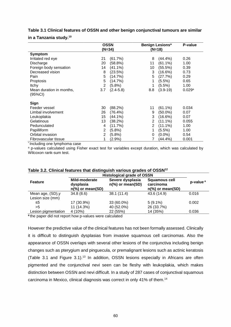
Table 3.1 Clinical features of OSSN and other benign conjunctival tumours are similar
in a Tanzania study.10
OSSN(N=34)
Benign Lesions*(N=18)
P-value
SymptomIrritated red eye 21 (61.7%) 8 (44.4%) 0.26Discharge 20 (58.8%) 11 (61.1%) 1.00Foreign body sensation 14 (41.1%) 10 (55.5%) 0.39Decreased vision 8 (23.5%) 3 (16.6%) 0.73Pain 5 (14.7%) 5 (27.7%) 0.29Proptosis 5 (14.7%) 1 (5.5%) 0.65Itchy 2 (5.8%) 1 (5.5%) 1.00Mean duration in months,(95%CI)
3.7 (2.4-5.8) 8.8 (3.9-19) 0.029a
SignFeeder vessel 30 (88.2%) 11 (61.1%) 0.034Limbal involvement 26 (76.4%) 9 (50.0%) 0.07Leukoplakia 15 (44.1%) 3 (16.6%) 0.07Gelatinous 13 (38.2%) 2 (11.1%) 0.055Pedunculated 4 (11.7%) 2 (11.1%) 1.00Papilliform 2 (5.8%) 1 (5.5%) 1.00Orbital invasion 2 (5.8%) 0 (0.0%) 0.54Fibrovascular tissue 1 (2.9%) 7 (44.4%) 0.001
* Including one lymphoma casea p-values calculated using Fisher exact test for variables except duration, which was calculated byWilcoxon rank-sum test.
Table 3.2. Clinical features that distinguish various grades of OSSN12
Feature
Histological grade of OSSN
p-value aMild-moderatedysplasian(%) or mean(SD)
Severe dysplasian(%) or mean(SD)
Squamous cellcarcinoman(%) or mean(SD)
Mean age, (SD),y 34.8 (8.6) 38.1 (11.4) 43.6 (14.9) 0.016Lesion size (mm) ≤5
>517 (30.9%)11 (14.3%)
33 (60.0%)40 (52.0%)
5 (9.1%)26 (33.7%)
0.002
Lesion pigmentation 4 (10%) 22 (55%) 14 (35%) 0.036a the paper did not report how p-values were calculated
However the predictive value of the clinical features has not been formally assessed. Clinically
it is difficult to distinguish dysplasias from invasive squamous cell carcinomas. Also the
appearance of OSSN overlaps with several other lesions of the conjunctiva including benign
changes such as pterygium and pinguecula, or premalignant lesions such as actinic keratosis
(Table 3.1 and Figure 3.1).13 In addition, OSSN lesions especially in Africans are often
pigmented and the conjunctival nevi seen can be fleshy with leukoplakia, which makes
distinction between OSSN and nevi difficult. In a study of 287 cases of conjunctival squamous
carcinoma in Mexico, clinical diagnosis was correct in only 41% of them.14
60

Figure 3.1 This montage shows lesions that may mimic OSSN. All the lesions in the left
column (A, C, E) are moderately differentiated squamous cell carcinoma (SCC), while
the lesions in the right column are not OSSN, but appear similar. (A) SCC with a
gelatinous surface, (B) pterygium, (C) SCC with leukoplakia, (D) actinic keratosis with
leukoplakia, (E) SCC with pigmentation and (F) a nevus.
2. Histopathology
The normal conjunctiva is divided into 3 regions: (1) the palpebral (tarsal) conjunctiva on the
inner aspect of the upper lid has stratified columnar epithelial cells with little substantia propria,
(2) the fornix has abundant goblet cells, loose stroma, lymphatics and accessory lacrimal
glands of Krause, and (3) the bulbar conjunctiva on the eyeball, in which the epithelium
changes from stratified columnar to stratified squamous near the limbus and has few goblet
cells and melanocytes (Figure 3.2a). Where the lesion is excised with a 2-4 mm margin of
normal tissue, an abrupt transition can be seen between the tumour and adjoining normal
tissue (Figure 3.2b).9
F
A
C E
A
E
B
F
D
61

Figure 3.2a. Histology of the conjunctiva showing the normal limbus. The cornea is to the left
and conjunctiva and sclera to the right of the picture. Cells at the base of the conjunctiva appear
more columnar but get more rounded as you move towards the surface where they appear
flattened. (H&E x10)
Figure 3.2b. This picture shows conjunctival intraepithelial neoplasia on the left hand side where
the conjunctiva appears thickened. Notice the loss of normal cell orientation in the lesion with
some rounded cells seen in superficial areas. The lesion involves the full thickness of the
epithelium but does not erode through the basement membrane into the underlying stroma. The
space between the epithelium and the stroma is an artefact created during slide preparation.
(H& E x20)
62
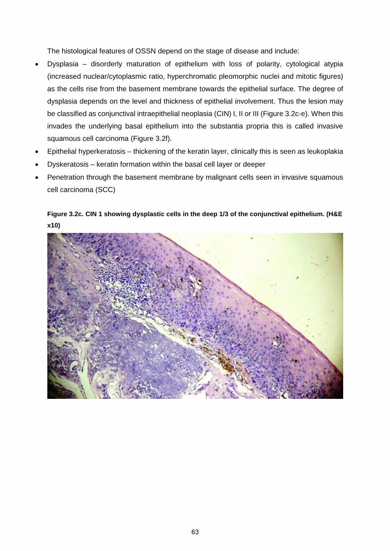
The histological features of OSSN depend on the stage of disease and include:
• Dysplasia – disorderly maturation of epithelium with loss of polarity, cytological atypia
(increased nuclear/cytoplasmic ratio, hyperchromatic pleomorphic nuclei and mitotic figures)
as the cells rise from the basement membrane towards the epithelial surface. The degree of
dysplasia depends on the level and thickness of epithelial involvement. Thus the lesion may
be classified as conjunctival intraepithelial neoplasia (CIN) I, II or III (Figure 3.2c-e). When this
invades the underlying basal epithelium into the substantia propria this is called invasive
squamous cell carcinoma (Figure 3.2f).
• Epithelial hyperkeratosis – thickening of the keratin layer, clinically this is seen as leukoplakia
• Dyskeratosis – keratin formation within the basal cell layer or deeper
• Penetration through the basement membrane by malignant cells seen in invasive squamous
cell carcinoma (SCC)
Figure 3.2c. CIN 1 showing dysplastic cells in the deep 1/3 of the conjunctival epithelium. (H&E
x10)
63

Figure 3.2d. CIN 2 showing dysplasia in the deep 2/3 closer to the epithelial surface. (H&E x10)
Figure 3.2e. CIN 3 showing dysplasia involving the whole thickness of the conjunctiva.
(H&E10)
64

Figure 3.2f. Invasive well differentiated squamous cell carcinoma. (H&E x10)
Basti originally suggested a classification for OSSN into benign, pre-invasive and invasive
stages (Table 3.3).15 The American Joint Committee on Cancer (AJCC) has suggested both
a clinical and pathological staging (Table 3.4).16
Table 3.3 Classification of OSSN after Basti & Macsai.15
Benign
PapillomaPseudoepitheliomatous hyperplasiaBenign hereditary intraepithelial dyskeratosis
Pre-invasive
Conjunctival/corneal intraepithelial neoplasia (CIN) grades I-IIICIN I (mild dysplasia) - confined to the lower third of the conjunctival epitheliumCIN II (moderate dysplasia) - extends into the middle third of the conjunctival epitheliumCIN III (severe dysplasia) – full thickness of the conjunctival epithelium also referred toas CIS (carcinoma-in-situ)
Invasive
Squamous cell carcinomaMucoepidermoid carcinoma
65
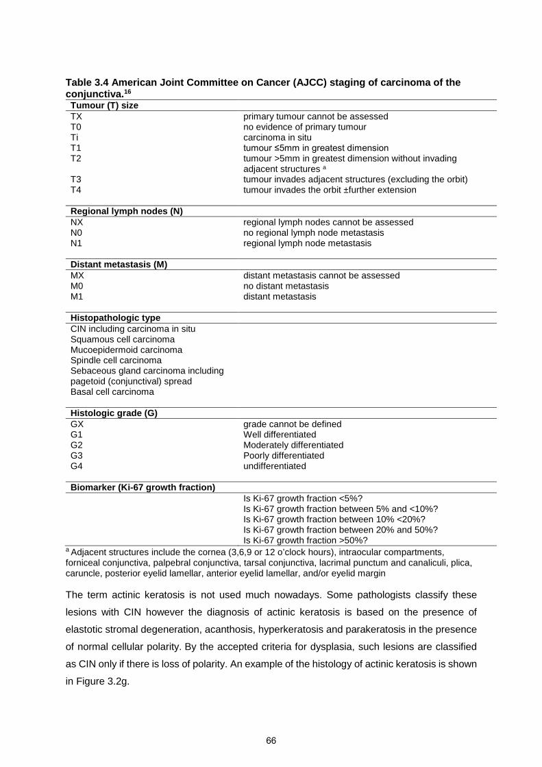
Table 3.4 American Joint Committee on Cancer (AJCC) staging of carcinoma of theconjunctiva.16
Tumour (T) sizeTX primary tumour cannot be assessedT0 no evidence of primary tumourTi carcinoma in situT1 tumour ≤5mm in greatest dimension T2 tumour >5mm in greatest dimension without invading
adjacent structures a
T3 tumour invades adjacent structures (excluding the orbit)T4 tumour invades the orbit ±further extension
Regional lymph nodes (N)NXN0N1
regional lymph nodes cannot be assessedno regional lymph node metastasisregional lymph node metastasis
Distant metastasis (M)MX distant metastasis cannot be assessedM0 no distant metastasisM1 distant metastasis
Histopathologic typeCIN including carcinoma in situSquamous cell carcinomaMucoepidermoid carcinomaSpindle cell carcinomaSebaceous gland carcinoma includingpagetoid (conjunctival) spreadBasal cell carcinoma
Histologic grade (G)GX grade cannot be definedG1 Well differentiatedG2 Moderately differentiatedG3 Poorly differentiatedG4 undifferentiated
Biomarker (Ki-67 growth fraction)Is Ki-67 growth fraction <5%?Is Ki-67 growth fraction between 5% and <10%?Is Ki-67 growth fraction between 10% <20%?Is Ki-67 growth fraction between 20% and 50%?Is Ki-67 growth fraction >50%?
a Adjacent structures include the cornea (3,6,9 or 12 o’clock hours), intraocular compartments,forniceal conjunctiva, palpebral conjunctiva, tarsal conjunctiva, lacrimal punctum and canaliculi, plica,caruncle, posterior eyelid lamellar, anterior eyelid lamellar, and/or eyelid margin
The term actinic keratosis is not used much nowadays. Some pathologists classify these
lesions with CIN however the diagnosis of actinic keratosis is based on the presence of
elastotic stromal degeneration, acanthosis, hyperkeratosis and parakeratosis in the presence
of normal cellular polarity. By the accepted criteria for dysplasia, such lesions are classified
as CIN only if there is loss of polarity. An example of the histology of actinic keratosis is shown
in Figure 3.2g.
66
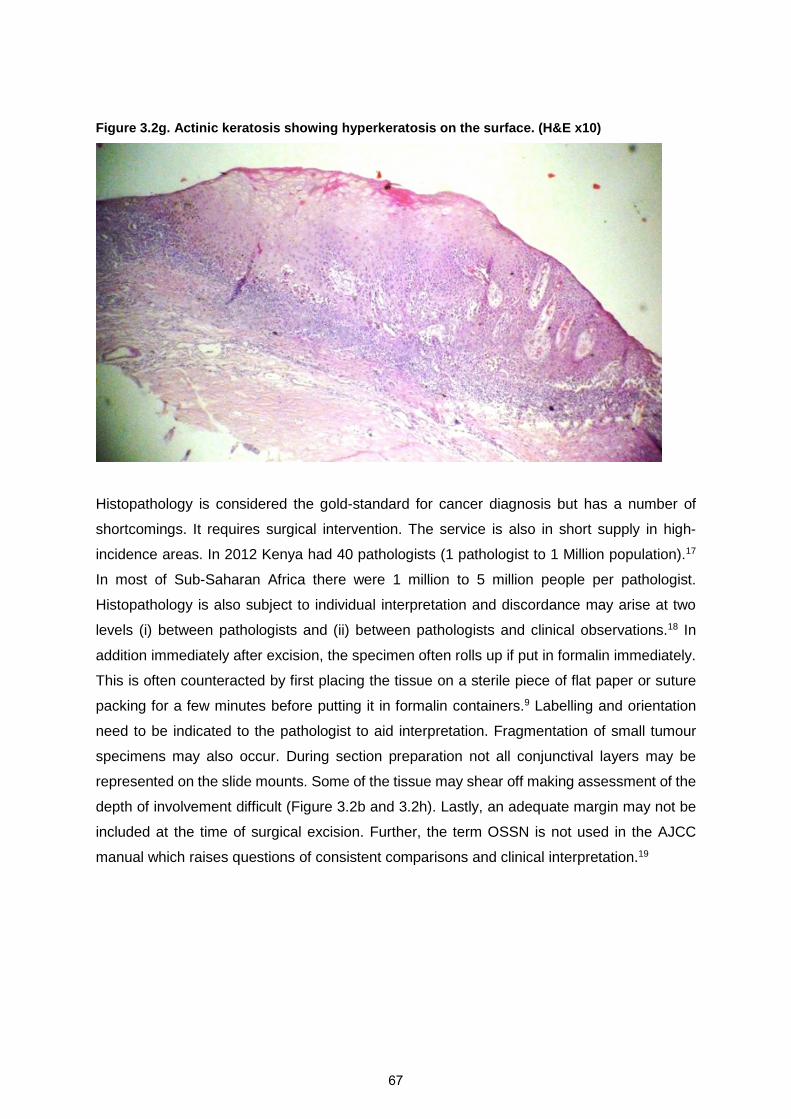
Figure 3.2g. Actinic keratosis showing hyperkeratosis on the surface. (H&E x10)
Histopathology is considered the gold-standard for cancer diagnosis but has a number of
shortcomings. It requires surgical intervention. The service is also in short supply in high-
incidence areas. In 2012 Kenya had 40 pathologists (1 pathologist to 1 Million population).17
In most of Sub-Saharan Africa there were 1 million to 5 million people per pathologist.
Histopathology is also subject to individual interpretation and discordance may arise at two
levels (i) between pathologists and (ii) between pathologists and clinical observations.18 In
addition immediately after excision, the specimen often rolls up if put in formalin immediately.
This is often counteracted by first placing the tissue on a sterile piece of flat paper or suture
packing for a few minutes before putting it in formalin containers.9 Labelling and orientation
need to be indicated to the pathologist to aid interpretation. Fragmentation of small tumour
specimens may also occur. During section preparation not all conjunctival layers may be
represented on the slide mounts. Some of the tissue may shear off making assessment of the
depth of involvement difficult (Figure 3.2b and 3.2h). Lastly, an adequate margin may not be
included at the time of surgical excision. Further, the term OSSN is not used in the AJCC
manual which raises questions of consistent comparisons and clinical interpretation.19
67

Figure 3.2h. This lesion is graded as CIN 3 but the shearing off during section preparation may
make it difficult to grade the depth of involvement. (H&E x5)
Histopathology may be augmented with immunocytochemistry. A study in Japan found that
proliferating cell nuclear antigen (PCNA) immunostaining, p53 immunostaining, and
argyrophilic nucleolar organizer regions (AgNORs) staining in formalin-fixed and paraffin-
embedded tissues could be used as markers of proliferative potency and cell differentiation.
The PCNA-positive staining proportion was highest in SCC, followed by severe dysplasia and
mild dysplasia. P53-positive staining was highest in severe dysplasia, followed by mild
dysplasia, and was negative in SCC. AgNORs-count increased with advancing histological
grade of malignancy.20
3. Vital staining
Vital stains are used to colour living tissues; various dyes are used extensively in
ophthalmology including rose Bengal, fluorescein, methylene blue and toluidine blue.21,22
Toluidine blue (ToB) is a vital dye that stains abnormal tissue dark royal blue by penetrating
into cancerous tissue where it has a selective affinity for nucleic acids (Figure 3.3).23 ToB has
been used safely for many years to help support the clinical diagnosis of oral, oesophageal
and cervical carcinoma and to demarcate tumours during surgical excision.24-28 Animal safety
studies found that intraocular injection (as opposed to topical use) of 1% and 2% ToB caused
irreversible damage to all the corneal layers; 0.5% damaged the stromal keratocytes and
corneal endothelium but 0.25% stained the lens capsule and did not damage the cornea layer
or trabecular meshwork.29 Wander et al conducted animal safety studies in rabbits and guinea
pigs applying eye drops of 0.01%, 0.1%, 0.25%, 0.5%, and 1.0% toluidine blue applied to stain
68
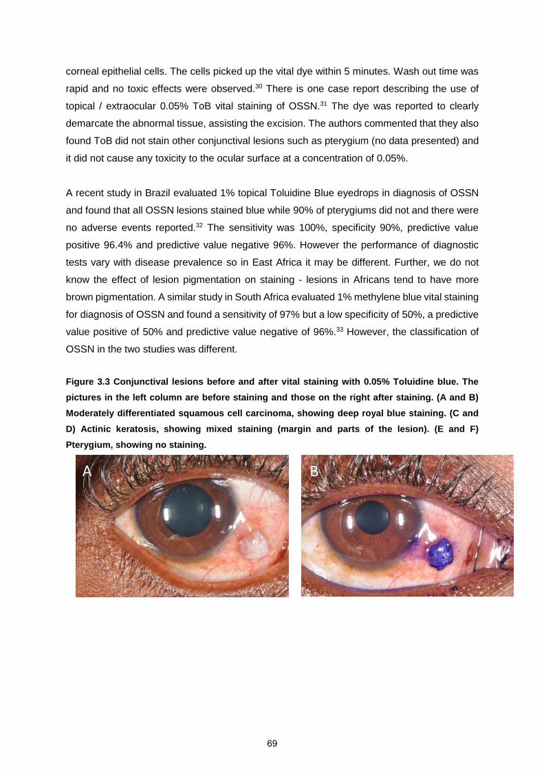
corneal epithelial cells. The cells picked up the vital dye within 5 minutes. Wash out time was
rapid and no toxic effects were observed.30 There is one case report describing the use of
topical / extraocular 0.05% ToB vital staining of OSSN.31 The dye was reported to clearly
demarcate the abnormal tissue, assisting the excision. The authors commented that they also
found ToB did not stain other conjunctival lesions such as pterygium (no data presented) and
it did not cause any toxicity to the ocular surface at a concentration of 0.05%.
A recent study in Brazil evaluated 1% topical Toluidine Blue eyedrops in diagnosis of OSSN
and found that all OSSN lesions stained blue while 90% of pterygiums did not and there were
no adverse events reported.32 The sensitivity was 100%, specificity 90%, predictive value
positive 96.4% and predictive value negative 96%. However the performance of diagnostic
tests vary with disease prevalence so in East Africa it may be different. Further, we do not
know the effect of lesion pigmentation on staining - lesions in Africans tend to have more
brown pigmentation. A similar study in South Africa evaluated 1% methylene blue vital staining
for diagnosis of OSSN and found a sensitivity of 97% but a low specificity of 50%, a predictive
value positive of 50% and predictive value negative of 96%.33 However, the classification of
OSSN in the two studies was different.
Figure 3.3 Conjunctival lesions before and after vital staining with 0.05% Toluidine blue. The
pictures in the left column are before staining and those on the right after staining. (A and B)
Moderately differentiated squamous cell carcinoma, showing deep royal blue staining. (C and
D) Actinic keratosis, showing mixed staining (margin and parts of the lesion). (E and F)
Pterygium, showing no staining.
A B
69
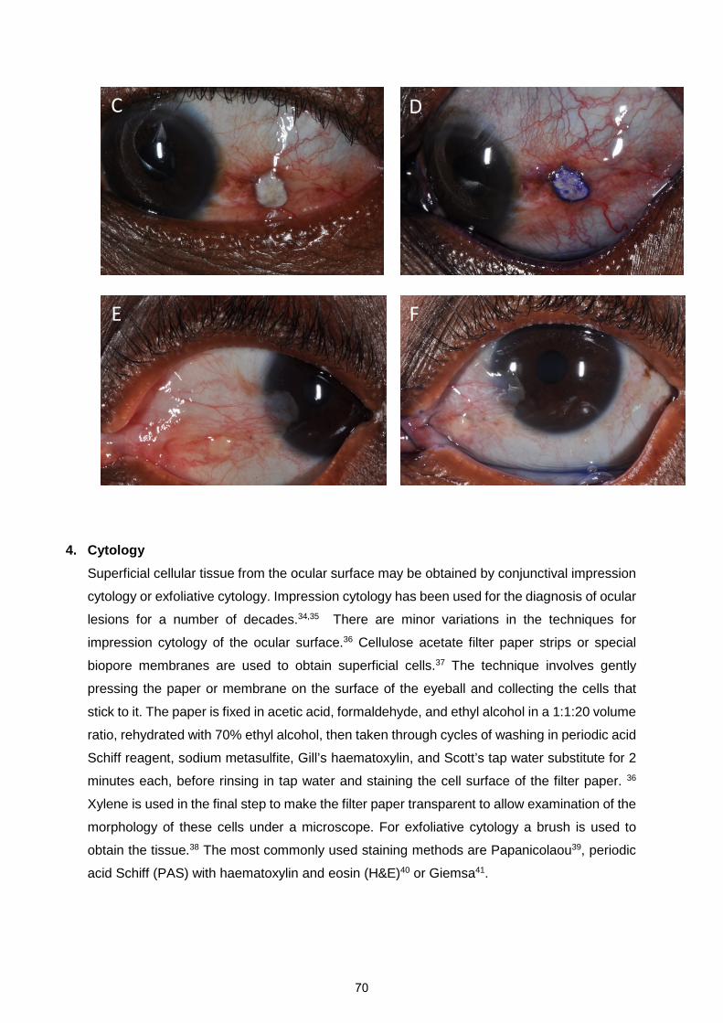
4. Cytology
Superficial cellular tissue from the ocular surface may be obtained by conjunctival impression
cytology or exfoliative cytology. Impression cytology has been used for the diagnosis of ocular
lesions for a number of decades.34,35 There are minor variations in the techniques for
impression cytology of the ocular surface.36 Cellulose acetate filter paper strips or special
biopore membranes are used to obtain superficial cells.37 The technique involves gently
pressing the paper or membrane on the surface of the eyeball and collecting the cells that
stick to it. The paper is fixed in acetic acid, formaldehyde, and ethyl alcohol in a 1:1:20 volume
ratio, rehydrated with 70% ethyl alcohol, then taken through cycles of washing in periodic acid
Schiff reagent, sodium metasulfite, Gill’s haematoxylin, and Scott’s tap water substitute for 2
minutes each, before rinsing in tap water and staining the cell surface of the filter paper. 36
Xylene is used in the final step to make the filter paper transparent to allow examination of the
morphology of these cells under a microscope. For exfoliative cytology a brush is used to
obtain the tissue.38 The most commonly used staining methods are Papanicolaou39, periodic
acid Schiff (PAS) with haematoxylin and eosin (H&E)40 or Giemsa41.
C D
E F
70

An Australian study identified a number of cytomorphological features associated with
OSSN.42 About 50% of CIN cases had keratinized dysplastic cells often accompanied by
hyperkeratosis, large syncytial-like groups in about 35% and individual non-keratinized
dysplastic cells in 10%, while 70% of invasive SCC had significant keratinization. In alcohol-
fixed Papanicolaou stained smears, squamous dysplasia or malignancy was indicated by
nuclear hyperchromasia, high nuclear/cytoplasmic ratio and pleomorphism.43 Conjunctival
impression cytology has been reported to have a sensitivity of 85% and specificity of 90% in
Thailand and USA.44 45 Recently a scoring system was proposed that identifies
cytomorphological features, which may help to reliably differentiate invasive conjunctival
squamous cell carcinoma from pre-invasive lesions.46 In that study the following seven
cytological parameters were found to be predictive of malignancy:
i) nuclear size enlargement >x3
ii) hyperchromasia (dark staining nuclei which is usually due to increased DNA content)
iii) prominent nucleoli
iv) syncytial-like groupings
v) increased nuclear-to-cytoplasm (NC) ratio - it is considered a slightly increased NC
ratio when the cell has an NC ratio of 2:1 or a marked increase in NC ratio when the
cell has an NC ratio up to 2:1
vi) eosinophilic cytoplasmic stain
vii) absence of distinct cytoplasmic borders
Each feature was given a score based on a logistic regression model. A score of 4.25 was the
best cut-off for malignancy.
The following 4 parameters were not predictive:
i) cell disposition - single or individual cells, cells within sheets or cells inside keratin
plaques
ii) cell size - very small or very large
iii) the presence of a regular or irregular nuclear contour
iv) cytoplasmic area - significant reduction in mean cytoplasmic area when the nucleus
fills at least two-thirds of the cell or increased cytoplasmic area when the nucleus
occupies less than half the total area of the cytoplasm
Cytology has also been applied in diagnosis of other diseases of the ocular surface such as
limbal stem-cell deficiency, viral keratoconjunctivitis, allergic/inflammatory
keratoconjunctivitis, vitamin A deficiency and melanocytic lesions.36,37
Impression cytology has a number of shortcomings. It is constrained by the need for
specialised equipment and personnel and this may not be easily available in peripheral care
71
centres. Inter- and intra-observer variation may occur. It is also likely to miss CIN as the
abnormal cells are deeper down. In one Australia study it was difficult to distinguish between
carcinoma-in-situ and minimally invasive disease.47
5. Confocal microscopy
Unlike conventional light microscopes whose image is degraded by reflections and light
scattering by objects outside of the focal plane, in in vivo confocal microscopy, light from a
point source is focused on a tissue and the light reflected from this focal point is collected by
a parallel objective lens and focused onto a duplicate pinhole aperture. Light that passes this
second aperture is collected by a detector. Both the illuminating point source and the
observation aperture of the detector are conjugate with the same point in the tissue, and are
said to be confocal.48 Initially it was used for corneal examination but later extended to
conjunctival studies such as trachoma.49 Confocal microscopy is non-invasive and may allow
serial visualization of the same living tissue down to the cellular level (hence the term in vivo
confocal microscopy – IVCM) over time unlike histology.
Several studies have evaluated the use of IVCM for the diagnosis of OSSN with differing
results. A study of 10 cases from Italy found that the main pathological features include:
cytonuclear atypias, epithelial folds into an inflammatory and vascularised conjunctival stroma,
fine vessels perpendicular to the surface, clear limit with normal epithelium, papillomatous
organization, and abnormal keratinisation.50 A study of 5 cases in China found cellular
anisocytosis (variation in cell size) and enlarged nuclei with high nuclear to cytoplasmic ratio
in 3 cases of CIN and nests partially formed by isolated keratinized, binucleated, and actively
mitotic dysmorphic epithelial cells in the other 2 cases diagnosed as carcinoma in situ (CIS).51
However, more recently we looked at a larger series of 52 cases of conjunctival lesions in
Tanzania and found that IVCM features such as hyper-reflective cells, anisocytosis and starry
night appearance of basal cells could not reliably distinguish OSSN from benign conjunctival
pathology because of an overlap in the features (see appendix 1).10
6. Anterior segment Optical Coherence Tomography (OCT)
OCT is a computer assisted optical instrument that produces cross-sectional images
(tomograms) similar to B-mode ultrasound only that it uses light instead of sound. A low
coherence infra-red light (820nm) is directed at a target tissue and the amount and position of
backscattered light displayed according to the optical properties of the structure in question. It
has the advantages of fast image acquisition speed and non-contact and is non-invasive with
some referring to it as a non-excisional 'optical biopsy'.52 It has been suggested that ultra-high
resolution anterior segment optical coherence tomography (UHR OCT) may be used in
72
diagnosis and follow up of OSSN however the studies in this area are small. One study showed
that UHR OCT images of the lesions showed a thickened hyper-reflective epithelium and
abrupt transition from normal to hyper-reflective epithelium in all 7 cases.53 In a study to
distinguish OSSN from pterygium, there was a statistically significant difference in the
epithelial thickness with an average 346µm (standard deviation [SD], 167) in 17 OSSN
patients and 101 µm (SD, 22) in 17 pterygium patients (p<0.001). By receiver operating
characteristic curve, the sensitivity and specificity of UHR OCT for differentiating between
OSSN and pterygia was found to be 94% and 100%, respectively, using a cut-off value of 142
µm.54
One of the major shortcomings of ultraound and OCT is the cost of equipment and the effect
of rapidly changing technology that may render it obsolete in a few years. In practice the
reliability of this technique is yet to be proven.
Conclusions
OSSN has serious consequences associated with recurrence and local metastasis that may
lead to death. Early diagnosis may reduce this. There seems to be a mismatch between
availability of diagnostic facilities and where OSSN is most frequently found. Highly affected
areas have short supply of diagnostic capability.
Some clinicians make management decisions without a tissue diagnosis but clinical signs are
not very reliable for diagnosis due to the overlap between the features of OSSN and other
conjunctival lesions. There is a risk of misclassification and inappropriate treatment with this
practice. Patients with benign disease would inappropriately be subjected to chemotherapy
and live with a diagnosis of ‘cancer’ and fear of recurrence.
Histology is the gold standard but is subjective and prone to inter-observer variability. Tissue
samples are small and may roll up if put in formalin immediately after excision making
orientation and interpretation of slides difficult. During tissue section preparation the surface
epithelium may be sheared off making classification by depth of involvement difficult.
Vital staining with toluidine blue is promising as a non-invasive diagnostic tool. Formal
evaluation in areas of differing prevalence is needed as the performance of diagnostic tests is
affected by disease prevalence. Toluidine blue vital staining may be useful for marking the
extent of tumour to aid surgical excision. Brown pigmentation of lesions however obscures the
dye. In addition, this will not replace a tissue diagnosis.
73

Cytology is a less invasive tissue diagnostic method. Scoring systems using cytology are
reliable. However cytology may miss CIN as it would not pick the deeper epithelial cells. There
are also relatively few trained cytologists.
In-vivo confocal microscopy, ultrasound and anterior segment optical coherence tomography
(OCT) are modern technologies that promise to circumvent surgical excision and facilitate
primary topical treatment. However there is overlap in the appearance of OSSN and other
conjunctival lesions so IVCM cannot reliably distinguish between the two. The technology is
very expensive and prone to short lifespans due to rapid new developments that may render
existing equipment obsolete. They are currently not a suitable option in Africa where OSSN
mainly occurs.
References1. Stone DU, Butt AL, Chodosh J. Ocular surface squamous neoplasia: a standard of care survey.
Cornea 2005; 24(3): 297-300.2. Lee GA, Hirst LW. Retrospective study of ocular surface squamous neoplasia. Australian and New
Zealand journal of ophthalmology 1997; 25(4): 269-76.3. Nelson KD, McSoley JJ. Clinical findings and management of conjunctival intraepithelial neoplasia.
Optometry (St Louis, Mo) 2011; 82(1): 15-21.4. Tunc M, Char DH, Crawford B, Miller T. Intraepithelial and invasive squamous cell carcinoma of
the conjunctiva: analysis of 60 cases. British Journal of Ophthalmology 1999; 83(1): 98-103.5. Waddell KM, Downing RG, Lucas SB, Newton R. Corneo-conjunctival carcinoma in Uganda. Eye
(Lond) 2006; 20(8): 893-9.6. Kamal S, Kaliki S, Mishra DK, Batra J, Naik MN. Ocular Surface Squamous Neoplasia in 200 Patients.
Ophthalmology.7. Usui Y, Waring GO, See RF, Rao NA, Marrone AC. Bilateral ocular surface squamous neoplasia: a
clinicopathological case report. The British journal of ophthalmology 2004; 88(4): 595-6.8. Kim BH, Kim MK, Wee WR, Oh JY. Clinical and pathological characteristics of ocular surface
squamous neoplasia in an Asian population. Graefe's archive for clinical and experimentalophthalmology = Albrecht von Graefes Archiv fur klinische und experimentelle Ophthalmologie2013; 251(11): 2569-73.
9. Mittal R, Rath S, Vemuganti GK. Ocular surface squamous neoplasia - Review of etio-pathogenesisand an update on clinico-pathological diagnosis. Saudi journal of ophthalmology : official journalof the Saudi Ophthalmological Society 2013; 27(3): 177-86.
10. Nguena MB, van den Tweel JG, Makupa W, et al. Diagnosing ocular surface squamous neoplasiain East Africa: case-control study of clinical and in vivo confocal microscopy assessment.Ophthalmology 2014; 121(2): 484-91.
11. Kao AA, Galor A, Karp CL, Abdelaziz A, Feuer WJ, Dubovy SR. Clinicopathologic correlation ofocular surface squamous neoplasms at bascom palmer eye institute: 2001 to 2010.Ophthalmology 2012; 119(9): 1773-6.
12. Makupa, II, Swai B, Makupa WU, White VA, Lewallen S. Clinical factors associated with malignancyand HIV status in patients with ocular surface squamous neoplasia at Kilimanjaro ChristianMedical Centre, Tanzania. The British journal of ophthalmology 2012; 96(4): 482-4.
13. Clear AS, Chirambo MC, Hutt MS. Solar keratosis, pterygium, and squamous cell carcinoma of theconjunctiva in Malawi. The British journal of ophthalmology 1979; 63(2): 102-9.
74
14. Cervantes G, Rodriguez AA, Jr., Leal AG. Squamous cell carcinoma of the conjunctiva:clinicopathological features in 287 cases. Canadian journal of ophthalmology Journal canadiend'ophtalmologie 2002; 37(1): 14-9; discussion 9-20.
15. Basti S, Macsai MS. Ocular surface squamous neoplasia: a review. Cornea 2003; 22(7): 687-704.16. American Joint Committee on Cancer. Carcinoma of the conjunctiva. In: Edge SB, Byrd DR,
Compton CC, Fritz AG, Greene FL, Trotti A, eds. Cancer staging handbook. 7 ed. New York:Springer; 2010: 600-2.
17. Adesina A, Chumba D, Nelson AM, et al. Improvement of pathology in sub-Saharan Africa. Thelancet oncology 2013; 14(4): e152-7.
18. Margo CE, Harman LE, Mulla ZD. The reliability of clinical methods in ophthalmology. Survey ofophthalmology 2002; 47(4): 375-86.
19. Margo CE, White AA. Ocular surface squamous neoplasia: terminology that is conceptuallyfriendly but clinically perilous. Eye (Lond) 2014; 28(5): 507-9.
20. Aoki S, Kubo E, Nakamura S, et al. Possible prognostic markers in conjunctival dysplasia andsquamous cell carcinoma. Japanese journal of ophthalmology 1998; 42(4): 256-61.
21. Rodrigues EB, Costa EF, Penha FM, et al. The use of vital dyes in ocular surgery. Survey ofophthalmology 2009; 54(5): 576-617.
22. Norn MS. Vital staining of cornea and conjunctiva. Acta ophthalmologica 1962; 40: 389-401.23. Herlin P, Marnay J, Jacob JH, Ollivier JM, Mandard AM. A study of the mechanism of the toluidine
blue dye test. Endoscopy 1983; 15(1): 4-7.24. Lingen MW, Kalmar JR, Karrison T, Speight PM. Critical evaluation of diagnostic aids for the
detection of oral cancer. Oral oncology 2008; 44(1): 10-22.25. Martin IC, Kerawala CJ, Reed M. The application of toluidine blue as a diagnostic adjunct in the
detection of epithelial dysplasia. Oral surgery, oral medicine, oral pathology, oral radiology, andendodontics 1998; 85(4): 444-6.
26. Onofre MA, Sposto MR, Navarro CM. Reliability of toluidine blue application in the detection oforal epithelial dysplasia and in situ and invasive squamous cell carcinomas. Oral surgery, oralmedicine, oral pathology, oral radiology, and endodontics 2001; 91(5): 535-40.
27. Allegra E, Lombardo N, Puzzo L, Garozzo A. The usefulness of toluidine staining as a diagnostictool for precancerous and cancerous oropharyngeal and oral cavity lesions. Actaotorhinolaryngologica Italica : organo ufficiale della Societa italiana di otorinolaringologia echirurgia cervico-facciale 2009; 29(4): 187-90.
28. Nishimura S, Ishiko O, Honda K, Hyun Y, Ogita S. New cervicoscopy with toluidine blue stainingfor superficial cervical invasion by endometrial carcinoma. Gynecologic and obstetricinvestigation 2000; 50(2): 133-5.
29. Gamal Eldin SA, el Mehelmy EM, el Shazli EM, Mostafa YM. Experimental staining of the anteriorlens capsule in albino rabbits. Journal of cataract and refractive surgery 1999; 25(9): 1289-94.
30. Wander AH, Neumeister RD, Tschismadia I, Choromokos EA, Masukawa T. In vivo corneal andconjunctival epithelial nuclear stain. Cornea 1985; 4(1): 8-13.
31. Kaji Y, Hiraoka T, Oshika T. Vital staining of squamous cell carcinoma of the conjunctiva usingtoluidine blue. Acta ophthalmologica Scandinavica 2006; 84(6): 825-6.
32. Romero IL, Barros JD, Martins MC, Ballalai PL. The Use of 1% Toluidine Blue Eye Drops in theDiagnosis of Ocular Surface Squamous Neoplasia. Cornea 2012.
33. Steffen J, Rice J, Lecuona K, Carrara H. Identification of ocular surface squamous neoplasia by invivo staining with methylene blue. The British journal of ophthalmology 2014; 98(1): 13-5.
34. Dykstra PC, Dykstra BA. The cytologic diagnosis of carcinoma and related lesions of the ocularconjunctiva and cornea. Transactions - American Academy of Ophthalmology and OtolaryngologyAmerican Academy of Ophthalmology and Otolaryngology 1969; 73(5): 979-95.
35. Naib ZM. Cytology of ocular lesions. Acta cytologica 1972; 16(2): 178-85.36. Singh R, Joseph A, Umapathy T, Tint NL, Dua HS. Impression cytology of the ocular surface. The
British journal of ophthalmology 2005; 89(12): 1655-9.
75
37. McKelvie P. Ocular surface impression cytology. Advances in anatomic pathology 2003; 10(6):328-37.
38. Ersoz C, Yagmur M, Ersoz TR, Yalaz M. Preoperative brush and impression cytology in ocularsurface squamous neoplasms. Acta cytologica 2003; 47(1): 13-5.
39. Gelender H, Forster RK. Papanicolaou cytology in the diagnosis and management of externalocular tumors. Archives of ophthalmology 1980; 98(5): 909-12.
40. Egbert PR, Lauber S, Maurice DM. A simple conjunctival biopsy. American journal ofophthalmology 1977; 84(6): 798-801.
41. Doughty MJ. Reliability of nucleus-to-cell and nucleus-to-cytoplasm calculations for conjunctivalimpression cytology specimens. Current eye research 2012; 37(7): 583-91.
42. Nolan GR, Hirst LW, Bancroft BJ. The cytomorphology of ocular surface squamous neoplasia byusing impression cytology. Cancer 2001; 93(1): 60-7.
43. Doughty MJ. Objective assessment of conjunctival squamous metaplasia by measures of cell andnucleus dimensions. Diagnostic cytopathology 2011; 39(6): 409-23.
44. Tananuvat N, Lertprasertsuk N, Mahanupap P, Noppanakeepong P. Role of impression cytologyin diagnosis of ocular surface neoplasia. Cornea 2008; 27(3): 269-74.
45. Semenova EA, Milman T, Finger PT, et al. The diagnostic value of exfoliative cytology vshistopathology for ocular surface squamous neoplasia. American journal of ophthalmology 2009;148(5): 772-8 e1.
46. Barros JN, Lowen MS, Ballalai PL, Mascaro VL, Gomes JA, Martins MC. Predictive index todifferentiate invasive squamous cell carcinoma from preinvasive ocular surface lesions byimpression cytology. The British journal of ophthalmology 2009; 93(2): 209-14.
47. McKelvie PA. Squamous cell carcinoma of the conjunctiva: a series of 26 cases. British Journal ofOphthalmology 2002; 86(2): 168-73.
48. Erie JC, McLaren JW, Patel SV. Confocal microscopy in ophthalmology. American journal ofophthalmology 2009; 148(5): 639-46.
49. Hu VH, Weiss HA, Massae P, et al. In vivo confocal microscopy in scarring trachoma.Ophthalmology 2011; 118(11): 2138-46.
50. Parrozzani R, Lazzarini D, Dario A, Midena E. In vivo confocal microscopy of ocular surfacesquamous neoplasia. Eye (Lond) 2011; 25(4): 455-60.
51. Xu Y, Zhou Z, Xu Y, et al. The clinical value of in vivo confocal microscopy for diagnosis of ocularsurface squamous neoplasia. Eye (Lond) 2012; 26(6): 781-7.
52. Chen J, Lee L. Clinical applications and new developments of optical coherence tomography: anevidence-based review. Clinical & experimental optometry : journal of the AustralianOptometrical Association 2007; 90(5): 317-35.
53. Shousha MA, Karp CL, Perez VL, et al. Diagnosis and management of conjunctival and cornealintraepithelial neoplasia using ultra high-resolution optical coherence tomography.Ophthalmology 2011; 118(8): 1531-7.
54. Kieval JZ, Karp CL, Abou Shousha M, et al. Ultra-high resolution optical coherence tomographyfor differentiation of ocular surface squamous neoplasia and pterygia. Ophthalmology 2012;119(3): 481-6.
76

Chapter 4. Interventions for squamous cell
carcinoma of the conjunctiva in HIV-infected
individuals (Cochrane Review)
77

RESEARCH PAPER COVER SHEET
PLEASE NOTE THAT A COVER SHEET MUST BE COMPLETED FOR EACH RESEARCH PAPER INCLUDEDIN A THESIS.
SECTION A – Student Details
Student
Principal Supervisor
Thesis Title
If the Research Paper has previously been published please complete Section B, if not please move toSection C
SECTION B – Paper already published
Where was the work published?
When was the work published?
If the work was published prior toregistration for your research degree,give a brief rationale for its inclusion
Have you retained the copyright for thework?* No
Was the work subject toacademic peer review? Yes
*If yes, please attach evidence of retention. If no, or if the work is being included in its published format, pleaseattach evidence of permission from the copyright holder (publisher or other author) to include this work.
SECTION C – Prepared for publication, but not yet published
Where is the work intended to bepublished?
Please list the paper’s authors in theintended authorship order:
Stage of publication Choose an item.
SECTION D – Multi-authored work
For multi-authored work, give full details of your role inthe research included in the paper and in the preparationof the paper. (Attach a further sheet if necessary)
Student Signature: Date:
Supervisor Signature: Date:
78
Gichuhi
Typewritten text
Stephen Gichuhi
Gichuhi
Typewritten text
Matthew J. Burton
Gichuhi
Typewritten text
Epidemiology and management of ocular surface squamous neoplasia in Kenya
Gichuhi
Typewritten text
4th February 2016
Gichuhi
Typewritten text
4th February 2016
Gichuhi
Typewritten text
I reviewed the search output from the Trial Search Coordinator, extracted the data from the papers, conducted the analysis with guidance from a statistician from the South African Cochrane Center, drafted and James Irlam and submitted the manuscript with consideration of comments from James Irlam. For the supplementary review of case series, I did the searches, data extraction and analysis with input from Matthew Burton.
Gichuhi
Typewritten text
The Cochrane Library
Gichuhi
Typewritten text
2013

JOHN WILEY AND SONS LICENSETERMS AND CONDITIONS
Feb 27, 2016
This Agreement between Stephen Gichuhi ("You") and John Wiley and Sons ("John Wileyand Sons") consists of your license details and the terms and conditions provided by JohnWiley and Sons and Copyright Clearance Center.
License Number 3817130105825
License date Feb 27, 2016
Licensed Content Publisher John Wiley and Sons
Licensed Content Publication Cochrane Database of Systematic Reviews
Licensed Content Title Interventions for squamous cell carcinoma of the conjunctiva inHIV-infected individuals
Licensed Content Author Stephen Gichuhi,James H Irlam
Licensed Content Date Feb 28, 2013
Pages 22
Type of use Dissertation/Thesis
Requestor type Author of this Wiley article
Format Print and electronic
Portion Full article
Will you be translating? No
Title of your thesis /dissertation
Epidemiology and management of ocular surface squamousneoplasia in Kenya
Expected completion date May 2016
Expected size (number ofpages)
22
Requestor Location Stephen GichuhiP.O Box 347
Nairobi, Kenya 00202Attn: Stephen Gichuhi
Billing Type Invoice
Billing Address Stephen GichuhiP.O Box 347
Nairobi, Kenya 00202Attn: Stephen Gichuhi
Total 0.00 USD
Terms and Conditions
TERMS AND CONDITIONSThis copyrighted material is owned by or exclusively licensed to John Wiley & Sons, Inc. orone of its group companies (each a"Wiley Company") or handled on behalf of a society withwhich a Wiley Company has exclusive publishing rights in relation to a particular work(collectively "WILEY"). By clicking "accept" in connection with completing this licensing
RightsLink Printable License https://s100.copyright.com/App/PrintableLicenseFrame.jsp?publisher...
1 of 5 27/02/2016 19:29
79
transaction, you agree that the following terms and conditions apply to this transaction(along with the billing and payment terms and conditions established by the CopyrightClearance Center Inc., ("CCC's Billing and Payment terms and conditions"), at the time thatyou opened your RightsLink account (these are available at any time athttp://myaccount.copyright.com).
Terms and Conditions
The materials you have requested permission to reproduce or reuse (the "WileyMaterials") are protected by copyright.
You are hereby granted a personal, non-exclusive, non-sub licensable (on astand-alone basis), non-transferable, worldwide, limited license to reproduce theWiley Materials for the purpose specified in the licensing process. This license, andany CONTENT (PDF or image file) purchased as part of your order, is for aone-time use only and limited to any maximum distribution number specified in thelicense. The first instance of republication or reuse granted by this license must becompleted within two years of the date of the grant of this license (although copiesprepared before the end date may be distributed thereafter). The Wiley Materials shallnot be used in any other manner or for any other purpose, beyond what is granted inthe license. Permission is granted subject to an appropriate acknowledgement given tothe author, title of the material/book/journal and the publisher. You shall also duplicatethe copyright notice that appears in the Wiley publication in your use of the WileyMaterial. Permission is also granted on the understanding that nowhere in the text is apreviously published source acknowledged for all or part of this Wiley Material. Anythird party content is expressly excluded from this permission.
With respect to the Wiley Materials, all rights are reserved. Except as expresslygranted by the terms of the license, no part of the Wiley Materials may be copied,modified, adapted (except for minor reformatting required by the new Publication),translated, reproduced, transferred or distributed, in any form or by any means, and noderivative works may be made based on the Wiley Materials without the priorpermission of the respective copyright owner.For STM Signatory Publishersclearing permission under the terms of the STM Permissions Guidelines only, theterms of the license are extended to include subsequent editions and for editionsin other languages, provided such editions are for the work as a whole in situ anddoes not involve the separate exploitation of the permitted figures or extracts,You may not alter, remove or suppress in any manner any copyright, trademark orother notices displayed by the Wiley Materials. You may not license, rent, sell, loan,lease, pledge, offer as security, transfer or assign the Wiley Materials on a stand-alonebasis, or any of the rights granted to you hereunder to any other person.
The Wiley Materials and all of the intellectual property rights therein shall at all timesremain the exclusive property of John Wiley & Sons Inc, the Wiley Companies, ortheir respective licensors, and your interest therein is only that of having possession ofand the right to reproduce the Wiley Materials pursuant to Section 2 herein during thecontinuance of this Agreement. You agree that you own no right, title or interest in orto the Wiley Materials or any of the intellectual property rights therein. You shall haveno rights hereunder other than the license as provided for above in Section 2. No right,license or interest to any trademark, trade name, service mark or other branding("Marks") of WILEY or its licensors is granted hereunder, and you agree that you
RightsLink Printable License https://s100.copyright.com/App/PrintableLicenseFrame.jsp?publisher...
2 of 5 27/02/2016 19:29
80

shall not assert any such right, license or interest with respect thereto
NEITHER WILEY NOR ITS LICENSORS MAKES ANY WARRANTY ORREPRESENTATION OF ANY KIND TO YOU OR ANY THIRD PARTY,EXPRESS, IMPLIED OR STATUTORY, WITH RESPECT TO THE MATERIALSOR THE ACCURACY OF ANY INFORMATION CONTAINED IN THEMATERIALS, INCLUDING, WITHOUT LIMITATION, ANY IMPLIEDWARRANTY OF MERCHANTABILITY, ACCURACY, SATISFACTORYQUALITY, FITNESS FOR A PARTICULAR PURPOSE, USABILITY,INTEGRATION OR NON-INFRINGEMENT AND ALL SUCH WARRANTIESARE HEREBY EXCLUDED BY WILEY AND ITS LICENSORS AND WAIVEDBY YOU.
WILEY shall have the right to terminate this Agreement immediately upon breach ofthis Agreement by you.
You shall indemnify, defend and hold harmless WILEY, its Licensors and theirrespective directors, officers, agents and employees, from and against any actual orthreatened claims, demands, causes of action or proceedings arising from any breachof this Agreement by you.
IN NO EVENT SHALL WILEY OR ITS LICENSORS BE LIABLE TO YOU ORANY OTHER PARTY OR ANY OTHER PERSON OR ENTITY FOR ANYSPECIAL, CONSEQUENTIAL, INCIDENTAL, INDIRECT, EXEMPLARY ORPUNITIVE DAMAGES, HOWEVER CAUSED, ARISING OUT OF OR INCONNECTION WITH THE DOWNLOADING, PROVISIONING, VIEWING ORUSE OF THE MATERIALS REGARDLESS OF THE FORM OF ACTION,WHETHER FOR BREACH OF CONTRACT, BREACH OF WARRANTY, TORT,NEGLIGENCE, INFRINGEMENT OR OTHERWISE (INCLUDING, WITHOUTLIMITATION, DAMAGES BASED ON LOSS OF PROFITS, DATA, FILES, USE,BUSINESS OPPORTUNITY OR CLAIMS OF THIRD PARTIES), AND WHETHEROR NOT THE PARTY HAS BEEN ADVISED OF THE POSSIBILITY OF SUCHDAMAGES. THIS LIMITATION SHALL APPLY NOTWITHSTANDING ANYFAILURE OF ESSENTIAL PURPOSE OF ANY LIMITED REMEDY PROVIDEDHEREIN.
Should any provision of this Agreement be held by a court of competent jurisdictionto be illegal, invalid, or unenforceable, that provision shall be deemed amended toachieve as nearly as possible the same economic effect as the original provision, andthe legality, validity and enforceability of the remaining provisions of this Agreementshall not be affected or impaired thereby.
The failure of either party to enforce any term or condition of this Agreement shall notconstitute a waiver of either party's right to enforce each and every term and conditionof this Agreement. No breach under this agreement shall be deemed waived orexcused by either party unless such waiver or consent is in writing signed by the partygranting such waiver or consent. The waiver by or consent of a party to a breach ofany provision of this Agreement shall not operate or be construed as a waiver of orconsent to any other or subsequent breach by such other party.
RightsLink Printable License https://s100.copyright.com/App/PrintableLicenseFrame.jsp?publisher...
3 of 5 27/02/2016 19:29
81
This Agreement may not be assigned (including by operation of law or otherwise) byyou without WILEY's prior written consent.
Any fee required for this permission shall be non-refundable after thirty (30) daysfrom receipt by the CCC.
These terms and conditions together with CCC's Billing and Payment terms andconditions (which are incorporated herein) form the entire agreement between you andWILEY concerning this licensing transaction and (in the absence of fraud) supersedesall prior agreements and representations of the parties, oral or written. This Agreementmay not be amended except in writing signed by both parties. This Agreement shall bebinding upon and inure to the benefit of the parties' successors, legal representatives,and authorized assigns.
In the event of any conflict between your obligations established by these terms andconditions and those established by CCC's Billing and Payment terms and conditions,these terms and conditions shall prevail.
WILEY expressly reserves all rights not specifically granted in the combination of (i)the license details provided by you and accepted in the course of this licensingtransaction, (ii) these terms and conditions and (iii) CCC's Billing and Payment termsand conditions.
This Agreement will be void if the Type of Use, Format, Circulation, or RequestorType was misrepresented during the licensing process.
This Agreement shall be governed by and construed in accordance with the laws ofthe State of New York, USA, without regards to such state's conflict of law rules. Anylegal action, suit or proceeding arising out of or relating to these Terms and Conditionsor the breach thereof shall be instituted in a court of competent jurisdiction in NewYork County in the State of New York in the United States of America and each partyhereby consents and submits to the personal jurisdiction of such court, waives anyobjection to venue in such court and consents to service of process by registered orcertified mail, return receipt requested, at the last known address of such party.
WILEY OPEN ACCESS TERMS AND CONDITIONSWiley Publishes Open Access Articles in fully Open Access Journals and in Subscriptionjournals offering Online Open. Although most of the fully Open Access journals publishopen access articles under the terms of the Creative Commons Attribution (CC BY) Licenseonly, the subscription journals and a few of the Open Access Journals offer a choice ofCreative Commons Licenses. The license type is clearly identified on the article.The Creative Commons Attribution LicenseThe Creative Commons Attribution License (CC-BY) allows users to copy, distribute andtransmit an article, adapt the article and make commercial use of the article. The CC-BYlicense permits commercial and non-Creative Commons Attribution Non-Commercial LicenseThe Creative Commons Attribution Non-Commercial (CC-BY-NC)License permits use,distribution and reproduction in any medium, provided the original work is properly citedand is not used for commercial purposes.(see below)
RightsLink Printable License https://s100.copyright.com/App/PrintableLicenseFrame.jsp?publisher...
4 of 5 27/02/2016 19:29
82
Creative Commons Attribution-Non-Commercial-NoDerivs LicenseThe Creative Commons Attribution Non-Commercial-NoDerivs License (CC-BY-NC-ND)permits use, distribution and reproduction in any medium, provided the original work isproperly cited, is not used for commercial purposes and no modifications or adaptations aremade. (see below)Use by commercial "for-profit" organizationsUse of Wiley Open Access articles for commercial, promotional, or marketing purposesrequires further explicit permission from Wiley and will be subject to a fee.Further details can be found on Wiley Online Library http://olabout.wiley.com/WileyCDA/Section/id-410895.html
Other Terms and Conditions:
v1.10 Last updated September 2015
Questions? [email protected] or +1-855-239-3415 (toll free in the US) or+1-978-646-2777.
RightsLink Printable License https://s100.copyright.com/App/PrintableLicenseFrame.jsp?publisher...
5 of 5 27/02/2016 19:29
83
Interventions for squamous cell carcinoma of the conjunctiva
in HIV-infected individuals (Review)
Gichuhi S, Irlam JH
This is a reprint of a Cochrane review, prepared and maintained by The Cochrane Collaboration and published in The Cochrane Library2013, Issue 2
http://www.thecochranelibrary.com
Interventions for squamous cell carcinoma of the conjunctiva in HIV-infected individuals (Review)
Copyright © 2013 The Cochrane Collaboration. Published by John Wiley & Sons, Ltd.
84

T A B L E O F C O N T E N T S
1HEADER . . . . . . . . . . . . . . . . . . . . . . . . . . . . . . . . . . . . . . .
1ABSTRACT . . . . . . . . . . . . . . . . . . . . . . . . . . . . . . . . . . . . . .
2PLAIN LANGUAGE SUMMARY . . . . . . . . . . . . . . . . . . . . . . . . . . . . . .
2BACKGROUND . . . . . . . . . . . . . . . . . . . . . . . . . . . . . . . . . . . .
Figure 1. . . . . . . . . . . . . . . . . . . . . . . . . . . . . . . . . . . . . . 5
5OBJECTIVES . . . . . . . . . . . . . . . . . . . . . . . . . . . . . . . . . . . . .
5METHODS . . . . . . . . . . . . . . . . . . . . . . . . . . . . . . . . . . . . . .
6RESULTS . . . . . . . . . . . . . . . . . . . . . . . . . . . . . . . . . . . . . . .
Figure 2. . . . . . . . . . . . . . . . . . . . . . . . . . . . . . . . . . . . . . 7
7DISCUSSION . . . . . . . . . . . . . . . . . . . . . . . . . . . . . . . . . . . . .
8AUTHORS’ CONCLUSIONS . . . . . . . . . . . . . . . . . . . . . . . . . . . . . . .
8ACKNOWLEDGEMENTS . . . . . . . . . . . . . . . . . . . . . . . . . . . . . . . .
8REFERENCES . . . . . . . . . . . . . . . . . . . . . . . . . . . . . . . . . . . . .
11CHARACTERISTICS OF STUDIES . . . . . . . . . . . . . . . . . . . . . . . . . . . . .
13DATA AND ANALYSES . . . . . . . . . . . . . . . . . . . . . . . . . . . . . . . . . .
13ADDITIONAL TABLES . . . . . . . . . . . . . . . . . . . . . . . . . . . . . . . . . .
15APPENDICES . . . . . . . . . . . . . . . . . . . . . . . . . . . . . . . . . . . . .
19WHAT’S NEW . . . . . . . . . . . . . . . . . . . . . . . . . . . . . . . . . . . . .
19HISTORY . . . . . . . . . . . . . . . . . . . . . . . . . . . . . . . . . . . . . . .
20CONTRIBUTIONS OF AUTHORS . . . . . . . . . . . . . . . . . . . . . . . . . . . . .
20DECLARATIONS OF INTEREST . . . . . . . . . . . . . . . . . . . . . . . . . . . . . .
20SOURCES OF SUPPORT . . . . . . . . . . . . . . . . . . . . . . . . . . . . . . . . .
20INDEX TERMS . . . . . . . . . . . . . . . . . . . . . . . . . . . . . . . . . . . .
iInterventions for squamous cell carcinoma of the conjunctiva in HIV-infected individuals (Review)
Copyright © 2013 The Cochrane Collaboration. Published by John Wiley & Sons, Ltd.
85
[Intervention Review]
Interventions for squamous cell carcinoma of the conjunctivain HIV-infected individuals
Stephen Gichuhi1, James H Irlam2
1Department of Ophthalmology, University of Nairobi, Nairobi, Kenya. 2Primary Health Care Directorate, University of Cape Town,
Cape Town, South Africa
Contact address: Stephen Gichuhi, Department of Ophthalmology, University of Nairobi, P.O Box 347, KNH, Nairobi, 00202, Kenya.
[email protected]. [email protected].
Editorial group: Cochrane HIV/AIDS Group.
Publication status and date: New search for studies and content updated (no change to conclusions), published in Issue 2, 2013.
Review content assessed as up-to-date: 19 February 2007.
Citation: Gichuhi S, Irlam JH. Interventions for squamous cell carcinoma of the conjunctiva in HIV-infected individuals. CochraneDatabase of Systematic Reviews 2013, Issue 2. Art. No.: CD005643. DOI: 10.1002/14651858.CD005643.pub3.
Copyright © 2013 The Cochrane Collaboration. Published by John Wiley & Sons, Ltd.
A B S T R A C T
Background
Squamous cell carcinoma of the conjunctiva is described in the ophthalmic literature as a rare, slow-growing tumour of the eye, normally
affecting elderly men around 70 years of age. In Africa, however, the disease is different. The incidence is rising rapidly, affecting young
persons (around 35 years of age), and usually affecting women. It is more aggressive, with a mean history of three months at presentation.
This pattern is related to the co-existence of the HIV/AIDS pandemic, high HPV exposure, and solar radiation in the region. Various
interventions exist, but despite therapy, there is a high recurrence rate (up to 43%) and poor cosmetic results in late disease. This review
was conducted to evaluate the interventions for treatment of conjunctival squamous cell carcinoma in HIV-infected individuals.
Objectives
To evaluate the effect of interventions for treating squamous cell carcinoma of the conjunctiva in HIV-infected individuals on local
control, recurrence, death, time to recurrence, and adverse events.
Search methods
Using a sensitive search strategy, we attempted to identify all relevant trials, regardless of language or publication status, from the
following electronic databases; PubMedPubMed, EMBASE and The Cochrane Library. We also searched clinical trial registries; WHO
International Clinical Trials Registry Platform (ICTRP) and the US National Institutes of Health Clinicaltrials.gov. We searched the
international conference proceedings of HIV/AIDS and AIDS-related cancers from the AIDS Education Global Education System
(AEGIS). Searches were conducted between January and February 2012.
Selection criteria
Randomised controlled trials (RCTs) involving HIV-infected individuals with ocular surface squamous neoplasia.
Data collection and analysis
We independently screened the results of the search to select potentially relevant studies and to retrieve the full articles. We independently
applied the inclusion criteria to the potentially relevant studies. No studies were identified that fulfilled the selection criteria.
1Interventions for squamous cell carcinoma of the conjunctiva in HIV-infected individuals (Review)
Copyright © 2013 The Cochrane Collaboration. Published by John Wiley & Sons, Ltd.
86

Main results
No RCTs of interventions currently used against conjunctival squamous cell carcinoma in HIV-infected individuals were identi-
fied.There is one ongoing RCT in Kenya that was registered in July 2012.
Authors’ conclusions
Implications for practice:
Current clinical practice in treatment of squamous cell carcinoma of the conjunctiva rests on a weak evidence base of case series and
case reports.
Implications for research:
Randomised controlled trials for treatment of this disease are needed in settings where it occurs most frequently. Preventive interventions
also need to be identified. HIV/AIDS research has not focused on treatment of this tumour.
P L A I N L A N G U A G E S U M M A R Y
Interventions for squamous cell carcinoma of the conjunctiva in HIV-infected individuals
Conjunctival squamous cell carcinoma, a tumour of the thin membrane that covers the white of the eye, is becoming more common,
more aggressive, and affecting more young people, especially women. This pattern is associated with the HIV/AIDS pandemic, exposure
to solar radiation, and infection with human papilloma virus (HPV). Various treatment modalities exist, but the recurrence rate is high
and the cosmetic outcome of late disease unsightly (Figure 1). Death may occur when the disease spreads to the surrounding structures
and the brain. This review was conducted to evaluate the effects of the current interventions. No randomised controlled trials of any
interventions for this cancer were found. Current clinical practice appears to be based on case series and case reports. These are weak
sources of evidence for the effectiveness of a treatment. Randomised controlled clinical trials are needed.
B A C K G R O U N D
Definition
Squamous cell carcinoma of the conjunctiva (SCC) is the end stage
of a spectrum of conditions called ocular surface squamous neopla-
sia (OSSN). Histologically, OSSN varies from a benign form that
includes papilloma, pseudoepitheliomatous hyperplasia and be-
nign hereditary intraepithelial dyskeratosis, through a pre-invasive
form called conjunctival intraepithelial neoplasia (CIN) graded as
I,II or III depending on depth of involvement and finally the in-
vasive form that includes squamous cell carcinoma and mucoepi-
dermoid carcinoma (Basti 2003). When the dysplastic changes are
confined to the basal one third of the epithelium, this is termed
CIN I or mild dysplasia, when they extend into the middle third
of the epithelium this is termed CIN II or moderated dysplasia
and when they extend into the superficial third of the epithelium
this is termed CIN III or severe dysplasia. Full thickness dysplasia
is also referred to as carcinoma-in-situ (CIS). Invasive squamous
cell carcinoma of the conjunctiva refers to the infiltration through
the basement membrane to involve the underlying stroma (Basti
2003; Shields 2004). Clinically, these stages may be indistinguish-
able except by histology.
Clinical features
The tumour may be asymptomatic or present with redness, irrita-
tion, severe pain, and visual loss (Tunc 1999). It commonly affects
the visible area between the upper and lower eyelids (interpalpe-
bral conjunctiva), usually on the nasal side at the margin of the
conjunctiva and the cornea (limbus). This slow-growing tumour
can present as a solitary or diffuse growth. Solitary tumours can
be nodular or gelatinous and may have a whitish plaque (leuko-
plakia). The lesion mimics benign conjunctival degenerations such
as pterygium and pinguecula. Most cases are unilateral(Cervantes
2002; Chisi 2006).
The morbidity from squamous cell carcinoma of the conjunctiva
relates to the effects of the disease and its treatment. It may ex-
tend into the eyeball, orbit, regional lymph nodes, surrounding
2Interventions for squamous cell carcinoma of the conjunctiva in HIV-infected individuals (Review)
Copyright © 2013 The Cochrane Collaboration. Published by John Wiley & Sons, Ltd.
87

paranasal sinuses and the brain. Death may result from regional
or distant metastases, as well as intracranial spread.
Aetiology
The aetiology of SCC has been associated with HIV infection
(Ateenyi-Agaba 1995;Waddell 1996), human papilloma virus
(HPV) infection (Waddell 1996; Nakamura 1997; Newton 2002),
immunosuppression in organ transplant recipients (Macarez
1999), and exposure to ultraviolet B rays of the sun (Ateenyi-Agaba
2004; Ng 2008; ). Chronic conjunctival diseases, such as aller-
gic conjunctivitis and trachoma, have also been implicated (Poole
1999).
The majority of affected persons in Africa are HIV-positive; 71%
in Uganda, 86% in Malawi (Waddell 1996) ,70.6% in South
Africa (Mahomed 2002) and 75% in Nigeria (Osahon 2011). In
a smaller study in Zimbabwe, 12 out of 13 persons with SCC or
CIS (92.3%) were HIV-positive (Porges 2003). Many ophthal-
mologists in Africa now consider SCC in young adults to be a
marker for HIV infection. The relationship between SCC and
HPV is less clear. Some case-control studies have found an as-
sociation (Moubayed 2004; de Koning 2008) while some have
not (Sen 2007; Guthoff 2009). Among HIV-infected people with
SCC, cutaneous types of HPV are detected more frequently than
the mucosal types (de Koning 2008; Ateenyi-Agaba 2010). In
another Ugandan study, HPV was detected more frequently in
the low-grade conjunctival neoplasias than in more advanced dis-
ease (Tornesello 2006). Ultraviolet solar radiation causes tissue
damange mediated through mutation of the tp53 tumour-sup-
pressor gene and tissue matrix metalloproteinases (Ateenyi-Agaba
2004; Ng 2008).
It is plausible that the aetiology involves an interaction of these
factors in a yet to be determined model.
Epidemiology
The epidemiology of SCC has transformed in the last few decades.
It used to be an uncommon slow-growing tumour found in elderly
males (Lee 1997; McKelvie 2002), but in Africa it is becoming
more common, more aggressive, and more likely to affect young
persons, especially females (Ateenyi-Agaba 1995). This pattern is
related to the co-existence of the HIV/AIDS pandemic, high HPV
exposure, and solar radiation in the region. The HIV pandemic
is mainly centered in Africa. In 2009 22.5 million of the 33.3
million infected people worldwide were living in Africa (UNAIDS
2010). Africa also has the highest prevalence of HPV infection in
the world, with an age-adjusted prevalence of 25.6% in women
of age 15 to 74 years, followed by South America (14.3%), Asia
(8.7%) and Europe (5.2%). (Clifford 2005). The equator bissects
the continent and over 75% of the continent is within the tropics
with consequent high ambient solar exposure all year round.
The Kampala Cancer Registry in Uganda recorded a six-fold in-
crease in the incidence of conjunctival squamous cell carcinoma,
from an average of six per million per year between 1970 and
1988, to 35 per million per year in 1992 (Ateenyi-Agaba 1995).
A study in Australia found that 78.5% of affected persons were
elderly males with a mean age of 60 years (Lee 1997). Similarly,
in Britain, 77% were males and 69% of them were more than
60 years old (McKelvie 2002). In contrast, a study in Zimbabwe
found that 70% of patients were young females with a median
age of 35 years (Pola 2003), while in South Africa the mean age
was 37 years (Mahomed 2002). In Tanzania the tumour has been
observed to be more aggressive than it was previously. The mean
length of history on presentation is three months (Poole 1999).
SCC is a common tumour of the ocular surface among adults in
Africa. Pola 2003 In Tanzania 45.8% of 168 conjunctival biopsies
seen between 1996 and 1997 were squamous cell carcinomas,
while 35% were pingueculum or pterygium (Poole 1999).
The prevalence of SCC in a hospital-based HIV-positive popula-
tion in Kenya was 7.8% (Chisi 2006).
Diagnosis
Most cases are diagnosed on clinical impression after a person
presents with a growth on the eyeball, which is then excised and
sent for histology to get a definitive diagnosis (Shields 1997).
Histopathology is the commonest mode of diagnosis in practice.
Although regarded as the gold standard, histopathological diag-
noses are subject to discordant interpretation (Margo 2002). Cy-
tology has also been used, especially for the follow-up of people
treated using chemotherapy or immune therapy to monitor re-
currence, since malignant cells are poorly adherent to each other
(McKelvie 2001).
Other methods include the use of DNA cytometry to identify tu-
mour-cell regression (Nadjari 1999); immunostaining for certain
molecular genetic markers, such as proliferating cells nuclear anti-
gens (PCNA), Ki67 expression, tp53 gene mutations, and argy-
rophilic nucleolar organiser regions (AgNORs) (Aoki 1998); and
toluidine blue staining to mark tumour margins for surgical exci-
sion (Kaji 2006).
Treatment options
The treatment of this condition since the pre-HIV era has been
simple: surgical excision alone or with additional adjunctive ther-
apy. The main objective of surgical excision is complete removal
of the tumour to minimise recurrence (Shields 1997). The result-
ing surface defect may need reconstruction using amniotic mem-
brane transplantation (Espana 2002; Gunduz 2005) or limbal au-
tografts (Copeland 1990). Adjunctive therapies augment surgical
excision. These include cryotherapy, chemotherapy, radiotherapy,
immune therapy, and amniotic membrane transplants. They work
3Interventions for squamous cell carcinoma of the conjunctiva in HIV-infected individuals (Review)
Copyright © 2013 The Cochrane Collaboration. Published by John Wiley & Sons, Ltd.
88

by killing residual malignant cells at the excision margins or any
that may be seeded during excision, although there are also re-
ports of their use as primary therapy (Cerezo 1990; Karp 2001;
Barbazetto 2004). Cidofovir, an antiviral active against human pa-
pilloma virus, has been described as a primary therapy, and was
reported to cause tumour regression (Sherman 2002). It under-
goes phosphorylation to active diphosphates independent of viral
enzymes, which act as both inhibitors and alternative substrates
of viral DNA polymerase, thus inhibiting DNA synthesis (Safrin
2004). A list of these interventions is shown in Table 1.
Cryotherapy kills malignant cells by repeated cycles of freezing
and thawing (Peksayar 1989). Chemotherapy involves using a class
of drugs called antimetabolites. They act by interfering with the
enzymes involved in the intermediary metabolism of replicating
cancer cells. An example is 5-Fluorouracil (5-FU) and mitomycin
C. The former (5-FU), is a prodrug, which means it is converted
to active molecules. One of them, 5- fluoro-2’- deoxyuridine-5’-
monophosphate (FdUMP), binds to the enzyme thymidylate syn-
thetase critical for the synthesis of thymidylate, which results in
the inhibition of DNA synthesis. Another one, 5-fluorouridine-5’-
triphosphate (FUTP) interferes with RNA synthesis (Chu 2004).
It is cell-cycle specific and acts in the S phase of the cell cycle. In
one study, 1% 5-FU eyedrops were used alone without concur-
rent surgery or radiotherapy to treat a recurrent tumour (Midena
2000). Mitomycin C is an antibiotic which acts by interfering
with the DNA of tumour cells. It undergoes metabolic activation,
which generates an alkylating agent that cross-links DNA, making
the cell unable to replicate (Chu 2004). It is a cycle-non-specific
agent that is most effective in the G1 and S phases. Radiotherapy
destroys the DNA and RNA of dividing cells (Goldberg 1963).
The source of beta radiation is strontium 90 (Cerezo 1990). Pho-
todynamic therapy combines a drug (called a photosensitizer or
photosensitizing agent) with light of a specific wavelength to pro-
duce activated oxygen species that promote tumour destruction
(Castano 2005). Another treatment option is amniotic membrane
transplantation. It is one of the layers of the placenta and is ob-
tained after elective Caesarean section. It has anti-angiogenic, anti-
scarring, and anti-inflammatory properties (Hao 2000). Immune
therapy for this condition involves the use of interferon alpha.
Interferon alpha is part of a group of proteins called cytokines. It
has three-pronged action with antiviral and oncostatic effects and
also activates natural killer cells, which are cells of the immune
system that are able to recognise and destroy tumour cells (Lake
2004). The exact mechanism of action is not known. Cidofovir,
an antiviral that is active against human papilloma virus, has been
reported to cause regression of this tumour (Sherman 2002). It
undergoes phosphorylation to active diphosphates independent of
viral enzymes, which act as both inhibitors and alternative sub-
strates of viral DNA polymerase, thus inhibiting DNA synthesis
(Safrin 2004).
Advanced disease where the tumour has spread requires removal
of the eyeball (modified enucleation) or the entire orbital contents
(exenteration) in an attempt to save life (Shields 1991).
Despite therapy, up to 43% of treated patients experience recur-
rence at variable times (but usually within two years) (Peksayar
1989; Tabin 1997; Yeatts 2000). Simple excision has a higher rate
of recurrence. A study in Australia reported 23% (Lee 1997). In the
USA, it was 28.5%, but was 7.7% when combined with cryother-
apy (Sudesh 2000). In one series of 17 cases treated with mito-
mycin C eye drops, 15 of them had undergone one to four surgi-
cal procedures before the mitomycin C treatment and six (35%)
recurred within six months (Frucht-Pery 1997). The efficacies of
treatment reported in the literature need to be interpreted cau-
tiously, since some of the treated cases have recurrences from prior
surgical excision (Frucht-Pery 1997; Midena 2000). The recur-
rence rate may be higher in Africa due to late presentation, expo-
sure to solar radiation, and lack of many of the adjunctive thera-
pies. Only cryotherapy and chemotherapy are likely to be found
in advanced centres.
It is not clear whether the efficacy of these interventions is dif-
ferent in people with HIV infection. The effect, if any, of highly
active antiretroviral therapy (HAART) on this condition is also
not clearly known.
Clinical and public health importance of this review
Although the number of people living with HIV infection world-
wide seems to have stabilized Africa remains the global epicentre
of the AIDS pandemic (UNAIDS 2010). The region also has the
highest prevalence of HPV in the world and high solar ultraviolet
exposure year-round (Clifford 2005). If this triad of risk factors
remains, the number of cases of conjunctival squamous cell carci-
noma is expected to remain high. The effect of HPV vaccination
in children on OSSN if any will take a long time to determine.
Recurrences still occur after surgical excision. Most of the cur-
rent adjunctive therapies described are not readily available in low-
income countries. They are expensive and inconvenient to use.
Cryotherapy requires special probes and canisters of liquid nitro-
gen gas that need refilling. Amniotic membrane transplantation
requires tissue banks to process and preserve the transplant mate-
rial. Medications for chemotherapy are not widely available, but
are probably the easiest to obtain and use in such settings, since
they are packed in small vials and do not require stringent storage
conditions.
The cosmetic outcome of advanced disease and its treatment is
very poor and leaves patients with an empty socket (Figure 1).
Patients who undergo orbital exenteration often experience social
problems because of their facial disfigurement leading to limited
social interaction (Bonanno 2010). Facilities for cosmetic surgery
and orbital implants in low-income countries are often unavail-
able.
4Interventions for squamous cell carcinoma of the conjunctiva in HIV-infected individuals (Review)
Copyright © 2013 The Cochrane Collaboration. Published by John Wiley & Sons, Ltd.
89

Figure 1.
With antiretroviral therapy, HIV-infected people can now enjoy
an improved physical appearance and quality of life, but unsightly
lesions on the face such as those caused by conjunctival squa-
mous cell carcinoma unmask what would otherwise be a ’hidden’
HIV infection. This potentially exposes the affected persons to the
stigma and discrimination associated with HIV infection.
Effective evidence-based interventions that allow early diagnosis
and treatment are therefore needed.
O B J E C T I V E S
To evaluate interventions for treating squamous cell carcinoma of
the conjunctiva in HIV-infected individuals (see Table 1 for list
of interventions).
M E T H O D S
Criteria for considering studies for this review
Types of studies
Randomised controlled trials (RCT).
Types of participants
HIV-positive people with ocular surface squamous neoplasia
(OSSN), whose diagnosis of HIV infection is based on either an-
tibody or antigen tests, and whose OSSN is determined by histol-
ogy, conjunctival impression cytology, or immunostaining.
Types of interventions
Any intervention used for treating OSSN, including the following:
• surgical excision, modified enucleation, and exenteration
• adjunctive therapies such as cryotherapy, chemotherapy,
radiotherapy, amniotic membrane transplants , and immune
therapies, such as interferon therapy
• cidofovir
• different regimens of HAART
• combinations of the above
5Interventions for squamous cell carcinoma of the conjunctiva in HIV-infected individuals (Review)
Copyright © 2013 The Cochrane Collaboration. Published by John Wiley & Sons, Ltd.
90
We were interested in trials that compared any of the interven-
tions with another intervention or a placebo, or trials comparing
different dosages or regimens of the same intervention.
Types of outcome measures
Primary outcomes
• local control, defined as a tumour-free period of two years
• recurrence at six months, then at six-month intervals
thereafter until two years after treatment, diagnosed by
histopathology, conjunctival impression cytology, or
immunostaining
• death
Secondary outcomes
• time to recurrence
• adverse events
Search methods for identification of studies
The HIV/AIDS review group search methods were used. We
searched electronic databases and abstracts of conference proceed-
ings. There were no language restrictions. The key words used in
the search included:
ocular surface squamous neoplasia, squamous cell carcinoma, con-
junctiva, carcinoma in situ, conjunctival intraepithelial neoplasia,
HIV, AIDS, excision, enucleation, exenteration, chemotherapy,
cryotherapy, interferon, immunetherapy, radiotherapy,
A search had been conducted in December 2009 using search
terms from the first edition of this review and found no trials. We
conducted an updated search beginning January 2010.
• Electronic databases
The following electronic databases were searched.
1. PubMed - from January 2010 to 27th January 2012
2. EMBASE - from January 2010 to 10th February 2012
3. The Cochrane Library - from January 2010 to 3rd February
2012
• Clinical trial registers
We searched the WHO International Clinical Trials Registry Plat-
form (ICTRP) on 10th February 2012 (WHO ICTRP 2012). We
also searched the clinical trial register of the US National Institutes
of Health (NIH) www.clinicaltrials.gov website (NIH 2012) on
12th February 2012. This database includes trials registered with
the US National Eye Institute (NEI) and US National Cancer In-
stitute (NCI).
• Conference proceedings
We searched abstracts of the proceedings of the HIV/AIDS con-
ferences covered in the AIDS Education Global Education System
(AEGIS) database (http://www.aegis.com/search).
This search covered all the following conferences which also shows
the latest conference abstracts available in the AEGIS database:
1. International AIDS conference by the the International AIDS
Society (IAS) upto the conference held from 18th-23rd August
2010
2. International AIDS Society (IAS) HIV Pathogenesis and Treat-
ment upto the 5th conference held from 19th-22nd July 2009
3. International Congress on Drug Therapy in HIV infection upto
the 9th meeting on 9th-13th November 2008
4. International Workshops on Adverse Drug Reactions and
Lipodystrophy in HIV upto the 11th workshop on 26th-28th Oc-
tober 2009
5. Conference on Retroviruses and Opportunistic Infections
(CROI) upto the 15th conference on 3rd-6th February 2008
6. Centers for Disease Control and Prevention (CDC) National
HIV Prevention conference upto the meeting on 12th-15th June
2005
7. The International Workshops on HIV Drug Resistance and
Treatment Strategies upto the 5th workshop on 4th-8th June 2001
8. British HIV Association (BHIVA) upto the 17th conference
held on 6th to 8th April 2011
9. European AIDS Clinical Society upto the 11th conference held
on 24th-27th October 2007
10. National HIV/AIDS Update Conference upto the 17th con-
ference on 10th-13th April 2005
• Reference lists
We checked the reference lists of all studies identified by the above
methods for additional relevant records.
Data collection and analysis
We independently screened the results of the search to select po-
tentially relevant studies and to retrieve the full articles (Table 2).
Where there was any uncertainty we obtained the full article. We
independently applied the inclusion criteria using a specially de-
signed eligibility form to the potentially relevant studies. No ran-
domised controlled trials met the inclusion criteria.
R E S U L T S
Description of studies
See: Characteristics of ongoing studies.
No studies were identified that fulfilled the selection criteria. The
PRISMA flowchart in Figure 2 summarizes the results (Figure
2). There is an ongoing study in Kenya that was registered in
the PanAfrican Clinical Trials Register on 19th July 2012. This
register feeds into the WHO International Clinical Trials Registry
Platform (ICTRP).
6Interventions for squamous cell carcinoma of the conjunctiva in HIV-infected individuals (Review)
Copyright © 2013 The Cochrane Collaboration. Published by John Wiley & Sons, Ltd.
91
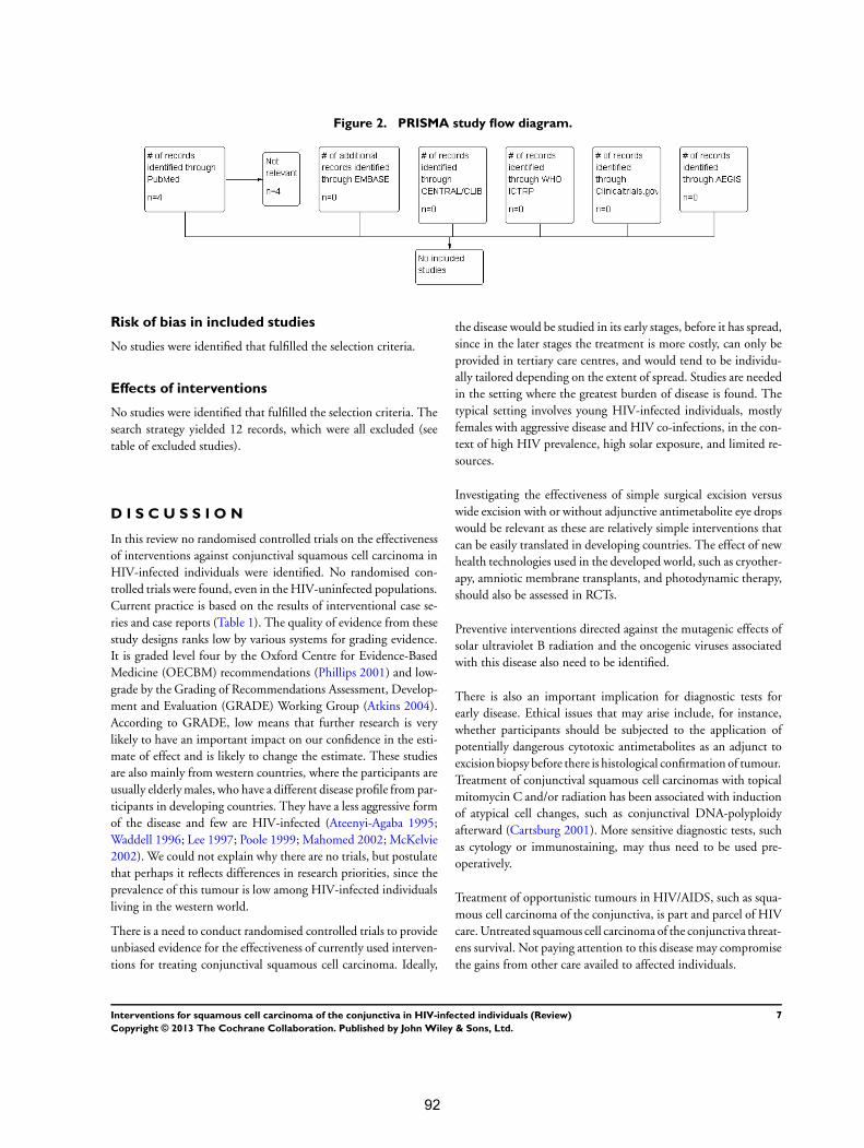
Figure 2. PRISMA study flow diagram.
Risk of bias in included studies
No studies were identified that fulfilled the selection criteria.
Effects of interventions
No studies were identified that fulfilled the selection criteria. The
search strategy yielded 12 records, which were all excluded (see
table of excluded studies).
D I S C U S S I O N
In this review no randomised controlled trials on the effectiveness
of interventions against conjunctival squamous cell carcinoma in
HIV-infected individuals were identified. No randomised con-
trolled trials were found, even in the HIV-uninfected populations.
Current practice is based on the results of interventional case se-
ries and case reports (Table 1). The quality of evidence from these
study designs ranks low by various systems for grading evidence.
It is graded level four by the Oxford Centre for Evidence-Based
Medicine (OECBM) recommendations (Phillips 2001) and low-
grade by the Grading of Recommendations Assessment, Develop-
ment and Evaluation (GRADE) Working Group (Atkins 2004).
According to GRADE, low means that further research is very
likely to have an important impact on our confidence in the esti-
mate of effect and is likely to change the estimate. These studies
are also mainly from western countries, where the participants are
usually elderly males, who have a different disease profile from par-
ticipants in developing countries. They have a less aggressive form
of the disease and few are HIV-infected (Ateenyi-Agaba 1995;
Waddell 1996; Lee 1997; Poole 1999; Mahomed 2002; McKelvie
2002). We could not explain why there are no trials, but postulate
that perhaps it reflects differences in research priorities, since the
prevalence of this tumour is low among HIV-infected individuals
living in the western world.
There is a need to conduct randomised controlled trials to provide
unbiased evidence for the effectiveness of currently used interven-
tions for treating conjunctival squamous cell carcinoma. Ideally,
the disease would be studied in its early stages, before it has spread,
since in the later stages the treatment is more costly, can only be
provided in tertiary care centres, and would tend to be individu-
ally tailored depending on the extent of spread. Studies are needed
in the setting where the greatest burden of disease is found. The
typical setting involves young HIV-infected individuals, mostly
females with aggressive disease and HIV co-infections, in the con-
text of high HIV prevalence, high solar exposure, and limited re-
sources.
Investigating the effectiveness of simple surgical excision versus
wide excision with or without adjunctive antimetabolite eye drops
would be relevant as these are relatively simple interventions that
can be easily translated in developing countries. The effect of new
health technologies used in the developed world, such as cryother-
apy, amniotic membrane transplants, and photodynamic therapy,
should also be assessed in RCTs.
Preventive interventions directed against the mutagenic effects of
solar ultraviolet B radiation and the oncogenic viruses associated
with this disease also need to be identified.
There is also an important implication for diagnostic tests for
early disease. Ethical issues that may arise include, for instance,
whether participants should be subjected to the application of
potentially dangerous cytotoxic antimetabolites as an adjunct to
excision biopsy before there is histological confirmation of tumour.
Treatment of conjunctival squamous cell carcinomas with topical
mitomycin C and/or radiation has been associated with induction
of atypical cell changes, such as conjunctival DNA-polyploidy
afterward (Cartsburg 2001). More sensitive diagnostic tests, such
as cytology or immunostaining, may thus need to be used pre-
operatively.
Treatment of opportunistic tumours in HIV/AIDS, such as squa-
mous cell carcinoma of the conjunctiva, is part and parcel of HIV
care. Untreated squamous cell carcinoma of the conjunctiva threat-
ens survival. Not paying attention to this disease may compromise
the gains from other care availed to affected individuals.
7Interventions for squamous cell carcinoma of the conjunctiva in HIV-infected individuals (Review)
Copyright © 2013 The Cochrane Collaboration. Published by John Wiley & Sons, Ltd.
92

A U T H O R S ’ C O N C L U S I O N S
Implications for practice
Current clinical practice in treatment of squamous cell carcinoma
of the conjunctiva is based on case series and case reports. This is
a weak evidence base.
Implications for research
RCTs for treatment of this disease are needed in the settings where
it occurs most frequently. Preventive interventions, such as HPV
vaccination, also need to be identified once scientific knowledge
of the serotypes involved is established. HIV/AIDS research has
not focused on treatment of this tumour.
A C K N O W L E D G E M E N T S
Dr S Gichuhi was awarded a Reviews for Africa Programme
Fellowship (www.mrc.ac.za/cochrane/rap.htm), funded from the
Nuffield Commonwealth Programme, through The Nuffield
Foundation. We would like to acknowledge the support pro-
vided by the staff at the South African Cochrane Centre and the
Cochrane HIV/AIDS Review Group mentoring programme.
R E F E R E N C E S
References to ongoing studies
Gichuhi 2012 {unpublished data only}
Adjuvant topical 5fluorouracil (5FU) for ocular surface
squamous neoplasia. Ongoing study 23/8/2012.
Additional references
Aoki 1998
Aoki S, Kubo E, Nakamura S, Tsuzuki A, Tsuzuki
S, Takahashi Y, et al.Possible prognostic markers in
conjunctival dysplasia and squamous cell carcinoma.
Japanese Journal of Ophthalmology 1998;42(4):256–61.
Ateenyi-Agaba 1995
Ateenyi-Agaba C. Conjunctival squamous cell carcinoma
associated with HIV infection in Kampala, Uganda. Lancet
1995;345(8951):695–6.
Ateenyi-Agaba 2004
Ateenyi-Agaba C, Dai M, Le Calvez F, Katongole-Mbidde
E, Smet A, Tommasino M, et al.TP53 mutations in
squamous cell carcinomas of the conjuntiva: evidence
for UV-induced mutagenesis. Mutagenesis 2004;19(5):
399–401.
Ateenyi-Agaba 2010
Ateenyi-Agaba C, Franceschi S, Wabwire-Mangen F, Arsian
A, Othieno E, Binta-Kahwa J, van Doorn LJ, Kleter
B, Weiderpass E. Human papillomavirus infection and
squamous cell carcinoma of the conjunctiva. British Journal
of Cancer 2010;102:262–7.
Atkins 2004
Atkins D, Best D, Briss PA, Eccles M, Falck-Ytter Y,
Flottorp S, et al.Grading quality of evidence and strengths
of recommendations. BMJ 2004;328(7454):1490–7.
Barbazetto 2004
Barbazetto IA, Lee TC, Abramson DH. Treatment of
conjunctival squamous cell carcinoma with photodynamic
therapy. American Journal of Ophthalmology 2004;138(2):
183–9.
Basti 2003
Basti S, Macsai MS. Ocular surface squamous neoplasia: a
review. Cornea 2003;22(7):687–704.
Bonanno 2010
Bonanno A, Esmaeli B, Fingere MC, Nelson DV, Randal
S, Weber RS. Social challenges of cancer patients with
orbitofacial disfigurement. Ophthalmic Plastic and
Reconstructive Surgery 2010;26:18–22.
Cartsburg 2001
Cartsburg O, Kallen C, Hillenkamp J, Sundmacher R,
Pomjanski N, Bocking A. Topical mitomycin C and
radiation induce conjunctival DNA-polyploidy. Analytical
Cellular Pathology 2001;23(2):65–74.
Castano 2005
Castano AP, Demidova TN, Hamblin MR. Mechanisms
in photodynamic therapy: Part three - photosensitizer
pharmacokinetics, biodistribution, tumor localization
and modes of tumor destruction. Photodiagnosis and
Photodynamic Therapy 2005;2(2):91–106.
Cerezo 1990
Cerezo L, Otero J, Aragon G, Polo E, de la Torre A,
Valcarcel F, et al.Conjunctival intraepithelial and invasive
squamous cell carcinomas treated with strontium-90.
Radiotherapy and oncology Journal of the European Society for
Therapeutic Radiology and Oncology 1990;17(3):191–7.
Cervantes 2002
Cervantes G, Rodriguez AA Jr, Leal AG. Squamous cell
carcinoma of the conjunctiva: clinicopathological features
in 287 cases. Canadian Journal of Ophthalmology 2002;37
(1):14–9.
Chisi 2006
Chisi SK, Kollmann MKH, Karimurio J. Conjunctival
squamous cell carcinoma in patients with Human
Immunodeficiency Virus infection seen at two hospitals in
Kenya. East African Medical Journal 2006;83(5):267–70.
8Interventions for squamous cell carcinoma of the conjunctiva in HIV-infected individuals (Review)
Copyright © 2013 The Cochrane Collaboration. Published by John Wiley & Sons, Ltd.
93

Chu 2004
Chu T, Sartorelli AC. Cancer chemotherapy. In: Katzung
BG editor(s). Basic and clinical pharmacology. 9th Edition.
Boston: McGraw Hill, 2004:898–930.
Clifford 2005
Clifford GM, Gallus S, Herrero R, Munoz N, Snijders
PJF, Vaccarella S, et al.Worldwide distribution of human
papillomavirus types in cytologically normal women in
the International Agency for Research on Cancer HPV
prevalence surveys: a pooled analysis. Lancet 2005;366
(9490):991–8.
Copeland 1990
Copeland RA. Limbal autograft for reconstruction after
conjunctival squamous cell carcinoma. American Journal of
Ophthalmology 1990;4(110):412–5.
de Koning 2008
de Koning MN, Waddell K, Magyezi J, Purdie K, Proby C,
Harwood C, Lucas S, Downing R, Quint WG, Newton R.
Genital and cutaneous human papillomavirus (HPV) types
in relation to conjunctival squamous cell neoplasia: a case-
control study in Uganda. Infectious agents and cancer 2008;
3:12.
Espana 2002
Espana EM, Prabhasawat P, Grueterich M, Solomon
A, Tseng SCG. Amniotic membrane transplantation
for reconstruction after excision of large ocular surface
neoplasias. British Journal of Ophthalmology 2002;86(6):
640–5.
Frucht-Pery 1997
Frucht-Pery J, Sugar J, Baum J, Sutphin JE, Pe’er J,
Savir H, et al.Mitomycin C treatment for conjunctival-
corneal intraepithelial neoplasia: a multicenter experience.
Ophthalmology 1997;104(12):2085–93.
Goldberg 1963
Goldberg JR, Becker SC, Rosenbaum HD. Gamma
radiation in the treatment of squamous cell carcinoma of the
limbus. American Journal of Ophthalmology 1963;55:811–5.
Gunduz 2005
Gunduz K, Ucakhan OO, Kanpolat A, Gunalp I. Non-
preserved human amniotic membrane transplantation for
conjunctival reconstruction after excision of extensive ocular
surface neoplasia. Eye 2005;20(3):351–7.
Guthoff 2009
Guthoff R, Marx A, Stroebel P. No evidence for a pathogenic
role of human papillomavirus infection in ocular surface
squamous neoplasia in Germany. Current Eye Research
2009;34:666–71.
Hao 2000
Hao Y, Ma DH, Hwang DG, Kim WS, Zhang F.
Identification of antiangiogenic and antiinflammatory
proteins in human amniotic membrane. Cornea 2000;19
(3):348–52.
Kaji 2006
Kaji Y, Hiraoka T, Oshika T. Vital staining of squamous cell
carcinoma of the conjunctiva using toluidine blue. Acta
Ophthalmologica Scandinavica 2006;84(6):825–6.
Karp 2001
Karp CL, Moore JK, Rosa RH Jr. Treatment of conjunctival
and corneal intraepithelial neoplasia with topical interferon
alpha-2b. Ophthalmology 2001;108(6):1093–8.
Lake 2004
Lake DF, Briggs AD, Akporiaye ET. Immunopharmacology.
In: Katzung BG editor(s). Basic and clinical pharmacology.
9th Edition. Boston: McGraw Hill, 2004:931–57.
Lee 1997
Lee GA, Hirst LW. Retrospective study of ocular surface
squamous neoplasia. Australian and New Zealand Journal of
Ophthalmology 1997;25(4):269–76.
Macarez 1999
Macarez R, Bossis S, Robinet A, Le Callonnec A, Charlin
JF, Colin J. Conjunctival epithelial neoplasias in organ
transplant patients receiving cyclosporine therapy. Cornea
1999;18(4):495–7.
Mahomed 2002
Mahomed A, Chetty R. Human immunodeficiency virus
infection, Bcl-2, p53 protein, and Ki-67 analysis in ocular
surface squamous neoplasia. Archives of Ophthalmology
2002;120(5):554–8.
Margo 2002
Margo CE, Harman LE, Mulla ZD. The reliability of
clinical methods in ophthalmology. Survey of Ophthalmology
2002;47(4):375–86.
McKelvie 2001
McKelvie PA, Daniell M. Impression cytology following
mitomycin C therapy for ocular surface squamous neoplasia.
British Journal of Ophthalmology 2001;85(9):1115–9.
McKelvie 2002
McKelvie PA, Daniell M, McNab A, Loughnan M,
Santamaria JD. Squamous cell carcinoma of the conjunctiva:
a series of 26 cases. British Journal of Ophthalmology 2002;
86(2):168–73.
Midena 2000
Midena E, Angeli CD, Valenti M, de Belvis V, Boccato P.
Treatment of conjunctival squamous cell carcinoma with
topical 5-fluorouracil. British Journal of Ophthalmology
2000;84(3):268–72.
Moubayed 2004
Moubayed P, Mwakyoma H, Schneider DT. High frequency
of Human Papillomavirus 6/11, 16, and 18 infections in
precancerous lesions and squamous cell carcinoma of the
conjunctiva in subtropical Tanzania. American Journal of
Clinical Pathology 2004;122:938–43.
Nadjari 1999
Nadjari B, Kersten A, Ross B, Motherby H, Krallmann
R, Sundmacher R, et al.Cytologic and DNA cytometric
diagnosis and therapy monitoring of squamous cell
carcinoma in situ and malignant melanoma of the cornea
and conjunctiva. Analytical and Quantitative Cytology and
Histology 1999;21(5):387–96.
9Interventions for squamous cell carcinoma of the conjunctiva in HIV-infected individuals (Review)
Copyright © 2013 The Cochrane Collaboration. Published by John Wiley & Sons, Ltd.
94

Nakamura 1997
Nakamura Y, Mashima Y, Kameyama K, Mukai M,
Oguchi Y. Detection of human papillomavirus infection
in squamous tumours of the conjunctiva and lacrimal
sac by immunohistochemistry, in situ hybridisation and
polymerase chain reaction. British Journal of Ophthalmology
1997;81(4):308–13.
Newton 2002
Newton R, Ziegler J, Ateenyi-Agaba C, Bousarghin
L, Casabonne D, Beral V, et al.The epidemiology of
conjunctival squamous cell carcinoma in Uganda. British
Journal of Cancer 2002;87(3):301–8.
Ng 2008
Ng J, Coroneo MT, Wakefield D, Di Girolamo
N. Ultraviolet radiation and the role of matrix
metalloproteinases in the pathogenesis of ocular surface
squamous neoplasia. Investigative Ophthalmology & Visual
Science 2008;49:5295–5306.
NIH 2012
U.S. National Institutes of Health. Information on clinical
trials and human research studies. www.clinicaltrials.gov
(accessed 10 February 2012).
Osahon 2011
Osahon AI, Ukponmwan CU, Uhunmwangho OM.
Prevalence of HIV seropositivity among patients with
squamous cell carcinoma of the conjunctiva. Asian Pacific
Journal of Tropical Biomedicine 2011;1(2):150–53.
Peksayar 1989
Peksayar G, Soyturk MK, Demiryont M. Long-term results
of cryotherapy on malignant epithelial tumours of the
conjunctiva. American Journal of Ophthalmology 1989;107
(4):337–49.
Phillips 2001
Phillips B, Ball C, Sackett D, Badenoch D, Straus S, Haynes
B, et al.Levels of evidence and grades of recommendation.
www.cebm.net/levels˙of˙evidence.asp (accessed 14 June
2006).
Pola 2003
Pola EC, Masanganise R, Rusakaniko S. The trend of ocular
surface squamous neoplasia among ocular surface tumour
biopsies submitted for histology from Sekuru Kaguvi Eye
Unit, Harare between 1996 and 2000. Central African
Journal of Medicine 2003;49:1–4.
Poole 1999
Poole TR. Conjunctival squamous cell carcinoma in
Tanzania. British Journal of Ophthalmology 1999;83(2):
177–9.
Porges 2003
Porges Y, Groisman GM. Prevalence of HIV with
conjunctival squamous cell neoplasia in an African
provincial hospital. Cornea 2003;22(1):1–4.
Safrin 2004
Safrin S. Antiviral agents. In: Katzung BG editor(s). Basic
and clinical pharmacology. 9th Edition. Boston: McGraw
Hill, 2004:801–27.
Sen 2007
Sen S, Sharma A, Panda A. Immunohistochemical
localization of human papilloma virus in conjunctival
neoplasias: a retrospective study. Indian Journal of
Ophthalmology 2007;55:361–3.
Sherman 2002
Sherman MD, Feldman KA, Farahmand SM, Margolis TP.
Treatment of conjunctival squamous cell carcinoma with
topical cidofovir. American Journal of Ophthalmology 2002;
134(3):432–3.
Shields 1991
Shields JA, Shields CL, Suvarnamani C, Tantisira M, Shah
P. Orbital exenteration with eyelid sparing: indications,
technique and results. Ophthalmic surgery 1991;22(5):
292–7.
Shields 1997
Shields JA, Shields CL, De Potter P. Surgical management
of conjunctival tumours: The 1994 Lynn B. McMahan
Lecture. Archives of Ophthalmology 1997;115(6):808–15.
Shields 2004
Shields JA, Shields CL. Tumors of the conjunctiva and
cornea. In: McLeod SD editor In: Tasman W, Jaeger EA
editor(s). Duane’s clinical ophthalmology CD-ROM. Revised
2004. Vol. 4, Philadelphia: Lippincott William and
Wilkins, 2004:Chapter 10.
Sudesh 2000
Sudesh S, Rapuano CJ, Cohen EJ, Eagle RC Jr, Laibson
PR. Surgical management of ocular surface squamous
neoplasms: the experience from a cornea center. Cornea
2000;19(3):278–83.
Tabin 1997
Tabin G, Levin S, Snibson G, Loughnan M, Taylor H. Late
recurrences and the necessity for long-term follow-up in
corneal and conjunctival neoplasia. Ophthalmology 1997;
104(3):485–92.
Tornesello 2006
Tornesello ML, Duraturo ML, Waddell KM, Biryahwaho
B, Downing R, Balinandi S, et al.Evaluating the role of
human papillomaviruses in conjunctival neoplasia. British
Journal of Cancer 2006;94(3):446–9.
Tunc 1999
Tunc M, Char DH, Crawford B, Miller T. Intraepithelial
and invasive squamous cell carcinoma of the conjunctiva:
analysis of 60 cases. British Journal of Ophthalmology 1999;
83(1):98–103.
UNAIDS 2010
Joint United Nations Programme on HIV/AIDS
(UNAIDS). Global report: UNAIDS report on the global
AIDS epidemic 2010. http://www.unaids.org/globalreport/
global˙report.htm 2010.
Waddell 1996
Waddell KM, Lewallen S, Lucas SB, Atenyi-Agaba C,
Herrington CS, Liomba G. Carcinoma of the conjunctiva
and HIV infection in Uganda and Malawi. British Journal
of Ophthalmology 1996;80(6):503–8.
10Interventions for squamous cell carcinoma of the conjunctiva in HIV-infected individuals (Review)
Copyright © 2013 The Cochrane Collaboration. Published by John Wiley & Sons, Ltd.
95

WHO ICTRP 2012
WHO ICTRP. WHO International Clinical Trials Registry
Platform. www.who.int/ictrp/ (accessed on 10 February
2012).
Yeatts 2000
Yeatts RP, Engelbrecht NE, Curry CD, Ford JG, Walker
KA. 5-Fluorouracil for the treatment of intraepithelial
neoplasia of the conjunctiva and cornea. Ophthalmology
2000;107(12):2190–5.∗ Indicates the major publication for the study
11Interventions for squamous cell carcinoma of the conjunctiva in HIV-infected individuals (Review)
Copyright © 2013 The Cochrane Collaboration. Published by John Wiley & Sons, Ltd.
96
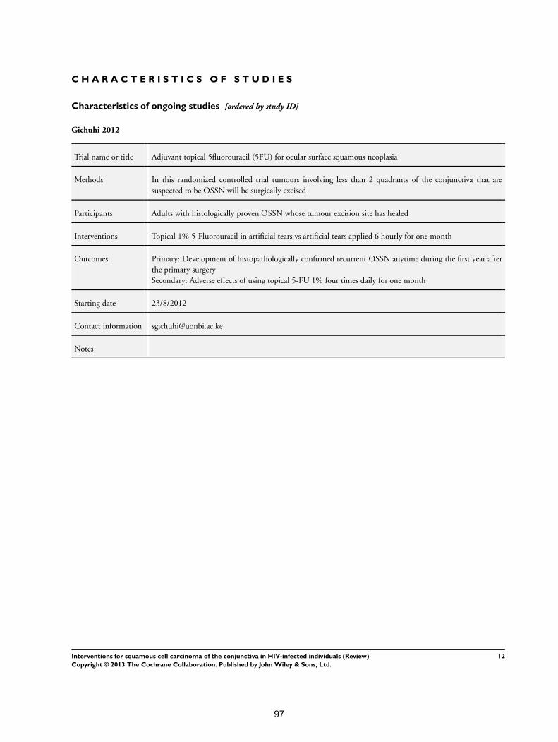
C H A R A C T E R I S T I C S O F S T U D I E S
Characteristics of ongoing studies [ordered by study ID]
Gichuhi 2012
Trial name or title Adjuvant topical 5fluorouracil (5FU) for ocular surface squamous neoplasia
Methods In this randomized controlled trial tumours involving less than 2 quadrants of the conjunctiva that are
suspected to be OSSN will be surgically excised
Participants Adults with histologically proven OSSN whose tumour excision site has healed
Interventions Topical 1% 5-Fluorouracil in artificial tears vs artificial tears applied 6 hourly for one month
Outcomes Primary: Development of histopathologically confirmed recurrent OSSN anytime during the first year after
the primary surgery
Secondary: Adverse effects of using topical 5-FU 1% four times daily for one month
Starting date 23/8/2012
Contact information [email protected]
Notes
12Interventions for squamous cell carcinoma of the conjunctiva in HIV-infected individuals (Review)
Copyright © 2013 The Cochrane Collaboration. Published by John Wiley & Sons, Ltd.
97
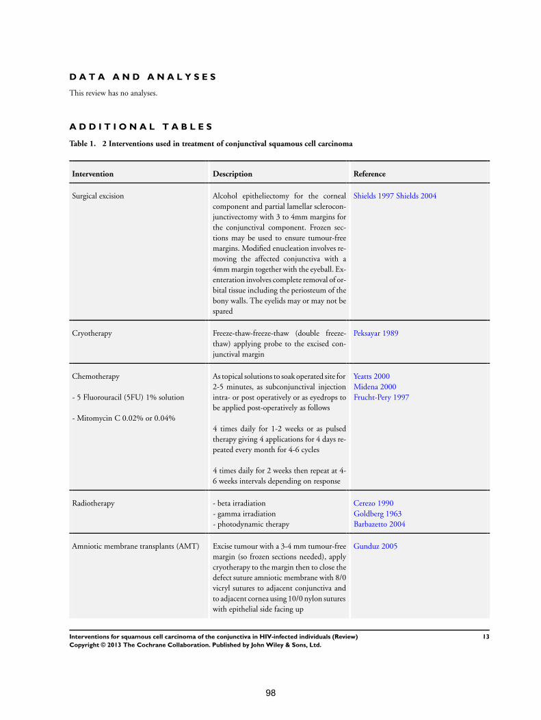
D A T A A N D A N A L Y S E S
This review has no analyses.
A D D I T I O N A L T A B L E S
Table 1. 2 Interventions used in treatment of conjunctival squamous cell carcinoma
Intervention Description Reference
Surgical excision Alcohol epitheliectomy for the corneal
component and partial lamellar sclerocon-
junctivectomy with 3 to 4mm margins for
the conjunctival component. Frozen sec-
tions may be used to ensure tumour-free
margins. Modified enucleation involves re-
moving the affected conjunctiva with a
4mm margin together with the eyeball. Ex-
enteration involves complete removal of or-
bital tissue including the periosteum of the
bony walls. The eyelids may or may not be
spared
Shields 1997 Shields 2004
Cryotherapy Freeze-thaw-freeze-thaw (double freeze-
thaw) applying probe to the excised con-
junctival margin
Peksayar 1989
Chemotherapy
- 5 Fluorouracil (5FU) 1% solution
- Mitomycin C 0.02% or 0.04%
As topical solutions to soak operated site for
2-5 minutes, as subconjunctival injection
intra- or post operatively or as eyedrops to
be applied post-operatively as follows
4 times daily for 1-2 weeks or as pulsed
therapy giving 4 applications for 4 days re-
peated every month for 4-6 cycles
4 times daily for 2 weeks then repeat at 4-
6 weeks intervals depending on response
Yeatts 2000
Midena 2000
Frucht-Pery 1997
Radiotherapy - beta irradiation
- gamma irradiation
- photodynamic therapy
Cerezo 1990
Goldberg 1963
Barbazetto 2004
Amniotic membrane transplants (AMT) Excise tumour with a 3-4 mm tumour-free
margin (so frozen sections needed), apply
cryotherapy to the margin then to close the
defect suture amniotic membrane with 8/0
vicryl sutures to adjacent conjunctiva and
to adjacent cornea using 10/0 nylon sutures
with epithelial side facing up
Gunduz 2005
13Interventions for squamous cell carcinoma of the conjunctiva in HIV-infected individuals (Review)
Copyright © 2013 The Cochrane Collaboration. Published by John Wiley & Sons, Ltd.
98

Table 1. 2 Interventions used in treatment of conjunctival squamous cell carcinoma (Continued)
Interferon alpha 2b Topical drops or intralesion injection Karp 2001
Cidofovir eyedrops 2mg/ml Applied every 2 hours for 2 weeks then 4
times daily for 2 weeks then 3 times daily
for 2 weeks
Sherman 2002
Table 2. 1 Review methods
Study selection The titles and abstracts of search results are scanned and compared to
predetermined selection criteria for studies. Articles written in foreign
languages are translated to English. Two authors working independently
select trials. The authors are blinded to the trial uthors and institutions
by asking the trial search co-ordinator to block this out when sending
abstracts. Where there is uncertainty about inclusion, the full text of the
article is obtained and read. If additional information is needed about
the trial in question, it is categorized as awaiting assessment until this is
obtained. If differences arise regarding trial inclusion, they are discussed
and if still unresolved, Dr Taryn Young at the South African Cochrane
Centre is asked to take a decision
Data extraction Data are collected and recorded in similar data extraction forms. The fol-
lowing information is extracted; citation details, study eligibility, study
quality and study characteristics. The study characteristics include meth-
ods, participants, interventions and outcomes
Assessment of methodological quality of included studies The methodological quality of the included studies is assessed to deter-
mine validity. Selection bias is checked by assessing whether the gener-
ation of a random allocation sequence and allocation concealment were
performed. Randomization is considered adequate if the allocation se-
quence is generated from a table of random numbers or by computer.
Allocation concealment is deemed adequate if undertaken by means of
sequentially prenumbered sealed opaque envelopes, a centralised system
or prenumbered coded identical containers. In addition, assessment of
blinding, losses to follow-up and whether the analysis was by intention-
to-treat (ITT) is undertaken. The definition of ITT is the requirement
that participants be analyzed in the groups to which they were random-
ized, regardless of which intervention they actually received
Data analysis Measures of treatment effect:The effect measures of choice here are rela-
tive risk (RR) for dichotomous data and hazard ratio (HR) for time-to-
event data with a 95% confidence interval
Dichotomous data include: - number of people who experience local
control in each comparison group
- number of people who experience recurrence during the specified time
periods in each comparison group
14Interventions for squamous cell carcinoma of the conjunctiva in HIV-infected individuals (Review)
Copyright © 2013 The Cochrane Collaboration. Published by John Wiley & Sons, Ltd.
99

Table 2. 1 Review methods (Continued)
- number of deaths in each comparison group
- number of people who experience adverse events in each comparison
group
- number of people who experience local control in each comparison
group
- number of people who experience recurrence during the specified time
periods in each comparison group
- number of deaths in each comparison group
- number of people who experience adverse events in each comparison
group
Time-to-event data include: - time to recurrence
Dealing with missing data: Participants lost to follow up are censored in
the survival analysis
Assessment of heterogeneity: To identify statistical heterogeneity we look
at the forest plot for overlapping confidence intervals and test it using
the Cochrane Q test with a p-value of 0.10. The impact of heterogeneity
on the meta-analysis is measured using the I-squared test. If I-squared
is greater than 70% we will investigate by checking the trials for data
entry errors and looking for existence of subgroups. Subgroup analysis
by age, sex, geographical location and diagnostic method will be done.
These factors have been shown to influence the occurrence of OSSN
and we anticipate that they may also influence treatment effects. If no
explanation for heterogeneity is found or its correction is not possible,
meta-analysis will not be done
Meta-analysis: A fixed-effect model is used for meta-analysis but when
there is heterogeneity that cannot be readily explained, a random-effects
model is incorporated
Sensitivity analysis: We examine how the magnitude of effect differs
according to study quality or trial size and also the results of per-protocol
analysis with those of intention-to-treat analysis
Assessment of reporting bias: Publication bias is assessed by using a funnel
plot to look for asymmetry
15Interventions for squamous cell carcinoma of the conjunctiva in HIV-infected individuals (Review)
Copyright © 2013 The Cochrane Collaboration. Published by John Wiley & Sons, Ltd.
100

A P P E N D I C E S
Appendix 1. PubMed search strategy and results
Title: Interventions for squamous cell carcinoma of the conjunctiva in HIV-infected individuals
Database: PubMed (2010 - 2012)
Date: 27 January 2012
Search Query Items found
#13 Search #1 AND #2 AND #11 AND (“2010/10/01”[Date -
Publication] : “2012/01/27”[Date - Publication])
4
#12 Search #1 AND #2 AND #11 23
#11 Search #9 OR #10 2290
#10 Search conjunctival neoplasms[mh] 1729
#9 Search #5 AND #8 1099
#8 Search #6 OR #7 546016
#7 Search carcinoma[tiab] OR carcinomas[tiab] OR neopla-
sia[tiab] OR neoplasm[tiab] OR neoplasms[tiab]
514787
#6 Search carcinoma, squamous cell[mh] 92643
#5 Search #3 OR #4 23573
#4 Search conjunctiva[tiab] OR conjunctivas[tiab] OR conjunc-
tival[tiab]
18906
#3 Search conjunctiva[MeSH Terms] 11829
#2 Search (randomized controlled trial [pt] OR controlled clin-
ical trial [pt] OR randomized [tiab] OR placebo [tiab] OR
drug therapy [sh] OR randomly [tiab] OR trial [tiab] OR
groups [tiab]) NOT (animals [mh] NOT humans [mh])
2499392
#1 Search HIV Infections[MeSH] OR HIV[MeSH] OR hiv[tw]
OR hiv-1*[tw] OR hiv-2*[tw] OR hiv1[tw] OR hiv2[tw]
OR hiv infect*[tw] OR human immunodeficiency virus[tw]
OR human immunedeficiency virus[tw] OR human im-
muno-deficiency virus[tw] OR human immune-deficiency
virus[tw] OR ((human immun*) AND (deficiency virus[tw])
) OR acquired immunodeficiency syndrome[tw] OR ac-
quired immunedeficiency syndrome[tw] OR acquired im-
muno-deficiency syndrome[tw] OR acquired immune-defi-
ciency syndrome[tw] OR ((acquired immun*) AND (defi-
ciency syndrome[tw])) OR “sexually transmitted diseases, vi-
282439
16Interventions for squamous cell carcinoma of the conjunctiva in HIV-infected individuals (Review)
Copyright © 2013 The Cochrane Collaboration. Published by John Wiley & Sons, Ltd.
101
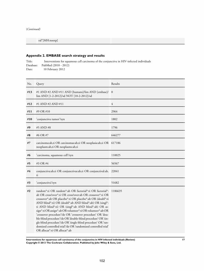
(Continued)
ral”[MH:noexp]
Appendix 2. EMBASE search strategy and results
Title: Interventions for squamous cell carcinoma of the conjunctiva in HIV-infected individuals
Database: PubMed (2010 - 2012)
Date: 10 February 2012
No. Query Results
#13 #1 AND #2 AND #11 AND [humans]/lim AND [embase]/
lim AND [1-2-2012]/sd NOT [10-2-2012]/sd
0
#12 #1 AND #2 AND #11 4
#11 #9 OR #10 2964
#10 ’conjunctiva tumor’/syn 1802
#9 #5 AND #8 1796
#8 #6 OR #7 646277
#7 carcinoma:ab,ti OR carcinomas:ab,ti OR neoplasia:ab,ti OR
neoplasm:ab,ti OR neoplasms:ab,ti
617186
#6 ’carcinoma, squamous cell’/syn 110025
#5 #3 OR #4 56567
#4 conjunctiva:ab,ti OR conjunctivas:ab,ti OR conjunctival:ab,
ti
22041
#3 ’conjunctiva’/syn 51682
#2 random*:ti OR random*:ab OR factorial*:ti OR factorial*:
ab OR cross?over*:ti OR cross?over:ab OR crossover*:ti OR
crossover*:ab OR placebo*:ti OR placebo*:ab OR (doubl*:ti
AND blind*:ti) OR (doubl*:ab AND blind*:ab) OR (singl*:
ti AND blind*:ti) OR (singl*:ab AND blind*:ab) OR as-
sign*:ti OR assign*:ab OR volunteer*:ti OR volunteer*:ab OR
’crossover procedure’/de OR ’crossover procedure’ OR ’dou-
ble-blind procedure’/de OR ’double-blind procedure’ OR ’sin-
gle-blind procedure’/de OR ’single-blind procedure’ OR ’ran-
domized controlled trial’/de OR ’randomized controlled trial’
OR allocat*:ti OR allocat*:ab
1186635
17Interventions for squamous cell carcinoma of the conjunctiva in HIV-infected individuals (Review)
Copyright © 2013 The Cochrane Collaboration. Published by John Wiley & Sons, Ltd.
102
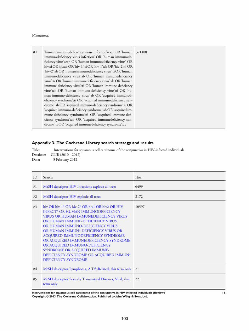
(Continued)
#1 ’human immunodeficiency virus infection’/exp OR ’human
immunodeficiency virus infection’ OR ’human immunode-
ficiency virus’/exp OR ’human immunodeficiency virus’ OR
hiv:ti OR hiv:ab OR ’hiv-1’:ti OR ’hiv-1’:ab OR ’hiv-2’:ti OR
’hiv-2’:ab OR ’human immunodeficiency virus’:ti OR ’human
immunodeficiency virus’:ab OR ’human immunedeficiency
virus’:ti OR ’human immunedeficiency virus’:ab OR ’human
immune-deficiency virus’:ti OR ’human immune-deficiency
virus’:ab OR ’human immuno-deficiency virus’:ti OR ’hu-
man immuno-deficiency virus’:ab OR ’acquired immunod-
eficiency syndrome’:ti OR ’acquired immunodeficiency syn-
drome’:ab OR ’acquired immuno-deficiency syndrome’:ti OR
’acquired immuno-deficiency syndrome’:ab OR ’acquired im-
mune-deficiency syndrome’:ti OR ’acquired immune-defi-
ciency syndrome’:ab OR ’acquired immunedeficiency syn-
drome’:ti OR ’acquired immunedeficiency syndrome’:ab
371108
Appendix 3. The Cochrane Library search strategy and results
Title: Interventions for squamous cell carcinoma of the conjunctiva in HIV-infected individuals
Database: CLIB (2010 - 2012)
Date: 3 February 2012
ID Search Hits
#1 MeSH descriptor HIV Infections explode all trees 6499
#2 MeSH descriptor HIV explode all trees 2172
#3 hiv OR hiv-1* OR hiv-2* OR hiv1 OR hiv2 OR HIV
INFECT* OR HUMAN IMMUNODEFICIENCY
VIRUS OR HUMAN IMMUNEDEFICIENCY VIRUS
OR HUMAN IMMUNE-DEFICIENCY VIRUS
OR HUMAN IMMUNO-DEFICIENCY VIRUS
OR HUMAN IMMUN* DEFICIENCY VIRUS OR
ACQUIRED IMMUNODEFICIENCY SYNDROME
OR ACQUIRED IMMUNEDEFICIENCY SYNDROME
OR ACQUIRED IMMUNO-DEFICIENCY
SYNDROME OR ACQUIRED IMMUNE-
DEFICIENCY SYNDROME OR ACQUIRED IMMUN*
DEFICIENCY SYNDROME
10597
#4 MeSH descriptor Lymphoma, AIDS-Related, this term only 21
#5 MeSH descriptor Sexually Transmitted Diseases, Viral, this
term only
22
18Interventions for squamous cell carcinoma of the conjunctiva in HIV-infected individuals (Review)
Copyright © 2013 The Cochrane Collaboration. Published by John Wiley & Sons, Ltd.
103
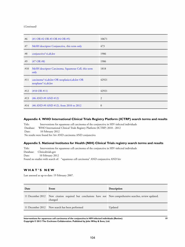
(Continued)
#6 (#1 OR #2 OR #3 OR #4 OR #5) 10671
#7 MeSH descriptor Conjunctiva, this term only 473
#8 conjunctiva*:ti,ab,kw 1906
#9 (#7 OR #8) 1906
#10 MeSH descriptor Carcinoma, Squamous Cell, this term
only
1818
#11 carcinoma*:ti,ab,kw OR neoplasia:ti,ab,kw OR
neoplasm*:ti,ab,kw
42921
#12 (#10 OR #11) 42921
#13 (#6 AND #9 AND #12) 2
#14 (#6 AND #9 AND #12), from 2010 to 2012 0
Appendix 4. WHO International Clinical Trials Registry Platform (ICTRP) search terms and results
Title: Interventions for squamous cell carcinoma of the conjunctiva in HIV-infected individuals
Database: WHO International Clinical Trials Registry Platform (ICTRP) 2010 - 2012
Date: 10 February 2012
No results were found for: hiv AND carcinoma AND conjunctiva
Appendix 5. National Institutes for Health (NIH) Clinical Trials registry search terms and results
Title: Interventions for squamous cell carcinoma of the conjunctiva in HIV-infected individuals
Database: Clinicaltrials.gov
Date: 10 February 2012
Found no studies with search of: “squamous cell carcinoma” AND conjunctiva AND hiv
W H A T ’ S N E W
Last assessed as up-to-date: 19 February 2007.
Date Event Description
11 December 2012 New citation required but conclusions have not
changed
New comprehensive searches, review updated.
11 December 2012 New search has been performed Updated
19Interventions for squamous cell carcinoma of the conjunctiva in HIV-infected individuals (Review)
Copyright © 2013 The Cochrane Collaboration. Published by John Wiley & Sons, Ltd.
104
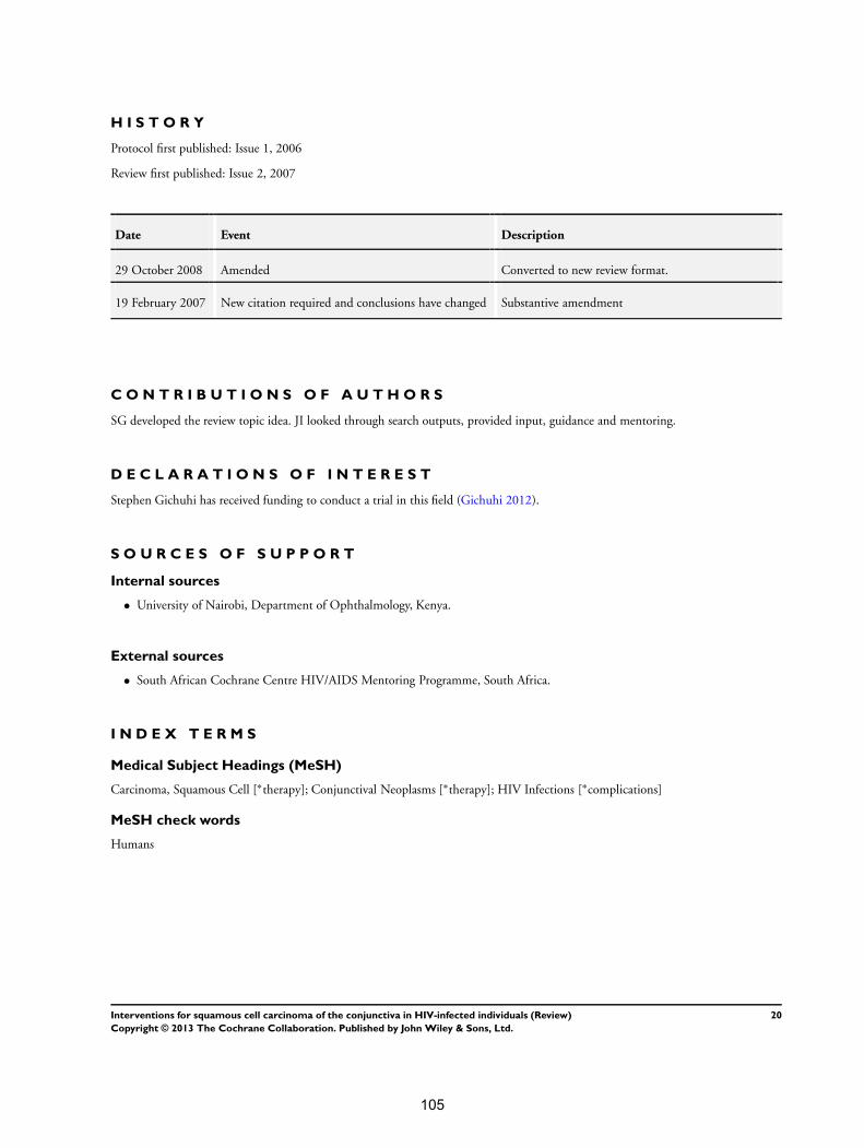
H I S T O R Y
Protocol first published: Issue 1, 2006
Review first published: Issue 2, 2007
Date Event Description
29 October 2008 Amended Converted to new review format.
19 February 2007 New citation required and conclusions have changed Substantive amendment
C O N T R I B U T I O N S O F A U T H O R S
SG developed the review topic idea. JI looked through search outputs, provided input, guidance and mentoring.
D E C L A R A T I O N S O F I N T E R E S T
Stephen Gichuhi has received funding to conduct a trial in this field (Gichuhi 2012).
S O U R C E S O F S U P P O R T
Internal sources
• University of Nairobi, Department of Ophthalmology, Kenya.
External sources
• South African Cochrane Centre HIV/AIDS Mentoring Programme, South Africa.
I N D E X T E R M S
Medical Subject Headings (MeSH)
Carcinoma, Squamous Cell [∗therapy]; Conjunctival Neoplasms [∗therapy]; HIV Infections [∗complications]
MeSH check words
Humans
20Interventions for squamous cell carcinoma of the conjunctiva in HIV-infected individuals (Review)
Copyright © 2013 The Cochrane Collaboration. Published by John Wiley & Sons, Ltd.
105

Review of case-series and case-reports of
interventions for ocular surface squamous
neoplasia (OSSN)
Rationale
A Cochrane systematic review of interventions found no randomized controlled trials except
one in Australia. Numerous case reports and case-series formed the evidence base for
treatment of OSSN which is low down in the hierarchy of evidence. We reviewed these case
series to see what could be learned from them.
Methods
In August 2015 using a sensitive search strategy we tried to identify all relevant trials,
regardless of language or publication status using the following search terms; “randomized
controlled trial”, “controlled clinical trial”, “randomized”, “placebo”, “drug therapy”, “randomly”,
“trial”, “conjunctiva*”, “ocular surface”, “carcinoma”, “cancer”, “neoplasia”, “neoplasm”,
“neoplastic”, “dysplasia”, “dysplastic”, “squamous”, “squamous cell”.
We searched electronic databases (PubMed, EMBASE, The Cochrane Library), clinical trial
registries (WHO International Clinical Trials Registry Platform and the US National Institutes
of Health Clinicaltrials.gov), and international conference proceedings of HIV/AIDS and AIDS-
related cancers from the AIDS Education Global Education System (AEGIS). Only series that
reported recurrence as an outcome were included.
Results
The interventions reported in various cases series are summarised in tables 1-10. There is
only one published trial on topical Mitomycin-C (MMC) in a patient group without HIV infection
in Australia. Some case series reported tumour resolution as an outcome and are not included
here. Only five studies involved African patients (Tunisia, Egypt, Uganda, and South Africa).
Sample sizes were variable. With all treatment regimens reported, there were varying doses,
inclusion criteria and follow up times.
Surgery ± Cryotherapy (Table 1)
Surgical excision was the most commonly employed treatment. Small tumours were excised
while more advanced orbital disease needed exenteration to clear all the orbital contents down
to the periosteum of the bone. The margins of the excision varied from 2m to 4mm. Some
surgeons used alcohol epitheliectomy (absolute alcohol) to aid removal of tumour extending
106
to the cornea. Some used the bare sclera technique where the tumour was dissected off down
to the sclera leaving the wound uncovered while some closed the resulting defect by mobilising
the surrounding conjunctiva (primary closure) or transplanted either amniotic membrane or
autologous conjunctiva from the same or other eye. Amniotic membranes are obtained from
tissue banks and there are no such facilities in Africa.
Surgical excision may be augmented by cryotherapy where 2-4 freeze-thaw cycles were used
to obliterate residual tumour at the bed and margins. Cryotherapy requires the use of liquefied
gas, either carbon dioxide or nitrous oxide, which is not easily available in most centres in sub-
Saharan Africa. Cryotherapy was the most commonly used adjuvant treatment with surgical
excision. Some articles mentioned local excision without qualifying whether cryotherapy was
used or not. A series consisting of advanced squamous cell carcinoma from Germany worse
than grade 3 using the American Joint Committee on Cancer (AJCC) system reported that
52% of excisions and 20% of exenterations recurred after a median of 24 months.
Surgical excision alone is associated with recurrences of 3.2% to 66.7% 32 months later. The
former involved surgical excision with the corneal extension removed using blade dissection
then wiped with alcohol and using primary closure without cryotherapy. This was a very
experienced surgeon in Uganda. With cryotherapy surgical excision was associated with
recurrences varying from zero at 6.5 years to 52% at 24 months (the latter being the advanced
tumour series aforementioned).
Surgery ± amniotic membrane transplant (Table 2)
Series involving surgery with amniotic membrane transplants reported recurrences ranging
from zero after a mean of 31 months to 12.5% after a mean 17.8 months of follow up. Those
who used the bare sclera technique sometimes applied Weck-cel sponges soaked with
cytotoxic agents such as 5FU or MMC to the tumour bed intraoperatively for periods varying
from 2 minutes to 5 minutes while others would employ this only when complete excision of
the tumour was not deemed possible. Surgical excision with adjuvant chemotherapy was
associated with recurrences varied between zero and 67% but most often in the 30% to 67%
range. In older series local resection would be supplemented by radiation therapy.
Interferon alpha 2b (IFNα2b) (Table 3)
IFN is an immune modulator used on its own or combined with surgical excision and
cryotherapy while in one series IFN was combined with retinoic acid eye drops. Preparations
available include eye drops or an injection. Some used it as primary treatment of presumed
107

OSSN or for residual OSSN unresponsive to excision, cryotherapy and MMC. Two
formulations of IFN eyedrops were reported, 1million IU/ml and 3 million IU/ml, used four times
daily for four weeks. Some continued the drops till a month beyond complete clinical
resolution. The injection preparations vary from 6-16 million IU/ml. Some infiltrated it around
the lesion (peri-lesion) while some injected it under the lesion (sub-lesion). Injections were
given every three days for seven doses or three times weekly. Recurrences varied from none
to 33.3% at one year. IFNα2b needs refrigeration and is continued long after clinical resolution.
It has local and systemic side effects such as conjunctival hyperemia, superficial punctate
keratitis and flu-like symptoms. The cost of the 3 million IU/ml dose of eyedrops was $260 for
a 5 ml bottle and the 1 million IU/ml dose is $120 in 2010 in Bascolm Palmer, Florida, USA.
Treatment for 1 month with IFN 1 million IU/ml drops four times daily for a month cost $179 in
2012 in Wills Eye Hospital, Philadelphia, USA. In Africa this would be too expensive besides
being unavailable.
Cyclosporin A (CsA) (Table 4)
CSA is an immunosuppressant that interferes with the activity of T-lymphocytes. It is prepared
as a 0.05% ophthalmic emulsion and used topically. In one series it was combined with
surgical excision and MMC while in another it was combined only with MMC. Its use allows a
lower concentration of MMC (0.01%) to be used. There were no recurrences reported after
two years of follow up. The ocular surface toxicity observed (punctate keratopathy,
conjunctival hyperemia) and eyelid toxicity were attributed to MMC but it was not clear how
this distinction was made.
Mitomycin C (MMC) (Table 5)
MMC is an antibiotic classified as an alkylating agent causing cross linking of DNA thus
inhibiting DNA synthesis. It is usually used topically as a 0.02% or 0.04% solution applied four
times daily for a week or two followed by one to three weeks off then the cycle is repeated to
make a total of 4 cycles. Most pharmacies compound it for themselves due to a short shelf life
and typically patients have to return after a few weeks to get the next bottle. It is often
combined with surgical excision although some use it on its own. Recurrences range from
zero to 29.4% when used alone; zero to 14% with surgical excision; and zero with IFN. A
caveat here is that the severity of disease treated and surgical margin involvement in these
three groups differs. The most common adverse effect is allergy and conjunctival hyperemia
usually in the second cycle while perhaps the most grievous is persistent epithelial defects
due to presumed limbal stem cell deficiency and corneal or scleral melting.
108

5-Fluorouracil (5FU) (Table 6)
5FU belongs to the family of antimetabolites meaning it is a chemical that interferes with cell
metabolism by inhibiting the use of a metabolite which is another chemical needed for normal
metabolism of the cell (typically an enzyme). It is a pyrimidine analogue which works through
inhibition of thymidylate synthetase, an enzyme used to produce one of the intermediate
products along the process of producing thymine, one of the nucleic acids in DNA. Like MMC
it is often used in combination with surgical excision and cryotherapy and is applied
intraoperatively to soak the excision bed or post operatively as eye drops or subconjunctival
injections at the end of surgery. Eyedrops are usually a 1% solution often prepared by diluting
the injection in normal saline or methylcellulose. A treatment course often involves using the
drops four times daily for 2 or 4 weeks. Some have used it in 2-3 week cycles lasting a week
followed by a 1-week drug holiday while others have used it for 4 weeks continuously.
Reported recurrences vary from 2% to 43% when used alone, none with MMC, and zero to
31% with surgery. 5FU is cheap. The cost of 5FU 1% eyedrops four times daily for 4 weeks
in 2016 February was 320 Kenyan shillings (US$ 3.2 or Sterling £ 2.1). A long-term safety
study in Italy reported that there was no difference between the treated eye and the fellow eye
with respect to endothelial cell count, pleomorphism and polymegathism, anterior stromal
keratocyte density, sub-basal nerve plexus fibre number, density, and beadings and central
cornea epithelium thickness. It has been used off-label for a long time so there is widespread
clinical experience and acceptability for it is used in OSSN and other areas of ophthalmology
such as trabeculectomy. Case series report its effectiveness; and regimes with some of the
other interventions are rather complicated. This makes it a feasible alternative for East Africa.
All Trans Retinoic acid (ATRA) (Table 7)
ATRA is a metabolite of vitamin A which mediates development of a healthy ocular surface. A
0.01% topical preparation was reported to be useful in one case that had not responded to
two IFN injections two months apart followed by IFN 1 Million IU/ml four time daily topical use
for a further three months. After a combined approach of IFN four times daily and ATRA
applied every second day for a month the tumour resolved without recurrence.
Anti-vascular endothelial growth factor (anti-VEGF) (Table 8)
Anti-VEGF agents interfere with growth of blood vessels into tumours hence cutting off their
blood supply and nutrition. They may be used alone before surgical excision topically or as
injections. Used alone recurrences were zero to 20% and with surgery zero to 25%.
109
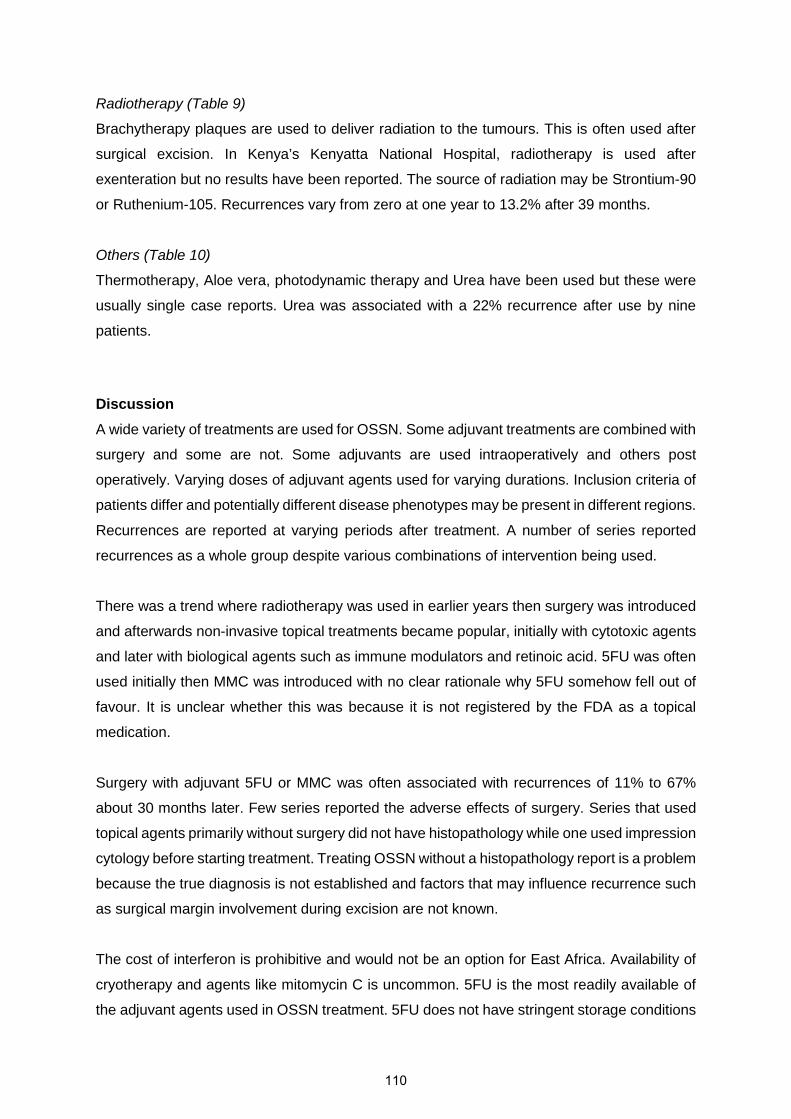
Radiotherapy (Table 9)
Brachytherapy plaques are used to deliver radiation to the tumours. This is often used after
surgical excision. In Kenya’s Kenyatta National Hospital, radiotherapy is used after
exenteration but no results have been reported. The source of radiation may be Strontium-90
or Ruthenium-105. Recurrences vary from zero at one year to 13.2% after 39 months.
Others (Table 10)
Thermotherapy, Aloe vera, photodynamic therapy and Urea have been used but these were
usually single case reports. Urea was associated with a 22% recurrence after use by nine
patients.
Discussion
A wide variety of treatments are used for OSSN. Some adjuvant treatments are combined with
surgery and some are not. Some adjuvants are used intraoperatively and others post
operatively. Varying doses of adjuvant agents used for varying durations. Inclusion criteria of
patients differ and potentially different disease phenotypes may be present in different regions.
Recurrences are reported at varying periods after treatment. A number of series reported
recurrences as a whole group despite various combinations of intervention being used.
There was a trend where radiotherapy was used in earlier years then surgery was introduced
and afterwards non-invasive topical treatments became popular, initially with cytotoxic agents
and later with biological agents such as immune modulators and retinoic acid. 5FU was often
used initially then MMC was introduced with no clear rationale why 5FU somehow fell out of
favour. It is unclear whether this was because it is not registered by the FDA as a topical
medication.
Surgery with adjuvant 5FU or MMC was often associated with recurrences of 11% to 67%
about 30 months later. Few series reported the adverse effects of surgery. Series that used
topical agents primarily without surgery did not have histopathology while one used impression
cytology before starting treatment. Treating OSSN without a histopathology report is a problem
because the true diagnosis is not established and factors that may influence recurrence such
as surgical margin involvement during excision are not known.
The cost of interferon is prohibitive and would not be an option for East Africa. Availability of
cryotherapy and agents like mitomycin C is uncommon. 5FU is the most readily available of
the adjuvant agents used in OSSN treatment. 5FU does not have stringent storage conditions
110

such as refrigeration and cytotoxics have a low risk of contamination. 5FU is on the WHO
essential medicines list, and is a widely available and low cost option particularly in sub-
Saharan Africa where the highest incidence of OSSN in the world is found.
In conclusion, the results from case series are difficult to compare. As most modes of
treatment such as cryotherapy, amniotic membrane transplants and MMC are unavailable in
most centres in East Africa, we believe that a randomized controlled trial using 5FU is
warranted. Availability of 5FU would make translation of these trial results into clinical practice
feasible.
111
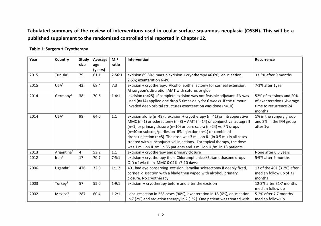
Tabulated summary of the review of interventions used in ocular surface squamous neoplasia (OSSN). This will be a
published supplement to the randomized controlled trial reported in Chapter 12.
Table 1: Surgery ± Cryotherapy
Year Country Studysize
Averageage(years)
M:Fratio
Intervention Recurrence
2015 Tunisia1 79 61·1 2·56:1 excision 89·8%; margin excision + cryotherapy 46·6%; enucleation2·5%; exenteration 6·4%
33·3% after 9 months
2015 USA2 43 68·4 7:3 excision + cryotherapy. Alcohol epitheliectomy for corneal extension.At surgeon's discretion AMT with sutures or glue
7·1% after 1year
2014 Germany3 38 70·6 1·4:1 excision (n=25). If complete excision was not feasible adjuvant IFN wasused (n=14) applied one drop 5 times daily for 6 weeks. If the tumourinvaded deep orbital structures exenteration was done (n=10)
52% of excisions and 20%of exenterations. Averagetime to recurrence 24months
2014 USA4 98 64·0 1:1 excision alone (n=49) ; excision + cryotherapy (n=41) or intraoperativeMMC (n=1) or sclerectomy (n=8) + AMT (n=14) or conjunctival autograft(n=1) or primary closure (n=10) or bare sclera (n=24) vs IFN drops(n=40)or subconj/perilesion IFN injection (n=1) or combineddrops+injection (n=8). The dose was 3 million IU (in 0·5 ml) in all casestreated with subconjunctival injections. For topical therapy, the dosewas 1 million IU/ml in 35 patients and 3 million IU/ml in 13 patients.
1% in the surgery groupand 3% in the IFN groupafter 1yr
2013 Argentina5 4 53·2 1:1 excision + cryotherapy and primary closure None after 6·5 years
2012 Iran6 17 70·7 7·5:1 excision + cryotherapy then Chloramphenicol/Betamethasone dropsQID x 1wk; then MMC 0·04% x7-10 days;
5·9% after 9 months
2006 Uganda7 476 32·0 1:1·2 401 had eye-conserving excision, lamellar sclerectomy if deeply fixed,corneal dissection with a blade then wiped with alcohol, primaryclosure. No cryotherapy.
13 of the 401 (3·2%) aftermedian follow up of 32months
2003 Turkey8 57 55·0 1·9:1 excision + cryotherapy before and after the excision 12·3% after 31·7 monthsmedian follow up
2002 Mexico9 287 60·4 1·2:1 Local resection in 258 cases (90%), exenteration in 18 (6%), enucleationin 7 (2%) and radiation therapy in 2 (1% ). One patient was treated with
5·2% after 7·7 monthsmedian follow up
112
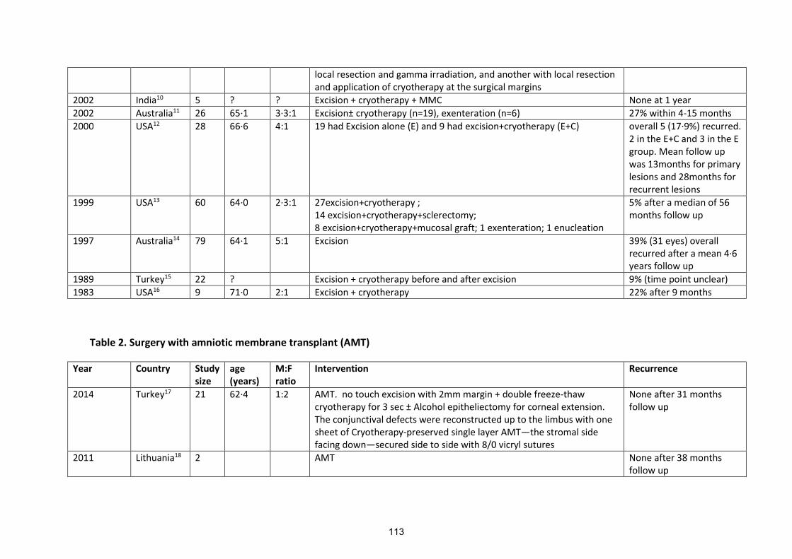
local resection and gamma irradiation, and another with local resectionand application of cryotherapy at the surgical margins
2002 India10 5 ? ? Excision + cryotherapy + MMC None at 1 year
2002 Australia11 26 65·1 3·3:1 Excision± cryotherapy (n=19), exenteration (n=6) 27% within 4-15 months
2000 USA12 28 66·6 4:1 19 had Excision alone (E) and 9 had excision+cryotherapy (E+C) overall 5 (17·9%) recurred.2 in the E+C and 3 in the Egroup. Mean follow upwas 13months for primarylesions and 28months forrecurrent lesions
1999 USA13 60 64·0 2·3:1 27excision+cryotherapy ;14 excision+cryotherapy+sclerectomy;8 excision+cryotherapy+mucosal graft; 1 exenteration; 1 enucleation
5% after a median of 56months follow up
1997 Australia14 79 64·1 5:1 Excision 39% (31 eyes) overallrecurred after a mean 4·6years follow up
1989 Turkey15 22 ? Excision + cryotherapy before and after excision 9% (time point unclear)
1983 USA16 9 71·0 2:1 Excision + cryotherapy 22% after 9 months
Table 2. Surgery with amniotic membrane transplant (AMT)
Year Country Studysize
age(years)
M:Fratio
Intervention Recurrence
2014 Turkey17 21 62·4 1:2 AMT. no touch excision with 2mm margin + double freeze-thawcryotherapy for 3 sec ± Alcohol epitheliectomy for corneal extension.The conjunctival defects were reconstructed up to the limbus with onesheet of Cryotherapy-preserved single layer AMT—the stromal sidefacing down—secured side to side with 8/0 vicryl sutures
None after 31 monthsfollow up
2011 Lithuania18 2 AMT None after 38 monthsfollow up
113
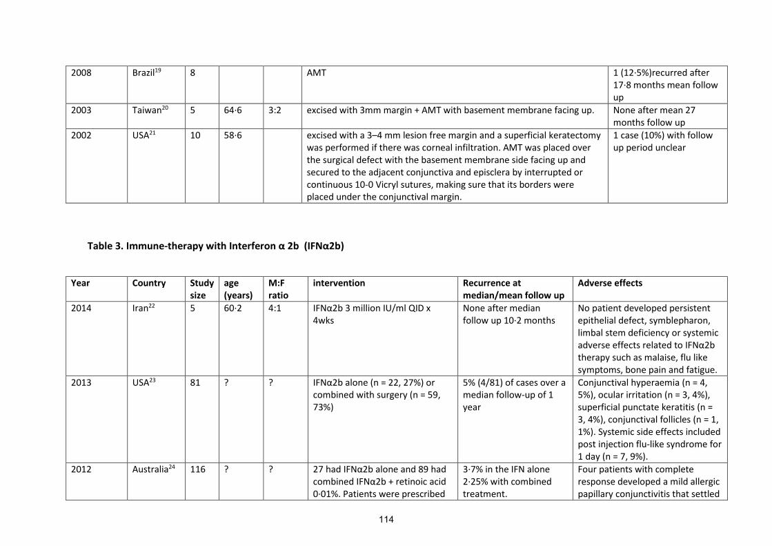
2008 Brazil19 8 AMT 1 (12·5%)recurred after17·8 months mean followup
2003 Taiwan20 5 64·6 3:2 excised with 3mm margin + AMT with basement membrane facing up. None after mean 27months follow up
2002 USA21 10 58·6 excised with a 3–4 mm lesion free margin and a superficial keratectomywas performed if there was corneal infiltration. AMT was placed overthe surgical defect with the basement membrane side facing up andsecured to the adjacent conjunctiva and episclera by interrupted orcontinuous 10-0 Vicryl sutures, making sure that its borders wereplaced under the conjunctival margin.
1 case (10%) with followup period unclear
Table 3. Immune-therapy with Interferon α 2b (IFNα2b)
Year Country Studysize
age(years)
M:Fratio
intervention Recurrence atmedian/mean follow up
Adverse effects
2014 Iran22 5 60·2 4:1 IFNα2b 3 million IU/ml QID x 4wks
None after medianfollow up 10·2 months
No patient developed persistentepithelial defect, symblepharon,limbal stem deficiency or systemicadverse effects related to IFNα2b therapy such as malaise, flu likesymptoms, bone pain and fatigue.
2013 USA23 81 ? ? IFNα2b alone (n = 22, 27%) or combined with surgery (n = 59,73%)
5% (4/81) of cases over amedian follow-up of 1year
Conjunctival hyperaemia (n = 4,5%), ocular irritation (n = 3, 4%),superficial punctate keratitis (n =3, 4%), conjunctival follicles (n = 1,1%). Systemic side effects includedpost injection flu-like syndrome for1 day (n = 7, 9%).
2012 Australia24 116 ? ? 27 had IFNα2b alone and 89 hadcombined IFNα2b + retinoic acid 0·01%. Patients were prescribed
3·7% in the IFN alone2·25% with combinedtreatment.
Four patients with completeresponse developed a mild allergicpapillary conjunctivitis that settled
114
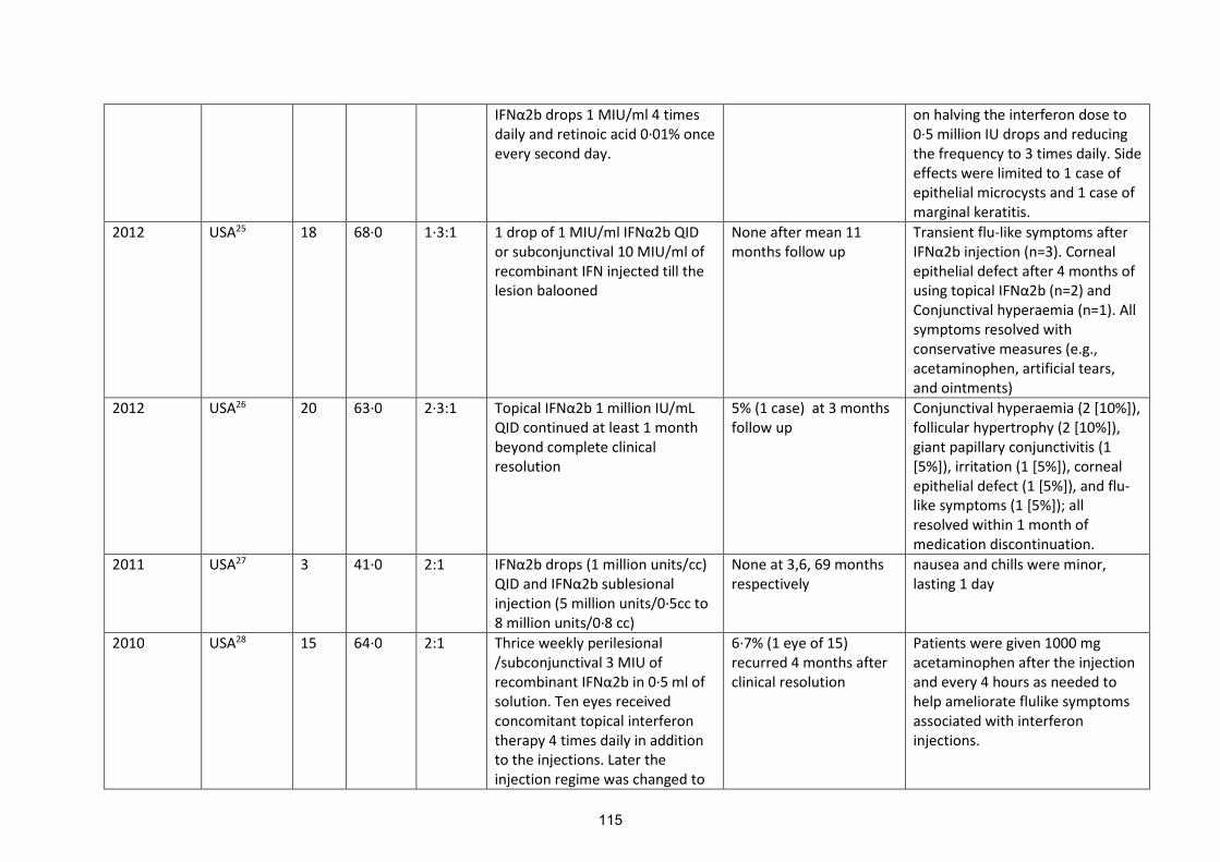
IFNα2b drops 1 MIU/ml 4 timesdaily and retinoic acid 0·01% onceevery second day.
on halving the interferon dose to0·5 million IU drops and reducingthe frequency to 3 times daily. Sideeffects were limited to 1 case ofepithelial microcysts and 1 case ofmarginal keratitis.
2012 USA25 18 68·0 1·3:1 1 drop of 1 MIU/ml IFNα2b QIDor subconjunctival 10 MIU/ml ofrecombinant IFN injected till thelesion balooned
None after mean 11months follow up
Transient flu-like symptoms afterIFNα2b injection (n=3). Corneal epithelial defect after 4 months ofusing topical IFNα2b (n=2) and Conjunctival hyperaemia (n=1). Allsymptoms resolved withconservative measures (e.g.,acetaminophen, artificial tears,and ointments)
2012 USA26 20 63·0 2·3:1 Topical IFNα2b 1 million IU/mLQID continued at least 1 monthbeyond complete clinicalresolution
5% (1 case) at 3 monthsfollow up
Conjunctival hyperaemia (2 [10%]),follicular hypertrophy (2 [10%]),giant papillary conjunctivitis (1[5%]), irritation (1 [5%]), cornealepithelial defect (1 [5%]), and flu-like symptoms (1 [5%]); allresolved within 1 month ofmedication discontinuation.
2011 USA27 3 41·0 2:1 IFNα2b drops (1 million units/cc)QID and IFNα2b sublesional injection (5 million units/0·5cc to8 million units/0·8 cc)
None at 3,6, 69 monthsrespectively
nausea and chills were minor,lasting 1 day
2010 USA28 15 64·0 2:1 Thrice weekly perilesional/subconjunctival 3 MIU ofrecombinant IFNα2b in 0·5 ml of solution. Ten eyes receivedconcomitant topical interferontherapy 4 times daily in additionto the injections. Later theinjection regime was changed to
6·7% (1 eye of 15)recurred 4 months afterclinical resolution
Patients were given 1000 mgacetaminophen after the injectionand every 4 hours as needed tohelp ameliorate flulike symptomsassociated with interferoninjections.
115

received weekly until clinicaldisease resolution
2010 USA29 3 63·7 Allmales
pegylated IFNα2β subconjunctival/perilesioninjection of 1 µg/kg in the area ofthe lesion
1 (33·3%) recurrenceafter 7months follow up
No adverse effects.Acetaminophen 1000mg was givento prevent flu-like symptoms.
2010 USA30 33 64·4 1:2 IFNα2b drops (1 millioninternational units (IU)/ml vs 3million IU/ml) for 1 month afterexcision
9% (3 eyes) 20,21 & 28months later
discomfort and photophobia
2008 USA31 29 66·7 13:1 IFNα2b drops vs no touchexcision +cryotherapy and 8 hadAMT too. 10 received IFNα2b 1 million IU/ml and 4 received 2million U/ml, all 4 times per day,and 1 received 3 million IU/mlthrice daily. Those whose lesionfailed to regress within 2 monthsof therapy crossed over tosurgery. IFNα2b was continued until 4 months after clinicalresolution
None a t 35·6 months Not described
2006 Australia32 10 73·4 2:1 topical IFN-α2b (1 million IU/ml) four times a day until clinicalresolution of the lesion or untilthe lesion appearednonresponsive
None at 55 weeks followup
Not described
2006 Australia33 3 69·3 1:2 IFNα2b 3Mill IU/ml every 3 daysfor 7 doses
None after 22·7 monthsfollow up
Not described
2005 USA34 2 69·0 Allmales
IFNα2b drops 1 million units/ml)QID
None after 3 and 6months follow uprespectively
Not described
2004 USA35 7 68·7 6:1 recombinant topical IFNalpha2bdrops (1 million IU/ml) 4 timesdaily until lesion resolution noted
2 (33·3%) patients hadanother recurrence of
Not described
116

noted at 1 year and 2months, respectively.
2002 Japan36 1 73·0 Female subconj IFNα2b x2 then drops for12wks (cryotherapy had beendone twice before and failed)
Not described
1999 USA37 6 64·3 2:1 singlesubconjunctival/perilesionalinjection of 3 MIU/mlrecombinant IFNα2b in 0·5 ml and then IFNα2b drops (1 MIU/ml) four times a day
None after 7·2 monthsfollow up
Not described
1976 USA38 1 24 Male dinitrochlorobenzene (DNCB).Initial sensitization with topicalDNCB 2000µg on volar aspect offorearm. Then 30-50 µL of DNCB2mg/ml solution in acetoneapplied to the tumour surface.Repeated topical treatment aweek later for 5 cycles
None after 3 years followup
Not described
Table 4. Immune-therapy with cyclosporin A (CSA)
Year Country Studysize
age(years)
M:Fratio
intervention Recurrence Adverse effects
2009 Egypt39 10 65·0 2:1 wide surgical excision + topicalcyclosporine A (0·05%) andtopical mitomycin C (0·01%) QID x12 weeks after surgery
None after a mean 2years follow up
Epithelial toxicity (punctatekeratopathy) occurred in 3 eyes,ocular irritation and mildconjunctival hyperemia in 5 eyes,and lid toxicity in 2 cases duringthe treatment with mitomycin C
2006 Turkey40 2 65·0 All male Combined CSA and MMC. CSA(0·05%) ophthalmic emulsion QID
None after 18 monthsfollow up
In this study they lowered theconcentration of MMC to 0·01%
117

for 12 weeks, and topical MMC(0·01%) QID was combined withCSA at the second, fourth, andsixth weeks
and combined it with CSA toprevent potential ocular surfaceside effects
Table 5. Chemotherapy with mitomycin C (MMC)
Year Country Studysize
age(years)
M:Fratio
intervention Recurrence Adverse effects
2014 Australia41 135 69·0 3·5:1 MMC 0·04% 4 times daily for 1week, followed by 3 weeks offtreatment. IFNα2b 1 MIU/mL 4 times daily if previous treatmentwith MMC had failed or unable totolerate MMC due to side effects.Again, this treatment wascontinued until an end point wasreached. Endpoint was resolutionor failure.
Overall 19 (14·1%)recurred. The mean timeto recurrence was 17·2months (range, 4 to 61months). 14 recurrences(15·1%) were in patientstreated only with MMC(n = 93), 4 recurrences(20·0%) were in patientstreated with both MMCand IFNα2b (n=20), and only 1 recurrence (4·6%)was in patients treatedonly with IFNα2b (n= 22).
Adverse effects occurred in 76(58·9%) patients using MMC and in14 (30·4%) patients using IFNα2b. The most common wereconjunctival hyperemia orirritation (MMC, n=63 [48·8%]; IFN,n=13 28·2%]), localized allergic ortoxic reactions (defined aspapillary conjunctivitis, lidswelling, or both; MMC,n = 12[9·3%]) and punctal stenosis(MMC, n = 7 [5·4%]). There wasone case of corneal erosion ineach of the MMC and IFN groups.Patients occasionally had morethan 1 adverse effect. There wereno cases of limbal stem celldeficiency identified. Most sideeffects were mild and weretolerated by patients. Side effectssignificant enough to result incessation of treatment beforeclinical resolution occurred in 12(8·4%) of 143 eyes; all
118
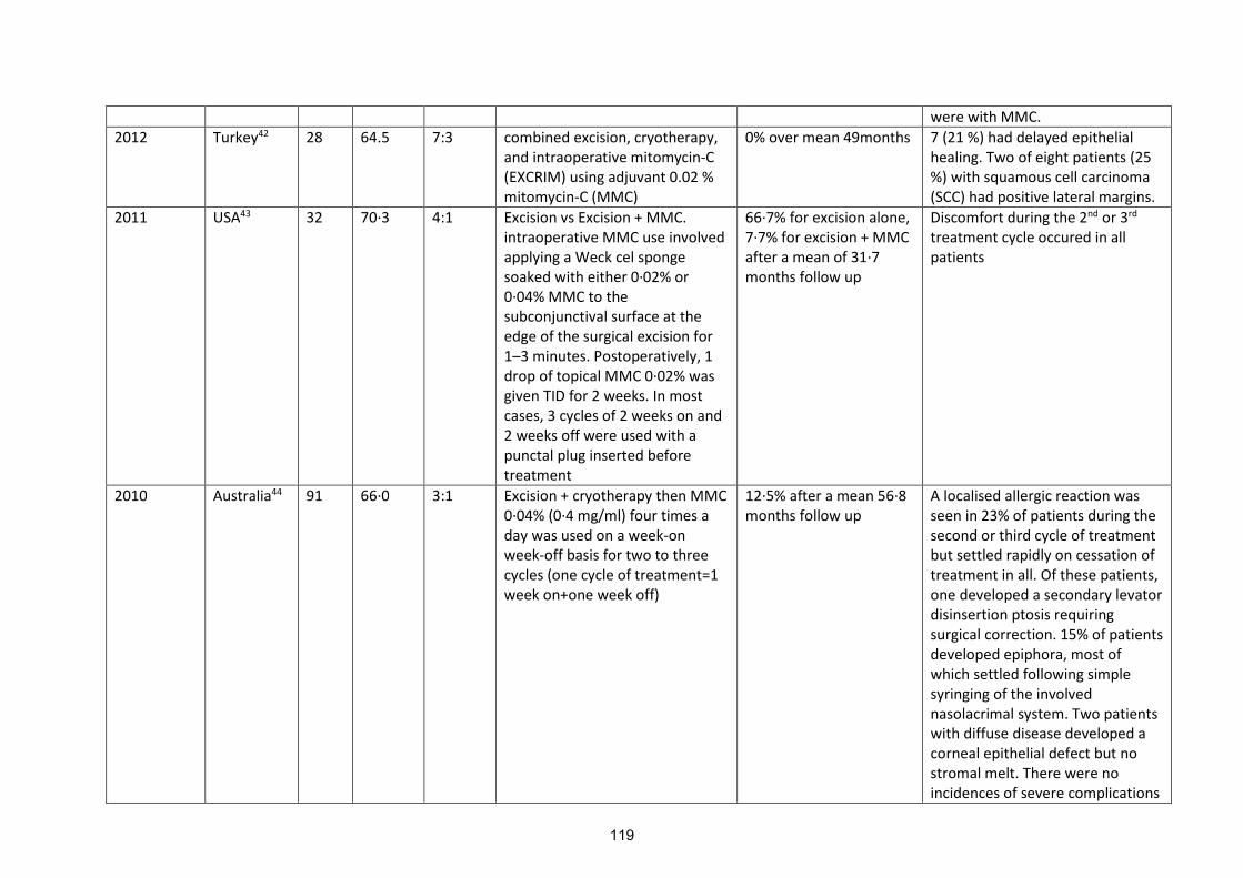
were with MMC.
2012 Turkey42 28 64.5 7:3 combined excision, cryotherapy,and intraoperative mitomycin-C(EXCRIM) using adjuvant 0.02 %mitomycin-C (MMC)
0% over mean 49months 7 (21 %) had delayed epithelialhealing. Two of eight patients (25%) with squamous cell carcinoma(SCC) had positive lateral margins.
2011 USA43 32 70·3 4:1 Excision vs Excision + MMC.intraoperative MMC use involvedapplying a Weck cel spongesoaked with either 0·02% or0·04% MMC to thesubconjunctival surface at theedge of the surgical excision for1–3 minutes. Postoperatively, 1drop of topical MMC 0·02% wasgiven TID for 2 weeks. In mostcases, 3 cycles of 2 weeks on and2 weeks off were used with apunctal plug inserted beforetreatment
66·7% for excision alone,7·7% for excision + MMCafter a mean of 31·7months follow up
Discomfort during the 2nd or 3rd
treatment cycle occured in allpatients
2010 Australia44 91 66·0 3:1 Excision + cryotherapy then MMC0·04% (0·4 mg/ml) four times aday was used on a week-onweek-off basis for two to threecycles (one cycle of treatment=1week on+one week off)
12·5% after a mean 56·8months follow up
A localised allergic reaction wasseen in 23% of patients during thesecond or third cycle of treatmentbut settled rapidly on cessation oftreatment in all. Of these patients,one developed a secondary levatordisinsertion ptosis requiringsurgical correction. 15% of patientsdeveloped epiphora, most ofwhich settled following simplesyringing of the involvednasolacrimal system. Two patientswith diffuse disease developed acorneal epithelial defect but nostromal melt. There were noincidences of severe complications
119

such as hypotony, corneo-scleralmelt or limbal stem cell failure.
2010 UK45 24 63·0 ? MMC as primary treatment orsurgical adjuvant. 0·04% MMCfour times a day for 3 weeks on, 3weeks off, 3 weeks on, withtopical steroid and lubricantsthroughout
67% after a mean 50months of follow up
hyperemia, allergy, epiphora,discharge, mildkeratoconjunctivitis, uveitis,corneal abrasion, corneal oedema,pyogenic granuloma, nose bleed.The most common short termcomplication was allergy, 64% ofwhich occurred in the second 3-week cycle. The most commonlong term complication wascontinuing mildkeratoconjunctivitis
2009 Iran46 17 58·7 2·4:1 2-3 alternate 7-day courses of0·04% mitomycin-C in artificialtears. 14 patients (82·4%)received 2 courses of MMC oneweek apart, and only 3 patients(17·6%) received 3 MMC courses
5 (29·4%) recurrencesover a mean follow up of30·8 months
All patients reported some degreesof mild to moderate eye rednessand irritation that were controlledby artificial tear, mildcorticosteroid drops, and warmcompress. There were no cases ofscleral melting or any systemiccomplications
2006 Spain47 1 82·0 Female Short term MMC 0·02% (2 cycles)followed by long term interferon1 MIU/ml QID until tumourdisappearance (took 75 days todisappear)
None after 1 year no clinical evidence of limbal stemcell deficiency
2004 USA48 1 52·0 Female 0·02% MMC for 14 days in thefirst cycle, 12 days in the secondcycle, and 3 days in the third cyclefollowed by topical interferonalfa-2b 1 x 10⁶ U/mL for 11 daysdue to intolerance to MMC.
Resolution lasted 10months. Recurrence notexplicitly mentioned in
Not described
2004 Australia49 27 64·0 2·7:1 excised with 2mm margin +cryotherapy (where available) +
None after 27 monthsmean follow up
5 patients developed granulomafollowing excision of OSSN, all of
120

Chloramphenicol and PrednefrineForte eye drops four times dailyuntil wound healing + at least two1 week courses of topical MMC0·04% four times a day, aftercomplete epithelial healing. Eachcourse was followed by 1 weekfree of MMC
which resolved rapidly withcontinued topical steroidtreatment. Eight patients receivedtwo courses because of allergy toMMC with redness, swelling, andsignificant itching, whichdeveloped late in the secondcourse. All MMC allergy symptomsresolved rapidly followingdiscontinuation of treatment.
2002 USA50 10 66·0 2·3:1 Mitomycin C 0·04% four timesdaily was applied for a median ofthree cycles
None after a median 15months follow up
temporary local irritation,erythema, chemosis
2002 Israel51 5 58·4 2:3 MMC, 0·02% or 0·04%, four timesdaily for 14 days. Second andthird identical cycles of MMCwere applied at 3- to 4-weekintervals. Wide excision of theconjunctiva was done 4 to 6weeks after the final cycle ofMMC
None after a mean 23·8months follow up
1 case of conjunctival hyperaemiaseen from day 8
2002 Greece52 7 73·8 3:4 During excision of the lesion,mitomycin-C 0·02% was appliedintraoperatively for 5 minutes. Intwo cases, excision was combinedwith conjunctival limbal autograft
14% ( n=1) after a meanof 16 months follow up
Not described
2002 UK53 11 66·7 2:9 Preoperative topical andintraoperative local mitomycin C.MMC 0·04% eye drops QID in twoweekly courses preoperativelyand/or a single intraoperativeapplication of 0·4 mg/ml. 7patients had additional limitedlocal excision of the residual
None after a mean of 9·5months follow up
1 developed a reaction to thedrops during the secondfortnightly course of MMC dropsbut treatment was continuedunder steroid cover. 1 persistentepithelial defect following bothcourses of topical mitomycin Cdrops
121

tumour mass and 1 hadcryotherapy.
2001 Germany54 9 ? ? mitomycin C eye drops 0·02%(MMC) after excision
11% (1 patient) 14months after surgery andafter 2 MMC-cycles.
self-limited conjunctivitis
1999 USA55 4 ? ? MMC 0·02% TID x2wk None after a mean of 20months follow up
mild discomfort, redness,photophobia, and punctateepithelial keratopathy thatsubsided on discontinuation of themedication
1997 UK56 7 ? ? one drop of MMC 0·04% QID for 7days in alternate weeks
None after 9 monthsfollow up
transitory ocular discomfort,conjunctival injection, tearing,photophobia, and punctateepithelial keratopathy
Table 6. Chemotherapy with 5-Fluorouracil (5FU)
Year Country Studysize
age(years)
M:Fratio
intervention Recurrence Adverse effects
2015 Australia57 38 70·0 6:1 1% 5FU in N/saline QIDx 2wks or0·04% MMC in N/saline x2-3cycles each cycle lasting 1 wkfollowed by 1 wk drug holiday.Some pts had excision with a2mm margin + cryotherapy
31% (10 patients)required furthertreatment for diseasepersistence orrecurrence; 7 on MMCand 3 on 5FU
5-FU 1% resulted in drug-relatedcomplications in seven of 12 cases(58·3%), and included a single caseof focal paracentral cornealstromal melt. MMC 0·04% resultedin transient drug relatedcomplications in 23 of 39 (59%)cases
2014 Australia58 153 65·5 7:3 89 on 5FU 1% QID x 2 wks and 64on MMC 0·04% QID for 2-3 1-week cycles.
2% (n=) 2 on 5FU; 0 onMMC. Mean follow up33·6months on 5FU and57·9months on MMC
Not described
122

2011 Australia59 65 66·3 3:1 a single cycle of 5FU 1% inN/saline QID x 2wk
1·5% (1 case) after amedian follow up 23months
37 (57%) patients had short-termcomplications. Lid toxicity 32(49·2%); epiphora 5 (7·7%);superficial keratitis 6 (9·2%);corneal epithelial defect 1 (1·5%);ectropion 1 (1·5%). Four patientswere unable to complete thecourse of 5-FU 1% because of localtoxicity
2011 Italy60 41 65·5 1·7:1 1% 5-FU QID x4 weeks (onecourse). 22 patients (53·7%) had5-FU as a sole treatment, and 19patients (46·3%) as adjuvantand/or debulking therapy
7·3% (3 tumours) treatedwith 5-FU alone recurredduring a follow-up of89·7 months
Clinical confocal microscopyshowed no long-term differencebetween the treated eye andfellow (control) eye in: endothelialcells count, pleomorphism andpolymegathism, anterior stromalkeratocyte density, sub-basalnerve plexus fibre number,density, and beadings and centralcornea epithelium thickness
2010 Yemen61 15 50·8 1:4 subconjunctival 5FU (5mg) at theend of surgery + 1% 5-FU eyedrops QID x 4days then repeatedat 30 day intervals x6 cycles. Allhad 6 months therapy
1/15 (6·7%) recurred mild temporary local irritation
2000 USA62 7 74·0 All male 1% 5-FU in methylcellulose wasadministered four times daily for2 to 4 days for each cycle. Thenumber of initial treatment cycleswas two to six, with the timebetween cycles being 30 to 45days.
3 recurrences (43%) No adverse effects noted
2000 Italy63 8 70·0 3:1 1% 5FU drops alone QID x4wkswithout surgery or radiotherapy
1 patient (12·5%) with amean 27 months followup
Acute transient toxickeratoconjunctivitis was observedin all treated cases; it wascontrolled with topical therapy.
123

1995 USA64 6 61·0 2:1 excision +5FU or 5FU alone 1 recurred (16·6%) after30 months
Punctate epithelial keratopathy,epithelial defect
Table 7. Retinoic acid
Year Country Studysize
age(years)
M:Fratio
intervention Recurrence Adverse effects
2010 Australia65 1 64·0 Female topical all-trans retinoic acid(ATRA), 0·01%, 1 eyedrop appliedevery second day x9mo. Afterthere was no response from 2intralesional injections of 3 Mill IUof recombinant IFN in 0·5 mL, 2months apart, followed by topicalinterferon alfa-2b (1 Mill IU/mL) 4times a day.
No recurrence Not described
Table 8. Anti-vascular endothelial growth factor (VEGF)
Year Country Studysize
age(years)
M:Fratio
intervention Recurrence Adverse effects
2015 Turkey66 6 66 2:1 topical 5mg/mL bevacizumab QIDx 8 weeks
None after 6 monthsfollow up
Not described
2014 Turkey67 10 60·5 bevacizumab (Avastin) 25mg/mlx 7·8wks (range 5-14) thenexcision biopsy+cyro+AMT
None after 6 monthsfollow up
Not described
2009 Egypt68 10 45·0 2:1 subconj Avastin (bevacizumab) 20% after 12 monthsfollow up
Not described
2009 USA69 4 51·5 ? Subconjunctival injections ofranibizumab 0·5 mg were givenevery 2 or 4 weeks if the tumourregressed or remained stable.
1 recurred (25%) and 1did not regress (25%)
No adverse ranibizumab-relatedocular or systemic side effects.Specifically, there were no local orsystemic allergies and no corneal
124

epitheliopathy, perforation,conjunctival hyperaemia, scleralthinning, uveitis orcataractformation.
Table 9. Radiotherapy, Thermotherapy
Year Country Studysize
age(years)
M:Fratio
intervention Recurrence Adverse effects
2015 S. Africa70 69 42·0 1:1 60 Gray Sr-90 brachytherapy infour divided doses after resectionwith a 2 mm margin. Nocryotherapy or alcoholdebridement
8 (11·6%) after a median27 months follow up
Five patients developed a dry eye,which was treated withocular lubricants
2011 Spain71 1 61·0 Male Orthovoltage was administeredwith direct field radiation focusedon the tumor. The patientreceived 500 cGy in 2sessions/week for 2 weeks (2000cGy) and then 7 sessions 300 cGydaily (2100 cGy).
No recurrence after 1year follow up
Not described
2009 France72 15 63·7 4:1 proton beam therapy and surgery 2 patients (13·2%). Meanfollow-up was 39.1months
Not described
2009 Australia73 11 60·8 1:3·7 Plaque brachytherapy. A 10- 15-mm-diameter circular Iodine-125plaque was applied underassisted local or generalanaesthesia during surgery(excised with histologicalconfirmation using a techniqueinvolving minimal lateralclearance and avoidance of deepscleral dissection)
None after 23·4 monthsmean follow up
Not described
125
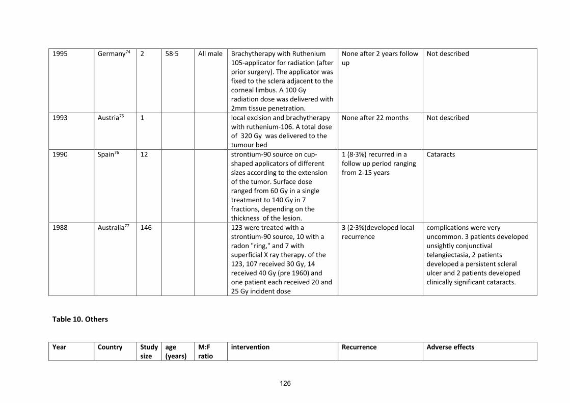
1995 Germany74 2 58·5 All male Brachytherapy with Ruthenium105-applicator for radiation (afterprior surgery). The applicator wasfixed to the sclera adjacent to thecorneal limbus. A 100 Gyradiation dose was delivered with2mm tissue penetration.
None after 2 years followup
Not described
1993 Austria75 1 local excision and brachytherapywith ruthenium-106. A total doseof 320 Gy was delivered to thetumour bed
None after 22 months Not described
1990 Spain76 12 strontium-90 source on cup-shaped applicators of differentsizes according to the extensionof the tumor. Surface doseranged from 60 Gy in a singletreatment to 140 Gy in 7fractions, depending on thethickness of the lesion.
1 (8·3%) recurred in afollow up period rangingfrom 2-15 years
Cataracts
1988 Australia77 146 123 were treated with astrontium-90 source, 10 with aradon "ring," and 7 withsuperficial X ray therapy. of the123, 107 received 30 Gy, 14received 40 Gy (pre 1960) andone patient each received 20 and25 Gy incident dose
3 (2·3%)developed localrecurrence
complications were veryuncommon. 3 patients developedunsightly conjunctivaltelangiectasia, 2 patientsdeveloped a persistent scleralulcer and 2 patients developedclinically significant cataracts.
Table 10. Others
Year Country Studysize
age(years)
M:Fratio
intervention Recurrence Adverse effects
126
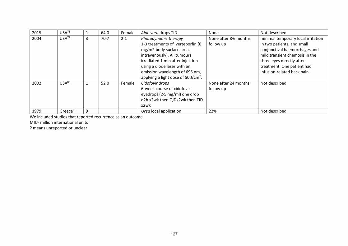
2015 USA78 1 64·0 Female Aloe vera drops TID None Not described
2004 USA79 3 70·7 2:1 Photodynamic therapy1-3 treatments of verteporfin (6mg/m2 body surface area,intravenously). All tumoursirradiated 1 min after injectionusing a diode laser with anemission wavelength of 695 nm,applying a light dose of 50 J/cm2.
None after 8·6 monthsfollow up
minimal temporary local irritationin two patients, and smallconjunctival haemorrhages andmild transient chemosis in thethree eyes directly aftertreatment. One patient hadinfusion-related back pain.
2002 USA80 1 52·0 Female Cidofovir drops6-week course of cidofovireyedrops (2·5 mg/ml) one dropq2h x2wk then QIDx2wk then TIDx2wk
None after 24 monthsfollow up
Not described
1979 Greece81 9 Urea local application 22% Not described
We included studies that reported recurrence as an outcome.MIU- million international units? means unreported or unclear
127

References
1. Chebbi A, Bouguila H, Boukari M, et al. [Prognosis of primary malignant tumors of theconjunctiva]. Journal francais d'ophtalmologie 2015; 38(6): 477-85.
2. Li AS, Shih CY, Rosen L, Steiner A, Milman T, Udell IJ. Recurrence of Ocular Surface SquamousNeoplasia Treated With Excisional Biopsy and Cryotherapy. American journal of ophthalmology2015; 160(2): 213-9 e1.
3. Miller CV, Wolf A, Klingenstein A, et al. Clinical outcome of advanced squamous cell carcinomaof the conjunctiva. Eye (Lond) 2014; 28(8): 962-7.
4. Nanji AA, Moon CS, Galor A, Sein J, Oellers P, Karp CL. Surgical versus medical treatment ofocular surface squamous neoplasia: a comparison of recurrences and complications.Ophthalmology 2014; 121(5): 994-1000.
5. Crim N, Fornies-Paz ME, Monti R, Esposito E, Maccio JP, Urrets-Zavalia JA. In situ carcinoma ofthe conjunctiva: surgical excision associated with cryotherapy. Clinical ophthalmology(Auckland, NZ) 2013; 7: 1889-93.
6. Kashkouli MB, Heirati A, Pakdel F, Kiavash V, Naseripour M, Aghamohammadi F. Long-termfollow-up of invasive ocular surface squamous cell carcinoma treated with excision,cryotherapy, and topical mitomycin C. Graefe's archive for clinical and experimentalophthalmology = Albrecht von Graefes Archiv fur klinische und experimentelle Ophthalmologie2012.
7. Waddell KM, Downing RG, Lucas SB, Newton R. Corneo-conjunctival carcinoma in Uganda. Eye(Lond) 2006; 20(8): 893-9.
8. Peksayar G, Altan-Yaycioglu R, Onal S. Excision and cryosurgery in the treatment of conjunctivalmalignant epithelial tumours. Eye (Lond) 2003; 17(2): 228-32.
9. Cervantes G, Rodriguez AA, Jr., Leal AG. Squamous cell carcinoma of the conjunctiva:clinicopathological features in 287 cases. Canadian journal of ophthalmology Journal canadiend'ophtalmologie 2002; 37(1): 14-9; discussion 9-20.
10. Khokhar S, Soni A, SinghSethi H, Sudan R, Sony P, Pangtey MS. Combined surgery, cryotherapy,and mitomycin-C for recurrent ocular surface squamous neoplasia. Cornea 2002; 21(2): 189-91.
11. McKelvie PA. Squamous cell carcinoma of the conjunctiva: a series of 26 cases. British Journal ofOphthalmology 2002; 86(2): 168-73.
12. Sudesh S, Rapuano CJ, Cohen EJ, Eagle RC, Jr., Laibson PR. Surgical management of ocularsurface squamous neoplasms: the experience from a cornea center. Cornea 2000; 19(3): 278-83.
13. Tunc M, Char DH, Crawford B, Miller T. Intraepithelial and invasive squamous cell carcinoma ofthe conjunctiva: analysis of 60 cases. British Journal of Ophthalmology 1999; 83(1): 98-103.
14. Tabin G, Levin S, Snibson G, Loughnan M, Taylor H. Late recurrences and the necessity for long-term follow-up in corneal and conjunctival intraepithelial neoplasia. Ophthalmology 1997;104(3): 485-92.
15. Peksayar G, Soyturk MK, Demiryont M. Long-term results of cryotherapy on malignant epithelialtumors of the conjunctiva. American journal of ophthalmology 1989; 107(4): 337-40.
16. Divine RD, Anderson RL. Nitrous oxide cryotherapy for intraepithelial epithelioma of theconjunctiva. Archives of ophthalmology 1983; 101(5): 782-6.
17. Palamar M, Kaya E, Egrilmez S, Akalin T, Yagci A. Amniotic membrane transplantation in surgicalmanagement of ocular surface squamous neoplasias: long-term results. Eye (Lond) 2014; 28(9):1131-5.
18. Asoklis RS, Damijonaityte A, Butkiene L, et al. Ocular surface reconstruction using amnioticmembrane following excision of conjunctival and limbal tumors. European journal ofophthalmology 2011; 21(5): 552-8.
19. Carvalho-Rego PR, Gomes JA, Ballalai PL, Cunha MC, Sousa LB, Erwenne CM. [Amnioticmembrane for ocular surface reconstruction after conjunctival squamous cell carcinomaresection]. Arquivos brasileiros de oftalmologia 2008; 71(1): 22-7.
128
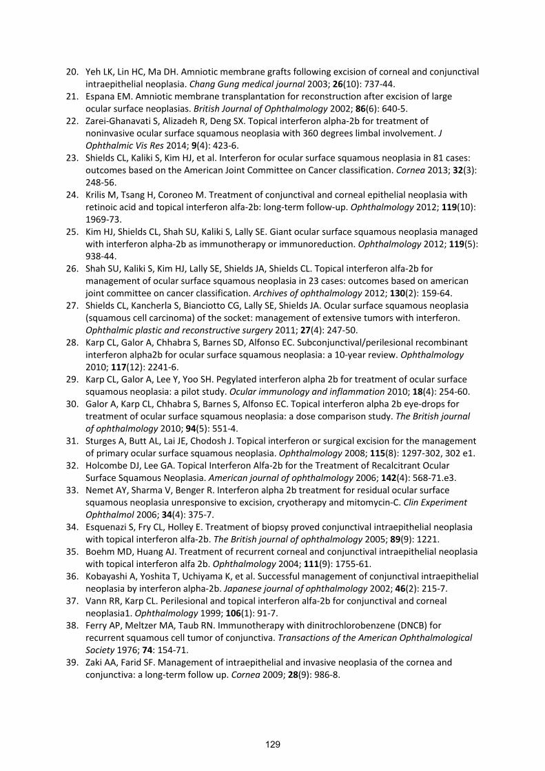
20. Yeh LK, Lin HC, Ma DH. Amniotic membrane grafts following excision of corneal and conjunctivalintraepithelial neoplasia. Chang Gung medical journal 2003; 26(10): 737-44.
21. Espana EM. Amniotic membrane transplantation for reconstruction after excision of largeocular surface neoplasias. British Journal of Ophthalmology 2002; 86(6): 640-5.
22. Zarei-Ghanavati S, Alizadeh R, Deng SX. Topical interferon alpha-2b for treatment ofnoninvasive ocular surface squamous neoplasia with 360 degrees limbal involvement. JOphthalmic Vis Res 2014; 9(4): 423-6.
23. Shields CL, Kaliki S, Kim HJ, et al. Interferon for ocular surface squamous neoplasia in 81 cases:outcomes based on the American Joint Committee on Cancer classification. Cornea 2013; 32(3):248-56.
24. Krilis M, Tsang H, Coroneo M. Treatment of conjunctival and corneal epithelial neoplasia withretinoic acid and topical interferon alfa-2b: long-term follow-up. Ophthalmology 2012; 119(10):1969-73.
25. Kim HJ, Shields CL, Shah SU, Kaliki S, Lally SE. Giant ocular surface squamous neoplasia managedwith interferon alpha-2b as immunotherapy or immunoreduction. Ophthalmology 2012; 119(5):938-44.
26. Shah SU, Kaliki S, Kim HJ, Lally SE, Shields JA, Shields CL. Topical interferon alfa-2b formanagement of ocular surface squamous neoplasia in 23 cases: outcomes based on americanjoint committee on cancer classification. Archives of ophthalmology 2012; 130(2): 159-64.
27. Shields CL, Kancherla S, Bianciotto CG, Lally SE, Shields JA. Ocular surface squamous neoplasia(squamous cell carcinoma) of the socket: management of extensive tumors with interferon.Ophthalmic plastic and reconstructive surgery 2011; 27(4): 247-50.
28. Karp CL, Galor A, Chhabra S, Barnes SD, Alfonso EC. Subconjunctival/perilesional recombinantinterferon alpha2b for ocular surface squamous neoplasia: a 10-year review. Ophthalmology2010; 117(12): 2241-6.
29. Karp CL, Galor A, Lee Y, Yoo SH. Pegylated interferon alpha 2b for treatment of ocular surfacesquamous neoplasia: a pilot study. Ocular immunology and inflammation 2010; 18(4): 254-60.
30. Galor A, Karp CL, Chhabra S, Barnes S, Alfonso EC. Topical interferon alpha 2b eye-drops fortreatment of ocular surface squamous neoplasia: a dose comparison study. The British journalof ophthalmology 2010; 94(5): 551-4.
31. Sturges A, Butt AL, Lai JE, Chodosh J. Topical interferon or surgical excision for the managementof primary ocular surface squamous neoplasia. Ophthalmology 2008; 115(8): 1297-302, 302 e1.
32. Holcombe DJ, Lee GA. Topical Interferon Alfa-2b for the Treatment of Recalcitrant OcularSurface Squamous Neoplasia. American journal of ophthalmology 2006; 142(4): 568-71.e3.
33. Nemet AY, Sharma V, Benger R. Interferon alpha 2b treatment for residual ocular surfacesquamous neoplasia unresponsive to excision, cryotherapy and mitomycin-C. Clin ExperimentOphthalmol 2006; 34(4): 375-7.
34. Esquenazi S, Fry CL, Holley E. Treatment of biopsy proved conjunctival intraepithelial neoplasiawith topical interferon alfa-2b. The British journal of ophthalmology 2005; 89(9): 1221.
35. Boehm MD, Huang AJ. Treatment of recurrent corneal and conjunctival intraepithelial neoplasiawith topical interferon alfa 2b. Ophthalmology 2004; 111(9): 1755-61.
36. Kobayashi A, Yoshita T, Uchiyama K, et al. Successful management of conjunctival intraepithelialneoplasia by interferon alpha-2b. Japanese journal of ophthalmology 2002; 46(2): 215-7.
37. Vann RR, Karp CL. Perilesional and topical interferon alfa-2b for conjunctival and cornealneoplasia1. Ophthalmology 1999; 106(1): 91-7.
38. Ferry AP, Meltzer MA, Taub RN. Immunotherapy with dinitrochlorobenzene (DNCB) forrecurrent squamous cell tumor of conjunctiva. Transactions of the American OphthalmologicalSociety 1976; 74: 154-71.
39. Zaki AA, Farid SF. Management of intraepithelial and invasive neoplasia of the cornea andconjunctiva: a long-term follow up. Cornea 2009; 28(9): 986-8.
129

40. Tunc M, Erbilen E. Topical cyclosporine-a combined with mitomycin C for conjunctival andcorneal squamous cell carcinoma. American journal of ophthalmology 2006; 142(4): 673-5.
41. Besley J, Pappalardo J, Lee GA, Hirst LW, Vincent SJ. Risk factors for ocular surface squamousneoplasia recurrence after treatment with topical mitomycin C and interferon alpha-2b.American journal of ophthalmology 2014; 157(2): 287-93 e2.
42. Sarici AM, Arvas S, Pazarli H. Combined excision, cryotherapy, and intraoperative mitomycin C(EXCRIM) for localized intraepithelial and squamous cell carcinoma of the conjunctiva. Graefe'sarchive for clinical and experimental ophthalmology = Albrecht von Graefes Archiv fur klinischeund experimentelle Ophthalmologie 2013; 251(9): 2201-4.
43. Birkholz ES, Goins KM, Sutphin JE, Kitzmann AS, Wagoner MD. Treatment of ocular surfacesquamous cell intraepithelial neoplasia with and without mitomycin C. Cornea 2011; 30(1): 37-41.
44. Gupta A, Muecke J. Treatment of ocular surface squamous neoplasia with Mitomycin C. TheBritish journal of ophthalmology 2010; 94(5): 555-8.
45. Russell HC, Chadha V, Lockington D, Kemp EG. Topical mitomycin C chemotherapy in themanagement of ocular surface neoplasia: a 10-year review of treatment outcomes andcomplications. The British journal of ophthalmology 2010; 94(10): 1316-21.
46. Rahimi F, Alipour F, Ghazizadeh Hashemi H, Hashemian MN, Mehrdad R. Topical mitomycin-Cfor treatment of partially-excised ocular surface squamous neoplasia. Archives of Iranianmedicine 2009; 12(1): 55-9.
47. Huerva V, Mateo AJ, Mangues I, Jurjo C. Short-term mitomycin C followed by long-terminterferon alpha2beta for conjunctiva-cornea intraepithelial neoplasia. Cornea 2006; 25(10):1220-3.
48. Di Pascuale MA, Espana EM, Tseng SC. A case of conjunctiva-cornea intraepithelial neoplasiasuccessfully treated with topical mitomycin C and interferon alfa-2b in cycles. Cornea 2004;23(1): 89-92.
49. Chen C, Louis D, Dodd T, Muecke J. Mitomycin C as an adjunct in the treatment of localisedocular surface squamous neoplasia. The British journal of ophthalmology 2004; 88(1): 17-8.
50. Shields CL, Naseripour M, Shields JA. Topical mitomycin C for extensive, recurrent conjunctival-corneal squamous cell carcinoma. American journal of ophthalmology 2002; 133(5): 601-6.
51. Frucht-Pery J, Rozenman Y, Pe'er J. Topical mitomycin-C for partially excised conjunctivalsquamous cell carcinoma. Ophthalmology 2002; 109(3): 548-52.
52. Siganos CS, Kozobolis VP, Christodoulakis EV. The intraoperative use of mitomycin-C in excisionof ocular surface neoplasia with or without limbal autograft transplantation. Cornea 2002;21(1): 12-6.
53. Kemp EG, Harnett AN, Chatterjee S. Preoperative topical and intraoperative local mitomycin Cadjuvant therapy in the management of ocular surface neoplasias. The British journal ofophthalmology 2002; 86(1): 31-4.
54. Cartsburg O, Kersten A, Sundmacher R, Nadjari B, Pomjanski N, Bocking A. [Treatment of 9squamous epithelial carcinoma in situ lesions of the conjunctiva (CIN) with mitomycin Ceyedrops in cytological and DNA image cytometric control]. Klinische Monatsblatter furAugenheilkunde 2001; 218(6): 429-34.
55. Akpek EK, Ertoy D, Kalayci D, Hasiripi H. Postoperative topical mitomycin C in conjunctivalsquamous cell neoplasia. Cornea 1999; 18(1): 59-62.
56. Wilson MW, Hungerford JL, George SM, Madreperla SA. Topical mitomycin C for the treatmentof conjunctival and corneal epithelial dysplasia and neoplasia. American journal ofophthalmology 1997; 124(3): 303-11.
57. Rudkin AK, Dempster L, Muecke JS. Management of diffuse ocular surface squamous neoplasia:efficacy and complications of topical chemotherapy. Clinical & Experimental Ophthalmology2015; 43(1): 20-5.
130

58. Bahrami B, Greenwell T, Muecke JS. Long-term outcomes after adjunctive topical 5-flurouracilor mitomycin C for the treatment of surgically excised, localized ocular surface squamousneoplasia. Clin Experiment Ophthalmol 2014; 42(4): 317-22.
59. Rudkin AK, Muecke JS. Adjuvant 5-fluorouracil in the treatment of localised ocular surfacesquamous neoplasia. The British journal of ophthalmology 2011; 95(7): 947-50.
60. Parrozzani R, Lazzarini D, Alemany-Rubio E, Urban F, Midena E. Topical 1% 5-fluorouracil inocular surface squamous neoplasia: a long-term safety study. The British journal ofophthalmology 2011; 95(3): 355-9.
61. Al-Barrag A, Al-Shaer M, Al-Matary N, Al-Hamdani M. 5-Fluorouracil for the treatment ofintraepithelial neoplasia and squamous cell carcinoma of the conjunctiva, and cornea. Clinicalophthalmology (Auckland, NZ) 2010; 4: 801-8.
62. Yeatts RP, Engelbrecht NE, Curry CD, Ford JG, Walter KA. 5-Fluorouracil for the treatment ofintraepithelial neoplasia of the conjunctiva and cornea. Ophthalmology 2000; 107(12): 2190-5.
63. Midena E. Treatment of conjunctival squamous cell carcinoma with topical 5-fluorouracil. BritishJournal of Ophthalmology 2000; 84(3): 268-72.
64. Yeatts RP, Ford JG, Stanton CA, Reed JW. Topical 5-fluorouracil in treating epithelial neoplasia ofthe conjunctiva and cornea. Ophthalmology 1995; 102(9): 1338-44.
65. Skippen B, Tsang HH, Assaad NN, Coroneo MT. Rapid response of refractory ocular surfacedysplasia to combination treatment with topical all-trans retinoic acid and interferon alfa-2b.Archives of ophthalmology 2010; 128(10): 1368-9.
66. Asena L, Dursun Altinors D. Topical Bevacizumab for the Treatment of Ocular Surface SquamousNeoplasia. Journal of ocular pharmacology and therapeutics : the official journal of theAssociation for Ocular Pharmacology and Therapeutics 2015.
67. Ozcan AA, Ciloglu E, Esen E, Simdivar GH. Use of topical bevacizumab for conjunctivalintraepithelial neoplasia. Cornea 2014; 33(11): 1205-9.
68. Zaki AA, Fouad H, Emera S, Labibi H. Subconjunctival Anti VEGF for Conjunctival Intraepithelialand Invasive Neoplasia. Australian Journal of Basic and Applied Sciences, 2009; 3(4): 3186-9.
69. Teng CC, Chin KJ, Finger PT. Subconjunctival ranibizumab for squamous cell carcinoma of theconjunctiva with corneal extension. The British journal of ophthalmology 2009; 93(6): 837-8.
70. Lecuona K, Stannard C, Hart G, et al. The treatment of carcinoma in situ and squamous cellcarcinoma of the conjunctiva with fractionated strontium-90 radiation in a population with ahigh prevalence of HIV. The British journal of ophthalmology 2015; 99(9): 1158-61.
71. Sanchez-Perez JL, Fuentes-Sanchez C, Acosta-Acosta B. Conjunctival-corneal intraepithelialneoplasia (Bowen disease) treated with orthovoltage. Cornea 2011; 30(4): 474-6.
72. Caujolle JP, Maschi C, Chauvel P, Herault J, Gastaud P. [Surgery and additional protontherapyfor treatment of invasive and recurrent squamous cell carcinomas: technique and preliminaryresults]. Journal francais d'ophtalmologie 2009; 32(10): 707-14.
73. Walsh-Conway N, Conway RM. Plaque brachytherapy for the management of ocular surfacemalignancies with corneoscleral invasion. Clin Experiment Ophthalmol 2009; 37(6): 577-83.
74. Haberle H, Pham DT, Scholman HJ, Wollensak J. [Ruthenium 105-applicator for radiationtreatment of carcinoma in situ of the cornea and conjunctiva]. Der Ophthalmologe : Zeitschriftder Deutschen Ophthalmologischen Gesellschaft 1995; 92(6): 866-9.
75. Zehetmayer M, Menapace R, Kulnig W. Combined local excision and brachytherapy withruthenium-106 in the treatment of epibulbar malignancies. Ophthalmologica Journalinternational d'ophtalmologie International journal of ophthalmology Zeitschrift furAugenheilkunde 1993; 207(3): 133-9.
76. Cerezo L, Otero J, Aragon G, et al. Conjunctival intraepithelial and invasive squamous cellcarcinomas treated with strontium-90. Radiotherapy and oncology : journal of the EuropeanSociety for Therapeutic Radiology and Oncology 1990; 17(3): 191-7.
131

77. Kearsley JH, Fitchew RS, Taylor RG. Adjunctive radiotherapy with strontium-90 in the treatmentof conjunctival squamous cell carcinoma. International journal of radiation oncology, biology,physics 1988; 14(3): 435-43.
78. Damani MR, Shah AR, Karp CL, Orlin SE. Treatment of ocular surface squamous neoplasia withtopical Aloe vera drops. Cornea 2015; 34(1): 87-9.
79. Barbazetto IA, Lee TC, Abramson DH. Treatment of conjunctival squamous cell carcinoma withphotodynamic therapy. American journal of ophthalmology 2004; 138(2): 183-9.
80. Sherman MD, Feldman KA, Farahmand SM, Margolis TP. Treatment of conjunctival squamouscell carcinoma with topical cidofovir. American journal of ophthalmology 2002; 134(3): 432-3.
81. Danopoulos ED, Danopoulou IE, Liarikos SB, Merkuris KM. Effects of urea treatment inmalignancies of the conjunctiva and cornea. Ophthalmologica Journal internationald'ophtalmologie International journal of ophthalmology Zeitschrift fur Augenheilkunde 1979;178(4): 198-203.
132
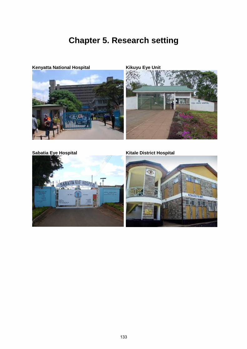
Chapter 5. Research setting
Kenyatta National Hospital Kikuyu Eye Unit
Sabatia Eye Hospital Kitale District Hospital
133

Kenya – an introduction to the research setting
5.1 Geography of Kenya
Kenya sits astride the equator in East Africa (Figure 5.1). The climate is tropical; hot and humid
at the Indian ocean coast and Lake Victoria basin, temperate in the highlands and very hot
and dry in the north and eastern parts of the country. The long rains occur from March to May
and a short rains from October to December. June and July are the coldest months of the year
while January and February are the hottest.
Figure 5.1 Map of Africa showing Kenya highlighted in a red circle in East Africa.
134
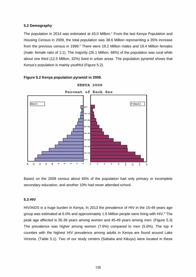
5.2 Demography
The population in 2014 was estimated at 43.0 Million.1 From the last Kenya Population and
Housing Census in 2009, the total population was 38.6 Million representing a 35% increase
from the previous census in 1999.2 There were 19.2 Million males and 19.4 Million females
(male: female ratio of 1:1). The majority (26.1 Million, 68%) of the population was rural while
about one third (12.5 Million, 32%) lived in urban areas. The population pyramid shows that
Kenya’s population is mainly youthful (Figure 5.2).
Figure 5.2 Kenya population pyramid in 2009.
Based on the 2009 census about 65% of the population had only primary or incomplete
secondary education, and another 10% had never attended school.
5.3 HIV
HIV/AIDS is a huge burden in Kenya. In 2013 the prevalence of HIV in the 15-49 years age
group was estimated at 6.0% and approximately 1.6 Million people were living with HIV.3 The
peak age affected is 35-39 years among women and 45-49 years among men. (Figure 5.3)
The prevalence was higher among women (7.6%) compared to men (5.6%). The top 4
counties with the highest HIV prevalence among adults in Kenya are found around Lake
Victoria. (Table 5.1). Two of our study centers (Sabatia and Kikuyu) were located in these
135

areas. (Figure 5.4) The areas with the highest prevalence include western Kenya and the Lake
Victoria region, Nairobi and pockets along the Indian Ocean coast (Figure 5.5)
Table 5.1 Counties in Kenya with the highest HIV prevalence among adults in 2013.3
County HIV prevalence in adults (%)
Homa Bay 25.7
Siaya 23.7
Kisumu 19.3
Migori 14.7
Kisii 8.0
Nairobi 7.6
Mombasa 7.4
These figures were generated by the National Aids Control Council (NACC) in Kenya using the Estimation andProjection Package (EPP) and Spectrum software recommended by the UNAIDS reference group with data fromantenatal clinic surveillance, population based surveys such as the Kenya AIDS Indicator Survey II (KAIS 2012)and other program data.
Figure 5.3 HIV prevalence by age and sex in the KAIS 2012 survey.4
NOTE: HIV prevalence is highest in the 35-39 years and 45-49 years age groups in women and men respectivelyAbbreviation: KAIS – Kenya AIDS Indicator Survey
136
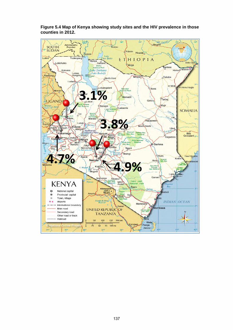
Figure 5.4 Map of Kenya showing study sites and the HIV prevalence in thosecounties in 2012.
3.1%
4.7%
3.8%
4.9%
137
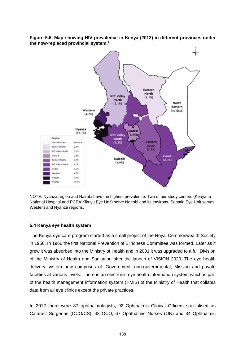
Figure 5.5. Map showing HIV prevalence in Kenya (2012) in different provinces underthe now-replaced provincial system.4
NOTE: Nyanza region and Nairobi have the highest prevalence. Two of our study centers (Kenyatta
National Hospital and PCEA Kikuyu Eye Unit) serve Nairobi and its environs. Sabatia Eye Unit serves
Western and Nyanza regions.
5.4 Kenya eye health system
The Kenya eye care program started as a small project of the Royal Commonwealth Society
in 1956. In 1966 the first National Prevention of Blindness Committee was formed. Later as it
grew it was absorbed into the Ministry of Health and in 2001 it was upgraded to a full Division
of the Ministry of Health and Sanitation after the launch of VISION 2020. The eye health
delivery system now comprises of: Government, non-governmental, Mission and private
facilities at various levels. There is an electronic eye health information system which is part
of the health management information system (HMIS) of the Ministry of Health that collates
data from all eye clinics except the private practices.
In 2012 there were 87 ophthalmologists, 92 Ophthalmic Clinical Officers specialised as
Cataract Surgeons (OCO/CS), 43 OCO, 67 Ophthalmic Nurses (ON) and 34 Ophthalmic
138

Assistants (OA).5 Eye care workers are unevenly distributed with the highest numbers being
in urban and non-arid areas. The University of Nairobi (UON) trains ophthalmologists while
the Kenya Medical Training Centre (KMTC) in Nairobi trains OCO/CS and OCO and nurses.
Training of OCOs started in 1959, Ophthalmologists in 1978 and ON in 2003. A number of
Mission hospitals and NGO clinics train OA. UON has the capacity to train 10 ophthalmologists
per year of which about 5 are from other African countries while KMTC can train 10 OCO/CS
and 16 ON per year.
The prevalence of blindness in people aged 50 years and older from a rapid assessment of
blindness study in Nakuru district of Kenya, a predominantly rural area, was 2.0% (95%CI;
1.5%-2.4%), while that of visual impairment was 5.8% (95%CI; 4.8%-6.8%).6 Cataract is the
leading cause of blindness and refractive error for MSVI. The cataract surgery rate in 2010
was 589 operations/million population/year far below the Vision 2020 goal of 3000.5 This is
attributed to lack of resources, lack of awareness by blind persons, high cost of treatment and
limited access to services.
5.5 Research Partners
5.5.1 Study centres
The study centres for this project were selected by obtaining data from the Kenya Ophthalmic
Services electronic eye health information system to show the centres that reported the
highest number of conjunctival excision between 2008 and 2011. The top 4 were:-
• Sabatia Eye Unit (219 cases/year) in the western highlands bordering Lake Victoria
300km from Nairobi and 20km from Kisumu
• PCEA Kikuyu Eye Unit (118 cases/year) about 25 kilometres (km) from Nairobi in Central
Kenya
• Homa Bay District Hospital (94 cases/year) in Nyanza on the shores of Lake Victoria
• Kitale District Hospital (83 cases/year) in the north Rift Valley 490km from Nairobi
Kenyatta National Hospital (KNH) Eye Clinic in Nairobi which did not submit data in that
period but is the national referral hospital was also chosen.
The location of the centres is shown in Figure 5.4.
Initially data collection started in PCEA Kikuyu Eye Unit and KNH in July 2012 but by October
2012 the recruitment at KNH had been rather slow due to a sudden increase in user fees in
KNH from KSh. 1500 for a conjunctival excision to KSh. 5000 earlier that year. We wrote to
the ethics committees in Nairobi and London School of Hygiene & Tropical Medicine to request
for a protocol amendment to include the other 3 centres above. We thus added Sabatia and
139
Kitale hospitals but for logistical reasons (difficult to transport pathology specimens to Nairobi)
Homa Bay district hospital was not activated.
From the foregoing description of Kenya, it is seen that the centres are located in the areas
with the largest ethnic groups and highest HIV prevalence in the country.
5.5.2 Laboratories
i) MP Shah Hospital in Nairobi - histopathology
ii) The Kenya Aids Vaccine Institute (KAVI) Institute for Clinical Research laboratory at
the University of Nairobi - HIV and CD4 testing and frozen tissue storage (serum for
vitamin A and fresh frozen tumour samples).
iii) The Tropical Diseases Research Centre’s (TDRC) Nutrition Unit in Ndola Hospital,
Zambia - vitamin A analysis.
iv) The University College London (UCL) Institute of Ophthalmology, Department of Eye
Pathology at the Moorfields Eye Hospital in London - Immunohistochemistry
v) The London School of Hygiene & Tropical Medicine - DNA extraction from tumour
specimens
vi) Genome Institute of Singapore – HPV genotyping
References
1. Kenya National Bureau of Statistics. Economic survey 2015., 2015.2. Kenya National Bureau of Statistics. The 2009 Kenya Population and Housing Census., 2010.3. National AIDS and STI Control Programme (NASCOP) Kenya. Kenya HIV estimates, 2014.4. National AIDS and STI Control Programme (NASCOP) Kenya. Kenya AIDS Indicator Survey 2012:
Final Report. Nairobi: NASCOP, 2014.5. Ministry of Health. National Strategic Plan For Eye Health and Blindness Prevention 2012-2018.
In: Ophthalmic Services Unit, editor.; 2012.6. Mathenge W, Kuper H, Limburg H, et al. Rapid assessment of avoidable blindness in Nakuru
district, Kenya. Ophthalmology 2007; 114(3): 599-605.
140

Chapter 6. Overview of project design
6.1 Research Questions
i) Are HIV infection, low CD4 lymphocyte count, HPV infection, vitamin A deficiency,
ultraviolet light exposure, cigarette smoking and occupation risk factors for OSSN?
ii) Is CD4 T lymphocyte count (in HIV positive individuals) associated with the severity of
OSSN?
iii) In patients with conjunctival tumours, what is the safety, sensitivity and specificity of
Toluidine blue dye 0.05% as a point-of-care diagnostic test for OSSN?
iv) Why do patients with OSSN present late and from their perspective what can be done to
reduce the delay?
v) Does adjuvant chemotherapy with topical 1% 5FU applied four times daily for one month
reduce the recurrence rate over a period of one year more than surgical excision alone?
6.2 Objectives
6.2.1 Broad Objectives
i) To improve the understanding of the epidemiology, aetiology and pathophysiology of
OSSN in East Africa.
ii) To develop a diagnostic algorithm based on clinical signs and vital staining of the
conjunctiva.
iii) To understand why patients with OSSN present late with advanced disease.
iv) To determine whether the recurrence of OSSN can be reduced through the use of
adjuvant topical chemotherapy.
6.2.2 Specific objectives
i) To determine if HIV infection, low CD4 lymphocyte count, HPV infection, vitamin A
deficiency, ultraviolet light exposure, cigarette smoking and occupation are risk factors for
OSSN in Kenya and describe the expression patterns of the SLRP family members.
ii) To determine the sensitivity and specificity of Toluidine blue dye 0.05% vital staining in
patients with conjunctival tumour for diagnosis of OSSN with histology as the gold
standard.
iii) To develop a diagnostic algorithm based on clinical signs and vital staining of the
conjunctiva to guide the management of possible OSSN in resource-limited settings.
141
iv) To describe the barriers to care, the care-seeking pathway of patients with OSSN and
possible solutions from the patients’ perspective.
v) To conduct a randomised controlled trial to determine the efficacy and safety of adjuvant
topical 5FU 1% eye drops applied 4 times daily for one month after surgical excision of
OSSN tumours which involve less than 2 quadrants of the conjunctiva by evaluating the
one-year recurrence rate and mean time-to-recurrence of OSSN; and both immediate and
delayed adverse effects of this therapy.
6.3 Methods/design
Participants passed through the following study sequence:
i) Case-Control Study: confirmed cases of OSSN were compared with controls. Clinical
features were recorded and various risk factors evaluated: HIV, CD4, vitamin A, HPV
types, ultraviolet exposure and occupational history. (Figure 6.1)
ii) Evaluation of Toluidine Blue (ToB) vital staining of conjunctival tumours as a point-of-care
test in the diagnosis of OSSN compared to histopathology as the gold standard.
iii) A narrative of the care-seeking pathway OSSN cases took.
iv) A randomised controlled trial to determine whether OSSN recurrence can be reduced by
adjuvant topical 5FU chemotherapy following excision. (Figure 6.2)
142
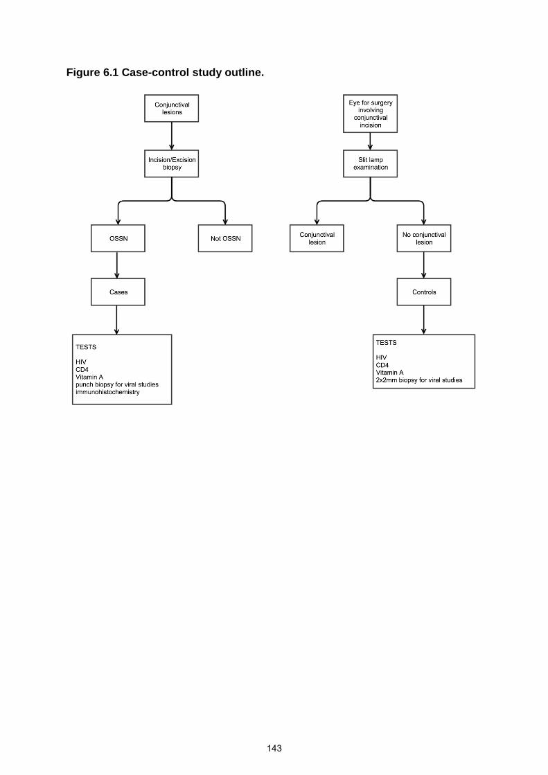
Figure 6.1 Case-control study outline.
143
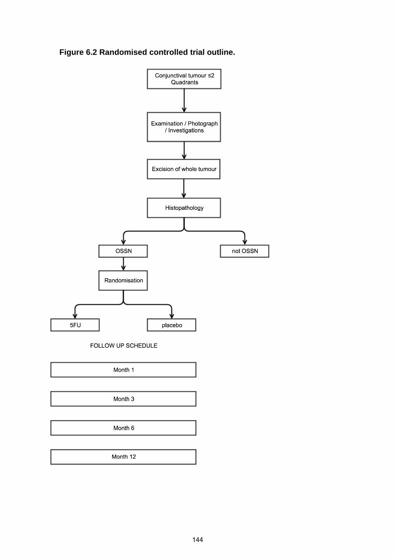
Figure 6.2 Randomised controlled trial outline.
144
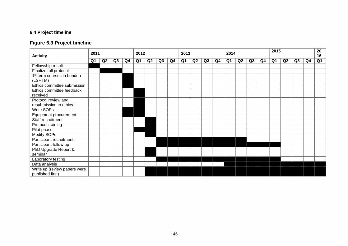
6.4 Project timeline
Figure 6.3 Project timeline
Activity2011 2012 2013 2014
2015 2016
Q1 Q2 Q3 Q4 Q1 Q2 Q3 Q4 Q1 Q2 Q3 Q4 Q1 Q2 Q3 Q4 Q1 Q2 Q3 Q4 Q1Fellowship resultFinalize full protocol1st term courses in London(LSHTM)Ethics committee submissionEthics committee feedbackreceivedProtocol review andresubmission to ethicsWrite SOPsEquipment procurementStaff recruitmentProtocol trainingPilot phaseModify SOPsParticipant recruitmentParticipant follow upPhD Upgrade Report &seminarLaboratory testingData analysisWrite up (review papers werepublished first)
145
During the project period certain untoward events occurred in Kenya that affected the timeline:
a) There were national elections held in March 2013 and by six months before that tension was
already rising high nationwide during campaigns. There was a lot of anxiety in the populace
at large related to major inter-ethnic violence that took place after the previous elections in
December 2007. People residing in areas where they were considered ‘outsiders’ migrated
back to their rural homes where they felt safer. From December 2012, about 50 or so
participants who had surgical excisions and were enrolled in the study did not return for their
results and we lost the opportunity to enrol them in the case-control study and trial. Some of
the participants who had been enrolled also missed their visits or were lost to follow up
completely.
b) In the year 2011, for example, a total of 21 industrial strikes were reported involving 13,499
employees with up to 175,329 man-days lost mainly affecting health workers, school teachers
and university academic staff.1 Nationwide industrial action by health workers from August
2011 to December 2011 and another in September 2012 paralysed services in the health
facilities from where we were receiving referrals to the study centres. There were additional
strikes by health workers and teachers in 2012 and 2013. In March 2014 teaching and non-
teaching staff in public universities went on strike. In one case, blood samples for 20 study
participants that were stored in the freezers of a district hospital laboratory neighbouring
Sabatia Eye Unit went missing and were never found again. We resorted to direct courier of
subsequent samples to Nairobi which considerably raised costs.
c) There was a sudden increase in user fees at KNH that slowed down recruitment. Time was
lost as we had to get the necessary ethical approval for additional study centres. We thus
started recruitment in Sabatia in January 2013 and in Kitale in February 2013. Recruitment in
the other centres had started about 8 months before (KNH in June 2012 and Kikuyu in July
2012).
d) The first batch of 16 tumour specimens from one centre (Kitale) arrived at the pathology
laboratory autolysed. It was suspected to be related to poorly constituted formalin which was
subsequently replaced.
References
1. Odhong E, Were S, Omolo J. Re-thinking industrial relations for enhanced organizationalperformance in Kenya. International Conference on Sustainable Research and Innovation; 2014;2014. p. 158-61.
146

Data Chapters
147

Chapter 7. Clinical presentation of ocular
surface squamous neoplasia in Kenya
148

RESEARCH PAPER COVER SHEET
PLEASE NOTE THAT A COVER SHEET MUST BE COMPLETED FOR EACH RESEARCH PAPER INCLUDEDIN A THESIS.
SECTION A – Student Details
Student
Principal Supervisor
Thesis Title
If the Research Paper has previously been published please complete Section B, if not please move toSection C
SECTION B – Paper already published
Where was the work published?
When was the work published?
If the work was published prior toregistration for your research degree,give a brief rationale for its inclusion
Have you retained the copyright for thework?* No
Was the work subject toacademic peer review? Yes
*If yes, please attach evidence of retention. If no, or if the work is being included in its published format, pleaseattach evidence of permission from the copyright holder (publisher or other author) to include this work.
SECTION C – Prepared for publication, but not yet published
Where is the work intended to bepublished?
Please list the paper’s authors in theintended authorship order:
Stage of publication Choose an item.
SECTION D – Multi-authored work
For multi-authored work, give full details of your role inthe research included in the paper and in the preparationof the paper. (Attach a further sheet if necessary)
Student Signature: Date:
Supervisor Signature: Date:
149
Gichuhi
Typewritten text
Stephen Gichuhi
Gichuhi
Typewritten text
Matthew J. Burton
Gichuhi
Typewritten text
Epidemiology and management of ocular surface squamous neoplasia in Kenya
Gichuhi
Typewritten text
4th February 2016
Gichuhi
Typewritten text
4th February 2016
Gichuhi
Typewritten text
I coordinated and actively led the field work for this paper. I identified the participants, carried out the clinical examinations and photo assessments. Field supervision was provided by Matthew Burton throughout. I also performed the statistical analysis, with some guidance from Helen Weiss. I wrote and submitted the paper with consideration of comments from co-authors.
Gichuhi
Typewritten text
JAMA Ophthalmology
Gichuhi
Typewritten text
November 2015

Permissions
Clinical Presentation of Ocular Surface Squamous Neoplasia in Kenya
Stephen Gichuhi, Ephantus Macharia, Joy Kabiru, Alain M’bongo Zindamoyen, Hilary Rono,Ernest Ollando, Leonard Wanyonyi, Joseph Wachira, Rhoda Munene, Timothy Onyuma,Mandeep S. Sagoo, Helen A. Weiss, and Matthew J. Burton
JAMA Ophthalmology. 2015; 133(11): 1305-1313.
To access the paper, press the CTRL button and at the same time click on the link below.
http://archopht.jamanetwork.com/article.aspx?articleId=2442775&guestAccessKey=7ee
32d94-5c35-463f-8f09-c864be6c6ab6
There was an invited commentary on this paper whose reference is shown below.
Ocular Surface Squamous Neoplasia: From Blue Skies to Blue Dyes-We Still Need
Our Ophthalmic Pathologists.
Shields CL, Shields JA.
JAMA Ophthalmology. 2015 Nov;133(11):1321-2.
Comment on
JAMA Ophthalmol. 2015 Nov;133(11):1305-13.
JAMA Ophthalmol. 2015 Nov;133(11):1314-21.
150
Chapter 8. Toluidine Blue 0.05% vital staining
for diagnosis of ocular surface squamous
neoplasia in Kenya
151

RESEARCH PAPER COVER SHEET
PLEASE NOTE THAT A COVER SHEET MUST BE COMPLETED FOR EACH RESEARCH PAPER INCLUDEDIN A THESIS.
SECTION A – Student Details
Student
Principal Supervisor
Thesis Title
If the Research Paper has previously been published please complete Section B, if not please move toSection C
SECTION B – Paper already published
Where was the work published?
When was the work published?
If the work was published prior toregistration for your research degree,give a brief rationale for its inclusion
Have you retained the copyright for thework?* No
Was the work subject toacademic peer review? Yes
*If yes, please attach evidence of retention. If no, or if the work is being included in its published format, pleaseattach evidence of permission from the copyright holder (publisher or other author) to include this work.
SECTION C – Prepared for publication, but not yet published
Where is the work intended to bepublished?
Please list the paper’s authors in theintended authorship order:
Stage of publication Choose an item.
SECTION D – Multi-authored work
For multi-authored work, give full details of your role inthe research included in the paper and in the preparationof the paper. (Attach a further sheet if necessary)
Student Signature: Date:
Supervisor Signature: Date:
152
Gichuhi
Typewritten text
Stephen Gichuhi
Gichuhi
Typewritten text
Matthew J. Burton
Gichuhi
Typewritten text
Epidemiology and management of ocular surface squamous neoplasia in Kenya
Gichuhi
Typewritten text
4th February 2016
Gichuhi
Typewritten text
4th February 2016
Gichuhi
Typewritten text
I coordinated and actively led the field work for this paper. I identified the participants, carried out the clinical examinations and photo assessments. Field supervision was provided by Matthew Burton throughout. I also performed the statistical analysis, with some guidance from Helen Weiss. I wrote and submitted the paper with consideration of comments from co-authors.
Gichuhi
Typewritten text
JAMA Ophthalmology
Gichuhi
Typewritten text
November 2015

Permissions
Toluidine blue 0.05% vital staining for diagnosis of ocular surface squamousneoplasia in Kenya.
Stephen Gichuhi, Ephantus Macharia, Joy Kabiru, Alain M’bongo Zindamoyen, Hilary Rono,Ernest Ollando, Leonard Wanyonyi, Joseph Wachira, Rhoda Munene, Timothy Onyuma,Walter Jaoko, Mandeep S. Sagoo, Helen A. Weiss, and Matthew J. Burton
JAMA Ophthalmology. 2015; 133(11): 1314-1321.
To access the paper, press the CTRL button and at the same time click on the link below.
http://archopht.jamanetwork.com/article.aspx?articleId=2442776&guestAccessKey=f9c
382af-d52a-401f-9478-b1dc32d80045
There was an invited commentary on this paper whose reference is shown below.
Ocular Surface Squamous Neoplasia: From Blue Skies to Blue Dyes--We Still Need
Our Ophthalmic Pathologists.
Shields CL, Shields JA.
JAMA Ophthalmology. 2015 Nov;133(11):1321-2.
Comment on
JAMA Ophthalmol. 2015 Nov;133(11):1305-13.
JAMA Ophthalmol. 2015 Nov;133(11):1314-21.
153

Chapter 9. Clinical algorithm for diagnosis and
management of ocular surface squamous
neoplasia in East Africa
154

RESEARCH PAPER COVER SHEET
PLEASE NOTE THAT A COVER SHEET MUST BE COMPLETED FOR EACH RESEARCH PAPER INCLUDEDIN A THESIS.
SECTION A – Student Details
Student
Principal Supervisor
Thesis Title
If the Research Paper has previously been published please complete Section B, if not please move toSection C
SECTION B – Paper already published
Where was the work published?
When was the work published?
If the work was published prior toregistration for your research degree,give a brief rationale for its inclusion
Have you retained the copyright for thework?* Choose an item. Was the work subject to
academic peer review?Choose anitem.
*If yes, please attach evidence of retention. If no, or if the work is being included in its published format, pleaseattach evidence of permission from the copyright holder (publisher or other author) to include this work.
SECTION C – Prepared for publication, but not yet published
Where is the work intended to bepublished?
Please list the paper’s authors in theintended authorship order:
Stage of publication Not yet submitted
SECTION D – Multi-authored work
For multi-authored work, give full details of your role inthe research included in the paper and in the preparationof the paper. (Attach a further sheet if necessary)
Student Signature: Date:
Supervisor Signature: Date:
I coordinated and actively led the field work for this paper. Supervision was provided by Matthew Burton throughout. I also performed the statistical analysis, with some guidance from Helen Weiss and David Macleod. I wrote the paper with consideration of comments from co-authors.
155
Gichuhi
Typewritten text
Stephen Gichuhi
Gichuhi
Typewritten text
Matthew J. Burton
Gichuhi
Typewritten text
Epidemiology and management of ocular surface squamous neoplasia in Kenya
Gichuhi
Typewritten text
Stephen Gichuhi, Ephantus Macharia, Joy Kabiru, Alain Mbongo Zindamoyen, Hilary Rono, Ernest Ollando, Joseph Wachira, Rhoda Munene, Timothy Onyuma, Mandeep S. Sagoo, David Macleod,Helen A. Weiss, and Matthew J. Burton
Gichuhi
Typewritten text
4th February 2016
Gichuhi
Typewritten text
4th February 2016
Gichuhi
Typewritten text
British Journal of Ophthalmology

Clinical algorithm for diagnosis of Ocular Surface Squamous Neoplasia
(OSSN) in East Africa
Corresponding Author:Stephen GichuhiInternational Center for Eye HealthLondon School of Hygiene & Tropical MedicineKeppel Street, London WC1E 7HTUnited KingdomTel. +44 (20) 7636 8636 ext. [email protected]
Authors:Stephen Gichuhi MBChB, M.Med 1, 2, Joy Kabiru MBChB, M.Med 3, Alain M’bongo Zindamoyen MBChB,M.Med 3, Hilary Rono MBChB, M.Med 4, Ernest Ollando MBChB, M.Med 5, Joseph Wachira MBChB, M.Med 6,Rhoda Munene MBChB, M.Med 6, Timothy Onyuma MBChB, M.Med 7, Mandeep S. Sagoo PhD, FRCOphth 8,
9, 10, David Macleod MSc1, Helen A. Weiss MSc, PhD1,11 and Matthew J. Burton PhD, FRCOphth1, 9
Email contactsStephen Gichuhi, [email protected] Kabiru, [email protected] M’bongo Zindamoyen, [email protected] Rono, [email protected] Ollando, [email protected] Wachira, [email protected] Munene, [email protected] Onyuma, [email protected] S. Sagoo, [email protected] Macleod [email protected] A. Weiss, [email protected] J. Burton, [email protected]
Affiliations:1International Centre for Eye Health, London School of Hygiene and Tropical Medicine, Keppel Street,London, WC1E 7HT, UK2Department of Ophthalmology, University of Nairobi, P.O Box 19676-00202, Nairobi, Kenya3PCEA Kikuyu Eye Unit, PO Box 45, Kikuyu, Kenya4Kitale District Hospital, PO Box 98-30200, Kitale, Kenya5Sabatia Eye Hospital, PO Box 214-50311, Wodanga, Kenya6Kenyatta National Hospital, PO Box 20723-00202, Nairobi, Kenya7MP Shah Hospital, Department of Pathology, PO Box 14497-00800, Nairobi, Kenya8UCL Institute of Ophthalmology, 11-43 Bath Street, London EC1V 9EL, UK9Moorfields Eye Hospital, 162 City Road, London, EC1V 2PD, UK10St. Bartholomew’s Hospital, W Smithfield, London EC1A 7BE, London, UK11 MRC Tropical Epidemiology Group, London School of Hygiene and Tropical Medicine, Keppel Street,London, WC1E 7HT,
Subtitle:Diagnostic algorithm for OSSN
Key words:Algorithm;DiagnosisOcular surface squamous neoplasiaEast Africa
Word count: text 2534; abstract 250
156
Abstract
Background
Clinical features of Ocular Surface Squamous Neoplasia (OSSN) and benign conjunctival lesions
overlap. Toluidine blue (ToB) 0.05% vital staining of OSSN has a high sensitivity (92%) but low
specificity (31%). This study was conducted to determine whether an algorithm combining clinical
features and vital staining increased the specificity.
Methods
Clinical features of participants presenting with conjunctival lesions in Kenya were evaluated and
lesions excised for histopathology to confirm the diagnosis. Participants were randomly divided into
two sets: “development” and “validation” sets. Using the development set, clinical criteria that had
the strongest association with OSSN were analysed using logistic regression, a specificity-based
and sensitivity-based model. Using the best combination of features in the three models, receiver
operator characteristic curves were plotted, cut-off determined, best model chosen and repeated in
the validation set. An algorithm showing the probability of OSSN with features at the cut-off was
developed.
Results
Out of 496 participants OSSN was confirmed in 187 (38%). The regression-based model had the
highest area under the curve (79.8%). The best cut-off was using 3 out of 8 features; prior excision,
corneal involvement, feeder vessels, dark blue ToB staining, papillary or gelatinous tumour surface,
severe inflammation, anti-retroviral therapy and temporal or circumlimbal tumours. At this cut-off
sensitivity was 89%, specificity 50%, and 65% of lesions were correctly classified. This specificity
was higher than any blue ToB staining (31%) but lower than clinical photo-examination (60%).
Conclusion
This algorithm may assist clinicians decide about the need for surgery but cannot replace
histopathology.
157

Introduction
Ocular surface squamous neoplasia (OSSN) is a relatively common, aggressive disease in Africa.
Compared to other parts of the world, it more frequently affects younger adults, particularly those
living with HIV, and has a higher incidence in women.1 Late presentation with large tumours is not
infrequent and the outlook of treatment limited by high recurrence rates.2-4 The gold standard for
diagnosis of OSSN remains histopathology.5 However most practitioners in Africa depend on their
clinical impression and lesions are often excised without histopathology due to a shortage of
pathologists, logistical problems of sending the specimens to distant histopathology service centres
and the added cost thereof.6 7 This means that it is difficult to know what the lesion was exactly and
whether the margins were involved or not. In some countries without such resource constraints,
treatment decisions are also being made without an excision biopsy for histopathology being
performed.8 Studies from East Africa have found an overlap in the clinical features of OSSN and
benign lesions, making it difficult to distinguish between them.9 10 In addition, different histological
grades of OSSN show overlapping phenotypes.9 10 Clinical examination of a series of conjunctival
lesions from photographs had a positive predictive value of 54%.9
We have previously reported the use of Toluidine blue (ToB) 0.05% vital staining in Kenya to assist
in the diagnosis of OSSN and found that although any blue staining was sensitive for OSSN (92%)
it lacked specificity (31%).11 The test was however easily interpreted by different examiners
(kappa=0.76). Dark royal blue staining had a sensitivity of 64% and specificity of 67%. Other studies
have reported that vital staining for OSSN has high sensitivity and relatively low specificity. In Brazil,
toluidine blue 1% had high sensitivity of 100% and a specificity of 50% while in South Africa
methylene blue 1% had a sensitivity of 92% and a specificity of 50%.12 13 Therefore, vital staining
alone cannot currently be relied upon to guide treatment decisions.
In the African setting a clinical diagnostic algorithm may help support the clinician’s impression of
OSSN to distinguish lesions that should be excised from those that can be followed up. We
conducted this study to develop an algorithm that combined ToB staining with other clinical features.
The key qualities sought were: simplicity, for clinicians to use easily; specificity, as we have a test
with high sensitivity already (any blue ToB stain) but it has an unacceptably low specificity (31%);
sensitivity (using dark blue ToB stain has a higher specificity of 67% but the sensitivity is only 64%).11
Methods
Ethical Approval
This study was approved by the Kenyatta National Hospital – University of Nairobi Ethics and
Research Committee and the London School of Hygiene & Tropical Medicine Ethics Committee. It
158

adhered to the tenets of the Declaration of Helsinki. All participants gave informed written consent
to take part.
Study population
The participants were part of an integrated set of studies on the epidemiology and management of
OSSN in Kenya. They were recruited between July 2012 and July 2014 in four eye care centres:
Kenyatta National Hospital in Nairobi, PCEA Kikuyu Eye Unit about 25 kilometres (km) from Nairobi
in Central Kenya, Kitale district hospital in the north Rift Valley 490km from Nairobi and Sabatia Eye
Hospital 300km from Nairobi in western Kenya near Lake Victoria. Adult patients (≥18 years)
presenting to these clinics with any conjunctival lesion (first presentation or a recurrence) and
scheduled for surgery were recruited.
Participant examination methods have been published elsewhere.9 Briefly, a comprehensive history
was taken, clinical examination at the slit lamp was performed to record features of the lesions, vital
staining done with ToB 0.05% and digital photographs of the lesions taken before and after staining.11
Lesions were excised and sent for histopathology. One pathologist examined all the histology slides.
OSSN was defined as any lesion with mild, moderate or severe conjunctival intraepithelial neoplasia
(CIN I, II, III respectively); any who had carcinoma-in-situ (CIS); or invasive squamous cell
carcinoma. Benign lesions included pterygium, actinic keratosis, papillomas, pyogenic granulomas,
nevi and rhinosporidiosis. During the post-operative visit about 3-4 weeks later, participants found to
have OSSN were offered HIV testing and a CD4 count if positive.
Generation of development and validation datasets
The original dataset, consisting of observations of both OSSN and benign lesions, was split into two
datasets, one for the purposes of developing the diagnostic test and the other for validating it. This
was done by setting a “seed” (in order to make the random allocation reproducible). Each individual
was allocated a random number between 0 and 1. They were then sorted by this random number
and split into a development set (consisting of 75% of the participants) and a validation set
(consisting of the remaining 25%). This, and all other statistical analysis, was performed using Stata
version 12.1 (StataCorp, College Station, Texas, USA).
Model construction and validation
Using the development dataset, univariable analysis was conducted to find which features were
associated with OSSN. Multivariable logistic regression analysis was used to estimate odds ratios
(ORs) and 95% confidence intervals (CIs). The likelihood ratio test was used to assess statistical
significance of associations. Variables that were associated with OSSN in the univariable analyses
at a level of p<0.05 were included in the multivariable analysis and then those with p<0.2 were
retained in the model. We took the features selected by the logistic regression model and for each
159
patient created a “diagnostic score” which was the number of features observed. We then plotted a
ROC curve to identify the most appropriate “cut-off” to classify a lesion as OSSN or not.
In addition to the regression-based model, specificity-based and sensitivity-based models were also
developed. The specificity-based model which aimed to reduce the proportion of false positives was
developed using a similar method to a Zimbabwe study that created an algorithm for verbal autopsy
of HIV.14 The positive likelihood ratio (LR) for a range of features from the history and clinical
examination findings was calculated in the development dataset to examine their strength of
association with OSSN. Features with a LR ≥1.92 (corresponding to a significant chi-squared test on
1 degree of freedom at p<0.05) were retained for further analysis. The selected features were then
ordered from the highest specificity to lowest and the following steps followed:
1) Take the feature with the highest specificity
2) Classify all observations where this feature is present as OSSN+
3) Calculate sensitivity and specificity of diagnostic test using this feature
4) Recalculate the specificity of all remaining features using only the observations not
classified as OSSN+
5) Take the feature with the highest specificity in the remaining list of variables
6) Classify all observations where this feature is present as OSSN+
7) Calculate sensitivity and specificity of diagnostic test
This iterative process was repeated until all the variables had been incorporated. For the sensitivity-
based model, the same method was used except that the selection of features to be included was
based on the highest sensitivity. These features from the specificity-based and sensitivity-based
models were then applied to the development dataset and used to plot a modified receiver operator
characteristic (ROC) curve to visualise the effect that adding each feature had on the sensitivity and
specificity and to choose a cut-off point which optimised the two. We modified the typical ROC curve,
which shows different cut-offs for the same parameter, usually a continuous variable, by creating an
ordered categorical variable in which different categories consisted of various combinations of
features. For example, category 1=feature A, 2= feature A + feature B, 3= feature A + feature B +
feature C and so on.
The results of the models from the development set were then applied to the validation set and ROC
curves plotted. To choose the best model among the three, the ROC that was closest to the upper
left hand corner of the plot was used. The cut-off value from the ROC curve was also selected by
choosing the point closest to the upper left hand-corner of the plot and specificity above 50%.
160
Algorithm development
Finally, an algorithm was built from the development dataset as a probability tree using identified
features from the best model, in the case of the regression model starting with the ones with the
highest odds ratios.
Results
There were 496 participants with conjunctival lesions. The development dataset included 372
participants and validation dataset 124. Histologically confirmed OSSN was present in 187 (38%)
participants: 139/372 (37%) of the development dataset and 48/124 (39%) of the validation dataset.
Model Development
The features from the history, clinical examination and laboratory testing that were evaluated are
listed in the Supplementary Table. Those that were associated with OSSN at the 5% significance
level on univariate analysis are shown in Table 9.1. In the multivariable logistic regression model
eight variables were considered significant predictors of OSSN (Table 9.2).
The three ROC curves (regression, sensitivity and specificity models) generated from the
development dataset are shown in Figure 9.1. The best cut-off for the specificity-based model was
the presence of any one of the following features; symblepharon, fornix involvement, history of prior
excision, anti-retroviral therapy (ART) use, severe inflammation or dark ToB vital stain. The best cut-
off for the sensitivity-based model was the presence of a papillary-gelatinous surface. For the
regression-based model, the best cut-off was the presence of any 3 of the 8 features listed in Table
9.2. With this cut-off in the regression model the sensitivity was 89% and specificity 50%. The curve
from the regression-based model had the highest area under the curve (79.8% vs 77.9% for the
specificity-based and 58.0% for the sensitivity-based).
Model Validation
The ROC curves from application of the three models to the validation dataset are shown in the
Supplementary Figure. The curve from the regression-based model had the highest area under the
curve (73% vs specificity 57% and sensitivity 58%). The cut-off giving the point closest to the top left
of the plot was 3, which means those individuals with 3 or more of the 8 selected features are
classified as OSSN. Using 2 features as a cut-off had a high sensitivity (94%) but low specificity
(26%) while 4 features, though with a higher specificity (74%), had a low sensitivity (56%), making 3
the best compromise. With 3 features the sensitivity was 83% and specificity 54%. The odds ratios
for many of the variables in Table 9.2 were not statistically significant in the validation dataset
because of sample size limitations.
161

A comparison of the performance, using the validation dataset, of the three models with ToB staining
and clinical assessment from photographs is shown in Table 9.3. The regression-based model was
more accurate (65%) than the specificity (57%) or sensitivity (50%) models. It offered the best
compromise with false positives at 46% (instead of 69% with any blue staining) and false negatives
at 17% (twice those of any blue staining but dark blue staining had four times as many false negatives
at 36%). Clinical photograph examination had a higher sensitivity (86% vs 83%), specificity (60% vs
50%) and accuracy (70% vs 65%) than the regression model.
Diagnostic Algorithm
Applying this to the four clinical features with the highest odds ratios from the multivariable regression
model we constructed the diagnostic probability tree shown in Figure 9.2 to show the probability of
OSSN in an individual patient with specific combinations of features in this dataset.
Discussion
Earlier studies from the East African region have found that the diagnosis of OSSN on clinical
grounds can be unreliable, due to an overlap in clinical features of OSSN with other ocular surface
lesions.9 In addition, we have previously reported the use of toluidine blue vital staining, which we
found to have a high sensitivity but low specificity.11 In this study we developed and evaluated several
models to try to identify a combination of variables that most reliably distinguishes OSSN from other
pathology.
We found that a simple logistic regression model, with a classifier threshold of 3 or more out of 8
features associated with OSSN from history, clinical examination and ToB vital staining provided the
best balance (of the options tested) between reasonable sensitivity (89%) and improved specificity
(50%) over using ToB alone.11 The sensitivity and specificity in the regression model compares well
with the quoted values in the clinical features we previously reported (86% sensitivity and 60%
specificity) although we do not know which specific features the examiners were using or how they
interpreted them when looking at the photographs.9 A high sensitivity is a critical requirement when
dealing with a serious disease like OSSN. However, the moderate specificity remains problematic,
as this is likely to result in more patients having surgery for suspected OSSN who turn out not to
have this disease.
The probability tree algorithm is a simple tool that clinicians may also find helpful in assessing
patients with ocular surface lesions. Tracing the presence or absence of certain features leads to a
probability score for the diagnosis of OSSN. However, it should be emphasised that this algorithm
pertains to this specific dataset. It is not a substitute for careful clinical assessment and judgment,
162

which should continue to form the basis for decisions around the need for surgical excision or other
treatment.
Formal diagnostic algorithms have not previously been evaluated in the management of OSSN,
although it seems likely that most clinicians implicitly go through a similar process when forming a
clinical impression. The results of our previous study in Kenya found that individual clinical features
were not specific for OSSN, being found in variable proportions in malignant and benign lesions.9
However, when clinicians were asked to provide an overall categorisation of OSSN or not OSSN
their sensitivity and specificity were similar to the performance of the model we describe here. This
suggests that clinicians who are experienced in examining such patients are integrating the
information provided by the combination of clinical features to draw similar conclusions about the
nature of the pathology.
This study had several limitations. Firstly, as with many diagnostic tests, there is a trade-off between
sensitivity and specificity. Secondly, it can be difficult to standardize the assessment of some clinical
features such as the degree of inflammation. Although histopathology is the gold standard for
diagnosis it is still open to individual interpretation. Some non-OSSN lesions such as pterygia and
actinic keratosis have been shown in several studies to share some histological and etiological
features with OSSN.15-17 Extrapolation of these results or direct comparison with other populations
of OSSN patients requires caution as the pattern of disease in this population appears quite distinct
from that seen in more temperate regions.
In conclusion, ToB staining is less specific and predictive than clinical examination by an
Ophthalmologist but it may be useful for other health care workers screening patients for the disease,
and when there is no ToB staining, to reassure that the disease is unlikely to be malignant. We
developed a simple model with various combinations of clinical features that could identify a high
proportion of OSSN cases and give an indication of the probability of disease. This could be used to
estimate the risk of OSSN. However, it is context-specific and given the clinical overlap between
OSSN and non-OSSN lesion, this should not be regarded as a substitute for histological assessment.
163
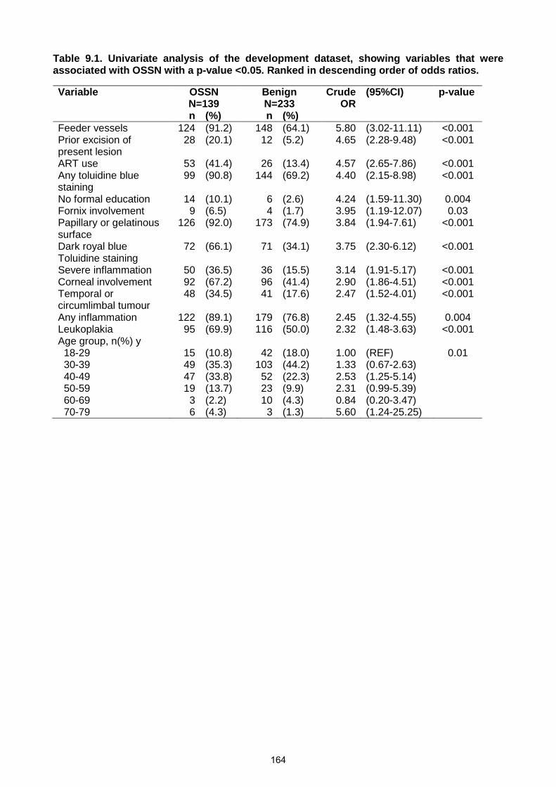
Table 9.1. Univariate analysis of the development dataset, showing variables that wereassociated with OSSN with a p-value <0.05. Ranked in descending order of odds ratios.
Variable OSSNN=139
BenignN=233
CrudeOR
(95%CI) p-value
n (%) n (%)Feeder vessels 124 (91.2) 148 (64.1) 5.80 (3.02-11.11) <0.001Prior excision ofpresent lesion
28 (20.1) 12 (5.2) 4.65 (2.28-9.48) <0.001
ART use 53 (41.4) 26 (13.4) 4.57 (2.65-7.86) <0.001Any toluidine bluestaining
99 (90.8) 144 (69.2) 4.40 (2.15-8.98) <0.001
No formal education 14 (10.1) 6 (2.6) 4.24 (1.59-11.30) 0.004Fornix involvement 9 (6.5) 4 (1.7) 3.95 (1.19-12.07) 0.03Papillary or gelatinoussurface
126 (92.0) 173 (74.9) 3.84 (1.94-7.61) <0.001
Dark royal blueToluidine staining
72 (66.1) 71 (34.1) 3.75 (2.30-6.12) <0.001
Severe inflammation 50 (36.5) 36 (15.5) 3.14 (1.91-5.17) <0.001Corneal involvement 92 (67.2) 96 (41.4) 2.90 (1.86-4.51) <0.001Temporal orcircumlimbal tumour
48 (34.5) 41 (17.6) 2.47 (1.52-4.01) <0.001
Any inflammation 122 (89.1) 179 (76.8) 2.45 (1.32-4.55) 0.004Leukoplakia 95 (69.9) 116 (50.0) 2.32 (1.48-3.63) <0.001Age group, n(%) y
18-29 15 (10.8) 42 (18.0) 1.00 (REF) 0.0130-39 49 (35.3) 103 (44.2) 1.33 (0.67-2.63)40-49 47 (33.8) 52 (22.3) 2.53 (1.25-5.14)50-59 19 (13.7) 23 (9.9) 2.31 (0.99-5.39)60-69 3 (2.2) 10 (4.3) 0.84 (0.20-3.47)70-79 6 (4.3) 3 (1.3) 5.60 (1.24-25.25)
164

Table 2. Multivariable logistic regression analysis of predictors of OSSN identified using the development dataset and applied to thevalidation dataset.
Variable Development set (N=372) Validation set (N=124)Adjusted OR (95%CI) p-value Adjusted OR (95%CI) p-value
Prior excision of the present lesion 4.51 (1.64-12.42) 0.004 0.55 (0.09-3.23) 0.51Corneal involvement 3.66 (1.93-6.95) <0.001 3.28 (1.13-9.55) 0.03Feeder vessels 3.38 (1.32-8.62) 0.01 2.57 (0.75-8.79) 0.13Dark royal blue Toluidine staining 3.08 (1.61-5.88) 0.001 2.56 (0.92-7.14) 0.07Papillary or gelatinous tumour surface 2.98 (1.09-8.09) 0.03 1.57 (0.46-5.39) 0.47Severe inflammation 2.91 (1.39-6.10) 0.005 1.20 (0.40-3.63) 0.74ART use 2.32 (1.10-4.89) 0.03 1.27 (0.41-3.91) 0.67Temporal or circumlimbal tumour 2.21 (1.09-4.45) 0.03 1.84 (0.55-6.13) 0.32
165

Figure 9.1. Receiver operator characteristic (ROC) curves plotted from the three models using the
development dataset: (A) Specificity-based model; (B) Sensitivity-based model; (C) Regression-based
model. The area under the curve is shown under each plot eg 0.7792=77.92%. The cut-off point is the point
at which we make the OSSN vs non-OSSN distinction. Read the curves from the origin of the x and y axis. For
instance in (A) the options are using symblepharon alone, then the next is using symblepharon or fornix, then
symblepharon or fornix or prior excision and so on such that the last point refers to a cut-off point where we
use any one of symblepharon or fornix involvement or prior excision or ART use or severe inflammation or
dark ToB staining. For (B) the cut-off point was papillary/gelatinous surface. For (C) similarly we read it from
the origin and use either all 8 features listed in Table 9.2, or 7 features or 6 features and so forth with the last
option being the use of only 1 feature. The cut off was any 3 of the 8 features listed in Table 9.2.
(A) Specificity-based model
dark ToB stain
severe inflammation
ART use
prior excision
fornix
symblepharon
0.0
00.2
50.5
00.7
51.0
0S
en
sitiv
ity
0.00 0.25 0.50 0.75 1.001 - Specificity
Area under ROC curve = 0.7792
166
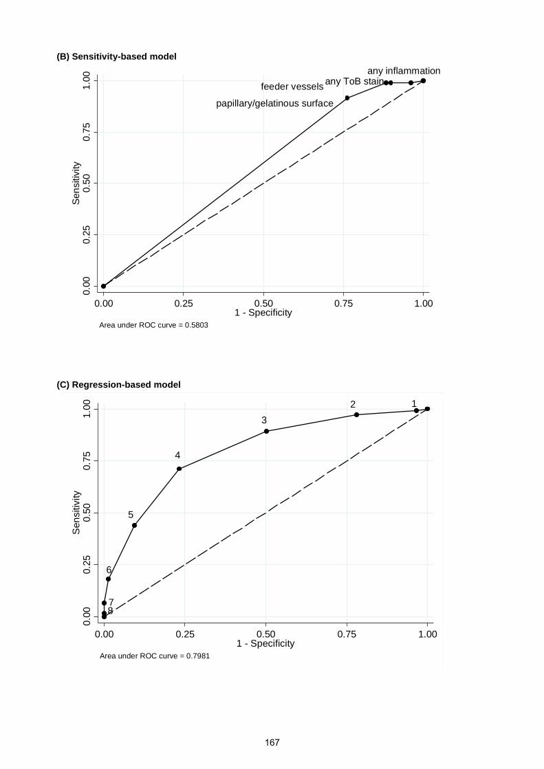
(B) Sensitivity-based model
(C) Regression-based model
any ToB stainfeeder vessels
papillary/gelatinous surface
0.0
00.2
50.5
00.7
51.0
0S
en
sitivity
0.00 0.25 0.50 0.75 1.001 - Specificity
Area under ROC curve = 0.5803
any inflammation
12
3
4
5
6
78
0.0
00.2
50.5
00.7
51.0
0S
en
siti
vity
0.00 0.25 0.50 0.75 1.001 - Specificity
Area under ROC curve = 0.7981
167

Table 9.3. Performance of different diagnostic tools for diagnosing OSSN in the validation dataset and Toluidine blue data and photographexamination from a previous study.9 11
Diagnostic tool Sensitivity Specificity PV+ PV- Correctlyclassified
F+rate
F-rate
LR+ LR-
Toluidine blue (any blue) 92% 31% 41% 88% 52% 69% 8% 1.33 0.26Toluidine blue (dark royal blue) 64% 67% 50% 78% 66% 33% 36% 1.95 0.53Clinical examination of photographsa 86% 60% 54% 88% 70% 40% 14% 2.15 0.23Regression based modelb 83% 54% 53% 84% 65% 46% 17% 1.81 0.31Specificity based modelb 75% 45% 47% 73% 57% 55% 25% 1.38 0.55Sensitivity based modelb 85% 31% 44% 77% 50% 69% 15% 1.22 0.50
NOTE: Abbreviations; PV means predictive value, F+ means false positive, F- means false negative, LR means likelihood ratioa This was the median score of 6 examiners looking at lesion photos without vital stainingb This is at the best cut-offs shown in the ROC curves we plotted.
168
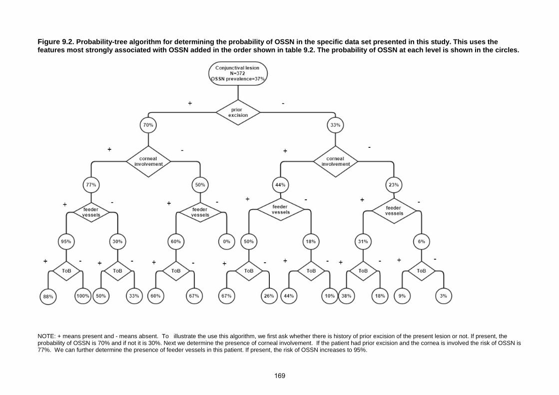
Figure 9.2. Probability-tree algorithm for determining the probability of OSSN in the specific data set presented in this study. This uses thefeatures most strongly associated with OSSN added in the order shown in table 9.2. The probability of OSSN at each level is shown in the circles.
NOTE: + means present and - means absent. To illustrate the use this algorithm, we first ask whether there is history of prior excision of the present lesion or not. If present, theprobability of OSSN is 70% and if not it is 30%. Next we determine the presence of corneal involvement. If the patient had prior excision and the cornea is involved the risk of OSSN is77%. We can further determine the presence of feeder vessels in this patient. If present, the risk of OSSN increases to 95%.
169
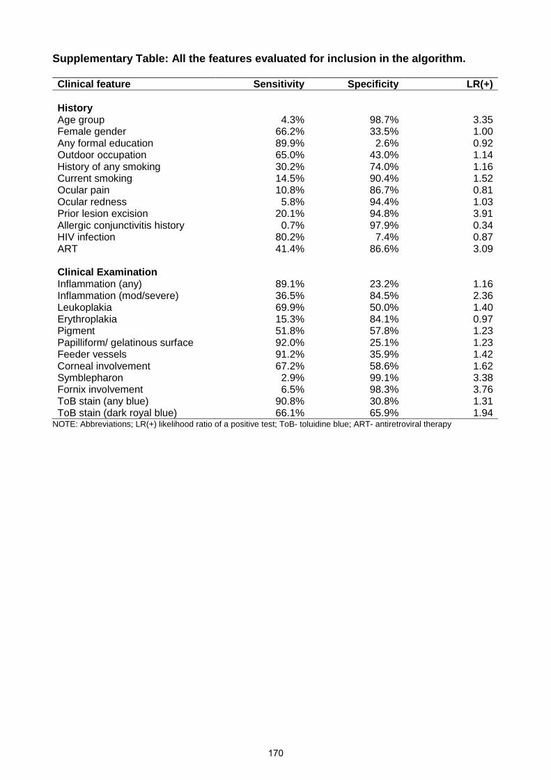
Supplementary Table: All the features evaluated for inclusion in the algorithm.
Clinical feature Sensitivity Specificity LR(+)
HistoryAge group 4.3% 98.7% 3.35Female gender 66.2% 33.5% 1.00Any formal education 89.9% 2.6% 0.92Outdoor occupation 65.0% 43.0% 1.14History of any smoking 30.2% 74.0% 1.16Current smoking 14.5% 90.4% 1.52Ocular pain 10.8% 86.7% 0.81Ocular redness 5.8% 94.4% 1.03Prior lesion excision 20.1% 94.8% 3.91Allergic conjunctivitis history 0.7% 97.9% 0.34HIV infection 80.2% 7.4% 0.87ART 41.4% 86.6% 3.09
Clinical ExaminationInflammation (any) 89.1% 23.2% 1.16Inflammation (mod/severe) 36.5% 84.5% 2.36Leukoplakia 69.9% 50.0% 1.40Erythroplakia 15.3% 84.1% 0.97Pigment 51.8% 57.8% 1.23Papilliform/ gelatinous surface 92.0% 25.1% 1.23Feeder vessels 91.2% 35.9% 1.42Corneal involvement 67.2% 58.6% 1.62Symblepharon 2.9% 99.1% 3.38Fornix involvement 6.5% 98.3% 3.76ToB stain (any blue) 90.8% 30.8% 1.31ToB stain (dark royal blue) 66.1% 65.9% 1.94
NOTE: Abbreviations; LR(+) likelihood ratio of a positive test; ToB- toluidine blue; ART- antiretroviral therapy
170

Supplementary Figure: Receiver operator characteristic (ROC) curves plotted from the threemodels using the validation dataset: (A) Specificity-based model; (B) Sensitivity-basedmodel; (C) Regression-based model.LEGEND: The area under the curve is shown under each plot eg 0.7792=77.92%. The cut-off point is the point at whichwe make the OSSN vs non-OSSN distinction. Read the curves from the origin of the x and y axis. For instance in A) theoptions are using symblepharon alone, then the next is using symblepharon or fornix, then symblepharon or fornix or priorexcision and so on such that the last point refers to a cut-off point where we use any one of symblepharon or fornixinvolvement or prior excision or ART use or severe inflammation or dark ToB staining.For (B) the cut-off point was papillary/gelatinous surfaceFor (C) similarly we read it from the origin and use either all 8 features listed in table 9.2 or 7 or 5 and so forth with the
last option being the use of only 1 feature. it was any 3 of the 8 features listed in table 9.2.
(A) Specificity-based model
(B) Sensitivity-based model
symblepharon
fornix
prior excision
ART use
severe inflammation
dark ToB stain
0.0
00.2
50.5
00.7
51.0
0S
en
sitiv
ity
0.00 0.25 0.50 0.75 1.001 - Specificity
Area under ROC curve = 0.5705
171
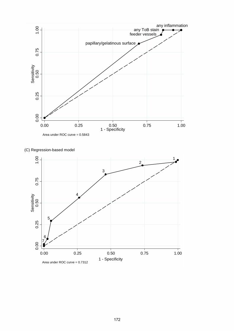
(C) Regression-based model
papillary/gelatinous surface
feeder vesselsany ToB stain
0.0
00.2
50.5
00.7
51.0
0S
en
siti
vity
0.00 0.25 0.50 0.75 1.001 - Specificity
Area under ROC curve = 0.5843
any inflammation
12
3
4
5
67
0.0
00.2
50.5
00.7
51.0
0S
en
siti
vity
0.00 0.25 0.50 0.75 1.00
1 - SpecificityArea under ROC curve = 0.7312
172

Competing interests
The authors have no conflict of interest disclosures. All authors have completed and submitted the
ICMJE Form for Disclosure of Potential Conflicts of Interest and none were reported.
Financial support
SG received funding from the British Council for Prevention of Blindness (BCPB) fellowship
programme. MJB is supported by The Wellcome Trust (Grant Number 098481/Z/12/Z). HAW and
DM are supported by the MRC and DFID (Grant Number G0700837). The funding organizations had
no role in the design or conduct of this research.
173
References
1. Gichuhi S, Sagoo MS, Weiss HA, et al. Epidemiology of ocular surface squamous neoplasia inAfrica. Tropical medicine & international health : TM & IH 2013;18(12):1424-43.
2. Chebbi A, Bouguila H, Boukari M, et al. [Prognosis of primary malignant tumors of theconjunctiva]. Journal francais d'ophtalmologie 2015;38(6):477-85.
3. Bonanno A, Esmaeli B, Fingeret MC, et al. Social challenges of cancer patients with orbitofacialdisfigurement. Ophthalmic plastic and reconstructive surgery 2010;26(1):18-22.
4. Birkholz ES, Goins KM, Sutphin JE, et al. Treatment of ocular surface squamous cellintraepithelial neoplasia with and without mitomycin C. Cornea 2011;30(1):37-41.
5. Shields CL, Shields JA. Ocular Surface Squamous Neoplasia: From Blue Skies to Blue Dyes-We StillNeed Our Ophthalmic Pathologists. JAMA ophthalmology 2015:1-2.
6. Adesina A, Chumba D, Nelson AM, et al. Improvement of pathology in sub-Saharan Africa. Thelancet oncology 2013;14(4):e152-7.
7. Rambau PF. Pathology practice in a resource-poor setting: Mwanza, Tanzania. Archives ofpathology & laboratory medicine 2011;135(2):191-3.
8. Adler E, Turner JR, Stone DU. Ocular surface squamous neoplasia: a survey of changes in thestandard of care from 2003 to 2012. Cornea 2013;32(12):1558-61.
9. Gichuhi S, Macharia E, Kabiru J, et al. Clinical Presentation of Ocular Surface Squamous Neoplasiain Kenya. JAMA ophthalmology 2015;133(11):1305-13.
10. Nguena MB, van den Tweel JG, Makupa W, et al. Diagnosing ocular surface squamous neoplasiain East Africa: case-control study of clinical and in vivo confocal microscopy assessment.Ophthalmology 2014;121(2):484-91.
11. Gichuhi S, Macharia E, Kabiru J, et al. Toluidine Blue 0.05% Vital Staining for the Diagnosis ofOcular Surface Squamous Neoplasia in Kenya. JAMA ophthalmology 2015;133(11):1314-21.
12. Romero IL, Barros Jde N, Martins MC, et al. The use of 1% toluidine blue eye drops in thediagnosis of ocular surface squamous neoplasia. Cornea 2013;32(1):36-9.
13. Steffen J, Rice J, Lecuona K, et al. Identification of ocular surface squamous neoplasia by in vivostaining with methylene blue. The British journal of ophthalmology 2014;98(1):13-5.
14. Lopman BA, Barnabas RV, Boerma JT, et al. Creating and validating an algorithm to measureAIDS mortality in the adult population using verbal autopsy. PLoS Med 2006;3(8):e312.
15. Di Girolamo N. Association of human papilloma virus with pterygia and ocular-surfacesquamous neoplasia. Eye (Lond) 2012;26(2):202-11.
16. Dushku N, Hatcher SL, Albert DM, et al. p53 expression and relation to human papillomavirusinfection in pingueculae, pterygia, and limbal tumors. Archives of ophthalmology1999;117(12):1593-9.
17. Hirst LW, Axelsen RA, Schwab I. Pterygium and associated ocular surface squamous neoplasia.Archives of ophthalmology 2009;127(1):31-2.
174

Chapter 10. Risk factors for ocular surface
squamous neoplasia in Kenya
175

RESEARCH PAPER COVER SHEET
PLEASE NOTE THAT A COVER SHEET MUST BE COMPLETED FOR EACH RESEARCH PAPER INCLUDEDIN A THESIS.
SECTION A – Student Details
Student
Principal Supervisor
Thesis Title
If the Research Paper has previously been published please complete Section B, if not please move toSection C
SECTION B – Paper already published
Where was the work published?
When was the work published?
If the work was published prior toregistration for your research degree,give a brief rationale for its inclusion
Have you retained the copyright for thework?* Choose an item. Was the work subject to
academic peer review?Choose anitem.
*If yes, please attach evidence of retention. If no, or if the work is being included in its published format, pleaseattach evidence of permission from the copyright holder (publisher or other author) to include this work.
SECTION C – Prepared for publication, but not yet published
Where is the work intended to bepublished?
Please list the paper’s authors in theintended authorship order:
Stage of publication Not yet submitted
SECTION D – Multi-authored work
For multi-authored work, give full details of your role inthe research included in the paper and in the preparationof the paper. (Attach a further sheet if necessary)
Student Signature: Date:
Supervisor Signature: Date:
I coordinated and actively led the field work for this paper.Supervision was provided by Matthew Burton throughout.I also performed the statistical analysis, with some guidancefrom Helen Weiss.I wrote the paper with consideration of comments from co-authors.
176
Gichuhi
Typewritten text
Stephen Gichuhi
Gichuhi
Typewritten text
Matthew J. Burton
Gichuhi
Typewritten text
Epidemiology and management of ocular surface squamous neoplasia in Kenya
Gichuhi
Typewritten text
Tropical Medicine & International Health
Gichuhi
Typewritten text
Stephen Gichuhi, Ephantus Macharia, Joy Kabiru, Alain Mbongo Zindamoyen, Hilary Rono, Ernest Ollando, Joseph Wachira, Rhoda Munene, Timothy Onyuma, Walter G. Jaoko, Mandeep S. Sagoo, Helen A. Weiss, and Matthew J. Burton
Gichuhi
Typewritten text
4th February 2016
Gichuhi
Typewritten text
4th February 2016
Risk factors for Ocular Surface Squamous Neoplasia in Kenya; acase-control study
Authors:Stephen Gichuhi MBChB, M.Med 1, 2, Ephantus Macharia HND3, Joy Kabiru MBChB, M.Med3, Alain M’bongo Zindamoyen MBChB, M.Med 3, Hilary Rono MBChB, M.Med 4, ErnestOllando MBChB, M.Med 5, Joseph Wachira MBChB, M.Med 6, Rhoda Munene MBChB,M.Med 6, Timothy Onyuma MBChB, M.Med 7, Walter G. Jaoko MBChB, PhD 8, Mandeep S.Sagoo PhD, FRCOphth 9, 10, 11, Helen A. Weiss MSc, PhD1,12 and Matthew J. Burton PhD,FRCOphth1, 10
Email contactsStephen Gichuhi, [email protected] Macharia, [email protected] Kabiru, [email protected] M’bongo Zindamoyen, [email protected] Rono, [email protected] Ollando, [email protected] Wachira, [email protected] Munene, [email protected] Onyuma, [email protected] G. Jaoko, [email protected] S. Sagoo, [email protected] A. Weiss, [email protected] J. Burton, [email protected]
Corresponding Author:Stephen GichuhiInternational Centre for Eye HealthLondon School of Hygiene & Tropical MedicineKeppel Street, London WC1E 7HTUnited KingdomTel. +44 (20) 7636 8636 ext. [email protected]
Financial supportSG received funding from the British Council for Prevention of Blindness (BCPB) fellowshipprogramme. HAW is supported by the MRC and DFID (Grant Number G0700837). MJB issupported by The Wellcome Trust (Grant Number 098481/Z/12/Z). The funding organizationshad no role in the design or conduct of this research.
Running headRisk factors for OSSN in Kenya
Affiliations:1International Centre for Eye Health, London School of Hygiene and Tropical Medicine, Keppel Street,London, WC1E 7HT, UK2Department of Ophthalmology, University of Nairobi, P.O Box 19676-00202, Nairobi, Kenya3PCEA Kikuyu Eye Unit, PO Box 45, Kikuyu, Kenya4Kitale District Hospital, PO Box 98-30200, Kitale, Kenya5Sabatia Eye Hospital, PO Box 214-50311, Wodanga, Kenya6Kenyatta National Hospital, PO Box 20723-00202, Nairobi, Kenya7MP Shah Hospital, Department of Pathology, PO Box 14497-00800, Nairobi, Kenya8KAVI Institute of Clinical Research, University of Nairobi, P.O Box 19676-00202, Nairobi, Kenya9UCL Institute of Ophthalmology, 11-43 Bath Street, London EC1V 9EL, UK
177
10Moorfields Eye Hospital, 162 City Road, London, EC1V 2PD, UK11St. Bartholomew’s Hospital, W Smithfield, London EC1A 7BE, London, UK12 MRC Tropical Epidemiology Group, London School of Hygiene and Tropical Medicine, KeppelStreet, London, WC1E 7HT,
Author contributions:Dr Gichuhi had full access to all the data in the study and takes responsibility for the integrityof the data and the accuracy of the data analysis.Study concept and design: Gichuhi, Jaoko, Sagoo, Weiss, BurtonAcquisition, analysis, or interpretation of data: All authorsDrafting of the manuscript: Gichuhi, Sagoo, Weiss, BurtonCritical revision of the manuscript for important intellectual content: All authorsStatistical analysis: Gichuhi, Weiss, BurtonObtained funding: Gichuhi, BurtonAdministrative, technical, or material support: Zindamoyen, Rono, Ollando, Jaoko, Sagoo,BurtonStudy supervision: Burton, Weiss
Conflict of interest disclosures:All authors have completed and submitted the ICMJE Form for Disclosure of PotentialConflicts of Interest and none were reported.
Word count:Text - 2992Abstract - 255
178

ABSTRACT
Introduction
In East Africa, ocular surface squamous neoplasia (OSSN) is a relatively common aggressive
disease affecting younger adults and more women than in other parts of the world. Studies
have implicated HIV, human papilloma virus (HPV) and solar radiation as risk factors. The aim
of this study was to determine modifiable risk factors of OSSN in Kenya using disease-free
controls.
Methods
Adults with conjunctival lesions were recruited at four eye care centres in Kenya and excision
biopsy performed. An equal number of controls having surgery for conditions not affecting the
conjunctiva and unrelated to ultraviolet light were group-matched to cases by age group, sex
and eye care centre. Associations of risk factors with OSSN were evaluated using
multivariable logistic regression to estimate odds ratios (ORs) and 95% confidence intervals
(95%CI). Continuous variables were compared using the t-test or the Wilcoxon-Mann-Whitney
U test depending on their distribution.
Results
A total of 131 cases and 131 controls were recruited. About two-thirds of participants were
female and the mean age of cases and controls was 42.1 years and 43.3 years respectively.
Risk factors for OSSN were HIV infection without antiretroviral therapy (ART) use (OR=48.30;
95%CI 7.53-309.90) and with ART use (OR=19.02; 95%CI 6.55-55.26), longer duration of
exposure to the sun in the main occupation (6.9 hrs/day vs 4.6 hrs/day, OR=1.23; 95%CI 1.08-
1.39) and a history of allergic conjunctivitis (OR=80.20; 95%CI 8.62-746.29). Wearing hats
was protective (OR=0.21; 95%CI 0.07-0.63).
Discussion
Measures to prevent and control HIV, prevent sun exposure such as wearing hats, and control
allergic conjunctivitis are recommended.
179
Background
Ocular surface squamous neoplasia (OSSN) is a spectrum of disease in the conjunctiva and
cornea that ranges from intraepithelial neoplasia to invasive squamous cell carcinoma. It
usually presents as a unilateral tumour on the eye, centred around the corneal limbus (the
junction between the cornea and conjunctiva).1
The epidemiology, aetiology and pathophysiology of OSSN are not well understood. There
appears to be two disease patterns: a milder form in temperate climates and a more
aggressive one in sub-Saharan Africa (SSA). In SSA both males and females have equal
incidence rates, although in prevalence series females constitute 60-70%, in contrast to other
regions where males predominate.2,3 It is unclear why the prevalence is not 1:1 in African
series. The presentation of OSSN in SSA also is relatively more aggressive, with a short
history of 3 to 8 months compared to one year in temperate climates4-7, and younger age of
onset (30-40 years in SSA compared with 60-70 years in temperate climates).2,3,8 Current
evidence suggests that multiple factors may combine to drive the disease process. Case-
control studies and cancer registry data implicate ambient solar ultraviolet radiation (UVR)
among other factors. The risk of OSSN is higher in those who spend more time outdoors and
the incidence worldwide varies with latitude peaking at 16° South, particularly in the sunny
areas of Eastern and Southern Africa, and also Australia.2,9-11 This may be related to the
Earth’s 23° tilt and elliptical orbit which brings it closer to the sun during the southern summers
(perihelion) than during the northern summer (aphelion).12 The TP-53 gene mutation, which is
associated with UV radiation, has been found more frequently in OSSN tissue.11,13
There is a strong association between OSSN and HIV in SSA, and incidence rose sharply with
the onset of the HIV pandemic.14 However, about 30% of people with OSSN in SSA are not
infected with HIV, suggesting that other factors also contribute.2 The association between
human papilloma virus (HPV) and OSSN is inconclusive.15-20 This is probably because of
variations in methodology and the specific HPV types that have been looked for.2
The importance of vitamin A in maintaining the health of the ocular surface is established and
its deficiency leads to goblet cell loss, desquamation and keratinization of the ocular surface.21
Vitamin A deficiency (serum retinol <30 µg/dL or <1.05 µmol/L) is common in HIV patients.22
The potential role of vitamin A deficiency in OSSN has not previously been investigated.
Cigarette smoking although associated with many cancers has not been conclusively shown
to be associated with OSSN.2
180

To date, case-control studies investigating risk factors for OSSN have enrolled individuals with
conjunctival lesions suspected to be OSSN, which were excised and sent for histopathology.
Individuals with OSSN were classified as “cases” and those with benign lesions as
“controls”.6,11,13,23 However, these are probably not appropriate controls, as benign conjunctival
lesions may have some risk factors in common with OSSN. For example, pterygia, actinic
keratosis and papillomas, the most common benign lesions, may also be associated with solar
UV radiation, p53 gene mutation, and human papillomavirus infection.24-27
The aim of this study was to investigate the role of multiple risk factors (HIV infection, vitamin
A, cigarette smoking, ultraviolet light exposure and occupation), which are preventable or
modifiable, by comparing OSSN cases to disease-free controls presenting at four eye care
centres in Kenya.
Methods
Ethical Approval
This study was approved by the Kenyatta National Hospital – University of Nairobi Ethics and
Research Committee and the London School of Hygiene & Tropical Medicine Ethics
Committee. All participants gave informed written consent to take part. It adhered to the tenets
of the Declaration of Helsinki.
Study design
A frequency matched case-control study design was used, with a 1:1 case to control ratio.
Matching was by age (10-year age groups) and sex, as both are strong constitutional factors
associated with OSSN. Cases of OSSN (subsequently confirmed by histology to have OSSN)
were compared with controls who had no conjunctival lesion, attending for routine ophthalmic
surgery that involved incision of the conjunctiva. Controls were recruited at the same
ophthalmic clinics after cases were enrolled.
Study setting
Cases were recruited between July 2012 and July 2014 while controls were enrolled between
January 2014 and May 2015 in four eye care centres in Kenya namely; Kenyatta National
Hospital (KNH) Eye Clinic, Nairobi; PCEA Kikuyu Eye Unit (KEU), Kikuyu near Nairobi;
Sabatia Eye Hospital (SEH), Vihiga about 350 kilometres north-west of Nairobi near Lake
Victoria; and Kitale District Hospital (KDH) in the North Rift Valley. These are busy eye units
that receive referrals from surrounding areas (Table 10.1).
181
Study population, inclusion & exclusion criteria
Both cases and controls were recruited from people attending the eye clinic. Adult patients
(aged >18 years) with any histologically confirmed OSSN tumour (first presentation or
recurrence) were included as cases. Controls were adult patients scheduled for ocular surgery
that involved a conjunctival incision for a disease not involving the conjunctiva and unrelated
to ultraviolet light. History of prior surgery in the index eye was an exclusion criterion for
controls. Eligible participants who gave written informed consent to participate were enrolled.
Demographic and Occupational Data
Data on age, sex, marital status and educational history were collected. Questions included:
residential history (regions lived in for >1 year), lifetime occupational history (including whether
based mainly indoors or outdoors), hours of sun exposure on an average day, patterns of
wearing hats, caps or sunglasses, and cigarette smoking history. Participants with regular
partners who smoked cigarettes were classified as passive smokers.
Clinical examination
For potential cases (participants with conjunctival lesions) a detailed clinical and slit lamp
examination was performed, to document clinical features of the tumours. High-resolution
digital photographs were taken. For potential controls the ocular surface was examined at the
slit lamp before surgery to exclude any conjunctival lesions.
Surgical excision & histopathology
All lesions were excised under local infiltration anaesthetic using an operating microscope with
a 3mm clear margin. Specimens were placed directly into buffered formalin and subsequently
examined at the histopathology laboratory at the MP Shah Hospital, Nairobi. All the histology
slides were stained with haematoxylin and eosin and examined by one pathologist (TO). For
controls a 2mm by 2mm piece of conjunctiva was excised and immediately placed in RNALater
tissue collection RNA stabilization solution (Ambion Inc, Austin, Texas, USA) for further
molecular studies.
HIV / CD4 Count Assays
Individuals with a confirmed diagnosis of OSSN were tested for HIV and CD4 cell count four
weeks after the surgery when they returned for results. Controls were tested for HIV and CD4
at the time of surgery. Pre-test counselling was provided individually to all participants by a
counsellor. Venous whole blood was tested at the Kenya Aids Vaccine Institute (KAVI) for
Clinical Research laboratory at the University of Nairobi for HIV initially using Vironostika
182
antigen/antibody kit (Biomerieux, Marcy l'Etoile, France) then later the Alere Determine HIV-
1/2 Ag/Ab (Alere, CITY, USA) and Trinity Unigold (Trinity Biotech, Jamestown, USA) when
Vironostika became unavailable. CD4 count was measured using FacsCount (Becton
Dickinson, Franklin Lakes, USA). Samples from Sabatia Eye Hospital and Kitale District
Hospital were tested for HIV using Alere Determine HIV-1/2 Ag/Ab (Alere, CITY, USA) and for
CD4 count using FacsCount (Becton Dickinson, Franklin Lakes, USA). We also asked about
antiretroviral (ART) use.
Serum vitamin A assays
Blood samples collected without EDTA were immediately wrapped in aluminium foil to prevent
degradation from light. They were transported to the KAVI laboratory in a cool box within an
hour from nearby centres (KNH and KEU) or couriered on ice packs from more distant centres
(SEH and KDH). On arrival samples were immediately centrifuged at 2500 revolutions per
minute for 10 minutes in a dimly lit room, aliquoted into cryovials, labelled, re-wrapped in
aluminium foil and stored in a freezer at -800C. Samples were batched and shipped on dry ice
to the Tropical Diseases Research Centre’s Nutrition Unit in Ndola, Zambia. Retinol was
quantified using high performance liquid chromatography.28 The quantitation of retinol was
done using the SHIMADZU Prominence HCT2010 HPLC (Kyoto, Japan). Individuals with
serum retinol <30µg/dl were considered vitamin A deficient.
Sample size determination
This case-control study was nested within a randomised controlled trial of post-operative 5-
fluorouracil eye drops to reduce recurrent OSSN. The sample size of 131 cases and 131
controls provides 90% power to detect a difference in prevalence of a given exposure of 40%
in controls and 60% in cases.
Statistical Analysis
Data were managed in an Access database (Microsoft), cleaned and transferred into STATA
version 12.1 (StataCorp, College Station, Texas, USA) for analysis. In this analysis we
compared the cases and controls for the proportions with HIV/ART status, vitamin A
deficiency, cigarette smoking and for levels of UV radiation exposure. We also compared
cases and controls for the mean or median for CD4 count, number of cigarettes smoked daily
and serum retinol levels. CD4 counts were graded by the WHO criteria.29 All analyses were
adjusted for age, sex and study centre. Multivariable logistic regression analysis was used to
estimate odds ratios (ORs) and 95% confidence intervals (CIs). The likelihood ratio test was
used to assess statistical significance of associations. Variables that were associated with the
183
outcome on univariable analyses at a level of p<0.05 were included in the multivariable
analysis and then those with p<0.2 were retained in the model.
Results
Five potential cases and six potential controls declined to participate. Two hundred and sixty-
two participants were recruited: 131 cases and 131 controls frequency-matched by age group,
sex and centre. About two-thirds were females (72% of cases and 65% of controls, p=0.23).
The mean age and standard deviation (SD) of cases and controls were 42.1 years (SD=13.1)
and 43.3 years (SD=13.2), respectively (p=0.43). The controls all had surgery for conditions
without primary conjunctival pathology: lacerations (n=41), traumatic cataract (n=39), vitreous
haemorrhage (n=29), retinal detachment (n=18) and squint surgery (n=4).
After adjusting for the matching variables, OSSN was significantly associated with lower
education level (OR=4.24, 95%CI 1.33-13.53 for no education vs more than secondary),
history of allergic conjunctivitis (OR=30.8, 95%CI 4.05-233) and outdoor occupations
(OR=1.73, 95%CI 1.03-2.91; Table 10.2). There was a strong association with HIV infection
(OR=19.0, 95%CI 8.87-40.5) and lower CD4 count (OR=38.16, 95%CI 8.15-178.7 for CD4
<200 cells/mm3 compared with CD4>500 cells/mm3). Participants living with HIV but not on
ART were at the greatest risk (OR=39.4, 95%CI 8.35-185) compared with HIV non-infected
participants, Table 10.2.
Independent risk factors for OSSN are shown in Table 10.3. Participants were who HIV
positive and not on ART were at the greatest risk (adjusted OR=48.30, 95%CI 7.53-309.9
compared with HIV negative participants), and there was also a very strong association with
history of allergic conjunctivitis (adjusted OR=80.20, 95%CI 8.62-746.29). The duration of
exposure to the sun per day in their occupations was higher among cases than controls (6.9
hours/day vs 4.6 hours/day, p<0.001). There was also a strong independent protective effect
of wearing a hat or cap outdoors (OR=0.21, 95%CI 0.07-0.63).
Discussion
OSSN appears to be a disease driven by multiple risk factors. In this study we set out to
examine several of these in a large case-control study in Kenya nested within a randomised
controlled trial, and confirmed established risk factors such as exposure to ultraviolet radiation
and HIV-infection and some potential new ones such as allergic conjunctivitis.
184
This study design has several strengths compared with previous case-control studies. Most
notably, previous studies have used controls with benign lesions which are on the causal
pathway for OSSN, and this is likely to under-estimate the magnitude of their effects. For
example, HIV is an established risk factor for OSSN, but we found a much stronger association
of HIV and OSSN than previously reported in a meta-analysis of six case-control studies in
East and Southern Africa (summary OR=6.17; 95%CI: 4.83-7.89).2 Five of those studies were
conducted in the era before ART programs were available or scaled up in the region while one
was conducted in 2010 when ART was available in Ugandan public hospitals and did not
report on ART use.30 HIV is also associated with more severe OSSN disease. A study in India
that compared HIV infected OSSN patients with HIV negative OSSN patients found that HIV
infection was associated with larger tumours and a higher incidence of extension to the
cornea, sclera and orbit but there was no data reporting ART use.31 Although we had CD4
data on only 59 cases and 76 controls, it was intriguing that CD4 count was not a significant
risk factor after multivariable adjustment. There may have been confounding with ART as
those with low CD4 counts would be started on treatment and their CD4 count would
subsequently rise. We observed that those on ART had higher mean CD4 counts than those
who were not. (see footnote table 10.3). There was also was an overlap in the confidence
intervals between HIV infected people on ART and not on ART suggesting that ART did not
seem to significantly change the effect of HIV infection on OSSN. This is further supported by
the observation that OSSN is still common in Africa despite availability of ART programs for
over a decade. Perhaps the effect of HIV on OSSN may not be through CD4 counts but rather
a yet to be understood pathway. HIV-associated premature ageing may play a role in the
aetiology as advancing age increases cancer risk.32
Another aspect of our study design is that we enrolled controls after cases had been recruited.
There may be systematic differences between incident and prevalent OSSN cases which are
difficult to identify in previous case-control studies that enrolled cases and controls
concurrently.
Our study also focused on modifiable risk factors in order to identify strategies for prevention.
We found a similar effect for duration of solar ultraviolet exposure outdoors to a study in
Uganda (adjusted OR=1.7; 95% CI: 1.2-2.4 for cases exposed to 2-4 hours and OR=1.8; 95%
CI: 1.1 - 3.1 for those exposed to ≥5 hours daily).11 The association between OSSN and UV
radiation has been shown by results from cancer registries and the previously mentioned case-
control study in Uganda.2,11,33 The latter had two types of controls per case; patients attending
the eye clinic for reasons other than OSSN, and patients originally recruited as cases but
whose histology was non-OSSN.11 UV radiation affects the nasal limbus more than other parts
185
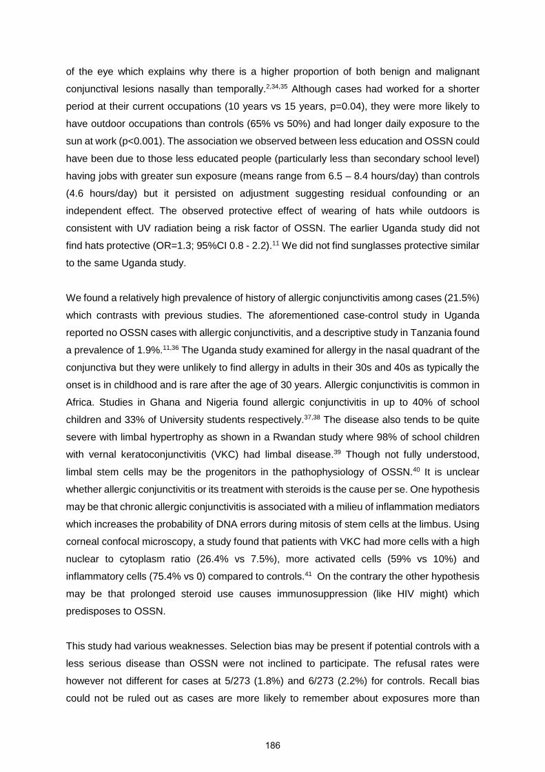
of the eye which explains why there is a higher proportion of both benign and malignant
conjunctival lesions nasally than temporally.2,34,35 Although cases had worked for a shorter
period at their current occupations (10 years vs 15 years, p=0.04), they were more likely to
have outdoor occupations than controls (65% vs 50%) and had longer daily exposure to the
sun at work (p<0.001). The association we observed between less education and OSSN could
have been due to those less educated people (particularly less than secondary school level)
having jobs with greater sun exposure (means range from 6.5 – 8.4 hours/day) than controls
(4.6 hours/day) but it persisted on adjustment suggesting residual confounding or an
independent effect. The observed protective effect of wearing of hats while outdoors is
consistent with UV radiation being a risk factor of OSSN. The earlier Uganda study did not
find hats protective (OR=1.3; 95%CI 0.8 - 2.2).11 We did not find sunglasses protective similar
to the same Uganda study.
We found a relatively high prevalence of history of allergic conjunctivitis among cases (21.5%)
which contrasts with previous studies. The aforementioned case-control study in Uganda
reported no OSSN cases with allergic conjunctivitis, and a descriptive study in Tanzania found
a prevalence of 1.9%.11,36 The Uganda study examined for allergy in the nasal quadrant of the
conjunctiva but they were unlikely to find allergy in adults in their 30s and 40s as typically the
onset is in childhood and is rare after the age of 30 years. Allergic conjunctivitis is common in
Africa. Studies in Ghana and Nigeria found allergic conjunctivitis in up to 40% of school
children and 33% of University students respectively.37,38 The disease also tends to be quite
severe with limbal hypertrophy as shown in a Rwandan study where 98% of school children
with vernal keratoconjunctivitis (VKC) had limbal disease.39 Though not fully understood,
limbal stem cells may be the progenitors in the pathophysiology of OSSN.40 It is unclear
whether allergic conjunctivitis or its treatment with steroids is the cause per se. One hypothesis
may be that chronic allergic conjunctivitis is associated with a milieu of inflammation mediators
which increases the probability of DNA errors during mitosis of stem cells at the limbus. Using
corneal confocal microscopy, a study found that patients with VKC had more cells with a high
nuclear to cytoplasm ratio (26.4% vs 7.5%), more activated cells (59% vs 10%) and
inflammatory cells (75.4% vs 0) compared to controls.41 On the contrary the other hypothesis
may be that prolonged steroid use causes immunosuppression (like HIV might) which
predisposes to OSSN.
This study had various weaknesses. Selection bias may be present if potential controls with a
less serious disease than OSSN were not inclined to participate. The refusal rates were
however not different for cases at 5/273 (1.8%) and 6/273 (2.2%) for controls. Recall bias
could not be ruled out as cases are more likely to remember about exposures more than
186

controls. We had considerable missing data on HIV, CD4 count and vitamin A as not all
excised cases returned post operatively for testing. The classification of occupations is not
ideal. We would have preferred one that classifies occupations by possible exposure to
carcinogens. Existing systems such as Kenya National Occupational Classification System
(KNOCS) which is derived from the International Standard Classification of Occupations
(ISCO) are designed for economic purposes not epidemiological studies.42
In conclusion we identified modifiable risk factors. HIV control interventions are now available,
exposure to UV radiation can be reduced and indeed we found that wearing of hats was
protective, while allergic conjunctivitis can be treated more aggressively in younger people. In
addition, the results of this study corroborate what is known about the pathophysiology of
OSSN where the limbus stem cells appear to be the main focus of disease. However, the
latency period between first exposure to these risk factors and causation of OSSN is still not
known. The role of CD4 count in OSSN still remains to be elucidated and why OSSN usually
affects one eye is still an enigma. Measures to control HIV, prevent sun exposure such as
wearing hats, and control allergic conjunctivitis are recommended.
187

References
1. Lee GA, Hirst LW. Ocular surface squamous neoplasia. Survey of ophthalmology 1995; 39(6): 429-50.
2. Gichuhi S, Sagoo MS, Weiss HA, Burton MJ. Epidemiology of ocular surface squamous neoplasiain Africa. Tropical medicine & international health : TM & IH 2013; 18(12): 1424-43.
3. Tunc M, Char DH, Crawford B, Miller T. Intraepithelial and invasive squamous cell carcinoma ofthe conjunctiva: analysis of 60 cases. British Journal of Ophthalmology 1999; 83(1): 98-103.
4. Gichuhi S, Macharia E, Kabiru J, et al. Clinical Presentation of Ocular Surface Squamous Neoplasiain Kenya. JAMA ophthalmology 2015; 133(11): 1305-13.
5. Nguena MB, van den Tweel JG, Makupa W, et al. Diagnosing ocular surface squamous neoplasiain East Africa: case-control study of clinical and in vivo confocal microscopy assessment.Ophthalmology 2014; 121(2): 484-91.
6. Porges Y, Groisman GM. Prevalence of HIV with conjunctival squamous cell neoplasia in an Africanprovincial hospital. Cornea 2003; 22(1): 1-4.
7. Holcombe DJ, Lee GA. Topical Interferon Alfa-2b for the Treatment of Recalcitrant Ocular SurfaceSquamous Neoplasia. American journal of ophthalmology 2006; 142(4): 568-71.e3.
8. McClellan AJ, McClellan AL, Pezon CF, Karp CL, Feuer W, Galor A. Epidemiology of Ocular SurfaceSquamous Neoplasia in a Veterans Affairs Population. Cornea 2013; 32(10): 1354-8.
9. Ateenyi-Agaba C, Dai M, Le Calvez F, et al. TP53 mutations in squamous-cell carcinomas of theconjunctiva: evidence for UV-induced mutagenesis. Mutagenesis 2004; 19(5): 399-401.
10. Newton R. A review of the aetiology of squamous cell carcinoma of the conjunctiva. British journalof cancer 1996; 74(10): 1511-3.
11. Waddell K, Kwehangana J, Johnston WT, Lucas S, Newton R. A case-control study of ocular surfacesquamous neoplasia (OSSN) in Uganda. International journal of cancer Journal international ducancer 2010; 127(2): 427-32.
12. NASA. Earth at perihelion. http://science.nasa.gov/science-news/science-at-nasa/2001/ast04jan_1/ (accessed 3rd September 2015.
13. Ateenyi-Agaba C, Weiderpass E, Smet A, et al. Epidermodysplasia verruciformis humanpapillomavirus types and carcinoma of the conjunctiva: a pilot study. British journal of cancer2004; 90(9): 1777-9.
14. Ateenyi-Agaba C. Conjunctival squamous-cell carcinoma associated with HIV infection inKampala, Uganda. Lancet 1995; 345(8951): 695-6.
15. Yu JJ, Fu P, Pink JJ, et al. HPV infection and EGFR activation/alteration in HIV-infected East Africanpatients with conjunctival carcinoma. PloS one 2010; 5(5): e10477.
16. Moubayed P, Mwakyoma H, Schneider DT. High Frequency of Human Papillomavirus 6/11, 16,and 18 Infections in Precancerous Lesions and Squamous Cell Carcinoma of the Conjunctiva inSubtropical Tanzania. American Journal of Clinical Pathology 2004; 122(6): 938-43.
17. Guthoff R, Marx A, Stroebel P. No evidence for a pathogenic role of human papillomavirusinfection in ocular surface squamous neoplasia in Germany. Current eye research 2009; 34(8):666-71.
18. de Koning MN, Waddell K, Magyezi J, et al. Genital and cutaneous human papillomavirus (HPV)types in relation to conjunctival squamous cell neoplasia: a case-control study in Uganda.Infectious agents and cancer 2008; 3: 12.
19. Sen S, Sharma A, Panda A. Immunohistochemical localization of human papilloma virus inconjunctival neoplasias: a retrospective study. Indian journal of ophthalmology 2007; 55(5): 361-3.
20. Simbiri KO, Murakami M, Feldman M, et al. Multiple oncogenic viruses identified in Ocular surfacesquamous neoplasia in HIV-1 patients. Infectious agents and cancer 2010; 5: 6.
21. Pfister RR, Renner ME. The corneal and conjunctival surface in vitamin A deficiency: a scanningelectron microscopy study. Investigative ophthalmology & visual science 1978; 17(9): 874-83.
188

22. Baeten JM, McClelland RS, Richardson BA, et al. Vitamin A deficiency and the acute phaseresponse among HIV-1-infected and -uninfected women in Kenya. Journal of acquired immunedeficiency syndromes (1999) 2002; 31(2): 243-9.
23. Tabrizi SN, McCurrach FE, Drewe RH, Borg AJ, Garland SM, Taylor HR. Human papillomavirus incorneal and conjunctival carcinoma. Australian and New Zealand journal of ophthalmology 1997;25(3): 211-5.
24. Hirst LW, Axelsen RA, Schwab I. Pterygium and associated ocular surface squamous neoplasia.Archives of ophthalmology 2009; 127(1): 31-2.
25. Joanna R, Renata Z, Witold P, Malgorzata S, Bernaczyk P, Chyczewski L. The evaluation of humanpapillomavirus and p53 gene mutation in benign and malignant conjunctiva and eyelid lesions.Folia histochemica et cytobiologica / Polish Academy of Sciences, Polish Histochemical andCytochemical Society 2010; 48(4): 530-3.
26. Chui J, Coroneo MT, Tat LT, Crouch R, Wakefield D, Di Girolamo N. Ophthalmic pterygium: a stemcell disorder with premalignant features. The American journal of pathology 2011; 178(2): 817-27.
27. Di Girolamo N. Association of human papilloma virus with pterygia and ocular-surface squamousneoplasia. Eye (Lond) 2012; 26(2): 202-11.
28. Thurnham DI, Smith E, Flora PS. Concurrent liquid-chromatographic assay of retinol, alpha-tocopherol, beta-carotene, alpha-carotene, lycopene, and beta-cryptoxanthin in plasma, withtocopherol acetate as internal standard. Clinical chemistry 1988; 34(2): 377-81.
29. WHO. WHO case definitions of HIV for surveillance and revised clinical staging and immunologicalclassification of HIV-related disease in adults and children. Geneva: WHO Press; 2007.
30. Vella S, Schwartlander B, Sow SP, Eholie SP, Murphy RL. The history of antiretroviral therapy andof its implementation in resource-limited areas of the world. AIDS (London, England) 2012;26(10): 1231-41.
31. Kamal S, Kaliki S, Mishra DK, Batra J, Naik MN. Ocular Surface Squamous Neoplasia in 200Patients: A Case-Control Study of Immunosuppression Resulting from Human ImmunodeficiencyVirus versus Immunocompetency. Ophthalmology 2015; 122(8): 1688-94.
32. Pathai S, Lawn SD, Gilbert CE, et al. Accelerated biological ageing in HIV-infected individuals inSouth Africa: a case-control study. AIDS (London, England) 2013; 27(15): 2375-84.
33. Newton R, Ferlay J, Reeves G, Beral V, Parkin DM. Effect of ambient solar ultraviolet radiation onincidence of squamous-cell carcinoma of the eye. Lancet 1996; 347(9013): 1450-1.
34. Coroneo M. Ultraviolet radiation and the anterior eye. Eye & contact lens 2011; 37(4): 214-24.35. Gichuhi S, Macharia E, Kabiru J, et al. Clinical Presentation of Ocular Surface Squamous Neoplasia
in Kenya. JAMA ophthalmology 2015.36. Poole TR. Conjunctival squamous cell carcinoma in Tanzania. The British journal of ophthalmology
1999; 83(2): 177-9.37. Kumah D, Lartey S, Yemanyi F, Boateng E, Awuah E. Prevalence of allergic conjunctivitis among
basic school children in the Kumasi Metropolis (Ghana): a community-based cross-sectionalstudy. BMC ophthalmology 2015; 15(1): 69.
38. Oladigbolu KK, Abah ER, Chinda D, Anyebe EE. Pattern of eye diseases in a university healthservice clinic in northern Nigeria. Nigerian journal of medicine : journal of the National Associationof Resident Doctors of Nigeria 2012; 21(3): 334-7.
39. De Smedt SK, Nkurikiye J, Fonteyne YS, Tuft SJ, Gilbert CE, Kestelyn P. Vernal Keratoconjunctivitisin School Children in Rwanda: Clinical Presentation, Impact on School Attendance, and Access toMedical Care. Ophthalmology 2012; 119(9): 1766-72.
40. Gichuhi S, Ohnuma SI, Sagoo MS, Burton MJ. Pathophysiology of ocular surface squamousneoplasia. Experimental eye research 2014; 129C: 172-82.
41. Leonardi A, Lazzarini D, Bortolotti M, Piliego F, Midena E, Fregona I. Corneal Confocal Microscopyin Patients with Vernal Keratoconjunctivitis. Ophthalmology 2012; 119(3): 509-15.
189

42. Kenya National Bureau of Statistics. Kenya National Occupational Classification Standard (KNOCS-2000). In: Ministry of Labour, editor.; 2000.
43. National AIDS and STI Control Programme (NASCOP) Kenya. Kenya HIV estimates, 2014.
190
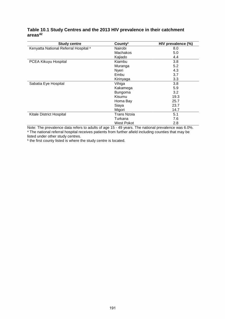
Table 10.1 Study Centres and the 2013 HIV prevalence in their catchmentareas42
Study centre Countyb HIV prevalence (%)Kenyatta National Referral Hospital a Nairobi 8.0
Machakos 5.0Kajiado 4.4
PCEA Kikuyu Hospital Kiambu 3.8Muranga 5.2Nyeri 4.3Embu 3.7Kirinyaga 3.3
Sabatia Eye Hospital Vihiga 3.8Kakamega 5.9Bungoma 3.2Kisumu 19.3Homa Bay 25.7Siaya 23.7Migori 14.7
Kitale District Hospital Trans Nzoia 5.1Turkana 7.6West Pokot 2.8
Note: The prevalence data refers to adults of age 15 - 49 years. The national prevalence was 6.0%.a The national referral hospital receives patients from further afield including counties that may belisted under other study centres.b the first county listed is where the study centre is located.
191

Table 10.2 Univariable analysis of potential risk factors for ocular surface squamous neoplasia. This analysis is adjustedfor age group, sex and study centre.
Exposure Cases(N=131)
Controls(N=131)
Crude OR (95% CI) P value
Marital status, No. (%) 0.12Single 22 (16.8) 26 (19.9) 1 [Reference]Married 83 (63.4) 89 (67.9) 1.12 (0.56 - 2.26)Divorced or Separated 8 (6.1) 6 (4.6) 1.55 (0.45 - 5.31)Widowed 18 (13.7) 10 (7.6) 1.88 (0.67 - 5.30)
Highest education level, No. (%) 0.01More than secondary 13 (9.9) 33 (25.2) 1 [Reference]Completed secondary school 33 (25.2) 35 (26.7) 2.26 (1.01 - 5.06)Some secondary school 12 (9.2) 14 (10.7) 2.00 (0.72 - 5.51)Completed primary school 41 (31.3) 25 (19.1) 3.55 (1.55 - 8.12)Some primary school 19 (14.5) 17 (13.0) 2.43 (0.94 - 6.30)None 13 (9.9) 7 (5.3) 4.24 (1.33 - 13.53)
Residential area (Province) 0.15Central 27 (20.6) 34 (26.0) 1 [Reference]Coast 1 (0.8) 1 (0.8) 1.25 (0.07 - 21.10)Eastern 13 (9.9) 9 (6.9) 1.81 (0.67 - 4.87)Nairobi 28 (21.4) 42 (32.1) 0.83 (0.41 - 1.70)North-Eastern 2 (1.5) 0 -Nyanza 14 (10.7) 7 (5.3) 1.79 (0.59 - 5.41)Rift Valley 36 (27.5) 28 (21.4) 0.97 (0.39 - 2.41)Western 10 (7.6) 10 (7.6) 0.75 (0.23 - 2.41)
Duration of residence in home province,median (IQR),y
24 (10 - 39) 28 (14 - 40) - 0.33a
History of allergic conjunctivitis b 28 (21.5) 1 (0.9) 30.78 (4.05 - 233.75) <0.001Location of current occupation, No. (%)b 0.01Indoor 45 (34.9) 62 (48.1) 1 [Reference]Outdoor 84 (65.1) 67 (51.9) 1.73 (1.03 - 2.91)
Duration in current occupation, median(IQR),y
10 (5 - 20) 15 (7 - 23) - 0.04 a
Sun exposure per day in current occupation,mean (SD), h
6.9 (3.8) 4.6 (3.2) 1.22 (1.12 - 1.32) <0.001c
Hat or cap worn outdoors, No. (%)b 19 (15.0) 32 (24.8) 0.60 (0.30 - 1.20) 0.04
192

Sunglasses worn outdoors, No. (%)b 10 (7.9) 16 (12.4) 0.66 (0.28 - 1.55) 0.06
Cigarette smoking (past), No. (%)b 0.05No 93 (71.5) 90 (70.3) 1 [Reference]Passive (spouse/partner smokes) 17 (13.1) 21 (16.4) 1.10 (0.47 - 2.56)Yes 20 (15.4) 17 (13.3) 1.12 (0.54 - 2.34)
Cigarette smoking (current), No. (%)b 0.02No 115 (89.2) 111 (88.1) 1 [Reference]Passive (spouse/partner smokes) 4 (3.1) 9 (7.1) 0.55 (0.15 - 2.00)Yes 10 (7.8) 6 (4.8) 1.63 (0.55 - 4.77)
Duration of cigarette smoking, mean (SD), y 13.5 (8.6) 12.7 (10.0) - 0.79 c
No. of cigarettes smoked daily, median(IQR), y
9 (5 - 20) 7 (5 - 10) - 0.60 a
HIV infection d, No. (%) 62 (74.7) 14 (13.6) 18.96 (8.87 - 40.51) <0.001HIV infection & ART use e, No. (%) <0.001HIV- 21 (16.0) 89 (67.9) 1 [Reference]HIV +/ ART- 17 (13.0) 2 (1.5) 39.37 (8.35 - 185.64)HIV+ / ART+ 40 (30.5) 12 (9.2) 14.50 (6.42 - 32.74)No HIV or ART data 53 (40.5) 28 (21.4) 7.22 (3.69 - 14.11)
CD4 count f, median (IQR), cells/mm3 251 (118 - 670) 796 (619 -1063) - <0.001a
CD4 WHO category, No.(%),cells/mm3 <0.001 ≥500 20 (15.3) 64 (48.9) 1 [Reference]350 - 499 6 (4.6) 9 (6.9) 2.42 (0.75 - 7.82)200 - 349 11 (8.4) 1 (0.8) 39.63 (4.73 - 332.14)<200 22 (16.8) 2 (1.5) 38.16 (8.15 - 178.70)No CD4 data 72 (55.0) 55 (42.0) 3.87 (2.07 - 7.23)
Vitamin A deficiency (<30 µg/dL) g, No. (%) 5 (8.8) 5 (5.8) 1.25 (0.30 - 5.23) 0.50Serum retinol, median (IQR), µg/dL 44.9 (37.6 - 56.2) 51.0 (39.7 - 64.7) - 0.03 a
Abbreviations: SD, standard deviation; IQR, interquartile rangea Wilcoxon-Mann-Whitney U testbThere were missing data on allergic conjunctivitis (24 participants), occupational location (4), hat or sunglasses use (6), cigarette smoking (7)c t testd The denominator here is 83 cases and 103 controls who were tested for HIVe We used the combined HIV/ART variable because the effect of HIV would be related to ART usef There were 59 cases and 76 controls who had a CD4 testg The denominator here is 57 cases and 86 controls who had a vitamin A test
193
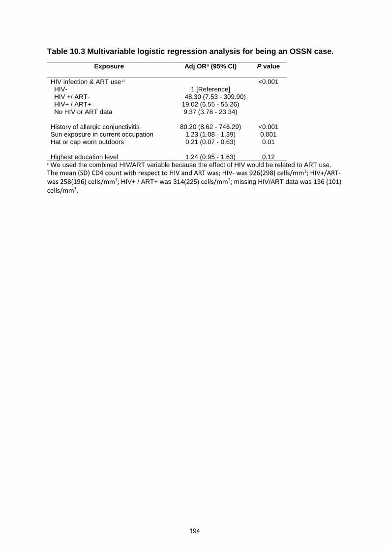
Table 10.3 Multivariable logistic regression analysis for being an OSSN case.
Exposure Adj ORa (95% CI) P value
HIV infection & ART use a <0.001HIV- 1 [Reference]HIV +/ ART- 48.30 (7.53 - 309.90)HIV+ / ART+ 19.02 (6.55 - 55.26)No HIV or ART data 9.37 (3.76 - 23.34)
History of allergic conjunctivitis 80.20 (8.62 - 746.29) <0.001Sun exposure in current occupation 1.23 (1.08 - 1.39) 0.001Hat or cap worn outdoors 0.21 (0.07 - 0.63) 0.01
Highest education level 1.24 (0.95 - 1.63) 0.12a We used the combined HIV/ART variable because the effect of HIV would be related to ART use.
The mean (SD) CD4 count with respect to HIV and ART was; HIV- was 926(298) cells/mm3; HIV+/ART-was 258(196) cells/mm3; HIV+ / ART+ was 314(225) cells/mm3; missing HIV/ART data was 136 (101)
cells/mm3.
194
Chapter 11. Delay along the care-seeking
pathway of patients with ocular surface
squamous neoplasia in Kenya
195

RESEARCH PAPER COVER SHEET
PLEASE NOTE THAT A COVER SHEET MUST BE COMPLETED FOR EACH RESEARCH PAPER INCLUDEDIN A THESIS.
SECTION A – Student Details
Student
Principal Supervisor
Thesis Title
If the Research Paper has previously been published please complete Section B, if not please move toSection C
SECTION B – Paper already published
Where was the work published?
When was the work published?
If the work was published prior toregistration for your research degree,give a brief rationale for its inclusion
Have you retained the copyright for thework?* Choose an item. Was the work subject to
academic peer review?Choose anitem.
*If yes, please attach evidence of retention. If no, or if the work is being included in its published format, pleaseattach evidence of permission from the copyright holder (publisher or other author) to include this work.
SECTION C – Prepared for publication, but not yet published
Where is the work intended to bepublished?
Please list the paper’s authors in theintended authorship order:
Stage of publication Not yet submitted
SECTION D – Multi-authored work
For multi-authored work, give full details of your role inthe research included in the paper and in the preparationof the paper. (Attach a further sheet if necessary)
Student Signature: Date:
Supervisor Signature: Date:
196
Gichuhi
Typewritten text
Stephen Gichuhi
Gichuhi
Typewritten text
Matthew J. Burton
Gichuhi
Typewritten text
Epidemiology and management of ocular surface squamous neoplasia in Kenya
Gichuhi
Typewritten text
Stephen Gichuhi, Ephantus Macharia, Joy Kabiru, Alain M’bongo Zindamoyen, Hilary Rono, Ernest Ollando, Joseph Wachira, Rhoda Munene, Timothy Onyuma, Mandeep S. Sagoo, David Macleod, Helen A. Weiss, and Matthew J. Burton
Gichuhi
Typewritten text
4th February 2016
Gichuhi
Typewritten text
4th February 2016
Typewritten text
I coordinated and actively led the field work for this paper. Supervision was provided by Matthew Burton throughout. I also performed the statistical analysis, with some guidance from Helen Weiss and David Macleod. I wrote the paper with consideration of comments from co-authors.

Delay along the care-seeking journey of patients with ocular surface
squamous neoplasia in Kenya
Authors:Stephen Gichuhi MBChB, M.Med 1, 2, Joy Kabiru MBChB, M.Med 3, Alain M’bongo ZindamoyenMBChB, M.Med 3, Hilary Rono MBChB, M.Med 4, Ernest Ollando MBChB, M.Med 5, Joseph WachiraMBChB, M.Med 6, Rhoda Munene MBChB, M.Med 6, Timothy Onyuma MBChB, M.Med 7, Mandeep S.Sagoo PhD, FRCOphth 8, 9, 10, David Macleod MSc1,11, Helen A. Weiss MSc, PhD1,11 and Matthew J.Burton PhD, FRCOphth1, 9
Affiliations:1International Centre for Eye Health, London School of Hygiene and Tropical Medicine, Keppel Street,London, WC1E 7HT, UK2Department of Ophthalmology, University of Nairobi, P.O Box 19676-00202, Nairobi, Kenya 3PCEAKikuyu Eye Unit, PO Box 45, Kikuyu, Kenya4Kitale District Hospital, PO Box 98-30200, Kitale, Kenya5Sabatia Eye Hospital, PO Box 214-50311, Wodanga, Kenya6Kenyatta National Hospital, PO Box 20723-00202, Nairobi, Kenya7MP Shah Hospital, Department of Pathology, PO Box 14497-00800, Nairobi, Kenya8UCL Institute of Ophthalmology, 11-43 Bath Street, London EC1V 9EL, UK9Moorfields Eye Hospital, 162 City Road, London, EC1V 2PD, UK10St. Bartholomew’s Hospital, W Smithfield, London EC1A 7BE, London, UK11 MRC Tropical Epidemiology Group, London School of Hygiene and Tropical Medicine, KeppelStreet, London, WC1E 7HT,
Corresponding Author:Stephen GichuhiInternational Centre for Eye HealthLondon School of Hygiene & Tropical MedicineKeppel Street, London WC1E 7HTUnited KingdomTel. +44 (20) 7636 8636 ext. [email protected]
Email contactsStephen Gichuhi, [email protected] Kabiru, [email protected] M’bongo Zindamoyen, [email protected] Rono, [email protected] Ollando, [email protected] Wachira, [email protected] Munene, [email protected] Onyuma, [email protected] S. Sagoo, [email protected] Macleod [email protected] A. Weiss, [email protected] J. Burton, [email protected]
Financial supportSG received funding from the British Council for Prevention of Blindness (BCPB) fellowship programme.MJB is supported by The Wellcome Trust (Grant Number 098481/Z/12/Z). HAW and DM are supportedby the MRC and DFID (Grant Number G0700837). The funding organizations had no role in the designor conduct of this research.
Conflict of interestNone of the authors have any proprietary interests or conflicts of interest related to this submission.
This submission has not been published anywhere previously and is not simultaneously being
considered for any other publication.
197

Running headDelayed care for ocular surface squamous neoplasia - Kenya
Word count: Abstract 249; Text 3554
198
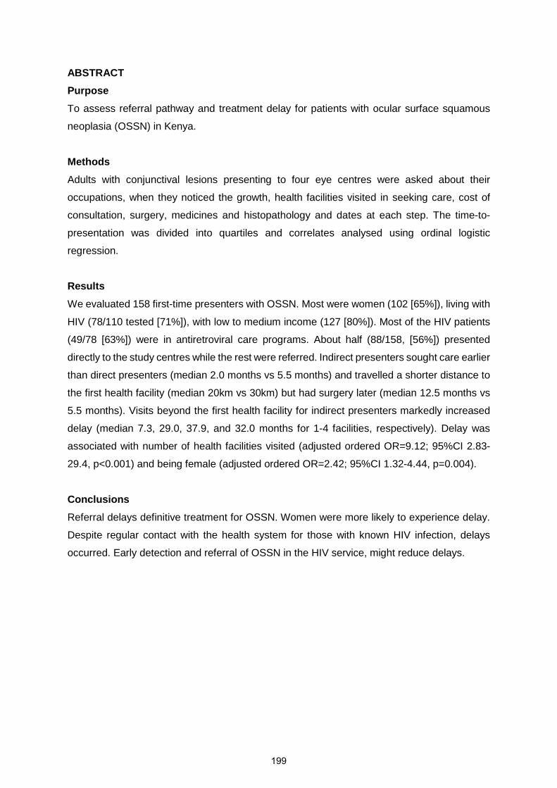
ABSTRACT
Purpose
To assess referral pathway and treatment delay for patients with ocular surface squamous
neoplasia (OSSN) in Kenya.
Methods
Adults with conjunctival lesions presenting to four eye centres were asked about their
occupations, when they noticed the growth, health facilities visited in seeking care, cost of
consultation, surgery, medicines and histopathology and dates at each step. The time-to-
presentation was divided into quartiles and correlates analysed using ordinal logistic
regression.
Results
We evaluated 158 first-time presenters with OSSN. Most were women (102 [65%]), living with
HIV (78/110 tested [71%]), with low to medium income (127 [80%]). Most of the HIV patients
(49/78 [63%]) were in antiretroviral care programs. About half (88/158, [56%]) presented
directly to the study centres while the rest were referred. Indirect presenters sought care earlier
than direct presenters (median 2.0 months vs 5.5 months) and travelled a shorter distance to
the first health facility (median 20km vs 30km) but had surgery later (median 12.5 months vs
5.5 months). Visits beyond the first health facility for indirect presenters markedly increased
delay (median 7.3, 29.0, 37.9, and 32.0 months for 1-4 facilities, respectively). Delay was
associated with number of health facilities visited (adjusted ordered OR=9.12; 95%CI 2.83-
29.4, p<0.001) and being female (adjusted ordered OR=2.42; 95%CI 1.32-4.44, p=0.004).
Conclusions
Referral delays definitive treatment for OSSN. Women were more likely to experience delay.
Despite regular contact with the health system for those with known HIV infection, delays
occurred. Early detection and referral of OSSN in the HIV service, might reduce delays.
199
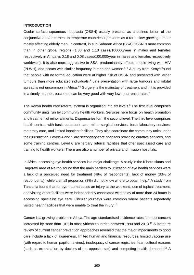
INTRODUCTION
Ocular surface squamous neoplasia (OSSN) usually presents as a defined lesion of the
conjunctiva and/or cornea. In temperate countries it presents as a rare, slow-growing tumour
mostly affecting elderly men. In contrast, in sub-Saharan Africa (SSA) OSSN is more common
than in other global regions (1.38 and 1.18 cases/100000/year in males and females
respectively in Africa vs 0.18 and 0.08 cases/100,000/year in males and females respectively
worldwide). It is also more aggressive in SSA, predominantly affects people living with HIV
(PLWH), and occurs with similar frequency in men and women.1, 2 A study from Kenya found
that people with no formal education were at higher risk of OSSN and presented with larger
tumours than more educated individuals.3 Late presentation with large tumours and orbital
spread is not uncommon in Africa.4-6 Surgery is the mainstay of treatment and if it is provided
in a timely manner, outcomes can be very good with very low recurrence rates.7
The Kenya health care referral system is organized into six levels.8 The first level comprises
community units run by community health workers. Services here focus on health promotion
and treatment of minor ailments. Dispensaries form the second level. The third level comprises
health centres with basic outpatient care, minor surgical services, basic laboratory services,
maternity care, and limited inpatient facilities. They also coordinate the community units under
their jurisdiction. Levels 4 and 5 are secondary-care hospitals providing curative services, and
some training centres. Level 6 are tertiary referral facilities that offer specialised care and
training to health workers. There are also a number of private and mission hospitals.
In Africa, accessing eye health services is a major challenge. A study in the Kibera slums and
Dagoretti area of Nairobi found that the main barriers to utilization of eye health services were
a lack of a perceived need for treatment (49% of respondents), lack of money (33% of
respondents), while a small proportion (8%) did not know where to obtain help.9 A study from
Tanzania found that for eye trauma cases an injury at the weekend, use of topical treatment,
and visiting other facilities were independently associated with delay of more than 24 hours in
accessing specialist eye care. Circular journeys were common where patients repeatedly
visited health facilities that were unable to treat the injury.10
Cancer is a growing problem in Africa. The age-standardised incidence rates for most cancers
increased by more than 10% in most African countries between 1990 and 2013.11 A literature
review of current cancer prevention approaches revealed that the major impediments to good
care include a lack of awareness, limited human and financial resources, limited vaccine use
(with regard to human papilloma virus), inadequacy of cancer registries, fear, cultural reasons
(such as examination by doctors of the opposite sex) and competing health demands.12 A
200
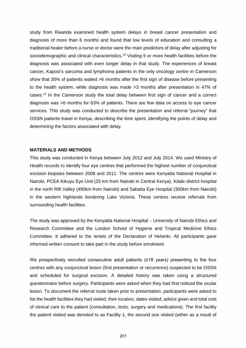
study from Rwanda examined health system delays in breast cancer presentation and
diagnosis of more than 6 months and found that low levels of education and consulting a
traditional healer before a nurse or doctor were the main predictors of delay after adjusting for
sociodemographic and clinical characteristics.13 Visiting 5 or more health facilities before the
diagnosis was associated with even longer delay in that study. The experiences of breast
cancer, Kaposi’s sarcoma and lymphoma patients in the only oncology centre in Cameroon
show that 35% of patients waited >6 months after the first sign of disease before presenting
to the health system, while diagnosis was made >3 months after presentation in 47% of
cases.14 In the Cameroon study the total delay between first sign of cancer and a correct
diagnosis was >6 months for 63% of patients. There are few data on access to eye cancer
services. This study was conducted to describe the presentation and referral “journey” that
OSSN patients travel in Kenya, describing the time spent, identifying the points of delay and
determining the factors associated with delay.
MATERIALS AND METHODS
This study was conducted in Kenya between July 2012 and July 2014. We used Ministry of
Health records to identify four eye centres that performed the highest number of conjunctival
excision biopsies between 2008 and 2011. The centres were Kenyatta National Hospital in
Nairobi, PCEA Kikuyu Eye Unit (25 km from Nairobi in Central Kenya), Kitale district hospital
in the north Rift Valley (490km from Nairobi) and Sabatia Eye Hospital (300km from Nairobi)
in the western highlands bordering Lake Victoria. These centres receive referrals from
surrounding health facilities.
The study was approved by the Kenyatta National Hospital – University of Nairobi Ethics and
Research Committee and the London School of Hygiene and Tropical Medicine Ethics
Committee. It adhered to the tenets of the Declaration of Helsinki. All participants gave
informed written consent to take part in the study before enrolment.
We prospectively recruited consecutive adult patients (≥18 years) presenting to the four
centres with any conjunctival lesion (first presentation or recurrence) suspected to be OSSN
and scheduled for surgical excision. A detailed history was taken using a structured
questionnaire before surgery. Participants were asked when they had first noticed the ocular
lesion. To document the referral route taken prior to presentation, participants were asked to
list the health facilities they had visited, their location, dates visited, advice given and total cost
of clinical care to the patient (consultation, tests, surgery and medications). The first facility
the patient visited was denoted to as Facility 1, the second one visited (either as a result of
201

formal referral from the first or self-initiated referral) was denoted Facility 2 and so on. A
comprehensive clinical examination was conducted at the slit lamp, including measurement of
the longest lesion diameter using the slit lamp beam and scale. Participants underwent
surgical excision under local anaesthesia. A histopathologist examined all the tissue
specimens at MP Shah Hospital laboratory, Nairobi. Individuals who had histologically
confirmed OSSN were offered a HIV test; if this was positive they were offered a referral for
care and a CD4 count test. The detailed description of clinical assessment, surgery, tissue
handling and testing have been previously published.3
Treatment “delay” was defined as the time between becoming aware of the lesion and having
surgery. For the purpose of this analysis we only included individuals who had histologically
confirmed OSSN. As recurrence after a prior excision is likely to influence health-seeking
behaviour and the journey followed, we excluded those with recurrent lesions from this
analysis.
Data on the other health facilities visited by patients prior to presentation to one of the four
study centres (location, health system level, ownership etc.) were obtained from the Kenya
Ministry of Health web-based master facility list (http://www.ehealth.or.ke/facilities/) accessed
on 15th November 2015. Information about the availability of ophthalmic surgical facilities (eye
operating theatre) at each of these other health facilities was obtained from the Kenya Ministry
of Health, Department of Ophthalmic Services (DOS). One-way distances travelled by road
from the patients’ home town to the various health facilities and study centres were estimated
using Google Maps following the roads that public vehicles would use.
We compared patients who presented directly to one of the four centres (“direct presenters”)
with those who had visited other health facilities prior to presenting to a study centre (“indirect
presenters”). Characteristics of the two groups were compared using the chi-squared test for
proportions, and the t-test or Mann-Whitney U-test as appropriate for continuous variables.
We traced the care seeking journey to describe the time taken, and the advice given at each
stage. To investigate factors associated with delayed presentation we subdivided the total
time in the range from awareness to surgery into quartiles and created an ordered categorical
outcome variable. Ordinal logistic regression was used to estimate odds ratios (OR) and 95%
confidence intervals (95%CI).
The likelihood ratio test was used to assess which factors from the univariable analysis would
be included in the multivariable regression model. Variables that were associated with delay
at a level of p<0.1 in the univariable model were included in an initial multivariable model. The
202

final multivariable model included variables which were independently associated with the
outcome (p<0.05). All statistical analyses were performed using Stata version 12.1
(StataCorp, College Station, Texas, USA).
RESULTS
500 individuals with conjunctival lesions enrolled in this study and underwent surgical excision.
Of these, 191 (38.2%) were diagnosed with OSSN by histopathology. Among these, 158
(83%) had new lesions and 33 (17%) had recurrent lesions (at least one prior excision at the
same site).
The care-seeking journey of the 158 patients with new lesions was analysed: 88 (55.7%)
presented directly to study centres and 70 (44.3%) presented indirectly via one or more other
health facilities (Figure 11.1). Overall, the mean age was 41.9 years (SD 12.0) and the majority
were females (102 [65%]). Education levels were variable, 125 (79%) had completed primary
school or above, and 67 (42%) had completed secondary school. The majority worked mostly
outdoors (101 [65%]). Most occupations were low or middle income in nature (127 [80%]) and
were predominantly in the agricultural sector. HIV status and CD4 count were obtained for
patients who returned after surgery (110 [70%]. Of these, 78 (71%) were HIV positive, of whom
49 (63%) were already on antiretroviral therapy (ART).
The numbers of individuals taking different routes to the study centres and the time spent
along each stage on the care-seeking journey is illustrated in Figure 11.1. Most indirect
presenters (56, [80%]) visited one other health facility before going to the study centres.
Overall, despite having a shorter time to first presentation, indirect presenters took longer to
receive surgery than direct presenters (5.5 vs 9.6 months, p=0.001). The additional time in
getting from the first health facility to the study centre was largely responsible for this
difference. The longest delay in the whole care pathway occurred when patients went from
the first facility to a second one (median 6.1 months, IQR 3.1-12.2). Referral beyond the first
facility markedly increased delay.
The demographic and clinical characteristics of participants, by type of presentation, are
shown in Table 11.1. There was little evidence of a difference between direct and indirect
presenters by age, marital status, education, occupation or HIV status. There was weak
evidence that indirect presenters were more likely to be female than direct presenters (71%
vs 59%; p=0.11). There was evidence that the main presenting symptom was different
(p=0.03) between the two groups, with pain more common in the direct presenters (17.1% vs
203
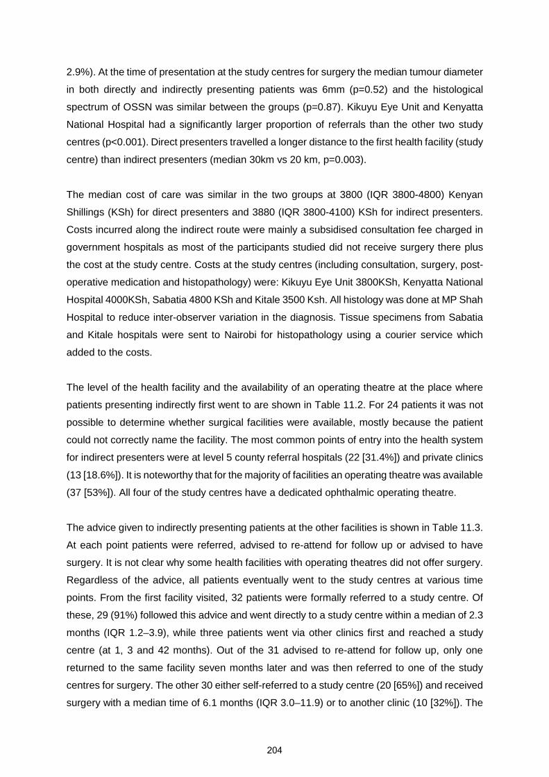
2.9%). At the time of presentation at the study centres for surgery the median tumour diameter
in both directly and indirectly presenting patients was 6mm (p=0.52) and the histological
spectrum of OSSN was similar between the groups (p=0.87). Kikuyu Eye Unit and Kenyatta
National Hospital had a significantly larger proportion of referrals than the other two study
centres (p<0.001). Direct presenters travelled a longer distance to the first health facility (study
centre) than indirect presenters (median 30km vs 20 km, p=0.003).
The median cost of care was similar in the two groups at 3800 (IQR 3800-4800) Kenyan
Shillings (KSh) for direct presenters and 3880 (IQR 3800-4100) KSh for indirect presenters.
Costs incurred along the indirect route were mainly a subsidised consultation fee charged in
government hospitals as most of the participants studied did not receive surgery there plus
the cost at the study centre. Costs at the study centres (including consultation, surgery, post-
operative medication and histopathology) were: Kikuyu Eye Unit 3800KSh, Kenyatta National
Hospital 4000KSh, Sabatia 4800 KSh and Kitale 3500 Ksh. All histology was done at MP Shah
Hospital to reduce inter-observer variation in the diagnosis. Tissue specimens from Sabatia
and Kitale hospitals were sent to Nairobi for histopathology using a courier service which
added to the costs.
The level of the health facility and the availability of an operating theatre at the place where
patients presenting indirectly first went to are shown in Table 11.2. For 24 patients it was not
possible to determine whether surgical facilities were available, mostly because the patient
could not correctly name the facility. The most common points of entry into the health system
for indirect presenters were at level 5 county referral hospitals (22 [31.4%]) and private clinics
(13 [18.6%]). It is noteworthy that for the majority of facilities an operating theatre was available
(37 [53%]). All four of the study centres have a dedicated ophthalmic operating theatre.
The advice given to indirectly presenting patients at the other facilities is shown in Table 11.3.
At each point patients were referred, advised to re-attend for follow up or advised to have
surgery. It is not clear why some health facilities with operating theatres did not offer surgery.
Regardless of the advice, all patients eventually went to the study centres at various time
points. From the first facility visited, 32 patients were formally referred to a study centre. Of
these, 29 (91%) followed this advice and went directly to a study centre within a median of 2.3
months (IQR 1.2–3.9), while three patients went via other clinics first and reached a study
centre (at 1, 3 and 42 months). Out of the 31 advised to re-attend for follow up, only one
returned to the same facility seven months later and was then referred to one of the study
centres for surgery. The other 30 either self-referred to a study centre (20 [65%]) and received
surgery with a median time of 6.1 months (IQR 3.0–11.9) or to another clinic (10 [32%]). The
204
five who were advised to have surgery all went to the study centres within a median (IQR)
duration of 5.6 (2.3 – 11.5) months.
There was a modest linear relationship between duration of symptoms and the lesion size
(Figure 11.2) for direct presenters (p=0.01), but not indirect presenters (p=0.41). For the direct
presenters, those who present later tended to have a larger lesion, with an expected increase
in size of 0.09mm per month (95% CI 0.03-0.16). There is little evidence that the relationship
between symptom duration and tumour size was different in direct and indirect presenters
(p=0.33). Delay was associated with larger tumour size at presentation (OR 1.07, 95%CI 1.01-
1.13, p=0.02). The median (IQR) tumour diameter was 5.6 (4.2-7.0), 5.0 (4.2-7.1), 6.4 (4.2-
11.5) and 7.6 (5.4-10.0) millimetres (mm) in the following delay categories; 0.4-3.2, 3.3-7.0,
7.1-15.6 and 15.7-190.5 months respectively.
Analysis of the predictors of delay is shown in Table 11.4. On multivariable ordered logistic
regression, the number of health facilities visited in the care pathway followed and being
female are associated with increased delay. Participants who followed an indirect path were
twice as likely to be in a higher delay group than those in the direct pathway (adjusted OR=2.3,
95%CI 1.5-3.7; p<0.001). Similarly, females were twice as likely to be in a higher delay group
than males (adjusted OR=2.1; 95% CI 1.15-3.81, p=0.02).
DISCUSSION
This study identified various challenges along the journey to treatment for new patients with
OSSN. Firstly, referral significantly delayed surgery and delay was associated with larger
tumours at the time of surgery. Patients in the indirect route initially presented earlier than
those who went directly to study centres perhaps because it was a shorter distance and the
cost of care was lower. However, surgery was more delayed in the indirect route compared to
the direct (5.5 months vs 9.6 months, p=0.001), particularly if the patients visited more than
one health facility. This correlates well with the finding that the number of facilities visited
before was an important predictor of delay in receiving surgery.
Secondly, we found being female was another important predictor of delay. Gender disparities
in access to health care services have also been identified in East Africa for other ocular
diseases such as eye trauma and cataract with females having relatively more difficulty.10, 15
A study on access to cataract surgery in Tanzania found that some women needed to seek
permission from their husbands before going to hospital or may rather put up with the adversity
of poor eye health for fear of being seen as a burden in the family.15 Women are less likely
205
than men to be the financial decision makers with regard to seeking health care. In addition,
they may have other responsibilities to consider before going to hospital such as child care
and home upkeep. Although there is limited information on gender-specific utilization of cancer
services in Africa, particularly for OSSN, we hypothesize that women with household and
child-care responsibilities have more difficulty attending health facilities, particularly if distant
referrals are made.
With respect to the patients reported advice and treatment provided at facilities other than the
four study centres there is an intrinsic limitation in that we do not know how many other (if any)
people had excisions in the other health facilities who never came to one of the study centres.
There is no reporting of this to the central Ministry of Health. However, it would appear that
advising follow up for suspicious lesions needs to be supported by more than just a clinical
impression. At the first health facility level, 20 of 31 patients who were advised to return for
follow up went to the study centres and had surgery within median 6 months (mean 13 months)
and were found to have OSSN (Table 11.3). Only one patient at the first facility took the follow
up advice and by the time of review seven months later was referred for surgery and was
found to have OSSN. While we have no information on how the lesions looked at earlier time
points, they may have appeared either benign or were suspicious of malignancy and
progressed rather rapidly within a few months, underscoring two things, that OSSN in East
Africa is an aggressive disease and that distinguishing OSSN from benign lesions clinically is
challenging as they can look very similar.3
It is still unclear what drives the decision to present to a first health facility once the person
become aware of the tumour. Perhaps it is more the absolute size rather than the rate of
growth or combination of the two. We found only a modest difference in growth rates between
direct and indirect presenters. It is likely that the duration and growth rates will vary. Pain
maybe an important driver of the decision; we found that pain was more common among the
direct presenters (p=0.02).
The majority of participants were living with HIV and were already in contact with HIV care and
treatment programs. Despite this, they still experienced delay. There could be various
explanations for this. Firstly, awareness about OSSN among HIV health workers may be
limited. Secondly, patients on ART may find it particularly challenging to seek care for OSSN.
Over half (44/81 [54%]) the patients on ART had advanced or severe immunosuppression and
thus at high risk for comorbidities such as tuberculosis.16 Thirdly, due to stigma around HIV
they may not feel safe or comfortable in other health facilities. A recent study in rural western
206

Kenya found that cervical cancer stigma was highly correlated with HIV stigma (correlation
coefficient 0.72).17
We did not find income or level of formal education a barrier to presentation despite most of
our patients (80%) being in the low to medium income group. The majority worked in farming.
Most employment (83%) nationwide is in the informal sector so it is difficult to know the real
earnings as they are not captured in tax systems.18 In Kenya half of healthcare expenditure
(51.1%) is paid for out-of-pocket.19 The gazetted monthly basic minimum wages for the
agricultural industry during the study period was an average of KSh 5704 in 2012 rising to KSh
6503 in 2013 and remained the same in 2014 which was comparable to the fees charged at
the study centres.20 However the opportunity cost of lost income during the care-seeking
period for the patient and other adults involved in supporting this care could not be estimated.
A study in Kenya found that households spend over 10% of their annual income on healthcare
payments with the poor spending more than 33%.21 The mean annual total spending per
household (both inpatient and outpatient) was KSh 16,000 (£114) in urban areas and KSh
5600 (£40) in rural areas. Only about 7% of Kenyans have private health insurance.20 The
National Health Insurance Fund (NHIF) run by the government is the dominant insurance
provider. The maximum contribution was KSh 320 (about £2) per month until April 2015 when
it was increased to KSh 1700 (£12) per month. NHIF used to pay for the inpatient bed charge
only, therefore day surgery (the norm for OSSN surgery) would not be covered. This situation
has now been reviewed. The cost of travel is not captured in this analysis. Usually most
patients in Kenya would travel to hospitals accompanied by a relative or friend, further
increasing the cost of care.
This study had several weaknesses. It did not address the availability of eye surgeons or other
surgeons skilled enough and adequately equipped with the right surgical instruments to excise
conjunctival lesion as a cause of referral and hence delay. This data was not available to us.
The presence or absence of an operating theatre is probably not the determining factor for
referral - it is likely to be the availability of an appropriately skilled surgeon. Our analysis of the
flow through the health system did not address why the patients did what they did. To answer
this would require a qualitative study.
In conclusion, women and those who visited more health facilities were at increased risk of
delay. We observed various health system delays. Referral beyond the first point of contact
was a major cause of delay. It is also unclear why patients who are in regular contact with the
health system through HIV care programs have delayed presentation. There is need to
evaluate if health education about OSSN and the ocular effects of HIV infection particularly
207

among HIV care workers is a barrier to the provision of timely intervention. Systematic OSSN
screening could be considered in HIV care programmes. Patients with pain presented more
rapidly. The cost to the patient of treating OSSN (excluding transport casts) was equivalent to
a month’s wages. It has been suggested that cancer care programs could learn from HIV
programs by mainstreaming cancer into the health system together with advocacy and
improved health worker training.22
208
References
1. Gichuhi S, Sagoo MS, Weiss HA, Burton MJ. Epidemiology of ocular surface squamousneoplasia in Africa. Trop Med Int Health. Dec 2013;18(12):1424-1443.
2. Tiong T, Borooah S, Msosa J, et al. Clinicopathological review of ocular surface squamousneoplasia in Malawi. Br J Ophthalmol. Aug 2013;97(8):961-964.
3. Gichuhi S, Macharia E, Kabiru J, et al. Clinical Presentation of Ocular Surface SquamousNeoplasia in Kenya. JAMA Ophthalmol. Nov 1 2015;133(11):1305-1313.
4. Chisi SK, Kollmann MK, Karimurio J. Conjunctival squamous cell carcinoma in patients withhuman immunodeficiency virus infection seen at two hospitals in Kenya. East Afr Med J. May2006;83(5):267-270.
5. Ogun GO, Ogun OA, Bekibele CO, Akang EE. Intraepithelial and invasive squamousneoplasms of the conjunctiva in Ibadan, Nigeria: a clinicopathological study of 46 cases. IntOphthalmol. Oct 2009;29(5):401-409.
6. Masanganise R, Magava A. Orbital exenterations and squamous cell carcinoma of theconjunctiva at Sekuru Kaguvi Eye Unit, Zimbabwe. Cent Afr J Med. Aug 2001;47(8):196-199.
7. Waddell KM, Downing RG, Lucas SB, Newton R. Corneo-conjunctival carcinoma in Uganda.Eye (Lond). Aug 2006;20(8):893-899.
8. Ministry of Health. Kenya health sector referral strategy 2014-2018; 2014.9. Ndegwa LK, Karimurio J, Okelo RO, Adala HS. Barriers to utilisation of eye care services in
Kibera slums of Nairobi. East Afr Med J. Oct 2005;82(10):506-508.10. Al-Attas AH, Williams CD, Pitchforth EL, O'Callaghan CO, Lewallen S. Understanding delay in
accessing specialist emergency eye care in a developing country: eye trauma in Tanzania.Ophthalmic Epidemiol. Mar 2010;17(2):103-112.
11. Fitzmaurice C, Dicker D, Pain A, et al. The Global Burden of Cancer 2013. JAMA Oncol. Jul2015;1(4):505-527.
12. Busolo DS, Woodgate RL. Cancer prevention in Africa: a review of the literature. Glob HealthPromot. Jun 2015;22(2):31-39.
13. Pace LE, Mpunga T, Hategekimana V, et al. Delays in Breast Cancer Presentation andDiagnosis at Two Rural Cancer Referral Centers in Rwanda. Oncologist. Jul 2015;20(7):780-788.
14. Price AJ, Ndom P, Atenguena E, Mambou Nouemssi JP, Ryder RW. Cancer care challenges indeveloping countries. Cancer. Jul 15 2012;118(14):3627-3635.
15. Geneau R, Lewallen S, Bronsard A, Paul I, Courtright P. The social and family dynamicsbehind the uptake of cataract surgery: findings from Kilimanjaro region, Tanzania. Br JOphthalmol. Nov 2005;89(11):1399-1402.
16. National AIDS/STI Control Program (NASCOP). Guidelines on Use of Antiretroviral Drugs forTreating and Preventing HIV Infection: A rapid advice; 2014.
17. Rosser JI, Njoroge B, Huchko MJ. Cervical Cancer Stigma in Rural Kenya: What Does HIV Haveto Do with It? J Cancer Educ. May 17 2015.
18. Kenya Institute for Public Policy Research and Analysis (KIPPRA). Kenya Economic Report2013. 2013.
19. Chuma J, Okungu V. Viewing the Kenyan health system through an equity lens: implicationsfor universal coverage. Int J Equity Health. 2011;10:22.
20. Kenya National Bureau of Statistics. Economic survey 2015. 2015.21. Chuma J, Maina T. Catastrophic health care spending and impoverishment in Kenya. BMC
Health Serv Res. 2012;12:413.22. International C. Scaling up cancer diagnosis and treatment in developing countries: what can
we learn from the HIV/AIDS epidemic? Annals of Oncology. April 1, 2010 2010;21(4):680-682.
209

Figure 11.1 The care-seeking journey followed by 158 new patients with ocularsurface squamous neoplasia and the time taken in months for each step.
NOTE: median(IQR), mean(SD).The term Facility in this figure refers to the sequence of facilities visited rather than the hierarchical position of thefacility in the health system. For instance, Facility 1 refers to the first facility a patient visited. Facility 2 is thesecond facility a patient who was referred from the first facility went to and so on.
210
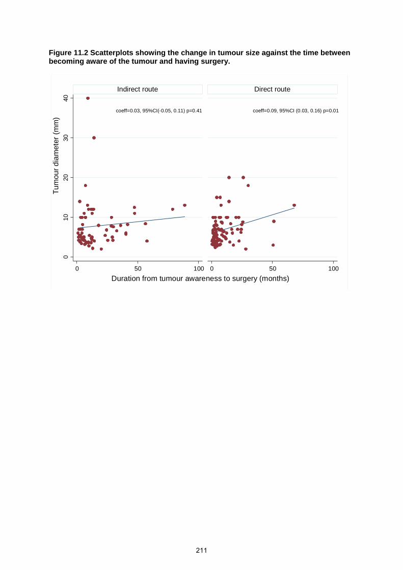
Figure 11.2 Scatterplots showing the change in tumour size against the time betweenbecoming aware of the tumour and having surgery.
coeff=0.03, 95%CI(-0.05, 0.11) p=0.41 coeff=0.09, 95%CI (0.03, 0.16) p=0.01
010
20
30
40
0 50 100 0 50 100
Indirect route Direct route
Duration from tumour awareness to surgery (months)
Tu
mou
rd
iam
ete
r(m
m)
211
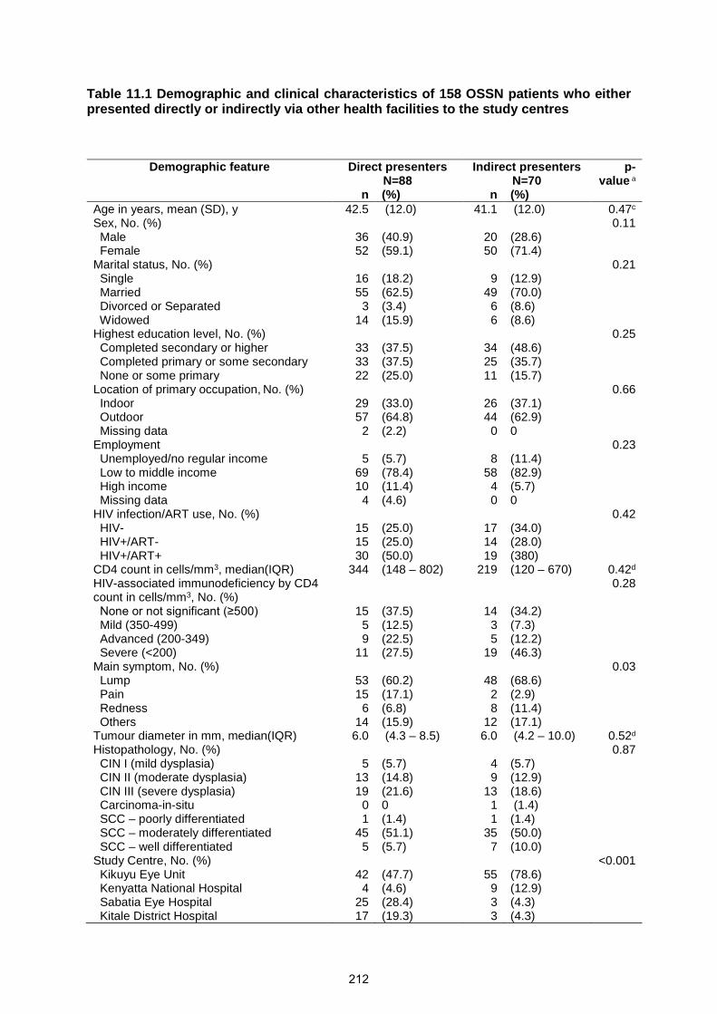
Table 11.1 Demographic and clinical characteristics of 158 OSSN patients who eitherpresented directly or indirectly via other health facilities to the study centres
Demographic feature Direct presentersN=88
Indirect presentersN=70
p-value a
n (%) n (%)Age in years, mean (SD), y 42.5 (12.0) 41.1 (12.0) 0.47c
Sex, No. (%) 0.11Male 36 (40.9) 20 (28.6)Female 52 (59.1) 50 (71.4)
Marital status, No. (%) 0.21Single 16 (18.2) 9 (12.9)Married 55 (62.5) 49 (70.0)Divorced or Separated 3 (3.4) 6 (8.6)Widowed 14 (15.9) 6 (8.6)
Highest education level, No. (%) 0.25Completed secondary or higher 33 (37.5) 34 (48.6)Completed primary or some secondary 33 (37.5) 25 (35.7)None or some primary 22 (25.0) 11 (15.7)
Location of primary occupation, No. (%) 0.66Indoor 29 (33.0) 26 (37.1)Outdoor 57 (64.8) 44 (62.9)Missing data 2 (2.2) 0 0
Employment 0.23Unemployed/no regular income 5 (5.7) 8 (11.4)Low to middle income 69 (78.4) 58 (82.9)High income 10 (11.4) 4 (5.7)Missing data 4 (4.6) 0 0
HIV infection/ART use, No. (%) 0.42HIV- 15 (25.0) 17 (34.0)HIV+/ART- 15 (25.0) 14 (28.0)HIV+/ART+ 30 (50.0) 19 (380)
CD4 count in cells/mm3, median(IQR) 344 (148 – 802) 219 (120 – 670) 0.42d
HIV-associated immunodeficiency by CD4count in cells/mm3, No. (%)
0.28
None or not significant (≥500) 15 (37.5) 14 (34.2)Mild (350-499) 5 (12.5) 3 (7.3)Advanced (200-349) 9 (22.5) 5 (12.2)Severe (<200) 11 (27.5) 19 (46.3)
Main symptom, No. (%) 0.03Lump 53 (60.2) 48 (68.6)Pain 15 (17.1) 2 (2.9)Redness 6 (6.8) 8 (11.4)Others 14 (15.9) 12 (17.1)
Tumour diameter in mm, median(IQR) 6.0 (4.3 – 8.5) 6.0 (4.2 – 10.0) 0.52d
Histopathology, No. (%) 0.87CIN I (mild dysplasia) 5 (5.7) 4 (5.7)CIN II (moderate dysplasia) 13 (14.8) 9 (12.9)CIN III (severe dysplasia) 19 (21.6) 13 (18.6)Carcinoma-in-situ 0 0 1 (1.4)SCC – poorly differentiated 1 (1.4) 1 (1.4)SCC – moderately differentiated 45 (51.1) 35 (50.0)SCC – well differentiated 5 (5.7) 7 (10.0)
Study Centre, No. (%) <0.001Kikuyu Eye Unit 42 (47.7) 55 (78.6)Kenyatta National Hospital 4 (4.6) 9 (12.9)Sabatia Eye Hospital 25 (28.4) 3 (4.3)Kitale District Hospital 17 (19.3) 3 (4.3)
212
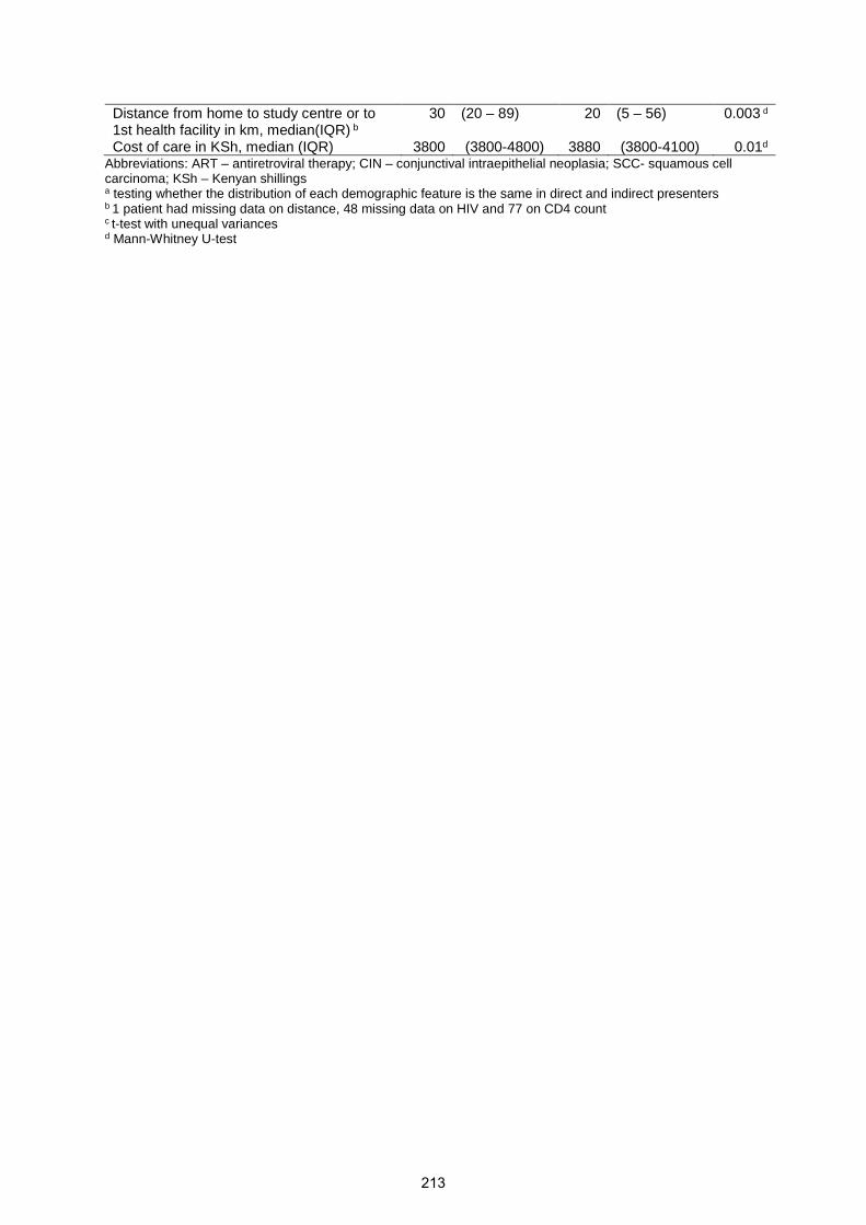
Distance from home to study centre or to1st health facility in km, median(IQR) b
30 (20 – 89) 20 (5 – 56) 0.003 d
Cost of care in KSh, median (IQR) 3800 (3800-4800) 3880 (3800-4100) 0.01d
Abbreviations: ART – antiretroviral therapy; CIN – conjunctival intraepithelial neoplasia; SCC- squamous cellcarcinoma; KSh – Kenyan shillingsa testing whether the distribution of each demographic feature is the same in direct and indirect presentersb 1 patient had missing data on distance, 48 missing data on HIV and 77 on CD4 countc t-test with unequal variancesd Mann-Whitney U-test
213
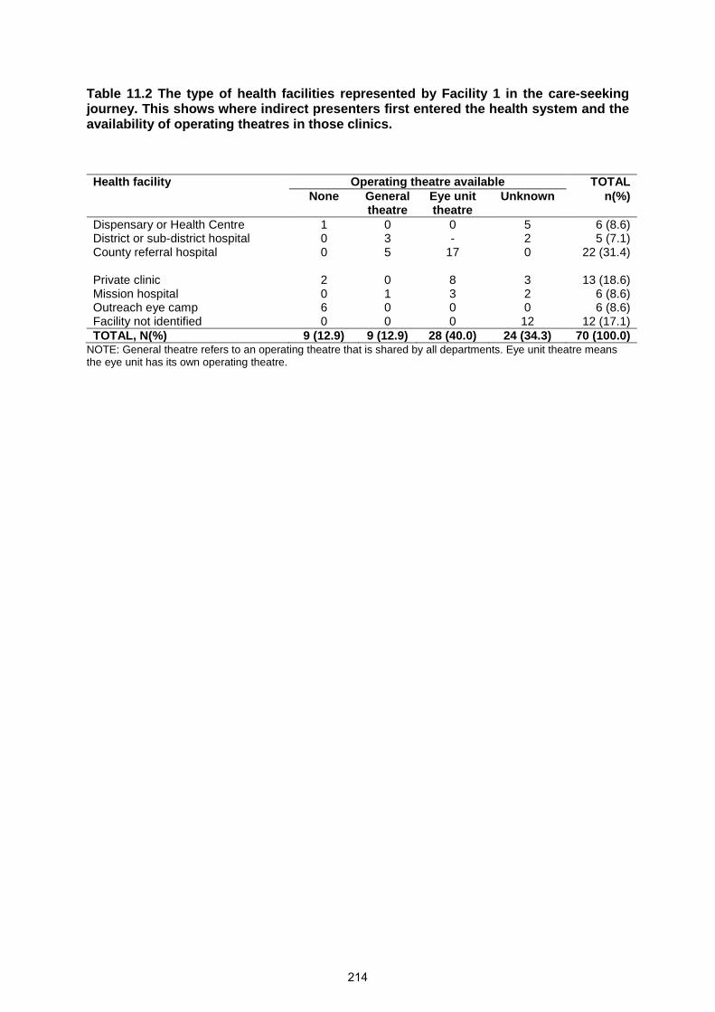
Table 11.2 The type of health facilities represented by Facility 1 in the care-seekingjourney. This shows where indirect presenters first entered the health system and theavailability of operating theatres in those clinics.
Health facility Operating theatre available TOTALNone General
theatreEye unittheatre
Unknown n(%)
Dispensary or Health Centre 1 0 0 5 6 (8.6)District or sub-district hospital 0 3 - 2 5 (7.1)County referral hospital 0 5 17 0 22 (31.4)
Private clinic 2 0 8 3 13 (18.6)Mission hospital 0 1 3 2 6 (8.6)Outreach eye camp 6 0 0 0 6 (8.6)Facility not identified 0 0 0 12 12 (17.1)TOTAL, N(%) 9 (12.9) 9 (12.9) 28 (40.0) 24 (34.3) 70 (100.0)
NOTE: General theatre refers to an operating theatre that is shared by all departments. Eye unit theatre meansthe eye unit has its own operating theatre.
214
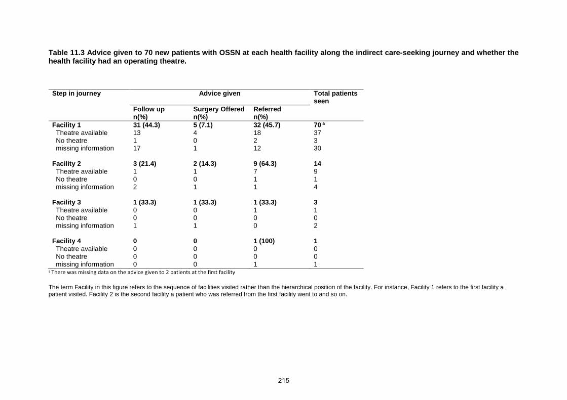
Table 11.3 Advice given to 70 new patients with OSSN at each health facility along the indirect care-seeking journey and whether thehealth facility had an operating theatre.
Step in journey Advice given Total patientsseen
Follow upn(%)
Surgery Offeredn(%)
Referredn(%)
Facility 1 31 (44.3) 5 (7.1) 32 (45.7) 70 a
Theatre available 13 4 18 37No theatre 1 0 2 3missing information 17 1 12 30
Facility 2 3 (21.4) 2 (14.3) 9 (64.3) 14Theatre available 1 1 7 9No theatre 0 0 1 1missing information 2 1 1 4
Facility 3 1 (33.3) 1 (33.3) 1 (33.3) 3Theatre available 0 0 1 1No theatre 0 0 0 0missing information 1 1 0 2
Facility 4 0 0 1 (100) 1Theatre available 0 0 0 0No theatre 0 0 0 0missing information 0 0 1 1
a There was missing data on the advice given to 2 patients at the first facility
The term Facility in this figure refers to the sequence of facilities visited rather than the hierarchical position of the facility. For instance, Facility 1 refers to the first facility apatient visited. Facility 2 is the second facility a patient who was referred from the first facility went to and so on.
215

Table 11.4 Predictors of delay in presentation of 158 new patients with ocular surface squamous neoplasia, sub-divided intoquartiles.
Factor 0.4 – 3.2months(N=38)
3.3 – 7.0months(N=41)
7.1 – 15.6months(N=40)
15.7 – 190.5months(N=39)
Univariateordered
proportional OR(95% CI)
p- value Adjusted orderedproportional OR
(95% CI)
p- value
Distance from home to first healthfacility or study centre, mean(SD) km
71 (102.0) 63 (82.3) 44 (49.6) 66 (96.3) 1.00 (0.99-1.00) 0.54 -
Total cost of care, mean (SD) Ksh 4102 (596.0) 4000 (496.3) 4218 (1594.7) 4289 (835.9) 1.00 (0.99-1.00) 0.32Care pathway followed, No (%) <0.001 <0.001Direct 27 (30.7) 25 (28.4) 19 (21.6) 17 (19.3) 1.00 (REF) 1.00 (REF))1 facility visited 11 (19.6) 15 (26.8) 18 (32.1) 12 (21.4) 1.56 (0.85-2.83) 1.38 (0.75-2.54)2 or more facilities visited 0 1 (7.1) 3 (21.4) 10 (74.1) 12.93 (3.79-44.06) 13.03 (3.78-44.94)
Sex, No. (%) 0.01Male 18 (32.1) 18 (32.1) 12 (21.4) 8 (14.3) 1.00 (REF) - 1.00 (REF)Female 20 (19.6) 23 (23.6) 28 (27.5) 31 (30.4) 2.29 (1.27-4.15) 0.006 2.31 (1.25-4.24)
Age, mean(SD) y 42 (13.0) 40 (11.8) 46 (13.6) 39 (8.3) 0.99 (0.97-1.02) 0.69 -Marital status, No. (%) 0.59 -Single 2 (8.0) 11 (44.4) 3 (12.0) 9 (36.0) 1.00 (REF)Married 28 (26.9) 24 (23.1) 31 (29.8) 21 (20.2) 0.60 (0.28-1.31)Divorced or Separated 1 (11.1) 5 (55.6) 1 (11.0) 2 (22.2) 061 (0.17-2.26)Widowed 7 (35.0) 1 (5.0) 5 (25.0) 7 (35.0) 0.81 (0.27-2.45)
Highest education level, No. (%) 0.23 -Completed secondary or higher 14 (20.9) 19 (28.4) 15 (22.4) 19 (28.4) 1.00 (REF)Completed primary or some
secondary12 (20.7) 16 (27.6) 14 (24.1) 16 (27.6) 1.01 (0.54-1.89)
None or some primary 12 (36.4) 6 (18.2) 11 (33.3) 4 (12.1) 0.55 (0.26-1.17)Occupation, No. (%) 0.95 -High income 3 (21.4) 3 (21.4) 6 (42.9) 2 (14.3) 1.00 (REF)Low-Medium income 31 (24.4) 35 (27.6) 30 (23.6) 31 (234) 0.96 (0.37-2.48)Unemployed 4 (30.8) 3 (23.1) 3 (23.1) 3 (23.1) 0.82 (0.21-3.12)
HIV infection & ART use, No. (%)a 0.07 -HIV- 11 (34.4) 10 (31.3) 6 (18.8) 5 (15.6) 1.00 (REF)HIV+/ART- 6 (20.7) 11 (37.9) 8 (27.6) 4 (13.8) 1.41 (0.58-3.40)HIV+/ART+ 11 (22.5) 9 (18.4) 12 (24.5) 17 (34.7) 2.57 (1.13-5.88)
216

Main symptom, No. (%) 1.07 (0.94-1.21) 0.32Lump 24 (23.8) 24 (23.8) 29 (28.7) 24 (23.8)Pain 7 (41.2) 5 (29.4) 2 (11.8) 3 (17.7)Redness 4 (28.6) 4 (28.6) 2 (14.3) 4 (28.6)Others 3 (11.5) 8 (30.8) 7 (26.9) 8 (308)
a There were 48 participants with missing HIV/ART data
217

Chapter 12. Topical 5-Fluorouracil (5FU)
following surgery for ocular surface squamous
neoplasia (OSSN) in Kenya: a randomised,
double-blind, placebo-controlled trial
218

RESEARCH PAPER COVER SHEET
PLEASE NOTE THAT A COVER SHEET MUST BE COMPLETED FOR EACH RESEARCH PAPER INCLUDEDIN A THESIS.
SECTION A – Student Details
Student
Principal Supervisor
Thesis Title
If the Research Paper has previously been published please complete Section B, if not please move toSection C
SECTION B – Paper already published
Where was the work published?
When was the work published?
If the work was published prior toregistration for your research degree,give a brief rationale for its inclusion
Have you retained the copyright for thework?* No
Was the work subject toacademic peer review? Yes
*If yes, please attach evidence of retention. If no, or if the work is being included in its published format, pleaseattach evidence of permission from the copyright holder (publisher or other author) to include this work.
SECTION C – Prepared for publication, but not yet published
Where is the work intended to bepublished?
Please list the paper’s authors in theintended authorship order:
Stage of publication Choose an item.
SECTION D – Multi-authored work
For multi-authored work, give full details of your role inthe research included in the paper and in the preparationof the paper. (Attach a further sheet if necessary)
Student Signature: Date:
Supervisor Signature: Date:
219
Gichuhi
Typewritten text
Stephen Gichuhi
Gichuhi
Typewritten text
Matthew J. Burton
Gichuhi
Typewritten text
Epidemiology and management of ocular surface squamous neoplasia
Gichuhi
Typewritten text
17th May 2016
Gichuhi
Typewritten text
17th May 2016
sgichuhi
Typewritten text
The Lancet Global Health
sgichuhi
Typewritten text
May 2016
sgichuhi
Typewritten text
I coordinated and actively led the field work for this paper. I recruited and followed up participants, carried out the clinical examinations and performed some of the surgeries. Field supervision was provided by Matthew Burton throughout. I also performed the statistical analysis, with some guidance from Helen Weiss. I wrote and submitted the paper with consideration of comments from co-authors.
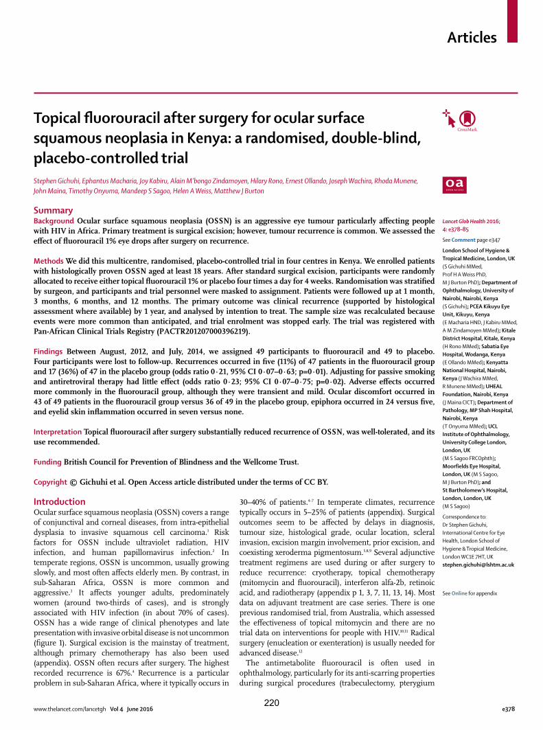
Articles
www.thelancet.com/lancetgh Vol 4 June 2016 e378
Topical fl uorouracil after surgery for ocular surface squamous neoplasia in Kenya: a randomised, double-blind, placebo-controlled trialStephen Gichuhi, Ephantus Macharia, Joy Kabiru, Alain M’bongo Zindamoyen, Hilary Rono, Ernest Ollando, Joseph Wachira, Rhoda Munene, John Maina, Timothy Onyuma, Mandeep S Sagoo, Helen A Weiss, Matthew J Burton
SummaryBackground Ocular surface squamous neoplasia (OSSN) is an aggressive eye tumour particularly aff ecting people with HIV in Africa. Primary treatment is surgical excision; however, tumour recurrence is common. We assessed the eff ect of fl uorouracil 1% eye drops after surgery on recurrence.
Methods We did this multicentre, randomised, placebo-controlled trial in four centres in Kenya. We enrolled patients with histologically proven OSSN aged at least 18 years. After standard surgical excision, participants were randomly allocated to receive either topical fl uorouracil 1% or placebo four times a day for 4 weeks. Randomisation was stratifi ed by surgeon, and participants and trial personnel were masked to assignment. Patients were followed up at 1 month, 3 months, 6 months, and 12 months. The primary outcome was clinical recurrence (supported by histological assessment where available) by 1 year, and analysed by intention to treat. The sample size was recalculated because events were more common than anticipated, and trial enrolment was stopped early. The trial was registered with Pan-African Clinical Trials Registry (PACTR201207000396219).
Findings Between August, 2012, and July, 2014, we assigned 49 participants to fl uorouracil and 49 to placebo. Four participants were lost to follow-up. Recurrences occurred in fi ve (11%) of 47 patients in the fl uorouracil group and 17 (36%) of 47 in the placebo group (odds ratio 0·21, 95% CI 0·07–0·63; p=0·01). Adjusting for passive smoking and antiretroviral therapy had little eff ect (odds ratio 0·23; 95% CI 0·07–0·75; p=0·02). Adverse eff ects occurred more commonly in the fl uorouracil group, although they were transient and mild. Ocular discomfort occurred in 43 of 49 patients in the fl uorouracil group versus 36 of 49 in the placebo group, epiphora occurred in 24 versus fi ve, and eyelid skin infl ammation occurred in seven versus none.
Interpretation Topical fl uorouracil after surgery substantially reduced recurrence of OSSN, was well-tolerated, and its use recommended.
Funding British Council for Prevention of Blindness and the Wellcome Trust.
Copyright © Gichuhi et al. Open Access article distributed under the terms of CC BY.
IntroductionOcular surface squamous neoplasia (OSSN) covers a range of conjunctival and corneal diseases, from intra-epithelial dysplasia to invasive squamous cell carcinoma.1 Risk factors for OSSN include ultraviolet radiation, HIV infection, and human papillomavirus infection.2 In temperate regions, OSSN is uncommon, usually growing slowly, and most often aff ects elderly men. By contrast, in sub-Saharan Africa, OSSN is more common and aggressive.3 It aff ects younger adults, predominately women (around two-thirds of cases), and is strongly associated with HIV infection (in about 70% of cases). OSSN has a wide range of clinical phenotypes and late presentation with invasive orbital disease is not uncommon (fi gure 1). Surgical excision is the mainstay of treatment, although primary chemotherapy has also been used (appendix). OSSN often recurs after surgery. The highest recorded recurrence is 67%.4 Recurrence is a particular problem in sub-Saharan Africa, where it typically occurs in
30–40% of patients.4–7 In temperate climates, recurrence typically occurs in 5–25% of patients (appendix). Surgical outcomes seem to be aff ected by delays in diagnosis, tumour size, histological grade, ocular location, scleral invasion, excision margin involvement, prior excision, and coexisting xeroderma pigmentosum.5,8,9 Several adjunctive treatment regimens are used during or after surgery to reduce recurrence: cryotherapy, topical chemotherapy (mitomycin and fl uorouracil), interferon alfa-2b, retinoic acid, and radiotherapy (appendix p 1, 3, 7, 11, 13, 14). Most data on adjuvant treatment are case series. There is one previous randomised trial, from Australia, which assessed the eff ectiveness of topical mitomycin and there are no trial data on interventions for people with HIV.10,11 Radical surgery (enucleation or exenteration) is usually needed for advanced disease.12
The antimetabolite fl uorouracil is often used in ophthalmology, particularly for its anti-scarring properties during surgical procedures (trabeculectomy, pterygium
Lancet Glob Health 2016; 4: e378–85
See Comment page e347
London School of Hygiene & Tropical Medicine, London, UK (S Gichuhi MMed, Prof H A Weiss PhD, M J Burton PhD); Department of Ophthalmology, University of Nairobi, Nairobi, Kenya (S Gichuhi); PCEA Kikuyu Eye Unit, Kikuyu, Kenya (E Macharia HND, J Kabiru MMed, A M Zindamoyen MMed); Kitale District Hospital, Kitale, Kenya (H Rono MMed); Sabatia Eye Hospital, Wodanga, Kenya (E Ollando MMed); Kenyatta National Hospital, Nairobi, Kenya (J Wachira MMed, R Munene MMed); UHEAL Foundation, Nairobi, Kenya (J Maina CICT); Department of Pathology, MP Shah Hospital, Nairobi, Kenya (T Onyuma MMed); UCL Institute of Ophthalmology, University College London, London, UK (M S Sagoo FRCOphth); Moorfields Eye Hospital, London, UK (M S Sagoo, M J Burton PhD); and St Bartholomew’s Hospital, London, London, UK (M S Sagoo)
Correspondence to:Dr Stephen Gichuhi, International Centre for Eye Health, London School of Hygiene & Tropical Medicine, London WC1E 7HT, [email protected]
See Online for appendix
220
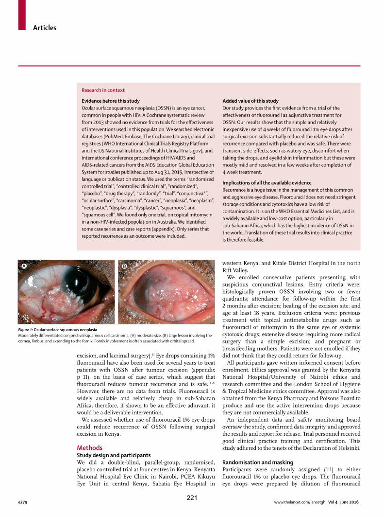
Articles
e379 www.thelancet.com/lancetgh Vol 4 June 2016
excision, and lacrimal surgery).13 Eye drops containing 1% fl uorouracil have also been used for several years to treat patients with OSSN after tumour excision (appendix p 11), on the basis of case series, which suggest that fl uorouracil reduces tumour recurrence and is safe.14–20 However, there are no data from trials. Fluorouracil is widely available and relatively cheap in sub-Saharan Africa, therefore, if shown to be an eff ective adjuvant, it would be a deliverable intervention.
We assessed whether use of fl uorouracil 1% eye drops could reduce recurrence of OSSN following surgical excision in Kenya.
MethodsStudy design and participantsWe did a double-blind, parallel-group, randomised, placebo-controlled trial at four centres in Kenya: Kenyatta National Hospital Eye Clinic in Nairobi, PCEA Kikuyu Eye Unit in central Kenya, Sabatia Eye Hospital in
western Kenya, and Kitale District Hospital in the north Rift Valley.
We enrolled consecutive patients presenting with suspicious conjunctival lesions. Entry criteria were: histologically proven OSSN involving two or fewer quadrants; attendance for follow-up within the fi rst 2 months after excision; healing of the excision site; and age at least 18 years. Exclusion criteria were: previous treatment with topical antimetabolite drugs such as fl uorouracil or mitomycin to the same eye or systemic cytotoxic drugs; extensive disease requiring more radical surgery than a simple excision; and pregnant or breastfeeding mothers. Patients were not enrolled if they did not think that they could return for follow-up.
All participants gave written informed consent before enrolment. Ethics approval was granted by the Kenyatta National Hospital/University of Nairobi ethics and research committee and the London School of Hygiene & Tropical Medicine ethics committee. Approval was also obtained from the Kenya Pharmacy and Poisons Board to produce and use the active intervention drops because they are not commercially available.
An independent data and safety monitoring board oversaw the study, confi rmed data integrity, and approved the results and report for release. Trial personnel received good clinical practice training and certifi cation. This study adhered to the tenets of the Declaration of Helsinki.
Randomisation and maskingParticipants were randomly assigned (1:1) to either fl uorouracil 1% or placebo eye drops. The fl uorouracil eye drops were prepared by dilution of fl uorouracil
Research in context
Evidence before this studyOcular surface squamous neoplasia (OSSN) is an eye cancer, common in people with HIV. A Cochrane systematic review from 2013 showed no evidence from trials for the eff ectiveness of interventions used in this population. We searched electronic databases (PubMed, Embase, The Cochrane Library), clinical trial registries (WHO International Clinical Trials Registry Platform and the US National Institutes of Health ClinicalTrials.gov), and international conference proceedings of HIV/AIDS and AIDS-related cancers from the AIDS Education Global Education System for studies published up to Aug 31, 2015, irrespective of language or publication status. We used the terms “randomized controlled trial”, “controlled clinical trial”, “randomized”, “placebo”, “drug therapy”, “randomly”, “trial”, “conjunctiva*”, “ocular surface”, “carcinoma”, “cancer”, “neoplasia”, “neoplasm”, “neoplastic”, “dysplasia”, “dysplastic”, “squamous”, and “squamous cell”. We found only one trial, on topical mitomycin in a non-HIV-infected population in Australia. We identifi ed some case series and case reports (appendix). Only series that reported recurrence as an outcome were included.
Added value of this studyOur study provides the fi rst evidence from a trial of the eff ectiveness of fl uorouracil as adjunctive treatment for OSSN. Our results show that the simple and relatively inexpensive use of 4 weeks of fl uorouracil 1% eye drops after surgical excision substantially reduced the relative risk of recurrence compared with placebo and was safe. There were transient side-eff ects, such as watery eye, discomfort when taking the drops, and eyelid skin infl ammation but these were mostly mild and resolved in a few weeks after completion of 4 week treatment.
Implications of all the available evidenceRecurrence is a huge issue in the management of this common and aggressive eye disease. Fluorouracil does not need stringent storage conditions and cytotoxics have a low risk of contamination. It is on the WHO Essential Medicines List, and is a widely available and low-cost option, particularly in sub-Saharan Africa, which has the highest incidence of OSSN in the world. Translation of these trial results into clinical practice is therefore feasible.
Figure 1: Ocular surface squamous neoplasiaModerately diff erentiated conjunctival squamous cell carcinoma, (A) moderate size, (B) large lesion involving the cornea, limbus, and extending to the fornix. Fornix involvement is often associated with orbital spread.
A B
221
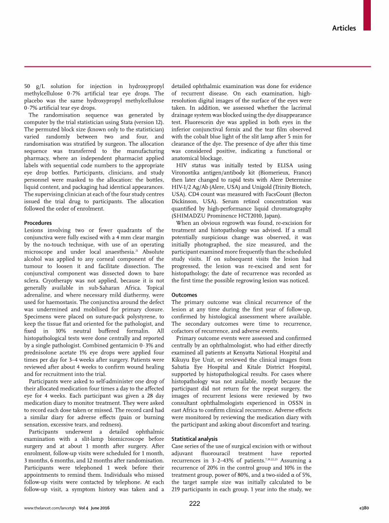
Articles
www.thelancet.com/lancetgh Vol 4 June 2016 e380
50 g/L solution for injection in hydroxypropyl methylcellulose 0·7% artifi cial tear eye drops. The placebo was the same hydroxypropyl methylcellulose 0·7% artifi cial tear eye drops.
The randomisation sequence was generated by computer by the trial statistician using Stata (version 12). The permuted block size (known only to the statistician) varied randomly between two and four, and randomisation was stratifi ed by surgeon. The allocation sequence was transferred to the manufacturing pharmacy, where an independent pharmacist applied labels with sequential code numbers to the appropriate eye drop bottles. Participants, clinicians, and study personnel were masked to the allocation: the bottles, liquid content, and packaging had identical appearances. The supervising clinician at each of the four study centres issued the trial drug to participants. The allocation followed the order of enrolment.
ProceduresLesions involving two or fewer quadrants of the conjunctiva were fully excised with a 4 mm clear margin by the no-touch technique, with use of an operating microscope and under local anaesthesia.21 Absolute alcohol was applied to any corneal component of the tumour to loosen it and facilitate dissection. The conjunctival component was dissected down to bare sclera. Cryotherapy was not applied, because it is not generally available in sub-Saharan Africa. Topical adrenaline, and where necessary mild diathermy, were used for haemostasis. The conjunctiva around the defect was undermined and mobilised for primary closure. Specimens were placed on suture-pack polystyrene, to keep the tissue fl at and oriented for the pathologist, and fi xed in 10% neutral buff ered formalin. All histopathological tests were done centrally and reported by a single pathologist. Combined gentamicin 0·3% and prednisolone acetate 1% eye drops were applied four times per day for 3–4 weeks after surgery. Patients were reviewed after about 4 weeks to confi rm wound healing and for recruitment into the trial.
Participants were asked to self-administer one drop of their allocated medication four times a day to the aff ected eye for 4 weeks. Each participant was given a 28 day medication diary to monitor treatment. They were asked to record each dose taken or missed. The record card had a similar diary for adverse eff ects (pain or burning sensation, excessive tears, and redness).
Participants underwent a detailed ophthalmic examination with a slit-lamp biomicroscope before surgery and at about 1 month after surgery. After enrolment, follow-up visits were scheduled for 1 month, 3 months, 6 months, and 12 months after randomisation. Participants were telephoned 1 week before their appointments to remind them. Individuals who missed follow-up visits were contacted by telephone. At each follow-up visit, a symptom history was taken and a
detailed ophthalmic examination was done for evidence of recurrent disease. On each examination, high-resolution digital images of the surface of the eyes were taken. In addition, we assessed whether the lacrimal drainage system was blocked using the dye disappearance test. Fluorescein dye was applied in both eyes in the inferior conjunctival fornix and the tear fi lm observed with the cobalt blue light of the slit lamp after 5 min for clearance of the dye. The presence of dye after this time was considered positive, indicating a functional or anatomical blockage.
HIV status was initially tested by ELISA using Vironostika antigen/antibody kit (Biomerieux, France) then later changed to rapid tests with Alere Determine HIV-1/2 Ag/Ab (Alere, USA) and Unigold (Trinity Biotech, USA). CD4 count was measured with FacsCount (Becton Dickinson, USA). Serum retinol concentration was quantifi ed by high-performance liquid chromatography (SHIMADZU Prominence HCT2010, Japan).
When an obvious regrowth was found, re-excision for treatment and histopathology was advised. If a small potentially suspicious change was observed, it was initially photographed, the size measured, and the participant examined more frequently than the scheduled study visits. If on subsequent visits the lesion had progressed, the lesion was re-excised and sent for histopathology; the date of recurrence was recorded as the fi rst time the possible regrowing lesion was noticed.
OutcomesThe primary outcome was clinical recurrence of the lesion at any time during the fi rst year of follow-up, confi rmed by histological assessment where available. The secondary outcomes were time to recurrence, cofactors of recurrence, and adverse events.
Primary outcome events were assessed and confi rmed centrally by an ophthalmologist, who had either directly examined all patients at Kenyatta National Hospital and Kikuyu Eye Unit, or reviewed the clinical images from Sabatia Eye Hospital and Kitale District Hospital, supported by histopathological results. For cases where histopathology was not available, mostly because the participant did not return for the repeat surgery, the images of recurrent lesions were reviewed by two consultant ophthalmologists experienced in OSSN in east Africa to confi rm clinical recurrence. Adverse eff ects were monitored by reviewing the medication diary with the participant and asking about discomfort and tearing.
Statistical analysisCase series of the use of surgical excision with or without adjuvant fl uorouracil treatment have reported recurrences in 3·2–43% of patients.7,19,22,23 Assuming a recurrence of 20% in the control group and 10% in the treatment group, power of 80%, and a two-sided α of 5%, the target sample size was initially calculated to be 219 participants in each group. 1 year into the study, we
222
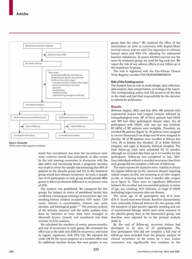
Articles
e381 www.thelancet.com/lancetgh Vol 4 June 2016
noted that recruitment was slow but recurrences were more common overall than anticipated, so after review by the trial steering committee in discussion with the data safety and monitoring board, a pragmatic decision was made to revise the sample size assuming that 30% of patients in the placebo group and 5% in the treatment group would have disease recurrence. As such, a sample size of 43 participants in each group would provide 80% power to detect an absolute diff erence in recurrence rates of 25%.
The analysis was predefi ned. We compared the two groups for balance in terms of predefi ned factors that could have a bearing on aetiology or recurrence: age, sex, smoking history, outdoor occupation, HIV status, CD4 count, vitamin A concentration, tumour size, prior excision, and histological grade.3,5,8,9 The primary analysis of the primary outcome and the safety analysis were done by intention to treat. Data were managed in Microsoft Access, cleaned, and transferred into Stata (version 12.1) for analysis.
We calculated the numbers of events, person-months, and rate of recurrence in each group. We estimated the eff ect size as the odds ratio (OR) for recurrence, estimated by logistic regression, with 95% CIs. We adjusted the crude OR for the seven surgeons as a random eff ect and for additional baseline factors that were greater in one
group than the other.24 We analysed the eff ect of the intervention on time to recurrence with Kaplan-Meier survival curves, and we used Cox regression to estimate hazard ratios and 95% CIs, adjusting for substantial baseline imbalances. To assess whether survival was the same by treatment group, we used the log rank test. We report the risk of any adverse eff ects at any follow-up in the treated eye by group.
The trial is registered with the Pan-African Clinical Trials Registry, number PACTR201207000396219.
Role of the funding sourceThe funders had no role in study design, data collection, data analysis, data interpretation, or writing of the report. The corresponding author had full access to all the data in the study and had fi nal responsibility for the decision to submit for publication.
ResultsBetween August, 2012, and July 2014, 496 patients with conjunctival lesions had surgical excision followed by histopathological tests. 187 of these patients had OSSN and 309 had other pathological disease types. For all participants with OSSN, only one eye was involved. 89 (48%) of 187 patients were ineligible. Therefore, we enrolled 98 patients (fi gure 2). 49 patients were assigned to receive fl uorouracil eye drops and 49 were assigned to placebo. 58 of 98 patients were enrolled at Kikuyu Eye Unit, 20 at Sabatia Eye Hospital, 12 at Kitale District Hospital, and eight at Kenyatta National Hospital. The fi nal follow-up visits were scheduled for 12 months, although we included data from up to 13 months for late participants. Follow-up was completed in July, 2015. Four individuals without a recorded recurrence (two from each group) did not complete a full year of follow-up.
The main reasons for exclusion were: inability to return for regular follow-up (n=41), extensive disease requiring radical surgery (n=24), not returning at all after surgery (n=16), or returning more than 2 months after surgery (n=3; fi gure 2). There were no signifi cant diff erences between the enrolled and non-enrolled patients in terms of age, sex, smoking, HIV infection, or stage of OSSN (excluding larger tumours; data not shown).
The mean age of all participants was 41·0 years (SD 11·3) and most were female. Baseline characteristics were reasonably balanced between the two groups with the exception of past passive cigarette smoking and use of antiretroviral therapy, which were more common in the placebo group than in the fl uorouracil group, and therefore were adjusted for in the primary analysis (table 1).
By the end of follow-up, recurrent lesions had developed in 22 eyes of 22 participants. The four participants who did not complete a full year of follow-up were excluded from the primary analysis of clinical recurrence of the lesion by 1 year. Lesion recurrence was signifi cantly less common in the
Figure 2: Trial profi leOSSN=ocular surface squamous neoplasia.
496 patients assessed for eligibility
187 OSSN confirmed
98 randomly assigned
49 allocated to and received fluorouracil 49 allocated to and received placebo
47 included in the analysis 47 included in the analysis
2 lost to follow-up 2 lost to follow-up
309 did not have OSSN
89 excluded 41 anticipated difficulty with follow-up 24 had extensive disease >two quadrants 16 did not return after surgery 3 returned more than 2 months after surgery 1 prior use of mitomycin eye drops 1 pregnant 1 breastfeeding 1 declined consent 1 sphenoid wing meningioma (referred)
223

Articles
www.thelancet.com/lancetgh Vol 4 June 2016 e382
fl uorouracil group (fi ve [11%] of 47 patients) than in the placebo group (17 [36%] of 47 patients; crude OR 0·21, 95% CI 0·07–0·63; p=0·01). This eff ect remained signifi cant when adjusted for use of antiretroviral therapy and passive cigarette smoking (OR 0·23, 95% CI 0·07–0·75; p=0·02). The relative risk of recurrence was reduced by 70·7% and the absolute diff erence was 25·6%. Treatment with fl uorouracil after surgery for four patients would therefore prevent an estimated one recurrence (number needed to treat 3·9, 95% CI 2·4–11·8).
16 of 22 recurrent lesions underwent repeated surgical excision and recurrent OSSN was confi rmed by histopathology in all cases. Re-excision was not done for six recurrent cases: four participants in the placebo group did not return for repeat surgery or further follow-up after the recurrence was noted, and two participants in the placebo group died before re-excision could be done (one from a presumed myocardial infarction and one from HIV-related complications). The images of these cases were reviewed by two ophthalmologists experienced in OSSN in east Africa. All were judged to be recurrent OSSN disease on clinical grounds, in the context of previous histologically confi rmed OSSN.
There was a signifi cant diff erence in the recurrence rate between the two groups over the follow-up period (hazard ratio [HR] 0·24, 95% CI 0·09–0·66; p=0·01; fi gure 3), which changed slightly after adjusting for smoking and use of antiretroviral therapy (HR 0·32, 95% CI 0·11–0·95; p=0·04). The test for proportional hazards assumption showed that the assumption of proportionality was appropriate (p=0·59). Median tumour-free survival was 7·3 months (IQR 2·3–13·5) in the fl uorouracil group and 4·8 months (3·0–7·6) in the placebo group but the diff erence was not statistically signifi cant (p=0·23).
Sensitivity analysis, assuming the four participants who did not complete 1 year of follow-up did not have a
Fluorouracil group (n=49)
Placebo group (n=49)
Sex
Male 17 (35%) 14 (29%)
Female 32 (65%) 35 (71%)
Age (years) 39·1 (9·2) 42·9 (13·0)
Marital status*
Single 6 (12%) 11 (22%)
Married 32 (65%) 28 (58%)
Divorced or separated 4 (8%) 2 (4%)
Widowed 7 (14%) 7 (14%)
Formal education (years)*
Tertiary (>12) 5 (10%) 4 (8%)
Secondary completed (12) 18 (37%) 20 (41%)
Some secondary (8–12) 4 (8%) 6 (12%)
Primary completed (8) 13 (27%) 11 (22%)
Some primary (<8) 6 (12%) 4 (8%)
None 3 (6%) 3 (6%)
Past cigarette smoking*
No 41 (84%) 32 (67%)
Yes 5 (10%) 4 (8%)
Passive (spouse or partner smokes) 3 (6%) 12 (25%)
Current cigarette smoking†
No 44 (90%) 41 (87%)
Yes 3 (6%) 0 (0%)
Passive (spouse or partner smokes) 2 (4%) 6 (13%)
Cigarettes smoked daily 9 (6) 13 (13)
Years of cigarette smoking 10·7 (6·8) 16·2 (7·4)
Location of current occupation*
Indoors 20 (41%) 19 (40%)
Outdoors 29 (59%) 29 (60%)
Wears hat or cap outdoors† 8 (16%) 6 (13%)
Wears sunglasses outdoors† 4 (8%) 4 (9%)
HIV infection‡ 29 (63%) 31 (71%)
Use of antiretroviral therapy§ 10 (22%) 19 (42%)
CD4 count (cells per μL)¶ 444 (370) 460 (421)
Serum retinol concentration (μg/L)|| 489 (157) 529 (215)
Vitamin A defi ciency (serum retinol <300 μg/L)||
3 (6%) 3 (6%)
Tumour diameter (mm) 5·9 (2·6) 6·3 (2·4)
Prior excision 8 (16%) 9 (19%)
Histological grading of tumours
CIN 1 4 (8%) 4 (8%)
CIN 2 13 (27%) 8 (16%)
CIN 3 11 (22%) 14 (29%)
Carcinoma in situ 0 (0%) 1 (2%)
Poorly diff erentiated squamous cell carcinoma
1 (2%) 1 (2%)
Moderately diff erentiated squamous cell carcinoma
17 (35%) 18 (37%)
Well diff erentiated squamous cell carcinoma
3 (6%) 3 (6%)
Surgical margin involvement 21 (43%) 19 (39%)
(Table 1 continues in next column)
Fluorouracil group (n=49)
Placebo group (n=49)
(Continued from previous column)
Stage of OSSN**
T1N0M0 15 (31%) 9 (18%)
T2N0M0 10 (20%) 9 (18%)
T3N0M0 23 (47%) 31 (63%)
T3N1M0 1 (2%) 0 (0%)
Data are n (%) or mean (SD). *Data missing for one participant in the placebo group. †Data missing for two participants in the placebo group. ‡Data missing for three participants in the fl uorouracil group and fi ve in the placebo group. §Data missing for three participants in the fl uorouracil group and four in the placebo group. ¶Data missing for eight participants in the fl uorouracil group and 11 in the placebo group. ||Data missing for ten participants in the fl uorouracil group and 13 in the placebo group. **As per the American Joint Committee on Cancer. CIN=cervical intra-epithelial neoplasia. OSSN=ocular surface squamous neoplasia.
Table 1: Baseline characteristics
224
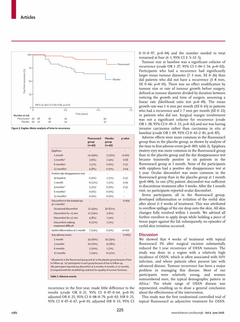
Articles
e383 www.thelancet.com/lancetgh Vol 4 June 2016
recurrence in the fi rst year, made little diff erence to the results (crude OR 0·21, 95% CI 0·07–0·64, p=0·01; adjusted OR 0·25, 95% CI 0·08–0·79, p=0·02; HR 0·25, 95% CI 0·07–0·67, p=0·01; adjusted HR 0·33, 95% CI
0·11–0·97, p=0·04) and the number needed to treat remained at four (4·1, 95% CI 2·5–12·5).
Tumour size at baseline was a signifi cant cofactor of recurrence (crude OR 1·27, 95% CI 1·04–1·54, p=0·02). Participants who had a recurrence had signifi cantly larger mean tumour diameter (7·3 mm, SE 0·26) than did patients who did not have a recurrence (5·8 mm, SE 0·66; p=0·01). There was no eff ect modifi cation by tumour size or rate of tumour growth before surgery, defi ned as tumour diameter divided by duration between noticing the growth and time of surgery, assuming a linear rate (likelihood ratio test p=0·49). The mean growth rate was 1·6 mm per month (SE 0·61) in patients who had a recurrence and 1·7 mm per month (SE 0·22) in patients who did not. Surgical margin involvement was not a signifi cant cofactor for recurrence (crude OR 1·28, 95% CI 0·49–3·33, p=0·62) and nor was having invasive carcinoma rather than carcinoma in situ at baseline (crude OR 1·09, 95% CI 0·42–2·81, p=0·87).
Adverse eff ects were more common in the fl uorouracil group than in the placebo group, as shown by analysis of the time to fi rst adverse event (p=0·005; table 2). Epiphora (watery eye) was more common in the fl uorouracil group than in the placebo group and the dye disappearance test became transiently positive in six patients in the fl uorouracil group at 1 month. None of the participants with epiphora had a positive dye disappearance test at 1 year. Ocular discomfort was more common in the fl uorouracil group than in the placebo group at 1 month (p=0·004). In one (2%) patient, discomfort was suffi cient to discontinue treatment after 3 weeks. After the 1 month visit, no participants reported ocular discomfort.
Seven participants, all in the fl uorouracil group, developed infl ammation or irritation of the eyelid skin after about 2–3 weeks of treatment. This was attributed to overfl ow spillage of the eye drop onto the skin. All skin changes fully resolved within 1 month. We advised all further enrollees to apply drops while holding a piece of tissue paper against the lid; subsequently, no episodes of eyelid skin irritation occurred.
DiscussionWe showed that 4 weeks of treatment with topical fl uorouracil 1% after surgical excision substantially reduced the 1 year recurrence of OSSN tumours. The study was done in a region with a relatively high incidence of OSSN, which is often associated with HIV infection, and where patients often present late with advanced disease. Tumour recurrence has been a major problem in managing this disease. Most of our participants were relatively young, and women outnumbered men, the typical demographic pattern in Africa.3 The whole range of OSSN disease was represented, enabling us to draw a general conclusion about the eff ectiveness of the intervention.
This study was the fi rst randomised controlled trial of topical fl uorouracil as adjunctive treatment for OSSN.
Figure 3: Kaplan-Meier analysis of time to recurrence
Number at riskFluorouracil
Placebo
Fluorouracil
0 0·5 1·0 1·5
4949
4841
4640
4534
4432
Time (years)
HR 0·24, 95% CI 0·09–0·66, p=0·010
25
50
75
100
Free
from
recu
rrenc
e (%
)
Placebo
Fluorouracil group (n=49)
Placebo group (n=49)
p value
Epiphora ·· ·· ··
1 month 24 (49%) 5 (10%) <0·001
3 months* 3 (6%) 2 (4%) 0·66
6 months† 1 (2%) 0 (0%) 0·32
12 months† 4 (8%) 0 (0%) 0·04
Positive dye disappearance test ·· ·· ··
At baseline 0 (0%) 1 (2%) 0·32
1 month 6 (12%) 1 (2%) 0·05
3 months* 1 (2%) 0 (0%) 0·32
6 months† 0 (0%) 0 (0%) -
12 months† 0 (0%) 0 (0%) -
Discomfort in the treated eye at 1 month‡
·· ·· 0·004
Occasional discomfort 21 (43%) 30 (61%) ··
Discomfort for <5 min 12 (24%) 3 (6%) ··
Discomfort for ≥5 min 4 (8%) 2 (4%) ··
Discomfort making treatment diffi cult
6 (12%) 1 (2%) ··
Eyelid infl ammation at 1 month 7 (14%) 0 (0%) <0·001
Any adverse event ·· ·· 0·005§
1 month 34 (69%) 19 (39%) ··
3 months 10 (20%) 9 (18%) ··
6 months 5 (10%) 1 (2%) ··
12 months 7 (14%) 6 (12%) ··
*48 patients in the fl uorouracil group and 47 in the placebo group because of loss to follow-up. †47 participants in each group because of loss to follow-up. ‡No participant reported any discomfort at 3 months, 6 months, or 12 months. §Computed with the stratifi ed log-rank test for equality of survivor functions.
Table 2: Adverse events
225
Articles
www.thelancet.com/lancetgh Vol 4 June 2016 e384
However, our results are consistent with those of non-randomised case series of adjuvant fl uorouracil, which have reported similarly low proportions of recurrence.14–20 Some investigators have reported the eff ectiveness of fl uorouracil as primary treatment in presumed OSSN lesions without surgical excision and histopathological assessment.14 We chose to test fl uorouracil in this setting because it is cheap and readily available with a history of use and wide acceptance for other types of ophthalmic surgery. Fluorouracil is on WHO’s list of essential drugs. It does not require stringent storage conditions such as refrigeration. Therefore, the translation of this result into clinical practice, given the resource limitations of the Kenyan health system and other similar settings, is realistic. Because surgical cryotherapy is not routinely available, topical fl uorouracil is therefore an alternative strategy to prevent recurrence.
The only other randomised study of treatment for OSSN was a placebo-controlled, crossover trial of topical mitomycin for 48 patients from Australia.10 However, that study has several distinct diff erences to our study, which probably limit the relevance to settings such as Kenya. First, only partial incisional biopsy samples were taken for diagnosis; the tumours were not excised. Given the advanced and aggressive disease in Africa, complete surgical removal of the lesion is the preferred approach. Second, the casemix was diff erent: patients with squamous cell carcinoma were excluded from the Australia study, the population group was older than ours (mean age 67 years), predominantly male (75%), and probably not infected with HIV (no data were provided). Although the lesions regressed clinically on treatment with mitomycin, more than half of patients had persistent OSSN on repeat histological assessment of the lesion site 1 year after treatment.
Overall, the use of topical fl uorouracil was associated with transient side-eff ects: watery eye, discomfort when taking the drops, and eyelid skin infl ammation. However, these were mostly mild and resolved after the completion of treatment. A transiently positive dye disappearance test indicates temporary reversible obstruction of the nasolacrimal duct, a known complication of fl uorouracil treatment.18 The most signifi cant adverse eff ect was the eyelid skin infl ammation. This was reliably prevented by protecting the skin with a tissue while applying the drops to catch any overfl ow. Epiphora was reported by 10% of participants at 1 month in the placebo group and 61% reported occasional discomfort at 1 month. We think that these eff ects were not caused by the placebo, which was a bland lubricant, but rather related to having recently had, often extensive, excision surgery to remove a tumour. It is quite common for excision of conjunctival lesions, OSSN or other pathology such as a pterygium, to result in a degree of ocular surface infl ammation and irritation that can persist for several weeks. Such eff ects are especially common in young people of African origin, who are more likely to scar and have infl ammation than
are older white patients.25 Overall, we think that these side-eff ects can be partly mitigated, do not usually represent a problem after cessation of treatment, and are outweighed by the benefi t of the reduced tumour recurrence.
Adjunctive fl uorouracil probably works through its eff ect on residual OSSN cells that are left after surgical excision. It interferes with DNA and RNA processes through several active metabolites.26 Fluorodeoxyuridine monophosphate inhibits thymidylate synthase, blocking thymidylate production, and thus DNA replication. Rapidly dividing neoplastic cells are much more vulnerable to thymidine depletion than are normal cells. Fluorodeoxyuridine triphosphate is misincorporated into DNA and fl uorouridine triphosphate is misincorporated into RNA. These diff erent metabolites disrupt crucial cellular mechanisms, triggering apoptosis.
We were able to follow up participants to 1 year. This was attributable to two factors. First, we excluded people who said that they would be unlikely to return for follow-up. Second, the study team were careful to build and maintain good relationships with study participants, and actively communicated with those who missed follow-up visits.
Our study has several limitations. First, recruitment was slower than anticipated, resulting in a smaller study than originally anticipated. The initial study size was based on previously reported recurrence rates from several case series, with heterogeneous inclusion criteria, treatment regimens, and follow-up.4,7,19,22,23,27,28 However, the higher than expected recurrence rate and high retention enabled us to have good power with a smaller sample size. In common with other studies from Africa, the recurrence rate in both groups was higher than that reported in many case series from temperate countries. This diff erence is possibly because OSSN in this population, with a high proportion of patients who are HIV positive, is more aggressive and patients probably present later with more advanced disease.
Second, not all the cases of clinical recurrence had re-excision and histopathological tests done. However, all suspected recurrences for which histopathological results were available were confi rmed as recurrent OSSN, which suggests that our clinical judgment in this situation is highly concordant with the pathology.
Third, we excluded a high proportion of potential participants. The most common reason for exclusion was that the patient was unlikely to return for follow-up. Excluding those who did not think they could return helped us achieve good follow-up among those who were enrolled in the trial. However, there was no systematic diff erence between participants and excluded patients in terms of age, sex, HIV status, smoking status, or OSSN grade (when those with large lesions requiring alternative radical surgery were excluded). This fi nding suggests that our results can be generalised. The participants who were lost to follow-up attended at least the fi rst visit after
226
Articles
e385 www.thelancet.com/lancetgh Vol 4 June 2016
randomisation and were recurrence-free at that point. The challenge of ensuring high follow-up rates in Kenyans with HIV has been reported previously.29
Fourth, we excluded individuals with very large tumours that required either enucleation or exenteration. This exclusion could reduce the generalisability of the fi ndings. However, such patients are not suitable for less radical surgery and topical chemotherapy, as the tumour is already invading the deeper tissues of the orbit. Finally, there were some diff erences in adverse events by group, which could have led to unmasking. However, we think that this is unlikely: discomfort was common and similar in each group at 1 month, and eyelid infl ammation, positive dye disappearance test, and epiphora after 1 month were uncommon.
In conclusion, 4 weeks of topical fl uorouracil 1% after surgical excision of OSSN substantially reduced the 1 year recurrence of tumours. The treatment is safe, generally well tolerated, and easy to use. Fluorouracil is widely available, aff ordable, and easy to formulate into eye drops. It is suitable in settings without cryotherapy. Fluorouracil eye drops are an eff ective and realistic intervention to improve outcomes for people with OSSN.ContributorsSG, MSS, HAW, and MJB designed the study and interpreted the data. SG did the literature search. SG, EM, JK, AMZ, HR, EO, JW, RM, JM, and TO collected data. SG, HAW, and MJB analysed data and obtained funding. SG wrote the fi rst draft of the Article, all authors revised it. JK, AMZ, HR, EO, JW, RM, and JM provided administrative, technical, and material support. HAW and MJB supervised the study.
Declaration of interestsWe declare no competing interests.
AcknowledgmentsSG received funding from the British Council for Prevention of Blindness fellowship programme. MJB is supported by the Wellcome Trust (grant number 098481/Z/12/Z).
References 1 Lee GA, Hirst LW. Ocular surface squamous neoplasia.
Surv Ophthalmol 1995; 39: 429–50. 2 Gichuhi S, Ohnuma S, Sagoo MS, Burton MJ. Pathophysiology of
ocular surface squamous neoplasia. Exp Eye Res 2014; 129: 172–82. 3 Gichuhi S, Sagoo MS, Weiss HA, Burton MJ. Epidemiology of
ocular surface squamous neoplasia in Africa. Trop Med Int Health 2013; 18: 1424–43.
4 Birkholz ES, Goins KM, Sutphin JE, Kitzmann AS, Wagoner MD. Treatment of ocular surface squamous cell intraepithelial neoplasia with and without mitomycin C. Cornea 2011; 30: 37–41.
5 Chebbi A, Bouguila H, Boukari M, et al. Prognosis of primary malignant tumors of the conjunctiva. J Fr Ophtalmol 2015; 38: 477–85 (in French).
6 Bonanno A, Esmaeli B, Fingeret MC, Nelson DV, Weber RS. Social challenges of cancer patients with orbitofacial disfi gurement. Ophthal Plast Reconstr Surg 2010; 26: 18–22.
7 Chisi SK, Kollmann MK, Karimurio J. Conjunctival squamous cell carcinoma in patients with human immunodefi ciency virus infection seen at two hospitals in Kenya. East Afr Med J 2006; 83: 267–70.
8 Galor A, Karp CL, Oellers P, et al. Predictors of ocular surface squamous neoplasia recurrence after excisional surgery. Ophthalmology 2012; 119: 1974–81.
9 Sudesh S, Rapuano CJ, Cohen EJ, Eagle RC Jr, Laibson PR. Surgical management of ocular surface squamous neoplasms: the experience from a cornea center. Cornea 2000; 19: 278–83.
10 Hirst LW. Randomized controlled trial of topical mitomycin C for ocular surface squamous neoplasia: early resolution. Ophthalmology 2007; 114: 976–82.
11 Gichuhi S, Irlam JH. Interventions for squamous cell carcinoma of the conjunctiva in HIV-infected individuals. Cochrane Database Syst Rev 2013; 2: CD005643.
12 Ogun GO, Ogun OA, Bekibele CO, Akang EE. Intraepithelial and invasive squamous neoplasms of the conjunctiva in Ibadan, Nigeria: a clinicopathological study of 46 cases. Int Ophthalmol 2009; 29: 401–09.
13 Abraham LM, Selva D, Casson R, Leibovitch I. The clinical applications of fl uorouracil in ophthalmic practice. Drugs 2007; 67: 237–55.
14 Nutt RJ, Clements JL, Dean WH. Ocular surface squamous neoplasia in HIV-positive and HIV-negative patients and response to 5-fl uorouracil in Angola. Clin Ophthalmol 2014; 8: 2435–40.
15 Bahrami B, Greenwell T, Muecke JS. Long-term outcomes after adjunctive topical 5-fl urouracil or mitomycin C for the treatment of surgically excised, localized ocular surface squamous neoplasia. Clin Experiment Ophthalmol 2014; 42: 317–22.
16 Al-Barrag A, Al-Shaer M, Al-Matary N, Al-Hamdani M. 5-Fluorouracil for the treatment of intraepithelial neoplasia and squamous cell carcinoma of the conjunctiva, and cornea. Clin Ophthalmol 2010; 4: 801–08.
17 Midena E. Treatment of conjunctival squamous cell carcinoma with topical 5-fl uorouracil. Br J Ophthalmol 2000; 84: 268–72.
18 Rudkin AK, Muecke JS. Adjuvant 5-fl uorouracil in the treatment of localised ocular surface squamous neoplasia. Br J Ophthalmol 2011; 95: 947–50.
19 Yeatts RP, Engelbrecht NE, Curry CD, Ford JG, Walter KA. 5-Fluorouracil for the treatment of intraepithelial neoplasia of the conjunctiva and cornea. Ophthalmology 2000; 107: 2190–95.
20 Parrozzani R, Lazzarini D, Alemany-Rubio E, Urban F, Midena E. Topical 1% 5-fl uorouracil in ocular surface squamous neoplasia: a long-term safety study. Br J Ophthalmol 2011; 95: 355–59.
21 Shields CL, Shields JA. Tumors of the conjunctiva and cornea. Surv Ophthalmol 2004; 49: 3–24.
22 Tabin G, Levin S, Snibson G, Loughnan M, Taylor H. Late recurrences and the necessity for long-term follow-up in corneal and conjunctival intraepithelial neoplasia. Ophthalmology 1997; 104: 485–92.
23 Waddell KM, Downing RG, Lucas SB, Newton R. Corneo-conjunctival carcinoma in Uganda. Eye (Lond) 2006; 20: 893–99.
24 Kahan BC. Accounting for centre-eff ects in multicentre trials with a binary outcome—when, why, and how? BMC Med Res Methodol 2014; 14: 20.
25 Broadway D, Grierson I, Hitchings R. Racial diff erences in the results of glaucoma fi ltration surgery: are racial diff erences in the conjunctival cell profi le important? Br J Ophthalmol 1994; 78: 66–75.
26 Longley DB, Harkin DP, Johnston PG. 5-Fluorouracil: mechanisms of action and clinical strategies. Nat Rev Cancer 2003; 3: 330–38.
27 Zaki AA, Farid SF. Management of intraepithelial and invasive neoplasia of the cornea and conjunctiva: a long-term follow up. Cornea 2009; 28: 986–88.
28 Russell HC, Chadha V, Lockington D, Kemp EG. Topical mitomycin C chemotherapy in the management of ocular surface neoplasia: a 10-year review of treatment outcomes and complications. Br J Ophthalmol 2010; 94: 1316–21.
29 Hassan AS, Fielding KL, Thuo NM, Nabwera HM, Sanders EJ, Berkley JA. Early loss to follow-up of recently diagnosed HIV-infected adults from routine pre-ART care in a rural district hospital in Kenya: a cohort study. Trop Med Int Health 2012; 17: 82–93.
227

General discussion and future work
228

Chapter 13. Discussion
Ocular surface squamous neoplasia is a complex multifaceted problem. (Figure 13.1) This
project aimed to address some of the knowledge gaps in this complex web. The most
prominent problems we see with regard to OSSN in sub-Saharan Africa are a high incidence,
recurrences after surgery and delayed presentation. This discussion will take a logical
framework approach examining each problem in turn and delving into the underlying causes
as shown in Figure 13.1.
The systematic review we conducted on the epidemiology of OSSN confirmed that the
incidence in Africa is the highest in the world and that women here were at similar risk as men
unlike other regions of the world where male disease predominated. The case-control study
we conducted showed that in the Kenyan setting this situation is driven by a combination of
modifiable risk factors all which are more common in Africa than in other parts of the world,
namely, HIV, exposure to solar UV radiation outdoors and allergic conjunctivitis. We also
identified that wearing hats was protective. The role of HPV remains unclear as studies show
a lot of variation. It is also still unclear why the incidence is equally high in both males and
females but prevalence seems higher in females. It may be related to HIV being a strong risk
factor for OSSN. Females have a higher biological susceptibility to HIV and other sexually
transmitted infections (STI) which may contribute to the high incidence.1 Women survive
longer with HIV than men perhaps from better ART compliance and acceptance which would
explain the high prevalence.2 The pathophysiology review implicated the limbal stem cells as
the possible nidus of neoplastic transformation in OSSN. The nasal limbus is particularly
susceptible to UV radiation. The finding of allergic conjunctivitis as a risk factor is new yet
consistent with the limbal stem cells being the origin of OSSN. The findings of this review
closely reflect what the case-control study found. Knowledge on how the risk factors interact
and the lead time between exposure and disease is evolving. However it is still not clear why
OSSN is often unilateral. Possible entry points for prevention would therefore need to focus
on women especially in prevention and control of HIV, wearing hats while outdoors and
effective control of allergic conjunctivitis in children and adolescents. A better understanding
of the pathophysiology would also highlight more options for therapeutic intervention focusing
on stem cells and genetic modification rather than just the rapidly dividing tumour cells that
current therapies seem to target.
229
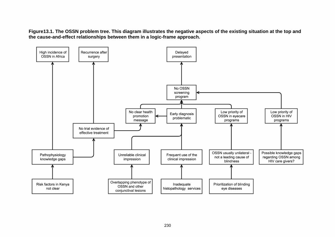
Figure13.1. The OSSN problem tree. This diagram illustrates the negative aspects of the existing situation at the top andthe cause-and-effect relationships between them in a logic-frame approach.
230
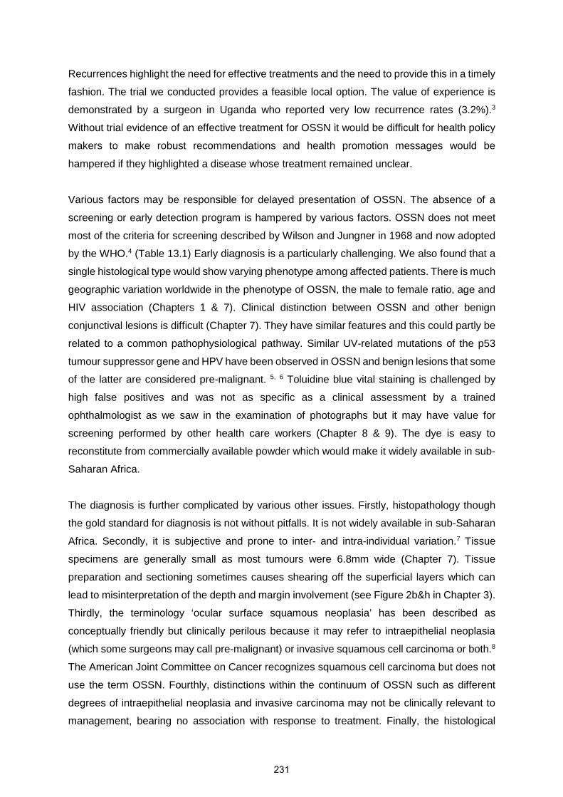
Recurrences highlight the need for effective treatments and the need to provide this in a timely
fashion. The trial we conducted provides a feasible local option. The value of experience is
demonstrated by a surgeon in Uganda who reported very low recurrence rates (3.2%).3
Without trial evidence of an effective treatment for OSSN it would be difficult for health policy
makers to make robust recommendations and health promotion messages would be
hampered if they highlighted a disease whose treatment remained unclear.
Various factors may be responsible for delayed presentation of OSSN. The absence of a
screening or early detection program is hampered by various factors. OSSN does not meet
most of the criteria for screening described by Wilson and Jungner in 1968 and now adopted
by the WHO.4 (Table 13.1) Early diagnosis is a particularly challenging. We also found that a
single histological type would show varying phenotype among affected patients. There is much
geographic variation worldwide in the phenotype of OSSN, the male to female ratio, age and
HIV association (Chapters 1 & 7). Clinical distinction between OSSN and other benign
conjunctival lesions is difficult (Chapter 7). They have similar features and this could partly be
related to a common pathophysiological pathway. Similar UV-related mutations of the p53
tumour suppressor gene and HPV have been observed in OSSN and benign lesions that some
of the latter are considered pre-malignant. 5, 6 Toluidine blue vital staining is challenged by
high false positives and was not as specific as a clinical assessment by a trained
ophthalmologist as we saw in the examination of photographs but it may have value for
screening performed by other health care workers (Chapter 8 & 9). The dye is easy to
reconstitute from commercially available powder which would make it widely available in sub-
Saharan Africa.
The diagnosis is further complicated by various other issues. Firstly, histopathology though
the gold standard for diagnosis is not without pitfalls. It is not widely available in sub-Saharan
Africa. Secondly, it is subjective and prone to inter- and intra-individual variation.7 Tissue
specimens are generally small as most tumours were 6.8mm wide (Chapter 7). Tissue
preparation and sectioning sometimes causes shearing off the superficial layers which can
lead to misinterpretation of the depth and margin involvement (see Figure 2b&h in Chapter 3).
Thirdly, the terminology ‘ocular surface squamous neoplasia’ has been described as
conceptually friendly but clinically perilous because it may refer to intraepithelial neoplasia
(which some surgeons may call pre-malignant) or invasive squamous cell carcinoma or both.8
The American Joint Committee on Cancer recognizes squamous cell carcinoma but does not
use the term OSSN. Fourthly, distinctions within the continuum of OSSN such as different
degrees of intraepithelial neoplasia and invasive carcinoma may not be clinically relevant to
management, bearing no association with response to treatment. Finally, the histological
231
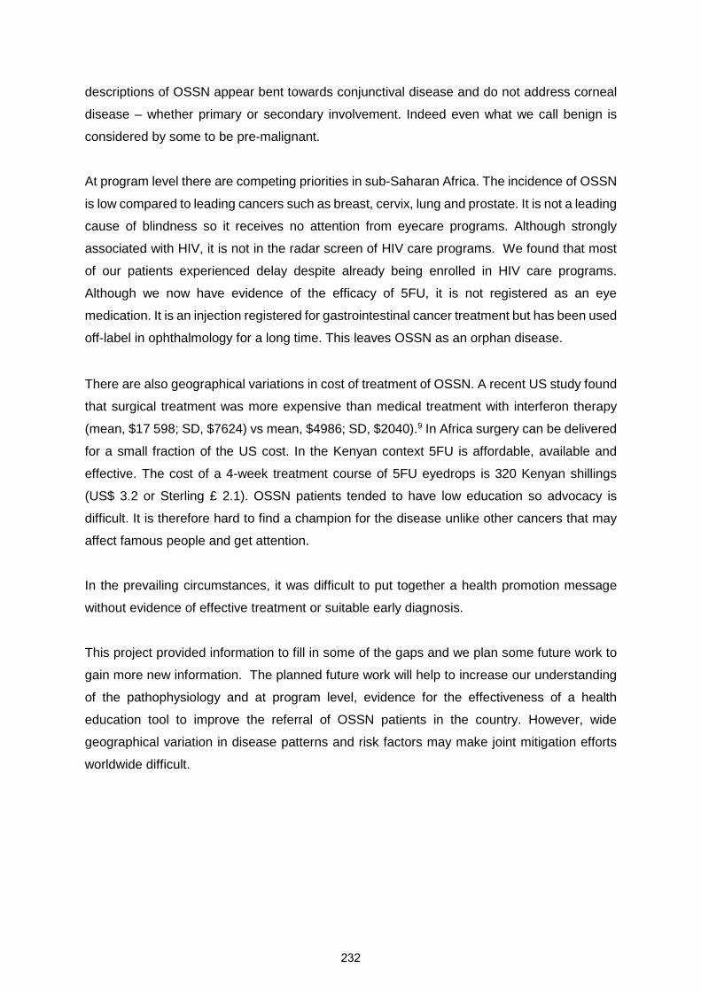
descriptions of OSSN appear bent towards conjunctival disease and do not address corneal
disease – whether primary or secondary involvement. Indeed even what we call benign is
considered by some to be pre-malignant.
At program level there are competing priorities in sub-Saharan Africa. The incidence of OSSN
is low compared to leading cancers such as breast, cervix, lung and prostate. It is not a leading
cause of blindness so it receives no attention from eyecare programs. Although strongly
associated with HIV, it is not in the radar screen of HIV care programs. We found that most
of our patients experienced delay despite already being enrolled in HIV care programs.
Although we now have evidence of the efficacy of 5FU, it is not registered as an eye
medication. It is an injection registered for gastrointestinal cancer treatment but has been used
off-label in ophthalmology for a long time. This leaves OSSN as an orphan disease.
There are also geographical variations in cost of treatment of OSSN. A recent US study found
that surgical treatment was more expensive than medical treatment with interferon therapy
(mean, $17 598; SD, $7624) vs mean, $4986; SD, $2040).9 In Africa surgery can be delivered
for a small fraction of the US cost. In the Kenyan context 5FU is affordable, available and
effective. The cost of a 4-week treatment course of 5FU eyedrops is 320 Kenyan shillings
(US$ 3.2 or Sterling £ 2.1). OSSN patients tended to have low education so advocacy is
difficult. It is therefore hard to find a champion for the disease unlike other cancers that may
affect famous people and get attention.
In the prevailing circumstances, it was difficult to put together a health promotion message
without evidence of effective treatment or suitable early diagnosis.
This project provided information to fill in some of the gaps and we plan some future work to
gain more new information. The planned future work will help to increase our understanding
of the pathophysiology and at program level, evidence for the effectiveness of a health
education tool to improve the referral of OSSN patients in the country. However, wide
geographical variation in disease patterns and risk factors may make joint mitigation efforts
worldwide difficult.
232
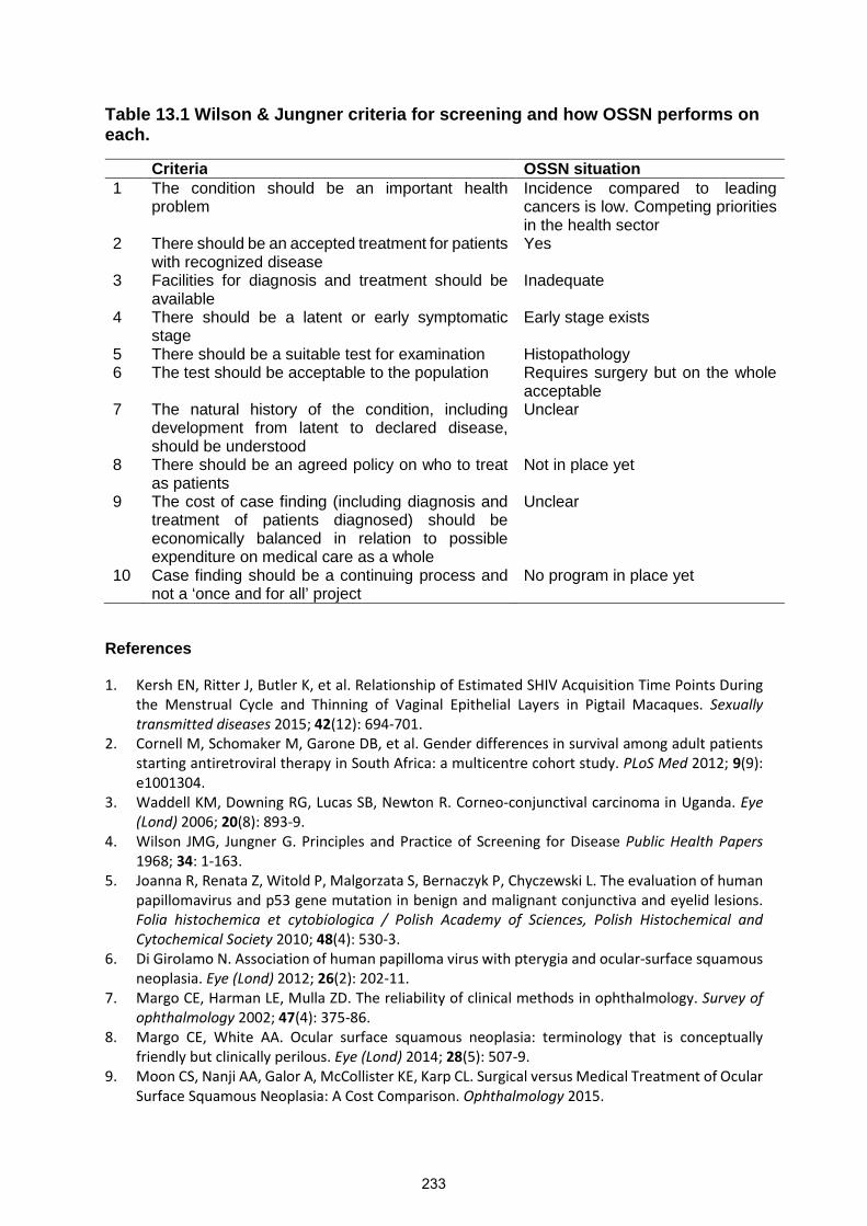
Table 13.1 Wilson & Jungner criteria for screening and how OSSN performs oneach.
Criteria OSSN situation1 The condition should be an important health
problemIncidence compared to leadingcancers is low. Competing prioritiesin the health sector
2 There should be an accepted treatment for patientswith recognized disease
Yes
3 Facilities for diagnosis and treatment should beavailable
Inadequate
4 There should be a latent or early symptomaticstage
Early stage exists
5 There should be a suitable test for examination Histopathology6 The test should be acceptable to the population Requires surgery but on the whole
acceptable7 The natural history of the condition, including
development from latent to declared disease,should be understood
Unclear
8 There should be an agreed policy on who to treatas patients
Not in place yet
9 The cost of case finding (including diagnosis andtreatment of patients diagnosed) should beeconomically balanced in relation to possibleexpenditure on medical care as a whole
Unclear
10 Case finding should be a continuing process andnot a ‘once and for all’ project
No program in place yet
References
1. Kersh EN, Ritter J, Butler K, et al. Relationship of Estimated SHIV Acquisition Time Points Duringthe Menstrual Cycle and Thinning of Vaginal Epithelial Layers in Pigtail Macaques. Sexuallytransmitted diseases 2015; 42(12): 694-701.
2. Cornell M, Schomaker M, Garone DB, et al. Gender differences in survival among adult patientsstarting antiretroviral therapy in South Africa: a multicentre cohort study. PLoS Med 2012; 9(9):e1001304.
3. Waddell KM, Downing RG, Lucas SB, Newton R. Corneo-conjunctival carcinoma in Uganda. Eye(Lond) 2006; 20(8): 893-9.
4. Wilson JMG, Jungner G. Principles and Practice of Screening for Disease Public Health Papers1968; 34: 1-163.
5. Joanna R, Renata Z, Witold P, Malgorzata S, Bernaczyk P, Chyczewski L. The evaluation of humanpapillomavirus and p53 gene mutation in benign and malignant conjunctiva and eyelid lesions.Folia histochemica et cytobiologica / Polish Academy of Sciences, Polish Histochemical andCytochemical Society 2010; 48(4): 530-3.
6. Di Girolamo N. Association of human papilloma virus with pterygia and ocular-surface squamousneoplasia. Eye (Lond) 2012; 26(2): 202-11.
7. Margo CE, Harman LE, Mulla ZD. The reliability of clinical methods in ophthalmology. Survey ofophthalmology 2002; 47(4): 375-86.
8. Margo CE, White AA. Ocular surface squamous neoplasia: terminology that is conceptuallyfriendly but clinically perilous. Eye (Lond) 2014; 28(5): 507-9.
9. Moon CS, Nanji AA, Galor A, McCollister KE, Karp CL. Surgical versus Medical Treatment of OcularSurface Squamous Neoplasia: A Cost Comparison. Ophthalmology 2015.
233

Chapter 14. Future work
The following future work is planned.
i) Stem cells study – we conducted immunohistochemistry staining of different histological types
of OSSN and some benign lesions for stem cell markers. Various stains reported to stain stem
cells in other tumours were used. The objective was to study the stem cell patterns if any in
OSSN. A manuscript is in preparation.
ii) Human papilloma virus (HPV) serotypes in OSSN – we extracted viral DNA from a set of
OSSN lesions for genome sequencing to study what types of HPV may be involved in OSSN.
Most other studies have used diagnostic kits that focused on cervical cancers. We hope our
study will reveal a wider array of genotypes of HPV. The sequencing component is still
ongoing.
iii) OSSN sequencing – we have held preliminary discussions with the Sanger Institute in
Cambridge to study the genetic mutations in OSSN tumours. The aim is to further explore the
gene mutations in OSSN that may be associated with UV radiation, HIV, HPV and smoking.
iv) Health Education and Early Detection (HEED) study – I have obtained a post-doctoral
fellowship grant (£90,000) from the Queen Elizabeth Jubilee Trust Fund through the
Commonwealth eye health consortium (http://cehc.lshtm.ac.uk/ ) to conduct a pilot project to
promote early awareness and early referral of OSSN in Kenya.
v) Validation study to see the effectiveness of the algorithm for screening by other health workers
(non-Ophthalmologists).
vi) Make 5FU available in Kenya as effectiveness has been demonstrated. We would need to
work with the Kenya Pharmacy and Poisons Control Board and the manufacturer who
produced it for the study.
vii)Long-term follow up of 5FU trial participants – we continue following up our trial participants
with the aim of reporting 3-year results.
234

Appendices1. Diagnosing ocular surface squamous neoplasia in East Africa: case-control study of clinical
and in vivo confocal microscopy assessment.
2. Ocular rhinosporidiosis mimicking conjunctival squamous papilloma in Kenya – a case report
3. Ethics committee approvals
4. RCT registration, regulatory authority approval and protocol
235

Creative Commons Attribution License (CC BY)This article is available under the terms of the Creative Commons Attribution License (CC BY).You may distribute and copy the article, create extracts, abstracts, and other revised versions,adaptations or derivative works of or from an article (such as a translation), to include in acollective work (such as an anthology), to text or data mine the article, including for commercialpurposes without permission from Elsevier. The original work must always be appropriatelycredited.
Permission is not required for this type of reuse.
Copyright © 2016 Copyright Clearance Center, Inc. All Rights Reserved.Comments? We would like to hear from you. E-mail us at [email protected]
Rightslink® by Copyright Clearance Center https://s100.copyright.com/AppDispatchServlet?publisherName=ELS...
1 of 1 04/05/2016 22:36
236
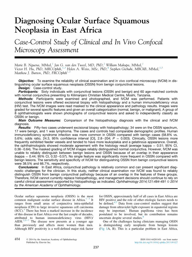
Diagnosing Ocular Surface SquamousNeoplasia in East AfricaCase-Control Study of Clinical and In Vivo ConfocalMicroscopy Assessment
Marie B. Nguena, MMed,1 Jan G. van den Tweel, MD, PhD,2 William Makupa, MMed,1
Victor H. Hu, PhD, MRCOphth,1,3 Helen A. Weiss, MSc, PhD,4 Stephen Gichuhi, MBChB, MMed,3,5
Matthew J. Burton, PhD, FRCOphth1,3
Objective: To examine the reliability of clinical examination and in vivo confocal microscopy (IVCM) in dis-tinguishing ocular surface squamous neoplasia (OSSN) from benign conjunctival lesions.
Design: Case-control study.Participants: Sixty individuals with conjunctival lesions (OSSN and benign) and 60 age-matched controls
with normal conjunctiva presenting to Kilimanjaro Christian Medical Centre, Moshi, Tanzania.Methods: Participants were examined and photographed, and IVCM was performed. Patients with
conjunctival lesions were offered excisional biopsy with histopathology and a human immunodeficiency virus(HIV) test. The IVCM images were read masked to the clinical appearance and pathology results. Images weregraded for several specific features and given an overall categorization (normal, benign, or malignant). A group of8 ophthalmologists were shown photographs of conjunctival lesions and asked to independently classify asOSSN or benign.
Main Outcome Measures: Comparison of the histopathology diagnosis with the clinical and IVCMdiagnosis.
Results: Fifty-two cases underwent excisional biopsy with histopathology; 34 were on the OSSN spectrum,17 were benign, and 1 was lymphoma. The cases and controls had comparable demographic profiles. Humanimmunodeficiency syndrome infection was more common in OSSN compared with benign cases (58.8% vs.5.6%; odds ratio, 24.3, 95% confidence interval [CI], 2.8e204; P ¼ 0.003). Clinically, OSSN lesions morefrequently exhibited feeder vessels and tended to have more leukoplakia and a gelatinous appearance. Overall,the ophthalmologists showed moderate agreement with the histology result (average kappa ¼ 0.51; 95% CI,0.36e0.64). The masked grading of IVCM images reliably distinguished normal conjunctiva. However, IVCM wasunable to reliably distinguish between benign lesions and OSSN because of an overlap in their appearance(kappa ¼ 0.44; 95% CI, 0.32e0.57). No single feature was significantly more frequent in OSSN compared withbenign lesions. The sensitivity and specificity of IVCM for distinguishing OSSN from benign conjunctival lesionswere 38.5% and 66.7%, respectively.
Conclusions: In East Africa, conjunctival pathology is relatively common and can present significant diag-nostic challenges for the clinician. In this study, neither clinical examination nor IVCM was found to reliablydistinguish OSSN from benign conjunctival pathology because of an overlap in the features of these groups.Therefore, IVCM cannot currently replace histopathology, and management decisions should continue to rely oncareful clinical assessment supported by histopathology as indicated. Ophthalmology 2014;121:484-491 ª 2014by the American Academy of Ophthalmology.
Ocular surface squamous neoplasia (OSSN) is the mostcommon malignant ocular surface disease in Africa.1e3 Itranges from small areas of conjunctiva intra-epithelialneoplasia (CIN) to large invasive squamous cell carcinoma(SCC).4 There has been a marked increase in the incidenceof this disease in East Africa over the last couple of decades,attributed to human immunodeficiency virus (HIV)/AIDS.3,5e7 The disease now presents at a younger agethan previously and affects more women than men.Although HIV positivity is a well-defined major risk factor
484 � 2014 by the American Academy of OphthalmologyPublished by Elsevier Inc.
237
for OSSN, approximately half of all cases in East Africa areHIV positive and the role of other etiologic factors needs tobe defined.4,7 Data from case-control studies suggest thatdamage from ultraviolet light exposure in equatorial regionsmay be important.7 Human papilloma virus has beenpostulated to be involved, but its contribution remainsuncertain despite several studies.8
One of the challenges facing clinicians managing OSSNis distinguishing early neoplastic from benign lesions(Fig 1A, B). This is a particular problem in East Africa,
ISSN 0161-6420/14/$ - see front matterhttp://dx.doi.org/10.1016/j.ophtha.2013.09.027

Figure 1. Photographs of a pterygium and various grades of ocular surface squamous neoplasia: pterygium (A), conjunctiva intra-epithelial neoplasia (CIN)1 (B), CIN 3 (C), carcinoma in situ (D), squamous cell carcinoma (SCC) grade 1 (E), SCC grade 2 (F), SCC grade 3 (G), and SCC with orbital invasion(H).
Nguena et al � Diagnosing OSSN in East Africa
where benign conjunctival changes (pterygium, pingeculum)are relatively common. There is probably an overlap inthe clinical features of these different benign lesionsand OSSN, with no signs being exclusive to OSSN,although this has not been formally evaluated. Somefeatures have been reported to be more common inadvanced cases of OSSN.4 Histopathology is necessaryto confirm a diagnosis of OSSN. However, pathologyservices are generally scarce in East Africa outside larger
238
centers. As a result, many lesions are excised withoutpathologic analysis.
The standard approach for treating suspected OSSN is anexcisional biopsy, often with cryotherapy to the conjunctivalmargin and sometimes with topical chemotherapy.9,10 Aninitial incisional biopsy is usually performed for largerlesions to make a diagnosis to plan treatment. In recentyears, there has been a move to consider primary topicalchemotherapy for suspected OSSN without a tissue
485

Ophthalmology Volume 121, Number 2, February 2014
diagnosis.11 Given the potential uncertainty of diagnosisbased on clinical signs alone, several groups haveinvestigated whether in vivo confocal microscopy (IVCM)can help distinguish cases of OSSN.12e16 These reports ofrelatively few cases (26 in the whole literature) have tendedto conclude that IVCM may be helpful in making a diag-nosis of OSSN. Several specific features, such as irregularcell size, hyper-reflectivity, prominent bright nucleoli(“starry night appearance”), and mitotic figures, have beensingled out as potential markers for OSSN.13
We report a prospective study of patients with conjunc-tival lesions presenting to an ophthalmic unit in Tanzania.We investigate the diagnostic accuracy of the clinicalexamination and whether certain features help to distinguishOSSN from benign lesions. Second, we evaluate the utilityof IVCM in identifying OSSN in a masked study ofconjunctival lesions.
Methods
Ethical Approval
This study adhered to the tenets of the Declaration of Helsinki. Itwas reviewed and approved by the Kilimanjaro Christian MedicalCentre (KCMC) Ethics Committee, Tanzania.
Study Design and Participants
In this case-control study, we compared individuals withconjunctival lesions (“cases,” either benign or OSSN) with indi-viduals with clinically normal conjunctiva (“controls”). Recruit-ment of both cases and controls was conducted at the Departmentof Ophthalmology, KCMC Hospital, Moshi, Tanzania, from Mayto November 2011. Cases were consecutive individuals presentingwith a conjunctival lesion that on clinical grounds required surgicalexcision. For each case, we recruited a control presenting fora condition that was not affecting the ocular surface and who hadhealthy conjunctiva on slit-lamp examination (e.g., cataract). Thesecontrol participants were age-matched to the corresponding case(�5 years). All participants were aged 18 years or older. The studywas explained to eligible study subjects, and written informedconsent was obtained before enrollment. All enrolled patients withconjunctival lesions received counseling and were offered testingfor HIV, which is standard of care at KCMC. Patients witha positive HIV test result were referred to the ComprehensiveTreatment Center.
Clinical Assessment
Patients with conjunctival lesions were asked about symptoms(ocular pain, irritation, itchiness, redness, decreased vision, prop-tosis, and discharge). Participants were asked where they livedwithin Tanzania and their main occupation. The surface of the eyewas carefully examined with a slit lamp for conjunctivalpathology. We recorded lesion location and size and the presenceof specific features, including fibrovascular tissue (Fig 1A, B),leukoplakia (Fig 1C), gelatinous appearance (Fig 1D), feedervessels (Fig 1D), papilliform structure (Fig 1F), limbalinvolvement, and orbital invasion (Fig 1H). The lesions werephotographed using a digital single lens reflex camera witha macro lens.
486
239
In Vivo Confocal Microscopy Examination
In vivo confocal microscopy examination of the bulbar conjunctivawas performed using the Heidelberg Retina Tomograph 3 incombination with the Rostock Corneal Module (Heidelberg Engi-neering GmbH, Dossenheim, Germany). A sterile, disposablepolymethylmethacrylate cap was mounted on the head of theconfocal microscope, and a drop of topical anesthetic (proparacaine0.5%) was instilled into the inferior fornix of the eye before theexamination. For cases, the lesion and surrounding areas werescanned. For controls, the temporal or nasal inter-palpebralconjunctiva was scanned. Representative images of the examinedconjunctiva were obtained using the “volume scan” mode, whichcollects a series of 40 parallel images 2 mm apart; both surface(starting at the epithelium) and deeper sets of scans wereperformed.
All confocal images were graded over a 2-day period, afterrecruitment had been completed. Two ophthalmologists (M.B.N.,M.J.B.) jointly examined all the images; they were masked to thecase/control status, clinical features, and histopathology results.The grading of the images was based on separately examining eachof the 40 parallel images in each of the volume scan stacks. Thefollowing features, which are said to be potentially discriminatingin previous studies, were evaluated: variable epithelial cell size,hyperreflectivity, presence of mitotic cells, and prominent brightnucleoli (“starry night appearance”) (Fig 2).13 An overallprovisional categorization based on the IVCM findings wasmade: (1) normal, (2) abnormal benign, or (3) abnormal malignant.
Surgical Treatment
Lesions were treated surgically. Small to medium-sized lesions thatwere clinically suspicious for OSSN were fully excised using the“no touch technique” with a 3- to 4-mm margin of healthy-lookingtissue. Surgical sponges saturated with 5-fluorouracil were appliedto the excision site for 5 minutes followed by irrigation with normalsaline. 5-Fluorouracil treatment was not used if the case was thoughtto be benign pathology, such as a pterygium. Incisional biopsieswere performed for large lesions to obtain tissue for histologyassessment. If malignant pathology was confirmed, radical surgery(enucleation or exenteration) was then offered to the patient. Biopsytissue was not collected from the normal controls.
Histopathology
Biopsy specimens from the cases were immediately placed in 10%formaldehyde solution. These were mounted in paraffin wax, and5-mm sections were cut perpendicular to the conjunctival surfaceand stained with hematoxylineeosin. All specimens were exam-ined by a single pathologist (J.G.T.). Ocular surface squamousneoplasia was classified using the standard definitions for mild(CIN I), moderate (CIN II), and severe (CIN III) dysplasia, carci-noma in situ, and invasive SCC.17 Squamous cell carcinoma wassubdivided by the degree of differentiation: well (I), moderate(II), and poor (III).
Clinical Diagnosis Study
To evaluate the ability of clinicians to correctly distinguishbetween benign and malignant conjunctival lesions, we showeda group of 8 ophthalmologists (3 consultants and 5 residents)a series of 43 photographs of lesions for which a histologic diag-nosis also was available by the time this component was con-ducted. This included a mixture of benign and neoplasticpathologies. They were asked to independently write down theirdiagnosis and whether they thought it was benign or malignant.
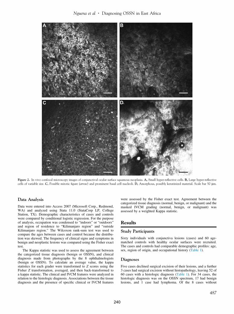
Figure 2. In vivo confocal microscopy images of conjunctival ocular surface squamous neoplasia. A, Small hyper-reflective cells. B, Large hyper-reflectivecells of variable size. C, Possible mitotic figure (arrow) and prominent basal cell nucleoli. D, Amorphous, possibly keratinized material. Scale bar 50 mm.
Nguena et al � Diagnosing OSSN in East Africa
Data Analysis
Data were entered into Access 2007 (Microsoft Corp., Redmond,WA) and analyzed using Stata 11.0 (StataCorp LP, CollegeStation, TX). Demographic characteristics of cases and controlswere compared by conditional logistic regression. For the purposeof analysis, occupation was condensed to “indoors” or “outdoors”and region of residence to “Kilimanjaro region” and “outsideKilimanjaro region.” The Wilcoxon rank-sum test was used tocompare the ages between cases and control because the distribu-tion was skewed. The frequency of clinical signs and symptoms inbenign and neoplastic lesions was compared using the Fisher exacttest.
The Kappa statistic was used to assess the agreement betweenthe categorized tissue diagnosis (benign or OSSN), and clinicaldiagnosis made from photographs by the 8 ophthalmologists(benign or OSSN). To calculate an average value, the kappastatistics for each grader were transformed to Z scores using theFisher Z transformation, averaged, and then back-transformed toa kappa statistic. The clinical and IVCM features were analyzed inrelation to the histologic diagnosis. Associations between the tissuediagnosis and the presence of specific clinical or IVCM features
240
were assessed by the Fisher exact test. Agreement between thecategorized tissue diagnosis (normal, benign, or malignant) and themasked IVCM grading (normal, benign, or malignant) wasassessed by a weighted Kappa statistic.
Results
Study Participants
Sixty individuals with conjunctiva lesions (cases) and 60 age-matched controls with healthy ocular surfaces were recruited.The cases and controls had comparable demographic profiles: age,sex, region of origin, and occupational history (Table 1).
Diagnoses
Five cases declined surgical excision of their lesions, and a further3 cases had surgical excision without histopathology, leaving 52 of60 cases with a histologic diagnosis (Table 1). For 34 cases, thehistologic diagnosis was on the OSSN spectrum, 17 had benignlesions, and 1 case had lymphoma. Of the 8 cases without
487
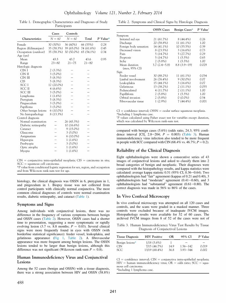
Table 2. Symptoms and Clinical Signs by Histologic Diagnosis
OSSN Cases Benign Cases* P Valuey
SymptomIrritated red eye 21 (61.7%) 8 (44.4%) 0.26Discharge 20 (58.8%) 11 (61.1%) 1.00Foreign body sensation 14 (41.1%) 10 (55.5%) 0.39Decreased vision 8 (23.5%) 3 (16.6%) 0.73Pain 5 (14.7%) 5 (27.7%) 0.29Proptosis 5 (14.7%) 1 (5.5%) 0.65Itchy 2 (5.8%) 1 (5.5%) 1.00Mean duration
(mos, 95% CI)3.7 (2.4e5.8) 8.8 (3.9e19) 0.029
SignFeeder vessel 30 (88.2%) 11 (61.1%) 0.034Limbal involvement 26 (76.4%) 9 (50.0%) 0.07Leukoplakia 15 (44.1%) 3 (16.6%) 0.07Gelatinous 13 (38.2%) 2 (11.1%) 0.055Pedunculated 4 (11.7%) 2 (11.1%) 1.00Papilliform 2 (5.8%) 1 (5.5%) 1.00Orbital invasion 2 (5.8%) 0 (0.0%) 0.54Fibrovascular tissue 1 (2.9%) 7 (44.4%) 0.001
CI ¼ confidence interval; OSSN ¼ ocular surface squamous neoplasia.*Including 1 lymphoma case.yP values calculated using Fisher exact test for variables except duration,which was calculated by Wilcoxon rank-sum test.
Table 1. Demographic Characteristics and Diagnoses of StudyParticipants
Characteristics
Cases Controls
Total P Value*N ¼ 60 N ¼ 60
Female 30 (50%) 36 (60%) 66 (55%) 0.28Region (Kilimanjaro) 35 (58.3%) 39 (65.0%) 74 (61.6%) 0.45Occupation (outdoor) 35 (58.3%) 30 (50.0%) 65 (54.1%) 0.34Age (yrs)Mean 43.5 43.7 43.6 0.95Range 23e80 21e75 21e80
Histologic diagnosisCIN I 2 (3.3%) e eCIN II 3 (5.0%) e eCIN III 5 (8.3%) e eCIS 5 (8.3%) e eSCC I 12 (20.0%) e eSCC II 4 (6.6%) e eSCC III 3 (5.0%) e eLymphoma 1 (1.6%) e ePterygium 5 (8.3%) e ePingueculum 3 (5.0%) e ePapilloma 3 (5.0%) e eOther benign lesions 6 (10.0%) e eNo histopathology 8 (13.3%) e e
Control diagnosisNormal examination e 26 (43.3%) eDiabetic retinopathy e 10 (16.6%) eCataract e 9 (15.0%) eGlaucoma e 3 (5.0%) eAstigmatism e 6 (10.0%) eHyperopia e 1 (1.6%) ePresbyopia e 3 (5.0%) eOptic atrophy e 1 (1.6%) eMyopia e 1 (1.6%) e
CIN ¼ conjunctiva intra-epithelial neoplasia; CIS ¼ carcinoma in situ;SCC ¼ squamous cell carcinoma.*P value from conditional logistic regression for sex, region, and occupationand from Wilcoxon rank-sum test for age.
Table 3. Human Immunodeficiency Virus Test Results by TissueDiagnosis of Conjunctival Lesions
Tissue Diagnosis HIV Positive OR 95% CI P Value
Benign lesions* 1/18 (5.6%) 1 e eCIN 7/15 (46.7%) 14.9 1.56e142 0.019SCC 13/19 (68.4%) 36.8 3.93e344 0.002
CI ¼ confidence interval; CIN ¼ conjunctiva intra-epithelial neoplasia;HIV ¼ human immunodeficiency virus; OR ¼ odds ratio; SCC ¼ squa-mous cell carcinoma.*Including 1 lymphoma case.
Ophthalmology Volume 121, Number 2, February 2014
histology, the clinical diagnosis was OSSN in 6, pterygium in 1,and pingeculum in 1. Biopsy tissue was not collected fromcontrol participants with clinically normal conjunctiva. The mostcommon clinical diagnoses of controls were normal examinationresults, diabetic retinopathy, and cataract (Table 1).
Symptoms and Signs
Among individuals with conjunctival lesions, there was nodifference in the frequency of various symptoms between benignand OSSN cases (Table 2). However, OSSN cases had a shortertime to presentation, suggesting a more symptomatic or rapidlyevolving lesion (3.7 vs. 8.8 months; P ¼ 0.03). Several clinicalsigns were more frequently found in eyes with OSSN (withborderline statistical significance): feeder vessel, leukoplakia, andgelatinous appearance (Fig 1, Table 2). A fibrovascularappearance was more frequent among benign lesions. The OSSNlesions tended to be larger than benign lesions, although thisdifference was not significant (Wilcoxon rank-sum P ¼ 0.8).
Human Immunodeficiency Virus and ConjunctivalLesions
Among the 52 cases (benign and OSSN) with a tissue diagnosis,there was a strong association between HIV and OSSN (58.8%)
488
241
compared with benign cases (5.6%) (odds ratio, 24.3; 95% confi-dence interval [CI], 2.8e204; P ¼ 0.003) (Table 3). Humanimmunodeficiency virus infection also tended to be more frequentin people with SCC compared with CIN (68.4% vs. 46.7%;P¼ 0.2).
Reliability of the Clinical Diagnosis
Eight ophthalmologists were shown a consecutive series of 43images of conjunctival lesions and asked to classify them into 2broad categories of benign and neoplastic. Their diagnoses werecompared with the histopathology results, and kappa statistics werecalculated: average kappa statistic 0.51 (95% CI, 0.36e0.64). Twoophthalmologists had “fair” agreement (kappas of 0.21 and 0.40), 3ophthalmologists had “moderate” agreement (0.41e0.60), and 3ophthalmologists had “substantial” agreement (0.61e0.80). Thecorrect diagnosis was made in 56% to 86% of the cases.
In Vivo Confocal Microscopy
In vivo confocal microscopy was attempted on all 120 cases andcontrols, and the scans were graded in a masked manner. Threecontrols were excluded because of inadequate IVCM images.Histopathology results were available for 52 of 60 cases. Thearchived IVCM images from 8 of 52 of the cases were not of

Table 4. In Vivo Confocal Microscopy Features by Disease Group,Based on Masked Grading
IVCM Features
ControlsBenignCases*
OSSNCases P
ValueyP
ValuezN/57 (%) N/18 (%) N/26 (%)
Hyper-reflectivecells
<0.001 0.9
None 54 (94.7%) 7 (38.3%) 12 (46.1%)<50% 3 (5.3%) 7 (38.8%) 8 (30.7%)>50% 0 (0.0%) 4 (22.2%) 6 (23.1%)
Variation in cellsize
<0.001 0.5
Uniform 55 (96.4%) 9 (50%) 17 (65.3%)Mild 1 (1.7%) 6 (33.3%) 4 (15.3%)Marked 1 (1.7%) 3 (16.7%) 5 (19.2%)
Starry nightappearance ofbasal cells
0.001 0.8
Nil 57 (100%) 14 (77.7%) 22 (84.6%)Few to many 0 (0.0%) 2 (11.1%) 1 (3.8%)Amorphous
material0 (0.0%) 2 (11.1%) 3 (11.5%)
IVCM ¼ in vivo confocal microscopy; OSSN ¼ ocular surface squamousneoplasia.*Including 1 lymphoma case.yP value is for the comparison of normal controls with all cases (benign andmalignant combined) by Fisher exact test.zP value is for the comparison of benign with malignant cases, by Fisherexact test.
Table 5. Comparison of the Overall Classification from theIn Vivo Confocal Microscopy Based on Masked Grading (Normal,
Benign, or Malignant) versus the Final Histologic Diagnosis
Tissue Diagnosis
IVCM Diagnosis
TotalNormal Abnormal Benign Malignant
Normal control 50 7 0 57Benign lesion 4 8 6 18Malignant lesion 5 11 10 26Total 59 26 16 101
IVCM ¼ in vivo confocal microscopy.Grey shading highlights where there was agreement.
Nguena et al � Diagnosing OSSN in East Africa
sufficient quality for masked grading. Therefore, the analysis ofIVCM was limited to 101 participants: 44 cases (with both histo-pathology and adequate scans) and 57 controls.
The IVCM images were examined for the presence of specificfeatures: hyper-reflective cells, variation of cell size, mitotic cells,and “starry night” appearance of the basal layer (Fig 2, Table 4).Several lesions showed marked surface changes on IVCM withextensive, hyper-reflective material with large irregular cellularstructure. A possible mitotic cell was observed in only 1 lesion (Fig2C). Among the 57 controls with IVCM images, 3 had somehyper-reflective cells and 2 showed some variation in epithelialcell size. For each of the graded features, there were statisticallysignificant differences in their frequency between the normalcontrols and cases (benign and malignant combined). However, wefound no evidence for a difference in the frequency of these IVCMfeatures between benign and malignant conjunctival lesions.
Each set of IVCM images was given an overall classification bythe masked readers: normal, benign abnormal, or malignant. Thecomparison of this overall IVCM classification with the histopa-thology diagnosis is shown in Table 5. Agreement was found to bemoderate with an unweighted kappa score of 0.44 (95% CI,0.32e0.57). The linear weighted kappa score was 0.54 (95% CI,0.40e0.67). The sensitivity and specificity of IVCM fordistinguishing OSSN from benign conjunctival lesions were 38.5%(95% CI, 0.21e0.59) and 66.7% (95% CI, 0.41e0.86), respectively.
Discussion
Timely detection and treatment of OSSN are challenges inEast Africa, with many patients only presenting when thelesion is already established. The reasons for this are many:Access to eye care services outside the major urban centersis generally limited; therefore, patients may delay traveling
242
for treatment until the lesion is more obvious and symp-tomatic. When patients do present, there can be further delayin making the diagnosis of OSSN because initially it may bethought to be a benign lesion. Delayed or missed diagnosiscan have considerable effects on ocular morbidity and evenmortality.
In this study based in an East African ophthalmic referralcenter, OSSN was a common cause of conjunctivalpathology, with presentation rates comparable to those inprevious reports from this unit.3,4 Time between onset ofsymptoms and presentation was shorter for OSSN than forbenign lesions, possibly because OSSN develops morerapidly. However, some individuals with OSSN had noticeda problem many months earlier. Strategies are needed toraise general awareness that growths on the eye need promptattention. This is particularly important in people who havea known diagnosis of HIV, but this needs to be done ina way that minimizes stigma.
There is an overlap in the clinical signs of (early) OSSNand some benign lesions (Fig 1A, B). However, a number offeatures tended to be more frequent in cases of OSSN:feeder vessels, gelatinous appearance, and leukoplakia.These and other signs could form the basis of a diagnosticalgorithm to assist the clinician in identifying OSSNcases, and they warrant further study in a larger data set.Overall, the 8 ophthalmologists, most of whom had seenmany cases of OSSN previously, achieved only moderateagreement with the histologic diagnosis. There was a widerange in their diagnostic accuracy, suggesting that if thekey features that point to a diagnosis of OSSN can beidentified and emphasized in training, there should bescope to improve the detection rate.
The prevalence of HIV infection in OSSN cases wascomparable to that in previous studies from the region.4
Human immunodeficiency virus seems to be a risk factorfor developing more severe disease, being more frequentamong cases with SCC than CIN. There was no differencein the time between the onset of symptoms and thepresentation between SCC and CIN, suggesting that HIVinfection permits a more rapid progression in the disease.
In vivo confocal microscopy is a potentially useful tool inthe management of several external eye diseases. It has beenparticularly helpful in the diagnosis of acanthamoeba andfilamentary fungal keratitis.18 We have also found thetechnique informative in the study of conjunctival scarring
489

Ophthalmology Volume 121, Number 2, February 2014
in trachoma.19 In the present study, masked analysis ofIVCM clearly distinguished between normal conjunctivaand lesions (benign or malignant). A few clinically normalcontrols had some minor changes on IVCM, which mightbe attributable to solar radiation damage. In contrast, itwas not possible to reliably distinguish between OSSNand benign conjunctival lesions because there seems to bean overlap in the IVCM features of these variousconditions. We did not identify any features that wereexclusive to OSSN. Of particular note was the finding thatthere were several cases of OSSN that were classified asnormal on IVCM.
Our conclusion that IVCM does not reliably distinguishOSSN from other conjunctival pathology contrasts withearlier reports that suggest it may be possible to use it forthis purpose.12e16 However, these earlier reports, mostlyfrom Europe (where the disease spectrum is different), havetended to be of relatively few cases. Moreover, with theexception of 1 series of 4 cases, benign lesion controls havenot been recruited for comparison. Finally, the assessmentof the images was not masked in all but 1 of the previousstudies.16 Without systematic masked comparison withbenign lesions, it is difficult to know how useful theobserved features are. Studies of the IVCM appearance ofpterygium have reported hyper-reflective epithelial cells,a feature previously said to be associated with OSSN.20,21
This is the first time IVCM has been used in Africa tostudy OSSN. Overall, the disease tends to be more advancedand aggressive in this context, and clinical/IVCM featuresmay show significant variation from studies of Caucasianpatients with disease largely unassociated with HIV. Furthervalidation is needed using masked grading of OSSN andother conjunctival pathology in other settings.
Study Limitations
First, histopathology was not available for all conjunctivallesions because the patient declined surgery or the surgeondid not send a sample for analysis. Second, it was notalways possible to obtain IVCM scans of sufficient qualityfor the masked grading, usually because the patient wasunable to tolerate the examination. Third, the maskedanalysis was limited to the set of stored volume scans; inpractice, the formation of a diagnostic impression withIVCM is based on a continuous, dynamic examination inwhich the examiner is not masked to the clinical appearanceof the eye. A limitation of current IVCM technology is thatit is not possible to identify exactly where on the lesion thescan is collected from; therefore, it is not possible to put it inexact correspondence with findings from histopathology.Finally, the conclusions of this study may not be general-izable to the use of IVCM to detect OSSN in other settings ifthe distribution of disease stage is different.
In conclusion, ocular surface squamous neoplasiaremains a major clinical challenge in the East Africancontext. A simple and cheap diagnostic test to help distin-guish benign from malignant pathology would be ofconsiderable utility in the East African context, wherepathology services may not be readily available.
490
243
References
1. Pola EC, Masanganise R, Rusakaniko S. The trend of ocularsurface squamous neoplasia among ocular surface tumourbiopsies submitted for histology from Sekuru Kaguvi EyeUnit, Harare between 1996 and 2000. Cent Afr J Med2003;49:1–4.
2. Nagaiah G, Stotler C, Orem J, et al. Ocular surface squamousneoplasia in patients with HIV infection in sub-Saharan Africa.Curr Opin Oncol 2010;22:437–42.
3. Poole TR. Conjunctival squamous cell carcinoma in Tanzania.Br J Ophthalmol 1999;83:177–9.
4. Makupa II, Swai B, Makupa WU, et al. Clinical factorsassociated with malignancy and HIV status in patientswith ocular surface squamous neoplasia at KilimanjaroChristian Medical Centre, Tanzania. Br J Ophthalmol2012;96:482–4.
5. Parkin DM, Wabinga H, Nambooze S, Wabwire-Mangen F.AIDS-related cancers in Africa: maturation of the epidemic inUganda. AIDS 1999;13:2563–70.
6. Ateenyi-Agaba C. Conjunctival squamous-cell carcinomaassociated with HIV infection in Kampala, Uganda. Lancet1995;345:695–6.
7. Waddell K, Kwehangana J, Johnston WT, et al. A case-controlstudy of ocular surface squamous neoplasia (OSSN) inUganda. Int J Cancer 2010;127:427–32.
8. Di Girolamo N. Association of human papilloma virus withpterygia and ocular-surface squamous neoplasia. Eye (Lond)2012;26:202–11.
9. Shields CL, Shields JA. Tumors of the conjunctiva and cornea.Surv Ophthalmol 2004;49:3–24.
10. Waddell KM, Downing RG, Lucas SB, Newton R. Corneo-conjunctival carcinoma in Uganda. Eye (Lond) 2006;20:893–9.
11. Sturges A, Butt AL, Lai JE, Chodosh J. Topical interferon orsurgical excision for the management of primary ocularsurface squamous neoplasia. Ophthalmology 2008;115:1297–302.
12. Duchateau N, Hugol D, D’Hermies F, et al. Contribution ofin vivo confocal microscopy to limbal tumor evaluation [inFrench]. J Fr Ophtalmol 2005;28:810–6.
13. Alomar TS, Nubile M, Lowe J, Dua HS. Corneal intra-epithelial neoplasia: in vivo confocal microscopic study withhistopathologic correlation. Am J Ophthalmol 2011;151:238–47.
14. Falke K, Zhivov A, Zimpfer A, et al. Diagnosis of conjunctivalneoplastic lesions by confocal in-vivo microscopy [inGerman]. Klin Monbl Augenheilkd 2012;229:724–7.
15. Xu Y, Zhou Z, Xu Y, et al. The clinical value of in vivoconfocal microscopy for diagnosis of ocular surface squamousneoplasia. Eye (Lond) 2012;26:781–7.
16. Parrozzani R, Lazzarini D, Dario A, Midena E. In vivoconfocal microscopy of ocular surface squamous neoplasia.Eye (Lond) 2011;25:455–60.
17. Font RL, Croxatto JO, Rao NA. Tumors of the Eye andOcular Adnexa. Washington, DC: American Registry ofPathology; 2006. AFIP Atlas of Tumor Pathology. Fourthseries, fasc. 5.
18. Hau SC, Dart JK, Vesaluoma M, et al. Diagnostic accuracy ofmicrobial keratitis with in vivo scanning laser confocalmicroscopy. Br J Ophthalmol 2010;94:982–7.
19. Hu VH, Weiss HA, Massae P, et al. In vivo confocalmicroscopy in scarring trachoma. Ophthalmology 2011;118:2138–46.

Nguena et al � Diagnosing OSSN in East Africa
20. Labbe A, Gheck L, Iordanidou V, et al. An in vivo confocalmicroscopy and impression cytology evaluation of pterygiumactivity. Cornea 2010;29:392–9.
244
21. Wang Y, Zhao F, Zhu W, et al. In vivo confocal microscopicevaluation of morphologic changes and dendritic cell distri-bution in pterygium. Am J Ophthalmol 2010;150:650–5.
Footnotes and Financial Disclosures
Originally received: May 14, 2013.Final revision: September 6, 2013.Accepted: September 18, 2013.Available online: December 9, 2013. Manuscript no. 2013-770.1 Department of Ophthalmology, Kilimanjaro Christian Medical Centre,Moshi, Tanzania.2 Department of Pathology, University Medical Centre, Utrecht, TheNetherlands.3 International Centre for Eye Health, Faculty of Infectious and TropicalDiseases, London School of Hygiene & Tropical Medicine, London, UnitedKingdom.4 Department of Infectious Disease Epidemiology, Faculty of Epidemiologyand Population Health, London School of Hygiene and Tropical Medicine,London, United Kingdom.
5 Department of Ophthalmology, University of Nairobi, Nairobi, Kenya.
Financial Disclosure(s):The author(s) have no proprietary or commercial interest in any materialsdiscussed in this article.
V.H.H. is supported by a fellowship grant from the British Council for thePrevention of Blindness (Barrie Jones Fellowship). M.J.B. is supported bythe Wellcome Trust (080741/Z/06/Z). The funding organizations had norole in the design or conduct of this research.
Correspondence:Matthew J. Burton, PhD, FRCOphth, International Centre for Eye Health,London School of Hygiene and Tropical Medicine, Keppel St., London,WC1E 7HT, UK. E-mail: [email protected].
491

Copyright
© 2014 Gichuhi et al.; licensee BioMed Central Ltd.This is an Open Access article distributed under the terms of the Creative CommonsAttribution License (http://creativecommons.org/licenses/by/2.0), which permitsunrestricted use, distribution, and reproduction in any medium, provided the original work isproperly credited.
Source: http://www.ncbi.nlm.nih.gov/pmc/articles/PMC4234295/
245

Gichuhi et al. BMC Ophthalmology 2014, 14:45http://www.biomedcentral.com/1471-2415/14/45
CASE REPORT Open Access
Ocular rhinosporidiosis mimicking conjunctivalsquamous papilloma in Kenya – a case reportStephen Gichuhi1,2*, Timothy Onyuma3, Ephantus Macharia4, Joy Kabiru4, Alain M’bongo Zindamoyen4,Mandeep S Sagoo5,6 and Matthew J Burton2,5
Abstract
Background: Ocular rhinosporidiosis is a chronic granulomatous infection caused by a newly classified organismthat is neither a fungus nor bacterium. It often presents as a benign conjunctival tumour but may mimic otherocular conditions. It is most often described in India. In Africa cases have been reported from South Africa, Kenya,Tanzania, Malawi, Uganda, Congo and Ivory Coast.
Case presentation: A 54 year old man was seen in Kenya with a lesion that resembled a conjunctival papilloma.We report resemblance to conjunctival papilloma and the result of vital staining with 0.05% Toluidine Blue.
Conclusion: Ocular rhinosporidiosis occurs in East Africa. It may resemble conjunctival squamous papilloma. Vitalstaining with 0.05% Toluidine blue dye did not distinguish the two lesions well.
Keywords: Ocular rhinosporidiosis, Rhinosporidium seeberi, Conjunctival papilloma, Toluidine-blue, Africa
BackgroundRhinosporidiosis is a chronic granulomatous infection ofthe mucous membranes (nasal, oral, ocular and rectal)caused by Rhinosporidium seeberi [1]. This is an unusualunicellular pathogen that is difficult to culture andwhose taxonomic classification has been controversial. Ithas been variously hypothesized to be a cyanobacterium(prokaryote), a eukaryotic Mesomycetozoa or a fungus[2]. Currently it is domiciled in the Mesomycetozoeaclass (also known as the DRIP clade, or Ichthyosporea).The term Mesomycetozoea derives from “Meso-” (in themiddle of ), “-myceto-” (fungi) and “-zoea” (animals).This is a heterogeneous group of microorganisms at theanimal-fungal boundary. The Mesomycetozoea are asmall group of protists, which are mostly parasites offish and other animals.Rhinosporidiosis affects both adults and children and
is commonly seen in otolaryngology. The largest re-ported case series of rhinosporidiosis consisting of 462cases in India found that the disease mainly occurs inthe nose and nasopharynx (81.1%) while the eye was af-fected in 14.2% [3]. Another series of 34 cases from
* Correspondence: [email protected] of Ophthalmology, University of Nairobi, Nairobi, Kenya2London School of Hygiene and Tropical Medicine, London, UKFull list of author information is available at the end of the article
© 2014 Gichuhi et al.; licensee BioMed CentraCommons Attribution License (http://creativecreproduction in any medium, provided the or
246
India also found nasal and nasopharyngeal involvementin 85% while the eye was affected in 9% of cases [4]. Acase involving multiple parts of the body; the nares, mul-tiple areas of the skin, the external urethral meatus,glans of penis and the perineum has been reported inIndia [5].Ocular rhinosporidiosis affecting the conjunctiva was
first described in India in 1912 [6]. Currently most pub-lished reports on rhinosporidiosis of the eye have beenreported from Asia mainly from India, Sri Lanka, Nepaland Bangladesh. In Africa it has been reported in SouthAfrica, Malawi, Zambia, Kenya, Uganda, Tanzania,Congo, Ivory Coast, and Cameroon [7-13]. None of thecases in Africa were initially diagnosed as ocular rhinos-poridiosis, perhaps a sign of its rarity.How the disease is acquired remains an enigma. Rhi-
nosporidiosis has been associated with migrants fromendemic areas [14-16]. Although it is an infectious dis-ease, as lesions are always associated with the presenceof the pathogen, there is limited data on how it might betransmitted [15]. It is presumed to be acquired throughtraumatized nasal mucosa and spread to other sites byautoinoculation. As most rhinosporidiosis lesions arisefrom the nose, it is feasible that ocular involvement mayoccur by spreading from the nose through the lacrimal
l Ltd. This is an Open Access article distributed under the terms of the Creativeommons.org/licenses/by/2.0), which permits unrestricted use, distribution, andiginal work is properly credited.

Figure 1 Pre-operative clinical photographs. A & B – shows the ocular rhinosporidiosis lesion in the region of the right medial canthus.C & D - shows squamous papilloma in the left medial canthus area from another patient for comparison. Both lesions are shown before and aftervital staining with 0.05% Toluidine Blue.
Gichuhi et al. BMC Ophthalmology 2014, 14:45 Page 2 of 4http://www.biomedcentral.com/1471-2415/14/45
sac to the plica of the conjunctiva. This hypothesis ishowever unproven.Ocular rhinosporidium most often presents as a polypoid
mass of the palpebral conjunctiva [17]. It may also presentas a lacrimal sac diverticulum [18], recurrent chalazion[19], conjunctival cyst [20], chronic follicular conjunctivitisin contact lens wearers [21], peripheral keratitis [22], scleralmelting [23], ciliary staphyloma [24] or simulate a tumourof the eyelid [25] or periorbital skin [26]. Large conjunc-tival lesions may cause mechanical ectropion [27]. Lacrimalsac disease may present with bloody tears [16].In a series of 63 cases from India that included nasal,
nasopharyngeal and ocular disease, routine haematologytests did not show any abnormality and while cytology
Figure 2 Photomicrograph of ocular rhinosporidiosis stainedwith Haematoxylin & Eosin (H & E ×10) showing multiplesporangia within the conjunctival stroma (block arrows).
247
of smears obtained via fine needle aspiration or cytologyhas a role in diagnosis, the mainstay remains histology[28]. Vital staining with Toluidine blue has been de-scribed for diagnosis of conjunctival tumours but not forrhinosporidiosis [29].The treatment is surgical excision with or without cau-
tery at the base and recurrence is described as rare [30].Scleral melting may be treated with a tectonic corneal graft[31]. There are reports that the following agents are effect-ive in vivo: imidocarb diproprionate, diminazine aceturate,cycloserine, dapsone, trimethoprim-suphadiazine, ketoco-nazole, sodium stibogluconate, and amphotericin B [32].Dapsone is the most commonly reported drug and com-bination therapy to prevent drug resistance is recom-mended. A laboratory study in India found that biocides
Figure 3 Multiple sporangia with a reactive mixedinflammatory cell infiltrate (H & E ×20).
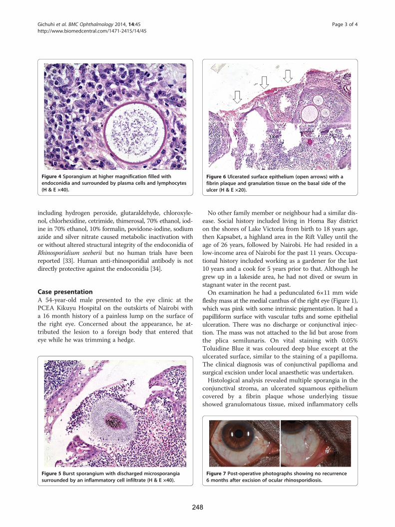
Figure 4 Sporangium at higher magnification filled withendoconidia and surrounded by plasma cells and lymphocytes(H & E ×40).
Figure 6 Ulcerated surface epithelium (open arrows) with afibrin plaque and granulation tissue on the basal side of theulcer (H & E ×20).
Gichuhi et al. BMC Ophthalmology 2014, 14:45 Page 3 of 4http://www.biomedcentral.com/1471-2415/14/45
including hydrogen peroxide, glutaraldehyde, chloroxyle-nol, chlorhexidine, cetrimide, thimerosal, 70% ethanol, iod-ine in 70% ethanol, 10% formalin, povidone-iodine, sodiumazide and silver nitrate caused metabolic inactivation withor without altered structural integrity of the endoconidia ofRhinosporidium seeberii but no human trials have beenreported [33]. Human anti-rhinosporidial antibody is notdirectly protective against the endoconidia [34].
Case presentationA 54-year-old male presented to the eye clinic at thePCEA Kikuyu Hospital on the outskirts of Nairobi witha 16 month history of a painless lump on the surface ofthe right eye. Concerned about the appearance, he at-tributed the lesion to a foreign body that entered thateye while he was trimming a hedge.
Figure 5 Burst sporangium with discharged microsporangiasurrounded by an inflammatory cell infiltrate (H & E ×40).
248
No other family member or neighbour had a similar dis-ease. Social history included living in Homa Bay districton the shores of Lake Victoria from birth to 18 years age,then Kapsabet, a highland area in the Rift Valley until theage of 26 years, followed by Nairobi. He had resided in alow-income area of Nairobi for the past 11 years. Occupa-tional history included working as a gardener for the last10 years and a cook for 5 years prior to that. Although hegrew up in a lakeside area, he had not dived or swum instagnant water in the recent past.On examination he had a pedunculated 6×11 mm wide
fleshy mass at the medial canthus of the right eye (Figure 1),which was pink with some intrinsic pigmentation. It had apapilliform surface with vascular tufts and some epithelialulceration. There was no discharge or conjunctival injec-tion. The mass was not attached to the lid but arose fromthe plica semilunaris. On vital staining with 0.05%Toluidine Blue it was coloured deep blue except at theulcerated surface, similar to the staining of a papilloma.The clinical diagnosis was of conjunctival papilloma andsurgical excision under local anaesthetic was undertaken.Histological analysis revealed multiple sporangia in the
conjunctival stroma, an ulcerated squamous epitheliumcovered by a fibrin plaque whose underlying tissueshowed granulomatous tissue, mixed inflammatory cells
Figure 7 Post-operative photographs showing no recurrence6 months after excision of ocular rhinosporidiosis.
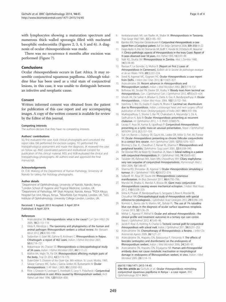
Gichuhi et al. BMC Ophthalmology 2014, 14:45 Page 4 of 4http://www.biomedcentral.com/1471-2415/14/45
with lymphocytes showing a maturation spectrum andnumerous thick walled sporangia filled with nucleatedbasophilic endoconidia (Figures 2, 3, 4, 5 and 6). A diag-nosis of ocular rhinosporidiosis was made.There was no recurrence 6 months after excision was
performed (Figure 7).
ConclusionsOcular rhinosporidiosis occurs in East Africa. It may re-semble conjunctival squamous papilloma. Although tolui-dine blue has been used as a vital stain of conjunctivallesions, in this case, it was unable to distinguish betweenan infective and neoplastic cause.
ConsentWritten informed consent was obtained from the patientfor publication of this case report and any accompanyingimages. A copy of the written consent is available for reviewby the Editor of this journal.
Competing interestsThe authors declare that they have no competing interests.
Authors’ contributionsSG first evaluated the case, took clinical photographs and conceived thereport idea. EM performed the excision surgery. TO performed thehistopathological assessment and made the diagnosis. JK reviewed the caseon follow up. AMZ coordinated patient and institutional consent forpublication of this clinical material. MSS and MJB evaluated the clinical andhistopathology photographs. All authors read and approved the finalmanuscript.
AcknowledgementsDr. E.W. Walong of the Department of Human Pathology, University ofNairobi for taking the histology photographs.
Author details1Department of Ophthalmology, University of Nairobi, Nairobi, Kenya.2London School of Hygiene and Tropical Medicine, London, UK.3Department of Pathology, MP Shah Hospital, Nairobi, Kenya. 4PCEA KikuyuHospital Eye Unit, Kikuyu, Kenya. 5Moorfields Eye Hospital, London, UK. 6UCLInstitute of Ophthalmology, University College London, London, UK.
Received: 1 August 2013 Accepted: 3 April 2014Published: 8 April 2014
References1. Arseculeratne SN: Rhinosporidiosis: what is the cause? Curr Opin Infect Dis
2005, 18(2):113–118.2. Vilela R, Mendoza L: The taxonomy and phylogenetics of the human and
animal pathogen Rhinosporidium seeberi: a critical review. Rev IberoamMicol 2012, 29(4):185–199.
3. Sudarshan V, Goel NK, Gahine R, Krishnani C: Rhinosporidiosis in Raipur,Chhattisgarh: a report of 462 cases. Indian J Pathol Microbiol 2007,50(4):718–721.
4. Makannavar JH, Chavan SS: Rhinosporidiosis–a clinicopathological studyof 34 cases. Indian J Pathol Microbiol 2001, 44(1):17–21.
5. Mallick AA, Majhi TK, Pal DK: Rhinosporidiosis affecting multiple parts ofthe body. Trop Dr 2012, 42(3):174–175.
6. Duke-Elder S: Diseases of the Outer Eye. IIIth edition. St. Louis: Mosby; 1965.7. Salazar Campos MC, Surka J, Garcia Jardon M, Bustamante N: Ocular
rhinosporidiosis. S Afr Med J 2005, 95(12):950–952.8. Pe’er J, Gnessin H, Levinger S, Averbukh E, Levy Y, Polacheck I: Conjunctival
oculosporidiosis in east Africa caused by Rhinosporidium seeberi. ArchPathol Lab Med 1996, 120(9):854–858.
249
9. Venkataramaiah NR, van Raalte JA, Shabe JK: Rhinosporidiosis in Tanzania.Trop Geogr Med 1981, 33(2):185–187.
10. Kaimbo KW, Parys-Van Ginderdeuren R: Conjunctival rhinosporidiosis: a casereport from a Congolese patient. Bull Soc Belge Ophtalmol 2008, 309–310:19–22.
11. Dago-Akribi A, Ette M, Diomande MI, Kioffi Y, Honde M, D’Ordock AF, BeaumelA: Clinico-pathologic aspects of rhinosporidiosis in the Ivory Coast. Report of9 cases observed over 18 years. Ann Pathol 1993, 13(2):97–99.
12. Naik KG, Shukla SM: Rhinosporidiosis in Zambia. Med J Zambia 1980,14(5):78–80.
13. Ravisse P, Le Gonidec G, Moliva B: [Report on first 2 cases ofrhinosporidiosis in Cameroon]. Bulletin de la Societe de pathologie exotiqueet de ses filiales 1976, 69(3):222–224.
14. Sood N, Agarwal MC, Gugnani HC: Ocular rhinosporidiosis: a case reportfrom Delhi. J Infect Dev Ctries 2012, 6(11):825–827.
15. Arseculeratne SN: Recent advances in rhinosporidiosis andRhinosporidium seeberi. Indian J Med Microbiol 2002, 20(3):119–131.
16. Belliveau MJ, Strube YN, Dexter DF, Kratky V: Bloody tears from lacrimal sacrhinosporidiosis. Can J Ophthalmol Can J Ophthalmol 2012, 47(5):e23–e24.
17. Ghosh AK, De Sarkar A, Bhaduri G, Datta A, Das A, Bandyopadhyay A: Ocularrhinosporidiosis. J Indian Med Assoc 2004, 102(12):732–764.
18. Varshney S, Bist SS, Gupta P, Gupta N, Bhatia R: Lacrimal sac diverticulumdue to Rhinosporidiosis. Indian j otolaryngol head and neck surgery: officialpublication of the Assoc Otolaryngologists of India 2007, 59(4):353–356.
19. Mukhopadhyay S, Shome S, Bar PK, Chakrabarti A, Mazumdar S, De A,Sadhukhan K, Bala B: Ocular rhinosporidiosis presenting as recurrentchalazion. Int Ophthalmol 2012, 1–3. PMID 22986579.
20. Lavaju P, Arya SK, Kumar B, Upadhaya P: Conjunctival rhinosporidiosispresenting as a cystic mass-an unusual presentation. Nepal J Ophthalmol:NEPJOPH 2010, 2(2):157–159.
21. Suh LH, Barron J, Dubovy SR, Gaunt ML, Ledee DR, Miller D, Fell JW, ForsterRK: Ocular rhinosporidiosis presenting as chronic follicular conjunctivitisin a contact lens wearer. Arch Ophthalmol 2009, 127(8):1076–1077.
22. Bhomaj S, Das JC, Chaudhuri Z, Bansal RL, Sharma P: Rhinosporidiosis andperipheral keratitis. Ophthalmic Surg Lasers 2001, 32(4):338–340.
23. De Doncker RM, de Keizer RJ, Oosterhuis JA, Maes A: Scleral melting in a patientwith conjunctival rhinosporidiosis. Br J Ophthalmol 1990, 74(10):635–637.
24. Talukder AK, Rahman MA, Islam MN, Chowdhury MH: Ciliary staphyloma:very rare sequelae of conjunctival rhinosporiodosis. Mymensingh Med J:MMJ 2004, 13(1):86–87.
25. Sharma KD, Shrivastav JB, Agarwal S: Ocular rhinosporidiosis simulating atumour. Br J Ophthalmol 1958, 42(9):572–574.
26. Vallarelli AF, Rosa SP, Souza EM: Rhinosporidiosis: cutaneousmanifestation. An Bras Dermatol 2011, 86(4):795–796.
27. Mandal SK, Bhakta A, Mandal A, Biswas BK: Giant mass conjunctivalrhinosporidiosis causing severe mechanical ectropion. J Indian Med Assoc2012, 110(5):328–329.
28. Sinha A, Phukan JP, Bandyopadhyay G, Sengupta S, Bose K, Mondal RK,Choudhuri MK: Clinicopathological study of rhinosporidiosis with specialreference to cytodiagnosis. J cytol/Indian Acad Cytologists 2012, 29(4):246–249.
29. Romero IL, Barros Jde N, Martins MC, Ballalai PL: The use of 1% toluidineblue eye drops in the diagnosis of ocular surface squamous neoplasia.Cornea 2013, 32(1):36–39.
30. Mithal C, Agarwal P, Mithal N: Ocular and adnexal rhinosporidiosis : theclinical profile and treatment outcomes in a tertiary eye care centre.Nepal J Ophthalmol 2012, 4(1):45–48.
31. Jacob P, Rose JS, Hoshing A, Chacko G: Tectonic corneal graft for conjunctivalrhinosporidiosis with scleral melt. Indian J Ophthalmol 2011, 59(3):251–253.
32. Arseculeratne SN: Chemotherapy of Rhinosporidiosis: a Review. J Infect DisAntimicrob Agents 2009, 26(1):21–27.
33. Arseculeratne SN, Atapattu DN, Balasooriya P, Fernando R: The effects ofbiocides (antiseptics and disinfectants) on the endospores ofRhinosporidium seeberi. Indian J Med Microbiol 2006, 24(2):85–91.
34. Arseculeratne SN, Atapattu DN, Eriyagama NB: Human anti-rhinosporidialantibody does not cause metabolic inactivation or morphologicaldamage in endospores of Rhinosporidium seeberi, in vitro. Indian J MedMicrobiol 2005, 23(1):14–19.
doi:10.1186/1471-2415-14-45Cite this article as: Gichuhi et al.: Ocular rhinosporidiosis mimickingconjunctival squamous papilloma in Kenya – a case report. BMCOphthalmology 2014 14:45.


Page 1 of 1254
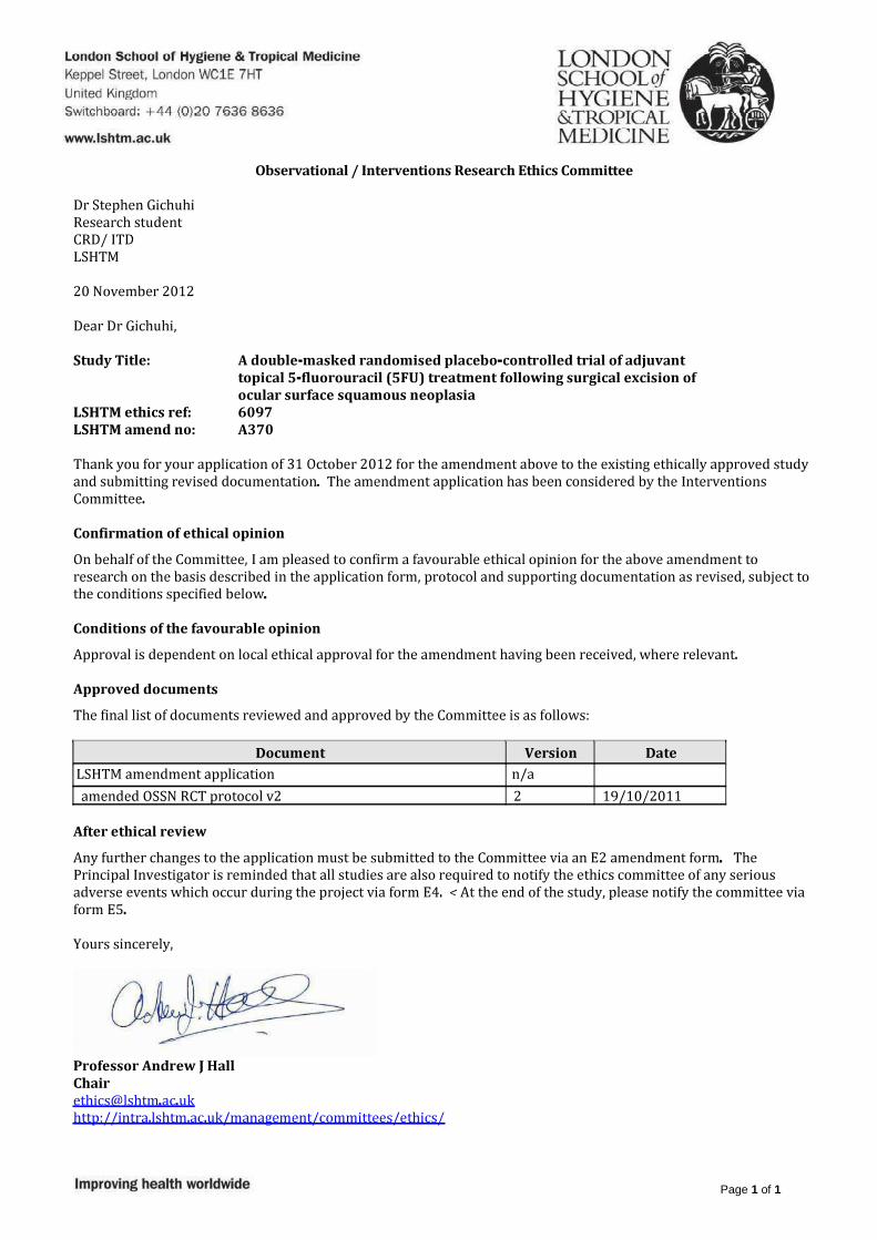
Page 1 of 1
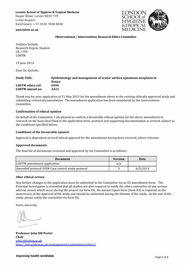
Page 1 of 1

OBSERVATIONAL/INTERVENTIONS RESEARCH ETHICS COMMITTEE
19 January 2012
Stephen Gichuhi
Dear Stephen
Study Title: A double-masked randomised placebo-controlled trial of adjuvanttopical 5-fluorouracil (5FU) treatment following surgical excision ofocular surface squamous neoplasia
LSHTM ethics ref: 6097Department: Infectious and Tropical Diseases
The Committee reviewed the above application.
Documents reviewed
The documents reviewed were:
Document Version DateLSHTM ethics application n/a 19/01/12Protocol V1.0 19/01/12Information Sheet V1.0 19/01/12Consent form V1.0 19/01/12
Provisional opinion
The Committee would be content to give a favourable ethical opinion of the research, subject to receiving acomplete response to the request for further information set out below.
The Committee delegated authority to confirm its final opinion on the application to the Chair.
Further information or clarification required
Please provide a reference for the 20% recurrence rate - the document mentions 30-66% recurrencewithin 2 years only currently.
Clarify why 5-fluorouracil was selected over mitomycin or interferon - was this simply an issue of cost?
When submitting your response to the Committee, please send revised documentation where appropriateunderlining or otherwise highlighting the changes you have made and giving revised version numbers and dates.
If the Committee has asked for clarification or changes to any answers given in the application form, please donot submit a revised copy of the application form; these can be addressed in a covering letter to the Committee.Yours sincerely,
Professor Andrew J HallChair
257

25th January 2012
Prof. Andrew J. Hall
Study Title: A double-masked randomised placebo-controlled trial of adjuvanttopical 5-fluorouracil (5FU) treatment following surgical excision ofocular surface squamous neoplasia
LSHTM ethics ref: 6097
Department: Infectious and Tropical Diseases
Thank you for your letter dated 19th January 2012 requesting further information and clarification.
Regarding the 20% recurrence rate, operationally, published rates mostly vary between 30-60%(summarised in the appendix attached) and involve a surgical technique in which the tumour isexcised and the sclera left bare. The lowest rate is 3.2% from a surgeon in Uganda who is probablythe most experienced in East Africa. In our study we will have fairly experienced surgeons and thesurgical technique will involve primary closure of the excision site by mobilizing adjoining conjunctivaor using a conjunctival autograft. We think this would reduce the recurrence rate to below 30%. Inaddition, if we used 30% for sample size calculation, we would require a smaller sample but 20%would increase the sample size. We chose to use a more conservative estimate.
With respect to the choice of 5FU, this was based on the fact that it is less costly, has lower oculartoxicity and is more easily available in East Africa. This would make translation of the trial resultseasier in the East African eye care setting. In addition, there is no published trial evidence that either5FU, Mitomycin C or Interferon is better than the other for the treatment of ocular surface squamousneoplasia.
Yours sincerely,
Stephen Gichuhi
Encs. Summary of published recurrence rates
258
259

260
261
262

263

264

265

Page 1 of 2
266
Page 2 of 2
267

268
269

24 July 2012
To Whom It May Concern:
re: Adjuvant topical 5fluorouracil (5FU) for ocular surface squamous neoplasia
As project manager for the Pan African Clinical Trial Registry (www.pactr.org) database, itis my pleasure to inform you that your application to our registry has been accepted. Yourunique identification number for the registry is PACTR201207000396219.
Please be advised that you are responsible for updating your trial, or for informin g us ofchanges to your trial.
Additionally, please provide us with copies of your ethical clearance letters as we musthave these on file (via email, post or fax) at your earliest convenience if you have notalready done so.
Please do not hesitate to contact us at +27 21 938 0506 or email [email protected] you have any questions.
Yours faithfully,
Amber Abramswww.pactr.org Project Manager+27 021 938 0506
272

version2 19/10/2012
1
1. TitleA double-masked randomised placebo-controlled trial of adjuvant topical 5-
fluorouracil (5FU) treatment following surgical excision of ocular surface
squamous neoplasia.
273

version2 19/10/2012
2
Table of Contents1. Title .................................................................................................................................................1
2. Investigators, co-investigators and supervisors.......................................................................3
3. Collaborating institutions .............................................................................................................5
4. Funding agency ............................................................................................................................5
5. Summary .......................................................................................................................................5
6. Background ...................................................................................................................................5
7. Rationale........................................................................................................................................8
8. Research question .......................................................................................................................8
9. Objectives ......................................................................................................................................8
10. Study design and Methodology..............................................................................................9
11. Ethical considerations............................................................................................................20
12. Data management..................................................................................................................21
13. Study limitations and how to minimize them ......................................................................23
14. Timeline ...................................................................................................................................23
15. References ..............................................................................................................................24
16. Appendix ..................................................................................................................................28
Appendix 1 Statistical analysis plan ............................................................................................28
Appendix 2 – Flow chart for reporting of safety events ............................................................31
274
version2 19/10/2012
3
2. Investigators, co-investigators and supervisorsDr Stephen Gichuhi MBChB, M.Med, MBA, MSc (Epidemiology)
Research Degree Student
International Centre for Eye Health (ICEH)
Faculty of Infectious and Tropical Diseases
London School of Hygiene & Tropical Medicine
Senior Lecturer
Department of Ophthalmology
University of Nairobi
PO Box 19676-00202
Nairobi.
Tel: +254-722873059 (m), +254-20-2726300 ext. 43776 (o)
Email: [email protected] Signed:_____________
Supervisor:
Dr Matthew Burton PhD, MA, MBBCh, DTM&H, MRCP, FRCOphth
Senior Lecturer
International Centre for Eye Health (ICEH)
Faculty of Infectious and Tropical Diseases
London School of Hygiene & Tropical Medicine
Keppel Street
London, WC1E 7HT
Tel: +44 (0)20 7958 8359
Fax: +44 (0)20 7958 8317
Email: [email protected] Signed:_____________
Co-supervisor:
Dr Helen Weiss MSc PhD
Reader (Epidemiology and Biostatistics)
Faculty of Epidemiology and Population Health
London School of Hygiene and Tropical Medicine
Keppel Street
London WC1E 7HT
Tel: +44 (0) 20-7612-7872
Fax: +44(0) 20-7958-8325
Email: [email protected] Signed:
275

version2 19/10/2012
4
Co-investigators:
Dr. Joy Kabiru MBChB, M.Med
Consultant Ophthalmologist
PCEA Kikuyu Hospital Eye Unit
Dr. T. Onyuma MBChB, M.Med
Consultant Pathologist
MP Shah Hospital
Prof. Walter Jaoko MBChB, MTMed, PhD
Kenya Aids Vaccine Initiative (KAVI)
University of Nairobi
Dr. Mandeep Sagoo MB PhD FRCS
Senior Lecturer and Consultant Ophthalmologist (Ocular Oncology)
UCL Institute of Ophthalmology & Moorfields Eye Hospital
London
Dr. Olando MBChB, M.Med
Consultant Ophthalmologist
Sabatia Eye Unit
Dr. Hilary Rono MBChB, M.Med
Consultant Ophthalmologist
Kitale District Hospital
Dr. Ochieng MBChB, M.Med
Consultant Ophthalmologist
Homa Bay District Hospital
276

version2 19/10/2012
5
3. Collaborating institutionsi) London School of Hygiene & Tropical Medicine
ii) University of Nairobi
iii) Kenyatta National Hospital
iv) PCEA Kikuyu Hospital Eye Unit
v) UCL Institute of Ophthalmology & UCL Cancer Institute
vi) Sabatia Eye Unit
vii) Kitale District Hospital
viii) Homa Bay District Hospital
4. Funding agencyFunding has been provided by the British Council for Prevention of Blindness (BCPB) to
Dr Stephen Gichuhi to conduct this project as a PhD fellowship, registered at the
London School of Hygiene & Tropical Medicine (LSHTM).
5. SummaryIn East Africa, ocular surface squamous neoplasia (OSSN) is a relatively common
aggressive disease affecting younger adults. Treatment usually involves surgical
excision, however, recurrent disease is quite common (up to 30-66% by 30 months
afterwards) and there have been no randomised controlled trials to investigate how to
reduce this. For a long time topical 5-fluorouracil (5FU) has been widely used as
adjuvant therapy following surgical excision, with many case reports and series in the
literature. However, it has not been formally tested in a randomised controlled trial and
so we currently do not know if it is effective in preventing recurrent tumours, particularly
in the context where more expensive adjuvants or surgical equipment are not readily
available.
We propose to conduct a randomised controlled trial to determine whether OSSN
recurrence can be reduced by adjuvant topical 5FU chemotherapy following excision.
Following surgical excision of histopathologically confirmed OSSN, participants will be
randomised to either 5FU eyedrops, or placebo drops to be taken four times a day for 1
month. Patients will be followed for one year to assess the effect. The primary outcome
measure is histopathologically confirmed recurrent OSSN.
6. BackgroundOcular surface squamous neoplasia (OSSN) is a spectrum of disease that ranges from
non-invasive intra-epithelial dysplasia of the conjunctiva and cornea (CCIN), through to
277

version2 19/10/2012
6
invasive squamous cell carcinoma (SCC).1 In recent decades OSSN has undergone an
epidemiological shift. In more temperate countries, it remains a rare, slow growing
tumour of elderly males.2 In contrast, in tropical countries, particularly in Eastern Africa,
it is now more common, more aggressive, affects younger people and slightly more
women than men.3-6 Patients often present late with advanced tumours.5 It seems likely
that much of this increased burden of disease is attributable to the HIV/AIDS epidemic.7
Even though OSSN is not a target condition within Vision 2020, a WHO initiative to
prevent avoidable blindness by the year 2020, it frequently leads to a poor quality of life,
visual disability and death. In September 2010 the “International Agency for Prevention
of Blindness (IAPB)/Vision2020 Workshop on Research for Global Blindness
Prevention” identified specific research priorities.8 It was recognised that for Africa there
was a need for research on HIV-related conditions to better define the epidemiology and
determine context-specific management approaches.
Reliable prevalence and incidence estimates of the numbers of individuals affected have
been difficult to ascertain and vary considerably. At one extreme, in one study from
Kenya based in HIV testing clinics, 7.8% of HIV-infected adults were found to have
conjunctival tumours which were found to be OSSN on histopathology.5 The Kenyan
national HIV prevalence is 7.4%% (2.22 million) so it was suggested that over 170,000
Kenyans might have some degree of OSSN.9 In contrast, a relatively low annual
incidence estimate of 2.2/100,000 has been suggested in a study from Tanzania, based
on the number of cases being operated in eye units.6 From the clinical perspective
OSSN represents a significant component of the ophthalmic “work-load” in East Africa
and is associated with a high level of morbidity for the patient, as exenteration is often
needed for those with late presentation or recurrent disease. However, OSSN receives
little or no attention from either ophthalmic or HIV care programs.
The epidemiology, aetiology and pathophysiology of OSSN in East Africa is not well
understood. There is an association with HIV; however, a significant proportion (about
30%) of people with OSSN are not infected with HIV, suggesting that other factors also
contribute to the excess burden of disease in this region.7, 10-12 The interaction between
CD4 T-lymphocyte levels in HIV+ individuals and OSSN has not been described. The
relationship between human papilloma virus (HPV) and OSSN remains unclear; some
studies have reported associations and others have not.13-18 This is probably because of
variations in methodology and the specific HPV types that have been looked for. There
are 120 genotypes of HPV.19 Some are cutaneous and others mucosal. Cutaneous
types are more frequently found in OSSN than the mucosal types.16, 20 Generally only a
278
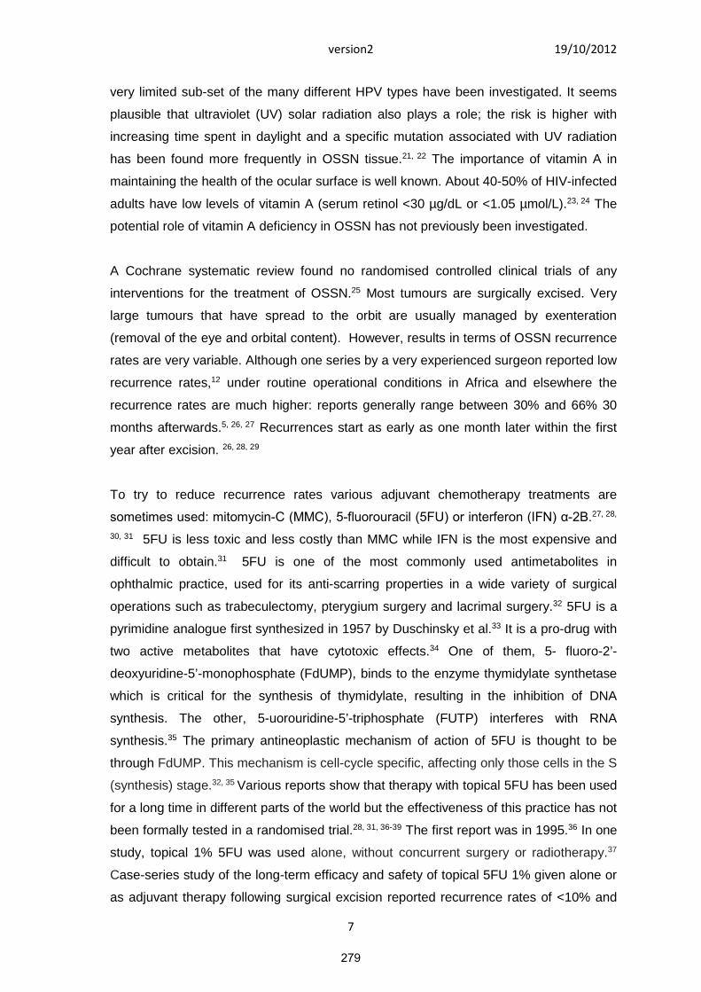
version2 19/10/2012
7
very limited sub-set of the many different HPV types have been investigated. It seems
plausible that ultraviolet (UV) solar radiation also plays a role; the risk is higher with
increasing time spent in daylight and a specific mutation associated with UV radiation
has been found more frequently in OSSN tissue.21, 22 The importance of vitamin A in
maintaining the health of the ocular surface is well known. About 40-50% of HIV-infected
adults have low levels of vitamin A (serum retinol <30 µg/dL or <1.05 µmol/L).23, 24 The
potential role of vitamin A deficiency in OSSN has not previously been investigated.
A Cochrane systematic review found no randomised controlled clinical trials of any
interventions for the treatment of OSSN.25 Most tumours are surgically excised. Very
large tumours that have spread to the orbit are usually managed by exenteration
(removal of the eye and orbital content). However, results in terms of OSSN recurrence
rates are very variable. Although one series by a very experienced surgeon reported low
recurrence rates,12 under routine operational conditions in Africa and elsewhere the
recurrence rates are much higher: reports generally range between 30% and 66% 30
months afterwards.5, 26, 27 Recurrences start as early as one month later within the first
year after excision. 26, 28, 29
To try to reduce recurrence rates various adjuvant chemotherapy treatments are
sometimes used: mitomycin-C (MMC), 5-fluorouracil (5FU) or interferon (IFN) α-2B.27, 28,
30, 31 5FU is less toxic and less costly than MMC while IFN is the most expensive and
difficult to obtain.31 5FU is one of the most commonly used antimetabolites in
ophthalmic practice, used for its anti-scarring properties in a wide variety of surgical
operations such as trabeculectomy, pterygium surgery and lacrimal surgery.32 5FU is a
pyrimidine analogue first synthesized in 1957 by Duschinsky et al.33 It is a pro-drug with
two active metabolites that have cytotoxic effects.34 One of them, 5- fluoro-2’-
deoxyuridine-5’-monophosphate (FdUMP), binds to the enzyme thymidylate synthetase
which is critical for the synthesis of thymidylate, resulting in the inhibition of DNA
synthesis. The other, 5-uorouridine-5’-triphosphate (FUTP) interferes with RNA
synthesis.35 The primary antineoplastic mechanism of action of 5FU is thought to be
through FdUMP. This mechanism is cell-cycle specific, affecting only those cells in the S
(synthesis) stage.32, 35 Various reports show that therapy with topical 5FU has been used
for a long time in different parts of the world but the effectiveness of this practice has not
been formally tested in a randomised trial.28, 31, 36-39 The first report was in 1995.36 In one
study, topical 1% 5FU was used alone, without concurrent surgery or radiotherapy.37
Case-series study of the long-term efficacy and safety of topical 5FU 1% given alone or
as adjuvant therapy following surgical excision reported recurrence rates of <10% and
279

version2 19/10/2012
8
no evidence of long term toxicity or morbidity except rarely epiphora (excessive watering
of the eyes) thought to be due to scarring of the tear drainage passage.39, 40 Adjuvant
therapy is however rarely used in sub-Saharan Africa, although 5FU is available and
affordable. Randomised controlled trials are needed to identify the optimal treatment for
this condition to minimise recurrence rates, visual loss and death from conjunctival
squamous cell carcinoma in the East African setting.
7. Rationale
Ocular surface squamous neoplasia is a common and often difficult to treat disease in
ophthalmic practice in East Africa. Recurrent tumours often complicate the treatment.
There is a need for interventions to reduce recurrence rates. Post-operative 5FU drops
have been used for many years to treat patients following tumour excision, although
there is no trial data on this. There is a need for a randomised controlled trial (RCT) to
determine whether this is effective and should become standard practice. If this trial has
a positive outcome, 5FU eye drops are cheap and easily available. They do not have
stringent storage conditions and have low risk of contamination. Patients can also apply
it for themselves. Provision of this intervention on a public health scale is therefore
feasible.
8. Research question
In adults with primary OSSN, does adjuvant chemotherapy with topical 1% 5FU applied
four times daily for one month reduce the recurrence rate over a period of one year than
surgical excision alone?
9. Objectives
a) Broad Objective
To determine whether the recurrence of OSSN can be reduced through the use of
adjuvant topical chemotherapy.
b) Primary Objective
To compare the one-year recurrence rate of OSSN with or without adjuvant topical
5FU 1% eye drops applied 4 times daily for one month after surgical excision of OSSN
tumours which involve less than 2 quadrants of the conjunctiva.
c) Secondary objectives
280
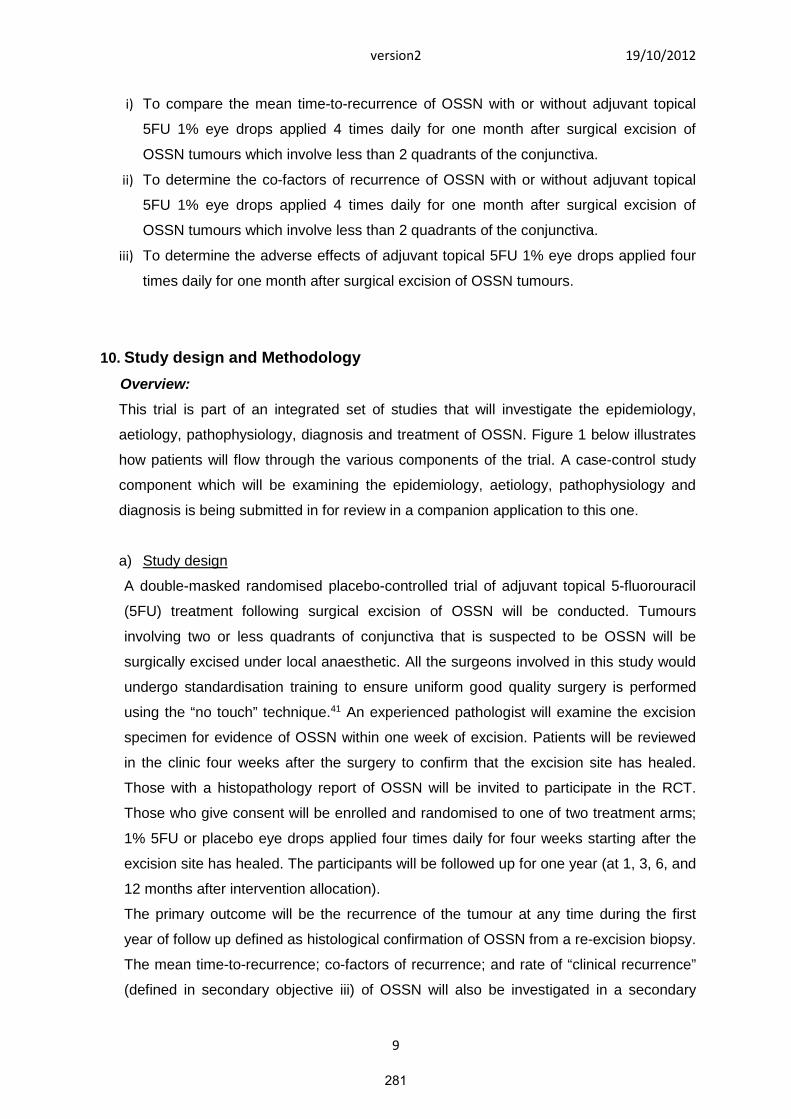
version2 19/10/2012
9
i) To compare the mean time-to-recurrence of OSSN with or without adjuvant topical
5FU 1% eye drops applied 4 times daily for one month after surgical excision of
OSSN tumours which involve less than 2 quadrants of the conjunctiva.
ii) To determine the co-factors of recurrence of OSSN with or without adjuvant topical
5FU 1% eye drops applied 4 times daily for one month after surgical excision of
OSSN tumours which involve less than 2 quadrants of the conjunctiva.
iii) To determine the adverse effects of adjuvant topical 5FU 1% eye drops applied four
times daily for one month after surgical excision of OSSN tumours.
10. Study design and Methodology
Overview:
This trial is part of an integrated set of studies that will investigate the epidemiology,
aetiology, pathophysiology, diagnosis and treatment of OSSN. Figure 1 below illustrates
how patients will flow through the various components of the trial. A case-control study
component which will be examining the epidemiology, aetiology, pathophysiology and
diagnosis is being submitted in for review in a companion application to this one.
a) Study design
A double-masked randomised placebo-controlled trial of adjuvant topical 5-fluorouracil
(5FU) treatment following surgical excision of OSSN will be conducted. Tumours
involving two or less quadrants of conjunctiva that is suspected to be OSSN will be
surgically excised under local anaesthetic. All the surgeons involved in this study would
undergo standardisation training to ensure uniform good quality surgery is performed
using the “no touch” technique.41 An experienced pathologist will examine the excision
specimen for evidence of OSSN within one week of excision. Patients will be reviewed
in the clinic four weeks after the surgery to confirm that the excision site has healed.
Those with a histopathology report of OSSN will be invited to participate in the RCT.
Those who give consent will be enrolled and randomised to one of two treatment arms;
1% 5FU or placebo eye drops applied four times daily for four weeks starting after the
excision site has healed. The participants will be followed up for one year (at 1, 3, 6, and
12 months after intervention allocation).
The primary outcome will be the recurrence of the tumour at any time during the first
year of follow up defined as histological confirmation of OSSN from a re-excision biopsy.
The mean time-to-recurrence; co-factors of recurrence; and rate of “clinical recurrence”
(defined in secondary objective iii) of OSSN will also be investigated in a secondary
281
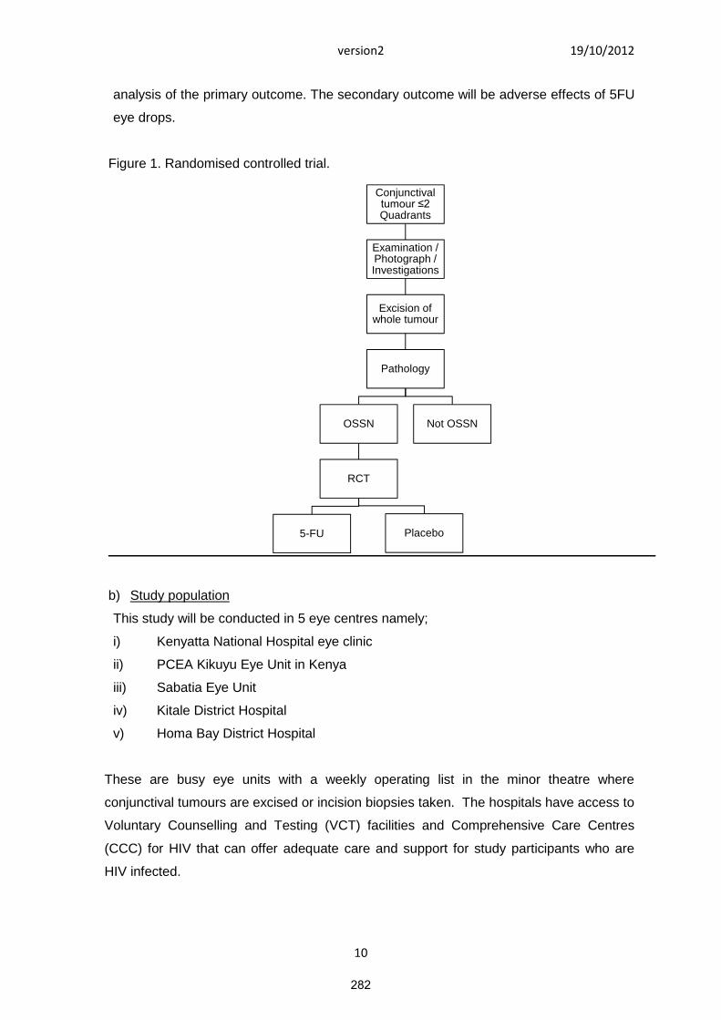
version2 19/10/2012
10
analysis of the primary outcome. The secondary outcome will be adverse effects of 5FU
eye drops.
Figure 1. Randomised controlled trial.
b) Study population
This study will be conducted in 5 eye centres namely;
i) Kenyatta National Hospital eye clinic
ii) PCEA Kikuyu Eye Unit in Kenya
iii) Sabatia Eye Unit
iv) Kitale District Hospital
v) Homa Bay District Hospital
These are busy eye units with a weekly operating list in the minor theatre where
conjunctival tumours are excised or incision biopsies taken. The hospitals have access to
Voluntary Counselling and Testing (VCT) facilities and Comprehensive Care Centres
(CCC) for HIV that can offer adequate care and support for study participants who are
HIV infected.
Conjunctivaltumour ≤2 Quadrants
Examination /Photograph /Investigations
Excision ofwhole tumour
Pathology
OSSN
RCT
5-FU Placebo
Not OSSN
282
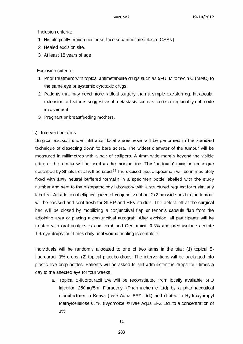
version2 19/10/2012
11
Inclusion criteria:
1. Histologically proven ocular surface squamous neoplasia (OSSN)
2. Healed excision site.
3. At least 18 years of age.
Exclusion criteria:
1. Prior treatment with topical antimetabolite drugs such as 5FU, Mitomycin C (MMC) to
the same eye or systemic cytotoxic drugs.
2. Patients that may need more radical surgery than a simple excision eg. intraocular
extension or features suggestive of metastasis such as fornix or regional lymph node
involvement.
3. Pregnant or breastfeeding mothers.
c) Intervention arms
Surgical excision under infiltration local anaesthesia will be performed in the standard
technique of dissecting down to bare sclera. The widest diameter of the tumour will be
measured in millimetres with a pair of callipers. A 4mm-wide margin beyond the visible
edge of the tumour will be used as the incision line. The “no-touch” excision technique
described by Shields et al will be used.39 The excised tissue specimen will be immediately
fixed with 10% neutral buffered formalin in a specimen bottle labelled with the study
number and sent to the histopathology laboratory with a structured request form similarly
labelled. An additional elliptical piece of conjunctiva about 2x2mm wide next to the tumour
will be excised and sent fresh for SLRP and HPV studies. The defect left at the surgical
bed will be closed by mobilizing a conjunctival flap or tenon’s capsule flap from the
adjoining area or placing a conjunctival autograft. After excision, all participants will be
treated with oral analgesics and combined Gentamicin 0.3% and prednisolone acetate
1% eye-drops four times daily until wound healing is complete.
Individuals will be randomly allocated to one of two arms in the trial: (1) topical 5-
fluorouracil 1% drops; (2) topical placebo drops. The interventions will be packaged into
plastic eye drop bottles. Patients will be asked to self-administer the drops four times a
day to the affected eye for four weeks.
a. Topical 5-fluorouracil 1% will be reconstituted from locally available 5FU
injection 250mg/5ml Fluracedyl (Pharmachemie Ltd) by a pharmaceutical
manufacturer in Kenya (Ivee Aqua EPZ Ltd.) and diluted in Hydroxypropyl
Methylcellulose 0.7% (Ivyomoicell® Ivee Aqua EPZ Ltd, to a concentration of
1%.
283

version2 19/10/2012
12
b. The placebo will be the same diluent solution used to make up the 5FU drops,
namely Hydroxypropyl Methylcellulose 0.7% (Ivyomoicell® Ivee Aqua EPZ
Ltd.).
d) Randomisation
A random allocation sequence will be generated on computer by a statistician at the
London School of Hygiene and Tropical Medicine (LSHTM) using STATA software (Stata
Corporation, College Station, TX, USA). Random permuted blocks will be used. The block
size will be randomly varied to prevent guessing the intervention assignment. Separate
randomisation lists will be prepared for each surgeon. The lists will be used by one
research staff member to label and allocate treatment bottles. The allocation sequence will
be concealed from the other investigators as only this staff member and the statistician will
handle the randomisation list.
e) Masking
The participants, the surgeon, the ophthalmologist performing the clinical examinations
and the pathologist examining the recurrence specimens will be masked to the treatment
arm allocation. The bottles of eye drops will appear similar and only identified by code
numbers. The ID number on the bottle will not provide a clue as to whether it is 5FU or
placebo. The pharmacy will not know the content of each bottle. The pharmaceutical
manufacturer will have a code generated by the statistician to use in making the eye
drops. Unmasking of an individual’s treatment will be performed on recommendation of the
data safety and monitoring board (DSMB).
f) Follow up
After the wound has healed participants will be allocated to the intervention arm then
followed-up at 1 month, 3 months, 6 months and 12 months after the date of intervention
allocation. They will be carefully examined for signs of recurrent tumours and
photographed on each occasion.
g) Primary end point and analyses
Primary analysis of primary endpoint:
The primary outcome will be the clinical recurrence of the tumour at any time during the
first year of follow-up, and where available confirmed by histology. During follow up, if the
outcome assessor notices a possible recurrence, it will be re-excised and sent for
histopathology. We will compare the proportion of recurrences at one year in the two
intervention arms. Intention-to-treat analysis will be used. The measure of effect will be the
284

version2 19/10/2012
13
odds ratio with 95% confidence intervals. Kaplan-Meier analysis will be used to plot the
survival curves for both arms up to the final visit at one year.
Secondary analysis of primary endpoint
I. mean time-to-recurrence of OSSN
The time-to-recurrence will be the number of person-months between surgery and the
date the recurrence was first noted clinically. Person-months will be calculated for each
arm and the two compared. Cox regression will be used to assess the impact of the
intervention on time-to-recurrence. The hazard ratio will be estimated with Cox
regression, adjusting for substantial baseline imbalances if appropriate.
II. Co-factors of recurrence
Factors that may influence the primary outcome will be analysed. This will include HIV
infection, CD4 count, age and sex of participant, use of highly active anti-retroviral
therapy (HAART), HPV infection and serum vitamin A levels. Effect modification by the
surgeon, size of initial OSSN tumour, and rate of growth will be analysed. The association
between any of these co-factors and the primary outcome will be reported using odds
ratios and their 95% confidence intervals.
h) Secondary outcome
The main secondary outcome will be to assess the safety of using topical 5-fluorouracil 1%
four times daily for one month. We would monitor adverse events. The most commonly
reported adverse effect is epiphora but we would monitor all other events as shown below.
DEFINITIONS - The following definitions shall be adopted for safety reporting:
Adverse Event (AE): any untoward medical occurrence in a clinical trial participant
administered a medicinal product and which does not necessarily have a causal
relationship with this treatment. An AE can therefore be any unfavourable and unintended
sign (including an abnormal laboratory finding), symptom, or disease temporally associated
with the use of an investigational medicinal product (IMP), whether or not considered
related to the IMP.
Adverse Reaction (AR): all untoward and unintended responses to an IMP related to any
dose administered. All AEs judged by either the reporting investigator or the sponsor as
having reasonable causal relationship to a medicinal product qualify as adverse reactions.
The expression reasonable causal relationship means to convey in general that there is
evidence or argument to suggest a causal relationship.
285

version2 19/10/2012
14
Unexpected Adverse Reaction: an AR, the nature or severity of which is not consistent with
the applicable product information (eg investigator’s brochure for an unapproved
investigational product or summary of product characteristics (SmPC) for an authorised
product). When the outcome of the adverse reaction is not consistent with the applicable
product information this adverse reaction shall be considered as unexpected. Side effects
documented in the SmPC which occur in a more severe form than anticipated are also
considered to be unexpected.
Serious Adverse Event (SAE) or Serious Adverse Reaction: any untoward medical
occurrence or effect that at any dose
• Results in death
• Is life-threatening – refers to an event in which the subject was at risk of death at
the time of the event; it does not refer to an event which hypothetically might have
caused death if it were more severe
• Requires hospitalisation, or prolongation of existing inpatients’ hospitalisation
• Results in persistent or significant disability or incapacity
• Is a congenital anomaly or birth defect
Medical judgement shall be exercised in deciding whether an AE/AR is serious in other
situations. Important AE/ARs that are not immediately life-threatening or do not result in
death or hospitalisation but may jeopardise the subject or may require intervention to
prevent one of the other outcomes listed in the definition above, shall also be considered
serious.
Suspected Unexpected Serious Adverse Reaction (SUSAR): any suspected adverse
reaction related to an IMP that is both unexpected and serious.
CAUSALITY - Most adverse events and adverse drug reactions that occur in this study,
whether they are serious or not, will be expected treatment-related toxicities due to the
drugs used in this study. The assignment of the causality will be made by the investigator
responsible for the care of the participant using the definitions in the table below.
If any doubt about the causality exists the local investigator shall inform the study site
coordinator who will notify the Chief Investigators. The pharmaceutical companies and/or
other clinicians may be asked to advise in some cases.
286
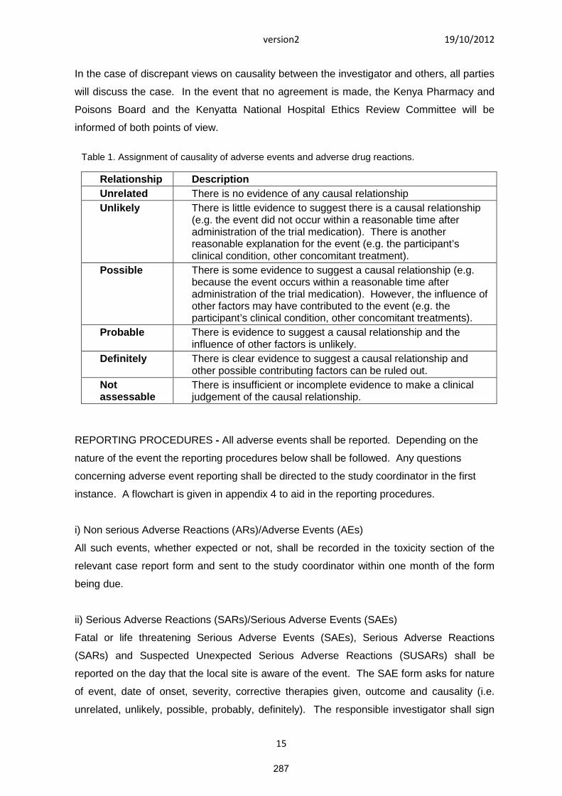
version2 19/10/2012
15
In the case of discrepant views on causality between the investigator and others, all parties
will discuss the case. In the event that no agreement is made, the Kenya Pharmacy and
Poisons Board and the Kenyatta National Hospital Ethics Review Committee will be
informed of both points of view.
Table 1. Assignment of causality of adverse events and adverse drug reactions.
Relationship Description
Unrelated There is no evidence of any causal relationship
Unlikely There is little evidence to suggest there is a causal relationship(e.g. the event did not occur within a reasonable time afteradministration of the trial medication). There is anotherreasonable explanation for the event (e.g. the participant’sclinical condition, other concomitant treatment).
Possible There is some evidence to suggest a causal relationship (e.g.because the event occurs within a reasonable time afteradministration of the trial medication). However, the influence ofother factors may have contributed to the event (e.g. theparticipant’s clinical condition, other concomitant treatments).
Probable There is evidence to suggest a causal relationship and theinfluence of other factors is unlikely.
Definitely There is clear evidence to suggest a causal relationship andother possible contributing factors can be ruled out.
Notassessable
There is insufficient or incomplete evidence to make a clinicaljudgement of the causal relationship.
REPORTING PROCEDURES - All adverse events shall be reported. Depending on the
nature of the event the reporting procedures below shall be followed. Any questions
concerning adverse event reporting shall be directed to the study coordinator in the first
instance. A flowchart is given in appendix 4 to aid in the reporting procedures.
i) Non serious Adverse Reactions (ARs)/Adverse Events (AEs)
All such events, whether expected or not, shall be recorded in the toxicity section of the
relevant case report form and sent to the study coordinator within one month of the form
being due.
ii) Serious Adverse Reactions (SARs)/Serious Adverse Events (SAEs)
Fatal or life threatening Serious Adverse Events (SAEs), Serious Adverse Reactions
(SARs) and Suspected Unexpected Serious Adverse Reactions (SUSARs) shall be
reported on the day that the local site is aware of the event. The SAE form asks for nature
of event, date of onset, severity, corrective therapies given, outcome and causality (i.e.
unrelated, unlikely, possible, probably, definitely). The responsible investigator shall sign
287

version2 19/10/2012
16
the causality of the event. Additional information shall be sent within 5 days if the reaction
has not resolved at the time of reporting.
iii) SAEs
An SAE form shall be completed and sent to the study coordinator for all SAEs within 24
hours. However, relapse and death due to ocular surface squamous neoplasia and
hospitalisations for elective treatment of a pre-existing condition will not need reporting as
SAEs.
iv)SUSARs
In the case of serious, unexpected and related adverse events, the staff at the site shall:
Complete the SAE case report form & send it immediately (within 24 hours, preferably by
email), signed and dated to the study coordinator together with relevant treatment forms
and anonymised copies of all relevant investigations.
Or
Contact the study coordinator by phone and then send the completed SAE form by email to
the study coordinator within the following 24 hours as above.
The study coordinator will notify the Kenya Pharmacy and Poisons Board and Kenyatta
National Hospital Ethics Review Committee of all SUSARs occurring during the study
according to the following timelines; fatal and life-threatening within 7 days of notification
and non-life threatening within 15 days. All investigators will be informed of all SUSARs
occurring throughout the study.
Investigators shall report any SUSARs and /or SAEs as required by and Kenyatta National
Hospital Ethics Review Committee.
Contact details for reporting SAEs and SUSARs
Please send SAE forms to: Dr. Stephen Gichuhi on [email protected]
Tel: 0722873059 (Mon to Fri 09.00 – 17.00)
288

version2 19/10/2012
17
i) Sample size
A power-based sample size calculation will be used. We anticipate a 20% OSSN
recurrence rate by one year after surgery in the placebo arm. From a programmatic
perspective in the East African setting we consider that adjuvant 5FU would be a
worthwhile intervention if it halved the recurrence rate to 10%. Using Stata IC 11 to
calculate sample size, a sample of 438 cases of OSSN (219 in each arm) would have
80% power and 95% confidence to detect a relative reduction in the risk of recurrence of
50% from 20% to 10% in the 5FU arm at one year. Including 15% extra for loss to
follow-up would make a total of 504, 252 in each arm.
j) Recruitment, consenting and retention procedures
Together the study centres surgically treat about 7 new cases of OSSN each week on
designated operating lists (about 360/year). Therefore we anticipate that we will be able
to comfortably recruit the necessary sample size within 15 months.
Participants for the trial will be recruited from a concurrent case-control study that will be
going on at the two centres. The project will be publicised using flyers and
announcements at various forums that ophthalmologists and ophthalmic clinical officers
meet and also eyecare institutions to encourage referrals. The websites of eye care
professional associations and also the East Africa Journal of Ophthalmology (EAJO) will
also carry the announcement.
During the case-control study participants with conjunctival tumours will have clinical
examinations, digital photographs, surgical excision and histology done. After excision,
they will be treated with oral analgesics and combined Gentamicin 0.3% and
Prednisolone acetate 1% eye-drops four times daily until wound healing is complete.
Usually this takes 3-4 weeks. In the interim period, histology reports will be ready. The
reports that indicate OSSN will be flagged and the patients noted for potential
recruitment into the trial. Their medical records will be checked to screen for eligibility.
During the review visit, the clinician will discuss treatment options and refer the patient
to a research nurse who will give more details about the study. The informed consent
process will be conducted by an ophthalmic research nurse trained in the procedures
involved in the study. The study information sheet will be given to them, read to them in
an appropriate language and discussed by the research nurse. Any questions will be
answered. The study information sheet will include questions to test understanding. For
illiterate patients, the study will be explained in an appropriate language with
illustrations. Enrolment into the study will be dependent on witnessed informed consent,
289

version2 19/10/2012
18
which will be recorded on a consent form marked by the patient (signature or thumb
print). Those who give consent will be enrolled into the study and given a study number.
A record of potentially eligible people who decline to participate will be kept for reporting
purposes.
To aid participant retention in the study locator information and telephone contacts will
be collected and updated on every visit. Contact information of a next of kin or other
person who may be contacted in case the participant is unreachable will be requested.
Participants will provide a sketch map of their area of residence. A research nurse will
remind participants about their appointments one week before via telephone. If the
participant does not make a clinic visit within a week of their expected date of
appointment they will be rang again and a convenient date set up within the next week.
If they are unreachable on phone after a reasonable number of attempts, the next of kin
will be contacted. If the latter is unavailable after a reasonable number of attempts, a
home visit will be made by the research nurse
k) Data collection procedures
Initial data will have been collected during the case-control study. A structured case
record form (CRF) will be used in an interview to collect socio-demographic data and an
occupational, medical and care-seeking history. The following will be done during the
case-control phase of the study.
Examination: The eyes will be examined using a stereoscopic biomicroscopic slit lamp.
The clinical features of the tumour will be noted (in particular, leukoplakia, redness,
whether the surface is gelatinous, fornix involvement, feeder vessels and regional lymph
node involvement). The size of the tumour will be estimated by measuring its widest
diameter and the maximum diameter perpendicular to the first one using the slit lamp
scale. Intraocular extension will be assessed using ocular ultrasound. Fornix
involvement will be assessed clinically and with orbital ultrasound. A line drawing of the
tumour will be made in the case record form (CRF). Digital photographs of the tumour
will be taken in primary gaze and with the tumour centred. The area of the tumour will be
measured in square millimetres from the photographs using Image J software (NIH,
Bethesda, USA).
Histopathology: Since the PCEA Kikuyu Eye Unit laboratory has no facility for
histology they have an arrangement where tissue specimens are usually transferred to
the Department of Pathology MP Shah hospital where one of the co-investigators
290
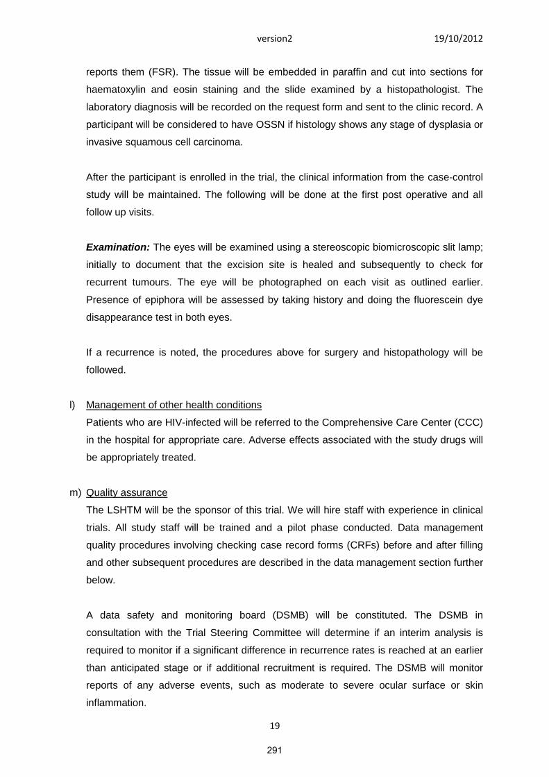
version2 19/10/2012
19
reports them (FSR). The tissue will be embedded in paraffin and cut into sections for
haematoxylin and eosin staining and the slide examined by a histopathologist. The
laboratory diagnosis will be recorded on the request form and sent to the clinic record. A
participant will be considered to have OSSN if histology shows any stage of dysplasia or
invasive squamous cell carcinoma.
After the participant is enrolled in the trial, the clinical information from the case-control
study will be maintained. The following will be done at the first post operative and all
follow up visits.
Examination: The eyes will be examined using a stereoscopic biomicroscopic slit lamp;
initially to document that the excision site is healed and subsequently to check for
recurrent tumours. The eye will be photographed on each visit as outlined earlier.
Presence of epiphora will be assessed by taking history and doing the fluorescein dye
disappearance test in both eyes.
If a recurrence is noted, the procedures above for surgery and histopathology will be
followed.
l) Management of other health conditions
Patients who are HIV-infected will be referred to the Comprehensive Care Center (CCC)
in the hospital for appropriate care. Adverse effects associated with the study drugs will
be appropriately treated.
m) Quality assurance
The LSHTM will be the sponsor of this trial. We will hire staff with experience in clinical
trials. All study staff will be trained and a pilot phase conducted. Data management
quality procedures involving checking case record forms (CRFs) before and after filling
and other subsequent procedures are described in the data management section further
below.
A data safety and monitoring board (DSMB) will be constituted. The DSMB in
consultation with the Trial Steering Committee will determine if an interim analysis is
required to monitor if a significant difference in recurrence rates is reached at an earlier
than anticipated stage or if additional recruitment is required. The DSMB will monitor
reports of any adverse events, such as moderate to severe ocular surface or skin
inflammation.
291

version2 19/10/2012
20
The laboratories to be used have been certified for good laboratory clinical practice
(GCLP). The laboratories participate in External Quality Assurance (EQA) of their
processes. An extra histology slide will be prepared from each tissue specimen to be
examined at the Institute of Ophthalmology, part of University College London (UCL) for
peer review.
The guidelines of the Kenyatta National Hospital-University of Nairobi Ethics Review
Committee (KNH/UON/ERC) and LSHTM on trial monitoring and reporting will be
followed. Protocol deviations will also be reported to the DSMB.
After approval by the KNH/UON/ERC this protocol will be submitted to the Kenya
Pharmacy and Poisons Board for registration of the 5FU eye drops to ensure they
conform to good manufacturing practices (GMP).
A Material Transfer Agreement (MTA) between the laboratories involved in the project
will be prepared and approved by the Kenya Ministry of Medical Services Department of
standards and regulatory services prior to any sample transfer.
n) Training procedures
Protocol training will be provided for all staff who will work on the project. In addition
they will be given training updates in good clinical practice (GCP) and research ethics.
They will be required to obtain GCP certification and update them regularly. The
surgeons will undergo training to standardize the excision procedure.
11. Ethical considerations
The principles of GCP and in particular the ethical principles of the Declaration of
Helsinki will be followed in this trial. Only participants who freely give written consent will
be enrolled into the study. The rights, safety and well being of participants will be
assured. The protocol will be submitted for ethics review at KNH/UON/ERC & LSHTM.
The investigators and research staff will have the appropriate education, training and
experience. Accurate data handling, protection and confidentiality will be ensured.
Quality assurance systems will be instituted as described in section (m) above. The
sponsor will monitor trial activities and provide insurance.
292

version2 19/10/2012
21
12. Data management
Data clerks supervised by a data manager will check the case record forms (CRFs) to
ensure full completion. Where data is missing, the CRF will be flagged on the database
and clinic register for supplementary interviews to be conducted during the next follow
up visit. The key unique identifier variable will be the study number. This will appear on
all pages of the CRF, specimen containers and laboratory request forms. When reports
are received from the laboratory, the results will be entered onto the CRF. The serial
number of the source documents (laboratory reports, medical records, digital
photographs) will be noted in the CRF.
Data will be transferred from CRF to a MS Access database with an interface that
appears similar to the CRF to minimize errors in data entry. The database will be
designed with must-enter variables, logic checks, legal values and specified range
checks built into the program to enable interactive checking during data entry.
Interactive checking allows the data clerk to be prompted on the screen if an erroneous
value is entered. After data entry, batch checking will be done. Batch checking will also
apply the standard checks (range, legal values etc) as well as consistency checks.
Verification of data entry will be done using the double entry method. This will involve
data being entered twice into two separate files by two different data clerks. The two
files will then be compared (using EpiData) and any discrepancies checked against the
CRFs and/or source documents. The clean dataset will then be transferred from MS
Access to Stata version IC 11 software (Stata Corporation, College Station, TX, USA)
for analysis. In the end all CRFs and laboratory reports will be filed and boxed (box 5
Figure 2) in order of study number.
All CRFs received from the interviewers and clinicians will be recorded in a register and
put in a box (Box 1 in figure 2). They will be manually checked for errors and if there is
no query they will be put in a box labelled 2 for the first entry. After the first entry, the
CRFs will be put in a box labelled 4 and taken for the second entry. The principal
investigator will compare the first and second entry. Any CRF with a query will be
marked and put in a special query box (Box 3 in Fig. 2).
On site, after the photographs are taken, the picture will be reviewed for clarity. Unclear
pictures will be repeated. The picture serial number from the camera will be noted on
the CRF. Pictures will be downloaded in computer in which photo databank
management software is installed and backed up at the end of each day. Similarly MS
293
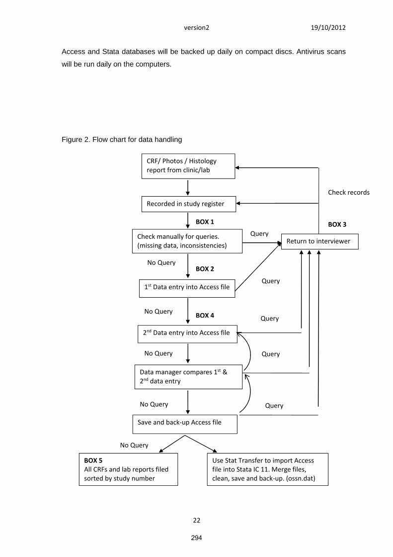
version2 19/10/2012
22
Access and Stata databases will be backed up daily on compact discs. Antivirus scans
will be run daily on the computers.
Figure 2. Flow chart for data handling
CRF/ Photos / Histologyreport from clinic/lab
Check records
Recorded in study register
BOX 3BOX 1
QueryReturn to interviewer
Check manually for queries.(missing data, inconsistencies)
No QueryBOX 2
Query1st Data entry into Access file
No QueryBOX 4 Query
2nd Data entry into Access file
No Query Query
Data manager compares 1st &2nd data entry
No Query
Save and back-up Access file
Query
No Query
Use Stat Transfer to import Accessfile into Stata IC 11. Merge files,clean, save and back-up. (ossn.dat)
BOX 5All CRFs and lab reports filedsorted by study number
294
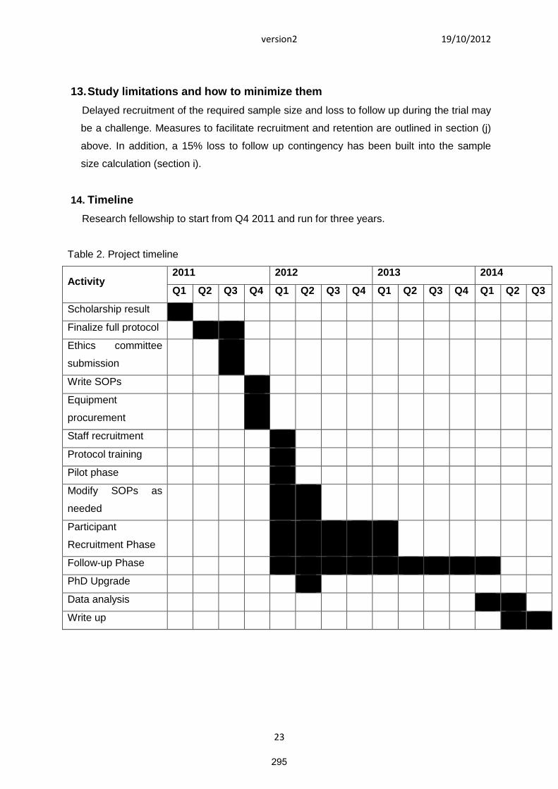
version2 19/10/2012
23
13.Study limitations and how to minimize them
Delayed recruitment of the required sample size and loss to follow up during the trial may
be a challenge. Measures to facilitate recruitment and retention are outlined in section (j)
above. In addition, a 15% loss to follow up contingency has been built into the sample
size calculation (section i).
14. Timeline
Research fellowship to start from Q4 2011 and run for three years.
Table 2. Project timeline
Activity2011 2012 2013 2014
Q1 Q2 Q3 Q4 Q1 Q2 Q3 Q4 Q1 Q2 Q3 Q4 Q1 Q2 Q3
Scholarship result
Finalize full protocol
Ethics committee
submission
Write SOPs
Equipment
procurement
Staff recruitment
Protocol training
Pilot phase
Modify SOPs as
needed
Participant
Recruitment Phase
Follow-up Phase
PhD Upgrade
Data analysis
Write up
295
version2 19/10/2012
24
15. References
1. Lee GA, Hirst LW. Ocular surface squamous neoplasia. Survey of ophthalmology.
1995; 39(6): 429-50.
2. Lee GA, Hirst LW. Retrospective study of ocular surface squamous neoplasia.
Australian and New Zealand journal of ophthalmology. 1997; 25(4): 269-76.
3. Poole TR. Conjunctival squamous cell carcinoma in Tanzania. The British journal of
ophthalmology. 1999; 83(2): 177-9.
4. Pola EC, Masanganise R, Rusakaniko S. The trend of ocular surface squamous
neoplasia among ocular surface tumour biopsies submitted for histology from Sekuru
Kaguvi Eye Unit, Harare between 1996 and 2000. The Central African journal of
medicine. 2003; 49(1-2): 1-4.
5. Chisi SK, Kollmann MK, Karimurio J. Conjunctival squamous cell carcinoma in
patients with human immunodeficiency virus infection seen at two hospitals in Kenya.
East African medical journal. 2006; 83(5): 267-70.
6. Furahini G, Lewallen S. Epidemiology and management of ocular surface squamous
neoplasia in Tanzania. Ophthalmic epidemiology. 2010; 17(3): 171-6.
7. Ateenyi-Agaba C. Conjunctival squamous-cell carcinoma associated with HIV
infection in Kampala, Uganda. Lancet. 1995; 345(8951): 695-6.
8. International Agency for Prevention of Blindness. Workshop Report: A VISION 2020
Workshop on Research for Global Blindness Prevention International Center for Eye
Health (ICEH); 2010 September 27-29 2010
9. National AIDS and STI Control Programme. Kenya AIDS Indicator Survey 2007:
Preliminary Report. : Ministry of Health, Kenya; 2008 Jul.
10. Mbulaiteye SM, Katabira ET, Wabinga H, Parkin DM, Virgo P, Ochai R, et al.
Spectrum of cancers among HIV-infected persons in Africa: the Uganda AIDS-
Cancer Registry Match Study. International journal of cancer Journal international du
cancer. 2006; 118(4): 985-90.
11. Waddell KM, Lewallen S, Lucas SB, Atenyi-Agaba C, Herrington CS, Liomba G.
Carcinoma of the conjunctiva and HIV infection in Uganda and Malawi. The British
journal of ophthalmology. 1996; 80(6): 503-8.
12. Waddell KM, Downing RG, Lucas SB, Newton R. Corneo-conjunctival carcinoma in
Uganda. Eye (Lond). 2006; 20(8): 893-9.
13. Yu JJ, Fu P, Pink JJ, Dawson D, Wasman J, Orem J, et al. HPV infection and EGFR
activation/alteration in HIV-infected East African patients with conjunctival carcinoma.
PloS one. 2010; 5(5): e10477.
14. Moubayed P, Mwakyoma H, Schneider DT. High Frequency of Human
Papillomavirus 6/11, 16, and 18 Infections in Precancerous Lesions and Squamous
296
version2 19/10/2012
25
Cell Carcinoma of the Conjunctiva in Subtropical Tanzania. American Journal of
Clinical Pathology. 2004; 122(6): 938-43.
15. Guthoff R, Marx A, Stroebel P. No evidence for a pathogenic role of human
papillomavirus infection in ocular surface squamous neoplasia in Germany. Current
eye research. 2009; 34(8): 666-71.
16. de Koning MN, Waddell K, Magyezi J, Purdie K, Proby C, Harwood C, et al. Genital
and cutaneous human papillomavirus (HPV) types in relation to conjunctival
squamous cell neoplasia: a case-control study in Uganda. Infectious agents and
cancer. 2008; 3: 12.
17. Sen S, Sharma A, Panda A. Immunohistochemical localization of human papilloma
virus in conjunctival neoplasias: a retrospective study. Indian journal of
ophthalmology. 2007; 55(5): 361-3.
18. Simbiri KO, Murakami M, Feldman M, Steenhoff AP, Nkomazana O, Bisson G, et al.
Multiple oncogenic viruses identified in Ocular surface squamous neoplasia in HIV-1
patients. Infectious agents and cancer. 2010; 5: 6.
19. Bernard HU, Burk RD, Chen Z, van Doorslaer K, Hausen H, de Villiers EM.
Classification of papillomaviruses (PVs) based on 189 PV types and proposal of
taxonomic amendments. Virology. 2010; 401(1): 70-9.
20. Ateenyi-Agaba C, Franceschi S, Wabwire-Mangen F, Arslan A, Othieno E, Binta-
Kahwa J, et al. Human papillomavirus infection and squamous cell carcinoma of the
conjunctiva. British journal of cancer. 2010; 102(2): 262-7.
21. Waddell K, Kwehangana J, Johnston WT, Lucas S, Newton R. A case-control study
of ocular surface squamous neoplasia (OSSN) in Uganda. International journal of
cancer Journal international du cancer. 2010; 127(2): 427-32.
22. Ateenyi-Agaba C, Dai M, Le Calvez F, Katongole-Mbidde E, Smet A, Tommasino M,
et al. TP53 mutations in squamous-cell carcinomas of the conjunctiva: evidence for
UV-induced mutagenesis. Mutagenesis. 2004; 19(5): 399-401.
23. Visser ME, Maartens G, Kossew G, Hussey GD. Plasma vitamin A and zinc levels in
HIV-infected adults in Cape Town, South Africa. The British journal of nutrition. 2003;
89(4): 475-82.
24. Fufa H, Umeta M, Taffesse S, Mokhtar N, Aguenaou H. Nutritional and
immunological status and their associations among HIV-infected adults in Addis
Ababa, Ethiopia. Food and nutrition bulletin. 2009; 30(3): 227-32.
25. Gichuhi S, Irlam JJ. Interventions for squamous cell carcinoma of the conjunctiva in
HIV-infected individuals. Cochrane database of systematic reviews (Online). 2007;
(2): CD005643.
297

version2 19/10/2012
26
26. Tabin G, Levin S, Snibson G, Loughnan M, Taylor H. Late recurrences and the
necessity for long-term follow-up in corneal and conjunctival intraepithelial neoplasia.
Ophthalmology. 1997; 104(3): 485-92.
27. Birkholz ES, Goins KM, Sutphin JE, Kitzmann AS, Wagoner MD. Treatment of ocular
surface squamous cell intraepithelial neoplasia with and without mitomycin C.
Cornea. 2011; 30(1): 37-41.
28. Yeatts RP, Engelbrecht NE, Curry CD, Ford JG, Walter KA. 5-Fluorouracil for the
treatment of intraepithelial neoplasia of the conjunctiva and cornea. Ophthalmology.
2000; 107(12): 2190-5.
29. Peksayar G, Soyturk MK, Demiryont M. Long-term results of cryotherapy on
malignant epithelial tumors of the conjunctiva. American journal of ophthalmology.
1989; 107(4): 337-40.
30. Schechter BA, Koreishi AF, Karp CL, Feuer W. Long-term follow-up of conjunctival
and corneal intraepithelial neoplasia treated with topical interferon alfa-2b.
Ophthalmology. 2008; 115(8): 1291-6, 6 e1.
31. Sepulveda R, Pe'er J, Midena E, Seregard S, Dua HS, Singh AD. Topical
chemotherapy for ocular surface squamous neoplasia: current status. The British
journal of ophthalmology. 2010; 94(5): 532-5.
32. Abraham LM, Selva D, Casson R, Leibovitch I. The clinical applications of fluorouracil
in ophthalmic practice. Drugs. 2007; 67(2): 237-55.
33. Duschinsky R, Pleven E, Heidelberger C. The synthesis of 5-fluoropyrimidines. J Am
Chem Soc 1957; 79(16): 4559–60.
34. Malet-Martino M, Jolimaitre P, Martino R. The prodrugs of 5-fluorouracil. Current
medicinal chemistry Anti-cancer agents. 2002; 2(2): 267-310.
35. Chu T, Sartorelli AC. Cancer chemotherapy. In: Katzung BG, editor. Basic and
clinical pharmacology. 9 ed. Boston: McGraw Hill; 2004. p. 898-930.
36. Yeatts RP, Ford JG, Stanton CA, Reed JW. Topical 5-fluorouracil in treating epithelial
neoplasia of the conjunctiva and cornea. Ophthalmology. 1995; 102(9): 1338-44.
37. Midena E. Treatment of conjunctival squamous cell carcinoma with topical 5-
fluorouracil. British Journal of Ophthalmology. 2000; 84(3): 268-72.
38. Al-Barrag A, Al-Shaer M, Al-Matary N, Al-Hamdani M. 5-Fluorouracil for the treatment
of intraepithelial neoplasia and squamous cell carcinoma of the conjunctiva, and
cornea. Clinical ophthalmology (Auckland, NZ). 2010; 4: 801-8.
39. Parrozzani R, Lazzarini D, Alemany-Rubio E, Urban F, Midena E. Topical 1% 5-
fluorouracil in ocular surface squamous neoplasia: a long-term safety study. The
British journal of ophthalmology. 2011; 95(3): 355-9.
298

version2 19/10/2012
27
40. Rudkin AK, Muecke JS. Adjuvant 5-fluorouracil in the treatment of localised ocular
surface squamous neoplasia. The British journal of ophthalmology. 2011; 95(7): 947-
50.
41. Shields CL, Shields JA. Tumors of the conjunctiva and cornea. Survey of
ophthalmology. 2004; 49(1): 3-24.
299
version2 19/10/2012
28
16.Appendix
Appendix 1 Statistical analysis plan
1) Participant flowA summary of the flow of participants through the different phases of the trial; enrolment,
allocation, follow up and analysis will be made following the 2010 CONSORT flowchart
(Schulz et al 2010 http://www.consort-statement.org/consort-statement/overview0/).
This will show the numbers eligible, consenting to take part, randomised, who received the
intended treatment by arm. For each follow-up time-point the numbers seen, censored,
defaulting, and permanently lost-to-follow-up will also be shown by arm. The numbers
excluded at each stage and reasons for their exclusion will be noted such as reasons for
declining to take part, not completing treatment, not having re-excision surgery, or
discontinuing follow-up.
2) Summary of the baseline characteristics of the trial populationA table will show a summary of baseline characteristics of the trial population such as total
number involved further subdivided by study centre, age and sex.
3) Comparison of treatment groups with respect to baseline variables thought to beassociated with main outcome
The following characteristics of participants at baseline will be tabulated by arm:
a) Socio-demographic factors: age, sex.b) Behavioural factors: smoking, outdoor occupation, wearing hats/caps.c) Clinical status: HIV status, HPV infection, CD4 count, serum vitamin A levels, tumour
size and histological grade.
Categorical variables will be reported as numbers and proportions while continuous
variables will be reported as mean and standard deviations if normally distributed or medians
and interquartile range if their distribution is skewed. We will eyeball for differences between
groups with respect to these variables but avoid any formal tests of the null hypothesis for no
between-group differences since it will be assumed to hold if randomization was done
properly. If one arm has appreciably more severe cases than the other – by chance – the
primary analysis will be adjusted during multivariable regression.
4) Primary analysis of the primary outcome measure by intention to treatDefinition of primary end-point
The primary outcome will be the clinical recurrence of the tumour at any time during the first
year of follow-up, and where available confirmed by histology. Where histology is not
available, photographs recurrent lesions will be reviewed by two consultant ophthalmologists
experienced in OSSN in East Africa.
300

version2 19/10/2012
29
Justification of primary outcome:
The aim of adjuvant therapy is to reduce the rate of recurrence and therefore increase
tumour-free survival after surgical excision.
a) We will present an estimated effect of treatment on recurrence of the tumour at anytime during the first year of follow up defined as clinical recurrence of OSSN OSSNfrom a re-excision biopsy. The numbers of events (recurrences in the first year) andrisk of recurrence in both arms will be shown in a table. The odds of recurrence willbe compared using the odds ratio (OR) and its 95% confidence interval adjusting forsurgeon. The null hypothesis will be no treatment effect. The number needed to treatwill also be estimated.
b) We hope to achieve no less than 85% follow up and have adjusted sample size forthis. If follow up is not as good as expected, sensitivity analysis will be conducted toexamine the possible effect of losses to follow up to see if these may affect thetreatment effect estimate.
5) Primary analysis adjusted for baseline variablesIt is expected that the important baseline characteristics that are known to affect the risk of
recurrence will be balanced between the two arms through randomisation. If the arms are
found to be substantially imbalanced an appropriately adjusted logistic regression model will
be used.
6) Secondary analysis of the primary outcomea) Effect of the intervention on time-to-first-recurrence.
The time-to-first-recurrence variable is an estimate of the tumour-free survival period. The
numbers of events, person-months and rate of recurrence in each arm will be shown.
Kaplan-Meier analysis will be used to plot the survival curves for both treatment arms up to
the final visit at 1 year. The Mantel-Cox hazard ratio will be used to assess the impact of the
intervention on time-to-first-recurrence. The hazard ratio will be estimated with Cox
regression, adjusting for substantial baseline imbalances if appropriate.
b) Risk factors for recurrenceA multivariate logistic regression model will be used to identify potential explanatory factors
for recurrent tumour at one year, in addition to the randomised adjuvant therapy allocation.
Baseline factors which will be examined in this model of recurrent tumour will include:
i) Ageii) Sexiii) HIV statusiv) Mean CD4 countv) HAARTvi) HPV infectionvii) Smokingviii) Serum Vitamin A level
301
version2 19/10/2012
30
c) Effect modificationWe will assess effect modification of the intervention on recurrence at one year with the
following factors by including an interaction term with treatment arm in the logistic regression
model.
i) Pre-operative tumour size – proxy measure will be the estimated area measuredfrom digital photographs
ii) Rate of tumour growth – estimated as a function of tumour size above and time inmonths since the participant noticed the tumour. We hypothesize that tumourswith a high rate of growth have a higher propensity to recur.
iii) Operating surgeon
7) Analysis of adverse effects (secondary outcome)A secondary outcome will be the prevalence of epiphora (watering of the eyes) at 12
months. The prevalence of any epiphora and a positive dye disappearance test at any follow
up visit in the treated eye will be described by arm.
8) Sensitivity analysisWhere expert opinion is unable to be unequivocally certain about the recurrence, sensitivityanalysis will be conducted, categorizing these as non-OSSN.
302
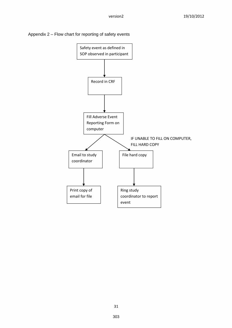
version2 19/10/2012
31
Appendix 2 – Flow chart for reporting of safety events
Safety event as defined in
SOP observed in participant
Record in CRF
Fill Adverse Event
Reporting Form on
computer
Email to study
coordinator
Print copy of
email for file
File hard copy
IF UNABLE TO FILL ON COMPUTER,
FILL HARD COPY
Ring study
coordinator to report
event
303