Detection of Sialomucin Complex (MUC4) in Human Ocular ...
11
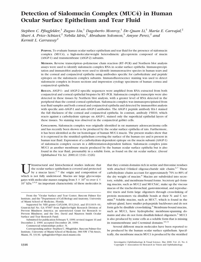
Detection of Sialomucin Complex (MUC4) in Human Ocular Surface Epithelium and Tear Fluid Stephen C. Pflugfelder, 1 Zuguo Liu, 1 Dagoberto Monroy, 1 De–Quan Li, 1 Maria E. Carvajal, 2 Shari A. Price–Schiavi, 2 Nebila Idris, 2 Abraham Solomon, 1 Amyee Perez, 2 and Kermit L. Carraway 2 PURPOSE. To evaluate human ocular surface epithelium and tear fluid for the presence of sialomucin complex (MUC4), a high-molecular-weight heterodimeric glycoprotein composed of mucin (ASGP-1) and transmembrane (ASGP-2) subunits. METHODS. Reverse transcription–polymerase chain reaction (RT–PCR) and Northern blot analysis assays were used to identify sialomucin complex RNA in ocular surface epithelia. Immunoprecipi- tation and immunoblot analysis were used to identify immunoreactive species in human tears and in the corneal and conjunctival epithelia using antibodies specific for carbohydrate and peptide epitopes on the sialomucin complex subunits. Immunofluorescence staining was used to detect sialomucin complex in frozen sections and impression cytology specimens of human cornea and conjunctival epithelia. RESULTS. ASGP-1– and ASGP-2–specific sequences were amplified from RNA extracted from both conjunctival and corneal epithelial biopsies by RT–PCR. Sialomucin complex transcripts were also detected in these tissues by Northern blot analysis, with a greater level of RNA detected in the peripheral than the central corneal epithelium. Sialomucin complex was immunoprecipitated from tear fluid samples and both corneal and conjunctival epithelia and detected by immunoblot analysis with specific anti–ASGP-1 and anti–ASGP-2 antibodies. The ASGP-1 peptide antibody HA-1 stained the full thickness of the corneal and conjunctival epithelia. In contrast, antibody 15H10, which reacts against a carbohydrate epitope on ASGP-1, stained only the superficial epithelial layers of these tissues. No staining was observed in the conjunctival goblet cells. CONCLUSIONS. Sialomucin complex was originally identified in rat mammary adenocarcinoma cells and has recently been shown to be produced by the ocular surface epithelia of rats. Furthermore, it has been identified as the rat homologue of human MUC4 mucin. The present studies show that it is expressed in the stratified epithelium covering the surface of the human eye and is present in human tear fluid. Expression of a carbohydrate-dependent epitope on the mucin subunit (ASGP-1) of sialomucin complex occurs in a differentiation-dependent fashion. Sialomucin complex joins MUC1 as another membrane mucin produced by the human ocular surface epithelia but is also found in the tear fluid, presumably in a soluble form, as found on the rat ocular surface. (Invest Ophthalmol Vis Sci. 2000;41:1316 –1326) U ltrastructural and histochemical studies indicate that the ocular surface epithelium is covered and protected by a mucus layer, 1–4 the origin and composition of which is not fully understood. Mucins are large glycoconju- gates with molecular masses ranging from 3 3 10 5 to over 4 3 10 7 kDa. 2,5,6 An important characteristic of these molecules is that they contain domains rich in serine and threonine residues with attached O-linked oligosaccharide side chains. 6,7 These carbohydrate chains account for approximately 70% to 80% of the dry weight of mucins. 6 Mucins are subdivided into secre- tory, soluble, and membrane-bound forms. Secretory gel–form- ing mucins, such as MUC2 and MUC5AC, make up the viscous mucus of the tracheobronchial, gastrointestinal, and reproduc- tive tracts and form large oligomers through cross-linking of protein monomers via disulfide bonds at their N- and C ter- mini. 8 Soluble mucins, such as MUC7, which is found in the salivary gland, have smaller polypeptide backbones and do not form gels by disulfide cross-linking. 9 Membrane-bound mucins, such as MUC1, have hydrophobic membrane-spanning do- mains and also do not form disulfide-linked oligomers. 5 MUC1 is also produced by some cells as a soluble form that is missing its transmembrane and C-terminal domains. 10,11 Several different mucin molecules have been reported to be produced by the human ocular surface epithelium. Specif- ically, the membrane mucin MUC1 is produced by the stratified From the 1 Ocular Surface and Tear Center, Bascom Palmer Eye Institute, and the 2 Department of Cell Biology and Anatomy, University of Miami School of Medicine, Florida. Supported by NIH grants CA52498, EY12343, and EY11915– 01; Grant-in-Aid No. GA 97097 from Fight-for-Sight Research Division of Prevent Blindness America; an unrestricted Grant for Research to Prevent Blindness; and the Drs. David and Maureen Smith Ocular Surface and Tear Research Fund. Submitted for publication February 5, 1999; revised August 16 and December 2, 1999; accepted December 28, 1999. Commercial relationships policy: N. Corresponding author: Stephen C. Pflugfelder, Bascom Palmer Eye Institute, University of Miami School of Medicine, 900 NW 17th Street, Miami, FL 33136. spfl[email protected] Investigative Ophthalmology & Visual Science, May 2000, Vol. 41, No. 6 1316 Copyright © Association for Research in Vision and Ophthalmology
-
Upload
khangminh22 -
Category
Documents
-
view
1 -
download
0
Transcript of Detection of Sialomucin Complex (MUC4) in Human Ocular ...
Detection of Sialomucin Complex (MUC4) in HumanOcular Surface Epithelium and Tear Fluid
Stephen C. Pflugfelder,1 Zuguo Liu,1 Dagoberto Monroy,1 De–Quan Li,1 Maria E. Carvajal,2
Shari A. Price–Schiavi,2 Nebila Idris,2 Abraham Solomon,1 Amyee Perez,2 andKermit L. Carraway2
PURPOSE. To evaluate human ocular surface epithelium and tear fluid for the presence of sialomucincomplex (MUC4), a high-molecular-weight heterodimeric glycoprotein composed of mucin(ASGP-1) and transmembrane (ASGP-2) subunits.
METHODS. Reverse transcription–polymerase chain reaction (RT–PCR) and Northern blot analysisassays were used to identify sialomucin complex RNA in ocular surface epithelia. Immunoprecipi-tation and immunoblot analysis were used to identify immunoreactive species in human tears andin the corneal and conjunctival epithelia using antibodies specific for carbohydrate and peptideepitopes on the sialomucin complex subunits. Immunofluorescence staining was used to detectsialomucin complex in frozen sections and impression cytology specimens of human cornea andconjunctival epithelia.
RESULTS. ASGP-1– and ASGP-2–specific sequences were amplified from RNA extracted from bothconjunctival and corneal epithelial biopsies by RT–PCR. Sialomucin complex transcripts were alsodetected in these tissues by Northern blot analysis, with a greater level of RNA detected in theperipheral than the central corneal epithelium. Sialomucin complex was immunoprecipitated fromtear fluid samples and both corneal and conjunctival epithelia and detected by immunoblot analysiswith specific anti–ASGP-1 and anti–ASGP-2 antibodies. The ASGP-1 peptide antibody HA-1 stainedthe full thickness of the corneal and conjunctival epithelia. In contrast, antibody 15H10, whichreacts against a carbohydrate epitope on ASGP-1, stained only the superficial epithelial layers ofthese tissues. No staining was observed in the conjunctival goblet cells.
CONCLUSIONS. Sialomucin complex was originally identified in rat mammary adenocarcinoma cellsand has recently been shown to be produced by the ocular surface epithelia of rats. Furthermore,it has been identified as the rat homologue of human MUC4 mucin. The present studies show thatit is expressed in the stratified epithelium covering the surface of the human eye and is present inhuman tear fluid. Expression of a carbohydrate-dependent epitope on the mucin subunit (ASGP-1)of sialomucin complex occurs in a differentiation-dependent fashion. Sialomucin complex joinsMUC1 as another membrane mucin produced by the human ocular surface epithelia but is alsofound in the tear fluid, presumably in a soluble form, as found on the rat ocular surface. (InvestOphthalmol Vis Sci. 2000;41:1316–1326)
Ultrastructural and histochemical studies indicate thatthe ocular surface epithelium is covered and protectedby a mucus layer,1–4 the origin and composition of
which is not fully understood. Mucins are large glycoconju-gates with molecular masses ranging from 3 3 105 to over 4 3107 kDa.2,5,6 An important characteristic of these molecules is
that they contain domains rich in serine and threonine residueswith attached O-linked oligosaccharide side chains.6,7 Thesecarbohydrate chains account for approximately 70% to 80% ofthe dry weight of mucins.6 Mucins are subdivided into secre-tory, soluble, and membrane-bound forms. Secretory gel–form-ing mucins, such as MUC2 and MUC5AC, make up the viscousmucus of the tracheobronchial, gastrointestinal, and reproduc-tive tracts and form large oligomers through cross-linking ofprotein monomers via disulfide bonds at their N- and C ter-mini.8 Soluble mucins, such as MUC7, which is found in thesalivary gland, have smaller polypeptide backbones and do notform gels by disulfide cross-linking.9 Membrane-bound mucins,such as MUC1, have hydrophobic membrane-spanning do-mains and also do not form disulfide-linked oligomers.5 MUC1is also produced by some cells as a soluble form that is missingits transmembrane and C-terminal domains.10,11
Several different mucin molecules have been reported tobe produced by the human ocular surface epithelium. Specif-ically, the membrane mucin MUC1 is produced by the stratified
From the 1Ocular Surface and Tear Center, Bascom Palmer EyeInstitute, and the 2Department of Cell Biology and Anatomy, Universityof Miami School of Medicine, Florida.
Supported by NIH grants CA52498, EY12343, and EY11915–01;Grant-in-Aid No. GA 97097 from Fight-for-Sight Research Division ofPrevent Blindness America; an unrestricted Grant for Research toPrevent Blindness; and the Drs. David and Maureen Smith OcularSurface and Tear Research Fund.
Submitted for publication February 5, 1999; revised August 16 andDecember 2, 1999; accepted December 28, 1999.
Commercial relationships policy: N.Corresponding author: Stephen C. Pflugfelder, Bascom Palmer Eye
Institute, University of Miami School of Medicine, 900 NW 17th Street,Miami, FL 33136. [email protected]
Investigative Ophthalmology & Visual Science, May 2000, Vol. 41, No. 61316 Copyright © Association for Research in Vision and Ophthalmology

epithelium covering the cornea and conjunctiva.12 RNAs en-coding the secretory mucins MUC4 and MUC5AC have beenidentified in the conjunctiva by Northern blot analysis.13 MUC4RNA was localized to the stratified conjunctival epithelium,whereas MUC5 RNA expression was limited to the conjunctivalgoblet cells by in situ hybridization.13 It is currently unknownwhether these mucin molecules produced by the ocular sur-face epithelium are also present in the preocular tear film.
Sialomucin complex (SMC) is another well-studied mem-brane mucin.14 This mucin was originally isolated from highlymetastatic 13762 rat mammary adenocarcinoma ascites cells,and it was characterized as a heterodimeric glycoprotein com-plex, in which the mucin subunit ASGP-1 (ascites sialoglyco-protein-1) is the major detectable glycoprotein.15 ASGP-1 istightly but noncovalently bound to an N-glycosylated integralmembrane glycoprotein, termed ASGP-2.15,16 The hetero-dimeric SMC has been shown to be expressed in a number ofsecretory epithelial tissues in the adult rat, including the smalland large intestines, trachea, uterus, lactating mammary gland,and cornea and conjunctiva.16–18 Sialomucin complex is tran-scribed from a single gene as a 9-kb transcript19,20 and istranslated into a polypeptide precursor that is proteolyticallycleaved into the ASGP-1 and ASGP-2 subunits early in its transitto the cell surface.21 Mature glycosylated ASGP-1 has a molec-ular mass greater than 500 kDa and contains three domains: anN-terminal unique sequence, 12 tandem repeat regions that arerich in O-glycosylated serine and threonine residues similar toother mucins, and a C-terminal unique sequence.20 The trans-membrane ASGP-2 subunit contains two epidermal growthfactor (EGF)–like domains that contain all the consensus resi-dues found in other proteins with EGF-like regions that possessgrowth factor activity.19 ASGP-2 has been shown to act as aligand for the receptor tyrosine kinase ErbB-2, a member of theEGF receptor family.17,22,23 Thus, SMC may be bifunctional,serving as a protective, lubricating mucin as well as an activegrowth factor.
Recent molecular studies have helped to define the placeof SMC among the hierarchy of mucins whose gene sequenceshave been determined. The full-length sequence of the humanMUC4 gene shows substantial homology with the rat SMCsequence at the N- and C-terminal unique portions of themolecule.24 The MUC4 gene has been found to have a similarorganization to the SMC gene with both mucin and transmem-brane subunits that have been designated MUC4a and MUC4b,respectively.25 The repeat domains of SMC and MUC4 appearto be different. Specifically, SMC does not have the 16 aminoacid repeat in the originally reported MUC4 cDNA se-quence.25,26 This may explain why the similarity between themolecules was not previously observed. However, the 70%identity between the human MUC4 analog of ASGP-2 and ratASGP-2 provides compelling evidence that they are homolo-gous proteins.
The purpose of this study was to determine whetherhuman ocular surface epithelia produce SMC (MUC4) andwhether this mucin is present in the preocular tear film.
METHODS
The Institutional Review Board of the University of MiamiSchool of Medicine approved this study protocol in accordancewith the tenets of the Declaration of Helsinki. Tear fluid and
impression cytology (conjunctiva and cornea) specimens werecollected from three normal human subjects with no symp-toms of ocular irritation, a Schirmer 1 test . 20 mm, and nocorneal fluorescein staining. Unstimulated tear fluid was care-fully collected from the inferior tear meniscus to avoid reflextearing using a previously reported technique.27 Impressioncytology was performed by applying nitrocellulose (4 mm2) orBiopore (Millipore, Bedford, MA) membranes against the infe-rior bulbar conjunctiva or inferior cornea by previously re-ported techniques.28 Human conjunctiva was obtained duringsurgery from the normal superior bulbar conjunctiva of pa-tients with pterygia. Human whole eyes and corneoscleralbuttons were obtained from the Florida Lions Eye Bank. Humancorneal limbal epithelium was cultured from explants of hu-man donor corneoscleral rims. Each corneoscleral rim wastrimmed, the endothelial layer and iris remnants were re-moved, and the tissue was treated with dispase (Life Technol-ogies, Gaithersburg, MD) for 15 minutes. Each rim was dis-sected into 12 equal parts, which were applied in 6-well plasticdishes, and covered with a drop of fetal bovine serum (FBS)overnight. The explants were cultured in Dulbecco’s modifiedEagle’s medium (DMEM)-F12 medium, enriched with 5% FBS,1% insulin, transferrin, selenium (ITS), hydrocortisone, EGF,and cholera toxin. Epithelial cells were removed from thedishes by gentle trypsinization when they reached 70% conflu-ence. Corneal epithelium was cultured in a similar mannerfrom explants taken from the central 6 mm of the donorcorneal tissue.
To culture stromal fibroblasts, the cornea, limbus, andconjunctiva were separated after mechanically removing theepithelium. The endothelium on the cornea and limbus wasremoved with a scalpel. Explant cultures were prepared in thesame manner as described above for epithelial cell culturesexcept that DMEM containing 10% FBS was used. Fibroblastswere subcultured at 80% to 90% confluence after treatmentwith 0.1% trypsin and 0.02% EDTA.
TABLE 1. Antibodies for Immunofluorescence Staining and WesternBlot Analysis
AntibodyName Specificity Species
OriginalReference
HA-1 Rat ASGP-1 peptide(AGYRPPRPAWTFGD)
Rabbit 18
15H10 ASGP-1 carbohydrateepitope
Mouse 30
13C4 N-gly-1/Cys-rich domainof ASGP-2
Mouse 18, 30, 32, 33
4F12 N-terminal 53 aminoacids of ASGP-2
18, 30, 32, 33
HA2-3 Human ASGP-2 peptideLDNQTVTFQPDHEDGG
Rabbit Unpublished
AM-3* Conjunctival goblet cellmucin
Mouse 42
CT-1† MUC1 peptideSSLSYTNPAVAATSANL
Rabbit 43
Ab-8‡ Human ErbB-2/HER-2/neuoncoprotein
Mouse 41
* Provided by Scheffer C. G. Tseng, University of Miami School ofMedicine.
† Provided by Daniel Carson, M. D. Anderson Cancer Center,Houston, TX.
‡ NeoMarkers (Fremont, CA).
IOVS, May 2000, Vol. 41, No. 6 SMC (MUC4) in Ocular Surface Epithelium 1317

The Department of Pathology at the University of MiamiSchool of Medicine provided normal human tongue and tonguecarcinoma tissues.
Reverse Transcription–Polymerase ChainReaction Analysis of Human Corneal andConjunctival Tissues
Reverse transcription–polymerase chain reaction (RT–PCR)was performed using the SuperScript One-Step RT–PCR System(GIBCO–BRL/Life Technologies, Gaithersburg, MD) on 1 mgsamples of total RNA isolated from normal human cornealepithelium obtained by scraping and conjunctival epithelium
obtained by impression debridement. Before RT, the RNAsamples were treated with DNase I for 15 minutes at 37°C,then the DNase I was inactivated at 75°C for 5 minutes.
The RT–PCR was performed in a 50 ml volume using thefollowing conditions: 0.2 mM of 59 primer, 0.2 mM of 39 primer,0.2 mM of each dNTP, 1.2 mM of MgSO4, and 2 U of reversetranscriptase/Taq polymerase mixture. The absence of ge-nomic DNA in RNA preparations was verified by omitting thereverse transcriptase/Taq Mix and substituting 2 U Taq DNApolymerase (Promega, Madison, WI) in the reaction. Plasmidscarrying full-length human ASGP-1 or ASGP-2 cDNA sequenceswere used as positive controls for the PCRs. RNA was reverse-transcribed into cDNA by 1 cycle of 55°C for 30 minutesfollowed by 1 cycle of 94°C for 2 minutes. The cDNA was
FIGURE 1. (A) Ethidium bromide–stained agarose gel of RT–PCR prod-ucts amplifying SMC RNA sequences in human colon (1control),cornea, and conjunctiva. Plus (1) and minus (2) signs indicatewhether cDNA synthesis reactions were performed with reverse tran-scriptase. Upper: 468-bp product specific for ASGP-2; Lower: 442-bpproduct specific for ASGP-1 (MUC4). (B) Northern blot analysis ofMUC4 mRNA in primary cultured epithelial cells and third-passagefibroblasts of human ocular surface tissues. Total RNA blots wereprobed with a 32P-labeled 2-kb fragment of ASGP-2 cDNA and thenreprobed with a 32P-GAPDH as a loading control after the first probewas stripped. CO, cornea; L, limbus (L1 and L2 are two different limbalepithelial cultures); CJ, conjunctiva. See Methods section for details.
FIGURE 2. (A) Immunoprecipitation of primary human corneal epi-thelial cultures with anti–ASGP-1 antibody 15H10 or preimmune sera(negative control). Immunoblots were probed with rat ASGP-1 pep-tide–specific polyclonal antibody HA-1 (upper panel) or HA-1 plusHA-1 peptide 2 mg/ml (lower panel). (B) Immunoprecipitation ofprimary human corneal epithelial cultures with anti–ASGP-1 antibody15H10 or preimmune sera (negative control). Immunoblots wereprobed with a human Erb-B2–specific monoclonal antibody (Ab-8) orAb-8 plus HA-1 peptide (2 mg/ml).
1318 Pflugfelder et al. IOVS, May 2000, Vol. 41, No. 6

amplified for 42 cycles, with each cycle consisting of 94°C for15 seconds, 55°C for 30 seconds, and 1 minute at 72°C (lastcycle at 72°C for 5 minutes). The sequences of PCR primers forASGP-1 were as follows: 59 CTTACTCTGGCCAACTCTGTAGTG39 and 59GAGAAGTTGGGCTTGACTGTC 39. This 442 bp se-quence was taken from a region upstream of the repeat sequenceof MUC4 that is homologous to the sequence encoding theASGP-1 mucin subunit of sialomucin.24 The sequences of thehuman ASGP-2 PCR primers were as follows: 59 GCTCTCCAA-CATCCTCCACT 39 and 59 TCACACGACCACCATTGATG 39.These were synthesized from the 59 end of the human ASGP-2sequence. The expected length of this amplified segment is 468
bp. After amplification, 15 ml of PCR product was electropho-resed on a 1.5% agarose gel in 13 TAE and was photographed.
Northern Blot Analysis Assay
A 2-kb fragment of the human MUC4b (ASGP-2 region)cDNA probe was used to probe for SMC (MUC4) RNA. A498-bp segment of the human GAPDH cDNA was used toprobe for GAPDH. This probe was purified from RT–PCRproducts by electrophoresis through a 1.2% low-meltingagarose gel using a Promega Wizard PCR Prep DNA purifi-cation kit (Promega) according to the manufacturer’s proto-col. The sequences of GAPDH primers used for PCR were
FIGURE 3. (A) Immunoprecipitationof MV-MAT C1 rat ascites mammaryadenocarcinoma cells or primary hu-man corneal epithelial cultures withanti-human ASGP-2 peptide antibodyHA2-3, antibody HA2-3 plus HA2-3peptide (0.25–2.0 mg/ml), or preim-mune sera (negative control). Immu-noblots were probed with ASGP-2–specific monoclonal antibody 4F12.(B) Immunoprecipitation of A375human melanoma cells or MV-MATC1 rat ascites mammary adenocarci-noma cells with anti-human ASGP-2peptide antibody HA2-3 or preim-mune sera (negative control). Immu-noblots were probed with a humanErb-B2–specific monoclonal anti-body (Ab-8) or Ab-8 plus HA2-3 pep-tide (2 mg/ml).
IOVS, May 2000, Vol. 41, No. 6 SMC (MUC4) in Ocular Surface Epithelium 1319

541 to 561 (sense) and 1018 to 1038 (antisense; GenBankAccession No. M33197). The 32P-labeled cDNA probes (1 to2 3 109 cpm/mg DNA) were prepared with [a-32P]– dCTP(3000 Ci/mmol) using a random primer DNA labeling system(Life Technologies).
Total RNA isolation and Northern hybridization wereperformed using a previously described method.29 Briefly,total RNA was isolated from primary epithelial cell culturesand third passage human corneal, limbal, and conjunctivalfibroblast cultures by acid guanidium thiocyanate–phenol–chloroform extraction. Total RNA (20 mg/lane) was electro-phoresed through 1.2% agarose containing formaldehyde,transferred to nitrocellulose membranes, and hybridizedwith 32P-labeled cDNA probes at 1 to 2 3 106 cpm/1.5 to 3ng/ml in the hybridization solution. After visualization of thehybridization product on the X-ray film, the membrane waswashed twice at 65°C for 1 hour in 5 mM Tris–HCl (pH 8.0),0.2 mM EDTA, 0.05% sodium pyrophosphate, and 0.13Denhardt’s solution to strip off the 32P-label before rehybrid-ization with another probe. The relative amount of SMCRNA was determined by scanning the autoradiogram with alaser scanning densitometer (model FB910; Fisher Scientific,Pittsburgh, PA) and normalized as a ratio to that of theGAPDH RNA band.
Immunoprecipitation and Immunoblot Analysis
Samples (tear fluid, 1.5–2 ml; human corneal and conjunctivalepithelial biopsies; cultured human corneal epithelial cells;MV-MATC1 rat ascites mammary carcinoma cells, and A375human melanoma cells) were mixed with 300 ml of RIPA buffer(150 mM NaCl, 50 mM Tris buffer, 1% NP-40, 5% deoxycholate,0.1% sodium dodecyl sulfate [SDS], pH 8.0). Sialomucin com-plex was immunoprecipitated by the addition of 15 ml ofASGP-1–specific antibody (15H10),30 ASGP-2 specific antibody(HA2-3), or rabbit preimmune sera (negative control) followedby rotation at 4°C overnight. Subsequently, 15 ml of Protein Aagarose (Sigma, St. Louis, MO) and 100 ml of ImmunoPure IgGbinding buffer (Pierce, Rockford, Il) were added with furtherincubation for 1 hour at 4°C. The immunoprecipitants werecollected by centrifugation at 3000g, washed 3 times in RIPA
buffer, and solubilized for SDS–polyacrylamide gel electro-phoresis (SDS—PAGE) and immunoblot analysis.30 Immuno-blots were probed with ASGP-1–, ASGP-2–, or human ErbB-2–specific antibodies (Table 1) and developed with a Renaissancechemiluminescence kit (New England Nuclear, Boston, MA).Western blot analysis of ocular surface epithelium and humancontrol tissues was performed as previously described.18 Thespecificity of polyclonal antibody HA2-3 that was generatedagainst the human ASGP-2–specific peptide LDNQTVTFQPD-HEDGG was evaluated by adding this peptide (0.25–2 mg/ml)to the immunoprecipitation mixture as a competitive sub-strate. The specificity of polyclonal antibody HA-1 that wasgenerated against the rat ASGP-1–specific peptide AGYRPPR-PAWTFGD was evaluated in immunoblots where 2 mg/ml ofthis competitive substrate was incubated with antibody HA-1.
Immunofluorescence Staining
Corneal and conjunctival biopsies were embedded in OCTcompound (Tissue Tek, Elkhart, IN), rapidly frozen in liquidnitrogen, and stored at 270°C. Within 72 hours, serial 4- to5-mm-thick sections were cut. Single or dual label indirectimmunofluorescence staining on tissue sections and cytologyspecimens was performed by a previously reported tech-nique31 using SMC antibodies (Table 1) as well as positive(cytokeratin) and negative (fluorescein isothiocyanate [FITC]–or Texas red–labeled secondary antibodies, preimmune sera,or irrelevant monoclonal antibodies plus secondary antibody)control antibodies. The specificity of polyclonal antibody HA-1that was generated against the rat ASGP-1–specific peptideAGYRPPRPAWTFGD in staining human ocular surface epithe-lial was evaluated by incubating this peptide alone (1 mg/ml)
FIGURE 4. Immunoprecipitation of two human tear samples and pos-itive control (TPK SUP) with anti–ASGP-1 antibody HA-1. Immunoblotswere probed with monoclonal antibodies 13C4 (ASGP-2–specific) and15H10 (ASGP-1–specific). TPK SUP, microvilli extracted from MV-MATC1 rat ascites mammary adenocarcinoma cells.
FIGURE 5. Immunoprecipitation of cornea and conjunctival epitheliaand positive control (TPK sup, microvilli extracted from MV-MAT C1rat ascites mammary adenocarcinoma cells) with antibody 15H10.Immunoblot was probed with antibody HA-1 (ASGP-1–specific).
1320 Pflugfelder et al. IOVS, May 2000, Vol. 41, No. 6
or a mixture of HA-1 or anti–ErbB-2 Ab-8 (Table 1) and peptide(1 mg/ml) overnight before adding the secondary antibody.Staining was evaluated and photographed with a Nikon Axio-phot epifluorescence microscope.
RESULTS
Detection of SMC RNA by RT–PCR and NorthernBlot Analysis Assay
PCR products of appropriate size were obtained from humancorneal and conjunctival epithelial biopsies using primers spe-cific for the ASGP-1 and for the ASGP-2 regions of SMC cDNA(Fig. 1A). To confirm the RT–PCR results, Northern blot anal-ysis was also performed on RNA isolated from primary humancentral corneal and limbal epithelial cultures, a human con-junctival biopsy, and fibroblasts cultured from human conjunc-tiva, limbus, and cornea. A 2-kb fragment of MUC4b (ASGP-2)cDNA was used to probe a Northern blot specimen containingtotal RNA of human ocular surface epithelial cells and fibro-blasts cultured from cornea, limbus, and conjunctiva. Onlyepithelial cells (Fig. 1B), not fibroblasts, expressed SMC mRNA.A strong hybridization signal was observed in conjunctivalepithelial cells (Fig. 1, CJ), a slightly less intense signal was
observed in limbal epithelial cells (Fig. 1, L1 and L2), and a faintsignal was observed in corneal epithelial cells (Fig. 1, CO).There were at least three SMC transcripts expressed by thesecultured ocular surface epithelial cells. The major band wasapproximately 22 kb, characteristic of high-molecular-weightmucin mRNAs, and the other two bands were approximately 5and 2 kb, respectively. In contrast, a 1.4-kb GAPDH mRNA wassimilarly expressed by all samples (Fig. 1B).
Antibody Specificity
The SMC antibodies used in this work (Table 1), 13C4, 4F12,HA-1, HA2-3, and 15H10, have been extensively characterizedin studies of both rat and human samples. Antibodies 13C4 and4F12 were prepared in mice; their epitopes map by deletionanalyses to the central region and N-terminal 53 amino acids ofASGP-2, respectively.30 Their specificity has been demon-strated by immunoblots of purified rat ASGP-2 and of immuno-precipitants of rat18,32,33 or human (Carvajal ME, Carraway KL,unpublished data, February 1999) SMC, staining a band of 120to 140 kDa. HA-1 is a rabbit polyclonal antibody made againsta peptide from the C-terminal of rat ASGP-1, which is highlyconserved (rat sequence, AGYRPPRPAWTFGD; human se-quence, ATYRPPQPAWMFGD). This antibody was previouslyused in the characterization of the expression of SMC of the ratocular surface and tear film.18 To confirm that HA-1 specificallyreacts with the SMC peptide sequence in human tissues, SMCwas immunoprecipitated from human corneal epithelial lysatesthen immunoblotted with HA-1 or HA-1 plus the HA-1 peptide(2 mg/ml). A strong band of staining was observed with HA-1antibody, but not with preimmune sera (Fig. 2A, upper panel).This staining was blocked when HA-1 was coincubated withthe HA-1 peptide (Fig. 2A, bottom panel). In contrast, HA-1peptide did not block the immunoreactivity of an ErbB-2–specific antibody with ErbB-2 that was coimmunoprecipitatedwith SMC from human corneal epithelial cells (Fig. 2B).
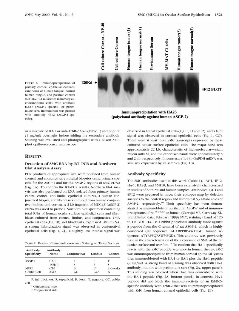
FIGURE 6. Immunoprecipitation ofprimary corneal epithelial cultures,carcinoma of human tongue, normalhuman tongue, and positive control(MV-MAT C1 rat ascites mammary ad-enocarcinoma cells) with antibodyHA2-3 (ASGP-2–specific) or preim-mune sera. Immunoblot was probedwith antibody 4F12 (ASGP-2–spe-cific).
TABLE 2. Results of Immunofluorescence Staining on Tissue Sections
AntibodySpecificity
AntibodyName Conjunctiva Limbus Cornea
ASGP-1 HA-115H10
FS
FS
FS
MUC1 CT-1 B B* S (weak)Goblet Cell AM-3 GC GC† N
F, full thickness; S, superficial; B, basal; N, negative; GC, gobletcell.
* Conjunctival side.† Conjunctival side.
IOVS, May 2000, Vol. 41, No. 6 SMC (MUC4) in Ocular Surface Epithelium 1321

HA2-3 is a rabbit polyclonal antibody made against a rela-tively nonconserved peptide (LDNQTVTFQPDHEDGG) fromhuman ASGP-2. Its reactivity to human ASGP-2 has been shownby immunoprecipitation analyses from human milk or salivarygland, followed by immunoblotting with 4F12 or HA-1. Toconfirm that HA2-3 specifically reacts with the SMC peptidesequence in human tissues, SMC was immunoprecipitatedfrom human corneal epithelial cell lysates with HA2-3 or HA2-3plus the HA2-3 peptide (0.25–2mg/ml), then blotted with anASGP-2 antibody 4F12. A strong band of staining was observedwith HA2-3 alone, but this staining decreased as the concen-tration of HA2-3 peptide in the immunoprecipitation reactionwas increased (Fig. 3A). HA2-3 peptide did not block theimmunoreactivity of an ErbB-2–specific antibody with ErbB-2that was coimmunoprecipitated with SMC from MV-MATC1cells or A375 human melanoma cells (Fig. 3B).
15H10 is a mouse monoclonal antibody made againstpurified rat SMC and has been used in the characterization ofASGP-1 in rat uterus33 and airway.33 Extensive studies haveshown that this antibody is carbohydrate-dependent.34 Anti-body 15H10 immunoprecipitated a high Mr band from culturedhuman corneal epithelial cells (Fig. 2A) that has a similarmotility to rat SMC (MUC4). HA-1 and 15H10 both stain asimilar high Mr band in immunoprecipitants of SMC fromhuman milk but do not react with immunoprecipitants ofMUC1 from that source (data not shown).
Immunodetection of SMC in the Ocular SurfaceEpithelia and Tear Fluid
Sialomucin complex was immunoprecipitated from the cor-neal and conjunctival epithelial specimens and from the tearfluid using ASGP-1–specific antibodies. After SDS–PAGE andimmunoblotting, the ASGP-1 and ASGP-2 subunits of SMC weredetected in tear fluid samples (Fig. 4). Sialomucin complex wasdetected in both corneal and conjunctival epithelia by twomethods. The first was by immunoprecipitation of tissue ly-sates with monoclonal antibody 15H10, followed by immuno-blotting with anti–ASGP-1 HA-1 (Fig. 5). In the second, tissuelysates were immunoprecipitated with HA2-3, followed byimmunoblotting with monoclonal antibody 4F12, both ofwhich recognize human ASGP-2 (Fig. 6). The sequential immu-noprecipitation and immunoblot analysis provides a higherdegree of specificity than straight immunoblot analysis. Fur-thermore, in each case the immunoblotted bands from thehuman tissues migrated similarly to the corresponding bandsobserved in rat ocular surface epithelia (data not shown).
Immunolocalization of SMC in the Ocular SurfaceEpithelia and Preocular Tear Layer
The results of immunofluorescence antibody staining of cor-neal and conjunctival tissue sections are summarized in Table2. The ASGP-1 peptide–specific antibody HA-1 stained the fullthickness of the stratified corneal and conjunctival epithelia
FIGURE 7. Immunofluorescence photomicrographs of corneal and conjunctival frozen tissue sections stained with antibodies 15H10 (A, D), HA-1(B, E), and both HA-1 (red) and 15H10 (green; C, F). Negative controls consisted of sections stained with FITC– (G) or Texas red– (H) conjungatedsecondary antibodies. The epithelial layer is labeled with an E in the sections, and the junction between the epithelium and stroma is delineatedwith a dotted line.
1322 Pflugfelder et al. IOVS, May 2000, Vol. 41, No. 6

(Figs. 7B, 7E). In contrast, antibody 15H10, which reactsagainst a carbohydrate epitope on ASGP-1, stained only thesuperficial layers of these epithelia (Figs. 7A, 7D). No stainingwas observed in conjunctival goblet cells, in agreement withresults in the rat reported previously.18 The secondary antibod-ies alone stained occasional cells in the conjunctival and limbalstroma, but no staining was observed in the corneal or con-junctival epithelium (Figs. 7G, 7H).
To confirm the specificity of polyclonal antibody HA-1staining of human ocular surface epithelia for the HA-1 peptidesequence, an additional experiment was performed in whichHA-1 was preincubated with HA-1 peptide (1 mg/ml) overnightbefore tissue sections were stained. Figure 8 demonstrates amarked (.50%) decrease in the intensity of HA-1 staining ofthe corneal and conjunctival epithelia when this antibody waspreincubated with HA-1 peptide. No staining was observedwith HA-1 peptide plus secondary antibody, preimmune seraplus secondary antibody, or secondary antibody alone. Prein-cubation of an Erb-B–specific antibody with HA-1 peptide didnot reduce the intensity of staining with this antibody.
The results of immunofluorescence antibody staining ofcorneal and conjunctival impression cytology specimens aresummarized in Table 3. Impression cytology specimens fromthe cornea and conjunctiva were evaluated for ASGP-1 expres-sion to determine whether the mucin subunit was present inthe preocular tear layer. The ASGP-1 peptide antibody HA-1stained the conjunctival and corneal epithelial cells on thecytology membranes and weakly stained cell-free areas on thecornea (Figs. 9B, 9E). In contrast, antibody 15H10 stained thecorneal and conjunctival epithelial cells as well as the cell-freeareas on cytology membranes, indicating that the specificallyglycosylated form of this mucin is abundant in the preoculartear layer (Figs. 9A, 9E). No staining was observed in cornealand conjunctival cytology specimens stained with the second-ary antibody alone (Figs. 9G, 9H).
The immunofluorescence staining patterns for themembrane mucin MUC1 and goblet cell mucin in the cor-neal and conjunctival epithelium were different from thoseobserved for SMC. The MUC1 antibody CT-1 strongly stainedthe basal epithelium in the conjunctiva and limbus and
FIGURE 8. Immunofluorescence pho-tomicrographs of human corneal fro-zen tissue sections stained withASGP-1 polyclonal antibody HA-1 (A)or HA-1 preincubated with HA-1 pep-tide, 1 mg/ml (B). Human conjunctivalfrozen tissue sections were stainedwith ASGP-1 polyclonal antibody HA-1(C) or HA-1 preincubated with HA-1peptide, 1 mg/ml (D), preimmune seraplus secondary antibody (E), second-ary antibody alone (F), HA-1 peptideplus secondary antibody (G), ErbB-2monoclonal antibody, Ab-8 (H), orErbB-2 monoclonal antibody Ab-8preincubated with HA-1 peptide, 1mg/ml (I).
TABLE 3. Results of Immunofluorescence Staining on Cytology Specimens
AntibodySpecificity Antibody
Conjunctiva Cornea
Cell Extracellular* Cell Extracellular*
ASGP-1 HA-115H10
111
21
111
111
MUC1 CT-1 1 2 1 6Goblet cell AM-3 GC 2 2 2
11, strong positive; 1, weak positive; 6, equivocal; 2, negative; GC, goblet cell.* Cell free area on cytology membrane.
IOVS, May 2000, Vol. 41, No. 6 SMC (MUC4) in Ocular Surface Epithelium 1323

weakly stained the superficial epithelium in the cornea (datanot shown). No staining was noted on cell-free areas onconjunctival cytology specimens and only weak staining wasnoted on cell-free areas on corneal cytology specimens (notshown). The goblet cell mucin antibody AM-3 stained onlythe goblet cells located in the conjunctiva and conjunctivalside of the limbus, but not the corneal epithelium or pre-corneal tear layer (data not shown).
DISCUSSION
Our study indicates that SMC (MUC4) is produced by thehuman ocular surface epithelium. A peptide epitope of themucin subunit was immunodetected throughout the entirethickness of the stratified epithelium in the conjunctiva, lim-bus, and cornea. In contrast, a carbohydrate-dependentepitope of ASGP-1 was expressed in a differentiation-depen-dent fashion, such that the greatest staining was observedprimarily in the superficial differentiated epithelial cells. Theglycosylated form of ASGP-1 was also detected in the preoculartear layer removed by impression cytology. The mature glyco-sylated form of this mucin in the cell membrane and surface ofthe superficial ocular surface epithelium could function to holdaqueous tear fluid produced by the lacrimal glands in thepreocular tear layer and serve as a lubricative barrier to fric-tional stresses exerted by the eyelids.
The pattern of multiple SMC RNA transcripts with anunusually large size that was observed in the ocular surfaceepithelium is consistent with previously reported studies that
evaluated MUC4 mRNA expression in nonocular tissues.35–39 Ahigh degree of polydispersity is a typical feature of the mucinmRNAs. Moreover, allelic variations in the length of thesemucin transcripts have been observed.36
In addition to the preocular tear layer, SMC was alsodetected in tear fluid collected from the inferior tear meniscus.It still remains to be determined whether SMC in the tear fluidis derived from the underlying epithelium or from othersources, such as the lacrimal gland. Mucins have been histo-chemically detected in subsurface cytoplasmic vesicles locatedwithin the superficial layers of the conjunctival epithelium instudies reported previously by Greiner and associates40 andDilly.3 These vesicles have been observed to fuse with the cellmembrane in a manner in which the inner lamella of thesevesicles becomes the outer layer of the cell surface mem-brane.6 This process transfers the mucus within the cytoplas-mic vesicles to the cell surface membrane and to the preoculartear layer. Future ultrastructural studies may determinewhether SMC is one of the mucin species processed andtransported to the epithelial cell surface in this manner.
The fact that a number of different mucin molecules areproduced by the ocular surface epithelium suggests that eachmay have a unique role. The detection of SMC in the preoculartear layer removed by impression cytology, as well as in thetear fluid, suggests that this mucin may play an important rolein ocular surface barrier function and in maintenance of tearfilm stability. In a separate series of experiments, we haveshown that there is a transient increase in fluorescein perme-ability associated with tear film instability and decreased re-
FIGURE 9. Immunofluorescence photomicrographs of impression cytology specimens of cornea and conjunctiva (Conj) stained with antibodies15H10 (A, D), HA-1 (B, E), and both HA-1 (red) and 15H10 (green; C, F). Negative controls consisted of conjunctival cytology specimens stainedwith FITC– (G) or Texas red– (H) conjugated secondary antibodies.
1324 Pflugfelder et al. IOVS, May 2000, Vol. 41, No. 6

flecting quality of the corneal surface in the focal area whereSMC is removed from the corneal surface. Based on thesefindings, we propose that ASGP-1 may serve as the glycocalyxcoating the microvilli and microplicae of the superficial layer ofthe ocular surface epithelium. Chemical attractions betweenthis adherent mucus layer and the soluble mucin in the over-lying aqueous layer may be integrally important for maintainingtear film structure and to stability, as presented in Figure 10.
Unlike SMC, goblet cell mucin (detected by antibodyAM-3) could not be immunodetected in corneal tissue sectionsor impression cytology membranes. This finding is consistentwith previous observations of Dilly3 that mucin produced bythe stratified epithelial cells, but not the goblet cells, can behistochemically detected in the preocular mucus layer. Furtherexperiments will be required to determine whether goblet cellmucin is indeed absent from the precorneal mucin layer, and ifit is present, how it interacts with SMC and the other mem-brane mucins that are produced by the ocular surface epithe-lium, such as MUC1. MUC1 expression in the conjunctival andlimbal epithelia was limited to the basal epithelium, and onlyminimal staining was observed in the superficial corneal epi-thelium. These findings suggest that MUC1 may have a differ-ent functional role than SMC.
The ASGP-2 subunit of SMC contains two functional EGF-like domains. Our group has detected the presence of ErbB-2receptors in the cell membranes of the ocular surface epithe-lium.41 We demonstrated (Fig. 2B) that ErbB-2 is coimmuno-precipitated with SMC. Thus, it is possible that proper process-ing of SMC places its EGF domains in a position in which theymay interact with these ErbB-2 receptors. If this proves to bethe case, ASGP-2 could potentially have an autocrine signalingfunction that may be important for promoting normal growthand differentiation of the ocular surface epithelium. Binding ofASGP-2 to the ErbB family receptor ErbB-2 has been noted topotentiate tyrosine phosphorylation of this receptor.23
Gene sequencing studies indicate that there is a significanthomology between SMC and MUC4 mucin.24,25 Inatomi re-ported that MUC4 RNA was expressed in the stratified epithe-lium of the conjunctiva by in situ hybridization but not incorneal sections.13 They also failed to detect MUC4 RNA incultured human corneal epithelium by Northern blot analysis.We detected SMC RNA transcripts in both human corneal andconjunctival biopsies by the sensitive RT–PCR technique. Oneof the segments amplified by PCR has been reported to beconserved between MUC4 and SMC.24,25 Our Northern blot
analysis also demonstrated expression of SMC/MUC4 RNA inprimary human corneal and limbal epithelial cultures, althoughthe observed level of expression was greater in the limbal thancentral epithelium. This observation might explain the differ-ence between our study and the study reported by Inatomi, ifthe cultured epithelium they used was obtained from thecentral cornea. It is also possible that the level of MUC4 RNA inthe human corneal epithelium was below the level of detectionby in situ hybridization. Our study also used several methods toimmunodetect SMC in the corneal epithelium. These studiescertainly indicate that SMC/MUC4 protein is expressed in thecornea.
References
1. Chen HB. Histochemical study on rat tear film and ocular surfaceepithelial cells. Curr Eye Res. 1998;17:642–649.
2. Corfield AP, Carrington SD, Hicks SJ, Berry M, Ellingham R. Ocularmucins: purification, metabolism and functions. Prog Retin EyeRes. 1997;16:627–656.
3. Dilly PN. Contribution of the epithelium to the stability of the tearfilm. Trans Ophthalmol Soc UK. 1985;104:381–389.
4. Nichols BA. Demonstration of the mucous layer of the tear film byelectron microscopy. Invest Ophthalmol Vis Sci. 1985;26:464–473.
5. Gendler SJ. Epithelial mucin genes. Annu Rev Physiol. 1995;57:607–634.
6. Strous GJ, Dekker J. Mucin-type glycoproteins. Crit Rev BiochemMol Biol. 1992;27:57–92.
7. Carraway KL, Hull SR. Cell surface mucin-type glycoproteins andmucin-like domains. Glycobiology. 1991;1:131–138.
8. Gum JRJ. Human mucin glycoproteins: varied structures predictdiverse properties and specific functions. Biochem Soc Trans.1995;23:795–799.
9. Bobek LA. Molecular cloning, sequence, and specificity of expres-sion of the gene encoding the low molecular weight human sali-vary mucin (MUC7). J Biol Chem. 1993;268:20563–20569.
10. Boshell M. The product of the human MUC1 gene when secretedby mouse cells transfected with the full-length cDNA lacks thecytoplasmic tail. Biochem Biophys Res Commun. 1992;185:1–8.
11. Williams CJ. Multiple protein forms of the human breast tumor-associated epithelial membrane antigen (EMA) are generated byalternative splicing and induced by hormonal stimulation. Bio-chem Biophys Res Commun. 1990;170:1331–1338.
12. Inatomi T. Human corneal and conjunctival epithelia expressMUC1 mucin. Invest Ophthalmol Vis Sci. 1995;36:1818–1827.
13. Inatomi T. Expression of secretory mucin genes by human con-junctival epithelia. Invest Ophthalmol Vis Sci. 1996;37:1684–1692.
14. McNeer RR. Sialomucin complex in tumors and tissues. FrontBiosci. 1997;2:d449–d459.
FIGURE 10. Proposed interaction ofmembrane and soluble SMC in theprecorneal tear layer. Membranebound SMC may serve as the glyco-calyx coating the microvilli and mi-croplicae of the superficial layer ofthe ocular surface epithelium. Chem-ical attractions between this adher-ent mucus layer and soluble sialomu-cin in the overlying aqueous layermay help to maintain tear film struc-ture and stability.
IOVS, May 2000, Vol. 41, No. 6 SMC (MUC4) in Ocular Surface Epithelium 1325

15. Sherblom AP, Carraway KL. A complex of two cell surface glyco-proteins from ascites mammary adenocarcinoma cells. J BiolChem. 1980;255:12051–12059.
16. Hull SR, Sheng Z, Vanderpuye OA, David C, Carraway KL. Isolationand partial characterization of ascites sialoglycoprotein-2 of thecell surface sialomucin complex of 13762 rat mammary adenocar-cinoma cells. Biochem J. 1990;265:121–129.
17. McNeer RR, Price–Schiavi S, Komatsu M, Fregein N, CarothersCarraway CA, Carrraway K. Sialomucin complex in tumors andtissues. Front Biosci. 1997;2:449–459.
18. Price–Schiavi SA, Meller D, Jing X, et al. Sialomucin complex at therat ocular surface: a new model for ocular surface protection.Biochem J. 1998;335:457–463.
19. Sheng Z, Wu K, Carraway KL, Fregein N. Molecular cloning of thetransmembrane component of the 13762 mammary adenocarci-noma sialomucin complex: a new member of the epidermalgrowth factor superfamily. J Biol Chem. 1992;267:16341–16346.
20. Wu K, Fregein N, Carraway KL. Molecular cloning and sequencingof the mucin subunit of a heterodimeric, bifunctional cell surfaceglycoprotein complex of ascites rat mammary adenocarcinomacells. J Biol Chem. 1994;269:11950–11955.
21. Sheng ZQ, Hull SR, Carraway KL. Biosynthesis of the cell surfacesialomucin complex of ascites 13762 rat mammary adenocarci-noma cells from a high molecular weight precursor. J Biol Chem.1990;265:8505–8510.
22. Carraway KL, Carothers Carraway CA, Carraway KLI. Roles ofErbB-3 and ErbB-4 in the physiology and pathology of the mam-mary gland. J Mammary Gland Biol Neoplasia. 1997;2:187–197.
23. Carraway KLI, Rossi EA, Komatsu M, et al. An intramembranemodulator of the ErbB2 receptor tyrosine kinase that potentiatesneuregulin signaling. J Biol Chem. 1999;274:5263–5266.
24. Nollett S, Moniaux N, Maury J, et al. Human mucin gene MUC4:organization of its 59-region and polymorphism of its central tan-dem repeat array. Biochem J. 1998;332:739–748.
25. Moniaux N, Nollet S, Porchet N, Degand P, Laine A, Aubert JP.Complete sequence of human MUC4: putative cell membrane-associated mucin. Biochem J. 1999;338:325–333.
26. Porchet N, Nguyen VC, Dufosse J, et al. Molecular cloning andchromosomal localization of a novel human tracheo-bronchial mu-cin cDNA containing tandemly repeated sequences of 48 basepairs. Biochem Biophys Res Commun. 1991;175:414–422.
27. Jones DT, Monroy D, Pflugfelder SC. A novel method of tearcollection: comparison of glass capillary tubes with porous poly-ester rods. Cornea. 1997;16:450–458.
28. Pflugfelder SC, Tseng SCG, Yoshino K, Monroy D, Felix C, Reis B.Correlation of goblet cell density and mucosal epithelial mem-brane mucin expression with rose bengal staining in patients withocular irritation. Ophthalmology. 1997;104:223–235.
29. Tseng SC, Li DQ, Ma X. Suppression of transforming growth factorbeta isoforms, TGF-beta receptor type II, and myofibroblast differ-entiation ion cultured human corneal and limbal fibroblasts byamniotic membrane matrix. J Cell Physiol. 1999;179:325–335.
30. Rossi EA, McNeer RR, Price–Schiavi S, et al. Sialomucin complex,a heterodimeric glycoprotein complex: expression as a soluble,secretable form in lactating mammary gland and colon. J BiolChem. 1996;271:33476–33485.
31. Jones DT, Monroy D, Ji Z, Atherton SS, Pflugfelder SC. Sjogren’ssyndrome: cytokine and Epstein-Barr virus gene expression withinthe conjunctival epithelium. Invest Ophthalmol Vis Sci. 1994;35:3493–3504.
32. McNeer RR, Huang D, Fregien NL, Carraway KL. Sialomucin com-plex in the rat respiratory tract: a model for its role in epithelialprotection. Biochem J. 1998;330:737–744.
33. McNeer RR, Carraway CAC, Fregien NL, Carraway KL. Character-ization of the expression and steroid hormone control of sialomu-cin complex in the rat uterus: Implications for uterine receptivity.J Cell Physiol. 1998;176:110–119.
34. Rossi EA. Characterization of the Tissue and Cell Specific Expres-sion of Ascites Sialoglycoprotein-2 and Its Association withp185neu. Miami, FL: University of Miami; 1996. Thesis.
35. Liu B, Offner GD, Nunes DP, Oppenheim FG, Troxler RF. MUC4 isa major component of salivary mucin MG1 secreted by the humansubmandibular gland. Biochem Biophys Res Commun. 1998;250:757–761.
36. Debailleul V, Laine A, Huet G, et al. Human mucin genes MUC2,MUC3, MUC4, MUC5AC, MUC5B, and MUC6 express stableand extremely large mRNAs and exhibit a variable lengthpolymorphism: an improved method to analyze large mRNAs.J Biol Chem. 1998;273:881–890.
37. Baeckstrom D, Hansson GC. The transcripts of the apomucingenes MUC2, MUC4, and MUC5AC are large and appear as distinctbands. Glycoconj J. 1996;13:833–837.
38. Balague C, Gambus G, Carrato C, et al. Altered expression ofMUC2, MUC4, and MUC5 mucin genes in pancreas tissues andcancer cell lines. Gastroenterology. 1994;106:1054–1061.
39. Bernacki SH, Nelson AL, Abdullah L, et al. Mucin gene expressionduring differentiation of human airway epithelia in vitro: muc4 andmuc5b are strongly induced. Am J Respir Cell Mol Biol. 1999;20:595–604.
40. Greiner JV, Kenyon KR, Henriquez AS, et al. Mucus secretoryvesicles in conjunctival epithelial cells of wearers of contactlenses. Arch Ophthalmol. 1980;98:1843–1846.
41. Liu Z, Carvajal M, Carraway CAC, Carraway KL, Pflugfelder SC.Increased expression of the type 1 growth factor receptor familyin the conjunctival epithelium of patients with keratoconjunctivi-tis sicca. Am J Ophthalmol. In press.
42. Huang AJ. Development of monoclonal antibodies to rabbit ocularmucin. Invest Ophthalmol Vis Sci. 1987;28:1483–1491.
43. Pemberton L, Taylor–Papadimitriou J, Gendler SJ. Antibodies tothe cytoplasmic domain of MUC1 mucin show conservationthroughout mammals. Biochem Biophys Res Commun. 1992;185:167–175.
1326 Pflugfelder et al. IOVS, May 2000, Vol. 41, No. 6