Glaucoma (Weinreb & Khaw)
10
For personal use. Only reproduce with permission from The Lancet publishing Group. SEMINAR Glaucoma is a group of progressive optic neuropathies that have in common a slow progressive degeneration of retinal ganglion cells and their axons, resulting in a distinct appearance of the optic disc and a concomitant pattern of visual loss. The biological basis of the disease is not yet fully understood, and the factors contributing to its progression are not yet fully characterised. However, intraocular pressure is the only proven treatable risk factor. Without adequate treatment, glaucoma can progress to visual disability and eventual blindness. This seminar will address primary open-angle glaucoma, an age-related and insidious form of the disease. Epidemiology It is estimated that glaucoma affects more than 66 million individuals worldwide with at least 6·8 million bilaterally blind. 1 Vision loss caused by glaucoma is irreversible, and glaucoma is the second leading cause of blindness in the world. Of the many types of glaucoma, primary open- angle glaucoma is perhaps the most common, particularly in populations of European and African ancestry. 2,3 The disease is the leading cause of blindness in African- Americans. In the USA, more than 7 million office visits occur per year to monitor patients who have glaucoma or are at risk of developing the disease. 3,4 Blindness from all forms of glaucoma in the USA is estimated to cost in excess of $1·5 billion annually. However, the scope of the problem is probably larger than these numbers suggest, and a substantial proportion of individuals remain either undiagnosed or inadequately treated. The number of individuals suspected to have glaucoma—usually those with raised intraocular pressure (ocular hypertension) or asymmetric optic disc appearance—far exceeds the number who have been diagnosed with the disease. Lancet 2004; 363: 1711–20 Hamilton Glaucoma Center and Department of Ophthalmology, University of California San Diego, 9500 Gilman Drive, La Jolla CA 92093-0946, USA (Prof R N Weinreb MD); and Glaucoma Unit and Ocular Repair and Regeneration Biology Unit, Moorfields Eye Hospital and Institute of Ophthalmology, University College London, London, UK (Prof P T Khaw PhD) Correspondence to: Prof R N Weinreb (e-mail: [email protected]) Moreover, the magnitude of the problem will increase as the population ages. 3 Anatomy and physiology Aqueous humour secretion and drainage Intraocular pressure is regulated by a balance between the secretion and drainage of aqueous humour (figure 1). This fluid is secreted posterior to the iris by the ciliary body and then flows anteriorly to the anterior chamber. Aqueous humour provides nutrients to the iris, lens, and cornea. It exits the eye into the venous circulation via the trabecular meshwork and independently through the uveoscleral outflow pathway. The optic nerve and inner retina Axons of retinal ganglion cells comprise the retinal nerve fibre layer, the innermost layer of the retina. The human optic nerve contains about one million nerve fibres (figure 2). These axons converge on the optic disc (also known as the optic nerve head) and form the optic nerve. The fibres exit the eye after traversing the lamina cribrosa, a series of perforated connective tissue sheets, and synapse in the lateral geniculate nucleus of the brain. The optic disc is about 1·5 mm in diameter and vertically oval. Its area varies up to sevenfold, and is largest in highly myopic individuals. The convergence of the axons forms a central depression in the disc, known as the cup. Most, but not all, optic nerves have a visible physiologic cup. The neuroretinal rim of the optic nerve head is pink and surrounds the cup. Trophic factors, including brain-derived neurotrophic factor, are retrogradely transported from the axonal terminals of retinal ganglion cells to their cell bodies in the inner retina, and are essential for the survival of the cells. Glutamate, a neurotransmitter, is normally present in low concentrations in the retina. Trophic factors also are transported via retinal ganglion cell axons in an anterograde fashion to the lateral geniculate nucleus. Primary open-angle glaucoma Robert N Weinreb, Peng Tee Khaw Seminar THE LANCET • Vol 363 • May 22, 2004 • www.thelancet.com 1711 Primary open-angle glaucoma is a progressive optic neuropathy and, perhaps, the most common form of glaucoma. Because the disease is treatable, and because the visual impairment caused by glaucoma is irreversible, early detection is essential. Early diagnosis depends on examination of the optic disc, retinal nerve fibre layer, and visual field. New imaging and psychophysical tests can improve both detection and monitoring of the progression of the disease. Recently completed long-term clinical trials provide convincing evidence that lowering intraocular pressure prevents progression at both the early and late stages of the disease. The degree of protection is related to the degree to which intraocular pressure is lowered. Improvements in therapy consist of more effective and better-tolerated drugs to lower intraocular pressure, and more effective surgical procedures. New treatments to directly treat and protect the retinal ganglion cells that are damaged in glaucoma are also in development. Search strategy and selection criteria We systematically searched MEDLINE with terminology relating to primary open-angle glaucoma discussed in this review. Keywords used were glaucoma, open-angle glaucoma, primary open-angle glaucoma, glaucoma blindness, ocular hypertension. Articles were reviewed up to June, 2003, and studies reported in full and in abstract form have been reported.
Transcript of Glaucoma (Weinreb & Khaw)

For personal use. Only reproduce with permission from The Lancet publishing Group.
SEMINAR
Glaucoma is a group of progressive optic neuropathiesthat have in common a slow progressive degeneration ofretinal ganglion cells and their axons, resulting in adistinct appearance of the optic disc and a concomitantpattern of visual loss. The biological basis of the disease isnot yet fully understood, and the factors contributing toits progression are not yet fully characterised. However,intraocular pressure is the only proven treatable riskfactor. Without adequate treatment, glaucoma canprogress to visual disability and eventual blindness. Thisseminar will address primary open-angle glaucoma, anage-related and insidious form of the disease.
EpidemiologyIt is estimated that glaucoma affects more than 66 millionindividuals worldwide with at least 6·8 million bilaterallyblind.1 Vision loss caused by glaucoma is irreversible, andglaucoma is the second leading cause of blindness in theworld. Of the many types of glaucoma, primary open-angle glaucoma is perhaps the most common, particularlyin populations of European and African ancestry.2,3 Thedisease is the leading cause of blindness in African-Americans.
In the USA, more than 7 million office visits occur peryear to monitor patients who have glaucoma or are at riskof developing the disease.3,4 Blindness from all forms ofglaucoma in the USA is estimated to cost in excess of$1·5 billion annually. However, the scope of the problemis probably larger than these numbers suggest, and asubstantial proportion of individuals remain eitherundiagnosed or inadequately treated. The number ofindividuals suspected to have glaucoma—usually thosewith raised intraocular pressure (ocular hypertension) orasymmetric optic disc appearance—far exceeds thenumber who have been diagnosed with the disease.
Lancet 2004; 363: 1711–20
Hamilton Glaucoma Center and Department of Ophthalmology,University of California San Diego, 9500 Gilman Drive, La Jolla CA92093-0946, USA (Prof R N Weinreb MD); and Glaucoma Unit andOcular Repair and Regeneration Biology Unit, Moorfields EyeHospital and Institute of Ophthalmology, University CollegeLondon, London, UK (Prof P T Khaw PhD)
Correspondence to: Prof R N Weinreb(e-mail: [email protected])
Moreover, the magnitude of the problem will increase asthe population ages.3
Anatomy and physiology Aqueous humour secretion and drainageIntraocular pressure is regulated by a balance between thesecretion and drainage of aqueous humour (figure 1).This fluid is secreted posterior to the iris by the ciliarybody and then flows anteriorly to the anterior chamber.Aqueous humour provides nutrients to the iris, lens, andcornea. It exits the eye into the venous circulation via thetrabecular meshwork and independently through theuveoscleral outflow pathway.
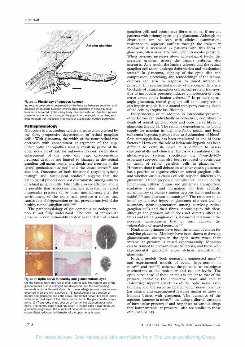
The optic nerve and inner retinaAxons of retinal ganglion cells comprise the retinal nervefibre layer, the innermost layer of the retina. The humanoptic nerve contains about one million nerve fibres(figure 2). These axons converge on the optic disc (alsoknown as the optic nerve head) and form the optic nerve.The fibres exit the eye after traversing the lamina cribrosa, aseries of perforated connective tissue sheets, and synapse inthe lateral geniculate nucleus of the brain. The optic disc isabout 1·5 mm in diameter and vertically oval. Its area variesup to sevenfold, and is largest in highly myopic individuals.The convergence of the axons forms a central depression inthe disc, known as the cup. Most, but not all, optic nerveshave a visible physiologic cup. The neuroretinal rim of theoptic nerve head is pink and surrounds the cup. Trophicfactors, including brain-derived neurotrophic factor, areretrogradely transported from the axonal terminals ofretinal ganglion cells to their cell bodies in the inner retina,and are essential for the survival of the cells. Glutamate, aneurotransmitter, is normally present in low concentrationsin the retina. Trophic factors also are transported via retinalganglion cell axons in an anterograde fashion to the lateralgeniculate nucleus.
Primary open-angle glaucoma
Robert N Weinreb, Peng Tee Khaw
Seminar
THE LANCET • Vol 363 • May 22, 2004 • www.thelancet.com 1711
Primary open-angle glaucoma is a progressive optic neuropathy and, perhaps, the most common form of glaucoma.Because the disease is treatable, and because the visual impairment caused by glaucoma is irreversible, earlydetection is essential. Early diagnosis depends on examination of the optic disc, retinal nerve fibre layer, and visualfield. New imaging and psychophysical tests can improve both detection and monitoring of the progression of thedisease. Recently completed long-term clinical trials provide convincing evidence that lowering intraocular pressureprevents progression at both the early and late stages of the disease. The degree of protection is related to the degreeto which intraocular pressure is lowered. Improvements in therapy consist of more effective and better-tolerated drugsto lower intraocular pressure, and more effective surgical procedures. New treatments to directly treat and protect theretinal ganglion cells that are damaged in glaucoma are also in development.
Search strategy and selection criteriaWe systematically searched MEDLINE with terminologyrelating to primary open-angle glaucoma discussed in thisreview. Keywords used were glaucoma, open-angle glaucoma,primary open-angle glaucoma, glaucoma blindness, ocularhypertension. Articles were reviewed up to June, 2003, andstudies reported in full and in abstract form have beenreported.
For personal use. Only reproduce with permission from The Lancet publishing Group.
PathophysiologyGlaucoma is a neurodegenerative disease characterised bythe slow, progressive degeneration of retinal ganglioncells.5 With glaucoma, the width of the neuroretinal rimdecreases with concomitant enlargement of the cup.Other optic neuropathies usually result in pallor of theoptic nerve head but, for unknown reasons, rarely showenlargement of the optic disc cup. Glaucomatousneuronal death is not limited to changes in the retinalganglion cell axons, soma, and dendrites;6 neurons in thelateral geniculate nucleus7–9 and the visual cortex9,10 arealso lost. Outcomes of both functional (psychophysical)testing11 and histological studies7,9 suggest that thepathological process does not discriminate among subsetsof retinal ganglion cells. Glial cells also are affected, and itis possible that astrocytes, perhaps activated by raisedintraocular pressure or by other mechanisms, alter theenvironment of the axons and produce a milieu thatcauses axonal degeneration or that prevents survival of thehealthy retinal ganglion cells.12,13
The pathophysiology of glaucomatous neurodegenera-tion is not fully understood. The level of intraocularpressure is unquestionably related to the death of retinal
ganglion cells and optic nerve fibres in some, if not all,patients with primary open-angle glaucoma. Although noobstruction can be seen with clinical examination,resistance to aqueous outflow through the trabecularmeshwork is increased in patients with this form ofglaucoma, often associated with high intraocular pressure.When pressure increases above physiological levels, thepressure gradient across the lamina cribrosa alsoincreases. As a result, the lamina cribrosa and the retinalganglion cell axons undergo deformation and mechanicalstress.14 In glaucoma, cupping of the optic disc andcompression, stretching, and remodelling12 of the laminacribrosa can arise in response to raised intraocularpressure. In experimental models of glaucoma, there is ablockade of retinal ganglion cell axonal protein transportdue to intraocular pressure-induced compression of opticnerve axons at the lamina cribrosa.5,15 In primary open-angle glaucoma, retinal ganglion cell axon compressioncan impair trophic factor axonal transport, causing deathof the cells by trophic insufficiency.
Independently or in addition to intraocular pressure,other factors can individually or collectively contribute todeath of retinal ganglion cells and optic nerve fibres inglaucoma (figure 3). The retina is dependent on its bloodsupply for meeting its high metabolic needs, and localischaemia-hypoxia, perhaps due to dysfunction of blood-flow autoregulation, has been implicated as one of thesefactors.16 However, the role of ischaemic-hypoxia has beendifficult to establish, since it is difficult to assessexperimentally and clinically. Excessive stimulation of theglutamatergic system, specifically the N-methyl-D-aspartate subtypes, has also been proposed to contributeto death of retinal ganglion cells in glaucoma.17–19
However, there is still debate on whether excess glutamatehas a positive or negative effect on retinal ganglion cells,and whether various classes of cells respond differently toglutamate. Other proposed contributors include poorlyfunctioning cellular pumps and glutamate transporters,oxidative stress and formation of free radicals,inflammatory cytokines (tumour necrosis factor and nitricoxide),20,21 and aberrant immunity.22,23 The response to aninitial optic nerve injury in glaucoma also can lead tosecondary neurodegeneration among surviving retinalganglion cells and their fibres. According to this view,although the primary insult does not directly affect allfibres and retinal ganglion cells, it causes alterations in theneuronal environment that in turn increase thevulnerability of spared neurons.20,22
Nonhuman primates have been the animal of choice forstudying glaucoma. Monkeys have been shown to developglaucomatous changes in the optic nerve when theirintraocular pressure is raised experimentally. Monkeyscan be trained to perform visual field tests, and those withexperimental glaucoma show deficits indicative ofglaucoma.10
Rodent models (both genetically engineered mice24–26
and experimental models of ocular hypertension inmice27,28 and rats29,30) enhance the potential to investigatemechanisms at the molecular and cellular levels. Theoptic nerve head of these animals is similar to that of theprimate, including the connective tissue and cellular(astrocyte) support structures of the optic nerve axonbundles, and the response of their optic nerve to injuryhas clinical and experimental features similar to those ofhumans beings with glaucoma. The dynamics of theaqueous humour in mice,31—including a diurnal variationof intraocular pressure,32 and responses to various drugsthat lower intraocular pressure—also are similar to thoseof human beings.
SEMINAR
1712 THE LANCET • Vol 363 • May 22, 2004 • www.thelancet.com
Cornea
Trabecular meshwork
Uveos
cleral
outflo
w
Ciliary body
Iris
Anterior chamber
Figure 1: Physiology of aqueous humourIntraocular pressure is determined by the balance between secretion anddrainage of aqueous humour. Arrows show direction of flow; aqueoushumour is secreted by the ciliary body into the posterior chamber, passesposterior to the iris and through the pupil into the anterior chamber, andexits through the trabecular meshwork or uveoscleral outflow pathways.
Normal
Glaucoma
A
A
B
B
C
CFigure 2: Optic nerve in healthy and glaucomatous eyes(A) The normal optic disc has a small central cup. The central cup of theglaucomatous disc is enlarged and deepened, and the surroundingneuroretinal rim is thinned. Optic disc haemorrhage (arrow) is sometimesobserved in an eye with glaucoma. (B) Longitudinal cross-section ofnormal and glaucomatous optic nerve. The retinal nerve fibre layer (arrow)is the innermost layer of the retina, and is thin in the glaucomatous opticnerve. (C) Transverse cross-section of normal and glaucomatous opticnerve. The normal optic nerve has about 1 million optic nerve fibres. Asglaucoma progresses, the number of nerve fibres is reduced, andconcomitant reduction in diameter of the optic nerve is seen.

For personal use. Only reproduce with permission from The Lancet publishing Group.
DiagnosisPrimary open-angle glaucoma is a chronic, generallybilateral, but often asymmetrical, disease that is character-ised by progressive damage of the optic nerve as shown bychanges in the optic disc, retinal nerve fibre layer, or visualfield. The disease has an adult onset, with open anteriorchamber angles of normal appearance and an absence ofother known explanations for the change in the opticnerve. If detected early, disease progression can frequentlybe arrested or slowed with medical and surgical treatment.
Assessment of the optic disc Examination of the optic disc is the most valuable methodof diagnosing early glaucoma, because the optic nerveappearance often changes before visual field loss isdetectable. Some studies have shown that as many as halfof retinal ganglion cells and their axons can be lost beforethe visual field test shows evidence of glaucoma.33,34
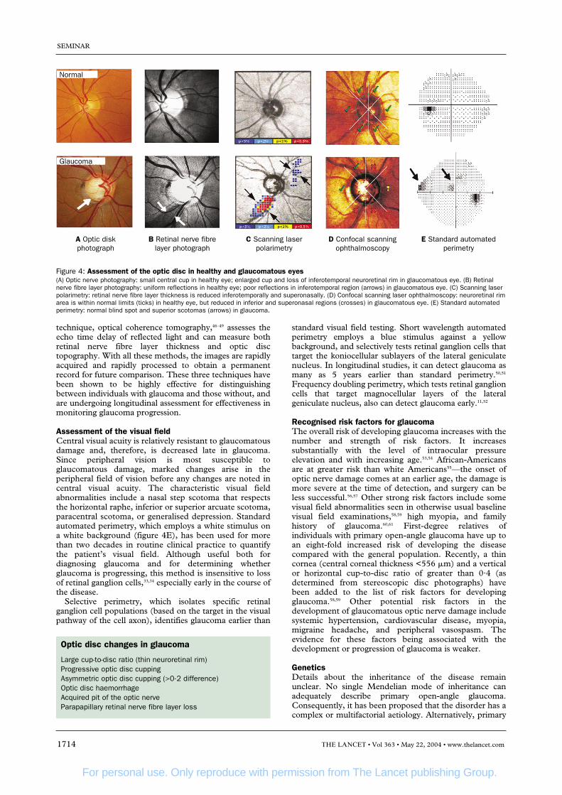
Therefore, vision loss is usually not perceived until thedisease is quite advanced. The optic disc should beexamined with a magnified stereoscopic view. Thisexamination is best done at the slit lamp biomicroscopewith an indirect lens or a contact lens. The directophthalmoscope is less desirable for examining the opticdisc because it provides a view that lacks the depth of astereoscopic image. Optic disc changes consist of diffuseor focal narrowing or notching of the disc rim, especiallyat the inferior or superior poles (figure 4A). Typical opticdisc changes in glaucoma are described in the panel.
Examination of the retinal nerve fibre layer adjacent tothe optic disc also provides useful information about
glaucoma. In the healthy eye, there are broad and brightreflections from the relatively thick retinal nerve fibre layerin the superior and inferior bundles. In glaucoma,reflectivity in these regions is reduced and there are evenfocal areas where reflections are absent (figure 4B).
During the past decade, several objective andquantitative methods have emerged for assessment of theoptic disc and the retinal nerve fibre layer.35 Scanning laserpolarimetry is a clinical technique for assessing thethickness of the retinal nerve fibre layer (figure 4C). Thistechnology measures the retardation (phase shift) of apolarised laser light passing through the eye possessing thephysical property of form birefringence.36 Formbirefringence occurs in tissue that is composed of parallelstructures, each of which is of a smaller diameter than thewavelength of light used to image it. Birefringence in theretinal nerve fibre layer arises from the microtubulescontained within the individual nerve fibres.37 The greaterthe number of microtubules, the greater the retardation ofthe polarised laser light, indicating the presence of moretissue. Scanning laser polarimetry thus gives an indirectassessment of the thickness of the layer. Although thetechnology has been available for several years, recentadvances have enhanced the ability to identify and followthe progression of glaucoma.38–42 Another technique,confocal scanning laser ophthalmoscopy43–45 (figure 4D)allows layer-by-layer imaging to measure the topographyof the optic disc. This technology quantifies the area ofthe optic disc cup and neuroretinal rim, and thesemeasurements can be evaluated longitudinally to assesswhether the glaucoma is stable or progressing. A third
SEMINAR
THE LANCET • Vol 363 • May 22, 2004 • www.thelancet.com 1713
Microcirculation
Retinalganglion
cell
TAstrocytes
Glial cells
Excessive glutamatestimulation
Lateral geniculate nucleus and other targets
Blockade ofneurotrophins and othertarget derived factors
Aberrantimmunity
Lamina cribrosa
Inflammatorycytokines
Ischaemia—hypoxia
Intraocularpressure
Figure 3: Factors contributing to pathophysiology of glaucomatous neurodegenerationIntraocular pressure can cause blockade at the lamina cribrosa of axonal protein transport, causing neuronal retinal ganglion cell death by trophicinsufficiency. Other implicated factors include local ischaemia-hypoxia, excessive stimulation of the glutamatergic system, alterations in glial cells orastrocytes, and aberrant immunity.
For personal use. Only reproduce with permission from The Lancet publishing Group.
technique, optical coherence tomography,46–49 assesses theecho time delay of reflected light and can measure bothretinal nerve fibre layer thickness and optic disctopography. With all these methods, the images are rapidlyacquired and rapidly processed to obtain a permanentrecord for future comparison. These three techniques havebeen shown to be highly effective for distinguishingbetween individuals with glaucoma and those without, andare undergoing longitudinal assessment for effectiveness inmonitoring glaucoma progression.
Assessment of the visual field Central visual acuity is relatively resistant to glaucomatousdamage and, therefore, is decreased late in glaucoma.Since peripheral vision is most susceptible toglaucomatous damage, marked changes arise in theperipheral field of vision before any changes are noted incentral visual acuity. The characteristic visual fieldabnormalities include a nasal step scotoma that respectsthe horizontal raphe, inferior or superior arcuate scotoma,paracentral scotoma, or generalised depression. Standardautomated perimetry, which employs a white stimulus ona white background (figure 4E), has been used for morethan two decades in routine clinical practice to quantifythe patient’s visual field. Although useful both fordiagnosing glaucoma and for determining whetherglaucoma is progressing, this method is insensitive to lossof retinal ganglion cells,33,34 especially early in the course ofthe disease.
Selective perimetry, which isolates specific retinalganglion cell populations (based on the target in the visualpathway of the cell axon), identifies glaucoma earlier than
standard visual field testing. Short wavelength automatedperimetry employs a blue stimulus against a yellowbackground, and selectively tests retinal ganglion cells thattarget the koniocellular sublayers of the lateral geniculatenucleus. In longitudinal studies, it can detect glaucoma asmany as 5 years earlier than standard perimetry.50,51
Frequency doubling perimetry, which tests retinal ganglioncells that target magnocellular layers of the lateralgeniculate nucleus, also can detect glaucoma early.11,52
Recognised risk factors for glaucomaThe overall risk of developing glaucoma increases with thenumber and strength of risk factors. It increasessubstantially with the level of intraocular pressureelevation and with increasing age.53,54 African-Americansare at greater risk than white Americans55—the onset ofoptic nerve damage comes at an earlier age, the damage ismore severe at the time of detection, and surgery can beless successful.56,57 Other strong risk factors include somevisual field abnormalities seen in otherwise usual baselinevisual field examinations,58,59 high myopia, and familyhistory of glaucoma.60,61 First-degree relatives ofindividuals with primary open-angle glaucoma have up toan eight-fold increased risk of developing the diseasecompared with the general population. Recently, a thincornea (central corneal thickness <556 �m) and a verticalor horizontal cup-to-disc ratio of greater than 0·4 (asdetermined from stereoscopic disc photographs) havebeen added to the list of risk factors for developingglaucoma.58,59 Other potential risk factors in thedevelopment of glaucomatous optic nerve damage includesystemic hypertension, cardiovascular disease, myopia,migraine headache, and peripheral vasospasm. Theevidence for these factors being associated with thedevelopment or progression of glaucoma is weaker.
GeneticsDetails about the inheritance of the disease remainunclear. No single Mendelian mode of inheritance canadequately describe primary open-angle glaucoma.Consequently, it has been proposed that the disorder has acomplex or multifactorial aetiology. Alternatively, primary
SEMINAR
1714 THE LANCET • Vol 363 • May 22, 2004 • www.thelancet.com
A Optic diskphotograph
B Retinal nerve fibrelayer photograph
C Scanning laserpolarimetry
D Confocal scanningophthalmoscopy
E Standard automatedperimetry
Normal
Glaucoma
Figure 4: Assessment of the optic disc in healthy and glaucomatous eyes(A) Optic nerve photography: small central cup in healthy eye; enlarged cup and loss of inferotemporal neuroretinal rim in glaucomatous eye. (B) Retinalnerve fibre layer photography: uniform reflections in healthy eye; poor reflections in inferotemporal region (arrows) in glaucomatous eye. (C) Scanning laserpolarimetry: retinal nerve fibre layer thickness is reduced inferotemporally and superonasally. (D) Confocal scanning laser ophthalmoscopy: neuroretinal rimarea is within normal limits (ticks) in healthy eye, but reduced in inferior and superonasal regions (crosses) in glaucomatous eye. (E) Standard automatedperimetry: normal blind spot and superior scotomas (arrows) in glaucoma.
Optic disc changes in glaucoma
Large cup-to-disc ratio (thin neuroretinal rim)Progressive optic disc cuppingAsymmetric optic disc cupping (>0·2 difference)Optic disc haemorrhageAcquired pit of the optic nerveParapapillary retinal nerve fibre layer loss

For personal use. Only reproduce with permission from The Lancet publishing Group.
open-angle glaucoma might represent a collection ofclinically indistinguishable disorders. The chromosomallocations of several genes that can independently cause thedisease have been mapped, indicating that at least someportion of primary open-angle glaucoma is caused bysingle gene defects. The glaucoma gene at the GLC1Alocus (myocilin) has been shown to be associated withboth juvenile and adult-onset primary open-angleglaucoma.62–67 More than 43 different myocilin mutationshave been reported in open-angle glaucoma patients, andseveral large studies have suggested that as a group thesemutations are associated with 3–4% of patients with thecondition in populations worldwide.68 Due to the lowprevalence of myocilin-associated glaucoma in the generalpopulation, screening tests of whole populations formyocilin defects are not especially useful.69 However,testing might be warranted in those at extremely high risk,such as family members of patients with known myocilin-associated glaucoma and members of families with a stronghistory of inherited glaucoma.
Detection and screeningIn most cases, the loss of vision caused by glaucoma canbe limited or prevented by currently available therapies ifthe disease is identified in its early stages. Most cases ofglaucoma are not discovered until vision has already beenpermanently lost, because clinical signs of early glaucomaare subtle, even to an eye specialist.
Programmes to detect individuals at risk in the generalpopulation seek to identify those with glaucoma, thosewho are suspected of having glaucoma, or those who havea high risk of developing glaucoma. These activities maybe more efficient and cost-effective when targeted towardgroups that have a higher risk of disease. Age and race are
two risk factors, in particular, that select for individuals atrisk. Table 1 shows the recommended frequency of eyeexaminations for individuals in the general populationbased on age and race.
The measurement of intraocular pressure is not aneffective method for screening populations for glaucoma.Moreover, the most widely used method formeasurement, Goldmann tonometry, underestimates thetrue intraocular pressure of patients with thin corneas andoverestimates it in patients with thick ones. Half of allpatients with primary open-angle glaucoma have pressuresbelow 22 mm Hg at a single screening.54 Additionally,most individuals with raised pressures do not have, andmight never develop, optic nerve damage, although suchrisk increases with the level of intraocular pressure.Therefore, screening should not rely solely onmeasurement of intraocular pressure; assessments of theoptic disc, retinal nerve fibre layer, and visual functionprovide complementary information. Screening is anessential component of the comprehensive adult eyeassessment; it is the most effective way to identifyindividuals with glaucoma.
ManagementGoals of glaucoma managementAs described in the Preferred Practice Patterns of theAmerican Academy of Ophthalmology70 and otherguidelines, glaucoma care aims to enhance the patient’shealth and quality of life by preserving visual functionwithout causing untoward effects from treatment. Specificgoals are: (1) to document the status of optic nerve onpresentation and during follow-up by assessment of theappearance of the optic disc, retinal nerve fibre layer, orboth, and assessment of the visual field; (2) estimation andmaintainance, through appropriate therapeutic inter-vention, of an intraocular pressure below which furtheroptic nerve damage is unlikely to occur (the targetintraocular pressure); (3) to reset the target intraocularpressure to a lower level if deterioration arises; (4) tominimise the side-effects of management and their effecton the patient’s vision, general health, and quality of life(including the cost of treatment); and (5) to educate andengage the patient in the management of his or her disease.
SEMINAR
THE LANCET • Vol 363 • May 22, 2004 • www.thelancet.com 1715
Asymptomatic Other asymptomatic patientsAfrican-Americans
Age (years)20–29 Every 3–5 years At least once30–39 Every 2–4 years At least twice40–64 Every 2–4 years Every 2–4 years�65 Every 1–2 years Every 1–2 years
Table 1: Frequency of examination to identify patients at risk70
Aim Result
Ocular Hypertension Efficacy and safety of topical ocular With mean IOP-lowering of 22·5%, the probability of developing glaucomatous Treatment Study58, 59 medications in preventing or delaying the change (optic disc or field change) was 4·4% in the medication group and 9·5% in
development of POAG in individuals with the observation group at 60 months. Baseline age, vertical cup disc ratio, visual raised IOP (1636 patients) field abnormalities, and IOP were good predictors of progression. Corneal
thickness was a powerful predictor of progression
Glaucoma Laser Trial75 Efficacy and safety of argon laser Eyes treated with laser trabeculoplasty had slightly reduced IOP (1·2 mm Hg) and trabeculoplasty or medicine as initial improved visual field (0·6 dB) after median follow-up of 7 yearstreatment in POAG (271 patients)
Collaborative Initial Effects of randomising patients to either Surgery lowered the IOP more than medical treatment (average during follow-up Glaucoma Treatment initial medical or surgical treatment 14–15 mm Hg vs 17–18 mm Hg), but with no statistical difference in visual field Study72 (607 patients) progression over 5 years
Early Manifest Glaucoma Effects of treatment with a topical � blocker Progression was less frequent in the treatment group (45% vs 62%) with median Treatment Trial71,76 and laser trabeculoplasty versus observation follow-up of 6 years, Other important predictors of glaucoma progression included
in patients with newly detected POAG lens exfoliation, bilateral glaucoma, IOP >21 mm Hg, more advanced visual field (255 patients) loss, disc haemorrhages, and age �68 years
Collaborative Normal Effect of pressure lowering (30%) on optic Only 12% of treated patients progressed (optic disc and visual field progression) Tension Glaucoma nerve damage and field loss in normal compared with 35% in the untreated group. Study77,78 tension glaucoma (140 patients)
Advanced Glaucoma Effect of treatment sequences of laser Outcome depended on race. In patients who had laser trabeculoplasty first, black Intervention Study79 trabeculoplasty and trabeculectomy patients were at a lower risk than white patients of failure. In patients who
(surgery) in advanced glaucoma (776 eyes received surgery first, black patients were at a higher risk of first failure than whiteof 581 patients) patients. Patients with lower IOP had less progression
POAG=primary open-angle glaucoma. IOP=intraocular pressure.
Table 2: Clinical trials in the past decade

For personal use. Only reproduce with permission from The Lancet publishing Group.
At present, treatment of primary open-angle glaucomais directed at lowering intraocular pressure, whichcontinues to be the only proven and treatable risk factorfor the disease. There are several modalities of treatmentfor lowering intraocular pressure, including drugs, lasersurgery, and incisional surgery. However, lowering ofintraocular pressure does not seem to halt all cases ofprogression.57,58,71,72 In some individuals with progession,it is not practical to sufficiently lower the intraocularpressure. In other individuals, factors other thanintraocular pressure may be damaging the optic nerve.
Current management of glaucoma is directed atestablishing and maintaining a target intraocularpressure70,73 (the degree of intraocular pressure at whichfurther glaucomatous damage is prevented). It is difficultto assess accurately and in advance the target intraocularpressure in every individual patient and eye.Furthermore, no degree of intraocular pressure is safefor every patient. In general, the initial target aims toachieve a 20–50% reduction from the initial pressure atwhich damage occurred. The least amount ofmedication and fewest side-effects to achieve thetherapeutic response are desirable goals. The greater thepre-existing damage due to glaucoma, the lower thetarget intraocular pressure should be. The likelihood ofprogressive damage is increased with high intraocularpressure, severe pre-existing damage, and the presenceof several risk factors. The target intraocular pressure ofan individual should be periodically re-assessed to judgeits appropriateness, by comparing optic nerve status withprevious (including baseline) examinations.
With the availability of practice guidelines,70,73 therehas also been interest in the extent to which actualpractice is consistent with recommended care. For somekey components of the examinations, patterns of care inthe USA are not consistent with the American Academyof Ophthalmology guidelines.74 For instance, nearly halfthe patients in one study did not have a photograph ordrawing of the optic disc at the time of their initialassessment. This problem is of particular concernbecause of the importance of having a baseline image forfuture comparison to assess progression. Anotheressential care process encouraged by recent guidelines isto document a specific target intraocular pressure.70,73
This process seems to be ignored by many eye-careproviders.74 This omission is especially problematic,since many of the recommendations for care depend onwhether the intraocular pressure is above or below thetarget. Primary open-angle glaucoma may be oftenundertreated, at least relative to the standards foroptimal preservation of vision established by recentclinical trials.
Clinical trial results Over the past decade, results from several multicentreclinical trials have confirmed the value of reducingintraocular pressure in patients with ocularhypertension58,59 (significantly raised intraocular pressurewithout glaucomatous visual field loss or optic discdamage) or primary open-angle glaucoma.56,57,71 Loweringthe intraocular pressure can reduce by one-half, onaverage, the number of ocular hypertensive patientsprogressing to glaucoma, and can also prevent progressionin patients with pre-existing glaucoma. However, not allpatients with ocular hypertension will progress toglaucoma. Therefore, the decision to treat depends on therisk of the individual patient progressing as well as thepatient’s preference for treatment. Several of these trialsare summarised in table 2.
Medical treatmentThe prostaglandin analogues and prostamides(latanoprost, travoprost, unoprostone, and bimatoprost)reduce intraocular pressure by increasing the outflow ofaqueous humour, primarily through the uveoscleralpathway.80,81 Some prostaglandins activate matrixmetalloproteinases, which then remodel extracellularmatrix and reduce outflow resistance, allowing theaqueous humour to flow out via this route.82,83 In general,these drugs have become the first line of treatmentbecause of their once daily application, minimal systemicside-effects, and effectiveness of intraocular pressurelowering. However, only latanoprost is currently approvedas a first line agent in Europe and the USA. These drugshave unusual side-effects, including a gradual irreversibledarkening of the iris in a small percentage of patients,most commonly visible in patients with hazel irides.84 Thiseffect seems to be due to an increase in melanosomesrather than a proliferation of melanocytes,85,86 and mightbe due to an upregulation of tyrosinase.87 Anotherinteresting side-effect is increased growth and darkness ofeyelashes.88
Several other classes of medications are used to lowerintraocular pressure in glaucoma. The �2 adrenergicagonists (brimonidine and apraclonidine) seem to reducesecretion of aqueous humour initially and then primarilyincrease aqueous outflow.89 They are less effective atlowering the intraocular pressure than are theprostaglandin analogues.90 Topical �-2 adrenergicagonists are associated with allergic conjunctivitis, cancause sedation, and have the potential for systemicsympathomimetic activity. Brimonidine should be usedwith caution in children because of the potential forrespiratory arrest.91 Carbonic anhydrase inhibitors reduceaqueous secretion. Topical forms of this medication
SEMINAR
1716 THE LANCET • Vol 363 • May 22, 2004 • www.thelancet.com
Examples Side-effects
Agents that suppress aqueous inflow� adrenergic blockers Betaxolol, carteolol, levobunolol, Ocular irritation and dry eyes. Contraindicated in patients with bradycardia, heart
metipranolol, timolol block, heart failure, asthma, or obstructive airway disease� adrenergic agonists Apraclonidine, brimonidine Red eye and ocular irritation. CNS effects and respiratory arrest in young children
(brimonidine). Caution in patients with cerebral or coronary insufficiency, Raynauds, postural hypotension, hepatic or renal impairment
Carbonic anhydrase Dorzolamide and brinzolamide (topical), Oral form can cause transient myopia, nausea, diarrhoea, loss of appetite and inhibitors acetazolamide and methazolamide (oral) taste, parasthesiae, lassitude, renal stones, and haematological problems.
Topical forms much less likely to cause systemic side-effects but can cause local irritation and redness
Agents that increase aqueous inflowProstaglandin analogues, Latanoprost, travoprost, unoprostone, Brown discolouration of iris, lengthening and darkening of eyelashes, ocular (prostamide) (bimatoprost) irritation and redness, macular oedema or iritis in susceptible individuals Cholinergic agonists Pilocarpine, carbachol Ciliary spasm leading to headaches especially in younger patients, myopia, dim
vision (small pupil). Cataracts and iris-lens adhesions in long term
Table 3: Agents that reduce intraocular pressure

For personal use. Only reproduce with permission from The Lancet publishing Group.
(eg, dorzolamide, brinzolamide) have few systemic side-effects compared with oral acetazolamide. However, thetopical forms do not reduce intraocular pressure aseffectively as does the oral form, and they should not beused in individuals with known sulfa allergy. � blockers,which are still widely used, also reduce aqueous secretion.They can have substantial cardiovascular and respiratoryside-effects, especially in the elderly.92 Cholinergicagonists (eg, pilocarpine) increase aqueous outflow buthave substantial ocular side-effects, in particular blurringof vision due to the small pupil and induced myopia,which restrict their use. Table 3 shows available agentswith their actions.
A topical medication can enter the systemic circulationthrough the nasal mucosa via the nasolacrimal duct. Inthis case, it bypasses the hepatic circulation and the first-pass effect, and can have systemic side-effects. Theseside-effects can be reduced substantially with the use ofpunctual occlusion or gentle lid closure for 2 minutes tominimise drug absorption into the systemic circulation.
Since glaucoma is a chronic and progressive disease, thepatient’s compliance is essential for successful manage-ment. Compliance with glaucoma medications is muchlower than presumed by doctors, and many patients fail toattend follow-up appointments. Glaucoma patients arefrequently elderly and often have diminished cognitiveabilities, poor hearing, and other ailments, like arthritis,which may reduce their ability to take medication.
Neuroprotective agentsGeneral principles shared by related disorders can holdpromise for a common therapeutic approach.Neuroprotective molecules being studied in amyotrophiclateral sclerosis, Parkinson’s disease, and stroke are primecandidates for testing in glaucoma. Several drugs that arebeing screened for activity in neurological disorders couldbe tested in cell and animal models of glaucoma.Experimental retinal ganglion cell loss induced by highintraocular pressure, or by glutamate toxicity or acutecrush injury, can be reduced by vaccination with theimmunomodulatory drug copolymer 1 (glatiramer).93,94 AnN-methyl-D-aspartate antagonist, memantine, is beingassessed in two parallel large clinical trials. However, asyet no clinical evidence exists that any agent providesneuroprotection and prevents disease progression inpatients with glaucoma.
Laser treatmentSeveral types of laser treatment for glaucoma are available.In primary open-angle glaucoma, the most widely usedform is laser trabeculoplasty.95,96 In this technique, laserlight is directed at the trabecular meshwork to reduce theresistance to aqueous humour outflow. Although a highproportion of patients respond in the first few months afterlaser, most will gradually lose this effect. There is a 5-yearsuccess rate of about 50% with a failure rate of about 10%per year. Patients older than 40 years and those with moretrabecular pigmentation tend to respond better thanyounger patients. Trabeculoplasty increases aqueoushumour outflow by inducing a biological change in thetrabecular meshwork to facilitate aqueous outflow.97–99
Although various wavelengths have been used in lasertrabeculoplasty, there is no convincing evidence that anywavelength is superior in lowering the intraocularpressure.100
Another procedure, laser diode cyclophotocoagulation,is useful in advanced cases of primary open-angleglaucoma, usually when medical treatment and surgeryhave failed. Unlike laser trabeculoplasty, which is applied
through the clear cornea, the diode laser is appliedthrough the opaque white sclera. Light is preferentiallyabsorbed and damages the pigmented ciliary processes toreduce aqueous secretion. This treatment usually has atemporary effect and often needs to be repeated.101–103
Surgical treatmentTrabeculectomy, a surgical procedure that consists ofexcision of a minute portion of the trabecular meshworkor surrounding tissue, is the most widely used incisionalsurgery to enhance aqueous humour drainage. Previousstudies from the UK reported that surgery was superior tomedical or laser therapy in reducing intraocular pressureand preserving vision.104,105 By contrast, findings of amore recent study72 showed no significant differencein glaucoma progression between initial surgeryand medicine over 5 years, although cataract progressionwas greater in the surgical patients. Surgery as a primaryform of treatment is now rarely practised, even in theUK.106,107
Several techniques have been introduced to improve theresults of trabeculectomy and reduce the post-operativecomplications. Tight suturing with post-operative suturemanipulation reduces the risk of over-filtration andhaemorrhage. Although new procedures have beenintroduced to reduce complications associated withtrabeculectomy (eg, deep sclerectomy and viscocana-lostomy), prospective randomised studies at present allshow that these methods do not reduce the intraocularpressure as well as standard trabeculectomy.108–112
Glaucoma tube implants which drain aqueous humour toa reservoir that is sutured to the sclera can also be used.113
Typically, implants have been reserved for use in patientswho have failed trabeculectomy or in whom trabeculec-tomy cannot be done because of conjunctival scarring.
The most common cause for failure of trabeculectomyis episcleral fibroproliferation that blocks the egress ofaqueous humour. Anti-cancer agents, such as fluorouraciland mitomycin, have been applied intra-operatively assingle applications on a cellulose sponge for a fewminutes, or with post-operative subconjunctival injectionto reduce the proliferative response.114,115 These agentshave revolutionised surgery, especially in patients at a highrisk of failure due to scarring. In this group of patients (eg,those with previous failed filtration surgery), these agentshave halved the failure rate. In patients with primaryopen-angle glaucoma in Africa undergoing first timesurgery, prospective randomised trials have shown theirefficacy and relative safety.116–118 The use of anti-canceragents might be associated with an increase incomplications such as infection and vision impairmentdue to thin leaking tissues and low pressures.119–121
Changes in the method of application of these agentsmight greatly reduce long-term complications.122
Adjunctive use of a human antibody to transforminggrowth factor �2
123,124 and other agents are being studied assafer and more effective alternatives to the anti-canceragents.
ConclusionThe worldwide prevalence of primary open-angleglaucoma is increasing. Although the pathophysiology ofglaucoma is still not well understood, results of large-scalelong-term clinical trials have shown that reduction ofintraocular pressure prevents the progression of early andlate glaucoma. These findings clearly show theimportance of early diagnosis to initiate pressure-loweringtreatment and early detection of progression to advancethis treatment.
SEMINAR
THE LANCET • Vol 363 • May 22, 2004 • www.thelancet.com 1717

For personal use. Only reproduce with permission from The Lancet publishing Group.
Conflict of interest statementRNW is a consultant for Pfizer, Allergan, and Alcon and receives researchsupport from Zeiss-Meditec, Talia, Laser Diagnostic Technologies,Heidelberg Engineering, and Accumap. PTK receives research fundingfrom Cambridge Antibody Technology.
AcknowledgmentsRNW is or has been funded by the US National Eye Institute (EY05990and EY11158) and the Physician-Scientist Award from Research toPrevent Blindness (New York). PTK is funded by the UK MedicalResearch Council, the Wellcome Trust, the Guide Dogs for the BlindAssociation, Moorfields Trustees, the Eranda Trust, the Hayman Trust,the Helen Hamlyn Trust (in memory of Paul Hamlyn), and the Michaeland Ilse Katz Foundation. These sponsors had no role in the preparationof the manuscript, other than funding the authors.
References1 Quigley HA. Number of people with glaucoma worldwide.
Br J Ophthalmol 1996; 80: 389–93.2 Rahmani B, Tielsch JM, Katz J, et al. The cause-specific prevalence of
visual impairment in an urban population. The Baltimore Eye Survey.Ophthalmology 1996; 103: 1721–26.
3 Quigley HA, Vitale S. Models of open-angle glaucoma prevalence andincidence in the United States. Invest Ophthalmol Vis Sci 1997; 38:83–91.
4 Javitt JC, Chiang YP. Preparing for managed competition: utilizationof ophthalmologic services varies by state. Arch Ophthalmol 1993; 111:1469–70.
5 Fechtner RD, Weinreb RN. Mechanisms of optic nerve damage inprimary open angle glaucoma. Surv Ophthalmol 1994; 39: 23–42.
6 Weber AJ, Chen H, Hubbard WC, Kaufman PL. Experimentalglaucoma and cell size, density, and number in the primate lateralgeniculate nucleus. Invest Ophthalmol Vis Sci 2000; 41: 1370–79.
7 Yucel YH, Zhang Q, Gupta N, Kaufman PL, Weinreb RN. Loss ofneurons in magnocellular and parvocellular layers of the lateralgeniculate nucleus in glaucoma. Arch Ophthalmol 2000; 118: 378–84.
8 Yucel YH, Zhang Q, Weinreb RN, Kaufman PL, Gupta N. Atrophyof relay neurons in magno- and parvocellular layers in the lateralgeniculate nucleus in experimental glaucoma. Invest Ophthalmol Vis Sci 2001; 42: 3216–22.
9 Yucel YH, Zhang Q, Weinreb RN, Kaufman PL, Gupta N. Effects ofretinal ganglion cell loss on magno-, parvo-, koniocellular pathways inthe lateral geniculate nucleus and visual cortex in glaucoma. Prog Retin Eye Res 2003; 22: 465–81.
10 Crawford ML, Harwerth RS, Smith EL 3rd, Mills S, Ewing B.Experimental glaucoma in primates: changes in cytochrome oxidaseblobs in V1 cortex. Invest Ophthalmol Vis Sci 2001; 42: 358–64.
11 Sample PA, Bosworth CF, Blumenthal EZ, Girkin C, Weinreb RN.Visual function-specific perimetry for indirect comparison of differentganglion cell populations in glaucoma. Invest Ophthalmol Vis Sci 2000;41: 1783–90.
12 Pena JD, Agapova O, Gabelt BT, et al. Increased elastin expression inastrocytes of the lamina cribrosa in response to elevated intraocularpressure. Invest Ophthalmol Vis Sci 2001; 42: 2303–14.
13 Wang L, Cioffi GA, Cull G, Dong J, Fortune B. Immunohistologicevidence for retinal glial cell changes in human glaucoma. Invest Ophthalmol Vis Sci 2002; 43: 1088–94.
14 Bellezza AJ, Rintalan CJ, Thompson HW, Downs JC, Hart RT,Burgoyne CF. Deformation of the lamina cribrosa and anterior scleralcanal wall in early experimental glaucoma. Invest Ophthalmol Vis Sci2003; 44: 623–37.
15 Quigley HA, McKinnon SJ, Zack DJ, et al. Retrograde axonaltransport of BDNF in retinal ganglion cells is blocked by acute IOPelevation in rats. Invest Ophthalmol Vis Sci 2000; 41: 3460–66.
16 Weinreb RN, Cioffi GA, Harris A. Optic nerve blood flow. In: Shields B, ed. 100 Years of progress in glaucoma. Philadelphia:Lippincott Raven Healthcare, 1997; 59–78.
17 Lipton SA. Possible role for memantine in protecting retinal ganglioncells from glaucomatous damage. Surv Ophthalmol 2003; 48(suppl 1):S38–46.
18 Dreyer EB, Zurakowski D, Schumer RA, Podos SM, Lipton SA.Elevated glutamate levels in the vitreous body of humans andmonkeys with glaucoma. Arch Ophthalmol 1996; 114: 299–305.
19 Yoles E, Schwartz M. Elevation of intraocular glutamate levels in ratswith partial lesion of the optic nerve. Arch Ophthalmol 1998; 116:906–10.
20 Liu B, Neufeld AH. Nitric oxide synthase-2 in human optic nervehead astrocytes induced by elevated pressure in vitro. Arch Ophthalmol2001; 119: 240–45.
21 Yan X, Tezel G, Wax MB, Edward DP. Matrix metalloproteinasesand tumor necrosis factor alpha in glaucomatous optic nerve head.Arch Ophthalmol 2000; 118: 666–73.
22 Schwartz M. Neurodegeneration and neuroprotection in glaucoma:development of a therapeutic neuroprotective vaccine—theFriedenwald lecture. Invest Ophthalmol Vis Sci 2003; 44: 1407–11.
23 Tezel G, Edward DP, Wax MB. Serum autoantibodies to optic nervehead glycosaminoglycans in patients with glaucoma. Arch Ophthalmol1999; 117: 917–24.
24 John SW, Anderson MG, Smith RS. Mouse genetics: a tool to helpunlock the mechanisms of glaucoma. J Glaucoma 1999; 8: 400–12.
25 Aihara M, Lindsey JD, Weinreb RN. Ocular hypertension in micewith a targeted type I collagen mutation. Invest Ophthalmol Vis Sci2003; 44: 1581–85.
26 Danias J, Lee KC, Zamora MF, et al. Quantitative analysis of retinalganglion cell (RGC) loss in aging DBA/2NNia glaucomatous mice:comparison with RGC loss in aging C57/BL6 mice. Invest Ophthalmol Vis Sci 2003; 44: 5151–62.
27 Aihara M, Lindsey JD, Weinreb RN. Experimental mouse ocularhypertension: establishment of the model. Invest Ophthalmol Vis Sci2003; 44: 4314–20.
28 Mabuchi F, Aihara M, Mackey MR, Lindsey JD, Weinreb RN. Opticnerve damage in experimental mouse ocular hypertension. InvestOphthalmol Vis Sci 2003; 44: 4321–30.
29 Morrison JC, Moore CG, Deppmeier LM, Gold BG, Meshul CK,Johnson EC. A rat model of chronic pressure-induced optic nervedamage. Exp Eye Res 1997; 64: 85–96.
30 Jia L, Cepurna WO, Johnson EC, Morrison JC. Patterns ofintraocular pressure elevation after aqueous humor outflowobstruction in rats. Invest Ophthalmol Vis Sci 2000; 41: 1380–85.
31 Aihara M, Lindsey JD, Weinreb RN. Aqueous humor dynamics inmice. Invest Ophthalmol Vis Sci 2003; 44: 5168–73.
32 Aihara M, Lindsey JD, Weinreb RN. Twenty-four-hour pattern ofmouse intraocular pressure. Exp Eye Res 2003; 77: 681–86.
33 Quigley HA, Dunkelberger GR, Green WR. Retinal ganglion cellatrophy correlated with automated perimetry in human eyes withglaucoma. Am J Ophthalmol 1989; 107: 453–64.
34 Quigley HA, Katz J, Derick RJ, Gilbert D, Sommer A. An evaluationof optic disc and nerve fiber layer examinations in monitoringprogression of early glaucoma damage. Ophthalmology 1992; 99:19–28.
35 Zangwill LM, Bowd C, Weinreb RN. Evaluating the optic disc andretinal nerve fiber layer in glaucoma II: optical image analysis. Sem Ophthalmol 2000; 15: 206–20.
36 Weinreb RN, Dreher AW, Coleman A, Quigley H, Shaw B, Reiter K.Histopathologic validation of Fourier-ellipsometry measurements ofretinal nerve fiber layer thickness. Arch Ophthalmol 1990; 108:557–60.
37 Knighton RW, Huang X, Zhou Q. Microtubule contribution to thereflectance of the retinal nerve fiber layer. Invest Ophthalmol Vis Sci1998; 39: 189–93.
38 Weinreb RN. Evaluating the retinal nerve fiber layer in glaucoma withscanning laser polarimetry. Arch Ophthalmol 1999; 117: 1403–06.
39 Greenfield DS, Knighton RW, Huang XR. Effect of cornealpolarization axis on assessment of retinal nerve fiber layer thickness byscanning laser polarimetry. Am J Ophthalmol 2000; 129: 715–22.
40 Zhou Q, Weinreb RN. Individualized compensation of anteriorsegment birefringence during scanning laser polarimetry. Invest Ophthalmol Vis Sci 2002; 43: 2221–28.
41 Weinreb RN, Bowd C, Zangwill LM. Glaucoma detection usingscanning laser polarimetry with variable corneal polarizationcompensation. Arch Ophthalmol 2003; 121: 218–24.
42 Bowd C, Zangwill LM, Weinreb RN. Association between scanninglaser polarimetry measurements using variable corneal polarizationcompensation and visual field sensitivity in glaucomatous eyes. Arch Ophthalmol 2003; 121: 961–66.
43 Weinreb RN. Assessment of optic disc topography for diagnosing andmonitoring glaucoma. Arch Ophthalmol 1998; 116: 1229–31.
44 Chauhan BC, McCormick TA, Nicolela MT, LeBlanc RP. Optic discand visual field changes in a prospective longitudinal study of patientswith glaucoma: comparison of scanning laser tomography withconventional perimetry and optic disc photography. Arch Ophthalmol2001; 119: 1492–99.
45 Zangwill LM, Bowd C, Berry CC, et al. Discriminating betweennormal and glaucomatous eyes using the Heidelberg RetinaTomograph, GDx Nerve Fiber Analyzer, and Optical CoherenceTomograph. Arch Ophthalmol 2001; 119: 985–93.
46 Schuman JS, Hee MR, Puliafito CA, et al. Quantification of nervefiber layer thickness in normal and glaucomatous eyes using opticalcoherence tomography. Arch Ophthalmol 1995; 113: 586–96.
47 Zangwill LM, Williams J, Berry CC, Knauer S, Weinreb RN. Acomparison of optical coherence tomography and retinal nerve fiberlayer photography for detection of nerve fiber layer damage inglaucoma. Ophthalmology 2000; 107: 1309–15.
48 Guedes V, Schuman JS, Hertzmark E, et al. Optical coherence
SEMINAR
1718 THE LANCET • Vol 363 • May 22, 2004 • www.thelancet.com

For personal use. Only reproduce with permission from The Lancet publishing Group.
tomography measurement of macular and nerve fiber layer thicknessin normal and glaucomatous human eyes. Ophthalmology 2003; 110:177–89.
49 Greenfield DS, Bagga H, Knighton RW. Macular thickness changesin glaucomatous optic neuropathy detected using optical coherencetomography. Arch Ophthalmol 2003; 121: 41–46.
50 Sample PA. Short-wavelength automated perimetry: its role in theclinic and for understanding ganglion cell function. Prog Retin Eye Res2000; 19: 369–83.
51 Polo V, Larrosa JM, Pinilla I, Perez S, Gonzalvo F, Honrubia FM.Predictive value of short-wavelength automated perimetry: a 3-yearfollow-up study. Ophthalmology 2002; 109: 761–65.
52 Landers J, Goldberg I, Graham S. A comparison of short wavelengthautomated perimetry with frequency doubling perimetry for the earlydetection of visual field loss in ocular hypertension. Clin Experiment Ophthalmol 2000; 28: 248–52.
53 Sommer A, Tielsch JM, Katz J, et al. Relationship betweenintraocular pressure and primary open angle glaucoma among whiteand black Americans: the Baltimore eye survey. Arch Ophthalmol1991; 109: 1090–95.
54 Mitchell P, Smith W, Attebo K, Healey PR. Prevalence of open-angleglaucoma in Australia: the Blue Mountains eye study. Ophthalmology1996; 103: 1661–69.
55 Tielsch JM, Katz J, Singh K, et al. A population-based evaluation ofglaucoma screening: the Baltimore eye survey. Am J Epidemiol 1991;134: 1102–10.
56 Anon. The Advanced Glaucoma Intervention Study (AGIS): 3.Baseline characteristics of black and white patients. Ophthalmology1998; 105: 1137–45.
57 Anon. The Advanced Glaucoma Intervention Study (AGIS): 4.Comparison of treatment outcomes within race. Seven-year results.Ophthalmology 1998; 105: 1146–64.
58 Kass MA, Heuer DK, Higginbotham EJ, et al. The OcularHypertension Treatment Study: a randomized trial determines thattopical ocular hypotensive medication delays or prevents the onset ofprimary open-angle glaucoma. Arch Ophthalmol 2002; 120: 701–13.
59 Gordon MO, Beiser JA, Brandt JD, et al. The Ocular HypertensionTreatment Study: baseline factors that predict the onset of primaryopen-angle glaucoma. Arch Ophthalmol 2002; 120: 714–20.
60 Wolfs RC, Klaver CC, Ramrattan RS, van Duijn CM, Hofman A, de Jong PT. Genetic risk of primary open-angle glaucoma.Population-based familial aggregation study. Arch Ophthalmol 1998;116: 1640–45.
61 Tielsch JM, Katz J, Sommer A, Quigley HA, Javitt JC. Family historyand risk of primary open angle glaucoma: the Baltimore eye survey.Arch Ophthalmol 1994; 112: 69–73.
62 Sheffield VC, Stone EM, Alward WL, et al. Genetic linkage offamilial open angle glaucoma to chromosome 1q21-q31. Nat Genet1993; 4: 47–50.
63 Stone EM, Fingert JH, Alward WL, et al. Identification of a gene thatcauses primary open angle glaucoma. Science 1997; 275: 668–70.
64 Alward WL, Fingert JH, Coote MA, et al. Clinical features associatedwith mutations in the chromosome 1 open-angle glaucoma gene(GLC1A). N Engl J Med 1998; 338: 1022–27.
65 Fingert JH, Heon E, Liebmann JM, et al. Analysis of myocilinmutations in 1703 glaucoma patients from five different populations.Hum Mol Genet 1999; 8: 899–905.
66 Polansky JR, Fauss DJ, Zimmerman CC. Regulation ofTIGR/MYOC gene expression in human trabecular meshwork cells.Eye 2000; 14: 503–14.
67 Clark AF, Kawase K, English-Wright S, et al. Expression of theglaucoma gene myocilin (MYOC) in the human optic nerve head.FASEB J 2001; 15: 1251–53.
68 Fingert JH, Stone EM, Sheffield VC, Alward WL. Myocilinglaucoma. Surv Ophthalmol 2002; 47: 547–61.
69 Parrish RK. When does information become medically useful?: therole of genetic testing in glaucoma. Arch Ophthalmol 2002; 120:1204–05.
70 American Academy of Ophthalmology, Preferred Practice PatternsCommittee, Glaucoma Panel. Preferred practice pattern: primaryopen-angle glaucoma. San Francisco, Calif: American Academy ofOphthalmology, 2000.
71 Heijl A, Leske MC, Bengtsson B, Hyman L, Hussein M. Reductionof intraocular pressure and glaucoma progression: results from theEarly Manifest Glaucoma Trial. Arch Ophthalmol 2002; 120:1268–79.
72 Lichter PR, Musch DC, Gillespie BW, et al. Interim clinicaloutcomes in the Collaborative Initial Glaucoma Treatment Studycomparing initial treatment randomized to medications or surgery.Ophthalmology 2001; 108: 1943–53.
73 Terminology and Guidelines for Glaucoma (European Guidelines)2nd ed. Savona, Italy: Editrice DOGMA, 2003.
74 Fremont AM, Lee PP, Mangione CM, et al. Patterns of care foropen-angle glaucoma in managed care. Arch Ophthalmol 2003; 121:777–83.
75 The Glaucoma Laser Trial (GLT) and glaucoma laser trial follow-upstudy: 7. Results. Glaucoma Laser Trial Research Group. Am J Ophthalmol 1995; 120: 718–31.
76 Leske MC, Heijl A, Hussein M, Bengtsson B, Hyman L, Komaroff E.Factors for glaucoma progression and the effect of treatment: the earlymanifest glaucoma trial. Arch Ophthalmol 2003; 121: 48–56.
77 Comparison of glaucomatous progression between untreated patientswith normal-tension glaucoma and patients with therapeuticallyreduced intraocular pressures. Collaborative Normal-TensionGlaucoma Study Group. Am J Ophthalmol 1998; 126: 487–97.
78 The effectiveness of intraocular pressure reduction in the treatment ofnormal-tension glaucoma. Collaborative Normal-Tension GlaucomaStudy Group. Am J Ophthalmol 1998; 126: 498–505.
79 The Advanced Glaucoma Intervention Study (AGIS): 9. Comparisonof glaucoma outcomes in black and white patients within treatmentgroups. Am J Ophthalmol 2001; 132: 311–20.
80 Gabelt BT, Kaufman PL. Prostaglandin F2 alpha increasesuveoscleral outflow in the cynomolgus monkey. Exp Eye Res 1989; 49:389–402.
81 Villumsen J, Alm A, Soderstrom M. Prostaglandin F2 alpha-isopropylester eye drops: effect on intraocular pressure in open-angleglaucoma. Br J Ophthalmol 1989; 73: 975–79.
82 Weinreb RN, Kashiwagi K, Kashiwagi F, Tsukahara S, Lindsey JD.Prostaglandins increase matrix metalloproteinase release from humanciliary smooth muscle cells. Invest Ophthalmol Vis Sci 1997; 38:2772–80.
83 Weinreb RN, Toris CB, Gabelt BT, Lindsey JD, Kaufman PL.Effects of prostaglandins on the aqueous humor outflow pathways.Surv Ophthalmol 2002; 47(suppl 1): S53–64.
84 Wistrand PJ, Stjernschantz J, Olsson K. The incidence and time-course of latanoprost-induced iridial pigmentation as a function of eyecolor. Surv Ophthalmol 1997; 41(suppl 2): S129–38.
85 Grierson I, Pfeiffer N, Cracknell KP, Appleton P. Histology and finestructure of the iris and outflow system following latanoprost therapy.Surv Ophthalmol 2002; 47(suppl 1): S176–84.
86 Grierson I, Lee WR, Albert DM. The fine structure of an iridectomyspecimen from a patient with latanoprost-induced eye color change.Arch Ophthalmol 1999; 117: 394–96.
87 Lindsey JD, Jones HL, Hewitt EG, Angert M, Weinreb RN.Induction of tyrosinase gene transcription in human iris organcultures exposed to latanoprost. Arch Ophthalmol 2001; 119: 853–60.
88 Johnstone MA. Hypertrichosis and increased pigmentation ofeyelashes and adjacent hair in the region of the ipsilateral eyelids ofpatients treated with unilateral topical latanoprost. Am J Ophthalmol1997; 124: 544–47.
89 Toris CB, Camras CB, Yablonski ME. Acute versus chronic effects ofbrimonidine on aqueous humor dynamics in ocular hypertensivepatients. Am J Ophthalmol 1999; 128: 8–14.
90 Einarson TR, Kulin NA, Tingey D, Iskedjian M. Meta-analysis of theeffect of latanoprost and brimonidine on intraocular pressure in thetreatment of glaucoma. Clin Ther 2000; 22: 1502–15.
91 Enyedi LB, Freedman SF. Safety and efficacy of brimonidine inchildren with glaucoma. J AAPOS 2001; 5: 281–84.
92 Diggory P, Franks W. Medical treatment of glaucoma: a reappraisalof the risks. Br J Ophthalmol 1996; 80: 85–89.
93 Schori H, Kipnis J, Yoles E, et al. Vaccination for protection of retinalganglion cells against death from glutamate cytotoxicity and ocularhypertension: implications for glaucoma. Proc Natl Acad Sci USA2001; 98: 3398–403.
94 Kipnis J, Yoles E, Porat Z, et al. T cell immunity to copolymer 1confers neuroprotection on the damaged optic nerve: possible therapyfor optic neuropathies. Proc Natl Acad Sci USA 2000; 97: 7446–51.
95 Wise JB, Witter SL. Argon laser therapy for open-angle glaucoma: apilot study. Arch Ophthalmol 1979; 97: 319–22.
96 Weinreb RN, Tsai C, Morsman D. Laser trabeculoplasty. In: Krupin T, ed. The Glaucomas, 2nd edition. St. Louis: CV Mosby,1995; 1575–90.
97 Acott TS, Samples JR, Bradley JM, Bacon DR, Bylsma SS, Van Buskirk EM. Trabecular repopulation by anterior trabecularmeshwork cells after laser trabeculoplasty. Am J Ophthalmol 1989;107: 1–6.
98 Parshley DE, Bradley JM, Fisk A, et al. Laser trabeculoplasty inducesstromelysin expression by trabecular juxtacanalicular cells. Invest Ophthalmol Vis Sci 1996; 37: 795–804.
99 Bradley JM, Anderssohn AM, Colvis CM, et al. Mediation of lasertrabeculoplasty-induced matrix metalloproteinase expression by IL-1beta and TNFalpha. Invest Ophthalmol Vis Sci 2000; 41:422–30.
100Damji KF, Shah KC, Rock WJ, Bains HS, Hodge WG. Selective laser
SEMINAR
THE LANCET • Vol 363 • May 22, 2004 • www.thelancet.com 1719
For personal use. Only reproduce with permission from The Lancet publishing Group.
trabeculoplasty v argon laser trabeculoplasty: a prospectiverandomised clinical trial. Br J Ophthalmol 1999; 83: 718–22.
101Gaasterland DE, Pollack IP. Initial experience with a new method oflaser transscleral cyclophotocoagulation for ciliary ablation in severeglaucoma. Trans Am Ophthalmol Soc 1992; 90: 225–43.
102Gupta N, Weinreb RN. Diode laser transscleralcyclophotocoagulation. J Glaucoma 1997; 6: 426–29.
103Bloom PA, Tsai JC, Sharma K, et al. “Cyclodiode”. Trans-scleraldiode laser cyclophotocoagulation in the treatment of advancedrefractory glaucoma. Ophthalmology 1997; 104: 1508–19.
104Jay JL, Murray SB. Early trabeculectomy versus conventionalmanagement in primary open angle glaucoma. Br J Ophthalmol 1988;72: 881–89.
105Migdal C, Gregory W, Hitchings R. Long-term functional outcomeafter early surgery compared with laser and medicine in open-angleglaucoma. Ophthalmology 1994; 101: 1651–56.
106Edmunds B, Thompson JR, Salmon JF, Wormald RP. The NationalSurvey of Trabeculectomy. I. Sample and methods. Eye 1999; 13:524–30.
107Edmunds B, Thompson JR, Salmon JF, Wormald RP. The NationalSurvey of Trabeculectomy. III. Early and late complications. Eye2002; 16: 297–303.
108El Sayyad F, Helal M, El-Kholify H, Khalil M, El-Maghraby A.Nonpenetrating deep sclerectomy versus trabeculectomy in bilateral primary open-angle glaucoma. Ophthalmology. 2000; 107:1671–74.
109Jonescu-Cuypers C, Jacobi P, Konen W, Krieglstein G. Primaryviscocanalostomy versus trabeculectomy in white patients with open-angle glaucoma: a randomized clinical trial. Ophthalmology 2001;108: 254–58.
110Chiselita D. Non-penetrating deep sclerectomy versustrabeculectomy in primary open-angle glaucoma surgery. Eye 2001;15: 197–201.
111O’Brart DP, Rowlands E, Islam N, Noury AM. A randomised,prospective study comparing trabeculectomy augmented withantimetabolites with a viscocanalostomy technique for themanagement of open angle glaucoma uncontrolled by medicaltherapy. Br J Ophthalmol 2002; 86: 748–54.
112Carassa RG, Bettin P, Fiori M, Brancato R. Viscocanalostomy versus
trabeculectomy in white adults affected by open-angle glaucoma: a 2-year randomized, controlled trial. Ophthalmology 2003; 110: 882–87.
113Wilson MR, Mendis U, Paliwal A, Haynatzka V. Long-term follow-up of primary glaucoma surgery with Ahmed glaucoma valve implantversus trabeculectomy. Am J Ophthalmol 2003; 136: 464–70.
114Chen CW, Huang HT, Bair JS, Lee CC. Trabeculectomy withsimultaneous topical application of mitomycin-C in refractoryglaucoma. J Ocul Pharmacol 1990; 6: 175–82.
115Smith MF, Sherwood MB, Doyle JW, Khaw PT. Results ofintraoperative 5-fluorouracil supplementation on trabeculectomy foropen-angle glaucoma. Am J Ophthalmol 1992; 114: 737–41.
116Egbert PR, Williams AS, Singh K, Dadzie P, Egbert TB. Aprospective trial of intraoperative fluorouracil during trabeculectomyin a black population. Am J Ophthalmol 1993; 116: 612–16.
117Singh K, Egbert PR, Byrd S, et al. Trabeculectomy withintraoperative 5-fluorouracil vs mitomycin C. Am J Ophthalmol 1997;123: 48–53.
118Yorston D, Khaw PT. A randomised trial of the effect ofintraoperative 5-FU on the outcome of trabeculectomy in east Africa.Br J Ophthalmol 2001; 85: 1028–30.
119Weinreb RN. Riding the Trojan horse of glaucoma surgery. J Glaucoma 1995; 4: 2–4.
120Higginbotham EJ, Stevens RK, Musch DC, et al. Bleb-relatedendophthalmitis after trabeculectomy with mitomycin C.Ophthalmology 1996; 103: 650–56.
121Greenfield DS, Suner IJ, Miller MP, Kangas TA, Palmberg PF,Flynn HW Jr. Endophthalmitis after filtering surgery with mitomycin.Arch Ophthalmol 1996; 114: 943–49.
122Wells AP, Cordeiro MF, Bunce C, Khaw PT. Cystic bleb formationand related complications in limbus versus fornix based conjunctivalflaps in pediatric and young adult trabeculectomy with mitomycin C.Ophthalmology 2003; 110: 2192–97.
123Khaw PT. Antifibrotic agents in glaucoma surgery. In: Yanoff M, ed.Ophthalmology:a practical textbook. London: Churchill Livingston,2003.
124Siriwardena D, Khaw PT, King AJ, et al. Human antitransforminggrowth factor beta(2) monoclonal antibody: a new modulator ofwound healing in trabeculectomy—a randomized placebo controlledclinical study. Ophthalmology 2002; 109: 427–31.
SEMINAR
1720 THE LANCET • Vol 363 • May 22, 2004 • www.thelancet.com
Diagnostic X-rays cause concernFebruary, 2004
1 We can’t save the NHS (Nov 1, 2003)Jeffcoate W. Contract for UK consultants––round 2: medicalprofession KO’d, OK? DOI:10.1016/S0140-6736(03)14728-9.Lancet 2003; 362: 1432.
2 X-rays and cancer (Jan 31, 2004)Berrington de González A, Darby S. Risk of cancer from diagnosticX-rays: estimates for the UK and 14 other countries.DOI:10.1016/S0140-6736(04)15433-0. Lancet 2004; 363:345–51.
3 Comments on X-ray cancer risk (Jan 31, 2004)Herzog P, Rieger CT. Risk of cancer from diagnostic X-rays.DOI:10.1016/S0140-6736(04)15470-6. Lancet 2004; 363:340–41.
4 Exploiting RNA interference for therapy (Oct 25, 2003)Wall NR, Shi Y. Small RNA: can RNA interference be exploited fortherapy? DOI: 10.1016/S0140-6736(03)14637-5. Lancet 2003; 362:1401–03.
5 Migraine explained (Jan 31, 2004)Silberstein SD. Migraine. DOI:10.1016/S0140-6736(04)15440-8.Lancet 2003; 362: 381–91.
6 Questionable HABITS (Feb 7, 2004)Holmberg L, Anderson H. HABITS (hormonal replacement therapyafter breast cancer––is it safe?), a randomised comparison: trial stopped.DOI:10.1016/S0140-6736(04)15493-7. Lancet 2004; 363: 453–55.
7 Discussing HABITS (Feb 7, 2004)Chlebowski RT, Col N. Menopausal hormone therapy after breastcancer. DOI:10.1016/S0140-6736(04)15519-0. Lancet 2004; 363:410–11.
8 vCJD transmission via blood transfusion (Feb 7, 2004)Llewelyn CA, Hewitt PE , Knight RSG, et al. Possible transmissionof variant Creutzfeldt-Jakob disease by blood transfusion.DOI:10.1016/S0140-6736(04)15486-X. Lancet 2004; 363: 417–21.
9 A weighty issue (Jan 31, 2004)The Lancet. Who pays in the obesity war. DOI:10.1016/S0140-6736(04)15469-X. Lancet 2004; 363: 339.
10 BSE in primates (Feb 7, 2004)Herzog C, Salès PN, Etchegaray N, et al.Tissue distribution ofbovine spongiform encephalopathy agent in primates afterintravenous or oral infection. DOI:10.1016/S0140-6736(04)15487-1. Lancet 2004; 363: 422–28.
10most wanted
The 10 most wanted Lancet articles downloaded from ScienceDirect (see Lancet 2003; 361: 1265. DOI:10.1016/S0140-6736(03)12982-0).