Cell death in the dorsal part of the chick optic cup, Evidence for a new necrotic area
Upload
khangminh22Category
view
3download
0
Optic Disc and Cup Segmentation inGlaucoma Screening using Mask RCNN
MSc Research Project
Data Analytics
Nandita SharmaStudent ID: x18185100
School of Computing
National College of Ireland
Supervisor: Dr. Rashmi Gupta
www.ncirl.ie
National College of IrelandProject Submission Sheet
School of Computing
Student Name: Nandita Sharma
Student ID: x18185100
Programme: Data Analytics
Year: 2020
Module: MSc Research Project
Supervisor: Dr. Rashmi Gupta
Submission Due Date: 17/08/2020
Project Title: Optic Disc and Cup Segmentation in Glaucoma Screening us-ing Mask RCNN
Word Count: 6171
Page Count: 19
I hereby certify that the information contained in this (my submission) is informationpertaining to research I conducted for this project. All information other than my owncontribution will be fully referenced and listed in the relevant bibliography section at therear of the project.
ALL internet material must be referenced in the bibliography section. Students arerequired to use the Referencing Standard specified in the report template. To use otherauthor’s written or electronic work is illegal (plagiarism) and may result in disciplinaryaction.
Signature: Nandita sharma
Date: 28th September 2020
PLEASE READ THE FOLLOWING INSTRUCTIONS AND CHECKLIST:
Attach a completed copy of this sheet to each project (including multiple copies). �Attach a Moodle submission receipt of the online project submission, toeach project (including multiple copies).
�
You must ensure that you retain a HARD COPY of the project, both foryour own reference and in case a project is lost or mislaid. It is not sufficient to keepa copy on computer.
�
Assignments that are submitted to the Programme Coordinator office must be placedinto the assignment box located outside the office.
Office Use Only
Signature:
Date:
Penalty Applied (if applicable):
Optic Disc and Cup Segmentation in GlaucomaScreening using Mask RCNN
Nandita Sharmax18185100
Abstract
Glaucoma is a prevalent ocular disease, which leads to irreversible loss in visionbecause the optic nerves, that are connected directly to the brain gets damaged.Compared to the healthy fundus image, enlargement of an optic cup could be ob-served by covering a portion of the optic disc in the fundus image of glaucoma.Ophthalmologists believe that it can be treated to some extent if early detection ispossible. Several studies have been done so far in this field. However, the detectionand segmentation of the optic cup and disc is a challenging task. Therefore, inthis paper, a different deep learning approach is adopted to detect and segment theprominent location of optic disc and cup from the fundus image using Mask RCNN.The promising result by Mask-RCNN could be seen in other state-of-art in detect-ing and segmenting salient objects from images. This work is formulated on highresolution public available RIGA dataset of fundus images comprises of ophthal-mologists labeled data. Various performance metrics such as F1 score, Precision,Recall, Accuracy has been analysed in this study.
1 Introduction
Glaucoma is a chronic eye disease. It is a major cause of blindness. It usually comesat early stages with no obvious symptoms. When there is a blockage in the eye canalfrom which aqueous humor flows, glaucoma sets in. This blockage causes the interocularpressure to gradually increase and thus the size of the optic cup to rise. The increasedsize of the optic cup causes a gradual loss of optic nerve fiber and is observed in itsvictim as a gradual loss of vision. Patients do not have symptoms at an early stage,but as the disease progresses, the visual field usually narrows from the peripheral pointof view. This is a destructive act of eye ailment that is irreversible and later makes aperson completely blind. Hence, there is a requirement of technology that could makeearly detection possible and can save a person’s vision from getting lost. Optic cup anddisc segmentation are usually performed manually by trained professionals. The workoutis a repetitive one affected by exhaustion and mental instability. They calculate cup todisc ratio (CDR), ration less than 0.5 is considered as non- glaucomatous, and greaterthan 0.5 is considered as glaucomatous. Several approaches have been applied so far tomeasure the CDR. Technology in machine learning and deep learning has also played amajor contribution in the medical field and given a modern turn to the physician in theirwork by reducing the time consuming manual work with minimal cost.
1

Figure 1: Approach to Glaucoma with Mask-RCNN
There is various approach presented by a researcher in the past to segment optic discand cup from eye fundus image to identify glaucoma. Method of thresholding has beenapplied to gauge optics disc candidate areas. The blood vessels and features derived fromcandidate areas that were used to estimate the optic disc. Some studies adapted the ellipsefitting method to evaluate the optic disc area. Recently, different deep learning tailoredto image detection and image segmentation from the provided image. The promisingresults were observed by previous research using FCN, FCN2, U-Net, etc. Nevertheless,accurate detection and segmentation is still a challenging task.
Therefore, this paper is implementing a new approach to automatic detection andsegmentation using Mask R-CNN. It is an advanced version of faster RCNN. The proposedtechnique used ResNet 101 architecture and generates Region of interests (ROI) in pixelto pixel manner from Region proposal Network (RPN). The segmented mask, imagedetection with the label could be possible with this method.
1.1 Research Question
The proposed research addresses the problem faced in achieving precise segmentation ofoptic cup and disc and would help pathologists for better decision making.
Research Question: How well Mask R-CNN model can segment the optic disc andoptic cup in screening the glaucoma?
To answer the research question. The rest of this research work is structured accord-ing to the following. Section 2 explains the related work. Section 3 explains the methodand proposed technique in detail. In Section 4 we discuss the design specification, Sec-tion 5 discuss the implementation, experimental techniques, and Section 6 ends with aconclusion and future work.
2 Related Work
2.1 Machine Learning
The brain tumor and edema segmented automatically in Schmidt et al. (2005), by com-bining Ab features with textural which has given a substantial increase in performance
2
level.Without prior knowledge, the non-linear distribution of image could be learned by one-class support vector machine (SVM), through the automated process of training SVMparameters and implicit learning. Then segmentation task is performed after the learningprocess in (Zhou et al.; 2005)
The Markerless gating and tracking are algorithms are based on machine learningformulated in research Lin et al. (2008). In this study, PCA and ANN were used toresolve the problem of two classifications of gating technic. Furthermore, the problemof tracking was a regression method, which uses the correlation between the locationof the tumor and nearby features like surrogate anatomic presented in an image. Theexperiment tested on 4 regression methods such as ANN, 1-degree, and 2-degree linearregression and SVM. The applied methods demonstrated the superb performance for 10fluoroscopic images that were sequences of 9 patients.
Murthi and Madheswaran (2012) followed the conversion of the grayscale and equal-ization of the histogram in their research in the selected area of interest. A thresholdvalue of 0.3 was used to differentiate between the two groups after localization of the opticcup and optic disc. The 20 fundus image was tested for the analysis of their research andconcluded that the technique of CDR in detecting glaucoma is successful.
Brain tumor segmentation is a difficult task. Gupta et al. (2015) proposed a methodto segment tumor using T2 weighted images from MRI images. The segmentation ap-proach followed in this work takes into account the symmetry properties of the humanbrain to classify different regions with a likelihood of tumor existence accompanied byarea-widening technique to classify the same tumor region. The result was evaluated inboth the term quantitatively and qualitative concerning ground truth which was given byradiologists. The study showed higher Dice Similarity Coefficient (DSC) scores comparedto another state of the art for tumor segmentation.
Unlike unsupervised learning, supervised learning approaches require classification andthen segmentation of all of the new instances. However, Soltaninejad et al. (2016) takenan approach of combining the random forest classifier with each superpixel detection andsegmentation for the FLAIR MRI (Magnetic resonance imaging) images.
Using machine learning, the Diagnostic tool for Diabetic retinopathy is designed andimplemented by Santhakumar R et al. (2016). The screening and diagnosis are possiblethrough these tools. The Image and patch level prediction are two segments of screeningtools. The algorithm such as support vector machine (SVM) is used in patch classificationfor the prediction of Hemorrhage and Hard Exudates in the fundus image. The resultobtained using the SVM model is 96% and 94% in accuracy and sensitivity respectivelyfor Hardexudates. However, accuracy and sensitivity are 85% and 77% respectively forhaemorrhage.
Also, Thangaraj and Natarajan (2017) have used classification techniques using a su-pervised learning method in their researcher for glaucoma detection with an accuracy of93%. The presence or absence of glaucoma is classified by calculating the CDR ratio inimage and PCA used as feature extraction. The DRIONS DB, RIM ONE, and STARKPROJECT database brought to test normality and abnormality in the image.
3
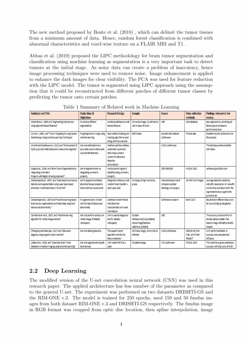
The new method proposed by Bonte et al. (2018) , which can delimit the tumor tissuesfrom a minimum amount of data. Hence, random forest classification is combined withabnormal characteristics and voxel-wise texture on a FLAIR MRI and T1.
Abbas et al. (2019) proposed the LIPC methodology for brain tumor segmentation andclassification using machine learning as segmentation is a very important task to detecttumors at the initial stage. As noisy data can create a problem of inaccuracy, henceimage processing techniques were used to remove noise. Image enhancement is appliedto enhance the dark images for clear visibility. The PCA was used for feature reductionwith the LIPC model. The tumor is segmented using LIPC approach using the assump-tion that it could be reconstructed from different patches of different tumor classes bypredicting the tumor onto certain patches.
Table 1 Summary of Related work in Machine Learning
2.2 Deep Learning
The modified version of the U-net convolution neural network (CNN) was used in thisresearch paper. The applied architecture has less number of the parameter as comparedto the general U-net. The experiment was performed on two datasets DRISHTI-GS andthe RIM-ONE v.3. The model is trained for 250 epochs, used 159 and 50 fundus im-ages from both dataset RIM-ONE v.3 and DRISHTI-GS respectively. The fundus imagein RGB format was cropped from optic disc location, then spline interpolation, image
4

resizing of 128x128, and histogram equalization were performed before feeding it into aneural network in (Joshua et al.; 2019a) proposed study. The IOU score and dice scorefor RIM-ONE v.3 dataset performed better than DRISHTI-GS database.
The new approach to automatically segment optic disc and cup in characterizing thepresence of glaucoma from Region of interest (ROI). Two steps was followed in this studyto estimate ratio: one is ROI from a fundus image then followed by the segmentationprocess. The researcher applied fully convolutional networks (FCN) along with U-Netarchitectures for their study to segment optic cup and disc. The binary and multi-classfully convolution networks are implemented by the researcher along with two differentRegions of interest as an input, one is the original image (Original ROI) and another isthe masked image (masked ROI) to get a result in the segmentation process. Binary classFCNs show better performance compared to multi-class FCNs. Also, the full convolutionnetwork for optic cup and disc segmentation performed better in ACC, f-measure, andJaccard index than existing other algorithms in performance. The applied FCNs tech-nique showed a promising result (Kim et al.; 2019).
Interpretable Glaucoma detector (InterGD) is a multitask model that was implemen-ted by Mojab et al. (2019). This InterGB consists of two-component. One componentis segmentation modules which used to segment the optic disc and cup from fundus im-age and another component is prediction modules employed in this study to predict thelikelihood of glaucoma in the image. Both components are integrated with a multi-taskframework to perform end to end training. The study is performed on three datasets,among which two are public datasets, DRISHTI-GS and RIGA and one is private datasetI-ODA. Mojab et al. (2019) used five models as a baseline. As a part of data prepro-cessing, resizing and channel-wise normalization techniques were introduced to all images.Data were divided into training and testing with 80% and 20% respectively. The follow-ing evaluation technique was used to evaluate both the model such as recall, f1-score,and precision for evaluating prediction model, Disc similarity coefficient (DSC) for thesegmentation model. Through this study, the capabilities of InterGD tries to resolve theexisting challenges by gaining interpretability at a minimal cost.
Qian et al. (2019) The researcher has used Mask RCNN model for object detection andobserved this algorithm outperformed in detecting objects as compared to other start-of-art models with an accuracy of 96.8 percent that to achieved in 116ms. The study isperformed on SAR images to locate ships in multitemporal. Both multiple and singlemasks are used in the experiment. The denoising and data augmentation was used aspre-processing. As a result, it was noticed that multi-mask proven to be more effectivethan a single- mask in detecting SAR ship. The study showed Mask RCNN can detectan object sufficiently within less time consumption for SAR image in ship detection.
The detailed comparison between FCNs and Mask RCNNs was implemented in the con-text of salient object detection (SOD) by Krinski et al. (2019). The architecture of boththe FCNs and mask R-CNN is explored in this study to detect the salient area fromthe image. The study aimed to demonstrate effectively Mask RCNNs dominance overFCNs in the SOD images and proved Mask RCNN achieved better performance level overFCNs on F-measures gain of up to 47%. The experiment was performed over 8 publicallyavailable datasets and four metrics were used for evaluation such as precision, f-measure,
5
recall, and MAE (Mean Absolute Error). The Mask RCNN has an RPN which is attachedbefore the segmentation module. This decreases the false-negative error that results inimproving segmentation. The researcher endorses the Mask RCNN is a promising solu-tion for SOD problem-solving.
There are different segmentation approaches and algorithms. Each has its advantageand limitation. Madhupriya et al. (2019) has proposed the U-net model, which is basedon a deep convolution network. BRATS 2017 datasets were used, which contain real im-ages of High-Grade Glioma (HGG) and Low Grade Glioma (LGG) patients. The modelshowed an efficient and robust result by achieving maximum DSC(Dice Similarity Coef-ficient ) metric of 0.82 and 0.81 for the BRATS dataset.
Another, study empirical on medical is nuclei segmentation. The U-net and Mask-RCNN are two popular segmentation framework was introduced by previous research.An ensemble model, by combining both models is proposed in (Vuola et al.; 2019). Thispresented the improvement in results by merging the predictive power of U-net and maskRCNN.
Kassani et al. (2019) approached a novel CNN model with Xception architecture in itfor classification of DR severity. The Xception is aggregated with a layer of deep CNN.In pre-processing min-pooling is employed on input image for color contrast. This studyperformed no data augmentation for training model. The overfitting issues are resolvedusing L1 and L2 regularization. This Xception deep extractor showed better perform-ance in comparison with original Xception architecture on APTOS dataset. This hasgiven promising results on the limited number of training data through this approach ofresearch with accuracy, sensitivity, a specificity of 83.09%, 88.24%, 87% respectively.
Glaucoma detection using AG-CNN (Attention-based CNN model) was implemented inLi et al. (2020). The LAG Large-scaled labelled dataset was used for the research. Theirconcept was to bridge the gap between pathological area and human attention, hence thestudy proposed to improve the projected focus maps by adding a feature analysis frame-work for the identification of glaucoma. It was learned in a weakly supervised manner.The back-propagation method is employed to identify the pathological area which wasbased on the predicted attention map. As a result, the focus maps could be optimizedand then used to illustrate the area which was most important in detecting glaucoma.The proposed attention-based mechanism could find salient parts of the feature in animage. The study achieved an accuracy of 96.7%.
The quality of the fundus image is assessed in this paper Raj et al. (2020). The multivari-ate regression based CNN model is used on the FIQuA dataset. A total of 1500 images areprepared along with seven subjective inputs that are given by ophthalmologists. Amongseven, six are quality parameters and left one is class of quality as good, fair, and poor.The model peculiarity is that using the subjective feedback given by ophthalmologists, itextracts the optimized features for classification. It has two blocks, Block1 manage theoptimized functionality from 4 pre-trained CNN models. There models as ResNet-151,Xception Inception-V3, and DenseNet-121 trained against the six subjective scores givenby the ophthalmologists through transfer learning. Alternatively, these streamlined fea-tures are integrated and forwarded Block-2 classifies the background images into three
6
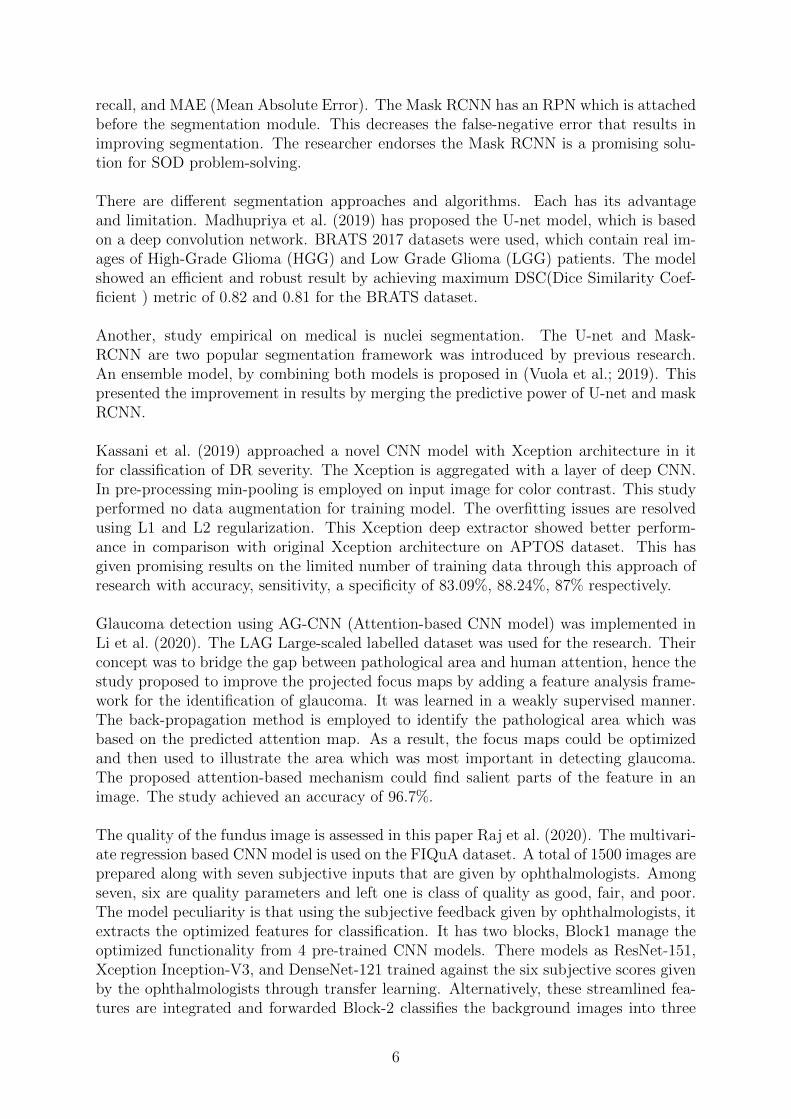
classes (good, fair, bad). The block2 technique achieved 95.66% accuracy.
Recently, the application of deep learning Mask RCNN expounds in the dental areaof medical. Zhu et al. (2020) performed tooth detection and segmentation in their studythat would automatically detect and segment the tooth in the mouth. Since no publicallydataset was available, therefore 100 images from the private hospital were considered forthe experiment, among training is carried out on 80 images, 10 for validation, and theremaining 10 used for testing. The model is trained for 50 epochs and observed a totalloss of 0.3093. In terms of accuracy, Pixel accuracy showed a result of 97.4%. It can bestated as in future applied technique is merged with other more networks to predict thepresence of diseases like orthodontics.
Table 2: Summary of Related work in Deep Learning
3 Methodology
The proposed methodology automatic segment and detect the optic cup and optic discfrom fundus images. This research implemented an advanced technique based on previousresearch Qin et al. (2019); Joshua et al. (2019a). The proposed research process segmentsthe optic disc and cup simultaneously from the input image. Both cups and discs at thesame could be detected in this research method. The Mask RCNN deep neural networkarchitecture is being utilized in this research. The CRISP-DM methodology is used inthis study.
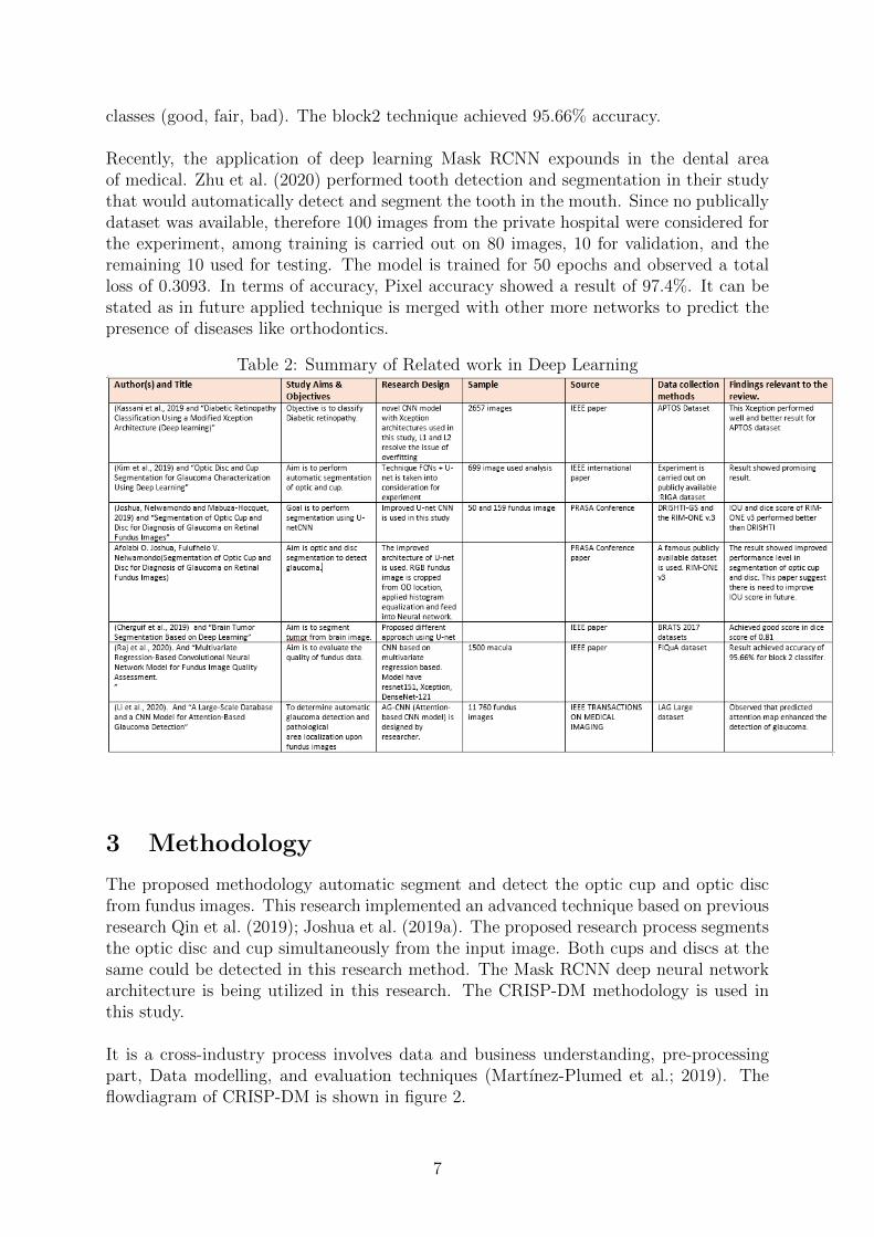
It is a cross-industry process involves data and business understanding, pre-processingpart, Data modelling, and evaluation techniques (Martınez-Plumed et al.; 2019). Theflowdiagram of CRISP-DM is shown in figure 2.
7
Figure 2: CRISP-DM methodology
3.1 Business Understanding
Glaucoma can be prevented from getting complete loss of vision if the early stage of it isdetected. It requires a precise method to segment cup and disc. Doctor and pathologistevaluate fundus images of person after performing pre-processing of images. The patho-logist finds estimating fundus image under the microscope is time-consuming and it ishighly affected by doctor experience in this field. Therefore, as discussed Mask RCNNmodel is used to reduce the time and manual human efforts required for evaluating thefundus image of Glaucoma. The segmentation is a tough and challenging task to getprecise segmentation of optic cup and disc. Thus, the proposed method is applied toovercome this problem.
3.2 Data Understanding and Collection
This research is performed on RIGA-dataset, which is extracted from open source ”DeepBlue Data” repository. The link for RIGA dataset is https://deepblue.lib.umich.
edu/data/concern/data_sets/3b591905z. The dataset consists of fundus images of theeye, provided by the University of Michigan. This data has three sets such as Magrabi,Bin Rushed, and MESSIDOR. The data is tested by high professional ophthalmologists.All fundus image consists of the optic cup and optic disc is marked and annotated by sixexperienced ophthalmologists. The data consists of high-quality TIFF and JPG formattotal 4500 fundus image. The size of a fundus image is 2160x1440.
3.3 Data Preparation
This is a prominent step to perform on images before training the model. In the dataset,the images are pathologically labelled. In the data cleaning, unlabelled fundus images areremoved and 1400 labelled images are kept. As different resolution of images may causea model to train inaccurately, thus high-resolution images are sized to 512 x 512 shapeto bring all images in the dataset at the same level. The Edge enhancement is applied inorder to enhance the salient portion from an image. The effect of edge enhancement canbe seen in figure 3.
8
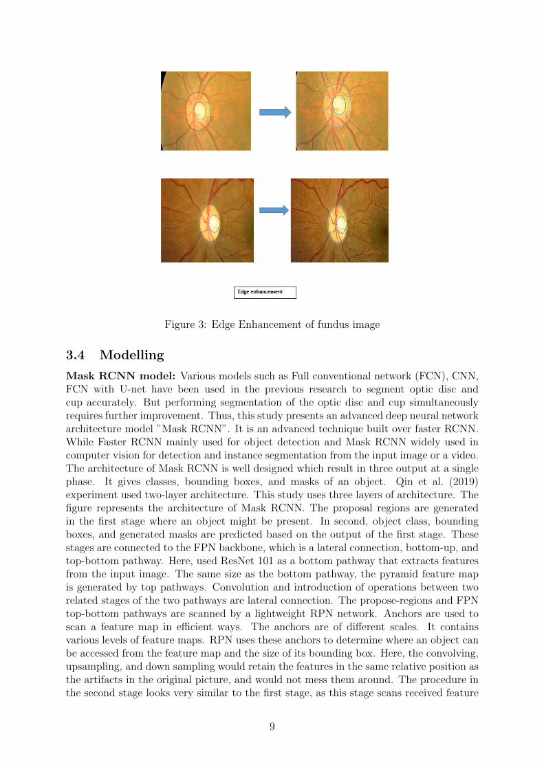
Figure 3: Edge Enhancement of fundus image
3.4 Modelling
Mask RCNN model: Various models such as Full conventional network (FCN), CNN,FCN with U-net have been used in the previous research to segment optic disc andcup accurately. But performing segmentation of the optic disc and cup simultaneouslyrequires further improvement. Thus, this study presents an advanced deep neural networkarchitecture model ”Mask RCNN”. It is an advanced technique built over faster RCNN.While Faster RCNN mainly used for object detection and Mask RCNN widely used incomputer vision for detection and instance segmentation from the input image or a video.The architecture of Mask RCNN is well designed which result in three output at a singlephase. It gives classes, bounding boxes, and masks of an object. Qin et al. (2019)experiment used two-layer architecture. This study uses three layers of architecture. Thefigure represents the architecture of Mask RCNN. The proposal regions are generatedin the first stage where an object might be present. In second, object class, boundingboxes, and generated masks are predicted based on the output of the first stage. Thesestages are connected to the FPN backbone, which is a lateral connection, bottom-up, andtop-bottom pathway. Here, used ResNet 101 as a bottom pathway that extracts featuresfrom the input image. The same size as the bottom pathway, the pyramid feature mapis generated by top pathways. Convolution and introduction of operations between tworelated stages of the two pathways are lateral connection. The propose-regions and FPNtop-bottom pathways are scanned by a lightweight RPN network. Anchors are used toscan a feature map in efficient ways. The anchors are of different scales. It containsvarious levels of feature maps. RPN uses these anchors to determine where an object canbe accessed from the feature map and the size of its bounding box. Here, the convolving,upsampling, and down sampling would retain the features in the same relative position asthe artifacts in the original picture, and would not mess them around. The procedure inthe second stage looks very similar to the first stage, as this stage scans received feature
9
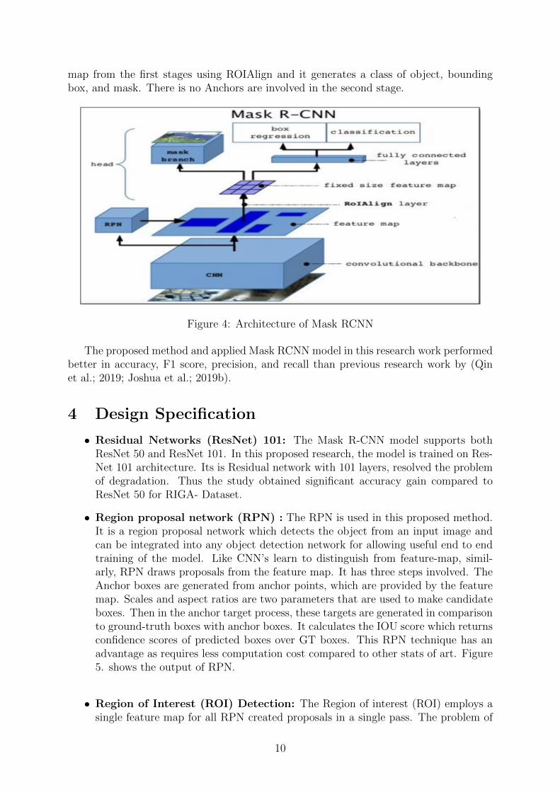
map from the first stages using ROIAlign and it generates a class of object, boundingbox, and mask. There is no Anchors are involved in the second stage.
Figure 4: Architecture of Mask RCNN
The proposed method and applied Mask RCNN model in this research work performedbetter in accuracy, F1 score, precision, and recall than previous research work by (Qinet al.; 2019; Joshua et al.; 2019b).
4 Design Specification
• Residual Networks (ResNet) 101: The Mask R-CNN model supports bothResNet 50 and ResNet 101. In this proposed research, the model is trained on Res-Net 101 architecture. Its is Residual network with 101 layers, resolved the problemof degradation. Thus the study obtained significant accuracy gain compared toResNet 50 for RIGA- Dataset.
• Region proposal network (RPN) : The RPN is used in this proposed method.It is a region proposal network which detects the object from an input image andcan be integrated into any object detection network for allowing useful end to endtraining of the model. Like CNN’s learn to distinguish from feature-map, simil-arly, RPN draws proposals from the feature map. It has three steps involved. TheAnchor boxes are generated from anchor points, which are provided by the featuremap. Scales and aspect ratios are two parameters that are used to make candidateboxes. Then in the anchor target process, these targets are generated in comparisonto ground-truth boxes with anchor boxes. It calculates the IOU score which returnsconfidence scores of predicted boxes over GT boxes. This RPN technique has anadvantage as requires less computation cost compared to other stats of art. Figure5. shows the output of RPN.
• Region of Interest (ROI) Detection: The Region of interest (ROI) employs asingle feature map for all RPN created proposals in a single pass. The problem of
10
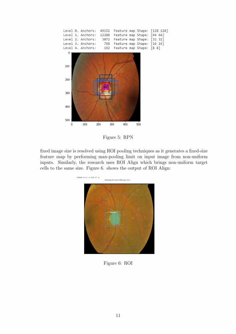
Figure 5: RPN
fixed image size is resolved using ROI pooling techniques as it generates a fixed-sizefeature map by performing max-pooling limit on input image from non-uniforminputs. Similarly, the research uses ROI Align which brings non-uniform targetcells to the same size. Figure 6. shows the output of ROI Align:
Figure 6: ROI
11
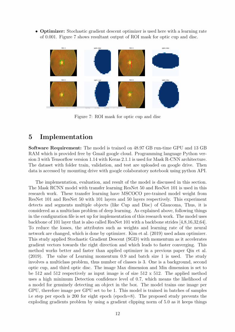
• Optimizer: Stochastic gradient descent optimizer is used here with a learning rateof 0.001. Figure 7 shows resultant output of ROI mask for optic cup and disc.
Figure 7: ROI mask for optic cup and disc
5 Implementation
Software Requirement: The model is trained on 48.97 GB run-time GPU and 13 GBRAM which is provided free by Gmail google cloud. Programming language Python ver-sion 3 with Tensorflow version 1.14 with Keras 2.1.1 is used for Mask R-CNN architecture.The dataset with folder train, validation, and test are uploaded on google drive. Thendata is accessed by mounting drive with google colaboratory notebook using python API.
The implementation, evaluation, and result of the model is discussed in this section.The Mask RCNN model with transfer learning ResNet 50 and ResNet 101 is used in thisresearch work. These transfer learning have MSCOCO pre-trained model weight fromResNet 101 and ResNet 50 with 101 layers and 50 layers respectively. This experimentdetects and segments multiple objects (like Cup and Disc) of Glaucoma, Thus, it isconsidered as a multiclass problem of deep learning. As explained above, following thingsin the configuration file is set up for implementation of this research work. The model usesbackbone of 101 layer that is also called ResNet 101 with a backbone strides [4,8,16,32,64].To reduce the losses, the attributes such as weights and learning rate of the neuralnetwork are changed, which is done by optimizer. Kim et al. (2019) used adam optimizer.This study applied Stochastic Gradient Descent (SGD) with momentum as it acceleratesgradient vectors towards the right direction and which leads to faster converging. Thismethod works better and faster than applied optimizer in a previous paper Qin et al.(2019). The value of Learning momentum 0.9 and batch size 1 is used. The studyinvolves a multiclass problem, thus number of classes is 3. One is a background, secondoptic cup, and third optic disc. The image Max dimension and Min dimension is set tobe 512 and 512 respectively as input image is of size 512 x 512. The applied methoduses a high minimum Detection confidence level of 0.7, which means the likelihood ofa model for genuinely detecting an object in the box. The model trains one image perGPU, therefore image per GPU set to be 1. This model is trained in batches of samplesi.e step per epoch is 200 for eight epoch (epoch=8). The proposed study prevents theexploding gradients problem by using a gradient clipping norm of 5.0 as it keeps things
12
stable. Another parameter is Weight decay is used. It prevents unnecessary weight gain.For the training model, datasets is split into 80 and 20 percent for train and validationset.
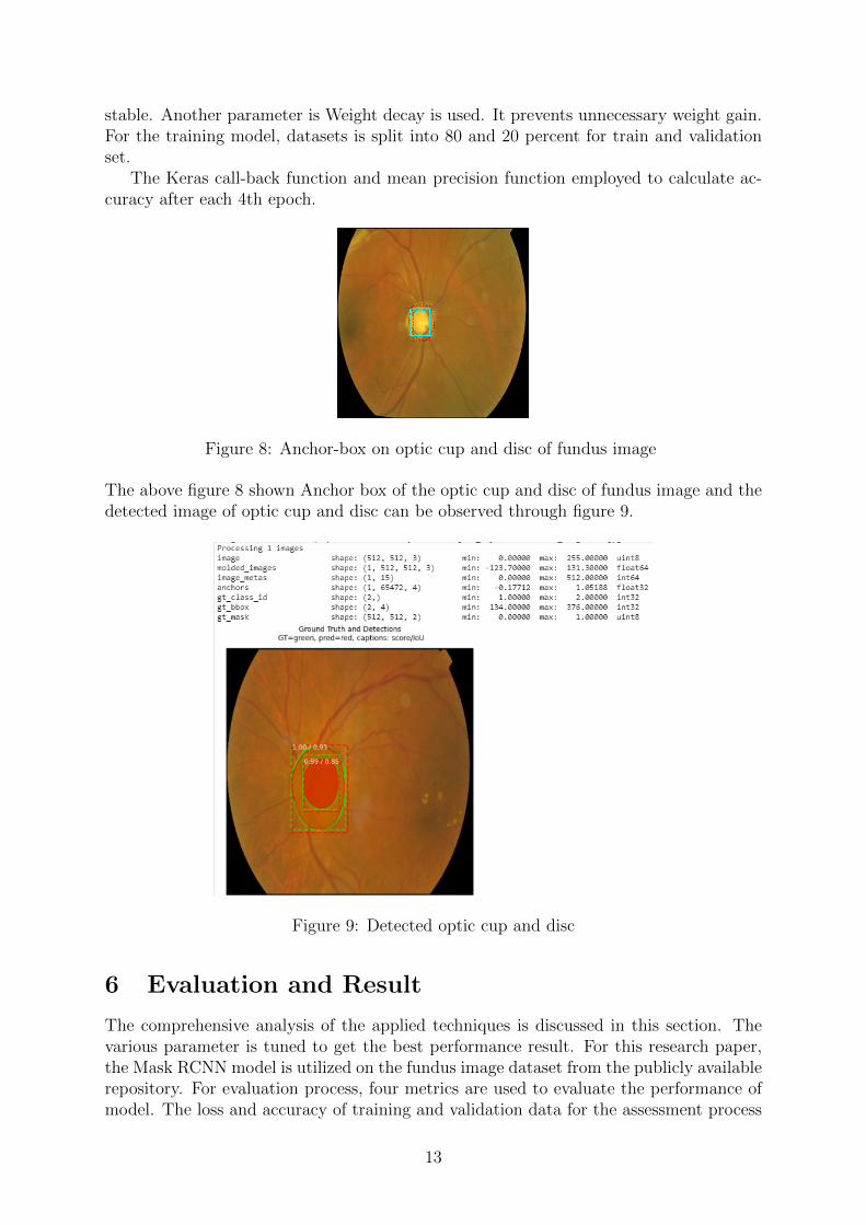
The Keras call-back function and mean precision function employed to calculate ac-curacy after each 4th epoch.
Figure 8: Anchor-box on optic cup and disc of fundus image
The above figure 8 shown Anchor box of the optic cup and disc of fundus image and thedetected image of optic cup and disc can be observed through figure 9.
Figure 9: Detected optic cup and disc
6 Evaluation and Result
The comprehensive analysis of the applied techniques is discussed in this section. Thevarious parameter is tuned to get the best performance result. For this research paper,the Mask RCNN model is utilized on the fundus image dataset from the publicly availablerepository. For evaluation process, four metrics are used to evaluate the performance ofmodel. The loss and accuracy of training and validation data for the assessment process
13
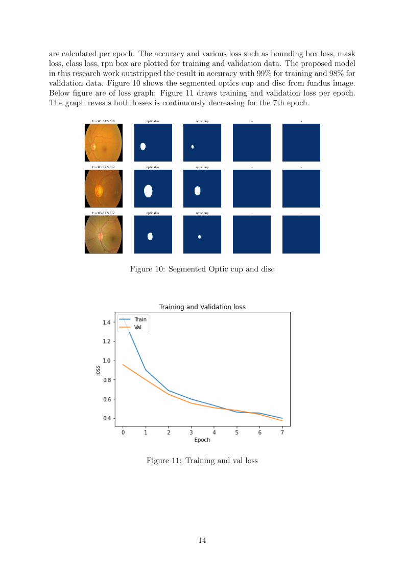
are calculated per epoch. The accuracy and various loss such as bounding box loss, maskloss, class loss, rpn box are plotted for training and validation data. The proposed modelin this research work outstripped the result in accuracy with 99% for training and 98% forvalidation data. Figure 10 shows the segmented optics cup and disc from fundus image.Below figure are of loss graph: Figure 11 draws training and validation loss per epoch.The graph reveals both losses is continuously decreasing for the 7th epoch.
Figure 10: Segmented Optic cup and disc
Figure 11: Training and val loss
14
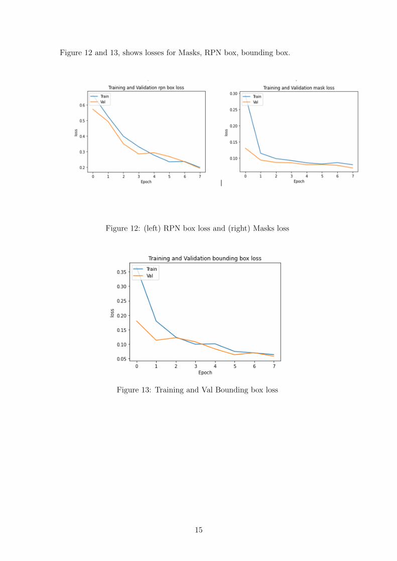
Figure 12 and 13, shows losses for Masks, RPN box, bounding box.
Figure 12: (left) RPN box loss and (right) Masks loss
Figure 13: Training and Val Bounding box loss
15
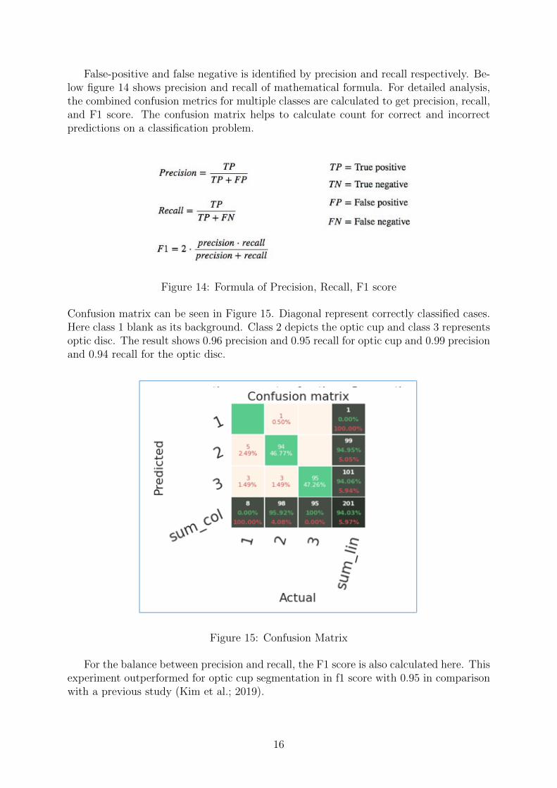
False-positive and false negative is identified by precision and recall respectively. Be-low figure 14 shows precision and recall of mathematical formula. For detailed analysis,the combined confusion metrics for multiple classes are calculated to get precision, recall,and F1 score. The confusion matrix helps to calculate count for correct and incorrectpredictions on a classification problem.
Figure 14: Formula of Precision, Recall, F1 score
Confusion matrix can be seen in Figure 15. Diagonal represent correctly classified cases.Here class 1 blank as its background. Class 2 depicts the optic cup and class 3 representsoptic disc. The result shows 0.96 precision and 0.95 recall for optic cup and 0.99 precisionand 0.94 recall for the optic disc.
Figure 15: Confusion Matrix
For the balance between precision and recall, the F1 score is also calculated here. Thisexperiment outperformed for optic cup segmentation in f1 score with 0.95 in comparisonwith a previous study (Kim et al.; 2019).
16
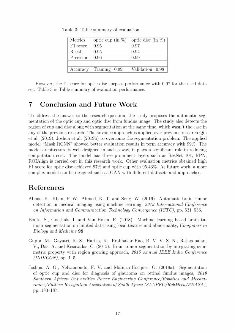
Table 3: Table summary of evaluation
Metrics optic cup (in %) optic disc (in %)F1 score 0.95 0.97Recall 0.95 0.94Precision 0.96 0.99
Accuracy Training=0.99 Validation=0.98
However, the f1 score for optic disc surpass performance with 0.97 for the used dataset. Table 3 is Table summary of evaluation performance.
7 Conclusion and Future Work
To address the answer to the research question, the study proposes the automatic seg-mentation of the optic cup and optic disc from fundus image. The study also detects theregion of cup and disc along with segmentation at the same time, which wasn’t the case inany of the previous research. The advance approach is applied over previous research Qinet al. (2019); Joshua et al. (2019b) to overcome the segmentation problem. The appliedmodel “Mask RCNN” showed better evaluation results in term accuracy with 99%. Themodel architecture is well designed in such a way, it plays a significant role in reducingcomputation cost. The model has three prominent layers such as ResNet 101, RPN,ROIAlign is carried out in this research work. Other evaluation metrics obtained highF1 score for optic disc achieved 97% and optic cup with 95.43%. As future work, a morecomplex model can be designed such as GAN with different datasets and approaches.
References
Abbas, K., Khan, P. W., Ahmed, K. T. and Song, W. (2019). Automatic brain tumordetection in medical imaging using machine learning, 2019 International Conferenceon Information and Communication Technology Convergence (ICTC), pp. 531–536.
Bonte, S., Goethals, I. and Van Holen, R. (2018). Machine learning based brain tu-mour segmentation on limited data using local texture and abnormality, Computers inBiology and Medicine 98.
Gupta, M., Gayatri, K. S., Harika, K., Prabhakar Rao, B. V. V. S. N., Rajagopalan,V., Das, A. and Kesavadas, C. (2015). Brain tumor segmentation by integrating sym-metric property with region growing approach, 2015 Annual IEEE India Conference(INDICON), pp. 1–5.
Joshua, A. O., Nelwamondo, F. V. and Mabuza-Hocquet, G. (2019a). Segmentationof optic cup and disc for diagnosis of glaucoma on retinal fundus images, 2019Southern African Universities Power Engineering Conference/Robotics and Mechat-ronics/Pattern Recognition Association of South Africa (SAUPEC/RobMech/PRASA),pp. 183–187.
17

Joshua, A. O., Nelwamondo, F. V. and Mabuza-Hocquet, G. (2019b). Segmenta-tion of optic cup and disc for diagnosis of glaucoma on retinal fundus images, 2019Southern African Universities Power Engineering Conference/Robotics and Mechat-ronics/Pattern Recognition Association of South Africa, pp. 183–187.
Kassani, S. H., Kassani, P. H., Khazaeinezhad, R., Wesolowski, M. J., Schneider, K. A.and Deters, R. (2019). Diabetic retinopathy classification using a modified xception ar-chitecture, 2019 IEEE International Symposium on Signal Processing and InformationTechnology (ISSPIT), pp. 1–6.
Kim, J., Tran, L., Chew, E. Y. and Antani, S. (2019). Optic disc and cup segmenta-tion for glaucoma characterization using deep learning, 2019 IEEE 32nd InternationalSymposium on Computer-Based Medical Systems (CBMS), pp. 489–494.
Krinski, B. A., Ruiz, D. V., Machado, G. Z. and Todt, E. (2019). Masking salient objectdetection, a mask region-based convolutional neural network analysis for segmentationof salient objects, 2019 Latin American Robotics Symposium (LARS), 2019 BrazilianSymposium on Robotics (SBR) and 2019 Workshop on Robotics in Education (WRE),pp. 55–60.
Li, L., Xu, M., Liu, H., Li, Y., Wang, X., Jiang, L., Wang, Z., Fan, X. and Wang, N.(2020). A large-scale database and a cnn model for attention-based glaucoma detection,IEEE Transactions on Medical Imaging 39(2): 413–424.
Lin, T., Cervino, L., Tang, X., Vasconcelos, N. and Jiang, S. B. (2008). Tumor tar-geting for lung cancer radiotherapy using machine learning techniques, 2008 SeventhInternational Conference on Machine Learning and Applications, pp. 533–538.
Madhupriya, G., Guru, N. M., Praveen, S. and Nivetha, B. (2019). Brain tumor seg-mentation with deep learning technique, 2019 3rd International Conference on Trendsin Electronics and Informatics (ICOEI), pp. 758–763.
Martınez-Plumed, F., Contreras-Ochando, L., Ferri, C., Hernandez Orallo, J., Kull, M.,Lachiche, N., Ramırez Quintana, M. J. and Flach, P. A. (2019). Crisp-dm twenty yearslater: From data mining processes to data science trajectories, IEEE Transactions onKnowledge and Data Engineering pp. 1–1.
Mojab, N., Noroozi, V., Yu, P. S. and Hallak, J. A. (2019). Deep multi-task learningfor interpretable glaucoma detection, 2019 IEEE 20th International Conference onInformation Reuse and Integration for Data Science (IRI), pp. 167–174.
Murthi, A. and Madheswaran, M. (2012). Enhancement of optic cup to disc ratio detec-tion in glaucoma diagnosis, 2012 International Conference on Computer Communica-tion and Informatics, pp. 1–5.
Qian, Y., Liu, Q., Zhu, H., Fan, H., Du, B. and Liu, S. (2019). Mask r-cnn for ob-ject detection in multitemporal sar images, 2019 10th International Workshop on theAnalysis of Multitemporal Remote Sensing Images (MultiTemp), pp. 1–4.
Qin, P., Wang, L. and Lv, H. (2019). Optic disc and cup segmentation based on deeplearning, 2019 IEEE 3rd Information Technology, Networking, Electronic and Automa-tion Control Conference (ITNEC), pp. 1835–1840.
18
Raj, A., Shah, N. A., Tiwari, A. K. and Martini, M. G. (2020). Multivariate regression-based convolutional neural network model for fundus image quality assessment, IEEEAccess 8: 57810–57821.
Santhakumar R, Tandur, M., Rajkumar, E. R., Geetha K S, Haritz, G. and Rajamani,K. T. (2016). Machine learning algorithm for retinal image analysis, 2016 IEEE Region10 Conference (TENCON), pp. 1236–1240.
Schmidt, M., Levner, I., Greiner, R., Murtha, A. and Bistritz, A. (2005). Segment-ing brain tumors using alignment-based features, Fourth International Conference onMachine Learning and Applications (ICMLA’05), pp. 6 pp.–.
Soltaninejad, M., Yang, G., Lambrou, T., Allinson, N., Jones, T., Barrick, T., Howe,F. and Ye, X. (2016). Automated brain tumour detection and segmentation usingsuperpixel-based extremely randomized trees in flair mri, International Journal of Com-puter Assisted Radiology and Surgery 12.
Thangaraj, V. and Natarajan, V. (2017). Glaucoma diagnosis using support vector ma-chine, 2017 International Conference on Intelligent Computing and Control Systems(ICICCS), pp. 394–399.
Vuola, A. O., Akram, S. U. and Kannala, J. (2019). Mask-rcnn and u-net ensembled fornuclei segmentation, 2019 IEEE 16th International Symposium on Biomedical Imaging(ISBI 2019), pp. 208–212.
Zhou, J., Chan, K. L., Chong, V. F. H. and Krishnan, S. M. (2005). Extraction of braintumor from mr images using one-class support vector machine, 2005 IEEE Engineeringin Medicine and Biology 27th Annual Conference, pp. 6411–6414.
Zhu, G., Piao, Z. and Kim, S. C. (2020). Tooth detection and segmentation with mask r-cnn, 2020 International Conference on Artificial Intelligence in Information and Com-munication (ICAIIC), pp. 070–072.
19
Copyright © 2022 FDOKUMEN