Essential Role of LAT in T Cell Development
10
Immunity, Vol. 10, 323–332, March, 1999, Copyright 1999 by Cell Press Essential Role of LAT in T Cell Development ITAM creates the binding sites for the tandem SH2 do- Weiguo Zhang,*# Connie L. Sommers, ² # mains of Syk family PTKs, Syk, or ZAP-70. Syk and ZAP- Deborah N. Burshtyn, ‡ Christopher C. Stebbins, ‡ 70, when bound to phosphorylated ITAMs, are further Jan B. DeJarnette, ² Ronald P. Trible,* activated by Src family kinases. Thus, the hallmark of Alexander Grinberg, ² Henry C. Tsay, ² the activated immunoreceptor is its association with Helena M. Jacobs, § Craig M. Kessler, § Eric O. Long, ‡ active PTKs. Paul E. Love, ² and Lawrence E. Samelson* k Though the effector functions of these various hema- * Section on Lymphocyte Signaling topoietic cells may differ widely, the various immunore- Cell Biology and Metabolism Branch ceptors couple to similar signaling pathways. Tyrosine ² Laboratory of Mammalian Genes and Development phosphorylation of phospholipase-Cg isoforms is ob- National Institute of Child Health and served after activation of these receptors. Activation of Human Development phospholipase-Cg isoforms leads via phosphoinositide ‡ Laboratory of Immunogenetics cleavage to calcium elevation and PKC activation. En- National Institute of Allergy and Infectious Diseases gagement of these receptors also leads to activation of National Institutes of Health small G proteins such as Ras and Rac. The details of Bethesda, Maryland 20892 how the immunoreceptors couple to these signaling § Division of Hematology-Oncology pathways are now under intense investigation. For these Lombardi Cancer Center different receptor systems, a central question is how Georgetown University Medical Center proteins in the signaling cascades are brought from the Washington, District of Columbia 20007 cytosol and elsewhere in the membrane to the activated receptors. A number of cytosolic linker or adaptor mole- cules have been described that directly bind additional Summary linker proteins and effectors. These include the ubiqui- tous Grb2, which binds SOS, SLP-76 and Cbl, and the The linker molecule LAT is a substrate of the tyrosine Grb2-related molecules Grap and Gads, which have sim- kinases activated following TCR engagement. Phos- ilar binding properties. The linker molecule SLP-76 per- phorylated LAT binds many critical signaling mole- forms its function by binding Vav, SLAP, and other pro- cules. The central role of this molecule in TCR-medi- teins (Liu et al., 1999; Peterson et al., 1998; Rudd, 1999; ated signaling has been demonstrated by experiments Samelson, 1999). In T cells these complexes have been in a LAT-deficient cell line. To probe the role of LAT shown to bind the integral membrane adaptor molecule in T cell development, the LAT gene was disrupted by LAT (Weber et al., 1998; Zhang et al., 1998a). How these targeting. LAT-deficient mice appeared healthy. Flow LAT-associated protein complexes comprised of en- cytometric analysis revealed normal B cell populations zymes and adaptor molecules are brought to immunore- but the absence of any mature peripheral T cells. Intra- ceptors is now an important area of study. thymic development was blocked within the CD4 2 CD8 2 LAT is a 36–38 kDa palmitoylated, integral membrane stage. No gross abnormality of NK or platelet function adaptor protein expressed in T cells, mast cells, NK was observed. LAT is thus critical to both T cell activa- cells, and megakaryocytes (Weber et al., 1998; Zhang tion and development. et al., 1998a, 1998b; Facchetti et al., 1999). It is not found in B cells or other cell types. Limited biochemical Introduction evidence shows that a 36–38 kDa protein, which is likely to be LAT, is phosphorylated on tyrosine residues in Immunoreceptors for antigen and immunoglobulin ex- mast cells upon FceRI engagement and in NK cells fol- pressed on several lineages of hematopoietic cells share lowing target interaction (Turner et al., 1995; Galandrini many structural and functional features. T cell and B et al., 1996; Valiante et al., 1996; Hendricks-Taylor et al., cell antigen receptors and the Fc receptors for IgE and 1997). The function of LAT is best characterized in T IgG are comprised of multiple subunits (Ravetch and cells. Upon TCR activation, LAT is phosphorylated on Kinet, 1991; Weiss and Littman, 1994; Chan and Shaw, multiple tyrosines, most likely by the ZAP-70 PTK. LAT 1996; Wange and Samelson, 1996; Buhl and Cambier, then associates with PLC-g1, Cbl, Vav, PI-3 kinase, SLP- 1997). Some of these components are dedicated to li- 76, Grb2, and other proteins directly or indirectly. Muta- gand binding, while other subunits with long cytosolic tion of two critical LAT tyrosines to phenylalanine abol- tails contain immunoreceptor tyrosine-based activation ishes the binding to these molecules. Overexpression motifs (ITAMs) that are subject to rapid phosphorylation. of this mutant LAT molecule in Jurkat cells inhibits AP-1 Members of the Src family of protein tyrosine kinases and NF-AT-directed transcriptional activation mediated (PTK) directly interact with these receptors or corecep- through the TCR, suggesting its important role in T cell tors and are primarily responsible for ITAM phosphoryla- activation (Zhang et al., 1998a). As further confirmation tion. Phosphorylation of the two tyrosines within an of the function of LAT, in LAT-deficient Jurkat cells the tyrosine phosphorylation of PLC-g1, Vav, and SLP-76 is reduced. Ca 21 mobilization, activation of MAPK, and k To whom correspondence should be addressed (e-mail: samelson@ activation of AP-1 and NF-AT transcription factors are helix.nih.gov). # These authors contributed equally to this work. also defective in these cells. Reintroduction of LAT can
-
Upload
independent -
Category
Documents
-
view
0 -
download
0
Transcript of Essential Role of LAT in T Cell Development

Immunity, Vol. 10, 323–332, March, 1999, Copyright 1999 by Cell Press
Essential Role of LAT in T Cell Development
ITAM creates the binding sites for the tandem SH2 do-Weiguo Zhang,*# Connie L. Sommers,†#mains of Syk family PTKs, Syk, or ZAP-70. Syk and ZAP-Deborah N. Burshtyn,‡ Christopher C. Stebbins,‡70, when bound to phosphorylated ITAMs, are furtherJan B. DeJarnette,† Ronald P. Trible,*activated by Src family kinases. Thus, the hallmark ofAlexander Grinberg,† Henry C. Tsay,†the activated immunoreceptor is its association withHelena M. Jacobs,§ Craig M. Kessler,§ Eric O. Long,‡active PTKs.Paul E. Love,† and Lawrence E. Samelson*‖
Though the effector functions of these various hema-*Section on Lymphocyte Signalingtopoietic cells may differ widely, the various immunore-Cell Biology and Metabolism Branchceptors couple to similar signaling pathways. Tyrosine†Laboratory of Mammalian Genes and Developmentphosphorylation of phospholipase-Cg isoforms is ob-National Institute of Child Health andserved after activation of these receptors. Activation ofHuman Developmentphospholipase-Cg isoforms leads via phosphoinositide‡Laboratory of Immunogeneticscleavage to calcium elevation and PKC activation. En-National Institute of Allergy and Infectious Diseasesgagement of these receptors also leads to activation ofNational Institutes of Healthsmall G proteins such as Ras and Rac. The details ofBethesda, Maryland 20892how the immunoreceptors couple to these signaling§Division of Hematology-Oncologypathways are now under intense investigation. For theseLombardi Cancer Centerdifferent receptor systems, a central question is howGeorgetown University Medical Centerproteins in the signaling cascades are brought from theWashington, District of Columbia 20007cytosol and elsewhere in the membrane to the activatedreceptors. A number of cytosolic linker or adaptor mole-cules have been described that directly bind additionalSummarylinker proteins and effectors. These include the ubiqui-tous Grb2, which binds SOS, SLP-76 and Cbl, and theThe linker molecule LAT is a substrate of the tyrosineGrb2-related molecules Grap and Gads, which have sim-kinases activated following TCR engagement. Phos-ilar binding properties. The linker molecule SLP-76 per-phorylated LAT binds many critical signaling mole-forms its function by binding Vav, SLAP, and other pro-cules. The central role of this molecule in TCR-medi-teins (Liu et al., 1999; Peterson et al., 1998; Rudd, 1999;ated signaling has been demonstrated by experimentsSamelson, 1999). In T cells these complexes have beenin a LAT-deficient cell line. To probe the role of LATshown to bind the integral membrane adaptor moleculein T cell development, the LAT gene was disrupted byLAT (Weber et al., 1998; Zhang et al., 1998a). How thesetargeting. LAT-deficient mice appeared healthy. FlowLAT-associated protein complexes comprised of en-cytometric analysis revealed normal B cell populationszymes and adaptor molecules are brought to immunore-
but the absence of any mature peripheral T cells. Intra-ceptors is now an important area of study.
thymic development was blocked within the CD42CD82
LAT is a 36–38 kDa palmitoylated, integral membranestage. No gross abnormality of NK or platelet function adaptor protein expressed in T cells, mast cells, NKwas observed. LAT is thus critical to both T cell activa- cells, and megakaryocytes (Weber et al., 1998; Zhangtion and development. et al., 1998a, 1998b; Facchetti et al., 1999). It is not
found in B cells or other cell types. Limited biochemicalIntroduction evidence shows that a 36–38 kDa protein, which is likely
to be LAT, is phosphorylated on tyrosine residues inImmunoreceptors for antigen and immunoglobulin ex- mast cells upon FceRI engagement and in NK cells fol-pressed on several lineages of hematopoietic cells share lowing target interaction (Turner et al., 1995; Galandrinimany structural and functional features. T cell and B et al., 1996; Valiante et al., 1996; Hendricks-Taylor et al.,cell antigen receptors and the Fc receptors for IgE and 1997). The function of LAT is best characterized in TIgG are comprised of multiple subunits (Ravetch and cells. Upon TCR activation, LAT is phosphorylated onKinet, 1991; Weiss and Littman, 1994; Chan and Shaw, multiple tyrosines, most likely by the ZAP-70 PTK. LAT1996; Wange and Samelson, 1996; Buhl and Cambier, then associates with PLC-g1, Cbl, Vav, PI-3 kinase, SLP-1997). Some of these components are dedicated to li- 76, Grb2, and other proteins directly or indirectly. Muta-gand binding, while other subunits with long cytosolic tion of two critical LAT tyrosines to phenylalanine abol-tails contain immunoreceptor tyrosine-based activation ishes the binding to these molecules. Overexpressionmotifs (ITAMs) that are subject to rapid phosphorylation. of this mutant LAT molecule in Jurkat cells inhibits AP-1Members of the Src family of protein tyrosine kinases and NF-AT-directed transcriptional activation mediated(PTK) directly interact with these receptors or corecep- through the TCR, suggesting its important role in T celltors and are primarily responsible for ITAM phosphoryla- activation (Zhang et al., 1998a). As further confirmationtion. Phosphorylation of the two tyrosines within an of the function of LAT, in LAT-deficient Jurkat cells the
tyrosine phosphorylation of PLC-g1, Vav, and SLP-76is reduced. Ca21 mobilization, activation of MAPK, and‖ To whom correspondence should be addressed (e-mail: samelson@activation of AP-1 and NF-AT transcription factors arehelix.nih.gov).
# These authors contributed equally to this work. also defective in these cells. Reintroduction of LAT can

Immunity324
Figure 1. Generation of LAT-Deficient Mice
(A) The genomic structure of murine LATgene, the XpPNT targeting construct, and thepredicted structure of the targeted locus afterhomologous recombination. The precise in-tron–exon structure of the entire LAT genehas not been determined. The open boxesindicate the exons of LAT from amino acid1–169. The first box contains the beginningof the LAT coding sequence. Exons con-taining 59 untranslated sequences have notbeen defined. Neo, neomycin resistancegene; TK, thymidine kinase; Bg, BglII; X, XbaI;B, BamHI; H, HindIII; S, SacI. The restrictionsites that are struck through are the sites thatwere abolished during subcloning.(B) Southern analysis of genomic DNA fromlittermates of LAT heterozygous (1/2) mat-ing. Genomic DNA was isolated from tails oflittermates and digested with BglII. DNA frag-ments were separated by electrophoresis,transferred to nylon membrane, and analyzedusing the probe as indicated in (A).(C) Analysis of LAT expression in thymocytesfrom LAT1/2 and LAT2/2 mice. Lysates from1.5 3 106 cells solubilized with Brij lysis bufferwere analyzed by SDS-PAGE and immu-noblotting with anti-LAT antiserum.
restore all these signaling defects (Finco et al., 1998). Syk-deficient mice, no bleeding disorder was noted inLAT2/2 (Cheng et al., 1995; Pivniouk et al., 1998; Clem-These results, in total, demonstrate that LAT is indis-
pensable for T cell activation via the TCR. ents et al., 1999). NK cell cytotoxic activity was normal.The most striking effect of LAT deficiency was in the TTargeted gene disruption has been widely used to
study the in vivo role of enzymes and adaptor molecules cell compartment. Mature ab and gd T cells were absentin LAT2/2 mice. Thymocyte development in LAT-defi-involved in signaling in hematopoietic cells (Pfeffer and
Mak, 1994; Bolen and Brugge, 1997; Killeen et al., 1998). cient mice showed an early arrest in the DN population,and most thymocytes were blocked at the CD251CD442A system that has been particularly amenable to such
experimentation is the analysis of T cell development. stage. These results indicate that the requirement forLAT expression is most important in the T cell lineage.This process is characterized by multiple, sequential
steps that can be easily defined by the expression of In this respect the LAT null mice are as severely impairedas those mice lacking both Lck and Fyn, both ZAP-70particular cell surface proteins. The majority of T cells
develop in the thymus through an ordered maturation and Syk, or SLP-76 (Groves et al., 1996; van Oers et al.,1996; Cheng et al., 1997; Clements et al., 1998; Pivnioukprocess, giving rise to mature T cells with antigen-spe-
cific TCRs (Fehling and von Boehmer, 1997). The earliest et al., 1998).immature T cells lack expression of CD4 or CD8 core-ceptors and are thus known as double negative (DN).
ResultsDN thymocytes express the TCR signaling subunits,TCRz, and the CD3 chains, but remain negative for TCR
Generation of LAT-Deficient Miceuntil rearrangement of the clonotypic TCR (g, d, b) genesTo generate LAT-deficient mice by targeted gene dis-commences. Successful rearrangement of the TCRgruption, the genomic fragment (from the first BamHI siteand TCRd genes results in the expression of gdTCRto the second XbaI) that contains exons 1–8 of the LATsurface complexes. gdTCR1 thymocytes remain DN andgene (encoding amino acids 1–169 region of murine LAT)exit the thymus, homing to specific sites within the pe-was replaced by a neomycin phosphotransferase generiphery. Alternatively, rearrangement and expression of(neo) in embryonic stem (ES) cells. Southern blot analy-the TCRb gene directs DN thymocytes to the ab (CD4/sis (using the probe as indicated in Figure 1A) confirmedCD8) lineage. TCRb chain associates with the invariantthe homologous recombination event in ten ES cell lines.surrogate TCRa chain to form a pre-TCR complex. Sig-Targeted ES cells were injected into blastocysts to es-naling through the pre-TCR drives the differentiation oftablish chimeric mice. Heterozygous mice (LAT1/2) wereDN cells to the CD41CD81 (double positive [DP]) stage,inbred to obtain mice homozygous for the disruptedwhere rearrangement of TCRa then leads to expressionLAT gene (LAT2/2). Transmission of the targeted alleleof the mature form of the abTCR. DP, abTCR1 thymo-was verified by Southern blot analysis of tail DNA fromcytes undergo a number of TCR-mediated selectionlittermates (Figure 1B). To confirm the absence of LATevents and ultimately differentiate into either CD41 orprotein in LAT2/2 mice, we analyzed lysates of thymo-CD81 mature single-positive (SP) T cells.cytes by immunoblotting with anti-LAT antiserum. AsTo elucidate the role of LAT in the cells in which it isshown in Figure 1C, murine LAT protein migrates as aexpressed, we generated mice deficient in LAT expres-
sion by targeted disruption. In contrast to SLP-76- or 36 kDa band instead of as a 36–38 kDa doublet observed
Essential Role of LAT in T Cell Development325
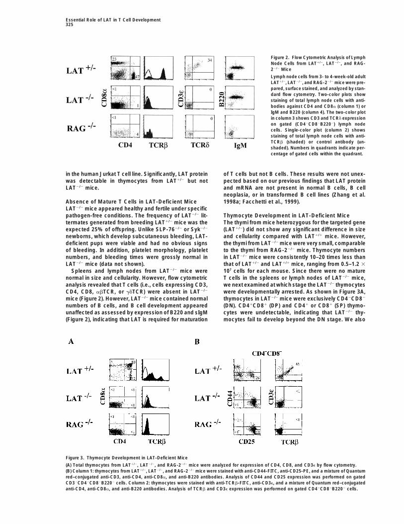
Figure 2. Flow Cytometric Analysis of LymphNode Cells from LAT1/2, LAT2/2, and RAG-22/2 Mice
Lymph node cells from 3- to 4-week-old adultLAT1/2, LAT2/2, and RAG-22/2 mice were pre-pared, surface stained, and analyzed by stan-dard flow cytometry. Two-color plots showstaining of total lymph node cells with anti-bodies against CD4 and CD8a (column 1) orIgM and B220 (column 4). The two-color plotin column 3 shows CD3 and TCRd expressionon gated (CD42CD82B2202) lymph nodecells. Single-color plot (column 2) showsstaining of total lymph node cells with anti-TCRb (shaded) or control antibody (un-shaded). Numbers in quadrants indicate per-centage of gated cells within the quadrant.
in the human Jurkat T cell line. Significantly, LAT protein of T cells but not B cells. These results were not unex-pected based on our previous findings that LAT proteinwas detectable in thymocytes from LAT1/2 but not
LAT2/2 mice. and mRNA are not present in normal B cells, B cellneoplasia, or in transformed B cell lines (Zhang et al.1998a; Facchetti et al., 1999).Absence of Mature T Cells in LAT-Deficient Mice
LAT2/2 mice appeared healthy and fertile under specificpathogen-free conditions. The frequency of LAT2/2 lit- Thymocyte Development in LAT-Deficient Mice
The thymi from mice heterozygous for the targeted genetermates generated from breeding LAT1/2 mice was theexpected 25% of offspring. Unlike SLP-762/2 or Syk2/2 (LAT1/2) did not show any significant difference in size
and cellularity compared with LAT1/1 mice. However,newborns, which develop subcutaneous bleeding, LAT-deficient pups were viable and had no obvious signs the thymi from LAT2/2 mice were very small, comparable
to the thymi from RAG-22/2 mice. Thymocyte numbersof bleeding. In addition, platelet morphology, plateletnumbers, and bleeding times were grossly normal in in LAT2/2 mice were consistently 10–20 times less than
that of LAT1/2 and LAT1/1 mice, ranging from 0.5–1.2 3LAT2/2 mice (data not shown).Spleens and lymph nodes from LAT2/2 mice were 107 cells for each mouse. Since there were no mature
T cells in the spleens or lymph nodes of LAT2/2 mice,normal in size and cellularity. However, flow cytometricanalysis revealed that T cells (i.e., cells expressing CD3, we next examined at which stage the LAT2/2 thymocytes
were developmentally arrested. As shown in Figure 3A,CD4, CD8, abTCR, or gdTCR) were absent in LAT2/2
mice (Figure 2). However, LAT2/2 mice contained normal thymocytes in LAT2/2 mice were exclusively CD42CD82
(DN). CD41CD81 (DP) and CD41 or CD81 (SP) thymo-numbers of B cells, and B cell development appearedunaffected as assessed by expression of B220 and sIgM cytes were undetectable, indicating that LAT2/2 thy-
mocytes fail to develop beyond the DN stage. We also(Figure 2), indicating that LAT is required for maturation
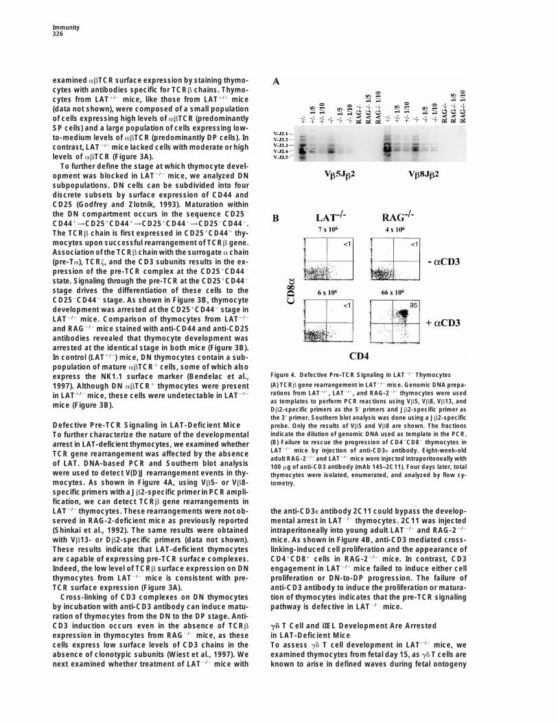
Figure 3. Thymocyte Development in LAT-Deficient Mice
(A) Total thymocytes from LAT1/2, LAT2/2, and RAG-22/2 mice were analyzed for expression of CD4, CD8, and CD3e by flow cytometry.(B) Column 1: thymocytes from LAT1/2, LAT2/2, and RAG-22/2 mice were stained with anti-CD44-FITC, anti-CD25-PE, and a mixture of Quantumred–conjugated anti-CD3, anti-CD4, anti-CD8a, and anti-B220 antibodies. Analysis of CD44 and CD25 expression was performed on gatedCD32CD42CD82B2202 cells. Column 2: thymocytes were stained with anti-TCRb-FITC, anti-CD3e, and a mixture of Quantum red–conjugatedanti-CD4, anti-CD8a, and anti-B220 antibodies. Analysis of TCRb and CD3e expression was performed on gated CD42CD82B2202 cells.
Immunity326
examined abTCR surface expression by staining thymo-cytes with antibodies specific for TCRb chains. Thymo-cytes from LAT1/2 mice, like those from LAT1/1 mice(data not shown), were composed of a small populationof cells expressing high levels of abTCR (predominantlySP cells) and a large population of cells expressing low-to-medium levels of abTCR (predominantly DP cells). Incontrast, LAT2/2 mice lacked cells with moderate or highlevels of abTCR (Figure 3A).
To further define the stage at which thymocyte devel-opment was blocked in LAT2/2 mice, we analyzed DNsubpopulations. DN cells can be subdivided into fourdiscrete subsets by surface expression of CD44 andCD25 (Godfrey and Zlotnik, 1993). Maturation withinthe DN compartment occurs in the sequence CD252
CD441→CD251CD441→CD251CD442→CD252CD442.The TCRb chain is first expressed in CD251CD441 thy-mocytes upon successful rearrangement of TCRb gene.Association of the TCRb chain with the surrogate a chain(pre-Ta), TCRz, and the CD3 subunits results in the ex-pression of the pre-TCR complex at the CD251CD442
state. Signaling through the pre-TCR at the CD251CD442
stage drives the differentiation of these cells to theCD252CD442 stage. As shown in Figure 3B, thymocytedevelopment was arrested at the CD251CD442 stage inLAT2/2 mice. Comparison of thymocytes from LAT2/2
and RAG2/2 mice stained with anti-CD44 and anti-CD25antibodies revealed that thymocyte development wasarrested at the identical stage in both mice (Figure 3B).In control (LAT1/2) mice, DN thymocytes contain a sub-population of mature abTCR1 cells, some of which also
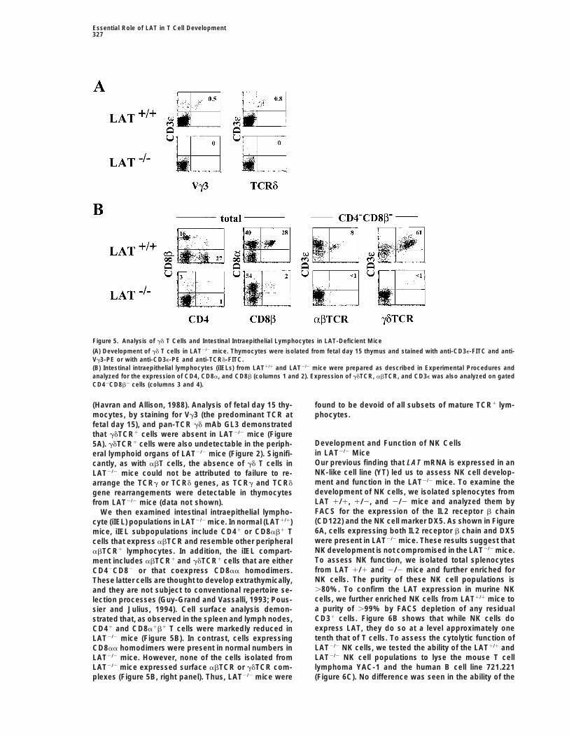
Figure 4. Defective Pre-TCR Signaling in LAT2/2 Thymocytesexpress the NK1.1 surface marker (Bendelac et al.,(A) TCRb gene rearrangement in LAT2/2 mice. Genomic DNA prepa-1997). Although DN abTCR1 thymocytes were presentrations from LAT1/2, LAT2/2, and RAG-22/2 thymocytes were usedin LAT1/2 mice, these cells were undetectable in LAT2/2
as templates to perform PCR reactions using Vb5, Vb8, Vb13, andmice (Figure 3B).Db2-specific primers as the 59 primers and Jb2-specific primer asthe 39 primer. Southern blot analysis was done using a Jb2-specific
Defective Pre-TCR Signaling in LAT-Deficient Mice probe. Only the results of Vb5 and Vb8 are shown. The fractionsindicate the dilution of genomic DNA used as template in the PCR.To further characterize the nature of the developmental(B) Failure to rescue the progression of CD42CD82 thymocytes inarrest in LAT-deficient thymocytes, we examined whetherLAT2/2 mice by injection of anti-CD3e antibody. Eight-week-oldTCR gene rearrangement was affected by the absenceadult RAG-22/2 and LAT2/2 mice were injected intraperitoneally with
of LAT. DNA-based PCR and Southern blot analysis 100 mg of anti-CD3 antibody (mAb 145–2C11). Four days later, totalwere used to detect V(D)J rearrangement events in thy- thymocytes were isolated, enumerated, and analyzed by flow cy-mocytes. As shown in Figure 4A, using Vb5- or Vb8- tometry.specific primers with a Jb2-specific primer in PCR ampli-fication, we can detect TCRb gene rearrangements inLAT2/2 thymocytes. These rearrangements were not ob- the anti-CD3e antibody 2C11 could bypass the develop-
mental arrest in LAT2/2 thymocytes. 2C11 was injectedserved in RAG-2-deficient mice as previously reported(Shinkai et al., 1992). The same results were obtained intraperitoneally into young adult LAT2/2 and RAG-22/2
mice. As shown in Figure 4B, anti-CD3 mediated cross-with Vb13- or Db2-specific primers (data not shown).These results indicate that LAT-deficient thymocytes linking-induced cell proliferation and the appearance of
CD41CD81 cells in RAG-22/2 mice. In contrast, CD3are capable of expressing pre-TCR surface complexes.Indeed, the low level of TCRb surface expression on DN engagement in LAT2/2 mice failed to induce either cell
proliferation or DN-to-DP progression. The failure ofthymocytes from LAT2/2 mice is consistent with pre-TCR surface expression (Figure 3A). anti-CD3 antibody to induce the proliferation or matura-
tion of thymocytes indicates that the pre-TCR signalingCross-linking of CD3 complexes on DN thymocytesby incubation with anti-CD3 antibody can induce matu- pathway is defective in LAT2/2 mice.ration of thymocytes from the DN to the DP stage. Anti-CD3 induction occurs even in the absence of TCRb gd T Cell and iIEL Development Are Arrested
in LAT-Deficient Miceexpression in thymocytes from RAG2/2 mice, as thesecells express low surface levels of CD3 chains in the To assess gd T cell development in LAT2/2 mice, we
examined thymocytes from fetal day 15, as gd T cells areabsence of clonotypic subunits (Wiest et al., 1997). Wenext examined whether treatment of LAT2/2 mice with known to arise in defined waves during fetal ontogeny
Essential Role of LAT in T Cell Development327
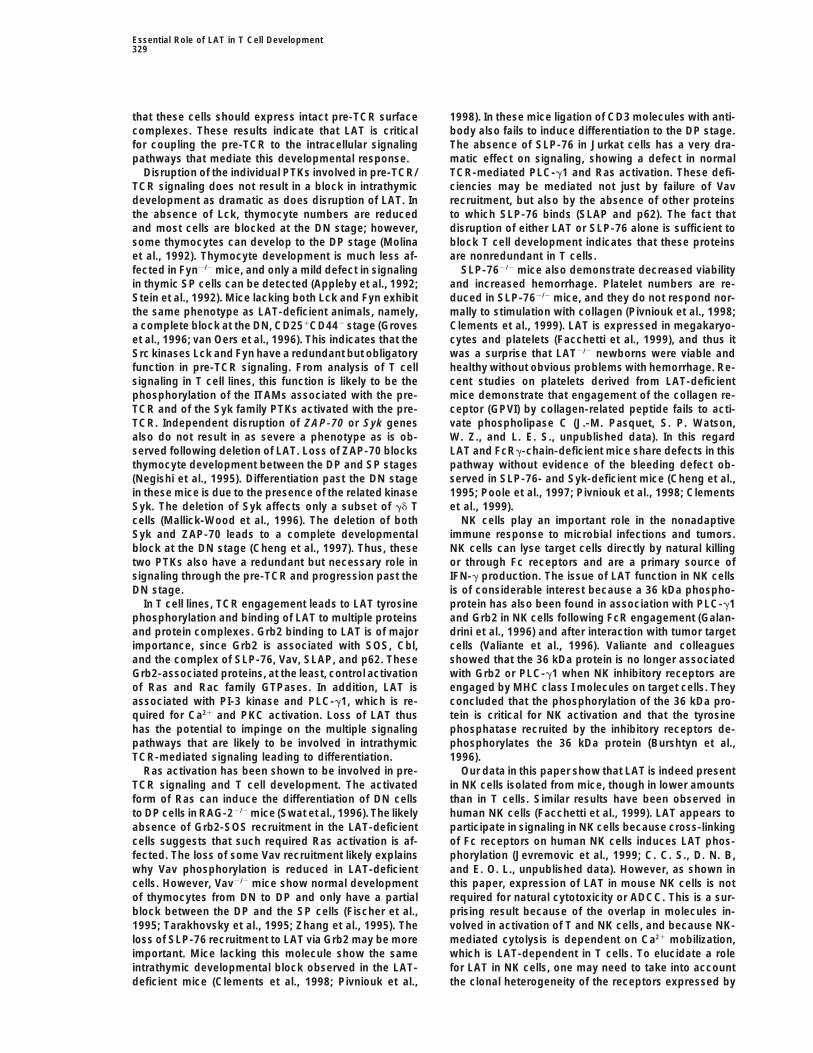
Figure 5. Analysis of gd T Cells and Intestinal Intraepithelial Lymphocytes in LAT-Deficient Mice
(A) Development of gd T cells in LAT2/2 mice. Thymocytes were isolated from fetal day 15 thymus and stained with anti-CD3e-FITC and anti-Vg3-PE or with anti-CD3e-PE and anti-TCRd-FITC.(B) Intestinal intraepithelial lymphocytes (iIELs) from LAT1/1 and LAT2/2 mice were prepared as described in Experimental Procedures andanalyzed for the expression of CD4, CD8a, and CD8b (columns 1 and 2). Expression of gdTCR, abTCR, and CD3e was also analyzed on gatedCD42CD8b2 cells (columns 3 and 4).
(Havran and Allison, 1988). Analysis of fetal day 15 thy- found to be devoid of all subsets of mature TCR1 lym-phocytes.mocytes, by staining for Vg3 (the predominant TCR at
fetal day 15), and pan-TCR gd mAb GL3 demonstratedthat gdTCR1 cells were absent in LAT2/2 mice (Figure
Development and Function of NK Cells5A). gdTCR1 cells were also undetectable in the periph-in LAT2/2 Miceeral lymphoid organs of LAT2/2 mice (Figure 2). Signifi-Our previous finding that LAT mRNA is expressed in ancantly, as with abT cells, the absence of gd T cells inNK-like cell line (YT) led us to assess NK cell develop-LAT2/2 mice could not be attributed to failure to re-ment and function in the LAT2/2 mice. To examine thearrange the TCRg or TCRd genes, as TCRg and TCRddevelopment of NK cells, we isolated splenocytes fromgene rearrangements were detectable in thymocytesLAT 1/1, 1/2, and 2/2 mice and analyzed them byfrom LAT2/2 mice (data not shown).FACS for the expression of the IL2 receptor b chainWe then examined intestinal intraepithelial lympho-(CD122) and the NK cell marker DX5. As shown in Figurecyte (iIEL) populations in LAT2/2 mice. In normal (LAT1/1)6A, cells expressing both IL2 receptor b chain and DX5mice, iIEL subpopulations include CD41 or CD8ab1 Twere present in LAT2/2 mice. These results suggest thatcells that express abTCR and resemble other peripheralNK development is not compromised in the LAT2/2 mice.abTCR1 lymphocytes. In addition, the iIEL compart-To assess NK function, we isolated total splenocytesment includes abTCR1 and gdTCR1 cells that are eitherfrom LAT 1/1 and 2/2 mice and further enriched forCD42CD82 or that coexpress CD8aa homodimers.NK cells. The purity of these NK cell populations isThese latter cells are thought to develop extrathymically,.80%. To confirm the LAT expression in murine NKand they are not subject to conventional repertoire se-cells, we further enriched NK cells from LAT1/1 mice tolection processes (Guy-Grand and Vassalli, 1993; Pous-a purity of .99% by FACS depletion of any residualsier and Julius, 1994). Cell surface analysis demon-CD31 cells. Figure 6B shows that while NK cells dostrated that, as observed in the spleen and lymph nodes,express LAT, they do so at a level approximately oneCD41 and CD8a1b1 T cells were markedly reduced intenth that of T cells. To assess the cytolytic function ofLAT2/2 mice (Figure 5B). In contrast, cells expressingLAT2/2 NK cells, we tested the ability of the LAT1/1 andCD8aa homodimers were present in normal numbers inLAT2/2 NK cell populations to lyse the mouse T cellLAT2/2 mice. However, none of the cells isolated fromlymphoma YAC-1 and the human B cell line 721.221LAT2/2 mice expressed surface abTCR or gdTCR com-
plexes (Figure 5B, right panel). Thus, LAT2/2 mice were (Figure 6C). No difference was seen in the ability of the

Immunity328
Figure 6. Analysis of NK Cells from LAT-Deficient Mice
(A) The development of NK cells is not affected by LAT deficiency. Splenocytes were surface stained with anti-IL-2Rb-FITC and the NK markerDX-5-PE.(B) LAT expression in NK cells. NK cells and T cells were purified from LAT1/1 mice. NK cells were further purified to .99% purity by FACSdepletion of CD31 cells. These cells were directly solubilized in sample buffer and analyzed by immunoblotting with anti-LAT antiserum. Lane1, 105 T cells; lane 2, 2 3 104 T cells; lane 3, 5 3 105 NK cells.(C) NK cytolytic activity is not affected by lack of LAT. The cytolytic activity was analyzed with 51Cr release assays using the mouse T lymphomacell line YAC-1 and the human B cell line 721.221. Antibody-dependent cellular cytotoxicity was measured after binding the mAb L243 to721.221 cells.
LAT2/2 and LAT1/1 NK cells to lyse these targets across pathways. The block in T cell development observed inLAT-deficient mice shares many features with thoseseveral E:T ratios. Similarly, addition of a monoclonal
antibody to coat the 721.221 targets in order to produce seen with RAG2/2 (Mombaerts et al., 1992; Shinkai etal., 1992), CD3e2/2 (Malissen et al., 1995; DeJarnette etantibody-dependent cellular cytotoxicity (ADCC) also
revealed no difference between the NK cells from the al., 1998), Lck2/2 3 Fyn2/2, (Groves et al., 1996; vanOers et al., 1996), ZAP-702/2 3 Syk2/2 (Cheng et al.,LAT2/2 and LAT1/1 mice.1997), and SLP-762/2 (Clements, et al., 1998; Pivniouket al., 1998) mice. Thymocytes in all these mice do notDiscussiondevelop beyond the CD251CD442 subset of the CD42
CD82 stage. The block at this point of differentiationDisruption of LAT expression might be expected to havea deleterious effect on development and function in the reflects the conclusion that expression of a pre-TCR
complex, coupled to an intracellular signaling pathwayT cell, NK, and platelet lineages. Instead, in our initialanalysis, we observed a wide range of results. Platelets resembling that utilized by the mature TCR, is required
for transition past this step. In particular, it is thoughtand NK cells seemed not to be affected. In contrast,disruption of the LAT gene resulted in a profound block that signaling via the pre-TCR is required for induction of
CD4 and CD8 expression and TCRa gene rearrangementin T cell development. Thymocytes in LAT2/2 mice werearrested at the DN, CD251CD442 stage, and there were (Fehling and von Boehmer, 1997). In RAG2/2 and CD3e2/2
mice, the developmental arrest can be attributed tono mature abT cells in the lymph nodes and spleens ofthese mice. gd T cell development was also blocked in structural defects resulting in failure to express the pre-
TCR. Experimentally, it is possible to drive differentiationLAT2/2 mice as was the maturation of “thymus-indepen-dent” iIELs. Together, these results demonstrate that past this block in RAG2/2 mice by cross-linking the very
low level of CD3 chains present on the surface of theseLAT is essential for the development of all T cell subsets.A number of previous studies detail the effects of early thymocytes with antibodies (Levelt et al., 1993).
However, such a response to CD3 engagement did notthe disruption of genes involved either in the surfaceexpression of the pre-TCR/TCR or in T cell signaling occur in the LAT-deficient thymocytes, despite the fact
Essential Role of LAT in T Cell Development329
that these cells should express intact pre-TCR surface 1998). In these mice ligation of CD3 molecules with anti-body also fails to induce differentiation to the DP stage.complexes. These results indicate that LAT is critical
for coupling the pre-TCR to the intracellular signaling The absence of SLP-76 in Jurkat cells has a very dra-matic effect on signaling, showing a defect in normalpathways that mediate this developmental response.
Disruption of the individual PTKs involved in pre-TCR/ TCR-mediated PLC-g1 and Ras activation. These defi-ciencies may be mediated not just by failure of VavTCR signaling does not result in a block in intrathymic
development as dramatic as does disruption of LAT. In recruitment, but also by the absence of other proteinsto which SLP-76 binds (SLAP and p62). The fact thatthe absence of Lck, thymocyte numbers are reduced
and most cells are blocked at the DN stage; however, disruption of either LAT or SLP-76 alone is sufficient toblock T cell development indicates that these proteinssome thymocytes can develop to the DP stage (Molina
et al., 1992). Thymocyte development is much less af- are nonredundant in T cells.SLP-762/2 mice also demonstrate decreased viabilityfected in Fyn2/2 mice, and only a mild defect in signaling
in thymic SP cells can be detected (Appleby et al., 1992; and increased hemorrhage. Platelet numbers are re-duced in SLP-762/2 mice, and they do not respond nor-Stein et al., 1992). Mice lacking both Lck and Fyn exhibit
the same phenotype as LAT-deficient animals, namely, mally to stimulation with collagen (Pivniouk et al., 1998;Clements et al., 1999). LAT is expressed in megakaryo-a complete block at the DN, CD251CD442 stage (Groves
et al., 1996; van Oers et al., 1996). This indicates that the cytes and platelets (Facchetti et al., 1999), and thus itwas a surprise that LAT2/2 newborns were viable andSrc kinases Lck and Fyn have a redundant but obligatory
function in pre-TCR signaling. From analysis of T cell healthy without obvious problems with hemorrhage. Re-cent studies on platelets derived from LAT-deficientsignaling in T cell lines, this function is likely to be the
phosphorylation of the ITAMs associated with the pre- mice demonstrate that engagement of the collagen re-ceptor (GPVI) by collagen-related peptide fails to acti-TCR and of the Syk family PTKs activated with the pre-
TCR. Independent disruption of ZAP-70 or Syk genes vate phospholipase C (J.-M. Pasquet, S. P. Watson,W. Z., and L. E. S., unpublished data). In this regardalso do not result in as severe a phenotype as is ob-
served following deletion of LAT. Loss of ZAP-70 blocks LAT and FcRg-chain-deficient mice share defects in thispathway without evidence of the bleeding defect ob-thymocyte development between the DP and SP stages
(Negishi et al., 1995). Differentiation past the DN stage served in SLP-76- and Syk-deficient mice (Cheng et al.,1995; Poole et al., 1997; Pivniouk et al., 1998; Clementsin these mice is due to the presence of the related kinase
Syk. The deletion of Syk affects only a subset of gd T et al., 1999).NK cells play an important role in the nonadaptivecells (Mallick-Wood et al., 1996). The deletion of both
Syk and ZAP-70 leads to a complete developmental immune response to microbial infections and tumors.NK cells can lyse target cells directly by natural killingblock at the DN stage (Cheng et al., 1997). Thus, these
two PTKs also have a redundant but necessary role in or through Fc receptors and are a primary source ofIFN-g production. The issue of LAT function in NK cellssignaling through the pre-TCR and progression past the
DN stage. is of considerable interest because a 36 kDa phospho-protein has also been found in association with PLC-g1In T cell lines, TCR engagement leads to LAT tyrosine
phosphorylation and binding of LAT to multiple proteins and Grb2 in NK cells following FcR engagement (Galan-drini et al., 1996) and after interaction with tumor targetand protein complexes. Grb2 binding to LAT is of major
importance, since Grb2 is associated with SOS, Cbl, cells (Valiante et al., 1996). Valiante and colleaguesshowed that the 36 kDa protein is no longer associatedand the complex of SLP-76, Vav, SLAP, and p62. These
Grb2-associated proteins, at the least, control activation with Grb2 or PLC-g1 when NK inhibitory receptors areengaged by MHC class I molecules on target cells. Theyof Ras and Rac family GTPases. In addition, LAT is
associated with PI-3 kinase and PLC-g1, which is re- concluded that the phosphorylation of the 36 kDa pro-tein is critical for NK activation and that the tyrosinequired for Ca21 and PKC activation. Loss of LAT thus
has the potential to impinge on the multiple signaling phosphatase recruited by the inhibitory receptors de-phosphorylates the 36 kDa protein (Burshtyn et al.,pathways that are likely to be involved in intrathymic
TCR-mediated signaling leading to differentiation. 1996).Our data in this paper show that LAT is indeed presentRas activation has been shown to be involved in pre-
TCR signaling and T cell development. The activated in NK cells isolated from mice, though in lower amountsthan in T cells. Similar results have been observed inform of Ras can induce the differentiation of DN cells
to DP cells in RAG-22/2 mice (Swat et al., 1996). The likely human NK cells (Facchetti et al., 1999). LAT appears toparticipate in signaling in NK cells because cross-linkingabsence of Grb2-SOS recruitment in the LAT-deficient
cells suggests that such required Ras activation is af- of Fc receptors on human NK cells induces LAT phos-phorylation (Jevremovic et al., 1999; C. C. S., D. N. B,fected. The loss of some Vav recruitment likely explains
why Vav phosphorylation is reduced in LAT-deficient and E. O. L., unpublished data). However, as shown inthis paper, expression of LAT in mouse NK cells is notcells. However, Vav2/2 mice show normal development
of thymocytes from DN to DP and only have a partial required for natural cytotoxicity or ADCC. This is a sur-prising result because of the overlap in molecules in-block between the DP and the SP cells (Fischer et al.,
1995; Tarakhovsky et al., 1995; Zhang et al., 1995). The volved in activation of T and NK cells, and because NK-mediated cytolysis is dependent on Ca21 mobilization,loss of SLP-76 recruitment to LAT via Grb2 may be more
important. Mice lacking this molecule show the same which is LAT-dependent in T cells. To elucidate a rolefor LAT in NK cells, one may need to take into accountintrathymic developmental block observed in the LAT-
deficient mice (Clements et al., 1998; Pivniouk et al., the clonal heterogeneity of the receptors expressed by

Immunity330
TCR Gene RearrangementNK cells and possible differences in the signaling path-TCR gene rearrangements were detected by PCR and Southernways of human and mouse NK cells.blot analysis using the following primers and probes as describedThe effect of LAT deficiency thus varies in some of(Anderson et al., 1992). For PCR reaction, Vb5-specific primer (GATT
the cell types in which it is expressed. In platelets and AAGTTACAGAAAGCCAGTAGC) or Vb8-specific primer (GTTCTCCTNK cells, further studies must define the function of LAT. TATTTCCTTTCTGTGCAGAG) was used as the 59 end primer and
Jb2-specific primer was used (TGAGAGCTGTCTCCTACTATCGATT)It appears that LAT also has a role in mast cell function,as the 39 end primer. A Jb2-specific probe (TTTCCCTCCCGGAGATas our preliminary studies with bone marrow–derivedTCCCTAA) was in Southern blot analysis.mast cells from LAT-deficient mice demonstrate a de-
fect in FceRI-mediated secretion (S. Saitoh, R. Arud-Western Blot Analysis
chandran, J. Rivera, W. Z., and L. E. S., unpublished Thymocytes (107) from LAT1/2 and LAT2/2 mice were lysed in 50 mldata). The next phase in further investigation of LAT lysis buffer (1% Brij97; 25 mM Tris-Cl, 150 mM NaCl, 5 mM EDTA)and thymocyte development will be reconstitution of the on ice for 10 min. After centrifugation, the supernatant was then
mixed with 23 SDS sample buffer. To detect LAT expression inLAT2/2 mice using transgenics expressing different LATmouse NK cells, enriched NK cells were further purified by removingmutants. These studies will attempt to determine howresidual CD31 cells from a LAT1/1 culture on day 6. Cells (105) werevarious signaling pathways couple to and are dependentlysed directly into 43 SDS sample buffer. The genomic DNA present
on LAT. in the samples was sheared by repeated pipetting. These sampleswere analyzed by SDS-PAGE and Western blotting with anti-LAT
Experimental Procedures antiserum.
Vector Construction AcknowledgmentA 9.0 Kb BglII fraction from a LAT genomic Bac clone was clonedinto a BamHI site of pBlusescript KS to generate pBKS/LAT plasmid. We thank Mary Peterson for technical assistance with the mouseA 1.6 Kb fragment from SpeI to BamHI was excised from pBKS/ NK cells. W. Z. is a Fellow of the Leukemia Society of America.LAT, blunt-ended with DNA polymerase Klenow fragment, and D. N. B. is supported by a Centennial Fellowship from the Medicalcloned into a blunted BamHI/EcoRI site of XpPNT vector as the 59 Research Council of Canada.targeting flank. A 3.5 kb XbaI–HindIII fragment from pBKS/LAT wascloned into pBluescript XbaI–HindIII sites. The resulting plasmid
Received January 29, 1999; revised February 22, 1999.was digested with XbaI and ligated with an annealed pair of twooligonucleotides with the sequences CTAGTGAGCTCGAGGTACC
Referencesand CTAGGGTACCTCGAGCTCA to generate an additional XhoI site.This plasmid was then digested with XhoI and the 3.5 Kb XhoI
Anderson, S.J., Abraham, K.M., Nakayama, T., Singer, A., and Perl-fragment was used as the 39-end targeting flank and cloned into themutter, R.M. (1992). Inhibition of T-cell receptor b-chain gene re-XhoI site of XpPNT containing the 59 targeting flank. The targetingarrangement by overexpression of the non-receptor protein tyrosineconstruct was linearized with NotI. The linearized DNA fragmentkinase p56lck. EMBO J. 11, 4877–4886.was gel purified and used to transfect 2 3 107 ES cells by electropor-Appleby, M.W., Gross, J.A., Cooke, M.P., Levin, S.D., Qian, X., andation. After transfection, ES cells were selected in the presence ofPerlmutter, R.M. (1992). Defective T cell receptor signaling in miceG418 and gancyclovir. Of 216 resistant clones, 10 had undergonelacking the thymic isoform of p59fyn. Cell 70, 751–763.homologous recombination as assessed by Southern blot analysis.
One targeted clone was injected into C57BL/6 mice. LAT-deficient Bendelac, A., Rivera, M.N., Park, S.H., and Roark, J.H. (1997). Mousemice (LAT2/2) were obtained by interbreeding LAT heterozygous CD1-specific NK1 T cells: development, specificity, and function.mice (LAT1/2). Annu. Rev. Immunol. 15, 535–62.
Bolen, J.B., and Brugge, J.S. (1997). Leukocyte protein tyrosineNK Cell Separation and Cytolytic Killing Assay kinases: potential targets for drug discovery. Annu. Rev. Immunol.NK cells from LAT1/1 and LAT2/2 mice were generated as previously 15, 371–404.described (Karlhofer and Yokoyama, 1991). Briefly, spleen cell sus-
Buhl, A.M., and Cambier, J.C. (1997). Co-receptor and accessorypension was passed over a nylon wool column at 378C and the
regulation of B-cell antigen receptor signal transduction. Immunol.pass-through fractions were cultured in the presence of IL-2 for 3
Rev. 160, 127–138.days. The nonadherent cells were then removed and the remaining
Burshtyn, D.N., Scharenberg, A.M., Wagtmann, N., Rajagopalan, S.,cells were cultured for 3–6 days. The purity of the NK populationsBerrada, K., Yi, T., Kinet, J.P., and Long, E.O. (1996). Recruitmentwas determined on day 6 of culture using the monoclonal antibodyof tyrosine phosphatase HCP by the killer cell inhibitor receptor.145–2C11 (anti-CD3e) directly conjugated to phycoerythrin (Phar-Immunity 4, 77–85.Mingen). The LAT1/1 and LAT1/2 contained 5%–20% CD31 cells,Chan, A.C., and Shaw, A.S. (1996). Regulation of antigen receptorand there were no CD31 cells in the LAT2/2 populations. Cytotoxicsignal transduction by protein tyrosine kinases. Curr. Opin. Immunol.assays were performed on day 9 by measuring specific release from8, 394–401.51Cr-labeled target cells. To measure ADCC, 51Cr-labeled 721.221
cells were preincubated with 1 mg/ml L243 (anti-HLA-DR) as de- Cheng, A.M., Rowley, B., Pao, W., Hayday, A., Bolen, J.B., andscribed (Rojo et al., 1997). Pawson, T. (1995). Syk tyrosine kinase required for mouse viability
and B-cell development. Nature 378, 303–306.Cell Preparation and Flow Cytometry Cheng, A.M., Negishi, I., Anderson, S.J., Chan, A.C., Bolen, J., Loh,Thymus, spleen, and lymph nodes were excised from mice and D.Y., and Pawson, T. (1997). The Syk and ZAP-70 SH2-containingsingle cell suspensions were prepared. Intestinal intraepithelial lym- tyrosine kinases are implicated in pre-T cell receptor signaling. Proc.phocytes (iIELs) were prepared from the small intestine as previously Natl. Acad. Sci. USA 94, 9797–9801.described (Shores et al., 1997). Standard flow cytometry was per-
Clements, J.L., Yang, B., Ross-Barta, S.E., Eliason, S.L., Hrstka,formed as previously described (Shores et al., 1997) using a BectonR.F., Williamson, R.A., and Koretzky, G.A. (1998). Requirement forDickinson FACScan and Cell Quest software (Becton Dickinsonthe leukocyte-specific adapter protein SLP-76 for normal T cell de-Immunoocytometry Systems). Commercial antibodies to mousevelopment. Science 281, 416–419.antigens used include anti-CD3e-FITC, anti-IgM-FITC, anti-CD44-Clements, J.L., Lee, J.R., Gross, B., Yang, B., Olson, J.D., Sandra,FITC, anti-abTCR-FITC, anti-gd-FITC, anti-CD8b-FITC, anti-Vg3-A., Watson, S.P., Lentz, S.R., and Koretzky, G.A. (1999). Fetal hemor-FITC, anti-CD4-PE, anti CD25-PE (Pharmingen), anti-CD8a-Quan-rhage and platelet dysfunction in SLP-76-deficient mice. J. Clin.tum red, anti-B220-Quantum red, anti-CD4-Quantum red, and
anti-CD3-Quantum red (Sigma). Invest. 103, 19–25.
Essential Role of LAT in T Cell Development331
DeJarnette, J.B., Sommers, C.L., Huang, K., Woodside, K.J., Em- Negishi, I., Motoyama, N., Nakayama, K., Nakayama, K., Senju, S.,mons, R., Katz, K., Shores, E.W., and Love, P.E. (1998). Specific Hatakeyama, S., Zhang, Q., Chan, A.C., and Loh, D.Y. (1995). Essen-requirement for CD3e in T cell development. Proc. Natl. Acad. Sci. tial role for ZAP-70 in both positive and negative selection of thymo-USA 95, 14909–14914. cytes. Nature 376, 435–438.
Facchetti, F., Chan, J.K.C., Zhang, W., Tironi, A., Chilosi, M., Parolini, Peterson, E.J., Clements, J.L., Fang, N., and Koretzky, G.A. (1998).S., Notarangelo, L.D., and Samelson, L.E. (1999). LAT (linker for Adaptor proteins in lymphocyte antigen-receptor signaling. Curr.activation of T cells): a novel immunohistochemical marker for T, Opin. Immunol. 10, 337–344.NK, mast cells and megakaryocytes. Evaluation in normal and patho- Pfeffer, K., and Mak, T.W. (1994). Lymphocyte ontogeny and activa-logical conditions. Am. J. Path., in press. tion in gene targeted mutant mice. Annu. Rev. Immunol. 12, 367–411.Fehling, H.J., and von Boehmer, H. (1997). Early abT cell develop- Pivniouk, V., Tsitsikov, E., Swinton, P., Rathbun, G., Alt, F.W., andment in the thymus of normal and genetically altered mice. Curr. Geha, R.S. (1998). Impaired viability and profound block in thymo-Opin. Immunol. 9, 263–275. cyte development in mice lacking the adaptor protein SLP-76. CellFinco, T.S., Kadlecek, T., Zhang, W., Samelson, L.E., and Weiss, A. 94, 229–238.(1998). LAT is required for TCR-mediated activation of PLCg1 and Poole, A., Gibbins, J.M., Turner, M., van Vugt, M.J., van de Winkel,the ras pathway. Immunity 9, 617–626. J.G., Saito, T., Tybulewicz, V.L., and Watson, S.P. (1997). The FcFischer, K.D., Zmuldzinas, A., Gardner, S., Barbacid, M., Bernstein, receptor g-chain and the tyrosine kinase Syk are essential for activa-A., and Guidos, C. (1995). Defective T-cell receptor signaling and tion of mouse platelets by collagen. EMBO J. 16,2333–2341.positive selection of Vav-deficient CD41 CD81 thymocytes. Nature Poussier, P., and Julius, M. (1994). Thymus independent T cell devel-374, 474–477. opment and selection in the intestinal epithelium. Annu. Rev. Immu-Galandrini, R., Palmieri, G., Piccoli, M., Frati, L., and Santoni, A. nol. 12, 521–553.(1996). CD16-mediated p21ras activation is associated with Shc and Ravetch, J.V., and Kinet, J.P. (1991). Fc receptors. Annu. Rev. Immu-p36 tyrosine phosphorylation and their binding with Grb2 in human nol. 9, 457–492.natural killer cells. J. Exp. Med. 183, 179–186.
Rojo, S., Burshtyn, D.N., Long, E.O., and Wagtmann, N. (1997). TypeGodfrey, D.I., and Zlotnik, A. (1993). Control points in early T-cell
I transmembrane receptor with inhibitory function in mouse mastdevelopment. Immunol. Today 14, 547–553.
cells and NK cells. J. Immunol. 158, 9–12.Groves, T., Smiley, P., Cooke, M.P., Forbush, K., Perlmutter, R.M.,
Rudd, C.E. (1999). Adaptors and molecular scaffolds in immune celland Guidos, C.J. (1996). Fyn can partially substitute for Lck in T
signaling. Cell 96, 5–8.lymphocyte development. Immunity 5, 417–428.
Samelson, L.E. (1999). Adaptor proteins and T cell antigen receptorGuy-Grand, D., and Vassalli, P. (1993). Gut intraepithelial T lympho-signaling. Prog. Biophys. Mol. Biol., in press.cytes. Curr. Opin. Immunol. 5, 247–252.Shinkai, Y., Rathbun, G., Lam, K.P., Oltz, E.M., Stewart, V., Mendel-Havran, W.L., and Allison, J.P. (1988). Developmentally ordered ap-sohn, M., Charron, J., Datta, M., Young, F., Stall, A.M., et al. (1992).pearance of thymocytes expressing different T-cell antigen recep-RAG-2-deficient mice lack mature lymphocytes owing to inability totors. Nature 335, 443–445.initiate V(D)J rearrangement. Cell 68, 855–867.
Hendricks-Taylor, L.R., Motto, D.G., Zhang, J., Siraganian, R.P., andShores, E., Flamand, V., Tran, T., Grinberg, A., Kinet, J.P., and Love,Koretzky, G.A. (1997). SLP-76 is a substrate of the high affinityP.E. (1997). FceRI g can support T cell development and functionIgE receptor-stimulated protein tyrosine kinases in rat basophilicin mice lacking endogenous TCR z-chain. J. Immunol. 159, 222–230.leukemia cells. J. Biol. Chem. 272, 1363–1367.Stein, P.L., Lee, H.M., Rich, S., and Soriano, P. (1992). pp59fynJevremovic, D., Billadeau, D.D., Schoon, R.A., Dick, C.J., Irwin, B.J.,mutant mice display differential signaling in thymocytes and periph-Zhang, W., Samelson, L.E., Abraham, R.T., and Leibson, P.J. (1999).eral T cells. Cell 70, 741–750.Cutting edge: a role for the adaptor protein LAT in human NK cell-Swat, W., Shinkai, Y., Cheng, H.L., Davidson, L., and Alt, F.W. (1996).mediated cytotoxicity. J. Immunol. 162, 2453–2456.Activated Ras signals differentiation and expansion of CD4181 thy-Karlhofer, F.M., and Yokoyama, W.M. (1991). Stimulation of murinemocytes. Proc. Natl. Acad. Sci. USA 93, 4683–4687.natural killer (NK) cells by a monoclonal antibody specific for theTarakhovsky, A., Turner, M., Schaal, S., Mee, P.J., Duddy, L.P.,NK1.1 antigen. IL-2-activated NK cells possess additional specificRajewsky, K., and Tybulewicz, V.L. (1995). Defective antigen recep-stimulation pathways. J. Immunol. 146, 3662–3673.tor-mediated proliferation of B and T cells in the absence of Vav.Killeen, N., Irving, B.A., Pippig, S., and Zingler, K. (1998). SignalingNature 374, 467–470.checkpoints during the development of T lymphocytes. Curr. Opin.Turner, H., Reif, K., Rivera, J., and Cantrell, D.A. (1995). RegulationImmunol. 10, 360–367.of the adapter molecule Grb2 by the FceR1 in the mast cell lineLevelt, C.N., Mombaerts, P., Iglesias, A., Tonegawa, S., and Eich-RBL2H3. J. Biol. Chem. 270, 9500–9506.mann, K. (1993). Restoration of early thymocyte differentiation in
T-cell receptor b-chain-deficient mutant mice by transmembrane Valiante, N.M., Phillips, J.H., Lanier, L.L., and Parham, P. (1996).Killer cell inhibitory receptor recognition of human leukocyte antigensignaling through CD3e. Proc. Natl. Acad. Sci. USA 90, 11401–11405.(HLA) class I blocks formation of a pp36/PLC-g signaling complexLiu, S.K., Fang, N., Koretzky, G.A., and McGlade, C.J. (1999). Thein human natural killer (NK) cells. J. Exp. Med. 184, 2243–2250.hematopoietic-specific adaptor protein Gads functions in T-cell sig-
naling via interactions with the SLP-76 and LAT adaptors. Curr. Biol. van Oers, N.S., Lowin-Kropf, B., Finlay, D., Connolly, K., and Weiss,A. (1996). ab T cell development is abolished in mice lacking both9, 67–75.Lck and Fyn protein tyrosine kinases. Immunity 5, 429–436.Mallick-Wood, C.A., Pao, W., Cheng, A.M., Lewis, J.M., Kulkarni, S.,
Bolen, J.B., Rowley, B., Tigelaar, R.E., Pawson, T., and Hayday, A.C. Wange, R.L., and Samelson, L.E. (1996). Complex complexes: sig-naling at the TCR. Immunity 5, 197–205.(1996). Disruption of epithelial gdT cell repertoires by mutation of
the Syk tyrosine kinase. Proc. Natl. Acad. Sci. USA 93, 9704–9709. Weber, J.R., Orstavik, S., Torgersen, K.M., Danbolt, N.C., Berg, S.F.,Ryan, J.C., Tasken, K., Imboden, J.B., and Vaage, J.T. (1998). Molec-Malissen, M., Gillet, A., Ardouin, L., Bouvier, G., Trucy, J., Ferrier,
P., Vivier, E., and Malissen, B. (1995). Altered T cell development ular cloning of the cDNA encoding pp36; a tyrosine-phosphorylatedadaptor protein selectively expressed by T cells and natural killerin mice with a targeted mutation of the CD3e gene. EMBO J. 14,
4641–4653. cells. J. Exp. Med. 187, 1157–1161.
Weiss, A., and Littman, D.R. (1994). Signal transduction by lympho-Molina, T.J., Kishihara, K., Siderovski, D.P., van Ewijk, W., Naren-dran, A., Timms, E., Wakeham, A., Paige, C.A., Hartmann, K.-U., cyte antigen receptors. Cell 76, 263–274.Veillette, A., et al. (1992). Profound block in thymocyte development Wiest, D.L., Bhandoola, A., Punt, J., Kreibich, G., McKean, D., andin mice lacking p56lck. Nature 357, 161–164. Singer, A. (1997). Incomplete endoplasmic reticulum (ER) retention
in immature thymocytes as revealed by surface expression of “ER-Mombaerts, P., Iacomini, J., Johnson, R.S., Herrup, K., Tonegawa,S., and Papaioannou, V.E. (1992). RAG-1-deficient mice have no resident” molecular chaperones. Proc. Natl. Acad. Sci. USA 94,
1884–1889.mature B and T lymphocytes. Cell 68, 869–877.

Immunity332
Zhang, R., Alt, F.W., Davidson, L., Orkin, S.H., and Swat, W. (1995).Defective signaling through the T- and B-cell antigen receptors inlymphoid cells lacking the vav proto-oncogene. Nature 374,470–473.
Zhang, W., Sloan-Lancaster, J., Kitchen, J., Trible, R.P., and Samel-son, L.E. (1998a). LAT: the ZAP-70 tyrosine kinase substrate thatlinks T cell receptor to cellular activation. Cell 92, 83–92.
Zhang, W., Trible, R.P., and Samelson, L.E. (1998b). LAT palmitoyla-tion: its essential role in membrane microdomain targeting and tyro-sine phosphorylation during T cell activation. Immunity 9, 239–246.