Neonatal diabetes mellitus: A model for personalized medicine
Upload
khangminh22Category
view
2download
0

This title is also available as an e-book.
For more details, please see
www.wiley.com/buy/9781119235811

Essential Neonatal MedicineSixth Edition
Sunil Sinha
Professor of Paediatrics University of Durham Consultant NeonatologistJames Cook University HospitalMiddlesbrough, UK
Lawrence Miall
Consultant Neonatologist, Leeds Children's HospitalHonorary Senior Lecturer, University of LeedsLeeds Teaching Hospitals NHS TrustLeeds, UK
Luke Jardine
Senior Staff Specialist Neonatology, Mater Mothers' Hospital Honorary Researcher, Mater Research Associate Professor, The University of Queensland Australia

This edition first published 2018 © 2018 John Wiley & Sons LtdEdition History John Wiley & Sons (1e 1987 ; 2e 1993; 3e 2000); Wiley-Blackwell (4e 2008; 5e 2012).All rights reserved. No part of this publication may be reproduced, stored in a retrieval system, or transmitted, in any form or by any means,electronic, mechanical, photocopying, recording or otherwise, except as permitted by law. Advice on how to obtain permission to reusematerial from this title is available at http://www.wiley.com/go/permissions.The right of Sunil Sinha, Lawrence Miall, Luke Jardine to be identified as the authors of this work has been asserted in accordance with law.Registered Office(s) John Wiley & Sons, Inc., 111 River Street, Hoboken, NJ 07 030, USA John Wiley & Sons Ltd, The Atrium, Southern Gate, Chichester, West Sussex, PO19 8SQ, UKEditorial Office 9600 Garsington Road, Oxford, OX4 2DQ, UKFor details of our global editorial offices, customer services, and more information about Wiley products visit us at www.wiley.com.Wiley also publishes its books in a variety of electronic formats and by print-on-demand. Some content that appears in standard printversions of this book may not be available in other formats.Limit of Liability/Disclaimer of Warranty The contents of this work are intended to further general scientific research, understanding, and discussion only and are not intended andshould not be relied upon as recommending or promoting scientific method, diagnosis, or treatment by physicians for any particular patient.The publisher and the authors make no representations or warranties with respect to the accuracy and completeness of the contents of thiswork and specifically disclaim all warranties, including without limitation any implied warranties of fitness for a particular purpose. In view ofongoing research, equipment modifications, changes in governmental regulations, and the constant flow of information relating to the use ofmedicines, equipment, and devices, the reader is urged to review and evaluate the information provided in the package insert or instructionsfor each medicine, equipment, or device for, among other things, any changes in the instructions or indication of usage and for addedwarnings and precautions. Readers should consult with a specialist where appropriate. The fact that an organization or website is referred toin this work as a citation and/or potential source of further information does not mean that the author or the publisher endorses theinformation the organization or website may provide or recommendations it may make. Further, readers should be aware that websites listedin this work may have changed or disappeared between when this works was written and when it is read. No warranty may be created orextended by any promotional statements for this work. Neither the publisher nor the author shall be liable for any damages arising herefrom.Library of Congress Cataloging-in-Publication Data Names: Sinha, Sunil K., M.D., Ph.D., author. | Miall, Lawrence, author. | Jardine, Luke, author. Title: Essential neonatal medicine / Sunil Sinha, Lawrence Miall, Luke Jardine. Other titles: Essentials (Wiley-Blackwell (Firm)) Description: Sixth edition. | Hoboken, NJ : John Wiley & Sons Inc., 2018. | Series: Essentials | Includes bibliographical references and index. Identifiers: LCCN 2017 007 280 (print) | LCCN 2017 008052 (ebook) | ISBN 97 81119235811 (paper) | ISBN 97 811192357 7 4 (Adobe PDF) | ISBN 97 811192357 50 (ePub) Subjects: | MESH: Infant, Newborn, Diseases | Neonatology | Infant, Newborn Classification: LCC RJ251 (print) | LCC RJ251 (ebook) | NLM WS 421 | DDC 618.92/01—dc23 LC record available at https://lccn.loc.gov/2017 007 280Cover Design: Wiley Cover Image: © ERproductions Ltd/Gettyimages

CONTENTS1. Preface to the Sixth Edition
2. Acknowledgements
3. Preface to the First Edition
4. Abbreviations
5. How to use your textbook
6. About the companion website
7. Chapter 1 The fetus, placenta and changes at birth
a. Introduction
b. Placental function
c. Fetal homeostasis
d. Fetal circulation
e. Assessment of fetal well-being
f. Screening during pregnancy
g. Fetal monitoring during labour
h. Fetal compromise
i. Acknowledgements
j. Further reading
8. Chapter 2 Perinatal epidemiology and audit
a. Introduction
b. Definitions of terms commonly used in perinatal medicine
c. The role of perinatal and neonatal audit
d. Classification of perinatal deaths
e. Factors affecting perinatal death rates
f. Prevention of perinatal mortality and �low birthweight
g. Changing trends
h. Further reading
9. Chapter 3 Multiple births
a. Introduction
b. Physiology of fertilization, implantation and placenta formation
c. Classification of multiple pregnancy
d. Assisted reproductive technology
e. Incidence of multiple pregnancies
f. Parental counselling
g. Complications of multiple pregnancy
h. Further reading
10. Chapter 4 Neonatal consequences of maternal conditions
a. Introduction
b. Congenital anomalies: malformations and deformations
c. Congenital anomalies associated with teratogens
d. Congenital malformation secondary to maternal infections
e. Consequences of maternal substance misuse
f. Neonatal manifestations of maternal medical diseases
g. Further reading
11. Chapter 5 Resuscitation at birth
a. Introduction
b. Fetal responses during labour
c. Fetal and neonatal responses to perinatal asphyxia
d. Perinatal asphyxia
e. Assessment of the infant at birth
f. Stabilization at birth

g. Resuscitation
h. Post-resuscitation care of the �asphyxiated infant
i. Further reading
12. Chapter 6 Examination of the newborn
a. Introduction
b. The newborn examination as a �screening test
c. Approach to the newborn examination
d. General appearance
e. Head and neck
f. Chest
g. Cardiovascular
h. Abdomen
i. Back
j. Extremities
k. Congenital abnormalities of the hips �and limbs
l. Skin disorders
m. Communication with parents
n. Further reading
13. Chapter 7 Birth injury
a. Introduction
b. Risk factors for birth injury
c. Injuries to the scalp, skull and brain
d. Bone and joint injuries
e. Peripheral nerve injuries
f. Soft-tissue injuries
g. Organ injuries
h. Injuries sustained in the neonatal intensive care unit (NICU)
i. Further reading
14. Chapter 8 Genetic disorders
a. Introduction
b. Gene structure
c. Commonly used investigations
d. Genetic variation
e. Multifactorial inheritance
f. Approach to the dysmorphic neonate
g. Prevention of congenital abnormalities
h. Further reading
15. Chapter 9 Infant feeding and nutrition
a. Introduction
b. Specific nutritional requirements
c. Breastfeeding
d. Artificial feeding/formulas
e. Techniques of artificial feeding
f. Feeding the preterm infant
g. Parenteral nutrition
h. Common feeding disorders
i. Further reading
16. Chapter 10 Infection in the newborn
a. Introduction
b. The immune system
c. Susceptibility of the neonate to infection
d. Congenital infection

e. Intrapartum (early-onset) infection
f. Postnatal (late-onset) infection
g. Further reading
17. Chapter 11 The extreme preterm infant
a. Introduction
b. Gestational age
c. Causes and management of preterm labour
d. Survival and outcome for the preterm infant
e. Preterm delivery at the margins of viability
f. Stabilization at birth and management in the ‘golden hour’
g. Common problems to be expected in the preterm infant
h. Supportive care on the NICU
i. Preparation for discharge home
j. Further reading
18. Chapter 12 The low-birthweight infant
a. Introduction
b. The infant who is small for gestational age
c. Causes of intrauterine growth restriction
d. Problems to be expected in the growth-restricted fetus and �SGA infant
e. Management of the low-birthweight infant
f. Further reading
19. Chapter 13 Respiratory physiology and respiratory support
a. Introduction
b. Fetal lung development
c. Pulmonary surfactants
d. Respiratory physiology
e. Assessment of respiratory function
f. Respiratory failure
g. Mechanical ventilation
h. Further reading
20. Chapter 14 Respiratory disorders
a. Introduction
b. Respiratory distress
c. Transient tachypnoea of the newborn
d. Respiratory distress syndrome (RDS)
e. Pneumonia
f. Pulmonary air leaks
g. Meconium aspiration syndrome
h. Pulmonary hypoplasia
i. Pulmonary haemorrhage
j. Congenital diaphragmatic hernia
k. Oesophageal atresia and �tracheo-oesophageal fistula
l. Congenital lobar emphysema
m. Congenital pulmonary airway malformation (CPAM) [formerly known as congenital cystic adenomatousmalformation; CCAM]
n. Chronic lung disease and bronchopulmonary dysplasia (BPD)
o. Further reading
21. Chapter 15 Apnoea, bradycardia and upper airway obstruction
a. Introduction
b. Physiology
c. Apnoea
d. Acute life-threatening events (ALTEs)
e. Sudden and unexpected infant death and sudden infant death syndrome

f. Upper airway obstruction
g. Further reading
22. Chapter 16 Cardiovascular disorders
a. Introduction
b. Physiology of the cardiovascular system
c. Blood pressure
d. Hypertension
e. Congenital heart disease
f. Investigations
g. Cyanotic heart disease
h. Congestive heart failure
i. Left-to-right shunts
j. Obstructive lesions
k. Dysrrhythmias
l. Circulatory maladaptation at birth
m. Further reading
23. Chapter 17 Gastrointestinal and abdominal disorders
a. Introduction
b. Development of the gastrointestinal tract
c. Malformations
d. Abdominal wall defects
e. Necrotizing enterocolitis
f. Short bowel syndrome
g. Rectal bleeding
h. Hernia
i. Hydrocoele
j. Undescended testis
k. Hypospadias
l. Further reading
24. Chapter 18 Renal disorders
a. Introduction
b. Role of amniotic fluid
c. Renal physiology
d. Normal urine output
e. Investigation of renal disease
f. Presentation of renal disease
g. Acute kidney injury
h. Urinary tract infection
i. Renal masses
j. Cystic disease of the kidneys
k. Haematuria
l. Ectopia vesicae (bladder exstrophy)
m. Further reading
25. Chapter 19 Jaundice
a. Introduction
b. Physiology of bilirubin metabolism
c. Clinical assessment of the jaundiced infant
d. Unconjugated hyperbilirubinaemia
e. Conjugated hyperbilirubinaemia
f. Further reading
26. Chapter 20 Haematological disorders
a. Introduction

b. Placental transfusion
c. Anaemia
d. Hydrops fetalis
e. Aplasia
f. Polycythaemia
g. Bleeding and coagulation disorders
h. Thrombocytopenia
i. Haemorrhagic disease of the newborn (Vitamin K-deficient bleeding)
j. Disseminated intravascular coagulation (DIC)
k. Inherited disorders of coagulation
l. Congenital deficiency of anticoagulant proteins (hypercoagulable states)
m. Further reading
27. Chapter 21 Endocrine and metabolic disorders
a. Introduction
b. Glucose homeostasis and its abnormalities
c. Disorders of calcium, phosphate and magnesium metabolism
d. Disorders of magnesium metabolism
e. Disorders of sodium and potassium metabolism
f. 21.6.1 Box 21.3 Causes of neonatal hyponatraemia.
g. Endocrine gland disorders
h. Abnormalities of the adrenal gland
i. Inborn errors of metabolism
j. Further reading
28. Chapter 22 The central nervous system
a. Introduction
b. Brain development
c. Malformations of the central nervous system
d. Disorders of head size and shape
e. Intracranial haemorrhage (ICH)
f. Periventricular leukomalacia
g. Neonatal stroke
h. Hypoxic–ischaemic encephalopathy
i. Neonatal convulsions
j. Neonatal hypotonia (‘floppy infant’)
k. Further reading
29. Chapter 23 Neurodevelopmental follow-up and assessment of hearing and vision
a. Introduction
b. Neurodevelopmental outcome
c. Hearing impairment (deafness)
d. Visual impairment
e. Further reading
30. Chapter 24 Developmental care and the neonatal environment
a. Introduction
b. Thermoregulation
c. Skin care on the neonatal intensive care unit
d. Optimizing the neonatal environment
e. Procedural pain and analgesia
f. Developmental care
g. Further reading
31. Chapter 25 Organization of perinatal services
a. Introduction
b. Organization of perinatal services

c. Levels of perinatal care
d. Neonatal networks
e. Further reading
32. Chapter 26 Neonatal transport
a. Introduction
b. Transport in utero
c. Preparation for transport
d. Transport equipment
e. The role of a neonatal transport service
f. Further reading
33. Chapter 27 Discharge and follow-up of high-risk infants
a. Introduction
b. Discharge of high-risk infants
c. Immunization
d. Specialized follow-up clinics
e. Follow-up of preterm infants
f. Further reading
34. Chapter 28 Parent–infant attachment and support for parents of critically ill infants
a. Introduction
b. Parent–infant attachment (bonding)
c. Care of parents of critically ill infants
d. Family integrated care
e. Further reading
35. Chapter 29 Ethical issues and decision-making process in the treatment of critically ill newborn infants
a. Introduction
b. Principles of ethical reasoning
c. Decision-making processes
d. The role of the Institutional �Ethics Committee
e. Withholding and withdrawing �life-sustaining treatment
f. Common neonatal ethical dilemmas
g. Parents in the decision-making process
h. Further reading
36. Chapter 30 End-of-life care and palliative care
a. Introduction – why babies die
b. Unexpected deaths (including sudden unexpected postnatal collapse)
c. What is palliative care?
d. Expected deaths and care planning
e. Making a care plan
f. Place of death: hospice versus home versus hospital
g. Symptom control
h. Organ donation
i. Autopsy
j. Caring for parents – grief and bereavement
k. Caring for staff
l. Further reading
37. Index
38. EULA
List of Illustrations1. Chapter 1
a. Figure 1.1 Diagram of placental structures showing blood perfusion.
b. Figure 1.2 Diagram of the fetal circulation through the heart and lungs, showing the direction of flow through theforamen ovale and ductus arteriosus.

c. Figure 1.3 A timeline for fetal assessment and monitoring during pregnancy.
d. Figure 1.4 Doppler measurement of blood flow in the fetal umbilical artery. The left-hand panel shows normalforward flow throughout the cardiac cycle. The right-hand panel shows pathological reversed flow during diastole (seearrow).
e. Figure 1.5 Cleft lip. Illustration courtesy of Dr Jason Ong.
f. Figure 1.6 Fetal MRI scan (coronal view) showing large cystic hygroma on the left side of the neck (arrow) and anassociated pleural effusion (arrow). Illustration courtesy of Dr Mike Weston.
g. Figure 1.7a CTG showing fetal heart rate accelerations.
h. Figure 1.7b CTG showing late decelerations.
i. Figure 1.7c CTG showing normal heart rate followed by severe prolonged fetal bradycardia.
j. Figure 1.7d CTG showing loss of beat-to-beat variability.
k. Figure 1.8 Clearance of lung fluid into the lymphatics with the first breaths.
2. Chapter 2
a. Figure 2.1 2014 ANZNN survival data to discharge home (with 95% CI) (Full data are available in Table 30 inChow, S.S.W., Le Marsney, R., Haslam, R., Lui, K. (2016) Report of the Australian and New Zealand NeonatalNetwork 2014. ANZNN, Sydney.
3. Chapter 3
a. Figure 3.1 Twin peak or lambda sign. Illustration courtesy of Dr Scott Peterson, Mater Mothers’ Hospital.Reproduced with permission of Dr Peterson.
4. Chapter 4
a. Figure 4.1 Problems leading to joint contractures.
b. Figure 4.2 Infant with typical features of fetal alcohol syndrome. From Lissauer, T. and Fanaroff, A. A. (2011)Neonatology at a Glance, 2nd edition. © 2011, Blackwell Publishing Ltd. Reproduced with permission of John Wiley& Sons.
5. Chapter 5
a. Figure 5.1 The physiological effect of acute asphyxia and the response to resuscitation. Illustration courtesy of DrSam Richmond.
b. Figure 5.2 Algorithm for resuscitation. Reproduced with permission from the Resuscitation Council UK (2015).
c. Figure 5.3 Mask inflation with the head in the neutral position.
d. Figure 5.4 (a) T-piece (Fischer Pykell Health Care). (b) Face masks designed for use in face mask ventilation ofterm and preterm newborns.
e. Figure 5.5 Laryngoscopy. The laryngoscope blade displaces the tongue and lifts the epiglottis anteriorly to exposethe cords �(Source: Baillière Tindall).
f. Figure 5.6 The stages of intubation. (a) Visualization of the uvula and oropharynx. (b) The epiglottis is seen with theoesophagus beyond it. (c) The cords are also seen.
g. Figure 5.7 Cardiac compressions performed by encircling the chest, whilst ventilation breaths are given by bag-valve-mask in a 3:1 ratio.
6. Chapter 6
a. Figure 6.1 Head-to-toe examination sequence. From Miall, L. (2009) The Newborn Examination. Paediatrics at aGlance, 3rd edition, �Wiley-Blackwell. Reproduced with permission of John Wiley & Sons.
b. Figure 6.2 Sagittal synostosis. (a) The baby has a palpable ridge on their skull. (b) 3D CT scan of the same childshowing fusion of the sagittal suture (arrow).
c. Figure 6.3 This baby (who is being examined under anaesthetic) has a normal red reflex in their left eye, but anabsent red reflex in the right eye (arrow) due to congenital cataract.
d. Figure 6.4 Severe micrognathia (with tracheostomy). This patient has cerebro-costo-mandibular syndrome.
e. Figure 6.5 Cystic hygroma of the neck (trans-illuminated).
f. Figure 6.6 (a) Teratoma of the neck. This child was intubated while still connected to the placental circulation (EXITprocedure) before having surgical excision. (b) MRI scan showing the same lesion (arrow).
g. Figure 6.7 A radiograph showing multiple vertebral anomalies (arrows).
h. Figure 6.8 Eliciting the Moro reflex.
i. Figure 6.9 (a) Talipes equinovarus; (b) talipes calcaneovalgus.
j. Figure 6.10 Ortolani’s test. The hip cannot be abducted because of posterior dislocation of the femoral head. The hipis pulled upwards and the head clunks into the acetabulum, permitting abduction. Barlow’s test. The adducted hip ispushed downwards and laterally to see whether it is dislocatable. Source: Lawrence Miall, Mary Rudolf, DominicSmith. Paediatrics at a Glance, 4th Edition. May 2016, ©2016, Wiley-Blackwell. Reproduced with permission of JohnWiley & Sons.
k. Figure 6.11 Arthrogryposis multiplex.
l. Figure 6.12 Thanatophoric dwarf.
m. Figure 6.13 Vascular haemangioma.

n. Figure 6.14 Congenital melanocytic naevus involving the buttock and loin.
o. Figure 6.15 Mongolian blue spot.
p. Figure 6.16 Harlequin ichthyosis with severe deep skin cracking.
q. Figure 6.17 Transient neonatal pustular melanosis.
r. Figure 6.18 Cutis aplasia of the scalp. Note the extensive area of hair loss, some of which is scabbed over.
7. Chapter 7
a. Figure 7.1 Anatomic location of injuries to the head. Source: Tom Lissauer, Avroy A. Fanaroff, Lawrence Miall,Jonathan Fanaroff. Neonatology at a Glance, 3rd Edition. Wiley-Blackwell. Reproduced with permission of John Wiley& Sons.
b. Figure 7.2 Cephalhaematoma. Note the swelling over the right parietal bone. This child also has hypotonia with acharacteristic drooping appearance to the mouth.
c. Figure 7.3 Right frontal depressed skull fracture (see arrow).
d. Figure 7.4 Right-sided clavicular fracture in a child born after shoulder dystocia. The baby is also receivingmechanical ventilation.
e. Figure 7.5 Left-sided facial nerve palsy.
f. Figure 7.6 Right-sided Erb’s palsy showing the typical ‘waiter’s tip’ position of the hand. Note the unilateral Mororeflex on the left.
g. Figure 7.7 Bruising to the foot from SaO2 probe.
h. Figure 7.8 Chemical burn from aqueous 2% chlorhexidine used prior to UAC insertion in an extreme preterm baby.Reproduced with permission from Lashkari, H.P., Chow, P., Godambe, S. (2011) Aqueous 2% chlorhexidine-inducedchemical burns in an extremely premature infant. Archives of Diseases in Childhood: Fetal and Neonatal Edition,97 , F64.© 2011, BMJ Publishing Group Ltd.
8. Chapter 8
a. Figure 8.1 Diagram of a DNA double helix.
b. Figure 8.2 Normal chromosome pattern and number after Giemsa staining. This is an example of a male karyotype.The hashed horizontal line is at the centromere and divides the chromosome into short (p) and long (q) arms.
c. Figure 8.3 Image showing FISH probes for chromosome 21 (red) and chromosome 13 (green). There are three redsignals (abnormal) and two green signals (normal); this patient therefore has trisomy 21.
d. Figure 8.4 A family pedigree showing autosomal dominant inheritance.
e. Figure 8.5 A family pedigree showing autosomal recessive inheritance.
f. Figure 8.6 A family pedigree showing X-linked recessive inheritance.
9. Chapter 9
a. Figure 9.1 Total body water and extracellular fluid expressed as percentages of body weight. Redrawn from Dear(1984), with permission from Reed Business Publishing.
b. Figure 9.2 Hormonal maintenance of lactation. PIF, prolactin-inhibiting factor; PRF, prolactin-releasing factor.
10. Chapter 10
a. Figure 10.1 Schematic representation of the clinical features of prenatal TORCH infections.
b. Figure 10.2 Routes of neonatal cross-infection.
c. Figure 10.3 Showing position of infant during lumbar puncture while taking care to avoid excessive bending.
d. Figure 10.4 Equipment required for insertion of a percutaneous intravenous central catheter (PICC). Illustrationcourtesy of Dr Emmanuel Erinaugha.
11. Chapter 11
a. Figure 11.1 Outcomes of all extreme preterm babies born in the UK in 2006.
12. Chapter 12
a. Figure 12.1 Monozygotic twins born at 32 weeks. The smaller twin weighed 750 g and the larger 1700 g. Thesmaller twin shows features of IUGR with relative head sparing (asymmetric IUGR).
13. Chapter 13
a. Figure 13.1 Stages of fetal lung development. Reproduced with permission from Attar, M.A., Donn, S.M. (2002)Mechanism of ventilator-induced lung injury in premature infants. Seminars in Neonatology, 7 , 353–360; © 2002,Elsevier.
b. Figure 13.2 Oxygen dissociation curve for fetal haemoglobin (upper red line) and adult haemoglobin (lower blueline).
c. Figure 13.3 Pressure–volume loop showing compliance of the lung.
d. Figure 13.4 Flow–volume loop, showing resistance to airflow. The loop on the left shows increased resistancecausing impedance to airflow, which has improved after treatment as shown in the loop on the right.
e. Figure 13.5 Lung volumes. TLC, total lung capacity; VC, vital capacity; RV, residual volume; IC, inspiratorycapacity; FRC, functional residual capacity; IRC, inspiratory respiratory capacity; ERC, expiratory respiratorycapacity; TV, tidal volume.

14. Chapter 14
a. Figure 14.1 Transient tachypnoea of the newborn (TTN). Note streaky bilateral shadows, fluid in the transversefissure (arrows) and relative cardiomegaly.
b. Figure 14.2 Incidence of RDS related to gestational age.
c. Figure 14.3 Schematic representations of two alveoli, demonstrating the Laplace law (see text for details).
d. Figure 14.4 Chest radiograph showing the characteristic ‘ground glass’ appearance of RDS. Note the ‘airbronchogram’.
e. Figure 14.5 Chest radiograph showing right-sided tension pneumothorax. Note this has occurred despite a chestdrain being in place, suggesting a massive air leak or a blocked chest drain.
f. Figure 14.6 Chest radiograph showing pneumomediastinum. �The heart and thymus are outlined by gas.
g. Figure 14.7 Chest radiograph showing extensive PIE. Note the overinflated chest with flattened diaphragm.
h. Figure 14.8 Chest radiograph showing left-sided PIE. The mediastinum and right lung are compressed by theoverinflated �left lung.
i. Figure 14.9 (a) Left-sided pneumothorax in a preterm baby. (b) The same baby after insertion of a 10 Fr pigtailcatheter.
j. Figure 14.10 Chest radiograph showing meconium aspiration syndrome (MAS). There is extensive discreteshadowing throughout both lung fields and hyperinflation.
k. Figure 14.11 Chest radiograph showing a left-sided diaphragmatic hernia.
l. Figure 14.12 Variants of tracheo-oesophageal fistula with or without oesophageal atresia. Type (c) accounts for 85%of cases, the others being equally uncommon.
m. Figure 14.13 Chest radiograph showing severe bronchopulmonary dysplasia.
15. Chapter 15
a. Figure 15.1 Neuromuscular pathway for control of respiration.
b. Figure 15.2 Suggested protocol for the management of apnoea alarm.
c. Figure 15.3 A normal upper airway. Reproduced with permission from South, M., Isaacs, D. (eds) PracticalPaediatrics, 7th edition. Elsevier Health Sciences, London.
d. Figure 15.4 Nasopharyngeal tube used for micrognathia. Reproduced with permission from South, M., Isaacs, D.(eds) Practical Paediatrics, 7th edition. Elsevier Health Sciences, London.
16. Chapter 16
a. Figure 16.1 Upper and lower centiles for (a) systolic and (b) diastolic blood pressure against gestational age. (c) Thechange in mean arterial blood pressure (MABP) with postnatal age at different gestational age bands.
b. Figure 16.2 Flow diagram showing a suggested graded management response to neonatal hypotension.
c. Figure 16.3 Real-time, two-dimensional echocardiograms of the normal neonatal heart. (a) Parasternal long-axisview. RV, right ventricle; LV, left ventricle; Ao, aorta; LA, left atrium. (b) Parasternal short-axis view showing colourDoppler (left-to-right) flow through patent ductus arteriosus (white arrow). Illustration courtesy of Dr J. Wyllie. (c)Apical four-chamber view, �RA, right atrium; RV, right ventricle; LV, left ventricle; LA, left atrium.
d. Figure 16.4 A diagnostic approach to cyanotic CHD.
e. Figure 16.5 Schematic diagram of simple transposition of the great vessels without VSD.
f. Figure 16.6 Tetralogy of Fallot.
g. Figure 16.7 A large mid-muscular ventricular septal defect.
h. Figure 16.8 Schematic diagram of a hypoplastic left heart.
17. Chapter 17
a. Figure 17.1 Cleft lip. (a) At birth the infant has a right-sided cleft lip. (b) The same infant following repair. Picturescourtesy of Mr Alistair Smyth. Reproduced with permission of John Wiley & Sons.
b. Figure 17.2 Duodenal atresia. Abdominal radiograph showing the ‘double bubble’ appearance.
c. Figure 17.3 Omphalocoele. Illustration courtesy of Dr Lawrence Miall. Reproduced with permission of Dr LawrenceMiall.
d. Figure 17.4 Gastroschisis being gradually reduced using a silo.
e. Figure 17.5 Congenital ascites and umbilical hernia.
f. Figure 17.6 Schema for the development of NEC.
g. Figure 17.7 Radiological appearance of NEC. The image shows extensive intramural gas in the bowel and dilatedloops of small bowel.
h. Figure 17.8 Left-sided inguinal hernia.
i. Figure 17.9 Hypospadius and potential urethra opening sites.
18. Chapter 18
a. Figure 18.1 Longitudinal ultrasound view of fetal abdomen showing bilateral renal pelvocalyceal dilatation.Illustration courtesy of Dr R. Cincotta.

b. Figure 18.2 Management of fetal renal pelvis dilatation.
c. Figure 18.3 Suprapubic aspiration of urine from the bladder. The needle should be aimed slightly superiorly in themidline and 0.5 cm above the pubis.
19. Chapter 19
a. Figure 19.1 Summary of neonatal bilirubin metabolism.
b. Figure 19.2 Phototherapy for hyperbilirubinaemia.
c. Figure 19.3 NICE clinical guideline: treatment threshold for babies with neonatal jaundice ≥38 weeks’ gestation.
d. Figure 19.4 Approach to the jaundiced infant.
20. Chapter 20
a. Figure 20.1 Physiological anaemia. The two graphs show the normal fall in haemoglobin with postnatal age inmature and premature infants.
b. Figure 20.2 The interrelationship between polycythaemia and hyperviscosity and their contribution towards clinicalsigns. CNS, central nervous system; GFR, glomerular filtration rate.
21. Chapter 21
a. Figure 21.1 Metabolic pathways involved in gluconeogenesis.
b. Figure 21.2 Characteristic appearance of the macrosomic infant of a poorly controlled diabetic mother. Note theright-sided brachial plexus injury (Erb’s palsy), arising from shoulder dystocia.
c. Figure 21.3 Radiograph of an infant’s forearm and wrist, showing the metaphyseal flaring of neonatal rickets(arrow).
d. Figure 21.4 A simplified diagram to illustrate the synthesis of adrenal hormones. The asterisk represents theenzyme 17-α-hydroxydehydrogenase.
e. Figure 21.5 Flow diagram showing a scheme for investigating infants with ambiguous genitalia.
f. Figure 21.6 Representation of metabolic pathways with a negative feedback loop.
g. Figure 21.7 Metabolism of phenylalanine. The broken arrow represents the enzyme defect in phenylketonuria.
22. Chapter 22
a. Figure 22.1 The sequence of brain development.
b. Figure 22.2 Occipital encephalocoele, prior to surgical repair.
c. Figure 22.3 The varieties of spina bifida.
d. Figure 22.4 Lumbosacral myelomeningocoele. Note the baby has talipes.
e. Figure 22.5 Premature suture closure leading to craniostenosis. (a) Scaphocephaly (sagittal suture); (b)turricephaly (coronal suture); (c) plagiocephaly (single lambdoid suture). The dotted line indicates different suturalsynostosis, Coronal and lambdoid sutures can be involved on one or both sides, giving different shapes.
f. Figure 22.6 Diagram to show intracerebral drainage of cerebrospinal fluid. Reproduced from Levene 1987, withpermission of Churchill Livingstone, Elsevier.
g. Figure 22.7 Coronal ultrasound scan showing massive dilatation of both lateral ventricles and the third ventricle.
h. Figure 22.8 Indication for intervention for significant ventriculomegaly. The lower line is the 97th centile for normalventricular size. The upper line defines ventricular dilatation severe enough to require treatment.
i. Figure 22.9 Post-mortem specimen showing bilateral intraventricular haemorrhage with ventricular dilatation.
j. Figure 22.10 Coronal ultrasound scan showing massive left-sided IVH with venous infarction of the left parietal lobewith porenchephalic cyst developing (arrow).
k. Figure 22.11 Cystic periventricular leukomalacia (PVL). (a) Cerebral ultrasound showing PVL; there is bilateralperiventricular ‘flare’ with cysts on the left side (arrowed) which appeared at 14 days of life; (b) T2-weighted MRIscan on the same patient 6 days later shows extensive bilateral cystic PVL (arrows).
l. Figure 22.12 Neonatal stroke. MRI scan showing ischaemic infarction of the brain (dark) in the territory of leftmiddle cerebral artery.
m. Figure 22.13 The prognostic values of different forms of aEEG tracings in babies with hypoxic–ischaemicencephalopathy. While the top and middle tracings are mostly indicative of good prognosis, the suppressed amplitudewith continuous low voltage with seizure activity (burst suppression) as seen in the bottom panel is invariablyassociated with a worst prognosis in terms of death and neurodisability.
n. Figure 22.14 Abnormality in the thalamic nuclei (arrows) in a term baby indicating a poor prognosis following acuteintrapartum asphyxia.
o. Figure 22.15 Trace from a cerebral function monitor. There are frequent electroconvulsive seizures (red arrows).The clinically evident seizures are marked in the upper panel with black arrows, showing a degree ofelectroconvulsive dissociation. The blue arrow indicates the onset of seizure activity on the raw EEG panel. Thebottom panel shows the raw EEG trace present at the point in time marked by the black arrow on the top panel. Thisshows the start of a seizure
p. Figure 22.16 An infant with severe hypotonia, showing the characteristic ‘frog’ posture.
23. Chapter 23
a. Figure 23.1 Stage 3 retinopathy of prematurity (ridging and vascular proliferation) with plus disease (tortuosity of

posterior retinal vessels).
24. Chapter 24
a. Figure 24.1 Heat loss. (a) By conduction; (b) by convection; (c) by radiation; (d) by evaporation. Reproduced fromWarren, I. (2010) Nursing the Neonate, 2nd edition, Wiley Blackwell.
b. Figure 24.2 Demonstration of the use of a plastic wrap and hat to aid thermoregulation in the newborn preterminfant. Reproduced with permission of Dr Wood.
c. Figure 24.3 (a) Neutral thermal environment during the first week of life, calculated from the measurements.Dewpoint of the air 18 °C, flow 10 l min–1; (b) Neutral thermal environment (°C) from day 7 to day 35. Dewpoint ofthe air 18 °C. flow 10 l min–1. Body weight is current weight. Values for body weight >2.0 kg are calculated byextrapolation. Source: Sauer PJ, Dane HJ, Visser HK. New standards for neutral thermal environment of healthyvery low birthweight infants in week one of life. Arch Dis Child. 1984 Jan;59(1):18-22. Reproduced with permission ofBMJ Publishing Group Ltd.
d. Figure 24.4 Three designs of intensive care incubator. (a) Closed incubator; (b) hybrid; (c) open platform.
e. Figure 24.5 Developmental care within the neonatal nursery.
f. Figure 24.6 (a) Kangaroo care. Reproduced with permission of Rady Children’s Hospital – San Diego; (b) Skin-to-skin contact. Reproduced with permission from Neama Firth.
25. Chapter 27
a. Figure 27.1 Corrected postnatal ages at which disabilities become evident in VLBW infants.
26. Chapter 30
a. Figure 30.1 A typical Limitation of Treatment Agreement (LOTA) agreement.
b. Figure 30.2 Memory box. Source: Tom Lissauer, Avroy A. Fanaroff, Lawrence Miall, Jonathan Fanaroff.Neonatology at a Glance, 3rd Edition August 2015, ©2014, Wiley-Blackwell. Reproduced with permission of JohnWiley & Sons.

Preface to the Sixth EditionNeonatology is coming of age as a speciality — when the First Edition of this book was published 30 years ago, neonatalmedicine was evolving rapidly and the emphasis was rightly on improving survival, especially at the margins of extremeprematurity. Now, survival is greater than 90% down to 28 weeks, and survival at 24 weeks — previously regarded as thethreshold of viability — exceeds 60%.
With this improvement in survival, emphasis has begun to turn to the quality of care, quality of family support, and to thelonger-term outcomes of graduates of the neonatal intensive care unit. Parents and siblings are now routinely welcomed intothe nursery, whereas 30 years ago they may have been restricted in their visiting, and family-centred and family integratedcare is becoming the normal. There is an increasing emphasis on risk reduction and minimizing harm — whether throughhospital-acquired infections, injury from lines and procedures, or preventing ventilator-associated lung injury with the use ofminimally invasive ventilation. There is also a greater recognition of the subtle but significant developmental and healthchallenges faced by only moderately pre-term babies, who are considerably greater in number than the extreme pretermbabies.
To reflect this evolution this book has also evolved, with new chapters on palliative and end-of-life care, a greater emphasison developmental and family care, and comprehensively updated chapters to include the latest developments in diagnosticimaging and genetic testing available. We believe that Essential Neonatal Medicine offers a comprehensive introduction tomodern neonatology for trainee doctors, neonatal nurses, nurse practitioners and allied health professionals. We thank themany colleagues who have made it possible.
Dr Sunil Sinha Dr Lawrence Miall Dr Luke Jardine

AcknowledgementsWe would like to thank all the many colleagues and families who have contributed to this edition. In particular, Mr AndrewBreeze for reviewing the obstetric chapter, and Dr Jayne Shillito, Dr Mike Weston, Dr Fiona Wood, Dr Shalabh Garg, Dr Sam Richmond, Dr Jonathan Wyllie, Mr Roly Squire, Mr Vernon Long, Dr ScottPeterson and Dr Liz McKechnie for providing clinical images.
This edition of the book would also not have been possible without the efforts of many ‘behind the scenes’ individuals,including Jennifer Seward (Senior Project Editor) and Loan Nguyen (Senior Editorial Assistant), and the editors are gratefulto them for their patience and guidance.
We would especially like to thank our families for their support with this project and their understanding during the manyevenings we spent writing this book.
And finally, we are indebted to the babies and their families that it has been our privilege to treat, who have taught us somuch over the years.

Preface to the First EditionThere has been an explosion of knowledge over the last decade in fetal physiology, antenatal management and neonatalintensive care. This has brought with it confusion concerning novel methods of treatment and procedures as well as theapplication of new techniques for investigating and monitoring high-risk neonates. The original idea for this book wasconceived in Brisbane, and a Primer of Neonatal Medicine was produced with Australian conditions in mind. We have nowentirely rewritten the book, and it is the result of cooperation between Australian and British neonatologists with, we hope,an international perspective.
We are aware of the need for a short book on neonatal medicine which gives more background discussion and is lessdogmatic than other works currently available. We have written this book to give more basic information concerningphysiology, development and a perspective to treatment which will be of value equally to neonatal nurses, paediatricians intraining, medical students and midwives. Whilst collaborating on a project such as this we are constantly aware of thevariety of ways for managing the same condition. This is inevitable in any rapidly growing acute speciality, and we make noapologies for describing alternative methods of treatment where appropriate. Too rigid an approach will be to the detrimentof our patients!
A detailed account of all neonatal disorders is not possible but common problems and their management are outlined givingan overall perspective of neonatology. Attention has been given to rare medical and surgical conditions where early diagnosisand treatment may be lifesaving. It is easy to be carried away with the excitement of neonatal intensive care and forget theparents sitting at the cotside. Our approach is to care for the parents as well as their baby, and we have included twochapters on parent–infant attachment as well as death and dying. The final chapter deals with practical procedures andgives an outline of the commonly performed techniques used in the care of the high-risk newborn. We have also provided anup-to-date neonatal Pharmacopoeia as well as useful tables and charts for normal age-related ranges.
Malcolm I. Levene David I. Tudehope M. John Thearle

Abbreviations

ABR auditory brainstem response
ADHD attention deficit hyperactivity disorder
ALTE acute life-threatening events
ART assisted reproductive technology
ASD atrial septal defect
BE base excess
BPD bronchopulmonary dysplasia
CAH congenital adrenal hyperplasia
CCAM congenital cystic adenomatous malformation
CDH congenital diaphragmatic hernia
CFM cerebral function monitoring
CHARGE coloboma, heart defects, choanal atresia, retardation, genital and/or urinary abnormalities, ear abnormalities
CHD congenital heart disease
CLD chronic lung disease
CPAP continuous positive airway pressure
CVP central venous pressure
DDH developmental dysplasia of the hip
DIC disseminated intravascular coagulation
EBM expressed breast milk
ELBW extremely low birthweight
FASD fetal alcohol spectrum disorder
FES fractional excretion of sodium
FHR fetal heart rate
FRC functional residual capacity
GFR glomerular filtration rate
GIFT gamete intrafallopian transfer
GORD gastro-oesophageal reflux disease
HCV hepatitis C virus
HIE hypoxic–ischaemic encephalopathy
HMF human milk fortifiers
ICH intracerebral haemorrhage
IDM infants of diabetic mothers
IPPV intermittent positive pressure ventilation
ITP idiopathic thrombocytopenic purpura
IUGR intrauterine growth restriction
IVF in vitro fertilization
IVH intraventricular haemorrhage
LBW low birthweight
LMP last menstrual period
LVH left ventricular hypertrophy

MAS meconium aspiration syndrome
NAS neonatal abstinence syndrome
NCPAP nasal continuous positive airway pressure
NICU neonatal intensive care unit
NIPPV non-invasive positive pressure ventilation
NTD neural tube defects
PCV pneumococcal conjugate vaccine
PDA patent ductus arteriosus
PEEP positive end-expiratory pressure
PET pre-eclampsia
PICC peripherally inserted central catheter
PIE pulmonary interstitial emphysema
PIP peak inspiratory pressure
PMR perinatal mortality rate
PPHN persistent pulmonary hypertension of the newborn
PROM premature rupture of membranes
RDS respiratory distress syndrome
ROP retinopathy of prematurity
RVH right ventricular hypertrophy
SGA small for gestational age
SIDS sudden infant death syndrome
SLE systemic lupus erythematosus
TAR thrombocytopenia with absent radii
TGA transposition of the great arteries
ToF tetralogy of Fallot
TORCH toxoplasmosis, other infections, rubella, cytomegalovirus, herpes simplex virus
TPN total parenteral nutrition
TSH thyroid-stimulating hormone
TTN tachypnoea of the newborn
TTTS twin-to-twin transfusion syndrome
UAC umbilical arterial catheter
UVC umbilical venous catheter
VACTERL vertebral anomalies, anal atresia, cardiovascular anomalies, tracheoesophageal fistula, oesophageal atresia,renal and/or radial anomalies, limb defects
VAPS volume-assured pressure support
VCV volume-controlled ventilation
VILI ventilator-induced lung injury
VLBW very low birthweight
VSD ventricular septal defect
VUR vesico-ureteric reflux

WHO World Health Organization

How to use your textbookFeatures contained within your textbookEvery chapter begins with a list of Key topics.
The website icon indicates that you can find accompanying self-assessment resources on the book's companion
website.

About the companion websiteThis book has a companion website at:
www.essentialneonatalmed.com
with:
Figures from the book in PowerPoint format
Interactive self-assessment questions and answers
Further reading list

CHAPTER 1 The fetus, placenta and changes at birthKey topics
Placental function
Fetal homeostasis
Fetal circulation
Assessment of fetal well-being
Screening during pregnancy
Fetal monitoring during labour
Fetal compromise
IntroductionThe discipline of ‘perinatal medicine’ spans the specialities of fetal medicine and neonatology. The obstetrician must have athorough knowledge of pregnancy and its effects on the mother and fetus, as well as fetal development and physiology. Theneonatologist specialises in the medical care of the infant immediately after birth, but must also have a thoroughunderstanding of fetal development and physiology. This chapter reviews fetal assessment and physiology to provide thepaediatrician and neonatal nurse with a better understanding of normal perinatal adaptation, and the adverse consequencesarising from maladaptation.
Placental functionThe placenta is a fetal organ that has three major functions: transport, immunity and metabolism.
The uterus is supplied with blood from the uterine arteries, which dilate throughout pregnancy, increasing blood supply 10-fold by term. Maternal blood bathes the intervillous space and is separated from fetal blood by the chorionic plate.Transport of nutrients and toxins occurs at this level. Oxygenated fetal blood in the capillaries of the chorionic plate leavesthe placenta via the umbilical vein to the fetus (Fig. 1.1).
Figure 1.1 Diagram of placental structures showing blood perfusion.
TransportThe placenta transports nutrients from the mother to the fetus, and waste products in the other direction. This occurs in anumber of ways, including simple diffusion (for small molecules) and active transport, which is used for larger molecules.The placenta is crucially also responsible for gaseous exchange of oxygen and carbon dioxide. Oxygen diffuses from themother (PO2 = 10–14 kPa, 75–105 mmHg) to the fetus (PO2 = 2–4 kPa, 15–30 mmHg), where it binds to fetalhaemoglobin. This has a higher affinity for oxygen than maternal haemoglobin for a given PO2. The dissociation of oxygenfrom maternal haemoglobin is also facilitated by a change in maternal blood pH.
ImmunityThe placenta trophoblast prevents the maternal immune system from reacting against ‘foreign’ fetal antigens. Rejectiondoes not occur because the trophoblastic cells appear to be non-antigenic, although it is known that fetal cells can cross intothe maternal circulation where they can trigger an immune reaction (e.g. rhesus haemolytic disease). Maternal IgG

antibody – the smallest of the immunoglobulins – can cross the placenta, where it provides the newborn with innateimmunity to infectious diseases. These IgG antibodies can also cause perinatal disease such as transient hyperthyroidism(see Chapter 21).
CLINICAL TIPBecause IgG antibody crosses the placenta, the presence of IgG antibody in the newborn’s blood does not necessarilymean it has been congenitally infected. This is of particular relevance when testing newborns for HIV infection, wherea positive IgG may just reflect maternal exposure. Instead, direct tests (e.g. viral RNA by PCR) are required (seeChapter 10).
MetabolismThe placenta is metabolically active and produces hormones, including human chorionic gonadotropin (hCG) and humanchorionic thyrotropin (hCT). It also detoxifies drugs and metabolites. Oestriol cannot be produced by the placenta alone.This is done by the fetal liver and adrenal glands. The metabolites are then sulphated by the placenta to form oestrogens,one of which is oestriol.
Because of its metabolic activity, the placenta has very high energy demands and consumes over 50% of the total oxygenand glucose transported across it.
Fetal homeostasisThe placenta is an essential organ for maintaining fetal homeostasis, but the fetus is capable of performing a variety ofphysiological functions:
The liver produces albumin, coagulation factors and red blood cells.
The kidney excretes large volumes of dilute urine from 10–11 weeks’ gestation, which contributes to amniotic fluid.
Fetal endocrine organs produce thyroid hormones, corticosteroids, mineralocorticoids, parathormone and insulin from 12weeks’ gestation.
Some immunoglobulins are produced by the fetus from the end of the first trimester.
Fetal circulationThe fetal circulation is quite different from the newborn or adult circulation. The umbilical arteries are branches of theinternal iliac arteries. These carry deoxygenated blood from the fetus to the placenta, where it is oxygenated as it comes intoclose apposition with maternal blood in the intervillous spaces. Oxygenated fetal blood is carried in the single umbilical vein,which bypasses the liver via the ductus venosus to reach the inferior vena cava (IVC). It then passes into the IVC and entersthe right atrium as a ‘jet’, which is shunted to the left atrium across the foramen ovale (Fig. 1.2). From here it passes into theleft ventricle and is pumped to the coronary arteries and cerebral vessels. In this way the fetal brain receives the mostoxygenated blood. Some relatively deoxygenated blood is pumped by the right ventricle into the pulmonary artery, but themajority bypasses the lungs via the ductus arteriosus (DA) to flow into the aorta, where it is carried back to the placenta.Only 7% of the combined ventricular output of blood passes into the lungs. The right ventricle is the dominant ventricle,ejecting 66% of the combined ventricular output.
Figure 1.2 Diagram of the fetal circulation through the heart and lungs, showing the direction of flow through the foramenovale and ductus arteriosus.

In summary, there are three shunts:
1. The ductus venosus bypasses blood away from the liver to the IVC.
2. The foramen ovale shunts blood from the right atrium to the left atrium.
3. The ductus arteriosus shunts blood from the pulmonary artery to the aorta.
The last two shunts only occur because of the very high fetal pulmonary vascular resistance and the high pulmonary arterypressure that is characteristic of fetal circulation.
Umbilical vesselsThere are two umbilical arteries and one umbilical vein, surrounded by protective ‘Wharton’s jelly’. In 1% of babies there isonly one umbilical artery, and this may be associated with growth retardation and congenital malformations, especially ofthe renal tract. Chromosomal anomalies are also slightly more common.
CLINICAL TIPIt used to be common practice to arrange a renal ultrasound if there was only one umbilical artery – this is no longerrequired as antenatal imaging of the kidneys is sufficiently high quality.
Assessment of fetal well-beingAssessment of fetal well-being is an integral part of antenatal care. It includes diagnosis of fetal abnormality, assessment ofthe fetoplacental unit and fetal maturity, and the monitoring of growth and well-being in the third trimester and duringlabour (Fig. 1.3).
Figure 1.3 A timeline for fetal assessment and monitoring during pregnancy.
Assessment of maturity
UltrasoundEarly measurement of fetal size is the most reliable way to estimate gestation, and is considered to be even more reliablethan calculation from the date of the last menstrual period (LMP). Ultrasound measurements that correlate well withgestational age include crown–rump length (CRL; until 14 weeks), biparietal diameter (BPD) or head circumference (HC)and femur length. The HC measurement at 14–18 weeks appears to be the best method for assessing the duration ofpregnancy. In in-vitro fertilization (IVF) pregnancy the date of fertilization is used to calculate the gestation.
Assessment of fetal growth and well-being
Clinical assessment

Monitoring fundal height is a time-honoured method of �assessing fetal growth. Unfortunately, up to 50% of small-�for-gestational age fetuses are not detected clinically.
UltrasoundSerial estimates of BPD, HC, abdominal circumference and femur length are widely used to monitor growth, often oncustomized fetal growth charts. In fetuses suffering intrauterine growth restriction (IUGR), head growth is usually the lastto slow down. Estimating fetal weight by ultrasound has become very accurate and provides critical information for perinataldecision-making about the timing of delivery.
Ultrasound imaging and Doppler blood flowThe location of the placenta can be confidently established using ultrasound. This is important to rule out placenta previa (acause of antepartum haemorrhage) and to avoid cutting through the placenta at caesarean section. Doppler flow velocitywaveforms of the umbilical artery are now used to assess fetal well-being. In near-term IUGR fetuses, abnormal Dopplerwaveforms are a reliable prognostic feature. As fetal blood flow becomes compromised there is reduced, then absent orreversed flow during diastole. Reversed diastolic flow may be an ominous sign and is associated with a high risk of imminentfetal demise (see Fig. 1.4). If end-diastolic flow (EDF) is absent, detailed Doppler studies of the middle cerebral artery(MCA) and ductus venosus are indicated. The umbilical artery Doppler flow pattern is used to determine the frequency ofongoing surveillance. In more preterm babies (32–37 weeks), EDF may be maintained even in severe compromise. Evidenceof cerebral redistribution should trigger intensive regular monitoring. Timing of delivery will be based on Doppler patterns,gestation and estimated fetal weight. Doppler measurement of peak systolic blood flow velocity in the MCA is useful in theassessment of fetal anaemia and isoimmunization. As anaemia becomes severe, the velocity increases (see Chapter 20).
Figure 1.4 Doppler measurement of blood flow in the fetal umbilical artery. The left-hand panel shows normal forward flowthroughout the cardiac cycle. The right-hand panel shows pathological reversed flow during diastole (see arrow).
Amniotic fluid volumeAmniotic fluid (liquor) is easily seen on ultrasound, and the ‘single deepest pool’ or maximum pool size in four quadrants ismeasured (amniotic fluid index). This is often combined with non-stress testing (NST), counting movement and breathing.Both excess (polyhydramnios) and reduced (oligohydramnios) amniotic fluid volumes can be associated with adverse fetaloutcome (see Table 1.1). Some centres assess fetal well-being using the ‘biophysical profile scan’, which includes fetalmovements and tone and liquor volume.
Table 1.1 Causes of abnormal amniotic fluid volumes and fetal consequences.
Causes of Polyhydramnios Causes of �Oligohydramnios
Maternal diabetes Preterm rupture of membranes (PPROM)
Twin-to-twin transfusion syndrome (recipient) Twin-to-twin transfusion (donor)
Obstruction to swallowing or absorption of liquor
Oesophageal atresia
Duodenal atresia
Abnormal swallowingAbnormal swallowing
Congenital myotonic dystrophy
Trisomy 18
Severe fetal growth restriction (IUGR)
Renal anomalies
Renal agenesis (Potter’s syndrome) or severe renal dysplasia.
ARPCKD
Posterior urethral valves �(in males)Chromosomal anomalies
Fetal consequences of polyhydramnios Fetal consequences of oligohydramnios
Increased risk of preterm labour and PPROM Increased risk of pulmonary hypoplasia
Abnormal presentation (e.g. transverse or breech) If severe, risk of fetal deformation
ARPCKD, Autosomal recessive polycystic kidney disease
Fetal breathing movementsThe breathing movements of the fetus show marked variability. The fetus breathes from about 11 weeks’ gestation, but thisis irregular until 20 weeks. Fetal breathing promotes a tracheal flux of fetal lung fluid into the amniotic fluid. An absence ofamniotic fluid (oligohydramnios) can lead to pulmonary hypoplasia. Abnormal gasping respiration, extreme irregularity ofbreathing in a term fetus and complete cessation of breathing are visible by ultrasound.

Fetal heart rate monitoring, non-stress test and biophysical profileThe response of the fetal heart to naturally occurring Braxton Hicks contractions or fetal movements provides informationon fetal health during the third trimester. A normal fetal heart trace has a baseline heart rate of 110–160 beats per minute,with good beat-to-beat variability and at least two accelerations and no decelerations in a 20-minute period. If abnormal, afurther assessment with ultrasound is recommended to gather further information about fetal well-being. Depending ongestation, an abnormal fetal heart rate will sometimes necessitate early delivery of the baby.
In late pregnancy the biophysical profile combines the NST and ultrasound assessment of fetal movements. A score (2) isgiven for each of: heart rate accelerations, fetal breathing movements, fetal limb movements, movement of the trunk andadequate amniotic fluid depth. A normal well fetus will score 10/10, and a score of less than 8 is abnormal.
Screening during pregnancy
Maternal blood screeningScreening programmes vary from country to country. In the UK, all pregnant women are routinely screened for syphilis,hepatitis B, immunity to rubella and haemoglobinopathies (sickle cell disease, thalassaemia), and HIV screening is stronglyencouraged.
Fetal imagingUltrasound examination of the fetus for congenital abnormalities is now offered as a routine procedure. Major malformationsof the central nervous system, bowel, heart, genitourinary system and limbs should be detected. Some disorders, such astwin-to-twin transfusion, pleural effusion and posterior urethral valves are amenable to fetal ‘surgery’. In-utero surgery forcongenital diaphragmatic hernia remains experimental. Advanced ‘4D’ (3D seen in real time) ultrasonography allowsvisualization of the external features of the fetus, such as the presence of cleft lip (see Fig. 1.5).
Figure 1.5 Cleft lip. Illustration courtesy of Dr Jason Ong.
Fetal magnetic resonance imaging (MRI) is now feasible and appears safe in pregnancy. The large field of view, excellentsoft-tissue contrast and multiple planes of construction make MRI an appealing imaging modality to overcome the problemswith ultrasound in cases such as maternal obesity and oligohydramnios, but MRI cannot be used for routine screening. It isuseful in the assessment of complex anomalies such as urogenital and spinal anomalies, some fetal cardiac disorders,complex head and neck malformations (Fig. 1.6) and congenital diaphragmatic hernia. Its main use is to provide furtherinformation about fetal brain development when abnormalities are suspected on ultrasound.

Figure 1.6 Fetal MRI scan (coronal view) showing large cystic hygroma on the left side of the neck (arrow) and anassociated pleural effusion (arrow). Illustration courtesy of Dr Mike Weston.
Down’s syndrome screeningTrisomy 21 affects 1 in 600 fetuses and 1 in 1000 live births. The incidence rises with maternal age (from 1 in 880 at 30years to 1 in 100 at 40 years), but as more younger women are pregnant screening in the UK is offered to all pregnantwomen, regardless of age. The screening tests vary and are summarized in Table 1.2. If the risks after screening are high,then a diagnostic test (amniocentesis or chorionic villus sampling; CVS) is offered.
Table 1.2 Screening tests for Down’s syndrome in UK.
Screening test
Timing(weeks ofgestation) Comments
Nuchal foldthickness
11–13 Measures translucency at nape of neck, which is increased in trisomy 18 and somecardiac defects. Gives age-related risk.
NIPT (Non-invasive prenataltesting)
10–22 Measures cell-free fetal DNA in the maternal circulation and can test for Trisomy 21and other aneuploidies. Sensitivity is >99% and false positive rate 0.2%. Does notscreen for neural tube defects. Only requires a maternal blood sample.
Triple test AFP hCG Oestriol
10–14 Gives age-related risk. AFP very high with neural tube defects.
Combined test Nuchal fold hCG h-PAPP
11–13 Biochemical screening with nuchal fold measurement to give age-related risk.
Quadruple test hCG AFP Oestriol InhibinA
15–20 Suitable for late booking when nuchal fold measurement no longer reliable. Gives age-related risk.
AFP, alpha-fetoprotein; hCG, human chorionic gonadotropin; �PAPP, pregnancy-associated plasma protein A.
CLINICAL TIPIt is important to remember that screening tests give a risk for Down’s syndrome (higher or lower than the age-related risk), but they do not give a definitive diagnosis. Some parents find it very difficult to understand that even ifthe risk is only 1 in 100, they may still be the couple that go on to have an affected child. Parents need to be counselledcarefully before undertaking screening.
AmniocentesisAmniocentesis is valuable for the diagnosis of a variety of fetal abnormalities. Trisomy 13, 18 and 21 can be detected by PCRwithin 48 h, and the cells cultured for chromosome analysis (14 days) or to study enzyme activity. Ultrasound-guidedamniocentesis is undertaken by passing a needle through the anterior abdominal wall into the uterine cavity. The risk ofmiscarriage is less than 1%. Larger volumes of amniotic fluid may be removed (amnioreduction) as a treatment for

polyhydramnios, although this treatment may need to be repeated frequently.
Chorionic villus samplingCVS involves the transcervical or transabdominal passage of a needle into the chorionic surface of the placenta after 11weeks’ gestation to withdraw a small sample of tissue. Because of the 1% risk of miscarriage, the test is reserved for thedetection of genetic or chromosomal abnormalities in at-risk pregnancies, rather than as a mere screening test. Preliminarychromosomal results can be obtained within 24–48 hours by fluorescence in-situ hybridization (FISH) or quantitative PCR.Direct analysis requires cell culture (14 days), but comparative genomic hybridization (CGH) array testing is now used inmost laboratories to analyze the chromosomes in detail.
Fetal blood sampling (cordocentesis)Fetal blood sampling is an ultrasound-guided technique for sampling blood from the umbilical cord to assist in the diagnosisof chromosome abnormality, intrauterine infection, coagulation disturbance, haemolytic disease or severe anaemia. It canalso be used for treatment, with in-utero transfusion of packed red blood cells during the same procedure. There is a 1% riskof fetal death, although this can be higher in babies who are already hydropic.
Fetal monitoring during labour
Intrapartum monitoringIn low-risk pregnancies, intermittent auscultation of the fetal heart rate (FHR) is all that is required. Continuous electronicmonitoring of the FHR can be performed non-invasively with a cardiotocograph (CTG) strapped to the abdominal wall, orinvasively with a fetal scalp electrode.
The CTG trace allows observation of four features:
Baseline heart rate
Beat-to-beat variability
Decelerations:
Early: slowing of the FHR early in the contraction with return to baseline by the end of the contraction.
Late: repetitive, periodic slowing of FHR with onset at middle to end of the contraction.
Variable: variable, intermittent slowing of FHR with rapid onset and recovery.
Prolonged: abrupt fall in FHR to below baseline lasting at least 60–90 s; pathological if last >3 min.
Accelerations: transient increases in FHR >15 bpm lasting 15 s or more. These are normal and are reassuring. Thesignificance of absent accelerations as a single feature is not known.
The interpretation of the CTG must then be classified as normal, non-reassuring or abnormal (Box 1.1; see also Table 1.3and Fig. 1.7a–d).

Table 1.3 Features of an intra-partum CTG (NICE 2014).
What to look for on the CTG
Baseline heart rate(bpm)
Variability aroundbaseline
Decelerations
Normal or‘reassuring’
100–160 5 bpm or more None or early
‘Non-reassuring’
161–180 <5 bpm for 30–90 min Variable decelerations:
Dropping from baseline by ≤60 bpm and taking<60 s to recover.
Present for over 90 min.
Occurring with more than half of allcontractions.
ORVariable decelerations:
Dropping from baseline by >60 bpm or taking>60 s to recover.
Present for up to 30 min.
Occurring with more than half of allcontractions.
ORLate decelerations (at or after the peak of thecontraction):
Present for up to 30 min.
Occurring with more than half of contractions.
Abnormal Above 180orbelow 100 bpm
<5 bpm for over 90 min Non-reassuring variable decelerations (see above)which are:
Still observed 30 min after starting conservativemeasures.
Occurring with more than half of contractions.OR
Late decelerations:
Present for over 30 min.
Does not improve with conservative measures.
Occurs with over 50% of contractions.OR
Bradycardia, or a single prolonged decelerationlasting 3 min or more
Figure 1.7a CTG showing fetal heart rate accelerations.

Figure 1.7b CTG showing late decelerations.
Figure 1.7c CTG showing normal heart rate followed by severe prolonged fetal bradycardia.
Figure 1.7d CTG showing loss of beat-to-beat variability.
Although fetal heart rate monitoring has been in widespread use for over 30 years, it has not been shown to reducemorbidity in term infants. It has, however, increased the rate of instrumental and caesarean section delivery. There is noevidence that routine FHR monitoring in the low-risk fetus improves outcome. Intermittent auscultation seems to beacceptable in these cases.

Box 1.1 Interpretations of the cardiotocograph.
Normal: all three features fall into a reassuring category. Continue monitoring.
Non-reassuring: One non-reassuring feature and two normal features. Start conservative measures such as leftlateral position, intravenous fluids, consider tocolysis.
Abnormal: One abnormal feature or two non-reassuring features, or significant bradycardia. Obtain a fetal bloodsample and consider expediting delivery.
Fetal scalp pHThis is used in conjunction with CTG monitoring. In the presence of an abnormal FHR, fetal scalp pH measurement may behelpful. Clinical decisions are made on the severity of the pH and lactic acidaemia (Table 1.4).
Table 1.4 Clinical decisions based on blood acidosis.
Lactate (mmol l−1) pH Action
≤4.1 ≥7.25 No action, continue to monitor fetus electronically
4.2–4.8 7.21–7.24 Repeat pH within 30 min
≥4.9 ≤7.20 Deliver urgently
Fetal electrocardiogram (ECG)The direct measurement of fetal ECG via a fetal scalp electrode can allow a more reliable FHR trace to be obtained thantransabdominal Doppler assessment. When used in conjunction with a CTG it allows S-T waveform analysis (STAN). Thismay reduce operative delivery for suspected fetal compromise, but it has not been widely adopted due to conflictingevidence of its value.
CLINICAL TIPSoftware is now available to allow real-time central monitoring and recording of CTGs. This can aid clinical decision-making and improves clinical governance.
Fetal compromise‘Fetal distress’ is a commonly used but emotive clinical term which usually refers to a stressed fetus showing signs ofcompromise, presumed due to a lack of oxygen. ‘Fetal compromise’ may be used to describe the ‘at-risk’ fetus, for exampleevidence of severe IUGR or abnormal Doppler flow. Factors causing fetal compromise are listed in Table 1.5.
Table 1.5 Causes of fetal compromise.
Maternal HypotensionHypertension, including pre-eclampsiaDiabetes mellitusCardiovascular diseaseAnaemiaMalnutritionDehydration
Uterine Hypercontractability, usually due to excessive use of oxytocin (Syntocinon) or prostaglandins
Placental Abnormal placentationAbruptionVascular degeneration
Umbilical Cord prolapsedTrue knot in cordCord entanglement (e.g. monochorionic twins)
Fetal compromise may lead to:
A reduction in fetal movements.
Passage of thick meconium into the amniotic fluid (this can be normal at term).
FHR abnormality on CTG or fetal scalp electrode, as described above.
Metabolic acidosis (pH <7.20) on fetal scalp sample or arterial umbilical cord blood gas sample.
Physiological changes at birth
At birth, the baby changes from being in a fluid environment, with oxygen provided via the umbilical vein, to an airenvironment, with oxygenation dependent on breathing. This remarkable adaptation requires considerable changes to therespiratory and cardiovascular systems within the first minutes after delivery. Other adaptations required includemaintenance of glucose homeostasis (see Chapter 21) and thermoregulation (see Chapter 24).
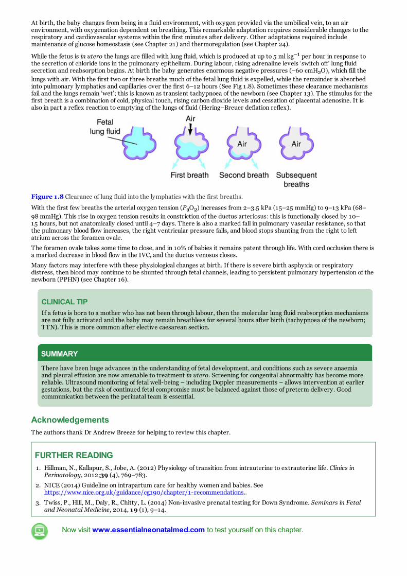
While the fetus is in utero the lungs are filled with lung fluid, which is produced at up to 5 ml kg−1 per hour in response tothe secretion of chloride ions in the pulmonary epithelium. During labour, rising adrenaline levels ‘switch off’ lung fluidsecretion and reabsorption begins. At birth the baby generates enormous negative pressures (–60 cmH2O), which fill thelungs with air. With the first two or three breaths much of the fetal lung fluid is expelled, while the remainder is absorbedinto pulmonary lymphatics and capillaries over the first 6–12 hours (See Fig 1.8). Sometimes these clearance mechanismsfail and the lungs remain ‘wet’; this is known as transient tachypnoea of the newborn (see Chapter 13). The stimulus for thefirst breath is a combination of cold, physical touch, rising carbon dioxide levels and cessation of placental adenosine. It isalso in part a reflex reaction to emptying of the lungs of fluid (Hering–Breuer deflation reflex).
Figure 1.8 Clearance of lung fluid into the lymphatics with the first breaths.
With the first few breaths the arterial oxygen tension (PaO2) increases from 2–3.5 kPa (15–25 mmHg) to 9–13 kPa (68–98 mmHg). This rise in oxygen tension results in constriction of the ductus arteriosus: this is functionally closed by 10–15 hours, but not anatomically closed until 4–7 days. There is also a marked fall in pulmonary vascular resistance, so thatthe pulmonary blood flow increases, the right ventricular pressure falls, and blood stops shunting from the right to leftatrium across the foramen ovale.
The foramen ovale takes some time to close, and in 10% of babies it remains patent through life. With cord occlusion there isa marked decrease in blood flow in the IVC, and the ductus venosus closes.
Many factors may interfere with these physiological changes at birth. If there is severe birth asphyxia or respiratorydistress, then blood may continue to be shunted through fetal channels, leading to persistent pulmonary hypertension of thenewborn (PPHN) (see Chapter 16).
CLINICAL TIPIf a fetus is born to a mother who has not been through labour, then the molecular lung fluid reabsorption mechanismsare not fully activated and the baby may remain breathless for several hours after birth (tachypnoea of the newborn;TTN). This is more common after elective caesarean section.
SUMMARY
There have been huge advances in the understanding of fetal development, and conditions such as severe anaemiaand pleural effusion are now amenable to treatment in utero. Screening for congenital abnormality has become morereliable. Ultrasound monitoring of fetal well-being – including Doppler measurements – allows intervention at earliergestations, but the risk of continued fetal compromise must be balanced against those of preterm delivery. Goodcommunication between the perinatal team is essential.
AcknowledgementsThe authors thank Dr Andrew Breeze for helping to review this chapter.
FURTHER READING1. Hillman, N., Kallapur, S., Jobe, A. (2012) Physiology of transition from intrauterine to extrauterine life. Clinics in
Perinatology, 2012;39 (4), 769–783.
2. NICE (2014) Guideline on intrapartum care for healthy women and babies. Seehttps://www.nice.org.uk/guidance/cg190/chapter/1-recommendations..
3. Twiss, P., Hill, M., Daly, R., Chitty, L. (2014) Non-invasive prenatal testing for Down Syndrome. Seminars in Fetaland Neonatal Medicine, 2014, 19 (1), 9–14.
Now visit www.essentialneonatalmed.com to test yourself on this chapter.

CHAPTER 2 Perinatal epidemiology and auditKey topics
Definitions of terms commonly used in perinatal medicine
The role of perinatal and neonatal audit
Classification of perinatal deaths
Factors affecting perinatal death rates
Prevention of perinatal mortality and low birthweight
Changing trends
IntroductionConception, embryonic and fetal development, parturition and subsequent neonatal growth and development form acontinuum. Obstetricians and neonatologists have, however, arbitrarily divided this into rigid categories, which are used toaudit standards of care during the perinatal and subsequent periods. Unfortunately, international agreement regardingsome of the terminology is lacking, and definitions within this developmental continuum given here are those used in the UKand Australia. It is recommended that the international reader familiarises themselves with local terminology and data.
Definitions of terms commonly used in perinatal medicineGestational age: this is calculated from the first day of the last normal menstrual period to the date of birth, and isexpressed in completed weeks.
Term delivery occurs when the infant is born at or after 37 and before 42 weeks’ gestation.
Preterm delivery occurs if the infant is born less than 37 weeks’ gestation. In the UK and Australia, 6–9% of infantsare born preterm.
Extremely Preterm delivery occurs if the infant is born at less than 28 weeks’ gestation.
Very Preterm delivery occurs if an infant is born at or after 28 weeks’ gestation, and less than 32 weeks’ gestation.
Moderately Preterm delivery occurs if an infant is born at or after 32 weeks’ gestation and less than 34 weeks’gestation.
Late Preterm delivery refers to an infant born at or after 34 weeks’ gestation and less than 37 weeks’ gestation.
Post-term delivery occurs if the infant is born at or after 42 completed weeks of gestation. Approximately 1% ofinfants are born post-term.
A live birth is one in which there are signs of life (breathing, heartbeat or spontaneous movement) after completeexpulsion from the mother, irrespective of the gestational age or birthweight.
A stillbirth, or fetal death, in the UK is defined as an infant delivered at or after 24 weeks of pregnancy who shows nosigns of life and has no heartbeat. In Australia, stillbirth is defined as an infant born at or after 20 weeks’ gestationand/or weighing 400 g with no signs of life. As the definition varies from country to country, comparison of figures maybe misleading.
The stillbirth rate is expressed as the number of infants born dead at or after 24 weeks per 1000 live births andstillbirths.
Low birthweight (LBW) refers to any infant who weighs less than 2500 g at birth (World Health Organization, 2016).In the UK and Australia, approximately 6% of live births are LBW. These infants are either born too early (preterm), orhave grown inadequately in the uterus and are classed as ‘small for gestational age’. Some LBW infants may be bothpreterm and small for gestational age.
Very low birthweight (VLBW) infants are those who weigh less than 1500 g at birth. Approximately 1–1.5% ofliveborn infants are VLBW.
Extremely low birthweight (ELBW) infants are those who weigh less than 1000 g at birth. This category accounts forapproximately 0.7% of all births.
Small for gestational age (SGA). This term is generally synonymous with the fetus who has suffered intrauterinegrowth restriction (IUGR). Diagnosis depends on accurate assessment of gestational age (See Chapter 1) and plotting ofweight on an appropriate growth chart. There is no international consensus on the definition of SGA, which varies fromless than the 10th, 5th or 3rd percentiles or more than two standard deviations below the mean birthweight.Accordingly, incidence figures will vary. In the UK, SGA is defined as a baby weighing below the 10th centile forgestational age. Asymmetrical SGA refers to a baby whose weight is below the 10th centile, but whose head is above the10th centile. This usually indicates late-onset IUGR (See Chapter 12).
Neonatal death is death occurring within 28 days of birth in an infant whose birthweight was at least 500 g or, if theweight was not known, an infant born after at least 22 weeks’ gestation.
Neonatal death rate in the UK and Australia refers to the number of deaths within 28 days of birth of any child whohad evidence of life after birth. Birthweight and/or gestational age criteria apply as for PMR.
Perinatal mortality rate (PMR):

Extended Perinatal Mortality Rate (EPMR) refers to the total number of stillbirths and neonatal deaths per 1000registered births.
Postneonatal death rate (or late infant deaths) refers to the number of deaths of liveborn infants dying after 28 daysbut before one year of age per 1000 live births.
Infant death is death occurring within one year of birth in a liveborn infant whose birthweight was at least 500 g, or atleast 22 weeks’ gestation if the birthweight was not known. This category includes neonatal deaths as defined above.
The role of perinatal and neonatal auditBy collecting epidemiological data and monitoring clinical indicators, it is hoped to improve care and clinical practice. Auditmay identify variations in morbidity or mortality which warrants further investigation. The process can also facilitatecollaboration and research.
Outcomes for preterm infants are discussed in Chapter 11. In many countries, large networks collect outcome data and thereader is recommended to familiarise themselves with their local network. Examples of these networks include the NationalInstitute of Child Health and Human Development (NICHD), the Australia and New Zealand Neonatal Network (ANZNN),the National Neonatal Audit Programme (NNAP) in the UK, the Vermont Oxford Network in the USA, and the CanadianNeonatal Intensive Care Unit Network. The International Network for Evaluating Outcomes in Neonates (iNeo) is acollaboration between national neonatal networks including Australia and New Zealand, Canada, Israel, Japan, Spain,Sweden, Switzerland and the UK.
Classification of perinatal deathsIt is often difficult for clinicians to agree on the cause of death in a diverse group of patients. In Australia, the PerinatalSociety of Australia and New Zealand (PSANZ) have developed a hierarchical classification, the PSANZ Perinatal DeathClassification (PSANZ-PDC), to aid in the identification of the single most important factor which led to the chain of eventsthat resulted in the perinatal death, and the PSANZ-Neonatal Death Classification (PSANZ-NDC) to identify the single mostimportant factor in the neonatal period which caused the death (Chan et al., 2004).
All perinatal deaths should be reviewed by a Perinatal Mortality Committee (or a Death Review Committee), includingdeaths of infants born within the service who may have died elsewhere. Membership of the committee should bemultidisciplinary. The committee serves a number of functions which may include:
Consistent classification of all deaths according to local requirements.
Evaluation of factors surrounding and contributing to death.
Development of recommendations for improving processes of care.
Feedback to clinicians and parents based on above recommendations.
Implementation of action required based on above recommendations.
Data collection and documentation of perinatal deaths.
Perinatal loss can have significant social and psychological impacts on the family and staff. Following a perinatal death,parents should be allowed to spend private time with their baby, they should be offered the opportunity to create mementos(e.g. footprints, lock of hair). Pastoral care services should be available if requested. Mothers may require advice withregards to lactation, and ongoing support services may be necessary.
The role of autopsyThe most reliable cause of death is obtained by an experienced perinatal pathologist conducting an autopsy examination, buteven following such examination the precise cause of death may remain undetermined, particularly when the infant diesbefore birth. Even if the cause of death is known, autopsy can still have a valuable role in confirming this and may sometimesidentify previously unrecognized abnormalities. Unfortunately, autopsy rates appear to be declining. If still possible, theplacenta should also be examined by a perinatal pathologist, and in some cases this alone may elicit the aetiology of aperinatal death.
CLINICAL TIP
The role of the coroner. Most jurisdictions have a Coroners’ Act (or equivalent) which requires all reportable deaths,including those where cause of death cannot be explained with certainty or those dying in an unusual circumstance, tobe reported to the coroner or other responsible official. Usually, a stillborn child is not reportable.
Factors affecting perinatal death ratesPerinatal deaths have a wide range of causes. They can result from maternal conditions, problems in the placenta, conditionsaffecting the fetus or newborn, or a combination of the above. Sometimes the cause of perinatal death is not identifiable. The2014 MBRRACE-UK report showed the extended PMR was approximately 5.9 per 1000 live births, approximately 70% ofwhich are stillbirths and 30% are neonatal deaths (Manktelow et al., 2016). The main causes of neonatal death werecongenital abnormalities (27.9%), neurological (11.9%) and extreme prematurity (11.7%).
Maturity is of course an important factor in the PMR (see Fig. 2.1). The extreme preterm infant is discussed in Chapter 11.Multiple births have a much higher PMR, and this is discussed in Chapter 3. The sex of the fetus or infant is also important,

with males known to have a slightly higher rate than females. Mortality rates also vary between regions and countries, andhave been decreasing over time (see http://www.who.int/gho/child_health/mortality/neonatal_infant/en/)
Figure 2.1 2014 ANZNN survival data to discharge home (with 95% CI) (Full data are available in Table 30 in Chow,S.S.W., Le Marsney, R., Haslam, R., Lui, K. (2016) Report of the Australian and New Zealand Neonatal Network 2014.ANZNN, Sydney.
CLINICAL TIP
Maternal factors which increase the risk of perinatal mortality include lower social class, coexisting medical condition,poor nutrition, underutilization of antenatal services, age <20 years, age >40 years, indigenous status, and use ofassisted reproductive technology (ART).
Prevention of perinatal mortality and �low birthweightImplementation of preventive measures, particularly in developing countries, could substantially improve perinataloutcomes. Factors which may reduce perinatal mortality and low birthweight include improvement in maternal health andeducation, reduction in unplanned pregnancy, and providing prenatal care in a comprehensive and coordinated manner.
Changing trendsIn developed countries, the PMR has decreased by almost two-thirds during the past 30 years, but now appears to bestabilizing. Reasons for decreasing death rates include improved survival in low birthweight and preterm infants, improvedantenatal care, and increased use of antenatal steroids.
SUMMARY
Despite inconsistencies in classification, the collection of epidemiological data has an important role in improving theoutcomes for mothers and babies. The main causes of perinatal death are congenital abnormalities, preterm birth andmaternal conditions. All perinatal deaths should be reviewed and classified by a Perinatal Mortality Committee. Theimplementation of a number of preventive measures could help to reduce the PMR, particularly in developingcountries.
Further reading1. Chan, A., King, J.F., Flenady, V., et al. (2004) Classification of perinatal deaths: development of the Australian and New
Zealand classifications. Journal of Paediatric and Child Health, 2004;40, 340–347.
2. Manktelow, B.N., Smith, L.K., Seaton, S.E., Hyman-Taylor, P., Kurinczuk, J.J., Field, D.J., Smith, P.W., Draper, E.S., onbehalf of the MBRRACE-UK Collaboration (2016). MBRRACE-UK Perinatal Mortality Surveillance Report, UKPerinatal Deaths for Births from January to December 2014. Leicester: The Infant Mortality and Morbidity Studies,Department of Health Sciences, University of Leicester, Leicester2016.
3. World Health Organization (2016) International Statistical Classification of Diseases and Related Health Problems,1CD-10. World Health Organization, Geneva, 2016.
Now visit www.essentialneonatalmed.com to test yourself on this chapter.

CHAPTER 3 Multiple birthsKey topics
Physiology of fertilization, implantation and placenta formation
Classification of multiple pregnancy
Assisted reproductive technology
Incidence of multiple pregnancies
Parental counselling
Complications of multiple pregnancy
IntroductionMultiple pregnancies are a common problem in the neonatal nursery. Rates have increased since the introduction of assistedreproductive technology (ART). This chapter briefly discusses the physiology of fertilization and placenta formation andtheir relevance in multiple gestations. Specific complications of multiple gestations are discussed in detail.
Physiology of fertilization, implantation and placenta formation
FertilizationNormal fertilization occurs in the fallopian tube. Once a sperm enters the ovum, the process of rapid mitotic cell divisionbegins. At about 3 days, a ball of 12–16 blastomeres enters the uterine cavity and is then called a morula. Cell divisioncontinues and spaces begin to appear between the cells within the morula to form the blastocyst cavity. The outer surface ofthis cavity (called the trophoblast) has two layers – an inner cytotrophoblast and outer syncytiotrophoblast. At thispoint the embryo is referred to as a blastocyst.
ImplantationAt about 6 days after fertilization, the syncytiotrophoblast comes into contact with the uterine epithelium. If conditions arefavourable, there is rapid proliferation and erosion into the endometrium. The process of eroding into the endometrium isreferred to as implantation. As cell division of the cytotrophoblast continues, a small space appears between the inner cellmass and the outer layers; this is the beginning of the amniotic cavity. The inner cell mass will ultimately go on to formthe embryo and amnion, while the outer layers will go on to form the chorion and placenta. The cells in between the outercytotrophoblast and inner cell mass are the extraembryonic mesoderm. Spaces (the chorionic cavity) then also developin the extraembryonic mesoderm so as to form the extraembryonic somatic mesoderm, which lines the trophoblast andcovers the amnion, and the extraembryonic splanchnic mesoderm, which surrounds the yolk sac.
PlacentaAfter implantation the syncytiotrophoblast erodes the endometrial blood vessels, allowing maternal blood to form networksof lacunae. These networks are the beginning of the decidual (or maternal) layer of the placenta. The fetal layer of theplacenta results from the proliferation of synctiotrophoblasts and formation of the primary chorionic villi at approximately 2weeks after fertilization. The external layers of the trophoblast and the extraembryonic somatic mesoderm constitute thechorion.
The chorion forms the chorionic sac within which lies the embryo, in its amniotic sac. The amniotic sac continues to enlargemuch faster than the chorionic sac. This causes their walls to fuse, obliterating the chorionic cavity and forming theamniochorionic membrane, which in turn comes into contact with the decidual layer of the placenta. The developing embryois now suspended from the placenta by the umbilical cord and is contained within the amniotic cavity.
Classification of multiple pregnancyMultiple pregnancies are normally classified by the number of placentas and the number of amniotic sacs. A multiplepregnancy may arise from the spontaneous splitting of a single zygote (monozygotic) or through the fertilization of morethan one oocyte (dizygotic). If they are dizygotic, or if division occurs before implantation, they will have separate placentasand separate amniotic cavities. If the division occurs after implantation then they will share the placenta (monochorionic)and either share the amniotic cavity (monoamniotic) or have their own (diamniotic). Higher-order multiples are usuallymultizygotic (from multiple ovum), but can arise from zygotic splitting and then resplitting again. They are also classified bythe number of placentas and amniotic sacs. For example, triplets may each have their own placenta (trichorionic) and theirown amnion (triamniotic), or may be combinations of the above.
CLINICAL TIP
Twins of different sexes must be dizygotic. Dizygotic twins must have separate placentas and amnions. The placentasmay be fused along a ridge. Monochorionic (MC) twins must be identical irrespective of whether or not they aremonoamniotic (MA) or diamniotic (DA). About one in four like-sexed twins who are dichorionic (DC) are identical, butdetermination of zygosity is not possible without DNA testing.

Assisted reproductive technologyAssisted reproductive technologies (ARTs) essentially involve manipulation of the egg or sperm to facilitate pregnancy.Technically, ARTs do not include therapies that simply stimulate the ovaries to produce more eggs (e.g. clomiphene).Techniques include in-vitro fertilization (IVF), intracytoplasmic sperm injection (ICSI), gamete intrafallopian transfer(GIFT) and zygote intrafallopian transfer (ZIFT).
With the advent of ART there was an increased incidence of multiple-fetus pregnancies. This now appears to be falling againin the UK and Australia since the introduction of legislation and professional guidelines to limit the number of embryostransferred. This is not the case in other countries where no such legislation exists.
Incidence of multiple pregnanciesThe incidence of multiple births was first studied by Hellin in 1895. Following this work, the Hellinic law was described,which stated that the incidence of twins was 1/89 pregnancies and of triplets 1/892 (1/7921) and quadruplets 1/893 (1/704969). The actual incidence of multiple births varies greatly between countries due to differing rates of dizygotic twins.Factors influencing the increased rate of dizygotic twinning other than ART are maternal age 35–39 years, high parity,central African origin, tall stature, periconceptual folate and familial factors (increased risk ×4 if mother, or ×2 if sister is adizygotic twin). As opposed to dizygotic twins, the incidence of monozygotic twins is relatively stable, occurring inapproximately 3.5 per 1000 births. Reasons for the development of monozygotic twins are poorly understood. There is afivefold increased risk of monozygotic twinning of an IVF embryo.
Of all naturally conceived twins, 40% are monozygotic, and of all monozygotic twins, 63% are monochorionic diamniotic(MCDA), 33% are dichorionic diamniotic (DCDA) with either separate or fused placentas, and 4% are monochorionicmonoamniotic (MCMA).
In developed countries with routine antenatal care and ultrasound assessment the birth of undiagnosed twins is a rareevent. Antenatal diagnosis enables the assessment of chorionicity, and detection of malpresentation and complications ofmultiple gestation.
Figure 3.1 Twin peak or lambda sign. Illustration courtesy of Dr Scott Peterson, Mater Mothers’ Hospital. Reproducedwith permission of Dr Peterson.
CLINICAL TIP
Zygosity can be diagnosed on antenatal ultrasound by looking for the twin peak sign, also known as the lambda sign. Itis best seen between 10 and 14 weeks and is strongly suggestive of a dichorionic pregnancy (see Fig. 3.1).
Parental counsellingThe perinatal mortality rate is much higher for multiple pregnancies than for singletons, with a fivefold increase for twinpregnancies and a 14-fold increase for triplet pregnancies for birth, stillbirth and neonatal death. Similarly, multiplepregnancy is markedly overrepresented in neonatal morbidity (see below). Knowing the increased risks, selective reductionis an option for some parents.
Parents of multiple gestations need a great deal of support and advice with the care of their children. The motherparticularly needs encouragement to breastfeed her infants. Voluntary community groups, such as the Twins Club andTwins and Multiple Birth Association, may be helpful in providing support and counselling.
Parents often wish to know if their twins are identical, or not. This may be of benefit in terms of rearing practices, the knownconcordance of medical conditions in 70–90% of twins, and if solid organ transplant is required. The medical profession isnotorious for imprecise counselling, and multiple birth support groups have requested that zygosity testing is provided as aroutine service in cases of multiple births, regardless of the high cost of DNA testing.
Complications of multiple pregnancy
Discordant growth ratesNormally, both twins will continue to grow at the rate of a singleton fetus until 30 weeks’ gestation, after which growthrestriction may occur. As the number of fetuses increases from twins to triplets to quadruplets, the gestational age at which

birthweight first falls below that of a singleton fetus also decreases. Twins have birthweights similar to singletons up to 30weeks, but thereafter are below singleton levels; similarly, birthweights in triplets deviate from the singleton curve at 27–28weeks, and in quadruplets at approximately 26 weeks. In addition, as the fetal number increases the rate of individual fetalgrowth in the third trimester decreases. Thus, the further the multiple gestation advances and the higher the fetal number,the more individual birthweights will differ from singleton values.
Twins usually grow at nearly identical rates, the mean intertwin birthweight difference being 11%. Small for gestational age(SGA) infants have been reported in 24–40% of twin gestations. In one study, 13% of twin gestations resulted in one SGAfetus, and 9.4% of gestations resulted in two SGA fetuses. Monozygotic twins grow less well than dizygotic twins, and majordiscrepancies in weight are commoner in monozygotic twins. Causes of discrepancy are poor insertion of the cord into theplacenta in one twin and, in dichorionic twins, poor placentation of one twin. The commonest cause is fetofetal transfusion(see below).
Disappearing twin phenomenonUltrasound studies have shown that up to 50% of pregnancies initially found containing twins in the first 8 weeks ofgestation absorb one conceptus and continue as singleton pregnancies. This has been referred to as the ‘disappearing twin’or ‘vanishing twin’ phenomenon. Rarely, the dead twin is not reabsorbed and fetus papyraceous ensues. It is suggested thatthe disruption to the surviving twin as a result of the early fetal loss of the co-twin predisposes the baby to cerebral palsy,but there is currently little direct evidence for this.
CLINICAL TIP
Fetus papyraceous occurs if one twin dies early in the first trimester and becomes mummified. The other twincontinues to grow normally. There is a strong association with the occurrence of embolic phenomena (from the deadfetus) affecting the surviving twin. Multicystic encephaloleukomalacia and multiple cutis aplasia have been reported inthe surviving twin.
Conjoined twinsConjoined twins are rare, occurring in 1–2 per 100 000 pregnancies. The fetuses can be connected via the head(craniopagus), chest (thoracopagus) or abdomen, and will often share organs as well (e.g. heart, liver). The site of connectionand nature of organ sharing will ultimately determine the prognosis. The condition is often incompatible with life.
PrematurityThe incidence of premature delivery in twin pregnancies is 20–30%, and the incidence is greater in higher-order multiplepregnancies. Typically, 50% of twins weigh less than 2500 g at birth. Premature birth relates to increased intrauterinevolume, premature rupture of the membranes, complications of multiple pregnancy (e.g. disconcordant growth, twin-to-twin transfusion syndrome) and third-trimester bleeding.
MalpresentationMultiple gestations may present in the uterus in many ways, some of which predispose them to trauma. Locked twins mayoccur as a result of unstable lies and cause obstruction to delivery. The types and frequency of presentation in twinpregnancies are listed in Table 3.1.
Table 3.1 Types and frequency of presentation in twin pregnancies.
Type of presentation Percentage of pregnancies
Vertex and vertex 45%
Vertex and breech 40%
Breech and breech 10%
Vertex and transverse 3%
Breech and transverse 1.5%
Transverse and transverse 0.5%
Congenital malformationsIn multiple gestations, the incidence of major malformation is 2%, and of minor malformation 4%. It is often stated thatmonozygotic twinning results from a chance teratogenic event. Malformations due to the event of twinning itself areconjoined twins, amorphous twins, sirenomelia, holoprosencephaly, neural tube defects and anencephaly. Othermalformations result from vascular interchange between monozygotic twins, and include acardia in one twin, disseminatedintravascular coagulation (DIC) from embolization, with defects such as microcephaly, hydranencephaly, intestinal atresia,cutis aplasia and limb amputation. There is an increased incidence of major birth defects in infants conceived via ICSI andIVF.
Postural deformities

Deformations such as talipes and hip dysplasia result from intrauterine crowding, occur in multiple gestations, and areindependent of zygosity.
Twin-to-twin transfusion (TTTS)In monochorionic twins, there are connections on the placenta that join the twins’ circulations. The majority of theseconnections are balanced, but in up to 15% of cases they are unbalanced. When the connection is unbalanced, one twin (thedonor twin) will donate some blood volume to the other twin (the recipient twin). Quintero staging is commonly used todefine the severity of TTTS. In TTTS Quintero stage I, the donor twin has decreased urine output, causing oligohydramnios,while the recipient twin has increased urine output, causing polyhdramnios. As TTTS becomes more severe, the donor twindevelops very severe oligohydramnios and becomes ‘stuck’ and immobile. TTTS Quintero stage V is the most severe andoccurs when one of the twins dies. TTTS is normally diagnosed on ultrasound when the amniotic fluid around the donor twinis 2 cm or less and the amniotic fluid around the recipient is 8 cm or more. There may also be changes in the umbilical cordblood flow, and in severe cases hydrops fetalis may develop in the recipient. It is recognized in the neonatal unit by eitherdiscordant growth (>20% birthweight difference), which is referred to as chronic TTTS, or a haemoglobin difference or morethan 5 g dl−1 (acute TTTS).
If one of the twins dies in utero, this may result in acute transfusion from the surviving twin. This can cause subsequentdeath of the other fetus, or if it survives can result in significant long-term disability. Overall, untreated TTTS will result in afetal loss of up to 90% and is associated with significant morbidity in survivors. Fetoscopic laser ablation of thecommunicating vessels can be used to successfully stop the process of TTTS and allows recovery in the twins. Suchtreatment is normally performed between 16 and 28 weeks’ gestation, and has been shown to decrease the risk of cerebralpalsy and neurodevelopmental disability in survivors. The risks of fetal laser ablation include preterm labour, pretermpremature rupture of membranes and fetal loss. Fetal medicine specialists now recommend scanning all monochorionictwins every 2 weeks to detect discordant amniotic fluid volume.
In the neonate, rapid treatment of the anaemic twin may be necessary (See Chapter 16), and the plethoric twin may requirea dilutional exchange transfusion. Because of the high risk of long-term neurodevelopmental disability, all TTTS survivorsshould receive long-term follow-up.
Twin anaemia polycythaemia sequence (TAPS)TAPS also occurs in monochorionic pregnancies when there is a connection in the placenta between the twins, allowing onetwin to donate blood to the other. In this situation there is a difference between the haemoglobin concentrations without theamniotic fluid disconcordance. Severity is based on the intertwin haemoglobin difference. As in TTTS, fetoscopic laserablation of the communicating vessels can also be used to successfully stop the process.
Cord entanglementIf the twins share the same amniotic cavity there is a chance that their umbilical cords can become intertwined, resulting ina potential obstruction of blood supply from the placenta to one (or both) of the fetuses. The umbilical cords of monoamniotictwins can start to become entwined within the first trimester of pregnancy. As the pregnancy progresses there is increasedrisk of cord compression. Cord entanglement can be monitored using Doppler ultrasound. As the pregnancy progresses,there is an increased risk of fetal loss, particularly after 32 weeks’ gestation.
CLINICAL TIPElective delivery at 32 weeks’ gestation is recommended for monoamniotic twins. Appropriate antenatal steroidsshould be administered prior to elective delivery.
InfectionThis occurs much more commonly in the first twin, and probably relates to premature rupture of the membranes of the firstamniotic sac.
Hypoxic ischaemic encephalopathyThe second twin is more vulnerable to hypoxic ischaemic encephalopathy (HIE) than the first twin. This is related todelayed delivery, cord prolapse, placental separation and malpresentation.
CLINICAL TIPNot only are second twins at higher risk of HIE, they also have higher rates of respiratory distress syndrome andhypoglycaemia.
Respiratory distress syndromeOverall, there is an 8.5% incidence of respiratory distress syndrome (RDS) in twins, with 29% of preterm twins beingdiagnosed as having RDS.
Neurodevelopmental outcomesChildren born from multiple pregnancies have much higher rates of cerebral palsy, learning difficulties and behaviouralproblems. Probable antecedents are premature birth, growth restriction, birth trauma, infection, hypoxia and theintrauterine demise of one twin. However, there still appears to be an increased risk even when the above confounders areaccounted for.

Twins are over-represented in populations with cerebral palsy (5–10% in most studies), particularly spastic diplegia. Therisk of a twin developing cerebral palsy is five- to sixfold higher than in a singleton pregnancy, and the risk in a triplet is 17-to 20-fold higher than in a singleton. Typical rates of cerebral palsy per 1000 births are singleton 2.3, twin 12.6 and triplet44.8. This risk is particularly increased where a monozygotic co-twin has died in utero. In all such cases the brain of thesurviving twin must be carefully examined with ultrasound or MRI to detect evidence of cerebral infarction (most probablyperiventricular leukomalacia). It is estimated that the risk of cerebral palsy in a surviving monozygotic twin whose co-twindies is 12-fold greater than when both twins survive.
SUMMARY
Multiple gestations may arise spontaneously or be due to ART. The introduction of ART significantly increased theincidence of multiple gestations, but careful implementation of legislation and guidelines can reduce their occurrence.Multiple gestations require a significant burden of surveillance and treatment, both antenatally and postnatally.Multiple gestations carry increased risk of mortality and morbidity. Parents need to be counselled extensively aboutrisks prior to undergoing ART and early in multiple pregnancy.
Further reading1. Eddleman, K.A., Stone, J. (2005) Long-term outcomes in multiple gestations. Clinics in Perinatology, 32 (2), 495–
513.
2. Fisk, N.M., Duncombe, G.J., Sullivan, M.H.F. (2009) The basic and clinical science of twin to twin transfusionsyndrome. Placenta, 30, 379–390.
3. Moore, K.L., Persaud, T.V.N., Torchia, M.G. (2015) The Developing Human: Clinically Orientated Embryology, 10thedition. Saunders Elsevier, Philadelphia, 2015.
Now visit www.essentialneonatalmed.com to test yourself on this chapter.

CHAPTER 4 Neonatal consequences of maternal conditionsKey topics
Congenital anomalies: malformations and deformations
Congenital anomalies associated with teratogens
Congenital malformation secondary to maternal infections
Consequences of maternal substance misuse
Neonatal manifestations of maternal medical diseases
IntroductionThe developing fetus is hugely influenced by its mother – 50% of its genes and all of its mitochondria are from the mother.Her womb provides the nurturing supportive environment and all the nutrition required during fetal development. It istherefore not surprising that if this environment is disturbed by maternal disease or behaviour, there may be consequencesfor the fetus, especially if that disturbance occurs in the first trimester during critical organ development. This chapter willconsider some of the fetal consequences of maternal conditions. Genetic conditions are discussed in Chapter 8.
Congenital anomalies: malformations and deformationsEstimates of the incidence of congenital anomalies vary widely, and range from 1% to 7% of live births, depending on thedefinition of what constitutes an anomaly. In about 2.5–3% there is a major congenital anomaly, but the incidence is 7% ifminor lesions such as skin haemangiomas and preauricular skin tags are included. It is highest in preterm and small forgestational age (SGA) infants. Congenital anomalies are the leading cause of perinatal mortality (20–25% of all perinataldeaths) and postneonatal deaths (25–30%). In addition, they constitute 25–30% of admissions to children’s hospitals andhave major financial costs for society.
Congenital anomalies may be classified into deformations and malformations:
Deformations result from late changes in previously normal structures by destructive pathological processes orintrauterine (extrinsic) forces; for example, talipes, hydrocephalus and bowel atresia.
Malformations result from a disturbance of growth during embryogenesis; for example, congenital heart disease.Some defects may arise by both mechanisms.
DeformationsDeformations may be either single or multiple. They have many common aetiological factors, which are listed in Box 4.1.
Extrinsic forces may cause a single localized deformation, such as talipes equinovarus or a deformation sequence.Examples of the latter are the oligohydramnios sequence, with contractures, facial dysmorphism and pulmonary hypoplasia,or the breech deformation sequence. Intrauterine contractures that give rise to joint fixation are known as arthrogryposis.The types of problems that lead to prenatal joint contractures are shown in Figure 4.1.
Figure 4.1 Problems leading to joint contractures.
Box 4.1 Risk factors for deformation.
Oligohydramnios.
Uterine abnormalities, e.g. fibroids, bicornuate or septate uterus.
Multiple pregnancy.
Abnormal presentation, e.g. breech position.
Intrauterine growth restriction.
Malformations
IncidenceMajor congenital anomalies are either lethal or significantly affect the individual’s function or appearance. Minor anomalieshave no functional or major cosmetic importance.

National registers of congenital malformations are available, but it is difficult to compare overall rates because of reportingdifferences. Most registers only include abnormalities present at birth. Due to miscarriage, stillbirth or termination ofpregnancy the overall incidence may be much higher. There may also be large regional variations within a single country.Registers are increasingly including metabolic disorders and genetic disorders (e.g. cystic fibrosis) even if they have nophysical characteristics present at birth.
The incidence of congenital malformations in the UK in 2014 was about 2.4% of all births (1 in 41), including terminations ofpregnancy. The commonest major congenital anomalies involve the central nervous and cardiovascular systems. Overall,about 60% are diagnosed antenatally and 45% result in termination of pregnancy. The incidence of neural tube defects isincreasing by about 5% per year, while the incidence of limb reduction defects is reducing by 8% per year. The incidences ofthe common congenital malformations in Europe are listed in Table 4.1.
Table 4.1 Major congenital anomalies by major anatomical systems (Eurocat European Congenital Anomalies Register2008–2012).
Congenital anomaly Rate (per 10 000 live births, fetal deaths and ToP for fetal anomaly)
Central nervous system 25
Spina bifida 4.8
Anencephaly 3.7
Encephalocoele 1.1
Congenital hydrocephalus 5.8
Cardiovascular system All 82, severe 23
Transposition of great vessels 3.5
Hypoplastic left heart 2.8
Coarctation of aorta 3.8
Respiratory system 4.1
Congenital diaphragmatic hernia
Cystic adenomatous malformation
2.8
1.1
Gastrointestinal system 19
Cleft lip ± cleft palate 8.6
Oesophageal atresia 2.6
Gastroschisis 2.8
Urogenital system 35
Hypospadias 18.2
Multicystic renal dysplasia 3.3
Posterior urethral valves 0.9
Skeletal and limb 42
Polydactyly
Syndactyly
Talipes
VACTERL
8.8
5.1
10.9
0.52
Trisomy 21 23
Trisomy 13 2.1
Trisomy 18 5.4
ToP, termination of pregnancy.
Causes of congenital malformations

Congenital anomalies result from a wide variety of mechanisms ranging from genetic (monogenic and polygenic) toenvironmental factors (teratogenesis), maternal metabolic disease, drugs and chemicals, infectious agents and irradiation(Table 4.2).
Table 4.2 Causes of congenital malformations.
Cause Estimated contribution (%)
Genetic (mendelian inheritance) 20
Chromosomal 10
Polygenic 25–30
Teratogenic effect: 6–9
Congenital infection 2–3
Drugs and environmental pollutants/insecticides 2–3
Maternal metabolic disease 1–2
Radiation exposure <1
Unknown/idiopathic 35–40
Genetic malformationsThese are discussed in Chapters 6 and 8.
Congenital anomalies associated with teratogensA teratogen is an agent (chemical, drug, virus or radiation) that affects the well-being of the fetus. A fetotoxin is an agentcausing damage of any kind to the fetus, and a mutagen is an agent that causes a permanent transmissible change in thegenetic material. The commonest potential teratogens are medications, and so in general drugs should be avoided duringpregnancy and, if essential, their safety profile must be checked before prescribing. Unfortunately, with some newer agentshuman safety data is lacking and animal data must be used to make a careful risk–benefit assessment. This is whycongenital anomaly registers are important, to identify unexpected clusters of congenital anomalies.
The critical periods in embryogenic development have been extrapolated from the effects of rubella exposure, and areexpressed in days from time of conception (not last menstrual period) in Table 4.3.
Table 4.3 Major organ system risk periods.
Organ Risk period (days after conception)
Brain 15–25
Eye 25–40
Heart 20–40
Limbs 24–36
Ear 40–60
During the first 2 weeks of development from conception to the first missed period the embryo is resistant to anyteratogenic effects of medicines. The critical period begins when organ development starts at 17 days after conception, andextends until this is complete at 60–70 days. In general, exposure to medicines beyond 70 days after conception is notassociated with major birth defects. However, drugs can interfere with functional development of organ systems in thesecond and third trimesters.
Pharmaceutical agentsDrugs may act by interfering with embryogenesis or by exerting their pharmacological actions on developing fetal organs.The timing and dose of agent, the efficiency with which the mother metabolizes the agent, placental transfer and theindividual susceptibility of the fetus are important. Table 4.4 lists some of the more common teratogenic drugs.

Table 4.4 Drugs as teratogens.
Hormones Progestogens, diethyl stilboestrol, male sex hormones
Antipsychotics, hypnotics, anticonvulsants Lithium, haloperidol, thalidomide
Anticonvulsants Hydantoins, sodium valproate, carbamazepine, primidone, phenobarbitone
Antimicrobials Tetracycline, chloramphenicol, streptomycin, flucytosine, amphotericin B
Antineoplastics Alkylating agents, folic acid antagonists
Anticoagulants Warfarin
Antithyroids Iodine, carbimazole, propylthiouracil
Antivirals Ribavirin
Hypoglycaemics Biguanides, sulphonylureas
Vitamin A analogues Isotretinoin, etretinate
Others Toluene, alcohol, marijuana, narcotics
IrradiationExposure to radiation in pregnancy is now rare, but previous experience with X-rays, atomic bomb irradiation and nuclearaccidents confirmed major teratogenic effects, including microcephaly. Carcinogenic effects and mutagenic effects (changesto DNA which can be passed to future generations) are also described.
ChemicalsPesticides and waste products have not been subjected to rigorous teratogenic studies in humans. However, experience withthe dioxin contaminant in the insecticide 2,4,5-T and its possible association with spina bifida and Potter,s syndrome suggestthat rigorous surveillance of chemicals is necessary. Long-term prenatal exposure to organic mercury causes disturbedbrain development. Methyl isocyanate gas released in the Bhopal disaster led to an increase in congenital anomalies in theoffspring of those exposed (mutagenic effect).
Congenital malformation secondary to maternal infectionsPrenatal infections cause inflammation which interferes with cell division. This can lead to congenital deafness, cataracts andlimb defects. Examples are outlined in Table 4.5. Infections are described in Chapter 10.
Table 4.5 Teratogenic effects of maternal infection.
Infection Effect
Rubella Heart defects
CNS defects
Eye defects (cataract)
Deafness
Toxoplasmosis Cerebral calcification
Epilepsy, Hydrocephalus
Chorioretinitis
Syphilis Skin lesions, saddle nose, osteochondritis
Varicella (herpes zoster) Brain, skeletal and eye defects
Dermatomal scarring
Parvovirus B19 Severe fetal anaemia, hydrops
Cytomegalovirus (CMV) Microcephaly, retinitis, deafness
Herpes simplex Microcephaly or hydranencephaly
Influenza Teratogenic effects from fever. Increased risk of schizophrenia
Zica virus Arrested CNS development with severe microcephaly

FeverVarious studies have suggested that it may be the high fever associated with viral infection (e.g. influenza) that causesmalformation, and may also be an aetiological factor in the later development of schizophrenia. Sauna baths and faultyelectric blankets producing very high body temperatures at a critical period of embryogenesis may be teratogenic.
Consequences of maternal substance misuseAll drug ingestion during pregnancy carries risk to the developing fetus, and maternal substance misuse can cause fetalmalformation and sometimes neonatal withdrawal. Cigarettes, alcohol, opiates, amphetamines and cocaine are all known tocause harm.
Fetal alcohol spectrum disorderFetal alcohol spectrum disorder (FASD) is the generic term covering fetal alcohol syndrome, fetal alcohol effects and alcohol-related neurodevelopmental disorders.
Excessive alcohol intake during fetal organogenesis can result in a specific syndrome of facial abnormalities, growth failure,microcephaly and skeletal and visceral abnormalities. This syndrome was identified in 1973 as the fetal alcohol syndrome.The facial features include hypoplasia of the mid-face, with beaking of the forehead; sunken nasal bridge; small upturnednose; micrognathia, a prominent philtrum and ear deformities (Fig. 4.2). Intrauterine growth retardation (IUGR) is usual,with poor postnatal growth. Major abnormalities include cleft lip and palate, and limb, ocular, cardiac and renalmalformations. There is usually delayed psychomotor development. Approximately one-third of infants born to chronicalcoholic women develop the fetal alcohol syndrome, whereas others may be growth-restricted or demonstrate only minorfeatures. Neonatal withdrawal symptoms, including convulsions, may occur.
Figure 4.2 Infant with typical features of fetal alcohol syndrome. From Lissauer, T. and Fanaroff, A. A. (2011)Neonatology at a Glance, 2nd edition. © 2011, Blackwell Publishing Ltd. Reproduced with permission of John Wiley & Sons.
It is not known whether social drinking of alcohol in pregnancy is associated with IUGR, but it is unlikely that veryoccasional consumption of small quantities of alcohol during later pregnancy will seriously adversely affect the fetus.However, current recommendation is to avoid all alcohol consumption during pregnancy.
There is no laboratory test for FASD. Diagnosis relies on a pattern of abnormalities that makes up the disorder, along with ahistory of alcohol misuse during pregnancy. Sometimes, the phenotype becomes more obvious in childhood. Various scoringsystems are in use, such as the four-digit diagnostic code (see Box 4.2).
Box 4.2 The four-digit diagnostic code for fetal alcohol spectrum disorder (FASD).
Diagnosis of FASD is made using four key diagnostic features:
Growth restriction (weight or length <10th centile)
Fetal alcohol syndrome facial phenotype (small eye openings, smooth philtrum, thin upper lip)
CNS damage/dysfunction
Prenatal alcohol exposure
Acute alcohol withdrawal in the neonateNeonates born to heavily alcoholic mothers (see above) can show early withdrawal at 3–12 hours with hyperactivity,irritability, excessive crying and poor feeding. Extreme tremulousness and seizures may occur and can be controlled withphenobarbitone. Withdrawal occurs independently of fetal alcohol syndrome.
Smoking in pregnancyAlthough there is overwhelming evidence of harm to the fetus and mother from smoking, up to 15% of pregnant women stillsmoke. Smoking has a dose-dependent effect on growth due to impaired uterine perfusion, with placental changes, increasedcarboxyhaemoglobin levels and increased fetal erythropoiesis. The risks of pre-eclampsia are slightly reduced. Passiveexposure to paternal smoking has almost as great an effect. Complications in smokers include subfertility, spontaneousabortion, impaired fetal growth, placenta praevia, placental abruption, amniotic fluid infection and premature rupture of themembranes. Children of smokers demonstrate increased hyperactivity at 4 years, reduced stature and mental function, and

increased risk of sudden infant death syndrome (SIDS) and malignancy. Smoking is not associated with any specificcongenital abnormalities.
Infants of substance-misusing mothersNeonatal withdrawal symptoms have been reported with alcohol, amphetamines, barbiturates, codeine, ethchlorvynol,heroin, pethidine, methadone, morphine and pentazocine. Methamphetamine (‘ice’) and ‘crack’ cocaine are currently ofmajor concern, but drug-using practices vary according to availability, geography and price.
Opiate use in pregnancyHeroin and methadone are the narcotic drugs most frequently abused during pregnancy. Their illicit use has been associatedwith increased fetal and neonatal deaths. Increased IUGR and prematurity means that the average birthweight of infantsborn to heroin addicts is 2500 g. The fetal brain becomes acclimatized to the chronic stimulation of opiate receptors in thebrainstem. After birth, the baby experiences a sudden cessation of this stimulation and increased adrenergic activity of thelocus caeruleus causes ‘withdrawal’ symptoms. Women should be encouraged to attend an addiction unit that can managetheir antenatal care and switch them to a safer alternative, such as prescribed methadone or buprenorphine.
About 70% of exposed infants exhibit withdrawal symptoms, usually within 48 hours of birth, although they can be delayedfor up to a week. Signs include extreme jitteriness, tachycardia, vomiting, diarrhoea and fever (see Table 4.6). Convulsionsoccur rarely. Infants born to drug-addicted mothers may continue to show irritable or restless behaviour for a number ofmonths after birth. Withdrawal can also occur if the mother has been on high doses of prescribed opiates for analgesia.
Table 4.6 Withdrawal symptoms seen in neonates born to opiate-abusing mothers.
Central nervous system Irritability and high-pitched cry
Hyperactivity with reduced periods of sleep
Tremors
Increased tone
Convulsions (rare)
Gastrointestinal Poor feeding
Vomiting
Diarrhoea
Other autonomic symptoms Sweating
Fever
Frequent yawning
Snuffles and sneezing
Tachycardia
Management of neonatal abstinence syndromeInfants born to drug-addicted mothers should be carefully monitored for withdrawal symptoms. About 30% of infants withdrug withdrawal can be managed conservatively. Methods include swaddling, frequent feeds and decreased sensorystimulation. Breastfeeding should be encouraged, as the small amount of opiate excreted in breast milk may help alleviatesymptoms.
If severe signs of withdrawal are present, drug treatment is necessary and the first-line treatment is to give opiates such asoral morphine syrup. A structured symptom scoring system can help guide whether to start treatment. Various scoringsystems are available; the most commonly used is the Finnegan score, which allocates a score to each of the symptomsshown in Table 4.6. Seizures should be treated with intravenous morphine, followed by anticonvulsants if the seizurecontinues. Non-specific sedatives such as phenobarbitone, diazepam or chlorpromazine have been used for neonatalabstinence syndrome (NAS), but the evidence for their use is not strong. As second-line treatment phenobarbitone orclonidine may be added to morphine replacement for those babies with polydrug exposure.
Screening for hepatitis B and C and HIV infection is important. If the mother has active hepatitis B the baby must be givenimmunoglobulin and vaccinated (see Chapter 10). In the UK, hepatitis B vaccination is recommended if there areintravenous drug users in the household.
PrognosisThis depends at least in part on the socioeconomic background of the family. Infants exhibit behavioural and physicalfeatures reflecting central and systemic dysfunction. These infants are at increased risk of SIDS, and they and their familiesrequire intensive follow-up after discharge by medical and community health and social services. Many will require fosterparenting.
Cocaine use in pregnancyPerinatal cocaine or ‘crack’ cocaine use is a major problem in the USA and elsewhere. Cocaine is a potent vasoconstrictoraffecting the uteroplacental bed as well as the fetal vessels. It causes maternal and fetal tachycardia and severe

hypertension. Cocaine abuse causes first-trimester abortion, placental abruption and premature birth. Malformationsinclude hydronephrosis, cryptorchidism, skeletal defects with delayed ossification, exencephaly and eye anomalies. Cerebralartery infarction due to the vasoconstriction is most likely to occur in the second and third trimesters.
Neonatal symptoms from cocaine exposure are often seen early, while drug levels are high. They include jitteriness,irritability, disturbed sleep–wake cycle and later neurodevelopmental impairment. Breastfeeding should be avoided if themother continues to take cocaine.
CLINICAL TIP
The easiest way to confirm which substances may be causing neonatal withdrawal symptoms (if there is doubt) is askthe mother to provide a urine sample for toxicology. Urine and meconium from the baby within 72 hours of birth canalso be analysed. Hair samples from the baby can be forensically analysed for drug exposure during the preceding 2–3months, although in practice this is rarely necessary.
Neonatal manifestations of maternal medical diseasesMany acute maternal conditions such as septicaemia, hypertension, seizures or collapse will have adverse effects on thefetus. These are discussed elsewhere, but this section considers the specific effects of certain chronic maternal medicalconditions on the fetus and newborn. Women with these conditions need appropriate preconception counselling and oftenrequire ‘shared care’ between their physician and obstetrician.
Diabetes mellitusPoorly controlled maternal diabetes predisposes to an increased risk of miscarriage and also to late intrauterine death.Diabetes carries a three- to fivefold increase in congenital malformations, and this risk is proportional to the level ofglycosylated haemoglobin (HbA1c), particularly during the period surrounding conception and early embryogenesis.Malformations include caudal regression syndrome (with sacral agenesis), which is 800-fold more common in diabeticpregnancies, hypoplastic left colon, renal vein thrombosis and congenital heart disease, especially transposition of the greatarteries (TGA) which is 20-fold more common. Fetal hyperinsulinaemia (in response to high circulating glucose levels) actsas a growth hormone, causing macrosomia and hypertrophic cardiomyopathy. If the diabetic woman is well controlled oninsulin and her blood sugars remain in the normal range before she becomes pregnant, the risk of congenital malformation ismuch reduced.
After delivery, the supply of glucose to the fetus stops abruptly, but the hyperinsulinaemia may persist, leading tohypoglycaemia. Breastfeeding is protective and some mothers are even encouraged to express and store colostrum beforebirth, for use in the early hours after delivery. The management of hypoglycaemia is discussed in Chapter 21.
Maternal hyperthyroidism (Graves, disease)Maternal hyperthyroidism results in transient neonatal thyrotoxicosis in approximately 10–20% of pregnancies (seeChapter 21). The best predictors are the outcome of previous siblings and assays of thyroid-stimulating immunoglobulin andthyroid receptor-binding inhibitors.
Maternal idiopathic thrombocytopeniaAnti-platelet IgG antibodies cross the placenta and can cause a transient low platelet count in the neonate. The risks ofserious intracranial bleeding are low. This is described in detail in Chapter 20.
Maternal systemic lupus erythematosusVasculopathy associated with maternal systemic lupus erythematosus (SLE) strongly increases the risk of recurrentmiscarriage. This can be reduced by low-dose aspirin, low-molecular-weight heparin and even plasmapheresis to maintainthe pregnancy. Mothers with SLE have a 0.5–2% chance of having a baby affected by congenital heart block due to thepresence of anti-Ro and anti-La antibodies, which can cross the placenta and permanently damage the conduction system inthe fetal heart. This may require treatment with a pacemaker (see Chapter 16).
Transient neonatal myaestheniaCongenital myasthenia is usually inherited as an autosomal recessive trait. However, occasionally the baby may developtransient weakness due to maternal acetylcholine receptor (AchR) IgG antibodies crossing the placenta. The diagnosis isoften known in the mother, but as onset occurs within hours of birth, the neonatal team must be prepared to supportfeeding, and sometimes respiration. The administration of anticholinesterase (neostigmine) leads to a rapid improvementand is diagnostic. Recovery usually occurs within 2 months as the levels of circulating antibodies wane.
Maternal epilepsyWomen with epilepsy must ensure they plan pregnancy carefully. They should be switched to an anti-convulsant that isrelatively safe in pregnancy, ideally well before conception so that good control can be established with the new agent.Seizures during pregnancy can cause fetal hypoxia. Sodium valproate is no longer recommended in women and girls of child-bearing age, unless there is no alternative, due to the high risks of teratogenicity.

SUMMARY
A healthy uterine environment is essential for normal fetal development. Although fetuses can continue to thrivedespite significant changes to the mother’s environment (e.g. brief periods of malnutrition, extremes of hot and cold),they can also be permanently affected by maternal diseases or by exposure to teratogenic drugs. Whenever a babybecomes ill or shows unusual symptoms, it is very important to take a thorough maternal and pregnancy history.
Mothers with pre-existing medical conditions should consult their physicians prior to conception to plan optimaltreatment before and during pregnancy. In all pregnant women, drugs should be prescribed only if they are known tobe safe. Tobacco, alcohol and street drugs should be avoided altogether.
Further reading1. Kocherlakota, P. (2014) Neonatal Abstinence Syndrome. Pediatrics, 134 (2), e547–e561.
2. Ogata, E.S. (2010) Problems of the Infant of the Diabetic Mother. NeoReviews, 11 (11), e627–e631.
Now visit www.essentialneonatalmed.com to test yourself on this chapter.

CHAPTER 5 Resuscitation at birthKey topics
Fetal responses during labour
Fetal and neonatal responses to perinatal asphyxia
Perinatal asphyxia
Assessment of the infant at birth
Stabilization at birth
Resuscitation
Post-resuscitation care of the asphyxiated infant
IntroductionIt is important to remember that the vast majority of babies are born healthy and do not need any intervention. However,perinatal asphyxia is the most frequent and serious preventable problem for the newborn baby. Severe perinatal asphyxiamay lead to a severely ill baby with major long-term sequelae such as intellectual impairment, cerebral palsy, blindness andepilepsy. Good perinatal care can minimize the risks of hypoxia. Neonatal depression at birth (Apgar score ≤6 at 1 min)occurs in about 14% of all births and is often unpredictable, so all professionals who are present at deliveries should havebasic life-support skills. Expert help capable of advanced resuscitation must be quickly available. Perinatal asphyxia remainsa major source of neonatal morbidity in developed countries, and ranks with perinatal infection and prematurity as the causeof at least two-thirds of neonatal mortality in developing countries. Owing to problems with definition, reported incidences infull-term neonates in developed countries vary from 0.5 to 4 per 1000 live births, and are higher for very low birthweight(VLBW) infants.
Fetal responses during labourEvery contraction during normal labour interrupts flow in the uterine arteries and may cause relative hypoxia andhypoperfusion. This can be considered ‘physiological’ hypoxia–ischaemia. During these episodes the fetus deploys variousprotective mechanisms (see Box 5.1), and only if these responses become overwhelmed does the fetus suffer injury. Thesefetal adaptations are designed to maintain function in vital organs such as the brain and myocardium. They include relativefetal bradycardia during contractions, and the ‘diving seal’ reflex. Transient periods of asphyxia may also induce anaerobicmetabolism in the fetal brain and myocardium, with utilization of ketones and lactate as alternate fuels.
Box 5.1 Responses of the normal fetus to transient episodes of ‘asphyxia’ in labour.
Redirection of blood flow towards:
Brain
Myocardium
Adrenals
and away from:
Skin
Bowel
Muscles
‘Diving seal reflex’ comprising:
Bradycardia
Increased blood pressure
Anaerobic metabolism resulting in metabolic acidosis
Fetal and neonatal responses to perinatal asphyxiaOnce the normal adaptive mechanisms to perinatal hypoxia have been exhausted, the fetus will show profound butpredictable reactions to ongoing hypoxia. These predominantly affect the respiratory and cardiovascular systems, and areobserved in babies born immediately after an acute intrapartum asphyxial event. However, with a severe insult early inlabour or a delayed delivery, many of these changes will be taking place in the unborn fetus (see Fig. 5.1). In the mostextreme cases this leads to still-birth, but for most babies adequate resuscitation can restore the infant to a normalphysiological state.

Figure 5.1 The physiological effect of acute asphyxia and the response to resuscitation. Illustration courtesy of Dr SamRichmond.
Respiratory activityInitially there is increased respiratory activity. When the respiratory centre in the brain becomes hypoxic there is then aperiod of apnoea (primary apnoea). Secondary respiratory centres in the spinal cord (which are normally inhibited by thebrain) then trigger a series of slower gasps, which eventually become less frequent until terminal apnoea occurs.Spontaneous recovery can only occur if the baby is able to breathe through an open airway before terminal apnoeabegins. Once terminal apnoea has occurred the baby will die without active resuscitation. The precise interval between acomplete asphyxial insult and terminal apnoea in the human fetus is not known as this often occurs before birth; however,extrapolation from animal studies and postnatal collapse suggests that newborns can withstand 10–15 min of severeasphyxia. Beyond this time, irreversible neurological damage or death will occur.
Cardiovascular activityHeart rate changes occur simultaneously with the respiratory changes described above. Initially, there is tachycardia,followed by a decline to about 60 bpm. This is mediated by vagal stimulation and the heart is able to continue to beat bymetabolizing myocardial glycogen stores. Central blood pressure is maintained by peripheral vasoconstriction and byincreasing stroke volume in response to bradycardia. Transient rises in heart rate and blood pressure occur when spinalgasps develop, but the heart rate and blood pressure fall again as apnoea develops. This is due to myocardial anoxia, andwithout resuscitation the baby will die. The pulmonary vascular resistance increases dramatically with terminal apnoea, andthe newborn circulation generally tends to revert to a fetal state. However, with effective resuscitation the oxygen level inthe coronary arteries rises, the heart rate increases and the baby starts to breathe spontaneously (see Fig. 5.1).
The interval between asphyxial insult and delivery is variable, and the appearance of the newborn will depend on wherethey are on the ‘physiological graph’ shown in Figure 5.1. A baby with primary apnoea will appear blue with some tone andreflex activity and the heart rate will be accelerating. The baby will usually recover spontaneously, provided the airway isopen. This can be accelerated by physical stimulation.
In contrast, the baby with terminal apnoea will not recover without intervention. In this case the baby is white(vasoconstricted) or intensely cyanosed, unresponsive and flaccid; the heart rate is less than 100 bpm and perfusion is poor.
Unfortunately, in the delivery room we may not know the severity of the asphyxia and we often cannot distinguish betweenprimary and terminal apnoea, so that prompt and effective resuscitation should be commenced in all apnoeic infants.Whether the apnoea was primary or terminal can usually be determined as the baby responds. Babies recovering fromprimary apnoea have a rapidly accelerating heart rate, and will either show a few gasps or start to breathe normally. Incontrast, babies resuscitated from terminal apnoea will show some initial rise in heart rate in response to ventilation, start togasp initially and then continue to do so for some time before normal, regular breaths ensue.
CLINICAL TIP
The presence of gasping during recovery from apnoea is an important sign to observe and record, as it indicates aprolonged episode of hypoxia.
Perinatal asphyxia
Defining perinatal asphyxiaIt is vital that perinatal asphyxia is defined accurately, both from a clinical perspective, to guide appropriate treatment, and

from a medico-legal point of view when establishing causation (see Box 5.2). Most asphyxiated babies will be born in a poorcondition with birth depression, but conversely by no means every baby with birth depression has experienced an asphyxialinsult. There are many alternative causes of birth depression (see Table 5.1).
Table 5.1 Causes of birth depression that may mimic intrapartum asphyxia.
Category Example Manifestation
Congenital brainmalformation
LissencephalyPolymicrogyria Encephalopathy, seizures
Intracerebral bleeding Unexpected haemophillia ASeverethrombocytopenia
Respiratory depression and seizures
Antenatal cerebral insult Antenatal cerebral infarction Cystic parenchymal damage
Congenital airwayanomaly
Choanal atresiaPharyngeal cyst Failure to establish open airway
Extreme prematurity RDS, inadequate respiratory effort Pulmonary insufficiency
Extrinsic lungcompression
Pleural effusion, congenital diaphragmatichernia
Pulmonary insufficiencyPulmonaryhypoplasia
Oligohydramnios Renal agenesis
Posterior urethral valvesPROM before 25weeks
Pulmonary hypoplasia
Cardiac Congenital heart block Extreme bradycardia
Drugs Maternal anaesthetic
Opiates (as analgesia or substance misuse)
MgSO4
Respiratory depression
Infection ListeriaGroup B StreptococcusHerpes simplexinfection
MeningitisPulmonaryhypertensionEncephalopathy
PROM, premature rupture of membranes; RDS, respiratory distress syndrome.
Box 5.2 Essential characteristics needed to diagnose significant perinatal asphyxia in thenewborn.
Evidence of birth depression: an Apgar score 0–3 at >5 min.
Evidence of intrapartum anaerobic metabolism:
Umbilical cord arterial pH <7.0.
Umbilical cord arterial base deficit ≥16 mmol l–1.
Neonatal neurological sequelae: early-onset encephalopathy characterized by hypotonia, seizures or coma).
Evidence of multiorgan system dysfunction in the immediate neonatal period (evidence of hypoxic–ischaemicinjury to other organs than the brain, such as the kidneys, heart and liver).
Absence of an alternative aetiology (see Table 5.1).
Clinical evidence of asphyxiaVirginia Apgar introduced the scoring system (see Table 5.2) that now bears her name in an attempt to describe thecondition of the infant shortly after birth. Although she did not intend it to refer to asphyxia, it has nevertheless becomewidely used for that purpose, and in some centres asphyxia is defined (incorrectly) on the basis of a low Apgar score alone.Using an Apgar score of 3 or less at 5 minutes as the criterion, the incidence of asphyxia is 3–9 per 1000 full-term infants. Ifasphyxia is defined as the requirement for intermittent positive-pressure ventilation for more than 1 minute, then 5 per1000 full-term infants had this condition. If it is defined by hypoxic–ischaemic encephalopathy the rate drops to 1 per 1000births.

Table 5.2 The five components of the Apgar score.
Sign 0 1 2
Heart rate Absent ≤100 bpm >100 bpm
Respiratory effort Absent Gasping or irregular Regular or strong cry
Muscle tone Limp Reduced Normal
Response to stimulation None Grimace Cough/cry
Colour Pale or blue Pink with blue extremities Pink
Medico-legal evidence of significant asphyxiaMost cerebral palsy is not asphyxial in origin and most perinatal hypoxic–ischaemic cerebral injury (90% of cases) originateswell before labour, with only a small component being solely attributed to intrapartum events. However, since childrendamaged by potentially preventable intra partum asphyxia have expensive ongoing care needs, their families may seekfinancial redress though the legal process. The legal criteria required to establish liability are summarized in Box 5.3.
CLINICAL TIP
The terms ‘birth asphyxia’ and ‘perinatal asphyxia’ were previously loosely used to describe depression at birth, butdue to the potential medico-legal implications stringent criteria must now be satisfied for their use. To stateobjectively that there was a likelihood of a prolonged acute hypoxic–ischaemic event in labour causing permanentbrain injury to a previously healthy fetus one also needs:
1. Evidence of a major ‘sentinel’ hypoxic event, such as a ruptured uterus, antepartum haemorrhage, cord prolapseor amniotic fluid embolism (i.e. something that significantly changes normal fetal oxygenation in labour). Clearevidence of a sentinel event is not always present at the time of delivery.
2. Evidence of possible fetal compromise (‘distress’) from the time of the sentinel event, such as major changes in thefetal heart rate and/or fetal acidosis on fetal blood sampling.
3. Evidence that encephalopathy is not due to other causes, such as infection, metabolic diseases or brainmalformation (see Table 5.1)
Box 5.3 Legal liability.
In order to successfully prove liability the claimant must establish:
1. That there was clear evidence of intrapartum asphyxia (causation):
Presence of clinical features described in Box 5.2 and absence of alternative diagnoses described in Table 5.1.
2. That acute asphyxial brain injury was preventable:
that the asphyxial episode could reasonably have been detected;
that an unnecessary delay in recovery occurred, due to either delayed delivery or inadequate resuscitation;
that there was another course of action or mechanism of delivery that could reasonably have been achievedwithout major risk to the mother.
3. That there was a duty of care which was breached.
Communication between obstetricians or midwives and the neonatal team is best served by replacing the term ‘fetaldistress’ with ‘fetal compromise’, followed by a further description of what is abnormal, such as ‘repetitive variabledecelerations’ or ‘fetal bradycardia of 60 bpm’. Similarly, when describing resuscitation it is much more helpful to documentthe condition of the baby, the actions taken and the clinical response rather than merely the Apgar scores. The generic term‘birth depression’ should be used until an asphyxial aetiology is established.
Causes of perinatal asphyxiaThe perinatal events most associated with hypoxic–ischaemic injury are listed in Table 5.3. Those events which presentwith birth depression and can mimic intrapartum asphyxia are shown in Table 5.1.

Table 5.3 The aetiology of perinatal hypoxia–ischaemia.
Cause Example Major effect on fetus/newborn
Antenatal
Chronic placentalinsufficiency
PET
Severe IUGR due to maternaldiabetes
Growth restriction and reduced glycogen stores leadsto poor tolerance of physiological intrapartum hypoxiaor pathological asphyxia
Acute placental failure Placental abruptionMaternal cardiac arrest
Hypoxia, acidosis
Haemorrhage Placenta previa
Vasa previa (cord runs throughmembranes)
Fetomaternal haemorrhage
Hypovolaemia, shock, poor oxygen delivery
Uterine Hyperstimulation of the uterus(oxytocin)
Uterine rupture
Hypoxia and acidosis
Umbilical cord Cord prolapse
Cord tight around neck (rare)Trueknot in cord
Interruption of blood supply (hypoxia and ischaemia)
Infection Chorioamnionitis
Group B streptococcal infection
Maternal H1N1 influenza infection
Does not cause asphyxia but may render fetusvulnerable to further insult and presentation maymimic asphyxia
Postmaturity Placental deteriorationPassage of meconium
Hypoxia–ischaemia and poor
tolerance of labour
Meconium aspiration and PPHN
Delivery
Mechanical Breech with head entrapmentShoulder dystocia
Hypoxia ± birth trauma
Prolonged 2nd stage Failure to recognize and manageabnormal�lie/presentation/cephalo-pelvicdisproportion
Hypoxia due to delayed delivery
Trauma Precipitant delivery
Difficult instrumental delivery
Intracranial haemorrhage mimics HIECervical cord injury presenting as HIE
Postnatal
Complications of out-of-hospital delivery (e.g.concealed pregnancy)
Failure to maintain open airway ordeliberate smothering (very rare)Cold environment
Failure of normal adaptation at birth
Hypoxia
Extreme hypothermia
Hypoglycaemia and PPHN
Acute postnatal collapse Accidental overlaying
Airway obstruction whilst at breast
Hypoxia
Iatrogenic Accidental oesophageal intubation
Inappropriate ‘blind’ suction
Excessive ventilation
Poorly clamped cord
Hypoxia
Vagal bradycardia
Pneumothorax – hypoxia
Haemorrhage – ischaemia

Poorly clamped cord Haemorrhage – ischaemia
Prevention of perinatal asphyxiaThe prevention of perinatal asphyxia involves the following:
Recognition of high-risk pregnancies.
Recognition of IUGR and placental insufficiency.
Accurate assessment of gestation and the use of antenatal steroids.
Appropriate management of postmaturity.
Assessment of fetoplacental function, e.g. Doppler ultrasound, fetal movements.
Appropriate intrapartum fetal heart rate monitoring.
Treatment of fetal distress in utero, e.g. mother in left lateral position, maternal oxygen and expedite delivery.
Ensuring that all birth attendants are trained in basic life support.
Good communication between maternity and neonatal teams.
Ensuring that a person with advanced resuscitation skills is available for high-risk deliveries or where there is ananticipated problem (see Table 5.4).
Table 5.4 Situations where advanced resuscitation may be needed and should be anticipated.
High-risk pregnancy Rhesus isoimmunization
Moderate to severe pre-eclampsia
Severe IUGR
Insulin-dependent diabetes
Antepartum haemorrhage
Prolonged rupture of the membranes
Abnormal labour Fetal distress, prolapsed cord
Deep transverse arrest
Cephalopelvic disproportion
Abnormal delivery Emergency (category 1) caesarean section
Heavy meconium staining of liquor
Rotational forceps delivery
Abnormal presentation Breech, face, brow, compound, shoulder
Abnormal gestation Preterm delivery
Abnormal fetus Severe oligo- or polyhydramnios
Known congenital abnormality
Multiple births
CLINICAL TIP
The degree of urgency of a caesarean section will depend on maternal and fetal factors. A classification system isshown above (RCOG, UK 2010, Reproduced with permission of the Royal College of Obstetricians andGynaecologists).
Assessment of the infant at birthTraditionally, Apgar scores (see Table 5.2) have been used to assess a baby’s condition at birth, 1 minute and 5 minutes.Unfortunately, the Apgar score has limited prognostic significance and is difficult to assess once medical intervention hasstarted. Many infants can be successfully resuscitated despite an Apgar score of 0 at birth, and may sustain no long-termneurological damage.
In view of the subjective nature of the Apgar score it is considered more appropriate to assess the infant’s condition in termsof heart rate, breathing, colour and tone. These assessments can be made at birth at regular intervals during resuscitation(see Table 5.5).

Table 5.5 Assessment during resuscitation.
Heart rate(bpm)
Breathing Colour Tone Action
Good condition >100 Crying Pink Normal Dry and give to mother
Moderate birthdepression
<100 Somerespiratoryeffort
Mildcyanosis
Reduced Open airway, consider inflation breaths
Severe depression <60 or absent Gasping or nobreathing
White Floppy Call for help, open airway and beginimmediate resuscitation
Stabilization at birthMost babies, even those from a high-risk pregnancy, are born in good condition and do not need active resuscitation.Attendants should dry the baby and wrap it in a warm towel, then make the assessments described above, while ensuringthat the airway is open (head in neutral position). There is no place for routine suction of the nasopharynx as normal liquorand lung fluid does not cause airway obstruction. If the baby is breathing, pink, with good tone and a normal heart rate, thenhe/she should be given to the mother to hold or placed on the mother’s chest for skin-to-skin contact. The baby should beobserved carefully and his/her condition documented at 1 minute and 5 minutes.
CLINICAL TIP
Skin-to-skin contact (kangaroo care) immediately after birth can promote successful breast feeding and, especially indeveloping countries, is an effective way to maintain normothermia. The baby should be dried, placed in direct contactwith the mother’s skin, and covered with a warm dry towel or blanket.
Resuscitation
PreparationIf the need for resuscitation is anticipated (see Table 5.4), the right team should be assembled. You should prepare fordelivery by:
introducing yourself to the parents;
rapidly reviewing the history including any analgesia given;
switching on the overhead radiant warmer and checking equipment (see Box 5.4).
Box 5.4 Resuscitation equipment on Resuscitaire.
Warm dry towels.
Plastic bag/wrap (preterm <32 weeks).
Pressure-limited inflation device or self-inflating bag.
Gas supply (preferably with air–oxygen blender).
Suitably sized face masks.
Large-bore (Yankauer) suction device.
Oropharyngeal (Guedel) airways.
Appropriate sized laryngoscope with bright light.
Endotracheal tubes (2.5–4 mm) and fixators.
CO2 detector and stethoscope.
Drugs: adrenaline (1:10 000), sodium bicarbonate (4.2%), glucose (10%).
PRACTICAL PROCEDURE TIP
Increasingly, simulation is being used to enable teams to develop and rehearse their resuscitation skills. By usingmanikins and some simple equipment, skills that are used infrequently (such as needle thoracocentesis) can bemastered in a safe environment. By utilizing effective debriefing techniques, clinicians can also improve theirleadership and effective team-working skills.
Initial assessment and delayed cord clampingThe need for resuscitation should be determined by a rapid assessment of heart rate, breathing, colour and tone. Heart rate

can be established by listening at the apex or by palpating umbilical pulsations. This can be done while the baby is stillconnected to the placenta via the umbilical cord. If the baby is in good condition, then allow at least 1 minute before clampingthe cord. This ‘physiological’ or ‘delayed’ cord-clamping allows placental transfusion to the baby to occur, and has beenshown to improve neonatal transition and neurodevelopmental outcome, and to reduce anaemia. The baby should be keptlevel with the placenta during this time.
If resuscitation is required, a standard algorithm should be followed (see Fig. 5.2). The cord should be clamped and the babyplaced on the resuscitaire and quickly dried and wrapped in warm towels, taking care to keep the head in the neutralposition. In very preterm babies thermoregulation is particularly important, and the baby should be placed (without drying)into a plastic bag or wrap under a radiant heater (see Fig. 24.2, Chapter 24).
Figure 5.2 Algorithm for resuscitation. Reproduced with permission from the Resuscitation Council UK (2015).
Establishing an open airwayThe head should be kept in the neutral position. In a hypotonic baby there may be airway occlusion due to the tonguedropping back and the posterior pharyngeal wall flopping forwards, which is best treated by jaw thrust or two-personairway control, or use of an oropharyngeal airway. Occasionally, failure to establish adequate breathing is due to obstructionof the airway with mucus, blood or meconium. The baby should then receive pharyngeal suction under direct vision witha large-bore suction catheter (size 12 or larger) or a paediatric Yankauer sucker. If there has been heavy meconium stainingof the liquor and the baby is floppy, the pharynx should be suctioned under laryngoscopic vision. There is no role forroutine suction of the baby’s nasopharynx at the perineum.
CLINICAL TIP
The relatively large occiput in babies tends to make the head flex forwards. Placing a folded towel under the baby’sshoulders will help keep the head in the neutral position, keeping the airway open.
Resuscitation of the infant with moderate depressionIf a baby does not breathe at birth, despite the airway being open, then the baby should be given five inflation breaths (each2–3 s) at a pressure of 30 cmH2O (20–25 cmH2O in a preterm baby). A response (heart rate increasing or visible chestmovement) should be seen by the fourth or fifth breath. If neither happens, assume that the airway is not open (repositionand consider the use of jaw thrust or oropharyngeal airway). After reassessment, if the baby is responding, ventilationbreaths at lower pressure may be necessary for a while until the baby is breathing regularly (reassess every 30 s).
VentilationAll medical, midwifery and nursing staff who work in a delivery suite should be proficient in providing basic lung inflationwith either a pressure-regulated T-piece/mask circuit or bag–valve–mask. The technique of mask ventilation is describedin the clinical box below. The jaw should be held forward as the operator ventilates at a rate of about 30 breaths per minute.

The chest should be observed for adequate inflation. Heart rate and colour should be continuously reassessed. The T-piececircuitsallow a set pressure (including positive end-expiratory pressure; PEEP) to be delivered, whereas self-inflating bagshave a blow-off valve (usually at 40 cmH2O). Transiently the latter can reach very high pressures if the bag is squeezedvery quickly. If the chest rise is unsatisfactory, a two-handed jaw thrust or pharyngeal airway may be needed. It should bepossible to ventilate all infants adequately until more experienced help arrives (see Fig. 5.3).
Figure 5.3 Mask inflation with the head in the neutral position.
There is now evidence that for term babies, resuscitation in room air (21% oxygen) is preferable to resuscitation in 100%oxygen. If the baby is not responding despite good chest movement, then the oxygen concentration should be increased. Aportable oxygen saturation monitor can help guide the amount of oxygen required and can also provide an accuratecontinuous reading of the pulse. Note that babies can take up to 10 minutes to achieve saturations greater than 90% (seeFig. 5.2).
CLINICAL TIP
How to perform mask ventilation: A soft face mask should be selected which should fit snugly around the bridge of thenose and chin, and not obstruct the nares or protrude over the orbits or the lower jaw. Rolling the mask into placefrom the chin towards the bridge of the nose, has been shown to obtain a better seal. Equal pressure should be appliedwith finger and thumb across the top of the mask. The 3rd, 4th and 5th fingers of the same hand can be used tosupport the baby’s jaw (see Fig. 5.3) Various types of mask are available (see Fig. 5.4a and b).
Figure 5.4 (a) T-piece (Fischer Pykell Health Care). (b) Face masks designed for use in face mask ventilation of term andpreterm newborns.
Intubation and positive-pressure ventilationIf good basic airway skills are employed, intubation will rarely be required (<1 in 500 normal deliveries). However, it isindicated in prolonged resuscitation to stabilize the airway (see Fig. 5.2) and this may be undertaken electively for conditionssuch as prematurity (to deliver surfactant) or diaphragmatic hernia (to avoid distension of the bowel). Emergency intubationshould be orotracheal with an appropriately sized tube (usually 3.5 mm for a term baby). Once inserted, the correct tubeposition should be confirmed by seeing a symmetrical chest rise, listening for equal air entry (at the axillae) and for anabsence of breath sounds over the stomach (see Figs 5.5 and 5.6). If there is any doubt about the tube being in the tracheathis can be quickly confirmed by using a colour-change CO2 detector or electronic capnograph in the circuit.

Figure 5.5 Laryngoscopy. The laryngoscope blade displaces the tongue and lifts the epiglottis anteriorly to expose thecords �(Source: Baillière Tindall).
Figure 5.6 The stages of intubation. (a) Visualization of the uvula and oropharynx. (b) The epiglottis is seen with theoesophagus beyond it. (c) The cords are also seen.
External cardiac massageIf the baby has a heart rate less than 60 bpm despite adequate ventilation (i.e. visible chest rise), then external cardiacmassage must be commenced. Both hands are placed around the infant’s chest, with the fingers on the back and the thumbsover the mid-sternum; the thumbs are then pressed down at a rate of 100 per minute (see Fig. 5.7). The compressionsshould be alternated with effective ventilation breaths at a rate of three compressions for each breath.
Figure 5.7 Cardiac compressions performed by encircling the chest, whilst ventilation breaths are given by bag-valve-mask in a 3:1 ratio.
Drugs in resuscitationIf basic life support is undertaken promptly and effectively, then drugs are rarely necessary. However, they are occasionallyneeded in the most severely asphyxiated newborn infants, when cardiopulmonary resuscitation (CPR) has not led to anincrease in heart rate. These drugs should be given centrally via an umbilical venous catheter, followed by a flush of 0.9%saline (see Box 5.5). The order in which the drugs should be given is not proven. If drugs are genuinely required despitegood CPR, the outlook is very likely to be poor.

Box 5.5 Drugs used in resuscitation.
Adrenaline: as an intravenous bolus. There is no good evidence to support its use via the endotracheal route. Ifused by this route a higher dose will be required.
Sodium bicarbonate (4.2%) can be given via a central vein to reverse myocardial acidosis. There is little evidenceto support its use, and adequate ventilation should be in place before it is used as it leads to a rise in blood CO2.
Glucose (10%): Following prolonged asphyxia the baby may be hypoglycaemic. If blood can be obtained, measurethe blood glucose. Otherwise, 10% glucose may be given empirically.
Naloxone: This is now rarely used. It should be considered to reverse severe respiratory depression secondary torecent maternal opiate administration, but should only be given after ventilation has been supported. A prolongedperiod of ventilation is also an acceptable treatment option. If naloxone is used, a full dose should be givenintramuscularly and the baby must be monitored for several hours afterwards. Naloxone should not be givenwithout evidence of neonatal opiate depression, and must not be given to the baby of an opiate-dependent motheras this may cause acute and severe withdrawal seizures.
Postnatal collapseVery occasionally, babies are found in a collapsed state in the hours or days after birth. There are many causes for this,including undiagnosed sepsis, metabolic disorders or duct-dependent congenital heart disease. As many as one-third mayhave suffered accidental airway occlusion. The approach to resuscitation is essentially the same as that at birth. A structuredapproach to taking a detailed history and performing essential investigations increases the chance of identifying a cause. Anexample is given in the Further reading (see below).
When to stop resuscitationAs mentioned above, babies born apparently dead can be resuscitated and survive without significant neurodevelopmentalsequelae. Vigorous resuscitation should be attempted on all infants who were believed to be alive immediately prior todelivery. Infants whose Apgar score remains zero at 1, 5 and 10 minutes, but who survive following vigorous resuscitation,either subsequently die (80%) or are moderately to severely disabled. It has been suggested that if there is no cardiacoutput after 10 minutes of witnessed effective life support, then resuscitation should be abandoned. However, recent datahave suggested that some infants with an apparent Apgar score of zero at 10 minutes can have a good outcome. It is clearthat resuscitation should never continue beyond 20 minutes of asystole, and between 10–20 minutes a senior clinicianshould assess whether it is appropriate to continue resuscitation.
The return of breathing is a much less robust prognostic sign. One study shows that, in infants who did not breathe for morethan 20 minutes, about two-thirds of survivors are without major handicap.
There are other possible reasons for a delay in establishing spontaneous respiration; we advise that if there is doubt as towhether to continue resuscitation, and if a senior experienced doctor is not available, the baby with a pulse but no breathingshould be taken to the neonatal unit for ventilation and further care until more detailed assessment can be undertaken.
Post-resuscitation care of the �asphyxiated infantThe sequelae of birth asphyxia may be divided into early and late.
Early sequelaeThe intensive care management of early sequelae of asphyxia (multiorgan failure) is briefly outlined in Box 5.6.

Box 5.6 The intensive care management of early sequelae.
1. Metabolic:
Metabolic acidosis: correct with sodium bicarbonate.
Hypoglycaemia, due to exhausted glycogen stores: commence 10% glucose infusion.
Inappropriate antidiuretic hormone secretion (SIADH): consider fluid restriction.
2. Respiratory:
RDS: acidosis/hypoxia in the perinatal period increases the severity of RDS, support respiration and considersurfactant if preterm.
Transient tachypnoea of the newborn: support respiration.
Meconium aspiration syndrome secondary to in-utero aspiration (see Chapter 13); may require ventilation orhigh-frequency oscillatory ventilation (HFOV).
3. Cardiac:
Myocardial ischaemia, with characteristic ECG changes (flat or inverted T waves, ST segment depression,abnormal Q waves): echocardiography may show a poorly contracting myocardium; treat hypotension withinotropes. Avoid fluid overload.
Persistent pulmonary hypertension of the newborn (PPHN): supportive care ± nitric oxide (see Chapter 16).
Patency of ductus arteriosus: monitor and treat if significant (see Chapter 16).
4. Renal impairment (see Chapter 18): 20% of asphyxiated infants develop significant renal compromise, andalmost all will demonstrate oliguria, proteinuria, haematuria and elevated serum creatinine secondary to acutetubular necrosis; careful fluid management and careful monitoring of gentamicin.
5. Haematological: disseminated intravascular coagulation (see Chapter 20) or other coagulopathy may occur;correct with fresh-frozen plasma (FFP), vitamin K and platelets as appropriate.
6. Gastrointestinal: necrotizing enterocolitis (see Chapter 17) may occur following a prolonged hypoxic insult;delay enteral feeding and encourage expression of breast milk.
7. Central nervous system: The presence of coma, abnormal reflexes and sometimes onset of seizures at 6–12 hours of age following an acute intrapartum asphyxial insult suggest hypoxic–ischaemic encephalopathy (HIE).This can usually be confirmed by cerebral function monitoring/electroencephalography. There is now clearevidence supporting the treatment of moderate to severe HIE with early therapeutic hypothermia (33.5 °C for72 h) to reduce secondary cytotoxic brain injury. Following acute intrapartum asphyxia, those infants who meetcertain criteria (see Box 5.7) should commence passive cooling immediately after resuscitation. This can beachieved by switching off the overhead heater and leaving the baby exposed. Temperature must be monitoredcontinuously with a low-reading rectal thermometer. Avoidance of hyperthermia is also crucial. If encephalopathyis confirmed, then active cooling can commence. The classification and management of HIE is discussed in detail inChapter 22.
Box 5.7 Criteria for early passive or active cooling.
Evidence of asphyxial insult during labour.
pH <7.0 or base excess > –16.
Need for ongoing resuscitation at 10 minutes.
Abnormal neurology suggestive of encephalopathy.
The degree of encephalopathy can be confirmed by amplitude-integrated electroencephalography, but this is notessential to initiate cooling. Once the child has been referred to a cooling centre, active cooling using a cooling mattresscan be started.
Late sequelaeThe long-term outlook depends on the severity of the asphyxia. The clinical severity of HIE is a better predictor of long-term outcome than Apgar scores or cord blood arterial pH. Neuroimaging and EEG during the first days of life can provideimportant prognostic information. An isoelectric or severely abnormal EEG/cerebral function monitoring at 6 hours,together with a repeat ‘flat’ EEG and abnormal cerebral artery Doppler at 24 hours, will predict a severe adverse outcome,which can be described to parents as representing ‘irreversible brain injury’. ‘Brain death’ can now be diagnosed in theneonatal period in term infants using strict criteria (see Further reading). Organ donation is possible, either afterneurological death criteria or cardiac death criteria. Whole organs or tissues such as heart valves may be used, depending onthe degree of ischemic injury. Parents often value this opportunity for something positive to come from their tragedy.

SUMMARY
Most babies are born healthy. Most can cope with the physiological stress of even a prolonged labour by utilizingalternative fuels and diverting blood to vital organs. When these mechanisms are exhausted, the baby may sufferasphyxial damage to a variety of organs, most importantly the brain. For the first time, therapeutic hypothermiaoffers the prospect of an effective rescue therapy. Normal transition after birth, and avoidance of furthercomplications, can be facilitated by stabilization and resuscitation where necessary. Honest communication betweenperinatal teams and the parents is vital to ensure an accurate understanding of the likely prognosis for the infant andto explain the aetiology of any insult.
Further reading1. Azzopardi, D.V., et al. (2009) Moderate hypothermia to treat perinatal asphyxial encephalopathy. New England
Journal of Medicine, 361 , 1349–1358.
2. Gluckman, P.D., et al. (2005) Selective head cooling with mild systemic hypothermia after neonatal encephalopathy:multicentre randomised trial. Lancet, 365 (9460), 663–670.
3. British Association of Perinatal Medicine (2011) Guidelines for the investigation of newborn infants who suffer asudden and unexpected postnatal collapse. Available at: http://www.bapm�.org/publications/documents/guidelines/SUPC_Booklet.pdf
4. Lawn, J.E. (2014) Causes of newborn deaths – Every newborn: survival and beyond. Lancet, 384, 189–205.
5. Royal College of Paediatrics and Child Health (2015) The diagnosis of death by neurological criteria (DNC) in infantsless than two months old. Available at: http://www.rcpch.ac.uk/improving-child-health/clinical-guidelines-and-standards/published-rcpch/death-neurological-criteria.
6. Wyllie, J., et al. (2015) European Resuscitation Council Guidelines for Resuscitation. Resuscitation, 95, 249–263.
Now visit www.essentialneonatalmed.com to test yourself on this chapter.

CHAPTER 6 Examination of the newbornKey topics
The newborn examination as a screening test
Approach to the newborn examination
General appearance
Head and neck
Chest
Cardiovascular
Abdomen
Back
Extremities
Congenital abnormalities of the hips and limbs
Skin disorders
Communication with parents
IntroductionThe birth of a baby is a time of great excitement and sometimes trepidation. The parents will always want to know the sexof the baby and be reassured that ‘everything is OK’. Most mothers will carefully scrutinize their newborn baby and willsometimes be anxious about minor features that they discover. Major congenital anomalies will often be obvious to theparents or the midwife and may need immediate explanation. Careful examination of the newborn by a trained professionalis also an opportunity to pick up abnormalities before they are symptomatic, and to discuss general concerns with the baby’sparents.
The newborn examination as a �screening testIn most developed countries, newborn babies are formally examined soon after birth. In the UK this occurs within the first72 hours and again by the family doctor or health visitor at 6–8 weeks of age. The purpose of these examinations is partly toreassure parents about any minor abnormalities, and also to detect anomalies which may cause significant harm if leftundetected. The conditions for which the newborn examination acts as a ‘screening test’ are:
developmental dysplasia of the hips (DDH);
congenital heart disease (CHD);
congenital cataract; and
undescended testes (in males).
In the UK these four conditions make up the Newborn Infant Physical Examination (NIPE), and are now part of the nationalnewborn screening programme, along with newborn blood spot screening and newborn hearing screening. Oxygensaturation screening for CHD is also practiced in may centres (see p. 52).
Who should perform the newborn �examination?Historically, the newborn examination was made in hospital, often by the most junior members of the paediatric team. Allnewborns are examined soon after birth by the attending midwife to check that there is no major congenital anomaly and toestablish the sex of the baby. The formal first newborn examination is now done by a variety of professionals – midwives,advanced nurse practitioners, paediatricians or neonatal doctors. With early discharge home, the examination is increasinglymade by community midwives or general practitioners. There is evidence that trained practitioners who specialize innewborn examinations are better than very junior paediatric doctors at detecting anomalies and also offer a more holisticapproach, addressing parents’ concerns about feeding, sleeping and general well-being.
CLINICAL TIP
It is important to realize that because the circulation is still in transition, an examination on day 1 may not be able todetect certain conditions, especially duct-dependent heart defects and left-to-right heart shunts. For this reason, inthe UK and in Australia, a second check is performed in the community at 6 weeks of age. This can also addressfeeding and growth and any parenting concerns.
Approach to the newborn examinationBefore examining, review any relevant family or obstetric history, including any abnormalities on antenatal ultrasound scan.Ask about the birth and whether the baby needed any intervention or admission to the neonatal nursery. The mothershould be present during the examination, and you should ask if she has any concerns about the baby. After careful hand

washing, the baby should be undressed completely and examined on a warm surface, ideally with an overhead heater. It iseasiest to approach the examination in a structured ‘head to toe’ manner (see Fig. 6.1), but some elements may need to beperformed opportunistically (e.g. fundoscopy while the eyes are open, auscultation while the baby is quiet). The examinationshould never interrupt breastfeeding.
Figure 6.1 Head-to-toe examination sequence. From Miall, L. (2009) The Newborn Examination. Paediatrics at a Glance,3rd edition, �Wiley-Blackwell. Reproduced with permission of John Wiley & Sons.
Growth measurementThe birthweight and head circumference should be noted and plotted on a standard growth chart appropriate tothe infant’s gestational age and sex. The maximum occipito-frontal circumference (OFC) is recorded to the nearestmillimetre after three readings.
Length: the crown–heel measurement is recorded; this is only reliable if performed on a neonatal measuring board (e.g.Harpenden neonatometer), which requires two people: one person to secure the infant’s head at the top of the board anda second person to extend the legs so that the foot board firmly touches the infant’s soles. For this reason this is notusually part of the routine newborn examination, but should be checked if a skeletal dysplasia is suspected.
CLINICAL TIP
Measuring the head circumference can sometimes upset a baby. It may be worth leaving this until the end of theexamination
General appearance
ColourThe infant should be uniformly pink, but acrocyanosis (blueness of the hands and feet) is not abnormal. Central cyanosis,pallor, jaundice, plethora, bruises and petechial haemorrhages are abnormal and should be investigated further.
PostureAt term the normal position is with the hips abducted and partially flexed, the knees flexed, and the arms adducted andflexed at the elbow. Limited movement, exaggerated or asymmetrical movements, hypotonia or stiffness must be noted (seebelow).
Skin appearanceThis varies with gestation; lanugo hair and relatively red skin in preterms and mild cracking and peeling in post-maturebabies. Abnormal appearances, rashes, pigmentation and naevi are described on page 59. African and Asian babies mayshow relatively little generalized pigmentation at birth, except for the genitalia.
Head and neck
FaceThere are many recognizable patterns of abnormalities based on facial features. A dysmorphology database or reference

book can be useful. Many are rare, but chromosomal disorders such as trisomy 21, 18 and 13 should be recognized (seeChapter 8). In addition, the following are often obvious: fetal alcohol syndrome (see Fig. 4.2, p. 29), Crouzon syndrome,Treacher–Collins syndrome and hemifacial microsomia (Goldenhar syndrome). Always compare with the parents’ facialappearance. The shape of the face should be assessed from the front and the side.
CryThis should be vigorous, but it should be possible to console the infant by cuddling. A cry that is weak, high-pitched orhoarse is abnormal. A cat-like cry, with microcephaly and hypotonia, is suggestive of cri-du-chat (5p-) syndrome.
Skull shapeMoulding (elongated skull shape with overlapping skull bones) and caput succedaneum (oedematous thickening ofthe scalp due to passage through the birth canal) are normal and disappear within 2–3 days.
Plagiocephaly (parallelogram head) is usually seen as a flattening of the occipital region on one side. It is thought to bedue to the position the infant has been lying in utero and has no pathological significance. It usually improves with age.Plagiocephaly seen in older infants suggests torticollis (see below). Advice on positioning and physiotherapy can help.
Scaphocephaly (long head with flattened temporo-parietal regions) occurs commonly in premature infants andbecomes less obvious with age. It can rarely be due to sagittal synostosis (Fig. 6.2).
Cephalhaematoma occurs when bleeding over the outer surface of a skull bone elevates the periosteum, causing a softfluctuant swelling confined to the limits of the bone (see Fig. 7.2, p. 67). It may be bilateral.
Fontanelles: the anterior and posterior fontanelles are very variable in size and are normally soft and flat. Visiblepulsation of the anterior fontanelle is normal. Bulging of the fontanelle may be due to raised intracranial pressure and isalways abnormal. A large fontanelle is seen in hypothyroidism, severe IUGR and Down’s syndrome. Separated sutures inthe presence of a large OFC should raise suspicion of hydrocephalus.
Craniosynostosis: this term refers to the (rare) premature fusion of one or more of the skull bones. Any bones may beaffected, but the sagittal suture is most commonly involved. Facial bones may be affected and cause dysmorphism, suchas is seen in Crouzon’s syndrome. On examination of the head, the anterior fontanelle is usually small and the suture isridged on palpation (Fig. 6.2). Craniosynostosis causes abnormal head growth, the pattern depending on the affectedsuture. Radiography or computed tomography (CT) scanning will confirm the synostosis.
Craniotabes (ping-pong ball skull) refers to the softening of the skull bones, and with pressure the skull may bemomentarily indented before springing out again. It is more common in preterm infants but also occurs in full-termbabies. It usually has no significance, but congenital rickets, osteogenesis imperfecta or congenital hypophosphatasia maycause craniotabes.
Figure 6.2 Sagittal synostosis. (a) The baby has a palpable ridge on their skull. (b) 3D CT scan of the same child showingfusion of the sagittal suture (arrow).
Eyes (see also Chapter 23)
SiteThe position of the eyes should be noted. If they are too far apart (hypertelorism) or too close together (hypotelorism), thismay be part of a generalized syndrome. Short palpebral fissures (eye openings) are a feature of fetal alcohol syndrome.Prominent epicanthic folds may be familial or seen in trisomy 21.
ConjunctivaThis is usually clear, but subconjunctival haemorrhages are not uncommon in otherwise normal infants. Jaundice may bevisible first in the sclera. Purulent conjunctivitis is a serious symptom and infection (e.g. Gonococcus, Chlamydia) mustbe excluded. Excessive lacrimation may be associated with a blocked nasolacrimal duct.
Cornea, iris and pupilThe cornea should be clear, and a red reflex elicited using an ophthalmoscope. The pupils should constrict to light. A darkshape or ‘oil-drops’ across the red reflex suggests a cataract, and needs prompt referral to ophthalmology (Fig. 6.3). A whitereflex can be the only sign of congenital retinoblastoma. A keyhole-shaped pupil (coloboma) needs further investigation.Asian babies may have a pale appearance to the red reflex. In preterm babies the presence of the tunica vasculosa lentis (anetwork of capillaries in the lens) may give a streaked appearance to the red reflex.

Figure 6.3 This baby (who is being examined under anaesthetic) has a normal red reflex in their left eye, but an absent redreflex in the right eye (arrow) due to congenital cataract.
CLINICAL TIP
Persuading a baby to open his/her eyes can be difficult. Be opportunistic and perform this test immediately if they arealready open. Cradling the baby’s head in your hand and talking to the baby may make them open their eyes. Askingthe mother to hold the baby against her chest and examining from over her shoulder can also help. Hold theophthalmoscope to your own eye and at a distance from the baby’s face to get the red reflex. Never force the baby’seyes open.
EyelidsMild lid oedema may be present following a long labour, particularly in a face or brow presentation. Colobomas or clefts ofthe eyelid are rare.
Ears
PositionThe top of the pinna should be at or above a horizontal line from the inner and outer canthi of the eye. Low-set ears are seenin a variety of conditions, including Potter’s syndrome.
ShapeThere is a wide familial variation. Very simple, floppy ears are seen in trisomy 21. Extreme ‘bat ears’ can be improved bytaping down in the neonatal period, but this is controversial. Check that there is a patent auditory meatus.
Preauricular skin tags or sinuses are common. The tags should not be tied off, but referred to a plastic surgeon. Ifmultiple, consider Goldenhar syndrome (hemifacial microsomia). Ear malformation may be part of Treacher–Collinssyndrome.
Nose
PatencyChoanal atresia should be suspected if the baby has respiratory distress which improves with crying (when the mouth isopen). It can be excluded by gently passing a feeding catheter through both nostrils. Atresias may be bilateral or, morecommonly, unilateral, and may be membranous or bony. Consider CHARGE syndrome.
Mouth
LipsUnilateral or bilateral cleft lip is a common congenital abnormality (1 per 1000 births). An absent philtrum (groove in theupper lip) and thin lips are seen in fetal alcohol syndrome (see Fig. 4.2, p. 29). ‘Sucking blisters’ may occasionally beseen on the lips.
PalateEpstein’s pearls (small inclusion cysts in the midline of the hard palate) are normal and eventually disappear.
Cleft palate: Use a torch to examine the mouth for cleft palate, bifid uvula or high arched palate. A submucosal cleftpalate can only be diagnosed by inserting a clean finger into the mouth to feel for a mucous membrane-covered bonycleft. Symptoms of cleft palate include difficult feeding and milk coming down the nose.
TongueIf the tongue is large and protruding, consider hypothyroidism (see Chapter 21), Down’s syndrome (usually accompanied bya small mouth), and Beckwith–Wiedemann syndrome. Tongue tie, due to a short frenulum, should not be cut unless it iscausing severe feeding difficulties; it rarely causes speech problems.
Primitive reflexes
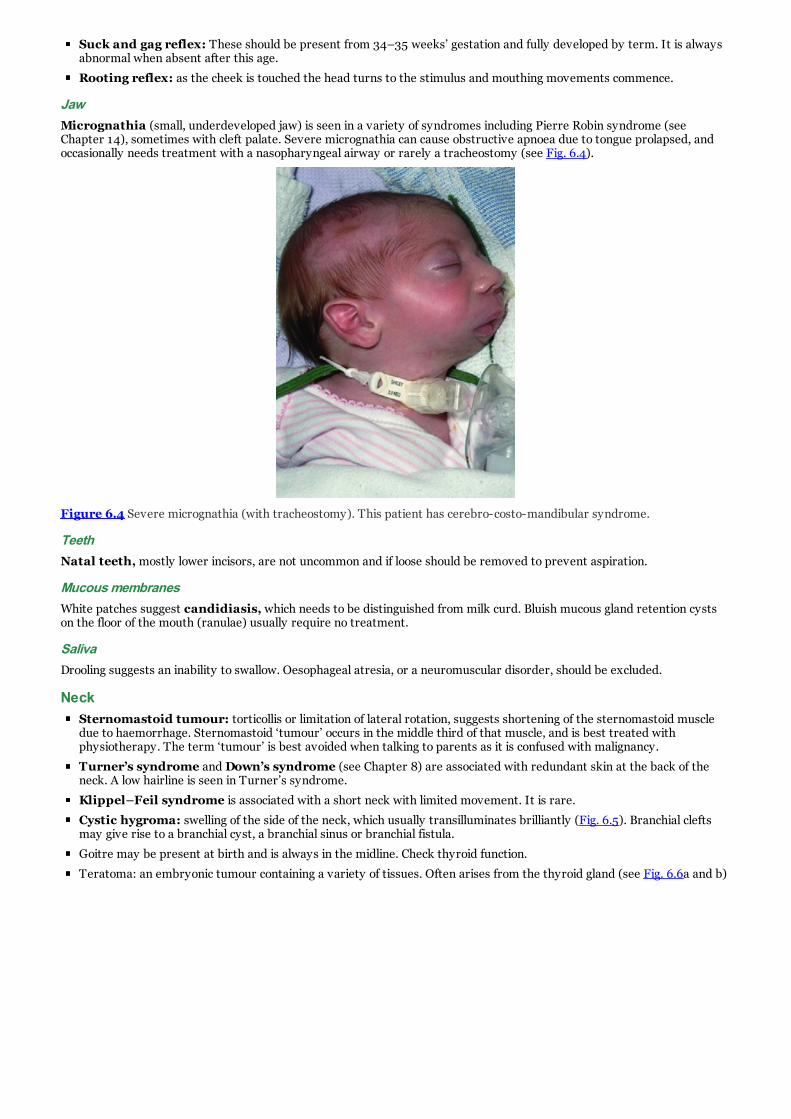
Suck and gag reflex: These should be present from 34–35 weeks’ gestation and fully developed by term. It is alwaysabnormal when absent after this age.
Rooting reflex: as the cheek is touched the head turns to the stimulus and mouthing movements commence.
JawMicrognathia (small, underdeveloped jaw) is seen in a variety of syndromes including Pierre Robin syndrome (seeChapter 14), sometimes with cleft palate. Severe micrognathia can cause obstructive apnoea due to tongue prolapsed, andoccasionally needs treatment with a nasopharyngeal airway or rarely a tracheostomy (see Fig. 6.4).
Figure 6.4 Severe micrognathia (with tracheostomy). This patient has cerebro-costo-mandibular syndrome.
TeethNatal teeth, mostly lower incisors, are not uncommon and if loose should be removed to prevent aspiration.
Mucous membranesWhite patches suggest candidiasis, which needs to be distinguished from milk curd. Bluish mucous gland retention cystson the floor of the mouth (ranulae) usually require no treatment.
SalivaDrooling suggests an inability to swallow. Oesophageal atresia, or a neuromuscular disorder, should be excluded.
NeckSternomastoid tumour: torticollis or limitation of lateral rotation, suggests shortening of the sternomastoid muscledue to haemorrhage. Sternomastoid ‘tumour’ occurs in the middle third of that muscle, and is best treated withphysiotherapy. The term ‘tumour’ is best avoided when talking to parents as it is confused with malignancy.
Turner’s syndrome and Down’s syndrome (see Chapter 8) are associated with redundant skin at the back of theneck. A low hairline is seen in Turner’s syndrome.
Klippel–Feil syndrome is associated with a short neck with limited movement. It is rare.
Cystic hygroma: swelling of the side of the neck, which usually transilluminates brilliantly (Fig. 6.5). Branchial cleftsmay give rise to a branchial cyst, a branchial sinus or branchial fistula.
Goitre may be present at birth and is always in the midline. Check thyroid function.
Teratoma: an embryonic tumour containing a variety of tissues. Often arises from the thyroid gland (see Fig. 6.6a and b)

Figure 6.5 Cystic hygroma of the neck (trans-illuminated).
Figure 6.6 (a) Teratoma of the neck. This child was intubated while still connected to the placental circulation (EXITprocedure) before having surgical excision. (b) MRI scan showing the same lesion (arrow).
Chest
RespiratoryThe features of respiratory distress are tachypnoea (>60 breaths per min), retraction, cyanosis, grunting and flaringof the nostrils. Chest retractions (sternal, substernal, intercostal or subcostal) suggest pulmonary disease.
Stridor (see Chapter 14) indicates upper airway obstruction. An inspiratory stridor implies extrathoracic obstruction,whereas biphasic noise implies intrathoracic obstruction. Other signs of upper airway obstruction include suprasternalretraction, croupy cough and a hoarse cry.
Breath sounds: auscultate with the diaphragm of the stethoscope for symmetry of air entry and adventitial breathsounds (if unilateral, suspect pneumothorax or diaphragmatic hernia). Crepitations may be normal in the first hours oflife.
Shape and sizeThe chest should be symmetrical in shape and move equally on respiration. A small chest occurs in infants withhypoplastic lungs and in a variety of rare syndromes, but is difficult to diagnose objectively.
Breast engorgement occurs commonly in both sexes and is due to maternal oestrogen effect. ‘Milk’ may be secretedfrom the nipple and is not abnormal. Supernumerary nipples are a common finding.
CLINICAL TIP
More information can usually be obtained by observing the chest carefully than by listening with a stethoscope. Taketime to watch the pattern of respiration and observe for intercostal recession and symmetry of chest rise.
Cardiovascular
Pulses

The normal heart rate varies between 90 and 160 beats per minute. The cause of tachycardia and bradycardia is discussedin Chapter 16. The brachial and femoral pulses should be palpable. Generally reduced peripheral pulses suggest hypoplasticleft heart or cardiogenic shock, and absent femorals suggest coarctation of the aorta. Collapsing or bounding pulses are afeature of a patent ductus arteriosus (PDA) (see Chapter 16).
Apex beatThis should be localized to the fourth intercostal space in the midclavicular line. A right-sided apex beat may due to left-sided pneumothorax, left diaphragmatic hernia or true dextrocardia.
AuscultationA triple or gallop rhythm is always abnormal. Systolic flow murmurs may be normal in the first 24 hours of life. CHD isdiscussed in Chapter 16. Auscultation of a heart murmur in systole is ascribed as 1/6 → 6/6 according to intensity. The siteof maximal intensity and any radiation should be noted. Even large septal defects may be inaudible in the first days as theright and left ventricular pressures are balanced and the shunt volume small.
Saturation screeningMany centres now use saturation monitoring to screen for duct-dependent CHD. A single postductal reading of less than95% requires further evaluation of the baby. Respiratory disorders and sepsis can sometimes be coincidentally detected bythis method. A drop in SAO2 from preductal (right arm) to postductal (either foot) of more than 3% is also suggestive ofduct-dependent CHD.
CLINICAL TIP
The preductal oxygen saturation should never be lower than the postductal saturation. If it is, consider complex CHDsuch as transposition of the great arteries or double outlet right ventricle.
AbdomenDistension suggests intestinal obstruction or an intra-abdominal mass. A scaphoid abdomen suggests diaphragmatichernia. A lax abdominal wall with much redundant skin is seen in the ‘prune-belly’ syndrome. Divarication of the rectusmuscles may produce a midline bulge in the abdominal wall. No treatment is required and the condition disappears with age.
LiverThis is normally palpable up to 1 cm below the costal margin. Hepatomegaly may be due to lung hyperinflation, cardiacfailure, sepsis, hepatitis, intrauterine infection or haemolysis.
SpleenThe tip can be palpated in about a quarter of normal infants. Splenomegaly suggests infection (prenatal or postnatal) orhaemolysis.
KidneyMay be palpable normally, particularly if the baby is relaxed. Moderate kidney enlargement may be due to hydronephrosis,dysplastic or cystic kidneys or rarely a Wilms’ tumour. An adrenal mass (e.g. haemorrhage or neuroblastoma) is difficult todistinguish from a renal mass.
AnusCheck for normal position and patency. The normal anus is located approximately midway between the coccyx and theposterior forchette/posterior border of the scrotum. An anterior anus may be associated with passage of ‘toothpaste’-likestool due to sphincter dysfunction. Imperforate anus is usually associated with a fistula into the vagina or in males, thebladder.
CLINICAL TIP
Always visualize the anus directly. It is easy to assume that if there is meconium in the nappy then an anus is presentand patent, but the meconium may have passed via the urethra or vagina due to the presence of a fistula.
Umbilicus
VesselsNormally, two thick-walled arteries and a thin-walled vein are seen, surrounded by clear Wharton’s jelly. In 1–2% of infantsthere is a single umbilical artery, sometimes with other congenital malformations. If a single umbilical artery is found andthe fetus has had a normal prenatal anomaly scan, no further investigations are required. If not, a renal ultrasound shouldbe undertaken.
Stump

The cord usually separates by 10 days, leaving a yellow or greenish eschar. A red flare around the base of the cord mayreflect early sepsis and should be taken seriously. Discharge and cellulitis are also signs of infection. Discharge of urine ormeconium from the stump suggests a patent urachus or patent omphalomesenteric duct, respectively. Exomphalos andgastroschisis are discussed in Chapter 17.
Umbilical granuloma (a small red fleshy swelling at the stump) is due to excessive granulation and if symptomaticcan be treated by the application of a silver nitrate stick on one or two occasions. Always protect the surrounding skinwith barrier cream. Regular wiping with alcohol wipes can also act as an effective sclerosant. Most resolve spontaneously.
Umbilical hernia is particularly common in African babies or those born prematurely, and usually develops in the firstmonth or so of life. These hernias require no treatment, and usually regress by 18 months. They do not strangulate.Distinguish from exomphalos minor.
GenitaliaEstablish that the baby has passed urine. This should occur within 24 hours of birth. If in doubt, a ‘urine bag’ can beattached over the genitalia.
TestesThe testes are present in the scrotum in 98% of full-term male infants. Failure to descend by 6 weeks after term isabnormal. Ectopic testes or arrest in the line of normal descent should also be referred for surgical opinion. Retractile testesthat come down when the baby is relaxed and warm do not need referral.
Penis and urethraThe foreskin in infants is not retractable. The urethral meatus normally opens at the tip of the glans penis.
Hypospadias occurs if the meatus opens on the ventral surface of the penis, and is most commonly glandular.
Epispadias occurs when it opens on the dorsal surface of the penis. Advise against circumcision as the foreskin may beneeded for reconstructive surgery.
ScrotumA hydrocoele transilluminates brilliantly. It may extend upwards along the spermatic cord. Most disappearspontaneously during the first year of life and require no treatment other than reassurance.
Inguinal hernia rarely presents at birth, but is common in premature infants due to a patent processus vaginalis. Thescrotal swelling can usually be reduced. Surgery is required in all cases to prevent complications.
Testicular torsion: a swollen, tender or red scrotum suggests either strangulated inguinal hernia or testicular torsion.Both require an urgent surgical opinion.
Pink nappiesOccasionally, urates may react with the urine in the newborn period, leaving a pinkish-red stain on the napkin that may beconfused with haematuria. This is self-limiting and only occurs in the first few weeks of life.
HymenHymenal skin tags are common in female babies and are due to a protrusion of redundant vaginal mucosa. No treatmentis required and they usually regress spontaneously in a few weeks.
Vagina and vulvaThe size of the labia majora depends on gestational age, but by term they should completely cover the labia minora. Inpreterm or IUGR girls the labia minora and clitoris can appear very prominent.
Labial fusion occurs in the adrenogenital syndrome, and labial adhesions are also not uncommonly seen inotherwise normal infants.
Mucoid vaginal discharge occurs in most mature female infants shortly after birth. It is normal and may continue for2–3 weeks. Vaginal withdrawal bleeding may occur in normal girls.
Hydrometrocolpos describes a bulging imperforate hymen, caused by the accumulation of secretions.
ClitorisThis is variable in size. If large, consider adrenogenital syndrome, a maternal progesterone effect or an intersex state.Ambiguous genitalia are discussed in Chapter 21.
BackIt is essential to examine the back. This is best done by placing one hand over the baby’s chest and one hand over the upperback, and then turning the baby prone to rest on your hand and forearm. This ‘ventral suspension’ is also an opportunity toassess muscle tone.
SpineRun your finger all the way down the spine, looking and feeling for any defects. Spina bifida is usually detected antenatally,but any lump, hairy patch or naevus over the midline warrants a spinal ultrasound as these can be a marker of underlyingspinal anomaly.
Scoliosis

Severe curvature of the spine (scoliosis) is usually due to hemivertebrae (see Fig. 6.7) or failure of segmentation. It is oftenfound incidentally on a radiograph. Orthopaedic consultation with regular follow-up is necessary. Congenital genitourinarymalformation occurs in 20% of babies with congenital scoliosis, and should be excluded by a renal ultrasound scan.
Figure 6.7 A radiograph showing multiple vertebral anomalies (arrows).
SacrumSacral ‘dimples’ are very common. If they are shallow (i.e. you can clearly see the base by pulling the skin laterally on eitherside) then they do not need further investigation. If the natal cleft is deviated or there are any surface markings, thenarrange for spinal ultrasound.
ExtremitiesThis is a good point to assess movement, tone and primitive reflexes. The baby should be awake but settled in order to gainan accurate impression. A hungry, crying baby cannot be assessed accurately.
PostureThe posture depends on the gestational age (see ‘Assessment of maturity’; Chapter 11). A hypotonic baby will tend to bein the ‘frog-leg position’ with reduced spontaneous movement. A hypertonic baby may be in a state of predominant flexionor extension. A baby with cerebral irritation may have extensor posturing with arching of the back, opisthotonus, scissoringof the legs, and thumbs tightly adducted in the cortical position.
MovementsThe quality and quantity of spontaneous movements are observed. Absence or reduction of movements may occur in onelimb only, two limbs on the same side of the body, or both legs. The absence of spontaneous movements is often suggestiveof a systemically unwell baby, and only very rarely a sign of a paralysed baby. Observe the quality of movements; are theysmooth (normal) or jerky? Note any abnormal movements such as tremors, jitteriness or convulsions. Tremulousness orjitteriness must be distinguished from convulsions (see Chapter 22).
Assessment of toneLimb tone is assessed by posture and resistance to passive movement. Gently flex, extend and rotate the limbs on each side.Compare the two sides of the body and the upper and the lower limbs. The head must be in the midline to prevent elicitingthe ‘asymmetric tonic neck reflex’ (see Chapter 22). Hip adductor tone is assessed by the hip angle when passivelyabducted. Neck tone is assessed with the arms held in traction and by observing the head control when the baby is held ina sitting position. Truncal tone is assessed with the baby held in ventral suspension.
Primitive reflexesMoro reflex: elicited by placing one hand under the infant’s shoulders and the other under his/her head and thensuddenly dropping the head by several centimetres. A full response consists of abduction and extension of the arms,followed by adduction and flexion (Fig. 6.8). Asymmetry suggests a brachial plexus palsy or a bone injury (see Chapter7).
Palmar grasp: apply pressure to the palm of the hand, resulting in flexion of the fingers and a firm grasp. If traction isthen applied the infant should lift his/her head off the bed so that head lag can be assessed.

Figure 6.8 Eliciting the Moro reflex.
Upper limbsAssess movement, tone and reflexes as above. The fingers and palms should be examined for abnormalities. A single palmarcrease may be present in Down’s syndrome, but also in 15% of the normal population.
FingersThere are a variety of common patterns of finger anomaly.
Clinodactyly: a lateral curvature of the fifth finger which may be familial or associated with various syndromes.
Syndactyly: fusion of the fingers – may be partial (webbing) or complete. Fusion of the third and fourth fingers can beassociated with cardiac arrhythmias.
Polydactyly: additional digits. These most often occur postaxially (on the ulnar aspect of fifth finger) and may or maynot contain bone. They should be referred to plastic surgery for removal and not tied off. Preaxial polydactyly isassociated with syndromes.
Cleft hand: one, two or three central rays are missing, often leaving just a thumb and opposing digit. The termectrodactyly or ‘lobster claw hand’ should no longer be used.
Symbrachydactyly: Shortened or absent fingers, with preservation of the thumb. Sometimes the distal part of eachfinger is present just above the metacarpals.
ThumbsAbnormal thumbs can be associated with a variety of haematological disorders:
TAR syndrome: thrombocytopenia with absent radii.
Aase’s syndrome: triphalangeal thumb and congenital anaemia.
Fanconi’s syndrome: radial hypoplasia, pancytopenia.
Diamond–Blackfan anaemia.
Lower limbsExamine the legs and feet carefully, including the toes. Then complete the examination by checking for dislocatable ordislocated hips (see below). This is left to the end of the examination as it may cause the baby to become upset, although itshould never cause pain.
FeetLower-limb abnormalities are common and are often associated with intrauterine compression due to oligohydramnios orneurological conditions leading to reduced fetal movement. They may vary from a mild postural problem to severe talipes.
Talipes equinovarus (clubfoot) occurs in 1 in 1000 births, and is 10-fold more common than talipes calcaneovalgus (seeFig. 6.9). When either is seen, other associated congenital abnormalities (e.g. dislocation of the hip and myelomeningocele)should be sought. If the foot can be passively corrected to the position opposite the deformity, it is considered to be mild andrequires only simple passive exercise treatment by a physiotherapist and the parents. More severe deformities will requirephysiotherapy, splinting, and very occasionally surgical correction. There is good evidence that serial stretching, casting andbracing (the Ponseti method) is as effective as surgery.

Figure 6.9 (a) Talipes equinovarus; (b) talipes calcaneovalgus.
ToesPolydactyly and syndactyly can occur in the toes. Syndactyly of the second and third toes is very common, and often there isa familial history. A wide gap between the 1st and 2nd toes (sandal gap) can be a feature of trisomy 21.
KneesGenu recurvatum: This is rare (1 in 100 000). The knees are hyperextensible and can bend forwards. In severe casesthere may be dislocation of the knee joint, and splinting may be necessary. The cause is multifactorial, with mechanicaland genetic elements. Recurvatum at the knee sometimes exceeds 90°. A radiograph shows the tibia anterior to thefemur.
Femoral retroversion: The child lies or stands with his/her legs externally rotated at 90° (‘Charlie Chaplin’ gait). Thisis sometimes seen at follow-up of premature infants. It should be preventable by good developmental care andpositioning (see Chapter 24). This condition often causes distress to the parents, but they should be strongly reassuredas it rapidly corrects within a year of the child learning to walk. It is, however, important to exclude congenital dislocationof the hip as the cause of the external rotation.
Congenital abnormalities of the hips �and limbs
Developmental dysplasia of the hipDevelopmental dysplasia of the hip (DDH), previously known as congenital dislocation of the hip, is an importantasymptomatic congenital condition that should be detectable during newborn examination. The consequences of missingDDH can be considerable. Hip dislocation occurs in 1–2 per 1000 newborns. Dislocation of the left hip is fourfold morecommon than the right hip, due to the fetal position. Risk factors for DDH are shown in Table 6.1.
Table 6.1 Risk factors for developmental dysplasia of the hips.
Risk factor Comment
Familial history (polygenic inheritance) Developmental dysplasia of the Hip (DDH) recurs in families at a rate of about 1in 30
Breech presentation (beyond 36 weeks’gestation)
10-fold increased risk. Incidence of DDH in singleton breech delivery is 14%
Female sex Female : male ratio is 6 : 1
Racial/cultural factors Increased in Italy and in Inuit population. Reduced in China and Africa
Neuromuscular disorders Spina bifida; Hypertonia (including cerebral palsy); Congenital hypotonia;Congenital arthrogryposis
Trisomy 13, 18
Multiple congenital anomalies
Severe oligohydramnios Due to lack of room for fetal movement
ExaminationExamination of the hip should start with observations for signs of established dislocation, such as unequal leg length andasymmetry of the thigh creases. The physical examination should be undertaken in two parts:
Ortolani’s (reduction) test: This assesses whether the hip is already dislocated. With the baby relaxed on a firm
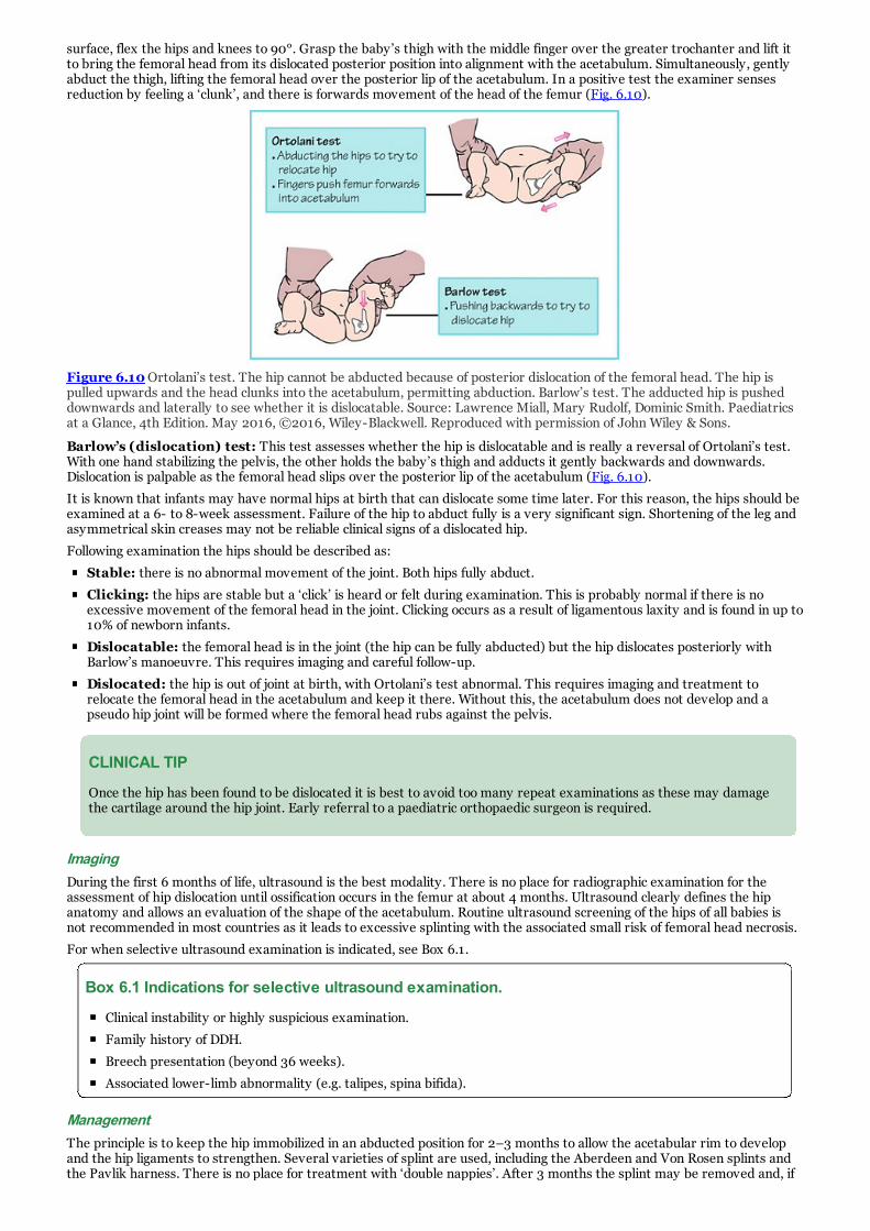
surface, flex the hips and knees to 90°. Grasp the baby’s thigh with the middle finger over the greater trochanter and lift itto bring the femoral head from its dislocated posterior position into alignment with the acetabulum. Simultaneously, gentlyabduct the thigh, lifting the femoral head over the posterior lip of the acetabulum. In a positive test the examiner sensesreduction by feeling a ‘clunk’, and there is forwards movement of the head of the femur (Fig. 6.10).
Figure 6.10 Ortolani’s test. The hip cannot be abducted because of posterior dislocation of the femoral head. The hip ispulled upwards and the head clunks into the acetabulum, permitting abduction. Barlow’s test. The adducted hip is pusheddownwards and laterally to see whether it is dislocatable. Source: Lawrence Miall, Mary Rudolf, Dominic Smith. Paediatricsat a Glance, 4th Edition. May 2016, ©2016, Wiley-Blackwell. Reproduced with permission of John Wiley & Sons.
Barlow’s (dislocation) test: This test assesses whether the hip is dislocatable and is really a reversal of Ortolani’s test.With one hand stabilizing the pelvis, the other holds the baby’s thigh and adducts it gently backwards and downwards.Dislocation is palpable as the femoral head slips over the posterior lip of the acetabulum (Fig. 6.10).
It is known that infants may have normal hips at birth that can dislocate some time later. For this reason, the hips should beexamined at a 6- to 8-week assessment. Failure of the hip to abduct fully is a very significant sign. Shortening of the leg andasymmetrical skin creases may not be reliable clinical signs of a dislocated hip.
Following examination the hips should be described as:
Stable: there is no abnormal movement of the joint. Both hips fully abduct.
Clicking: the hips are stable but a ‘click’ is heard or felt during examination. This is probably normal if there is noexcessive movement of the femoral head in the joint. Clicking occurs as a result of ligamentous laxity and is found in up to10% of newborn infants.
Dislocatable: the femoral head is in the joint (the hip can be fully abducted) but the hip dislocates posteriorly withBarlow’s manoeuvre. This requires imaging and careful follow-up.
Dislocated: the hip is out of joint at birth, with Ortolani’s test abnormal. This requires imaging and treatment torelocate the femoral head in the acetabulum and keep it there. Without this, the acetabulum does not develop and apseudo hip joint will be formed where the femoral head rubs against the pelvis.
CLINICAL TIP
Once the hip has been found to be dislocated it is best to avoid too many repeat examinations as these may damagethe cartilage around the hip joint. Early referral to a paediatric orthopaedic surgeon is required.
ImagingDuring the first 6 months of life, ultrasound is the best modality. There is no place for radiographic examination for theassessment of hip dislocation until ossification occurs in the femur at about 4 months. Ultrasound clearly defines the hipanatomy and allows an evaluation of the shape of the acetabulum. Routine ultrasound screening of the hips of all babies isnot recommended in most countries as it leads to excessive splinting with the associated small risk of femoral head necrosis.
For when selective ultrasound examination is indicated, see Box 6.1.
Box 6.1 Indications for selective ultrasound examination.
Clinical instability or highly suspicious examination.
Family history of DDH.
Breech presentation (beyond 36 weeks).
Associated lower-limb abnormality (e.g. talipes, spina bifida).
ManagementThe principle is to keep the hip immobilized in an abducted position for 2–3 months to allow the acetabular rim to developand the hip ligaments to strengthen. Several varieties of splint are used, including the Aberdeen and Von Rosen splints andthe Pavlik harness. There is no place for treatment with ‘double nappies’. After 3 months the splint may be removed and, if

the hip is clinically stable, check radiographs are taken usually at 6 and 12 months of age. Fixed dislocations of the hip,failure to respond to treatment and late-diagnosed dislocated hips will require individual orthopaedic management. Thesuccess of treatment depends on accurate and early diagnosis of the condition.
Limb malformationsThese can affect the arms or legs and be unilateral or bilateral. The main types are:
Amelia: absence of one or more limbs.
Ectromelia: gross hypoplasia of one or more limbs.
Phocomelia: partial deficiency of the proximal segment with preservation of the distal parts (e.g. hands can appear toarise from the shoulder).
Hemimelia: rudimentary formation of the distal part of a limb with normal proximal development.
Amniotic band amputations: these occur in utero and cause a linear amputation which does not follow a normalembryological developmental pattern.
Absent radius: This is seen in the VACTERL sequence and TAR syndrome (see Chapter 20), and leads to a curvedforearm with radial deviation of the hand.
Arthrogryposis multiplexThis term describes multiple joint deformities involving more than one limb (Fig. 6.11). It is caused by a restriction of jointmovement in utero, and there may be a variety of underlying causes:
Neuromuscular – congenital dystrophy, spinal muscular atrophy.
Connective tissue disorders.
Oligohydramnios.
Bicornuate uterus.
Genetic syndromes (<30%).
Figure 6.11 Arthrogryposis multiplex.
All require orthopaedic referral and intense physiotherapy. An underlying neuromuscular, connective tissue or geneticproblem should be considered. Investigations are described in Chapter 22.
Neonatal dwarfismDwarfism may be proportional (the whole body is short but in proportion) or be due to skeletal dysplasia (disproportionateshortening of the limbs and trunk). Examples of proportional dwarfism syndromes include Russell–Silver, Cornelia deLange, Conradi, Seckel’s bird head, Cockayne, leprechaunism and progeria. These children are extremely small forgestational age (SGA) at birth.
Skeletal dysplasiaAn underlying skeletal dysplasia should be suspected in any baby who is SGA with other congenital abnormalities and whohas disproportionate shortening of the limbs and trunk.
Skeletal dysplasias may present simply as short stature or a disproportionate body, but a variety of other anomalies, such ashydrocephalus, craniostenosis, cleft lip and palate, polydactyly, syndactyly, dislocated hips and dysmorphic facial features,may coexist.
There are many classifications of the more than 100 distinct syndromes. The most commonly recognized syndromes in thenewborn are:
Achondroplasia.
Osteogenesis imperfecta types I–V.
Asphyxiating thoracic dystrophy.
Thanatophoric dwarf (Fig. 6.12).
Osteopetrosis (marble bone disease).

Diastrophic dwarfism.
Ellis–van Creveld syndrome.
Figure 6.12 Thanatophoric dwarf.
AchondroplasiaThis is the most common skeletal dysplasia with short limbs. The proximal segment (i.e. humerus and femur) is moreinvolved than the lower segments (i.e. radius and ulna or tibia and fibula). The head is typically enlarged, but the trunkappears normal. It is inherited as an autosomal dominant trait but approximately 80% are new mutations.
Osteogenesis imperfectaThis is an inherited connective tissue disorder. In its milder form, affected infants have an increased susceptibility tofracture and hearing loss in later childhood, but at its most severe it is lethal and perinatal death is common. These babiesoften have blue sclerae and their bones fracture in utero, presenting at birth with short, deformed limbs. Mild forms mayrespond to treatment with bisphosphonates.
Skin disordersCutaneous lesions are present at birth in 8% of newborn infants. They are usually immediately obvious and may causeconsiderable concern. Sometimes they are part of a neurocutaneous syndrome where they may be skin markers ofneurological disorders. Examples include Sturge–Weber syndrome, neurofibromatosis and incontinentia pigmenti.
Cutaneous lesions in the newborn can be classified in the following major groups:
Vascular birthmarks.
Epidermal naevi.
Pigmented birthmarks.
Ichthyotic disorders.
listering and bullous disorders.
Miscellaneous.
Vascular birthmarksVascular birthmarks can be either haemangiomas (neoplastic proliferation of vascular endothelium) or vascularmalformations (abnormally sited or connected capillaries, veins or arteries). Older descriptive terminology, such as‘cavernous’, ‘strawberry’ and ‘naevus flammeus’ is confusing and should be avoided.
HaemangiomasThese vascular proliferation tumours are characterized by rapid endothelial growth followed by slow involution.Approximately 30% are present at birth as a blanched or telangiectatic patch. The majority appear during the first month,and grow rapidly over the next 5 months (Fig. 6.13). Slow involution subsequently occurs over the next 5–10 years. Theyare much more common in preterm infants and in girls. They were previously referred to as ‘strawberry naevi’. Superficialhaemangiomas deep in the dermis, subcutaneous fat or muscle produce a bluish colour in the overlying skin.

Figure 6.13 Vascular haemangioma.
The majority of haemangiomas do not require treatment. Indications for treatment include lesions near the airway or eyes,those associated with high cardiac output failure, and extensive facial lesions. Oral corticosteroids commenced during therapid growth phase are effective in slowing growth and hastening resolution, but recent evidence suggest that beta-blockers(e.g. propranolol) can have a dramatic effect with less side effects. Bleeding is rarely a problem, even after trauma, but largelesions can be associated with thrombocytopenia, intravascular coagulation and severe bleeding (Kasabach–Merrittsyndrome).
Vascular malformationsThese represent structural anomalies, are present at birth, grow in proportion to the infant’s growth, and in general do notresolve with time. They can affect any kind of blood and lymphatic vessel, and sometimes cause Kasabach–Merrittsyndrome or contain arteriovenous shunts which may cause heart failure.
Capillary malformationsFine capillary malformations, which are deep red in colour and blanch with pressure, occur on the glabella, upper eyelids,upper lips and nape of the neck. They are of no significance and tend to fade during the first 6 months. They were previouslyreferred to as ‘naevus flammeus’ or ‘stork marks’.
Larger, flat, deep, purple-red (‘port-wine’) lesions may occur on any part of the body. Those in the distribution of thetrigeminal nerve can be associated with retinal and intracranial vascular malformations, requiring ophthalmological follow-up. Association of this type of lesion with vascular malformation of the ipsilateral meninges and cerebral cortex is termed theSturge–Weber syndrome. It can be associated with epilepsy. The skin manifestations may be treated with cosmetic covercreams or by pulsed dye laser in later infancy.
Epidermal naeviThese birthmarks presenting at birth or in the first few months of life represent a proliferation of keratinocytes or skinappendage cells. They are rare lesions that may involve any area, including the oral cavity. On the scalp and face they areoften yellowish because of a prominent sebaceous gland component. Trunk and limb epidermal naevi are scaly, flat or raised,varying in colour from black or brown to pale grey, with plaque or linear streak distribution. Excision may be indicated forsmall and linear lesions and for irritating and cosmetically troublesome naevi.
Hyperpigmented and hypopigmented birthmarksThese must be differentiated from the skin lesions of generalized disorders, such as neurofibromatosis and tuberoussclerosis (neurocutaneous syndromes), which usually appear after birth.
Congenital hyperpigmented patchesThese common pale or dark-brown macular or flat hypermelanotic patches may be solitary or extensive, involving largeareas of the trunk or limbs. They must be differentiated from the café au lait spots of classic neurofibromatosis.
Congenital melanocytic naeviThese are collections of melanocytes in the epidermis or dermis. When present at birth they appear as raised lesions ofvarious shades of brown to black (Fig. 6.14). They vary in size and may have blue or pink components, often growing longblack hairs. They grow in proportion to the infant. Controversy exists about the risk of malignant change. Lesions over thelower spine may be associated with a tethered spinal cord. Large or multiple lesions may be associated with benign or,rarely, malignant proliferation of melanocytes in the leptomeninges, demonstrated by MRI. Small lesions are easily removedsurgically; for larger lesions staged surgery may be possible.

Figure 6.14 Congenital melanocytic naevus involving the buttock and loin.
Mongolian blue spotsThese are flat, blue or slate-grey lesions comprising collections of melanocytes in the dermis (Fig. 6.15). They are seen in themajority of oriental and black infants. In white infants there is usually a Mediterranean ancestry. Single or multiple, theyoccur particularly in the lumbosacral area, less often on the shoulders or back, and tend to fade with age. These lesionsshould be documented so they are not confused with non-accidental bruising in the future. They do not require anyintervention other than reassurance.
Figure 6.15 Mongolian blue spot.
Congenital hypopigmented patchesThese are pale areas of reduced melanin varying in size from a few centimetres to large areas covering the trunk and limbs.They do not involute. Similar lesions occur in incontinentia pigmenti, a rare genetic neurocutaneous condition affectingfemales and associated with multiple abnormalities, especially of the eye, skeleton and central nervous systems.
Ichthyotic disordersThese are a rare group of skin disorders where the skin at birth is dry and scaly. There are several varieties:
Ichthyosis vulgaris: This is an autosomal dominant disorder (1: 250) and there may be a family history of atopy. Itrarely causes problems in the newborn and is treated with moisturizer.
Recessive X-linked ichthyosis: This condition only affects males and is associated with placental sulphatasedeficiency. Unrecordable oestriol measurements during pregnancy should alert the clinician to this possibility in maleinfants.

Collodion baby: The mildest form of this group of disorders is the collodion baby. At birth the infant looks as if it iscovered in a dry plastic-like membrane, which cracks easily. These infants often later develop lamellar ichthyosis, butsome may have no persistent skin abnormality. The more severe harlequin icthyosis shows severe hyperkeratosis,everted eyelids and deep skin cracking and is due to a mutation of the ABCA12 gene (Fig. 6.16). Treatment with retinoicacids is indicated and in the most severe forms palliative care is sometimes discussed.
Figure 6.16 Harlequin ichthyosis with severe deep skin cracking.
Blistering and bullous disordersThese constitute a wide group of unrelated disorders characterized by blistering of the skin. They can be divided intotransient and chronic disorders (Table 6.2).
Table 6.2 Classification of blistering and bullous disorders affecting the neonate.
Transient Chronic
Erythema toxicum Epidermolysis bullosa:
Congenital candidiasis Non-scarring
Congenital herpes Scarring
Impetigo neonatorum
Toxic epidermal necrolysis
Transient neonatal pustular dermatosis (melanosis)
Erythema toxicumThese extremely common lesions (70% of term babies) appear in the first few days of life as multiple vesicles. The vesiclesare differentiated from infection by the fact that they ‘come and go’ and the baby is completely well. Each has a macular redsurround and the presence of multiple eosinophils within the vesicular fluid. No treatment is necessary.
Transient neonatal pustular melanosisUnlike erythema toxicum these lesions are present at birth. The pustules are fragile; the top easily wipes off leaving a halo.There is no surrounding erythema. In pigmented skin a dark hyperpigmented area is left as the pustule resolves. This isnon-infectious and needs no treatment (see Fig. 6.17).

Figure 6.17 Transient neonatal pustular melanosis.
Candida vesiclesThese are usually associated with oral candidiasis, but can occasionally be present at birth. Diagnosis is by the identificationof hyphae or budding spores from the blister. Treatment is with topical nystatin or miconazole.
Congenital herpes vesiclesA ‘crop’ of vesicles on the vertex of the baby can very rarely represent congenital herpes infection from contact with cervicallesions. Confirmation is by herpes simplex virus RNA analysis (PCR method) of the vesicle fluid. This needs to be treatedaggressively with aciclovir to prevent disseminated herpes infection (see Chapter 10).
Impetigo neonatorumThis term is used to describe staphylococcal bullous lesions appearing on the second or third day of life. The pustulesdevelop on an erythematous base and are often seen in moist areas (neck, axillae or groin). The infant may show signs ofsystemic infection. Intravenous flucloxacillin should be given while culture from the pustules is awaited, as the condition mayspread quickly.
Toxic epidermal necrolysis (scalded skin syndrome)This is characterized by generalized erythema accompanied by fever and irritability, followed within a few hours by theformation of flaccid bullae filled with serous fluid. Sheets of epidermis can be stripped away with minimal shearing force,revealing a raw, oozing surface (Nikolsky’s sign). This is most commonly associated with a Staphylococcus aureus infection(phage type 50 or 71) which produces toxins. Treatment is with systemic flucloxacillin. The large denuded areas of skinmust be treated like a severe burn, requiring isolation to prevent secondary infection, and careful fluid and electrolytemanagement.
Epidermolysis bullosaThis includes a group of conditions in which blistering or bullous eruptions occur at birth or during the first week of life aftermild trauma. They can be divided into non-scarring and scarring forms:
Non-scarring (epidermolysis bullosa simplex): This is a relatively mild form and is autosomal dominant. Thesoles, toes and fingers are most often affected. Blisters may be present within minutes of delivery. A more severe form,inherited as an autosomal recessive condition, may heal to leave atrophic areas. Differentiation is only possible onelectron microscopy. The non-scarring types tend to improve by puberty, but lesions frequently become secondarilyinfected.
Scarring (dystrophic) forms: These conditions may be inherited as either autosomal dominant or recessivedisorders. The blisters occur early and are deep. The recessive form is usually more severe and the gastrointestinal tractmay be involved. The scarring is often extremely destructive, with loss of nails, the formation of ugly scars andcontractures, fusion of digits, and sometimes digit or limb amputations.
Management will depend on the individual case, but the parents should be encouraged to ensure that the child’s lifestyle isas normal as possible. Nevertheless, the avoidance of mechanical trauma, hot baths and high temperatures is important.Genetic counselling is advised.
Miscellaneous
Cutis marmorataThis represents a normal physiological reaction to cold temperature that is commonly seen in the neonate and young infant.The bluish mottling usually exhibits a characteristic reticulated pattern on the trunk and extremities. It is symptomless andtransient, requiring no treatment. It is more common in Trisomy 21.
Cutis aplasiaThis presents as a focal absence of skin at birth. Most often the lesions are on the scalp in the midline (see Fig. 6.18), butthey can occur on the trunk or extremities. It is associated with trisomy 13. The combination of cutis aplasia and cutis

marmorata is seen in Adams–Oliver syndrome. Prevention of infection in cases of ulceration and surgical skin grafting (forlarger defects) may be required.
Figure 6.18 Cutis aplasia of the scalp. Note the extensive area of hair loss, some of which is scabbed over.
Subcutaneous fat necrosisA firm, woody, red nodule is felt, often over the arms, thighs, shoulders or face. This can occur after mild perinatal asphyxiaor in babies who have become cold or who have received therapeutic hypothermia. It resolves spontaneously over manyweeks, but if extensive can be associated with significant hypercalcaemia.
Cutis laxaCongenital cutis laxa is inherited. Affected infants have a diminished resilience of the skin, which hangs in folds. Thiscondition is not associated with joint laxity and bruising, as is seen in cases of Ehlers–Danlos syndrome. In Ehlers–Danlos theskin is hyperelastic but snaps back to normal and does not hang in folds.
Harlequin colour changeThis refers to a unilateral colour change commonly seen during the first few days of life in preterm, but also in term infants.It occurs with axial rotation: when the infant is lying on one side the upper part of the body is paler than the lower half,which is normal or reddish in colour. It may last for seconds or minutes and requires no treatment.
Neonatal eczema (atopic dermatitis)Eczema is a common problem after 2 months of age, but is unusual in the newborn. Management consists of avoidingpotential allergens and plentiful use of emollients and moisturizer.
Seborrhoeic dermatitis (cradle cap)This inflammatory condition appears after a few weeks. There is crusting and greasy yellow scales on an erythematous base,usually over the scalp. It may spread to the face, neck, behind the ears, axillae and napkin area. Treat with olive oil, followedby gentle brushing to remove the scales. Severe cases respond to coal tar shampoo and steroid or antifungal creams.
Ectodermal dysplasiaThis is an abnormality affecting the skin, sweat glands, hair and nails. Infants have sparse hair, hypoplastic nails and maydevelop peg-like teeth. Sweating may be abnormal.
Communication with parentsAt the end of the examination it is important to document your findings carefully, either in the hospital record or in aparent-held child health record. If everything is normal then parents should be reassured. It is always sensible to ask if theyhave any other concerns about their baby’s health at this time. If minor abnormalities have been found, these should beexplained carefully and also communicated to the family doctor. If more significant anomalies have been identified, theseshould be explained carefully and appropriate follow-up arranged. Parents must be left with a clear plan and, ideally, withwritten information about the condition. Many parents will turn to online resources, and it is useful to point them in the rightdirection of support groups and reliable websites. It is also a good opportunity for health promotion. Topics to be consideredinclude:
Explaining the risks of passive smoking.
Actions to reduce sudden infant death syndrome.
Child safety including car seats on the journey home from hospital.
The importance of immunization.

SUMMARY
Carefully examining every newborn baby provides an opportunity to reassure parents about trivial congenitalanomalies and to explain more significant ones. Examination of the heart, hips, eyes and testes (in boys) forms part ofthe newborn screening programme. It is important that examinations are performed in a thorough and systematicmanner, and to be familiar with normal variants and the common patterns of abnormality. Communication withparents is very important at this time – they need to know what the problem is, whether anything needs to be done,and what is going to happen next.
FURTHER READING1. de-Wahl Granelli, A., Wennergren, M., et al. (2009) Impact of pulse oximetry screening on the detection of duct
dependent congenital heart disease: a Swedish prospective screening study in 39 821 newborns. British MedicalJournal, 338, 3037.
2. Fletcher, M.A. (1997) Physical Diagnosis in Neonatology. Lippincott-Williams and Wilkins. This remains an excellentreference book covering all the main abnormalities found on newborn examination.
3. Newborn Infant Physical Examination (NIPE). Resources for those undertaking newborn examination. Available at:http://cpd.screening.nhs.uk/nipe.
4. Sewell, M.D., Rosendhal, K., Eastwood, D.M. (2009) Developmental dysplasia of the hip. British Medical Journal, 339,4454.
Now visit www.essentialneonatalmed.com to test yourself on this chapter.

CHAPTER 7 Birth injuryKey topics
Risk factors for birth injury
Injuries to the scalp, skull and brain
Bone and joint injuries
Peripheral nerve injuries
Soft-tissue injuries
Organ injuries
Injuries sustained in the neonatal intensive care unit (NICU)
IntroductionDespite skilled obstetric and midwifery care, injuries may be sustained either during labour or during delivery. Injuries canalso occur as a result of inappropriate use of excessive force. Sometimes, bony injury may be an unintended consequence ofsaving the baby’s life when there is severe shoulder dystocia. Preterm babies are particularly susceptible to injury, either atdelivery or after admission to the neonatal intensive care unit (NICU) where they are vulnerable to preventable iatrogenicinjuries.
The decreased incidence in birth trauma over recent years has been attributed to changing trends in obstetric management,such as caesarean section instead of difficult vaginal delivery. Despite the falling incidence, birth injury is still a cause forconcern to the obstetrician and neonatologist. Parents sometimes attribute birth injury to obstetric mismanagement, andthis may result in litigation. Unfortunately, such events encourage the practice of defensive obstetrics, and a high caesareansection rate may be a consequence of this. There is evidence that the presence of senior experienced obstetricians on thedelivery suite can reduce the caesarean section rate.
Where iatrogenic or preventable injury has occurred it is best to be honest and explain the nature of the circumstances ofthe injury carefully to parents. However, injury – especially brain injury – can occur even after normal labour and deliveryand mismanagement should not be implied without evidence.
Table 7.1 lists the major causes of birth injury and their incidence.
Table 7.1 The commoner types of birth injury and �their incidence.
Cephalhaematoma 1:100
Brachial plexus injury 0.5–1:1000
Facial nerve palsy 0.5–1:1000
Bony (non-skull) fracture 1–2:1000
Skull fractures Rare
Subaponeurotic haemorrhage 1:1250
Major subdural haemorrhage 1:50 000
Spinal cord injuries Very rare
Overall incidence 6–8:1000 births
Risk factors for birth injuryThe effect of changing patterns of obstetric practice on birth-associated mechanical injuries is difficult to evaluate. However,a number of risk factors for birth injury have been identified, especially vaginal breech delivery (Tables 7.2 and 7.3).

Table 7.2 Risk factors for birth injury.
Fetal condition Prematurity
Small for gestational age
Multiple pregnancy
Fetal distress
Malpresentation Breech presentation (see Table 7.3)
Brow, face, compound presentation
Malposition Unengaged head
Occipitoposterior arrest
Deep transverse arrest
Cephalopelvic disproportion Macrosomia, e.g. infant of diabetic mother, hydrops fetalis
Macrocephaly, e.g. hydrocephalus
Previous pelvic fracture
Maternal factors Nulliparity
Short stature
Obesity
Shoulder dystocia
Prolonged labour
Precipitate labour
Inexperienced obstetrician or midwife
Table 7.3 Injuries more likely to occur in infants delivered by breech.
Haemorrhage Subdural tears due to tentorial rupture
Rupture of intra-abdominal viscus (usually liver or kidney)
Occipital osteodiastasis (bone trauma) with cerebellar haemorrhage
Orthopaedic Dislocation: shoulder, cervical vertebrae, hip, kneeFracture: clavicle, humerus, femurDamage tosternomastoid muscle
Neurological Asphyxia secondary to cord prolapse
Cervical brachial plexus injury: Erb’s or Klumpke’s paralysis
Facial nerve palsy
Soft-tissueinjury
Extensive bruising, particularly genitals (presenting part)
Injuries to the scalp, skull and brainSwelling or bleeding can occur at various levels in the scalp, as shown in Figure 7.1. The nature of the lesion can be moreeasily characterized by whether it is defined by the margins of the skull bones and whether it is deep or superficial.

Figure 7.1 Anatomic location of injuries to the head. Source: Tom Lissauer, Avroy A. Fanaroff, Lawrence Miall, JonathanFanaroff. Neonatology at a Glance, 3rd Edition. Wiley-Blackwell. Reproduced with permission of John Wiley & Sons.
Caput succedaneumThis is benign swelling of the subcutaneous tissue of the scalp as a result of prolonged pressure of the fetal head against thedilating cervix. It usually crosses the suture lines and resolves within a few days, occasionally with bruising.
ChignonThis is a well-defined swelling over the presenting part of the scalp, caused by the application of a Ventouse vacuum cup(Kiwi cup). It usually settles within hours.
Erythema, abrasions and lacerationsErythema and abrasions may be seen following forceps and vacuum delivery, and with cephalopelvic disproportion.Lacerations to the scalp or face can occur during episiotomy, uterine incision at caesarean section, and scalp electrodemonitoring.
CephalhaematomaThis occurs in about 1% of newborn infants and is due to bleeding between the periosteum and the cranial bones (usuallyparietal, less commonly occipital) as a result of shearing or tearing of communicating veins during delivery. The extent of theswelling is limited by the underlying skull bone and does not cross suture lines (see Fig. 7.2). It is due to buffeting of the fetalskull against the maternal pelvis, which is seen especially in prolonged labour. It may also occur following a forceps orvacuum delivery. Subperiosteal bleeding is slow and may not appear until the second day of life. Enlargement may occurduring the first week, and the swelling may persist for several weeks. Box 7.1 lists complications associated with acephalhaematoma.
Figure 7.2 Cephalhaematoma. Note the swelling over the right parietal bone. This child also has hypotonia with acharacteristic drooping appearance to the mouth.

Box 7.1 Complications associated with a cephalhaematoma.
These may include:
Underlying linear skull fracture.
Jaundice due to haemolysis.
Calcification at its edge, which may occur some weeks later during the resorption process; this should not beconfused with a depressed skull fracture.
Intracranial haemorrhage may rarely be associated with cephalhaematoma.
Subaponeurotic haemorrhage (subgaleal haemorrhage)On rare occasions, haemorrhage may occur beneath the aponeurotic sheet joining the two portions of the occipitofrontalismuscle of the scalp (see Fig. 7.1). This is seen particularly in black infants. It may be the consequence of trauma duringdelivery, especially with vacuum extraction, or may indicate the presence of a coagulation defect. The scalp becomesfluctuant (‘hot water bottle’ sign) and enlarges rapidly, with a marked drop in haemoglobin. Affected babies require rapiddiagnosis and appropriate transfusion with volume expanders or blood.
CLINICAL TIP
The severity of blood loss with sub-aponeurotic (sub-galeal) bleeds may be underestimated, and ensuing shock maybe life-threatening. Admission to the neonatal nursery for observation and haemoglobin measurement is advisable.
Skull fracturesGenerally, linear fractures require no specific treatment. Depressed fractures are usually associated with forceps applicationor head compression produced by the maternal sacral promontory. Large fractures may be associated with cerebralcontusion, and neurosurgical consultation may be necessary. Depression of the skull may remain for many months afterbirth (Fig. 7.3).
Figure 7.3 Right frontal depressed skull fracture (see arrow).
Intracranial haemorrhagesAlthough mostly related to hypoxic–ischaemic injury, some forms of intracranial haemorrhage can result directly as aconsequence of trauma. These often present with encephalopathy or seizures, and are described in Chapter 22.
Bone and joint injuriesTraumatic fractures of bones during birth are relatively rare, and present with the classic signs of pain on handling,avoidance of movement, swelling, deformity and crepitus. Fractures of the upper-limb bones may be suspected by anasymmetrical Moro reflex or failure to move one arm spontaneously. Fractures heal very fast in newborn babies, and theconsiderable bone remodelling that occurs almost invariably leads to resolution of normal anatomy.
ClavicleThis is the bone most frequently fractured during the birth process. It may be associated with impacted shoulders,especially in the infant of a diabetic mother or a difficult breech delivery. Specific treatment is not required. Simple analgesiashould be offered if the baby seems in pain (see Fig. 7.4).

Figure 7.4 Right-sided clavicular fracture in a child born after shoulder dystocia. The baby is also receiving mechanicalventilation.
HumerusFracture of the humerus generally involves the upper third of the shaft, and there is usually considerable deformity. Radialnerve injury sometimes also occurs. Treatment consists of an immobilizing plastic backslab or immobilization with a binderto the chest.
FemurFracture of the femur occurs rarely during breech extraction, either vaginally or at caesarean section. It occurspredominantly in the breech birth with extended legs as flexion occurs. Treatment consists of immobilization in a plastercase, traction, and a Thomas splint.
Multiple or unusual fracturesMultiple fractures are rare and should raise the possibility of osteogenesis imperfecta. Any fracture in an infant who hasbeen home should always raise the question of non-accidental injury. Iatrogenic fractures may occur on the neonatal unit invery preterm babies with severe osteopenia of prematurity.
Dislocation of joints and separation of epiphysesThese are difficult to diagnose and require specialized orthopaedic assessment. Congenital dislocation of the knee joint is notdue to trauma (see Chapter 6).
Peripheral nerve injuriesNerve injuries in the newborn infant may be due to stretching, compression, twisting, hyperextension or separation of thenervous tissue. They may be classified pathologically as:
Neuropraxia (swelling of the nerve, with total recovery).
Axonotmesis (complete peripheral degeneration, may be partial recovery).
Neurotmesis (complete division of nerve and nerve sheath).
Nerve conduction studies and MRI can distinguish a neuropraxia from a neurotmesis. The following nerve injuries occur inthe newborn.
Facial nerve palsyThis may be due to an upper or lower motor neuron lesion, but clinical distinction between these is difficult. Lower motorneuron (LMN) facial palsy usually occurs due to oblique application of the forceps blade. The nerve may also be damaged byprolonged pressure on the maternal sacral promontory. Upper motor neuron lesions are much less common and are due tobrain injury or nuclear agenesis (Mobius syndrome).
The lesion is recognized clinically by an inability to close the eye and a lack of lower lip depression on crying, both on theaffected side (Fig. 7.5). The nasolabial fold is absent on the affected side. In most cases complete recovery occurs by 6weeks; only rarely is permanent facial palsy seen. If the eye cannot be closed, patching and ‘artificial tear’ eye-drops shouldbe used to prevent corneal damage.

Figure 7.5 Left-sided facial nerve palsy.
CLINICAL TIP
Lower motor neuron facial nerve palsy must be distinguished from ‘asymmetrical crying facies’ syndrome, wherethere is an absence of the depressor anguli oris muscle. In this condition eye closure is normal but there is failure ofthe mouth to move downwards and outwards on the affected side when the infant cries. Asymmetry usually remainsinto adult life but becomes less obvious with time. It often runs in families.
Obstetric brachial palsyThe majority of babies with obstetric brachial palsy (OBP) are thought to have sustained birth trauma, but a very few casescannot be explained in this way and a prenatal causation has been proposed for these. Trauma to the brachial plexus may bedue to excessive lateral flexion, rotation or traction upon the neck. Such trauma may be seen with normal delivery, withimpacted shoulders or during delivery of the aftercoming head of a breech. There is a strong association with shoulderdystocia.
Three types of brachial plexus injury can be described:
Erb’s palsy (C5–6) (Fig. 7.6). In this lesion the fifth and sixth cervical nerve roots are injured and the arm will be held inadduction, with the elbow extended and the forearm pronated with the wrist flexed. This is traditionally known as the‘waiter’s tip’ position. Note that the fingers can still move.
Klumpke’s paralysis (C7–T1). In this uncommon lesion the seventh and eighth cervical and first thoracic segment nerveroots are injured: this involves the small muscles of the hand, with localized wrist drop and flaccid paralysis of the hand.The grasp reflex will be absent. This is especially associated with difficult delivery of the head following preterm breechdelivery.
Total paralysis of the arm (C5–T1). Where all trunks of the brachial plexus have been damaged there will be completeparalysis of the arm, with flaccidity and often cutis marmorata due to vaso-motor disturbance. Spinal injury should besuspected when paralysis is bilateral. It may or may not be associated with Horner’s syndrome.

Figure 7.6 Right-sided Erb’s palsy showing the typical ‘waiter’s tip’ position of the hand. Note the unilateral Moro reflex onthe left.
Mild injuries recover within a few days, and more severe lesions can be expected to largely recover in 2–4 months.However, complete recovery only occurs in 50% of affected babies, and partial recovery in another 48%. Absence of anyrecovery is very rare.
Management requires physiotherapy in all cases. Specialist assessment at 3 months allows a decision to be made as towhether early nerve reconstructive surgery is needed, but this remains controversial. Achieving function of the shoulderand elbow is crucial.
Radial nerve injuryRarely, radial nerve paralysis may result from fracture of the humerus, as may occur when there is difficulty in deliveringthe arm during breech extraction. The use of the ‘deltoid region’ as a site for intramuscular injection is now avoided as thiswas associated with radial nerve injury.
Sciatic nerve injuryMisplacement of the needle tip during intramuscular injection into the buttock region carries with it a risk of injuring thesciatic nerve.
Phrenic nerve injuryThis is caused by injury to cervical nerve roots C3, C4 and C5, and is generally associated with brachial plexus palsy. As thenewborn infant predominantly breathes with the diaphragm rather than the intercostal muscles, there is often respiratorydistress. The clinical diagnosis is confirmed by chest radiography, which shows an elevated hemidiaphragm, and radiographyor an ultrasound scan shows an immobile diaphragm.
Recurrent laryngeal nerveThis is a rare cause of laryngeal stridor, and may be due to birth injury associated with excessive lateral traction of the neck.Occasionally, trauma to the recurrent laryngeal nerve occurs during cardiac surgery. The diagnosis is made bymicrolaryngoscopy, and treatment is expectant.
Spinal cord injuryThis is rare and usually associated with difficult breech deliveries involving internal version and breech extraction; rarely itmay occur with shoulder dystocia. The injury is due to stretching of the cervical spinal cord and occasionally due to completeseverance with fatal consequences. There are three modes of clinical presentation:
Poor condition from birth, respiratory depression, shock and hypothermia leading to death.
Normal at birth but later develops respiratory depression, paralysed legs and urinary retention.
Paralysis from birth; the extent of the paresis depending on the spinal level involved.
The investigation of choice is spinal MRI. Neurosurgical decompression is necessary for a large haematoma in the spinalcanal. Infarction of the spinal cord is also reported as a complication of hypoxic–ischaemic insult and as the result of embolicinfarction associated with an umbilical arterial catheter (UAC) or extravasation of parenteral nutrition into lumbar plexusveins from a misplaced peripherally inserted central catheter (PICC) line.
Soft-tissue injuries
Traumatic petechiaeTraumatic petechiae occur over the head, neck and upper chest following a difficult delivery can be mistaken for cyanosis.

Oxygen saturation testing will be normal. They often occur with breech presentation and in infants born with the umbilicalcord tightly around the neck. They may be related to a sudden increase in intrathoracic pressure during passage of the chestthrough the birth canal. These petechiae usually fade within 2–3 days and require no treatment other than parentalreassurance. Traumatic petechiae must be clearly distinguished from generalized petechiae associated with coagulopathy orlow platelets.
Ecchymoses (bruising)Bruising may be seen with traumatic deliveries, precipitate labour, preterm infants, poorly controlled deliveries, abnormalpresentations (e.g. face, brow, breech), instrumental births or with thrombocytopenia.
Subcutaneous fat necrosisThis term is applied to well-demarcated indurated areas in the skin occurring where pressure has been applied (e.g. forcepsblades on the face). It is noted in some babies with birth asphyxia, especially after therapeutic hypothermia. Fat necrosis canlead to hypercalcaemia, often lasting many weeks and so the plasma calcium concentration should always be checked in ababy with subcutaneous fat necrosis.
Sternomastoid tumourOften associated with torticollis, this benign fibroblastic swelling is described in Chapter 6.
Organ injuries
Liver and spleenSubcapsular haematoma of the liver can occur after a breech extraction or following external cardiac massage. It may alsoresult from ‘atraumatic’ vertex deliveries. Rupture of the liver or spleen is more likely to occur where there ishepatosplenomegaly (rhesus haemolytic disease, diabetic mother). In babies who have been home, visceral trauma is highlyassociated with non-accidental injury (physical abuse).
AdrenalsAdrenal haemorrhages may occur with breech extraction, although they are usually a postmortem finding and areunsuspected in live infants. They are more commonly associated with overwhelming bacterial infection and disseminatedintravascular coagulopathy.
KidneysRuptured kidneys may occur very rarely during delivery in breech preterm infants.
TesticlesTesticular bruising and haemorrhage are commonly seen in breech presentations. No treatment other than simple analgesiais necessary.
Injuries sustained in the neonatal intensive care unit (NICU)An increasing number and range of traumatic lesions are due to iatrogenic insults sustained in the modern NICU. Theselesions occur predominantly in preterm infants, and relate to the invasive procedures and technology used to treatincreasingly small and fragile infants (Table 7.4; Figs 7.7 and 7.8). Most of these are preventable, and good risk managementprocesses should be put in place to ensure that their occurrence is minimized or prevented. Honest explanation to theparents and urgent appropriate treatment can mitigate the adverse effects. Superficial extravasation injuries can be treatedby urgent flushing with large volumes of subcutaneous saline.

Table 7.4 Postnatal iatrogenic injury occurring in newborns, often on the NICU.
Skin lesions Extravasation injury due to leakage of intravenous solutions Injury fromsaturation monitor pressure (Fig. 7.7)
Calcified heel nodules from repeated heel-pricks
Chemical burns from skin antiseptics (Fig. 7.8)
Scarring from chest drains or central line insertion
Rib or limb fractures Associated with severe osteopenia of prematurity (see Chapter 21)
Direct trauma from catheters,tubes and needles
Digit damage, nerve palsies, amputation, retained foreign bodies
Vascular injury Thrombotic or ischaemic injury following insertion of peripheral arterial lines
Inappropriate infusion via an arterial line (e.g. inotropes)
Respiratory injuries Nares or septum can be damaged by pressure on nasogastric tubes or poorlyfitting CPAP nasal prongs
Subglottic stenosis from excessively large endotracheal tube
Postural deformities Scaphocephaly
External rotation of hips and feet
These can be minimized by good positioning and developmental care (seeChapter 24)
Nosocomial infection Poor infection prevention and control measures Poor hand-washing or asepsistechnique Overcrowding in the NICU
Figure 7.7 Bruising to the foot from SaO2 probe.

Figure 7.8 Chemical burn from aqueous 2% chlorhexidine used prior to UAC insertion in an extreme preterm baby.Reproduced with permission from Lashkari, H.P., Chow, P., Godambe, S. (2011) Aqueous 2% chlorhexidine-inducedchemical burns in an extremely premature infant. Archives of Diseases in Childhood: Fetal and Neonatal Edition, 97 ,F64.© 2011, BMJ Publishing Group Ltd.
SUMMARY
Injury to any newborn baby, whether it occurs during natural birth, during operative delivery or on the NICU, isalways a tragedy for the parents, especially if it is easily preventable. An open and honest approach is required –parents will want a careful explanation of what happened, what injury has occurred, and how it will be treated. Someinjuries may be an expected complication of treatment and some will remain unexplained, even after extensiveinvestigation. It is vital that perinatal and neonatal services put in place risk-reduction systems to prevent injuriesoccurring.
FURTHER READING1. Evans-Jones, G., Kay, S.P.J., et al. (2003) Congenital brachial palsy: incidence, causes, and outcome in the United
Kingdom and Republic of Ireland. Archives of Diseases in Childhood: Fetal and Neonatal Edition, 88, F185–F189.
2. Subhedar, N.V., Parry, H.A. (2010) Critical incident reporting in neonatal practice. Archives of Diseases in Childhood:Fetal and Neonatal Edition, 95 (5), F378–F382.
Now visit www.essentialneonatalmed.com to test yourself on this chapter.

CHAPTER 8 Genetic disordersKey topics
Gene structure
Commonly used investigations
Genetic variation
Multifactorial inheritance
Approach to the dysmorphic neonate
Prevention of congenital abnormalities
IntroductionHuman genetics is a rapidly expanding science in almost all fields of medicine. The list of genetic loci and mutations that maycause genetic disease is ever-expanding and they are catalogued in the Online Mendelian Inheritance in Man website(http://www.ncbi.nlm.nih.gov/entrez/query.fcgi?db=OMIM). It is now possible to sequence an individual’s DNA, therebyidentifying risk factors for the development of future disease. There is an increasing recognition of polygenic diseases, wherea number of genes interact to increase the risk of disease. A detailed discussion of all the causes and types of mutations thatmay occur is beyond the scope of this book. For further information the reader should refer to specific texts on the molecularbasis of genetic disease.
Gene structureEvery cell in the body normally contains two sets of 23 chromosomes. Therefore, there are 46 chromosomes (the diploidnumber) in every cell – two are sex chromosomes (either XX in females or XY in males) and 44 are autosomes. Achromosome contains a double strand of DNA tightly wound together. During meiosis the chromosomes separate and alignthemselves around the centre of the cell, and half of them migrate into each daughter cell. Fertilization requires fusion of thematernal oocyte (containing 23 chromosomes – the haploid number) and the paternal sperm (containing 23 chromosomes).Mitochondria also have their own distinct genome. Mitochondria are passed only through the ova, and therefore allmitochondrial DNA comes from the mother.
DNA consists of two strands of very long sequences of nucleotides (see Fig. 8.1). A nucleotide consists of a five-carbon ringedsugar, a triphosphate (attached to the 5-carbon), a hydroxyl group (attached to the 3-carbon), a hydrogen group (attachedto the 2-carbon) and one of four bases (attached to the 1-carbon): adenine (A), cytosine (C), guanine (G) and thymidine (T).The other strand of DNA (also called the antisense strand) contains the complementary bases; wherever there is an A theopposite strand has a T (and vice versa), and wherever there is a G the opposite strand has a C (and vice versa). In RNA,thymidine is replaced by uracil (U). A series of three nucleotides makes a codon (e.g. UAA, AUG, UGA), and each is a specificinstruction for starting or stopping transcription, or insertion of a specific amino acid. In total there are 64 possible codonsfor the 20 amino acids.
Figure 8.1 Diagram of a DNA double helix.
The total amount of chromosomal DNA codes for about 3 billion base pairs. However, only 1.5% contain codes for genes (of

which there are approximately 23 000). This coding DNA is subsequently translated into proteins. The remainder is RNAgenes, sections involved in gene regulation, and other non-coding sections which have poorly understood functionalproperties but do not translate into proteins.
Commonly used investigationsGenetic testing can now be undertaken prior to conception (e.g. test parents for gene), in vitro (e.g. test embryopreimplantation), during gestation (e.g. perform chorionic villus sampling, amniocentesis, non-invasive fetal testing), or afterbirth. For any genetic investigation, a specimen containing genetic material is required. Various tissues can be sampled orcultured, depending on the timing and urgency of the diagnostic test being undertaken. Commonly used tissues are listed inBox 8.1.
Box 8.1 Tissue specimens suitable for genetic testing.
Blood: lymphocytes (collected in a heparinized tube), bone marrow.
Embryo.
Skin (fibroblast culture): useful up to 2 days after death.
Desquamated cells in amniotic fluid.
Chorionic villus sample.
Free fetal DNA in maternal blood.
Chromosome analysisA Giemsa (G) stain gives the overall appearance of the karyotype. G-banding of the chromosomes gives detailed informationabout more subtle abnormalities. Chromosomes are grouped according to banding characteristics, size of chromosomes andcentromere position. The larger chromosomes are designated by the lowest numbers, and each chromosome is divided bythe centromere into a short arm (p) and a long arm (q) (see Fig. 8.2). Karyotype analysis should be available in 24–48 hours,but banding can take up to 14 days. This has largely been replaced by microarray (see below).
Figure 8.2 Normal chromosome pattern and number after Giemsa staining. This is an example of a male karyotype. Thehashed horizontal line is at the centromere and divides the chromosome into short (p) and long (q) arms.
Polymerase chain reactionAlthough it is not itself a genetic test, the development of the polymerase chain reaction (PCR) technique has provedincredibly useful for providing genetic material for other investigations. This technique allows for the rapid amplification ofsmall fragments of DNA. It is extremely sensitive, to the point that DNA from a single cell can be amplified a million times inonly a couple of hours. This technique is relatively inexpensive and is performed in most laboratories. Quantitative PCR cannow be performed to determine the amount of DNA in the original sample. Real-time PCR refers to a process that allows therapid detection and quantification of DNA using a fluorescent reporter dye, and has now largely replaced fluorescence in-situhybridization (see below).
Fluorescence in-situ hybridizationThe fluorescence in-situ hybridization (FISH) technique allows a more careful examination of chromosomes for very smallabnormalities of which the DNA sequence is already known. The DNA is initially denatured, after which a specific sequenceof DNA with a fluorescent marker is added and the culture is incubated to allow hybridization. The DNA is then washed toremove any unbound sequences. If the marker has bound to its complementary sequence it will be detectable onfluorescence microscopy examination. FISH can be used for the rapid diagnosis of conditions such as trisomy 21, trisomy 13and trisomy 18, as well as others such as velocardiofacial syndrome (see Fig. 8.3 for an example).

Figure 8.3 Image showing FISH probes for chromosome 21 (red) and chromosome 13 (green). There are three red signals(abnormal) and two green signals (normal); this patient therefore has trisomy 21.
MicroarraysMicroarray testing is a method of analysing whether or not a number of specific genes are being expressed in cells. Amicroarray works by measuring the amount of sample mRNA molecules bound to a specific template of the gene. By usingan array of hundreds or thousands of gene templates, the amount of mRNA bound to each site on the array can bemeasured. This process generates a profile of gene expression in the cell and can accomplish many genetic tests in parallel.
Indications for investigationsWhile it is now much easier to perform many of the above investigations, there are still a number of important issues thatneed to be considered before they are ordered. Some of these include: purpose of test (e.g. diagnosis versus screening);tissue sample required to get the result; length of time before results will be available; cost of the investigation; and ethicalimplications for child/siblings/parent/relatives. Consultation with a clinical geneticist may be required. Some possibleindications for genetic testing are listed in Box 8.2.
Box 8.2 Possible indications for genetic testing.
Recurrent unexplained first-trimester pregnancy loss.
High risk of genetic abnormality on antenatal testing (e.g. increased risk of chromosomal disorder on triple test orabnormalities on ultrasound).
Fresh stillbirth with physical abnormalities.
Infant with previous chromosomal disorder.
Known genetic disorder in family.
Neonate with dysmorphic features.
Genetic variationChanges in the genome account for our individuality, but can also impact on the normal function of cells and organs. Thereare three basic categories for genetic variation:
Structural changes or changes in large segments of DNA that affect more than one gene.
Changes in the DNA sequence.
Multifactorial changes.
These changes can lead to gene deletions, gene duplications, gene fusions, abnormal proteins and abnormal cell functions.
Structural changesThe commonly found structural changes include:
Changes in the number of chromosomes.
Deletions or additions of segments of chromosomes.
Translocations of chromosomes.
Other changes such as inversions and ring chromosomes have also been reported.
Changes in chromosome numberAneuploidy is defined as an abnormal number of chromosomes (either too many or too few). One extra chromosome iscalled trisomy, and one chromosome deletion is called a monosomy. These abnormalities may occur with autosomes orsex chromosomes. Some commonly found examples are listed in Table 8.1. Most monosomies are incompatible with life andcause spontaneous abortion; one commonly encountered exception is Turner’s syndrome, where only one of the Xchromosomes is present (labelled 45,XO; see Table 8.1). Polyploidy is a multiple of the haploid number. For example, thetriploidy 69 karyotype usually results in abortion, stillbirth or early neonatal death.

Table 8.1 Specific chromosomal abnormalities.
Trisomy 21(Down’ssyndrome; Fig.8.5)
Trisomy 13(Patau’ssyndrome)
Trisomy 18(Edward’ssyndrome;Fig. 8.6)
45,XOsyndrome(Turner’ssyndrome,ovariandysgenesis)
XXY syndrome(Klinefelter’ssyndrome)
Incidence 1 in 600 births 1 in 5000 births 1 in 3500 births(femalespredominate)
1 in 2500 of livefemale births(however,majority abortin first fewprenatalmonths)
1 in 500 male births
Karyotype 94% result fromnon-dysjunction,3.5%translocations,2.5% mosaicism
75% non-dysjunction, 20%translocation, 5%mosaicism
85–95% non-dysjunction, 5%mosaicism
60% 45,XO.Remainder –great varietyincludingmosaics,deletions
80% XXY, 10% mosaic,10% XXYY or XXXY
Maternal age Important. 1 in1500 incidencewith maternal age15–29 years, 1 in50 at 45 years
Older maternal ageseems to beimportant. Meanmaternal ageapprox. 31 years
Mean maternalage 32
No apparentinfluence
Average maternal ageslightly increased
Clinicalfeatures
Hypotonia, flatfacies, slantedpalpebral fissures,small ears, etc.
Large ‘onion nose’,defects of fusion ofeyes, nose, lip andforebrain(holoprosencephaly),polydactyly,microcephaly, singleumbilical artery;
Triangular face,hooked flexiondeformity 2ndfinger, rockerbottom feet,single umbilicalartery, shortsternum
Short stature(length),‘shield-like’chest. Widelyspaced nipples,neck webbing,lymphoedemaof hands andfeet
Appear normal malebaby. Usuallydiagnosed at pubertywith failure ofappearance ofsecondary sexcharacteristics; tallstature. Small testespost puberty
Associatedmajorabnormalities
Congenital heartdisease (commonA-V canal),duodenal atresia
Congenital heartdisease, scalpdefects
Heart disease,kidneymalformation
Coarctation ofaorta,horseshoekidneys
Survival This will depend onassociatedmalformation.Usually good
Majority die in earlyinfancy
Majority diewithin first �3months
Normal exceptfor those withserious cardiacand renalproblems
Recurrencerisk
Depends onmaternal age.Overallapproximately 1 in100 for non-disjunction type. Intranslocationdepends onkaryotype ofparents
Low in non-disjunction type.Higher intranslocation typedepending onkaryotype ofparents
Less than 1 in100 recurrencerisk
No significantrisk
No increased risk
CLINICAL TIP
Mosaicism occurs when there is more than one distinct cell line, one line usually being normal and the other abnormal.Thus, only a percentage of the cells will have the abnormal chromosomes. Approximately 2% of Down’s syndromebabies are mosaics, and these usually have less obvious stigmata and often higher intellectual ability than in the morecommon trisomy 21.
Deletions or additionsDeletions of large sections of chromosomes are labelled according to the position on the chromosome where they occur; for

example, cri du chat syndrome is secondary to a deletion of the short arm of chromosome 5 (labelled 5p-). A long-armdeletion would be labelled (q-). Additions are rare, and normally only compatible with life if the addition is small. Oneexception is triplet repeat expansions. In these cases, a gene has an abnormal repeated number of copies of certain basetriplets (can be in the thousands), and the greater the number of repeats the more severely affected is the person. Geneticanticipation refers to a process by which the number of repeats increases as it passes from one generation to the next, oftenresulting in increased disease severity. Examples of a triplet repeat expansion disorder seen in neonates that may also havegenetic anticipation include myotonic dystrophy and fragile X syndrome.
TranslocationsA translocation refers to the breakage of two non-homologous chromosomes with rejoining of the broken pieces in new ways.When there is significant alteration of genetic material (addition or deletion), the translocation is said to be unbalanced andthe individual is phenotypically abnormal. Where there is a normal amount of genetic material, the individual has a balancedtranslocation and is phenotypically normal.
CLINICAL TIP
Individuals with a balanced translocation convey a considerable risk to their offspring of spontaneous abortion orunbalanced translocation (risk of unbalanced translocation is 4/6). Alternatively, the infant may inherit the samebalanced translocation (risk of balanced translocation is 1/6) or be completely normal (chance of being normal is 1/6).
CLINICAL TIP
Chromosome disorders are complex, but in general:
Aneuploidy is better tolerated than polyploidy.
Additions of chromosomes are better tolerated than deletions (i.e. trisomy is better than monosomy, �p+ betterthan p–).
Abnormalities of sex chromosomes are better tolerated than abnormalities of autosomes.
The higher the chromosome number, the better tolerated is the abnormality e.g. trisomy 21 is better than trisomy13).
Changes in DNA sequenceChanges can occur to the DNA sequence in a variety of ways including:
Change in single nucleotide (called a single nucleotide polymorphism if common in the population or a point mutation ifuncommon).
Insertion or deletion of a codon or a nucleotide (leading to a ‘frame shift’) which alters the protein structure and function.
Short tandem repeats.
Copy number variations.
Many of these changes can be inherited from parents by mendelian modes of inheritance. These are single-gene defects orabnormalities, caused by a defective gene occurring either on autosomes (autosomal disorders) or on sex chromosomes(sex-linked, X-linked disorders). They usually exhibit obvious and characteristic pedigree patterns.
Autosomal dominant disordersAn example of a pedigree showing autosomal dominant inheritance is shown in Figure 8.4. Some features of this group arelisted in Box 8.3.
Figure 8.4 A family pedigree showing autosomal dominant inheritance.
The mutation may be present in only one chromosome of a pair (matched with a normal allele on the homologous

chromosome) or on both. In either case the cause is a single critical error in the genetic information.
Autosomal dominant disorders include:
Major malformations: polycystic kidneys.
Minor malformations: finger anomalies – shortening, fusion, additions.
Central nervous system (CNS) disorders: Huntington’s disease, tuberous sclerosis, neurofibromatosis.
Mesenchymal disorders: osteogenesis imperfecta, achondroplasia, Marfan’s syndrome.
Tumours: retinoblastoma, colon polyposis.
Haematological: hereditary (congenital) spherocytosis.
Box 8.3 Features of autosomal dominant inheritance.
Males and females are affected in equal proportions.
Marked variation in expressivity (e.g. osteogenesis imperfecta may range from blue sclerae or deafness to severebone fragility).
Inherited from one parent.
Half the offspring of an affected parent can be expected to have the disorder.
New cases commonly arise as spontaneous mutations, and are more common with advanced paternal age.
Common examples include Marfan syndrome, tuberous sclerosis and myotonic dystrophy.
Autosomal recessive disordersAn example of a pedigree showing autosomal recessive inheritance is shown in Fig. 8.5. The features of this group are listedin Box 8.4.
Figure 8.5 A family pedigree showing autosomal recessive inheritance.
The commonest severe autosomal recessive disease in Europe is cystic fibrosis. The carrier rate in the community of cysticfibrosis is 1 in 25, so that the chance of both parents being carriers is 1 in 625. One in four of their offspring will be affected,so that the incidence of cystic fibrosis in the community is 1 in 2500.
Other autosomal recessive disorders include:
Haemoglobinopathies: thalassaemia syndromes, sickle-cell disease.
Storage diseases: glycogen storage, e.g. type I (von Gierke), type II (Pompe); other, e.g. Hurler’s syndrome.
CNS degenerations: white matter, e.g. metachromatic leukodystrophy; grey matter, e.g. Tay–Sachs, hepatolenticulardegeneration (Wilson’s disease).
Treatable inborn errors of metabolism: phenylketonuria, galactosaemia, adrenogenital syndrome.
Neuromuscular: spinal muscle atrophy (Werdnig–Hoffman).
Box 8.4 Features of autosomal recessive disorders.
The disorder is inherited from both parents, each of whom is a heterozygote (carrier).
The recurrence risk is 1 in 4.
Males and females are equally affected.
If both parents are heterozygous for the condition, two-thirds of the unaffected offspring can be expected to beheterozygotes and only 1 in 4 of the offspring will be genotypically normal.
Relatively few recessive disorders arise from new mutations.
Consanguinity increases the likelihood of carriers of rare genes mating, thereby increasing the incidence of affectedindividuals.
Common examples include cystic fibrosis, thalassaemia and Tay–Sachs disease.
X-linked recessive disorders

An example of a pedigree showing X-linked recessive inheritance is shown in Figure 8.6.
Figure 8.6 A family pedigree showing X-linked recessive inheritance.
An X-linked recessive trait is caused by an abnormal gene on the X chromosome. The pattern of X-linked inheritancedepends on the fact that females have two X chromosomes, whereas males have only one chromosome. Thus, males are saidto be ‘hemizygous’ with respect to X-linked traits, and any gene on the male’s single X chromosome is expressed. The‘carrier’ female is protected from the effects of the recessive gene by the normal gene on her other X chromosome.
The features of this group are listed in Box 8.5. X-linked recessive diseases include:
Haemophilia A, haemophilia B (Christmas disease).
Duchenne muscular dystrophy.
Agammaglobulinaemia.
Glucose 6-phosphate dehydrogenase (G6PD) deficiency.
Colour blindness.
Box 8.5 Features of X-linked recessive disorders.
Predominantly males are affected.
The female carries the trait (rarely the female carrier may be mildly affected).
Half the male offspring of a female carrier are affected.
Half the female offspring of a carrier will themselves become carriers.
All the female offspring of an affected male will be carriers.
Often expressed as a new mutation, making genetic counselling difficult (probably 33% of all cases of lethal X-linked recessive diseases).
Many X-linked recessive diseases are lethal (muscular dystrophy, X-linked agammaglobulinaemia, Lesch–Nyhansyndrome), so that the disorder can only be handed on by the carrier female. However, in glucose 6-phosphatedehydrogenase (G6PD) deficiency and colour blindness the disorder may be passed on by the affected males todaughters, but not to sons.
CLINICAL TIP
The Lyon hypothesis refers to the random inactivation of one X chromosome in a female (either maternal X orpaternal X) at a stage during cell division. This enables heterozygous females to exhibit some features of an X-linkedrecessive disease, for example, elevated creatine phosphokinase in Duchenne muscular dystrophy.
X-linked dominant inheritanceThese conditions are rare, with the only clinically important disorders being vitamin D-resistant rickets, incontinentiapigmenti and pseudohypoparathyroidism. Some X-linked dominant disorders appear to be lethal to the male, so that onlyfemales are affected (e.g. incontinentia pigmenti).
CLINICAL TIP
Genomic imprinting is the phenomenon in which the expression of certain genes is determined by whether the gene isinherited from the female or male parent. In neonatal practice the best example of this is expression of the Prader–Willi syndrome if the gene is expressed on the paternal allele, or Angelman’s syndrome associated with expression ofthe maternal allele.

Multifactorial inheritanceMany congenital abnormalities may be caused by the interaction of multiple factors, both genetic and environmental. Thesedisorders do not exhibit the characteristic patterns of mendelian inheritance, although their recurrence within families isgreater than that predicted for the general population. Some of the associations can be explained by number of genesinvolved, gene interaction, penetrance and phenotypic expression. Other environmental factors (e.g. exposure, timing ofexposure, dose) may also play important roles. Some examples of diseases with evidence of a genetic basis include:
Anencephaly.
Spina bifida.
Cleft lip.
Cleft palate.
Clubfoot (talipes equinovarus).
Congenital dislocation of the hip.
Congenital heart diseases.
Hirschsprung’s disease.
Pyloric stenosis.
Some diseases that present later in life, such as diabetes mellitus, schizophrenia and hypertension, are also examples ofmultifactorial inheritance.
The genetic basis of many of these diseases is increasingly being described.
EpigeneticsEpigenetics refers to changes in the non-coding structure of the DNA. Although they cause no change in the DNA code, theseepigenetic changes still influence gene expression (they can change the phenotype as opposed to the genotype). They canoccur naturally or can be influenced by a number of factors including age, environment and diseases. The changes maycontinue with future divisions of that cell and remain for multiple generations. A number of processes are probably involvedincluding DNA methylation, histone modification and non-coding RNA mechanisms. Epigenetic research is a rapidlyexpanding field.
Approach to the dysmorphic neonateIn some instances, the suspected cause of dysmorphism in a neonate may be obvious from classic clinical features, such aswith trisomy 21. In others, the cause may not be obvious, with single or multiple birth defects, some of which may appearunrelated. Box 8.6 provides a systematic approach to diagnosis, which may identify treatment options and help withprognosis.
Box 8.6 Approach to the dysmorphic infant.
The following steps are required:
1. A detailed medical history to obtain information about the pregnancy and the possibility of exposure to teratogens.A family pedigree should be obtained when a genetic disorder is suspected.
2. A detailed clinical examination should be carried out and all abnormalities documented.
3. Investigations, which initially include:
4. microarray/chromosomal analysis;
5. metabolic studies on blood and urine in suspected inborn errors of metabolism;
6. radiology, including head, renal ultrasound and radiographs to distinguish bone dysplasia; autopsy if the infantdies.
7. Consultation with specialists: radiologist, clinical geneticist, biochemist, ophthalmologist, neurologist, etc.
8. Database consultation may aid in the diagnosis of multiple birth defects, e.g. OMIM, London DysmorphologyDatabase.
Prevention of congenital abnormalitiesBirth defects are a major cause of perinatal and postnatal mortality and morbidity, which have a significant impact on familylife and the community as a whole. It is therefore important to have an approach to preventing such defects and identifyingcauses when they occur.
Strategies used to prevent birth defects include:
Rubella immunization of adolescent girls.
Preconceptual counselling in families with known genetic conditions (e.g. cystic fibrosis) or maternal conditions thatincrease the risk (e.g. insulin-dependent diabetes mellitus).
Avoidance of drugs known to be teratogens (see Table 4.4).
Administration of folic acid for at least 1 month before conception in planned pregnancies and in the early months ofpregnancy to reduce the incidence of neural tube defects (see Chapter 22). Some countries, such as Australia, havelegislated to make folate supplementation mandatory in some food products (e.g. bread).

Avoidance of maternal exposure to known chemical teratogens and irradiation (see Chapter 4).
Community education in the avoidance of harmful practices and the use of potential teratogens, including smoking,alcohol and cocaine during pregnancy.
Prenatal diagnosis using ultrasound, chorionic villus sampling, amniocentesis, maternal serum, fetal blood sampling andfetal biopsy, as early prenatal diagnosis will provide an opportunity for counselling and possibly termination ofpregnancy.
Neonatal screening for the early detection and treatment of diseases or conditions before permanent damage is incurred,e.g. phenylketonuria, galactosaemia, hypothyroidism, cystic fibrosis and congenital dislocation of the hip.
SUMMARY
Genetics is a complex and rapidly expanding field. The genetic basis of many diseases has now been identified.Investigations which help to identify genetic disease include clinical examination, family history, specific geneticinvestigations (e.g. chromosome analysis and microarray) and metabolic investigations. Early diagnosis of a geneticdisease has a number of benefits including prognosis, initiation of appropriate treatment to prevent or minimizedamage, possible termination of pregnancy or palliative care of affected infants, and prevention of reoccurrence infuture pregnancies.
FURTHER READING1. Jones, K., Jones, M., del Campo, M. (eds) (2013) Smith’s Recognizable Patterns of Human Malformation, 7th edition.
W.B. Saunders, Philadelphia.
2. Online Mendelian Inheritance in Man (OMIM). Available at: http://www.ncbi.nlm.nih.gov/entrez/query.fcgi?db=OMIM
3. Tobias, E.S., et al. (2011) Essential Medical Genetics, 6th edition. Wiley-Blackwell, Chichester.
Now visit www.essentialneonatalmed.com to test yourself on this chapter.

CHAPTER 9Infant feeding and nutritionKey points
Specific nutritional requirements
Breastfeeding
Artificial feeding/formulas
Techniques of artificial feeding
Feeding the preterm infant
Parenteral nutrition
Common feeding disorders
IntroductionBesides meeting nutritional requirements and promoting growth, early nutrition has biological effects on the individual withimportant implications for their later life as an adolescent and adult. Over the past decade or so it has also been shown thathuman infants, like other species, are ‘programmed’ by early nutrition for long-term outcomes such as effect on bloodpressure (hypertension), insulin resistance (diabetes), tendency to obesity, allergies and neurodevelopmental outcomesincluding cognitive function. Thus, it makes sense that these findings are factored into the design of modern nutritionalstrategies.
The ideal food for healthy full-term infants is breast milk. If the baby is born growth-restricted, the volume of milk requiredis higher than for a normally grown baby of the same age (see below). Premature infants have different nutritionalrequirements from full-term infants and need to be fed either directly into the bowel (enteral feeding) or intravenously(parenteral nutrition).
Specific nutritional requirements
FluidsWater is the major constituent of infants, but its proportion varies with maturity (Fig. 9.1). Newborn–especiallypreterm – infants have a higher percentage of total body water than children and adults, but the proportion decreases withage as the ability to conserve water increases. For this reason, the daily requirement of the newborn is relatively higher thanthat of older children, and that of the premature infant is higher still. Mechanisms for conserving water are often poorlydeveloped in immature infants, and their requirements depend on conceptional age, postnatal age and environment.
Figure 9.1 Total body water and extracellular fluid expressed as percentages of body weight. Redrawn from Dear (1984),with permission from Reed Business Publishing.
A healthy breastfed infant will consume as much fluid as is required, given ready access to the breast. Healthy bottle-fedinfants will also ‘know’ how much fluid they need and should be allowed to consume milk on demand, to the volume thatsatisfies them. Unfortunately, many mothers feel that their baby should take all the milk at every feed, and overfeedingmay become a problem. In addition, ill or premature babies need to be given their requirements as they cannot be reliedupon to take what they need. For these reasons, recommended feeding schedules have been devised (Table 9.1). These fluidrequirements will be maintained up to 3 months of age, and then slowly reduced to 100 ml kg–1 per day by the age of �1year.

Table 9.1 Recommended feeding schedules (fluid volume ml kg–1 according to day of life).
Day of life
1 2 3 4 5 6 7
Term infant 30–60 60 90 120 130 140 150
Preterm infant 60 80 100 120 140 160 160–180
‘Sick’ preterm infant 60 70 80 90 100 110 120
Infants who have suffered intrauterine growth restriction should be fed to their birth weight rather than their expectedweight to avoid potential long-term effects of rapid weight gain (the Barker hypothesis). Ill preterm infants – particularlythose with lung disease – may tolerate fluids poorly and develop worsening lung disease, patent ductus arteriosus andoedema. Some very immature infants may lose large amounts of water through their kidneys or skin. In such cases asignificantly higher fluid intake may be required.
For these reasons, recommended feeding schedules are of little value in sick infants: the fluid intake must be tailored to theinfant’s requirements and state of hydration. Clinical assessment of hydration is made by skin turgor, elasticity of skin,fontanelle tension, moistness of the mucous membranes and urine output. In practice, clinical methods for assessing fluidrequirements are unreliable: laboratory investigations are more helpful. Serum sodium, potassium, creatinine, osmolalityand haemoglobin (haematocrit) should be measured. In addition, urinary osmolality or specific gravity (SG) is an importantmeasurement. Daily weighing is a most valuable assessment of hydration and growth.
CLINICAL TIP
Acceptable upper limits for normal weight loss is 10% for term infants and 15% for preterm infants.
Serum osmolality may be affected by hyponatraemia, hyperglycaemia or uraemia, and may be unreliable under thesecircumstances. Similarly, glycosuria or proteinuria may affect urinary SG. In practice, the aim is to keep the urinary SGbetween 1005 and 1010, increasing the fluid volume if the urine becomes more concentrated. Daily weighing and fluid input-output recording provides valuable information.
Syndrome of inappropriate antidiuretic hormone (SIADH) secretion is a relatively common condition in bothpremature and full-term infants, especially those with severe lung or brain disease. It may also be seen following neonatalsurgery. The hallmark is dilute serum (low serum sodium and osmolality) with concentrated urine. Peripheral oedema isoften present. The treatment is fluid restriction until the serum osmolality returns to the normal range (270–285 mmol l–1).If serum sodium is dangerously low (<118 mmol l–1), careful correction with hypertonic saline infusion may be needed toavoid complications such as cerebral oedema (water intoxication).
Energy and macronutrientsEnergy requirements for optimal growth depend on the baby’s birthweight, gestational age and state of health. Ill babies arelikely to be more catabolic and will have greater energy requirements. In addition the smaller the infant, the greater are therequirements per kilogram, and this is particularly so in infants who have suffered intrauterine growth restriction. Table 9.2gives the energy requirements of various groups of infants for optimal growth. Of the recommended 120 kcal kg–1 per dayof energy for the preterm infant, only 25 kcal kg–1 per day are for energy storage for growth.
Table 9.2 Energy requirements foroptimal growth at the end of the firstweek of life.
Daily energyrequirement
(kcalkg–1)
(kJ kg–1)
Premature infant 120 516
Small forgestational age
140+ 602
Term infant 100 403
Carbohydrate

The carbohydrate of human milk is predominantly lactose (90–95%) with small amounts of oligosaccharides (5–10%).Lactose (a disaccharide made up of glucose and galactose) must be metabolized to glucose for energy utilization in the brainand other organs. Approximately 40% of the infant’s total energy requirement comes from the carbohydrate in milk.
FatFat in milk provides approximately half of the infant’s energy requirements. Human milk fat is better absorbed than cows’milk fat. The mature infant absorbs about 90% of the fat in human milk, but infants weighing less than 1500 g absorb only75–80% of fat from human milk, and less from artificial milk. Unsaturated fatty acids are better absorbed than saturatedfatty acids, and medium-chain triglyceride better than long-chain triglyceride. Infants require 4–6 g fat kg–1 per day.
Considerable interest has developed in the role of long-chain polyunsaturated fatty acids (LC-PUFAs) in milk and their rolein brain development. LC-PUFAs are present in human, but not bovine, milk. As accumulation of fetal LC-PUFAs isincreased up to fivefold in the last trimester, prematurely born infants are thought to be particularly at risk of deficiency. Allready-to-feed preterm formulas are supplemented with LC-PUFAs. The predominant polyunsaturated fats are omega-6(linoleic acid) and omega-3 (alpha-linolenic acid [ALA]), which are required for the production of arachidonic acid anddocosahexanoic acid, respectively.
ProteinApproximately 10% of the infant’s energy requirements is provided by protein. The recommended daily intake is 2.5–3.5 gkg–1 per day for full-term infants and 3.0–3.8 g kg–1 per day for very premature infants; European Society for PediatricGastro-enterology, Hepatology and Nutrition (ESPGHAN) now recommend 4–4.5 g kg–1 per day for infants weighing lessthan 1000 g. Milk protein is divided into curd (mainly casein) and whey (predominantly lactalbumin). Human milk containsmore whey, and cows’ milk considerably more curd. There are also important differences in the amino acid profile of humanand cows’ milk.
MineralsThe recommended minimal mineral requirements for optimal nutrition are shown in Table 9.3. In sick infants, particularlythose receiving intravenous nutrition, it is important to monitor serum levels of these minerals and adjust intakesaccordingly. Extra sodium may be required by the very-low-birthweight (VLBW) infant, as there may be a high urinary lossfor the first weeks of life. Potassium should not be given until adequate renal function has been established.
Table 9.3 Recommended enteral intake ofminerals for preterm infants.
Enteral mineral intake (mmolkg–1 per day)
mmol kg–1 day–1 mg kg–1 day–1
Sodium 3–5 69–115
Potassium 1.7–3.4 66–132
Chloride 3–5 105–177
Phosphorus 2–3 60–90
Calcium 3–3.5 120–140
Magnesium 0.35–0.65 8–15
Source: Agostoni, C., et al. (2010) ESPGHANCommittee on Nutrition. Journal of PediatricGastroenterology and Nutrition, 50 (1), 85–91.
Trace elementsRecommended enteral requirements of trace elements are shown in Table 9.4. Copper and zinc have been shown to beessential trace elements for newborn infants. Other trace elements thought to be essential are chromium, manganese,iodine, cobalt and selenium. Breast milk and modern formula feeds contain some of these elements.

Table 9.4 Recommended daily enteralintake of some trace minerals inpreterm babies.
Daily requirement
(mmol kg–1)
mg kg–1
Iron 0.04–0.05 2–3
Zinc 16.7–31 1.1–2.0
Copper 1.6–2.1 0.11–0.13
Manganese <0.5 <27.5
Chromium 0.0006–0.02
0.03 –1.23 μg kg–1
Source: Agostoni, C., et al. (2010) ESPGHANCommittee on Nutrition. Journal of PediatricGastroenterology and Nutrition, 50 (1), 85–91.
IronBoth preterm and term infants born to healthy mothers have sufficient iron stores at birth to double their haemoglobinmass. Depletion of these stores occurs at 3 months in premature infants, and 5 months in term infants. If the baby is notreceiving adequate iron in the diet by this age, iron-deficiency anaemia will develop. Although the iron content of breast milkdeclines from 0.6 mg l–1 at 2 weeks to 0.3 mg l–1 at 5 months, breast milk provides sufficient iron for a term infant up to 6months of age. Preterm infants (irrespective of which milk they receive) should be monitored for iron-deficiency anaemiaand supplemented with oral iron if needed. Some units routinely provide iron supplement without the need for blood tests.
VitaminsThe daily vitamin requirements for the newborn and young infant are shown in Table 9.5. The fat-soluble vitamins A, D, Eand K are stored in the body, and large doses may result in toxicity. Excess doses of water-soluble vitamins are readilyexcreted. The preterm infant probably requires more vitamin D (1000 IU per day). There is no good evidence that routinesupplementation with vitamin E prevents late anaemia, or bronchopulmonary dysplasia. Vitamin K is the only vitamin inwhich the normal breastfed infant may become seriously deficient; deficiency of this vitamin may cause haemorrhagicdisease of the newborn (see Chapter 20). Despite controversies regarding the best dose and route of administration, allbreastfed babies should be given vitamin K supplementation, except for those who have already received intramuscularinjection of vitamin K because of either their prematurity or sickness.
Table 9.5 Recommended daily dosage for vitamin supplementation.
Vitamin A 500–1500 IU
Vitamin D 400 IU
Vitamin E 5 IU
Vitamin C (ascorbic acid) 35 mg
Folate 50 μg
Niacin 5 mg
Riboflavin 0.4 mg
Thiamine 0.2 mg
Vitamin B6 0.2 mg
Vitamin B12 1 μg
Vitamin K 15 μg
Breastfeeding

Breastfeeding brings many benefits to both mother and baby (see Box 9.1).
Box 9.1 Benefits of breastfeeding.
For the baby:
It provides a source of nutrition that changes with the baby’s changing metabolic needs.
It confers an advantage in intellectual attainment (see below).
It is anti-infective (see below).
It is antiallergic. The avoidance of foreign proteins in formula feeds reduces the risk of asthma and eczema ininfants predisposed to these conditions.
It provides protection against various illnesses (e.g. gastroenteritis), although apparent protection against SIDSprobably relates to maternal education, socioeconomic status and birthweight, rather than to breastfeeding per se.
Reduced likelihood and severity of cows’ milk protein allergy.
Decreased incidence of infant obesity and subsequently type 2 diabetes, hypertension and hyperlipidaemia.
For the mother:
Successful breastfeeding brings a sense of personal pride and achievement.
It promotes a close mother–baby relationship, which provides security, warmth and comfort to baby.
Lactation helps the mother lose weight acquired in pregnancy.
Convenience – there is no preparation of formula. Breast milk can also be simply expressed, stored and given tothe baby by others.
Lactational amenorrhoea remains the world’s most important contraceptive by delaying the return of ovulation.
Oxytocin release during breastfeeding contracts the uterus and helps its involution.
Financial benefit, as breastfeeding does not cost.
It is possible to continue breastfeeding if a mother needs to return to paid work.
Breastfeeding confers some health advantages on the mother, as there appears to be some protection againstovarian and premenopausal breast cancer and osteoporosis.
In 1991, UNICEF and the World Health Organization (WHO) introduced the global baby-friendly hospital initiative toimprove breastfeeding programmes. The ‘10 steps’ to successful breastfeeding are intended as a standard of good practice(Box 9.2).
Box 9.2 The ‘10 steps’ to successful breastfeeding.
Step 1. Have a written breastfeeding policy that is routinely communicated to all healthcare staff.
Step 2. Train all healthcare staff in the skills necessary to implement this policy.
Step 3. Inform all women (face to face and with leaflets) about the benefits and management of breastfeeding.
Step 4. Help mothers initiate breastfeeding shortly after delivery.
Step 5. Show mothers how to breastfeed and how to maintain lactation (by expressing milk) even if they may beseparated from their infants.
Step 6. Give newborn infants no food or drink unless ‘medically’ indicated (no promotion of formula milks).
Step 7. Practise ‘rooming-in’; all mothers should have their infant’s cots next to them 24 hours a day.
Step 8. Encourage breastfeeding on demand.
Step 9. Give no artificial teats or pacifiers (‘dummies’) to breastfeeding infants.
Step 10. Foster the establishment of breastfeeding support groups and refer mothers to them.
Physiology of lactationDuring pregnancy there is a marked increase in the number of ducts and alveoli within the breast in response to oestrogens,progesterone and placental lactogen. In the third trimester, prolactin secreted by the anterior pituitary sensitizes theglandular tissue with the secretion of small amounts of colostrum. The flow of milk after birth is under the control of the let-down reflex. The baby rooting at the nipple causes afferent impulses to pass to the posterior pituitary, which secretesoxytocin. This stimulates the smooth muscle fibres surrounding the alveoli to force the milk into the large ducts. After birth,there is an increase in prolactin levels, which maintains milk production. The hormonal maintenance of lactation issummarized in Fig. 9.2.

Figure 9.2 Hormonal maintenance of lactation. PIF, prolactin-inhibiting factor; PRF, prolactin-releasing factor.
Stress inhibits oxytocin release and may reduce milk production. This may cause the baby to cry more, thereby heighteningmaternal stress and further inhibiting milk production. This may be an important factor in the failure of long-term lactation(see below).
Milk production is controlled by endogenous and exogenous factors:
Endogenous (maternal) factors. In the first weeks of lactation, prolactin secretion occurs in response to feeding andcontrols milk production.
Exogenous (baby) factors. After a few weeks of successful breastfeeding the baby exerts the major control on breastmilk production. The amount of milk produced is related to effective and frequent removal of milk from the breast by thebaby.
Nutritional aspectsHuman milk is uniquely adapted to the requirements of babies, with low levels of protein and minerals compared with themilks of other species. The energy content of human milk (67 kcal per 100 ml) is provided by fat (54%), carbohydrate (40%)and protein (6%). Human milk has a very low protein content of only 0.9 g per 100 ml, with a whey:casein ratio of 0.7. Alarger proportion of the nitrogen in human milk is derived from non-protein sources compared with cows’ milk. Human milkcontains twice as much lactalbumin as cows’ milk (and is immunologically different) but no lactoglobulin, which is asignificant component of the protein constitution of cows’ milk. The levels of amino acids such as taurine, aspartic acid,glutamic acid and asparagine are especially high. Human milk fat is better absorbed than cows’ milk fat because of thesmaller size of the emulsified fat globules and the presence of lipase in human milk.
The main differences between human and cows’ milk are shown in Table 9.6. There is a higher proportion of unsaturatedfatty acids and a lower proportion of saturated fatty acids in human milk than in cows’ milk. Human milk contains morevitamins A, C and E and nicotinic acid than does cows’ milk, but less vitamin B1, B2, B6, B12 and K. The low mineral level inhuman milk results in a low renal solute load for the immature kidney. Although calcium and phosphate levels in cows’ milkare higher than in human milk, their absorption from cows’ milk is much lower.

Table 9.6 Proteins in human milk and cows’milk.
Protein content (g per100 ml)
Human milk Cows’ milk
Total protein 0.89 3.30
Caseins 0.25 2.60
Total wheyprotein 0.64
0.70
α-Lactalbumin 0.26 0.12
α-Lactoglobulin – 0.30
Lactoferrin 0.17 Trace
Serum albumin 0.05 0.03
Lysozyme 0.05 Trace
IgA 0.10 0.003
IgG 0.003 0.06
IgM 0.002 0.003
Others 0.07 0.15
Source: Reproduced from Hambraeus, L. (1984)Human Milk Composition. Reviews in ClinicalNutrition, 54 (4), 219–236.
Variations in breast milkEarly breast milk contains a higher sodium and protein concentration than milk from mothers who have fed their infants forseveral months. The amount of lactose in breast milk increases with postnatal age as the intestinal disaccharidase enzymemechanism matures. Interestingly, up to 90% of milk is taken from the breast in the first 4 minutes of feeding; the rest ofthe ‘feed’ is largely non-nutritive. Bottle-fed infants have quite a different pattern of intake, less than 40% of the feed beingtaken in the first 5 minutes.
The milk constituents of mothers who deliver prematurely are different from those of women who deliver at full term.Preterm milk contains a higher protein and sodium content than term milk. In addition, preterm milk contains considerablymore immunoglobulin (IgA) and other immune factors, and is more protective against necrotizing enterocolitis, than termmilk. The composition of preterm human milk varies markedly, but there is a progressive decline in energy, lactose and fatcontent over time. For mothers whose breast milk supply exceeds preterm requirements, feeding with energy and fat-rich‘hind milk’ may increase weight gain.
Anti-infective properties of breast milkBreastfeeding is an effective method of protection from infection while a baby’s immunological system is developing.Breastfed infants are less likely to develop gastroenteritis, necrotizing enterocolitis (NEC), otitis media and other seriousinfections, such as septicaemia and meningitis. This is due to numerous host resistance factors in human milk, which include:
Immunoglobulins: IgA is the most important immunoglobulin secreted in breast milk, and is in very highconcentrations in colostrum.
Cells: breast milk contains vast numbers of macrophages, polymorphs, and both T and B lymphocytes.
Lysozyme: this acts to lyse the Escherichia coli cell membrane.
Lactoferrin: an iron-binding protein supposed to prevent disease-causing microbes to multiply. Its usefulness innewborn population is currently subject to large clinical trial.
Antiviral properties, possibly due to secretory IgA or interferon production by lymphocytes.
Probiotics: the stool of breastfed babies is more acid than that of artificially fed infants. This, together with thecarbohydrate bifidus factor, encourages lactobacilli to flourish, which has the effect of inhibiting the growth of E. coli.
Contraindications to breastfeedingThese are uncommon and most are only relative rather than absolute contraindications.
In the mother

Acute illness: a relative contraindication, as illness is usually over quickly.
Chronic illness: receiving chemotherapy; mental illness (it is not the illness but effect of medication and its interactionwhich affects feeding); tuberculosis (active treatment).
Breast infection/abscess: management is either temporary expression of that breast or continue feeding, dependingon severity.
HIV infection in the mother can be transmitted to the baby through the cells in breast milk. In developing countriesthe risk of gastroenteritis from contaminated feeds outweighs the risk of acquiring HIV infection from breast milk, but indeveloped countries, where clean water is taken for granted, breastfeeding by HIV-positive women is contraindicated.However, some very carefully selected women with undetectable viral load are now able to breast feeding with carefulmonitoring of mother and baby.
Illicit drugs and substance abuse: breastfeeding is not an absolute contraindication. Ideally, breastfeeding shouldoccur before planned use of such drugs or substances. Mothers on prescribed methadone are still able to breastfeed, andthis in fact could be beneficial in reducing withdrawal symptoms in babies.
In the infantAcute illness: this may be temporary and the expression and storage of breast milk should be encouraged.
Mechanical problems: severe cleft lip and/or palate.
Metabolic problems: galactosaemia, phenylketonuria, lactose intolerance.
Breast milk jaundice (see Chapter 19): interruption of breastfeeding is not necessary; usually, diagnosis is made byexclusion of other pathological causes.
Excretion of drugs in breast milkFactors that enhance the transfer of drugs in breast milk include:
Lipid solubility of the drug.
pH: the pH of milk is 6.6–7.4, so that basic drugs such as antihistamines, aminophylline, opiates and antidepressants aremore likely to be excreted in higher concentrations.
Degree of ionization: unionized drugs are more likely to be excreted.
Protein binding: drugs that are not protein bound show a greater tendency to be excreted.
Molecular weight: drugs of low molecular weight are excreted to a greater extent.
The suitability or unsuitability of breastfeeding when a mother has to take a drug should be judged on an individual basis,taking into account the drug’s pharmacological properties and its risk–benefit analysis.
Techniques of breastfeeding
Establishment of lactationPreparation for breastfeeding should begin in the antenatal period.
The infant should be put to the breast as soon as possible after delivery, and the timing and duration of feeds should beresponsive to the needs of the baby. Breastfeeding at night should be encouraged. Supplementing breast milk with water orglucose reduces the duration of breastfeeding with no compensating benefit, and should be strongly discouraged.
The baby should never be pulled off the breast. The jaw should be released by depressing with the finger at the corner of thebaby’s mouth. Feeds should be started on alternate breasts. Nipple creams and sprays are unnecessary; application ofbreast milk to nipples after a feed accelerates healing.
Problems with breastfeeding
Ill or preterm babyThe mother should be encouraged to express by hand within a few hours of birth, as frequent breast expression up to 8–10times a day increases lactation. A pump should only be used if it is comfortable and convenient and the nipples are notalready traumatized. Current recommendation is to commence use of an electric pump after 24 hours.
Jaundiced babyThe sleepy, jaundiced baby should be encouraged to breastfeed first but may need to take expressed breast milk from abottle, so that an adequate intake of milk may be given and documented.
Insufficient breast milkWhile in hospital the mother will require a great deal of support and reassurance and continued nipple stimulation toestablish an adequate supply of milk. After discharge, social and emotional problems may influence the milk supply.
Reasons for low supply include maternal anxiety, inadequate sucking stimulus due to inappropriate feeding routines andbehaviours, and inadequate glandular response. The most common reason for early weaning is the mother’s perception ofinadequate milk production.
Milk engorgementThis can largely be prevented by demand feeding. Once breast engorgement has occurred, hot packs before feeding and coldpacks after feeding are helpful. The mother should only express sufficient milk to soften the areola, to enable the baby tolatch on to the breast.

Cracked nipplesCracks are caused by incorrect positioning of the baby on the breast. Nipple soreness may be due to positional soreness,nipple fissure, infection such as Candida, and dermatitis. Rarely is it necessary to take the baby off” the breast, and there islittle evidence that this practice accelerates healing. Nipple care with the application of lanolin or masse cream may help healthe cracks. Hindmilk left on the nipples to dry after a feed may aid the healing process.
Inverted or retracted nipplesThere is no evidence that inverted or retracted nipples inhibit the woman’s ability to breastfeed her baby successfully, orthat treatment prior to birth with nipple exercises or shields improves breastfeeding rates.
Sleepy babiesBabies may be sleepy in the first day or two after delivery, and this state may relate to analgesia and anaesthesia or mildbirth asphyxia. They should not be left without feed for too long because of the danger of hypoglycaemia, particularly in low-birthweight babies and those with intrauterine growth restriction.
‘Fighting the breast’This is often associated with maternal anxiety, and is aggravated if there are difficulties in getting the baby to the breast.Careful support and reassurance are necessary to establish successful lactation under these circumstances.
Twin feedingIf the twins are full term and vigorous, twin feeding should be possible even from day 1. Often the twins are small and do notreadily suckle at the breast, therefore expression may be necessary initially. It is an individual choice as to whether thebabies are fed together or separately.
Changing from breast milk to another milkThe decision on when to wean is an individual one between mother and baby. If breastfeeding has been established for morethan 4–6 months, weaning should be a gradual process over at least 2–3 weeks. The last feed to be stopped should be theevening feed. It is advisable to put the infant on a formula milk and not to start ‘doorstep’ cow’s milk until 12 months of age.
Advice to breastfeeding mothersIf there are problems with breastfeeding after discharge from hospital, midwives and/or health visitors can provide help andsupport. Most countries have a wide range of community resources that are well equipped to deal with the mother understress. Lactation consultants are readily available both in hospital and after discharge, and so are the breastfeeding supportgroups.
Iron and multivitamin supplementationTerm infants receive sufficient supplemental iron in breast milk alone, at least until 6 months of age, and probably until 9months. Breast milk contains sufficient vitamins for the full-term infant until solids are introduced. The preterm infantneeds supplemental iron, whether breastfed or not. There is, however, no generally agreed principle as to when and for howlong to give iron supplements to these babies. Multivitamins are now recommended for all breastfed babies.
Breast milk banksThere are many advantages to feeding infants with breast milk, namely the nutritional balance of breast milk, itsantimicrobial properties, and the avoidance of foreign protein. For these reasons, milk banks have been established in orderto provide breast milk for infants in whom artificial feeds may be disadvantageous. The practice of breast milk banking wasput in doubt with the worldwide epidemic of HIV/AIDS, but by careful screening of donor mothers, milk banking is enjoyingrenewed enthusiasm. Donor mothers are invariably well-established breastfeeders. The milk is collected in sterile plasticbottles and stored in the mother’s own domestic refrigerator at 4 °C for no longer than 48 hours. It is then delivered to themilk bank and frozen (−20 °C) prior to heat treatment.
Milk is not accepted from women who are HIV- or hepatitis B-positive. Some of the milk banks pasteurise their milk, butthis alters the protein and babies do not grow as well with donor expressed breast milk.
Breast milk and intelligenceThere has been considerable debate as to whether babies who have been breastfed subsequently have a higher intelligencequotient (IQ) than those given formula milk during the early months of life. This debate is beset with the difficulties of self-selection, as it is often women of higher socioeconomic class who elect to breastfeed, and intelligence has a major geneticinfluence.
Artificial feeding/formulasIf a mother fails in her attempts to breastfeed, or does not wish to undertake breastfeeding, she must not be made to feelinadequate or guilty. Successful bottle-feeding is much better than unsuccessful breastfeeding. Some mothers make aninformed decision not to breastfeed because of early return to work, adoption and previous lack of success.
Cows’ milkUnmodified cows’ milk is not recommended during the first 12 months of life because:
its iron content is low and of poor bioavailability;

it has higher levels of protein, sodium, potassium, phosphorus and calcium compared with human milk or formula;
it has a high renal solute load;
it is lacking in vitamin C and essential fatty acids; and
there is potential gastrointestinal tract blood loss, secondary to cow’s milk colitis.
Cows’ milk can be introduced into the infant’s diet during the second 6 months of life as custard, yoghurt and cheese.Complementary feeding is not recommended until 6 months of age.
Cows’ milk-based formulasCommercially produced infant formulas that are reconstituted with water are sold as powders or ‘ready-to-feed’ liquids. Theinfant milk manufacturers produce milks that are similar to mature breast milk and are often marketed as ‘breast milksubstitutes’. The World Health Organization has set down very strict criteria in the International Code of Marketing ofBreast-milk Substitutes. More recently, formula feeds have been developed that are better suited to the needs of prematureinfants.
The milk pharmaceutical industry has modified cow’s milk in attempts to mimic human milk in nutritional content (seeTable 9.7). These modifications increase the cost of infant formulas. Casein-dominant formulas are usually cheaper thanwhey-dominant ones. Formula milk now contains pre-biotic oligosaccharides that encourage a healthy gut flora.
Table 9.7 Modifications to cows’-milk-basedformulas.
Component
Usualform incows’milk Modification
Protein Casein-dominant
Whey-dominantprotein
Hydrolysates of whey,casein or amino acids
Added nucleotides
Fat LCT Medium-chain TGs
Added LC-PUFAs
Carbohydrate Lactose Lactose-free
Lactose, sucrose- andfructose-free Addedthickening agents
Added prebiotics
Minerals High Na, K,calcium,phosphate
Demineralized
LCT, long-chain triglycerides.
Modification of cows’ milk-based formulas
Starter and follow-on formulasStarter formulas are suitable from birth to 1 year. Follow-on formulas have slightly higher protein and electrolyte levels, andare usually casein-dominant. They are often recommended from 6 months of age.
Preterm formulasSpecial formulas have been developed to meet the nutritional requirement of growing preterm infants who may haveongoing and catch-up need for extra nutrients that may not be met by term formulas. Hence, preterm babies should bedischarged on formulas that have nutritional content somewhere halfway between preterm and standard formula. This isparticularly beneficial for babies who have chronic lung disease or who were born too early. The protein content of pretermformulas (whey-based) is higher than that of term formula (2.7–3 g per 100 kcal), and they also have adequate levels ofvitamins and minerals.
Major nutrients in milk
Protein modification
Whey/caseinBreast milk is 60–70% whey-dominant, whereas cows’ milk is 60% casein-dominant. Infant formula can be either whey- or

casein-dominant. Whey-dominant formulas form an easier-to-digest curd in the stomach, are more similar to breast milk,and are probably more suitable for young babies.
HydrolysatesIn hydrolysed formulas the protein molecules have been partly or completely broken down to smaller molecules, which maybe less allergenic. Formulas may be hydrolysed whey or casein or amino acids. Hydrolysed preterm formula milk is nowavailable and is sometimes used after gastrointestinal surgery if breast milk if not available or normal formula is nottolerated. There is variation amongst different centres, depending on local preference.
Nucleotides are present in breast milk and are progressively being introduced into formulas because of their potential tobenefit the development of the gastrointestinal tract, immune system and intestinal microflora, and help iron absorption.
Fat modificationLong-chain polyunsaturated fatty acids (LC-PUFAs) are present in breast milk as omega-3 (alpha-linolenic acid →docosahexanoic acid [DHA]) and omega-6 (linoleic acid → arachidonic acid [AA]), and are major structural components ofbrain, retina and myelin for nerve sheaths. As preterm infants are deficient at birth in LC-PUFAs and cannot readily formDHA, LC-PUFAs are considered an essential supplement of preterm formulas. Although many formulas for term infantscontain added LC-PUFAs, clinical trials are still under way to see whether they confer long-term benefits. In some modifiedcows’ milk formulas, such as some preterm formulas and semi- and complete elemental formulas, fat content is altered fromlong-chain triglycerides (LCT) to medium-chain triglycerides (MCT) in order to enhance absorption.
Carbohydrate modificationLactose intolerance (LI) results from a deficiency of the enzyme lactase, and may be primary (very rare) or secondaryfollowing damage to the gut lining from conditions such as gastroenteritis, malnutrition, protein allergy, coeliac disease orinflammatory bowel disease. Lactose-free cow’s milk-based formulas are usually the preferred choice for secondary LI,whereas more elemental formulas will be required for prolonged LI, especially if there is evidence of other malabsorption.Several of the soy formulas are both lactose- and sucrose-free and can be used for primary LI or lactose and sucroseintolerance.
Thickening agentsThickening agents such as rice starch, carob bean flour or pregelatinized corn starch may be added to a standard formula ormay be available as an antireflux formula substituted for lactose.
Prebiotics and probioticsProbiotics are live microbial supplements that colonize the gut and provide benefits to the host. Common examples arebifidobacteria and lactobacilli. A large number of other species have also been developed. Probiotics are thought to beefficient in preventing and treating diarrhoea in children, and evidence suggests that they may prevent NEC (see Chapter17).
Prebiotics are non-digestible foods that make their way through the digestive system and help good bacteria grow andflourish. Prebiotics keep beneficial bacteria healthy. However, the scientific evidence to support their routine use innewborns is not fully established as yet. Recently, a number of studies have shown the benefit of probiotics in the preventionof NEC, and some units have started to use probiotics routinely. There are still some ongoing debates about the doses,preparation, and so forth.
Elemental or partially elemental formulasThese have modified protein (hydrolysates of whey or casein, or amino acids), carbohydrate (glucose polymers, lactose free)and fat (variable amounts of MCT). They are used in cases of allergy or intolerance to cows’, goats’ or soy protein, fatmalabsorption, short gut syndrome, chylous effusions and sometimes severe infantile colic.
Other modified milks required in the management of specific malabsorption conditions are shown in Table 9.8. Themanagement of these conditions requires the support of a metabolic physician or other specialist and the close support of anexperienced nutritionist.

Table 9.8 Adapted formulas for variousmalabsorptive conditions.
Condition Recommendedformula
Allergy/intolerance tocows’/goats’ or soy milk
Hydrolysed orpartially elementalformula
Fat malabsorption (cysticfibrosis, biliary atresia, shortgut syndrome)
Elemental orpartially elementalformulas
Lactose intolerance Lactose-free
Sucrose intolerance Sucrose-free
Fructose intolerance Specialcarbohydrate-modified (cows’ orsoy)
Galactosaemia Lactose-free milks(cows’ or soy)
Phenylketonuria Amino acid formula(low protein)
Other inborn errors ofmetabolism
Other specializedmilks
Techniques of artificial feeding
VolumesThe amounts as suggested in the first section of this chapter should be given.
Interval between feedsIn general, term infants weighing less than 3 kg are initially fed 3-hourly. Infants over 3 kg can be fed on demand or 4-hourly. It is reasonable to delete the night feed if the infant does not wake and is over 3 kg in weight. In preterm babies,feeds are given 2-hourly, hourly, or in some cases continuously via a pump.
SterilizationA suitable technique for teats and bottles should be followed. Water used in the preparation of feeds must bebacteriologically safe. This may be conveniently achieved by boiling the water for 10 minutes before it is used for feedpreparation.
Preparation of feedsGuidelines suggested by milk manufacturers for their products should be carefully followed. The preparation of over-concentrated feeds is an important cause of hypernatraemic dehydration.
Feeding the preterm infant
GrowthIn utero, the fetus grows at about 15 g kg–1 per day, and this is usually the goal for preterm infants of birthweight less than2 kg. For infants of birthweight greater than 2 kg, the expected weight gain is more than 20 g per day with growth in lengthof 0.7–1.0 cm per week and increase in head circumference of 0.7–1.0 cm per week. The long-term goal of neonatalnutrition is to allow maximum attainment of potential growth and development.
Preterm infants have low body reserves of nutrients and high requirements compared with term infants (see Tables9.1–9.4), because most nutrient accretion occurs during the last 3 months of pregnancy. Early undernutrition maycontribute to long-term problems such as poor bone mineralization and metabolic syndrome (Barker hypothesis). Theshort-term nutritional goal is to mimic the growth and development that occurs in utero.
Feeding the very-low-birthweight infantFew subjects in neonatal medicine generate as much controversy as the appropriate way to feed the very premature infant.Some ill VLBW infants will not tolerate milk feeds and require parenteral nutrition. For those to whom milk may safely begiven there is sometimes a choice between breast milk (expressed from the infant’s own mother [EBM] or banked breastmilk [BBM]) or a formula feed. Unfortunately there are still insufficient human breast milk banks, even in developedcountries, as they are costly to set up and pasteurization and/or freezing of the EBM will reduce some of its unique

immunological properties. Specially adapted formula milks have been developed for premature babies that meet theirnutritional requirements better than standard formula feeds.
Expressed breast milk (EBM)The preferred milk for preterm and low-birthweight infants is EBM from the biological mother. Breast milk expressed fromthe mother who delivers preterm has many important properties that make it the milk of choice for the preterm infant.These include:
reduced risk of infection owing to the anti-infective properties described earlier;
possible reduced risk of NEC in premature infants;
the presence of growth factors in milk, which may be absorbed intact through the baby’s gut;
advantages for cognitive development;
enhanced absorption, less steatorrhoea; and
contains protein of high quality and bioavailability, plus unique fats and carbohydrates.
Breast or human milk fortifiersAlthough the composition of preterm breast milk is more appropriate for the nutritional needs of the preterm infant thanterm breast milk, it does not meet the fetal accretion rate of nutrients. Human milk fortifiers (HMFs) can be added to EBMwhen a preterm infant (<32 weeks’ gestation) is fully enterally fed, to boost the energy, protein, sodium, calcium,phosphorus and vitamin levels. A number of fortifiers are available commercially. They have shown advantages in the shortterm, but long-term outcomes and safety are still not known. Recent evidence suggests that very preterm infants (<1000 g)may benefit from even greater protein supplementation.
Low-birthweight formulasTable 9.9 shows the typical contents of low-birthweight formulas compared with formulas for mature infants. The formeroften contain more sodium, potassium, protein and carbohydrate than breast milk or regular artificial feeds. We continue tofeed the preterm infant on one of these formulas until the infant’s postconceptual age is 35 weeks, and then change to aregular formula feed.
Table 9.9 Typical contents of various milks fed to mature and immature infants (contents per 100 ml ofmilk).
Mature humanmilk
Modified milkformula
Demineralized wheyformula
Pretermformula
Energy (kcal) 70 65 68 80
Protein (g) 1.34 1.7 1.45 2.0
Sodium (mg) 15 25 19 35
Calcium (mg) 35 61 35 90
Phosphorus(mg)
15 49 29 50
Carbohydrate(g)
7.0 2.8 7.0 6.6–8.6
Fat (g) 4.2 2.6 3.82 4.8
Preterm milks are available as ready-to-feed formulas, usually as 81 kcal per 100 ml. They have high levels of nutrients andlow osmolality (<325 mosmol l–1). They are supplemented with LC-PUFAs and nucleotides, and have a high Ca:phosphateratio and variable proportions of MCTs.
Trophic feedingEarly exposure of the baby’s gut to enteral feeds, even if they are receiving parenteral feeding, has been shown to maturegut function. Low-volume hypocaloric feeds (2 ml kg–1 per hour) significantly reduce the time for a baby to reach fullfeeding compared with babies who do not have early feeding. This effect is probably mediated through the stimulation of guthormones (priming the gut).
Practical management guidelinesThere is evidence that having and following a feeding policy reduces the risk of NEC. In developing a policy for feeding theVLBW infant, the principles in Box 9.3 should be considered.

Box 9.3 Principles in feeding the VLBW infant.
Early introduction of breast milk is beneficial even if the milk is given in small volumes (trophic feeding for boweladaptation). Fresh (rather than frozen) breast milk should be used wherever possible.
Increase the volume of enteral feeds slowly in babies who are at very high risk of NEC (critically ill, severe IUGR,particularly if absent or reversed diastolic flow on antenatal Doppler studies) even though current trials do notconfirm any benefit of slow feeding.
Once feeding with breast milk is established, supplement the milk with either breast milk fortifiers or low-birthweight formula. If breast milk is not available, a preterm formula should be used.
The nasogastric route is preferred; nasojejunal feeding should not be used unless there is a particular indication.
There appears to be no advantage of continuous feeding over bolus feeding, which is more physiological. In babieswith gastro-oesophageal reflux (GOR), continuous feeding may be more appropriate to reduce reflux.
Parenteral nutritionTotal parenteral nutrition (TPN) is not the preferred method for feeding infants, and should be used only when appropriateenteral feeding is not possible. Supplemental parenteral nutrition (PN) is frequently practised while the preterm infant (<30weeks’ gestation) slowly adapts to increasing enteral feeds. If PN is going to be required, it should be introduced as early asthe first day.
IndicationsThe main indications for PN are:
Preterm infants <30 weeks’ gestation and/or <1000 g.
Preterm infants >30 weeks but unlikely to receive full feeds by day 7.
Severe IUGR with abnormal Doppler flow studies.
NEC.
Gastrointestinal tract anomalies.
Methods of deliveryThe solutions used in PN are highly irritant and may cause severe tissue damage if subcutaneous extravasation occurs. Forthis reason, the administration of PN through a long peripheral or central venous line is advisable.
The PN regimen aims to provide carbohydrate, fat, protein, electrolytes, minerals, vitamins and trace elements. PN can beconveniently ordered and administered through standardized bags with modifications as necessary to dextrose, sodium,calcium and phosphate. Generally, amino acid bags will be left connected for 48 hours with a long-life bacterial filter. Lipidemulsion syringes need to be changed daily.
CarbohydrateThis is usually in the form of 5%, 7.5% or 10% dextrose (some units keep 12.5% dextrose solution), and it is to this that theelectrolyte and mineral mixture is added. Dextrose is mixed with the amino acid solution in the ratio of 3:1. This combinedsolution is both acidic and hypertonic.
ProteinThis is provided as a mixture of crystalline L-amino acids. The provision of amino acids in these solutions does not closelymatch the amino acid requirements of ill infants, and regular serum and urinary amino acid profiles should be measured.The rate of infusion for protein is 2.5–4.0 g kg–1 per day, and the protein is mixed with the dextrose–electrolyte solutionbefore infusion. Newer protein solutions have an amino acid profile similar to cord blood, including all the essential aminoacids.
FatFat emulsions are made from soya bean oil, egg yolk lecithin and glycerol or olive oil, and contain a high proportion ofessential long-chain fatty acids. Fats are protein-bound in the plasma and may displace bilirubin from albumin; hence,caution with infusion is necessary in jaundiced infants. Many 20% fat emulsions have appropriate phospholipid:triglycerideratios and are rapidly cleared from plasma. Commencement at 1 g kg–1 per day as early as days 1–2 of life, increasing inincrements of 1 g kg–1 per day every day or up to 3–4 g kg–1 per day is recommended. Intravenous fat emulsions should beinfused simultaneously with carbohydrate and protein solutions through a Y-connector distal to a bacterial filter.
MineralsSodium and potassium requirements are estimated daily and added to the dextrose as required. A mineral mix such asPeditrace provides the necessary trace minerals.
Vitamins
Water-soluble vitamins (e.g. Solvito 0.5 ml kg–1 per day) and fat-soluble vitamins (e.g. Vitialipid 1 ml kg–1 per day) areadded to the solutions. Vitialipid is mixed with fat emulsion, but is photosensitive; the syringe to which it is added should beprotected from light.

Trace elementsTrace elements such as copper and zinc are present in a number of commercial TPN products. Other trace elements(Peditrace) should be added to the TPN solution when TPN is used for more than 7 days.
Clinical managementIndividual neonatal units need to develop their own clinical and laboratory monitoring of TPN. However, the followingprocedures play a crucial part:
Before infusion commences take a mandatory radiograph, with radio-opaque contrast if necessary, after insertion of along/central line to confirm the correct position.
Observe the infusion site closely, especially when a peripheral line is used.
Measure blood glucose regularly.
Weigh regularly.
Observe serum for lipaemia or measure triglyceride (TG) levels.
Laboratory testsLaboratory investigations for monitoring the safety of PN should include the following:
Full blood count (FBC), electrolytes and creatinine – twice during first week and then weekly.
Plasma calcium, phosphate and magnesium twice weekly.
Liver function tests if PN >2 weeks.
ComplicationsComplications from PN have been reduced with use of newer commercial products. However, the major areas of concern areas follows:
Infection. There is a constant risk of septicaemia when giving PN. Coagulase-negative Staphyloccus is thecommonest cause of systemic infection. Regular monitoring for sepsis is required, including FBC, C-reactive protein(CRP) and blood cultures.
Hyperglycaemia. Premature babies do not tolerate glucose well. Stick tests for glucose should be carried out regularly.Insulin may be necessary. Minimal enteric feeds have a glucose-lowering effect.
Metabolic disturbances. Careful biochemical surveillance is essential in all infants receiving TPN (see above).
Cholestatic jaundice resulting from prolonged PN, especially due to lack of hepatic stimulation in the absence ofenteral feeding. This, however, seems to be much less of a problem with the new soy-based lipids.
Hyperammonaemia.
Lipaemia and fat accumulation in the lungs. Lipid peroxidation when exposed to light is prevented with a silverfoil covering lipid infusate or addition of vitamin C. Liberated free fatty acids from lipid infusate can displace bilirubinfrom albumin.
Metabolic acidosis.
Venous thrombosis (long line).
Tissue injury due to extravasation (peripheral line).
Common feeding disordersThe most frequent problems are possetting, vomiting, colic, constipation, diarrhoea and failure to thrive.
VomitingPossetting. Many babies have small spills of feed (posset), and this is considered normal.
Vomiting. This is a common symptom in the newborn and its causes may be non-organic or organic:
Non-organic: overfeeding; incorrect preparation of feeds; overstimulation or excessive handling of baby; crying; airswallowing
Organic: infection (urinary tract infection, gastroenteritis, meningitis, otitis media); gastro-oesophageal reflux, gastritis(meconium, blood), hiatus hernia; organic bowel obstruction, pyloric stenosis, small bowel obstruction, large bowelobstruction; transient gastrointestinal intolerance (e.g. prematurity, metabolic disorders); food allergy (cows’ milk). Bile-stained vomiting must always be urgently investigated to exclude acute surgical conditions of the bowel.
InvestigationThe cause of infant vomiting can usually be determined if a careful history of feeding technique, description of the vomiting,preparation of the infant formula and other symptoms are assessed. The physical examination must be complete. If stillinconclusive, appropriate investigations are necessary, for example:
Abdominal radiograph (bowel obstruction).
Ultrasound examination.
Contrast radiograph of upper gastrointestinal tract (hiatus hernia, gastro-oesophageal reflux).
Urine microscopy (urinary tract infection).

Septic screen including urine for microscopy and culture (infection, sepsis).
Stool cultures and microscopy (infection).
Oesophageal pH probe monitoring (gastro-oesophageal reflux).
Management of the vomiting babyIdentify and treat the cause wherever possible.
Maintain fluid balance: parenteral therapy may be indicated.
Organic causes of vomiting
GastritisThis may be due to meconium or blood swallowed before or during birth. Characteristically it is associated with mucousvomiting.
For treatment, aspiration of the stomach, and instillation of small amount of milk (trophic feeding) will usually suffice, butoccasionally some antacids or ranitidine may be given. Routine washout of stomach and gastric lavage with normal saline isnot recommended.
Gastro-oesophageal reflux and �gastro-oesophageal reflux diseaseGastro-oesophageal reflux (GOR) is a common cause of vomiting in the newborn infant. It is not necessarily a pathologicalentity and does not routinely require any pharmacological treatment. It results from an incompetent lower gastro-oesophageal sphincter, and is particularly prevalent in preterm infants, as well as neurologically abnormal infants withsevere hypo- or hypertonia. It frequently occurs following the repair of diaphragmatic hernia and oesophageal atresia. Thevomiting usually occurs at the end of a feed, and particularly when ‘winding’ the baby. Preterm infants are particularlyprone to vomiting due to lax lower oesophageal sphincter tone, delayed gastric emptying and poor oesophageal contractility,all of which improve with maturity.
When GOR is associated with symptoms and complications causing harm to patients (see Box 9.4), it is called gastro-oesophageal reflux disease (GORD). Whereas GOR does not require treatment, GORD often requires further assessmentand treatment:
Box 9.4 Signs and symptoms of GORD.
Persistent vomiting leading to failure to thrive
Oesophagitis, hiatus hernia, oesophageal stricture
Obstructive apnoea, apparent life threatening event (ALTE)
Recurrent aspiration of milk, with the development of a brassy cough, wheeze and stridor
InvestigationsInfants with mild symptoms do not require investigation. In severe cases of GOR or GORD further tests may be required,including contrast video fluoroscopy of upper gastrointestinal tract (GIT) and oesophageal pH monitoring. Oesophagoscopy(upper GIT endoscopy) may be useful in some infants.
General measuresCertain general measures can help infants with reflux:
Prone position with a 30–40° head-up tilt appears to be the position of choice. However, the increased risk of suddeninfant death syndrome (SIDS) with this sleep position makes it untenable. Mild cases usually improve when the infant isnursed in a more upright position.
Thickening the feeds with an agent such as rice starch, carob bean flour or corn starch is usually effective, incombination with upright feeding. Antireflux milks use similar thickeners. Some formula feeds containing rice starch (e.g.Enfamil AR) are designed to thicken when in contact with warm stomach acid.
Acid suppression. H2 receptor antagonists such as ranitidine have proven to be effective. However, prolonged use ofacid suppressant drugs have been recognized to increase the risk of gut sepsis as they inhibit the growth of naturalbacteria flora. Antacids are not recommenced in the newborn period, but may reduce stomach acidity in later infancy.Proton pump inhibitors such as omeprazole are the most potent acid suppressors used after other therapies have failed,or they may be used as first-line agents for selected infants with respiratory or neurological symptoms.
Continuous feeding via a nasogastric tube or gastrostomy or even transpyloric tube is sometimes required.
Surgery. Rarely, fundoplication will be necessary when medical treatment fails or when respiratory complications occur.
Pyloric stenosisThis condition usually presents after the first month of life: rarely, it may occur in the first week. It is characterized byprojectile vomiting (more commonly in boys) and is associated with visible peristalsis and a palpable ‘tumour’. There is oftena family history. The baby is usually irritable and there may be hypokalaemia, metabolic acidosis and low chloride. Thediagnosis is often confirmed by ultrasound prior to surgery, which consists of splitting the muscle of the pylorus (Ramstedt’soperation).
Infant colic

Some apparently healthy infants, who are feeding well and gaining weight, cry at certain times during the day, but especiallyin the evening around 6 pm. The infant has attacks of screaming, draws up his/her legs and cannot be comforted. Thecondition tends to disappear spontaneously at about 3 months. There is often no obvious cause, although many explanationshave been given including overfeeding, underfeeding, milk allergy, spoiling and boredom.
Treatment includes attention to feeding techniques, posture feeding and warmth to the abdomen (e.g. baths). The removalof dairy products from the maternal diet when breastfeeding occasionally helps.
ConstipationThis term means hard, dry stool without regard to the frequency. Often, when mothers talk of constipation they mean anabsence of stools for 2–3 days, which may be normal. Breastfed babies are unlikely to be constipated and yet may not have astool for several days.
Aetiological factorsThere are several possible causes of constipation:
Inadequate or improper feeding, e.g. a milk formula that is too concentrated.
Anatomical abnormalities, e.g. anal stenosis, Hirschsprung’s disease, fissure in ano.
TreatmentThe management will depend on the underlying cause and age of the infant. Local anaesthetic cream (e.g. xylocaine) is usedfor fissure in ano. Alteration of the feeds may be indicated. The addition of a stool softener such as dioctyl sodium orlactulose increases the fluid in the stools. Glycerine suppositories and/or small enemas may occasionally be indicated.
DiarrhoeaThis term is used to mean loose frequent stools with stool volume greater than 10 ml kg–1 per day. Acute diarrhoea lasts forless than 10 days, whereas chronic diarrhoea persists beyond 2–3 weeks. The pathological mechanisms include osmotic,secretory and inflammatory processes. Stools initially change colour from a dark green-black (meconium), through agreenish-yellow transitional stage, and attain the typical yellow colour by 4–5 days. Stools may normally be very frequentinitially, especially in breastfed babies, and this situation should not be confused with diarrhoea. Infants undergoingphototherapy commonly have greenish loose stools, and these must be distinguished from diarrhoeal stools.
Management will involve treatment of the specific cause whenever possible, and attention to fluid balance as necessary.Anti-diarrhoeal drugs are not used in the newborn period.
Failure to thriveThis is a term used to describe infants whose weight gain is inadequate. Weight gain in the first year of life is not linear, butfrequently occurs in spurts. Generally, babies at least double their birthweight by 5 months, and treble it by 1 year. Theinfant who fails to thrive shows a characteristic fall-off in weight gain and linear growth. These measurements cross centilelines in a downward direction, and this is more significant than an infant whose measurements are on or below the thirdcentile but who grows along a line parallel to the centile line. Normal head growth may continue despite poor weight gain, asbrain growth is the last to fail. A broad approach to management of an infant with failure to thrive can start withclassification into non-organic and organic causes:
Non-organic: inadequate parenting and poor nutrition.
Organic: failure of intake, abnormal losses or failure of utilization (malabsorption) or chronic illness.
Such babies require close follow-up and detailed evaluation of the underlying cause and response to intervention.
SUMMARY
Breast milk is the ideal food for normal babies, both term and preterm. The preterm infant fed unfortified human milkreceives inadequate energy and nutrients to equal fetal accretion rates. Unmodified cow’s milk is not recommendedduring the first 12 months of life. Survival reflexes of suck, swallow, cough and gag are usually not developed fullyuntil 34 weeks’ gestation. The nutritional reference standard for the term newborn is the exclusively breastfed infant.TPN should be used when enteral feeding is not possible or is going to be delayed.
Further reading1. Agostoni, C., Buonocore, G., Carnielli, V.P., et al for the ESPGHAN Committee on Nutrition (2010) Enteral nutrient
supply for preterm infants: Commentary from the European Society for Paediatric Gastroenterology, Hepatology andNutrition Committee on Nutrition. Journal of Pediatric Gastroenterology and Nutrition, 50 (1), 85–91.
2. Keltzko, B., Poindexter, B., Uauy, R. (2014) Nutritional Care of Preterm Infants. Scientific Basis and PracticalGuidelines. Karger. ISBN: 978-3-318-02640-5.
Now visit www.essentialneonatalmed.com to test yourself on this chapter.

CHAPTER 10 Infection in the newbornKey topics
The immune system
Susceptibility of the neonate to infection
Congenital infection
Intrapartum (early-onset) infection
Postnatal (late-onset) infection
IntroductionInfection may be acquired in utero (congenital), during labour and delivery (intrapartum or early onset), or after birth(postnatal or late-onset). In the neonate, infection can present in many different ways and may involve almost any systemin the body. Infection must be considered in almost every differential diagnosis of any condition affecting the newborn.Infection poses a significant risk of mortality and is associated with major morbidity. The incidence of infection isapproximately 5 per 1000 live births, and is more common in premature infants.
The immune systemTo allow growth and development, the fetal and neonatal immune system must achieve a number of important tasks,including:
Avoidance of inflammatory responses that can induce alloimmune reactions between mother and fetus.
Transition between the normally sterile intrauterine environment to the antigen-rich outside world.
Defence against viral and bacterial pathogens.
Primary colonization of the skin and intestinal tract by microorganisms.
The immune system develops from early in fetal life, but is not functionally fully integrated until about 1 year of age.Protection is initially provided by physical and chemical barriers in the epithelial and mucous membranes. The next line ofdefence is provided by the immune system, and can be divided into non-specific (or innate) and specific (or adaptive)components (see Table 10.1). While these components are considered separately, they are integrally related and interactwith each other to enhance and fine-tune the immune response.
Table 10.1 Summary of the components of the immune system.
Components Role
Innateimmunity
Humoralresponse
Complement Cascade of activation leads to lysis of cellmembranes
Interferons Make other cells resistant to infection. Killintracellular organisms
Lactoferrin Binds iron, reducing bacterial growth
Lysosomes Enzymes which break down ingestedcomponents
Cellularresponse
Neutrophils, macrophages, monocytes,natural killer cells
Adhere to and ingest bacteria
Specificimmunity
Blymphocytes
IgA, IgM, IgG Fix complement, leading to lysis of cellmembranes
Tlymphocytes
TH1 Activate cytotoxic T cells to kill intracellularorganisms
TH2 Secrete cytokines to help B lymphocytesproduce Antibodies
Cytotoxic T cells Kill infected cells and prevent viral replication
Non-specific immunityNon-specific immunity is the first line of defence against invasive pathogenic organisms in the neonate. It can be furtherdivided into humoral and cellular responses.
The humoral response includes complement, interferons, lactoferrin and lysozymes. Complement activation causes acascade of events that leads to the lysis of cell membranes. Alpha and beta interferon are proteins produced by cells

infected with virus, and make other cells resistant to infection. Gamma interferon increases the immune response byincreasing the killing of intracellular organisms. Lactoferrin (present in human milk) binds iron and reduces the growth ofE. coli. Lysozymes (found in tears, saliva and neutrophils) have antibacterial activity.
The cellular response consists primarily of phagocytic white cells (neutrophils, macrophages, monocytes, naturalkiller cells) which adhere to and ingest bacteria. Enzymes and free oxygen radicals are then released which kill theorganism. Phagocytic cells are attracted to sites of infection by chemicals released in the acute inflammatory response.
CLINICAL TIP
Erythema toxicum neonatorum is a rash consisting of pustules surrounded by erythema and develops inapproximately 50% of all newborns. The exact cause is unknown, but the pustules contain eosinophils and neutrophilsand may be caused by macrophages in the skin reacting to colonizing bacteria (in particular staphylococci).
Specific immunitySpecific immunity is mediated through antibodies (produced by B lymphocytes) and specific cytotoxic cells (T lymphocytes).When a foreign antigen binds to a specific receptor, either an antibody or a specific T lymphocyte is produced. Activation ofspecific immunity results in immunological memory.
B lymphocytesWhen stimulated, B lymphocytes (or B cells) transform to plasma cells and produce immunoglobulin (Ig). IgM is the firsttype to be produced at 15 weeks’ gestation, and IgG is first produced at 20 weeks. Initially, fetal levels of the three major Igtypes are minimal and they remain very low at birth. IgM is normally the first antibody produced in the primary immuneresponse. Its major role is to fix complement. Adult levels of IgM are not normally attained until 5 years of age. IgG is theonly immunoglobulin that crosses the placenta. Maternal IgG starts to cross the placenta from as early as 9 weeks, butmaximum transfer occurs after 32 weeks’ gestation and peaks at term; hence, premature infants may miss out on thisimportant antibody transfer. At birth the baby has high levels of maternally derived IgG, giving the neonate effectivepassive immunity. However, the levels fall during the months after birth and are not detectable by 9 months of age. By 2months of age infants are able to produce a good IgG response, but adult levels are not attained until 7–8 years of age. Thephysiological nadir in IgG levels occurs between 3 and 6 months of age. IgG leads to bacterial cell lysis by opsonization andcomplement fixation, and also neutralizes viruses and toxins. IgA is produced by secretory cells and is found in mucousmembranes and breast milk. Levels rise slowly and may not peak until the teenage years. Its major role is to protect thegastrointestinal tract from infection.
CLINICAL TIP
Breast milk has an important role in protection against infection. It contains all of the immunoglobulins as well aslactoferrin, lysozymes and leukocytes. Breast milk contains a number of pre- and probiotics. Preterm infants fedbreast milk compared to formula have a lower rate of mortality and decreased incidence of necrotizing enterocolitis(NEC) (see Chapter 17).
T lymphocytesT lymphocytes (or T cells) are produced in the fetal bone marrow and migrate to the thymus – hence the term thymus (orT)-related lymphocytes. They can be divided into T helper (TH) cells and cytotoxic T cells. TH cells are CD4+, and onceswitched on can develop into TH1 cells or TH2 cells. TH1 cells activate cytotoxic T cells to kill intracellular organisms. TH2cells secrete cytokines to help B lymphocytes produce antibodies. Cytotoxic T cells are CD8+ and are responsible for killinginfected cells and preventing viral replication.
CLINICAL TIP
The hygiene hypothesis proposes that early exposure to microbial components serves to polarize the immuneresponse towards TH1 and away from TH2 responses, thereby reducing the likelihood of allergy and/or atopy. This issupported by the finding that, as the rates of common infections have dropped in developed countries, the rates ofallergy and autoimmune disease have risen.
Susceptibility of the neonate to infectionA number of differences exist between the neonatal immune system and adult immune systems, and these are listed in Box10.1. In particular, the decreased production of TH1-cell-polarizing cytokines leaves the newborn susceptible to microbialinfection and contributes to the impairment of neonatal immune responses to most vaccines. Many of the treatments used tokeep preterm infants alive significantly increase the risk of nosocomial infection (e.g. parenteral nutrition, central accessdevices). Other exogenous factors may also predispose the infant to infection and are also listed in Box 10.1.

Box 10.1 Endogenous and exogenous factors that increase the risk of infection in the neonate.
Endogenous factors
Decreased numbers and function of neutrophils.
Decreased production of tumour necrosis factor (TNF) by monocytes.
Phagocytic action is less effective in the newborn.
Decreased complement levels compared with adults.
Low levels of immunoglobulins, particularly IgM and IgA.
Premature infants fail to receive normal passive IgG transfer during the last trimester of pregnancy.
Decreased levels of the IgG G2 subclass (increases risk of infection with encapsulated organisms).
Naive T lymphocytes are harder to switch on.
Neonatal lymphocytes do not function as efficiently as mature lymphocytes owing to a reduced production ofcytokines.
T lymphocyte response is predominantly TH2 rather than TH1 which may predispose to Listeria and Salmonellainfection (as they rely on a TH1 response).
Exogenous factors
Breaches of the mucosal barriers such as endotracheal tubes, umbilical catheters, long lines, cannulas andvenepuncture for blood tests may allow entry of bacteria to the baby.
Lipid emulsions. Agents such as Intralipid may impair the phagocytic function of the white cells.
The baby is born bacteriologically sterile, with little competition from existing bacterial flora when exposed topotential pathogens. Babies exposed to very early antibiotic use, either as newborns or as fetuses, may bepredisposed to colonization with potentially pathogenic organisms.
Drugs may further impair immune function, with corticosteroids being the main offenders.
Hyperbilirubinaemia reduces immune function in several different ways.
Congenital infectionIntrauterine infections may have devastating effects on the fetus. Maternal infection may be completely asymptomatic orhave only non-specific symptoms such as fever, malaise, myalgia, headache and nausea. Maternal infection may result invertical transmission to the fetus. Several congenital infections have a similar clinical picture, and it is convenient to thinkabout, and investigate, the TORCH group as a whole. TORCH is an acronym derived from the first letter of the followingconditions: toxoplasmosis, other (e.g. Coxsackie B virus, varicella, HIV), rubella, cytomegalovirus (CMV), herpes simplex.
Infection at the embryonic stage (first 12 weeks) may lead to multiple abnormalities (Fig. 10.1). With infection occurringlater, the baby may be born with a viraemia and may have neonatal illness associated with jaundice, enlarged liver andspleen, anaemia and thrombocytopenia. The investigation of infants with suspected congenital infections should include:
Figure 10.1 Schematic representation of the clinical features of prenatal TORCH infections.
Review of maternal history for immunization and exposure to infectious agents.

Serological tests, including quantitative IgM and specific antibody serology for TORCH infections, and polymerase chainreaction (PCR) to amplify DNA particles
Urine for CMV PCR.
Throat and nose swabs, cerebrospinal fluid (CSF) and faeces for viral culture.
Cranial ultrasound and/or MRI to detect intracranial calcification in toxoplasmosis, CMV and rubella, and radiographs oflong bones to show periostitis in syphilis and viral osteopathy in rubella and CMV.
Ophthalmological examination to detect chorioretinitis (toxoplasmosis, CMV) and cataracts, retinitis and microphthalmiain rubella.
Cytomegalovirus (CMV)CMV is the commonest congenital infection and may affect as many as 1% of live births in the UK. In primary maternal CMVinfection there is a 50% risk of vertical transmission, while in secondary maternal infection the risk of transmission is 1–13%,depending on the length of time elapsed since the primary infection (higher risk with duration <4 years). Only 10% ofcongenitally infected infants will develop symptoms in primary maternal infection, and in secondary maternal infection lessthan 1% will develop symptoms. The risk of serious adverse neurological outcome increases if primary infection occursduring the first half of pregnancy.
Signs and symptoms of congenital CMV infection include small for gestational age, thrombocytopenia, seizures,hepatomegaly, splenomegaly, pneumonia, pleural effusions, pericardial effusions, intra-abdominal calcifications, ascites,hydrops fetalis, intracranial calcifications, microcephaly, hydrocephalus, sensory neural hearing loss, and chorioretinitis.Mortality rates for symptomatic congenital CMV are 10–30%, but these may potentially be decreased by the use ofintravenous antiviral agents such as Ganciclovir.
CMV acquired postnatally is an important source of this condition and may be contracted from caregivers, infected blood (allblood products should be screened for CMV) and breast milk.
In term infants with congenital CMV there is preliminary evidence that a prolonged treatment course with Ganciclovirstarted within 28 days of birth may improve neurological outcome, particularly a decreased risk of hearing loss (See Table10.2).
Table 10.2 Principles of management of congenital infection.
Organism Maternal treatment Infant treatment
CMV Ganciclovir or Valganciclovir for 3 months
Toxoplasmosis Spiramyicin and sulfadiazine orpyrimethamine + sulfadiazine
Spiramycin alternating with pyrimethamine + sulfadiazinefor 1 year
Syphilis Penicillin Penicillin for 10 days
Hepatitis B HepB immunoglobulin AND vaccination at birth, 1 and 6months
HIV Combined antiretroviral treatment (cART)
Additional antiretroviral at delivery ifdetectable viral load
Oral zidovudine for 4–6 weeks if undetectable maternalviral load.
If detectable load – then triple therapy.
Deliver by elective caesarean section if untreated motheror detectable viral load.
Avoid breastfeeding
Varicella ZIG and, if vesicles, start acyclovir
RubellaBecause of high immunization rates congenital rubella syndrome is now very rare, with only 20 cases being reported in theUK since 1987. When maternal infection occurs before 8 weeks of pregnancy, 80–90% of infants will have symptoms; thisrate falls to 50% if infection occurs at 8–12 weeks, and to 20% if infection occurs between 12 and 20 weeks. The risk ofcongenital infection is less than 1% after 20 weeks’ gestation. The risk of a fetus being damaged as a result of inadvertentrubella vaccination given to a pregnant woman is very small.
ToxoplasmosisToxoplasmas are protozoal organisms that rarely cause congenital infection in the UK and Australia; such infection isconsiderably more common in France. Infection may be prevented by avoiding raw/undercooked meat, washing hands aftergardening, and washing raw vegetables. Fetal infection after maternal seroconversion may be diagnosed by PCR orserological testing on fetal blood obtained by cordocentesis. Maternal treatment is with sulfadoxine and pyrimethaminealone, or alternating with spiramycin (not before 16 weeks’ gestational age). Maternal infection in the first trimester has alow risk of transmission (5–15%) but a high risk of damage to the fetus if it occurs (60–80%). Maternal infection in thesecond trimester has a 25–40% risk of vertical transmission and a 15–25% risk of damage to the baby. Maternal infection inthe last trimester of pregnancy has a high risk of vertical transmission but a low risk (2–10%) of damage to the baby. Theclassical features are the triad of chorioretinitis, hydrocephalus and periventricular calcification. Even if asymptomatic atbirth, signs and symptoms may not appear for several years.

The congenitally infected baby (even if asymptomatic) should be treated with pyrimethamine and sulfadiazine (plus folinicacid) continuously or alternating with spiramycin (See Table 10.2).
SyphilisThe incidence of this condition has increased recently in developed countries. Congenital infection is seen if maternalinfection occurs after the fourth month of gestation. Penicillin is an effective treatment for mother and fetus. Classically, theinfant at birth is found to have persistent snuffles, skin eruptions and widespread metaphyseal bony lesions. However, manyinfants show no symptoms at birth. Interstitial keratitis is the commonest feature of congenital infection, and hepatomegalyis present in almost all cases.
While treatment of maternal syphilis in pregnancy usually eradicates infection in the infant, the mother will still havepositive non-treponemal tests for VDRL (Venereal Disease Research Laboratory), TPHA (Treponema PallidumHaemagglutination Assay), and RPR (Rapid Plasma Reagin). The infant will also be positive as a result of passive transfer ofIgG to the fetus. Non-treponemal tests are non-specific and have a high false positive rate but are a useful screening test.Due to the rate of false positive tests with non-treponemal tests, some laboratories no longer perform them and use morespecific investigations.
Treponemal tests which detect antibodies against trepenomal antigens are more specific but are generally more complexand expensive to perform. An example is the fluorescent treponemal antibody absorption (FTA-ABS) test, which should beperformed for both IgG and IgM. The IgG response will remain positive for several weeks but should be negative by 6months. The IgM response should be negative at birth. If the IgM is positive or if the VDRL/RPR titre is fourfold higher inthe baby than in the mother, the infant should be treated for congenital syphilis with parenteral benzylpenicillin for at least10 days (See Table 10.2). The CSF should be examined before treatment. Some laboratories have now also developed PCRtesting protocols, but these appear to have lower sensitivity.
Hepatitis BThis is largely a disease of developing countries (particularly Asia and Africa), but with worldwide travel it is now relativelycommon in the UK and Australia. Mothers who develop acute hepatitis B infection in the first or second trimester ofpregnancy have a 10% risk of perinatal transmission. Mothers who develop acute hepatitis B infection in the third trimesterof pregnancy have a 75% risk of the infant developing the disease, but the neonatal disease is rarely severe or fatal. Morecommonly, mothers who are chronically hepatitis B ‘surface antigen’-positive pass the infection vertically to their baby viathe placenta. Some 40% of children with persistent hepatitis B infection will die in adult life of hepatocellular carcinoma orchronic liver disease.
All women should be screened for hepatitis B at booking, with serological tests for surface antigen (HBsAg). They are atgreatest risk of infecting their babies if they are also HBe antigen (HBeAg) -positive. If a woman has antibody to HBe(antiHBeAb), then the risk of serious disease to her infants is very small. Therefore, women who are HBsAg-positive andnegative for HBeAg and have antiHBeAb, are at low risk of their infants developing severe disease. Their infants maybecome chronic carriers of HBsAg and are at risk of developing carcinoma of the liver. Symptoms of liver disease in theneonatal period are rare in affected babies.
Prevention of transmission is possible by immunizing infants born to women who have acute hepatitis B in the last trimesterof pregnancy, or where the mother is HBsAg-positive.
Treatment of the baby is with hepatitis B immunoglobulin (HBIG) and vaccination, which should be given within 12 hours ofbirth (See Table 10.2). Further immunizations with hepatitis B should be done according to local schedules. Follow-upserology should be performed at 12 months. Women positive for hepatitis B should be encouraged to breastfeed.
Hepatitis CPerinatal transmission of hepatitis C virus (HCV) occurs during pregnancy, and is more likely to occur if the mother has ahigh viral load. Pregnant women who are anti-HCV-positive should have their antibody and viral loads measured duringpregnancy. Infants born to pregnant women who are HCV RNA-positive have approximately a 5% risk of acquiring theinfection, with the risk being proportional to the RNA load. There is no evidence that breastfeeding increases the risk ofperinatal transmission. Infection in the baby can be diagnosed at 3 months of age by looking for HCV RNA, or at 18 months ifHCV antibodies are still positive. Currently, women in high-risk groups, such as those with substance dependency, sexindustry workers, and those with hepatitis B or who are HIV-positive, are tested for HCV antibodies. Some genotypes ofhepatitis C can now be effectively treated with direct-acting antivirals.
HIVThe HIV retrovirus damages the immune system and causes AIDS. HIV is an important perinatal pathogen, but witheffective treatment the risk of transmission from mother to infant can now be virtually eliminated. In worldwide termsHIV/AIDS is common, with over 30 million people infected, 90% of whom live in the developing world. In the UK HIV/AIDSis relatively rare (0.9 per 1000 women), but the incidence in London is sixfold higher. The UK has a rapidly increasingpopulation of asylum seekers from Africa, and consequently the prevalence of women who are HIV-positive is increasingthroughout the country. The incidence of newly diagnosed HIV in black Africans has, however, significantly decreased since2004, and the majority of new HIV diagnoses are now white men. All women should be offered routine screening for HIV inearly pregnancy after appropriate counselling. Most women of childbearing age with HIV are asymptomatic, and AIDS israre in pregnancy. Women at particularly high risk of transmitting HIV to their babies are those with symptomaticHIV/AIDS, a falling or low CD4 T-lymphocyte count, or high viral load (>10 000 copies ml–1). It is estimated that inuntreated women, one-third of HIV transmission occurs in utero and two-thirds during the peripartum period.
ManagementIn developed countries, the risk of vertical transmission from mother to child for a non-breastfeeding woman is 15–20%,and with appropriate antiretrovirals and advice in pregnancy and early infancy this can be reduced to <1%. Managementcan be considered under the following headings.

AntenatalAntiretroviral therapy from 28 weeks of gestation until delivery; zidovudine monotherapy reduces the risk of verticaltransmission by 50%. Highly active antiretroviral therapy (HAART) with multiple drugs such as zidovudine together withnevirapine or lamivudine may be recommended depending on local circumstances, the presence of high maternal viral loadand the health of the mother. The local specialist in HIV should be consulted in each case.
Avoid chorionic villus sampling or second-trimester amniocentesis wherever possible. If these are considered necessary,HAART may be indicated.
IntrapartumAvoid allowing spontaneous membrane rupture by performing a planned caesarean section at 38 weeks, particularly if theviral load is greater than 1000 ml–1. This has been shown to reduce the risk of mother–infant transmission significantly inaddition to prenatal drug treatment. In women with an ‘undetectable’ viral load at 36 weeks, normal vaginal delivery maybe offered in some centres.
NeonatalAt delivery the baby should be considered to be potentially infective. Gloves, mask and goggles should be worn. Avoidintramuscular vitamin K administration until the baby has been washed. Discourage breastfeeding as HIV may betransmitted to the infant through breast milk. It is estimated that breastfeeding causes HIV acquisition in 20–30% ofbreastfed infants. Some carefully selected women with undetectable viral load are able to breastfeed, but both mother andbaby need monthly blood tests.
All babies born to HIV-positive women should be treated with oral zidovudine within 6 hours of delivery for 4–6 weeks afterbirth. The precise regimen will depend on local expert advice.
The baby will require regular follow-up testing (for up to 18 months), but recommendations vary between countries. Do notsend cord blood as it, may be contaminated by maternal blood.
Parvovirus B19This virus most commonly causes a mild illness known as erythema infectiosum (fifth disease). It is characterized by mildsystemic symptoms, including fever and a distinctive facial rash with a ‘slapped cheek’ appearance. Approximately 40% ofwomen in Australia are susceptible to parvovirus infection in pregnancy. Infection in pregnancy, which is oftenasymptomatic in the mother, can cause severe anaemia with resultant hydrops and fetal death, but the risk of death is lessthan 10% after proven maternal infection. Fetal anaemia may be successfully treated by intrauterine transfusion.
VaricellaVaricella exposure normally occurs in childhood, and 90% of adults in Western countries are immune. Varicella infection inpregnancy can have various effects depending on the timing of infection. If maternal chickenpox develops before 20 weeks’gestation there is a 2–3% risk of the fetus developing congenital varicella syndrome. This consists of skin scarring, eyeabnormalities (cataracts, chorioretinitis, microphthalmia), hypoplasia of limbs, cortical atrophy, microcephaly, intellectualimpairment, neurogenic bladder, oesophageal dilatation and early death. Maternal chickenpox developing in the period of aweek before or up to 28 days after birth predisposes the infant to a very high risk of potentially fatal varicella. Zosterimmunoglobulin (ZIG) should be given immediately to infants at high risk of congenital varicella as the mortality rate maybe as high as 30% (See Table 10.2). The infant is then carefully watched and, at the first sign of a vesicle, respiratorydistress or the baby becoming unwell, intravenous aciclovir should be commenced for 10 days. This treatment shouldprevent serious infections. Varicella in the first and second trimesters rarely causes congenital defects. If the pregnantwoman is not immune to varicella zoster, and the infection occurs before 20 weeks’ gestation, then she should receive ZIG assoon as possible after contact.
Zika virusZika virus is carried by the Aedes mosquito. This virus has recently caused outbreaks in many countries, particularly inLatin America. Initial infection causes mild fever or can even be asymptomatic. Zika virus can also be sexually transmitted.If Zika infection occurs during pregnancy it can cause severe microcephaly and other problems, such as eye defects, hearingloss and impaired growth.
Intrapartum (early-onset) infectionAt birth it may be difficult to decide whether a baby is infected, or not. The presence of a number of maternal factors (Box10.2) significantly increases the risk of infection in the newborn period. Prophylactic antibiotics administered to the mothershould be considered, but guidelines vary between countries. A newborn infant with early-onset infection maybe initiallyasymptomatic or have any of the clinical signs and symptoms listed in Box 10.3.

Box 10.2 Maternal risk factors for early-onset neonatal infection.
Maternal features of sepsis or chorioamnionitis, e.g. fever ≥38 °C, high white cell count, tender uterus, offensive orpurulent liquor.
Preterm labour <37 weeks’ gestation.
Membranes ruptured for more than 18–24 hours.
Prolonged labour beyond 12 hours.
Frequent vaginal examinations.
Group B streptococci (GBS) colonization or bacteriuria in current pregnancy.
Previous infant with early-onset GBS disease (EOGBSD).
Box 10.3 Signs and symptoms of neonatal infection.
Abnormal FBC.
Increased C-reactive protein (CRP).
Unexpected need for resuscitation at birth.
Respiratory distress (can range from mild tachypnoea, nasal flaring, accessory muscle use, chest recession, andexpiratory grunt through to severe respiratory distress requiring intubation).
Apnoea.
Temperature instability (especially hypothermia).
Lethargy.
Hypotonia.
Irritability.
Weak cry.
Rashes/purpura.
Poor perfusion.
Hypotension.
Renal failure.
Hypoglycaemia.
Hyperglycaemia.
Poor feeding.
Feed intolerance (vomiting, abdominal distension, increased gastric aspirates).
Diarrhoea.
Jaundice.
Disseminated intravascular coagulation (DIC).
Seizures.
Bulging fontanelle.
Metabolic or respiratory acidosis.
InvestigationsIdeally, a blood culture (>1 ml) should be collected before antibiotics are commenced. A full blood count (FBC) should also bedone. Abnormal findings can include increased white cell count, neutropenia, increased numbers of immature neutrophils(e.g. bands, blasts, myelocytes, metamyelocytes), increased immature:mature (IM) or immature:total (IT) neutrophilratios, left shift, toxic granulation, vacuolation, Dohle bodies, intracellular organisms and thrombocytopenia. An elevated C-reactive protein (CRP) may be a marker of infection.
Lumbar puncture should be considered, and is essential if the blood culture becomes positive or if the baby has signs orsymptoms of meningitis.
ManagementEvery newborn baby is at risk of infection. Surveillance for infection includes monitoring for signs and symptoms of infection(see Box 10.3), as well as regular assessment of temperature, pulse rate and respiratory rate. Any baby at risk of infectionor who is symptomatic should commence intravenous antibiotics. Antibiotics should be continued for 36–48 hours and bloodculture results checked before the decision to cease antibiotics is made. Regimes vary according to local practices but shouldconsist of adequate Gram-positive cover (e.g. benzylpenicillin or ampicillin) and Gram-negative cover (e.g. gentamicin).Anaerobic cover (e.g. metronidazole) may be required in some circumstances (e.g. suspected NEC).

CLINICAL TIP
A normal FBC does not exclude neonatal infection. If the baby has clinical signs or symptoms suggestive of infection, ablood culture should be collected and the baby should be commenced on intravenous antibiotics.
Early-onset group B β-haemolytic streptococciGroup B β-haemolytic streptococci (GBS) is the most common cause of early-onset sepsis. Although the prognosis hasimproved, it is fatal in 10–20% of cases depending on gestational age and age of onset. Vaginal or rectal colonization withGBS is found in 15–30% of pregnant women depending on the local population, and 10–20% of infants born to colonizedwomen will themselves be colonized. Early-onset GBS infection in the neonate occurs in only 1% of colonized women. In theUK and Australia the incidence of early-onset GBS infection in the neonate is approximately 0.4–1.0 in 1000 liveborninfants. In areas that are known to have high rates of colonization (e.g. the USA), routine screening for GBS in pregnancy isoften undertaken. Intrapartum prophylaxis for women with risk factors (see Box 10.2) significantly reduces the risk ofearly-onset infection (but does not eliminate it). Local guidelines for intrapartum prophylaxis should be used to guidetreatment.
Presentation and diagnosisBabies with early-onset GBS disease may be asymptomatic or have any of the signs in Box 10.3. Some neonates appear tobe particularly susceptible to this infection if their mother has low circulating anti-GBS IgG. Premature infants areparticularly poor at killing this organism.
Some 80% of babies who develop early-onset disease will have at least one of the risk factors identified in Box 10.2.
Most babies presenting with early-onset disease develop symptoms in the first 4–6 hours of life. The baby may initiallydevelop signs of respiratory distress, which clinically and radiologically may be indistinguishable from respiratory distresssyndrome (RDS). GBS infection must be considered as a possible cause of any baby presenting with early-onset illness.
ManagementEarly administration of intravenous antibiotic is essential, as is supportive treatment to maintain the baby in good conditionand early recognition of complications of the infection. Where the diagnosis is unconfirmed, penicillin (or amoxicillin) togetherwith gentamicin is the best choice, and these two drugs act synergistically against GBS. When the diagnosis is confirmed,antibiotic treatment with benzylpenicillin should be administered for 10–14 days.
Escherichia coliE. coli (particularly the K1 strain), is associated with perinatal infection. E. coli may cause septicaemia or meningitis. Thesensitivity of E. coli to antibiotics is variable. The incidence of E. coli infection appears to be increasing, due to the increasinguse of antibiotics aimed at preventing GBS.
Listeria monocytogenesThis is a not uncommon perinatal pathogen that may invade the fetus through intact membranes. Characteristically,infected infants pass meconium in utero, and if this is seen in premature infants Listeria should be strongly suspected. Theorganism has a predilection for the lungs and brain. Hydrocephalus is a common sequel to Listeria meningitis. The organismis usually sensitive to ampicillin.
Herpes simplex virus (HSV)Neonatal herpes simplex infection is a rare but devastating condition. The incidence in Australia is approximately 3 per100 000 live births. HSV can be classified as either type 1 or 2. Neonatal infection now occurs as a result of HSV type 1 and2 in equal proportions. Caesarean section reduces the risk of infection if there is active maternal shedding of HSV. In mostbabies with neonatal HSV there is no history of genital herpes and their mothers are asymptomatic. The virus enters thebaby through the skin, eye or mouth, and may disseminate to the brain or other organs.
The risk of an infant being infected from a parent, nurse or midwife with cold sores is small, but not negligible. Careful hand-washing is all that is necessary to avoid this risk. Rarely, paronychia may be due to herpesvirus, and staff with this type ofactive infection must not handle infants.
Presentation and diagnosisNeonatal HSV presents in one of three ways:
Neurological symptoms. Approximately one-third of babies present with encephalopathic signs ofmeningoencephalitis, most commonly at 10–14 days.
Systemic symptoms. These babies present in the first few days of life with signs of major overwhelming illnessincluding shock, respiratory failure and often severe hepatitis and coagulation disorders. Meningoencephalitis may alsodevelop.
Cutaneous symptoms, including rash and keratoconjunctivitis, in the second week of life. These babies rarely becomeseriously ill.
HSV infection must be considered in the differential diagnosis of any ill baby. Rapid diagnosis is made byimmunofluorescence or PCR amplification from blood and CSF. Always consider HSV infection when a lumbar puncturereveals an excess of white cells that is bacterial culture-negative. In babies with encephalopathy the EEG may show acharacteristic appearance. Brain imaging may also show specific abnormalities.

ManagementIf the mother has recurrent HSV infection, or has seroconverted with primary infection well before delivery, the baby is atlow risk of neonatal HSV disease. In low-risk cases, surface swabs (eye, throat, umbilicus, rectum and urine) should becollected and processed for HSV DNA using PCR. The baby should also be monitored for signs of infection. If the swab ispositive or the baby becomes unwell, a lumbar puncture should be performed and intravenous aciclovir commenced. If themother has primary infection close to delivery or active primary infection at the time of delivery, the baby is at high risk ofneonatal HSV disease. In high-risk babies swabs should also be collected, but the baby should be immediately commencedon intravenous aciclovir. Intravenous aciclovir should be administered for at least 14 days, but for up to 21 days if there isevidence of encephalitis or systemic disease. All babies with confirmed systemic HSV infection should receive follow-uptreatment with oral aciclovir for a further 6 months, as this has been shown to reduce the risk of cutaneous recurrences andimproves neurological outcome in those infants with CNS involvement. Among babies with systemic disease 35–60% die,and of those with neurological disease 10–15% die. HSV-2 has a worse prognosis than HSV-1. The outcome for survivinginfants is poor, with over 50% being left severely disabled. The prognosis is good for cutaneous disease, but lateneurodevelopmental sequelae have been reported in up to 30%.
Chlamydia trachomatisChlamydia is found in the vagina of 4% of pregnant women. Up to 70% of infants born through an infected cervix will acquireChlamydia, but most show no symptoms. Chlamydia conjunctivitis and, less commonly, pneumonia occur in a relativelysmall proportion of infants. The conjunctivitis is purulent and is clinically indistinguishable from that of gonococcalophthalmia. Specific culture media are necessary for this organism, although diagnosis can be made by detecting DNA withPCR. Infants should be treated with tetracycline eye ointment and oral erythromycin.
OthersPneumococcus sp., Haemophilus influenzae and anaerobic organisms may cause significant perinatal infection. Anaerobesare contracted from the birth canal and require special culture media for their identification. The first two organisms areprobably spread haematogeneously from maternal septicaemia. They may cause profound shock in the infant,indistinguishable clinically from group B β-haemolytic streptococci.
Postnatal (late-onset) infectionUnfortunately, the definition of late-onset infection is not universal. The most common definition is signs of infectiondeveloping at least 48 hours after birth, but another definition is signs of infection developing 7 days or more after birth. Theimplication is that the infection has been acquired from the baby’s environment or caregivers rather than vertically from themother. It may, however, be very difficult to differentiate infections acquired during delivery from those acquiredpostnatally. Almost every bacterium and virus known to infect humans may cause clinical infection in the newborn.Staphylococcus species are probably the most important group of infections that infants acquire in the neonatal unit.
When a nosocomial infection is suspected, the following risk factors should be reviewed:
Direct contamination by the hands of medical staff or parents due to inadequate hand-washing techniques.
Frequently performed procedures, such as intubation, endotracheal suction, insertion of catheters, collection ofblood samples, humidification of oxygen, parenteral nutrition and blood transfusions, all predispose sick babies toinfection.
Cross-infection: the routes of neonatal cross-infection are illustrated in Figure 10.2.
Figure 10.2 Routes of neonatal cross-infection.
Clinical featuresThere are no pathognomonic signs of infection, nor any totally reliable way to make an early diagnosis in the laboratory. Thedoctor must have a high level of suspicion for infection and not be too reliant on blood tests to make the diagnosis. If indoubt, treat the baby. Clinical signs and symptoms are listed in Box 10.3.
InvestigationsIf infection is suspected, the following investigations may be useful in finding the causative organism:
Blood culture.
Urine for microscopy/culture and sensitivity (M/C/S). CSF for M/C/S, consider PCR for selected organisms (e.g. HSV).
Tracheal aspirates are sent to the laboratory for microscopy, culture and antibiotic sensitivity testing.

Nasopharyngeal aspirate for M/C/S and viral studies.
Collection of skin swabs (e.g. ear, anus) may identify pathogenic organisms and help guide antibiotic choice; they are alsouseful in infection control by identifying possible cases of cross-infection.
FBC.
CRP.
IL-6 and procalcitonin (mostly used in research).
CLINICAL TIP
Collection of a ‘bag’ urine sample may be useful to initially exclude a urinary tract infection (UTI), but a positive resultmay be due to contamination. Analysing a suprapubic sample significantly decreases the risk of a false-positive result,but it is more difficult to collect.
ManagementGeneral supportive measures include monitoring of vital signs (heart rate/respiratory/blood pressure), thermoregulation,respiratory support, cardiovascular support and intravenous fluids/nutrition.
Specific therapyThe clinical dilemma is whether to treat blindly without knowing which organism is causing the infection. A best-guess policyis required, but review of all cultures taken over the previous few weeks is helpful to establish whether the infant has beencolonized by potentially pathogenic organisms. It is usual to commence with two or three antibiotics until bacteriologicalculture and sensitivity results are obtained. Knowledge of the sensitivities of ‘local’ pathogens is also important in order tochoose the best antibiotics. Recommendations vary according to local guidelines. Once the organism and sensitivity havebeen identified, only the most appropriate antibiotic should be used in an attempt to decrease the emergence ofmultiresistant organisms.
CLINICAL TIP
Because of the high rate of nosocomial infection in neonates with vascular access devices (e.g. umbilical catheters,central venous lines, etc.), a number of studies have looked at using prophylactic antibiotics to prevent infection. Whilethis does decrease the rate of infection it may increase the risk of antimicrobial resistance, and is not currentlyrecommended.
Common acute acquired infections
Late-onset group B β-haemolytic streptococci infectionLate-onset GBS infection is often characterized by meningitis, which usually develops some time after 5–7 days. The typeIII serotype of GBS has been more frequently associated with late-onset disease, which may be acquired from infectedbreast milk or from a caregiver other than the mother. Intrapartum or immediate postpartum antibiotic treatment for GBSdoes not decrease the risk of late-onset GBS disease. Recurrent neonatal GBS infection is reported in 1% of neonates.
Coagulase-negative staphylococciApart from perinatally acquired GBS and E. coli, the most common organisms to cause septicaemia in the newborn unit arecoagulase-negative staphylococci. This should not be dismissed as a contaminant if it is both cultured from blood culturebottles and the infant is unwell; it must be treated as a pathogen. It is particularly likely to occur when parenteral nutritionis being administered, especially if a silastic catheter ‘long line’ is in situ. The long line should be removed when antibioticsare started if the infant is ill.
Staphylococcus aureusS. aureus is a frequent pathogen in the neonatal unit and has a predisposition for sites such as bone and the soft tissues.Methicillin-resistant S. aureus (MRSA) is an emerging problem in most intensive care units, and requires rigoroustreatment as well as preventive measures to stop its spread to other patients.
OthersOther organisms that can commonly cause late-onset septicaemia include Pseudomonas, Proteus, Klebsiella, Serratia andCandida.
MeningitisMeningitis is more common, and mortality and morbidity rates higher, in the first month of life than at any other age. Theorganisms most usually encountered are E. coli and group B haemolytic streptococci. In addition to the other organisms seenwith septicaemia, epidemics of Listeria monocytogenes occur. Table 10.3 shows the most common causes of pathogens indeveloped countries. Many of the early clinical manifestations are non-specific. Coma, convulsions, opisthotonus, increasinghead size and a bulging fontanelle are late signs of meningitis.
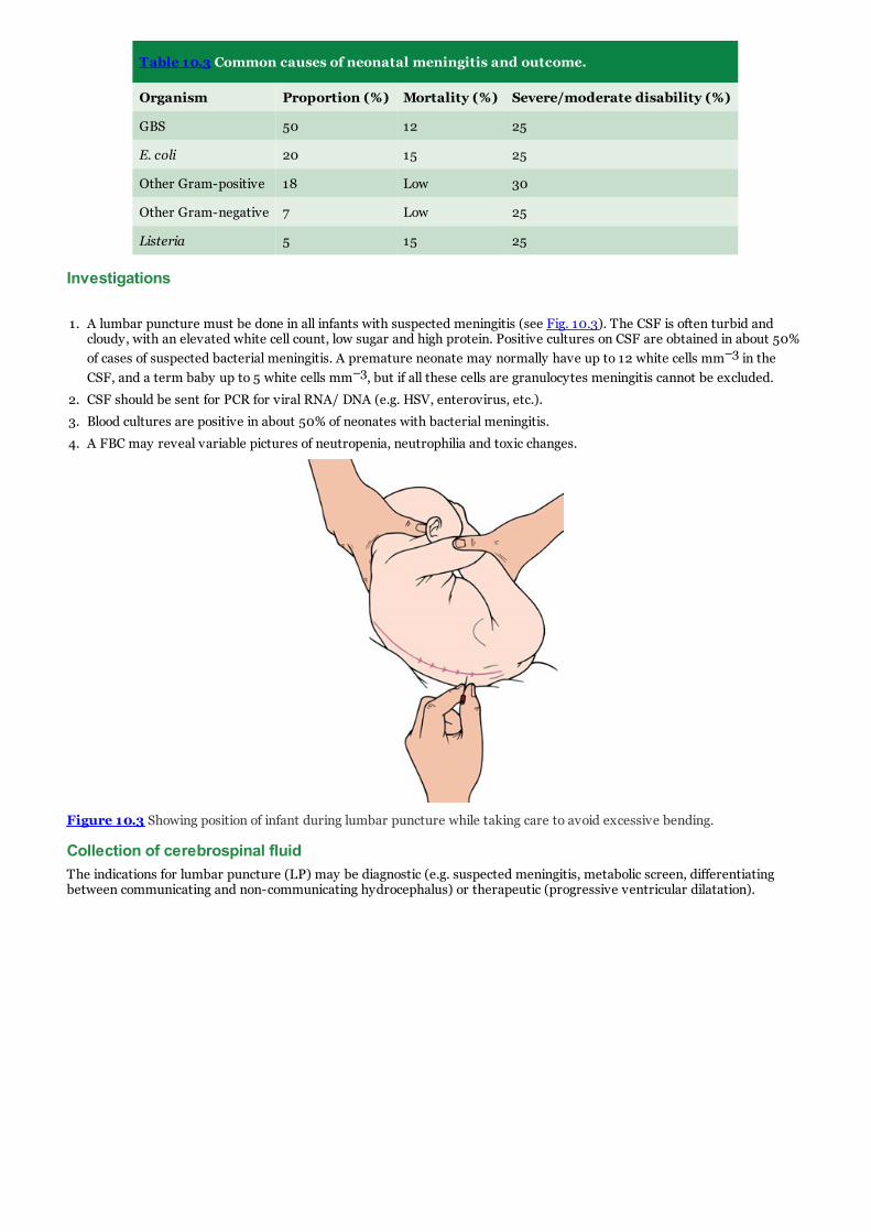
Table 10.3 Common causes of neonatal meningitis and outcome.
Organism Proportion (%) Mortality (%) Severe/moderate disability (%)
GBS 50 12 25
E. coli 20 15 25
Other Gram-positive 18 Low 30
Other Gram-negative 7 Low 25
Listeria 5 15 25
Investigations
1. A lumbar puncture must be done in all infants with suspected meningitis (see Fig. 10.3). The CSF is often turbid andcloudy, with an elevated white cell count, low sugar and high protein. Positive cultures on CSF are obtained in about 50%of cases of suspected bacterial meningitis. A premature neonate may normally have up to 12 white cells mm–3 in theCSF, and a term baby up to 5 white cells mm–3, but if all these cells are granulocytes meningitis cannot be excluded.
2. CSF should be sent for PCR for viral RNA/ DNA (e.g. HSV, enterovirus, etc.).
3. Blood cultures are positive in about 50% of neonates with bacterial meningitis.
4. A FBC may reveal variable pictures of neutropenia, neutrophilia and toxic changes.
Figure 10.3 Showing position of infant during lumbar puncture while taking care to avoid excessive bending.
Collection of cerebrospinal fluidThe indications for lumbar puncture (LP) may be diagnostic (e.g. suspected meningitis, metabolic screen, differentiatingbetween communicating and non-communicating hydrocephalus) or therapeutic (progressive ventricular dilatation).

CLINICAL TIPLumbar Puncture
1. A full surgical aseptic technique is necessary with gloves, gown and mask.
2. An experienced nurse needs to hold the baby in the left lateral position with the head, hips and knees wellflexed. The back needs to be completely parallel to the edge of the table. Make sure the airway is not obstructed.
3. The skin is cleansed with a suitable cleaning agent. Generally a 22G or 24G LP needle with stylet is used.
4. Use lumbar spaces L3–L4 or L4–L5. Use topical anaesthetic (EMLA cream) over the area for 30 minutes. Thelumbar puncture needle is then inserted and directed towards the umbilicus. Once the subarachnoid space ispenetrated, the stylet is removed and CSF is allowed to drip into each of three bottles. Sometimes, the needleneeds to be gently rotated to check for CSF flow. It is easy to push the needle too far into the anterior vertebralvenous plexus.
5. Measurement of lumbar CSF pressure may be necessary in infants with posthaemorrhagic ventricular dilatation(see Chapter 22). This can be done by attaching a Luer locking pressure transducer directly to the hub of theneedle, or by using a manometer tube. Pressure measurements are only valid if the infant is breathing quietlyand not crying. The head and trunk must be flat and in the same plane, and the infant not too tightly curled up.
6. CSF should always be sent for cell count, Gram stain and culture and glucose and protein estimations. Othertests may include viral PCR studies, syphilis serology or metabolic studies (lactate, glycine).
ManagementPrinciples of management are outlined in Box 10.4. The initial therapy for suspected meningitis in or after the first week oflife is a combination of ampicillin, gentamicin and cefotaxime. Always consider herpes simplex as a cause of meningitis andadd aciclovir if suspected (see page 106).
Box 10.4 Principles of management of neonatal meningitis.
Perform lumbar puncture if baby is at risk of meningitis or is ill.
Perform full infection screen (50% of babies will also have septicaemia).
Start ampicillin, gentamicin and cefotaxime together.
Always consider herpes simplex and add aciclovir if in doubt.
Repeat lumbar puncture after 48 hours to establish CSF is sterile.
Once sensitivities are known, stop inappropriate antibiotics.
Continue treatment for at least 2 weeks if GBS and >3 weeks if other organism.
Ultrasound imaging weekly to assess ventricular size.
If persistence of symptoms, consider cerebral abscess (MRI or CT brain scan).
Perform hearing test on recovery.
Once the organism has been identified, then microbiological advice as to the most appropriate antibiotic should be sought. Beaware of local patterns of antibiotic resistance. Meropenem may be indicated where multiple-resistant Gram-negativeorganisms are known to be prevalent.
Rapid sterilization of the CSF is essential, and it is recommended that a second lumbar puncture be performed after 48 s oftherapy to ensure that the CSF is sterile.
Continue intravenous antibiotics for at least 2 weeks in GBS infection, and for at least 3 weeks in Gram-negative infections.There is no evidence that intrathecal antibiotics improve outcome.
Ultrasound brain imaging should be performed in all infants with meningitis to detect progressive hydrocephalus, which mayneed treatment.
OutcomePostmeningitis hydrocephalus is a well-recognized complication, particularly in Listeria meningitis. Brain abscess may occur,particularly with Citrobacter and Enterobacter meningitis. Deafness is a recognized complication, and a hearing test (inaddition to the routine screening test) should be performed on discharge from hospital.
Approximately 50% of all babies with neonatal meningitis will show some neurodevelopmental problems at 5 years of age.The risk of severe or moderately adverse outcome is shown in Table 10.3.
Systemic candidiasisDiagnosis of this condition depends on a high clinical suspicion for the organism. It may rarely be congenital, associated withmaternal vaginitis, or acquired in a baby receiving neonatal intensive care. Candida albicans accounts for over 90% ofinfections, although C. parapsilosis is also seen rarely. Preterm infants undergoing intensive care are particularly vulnerableto Candida sepsis. Risk factors include:
Extremely low birthweight and current weight less than 1000 g.
Parenteral nutrition.

Central vascular catheter.
Mechanical ventilation.
Course of antibiotics.
The best method of diagnosis is to identify budding hyphae in urine from a suprapubic stab. The organism may also beisolated from arterial and/or venous blood cultures. Diagnosis may also be strongly suspected by observing ‘fungal balls’ on arenal ultrasound scan, or a pathognomonic appearance on ophthalmoscopy.
Treatment is with amphotericin B, usually in a liposomal formation (AmBisome) as it is less toxic. 5-Flucytosine actssynergistically with amphotericin and may be added in severe infection. Fluconazole may be used in amphotericin resistanceor increasingly as a first-line antifungal agent in sensitive cases of C. albicans. Central-vein lines should be removed onstarting treatment.
Death has been reported to occur in 40% of babies who are infected at <37 weeks’ gestation.
Fungal prophylaxisProphylactic systemic fluconazole significantly decreases the incidence of invasive fungal infection in extremely lowbirthweight (ELBW) infants. There is limited information on the long-term risks associated with systemic prophylaxis.Oral/topical non-absorbed antifungals (nystatin or miconazole) are commonly used in many neonatal units for fungalprophlyaxis.
Upper respiratory tract infectionsUpper respiratory tract infections can easily be transmitted within the nursery and sometimes will occur in outbreaks.Symptoms may include cough, increased nasal secretions, respiratory distress, oxygen requirement, temperatureinstability, rash and apnoea. Common viral upper respiratory tract infections are caused by rhinovirus, adenovirus,parainfluenza, respiratory syncitial virus (RSV), and human metapneumovirus (hMPV). Treatment is generally supportiveand the infant should be isolated and nursed using droplet precautions.
CLINICAL TIP
Anyone who is unwell with fever, cough, runny nose or sore throat should not visit the neonatal nursery untilsymptoms have completely resolved. Wearing a mask is insufficient protection to prevent transmission.
Lower respiratory tract infectionsThese are discussed in Chapter 13.
Urinary tract infectionThis is discussed in Chapter 18.
Conjunctivitis (‘sticky eyes’)Mild eye infections are very common and are referred to as ‘sticky eyes’. Purulent conjunctivitis may be either a congenitalor an acquired infection. Conjunctivitis is sometimes secondary to a blocked nasolacrimal duct. Usually, the organismsinvolved are S. aureus, E. coli, Pseudomonas spp., Chlamydia trachomatis and Neisseria gonorrhoeae. Reddened swellingin the region of the lacrimal sac (dacrocystitis) may occur following infection of a blocked nasolacrimal duct. The termophthalmia neonatorum is reserved for infection with N. gonorrhoeae.
ManagementConjunctival swabs should be taken and sent promptly to the laboratory. If ophthalmia neonatorum or Chlamydiatrachomatis is suspected, specific swabs for PCR testing can be performed. If maternal gonorrhoea is suspected,prophylactic treatment of the newborn’s eyes with 1% silver nitrate eyedrops may prevent purulent conjunctivitis.
With mild conjunctivitis, or ‘sticky eyes’, frequent eye toilet with normal saline may be all that is required. More floridinfections will need the frequent instillation of antibiotic drops (e.g. chloramphenicol or sulfacetamide) after cultures havebeen taken and eye toilet carried out. After the conjunctivitis has settled somewhat, antibiotic ointment may be used.
Treatment of gonococcal ophthalmia varies between institutions, but will normally involve a combination of topical eye dropsand systemic antibiotics for at least 10 days. For the treatment of Chlamydia, see page 107.
Infection of the skin and subcutaneous tissues
Omphalitis and funisitisInfections of the umbilical cord (funisitis) and umbilical stump (omphalitis) are usually due to S. aureus or E. coli, or otherGram-negative bacteria. Infection may arise as the result of chorioamnionitis. Because of the potential seriousness of thespread of infection to the portal vein and subsequent portal hypertension, infection in this area must be treated promptly. Ifthere are signs of spread with surrounding cellulitis, parenteral antibiotics such as flucloxacillin and gentamicin are necessaryafter appropriate swabs and cultures have been taken. Topical treatment with an antibiotic is also necessary.
ParonychiaReddening of the skin in the nailfold is common, and may proceed to pus formation when staphylococcal infection is present.Topical treatment consists of applications of mupirocin calcium (Bactroban) and this is usually all that is necessary toeradicate minor bacterial infections. Parenteral and topical antibiotics are indicated if systemic spread is suspected with

bacterial infection.
ThrushYeast infection is common, particularly if there has been C. albicans colonization of the maternal genital tract. It is also likelyto occur in association with the use of broad-spectrum antibiotics and in debilitated neonates receiving total parenteralnutrition. It commonly occurs in the mouth and looks like milk curds that cannot be removed with a swab stick. Theinfection usually occurs towards the end of the first week of life. In the napkin area it appears as a spreading centrifugalrash. It sometimes occurs as a secondary infection following ammoniacal dermatitis.
Napkin psoriasis is a rare condition that starts in the napkin area and may become widespread, usually secondary to thrush.The treatment of oral candidiasis consists of the use of nystatin drops or miconazole (Daktarin) gel, and its use should becontinued for at least 3 days after the lesions disappear. For the napkin area, nystatin or clotrimazole (Canesten) creamapplied four times daily is usually sufficient.
GastroenteritisGastroenteritis usually occurs in epidemic form in neonatal nurseries. Outbreaks have in the past been associated withenteropathogenic E. coli, and occasionally with Salmonella and Shigella. Viral infections are the most frequent nurserypathogen, and possible causes include enterovirus, echovirus and rotavirus.
Because the neonate withstands salt and water loss poorly, dehydration may rapidly occur. The infant shows signs such asloss of skin turgor, sunken eyes and fontanelle, dry mouth and oliguria.
Treatment consists of isolation from non-infected infants and the replacement of electrolytes and water. Antibiotics are of novalue unless there is evidence of systemic bacterial infection.
Prevention of acquired infectionsThe procedures detailed below will help to reduce and control infections. The ‘Matching Michigan’ initiative to preventcentral venous catheter blood stream infections is now followed in the majority of units in the UK (more information isavailable at: www.patientsafetyfirst.nhs.uk/).
Labour wardFastidious care in hand-washing.
The use of gloves for vaginal examinations, insertion of scalp electrodes and instrumental deliveries.
Identification of risk factors for group B streptococci and commencement of intrapartum antibiotics where appropriate.
NurseryCareful hand-washing is the key to reducing nosocomial infection. On entry into a neonatal unit, all staff and visitorsshould wash their hands carefully. Hands should be washed or an alcohol hand rub should then be used every time beforeand after an infant or the surrounding work space is handled.
Cleansing of incubators and equipment, and frequent changes of humidifier and incubator sterile water.
Have one stethoscope per baby.
Routine baths for babies, using hygienic soap solutions.
Aseptic surgical techniques for procedures such as intubation, umbilical catheterization, peripherally inserted centralcatheters, intravenous cannulation, intercostal catheters and lumbar punctures. Chlorhexidine has been shown to betwice as effective as iodine.
Fungal prophylaxis with fluconazole or nystatin/mycostatin.
Isolation techniques for infectious babies.
Avoidance of overcrowding and restriction of nursery ‘traffic’ to a minimum.

CLINICAL TIPPeripheral insertion of central catheters. This is a useful technique which provides long-lasting central venous access.
A peripherally inserted central catheter (PICC) pack is used which contains the insertion introducer and needle, thePICC line and all the other equipment needed for insertion (see Fig. 10.4).
This procedure must be carried out using strict aseptic precautions, and the operator should be fully masked, gownedand gloved. The veins on the hand, forearm and long saphenous are the most suitable for cannulation.
1. The area is exposed and thoroughly cleansed with alcohol/chlorhexidine. Sterile drapes are applied. Thepreparation must be allowed to dry before touching. Estimate the length of insertion (the tip ideally should be inthe superior vena cava; SVC). Pre-flush the longline with saline.
2. The needle and introducer is inserted into the vein as for venepuncture. When blood is seen to drip out of theneedle it is removed and the introducer remains within the vein; the silastic catheter is then threaded up theintroducer until the correct length is reached. The introducer is then pulled out from the skin. If it is the typethat splits it is removed, otherwise it is secured at the distal end of the line. Care needs to be taken to not pullout the long line when removing the introducer.
3. If a guide wire was included in the kit it should be removed. The whole line should then be gently flushed withheparinized saline.
4. Secure the line firmly with sterile adhesive strips, and cover with a sterile transparent dressing (e.g. OpSite).
5. Before commencing the infusion the position of the catheter tip should be checked to ensure it is in a centralvein, by radiography. Many use a radiopaque contrast solution. If the tip is in too far it may be pulled back to amore suitable position. The tip of the line must not lie within the heart as there is a risk of pericardialtamponade.
Figure 10.4 Equipment required for insertion of a percutaneous intravenous central catheter (PICC). Illustration courtesyof Dr Emmanuel Erinaugha.
Complications of long linesPICCs have a number of potential complications, and these should be checked for routinely. They include:
Blood loss/leakage at the site – this can cause the dressing to lift.
Infection.
Thrombosis.
Reduced perfusion of limbs.
Extravasation (into subcutaneous tissues/pleural space/peritoneum/pericardium).
Extravasation injurySubcutaneous extravasation of fluids and drugs is a common complication in neonatal intensive care units. Minorextravasations do not cause any problem, but if the volume of the leaked fluid is large or the fluid is irritant or hypertonic(such as calcium injection or TPN) this can cause significant problems that include skin blistering, ischaemia and tissuenecrosis. Although not completely unavoidable, the incidence of this complication can be greatly reduced by improvedvigilance.
Where significant extravasation has occurred, certain measures should be used to minimize the pain and extent of thedamage. One way to reduce further damage is to dilute the effect of irritation caused by the infusate by injectinghyaluronidase into the subcutaneous tissue under the area of damaged skin, either by injecting through the cannula itself (ifstill in situ) or, better still, by making three to four small openings in the skin overlying the damaged tissue with either a finescalpel or wide-bore needle, and irrigating the tissue through these openings in turn with 20–100 ml of 0.9% normal saline.The saline should run out of these openings like a fountain. The remaining fluid can be massaged out of the openings bygentle manipulation. The damaged skin is then kept reasonably moist with dressings such as paraffin gauze. Document thelesion before and after flushing using photographs, and refer early to plastic surgery if there is extensive tissue damage.

Adjunctive therapy
Intravenous immunoglobulin therapyIntravenous immunoglobulin (IVIG) infusion has been used for sepsis prophylaxis in preterm and low-birthweight infants.The Cochrane Review found a reduction in sepsis but no difference in mortality or other clinically important outcomes. IVIGhas also been used in infants with suspected or proven sepsis. While there appears to be a modest reduction in mortalitycompared with controls, the Cochrane Review concluded that there is insufficient evidence to support the routine use ofIVIG in infants with suspected or proven infection.
G-CSF or GM-CSF treatmentHaemopoietic colony-stimulating factors (CSFs) for granulocytes (G-CSF) or granulocyte-macrophages (GM-CSF) havebeen evaluated either for prophylaxis or in the management of septic infants in a number of studies. The Cochrane Reviewfailed to establish a clear benefit, and there is currently insufficient evidence to support the practice.
SUMMARY
Infection is a commonly encountered problem in neonatal medicine. It has significant morbidity and mortality. Signsand symptoms may be present at or shortly after birth, or may occur at any time during admission. Staff looking afterneonates must have a high suspicion of infection, as prompt treatment is required. A number of measures have beenproven to decrease the risk of nosocomial infection, with the most important being careful hand-washing.
Further reading1. Levy, O. (2007) Innate immunity of the newborn: basic mechanisms and clinical correlates. Nature Reviews in
Immunology, 7 , 379–390.
2. Liu, A.H., Leung, D.Y. (2006) Renaissance of the hygiene hypothesis. Journal of Allergy and Clinical Immunology,117 , 1063–1066.
3. Turner, M., Visintin, C., Mugglestone, M., Murphy, M. (2012) Antibiotics for the Treatment and Prevention ofNeonatal Early Onset Infection: NICE Guideline. Archives of Disease in Childhood, 97 (Suppl. 2), A382.
4. Wilson, C., Nizet, V., Maldonado, Y., Remington, J., Klein, J. (2016) Remington and Klein’s Infectious Diseases of theFetus and Newborn Infant, 8th edition. Elsevier/Saunders, Philadelphia.
Now visit www.essentialneonatalmed.com to test yourself on this chapter.
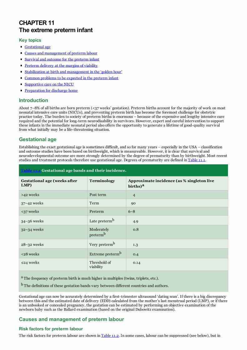
CHAPTER 11 The extreme preterm infantKey topics
Gestational age
Causes and management of preterm labour
Survival and outcome for the preterm infant
Preterm delivery at the margins of viability
Stabilization at birth and management in the ‘golden hour’
Common problems to be expected in the preterm infant
Supportive care on the NICU
Preparation for discharge home
IntroductionAbout 7–8% of all births are born preterm (<37 weeks’ gestation). Preterm births account for the majority of work on mostneonatal intensive care units (NICUs), and preventing preterm birth has become the foremost challenge for obstetricpractice today. The burden to society of preterm births is enormous – because of the expensive and lengthy intensive carerequired and the potential for long-term neurodisability in survivors. However, expert and careful intervention to supportthese infants in the immediate neonatal period also offers the opportunity to generate a lifetime of good-quality survivalfrom what initially may be a life-threatening situation.
Gestational ageEstablishing the exact gestational age is sometimes difficult, and so for many years – especially in the USA – classificationand outcome studies have been based on birthweight, which is measureable. However, it is clear that survival andneurodevelopmental outcome are more strongly determined by the degree of prematurity than by birthweight. Most recentstudies and treatment protocols therefore use gestational age. Degrees of prematurity are defined in Table 11.1.
Table 11.1 Gestational age bands and their incidence.
Gestational age (weeks afterLMP)
Terminology Approximate incidence (as % singleton livebirths)a
>42 weeks Post term 4
37–42 weeks Term 90
<37 weeks Preterm 6–8
34–36 weeks Late pretermb 4.9
32–34 weeks Moderatelypretermb
0.8
28–32 weeks Very pretermb 1.3
<28 weeks Extreme pretermb 0.4
≤24 weeks Threshold ofviability
0.14
a The frequency of preterm birth is much higher in multiples (twins, triplets, etc.).
b The definitions of these gestation bands vary between different countries and authors.
Gestational age can now be accurately determined by a first-trimester ultrasound ‘dating scan’. If there is a big discrepancybetween this and the estimated date of delivery (EDD) calculated from the mother’s last menstrual period (LMP), or if thereis an unbooked or concealed pregnancy, the gestation can be estimated by performing an objective examination of thenewborn baby such as the Ballard examination (based on the original Dubowitz examination).
Causes and management of preterm labour
Risk factors for preterm labourThe risk factors for preterm labour are shown in Table 11.2. In some cases, labour can be suppressed (see below), but in

many preterm delivery becomes inevitable. The biggest global causes of prematurity are social deprivation and multiplepregnancy: 6% of singletons, 51% of twins and 91% of triplets are delivered preterm. Infection is strongly implicated in 25–40% of preterm labour – evidence of chorioamnionitis is found in more than 50% of deliveries at less than 25 weeks. Insome cases the ‘cause’ of preterm labour cannot be established.
Table 11.2 Risk factors for preterm labour and prematurity.
Factor Comment
Maternal age Extremes of age (<18 yrs or >35 yrs) are associated with preterm labour
Maternal ethnicity Afro-Caribbean mothers have a 15% incidence of preterm labour
Multiple pregnancy The higher the multiple, the greater the chance of preterm delivery
Infection Chorioamnionitis is strongly associated with extreme preterm labour
Hypertension, PET Labour often induced early to maintain maternal health
Cervical weakening Previous midtrimester pregnancy loss or cervical surgery (e.g. cone biopsy)
Uterine malformation Bicornuate uterus or massive fibroids
Antepartum haemorrhage Abruption or placenta praevia
Amniotic fluid volume Polyhydramnios and oligohydramnios are both risk factors
Maternal substance abuse Alcohol, cocaine and cigarette smoking all associated with preterm labour
Fetal abnormality Congenital anomalies and trisomies are associated with preterm delivery
Predicting preterm deliveryAccurate prediction of imminent preterm delivery allows targeted intervention and transfer, but is not yet an exact science.The following may be useful:
Ultrasound measurement of the length of the cervix; cervical shortening is associated with a high risk of delivery.
Fetal fibronectin (a glycoprotein produced by the fetus which acts as ‘biological glue’); its presence in vaginal/cervicalsecretions in mid-trimester is highly predictive of preterm labour, but is not valid in the presence of rupturedmembranes.
Preterm pre-labour rupture of membranes (PPROM); this occurs in only 2% of all pregnancies but 40% of pretermdeliveries. Of those with PPROM, 90% will usually deliver within the next week.
Frequency and duration of regular contractions.
Clinical management of preterm labourAlthough the high mortality and morbidity associated with preterm delivery have prompted a great deal of research intoways of predicting preterm birth, only limited benefits have resulted. Where possible, the woman in preterm labour at lessthan 32 weeks’ gestation should be transferred with the fetus in utero to a perinatal unit where optimal delivery,stabilization and subsequent management of her infant can be carried out. In-utero transfer should only be attempted whenthe preterm labour is not advanced or can be safely and effectively suppressed with tocolytic agents (see Chapter 25).
When a patient is admitted with a diagnosis of possible ‘preterm labour’, the management plan depends on the criteria listedin Box 11.1.
Obstetric management of preterm labour is rarely clear-cut and is a balanced decision between the relative risks to motherand fetus of continuing the pregnancy versus those of early delivery. Suppression of preterm labour is sometimes possibleusing tocolytic agents such as oxytocin receptor antagonists (e.g. atosiban) or calcium channel blockers (e.g. nifedipine)..These can delay preterm delivery by 2–7 days but have not been shown to reduce neonatal mortality or morbidity.Magnesium sulphate does not delay delivery but has been shown to reduce the risk of cerebral palsy in the preterm infantand is recommended below 30 weeks gestation.
Temporary suppression of labour may ‘buy time’ for in utero transport to a perinatal centre or for the acceleration of fetallung maturity with a course of corticosteroids. Betamethasone has been shown to be associated with fewer adverse effectsthan dexamethasone. Meta-analyses have consistently shown that corticosteroids given for 48 h before delivery significantlyreduce the incidence and severity of respiratory distress syndrome (RDS) and the incidence of intraventricularhaemorrhage (IVH), and possibly improve neurodevelopmental outcome. Suppression of labour is contraindicated in thepresence of intrauterine infection, congenital anomaly, signs of fetal compromise or significant antepartum haemorrhage.

Box 11.1 Approach to threatened preterm delivery.
Is the patient in active preterm labour?
Is there an underlying cause for the onset of labour?
Should steroids be given to the mother to enhance fetal lung maturity?
Should drugs be used to reduce uterine activity?
Are the mother and fetus at risk of infection?
Where is the baby to be delivered?
Is in-utero transfer possible?
How is the baby to be delivered?
Is a suitable neonatal intensive care cot available?
Mode of delivery in preterm labour
Caesarean section or vaginal deliveryIf delivery is inevitable, then the best mode of delivery must be chosen. Where the mother’s life is in danger (e.g. pre-eclampsia), urgent caesarean section is indicated. Where delivery is elective (e.g. for severe intrauterine growth retardation;IUGR) a balance must be struck between the risks to the fetus of labour and vaginal delivery and the risk to the motherfrom a caesarean section. In very early gestations (<26 weeks) a lower segment caesarean section (LSCS) may not bepossible and a classical (vertical incision) caesarean is needed. This carries a greater risk of uterine rupture in futurepregnancies.
Preterm breech presentationMany extreme preterm babies are still in the breech position when the mother enters labour. The preferred mode ofdelivery for the preterm breech fetus has been the subject of considerable controversy. The hazards to the fetus of vaginalbreech delivery include difficulty with delivery of the head and, rarely, entrapment of the aftercoming head. Althoughcaesarean section in the preterm breech may be associated with some difficulties, it is probably the preferred method ofdelivery beyond 25 weeks’ gestation.
Survival and outcome for the preterm infantDecision-making in the management of the extremely preterm delivery is influenced by the improved short- and long-termoutcomes observed with advances in perinatal care. Aggressive obstetric management and intervention for fetal reasons inlate second-trimester deliveries is now practised in many tertiary perinatal centres, and these attitudes are partlyresponsible for the improved outcomes obtained. Units should develop their own guidelines for the management ofextremely preterm labour.
Short-term survival and outcomePublished survival rates vary markedly from country to country and from centre to centre. Large geographically basedpopulation studies offer the least biased data. It is important to consider the following points:
Are babies with lethal congenital abnormalities excluded?
Is birthweight <500 g or gestation <24 weeks included or excluded?
Are infants born alive but not resuscitated or not resuscitatable included?
Is the denominator ‘all live births’ or ‘all admissions to NICU’ (the latter will have better outcomes)?
Have late neonatal deaths (>28 days) been included?
Representative figures for survival after NICU admission are presented by birthweight and gestational age in Table 11.3.Figure 11.1 shows the survival and outcomes for a geographically defined population (all preterm births in UK in 2006).

Table 11.3 Survival rates to discharge home by gestational age and birthweight for babies admitted aliveinto Australian and New Zealand intensive care nurseries in 2013. Note that these data do not includebabies who die before admission or who are not resuscitated.
Gestation at birth (weeks) Survival (%) Birthweight (g) Survival (%)
<24 43 400–499 56
24 66 500–749 73
25 84
26 88 750–999 90
27 94 1000–1249 95
28–32 97 1250–1500 95
Source: Chow, S.W. (2015) Reports of the Australian and New Zealand Neonatal Network 2013. ANZNN, Sydney.
Figure 11.1 Outcomes of all extreme preterm babies born in the UK in 2006.
Source: EPICURE 2 http://www.epicure.ac.uk/overview/overall-outcome/.
Long-term outcomesAs more extreme preterm babies survive and are followed-up into teenage and adult life, it is clear that there is anadditional hidden burden of subtle neurodisability occurring in the most preterm babies (<26 weeks). This can occur even inthe absence of obvious central nervous system (CNS) damage or haemorrhage, and may be related to a subtle disruption ofthe normal neuronal pathways that are developing in the third trimester. There is a higher than expected incidence ofattention deficit hyperactivity disorder (ADHD), autistic features and subtle learning difficulties than in term-born controls.Final stature, IQ and visual function may all be impaired, but often not to a degree that interferes with activities of dailyliving, and not to a degree that is perceived as detrimental by the children themselves.
Preterm delivery at the margins of viabilityBabies born at the margins of physiological viability (≤24 weeks) present particular challenges. These infants have a highchance of dying during labour, in the delivery room, or in the days and weeks after admission to the NICU. Data from theEPICURE studies in the UK suggest the chances of survival without disability are improving rapidly but remain low at theseextreme gestations (see Fig. 11.1 and Table 11.4).

Table 11.4 Survival without serious disability (EPICURE 1,2).
EPICURE 1 (1995) EPICURE 2 (2006)c
All live birthsa All admissions to NICUb (All admissions to NICU)
22 weeks 1% 5% –
23 weeks 3% 6% 14
24 weeks 9% 12% 28
25 weeks 20% 24% 47
a Survival without serious disability.
b Survival without serious disability at 6 years of age.
c Survival without disability at 3 years.
Practitioners will approach extreme preterm delivery differently depending on their experience, cultural and religiousbeliefs, and the healthcare system they operate in. Extreme preterm babies less than 28 weeks are sadly not able to betreated in most resource-poor countries, where access to NICUs is extremely limited. Even in developed countries attitudesvary widely. In the Netherlands, infants <25 weeks are not routinely offered intensive life support. In most UK, US andAustralian perinatal centres, infants of 22 weeks are not offered intensive life support; at 23 weeks NICU care may bestarted if parents expressly want this and the baby is in good condition; and at 24 weeks most will offer intensive care,provided that there are no other mitigating factors such as a severe congenital anomaly or perinatal asphyxia. In somecountries, national professional or ethical bodies have published guidelines to help doctors and parents make the rightdecisions. The important points outlined in Box 11.2 should be considered.
Box 11.2 Ethical decision-making at extreme preterm gestations (<25 weeks).
Discussions with parents should be undertaken by a senior, experienced neonatologist, in conjunction with theobstetrician.
Doctors must act with the best interests of the baby in mind.
A management plan should ideally be agreed with both parents before delivery.
It is important to be sure about the gestation – on what basis has it been calculated? Remember 23 weeks refersto 23+0 to 23+6 days.
If in doubt, there should be a presumption of offering life support. A palliative care pathway may be chosen later ifthe progress is not favourable.
If a baby at these gestations has a very low or absent heart rate at birth, then assessing the response to fiveinflation breaths via a mask is important before deciding on further intensive resuscitation. There is no evidence tosupport the use of adrenaline and CPR at these gestations.
The ethical dilemmas posed by treating the most extreme preterm babies are discussed further in Chapter 28.
Stabilization at birth and management in the ‘golden hour’Ideally, the baby will have been transferred in utero to a perinatal centre with a suitable NICU. Experienced personnelshould be present at delivery, preferably having had an opportunity to introduce themselves to the parents and to haveprepared equipment. They should be aware of any treatment plan agreed with the parents (see above) and be familiar withthe obstetric history. Special considerations for extreme preterm births are listed in Table 11.5. In general, it is best to thinkof the process as ‘stabilization’ rather than ‘resuscitation’. These babies are fragile, and usually not severely asphyxiated.They benefit from a calm transition and avoidance of trauma or hyperoxygenation. Once in the NICU they should havevascular access secured and from then on have ‘minimal handling’ (see Chapter 24).

Table 11.5 Approach to stabilization of a preterm infant ≤28 weeks.
Factor Comment
Personnel Experienced staff required – usually two neonatal doctors/practitioners and a neonatal nurse toattend
Thermoregulation The room must be warm. An overhead heater should be turned on. Place the baby in a clear plasticbag/wrap immediately at delivery (without drying first) and place under the overhead heater. Put abonnet over the head.
Monitoring of the heart rate can take place through the plastic bag. Do not remove until the baby is ina warm, humidified incubator. Hypothermia <36 °C massively increases the mortality in pretermbabies
Airway As at term, keep the head in the neutral position. You will need a small face mask and should have asmall laryngoscope and ET tubes (2.0, 2.5 and 3.0 mm) available
Breathing Use lower peak pressures (20–25 cmH2O) to prevent lung damage. PEEP (5–6 cmH2O) is veryimportant and must be maintained throughout the stabilization. Use an air–oxygen mix and beprepared to increase the oxygen concentration. Use a saturation monitor and avoid hyperoxia (seebelow). Give surfactant early, once the ET tube has been confirmed clinically to be correctly located.If attempting CPAP (see below), it is vital that PEEP is maintained throughout
Circulation A rise in heart rate is a valuable sign of effective lung aeration, as chest movement is harder to seethan at term. Saturation monitoring or ECG leads can measure the pulse.
Drugs These are rarely indicated. If there has been sufficient asphyxia that the heart rate does not respondto lung inflation, a senior doctor should consider whether it is in the best interests to continueaggressive resuscitation as the prognosis will be poor. A lower dose of prophylactic vitamin K isrequired than at term.
Parents Transfer the baby to the NICU as soon as possible, but try to allow the mother to see her baby, evenif only briefly, before you leave the room.
Vascular access Once the baby is in a warm, humidified incubator, a skilled operator should insert an umbilical venousand arterial line (see CLINICAL TIP above).
ET, endotracheal; PEEP, positive end-expiratory pressure; CPAP, continuous positive airway pressure.
Transportation within and between hospitalsThe baby must be transported from delivery suite to the NICU in a safe but efficient manner. A transport incubator thatprovides warmth and oxygen saturation monitoring is ideal. If the distance is very short the Resuscitaire trolley may beused, provided that warmth and ventilation can be maintained. If the baby has been delivered in a low-intensity perinatalunit, transfer to a regional NICU must be arranged urgently. Transport is discussed in detail in Chapter 25.
CLINICAL TIPIf the mother cannot be with the baby, either because she is ill or the baby has had to be transferred to anotherhospital, a photograph of the baby should be provided. Sometimes the mother will provide an item of clothing so thatthe baby can smell her, or mother and baby can exchange ‘bonding squares’. Achieving and maintaining breast milkexpression is vitally important in this situation.
Common problems to be expected in the preterm infantDuring the final 3 months of intrauterine life most organ systems undergo important structural and functional development.Premature birth requires rapid adaptation to extrauterine life before these organ systems are fully developed. Theincidence and severity of all complications of prematurity are normally related to gestational age and birthweight. The mainproblems are shown in Table 11.6.

Table 11.6 Common problems in the preterm infant.
Problem Impact
Pulmonaryimmaturity
Apnoea, RDS, and chronic lung disease of prematurity (bronchopulmonary dysplasia) are common.Pneumothorax and pneumonia can occur.
Fragilecapillarynetwork in thesubependymalarea
High risk of IVH, especially in response to swings in cerebral perfusion pressure and carbon dioxidelevel. Large IVH can cause venous infarction or hydrocephalus (see Chapter 22)
White matterinjury
Periventricular white matter susceptible to ischaemic damage, especially if sensitized by fetalinflammation (infection). Preterms are less able to tolerate perinatal asphyxia. High risk ofperiventricular leukomalacia (PVL) – see Chapter 22)
Thermalinstability
Hypothermia exacerbates RDS and increases mortality. If environmental temperature is too low thebaby will expend energy keeping warm at the expense of growth
Feedintoleranceand lack ofsuck-swallowreflexes
Immature or absent suck–swallow and gag reflex. Need NG feeding. Poor gut motility may cause feedintolerance. Prematurity is also the main risk factor for necrotizing enterocolitis (NEC) – see Chapter 17.
Patent ductusarteriosus(PDA)
Risk of congestive heart failure and risk factor for NEC and IVH
Immaturevisual system
Risk of ROP (see Chapter 23) and myopia/strabismus
Jaundice High red cell mass and poor liver conjugation makes hyperbilrubinaemia almost inevitable. Acidosis anda poor blood–brain barrier increases the risk of kernicterus (see Chapter 19)
Renalimmaturity
Inability to concentrate urine, and to excrete an acid load with a low renal bicarbonate threshold,resulting in late metabolic acidosis which may be associated with failure to gain weight satisfactorily.Treatment with sodium bicarbonate and feeding with breast milk or appropriate preterm formula feedsusually improves the acidosis
Metabolicdisturbance
Hypoglycaemia, hypocalcaemia, hypomagnesaemia, hyponatraemia, hypernatraemia, hyperkalaemia areall common and must be anticipated
Infection Relative immunodeficiency and breech of natural barriers (e.g. by central venous lines) predispose toinfection, which may be nosocomial or maternal in origin
Haematologicaldisorders
Disseminated intravascular coagulation, vitamin K deficient bleeding and iatrogenic or iron-deficientanaemia are more common, but not inevitable
Surgicalproblems
Undescended testes, inguinal and umbilical hernias
Supportive care on the NICU
MonitoringHeart rate, respiratory rate, blood pressure and temperature must be monitored continuously, with appropriate alarmsignals. An apnoea monitor is often used in the special care nursery.
Oxygen therapyMany sick preterm infants require oxygen therapy to relieve hypoxia. Oxygen is a potentially toxic substance in thepreterm infant, and may cause retinopathy of prematurity (ROP) and exacerbate lung inflammation. It must therefore beadministered with the utmost care and the response continuously monitored with a pulse oximeter or transcutaneousmonitor (Box 11.3). Regular measurements of PaO2 are made in premature infants requiring extra oxygen to minimizecomplications such as ROP and chronic lung disease. It is dangerous to treat ill premature infants without adequate blood-gas monitoring being available. Oxygen can be administered directly into the incubator or by nasal prongs but nasalcontinuous positive airway pressure (CPAP) or high-flow humidified oxygen therapy (HFNT) are usually used in theimmediate newborn period. Oxygen should be warmed to 35–37 °C, humidified to 28–38 mg H2O l–1, and the inspiredconcentration continuously recorded with high and low saturation alarms set.

Box 11.3 Blood gas monitoring.
Pulse oximetry analyses red light transmitted through tissues to measure haemoglobin oxygen saturation inarterial blood. This provides continuous, non-invasive and well-tolerated monitoring. Recent research suggeststhat an O2 saturation target of 91–95% improves survival when compared to lower target of 85–89%.
Blood gas sampling. Insertion of a catheter into an umbilical or peripheral artery (see Clinical Tip box above)provides a readily available site for sampling arterial blood for blood gases, electrolytes and blood glucose, andcontinuous BP monitoring. The arterial oxygen tension should be maintained between 50 and 80 mmHg (6.2–10 kPa). Intermittent arterial stabs are less reliable for monitoring PaO2, and capillary sampling is not reliable atall, although it can be used to monitor pH and pCO2 in the more stable infant
Transcutaneous oxygen (± transcutaneous carbon dioxide) monitors utilize a small heating element to inducehyperaemia in the skin and to arterialize the capillary blood and oxygen (±CO2) diffusing through the skin, which ismonitored continuously. Less frequently used now as can cause skin burns.
End-tidal carbon dioxide monitors are now available with a low dead space such that they are suitable forpreterm babies. These give a continuous measure of expired CO2, which is usually slightly lower than pCO2.
CLINICAL TIPUmbilical arterial and venous catheter insertion.
An umbilical arterial catheter (UAC) can be inserted into the umbilical artery under sterile conditions. The tip shouldlie at the T6–T10 vertebral level, avoiding the renal and mesenteric arteries. The length required can be calculatedusing the formula:
Once inserted to the correct depth the catheter is secured in situ, preferably by placing a zinc oxide tape across thecatheter and then suturing it to umbilical stump. The catheter is connected to a pressure transducer for BPmonitoring. Circulation to the lower limbs should be closely observed. A heparin–saline infusion should be started toreduce the risk of thrombosis. The position is confirmed using an abdominal X-ray (see figure). An umbilical venouscatheter can be inserted using a similar technique, but the distance inserted is less. The UVC tip should sit at the levelof the diaphragm (above T10) but outside the heart. Great care must be taken to ensure it is not deviated into thehepatic veins, as this can cause extravasation and liver injury. UVCs are sometimes inserted as an emergency duringresuscitation. This should still be performed in as aseptic manner as can be achieved in the circumstances.
Figure. Abdominal X-ray showing UAC (red arrow) and UVC (blue arrow). Note the UAC always dips down as itpasses through the iliac arteries. The UVC passes straight through the ductus venosus to the IVC. In this case theUVC is too long, with the tip in the right atrium. It should be pulled back to the level of the T8–T10 vertebra.
Respiratory distress syndrome (RDS)Even with the advent of drugs to suppress preterm labour, antenatal corticosteroids and postnatal surfactant, which haveradically reduced mortality and morbidity, RDS remains the major problem for the preterm baby in the first week of life.Surfactant-deficient RDS (hyaline membrane disease) is almost inevitable in babies born at less than 28 weeks’ gestation.Those preterm infants who die usually have hyaline membranes in the lung at autopsy. In this century the challenge hasmoved from preventing death from RDS to preventing chronic lung disease of prematurity. There has been a recent shifttowards the early use of non-invasive ventilation (CPAP or high-flow nasal therapy; HFNT), even in the most extremepreterm infants, with the aim of avoiding mechanical ventilation. Up to 40–50% of even very preterm babies studied appearto manage without intubation or surfactant.

For infants of birthweight less than 800 g or less than 26 weeks’ gestation it may be desirable to intubate and ventilate frombirth to provide initial airway stabilization, with the aim of early extubation on to CPAP. For other infants at less than 32weeks’ gestation, airway stabilization at birth with CPAP is widely practised. If the baby is intubated, exogenous‘prophylactic’ surfactant should be administered as soon after birth as possible. ‘Rescue’ surfactant treatment for thosebabies who do not cope on CPAP or HFNT may be delivered by the ‘InSurE’ technique: Intubation, Surfactant, Extubation.A modification of this is to instil surfactant into the trachea via a fine catheter while the baby remains breathing on CPAP.This is known as minimally invasive surfactant therapy (MIST). Nebulised surfactant is under development.
Blood pressureArterial blood pressure (BP) is an important variable to measure. If an indwelling arterial catheter is in place, it can beattached to an electronic pressure transducer to measure BP. Alternatively, an oscillometric device can be used, attached toa BP cuff. Normal BP ranges are shown in Chapter 16. Blood pressure varies with gestational age and birthweight, andnormally increases over the first 24 hours of life. Maintenance of an adequate and stable cerebral blood flow is important toavoid cerebral ischaemic and haemorrhagic injury. Management of hypotension is discussed in Chapter 16.
Intraventricular haemorrhage (IVH)The preterm brain contains a germinal matrix, just outside the lateral ventricles, from which new brain cells migrate to thecortex. This area is highly vascular with a network of capillaries that are vulnerable to changes in cerebral perfusionpressure due to mechanical ventilation, fluctuations in pCO2 and complications such as hypotension, PDA andpneumothorax. Bleeding from these capillaries can cause a sub-ependymal or IVH. This is occurs in about 15–20% of allextreme preterms.
Periventricular leukomalacia (PVL)PVL is a white matter injury occurring in preterm babies usually less than 32 weeks and triggered by inflammation, cerebralischaemia or hypoxia. It can have devastating consequences, with a high risk of cerebral palsy. Administering intravenousmagnesium to the mother has been shown to reduce the risk of cerebral palsy in preterm infants <30 weeks.
IVH and PVL are discussed in Chapter 22.
Patent ductus arteriosus (PDA)The arterial duct usually closes within hours of birth but in preterm babies can remain open for weeks. The PDA causes aleft to right shunt from the aorta to the pulmonary artery. This causes pulmonary congestion and a reduction in systemicblood flow during diastole. It is a risk factor for NEC, CLD and IVH. There is controversy about if and when to close a ductusing anti-inflammatory agents (ibuprofen or paracetamol) or surgical closure. PDA is discussed in Chapter 16.
ThermoregulationBody temperature must be maintained in the normal range by nursing the preterm infant in a closed, humidified incubator(see Chapter 24). Nursing on a servo-controlled heated platform is less desirable as humidity is not maintained, but may berequired for some surgical conditions.
FeedingInfants of less than 33–34 weeks’ gestational age do not have a good suck or swallow reflex and should usually be fed via anorogastric or nasogastric tube. Premature infants with small gastric capacity require frequent feeding. Feeding the preterminfant is discussed in detail in Chapter 9. The ideal milk is mother’s freshly expressed breast milk (EBM). This providesimmunity, is easily absorbed, and reduces the risk of NEC. If this is not available, stored (frozen) EBM or sometimes donorEBM may be used. Preterm breast milk fortifiers are available to increase the protein and mineral content. Encouraging andsupporting the mother to produce breast milk is vital. If formula is used it should be a specialized preterm formula (seeChapter 9).
Necrotizing enterocolitis (NEC)This occurs in about 10% of all extremely preterm infants and carries a high mortality. It is an ischaemic inflammation of thegut associated with translocation of bacteria into the gut wall. There are a number of risk factors, including low gestationalage, hypotension, PDA and absence of breast milk feeds. Excessively aggressive increase of feed volumes and use of formulafeeds may lead to NEC. There is emerging evidence that the use of probiotics (e.g. Lactobacillus and Bifidobacter species)from birth to 34 weeks can reduce the risk of NEC. Their use is becoming more widespread. NEC is discussed in Chapter 17.
Intravenous fluidsSick babies and infants of less than 1500 g or 30 weeks’ gestation may need intravenous fluids (10% dextrose andelectrolyte solution) until feeding is established. Nutrition is vital, and if feeds are going to be delayed or increased slowlythen parenteral nutrition (PN) should be administered early, even on the first day of life. PN is discussed fully in Chapter 9.
The fluid requirements of ill preterm infants depend on a variety of factors, including postnatal age, renal function andtransepidermal water loss. Consequently, the fluid intake is adjusted (at least daily) on the basis of urine output, weight,serum electrolytes and urinary concentration. Water losses will be increased by multiple phototherapy and open radiantincubators (approximately an extra 20 ml kg–1 per day). Sick preterm infants, especially those with RDS and PDA, shouldreceive restricted fluid volumes until renal output is well established. Insensible losses can be reduced by environmentalmeasures (see Chapter 24).
On the first day, intravenous fluids should not contain sodium, but the sodium and electrolyte requirement for prematureinfants will increase as immature kidneys do not conserve sodium well. Close monitoring of serum electrolytes, includingcalcium, is necessary.

JaundiceThis is extremely common in the preterm infant and must be tracked with frequent bilirubin estimations. Jaundice ofprematurity is a diagnosis of exclusion after other causes have been eliminated. Preterm infants are more prone to bilirubinencephalopathy than term infants, and factors that affect the entry of free bilirubin into the brain include low albumin levels,acidosis, hypoxia, hypoglycaemia, hypothermia, certain drugs and starvation. Jaundice is discussed thoroughly in Chapter19.
VitaminsA single intramuscular dose of 400 μg kg–1 vitamin K is adequate. If given intravenously, or the baby is receiving prolongedbroad-spectrum antibiotics, further doses may be required. Preterm infants who require PN receive supplemental vitaminsand preterm formulas are supplemented with vitamins. Preterm infants of less than 32 weeks’ gestation should have humanbreast milk fortifier added to EBM. Once a preterm infant of less than 34 weeks’ gestation is tolerating full feeds, mostrecommend a multivitamin preparation such as Pentavite or Abidec drops given daily and continued until the baby is fullyweaned on to a mixed diet.
AnaemiaSome sick preterm infants will be anaemic at birth or develop anaemia as a result of frequent blood sampling, and willrequire a transfusion with packed red blood cells. The venous haematocrit should be maintained at more than 0.35%(haemoglobin 12 g dl–1) in all sick babies during the acute phase of RDS. However, during the physiological nadir of anaemia(at 5–7 weeks) preterm infants may tolerate a haemoglobin concentration of 7 g dl–1 (haematocrit 0.25%), especially if thereis an adequate reticulocyte response (>5%). To prevent iron-deficiency anaemia, preterm infants of less than 2000 gbirthweight or 34 weeks’ gestation are usually prescribed supplemental iron (ferrous sulphate) from 2–6 weeks until fullyweaned, unless they are on formula milk.
Preparation for discharge homeOnce the need for intensive care is over, the very preterm baby will still need monitoring, incubator care and nasogastricfeeding for a number of weeks. This can be a very frustrating time for the parents, who have already been through a lot ofstress. Early discharge at 34–35 weeks can be safely practised, provided that feeding is progressing well, temperaturecontrol is good, weight gain is steady, the mother is handling her baby competently and the home situation is good. Someunits promote short-term nasogastric feeding at home for selected, trained parents, with outreach nurse support, as a wayof speeding the transition to full feeding. Other units will have a transitional care facility or invite the mothers in to be‘resident’ on the neonatal unit for a few days before discharge to gain confidence in being the prime carer for their baby atlast. The transition from hospital to home is a crucial step in the future well-being of the family and is discussed in Chapter27.
SUMMARY
About 7% of all births occur preterm (<37 weeks), but it is those at less than 32 weeks and especially less than 28weeks that provide the greatest challenge. These births often occur unexpectedly and a skilled team must bemobilized. The use of antenatal corticosteroids has dramatically improved survival, and every opportunity should betaken to administer these, even if labour needs to be delayed.
A philosophy of gentle ‘stabilization’ rather than aggressive resuscitation, with careful avoidance of extremes oftemperature, BP, oxygen and carbon dioxide concentration will
be beneficial in the longer term. In all cases, and especially those babies born at the ‘threshold of viability’, goodcommunication with the parents is essential. The right decisions need to be made in the best long-term interests ofthe baby, and they must be clinically, ethically and morally sound decisions that all can agree on.
The survival of extremely preterm infants is one of the successes of modern medicine. It is important that the long-term aim (survival with a good quality of life) is kept in mind and that these advances are also enabled and promotedin resource-poor countries.
Further reading1. Boyle, E.M., et al. (2012) Effects of gestational age at birth on health outcomes at 3 and 5 years of age: population
based cohort study. British Medical Journal, 344, e896.
2. Costeloe, K.L., et al. (2012) Short term outcomes after extreme preterm birth in England: comparison of two birthcohorts in 1995 and 2006 (the EPICure studies). British Medical Journal, 345, e7976.
3. ESPGHAN Committee on Nutrition (2010) Enteral Nutrient Supply for Preterm Infants: Commentary From theEuropean Society of Paediatric Gastroenterology, Hepatology and Nutrition Committee on Nutrition. Journal ofPediatric Gastroenterology and Nutrition, 50 (1), 85–91.
4. Evans, N. (2006) Which inotrope for which baby? Archives of Disease in Childhood. Fetal and Neonatal Edition, 91(3), F213–F220.
5. Saigal, S., Doyle, L.W. (2008) An overview of mortality and sequelae of preterm birth from infancy to adulthood.Lancet, 371 , 261–269.

CHAPTER 12 The low-birthweight infantKey topics
The infant who is small for gestational age
Causes of intrauterine growth restriction
Problems to be expected in the growth-restricted �fetus and SGA infant
Management of the low-birthweight infant
IntroductionNewborn infants can be classified according to birthweight, gestational age or size for gestational age (Table 12.1). Lowbirthweight is defined as less than 2500 g. However, a low-birthweight (LBW) infant may be that size because they areeither preterm or small for gestational age (SGA), or both. The problems that these infants develop tend to be determinedby whether they have been born too early or are born too small for the duration of gestation. The problems associated withLBW are described here. Those predominantly associated with prematurity are described in Chapter 11.
Table 12.1 Classification of newborn infants according to birthweight, gestational age or size forgestational age.
Factor Terminology Incidence (%)
Birthweight
<2500 g Low birthweight (LBW) 6.5*
<1500 g Very low birthweight (VLBW) 1.3
<1000 g Extremely low birthweight (ELBW) 0.6
Gestational age (completed weeks after last normal menstrual period)
<37 weeks Preterm 8.4
>41 weeks Post-term 0.6
Size for gestational age
Weight between 90th and 10th centiles for gestation Appropriate for gestational age (AGA) 80
Weight <10th centile for gestation Small for gestational age (SGA) ≈10
Weight >90th centile for gestation Large for gestational age (LGA) ≈10
*These figures are for developed countries. When we include resource-poor countries the incidence of low birth weight is16% (UNICEF, 2013)
The infant who is small for gestational ageAlthough several synonyms have been used to describe the growth-restricted infant, including dysmaturity, light for dates,small for dates and intrauterine growth restriction (IUGR), the preferred term is now SGA. There has been no suchuniformity in the definition of SGA. From a statistical viewpoint, infants weighing more than two standard deviations belowthe mean can be defined as SGA, regardless of gestation. However, because they share common clinical problems, infantsbelow the 10th centile for weight are often regarded as ‘SGA’. It must be recognized that some of these will in fact beappropriately grown and are just smaller than average, for familial or racial reasons. Furthermore, many neonates withbirthweight above the 10th centile will demonstrate evidence of acute or chronic weight loss, and should therefore fall intothe spectrum of the growth-restricted infant. Variables such as sex, race and altitude should be considered whendetermining growth curves, because a birthweight below the 10th centile in one population may not fall below the 10thcentile in another, even though both may be growth-restricted. Table 12.2 lists important factors that predict birthweight of

an individual fetus or neonate for a given population.
Table 12.2 Determinants of birthweight.
Maternal factors before conception Stature
Weight
Genotype
Race
Age
Parity
Socioeconomic status (occupation, education, income)
Factors at or around conception Fetal genotype
Singleton or multiple conception
Fetal sex
Genetic anomaly (chromosomal or major gene locus)
Factors between conception and birth Altitude above sea level
Fetal or maternal infection (rubella, malaria)
Maternal work and ability to rest
Maternal cigarette smoking
Maternal diet
Maternal alcohol, drugs, medications
Placental dysfunction (e.g. PET)
CLINICAL TIP
It is important that weight and length measurements are plotted on an appropriate growth chart. WHO have issuedgrowth charts for preterm and term babies based on a breastfed population, which mean breastfed infants will nolonger appear to be dropping centiles. Down’s syndrome specific charts also exist. Growth-restricted infants should beplotted on a normal growth chart appropriate to their gestational age.
Classification of small for gestational age infantsSubdividing the heterogeneous SGA population into subgroups offers the potential to better understand the underlyingcause, and therefore allows a more accurate prognosis and appropriate postnatal management:
SGA but appropriately grown (constitutionally small).
SGA due to in-utero growth restriction (IUGR) (see Fig. 12.1).

Figure 12.1 Monozygotic twins born at 32 weeks. The smaller twin weighed 750 g and the larger 1700 g. The smaller twinshows features of IUGR with relative head sparing (asymmetric IUGR).
It is important to attempt to define the neonate ‘starved’ as a result of IUGR. These babies show greatest growth failure interms of weight, then length; head circumference is the least affected. This gives them an asymmetrical appearance withrelative head sparing. There is little subcutaneous fat, the skin may be loose and thin, muscle mass is decreased (especiallythe buttocks and thighs), and the infant often has a wide-eyed, anxious look. This pattern can be distinguished from thesymmetrically growth-restricted type, which implies either a fetal cause (e.g. chromosomal) or a very early insult.
Subcutaneous fat can be objectively assessed by measuring the circumference of the mid-upper arm or by measuringskinfold thickness with special callipers.
Causes of intrauterine growth restrictionThe causes of IUGR can be classified according to whether they are fetal, placental or maternal (Table 12.3. The growthfailure can be classified as ‘intrinsic’, which implies an abnormality at the time of conception or within the first trimester(fetal and some maternal causes), or ‘extrinsic’, implying a later onset of growth restriction (placental or maternal causes)(see Table 12.4).
Table 12.3 Cause of intrauterine growth restriction.
Fetal Placental Maternal
Chromosomal abnormalities Toxaemia of pregnancy Maternal disease
Prenatal viral infection Multiple pregnancy Alcohol
Dysmorphic syndromes Small placental size Smoking
X-rays Site of implantation Malnutrition
Vascular transfusion in monochorial twin placentas Altitude
Table 12.4 Classification of the growth-restricted fetusand the SGA infant. Many growth-restricted infantsexhibit a mixture of classifications.
Proportionate (type IIUGR)
Disproportionate (typeII IUGR)
Symmetrical vs Asymmetrical

Intrinsic cause vs Extrinsic cause
Hypoplastic (less cells andsmaller cells)
vs Hypotrophic (smaller cellsor organs)
Intrinsic fetal growth restrictionEarly-onset IUGR, which tends to give rise to an SGA infant who is proportionate, (symmetrical), is more likely to have anintrinsic cause of growth failure. During the first trimester, global insults include chromosomal anomalies (e.g. trisomysyndromes), perinatal infections (e.g. congenital viral infections; TORCH), dwarfing syndromes (e.g. achondroplasia,chondrodystrophic dwarfism, Russell–Silver syndrome), maternal substance abuse (e.g. alcohol, opiates, cocaine) andexposure to teratogenic drugs and rarely, ionizing radiation.
These infants, sometimes referred to as having type I IUGR, show symmetrical growth restriction, with similar growthreductions in weight, length and head circumference (see Table 12.4). They do not exhibit a head-sparing effect and,because of a decreased number of cells as well as decreased cell size, do not have the potential for normal growth. This groupcomprises only about 10–30% of all SGA babies in modern Western societies. There is potential for miscalculation ofgestational age if this is based on biparietal measurement at a late scan.
Extrinsic fetal growth restrictionLater-onset fetal growth restriction results from disorders of the placenta or from maternal problems. These are associatedwith impaired delivery of oxygen and nutrients from the placenta. These factors may become operative at different timesduring pregnancy, resulting in less predictable effects on fetal growth. Maternal factors include hypertension (e.g. essential,pregnancy-induced, pre-eclampsia), diabetes mellitus with vascular complications, renal disease, cardiac disease, sickle celldisease and collagen disorders. Maternal smoking, alcohol and narcotic abuse and maternal hypoxia (e.g. cardiac disease,pulmonary disease, migration to high altitude) may also cause extrinsic IUGR.
Placental factorsThese all have the common feature of diminished placental blood flow, which can become more severe later in pregnancy.Twins show a normal rate of intrauterine growth until the demands of the two fetuses outstrip the placental blood supply.This usually occurs at about 32 weeks’ gestation, and fetal growth rates fall off from this time. SGA infants occur in 24–40%of twin pregnancies (see Fig. 12.1). Placental disorders associated with IUGR include chronic villitis, haemorrhagicendovasculitis, chorioangioma, chronic abruptio placentae, hydatidiform degeneration, single umbilical artery and twin–twintransfusion syndrome.
CLINICAL TIP
Very disproportionate growth-restricted babies with large heads and small bodies were once a common site on specialcare baby units. This picture is now unusual as obstetricians usually make a decision to deliver the baby earlier, on thebasis of serial ultrasound scans and Doppler measurements. They are still occasionally seen in the context of unbookedpregnancy or when one of twins or triplets is very growth-restricted.
Problems to be expected in the growth-restricted fetus and �SGA infantWhen IUGR results from a restricted nutrient supply, the fetus adapts to maximize the prospect of good outcome byredistributing blood to the brain, accelerating pulmonary maturity, and increasing the red cell mass (polycythaemia). Thesefeatures may initially represent useful adaptation, but they later become pathological when deprivation is more extreme andfetal distress supervenes. Table 12.5 summarizes the problems that these babies may face.

Table 12.5 Anticipated problems in the fetus/neonate who is small for gestational age (SGA).
Fetal problem Neonatal manifestation Subsequent outcome
Congenital malformation Dependent on malformation Dependent on malformation
Stillbirth
Fetal distress or cord compression Asphyxia Intellectual deficit, poor growth
Meconium aspiration Meconium aspirationsyndrome
Recovery or cerebral palsy (if severeasphyxia)
Oligohydramnios with possible deformation
Less subcutaneous fat Hypothermia Poor growth
Less glycogen stored Hypoglycaemia Intellectual deficit, visual impairment ifsevere.
Chronic asphyxia Hypocalcaemia Fits
Polycythaemia Hyperviscosity, risk of thrombosis,NEC
Thrombocytopenia Risk of IVH
Chronic ischaemia to organs (secondary tobrain sparing)
Liver and renal impairment Coagulopathy or oliguria
Premature rupture of membranes Infection (impaired immunesystem)
Preterm delivery Pulmonary haemorrhage Chronic lung disease of prematurity
Poor fetal growth Rapid postnatal growth Short stature, type 2 diabetes
Problems manifesting in the neonatal period
Perinatal asphyxiaFetuses with chronic distress do not tolerate the additional hypoxic stress of delivery, and they readily develop hypoxia andacidosis.
Congenital malformationsThere is a 20-fold increased incidence of congenital malformation in SGA babies compared with their normal-birthweightpeers. Cause and effect needs to be established.
InfectionThere is a ninefold increased incidence of infection. This is partly explained by the association with TORCH infections, butalso by a greater incidence of acquired infection.
HypoglycaemiaSGA infants have insufficient hepatic and cardiac muscle glycogen stores and a limited capacity for gluconeogenesis.Hypoglycaemia is often asymptomatic.
HypocalcaemiaThe increased incidence of hypocalcaemia relates to perinatal asphyxia and not to SGA infants per se.
Polycythaemia

This is a common problem, when prolonged intrauterine hypoxia results in elevated levels of erythropoietin.Thrombocytopenia may also coexist.
Thermal instabilityMaintenance of body temperature is a problem for the SGA infant, but less so than for the preterm infant. This relates tothe large surface area-to-body weight ratio and reduction in subcutaneous fat.
Respiratory distressRespiratory distress in the SGA infant may be due to meconium aspiration, polycythaemia, massive pulmonaryhaemorrhage or pneumonia, but is not usually due to RDS as fetal ‘stress’ leads to increased cortisol release and surfactantproduction. Growth restriction in extremely preterm infants, however, does not protect them against RDS to the samedegree.
Problems manifesting in infancy and childhood
GrowthIn the neonatal period the infant loses little weight and begins to gain weight rapidly after birth. This growth spurt is oftennot maintained, and a permanent deficit in somatic growth may persist into childhood.
Full-term IUGR infants stand a good chance of catching up, particularly if growth restriction is due to maternal factors.Infants who are both severely preterm and growth-restricted are much less likely to reach average size than infants of thesame degree of prematurity but who are normally grown. If catch-up growth is going to occur, it usually does so by 6months. Infants with intrinsic causes of fetal growth restriction exhibit minimal catch-up growth and remain lighter, shorterand with smaller head circumference at 3 years of age.
The Barker hypothesis (fetal programming)A large number of epidemiology studies have shown a relationship between small birth weight and the subsequent risk oftype 2 diabetes, insulin resistance, hypertension, cardiovascular disease and stroke. This is thought to be mediated throughinsulin-like growth factor 2 (IGF-2), and is referred to as the Barker hypothesis after David Barker, who first made theseobservations. This programming is thought to be due to an evolutionary adaptation whereby the IUGR fetus alters itsphysiology in expectation of a ‘thrifty’ postnatal environment, which is an advantage in a hostile environment where food isnot plentiful. In a modern society where food is plentiful and where small infants are often encouraged to gain weightrapidly, this phenotype predisposes to atherogenesis and cardiovascular disease in later life. For this reason, infants withsevere IUGR should have their weight gain targeted to their birth centile, not the 50th centile.
DevelopmentThe ultimate developmental outcome relates to the cause of the IUGR, timing and duration of the insult, severity of growthrestriction, degree of asphyxia, the postnatal course and the socioeconomic status of the infant’s family.
A number of follow-up studies have shown that SGA infants born at term are prone to more developmental, behavioural andlearning problems than infants of the equivalent weight due to prematurity. Males are more vulnerable than females. Ababy who is both SGA and premature is predisposed to a greater risk of serious neurological abnormalities than if he/shewere only premature or only SGA. Catch-up in head circumference (which reflects brain growth) is a good prognostic signfor cognitive development.
CLINICAL TIP
A careful examination for congenital malformations should always be made in SGA babies. The most common anomalyis a single umbilical artery. Look for features suggestive of intrauterine infection, such as hepatosplenomegaly,petechiae, cataracts and microcephaly, and check for dysmorphism which may indicate a genetic anomaly.
Management of the low-birthweight infant
Perinatal managementAntenatal identification that a fetus is SGA will greatly influence management. If IUGR is suspected, careful monitoring offetal and utero-placental function will be necessary, with serial ultrasound scans, Doppler assessment of blood flow andbiophysical monitoring (see Chapter 1).
A decision regarding the best method and timing of delivery needs to be made. If vaginal delivery is planned, thencontinuous intrapartum fetal heart rate monitoring should be undertaken. A practitioner skilled in resuscitation should bepresent at delivery.
If severely IUGR, the baby should be transferred to a special care nursery or transitional care facility for close observation,monitoring for hypoglycaemia and temperature instability. Incubator care may be necessary.

CLINICAL TIP
Customised growth charts utilize software and a large database to produce an individualised growth chart specific to awoman’s height and weight, ethnicity, and number of previous babies. This allows the midwife or obstetrician tobetter identify IUGR babies from constitutionally small babies who are following their normal genetic centile.
Investigations for IUGRInvestigations as to the aetiology are only required if the cause is not obvious. Investigations to identify complicationsdepend on the individual symptoms. Relevant investigations are listed in Table 12.6.
Table 12.6 Approach to the investigation of IUGR.
Investigation Rationale
Placental histology May identify evidence of infarction or TORCH infection
Placental chromosomeanalysis
Confined placental mosaicism (e.g. trisomy or monosomy) is sometimes seen in IUGR fetuses.The long-term developmental consequences are unclear.
FBC If petechiae (low platelets) or plethora or hypoglycaemia (polycythaemia)
Glucose To ensure normoglycaemia
Chest radiography Only if respiratory symptoms or very dysmorphic
Abdominalradiography
If any concern about NEC
TORCH infectionscreen (IgG, IgM)
If hepatosplenomegaly, petechiae, cataracts or microcephaly present
CMV PCR (urine) This is the most sensitive test for congenital CMV
Chromosomal analysis(micro-array)
If dysmorphic features present in addition to IUGR
Renal and brain USS,echocardiogram
Only if dysmorphic or TORCH infection suspected
LFT, clotting screen Only if evidence of chronic fetal hypoxic–ischaemic injury
CMV, cytomegalovirus; FBC, full blood count; LFT, liver function test; NEC, necrotizing enterocolitis; PCR, polymerasechain reaction; USS, ultrasound scan.
FeedingIf catch-up growth is to occur and the child is to reach his/her full growth and intellectual potential, adequate early nutritionis essential. The SGA infant should commence feeds soon after birth, and initially feeding should be frequent with colostrumor high-energy formula. Estimates of blood glucose should be made before feeds. The infant should receive at least 60 mlkg–1 on day 1, and increase up to 180 ml kg–1 per day. In preterm IUGR infants enteral feeding should progress cautiouslybecause of the increased risk of necrotizing enterocolitis (NEC) due to fetal redistribution of blood away from the gut. Afeeding regime is helpful (see Chapter 9). Aggressive over-feeding to a calculated expected weight (e.g. the 50th centile) isno longer recommended because of the long-term risks of type 2 diabetes and cardiovascular disease.

CLINICAL TIP
In the preterm baby with severe IUGR or abnormal antenatal Doppler studies, rapid escalation of milkfeeds – especially if not breast milk – may increase the risk of NEC. Feeds should be increased slowly, using astructured feeding protocol. However, there is no evidence to delay the onset of feeds beyond 24 hours of age.Increments of up to 30ml/kg/day have been shown to be safe after the first 24 hours.
Supportive careIt is important to monitor the blood glucose in SGA infants. They may have low glycogen stores, polycythemia and are proneto thermal stress, all of which may cause hypoglycaemia. If three pre-feed blood glucose estimates are normal, thenmonitoring may be relaxed. If the infant develops hypoglycaemia despite early feeding, 10% dextrose intravenous infusionmay be required, in addition to breast milk. There is emerging evidence that giving 40% glucose gel with early breast feedscan avoid serious hypoglycaemia in at-risk infants. A capillary haematocrit should be performed. If it is greater than 70%, avenous haematocrit is indicated. If the venous haematocrit is greater than 70–75%, or the baby has symptomaticpolycythaemia, a dilutional exchange transfusion with normal saline (30 ml kg–1) is indicated.
SUMMARY
When a baby is born with low birthweight it is important to correlate this to the gestation. If the baby is small for thegestational age, then consider whether this is constitutional (familial) or whether there is growth restriction. If there isevidence of IUGR, decide whether this is intrinsic or extrinsic and investigate appropriately. Early nutrition isimportant, but beware of over-aggressive feeding because of the short-term risk of NEC and the longer-term risk ofcardiovascular disease. Severe IUGR may have lifelong consequences, including short stature, metabolic disease andreduced cognitive ability.
Further reading1. Breeze, A.C.G., et al. (2007) Prediction and perinatal outcomes of fetal growth restriction. Seminars in Fetal and
Neonatal Medicine, 12 (5), 383–397.
2. Kunz, L.H., King, J.C. (2007) Impact of maternal nutrition and metabolism on health of the offspring. Seminars inFetal and Neonatal Medicine, 12 (1), 71–77.
3. Levy-Marchal, C., Jaquet, D., Czernichow, P. (2004) Long-term metabolic consequences of being born small forgestational age. Seminars in Fetal and Neonatal Medicine, 9 (1), 67–74.
4. Simpson, L.L. (2013) Twin-twin transfusion syndrome. American Journal of Obstetrics and Gynecology, 208 (1),3–18.

CHAPTER 13 Respiratory physiology and respiratory supportKey topics
Fetal lung development
Pulmonary surfactants
Respiratory physiology
Assessment of respiratory function
Respiratory failure
Mechanical ventilation
IntroductionPrenatal development of the respiratory system occurs in stages and is not complete until sufficient gas exchange surfacehas formed to support normal breathing at birth. Structural maturity of the airways and chest wall is a part of lungdevelopment. Fundamental processes that impact on respiratory function include ventilation and distribution of gasvolumes, gas exchange and transport, and establishment of pulmonary circulation. There is also a unique interactionbetween lungs and brain; hence, maturation of the respiratory centres in the brain is integral to normal respiratory function.
Fetal lung developmentAn insight into lung development is important to understand the diseases of the lungs and lung function in newborns. Fetallung development takes place in several phases (Fig. 13.1). Demarcations are not exact, with transition and progressionoccurring between each. The lungs of premature babies born at around 24–25 weeks’ gestation and onward are in thecanalicular or saccular stages. The saccule or terminal sac is the distal airway structure which further branches intorespiratory bronchioles by about 32 weeks’ gestation. Alveolarization starts from about 32 weeks and continues until 2years of age. The large increase in lung surface area does not occur until after the saccular lung begins to alveolize.
Figure 13.1 Stages of fetal lung development. Reproduced with permission from Attar, M.A., Donn, S.M. (2002)Mechanism of ventilator-induced lung injury in premature infants. Seminars in Neonatology, 7 , 353–360; © 2002,Elsevier.
Physical factors such as fetal breathing and movements play a crucial role in structural development and may influence thesize and capacity of the lungs. Hormonal factors also influence alveolization, and can be either stimulatory (vitamin A,retinoids) or inhibitory (antenatal and postnatal glucocorticoids). Congenital infection (chorioamnionitis), inflammation andmechanical ventilation also inhibit lung development through arrest of alveolarization.
Pulmonary surfactantsPulmonary surfactant, a multicomponent complex of several phospholipids, neutral lipids and specific proteins, issynthesized and secreted into alveolar spaces by type II epithelial cells. Pulmonary surfactant reduces the collapsing forcesin the alveoli, conserves mechanical stability of the alveoli, and maintains the alveolar surface relatively free of liquid.Preterm infants are deficient in endogenous surfactant, and the administration of exogenous surfactant in such casesreduces the pressure required to open the lung, increases the maximum lung volume, and prevents lung collapse(atelectasis) at low pressure. Surfactant is primarily composed of phospholipids (85%) and proteins (10%). Dipalmitoylphosphatidylcholine (DPPC) is the predominant phospholipid in surfactants. The structure of DPPC is suited to form a stablemonolayer, generating the low surface tension required to prevent alveolar collapse at end-expiration.
Phospholipids alone do not make up for all the biophysical properties of pulmonary surfactant, however. The contribution ofsurfactant proteins, especially the low-molecular-weight components SP-B and SP-C, is essential for structural organizationand functional durability of surfactants. They both promote the rapid dispersion of phospholipids at the air–liquid interface,and account for the sustained low surface tension activity after dynamic compression. However, in several laboratory

investigations, SP-B has been found to reduce surface tension to a greater extent than SP-C. SP-B also participates inintracellular and extracellular regulation of surfactant structure and function, and without SP-B, SP-C is not expressedproperly. A congenital absence of SP-B is lethal, with respiratory failure occurring soon after birth. In contrast, SP-Cdeficiency is not associated with respiratory failure at birth, but may lead to interstitial lung disease in later childhood. Thestructure and function of the SP-B and SP-C proteins, including the effect of their mutations on lung function, have been thesubject of recent reviews, which show a clear difference between the structures of SP-B and SP-C, thus predicting differingfunctions. There are two other surfactant proteins, SP-A and SP-D, which are only marginally involved in the surfacetension-lowering ability of pulmonary surfactant but play an important role in the innate lung defence barrier againstpathogenic organisms.
Respiratory physiology
Oxygen transportOxygen is essential for the production of adenosine triphosphate (ATP), a molecule that stores and releases energy. Severehypoxaemia greatly reduces the production of ATP, and the resultant anaerobic metabolism produces lactic acid. Lactic acidaccumulation significantly contributes to the development of metabolic acidosis.
Oxygen diffuses across the alveolar membrane from a higher to a lower concentration. It is carried in the blood attached tohaemoglobin, or to a lesser degree dissolved in plasma. The partial pressure of oxygen (PO2) refers to the amount dissolvedin plasma. Delivery of oxygen to the tissues depends on oxygen concentration and tissue perfusion. Oxygen combines withhaemoglobin to be transported as oxyhaemoglobin. The saturation of the haemoglobin refers to the percentage of it thatcarries oxygen. The relationship between oxygen saturation and PO2 is not constant, so that at a relatively low tissue PO2larger amounts of oxygen are released than when the PO2 is higher. The P50 is the PaO2 (partial pressure in artery) atwhich the haemoglobin is half-saturated with O2; the higher the P50, the lower the affinity of haemoglobin for oxygen. Theoxygen dissociation curve for fetal haemoglobin (HbF) is different than that for adult haemoglobin (HbA) in that it is shiftedto the left (Fig. 13.2). The fetus lives in a relatively hypoxic environment, and the oxyhaemoglobin saturation is greater atlower partial oxygen pressures. This is of benefit to the fetus but is disadvantageous to the newborn infant who, at any givenPO2 level, gives up less oxygen to the tissues than older children with HbA. Table 13.1 lists the factors responsible for a shiftin the oxygen dissociation curve. A right shift of the curve means a higher P50 (i.e. a higher PaO2 is required forhaemoglobin to bind a given amount of O2).
Figure 13.2 Oxygen dissociation curve for fetal haemoglobin (upper red line) and adult haemoglobin (lower blue line).

Table 13.1 Factors that influence the oxygen �dissociation curve.
Factors producing (left) shift (↓ Decreased oxygenrelease)
Factors producing (right) shift (↑ Increased oxygenrelease)
↓ Temperature ↑ Temperature
↓ Basal metabolic rate ↑ Basal metabolic rate
↓ PaCO2 ↑ PaCO2
↑ pH ↓ pH
↓ 2,3-DPG ↑ 2,3-DPG
↑ Fetal haemoglobin ↓ Fetal haemoglobin
2,3-DPG, 2,3-diphosphoglycerate.
Carbon dioxide transportCarbon dioxide may be transported dissolved in the blood, but is mainly present as bicarbonate ions (HCO3
–). Carbondioxide is produced as a byproduct of cellular respiration and is liberated into the blood. It diffuses through the alveoli to beexcreted in the expired air. The CO2 content of the blood is referred to as the partial pressure of CO2, or PCO2.
Acid–base balanceIn the presence of the enzyme carbonic anhydrase (CA), carbon dioxide dissolved in plasma forms carbonic acid, which inturn dissociates to H+ and HCO3
– ions.
As a result of this dissociation carbonic acid can be indirectly excreted through the lungs. Lactic acid must be excretedthrough the kidneys, and this occurs more slowly.
Buffer systems exist in the body to prevent rapid pH changes. The HCO3–/CO2 relationship is an important buffer system;
other buffers include haemoglobin and, to a lesser extent, plasma proteins. Disturbance of the acid–base balance canproduce either acidosis (decrease in pH) or alkalosis (increase in pH). Respiratory acidosis occurs when an increase in PaCO2causes a decrease in pH. Respiratory alkalosis occurs when a decrease in PaCO2 causes an increase in pH. Abnormalities ofacid–base balance may be due to either metabolic or respiratory causes.
For example, during aerobic metabolism CO2 is produced which, in the presence of respiratory disease, is less effectivelyexcreted, causing the PaCO2 to rise. On the other hand, if the infant with relatively normal lungs breathes faster or isventilated rapidly, more CO2 is excreted and the reaction moves in the other direction to produce a respiratory alkalosis.
Lactic acid, the product of anaerobic metabolism, can only be excreted by the kidneys and, if in excess, the H+ ions arebuffered by haemoglobin, proteins and H2CO3. The HCO3
– cannot be excreted through the lungs to compensate, thereby

leaving an excess of H+, causing a metabolic acidosis. Persistent vomiting causes loss of H+, resulting in an excess of HCO3–
with consequent metabolic alkalosis.
In practice a simple respiratory acidosis rarely occurs, and in neonatal lung disease there is likely to be impairment of CO2excretion, together with production of lactic acid as a result of anaerobic metabolism, leading to a mixed respiratory andmetabolic acidosis.
The base excess is derived from the relationship between pH and PCO2 using the Siggaard Anderson nomogram. It is theamount of buffer base added to blood, assuming that PCO2 is <5.5 kPa (40 mmHg), to correct the pH to 7.40. Thus, it is adirect measurement of the metabolic component of acidosis or alkalosis. Positive values represent excess of base andnegative values excess of acid. The base excess is therefore valuable in assigning a predominant metabolic or respiratorycomponent to acidosis or alkalosis.
The acid–base status of the infant can be derived from the blood gases. Normal biochemical ranges for arterial blood in termand preterm infants are shown in Table 13.2.
Table 13.2 Normal biochemical range for arterial blood in term and preterm infants.
Parameter Term Preterm
PaO2 mmHg (kPa) 60–90 (8–12) 50–80 (6.7–10.7)
PCO2 mmHg (kPa) 35–42 (4.7–5.6) 30–40 (4–5.3)
pH 7.35–7.42 7.32–7.40
Base excess (BE) (mmol l–1) –2 to 0 –4 to 0
Bicarbonate (mmol l–1) 22–26 19–24
Specific treatments for various abnormalities of acid–base balance are described below.
Metabolic acidosisMetabolic acidosis arises when there is tissue hypoxia (inadequate perfusion, hypoxaemia), excessive acid intake (totalparenteral nutrition (TPN)), the production of excessive or abnormal acids (inborn errors of metabolism) or renal inability toexcrete the acid load (tubular acidosis). In the presence of normal lungs the pH may be normal but the PCO2 is low, and the
base excess may be –8 mmol l–1 or more. Treatment is directed at the underlying problem.
In addition, metabolic acidosis may be treated by the infusion of a molar (8.4%) solution of sodium bicarbonate. A solutioncontaining 8.4% NaHCO3 has 1 mmol of NaHCO3 in 1 ml of solution, and is usually infused slowly as a half-strength solution(4.2%), preferably through a central venous line to the reduce risk of IVH. The dose (for a full correction) is calculated asfollows:
The factor 0.6 represents the proportion of extracellular fluid to body weight in infants. Many people will initially give a half-correction first.
Sodium bicarbonate combines with excess hydrogen ions to produce unstable carbonic acid, which in turn is hydrolysed toCO2 and H2O. The CO2 is excreted by the lungs. For this reason, bicarbonate should not be given in the presence of highPaCO2. Under these circumstances, trishydroxyaminomethane (THAM) is preferable to bicarbonate.
Metabolic alkalosisThis metabolic derangement usually occurs following excessive vomiting or the overuse of sodium bicarbonate. The pH maybe normal or high. The PaCO2 is usually raised and the base excess is positive. Treatment is by infusion of normal saline.
Respiratory acidosisThis is due to lung disease, and is usually aggravated by a metabolic acidosis as a result of anaerobic metabolism and poortissue perfusion. If there is a mixed acidosis the pH is low, PaCO2 high, and base excess strongly negative. Treatmentdepends on the severity of the lung disease, but if the pH is below 7.25 the infant will usually require mechanical ventilation.Sodium bicarbonate should not be given if the PaCO2 is more than 6 kPa (45 mmHg).
If the base excess is more than –10 mmol l–1 and the PaCO2 exceeds 6 kPa (45 mmHg), THAM can be used. THAM buffers
H+ and binds CO2, thus lowering the PaCO2. It may cause apnoea and should only be used in infants being mechanicallyventilated.

Respiratory alkalosisThis is due to overventilation, and if it occurs while an infant is receiving mechanical ventilation it is important to quicklycorrect by reducing the ventilator rate or inspiratory pressure/volume.
Typical examples of acid–base derangements in the newborn, with interpretation and possible causes, are shown in Table13.3.
Table 13.3 Examples of abnormal arterial blood gas results and their interpretation.
FiO2 PaO2mmHg(kPa)
pH PaCO2mmHg(kPa)
Baseexcess
Bicarbonate(mmol l–1)
Interpretation Possible cause
0.21 75 (10) 7.13 60 (8) –10 13 Mixed acidosis Severe birthasphyxia
0.65 53 (7) 7.18 60 (8) –8 15 Mixed acidosis
Hypoxia
Acute RDS
0.35 53 (7) 7.33 68 (9) 8 34 Chronic respiratory acidosiswith renal compensation
Bronchopulmonarydysplasia
0.21 83 (11) 7.53 53 (7) 15 44 Metabolic alkalosis with mildrespiratory compensation
Vomiting
Pyloric stenosis
RDS, respiratory distress syndrome.
Assessment of respiratory functionThis should be done on a composite of information obtained from:
Clinical observation.
Blood gas measurements.
Chest radiography.
Pulmonary function tests.
Echocardiography.
Information obtained from these observations provides a useful assessment of the underlying respiratory status and theirprogression, but may have poor specificity when used individually.
Clinical assessmentFive common physical signs associated with respiratory disorders are respiratory rate, chest wall retraction, nasal flaring,grunting and cyanosis.
Blood gas measurementsSee Table 13.3.
Radiographic evaluationRadiography of the chest is an integral part of the diagnostic evaluation of respiratory disorders, and remains theintroductory and primary imaging examination for evaluation of the neonatal chest. Other modalities, including CT and MRI,are valuable only in certain selected cases.
Pulmonary function testsThese tests provide information about pulmonary mechanics and lung volumes. They are now available as bedsidemeasurements and can be used for newborns.
Pulmonary mechanicsPulmonary mechanics data is about the force (pressure) required to drive an amount of gas (volume), and providesinformation as to how this gas is flowing in and out of the lungs (airflow). This can be presented either in scalar form or asloops showing the relationship between two variables such as pressure/volume and flow/volume loops (see Figs 13.3 and13.4). These data then automatically calculate compliance, resistance and time constant.

Figure 13.3 Pressure–volume loop showing compliance of the lung.
Figure 13.4 Flow–volume loop, showing resistance to airflow. The loop on the left shows increased resistance causingimpedance to airflow, which has improved after treatment as shown in the loop on the right.
ComplianceThe compliance is a measure of elasticity or distensibility of the lungs (see Fig. 14.3) and is calculated by the formula:
A typical value for lung compliance in a young healthy newborn is 1.5–2.0 ml cmH2O–1kg–1. Compliance in infants with RDS
is lower, and often ranges from 0.1 to �1 ml cmH2O–1kg–1.
ResistancePulmonary resistance is a measure of the friction encountered by the gas flowing through the airways. By definition,resistance to airflow is equal to the resistive component of the driving pressure divided by the resulting airflow (V); thus:
resistance = pressure/flow
Nearly 80% of the total resistance to airflow occurs in large airways, and since the newborn airway lumen is approximatelyhalf the adult size the neonatal airway resistance is considerably higher than that of an adult. Normal airway resistance in aterm newborn is approximately 20–40 cmH2O l–1 s–1, which is about 16-fold the value observed in adults (1–2 cmH2O l–1
s–1).
Time constantThe time constant is the time (expressed in seconds) necessary for the alveolar pressure (or volume) to reach 63% of achange in airway pressure (or volume). Duration of inspiration or expiration equivalent to 3–5 × time constant is requiredfor relatively complete inspiration or expiration. It is calculated as
Time constant = compliance × resistance
These days, it is possible to assess this by using flow–volume loops on a pulmonary graphics machine, which provideinformation as to how the gas is moving in and out of the lung, as well as giving an estimate of the work of breathing done bythe patient.
Lung function testingBedside pulmonary mechanics testing requires calibration and careful interpretation, but the information obtained is useful,particularly in babies requiring assisted ventilation for respiratory failure.

Lung function tests include assessment and measurement of total volume of gas in the lungs and its different components(Fig. 13.5). Helium dilution and nitrogen washout techniques have been adapted to measure functional residualcapacity (FRC) in infants. FRC is the volume of gas in the lung that is in direct communication with the airways at the endof expiration. The volume of gas in the FRC serves as an oxygen storage compartment in the body and a buffer so that largechanges in alveolar gas tension are reduced.
Figure 13.5 Lung volumes. TLC, total lung capacity; VC, vital capacity; RV, residual volume; IC, inspiratory capacity; FRC,functional residual capacity; IRC, inspiratory respiratory capacity; ERC, expiratory respiratory capacity; TV, tidal volume.
Cardiac assessmentThe respiratory system has a unique relationship with the cardiovascular system, and hence assessment of respiratoryfunction is not complete without the evaluation of cardiac status (cardiorespiratory assessment). This requires bedsideechocardiography (ultrasound Doppler studies) which provides important information regarding the systemic andpulmonary circulation, and is increasingly being used in many neonatal intensive care units as a routine investigation,particularly in babies with refractory respiratory failure.
Respiratory failureRespiratory failure is present when there is a major abnormality of gas exchange resulting in hypoxaemia, hypercarbia andacidosis. Hypoxaemia may be due to either respiratory or cardiac abnormalities and, as an isolated factor, is not of greatvalue in predicting the need for respiratory support. Hypercarbia is a much better indicator of respiratory failure. A risingPaCO2, particularly in the presence of a falling pH (respiratory acidosis), is an ominous sign. A value for PaCO2 of above60 mmHg (8 kPa), with a respiratory acidosis (pH <7.25), indicates the need to consider mechanical ventilation. Someinfants, particularly if very small, may require ventilation before these criteria are met. Hypercarbia and acidosis arestrongly associated with the development of intraventricular haemorrhage (IVH) in premature infants. The causes ofrespiratory failure are listed in Table 13.4.
Table 13.4 Causes of respiratory failure in the neonate.
Poor respiratoryeffort
Extreme immaturity, CNS depression, postoperative infections, metabolic, neuromusculardisease
Abnormal lungs Surfactant deficiency – RDS
Retained fetal lung fluid –TTN
Pulmonary oedema – hydrops, CCF
Abnormal airways Infections – pneumonia
Obstructed airways – meconium, pneumothorax
Hypoplastic lungs Obstructive uropathy
Very early PROM
Diaphragmatic hernia
Small thoracic volume Bowel obstruction, exomphalos/gastroschisis repair, ascites, chondrodystrophic dwarfism
CCF, congestive cardiac failure; PROM, premature rupture of membranes; RDS, respiratory distress syndrome; TTN,transient tachypnoea of the newborn.

Treatment of respiratory failureBabies with signs of respiratory failure require artificial respiratory support, which can be given as follows:
Oxygen therapy.
Continuous positive airway pressure (CPAP).
High flow humidified oxygen therapy.
Non-invasive forms of mechanical respiratory support.
Assisted ventilation through mechanical ventilators.
Oxygen therapyOxygen is the most commonly used therapy in the neonatal intensive care unit (NICU). The ultimate aim of oxygen therapyis to achieve adequate tissue oxygenation without creating oxygen toxicity and harmful oxidative stress. Tissue oxygenationin turn depends on a number of factors (see Box 13.1).
Commonly used indices of oxygenation are:
Alveolar–arterial oxygen pressure difference (P(A–a)O2).
Oxygenation index (OI) OI = Mean Airway Pressure × FiO2 (%)/PaO2 (mmHg), multiplied by 100.
Arterial to alveolar oxygen tension ratio (a/A ratio).
Box 13.1 Factors affecting tissue oxygenation.Inspired oxygen (ambient oxygen).
Adequacy of gas exchange in lungs (ventilation/ perfusion balance).
Oxygen-carrying capacity of the blood.
Cardiac output and right to left shunt.
Local tissue oedema or ischaemia.
Altitude.
Ideally, when guiding treatment decisions OI should be calculated using pre-ductal PaO2 (form a right radial arterial line)rather than from an umbilical arterial catheter (UAC), which represents post-ductal blood. An OI greater than 25 isindicative of severe ventilation/perfusion imbalance, and values reaching 40 are associated with very poor prognosis.Although OI is the commonly used index, both for clinical and research studies, none of the above indices has a clearadvantage over the others, and any of them can be used to assess the severity or progression of the respiratory illnessrequiring change in treatment.
13.7.2 Blood oxygen monitoringOxygen therapy is associated with toxicity and hence should be monitored closely. This can be done either by continuousnon-invasive monitoring using pulse oxygen saturation (pulse oximetry) or transcutaneous oxygen levels (TCPO2).However, where possible, arterial blood sampling for both PaO2 and other blood gas indices remains the ‘gold standard’.Although pulse oximetry is simple and easy to use, it is important to know the limitation of any device used. For example, apulse oximeter showing a saturation above 95% may be associated with PaO2 ranging anywhere from 10 to 16 kPa (80–120 mmHg), which is recognized to cause damage to the eyes (retinopathy of prematurity; ROP). The current saturationtarget is 91–95%, based on the results of a recent trial in which there was increased mortality if babies were nursed at 85–89% saturation. However, the higher range increases the risk of ROP and visual deficit.
13.7.3 Continuous positive airway pressure (CPAP)CPAP is a form of positive pressure applied to the airways of a spontaneously breathing baby throughout the respiratorycycle. A similar positive pressure when applied through an endotracheal tube in a ventilated infant is called positive end-expiratory pressure (PEEP). CPAP and PEEP do not provide ventilation, and hence should not be given if the baby is notbreathing adequately. The rationale for using CPAP and PEEP is to maintain patency of the airways and avoid alveolarcollapse on expiration and thus maintain FRC. The application of CPAP to preterm infants also helps to stabilize the chestwall and prevents collapse of the upper airways. CPAP/PEEP improves lung compliance and decreases airway resistance.This allows a greater tidal volume for a given negative pressure, with subsequent reduction in the work of breathing. CPAPimproves oxygenation, but has a variable effect on PaCO2.
13.7.3.1 Indications for CPAPCPAP is indicated:
as a primary mode of support for respiratory distress at or soon after birth;
to facilitate the extubation of a ventilated infant; and
for the treatment of obstructive apnoea.
Applying CPAPThe optimal pressure is difficult to predict. Generally, CPAP of 4–6 cmH2O is appropriate, but some infants benefit fromhigher pressures. The lowest pressure that produces the best arterial oxygenation is the optimal CPAP pressure. Regular

evaluation of PaCO2 and arterial pH is necessary to assess the infant’s response to CPAP, and mechanical ventilation may benecessary if deterioration occurs.
CPAP can be administered in a variety of ways:
Nasal mask: this can be effective but is difficult to attach and get a good seal without undue pressure.
Endotracheal tube: this should only be used for a short period in a ventilated infant who is ready for extubation.
Nasal prongs: this seems to be the most satisfactory (or least unsatisfactory) method of delivering CPAP. It can beaccomplished by inserting nasopharyngeal tubes, but short nasal prongs are more suitable as this causes the leastresistance. Binasal (double) short prongs are more effective than a single long prong.
Contraindications for CPAPCPAP should not be used in the following situations:
Severe acute episode(s) of complete apnoea.
Inability to maintain normal oxygenation with an FiO2 >0.6, especially if the CO2 is rising above 8 kPa (60 mmHg) or thepH is falling below 7.25.
Upper airway abnormality (cleft palate, choanal atresia, tracheo-oesophageal fistula).
Severe cardiovascular instability.
Unstable respiratory drive with frequent apnoeas and or bradycardia.
Severe gaseous distension of the bowel (e.g. ileus, NEC).
Complications of CPAPThe possible complications are:
Pneumothorax or pulmonary interstitial emphysema due to overdistension of alveoli.
Gastric distension and vomiting with face mask and nasal prongs.
Hypercapnia.
Nasal septum injury due to pressure necrosis.
Nasal secretion, mucus plugs.
Loss of pressure through an open mouth.
13.7.4 Other forms of non-invasive respiratory support
13.7.4.1 High-flow nasal cannulae (HFNC)These have recently been introduced to the neonatal unit and seem to provide similar benefit to CPAP in terms of efficacyand safety. A blend of oxygen and air delivered through a nasal cannula at the rate of >2 l min–1 (1–8 l min–1) seems toprovide effective CPAP, but this cannot be measured accurately and can fluctuate depending on the size of babies and thediameter of the nasal prongs being used. For safety, the size of the cannula should be <50% of the nostril area to allowsufficient leak. The high flow of the air through the nasopharynx helps promote gas exchange and elimination of CO2.Following extubation, HFNC has been shown to be as effective as CPAP, with less nasal trauma and possibly less air leaks.Emerging evidence suggests it could be used as first line treatment for RDS but there is little RCT evidence in the mostextreme preterm babies and currently no evidence of superiority over CPAP in this group.
Bilevel pressure supportThis describes the delivery of two different pressure levels during the respiratory cycle. A baseline continuous airwaydistending pressure is provided which is then augmented by intermittent pressure rises. These pressure rises can be large,similar to those delivered via an endotracheal tube during invasive ventilation, or small (2–3 cmH2O), and can be eithersynchronized or non-synchronized:
Synchronized pressure rises are triggered by the baby’s own respiratory effort: every time the baby starts to breathein, the pressure will rise to a set peak pressure.
Non-synchronized pressure rises occur intermittently at regular intervals which can be set by the clinician. As withinvasive ventilation, the duration of these pressure rises can also be set by the clinician.
Non-invasive/nasal intermittent positive pressure ventilation (NIPPV)This describes the use of a conventional ventilator to provide bilevel support with large pressure rises, which can be eithersynchronized or non-synchronized. Ventilation is delivered through a nasal patient interface (nasal prongs or mask) insteadof an endotracheal tube. Bilevel pressure support with small pressure rises can be delivered by specialized devices whichoffer both synchronized and non-synchronized options.
Mechanical ventilationDespite the successes of CPAP and NIPPV, a significant proportion of babies, particularly those born too prematurely (<27–28 weeks’ gestation) require mechanical ventilators through an endotracheal tube. Indications for mechanical ventilation aresummarized in Box 13.2, and its aims in Box 13.3.
Box 13.2 Indications for mechanical ventilation.

Failure to initiate or establish spontaneous breathing.
Worsening apnoeas and bradycardias.
Major respiratory or cardiac collapse.
Surfactant administration.
Need to maintain airway patency.
Impaired pulmonary gas exchange.
Box 13.3 Aims of mechanical ventilation.These should be considered no matter what ventilatory device, mode or modality is chosen:
Achieve adequate pulmonary gas exchange.
Decrease the patient’s work of breathing.
Overcome alveolar atelectasis.
Avoid ventilator-induced lung injury.
Maximize patient comfort.
Basic principles of mechanical ventilationThe ventilatory need of a patient depends largely on the mechanical properties of the respiratory system (compliance,resistance and time constant), and the type of abnormality in gas exchange (level of PO2 and PCO2). The ventilatoryparameters which control oxygenation and CO2 removal are shown in Box 13.4.
Box 13.4 Factors affecting PO2 and PCO2.Determinants of oxygenation:
Fraction of inspired oxygen (FiO2).
Mean airway pressure, which is determined by:
Positive end-expiratory pressure (PEEP);
Peak inspiratory pressure (PIP);
Inspiratory time;
Gas flow rate; and
Respiratory rate (sometimes).
Determinants of CO2:
Minute volume = frequency × tidal volume (tidal volume is dependent on distending pressure ΔP (= differencebetween PIP and baseline PEEP).
Classification of mechanical ventilatorsOver the past decade a number of newer techniques of ventilation have evolved with different terminology andnomenclatures. This often causes confusion and can be better understood by using a simple classification using a hierarchalsystem of control mechanism.
Control variables (ventilatory modalities)At any one time, a ventilator can only be operated using one of the following controls:
Pressure control.
Volume control.
Flow control (same as volume control, as flow is the integral of volume).
Time control (e.g. high-frequency ventilators).
Phase variables (ventilatory modes)These variables determine the type of the delivered breath and are interchangeable:
Trigger (what starts inspiration).
Limit (what sustains inspiration).
Cycle (what ends inspiration and starts expiration).
Various types of ventilatory modes and modalities
Pressure-controlled ventilationPressure-targeted modalities limit the amount of pressure that can be delivered during inspiration. The operator sets themaximum pressure and the ventilator does not exceed this level. Although pressure is fixed, the volume of gas delivered tothe lungs varies according to lung compliance. If compliance is low (stiff lungs), less volume is delivered than if compliance is

high.
Volume-controlled ventilationIn volume-controlled modalities, a set tidal volume is delivered irrespective of lung compliance or the pressure required todeliver it. Increasing recognition that it is the volume rather than the pressure which is more important in terms of efficacyand safety of mechanical ventilation means that volume-targeted modes of ventilation are becoming more popular. Theseinclude:
Volume-controlled ventilation (VCV).
Volume-guarantee ventilation (VG) (also known as volume-guided ventilation).
Pressure-regulated volume-controlled ventilation (PRVC).
Volume-assured pressure support ventilation (VAPS).
The principal feature of VCV is that it delivers the set tidal volume regardless of the underlying lung compliance byautomatically adjusting the peak inspiratory pressure (PIP). Another important feature of VCV that differentiates it frompressure-limited ventilation (PLV) is the way that gas is delivered to the lungs during the inspiratory phase. In traditionalVCV, a square flow waveform is generated so that alveolar filling is complete only at the end of inspiration. VCV, like PLV,can be provided as intermittent mandatory ventilation (IMV), synchronized intermittent ventilation (SIMV), and assistcontrol ventilation. It may also be combined with PSV during SIMV.
Volume-guarantee ventilation (VG)VG is primarily a pressure form of ventilation but has inbuilt microprocessor technology (closed-loop ventilation) to adjustthe pressure up or down, to maintain the tidal volume delivery in the desired range. VG can only be used in conjunction withpatient-triggered modes, such as assist/control (see below), SIMV and PSV.
Pressure-regulated volume-control ventilation (PRVC)PRVC is another modality of ventilation which combines the benefits of PLV and VCV. PRVC is also a form of closed-loopventilation in which pressure is automatically adjusted.
Volume-assured pressure support ventilation (VAPS)VAPS combines the advantage of both pressure and volume ventilation within a single breath and makes adjustments on abreath-to-breath basis, in contrast to VG and PRVC where adjustment takes place on an average of four to six breaths.
Intermittent mandatory ventilation (IMV)IMV delivers mechanical breaths at a rate set by the operator. Breaths are provided at regular intervals and the baby maybreathe spontaneously with, between or even against mechanical breaths. This can cause patient–ventilator asynchrony andresult in an inconsistency in tidal volume delivery affecting gaseous exchange and causing complications such as air leaks andpneumothorax.
Synchronized intermittent mandatory �ventilation (SIMV)SIMV is a ventilatory mode in which mechanical breaths are synchronized to the onset of a spontaneous breath anddelivered at a fixed rate set by the clinician. Between these set breaths, the patient breathes spontaneously, supported onlyby the machine’s baseline pressure. Thus, SIMV offers some improvement over IMV in that it provides synchrony duringthe inspiratory phase, but because the inspiratory time of the machine-delivered breath and the baby’s spontaneousbreaths are not the same, this mode provides only partial synchrony.
Assist/control ventilation (A/C)In assist/control mode – synchronized intermittent positive pressure ventilation (SIPPV), also called patient-triggeredventilation (PTV) – a synchronized mechanical breath is delivered each time the machine detects the patient’s inspiratoryeffort (assist). However, if the patient fails to breathe (becomes apnoeic) the machine will deliver the breaths at a ‘back-up’rate as a safety net. Although the spontaneous and mechanical breaths are completely synchronized to the onset ofinspiration, again the possibility exists (as with SIMV) that dyssynchrony will occur during the expiratory phase because ofdifferent inspiration times. This problem has been overcome by the introduction of a second signal detection system thatdetermines when the patient’s inspiratory effort is about to cease and then synchronizes the termination of the mechanicalbreath to this signal (assist/control, flow cycled).
Pressure support ventilation (PSV)PSV is also a patient-triggered (synchronized) mode of ventilation in which spontaneous breaths are fully or partiallysupported by an inspiratory pressure assist (‘boost’) above baseline pressure, thus decreasing any imposed work ofbreathing such as that created by the endotracheal tube. The amount of flow delivered to the patient during the inspiratoryphase is variable and proportional to the patient effort in this mode. PSV is generally used during the weaning stages ofventilation. Thus PSV provides complete synchrony and resembles a normal breath.
Proportional assist ventilation (PAV)In PAV the ventilator is continuously sensitive to the spontaneous respiratory efforts and adjusts the assist pressure in aproportionate and ongoing fashion. During episodes of apnoea or hypoventilation, PAV reverts to back-up conventionalventilation.
Neurally Adjusted Ventilatory Assist (NAVA)This uses the diaphragm electrical activity (Edi) to trigger, cycle-off, and control the level of assist in both invasive and non-invasive forms of ventilation. In this respect, NAVA seems to provide much-improved synchrony and assist in proportion to

invasive forms of ventilation. In this respect, NAVA seems to provide much-improved synchrony and assist in proportion tothe baby’s need, but so far there is not enough experience with this technique.
High-frequency ventilation (HFV)HFV is conceptually different from conventional ventilation (CV). In HFV, gas exchange is achieved using a very highbreathing rate but with much smaller tidal volumes and pressure amplitude than with CV – features that are thought toreduce ventilator-associated lung injury. A comparison of the basic parameters of HFV and CV is given in Table 13.5.
Table 13.5 Comparison of basic parameters of �high-frequency ventilation (HFV) and conventionalventilation (CV).
Parameter HFV CV
Rate (breaths min–1) 180–1200 0–60
Vt (ml kg–1) 0.1–5 4–20
Alveolar pressure (cmH2O) 0.1–20 5–50
End-expiratory lung volume High Low
Minute ventilation Rate(0.5–1) × Vt(1.5–2) Rate × Vt
Vt, tidal volume
HFV can be accomplished in various ways. The most commonly used method is high-frequency oscillatory ventilation(HFOV). Essentially, this is a safer way of using ‘super’ PEEP. The lungs can be inflated to higher mean volumes withouthaving to use a high peak airway pressure to maintain ventilation (CO2 removal). This produces more uniform lung inflationand reduces the risk of air leaks. Nonetheless, as with CV, there is the potential for gas trapping, which can causeoverinflation of lungs with potential adverse consequences. Despite the putative advantages of HFOV in physiological termsthere is no scientific evidence of its superiority over CV when used as the primary mode of ventilation. However, when it isused as an early rescue treatment in specific situations such as severe RDS, air leak syndromes and pulmonary hypoplasia,the results are encouraging.
High-frequency jet ventilation (HFJV) is another method of providing HFV, but this is not practised as widely as HFOV. Itworks by providing pulses of high-velocity gas (jet-stream) down the centre of the airway, penetrating through the deadspace gas. The kinetic energy of the gas emerging from the jet nozzle at high velocity, rather than the pressure gradient,drives gas movement in large airways. Air leak syndrome has been the most commonly treated underlying disorder, but jetventilation has been successfully used in a variety of other respiratory conditions in newborns.
Alternative strategies for refractory �respiratory failureDespite the introduction of newer modes of ventilation and strategies such as HFV, a number of babies still fail treatmentand require adjunctive treatment. Two such methods, inhaled nitric oxide (iNO) therapy and extracorporeal membraneoxygen (ECMO), are now considered to be a part of the armamentarium of various treatment strategies and have beenproven to be of value in selected cases (Cochrane Review).
Inhaled nitric oxide (iNO) therapyThis form of treatment of newborns with hypoxaemic respiratory failure and pulmonary hypertension has dramaticallychanged management strategies for this critically ill group of babies. In ‘term’ newborns it causes potent, selective andsustained pulmonary vasodilatation (without causing systemic hypotension), and improves oxygenation with dramaticresults and a reduced need for extracorporeal membrane oxygenation (ECMO). However, the potential role of iNO in‘preterm’ newborns is currently controversial and remains under investigation. The recommended starting dose for iNO interm newborns is 20 ppm, and the typical duration of therapy is less than 5 days, which parallels the clinical resolution ofpersistent pulmonary hypertension. Babies undergoing iNO therapy require monitoring of nitrogen dioxide (NO2) andmethaemoglobin. A combination of iNO and HFOV seems to provide good results in babies with persistent pulmonaryhypertension of the newborn (PPHN). Other agents which have been tried but are still investigational include sildenafil,bosefan and prostacycline/epiprostenol.
Extracorporeal membrane oxygenation (ECMO)In ECMO an artificial lung circuit is used to provide ‘lung rest’ in babies (usually term) with reversible lung disease. Such aperiod of rest may allow lung recovery and help survival of the infant who has intractable respiratory failure not respondingto conventional methods of respiratory support. ECMO circuits are of two types: (i) venovenous (VV), which increasesoxygen content; and (ii) venoarterial (VA), which also increases cardiac output (pump flow).
For VA bypass the right internal jugular vein and common carotid artery are used as access points and are often ligated as apart of the bypass procedure. Venous blood is passively drained via the right atrium and passed via a roller pump to avenous capacitance reservoir (bladder box), membrane lung, heat exchanger and arterial perfusion cannula.

For VV bypass, a double-lumen cannula is used to remove and return the blood to the right atrium; the rest of the circuit isthe same as in VA ECMO. To prevent thrombotic complications while on ECMO, the infant is treated with systemicheparinization.
A randomized controlled trial of ECMO versus CV in term infants with severe respiratory failure showed that ECMO wasvery effective in reducing mortality compared with conventional respiratory management. The developmental outcome inbabies treated with ECMO was also very good.
General management of ventilated infantsThe following aspects are of fundamental importance when managing ventilated infants:
Regular arterial blood gas assessment, initially 4-hourly and less frequently as the infant stabilizes.
Continuous monitoring of oxygenation with pulse oximetry or transcutaneous PO2 monitor.
Continuous monitoring of CO2 with a transcutaneous PCO2 monitor or with end tidal CO2 capnography. It is importantthat the latter does not add too much ‘dead-space’ to the circuit in the most preterm babies. Continuous end-tidal CO2monitoring can provide a useful trend in PaCO2 (the latter is usually slightly higher) and can help identify accidentalextubation as there will be loss of the expiratory flow pattern. Avoidance of hypocapnia is important as this can impaircerebral perfusion, leading to cerebral palsy
Most of the newer ventilators now provide online continuous pulmonary graphics that can provide useful informationregarding pulmonary mechanics.
There is currently experimental evaluation of using electromagnetic and optoelectronic plethysmography techniques toaccurately measure lung volumes.
Regular or continuous blood pressure monitoring with appropriate treatment of hypotension (see Chapter 16) byintravenous volume expansion with normal saline and inotropes such as dobutamine and/or dopamine.
Regular bacteriological surveillance and appropriate use of antibiotics.
Nutritional care through enteral feeding and/or parenteral nutrition (TPN).
Adequate humidification of inspired gases.
Nursing in a thermoneutral environment.
Careful assessment of fluid balance.
Provision of adequate analgesia and sedation.
TroubleshootingIf a sudden deterioration occurs during mechanical ventilation, then check ventilator failure, for example tube disconnection,tube blockage or accidental extubation into the oesophagus.
When hypoxia or hypercapnia occur during ventilation it is essential to check whether a mechanical mishap or medicalcomplication such as tension pneumothorax has occurred. If these factors are excluded, it may be appropriate to alter theventilator settings.
Depending on the clinical circumstances, hypoxia would generally be defined as a PO2 <50 mmHg (6.7 kPa); the clinicalresponse would depend on circumstances and ventilator settings, but would include increasing FiO2, increasing PIP,increasing PEEP or lengthening inspiratory time.
The concept of permissive hypercapnia (PaCO2 = 45–60 mmHg) is widely accepted, but a PaCO2 greater than 60 mmHgwould generally necessitate a change in ventilator settings, including increasing rate, increasing PIP, reducing PEEP andminimizing dead space if possible.
Analgesia and sedationMechanical ventilation in immature infants has been shown to be stressful, and many neonatologists sedate babies for atleast some of the time that they are ventilated. An opiate (morphine or fentanyl) by continuous infusion is the most widelyused sedative drug, and also provides analgesia. If sedation without analgesia is required then midazolam or lorazepam isused, also by continuous infusion. These drugs may be required only for the first few days of mechanical ventilation andmust be stopped before elective extubation for fear of causing respiratory depression. The use of morphine is currently thesubject of some controversy, as one study (NEOPAIN study; Anand et al., 2004) showed an adverse neurodevelopmentaloutcome in those babies given morphine.
Some babies on conventional ventilation breathe out of synchrony with the ventilator. This is often termed ‘fighting theventilator’, and has been associated with the development of pneumothorax and intraventricular haemorrhage. If the baby‘fights’ with vigorous out-of-phase spontaneous breaths, adequate sedation and occasionally muscle paralysis withatricurium is necessary. The newer techniques such as patient-triggered ventilation (PTV) and HFV are known to reducethis asynchrony between patient and ventilator.
Complications of mechanical ventilation
Ventilator-induced lung injury (VILI)Although life saving, the use of mechanical ventilation is associated with complications which are mostly iatrogenic and canbe ameliorated by using appropriate ventilatory strategies to match the underlying pathophysiology. Various terms havebeen used to describe the individual components of VILI but they are probably inter-related and likely act synergistically.
The mechanisms of VILI include:

Volutrauma (overstretching of lungs).
Atelectotrauma (incomplete expansion of lungs).
Barotrauma (use of excessive inspiratory pressure).
Biotrauma (oxygen toxicity, inflammatory agents).
Rheotrauma (turbulent and excessive gas flow).
Other complicationsOther possible complications of mechanical ventilation include:
Pneumothorax and pulmonary air leak.
Intraventricular haemorrhage.
Patent ductus arteriosus.
Subglottic stenosis.
Bronchopulmonary dysplasia.
SUMMARY
Respiratory disorders constitute the major workload in neonatal intensive care units. It is therefore important to havea good understanding of respiratory physiology. Although mostly related to prematurity, pathophysiology occurs witha variety of conditions and a significant proportion of affected babies develop respiratory failure requiring artificialrespiratory support. Moderate degrees of respiratory failure can be treated non-invasively by giving oxygen therapyand/or CPAP, which provides a continuous distending pressure and helps to improve lung volumes or other modalitiesof non-invasive respiratory support which are equally effective. A proportion of babies, especially preterm withsevere respiratory failure, require intubation and assisted mechanical ventilation, which can be given by usingdifferent ventilatory techniques. Although based on sound physiological principles, these techniques are relatively newand require further scientific validation through clinical trials. Despite adequate ventilatory support throughconventional methods, some babies may require alternative methods of treatment, including high-frequencyventilation, inhaled nitric oxide therapy and extracorporeal membrane oxygenation. Whichever method is used, oneshould always compare the risks and benefits in order to prevent iatrogenic complications such as VILI.
Further reading1. Donn, S.M., Sinha, S.K. (eds) (2017) Manual of Neonatal Respiratory Care, 4th edition, Springer Publishing (in press).
2. Sinha, S.K., Donn, S.M. (2006) Fetal-to-neonatal maladaptation. Seminars in Fetal and Neonatal Medicine, �11 ,166–173.
3. Wilkinson, D., Andersen, C., O’Donnell, C.P.F., De Paoli, A.G., Manley, B.J. (2016) High flow nasal cannula forrespiratory support in preterm infants. Cochrane Database of Systematic Reviews 2016, Issue 2. Art. No.:CD006405. DOI: 10.1002/14651858.CD006405.pub3.
Now visit www.essentialneonatalmed.com to test yourself on this chapter.

CHAPTER 14 Respiratory disordersKey topics
Respiratory distress
Transient tachypnoea of newborn
Respiratory distress syndrome (RDS)
Pneumonia
Pulmonary air leaks
Meconium aspiration syndrome
Pulmonary hypoplasia
Pulmonary haemorrhage
Congenital diaphragmatic hernia
Oesophageal atresia and trachea-oesophageal fistula
Lobar emphysema
Congenital pulmonary airway malformation (CPAM)
Chronic lung disease and bronchopulmonary dysplasia (BPD)
IntroductionAt birth, major cardiorespiratory changes take place in order to prepare the baby for extrauterine existence. This includesthe replacement of fetal lung fluid by air and the establishment of regular spontaneous breathing. Associated with lunginflation and increased oxygenation there is a marked decrease in the pulmonary vascular resistance, with consequentincreased pulmonary blood flow and closure of the ductus arteriosus, foramen ovale and ductus venosus. As a result, thelungs take over the respiratory function previously carried out by the placenta. Many pathological processes can interferewith this normal sequence of events and cause respiratory disorders. Their clinical presentation varies according to theseverity of the illness and depends on which part of the respiratory apparatus is primarily involved such as:
Respiratory distress (due to primary lung disease).
Apnoea and bradycardia (due to immaturity or dysfunction of respiratory centres in brain).
Stridor (due to upper airway obstruction).
Chronic respiratory insufficiency (due to fatigue or weakness of muscles of breathing such as in neuromuscular diseases;see Chapter 22).
Respiratory distressRespiratory distress is a general term used to describe respiratory symptoms, and is not synonymous with respiratorydistress syndrome (RDS). Signs of respiratory distress include:
Tachypnoea – a respiratory rate greater than 60 per minute.
Expiratory grunt – breathing against a closed glottis.
Chest retraction or recession.
Flaring of the ala nasae and use of accessory muscles, such as head bobbing.
Cyanosis or low arterial oxygen saturation in air.
DiagnosisThe presence of two or more of the above signs suggests respiratory distress. There are many causes of respiratory distressin the newborn (Table 14.1).

Table 14.1 Common causes of respiratory distress in newborns.
Primary respiratory causes Secondary to extrapulmonary pathology
TTN (or retained fetal lung fluid) Following birth asphyxia:
Surgical conditions, e.g. choanal atresia, Pierre Robin sequence, diaphragmatichernia, lobar emphysema, oesophageal atresia with TOF
RDS due to surfactant deficiency Persistence of fetal circulation (or PPHN)
Pneumonia Congenital heart disease
Pneumothorax; pneumomediastinum;pulmonary interstitial emphysema
Anaemia
Aspiration syndromes, e.g. meconium,milk, blood
Polycythaemia
Pulmonary hypoplasia, e.g. associatedwith oligohydramnios
Infection
Pulmonary haemorrhage Metabolic disease
Chronic neonatal lung disease, e.g. BPD,pleural effusion, CPAM
BPD, bronchopulmonary dysplasia; CPAM, cystic pulmonary airway malformation; PPHN, persistent pulmonaryhypertension of newborn; �RDS, respiratory distress syndrome; TOF, tracheo-oesophageal fistula; TTN, transienttachypnoea of the newborn.
Diagnosis will be made by a full clinical history, physical examination and appropriate investigation, including a chestradiograph (Box 14.1). Perinatal history should include gestational age, the presence of polyhydramnios or oligohydramnios,anomalies on ultrasound, risk factors for sepsis, the passage of meconium, poor condition at birth and the duration ofamniotic membrane rupture. Physical examination includes observation of vital signs and auscultation of the lungs forsymmetry of air entry, and heart sounds.
Box 14.1 Investigations for respiratory distress.Chest radiography.
Bacteriological cultures on blood, urine and cerebrospinal fluid (CSF).
Viral cultures and rapid-yield immunodiagnostic tests.
Haematocrit and full blood count.
Chest transillumination if pneumothorax suspected.
Passage of nasogastric catheters if choanal and oesophageal atresias suspected.
Hyperoxia test to differentiate between cardiac and respiratory disease.
Echocardiography.
Treatment of respiratory distress
Supportive careSupportive care of the infant with respiratory distress is similar regardless of aetiology. Infants with respiratory distressrequire frequent or continuous observations of respiratory and heart rate, temperature, blood pressure and signs ofrespiratory distress. Accurate fluid balance charts are essential. Adequate temperature control and provision of nutritionessential part of respiratory care in newborns.
Oxygen therapyOxygen is a useful and life-saving therapeutic agent, but is also potentially dangerous, particularly in the preterm baby, as itmay damage the eyes (retinopathy of prematurity) and the lungs (bronchopulmonary dysplasia). When administered, itshould be warmed to 34–37 °C and humidified. Oxygen therapy is discussed in Chapter 13.
Fluids

Infants with acute moderate to severe respiratory distress should not be fed enterally. With mild respiratory distressnasogastric feeding may be adequate, but with severe respiratory distress intravenous fluids will be required. Usually, a10% dextrose saline solution or total parenteral nutrition (TPN) is used.
Monitoring of blood gases and acid–base statusWith moderate or severe respiratory distress, assessment of the arterial acid–base status, with samples from an intra-arterial catheter or capillary blood gases may be necessary. Continuous transcutaneous monitoring of PO2 and PCO2decreases the requirement for blood sampling and enables the rapid detection of fluctuations in clinical status. If respiratoryacidosis is severe (pH <7.20 with PCO2 >60 mmHg; 8 kPa), assisted ventilation may be necessary. Arterial catheterizationis indicated when there is a need for frequent sampling for gas analysis, and is used for direct aortic blood pressuremonitoring.
Artificial respiratory supportIn more severe cases artificial respiratory support may be necessary. This may be in the form of non-invasive respiratorysupport such as continuous positive airway pressure (CPAP), high-flow nasal cannula (HFNC), bilevel positive airwaypressure (BiPAP) or non-invasive positive-pressure ventilation (NIPPV) if needed. Yet, in a proportion of cases, suchtreatment may fail and hence the necessity to switch to mechanical ventilation.
Transient tachypnoea of the newbornTransient tachypnoea of the newborn (TTN) occurs in approximately 1–2% of all newborn infants, and is due to respiratorymaladaptation at birth causing retention of fluids in the lungs (which are normally replaced by air by this time). Tachypnoeais generally the outstanding feature. TTN is usually benign and self-limiting, with symptoms rarely persisting beyond48 hours.
PathogenesisMost of the fetal lung fluid is reabsorbed under the influence of catecholamines during descent through the birth canal orwithin the first few breaths after birth, but some is reabsorbed into the pulmonary capillaries and lymphatics. Occasionally,there is an excess of fluid or the clearance mechanisms are inefficient. In these cases, retained fluid causes respiratorydistress by making the lungs stiff.
Predisposing factors for TTN are late preterm delivery (34–36 weeks), heavy maternal analgesia, birth asphyxia, prelabourcaesarean section, breech presentation, hypoproteinaemia and excessive fluid administration to the mother in labour.Polycythaemia may produce a similar clinical picture resulting from hyperviscosity with resultant pulmonary plethora.
DiagnosisThe diagnosis of TTN is confirmed by the clinical course. Resolution of the respiratory distress within 48 hours confirms theclinical diagnosis retrospectively. Chest radiography shows non-specific findings such as perihilar streaking (Fig. 14.1), and ismainly used to rule out other causes of respiratory distress. Pleural effusions and cardiomegaly may sometimes be seen.
Figure 14.1 Transient tachypnoea of the newborn (TTN). Note streaky bilateral shadows, fluid in the transverse fissure(arrows) and relative cardiomegaly.
TreatmentTTN does not usually require respiratory support, other than extra inspired oxygen. Regular blood-gas measurementsshould be performed in the early stages of the illness. In more severe cases, CPAP or HFNC may aid resolution. If the bloodgases deteriorate the diagnosis should be reconsidered, or complications such as pulmonary hypertension or pneumothoraxmay have developed. Sometimes, the initial picture of TTN may prove incorrect and evolves into one of RDS.
Respiratory distress syndrome (RDS)

RDS is a specific clinical entity occurring predominantly, but not exclusively, in preterm infants owing to a lack of surfactant(a surface tension-lowering agent) in the alveoli. It has a characteristic clinical course and specific radiographic changes.
IncidenceThe incidence of RDS is related to the degree of prematurity and is unusual in full-term infants (Fig. 14.2). Somepredisposing factors are listed in Box 14.2.
Figure 14.2 Incidence of RDS related to gestational age.
Box 14.2 Predisposing factors to RDS.Prematurity.
Infant of a diabetic mother.
Antepartum haemorrhage.
Second twin.
Hypoxia, acidosis, shock.
Male sex.
Prelabour caesarean section (many factors involved).
Aetiology and pathogenesisThe major hallmark of RDS is a deficiency of surfactant which leads to higher surface tension at the alveolar surface andinterferes with the normal exchange of respiratory gases. The higher surface tension requires greater distending pressure toinflate the alveoli, according to the Laplace law:
(14.1)
where P is the pressure required to inflate a sphere, σ the surface tension, and r the radius.
As the radius of the alveolus decreases (atelectasis) and as surface tension increases, the amount of pressure required toovercome these forces increases. The Laplace equation explains why, in the presence of high surface tension, large alveolitend to become larger and small ones remain collapsed (see Fig. 14.3). Assuming that both small and large alveoli receiveequal perfusion with blood, there will be a ventilation/perfusion (V/Q) imbalance. This results in severe biochemicaldisturbances, with hypoxia and acidosis, which give rise to a deterioration in pulmonary perfusion, thus causing furtherdeterioration in V/Q. This may become progressively more severe and lead to persistent pulmonary hypertension (seeChapter 16).
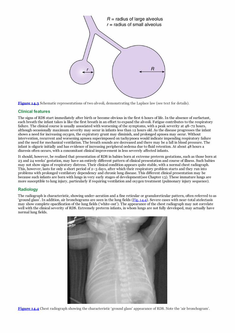
Figure 14.3 Schematic representations of two alveoli, demonstrating the Laplace law (see text for details).
Clinical featuresThe signs of RDS start immediately after birth or become obvious in the first 6 hours of life. In the absence of surfactant,each breath the infant takes is like the first breath in an effort to expand the alveoli. Fatigue contributes to the respiratoryfailure. The clinical course is usually associated with worsening of the symptoms, with a peak severity at 48–72 hours,although occasionally maximum severity may occur in infants less than 12 hours old. As the disease progresses the infantshows a need for increasing oxygen, the expiratory grunt may diminish, and prolonged apnoea may occur. Withoutintervention, recurrent and worsening apnoea superimposed on tachypnoea would indicate impending respiratory failureand the need for mechanical ventilation. The breath sounds are decreased and there may be a fall in blood pressure. Theinfant is oliguric initially and has evidence of increasing peripheral oedema due to fluid retention. At about 48 hours adiuresis often occurs, with a concomitant clinical improvement in less severely affected infants.
It should, however, be realized that presentation of RDS in babies born at extreme preterm gestations, such as those born at23 and 24 weeks’ gestation, may have an entirely different pattern of clinical presentation and course of illness. Such babiesmay not show signs of respiratory distress. Their clinical condition appears quite stable, with a normal chest radiograph.This, however, lasts for only a short period of 2–3 days, after which their respiratory problem starts and they run intoproblems with prolonged ventilatory dependency and chronic lung disease. This different clinical presentation may bebecause such infants are born with lungs in very early stages of development(see Chapter 13). These immature lungs aremore susceptible to lung injury, particularly if requiring ventilation and oxygen treatment (pulmonary injury sequence).
RadiologyThe radiograph is characteristic, showing under-aeration and a fine reticular or granuloreticular pattern, often referred to as‘ground glass’. In addition, air bronchograms are seen in the lung fields (Fig. 14.4). Severe cases with near-total atelectasismay show complete opacification of the lung fields (‘white-out’). The appearance of the chest radiograph may not correlatewell with the clinical severity of RDS. Extremely preterm infants, in whom lungs are not fully developed, may actually havenormal lung fields.
Figure 14.4 Chest radiograph showing the characteristic ‘ground glass’ appearance of RDS. Note the ‘air bronchogram’.

Figure 14.5 Chest radiograph showing right-sided tension pneumothorax. Note this has occurred despite a chest drainbeing in place, suggesting a massive air leak or a blocked chest drain.
Figure 14.6 Chest radiograph showing pneumomediastinum. �The heart and thymus are outlined by gas.
Figure 14.7 Chest radiograph showing extensive PIE. Note the overinflated chest with flattened diaphragm.
Laboratory abnormalitiesArterial oxygen tension is usually decreased. Arterial carbon dioxide tension may initially be normal (when breathing fasterto compensate for respiratory difficulty), but is usually increased. Blood pH may reflect respiratory acidosis (fromhypercarbia), metabolic acidosis (from tissue hypoxia) or mixed acidosis.
Treatment

Surfactant replacement therapyThe development and use of surfactant replacement therapy has revolutionized the treatment of RDS. Numerouspreparations, including animal-derived surfactants and equally effective newer synthetic surfactants, have been developedand tested in numerous randomized controlled trials, all of which confirm the efficacy of surfactant in improving survival andreducing complications such as pneumothorax.
Surfactants can be used either ‘prophylactically’ in at-risk infants (such as those born after less than 27 weeks’ gestation), oras a ‘rescue’ treatment whenever babies develop respiratory distress. Different surfactant preparations have differentcompositions and dosing schedules, but they are all effective. Normally, one or two doses of surfactant should be sufficientand there is no evidence that further doses provide any real benefit.
Surfactant is administered into the lungs through an endotracheal tube, but recently some centres have used a less invasivemethod of administration such as through a nasogastric tube or laryngeal mask to avoid intubation. Newer methods stillrequire further evolution for safety and efficacy.
Establishment of adequate gas exchange
OxygenThe aim of treatment is to maintain the PaO2 within the normal range. This is done in very mild cases by increasing theinspired oxygen concentration (FiO2). In more severe cases respiratory support may be necessary, but the furthermanagement depends on the size of the infant and degree of gaseous exchange abnormality.
Although CPAP may be administered shortly after birth, smaller infants are more likely to require mechanical ventilation.Indications for mechanical ventilation are listed in Box 14.3.
Box 14.3 Indications for mechanical ventilation in RDS.Apnoea and bradycardia of increasing severity.
FiO2 exceeding 40% to maintain PaO2 >60 mmHg �(8 kPa).
Rising PaCO2 that exceeds 60 mmHg (8 kPa), particularly if there is a falling pH (<7.20).
Use of rescue surfactant treatment.
Supportive treatment
Chest physiotherapyInfants receive regular position changes and airway suctioning to reduce the risk of mucus retention, airway plugging andpulmonary collapse. Routine chest physiotherapy is of no proven value in the management of RDS and is not recommended.
AntibioticsBroad-spectrum antibiotics are usually administered to all babies with RDS after suitable cultures have been taken. This isbecause sepsis/pneumonia, especially group B streptococcal infection, can produce a nearly identical picture. Antibioticsshould be stopped once cultures, blood count and ancillary investigations for sepsis are reported as negative. The choice ofantibiotics depends on the local preference, keeping in mind the prevalence of local bacteriological spectrum.
Appropriate fluid balanceSee Chapter 9.
Blood pressure monitoringSee Chapter 16.
Minimal handling and nursing in a neutral environmental temperatureSee Chapter 24.
ComplicationsIn general, the risk of complications is related to the severity of the underlying RDS and its treatment. Box 14.4 lists thecommon complications. These complications in turn can lead to long-term morbidities such as cerebral palsy, andbronchopulmonary dysplasia (BPD).
Box 14.4 Common complications associated with RDS and its treatment.Pneumothorax.
Patent ductus arteriosus (PDA).
Necrotizing enterocolitis (NEC).
Subglottic stenosis (causing stridor).
Chronic lung disease (CLD) and bronchopulmonary dysplasia (BPD).
Periventricular–intraventricular haemorrhage �(PVH-IVH).
Periventricular leukomalacia (PVL).
Retinopathy of prematurity (ROP).

PrognosisMost infants who die do so not as a direct result of RDS but rather of a related complication (IVH, infection, chronic lungdisease). Those weighing 1000 g or less are most at risk. More mature infants–even with severe RDS–survive but mayrequire respiratory support for prolonged periods. Survival figures by birthweight are shown in Table 14.2.
Table 14.2 Typical incidence and survival rates for infants of different birthweight groups with adiagnosis of RDS.
Birthweight categories (g) Incidence of RDS (%) Survival (%)
<500 100 10
500–999 80 80
1000–1499 55 95
>1499 20 100
Total 500–5000 N/A 94
PneumoniaPneumonia in the newborn infant is relatively common, especially among those who require ventilation or have undergoneother invasive procedures as a part of their treatment or monitoring. The infecting agent may be viral, bacterial or fungal.Pneumonia may be contracted in utero and be present at birth (congenital), or acquired after birth (nosocomial). Congenitalpneumonia may be due to ascending infection with prolonged rupture of membranes, or less frequently due to atransplacental infection (see Chapter 4). The diagnosis is suspected on maternal history, clinical examination and chestradiography, and confirmed by bacteriological cultures from blood and tracheal aspirate. However, blood cultures may notbe positive in all cases of pneumonia. Indirect parameters such as white blood cell (WBC) count and C-reactive protein (CRP;a form of acute-phase reactant) may help in diagnosing sepsis in a baby.
AetiologyPneumonia in the newborn is usually bacterial, and the most frequent bacterial pathogens causing congenital and acquiredpneumonias are the Gram-negative bacilli (Escherichia coli, Klebsiella, Pseudomonas, Serratia), group B Streptococcusand Staphylococcus.
Less common bacterial infections include Listeria monocytogenes and anaerobic bacilli. Occasionally, pneumonia is due toChlamydia trachomatis, Mycoplasma pneumoniae or opportunistic pathogens such as Candida albicans. Viral pneumonitisis rare but occurs with cytomegalovirus (CMV), Coxsackie virus, respiratory syncytial virus (RSV) and humanmetapneumovirus (hMPV).
Ventilator-associated pneumonia (VAP) accounts for a significant proportion of postnatally acquired infections and should besuspected in a hospitalised newborn who exhibits an unexplained deterioration in respiratory condition. Chest X-ray findingsare non-specific VAP is frequently polymicrobial.
Clinical featuresThe early clinical signs and symptoms are nonspecific and may include lethargy, apnoea, bradycardia, temperatureinstability and intolerance of feeds. At birth it may be difficult to distinguish pneumonia from other lung disease causingrespiratory distress. The maternal and birth histories may reveal predisposing factors for neonatal infection.
RadiologyThe appearances are non-specific, and pneumonia may be difficult to distinguish radiologically from aspiration syndromesand even TTN. There are usually patchy opacities or more confluent areas of radiodensity through the lung. Lobarpneumonia is rarely seen in the newborn.
Treatment
AntibioticsBroad-spectrum antibiotics should be administered after suitable cultures have been taken. The choice of antibioticsdepends on the predominant pathogen causing sepsis and the antibiotic sensitivity pattern for the microorganisms causingearly-onset sepsis in a given region (or hospital). Empirical therapy must cover both Gram-positive and Gram-negativeorganisms. The most frequently used combination is penicillin or ampicillin (or amoxicillin) and an aminoglycoside(frequently gentamicin). After an organism has been identified, the antibiotic therapy may be tailored according to thesensitivities.
Respiratory support

This is given according to the severity of signs and symptoms (see above as in RDS).
Pulmonary air leaksThis term refers to a collection of gas outside the pulmonary compartment, and a variety of disorders are included in thiscategory (see Table 14.3).
Table 14.3 Disorders categorized as pulmonary air leaks.
Pneumothorax Air in the pleural space (Fig. 14.5)
Pneumomediastinum Air in the anterior mediastinum (Fig. 14.6)
Pneumopericardium Air in the pericardial sac
Pulmonary interstitial emphysema (PIE) Air in interstitial lung spaces �(Fig. 14.7)
Pneumoperitoneum Air in the peritoneal cavity
Air embolus Air dissecting into pulmonary veins
The pathophysiology of these conditions is similar in that the alveoli become hyperinflated and rupture. Air then escapesinto the lung interstitium (pulmonary interstitial emphysema; PIE) and tracks along the perivascular spaces into themediastinum (pneumomediastinum) through the visceral pleura (pneumothorax), or rarely into the pericardium(pneumopericardium). Spontaneous pneumothorax occurs in approximately 1% of vaginal deliveries and 1.5% of caesareansections. Many of these have only minor symptoms, are discovered unexpectedly on chest radiography, and may not requireintervention. Resuscitation with positive-pressure ventilation is a recognized risk factor for the development ofpneumothorax. Antenatal steroids and surfactant therapy have markedly reduced the incidence of pulmonary air leaks inventilated infants.
Pneumothoraces commonly occur in the following conditions or situations:
Stiff lungs (e.g. RDS).
Hyperinflated lungs (e.g. in meconium aspiration syndrome).
Hypoplastic lungs (e.g. Potter’s syndrome, diaphragmatic hernia).
Any form of intermittent positive-pressure ventilation or continuous distending pressure such as CPAP. Prolongedinspiratory time and high end-expiratory pressures are particularly likely to cause pneumothorax in ventilated infants.Active expiration against a ventilator ‘breath’ (asynchrony between mechanical and spontaneous breaths) is also animportant cause, as described earlier.
Clinical featuresInfants with a tension pneumothorax exhibit signs of severe respiratory distress. Frequently, a pulmonary air leak occurs ina baby who already has respiratory distress and there may be sudden deterioration, with cyanosis, poor peripheralperfusion and bradycardia. Specific signs of a tension pneumothorax are a shift of the mediastinum (apex beat) to theopposite side, asymmetrical chest expansion, asymmetrical air entry and weak peripheral pulses. A prominent sternum maysuggest a pneumomediastinum.
Occasionally, unilateral PIE develops as a result of a ball-valve effect in a main bronchus. The emphysematous lung maycause compression of the more normal lung, thereby further embarrassing ventilation (Fig. 14.8).

Figure 14.8 Chest radiograph showing left-sided PIE. The mediastinum and right lung are compressed by the overinflated�left lung.
CLINICAL TIP
If a baby has been stable on a ventilator, one of the first signs of a pneumothorax will be an increasing requirement ofpeak inflation pressure (PIP) to achieve adequate blood gases. The deterioration in blood gases can be either gradualor catastrophic, as in acute tension pneumothorax.
DiagnosisThe definitive diagnosis is made by an anteroposterior chest radiograph. A lateral film may be necessary to diagnose air inthe anterior mediastinum. When a pneumomediastinum is present, the chest radiograph may reveal the ‘sail’ or ‘spinnaker’sign (air around the thymus). In the critically ill infant who has a tension pneumothorax, chest transillumination may beused to make the diagnosis. Occasionally, the insertion of a 21G butterfly needle with three-way tap and syringe may be life-saving in suspected tension pneumothoraces. This is an emergency diagnostic and therapeutic procedure undertaken whenthere may be delay in obtaining a radiograph in a critically ill infant. Blind needling of the chest may create a pneumothoraxand should not be attempted, except in an emergency.
ManagementA tension pneumothorax will need to be released immediately by inserting a needle and/or chest drain into the intercostalspace, connected to underwater seal. The practical procedure related to chest drain insertion is described below (seeCLINICAL TIP). Occasionally, evacuation of the pneumothorax will be incomplete with this catheter, and an additionalcatheter will be necessary. For non-tension pneumothoraces in full-term infants, nursing in 100% oxygen for up to 12 hoursmay accelerate reabsorption of the pneumothorax as oxygen is absorbed into the blood, unlike nitrogen in air.
Generalized management of PIE is focused on reducing or preventing further barotraumas to the lung by decreasing peakinspiratory pressure (PIP) to the minimum required to attain acceptable arterial blood gases. A reduction in positive end-expiratory pressure (PEEP) may also help in decreasing the PIE. High-frequency oscillatory or jet ventilation is a successfulmeans of ventilation for infants with PIE. Localized PIE may resolve spontaneously or may persist for several weeks, andmay or may not cause any further deterioration in the baby. Effort should be made to avoid any form of positive-pressureventilation (PPV) or continuous distending pressure unless really needed.
Pneumomediastinum is often of little clinical importance and usually does not need to be drained. Pneumopericardium cancause life-threatening cardiac tamponade (compression), which requires pericardiocentesis (needle aspiration via thesubxiphoid route). The procedure can be facilitated by ultrasound guidance. Pericardial tube placement and drainage (like achest tube for pneumothorax) may be necessary if the pericardial air reaccumulates.

CLINICAL TIP
Insertion of a chest drain.
Once the pneumothorax has been confirmed by chest radiography (or transillumination in a critically ill baby) a chesttube should be inserted as soon as possible. There are two methods: (i) placing a chest tube under direct vision afterblunt dissection down to the pleura using surgical instruments; or (ii) insertion of a percutaneous ‘pig tail’ drain usingthe Seldinger technique. The latter is now the more accepted method as the first stage of the procedure (insertion of aneedle) relieves any tension. In addition, the long-term cosmetic outcome is often much better with reduced scarring.
The drain is usually inserted in the 4th-5th intercostal space in the anterior to mid-axillary line. It should be insertedjust above the lower rib to avoid damaging the neurovascular bundle that runs under the lower aspect of each rib. Theneedle should be directed towards the sternum so that the drain lies anterior to the lung (where the air pocket is). If asecond drain is required this can be inserted in the 2nd intercostal space, mid-clavicular line.
The percutaneous Seldinger technique involves inserting a needle (or large-bore cannula) through which a soft-tippedguide wire is placed, once a small amount of air has been aspirated to confirm you are in the right space. The needle isthen removed over the wire and replaced with a dilator. After this, the drain itself is threaded into the pleural spaceand then the guide wire can be removed. The tip of the pig-tail catheter will curl up within the air cavity and helpshold the drain in (Fig. 1414.9b). Stitches are not usually required, but the drain can be dressed with opposing clearadhesive dressings.
Figure 14.9 (a) Left-sided pneumothorax in a preterm baby. (b) The same baby after insertion of a 10 Fr pigtail catheter.
Meconium aspiration syndromeMeconium aspiration syndrome (MAS) is a serious cause of respiratory distress in the newborn. It is not synonymous withmeconium staining of the amniotic fluid (MSAF). MSAF occurs in about 13% of all deliveries, whereas MAS occurs only in 4–5% of babies born through MSAF. The passage of meconium often indicates fetal distress, but in the breech presentationmay be normal. Aspiration most commonly occurs in utero and less so after birth. The response of the infant to intrapartumasphyxia is to gasp, and if meconium is present it will be aspirated deep into the bronchi. Once respiration begins, distalmigration of the meconium into small airways occurs.
Clinical featuresThere is a wide spectrum of presentations of MAS, ranging from severe birth asphyxia requiring active resuscitationthrough early onset of respiratory distress to a vigorous baby with no major problems. Typically, the infant is born coveredin meconium-stained liquor and has meconium staining of the umbilical cord, skin and nails. The chest appears to behyperinflated and there may be a prominent sternum. Respiratory distress may be mild initially, becoming rapidly moresevere after several hours. If asphyxia has occurred, the baby may also show signs of early-onset encephalopathy (seeChapter 22).
Pathogenesis and aetiologyMeconium causes a number of anatomical and physiological problems that make lung function worse:
Plugging of the airways, with consequent atelectasis. It also causes a ‘ball-valve’ obstruction with hyperinflation of thelungs and a high risk of pulmonary air leaks.
Irritation of the airways, causing a chemical pneumonitis.
Antagonism of surfactant production.
Possible secondary bacterial infection.
In a proportion of babies with severe MAS there is development of marked ventilation/perfusion inequality and right-to-left shunt due to persistent pulmonary hypertension (PPHN; see Chapter 16).

RadiologyChest radiographs show hyperinflation (flat diaphragms, widening of rib spaces) with diffuse patchy opacities throughoutboth lung fields (Fig. 14.10). Pneumothorax or pneumomediastinum may also be seen.
Figure 14.10 Chest radiograph showing meconium aspiration syndrome (MAS). There is extensive discrete shadowingthroughout both lung fields and hyperinflation.
Prophylactic managementMorbidity and mortality from MAS can be prevented or minimized by optimal perinatal management. There is controversyas to whether a neonatal paediatrician should attend every delivery where there is meconium staining of the liquor. It isimportant, however, that an experienced neonatal paediatrician be present for the delivery if thick meconium is present,especially if there are signs of intrapartum fetal distress.
Various therapeutic interventions have been used. Results of large controlled trials and systematic reviews suggest that:
Amnioinfusion therapy for thick-consistency MSAF does not reduce the risk of MAS.
Intrapartum naso- and oropharyngeal suctioning does not reduce the incidence of MAS and hence is not recommended.
Routine endotracheal intubation and suctioning is of no benefit. This should, however, be performed in infants if they aredepressed, and need PPV.
In such cases, the baby is intubated with a wide-bore suction catheter or endotracheal tube and the trachea is suctionedclear. Suctioning is facilitated by the use of a meconium aspirator. The stomach should be aspirated following intubation.All depressed babies who are born through thick MSAF should be carefully assessed and regularly monitored for signs ofMAS.
Treatment of established meconium aspiration syndromeThe treatment will be the same as for respiratory distress (see above). Particular emphasis or consideration should be givento the following points:
Humidification of inspired oxygen.
Postural drainage positioning, suctioning of airways and chest percussion are of unproven benefit.
Antibiotics are usually given, although the efficacy has not been established.
Both conventional and high-frequency ventilation can be used with multiple strategies to achieve normal gaseousexchange and prevent complications such as air trapping and air leaks, which are common in such infants.
Exogenous surfactant has been used with success in some studies. In some centres lavage with surfactant is practiced,but its efficacy is not fully established.
Inhaled nitric oxide (iNO) therapy should be considered in infants with concomitant PPHN who are not responding toconventional treatment. Other agents used in such condition are prostacyclin and sildenafil.
Extracorporeal membrane oxygenation (ECMO) is considered as a rescue treatment when predicted mortality is runninghigh. The overall success rate of ECMO in babies with MAS is over 80%.
Pulmonary hypoplasiaFor adequate fetal lung development the fetus must be able to make breathing movements and move a column of amnioticand lung fluid up and down the trachea and main bronchi. Hypoplasia may therefore be due to:
Failure of fetal breathing (neuromuscular disorders).
Inability to expand the lungs (diaphragmatic hernia, pleural effusions).
Lack of liquor (oligohydramnios) due to renal or urethral abnormalities or prolonged preterm rupture of the membranescausing leakage of amniotic fluid.

Clinical featuresThe infant develops severe respiratory distress from birth, with marked hypoxia, hypercapnia and metabolic acidosis.Pneumothorax is a common complication. The lungs are very stiff and there is little chest movement with mechanicalventilation. It may be difficult to diagnose lung hypoplasia on chest radiography. Severe lung hypoplasia is incompatible withlife, and less severe forms contribute towards chronic ventilator dependency and bronchopulmonary dysplasia.
Management and prognosisManagement of lung hypoplasia starts antenatally if diagnosed earlier, and includes counselling by the obstetrician,neonatologist, clinical geneticist and surgeon (if appropriate). Pulmonary hypoplasia results from a large number of differentconditions, and hence the prognosis is governed mainly by the aetiology and associated anomalies. Lung biopsy may bedifficult to perform safely while the infant is alive, and hence postmortem studies should be made where possible. Wherepermission for a full autopsy is not granted an open or needle biopsy of the lung obtained soon after death may provide atissue diagnosis.
Pulmonary haemorrhagePulmonary haemorrhage has a characteristic clinical presentation in newborn infants, with cardiovascular collapse associatedwith an outpouring of bloodstained fluid from the trachea and mouth. The condition is often fatal and occurs in about 1 per1000 births. Among survivors, there is a high incidence of chronic lung disease.
PathogenesisThe underlying mechanism for pulmonary haemorrhage can be classified as:
Haemodynamic: a manifestation of an exaggerated haemorrhagic pulmonary oedema brought about by an acuteincrease in pulmonary blood flow as in large PDA.
Haematological: due to associated coagulation abnormalities such as in disseminated intravascular coagulation (DIC)associated with sepsis or other coagulopathy.
Pulmonary haemorrhage is often associated with bleeding in other organs such as brain or gut.
Risk factorsThe incidence of pulmonary haemorrhage is inversely related to the gestational age of the babies. Prematurity, RDS andexogenous surfactant therapy (in combination) are the three most important risk factors. Other antecedent risk factorsinclude severe birth asphyxia, hypothermia, small for gestational age (SGA) infants, coagulation disturbances and congenitalheart disease.
The severity and magnitude of clinical signs depend on the magnitude of haemorrhage and the severity of the underlyingcondition leading to the episode. The clinical manifestations result from several interrelated pathophysiological consequencesof blood loss and haemorrhage into the lung parenchyma and airways.
TreatmentThe treatment will be that of respiratory distress as described earlier, but particular emphasis must be given to thefollowing:
Resuscitation of cardiovascular collapse with volume expanders such intravenous infusion of plasma protein fraction,blood or normal saline.
Treatment of pulmonary oedema with frusemide (furosemide) and perhaps morphine.
Correction of coagulation disturbances; fresh-frozen plasma and/or recombinant factor Vila (rFVIIa).
If mechanical ventilation is required, high PEEP may help to decrease bleeding.
Exogenous surfactant seem to improve the respiratory status in infants with pulmonary haemorrhage.
Congenital diaphragmatic herniaCongenital diaphragmatic hernia (CDH) is a failure of normal development of the diaphragm during the first trimester. Thehernia is usually a posterolateral (Bochdalek) type; 80% occur on the left side. This occurs through a defect in the diaphragmas a result of a persistent pleuroperitoneal canal caused by failure of the muscular components to develop. The defect in thediaphragm permits herniation of the abdominal contents into the thorax. Consequently, there is hypoplasia or compressionof the lung on the side of the hernia, with displacement of mediastinum to the contralateral side. Sometimes, there ishypoplasia of the contralateral lung. Babies with a large diaphragmatic hernia have rapidly progressive respiratory failureafter birth, with persistent cyanosis. A less acute diaphragmatic hernia may present in the nursery, with respiratorydistress, or occasionally on routine examination a ‘dextrocardia’ or scaphoid abdomen will be found. On auscultation of thelungs, bowel sounds may be heard on the side of the lesion if gas has entered the gastrointestinal tract. The lungs will notinflate adequately with normal pressure.
DiagnosisMost cases of CDH are now diagnosed in utero by routine obstetric ultrasound at 18–20 weeks’ gestation, or followinginvestigation of polyhydramnios. The diagnosis of a diaphragmatic hernia is confirmed by chest radiography, which showsbowel loops in the thorax (Fig. 14.11). If the radiograph is taken shortly after birth, there may be some difficulty indetermining whether the bowel is in the chest, especially if there is little air in the gastrointestinal tract. Similarly, diagnosismay be delayed when the liver is in the chest, in cases of right-sided diaphragmatic hernia. The radiograph should includethe abdomen, to show a paucity of abdominal gas pattern. A more rare form of diaphragmatic hernia is through ananteromedial defect beneath the sternum (Morgagni type). This typically contains part of the colon. It is usually

symptomless and often detected on a lateral chest radiograph. Eventration, which results from a failure of muscledevelopment in the primitive diaphragm, is not a true hernia. CDH can be a part of a syndrome or associated with achromosomal abnormality.
Figure 14.11 Chest radiograph showing a left-sided diaphragmatic hernia.
TreatmentIf CDH is suspected, an orogastric tube should be inserted and the abdominal contents in the chest aspirated. If the babyrequires assisted ventilation, it should be performed via an endotracheal tube and never by bag and mask, in order toprevent increasing gaseous distension of the bowel within the chest. Care must be taken to prevent rupture of thecontralateral lung, as the baby is almost exclusively dependent on this lung for ventilation. Following an initial diagnosis thebaby should be stabilized and referred to a paediatric surgeon for operative treatment. Surgery for the more severe cases isoften delayed for a few days to enable maximum stabilization, often with high-frequency oscillation ventilation. Surgeryconsists of reduction of the abdominal contents, closure of the diaphragmatic defect and correction of any bowel malrotation.
PPHN is a common complication of severe diaphragmatic hernia. Adequate ventilatory support, through either conventionalor high-frequency ventilation, is required in all cases. Inhaled nitric oxide empirically would appear to be the drug of choice,but data in relation to CDH suggest that overall its use is unhelpful. ECMO has been used to provide stability and forassociated PPHN; however, it is of no clear benefit in terms of long-term outcome.
Prenatal fetal surgery aimed at in-utero repair has been attempted but failed and is not currently recommended. Instead,fetal endoscopic tracheal occlusion (FETO) is the current preferred mode of treatment for the fetus with moderate to severeCDH to promote lung growth and restrict severity of pulmonary hypoplasia. Using fetal trachoscopy, a balloon isinserted – ideally at 26–28 weeks – and the occlusion is reversed at 34 weeks. Results are encouraging but results are stillnot satisfactory and require further trials. Complications can include tracheal dilation or hydrops.
PrognosisThe prognosis depends on the degree of pulmonary hypoplasia and the age at presentation. Infants with respiratory distressin the first 6 hours have a high mortality (about 70%), whereas those presenting between 6 and 24 hours have a muchbetter prognosis (mortality rates 10–15%). Later presentation is not likely to be associated with pulmonary hypoplasia, andthe prognosis is very good.
Infants with CDH often require prolonged hospitalization and may have other congenital anomalies. After discharge fromhospital, they may have long-term sequelae such as respiratory insufficiently, gastro-oesophageal reflux, poor growth,neurodevelopmental delay and hernia recurrence.
Oesophageal atresia and �tracheo-oesophageal fistulaOesophageal atresia is a congenital anomaly in which there is usually complete interruption of the lumen of the oesophagus,in the form of a blind upper pouch. This is commonly associated with a tracheo-oesophageal fistula (TOF). The commonestvariety of this condition is a blind-ending upper oesophageal pouch, with the lower oesophagus arising from the tracheaabove the carina (Fig. 14.12c). A variety of other patterns occur, much these are less common (Fig. 14.12a,b,d,e).
Figure 14.12 Variants of tracheo-oesophageal fistula with or without oesophageal atresia. Type (c) accounts for 85% ofcases, the others being equally uncommon.

Clinical featuresMaternal polyhydramnios occurs in 60% of cases, and is largely responsible for the high frequency of premature births. Theoesophageal atresia often presents with excessive saliva and mucus production, with a high incidence of aspirationpneumonia. Abdominal distension is due to air passing down the fistula into the stomach, and may develop rapidly andirreversibly. Coexistent congenital anomalies may be present and may be either major or minor (e.g. VACTERL association– vertebral anomalies, anal atresia, cardiovascular anomalies, tracheoesophageal fistula, oesophageal atresia, renalanomalies, limb defects).
The most important aspect of oesophageal atresia is that it should be recognized as soon as possible after birth, preferablyprior to the first feed, so that pulmonary complications (due to aspiration) are less likely to occur. All babies with suspectedoesophageal atresia should have a size 5 nasogastric tube passed down the nostril in turn and into the stomach shortly afterbirth, preferably before the first feed. If this is not done, oesophageal atresia is generally diagnosed by excessive secretionsor cyanosis and coughing with feeds. The definitive diagnosis is made by the inability to pass a firm radio-opaque no. 10catheter or a replogle tube into the stomach, as it arrests about 10 cm from the lips. Once the catheter has been passed asfar as possible, a radiograph of the neck and upper chest should be taken to confirm that it has become obstructed at thislevel. The abdominal radiograph should be inspected for gas in the stomach, and the chest radiograph assessed for areas ofcollapse. Air is a useful contrast medium in the upper pouch. It is not usually necessary to put radio-opaque contrastmedium into the upper pouch to confirm the diagnosis, but if this is done a non-irritant substance should be used. For an H-type fistula, which presents with recurrent aspiration or infections, radiological diagnosis may be difficult. A video-fluoroscopy contrast swallow will usually confirm the diagnosis.
TreatmentA baby with oesophageal atresia and TOF should be nursed supine and propped head-up at 60° to avoid the gastric contentsspilling into the lung through the fistula. A replogle tube should be placed in the oesophagus and put on suction. Surgeryshould be performed by a paediatric surgeon as soon as the baby has been adequately resuscitated and stabilized. Surgeryconsists of division of the TOF and anastomosis of the two segments of the oesophagus if possible. Occasionally, the atreticsegment is too long to enable a primary anastomosis, and exteriorization of the cervical oesophagus as an oesophagostomyand the stomach as a gastrostomy will be necessary. A definitive operation can be delayed to a later date.
With a cervical oesophagostomy the baby should be encouraged to practise sucking on a dummy during gastrostomyfeedings (‘sham feeding’). Complications and sequelae from surgery are frequent, and include a brassy cough, associatedwith coexistent tracheomalacia, oesophageal stricture, breakdown of the anastomosis with mediastinitis, recurrence of theTOF and gastro-oesophageal reflux.
Congenital lobar emphysemaCongenital lobar emphysema is a rare anomaly due to a cartilaginous deficiency of the lobar bronchus, most commonlyinvolving the left upper lobe (50%), the right middle lobe (24%) or the right upper lobe (18%), and frequently associatedwith congenital heart disease (30%), such as tetralogy of Fallot, ventricular septal defect (VSD) or total anomalouspulmonary venous drainage (TAPVD). The onset of respiratory distress is frequently insidious, usually taking 2–3 weeks todevelop, and is caused by lung collapse around the hyperinflated lobe. The mediastinum becomes displaced and the chestwall is prominent over the affected area. Breath sounds are diminished and the percussion note is hyper-resonant. Once adefinitive diagnosis is made a lobectomy is generally required. Acquired lobar emphysema may be secondary to an extrinsicor intrinsic bronchial obstruction, such as a mucous plug. This type should be treated conservatively, but may requirebronchoscopy.
Congenital pulmonary airway malformation (CPAM) [formerly known as congenitalcystic adenomatous malformation; CCAM]CPAM is a rare cystic lesion found more often in males than females, and often diagnosed on antenatal ultrasound. Thisemphysematous lesion may be associated with polyhydramnios, hydrops fetalis, prematurity and stillbirth. There are threedistinct types: type I CAM (70%) presents as single or multiple large cyst(s) confined to one lobe; type II (18%) is composedof multiple medium-sized cysts and 50% have other anomalies; type III (10%) is a large bulky lesion with evenly distributedsmall cysts. Asymptomatic lesions may not require removal. This is one of the few conditions that is successfully treatableby fetal surgery in selected cases.
Chronic lung disease and bronchopulmonary dysplasia (BPD)The term chronic lung disease (CLD) is a non-specific term, commonly used to describe a group of heterogeneous conditionscharacterized by signs of chronic respiratory signs and symptoms. It should not be used interchangeably with a diagnosis ofBPD, which is a specific condition that develops in newborns who are born preterm. It is usually defined as the presence ofchronic respiratory signs, a persistent oxygen requirement, or dependence on any form of respiratory support, and anabnormal chest radiograph persisting at 36 weeks corrected age. Other causes of CLD are shown in Box 14.5.
Although surfactant treatment has improved the overall survival of premature infants, the incidence of BPD remainsapproximately 30–40% in very preterm babies. With the use of standardized oxygen saturation monitoring, as required inthe new physiological definition of BPD, the incidence may further decrease by as much as 10%.
BPD is usually associated with severe RDS, but occasionally complicates meconium aspiration, pulmonary haemorrhage andsevere neonatal pneumonia. This was first described by Northway et al. (1967), but since then the characteristics of thedisease have changed and it is now essentially a condition seen mostly in babies who are born very premature and requiremechanical ventilation. The incidence of BPD varies according to the definition used, and recently a physiological definition ofBPD has been introduced. The latter facilitates the measurement of BPD as an outcome in clinical trials and comparisonsbetween and within centres over time.

Box 14.5 Conditions causing chronic lung disease.Bronchopulmonary dysplasia.
Recurrent aspiration: Pharyngeal incoordination; gastro-oesopheageal reflux; tracheo-oesophageal fistula.
Interstitial pneumonitis:
Cytomegalovirus
Candida albicans
Chlamydia
Pneumocystis jirovecii
Ureaplasma urealyticum
Chronic pulmonary oedema (due to left-to-right shunt, as in PDA).
Rickets of prematurity.
Neuromuscular diseases affecting intercostal muscles and diaphragm (spinal muscular atrophy, myotonic dystrophyand myasthenia gravis).
AetiologyBPD is multifactorial in origin and results from a sequence of events occurring in a preterm infant which increases theinfant’s dependency on ventilation and oxygen (known as the ‘pulmonary injury sequence’). The incidence relates to theseverity of RDS and the degree of prematurity. It has only been described in infants who have received positive-pressureventilation, and probably relates to barotrauma or more importantly volutrauma (alveolar overdistension due to excessivetidal volume delivery). Oxygen therapy for prolonged periods of time also appears to be linked with its development.
Clinical featuresInfants who develop BPD have signs of persistent chest retractions, crepitations and rhonchi on chest auscultation, grosshyperinflation of the lungs, and an increased anteroposterior chest diameter. Most of these infants also have a PDA and,after a period of time, may develop right heart failure (cor pulmonale). There is often associated developmental delay insuch children, requiring input from multiple agencies.
RadiologyThe original description of BPD by Northway et al. (1967) staged the radiological appearances in four grades. The first twogrades are indistinguishable from RDS and are not helpful for descriptive purposes. In the most severe form, there is anirregular honeycomb appearance to the lung, with overinflated lung fields, extensive fibrosis and multiple cysts of irregularsize (Fig. 14.13). The majority of infants with BPD now do not develop these gross radiological signs but exhibit a finer, morehomogeneous pattern of abnormality.
Figure 14.13 Chest radiograph showing severe bronchopulmonary dysplasia.
ManagementThe prevention of BPD requires careful management of infants who are receiving mechanical ventilation. Pulmonaryinterstitial emphysema, pulmonary oedema, PDA and sepsis appear to increase the risk of BPD developing. The followingtherapies have been used to treat BPD with varying effects, but they are not supported by any robust scientific evidence
Dexamethasone. Studies have shown that dexamethasone reduces the ventilatory dependency and should beconsidered after the first 2 weeks of life in infants with severe lung injury who remain ventilator-dependent. The doseand duration of treatment must be kept to the minimum necessary to achieve the desired effects, as indiscriminate useof postnatal dexamethasone has been linked with excessive prevalence of cerebral palsy.
Diuretics. Diuretic therapy reduces interstitial lung fluid and has been used for acute deterioration associated with

pulmonary oedema. It can transiently reduce oxygen requirements. However, routine prolonged use is notrecommended because of a lack of evidence that it improves the incidence or severity of BPD, as well as concern overside effects.
Therapy directed at reducing oxygen toxicity. Antioxidants such as vitamin E and superoxide dismutase appearto be of no benefit, but vitamin A has been shown to reduce BPD and death in very low birthweight (VLBW) infants.
Adequate oxygenation throughout all daily activities (O2 saturation 92–96%) minimizes progressive pulmonaryvascular disease.
Nutritional supplementation is necessary to obtain optimal growth in the presence of high energy consumption dueto the work of breathing. Adequate nutrition is a key aspect of care for the infants with CLD.
PrognosisChildren with BPD have a higher mortality before discharge from hospital and an increased risk of neurodevelopmentaldelay. They have also been shown to have increased airway resistance and are more likely to wheeze. They also haveincreased airway reactivity and viral-induced wheeze. Episodic wheezing is commonly seen in infants and children who havehad BPD during the newborn period.
Lower airway infection in the first year of life is a particular risk to children who have had BPD in the neonatal period.Pneumococcal vaccine is now in routine use. Such children appear to be particularly likely to develop RSV-positivebronchiolitis, which may cause very severe respiratory failure. A monoclonal RSV antibody (palivizumab) is used forprophylaxis during the RSV season. Advice must be given to the parents to avoid exposing the baby to the risk of viralinfection during the first year or two of life.
SUMMARY
Respiratory disorders are the commonest cause of illness in newborns. Their severity varies according to theunderlying pathology. Respiratory distress is a non-specific term used to describe a combination of symptoms whichindicate the involvement of respiratory system including lungs, airways, thoracic cage and breathing centre in thebrain. RDS is a specific diagnosis related to surfactant deficiency due to immaturity of lungs in preterm babies. Insevere cases it can lead to severe respiratory failure and death. RDS is a specific diagnosis related to surfactantdeficiency due to immaturity of lungs in preterm babies. In severe cases it can lead to severe respiratory failure anddeath. Other causes of respiratory distress include congenital and acquired pneumonia, cardiorespiratorymaladaptation at birth (see Chapter 16), aspiration syndromes and congenital abnormalities of lungs. Treatment ofrespiratory disorders ranges from simple supportive measures such as ambient oxygen and artificial respiratorysupport to intensive cardiorespiratory support, including mechanical ventilation and other strategies (see Chapter 13).
Further reading1. Donn, S.M., Sinha, S.K. (eds) (2016) Manual of Neonatal Respiratory Care, 4th edition. Springer Publications.
2. Rennie, J.M. (ed.) (2012) Textbook of Neonatology, 5th edition. Elsevier Publications, pp. 443–616.
3. Martin, R.J., Fanaroff, A.A., Walsh, M.C. (eds) (2015) Neonatal-Perinatal Medicine, 10th edition. ElsevierPublications, �pp. 1113–1146.
4. Miall, L.S., Wallis, S. (2011) The management of respiratory distress in the moderately preterm newborn infant.Archives of Disease in Childhood: Education and Practice Edition, 96 (4), 128–135.
Now visit www.essentialneonatalmed.com to test yourself on this chapter.

CHAPTER 15 Apnoea, bradycardia and upper airway obstructionKey topics
Physiology
Apnoea
Acute life-threatening events (ALTEs)
Sudden and unexpected infant death and sudden infant �death syndrome
Upper airway obstruction
IntroductionNormal respiratory function is essential for successful transition to the extrauterine environment. The control of breathing iscomplex and is not fully developed at birth, particularly if the infant is preterm. Apnoea is one of the commonly encounteredproblems in the neonatal intensive care unit (NICU), and considerable time and effort goes into monitoring, preventing andtreating babies with apnoea. The aetiology, investigation and management of apnoea is therefore discussed in detail in thischapter. Acute life-threatening events (ALTE) and sudden infant death syndrome (SIDS) are uncommon but serious eventswhich are more commonly encountered in graduates of the intensive care nursery. Obstruction to gas flow in the upperairway can present in babies as apnoea, respiratory distress, coughing, a hoarse cry or stridor. Common causes of airwayobstruction and their management will be discussed.
PhysiologyThe onset of breathing begins early in fetal life. It is initially intermittent and irregular, and occurs only during periods ofactive rapid eye movement (REM) sleep. The function of fetal breathing is not fully understood but, along with adequateamniotic fluid volume, it probably has an essential role in lung growth. Fetuses that do not breathe (due to eitherneurological or muscular disorders) are at significant risk of lung hypoplasia (see Chapter 13).
Successful transition to the extrauterine environment requires a marked change from the intermittent breathing of a fetusto the continuous breathing of a newborn baby. The control of breathing is complex (see Fig. 15.1), but it is known thatrespiration is initiated in the respiratory nuclei found in the brainstem (pons and medulla). Hypercapnia increasesrespiratory activity, while hypoxia causes an initial increase in respiratory activity lasting several minutes, followed by adecrease in ventilatory frequency. Preterm infants have less well-developed chemoreceptor responses to hypoxia andhypercapnia.
Figure 15.1 Neuromuscular pathway for control of respiration.
Breathing patterns in the newborn can be divided into four different types:
Regular: there are nearly equal breath-to-breath intervals; this is infrequent.
Irregular: unequal breath-to-breath intervals; this is particularly common in preterm infants.
Periodic breathing: cycles of hyperventilation alternating with periods of hypoventilation and pauses in breathinglasting about 3 s.
Apnoea (see below).
With advancing gestation to term, the proportion of time that the infant is breathing regularly increases, and phases ofirregular, periodic and apnoeic periods decline. Further maturation occurs during the months after birth.

ApnoeaClinically significant apnoea is defined as a cessation of breathing (absence of respiratory airflow) lasting for 20 seconds ormore. Apnoea lasting for 10 seconds or more is also significant if accompanied by a desaturation (cyanosis or decreasedSpO2), or bradycardia of less than 100 beats per minute.
Apnoea is most common in preterm babies, particularly those born at less than 35 weeks’ gestation. It is seen in 25% ofinfants of birthweight less than 2500 g, and in over 80% of infants with birthweight below 1000 g. It is uncommon in thefirst 24 hours of life, increasing towards the end of the first week of life, and then tends to become less frequent as the babymatures.
Types of apnoea
Central apnoeaCentral apnoea accounts for 25% of all cases of neonatal apnoea. It is due to factors affecting the respiratory centre in thebrainstem or the higher centres in the cerebral cortex.
Obstructive apnoeaComplete obstruction of the airways will cause obstructive apnoea. Nasal obstruction (by bone/mucus/milk) will causeobstructive apnoea in babies as they are obligatory nose breathers. Obstructive apnoea is characterized initially byincreased respiratory effort (baby attempts to overcome the obstruction) before the cessation of breathing. Obstructiveapnoea accounts for 15% of cases, and occurs with congenital malformations, such as choanal atresia and the Pierre Robinsequence. Preterm infants with small upper airways may have apnoea when in the supine position, especially during active(REM) sleep. Apnoea of this type may be minimized by nursing the infant in the prone position. The presence of milk, mucusor meconium in the upper airway is likely to provoke severe episodes of obstructive apnoea.
Mixed apnoeaMixed apnoea accounts for the majority (approximately 60%) of cases particularly in preterm infants, but may be difficult todiagnose clinically. Superficially it resembles central apnoea initially, with cessation of respiration, but then the baby makesintermittent respiratory efforts without achieving gas exchange.
Reflex apnoeaBabies may develop reflex apnoea or vagally mediated apnoea due to vigorous suction of the pharynx, passage of anasogastric tube, physiotherapy, or sometimes even in response to defecation. Apnoea associated with gastro-oesophagealreflux may be reflex and/or obstructive.
Aetiology of apnoeaA number of factors can increase the frequency of apnoea. Common causes are listed in Box 15.1. In the term infant theaetiology is usually identified. In the preterm infant, ‘apnoea of prematurity’ is the primary cause, but should be considereda diagnosis of exclusion. Apnoea of prematurity is predominantly a mixed apnoea.
Box 15.1 Causes of apnoea.Apnoea of prematurity.
Lung disease (e.g. RDS, pneumothorax).
Infection.
Airway obstruction (e.g. secretions, micrognathia, choanal atresia).
Hypoxia.
Intracranial haemorrhage.
Metabolic causes (acidosis, hypoglycaemia, hypocalcaemia, hypomagnesaemia).
Drugs (e.g. maternal narcotics, trishydroxyaminonethane (THAM), prostin (PGE1), magnesium sulphate).
Gastro-oesophageal reflux.
Seizures.
NEC.
Patent ductus arteriosus.
Temperature instability.
Polycythaemia with hyperviscosity syndrome.
CNS abnormalities (including Ondine’s curse; see CLINICAL TIP)
Investigation of apnoeaInvestigations are carried out to determine treatable causes of apnoea, and will depend on the prevailing clinical condition.Investigations are listed in Box 15.2.
Box 15.2 Useful investigations in determining the underlying cause of apnoea.Full blood count.

Blood culture.
Other cultures as indicated (urine, CSF, tracheal aspirate, nasal/pharyngeal, surface swabs, etc.).
Chest radiography.
Blood glucose.
Serum electrolytes, including calcium, magnesium and sodium.
Blood gas (arterial, venous or capillary).
Continuous monitoring of oxygen saturation.
Ultrasound examination of the brain.
(In special circumstances) further neurological investigations, i.e. electroencephalography (EEG), polygraphic sleepstudies; PCR for the PHOX2B gene may identify cases of primary alveolar hypoventilation syndrome.
CLINICAL TIP
New-onset or increasing frequency of apnoea in a premature baby is of great clinical concern. It should not bepresumed to be secondary to apnoea of prematurity. The baby should be carefully examined for the other possiblecauses. A septic work-up and commencement of intravenous antibiotics should be seriously considered if no obviouscause is found. Apnoea in a term infant is always abnormal, and a cause must be sought.
Apnoea monitoringA variety of respiratory monitors are available, including a pressure-sensitive pad on which the infant lies, an air-filledplastic blister attached to the abdomen (SIMS Graseby Medical Ltd, Watford, UK), and impedance monitors usingelectrodes attached to the chest wall. None of these will detect obstructive apnoea until the infant stops fighting for breath.The use of an oxygen saturation monitor or an ECG monitor together with an apnoea monitor is recommended in high-riskinfants in order to recognize bradycardia occurring with an obstructed airway.
Treatment of apnoea
General managementAny obvious cause of apnoea (see Box 15.1) should be treated. Apnoeic episodes should be prevented whenever possible.This involves careful handling of low-birthweight infants and attention to feeding techniques, with avoidance of stomachdistension and rapid feeding. The infant’s temperature needs to be maintained in the thermoneutral range. Carefulsuctioning of the airway and positioning of the infant should minimize obstruction.
Management of acute apnoeaA suggested approach to managing an acute episode of apnoea on the NICU is shown in Figure 15.2.
Figure 15.2 Suggested protocol for the management of apnoea alarm.
Management of recurrent apnoeaRecurrent apnoea usually occurs in preterm infants and may be very difficult to manage. There are clinical concerns that

these episodes might be harmful to the developing brain or cause dysfunction to the gut and developing organs.
The following techniques are employed in the management of recurrent apnoea:
Nurse in the prone position with careful attention to maintenance of the airway.
Stimulation (see below).
Consider altering the feeding regimen; i.e. very occasionally, a continuous milk infusion may be better tolerated thanintermittent feeds.
If anaemia is thought to be a contributing factor, then it should be treated by blood transfusion, but the lowest acceptablelevel of haemoglobin may be difficult to determine (see Chapter 20).
Increasing the inspired oxygen will decrease the incidence of apnoea. Oxygen saturations should be targeted at 91–95%in preterm infants, and oxygen concentrations should be decreased if saturations are above 95%.
Specific treatment includes drugs, continuous positive airway pressure (CPAP), non-invasive positive-pressureventilation (NIPPV) and mechanical ventilation (see below).
StimulationTactile stimulation by regular stroking of the infant has been shown to reduce the number of apnoeic episodes, but this is nota feasible method for routine use. In the past, a variety of rocking mattresses that gently rock or undulate the baby havebeen used; although these appeared to reduce the frequency of apnoea, they have been largely replaced.
Drug treatmentThis is only indicated when specific causes of apnoea have been treated. They are frequently prescribed in extremelypreterm infants prior to extubation. Drugs for apnoea are usually continued until the baby is 32–34 weeks of gestational age.
Caffeine citrate is now the drug of choice for apnoea of prematurity. Its mechanism of action is not certain. Possibilitiesinclude increased chemoreceptor responsiveness, enhanced respiratory muscle performance, and generalized centralnervous system excitation. The main mechanism of action appears to occur centrally as a respiratory stimulant. An initialloading dose (given intravenously or orally) is followed by a daily dose. Serum levels of caffeine do not need to be routinelymonitored but can be considered if there are any side effects. While caffeine does reduce the rate of apnoea, the majorbenefit is that it improves the rate of survival without neurodevelopmental disability at 18–21 months in infants withVLBW.
Older methylxanthines such as theophylline and aminophylline are still used in some centres. Tachycardia and irritabilityare the first signs of overdosage. Doxapram has been used only in selected infants in whom recurrent apnoea cannot becontrolled by methylxanthines. Extreme jitteriness is a well-recognized side effect.
Ventilatory supportContinuous positive airway pressure (CPAP) is effective in treating or preventing apnoea. The major benefit is that it holdsthe airway open and prevents collapse during expiration. It is probably most useful in treating the obstructive component ofapnoea, but it may also have an effect on central apnoea by stimulating respiration via the Hering–Breuer reflex. Ahumidified high-flow nasal cannula (HHFNC) may also help in the treatment of apnoea, probably via similar mechanisms tothat of CPAP. (The administration of CPAP and HHFNC is described in Chapter 13.) In infants who are resistant to CPAPand respiratory stimulants, and who continue to have frequent or severe episodes, an attempt at NIPPV may be worthwhile(see Chapter 13). Occasionally, intubation and mechanical ventilation are required to avoid major physiological changesassociated with the apnoeic episodes. Only minimal pressures and rates are usually required.
PrognosisWith respect to prognosis it is important to distinguish the cause of the apnoea from its effect. There is an associationbetween recurrent apnoea and later cerebral palsy, but this may reflect the common origin of the brain lesion that causesthe movement disorder, the prematurity, and the resultant apnoea. In general, outcome is related to the underlying cause,and when correction is made for confounding factors, apnoea per se has no additional deleterious effect on outcome.
Recurrent apnoea of prematurity has usually resolved by 37 weeks’ gestational age, but in some infants apnoea may persistbeyond the expected date of delivery and no cause can be found. In rare cases discharge home on caffeine may be required,and home apnoea monitors may be of benefit.
CLINICAL TIP
Ondine’s curse (also known as primary alveolar hypoventilation syndrome or congenital central hypoventilationsyndrome) is a very rare syndrome where infants have life-threatening apnoea while sleeping. It is fatal if leftuntreated, but can be managed with home ventilation. A mutation in the PHOX2B gene is found in the majority ofcases.
Acute life-threatening events (ALTEs)ALTEs are sudden unexpected episodes of colour change with limpness, collapse or apnoea in an otherwise apparently wellbaby. These are very worrying episodes, both for the parents and for clinical staff. Appropriate investigations must beperformed to elucidate a cause, but if no cause is found then anxiety may persist about the risk of a similar event occurringagain once the baby has gone home.
Research has confirmed that there is an association between ALTE and SIDS cases, but when multivariate regressionanalysis is performed these studies show that the risk is not increased if the data are adjusted for factors such as maternal

age, education, smoking, alcohol consumption, social deprivation, tog value of the bedding, co-sleeping with a parent, and thebaby being placed to sleep prone. Consequently, it is believed by some authorities that ALTEs do not predict an increasedrisk of SIDS when the child goes home.
Sudden and unexpected infant death and sudden infant death syndromeSudden and unexpected infant death (SUID) is the unexpected death of an infant between 1 month and 1 year of age.Sudden infant death syndrome (SIDS) is a death that remains unexplained despite extensive review of the medical records,a postmortem examination, and a death scene investigation. Therefore, SIDS is one of the causes of SUID. To date, no exactcause for SIDS has been identified and it appears likely that it represents a final common pathway for a number of unrelatedclinical phenomena.
Neonatologists must be aware of the risk of SIDS in survivors of neonatal care, and give appropriate advice to reduce therisk once the baby has gone home. There are a number of risk factors for SIDS. These include:
Maternal smoking or drug abuse.
Prematurity.
Low birthweight.
Multiple births.
Infant–parent co-sleeping.
The introduction of risk-reduction procedures has decreased the incidence of SIDS by up to 75%. These are listed in Box15.3.
Additional factors that may be important include early advice about minor illnesses, using a new mattress, and discouragingco-sleeping. Co-sleeping is completely contraindicated if either parent is taking drugs or sedative medications. It isimportant for staff on neonatal units to recognize those babies at high risk and to discuss risk-reduction procedures with theparents. Instruction on resuscitation (showing the parents an appropriate video followed by a question and answer session)should be provided.
Box 15.3 Measures to reduce risk of SIDS.Put babies to sleep on their backs with their feet at the bottom of the cot.
Do not place soft toys or cushions in the cot.
Avoid exposing babies to tobacco smoke.
Avoid overwrapping babies and prevent overheating.
Breastfeed.
CLINICAL TIP
The parents of premature babies may request apnoea monitors for use at home. There is no evidence that homemonitors reduce the risk of a major life-threatening event occurring out of hospital, nor do they prevent death: babieshave died despite being monitored. Each case should be considered individually and the parents carefully counselled.In any case, before giving the parents an apnoea monitor for home use it is essential to show them how to apply basicresuscitation skills in case the baby is found apnoeic or collapsed at home.
Upper airway obstructionThe normal anatomy of the upper airway is shown in Figure 15.3. Upper airway obstruction frequently presents in thedelivery room or nursery as a result of foreign material in the airway, and can readily be relieved by suction. Upper airwayobstruction not relieved by suction is unusual and may be mild, occurring only when the infant is distressed, crying, orduring feeds. Severe airway obstruction at birth due to anatomical abnormalities may be life-threatening.

Figure 15.3 A normal upper airway. Reproduced with permission from South, M., Isaacs, D. (eds) Practical Paediatrics,7th edition. Elsevier Health Sciences, London.
A spectrum of pathological conditions can affect the neonatal upper airway, and these can be conveniently divided into thefollowing groups according to the site of the lesions:
Nasal and nasopharyngeal lesions: choanal atresia, intranasal tumours, nasolacrimal duct cysts.
Oral and oropharyngeal lesions: micrognathia, large tongue, lymphatic malformation at base of tongue, tonguecysts.
Laryngeal lesions: laryngomalacia, bifid or absent epiglottis, vocal cord paralysis, laryngeal web, subglottic stenosis,laryngeal cleft, subglottic haemangioma, intubation trauma.
Tracheal lesions: tracheomalacia, tracheal stenosis, haemangioma.
Clinical featuresDespite the varied pathophysiology, the clinical presentations of these disorders can be remarkably similar and may includeany of the following:
Stridor: this is the most common symptom. It is mostly inspiratory but there may be an expiratory component if theobstruction is below the glottis (intrathoracic).
Suprasternal and sternal retractions: although chest retractions may be evident, the most marked retractions willbe suprasternal.
Croupy cough.
Hoarse cry.
Difficulty in feeding.
With increasing upper airway obstruction the baby may develop cyanosis, followed by apnoea and bradycardia. Besideslooking for the cause of the upper airway obstruction, it is of primary importance to determine the degree of emergency anddecide whether the infant needs an artificial airway (nasopharyngeal, oropharyngeal, endotracheal or tracheostomy). Thisdecision can be made mostly on clinical evaluation.
Causes of persistent stridor are shown in Box 15.4.
Box 15.4 Causes of persistent stridor.Supraglottic – 62% (mostly laryngomalacia, rarely lingual cysts).
Vocal cord – 15% (nerve palsies, webs, papilloma, foreign body).
Subglottic stenosis – 14%.
Tracheomalacia – 9%.
Laryngomalacia (infantile larynx)The larynx in children with this condition is unusually floppy and narrows on inspiration, with resultant stridor. The stridoris often only present on crying. In most cases, it is a benign condition that improves with age, but the stridor may notdisappear until 6–9 months from birth. Diagnosis is made by seeking a paediatric ear, nose and throat (ENT) opinion andhaving laryngoscopy performed. In mild cases the parents should be reassured that the condition is self-limiting. In moresevere cases, endoscopic aryepiglottic fold excision can provide symptomatic relief. Rarely, for cases of laryngomalaciaresulting in significant airway obstruction, tracheostomy may be required.

CLINICAL TIP
Tracheostomy is the creation of an artificial airway through the trachea for the purposes of establishing either airwaypatency below an obstruction or an airway for prolonged ventilatory support. It is used either as an emergencyprocedure in cases of acute upper airway obstruction (rarely) or as an elective procedure, as in chronic lung disease orneuromuscular diseases, where the infant requires prolonged ventilatory support. Caregivers require training on howto replace the tube if it becomes dislodged or blocked. The tracheostomy can be closed when the underlying pathologyimproves.
Choanal atresiaThis is a developmental anomaly of the nasal airways and can present as an acute emergency at birth. The condition issuspected by the finding of apnoea when the baby stops crying and confirmed by an inability to pass a catheter through thenasal passage. It is caused by a bony or membranous obstruction of the nasopharyngeal meatus, which is usually unilateraland rarely bilateral. Bilateral obstruction can be temporarily managed by the insertion of an oral airway but will requiresurgical correction, with stents left in the posterior nares for up to 6 weeks to prevent the bone overgrowing and closing theposterior nares again. Unilateral atresia rarely requires surgical intervention during infancy. Associated anomalies occur in20–50% of infants with choanal atresia, such as CHARGE syndrome, which includes coloboma or other ophthalmicabnormality, heart disease, choanal atresia, developmental delay, genital hypoplasia, and ear abnormality with hearing loss.
Pharyngeal obstructionMicrognathia (e.g. Pierre Robin sequence) causes a backward displacement of the tongue which obstructs the pharynx.Severe macroglossia (sometimes seen in trisomy 21 or Beckwith–Wiedemann syndrome) can also cause pharyngealobstruction. Pharyngeal obstruction can be temporarily managed by the insertion of a nasopharyngeal airway (see Fig.15.4).
Figure 15.4 Nasopharyngeal tube used for micrognathia. Reproduced with permission from South, M., Isaacs, D. (eds)Practical Paediatrics, 7th edition. Elsevier Health Sciences, London.
Subglottic stenosisThis is most commonly an acquired condition, and in the rarer congenital cases the baby often presents early with stridor.Acquired subglottic stenosis is usually related to trauma of the glottic structures by vigorous suction or intubation. Morecommonly it is related to prolonged intermittent positive pressure ventilation (IPPV) through an endotracheal tube. Withmodern endotracheal tubes the incidence of this condition has fallen, and it now occurs in only 1% of ventilated infants.
The mechanisms by which stenosis develops are multifactorial, but physical ulceration from the tube, together with thepiston action of the ventilator, is a very important causative factor. In addition, poor humidification of inspired gases andlocal infection may be contributory. The duration of intubation is important, but stenosis has been reported not uncommonlyin infants intubated for only a few days.
Most infants with stridor following extubation have some subglottic oedema. If subglottic stenosis is suspected, directlaryngoscopy by an experienced ENT surgeon must be urgently arranged before extubating the baby, if possible. Those withmild stenosis usually do not present until after discharge, when an intercurrent upper respiratory tract infection precipitatesserious upper airway obstruction. Tracheostomy should be avoided wherever possible as, in many of these children, laterclosure of the tracheostomy is difficult. Surgical procedures such as repeated dilatation, or cryo- or laser surgery to split thecricoid cartilage anteriorly, may be successful in managing the condition.
Tracheal obstructionTracheal obstruction may be caused by a number of factors including mucus, blood, haemangiomas or tumours (either in theairway itself or externally causing compression). Congenital airway haemangiomas respond very rapidly to treatment with

propanolol. Tracheomalacia or bronchomalacia is a condition where the airway cartilage is incompletely formed, allowing theairway to fold in on itself during inspiration. In the majority of cases it is self-limiting.
CLINICAL TIP
Bronchoscopy is another useful procedure for evaluating upper airway problems. If possible, a flexible 2.2-mm or 2.7-mm bronchoscope should be passed through a 2.5-mm or 3.0-mm endotracheal tube so as to visualize the upperairway structures. Common neonatal diagnoses that may be made by bronchoscopy include laryngomalacia, subglotticstenosis, laryngeal oedema and/or inflammation, laryngeal haemangioma, and broncho-tracheomalacia.
SUMMARY
Successful transition to the extrauterine environment requires the establishment of continuous breathing. Apnoea isvery common in preterm infants at less than �34 weeks’ gestation, and is called apnoea of prematurity if no othercause can be identified. It is always abnormal in a term infant, and specific causes should be sort. CPAP and caffeineare the mainstays of treatment for apnoea of prematurity.
SIDS or SUID is the unexpected death of an infant aged between 1 month and 1 year. A number of importantpreventive measures can be undertaken to reduce the risk of SIDS, including avoidance of cigarette smoke, safesleeping position, and prevention of overheating. Upper airway obstruction has a number of causes, and babies willoften present in the delivery room with respiratory distress or apnoea.
Further reading1. Kinney, H.C., Thach, B.T. (2009) The sudden infant death syndrome. New England Journal of Medicine, 361 , 795–
805.
2. Henderson-Smart, D.J., De Paoli, A.G. (2010) Methylxanthine treatment for apnoea in preterm infants. CochraneDatabase of Systematic Reviews 2010, Issue 12, Article No.: CD000140.
3. Lemyre, B., Davis, P.G., De Paoli, A.G. (2002) Nasal intermittent positive pressure ventilation (NIPPV) versus nasalcontinuous positive airway pressure (NCPAP) for apnea of prematurity. Cochrane Database of Systematic Reviews2002, Issue 1, Article No.: CD002272.
Now visit www.essentialneonatalmed.com to test yourself on this chapter.

CHAPTER 16 Cardiovascular disordersKey topics
Physiology of the cardiovascular system
Blood pressure
Hypertension
Congenital heart disease
Investigations
Cyanotic heart disease
Congestive heart failure
Left-to-right shunts
Obstructive lesions
Dysrrhythmias
Circulatory maladaptation at birth
IntroductionCongenital heart disease is one the most common significant abnormalities in the newborn, and acquired cardiovascularproblems are common in preterm or sick infants in the neonatal intensive care unit (NICU). A thorough understanding ofthe presentation and management of the common cardiovascular disorders is therefore important. This chapter focuses onthe medical management of common disorders. Surgical management is not discussed, as this generally takes place after theneonatal period.
Physiology of the cardiovascular systemThe cardiovascular system undergoes major changes in the hours and days after birth. Transition of the circulation fromfetal to neonatal is described in Chapter 1. Failure of organ perfusion is a major component of many neonatal disorders, andan understanding of cardiovascular physiology is important in analysing the most appropriate management strategies.
Cardiac outputCardiac output (CO) is the volume of blood ejected from the heart per minute. In the neonatal period pulmonary vascularresistance falls rapidly after birth, with a consequent reduction of right ventricular afterload, whereas systemic vascularresistance gradually increases, resulting in an increasing left ventricular afterload; this leads to a doubling of the leftventricular stroke volume with no significant change in right ventricular stroke volume. This means that cardiac outputincreases after birth, with cardiac performance near the upper limit of its range. Cardiac output (ml min–1) is defined asstroke volume (ml per beat) times heart rate (bpm):
CO = stroke volume (SV) × heart rate (HR)
Cardiac output can only be measured directly using an invasive pulmonary artery catheter. Proxy measures useechocardiography to measure the velocity of flow through the aortic valve in conjunction with the diameter of the valve.Ultrasound cardiac output monitors are now becoming available.
Stroke volumeStroke volume (the volume ejected per heart beat) is a complicated function, dependent on the stretch undergone byindividual heart myofibrils.
stroke volume (SV)~ preload + afterload + contractility
Preload. This represents the passive stretching of the resting heart (end-diastole), and is largely influenced by venousreturn and hence vascular volume. Starling’s law states that stroke volume increases with increasing end-diastolicvolume until a maximum myofibril stretch is reached. Underfilling (reduced preload) or overfilling (increased preload)causes contraction to be less efficient.
Afterload. This is the resistance to ventricular contraction distal to the ventricles. A variety of factors are involved,including peripheral vascular resistance and the viscosity of the blood.
Contractility. This refers to the metabolic state of the heart muscle itself and is largely independent of both preloadand afterload.
Cardiac output can be increased by increasing myocardial contractility (inotropy) or increasing the heart rate (chronotropy).These changes are mediated through α, β or dopaminergic adrenergic receptors. These effects are summarized in Table16.1.

Table 16.1 Adrenergic receptors and effect of stimulation.
Receptor Effect
β1 Increases myocardial contractility and heart rate
β2 Increases pulmonary and systemic vasodilatation
α1 Causes arteriolar constriction (vasoconstriction)
Dopaminergic Vasodilatation in vascular beds such as the kidney, brain and gut
Blood pressureBlood pressure (BP) is important to maintain organ perfusion. It is defined as the product of flow and resistance according tothe formula:
BP = blood flow × peripheral resistance
Normal rangeBlood pressure normally varies with gestational age and postnatal age, and both need to be considered when decidingwhether a baby needs treatment for low BP. The normal range against birthweight is shown in Figure 16.1a and b.
Figure 16.1 Upper and lower centiles for (a) systolic and (b) diastolic blood pressure against gestational age. (c) The changein mean arterial blood pressure (MABP) with postnatal age at different gestational age bands.
Hypotension and organ perfusionHypotension is a common and important complication of the sick newborn infant, but BP is not in itself the critical measurein which the clinician is interested. What is more important than the actual blood pressure value is the adequacy of tissueperfusion, because once this falls below a critical limit, organ function will fail. For example, if there is good urine output(>1 ml kg–1 h–1) this means there is adequate perfusion to the kidneys. Regional near infra-red spectroscopy (NIRS) can beused to measure changes in perfusion to regions of the body (e.g. the brain or the gut), but does not give an absolute valuethat can be compared between patients. The clinical measurement of blood flow and vascular resistance is not practicallypossible, and so BP is the physiological measurement that is used as a proxy for tissue perfusion (see Box 16.1).

Box 16.1 Consequences of failure of organ perfusion.Metabolic acidosis. Tissue hypoxia leads to anaerobic metabolism and lactate production.
Reduced urine output. Failure to perfuse kidneys leads to oliguria or eventually anuria.
Poor skin perfusion. Capillary refill time (normally ≤2 s) is a good way to assess the circulation. Prolonged capillaryrefill may indicate shock. Mottling of the skin (cutis marmorata) occurs with prolonged underperfusion.
Reduced conscious level. With severe hypotension there is loss of auto-regulation of blood flow in the brain(passive pressure circulation) and the baby may become encephalopathic.
Reduced gut perfusion. The splanchnic blood vessels may vasoconstrict to maintain central BP. This relative gutischaemia can predispose to NEC.
Management of hypotensionIn general, the mean arterial blood pressure (MABP) or the systolic BP is used to guide therapy. An open arterial duct(PDA) may cause a very low diastolic BP. Considerable controversy persists about what is the correct BP threshold fortreatment. A common rule of thumb is to maintain the minimum MABP above the infant’s gestational age in weeks +postnatal age in days, up to 4 days of age (e.g. a 27-week infant on day 2 of life should have a MABP no lower than29 mmHg). Others recommend keeping the MABP >30 mmHg in all infants. There is also emerging evidence that thetreatment for hypotension (fluid boluses and inotropes) may in itself be associated with adverse neurodevelopmentaloutcomes.
Observed ‘normal’ (i.e. untreated) BP rises with gestational age and with days of age. The normal levels of systolic anddiastolic BP at different gestations and ages are shown in Figure 16.1. In addition to the BP measurement there are twoimportant factors to consider:
What is the cause of the hypotension? This must be identified and treated.
What is the best therapeutic option in restoring adequate BP? This may vary depending on the cause (see Fig. 16.2).
Figure 16.2 Flow diagram showing a suggested graded management response to neonatal hypotension.
In considering the first-line management of hypotension it is important to think through the possible physiologicalmechanisms:
Preload: is the vascular compartment adequately filled? Is there volume depletion (e.g. blood loss)?
Afterload: is this reduced (e.g. septic shock); should vascular resistance be increased?
Contractility: is the myocardium working efficiently? Profound acidosis will affect muscle function.
Heart rate: is it fast enough to maintain adequate cardiac output?
Figure 16.2 illustrates an approach to the incremental management of neonatal hypotension. This protocol defines variouscommon clinical scenarios and attempts to provide a logical approach to treatment based on evidence for individualinotropes. However, the overall protocol has not been subject to a randomized controlled trial.

CLINICAL TIP
If a baby is not responding to inotrope therapy for hypotension, then consider the following:
Technical problem with BP measurement-check cuff/transducer.
Prescription or administration error – check all infusions and doses.
Pneumothorax.
Pericardial tamponade.
Extreme hypovolaemia.
High mean airway pressure (especially if on HFOV).
Hidden blood loss (IVH or scalp, bowel, twin to twin or feto-maternal bleeding).
PPHN (check pre- and post-ductal oxygen saturation).
Congenital heart defect.
Drug side effects.
PRACTICAL PROCEDURE TIPPericardial Aspiration
Cardiac tamponade can occur due to a pneumopericardium or a large pericardial effusion or haemorrhage. Pericardialeffusion can be associated with hydrops fetalis or due to erosion of a malpositioned PICC line through the myocardium.Ideally, a pericardial effusion should be confirmed using ultrasound prior to aspiration, but in a cardiac arrest theremay not be time.
1. Use a 23G needle connected to a three-way stopcock and a 10 ml syringe.
2. Enter under the thorax to the left of the xiphisternum and advance upwards towards the left shoulder at an angleof 45° while applying gentle suction.
3. The pericardium is entered and air or fluid withdrawn. An echocardiogram should be performed as soon aspossible after the procedure.
Volume replacementIn the absence of blood loss, few infants are born hypovolaemic; however, many sick premature infants becomehypovolaemic due to illness or high insensible water losses. It is difficult to measure circulating blood volume, but serumelectrolytes, urinary specific gravity (SG), capillary refill time and daily weight may be helpful in the assessment ofcirculating volume. If the baby is thought to be hypotensive due to volume depletion, an infusion of isotonic crystalloidshould be given (10 ml kg–1, repeated once if necessary). There is considerable controversy as to what fluid should be used.Normal saline (0.9%) is the first-line volume expander. If the vasculature is ‘leaky’, both crystalloid and colloid will berapidly lost into the tissues, causing a further osmotic loss of fluid from the vascular compartment.
Giving fluid to a baby who already has an increased preload may further decompensate cardiac output. Repeated fluidboluses should not be given in the absence of hypovolaemia. Measuring central venous pressure (CVP) via a central line orassessing cardiac filling by echocardiogram can be helpful.
Inotropic agentsThe action of dopamine depends on the dose. In low doses (1–5 µg kg−1 min−1) it primarily has dopaminergic actions andvasodilates the renal, coronary and possibly the cerebral circulation. In higher doses (5–10 µg kg−1 min−1) it stimulates β1receptors, enhances myocardial contractility and increases heart rate. At yet higher dosage (10–20 µg kg−1 min−1) the maineffects are α-adrenergic, with an increase in peripheral vascular resistance and a reduction in renal blood flow (see Table16.1).
Dobutamine (5–20 µg kg−1 min−1 by continuous infusion) has mainly β2 effects, increasing BP by increasing myocardialcontractility but with some reduction in systemic resistance, thereby reducing afterload, which may be valuable in the failingheart. There is little effect on heart rate.
Noradrenaline (norepinephrine; 0.1–0.2 µg kg−1 min−1 by continuous infusion) is sometimes used once dopamine anddobutamine have been given in maximum dosage. It has both α and β effects, causing an increase in contractility andtachycardia, but the main effect is vasoconstriction leading to increased vascular resistance. Peripheral perfusion maybecome compromised.
Adrenaline (epinephrine) acts on both α and β receptors, increasing the contractility of the myocardium directly andincreasing the heart rate.
Milrinone is a phosphodiesterase 3 inhibitor, which is not used to treat hypotension but is used to improve cardiac outputand blood flow, especially after cardiac surgery. It can cause hypotension so may need to be given with adrenaline. It’s use inneonatal care, especially in PPHN, is increasing.
Hydrocortisone (2.5 mg kg–1, 4- to 6-hourly) can be used to treat intractable hypotension in extreme preterm infants, someof whom have adrenal dysfunction. It has a slow onset of action and is associated with small bowel perforation, but can be

life-saving.
HypertensionNeonatal hypertension is not uncommon in sick neonates. It is rarely due to essential hypertension, and can be associatedwith congenital malformations such as coarctation of the aorta, endocrine disorders, renal artery thrombosis,bronchopulmonary dysplasia (BPD) or steroid therapy. Table 16.2 lists the commoner causes of neonatal hypertension.
Table 16.2 Causes of neonatal hypertension.
Vascular Renal artery thrombosis
Coarctation of the aorta
Renal Renal vein thrombosis
Renal dysplasia
Obstructive uropathy
Polycystic/multicystic disease
Drugs Corticosteroids
Methylxanthines (e.g. caffeine)
Endocrine Congenital adrenal hyperplasia (rare forms)
Phaeochromocytoma
Neuroblastoma
Miscellaneous Bronchopulmonary dysplasia
Intracranial hypertension
Convulsions
Extracorporeal membrane oxygenation (ECMO)
Essential
Management is directed at the underlying cause, and may sometimes include renal or aortic surgery. Thrombolysis for renalartery thrombosis remains controversial; the potential benefit (rescuing the kidney) has to be balanced against the risk ofintracranial haemorrhage.
The threshold at which antihypertensives should be commenced is difficult to define. Empirically, systolic BP >90 mmHg(diastolic >60 mmHg) in a term infant and systolic >80 mmHg (diastolic >50 mmHg) in a preterm infant should be treated.The BP should be reduced slowly. Antihypertensives used include angiotensin-converting enzyme (ACE) inhibitors (e.g.captopril), nifedipine, beta-blockers (e.g. propranolol) and hydralazine.
Congenital heart diseaseCongenital heart disease (CHD) refers to abnormal structure or function of the heart from birth. Some abnormalities remainasymptomatic and undetected in the neonatal period, only to be diagnosed weeks or years later. The generally acceptedincidence is approximately 8 per 1000 live births, and CHD is the commonest form of major congenital abnormality indeveloped countries. About 25% of CHD will require surgery in infancy. Table 16.3 lists the frequency of different congenitalcardiac anomalies. With advances in second-trimester ultrasound examination, 70% of major congenital heart lesions arediagnosed antenatally.

Table 16.3 Relative frequency of different congenital heart defects.
Malformation % of total
Ventricular septal defect 25
Patent ductus arteriosus 15
Atrial septal defect 15
Pulmonary stenosis 10
Aortic stenosis 5
Coarctation of the aorta 5
Transposition of the great arteries 5
Tetralogy of Fallot 5
Tricuspid atresia 1
Other individually rare conditions 14
AetiologyThe aetiology of CHD is multifactorial and depends on the type of abnormality, but overall 75% have no identifiable cause.The main aetiological groups are shown in Table 16.4.

Table 16.4 Aetiological factors in congenital heart disease.
Factor Examples
Chromosomal disorders(5% of allchildren with CHD)
Trisomy 21: 30–40% have CHD – AVSD, VSD, ToF, ASDTrisomy 18: 90% havemajor cardiac defect, especially VSD
Trisomy 13: 80% have major CHD – VSD
Turner’s syndrome: 10% have coarctation of aorta, also aortic or mitral stenosis
Single-gene defects(3% of CHD) Noonan’s syndrome: pulmonary valve stenosis
Marfan’s syndrome: aortic valve disease and aortic dissection
Holt–Oram syndrome: VSD and ASD
Williams’ syndrome: supravalvular aortic stenosis
DiGeorge syndrome (deletion 22q11): interrupted aortic arch, ToF
Cornelia de Lange syndrome: VSD
Associations with other defects Oesophageal atresia: VACTERL
Pierre Robin: VSD
CHARGE: ToF, AVSD, DORV, TGA
Polygenic Sibling with CHD = 2–5% risk, parent with CHD = 5–10% risk
Infection Maternal rubella infection: coarctation, VSD, PDA
Drugs and teratogens Maternal lithium is associated with Ebstein’s anomaly.
Amphetamines, antimetabolites and anticonvulsants have been associated withCHD
Alcohol: 30% of infants with fetal alcohol syndrome have CHD
Maternal conditions Diabetes mellitus: VSD, TGA, hypertrophic cardiomyopathy SLE: congenitalheart block
ASD, atrial septal defect; AVSD, atrioventricular septal defect; DORV, double-outlet right ventricle; PDA, patent ductusarteriosus; ToF, tetralogy of Fallot; TGA, transposition of the great arteries; VSD, ventricular septal defect.
Mode of presentationAbout 35% of CHD is detected on the standard ‘four-chamber’ view during the 18–20-week anomaly scan. Adding ‘outletviews’ improves the pick-up rate, and with specialist fetal echocardiography the detection rate can rise to 85–90%. Fetalechocardiography is increasingly available and is recommended for those with an antenatally detected anomaly or abnormalfunction such as hydrops or arrhythmia, and for those with an increased nuchal translucency in the first trimester or strongfamily history of CHD. After birth, CHD is either detected by a murmur found on routine examination or by the babydeveloping symptoms of cyanosis or heart failure (see Box 16.2).
Box 16.2 Cardiac disease in the newborn.This presents in the following ways:
Antenatally diagnosed: expected complex CHD should be delivered in a cardiac centre.
Murmur heard on routine examination in hospital or by family doctor.
Circulatory maladaptation at birth (especially PPHN).
Cyanosis: respiratory causes of cyanosis must be quickly excluded.
Congestive cardiac failure: usually left-to-right shunts (e.g. VSD, PDA) or obstructive lesions (e.g. coarctation).
Dysrrhythmias: usually congenital heart block or supraventricular tachycardia.
Low (post-ductal) oxygen saturation: represents right to left shunting across the ductus arteriosus.
Heart murmur found on newborn examinationWhen a murmur is heard on routine examination in a healthy, asymptomatic infant, and no pathological features arepresent, then outpatient follow-up can be arranged. The parents will need reassurance. Most murmurs detected in theneonatal period will disappear in infancy, but if the murmur persists more than a few weeks or there are other abnormal

signs then referral for echocardiography must be arranged.
Increasingly, the newborn examination includes a post-ductal saturation measurement, to detect duct-dependent lesions(see Chapter 6). Obstructive lesions will often present acutely as the duct closes within the first days of life. Heart failure dueto left-to-right shunting presents later, at 3–6 weeks of age, as the pulmonary resistance falls. Arrhythmias are relativelyrare and may present antenatally (fetal tachycardia) or much later in childhood.
CLINICAL TIP
Saturation screening for CHD measures post-ductal saturations alone (<95% requires evaluation) or compares pre-and post-ductal SaO2 (<3% drop expected). As well as detecting some duct-dependent defects, this can also pick upother sick newborns with sepsis or respiratory disorders. Remember, pre-ductal saturations must be measured onthe right arm (see Chapter 6).
InvestigationsWhere there is no direct access to a paediatric cardiologist, it may be necessary to undertake basic investigations to decidewhich babies require referral for definitive echocardiography. These include chest radiography, ECG and nitrogen washouttest. Modern echocardiography has supplanted the need for many of these investigations, but echocardiography requiresconsiderable expertise and is not available in all centres. Telemedicine is being used in some networks to prevent babiesbeing transferred unnecessarily.
Chest radiographyThis is used to assess:
Heart size. This is assessed by measuring the cardiothoracic ratio. Cardiomegaly is seen in hypertrophy, congestiveheart failure and cardiomyopathy.
Abnormal heart shape, e.g. ‘egg on side’ for transposition of the great arteries (TGA), ‘boot shaped’ for tetralogy ofFallot.
Vascularity of lungs. This may be difficult to interpret in the newborn, especially if taken on day 1 of life, due toresidual fetal lung fluid. Increased vascularity (pulmonary plethora) suggests a left-to-right shunt, and reducedvascularity (oligaemia) suggests obstruction of right-sided flow to the lungs.
Position of heart. Dextroposition is when the heart is further to the right than normal. Dextrocardia is when the apexof the heart points to the right. It may be associated with situs inversus (stomach gas bubble on the right).
Vertebral anomalies that may suggest cardiac anomalies (e.g. VACTERL, Goldenhar syndrome).
Electrocardiograph (ECG)This may be helpful in elucidating the nature of a cardiac abnormality prior to transfer to a cardiac centre for fullassessment, and is mandatory in the assessment of arrhythmias. In infants it is necessary to record from the V4R position(over the right nipple) as well as the traditional chest leads V1–V6. The parameters that must be assessed are listed in Box16.3.
Nitrogen washout testThis may be helpful in distinguishing congenital cyanotic heart lesions from respiratory pathology. Arterial blood-gasestimations are performed before and after the infant has been breathing 100% oxygen (measured with an oximeter) for10 minutes. In normal babies the PaO2 should rise to 80 kPa (600 mmHg). With intrinsic lung disease the PaO2 increases to20–53 kPa (150–400 mmHg), depending on the severity of disease. In the presence of cardiac abnormalities with a right-to-left intracardiac shunt there is little increase in PaO2.
Box 16.3 Features to assess on the ECG.Heart rate. The heart rate varies with gestational and postnatal age. Normal heart rates are 110–180 bpm inpremature infants and 90–160 bpm at full-term.
QRS axis. The QRS axis in neonates is further to the right than in older children, but moves to the left within the firstmonth of life. The QRS vector is markedly abnormal in tricuspid atresia (left axis −45°). The axis is vertical (−90°) inendocardial cushion defects (AVSD).
P wave. A tall P wave exceeding 3 mm in lead II indicates right atrial hypertrophy, and a broad P wave suggests leftatrial hypertrophy but is rarely seen in the newborn.
P-R interval. This measures the time from the onset of atrial contraction to the onset of ventricular contraction. Aprolonged P-R interval indicates a degree of heart block. Normal PR interval is 0.08–0.14 s, typically 0.12 s (3 smallsquares). It can be shorter in preterm infants.
Right ventricular hypertrophy (RVH). This is estimated from the right chest leads. The criteria for diagnosis ofRVH are:
1. Upright T wave in V4R or V1 with dominant R after the first 5 days of life.
2. The voltage of R or S in V1 or V6 exceeds the normal range (approximately 10 mm).
3. Q wave in V1.

4. Right-axis deviation.
Left ventricular hypertrophy (LVH). This is diagnosed according to the following criteria:
1. Tall R waves in V6 (>12 mm) and deep S waves in V1 (>20 mm).
2. A combined voltage of R in V5 or V6 and S in V1 exceeding 30 mm.
3. Inverted T waves in the left chest leads – this suggests ischaemia, but digoxin may also cause this appearance.
QT interval. The time from the start of the Q wave to the end of the T wave represents the time the ventricles taketo depolarize and repolarize. It should be less than 0.44 s after correction for heart rate. In long-QT syndromes thereis a risk of ‘torsades de pointes’ ventricular tachycardia. QT interval can be prolonged by certain drugs (e.g.erythromycin).
CLINICAL TIP
In centres with immediate access to a cardiologist or a neonatologist with skills in echocardiography the nitrogenwashout test is no longer performed, as an echocardiogram will be a more accurate assessment of whether there iscyanotic CHD and avoids the potential danger of hyperoxia constricting the arterial duct.
More-specialized investigations
EchocardiographyThe investigation of CHD has been revolutionized by high-resolution, two-dimensional echocardiography. This allows theheart to be scanned in a number of standard planes, giving fine detail of anatomical structure:
Parasternal long-axis view shows the left-sided structures, including left atrium, mitral valve, ventricles, septum,aortic valve and ascending aorta (Fig. 16.3a).
Parasternal short-axis view shows the structure of the aortic and pulmonary valves and the main pulmonary artery(Fig. 16.3b).
Apical four-chamber view shows all four chambers simultaneously – the atrioventricular (a-v) valves areparticularly well seen (Fig. 16.3c).
Figure 16.3 Real-time, two-dimensional echocardiograms of the normal neonatal heart. (a) Parasternal long-axis view. RV,right ventricle; LV, left ventricle; Ao, aorta; LA, left atrium. (b) Parasternal short-axis view showing colour Doppler (left-to-right) flow through patent ductus arteriosus (white arrow). Illustration courtesy of Dr J. Wyllie. (c) Apical four-chamberview, �RA, right atrium; RV, right ventricle; LV, left ventricle; LA, left atrium.
Most abnormalities can be readily recognized by an experienced cardiologist. Colour Doppler flow imaging is used tohighlight blood turbulence through small lesions and identify the direction of flow. Blood flow velocity can be measured, andif increased to more than 2 m s–1 suggests restrictive flow through a valve or vessel. Increasingly, functionalechocardiography is being performed by neonatologists to assess the transitional circulation after birth, in particular the sizeand direction of any shunt across the arterial duct as well as to obtain an assessment of ventricular contractility and filling.Cardiac output can be estimated by measuring flow across the aortic valve or venous return via the superior vena cava(SVC), but there may be considerable inter-observer variability.

Cardiac catheterizationThis is only undertaken in a specialist children’s cardiac unit. Catheters are inserted via the femoral artery or vein or SVCand contrast is injected under radiographic control to confirm anatomy (e.g. total anomalous pulmonary venous drainage;TAPVD). Interventional catheterization can be used to dilate stenosed valves or to insert coils to occlude a PDA.Transcatheter radioablation of an aberrant conduction pathway can be performed to prevent supraventricular tachycardia(SVT). Cardiac catheterization in the newborn has a very low mortality rate but a significant morbidity rate, includinginfection and necrotizing enterocolitis (NEC).
Cyanotic heart diseasePersistent central cyanosis in the newborn is usually due to respiratory or cardiac disease, but rarely may be due topersistent pulmonary hypertension, methaemoglobinaemia or shock. Central cyanosis occurs when there is more than5 g dl–1 of deoxygenated haemoglobin in the blood. Apparent cyanosis can occur in the absence of hypoxia with severepolycythaemia (Hb >20 g dl–1). Central cyanosis must be distinguished from peripheral cyanosis with a poor circulation dueto cold, shock or hyperviscosity.
Most neonates presenting with CHD do not pose a diagnostic problem as they show central cyanosis with little or norespiratory distress. However, if there is difficulty distinguishing pulmonary disease from cyanotic CHD, a hyperoxia(nitrogen washout) test or echocardiogram should be performed.
The causes of cyanotic CHD are shown in Table 16.5. Most are due to right-to-left shunting (blood bypassing the lungs),abnormal connections (e.g. TGA or TAPVD) or ‘common mixing’ of oxygenated and deoxygenated blood (truncus arteriosusor a huge atrioventricular septal defect; AVSD).
Table 16.5 Causes of cyanotic congenital heart disease.
Restrictive pulmonary blood flow (right-to-left shunt) Tricuspid atresia
Pulmonary atresia or pulmonary stenosis
Hypoplastic right heart syndrome
ToF with severe pulmonary stenosis
Ebstein’s anomaly
Abnormal connections TGA
TAPVD
Common mixing Truncus arteriosus
Massive AVSD (usually presents with failure)
AVSD, atrioventricular septal defect; TAPVD, total anomalous pulmonary venous drainage; ToF, tetralogy of Fallot.
Some causes of CHD rarely present in the neonatal period because the cyanosis does not develop for several months (see‘Tetralogy of Fallot’ below).
An approach to the diagnosis of cyanotic CHD is shown in Fig. 16.4.

Figure 16.4 A diagnostic approach to cyanotic CHD.
Transposition of the great arteries (TGA)TGA is the commonest heart defect presenting with cyanosis in the newborn (1 in 3500 births). The aorta arises from theright ventricle and the pulmonary artery from the left, with the aorta lying in front of the pulmonary artery. The degree ofcyanosis depends on the mixing of pulmonary and systemic blood. Without a large ventricular septal defect (VSD) or atrialseptal defect (ASD) or an open duct the systemic and pulmonary circulations are totally separate and the baby will die. If aVSD is present a murmur may also be heard (see Fig. 16.5).
Figure 16.5 Schematic diagram of simple transposition of the great vessels without VSD.
InvestigationsThere is a characteristic chest radiograph appearance with a narrow pedicle (an ‘egg on its side’). The lung markings areoften increased. The ECG shows right-axis deviation and right ventricular hypertrophy (RVH).
TreatmentMetabolic acidosis may be severe due to tissue hypoxaemia and should be corrected. Keeping the duct open withprostaglandin E1 infusion is crucial. If oxygenation does not improve with prostaglandin therapy, the foramen ovale needs to

be enlarged by a balloon atrial septostomy (Rashkind procedure). Many cardiologists prefer to perform a balloonseptostomy before the switch operation so that PGE1 can be discontinued and surgery performed more electively. Thetreatment of choice for simple transposition is the arterial switch operation. In experienced hands this has a mortality of lessthan 5%, and involves a full anatomical correction by switching the two major arteries and reconstructing the insertion of thecoronary arteries. This is usually performed within the first 2 weeks of life. High-risk patients are those with additionalcardiac lesions or low birthweight.
Tricuspid atresiaTricuspid atresia (TA) affects 1 in 10 000 births. There is obstruction at the level of the right atrium to ventricle, often withhypoplasia of the right ventricle. It may be associated with pulmonary stenosis (PS) and a VSD. Pulmonary blood flow isoften poor and the pulmonary arteries small. A systolic murmur is usually heard and the infant is severely cyanosed.Pulmonary blood flow is derived from the left ventricle via an open ductus arteriosus.
InvestigationsOn a chest radiograph there is pulmonary oligaemia but not cardiomegaly. The ECG shows left-axis deviation and leftventricular hypertrophy (LVH), and usually tall P waves in lead II. Echocardiography will confirm the diagnosis.
TreatmentMaintaining the ductus open with infusion of PGE1 may keep the infant relatively pink so that growth can occur and apalliative shunt operation can be performed. When the child is older a further procedure will be necessary.
Pulmonary atresia or pulmonary stenosisThese conditions occur in 1 in 5000 births and involve a number of different anomalies of the pulmonary valve. TGA ispresent in 30% of cases of pulmonary atresia (PA). The symptoms depend on the associated cardiac abnormalities. Morethan 90% of babies with PA have an associated VSD. PA or pulmonary stenosis with VSD is very similar to tetralogy ofFallot, but usually presents earlier. Rarely, the ventricular septum is intact: the right ventricle is then hypoplastic and theprognosis is very poor.
InvestigationsThe lung fields are oligaemic. There is right-axis deviation with right atrial and RVH on ECG if there is a coexistent VSD.LVH is seen in PA without VSD.
TreatmentPGE1 infusion is useful to maintain the ductus open and allow pulmonary perfusion. Later, the stenotic valve may be dilatedor ablated by cardiac catheter or a shunt may be needed to deliver blood from the right ventricle to the pulmonary artery.
Tetralogy of FallotAffecting 1 in 3600 births, this does not classically present with cyanosis in the newborn period, but a murmur may bedetected early and on investigation some infants are found to be cyanosed. There is a combination of VSD, over-riding aorta,RVH and PS (Fig. 16.6). Treatment is ideally complete intracardiac repair in infancy, but occasionally a temporary Blalock–Taussig shunt is required as a temporary measure. The long-term prognosis following successful surgery is excellent.
Figure 16.6 Tetralogy of Fallot.
Ebstein’s anomalyIn this rare condition (1 in 25 000 births) there is a downward displacement of the tricuspid valve into the right ventricle,with obstruction to pulmonary blood flow. In some it presents early with intense cyanosis and cardiac failure. The tricuspidvalve is regurgitant, resulting in marked right atrial enlargement, cardiomegaly and congestive heart failure (often occurringin utero). Chest radiography shows a very large heart (‘wall to wall’) with oligaemic lung fields. ECG is pathognomonic, with

complete bundle branch block and right atrial hypertrophy (very tall P waves).
Medical management aims to support the baby through the transitional circulation by decreasing PVR with �PGE1 infusion,high FiO2 and inhaled nitric oxide (iNO). Definitive surgical repair (valve repair and closure of ASD) follows as a multistageprocedure. Further complications include in utero SVT and Wolff–Parkinson–White syndrome in 30%.
Total anomalous pulmonary venous drainage (TAPVD)In TAPVD, which affects 1 in 15 000 births, the pulmonary veins drain either directly or indirectly into the right atriumrather than the left atrium. If the drainage is obstructed, little oxygenated blood enters the right atrium and the infant iscyanosed. There is also pulmonary congestion, and sometimes cardiac failure with respiratory distress. It is one of very fewCHD conditions that presents with both cyanosis and failure. It can be very difficult to distinguish from RDS, and mayrequire extensive investigation.
The anomalous pulmonary veins may be supracardiac (usually right-sided superior vena cava), cardiac (right atrium orcoronary sinus), infracardiac (portal system), or mixed.
Chest radiography shows a normal-sized heart with pulmonary oedema. Echocardiography may be diagnostic, but withsevere obstruction the pulmonary veins may be small and difficult to visualize, even with colour flow Doppler. The diagnosismay require cardiac catheterization. Medical support with PGE1, ventilation and extracorporeal membrane oxygenation(ECMO) is often necessary until neonatal surgical repair can be performed.
Prostaglandin treatment in congenital �heart diseaseProstaglandin causes the ductus arteriosus to open, and can be given as an emergency to any severely cyanosed infant in anattempt to improve pulmonary blood flow. There are two preparations: PGE1 (alprostadil) and PGE2 (dinoprostone). As the
infusion starts the duct widens and, if successful, the infant rapidly ‘pinks up’. Doses of 10–100 ng kg−1 min−1 are given byinfusion, preferably through a vein draining into the SVC. Oral therapy is usually problematic, and prolonged prostaglandintherapy is associated with cortical bone proliferation. PGE1 may also be life-saving in left-sided obstructive lesions, such ashypoplastic left heart and critical coarctation of the aorta, when systemic blood flow is achieved from the right ventriclethrough the duct.
Prostaglandins may cause pyrexia, jitteriness and apnoea requiring ventilation in doses >15 ng kg−1 min−1. Great careshould be taken to avoid flushing the intravenous infusion, thereby giving a bolus of prostaglandin. Infants should not betransported between units on high-dose prostin without the facility to intubate and ventilate.
Congestive heart failureAcute left ventricular failure rapidly progresses to congestive heart failure and occasionally to cardiovascular collapse. Table16.6 lists the causes of congestive cardiac failure in the neonatal period, which can be divided into left-to-right shunts,obstructive lesions, or ‘pump failure’ (arrhythmias and cardiomyopathy).

Table 16.6
Causes of congestive heart failure in the neonatal period.
Left-to-right shunts PDA
VSD
AVSD
Truncus arteriosus
Peripheral AV malformation (e.g. vein of Galen or haemangioma)
Obstructive lesions Hypoplastic left heart syndrome
Aortic stenosis
Coarctation of the aorta
Interrupted aortic arch
Hypertrophic subaortic stenosis (especially in infant of diabetic mother)
Abnormal drainage TAPVD
Pump failure Transient myocardial ischaemia (with perinatal asphyxia)
Viral myocarditis (especially coxsackie, echovirus, rubella, CMV)
Endocardial fibroelastosis
Severe polycythaemia
Hydrops fetalis (antenatal heart failure)
Fluid overload
Electrolyte imbalance: hypoglycaemia, hypocalcaemia
Cardiomyopathy
Arrhythmias
AVSD, atrioventricular septal defect; CMV, cytomegalovirus; PDA, patent ductus arteriosus; TAPVD, total anomalouspulmonary venous drainage; VSD, ventricular septal defect.
Clinical featuresCongestive heart failure causes respiratory compromise and presents with breathlessness, feeding difficulties andsometimes failure to thrive. On examination, there is tachypnoea, tachycardia, recession and sometimes sweating. There isusually palpable hepatomegaly, a vigorous precordium and cardiomegaly (often only detected on chest radiography). Othersigns, such as oedema or excessive weight gain, triple or gallop rhythm, crepitations on chest auscultation, peripheralcyanosis and cardiovascular collapse, are variable. The position, quality and radiation of any murmurs may be useful inidentifying the cardiac lesion.
Left-to-right shunts
Ventricular septal defectVSD occurs as an isolated lesion in 1 in 280 live births, and accounts for 40% of all cardiac abnormalities. Lesions may varyin size, but only large defects cause symptoms in infancy. Cardiac failure due to VSD usually occurs at 4–6 weeks as thepulmonary vascular resistance naturally falls (Fig. 16.7). Usually, the only sign in the first weeks of life is a murmur. About70% will close spontaneously, especially apical muscular and small VSDs. Large defects in the membranous part of theventricular septum will need surgical closure in early childhood; otherwise, the persistently increased flow to the lungs willcause pulmonary hypertension.

Figure 16.7 A large mid-muscular ventricular septal defect.
InvestigationsIf there is a significant left-to-right shunt, the heart is enlarged on radiography and pulmonary plethora may be seen. TheECG shows RVH and LVH. A VSD should be visible using echocardiography, and flow through the defect can be measured tosee whether it is ‘restrictive’ or not.
TreatmentDiuretics are the first-line treatment for failure due to a VSD shunt. If growth can be achieved, then a primary closure ispreferable. However, if there is intractable heart failure then banding of the pulmonary artery will protect the pulmonarycirculation. Later, surgery to patch the defect will be necessary, even after banding.
Patent ductus arteriosus (PDA)Normally, the ductus arteriosus is functionally closed 10–15 hours after birth, and anatomically closed by 5–7 days of age. Inpreterm infants, patency of the ductus arteriosus (PDA) is common. It is rare in term infants unless they are born at highaltitude or have congenital rubella. A relatively high proportion of very preterm (<32-week) infants requiring mechanicalventilation develop delayed closure of the ductus. High fluid volumes are a major contributory factor. The incidence of PDAis commoner in those units that actively screen for it with early echocardiography.
Clinical featuresPreterm infants with respiratory distress develop clinical features of a PDA within the first week or two of life, unlike terminfants, who present at 4–6 weeks when the pulmonary vascular resistance has decreased.
The classic signs are bounding (collapsing) pulses, a wide pulse pressure (due to diastolic steal) and a hyperdynamicprecordium with initially a systolic murmur at the upper left sternal edge. A diastolic component develops later, giving a‘machinery’ murmur. If the duct is wide open there may be little audible murmur (the ‘silent duct’), but the peripheralpulses are always abnormal.
Once cardiac decompensation occurs congestive heart failure develops, with tachycardia, tachypnoea, cardiomegaly,hepatomegaly, gallop rhythm and crepitations. Preterm infants with a PDA often develop increasing apnoea and ventilatorand oxygen dependence and may be predisposed to NEC.
InvestigationsChest radiography may show an enlarged heart and pulmonary oedema. The diagnosis is confirmed by echocardiography.The significance of a PDA shunt can be assessed by measuring the direction of Doppler flow, the diameter of the duct(>3 mm) (see Fig. 16.3b) and the left atrial/aortic ratio (>1.3:1) (see Fig. 16.3a).
TreatmentCareful fluid management with avoidance of fluid overload is essential in preventing this condition. Once evidence of asignificant shunt exists, fluid restriction should be instituted. Diuretic therapy and correction of anaemia may be beneficial.
The duct can be successfully closed in about two-thirds of cases with a prostaglandin synthetase inhibitor such as ibuprofen(or previously indomethacin) administered over 3–6 days. Medical closure is mostly likely to be successful within the first2–3 weeks of life. Prophylactic indomethacin has been shown to reduce the risk of significant intraventricular haemorrhage(IVH), but can have complications as indomethacin has an effect on all vascular beds, reducing renal, gut and cerebral bloodflow. Ibuprofen has been shown to be as effective as indomethacin in closing the duct, with fewer complications for the renalvascular bed. Ibuprofen is contraindicated in NEC, thrombocytopenia, renal impairment, and severe unconjugatedhyperbilirubinaemia. There is emerging evidence that paracetamol may also have some efficacy in closing the duct, and ithas a relatively good safety profile compared with ibuprofen.
If medical closure fails and the infant remains dependent on respiratory support owing to the large left-to-right shunt, then

surgical ligation is necessary. This involves applying a clip across the PDA. In some cardiac centres this may be undertakenon the NICU to avoid moving an already unstable baby.
Obstructive lesions
Hypoplastic left heartIn this condition, affecting 1 in 5500 births, there is failure of development of the left atrium and ventricle and the aortic andmitral valves are usually atretic (Fig. 16.8). The ascending aorta is hypoplastic and blood reaches the systemic circulationretrogradely through the duct. The infant usually develops severe cardiac failure in the first week of life. The pulses areweak and there is often cyanosis with pallor and marked hepatomegaly.
Figure 16.8 Schematic diagram of a hypoplastic left heart.
Clinical presentationTypically, this is diagnosed antenatally, after which a specialized neonatal team manages a metabolically stable infant withPGE1. Alternatively, the infant may present with mild heart failure or a profound circulatory collapse, with multiorganfailure and acidosis after the ductus arteriosus closes.
InvestigationsChest radiography shows a large heart with plethoric lung fields. The ECG, if performed, shows little left ventricular activityand RVH. Echocardiography confirms the diagnosis.
TreatmentInfants with a prenatal diagnosis should be delivered in a cardiac centre. Palliative surgery is offered to establish a viable,haemodynamically stable heart. This may require a series of operations in the first 2 years of life. In some centres cardiactransplantation is performed following palliative management if the brain and other organs are intact. Before surgicalmanagement is considered, parents must be fully counselled regarding the mortality risks associated with stage 1reconstruction (Norwood procedure), followed by two further cardiac operations and very likely a heart transplant andpotential cognitive impairment. In some centres a ‘hybrid’ procedure is performed with stenting of the duct via a catheterprocedure to improve systemic circulation and banding of the pulmonary arteries to protect the lungs.
Coarctation of the aortaCoarctation of the aorta (CoA) affects 1 in 2500 births. It is an anatomical narrowing of the descending aorta, mostcommonly at the site of the ductus arteriosus, and is normally associated with bicuspid aortic valve (in 80%) and VSD (in40%). If outflow obstruction is severe, then cardiac failure occurs in infancy. Blood has to enter the descending aortaretrogradely through the ductus arteriosus, but if the latter closes then the infant rapidly deteriorates, presenting withbreathlessness, shock and cardiac failure. The femoral pulses are weak and the BP considerably higher in the arms than inthe legs. The right-to-left shunt across the duct means that oxygen saturation measurements are lower in the legs than theright arm (pre-ductal). About half of patients with CoA have a VSD or aortic stenosis, and a murmur is usually audible.Coarctation is associated with Turner’s syndrome (45X0).
InvestigationsRadiographs show a large heart with pulmonary plethora. ECG reveals severe RVH with little evidence of left ventricularactivity. Echocardiography can confirm the diagnosis, but excluding CoA is difficult while the PDA is open. MRI is useful inshowing the extent of the coarctation, as well as the presence of hypoplasia of the ascending aorta. Balloon angioplasty of theaorta may be remarkably successful.
Treatment
At presentation, an infusion of PGE1 may be life-saving in maintaining ductal flow. Dopamine infusion (5 µg kg−1 min−1) willimprove compromised renal blood flow. If CoA is suspected antenatally, the child should be delivered in a cardiac centre,

where if asymptomatic they may be monitored with serial echocardiography and regular assessment of the femoral pulsesas the duct closes.
Aortic stenosisAortic stenosis affects 1 in 4500 births, but rarely presents in the neonatal period unless it is very severe, when it is likely tobe associated with a hypoplastic left ventricle. The prognosis is poor, although PGE1 and balloon septostomy or urgentballoon valvotomy may help to stabilize the patient. Critical aortic stenosis has a gradient between the left ventricle andascending aorta of 60 mmHg or more, and usually there is severe obstruction in utero with marked left ventricularhypertrophy (LVH) and endocardial fibroelastosis. The presentation is with congestive heart failure and shock due to severemyocardial dysfunction.
Management of congestive heart failure
General managementSupportive treatment consists of oxygen therapy and respiratory support as required, elevation of the head of the cot,correction of metabolic acidosis with sodium bicarbonate and nasogastric tube feeding with high-energy milk orsupplemented expressed breast milk (EBM).
Blood transfusionAn increase in oxygen to the tissues can best be obtained by ensuring an adequate haemoglobin concentration. Thehaemoglobin should be maintained at over 12 g dl–1 in the acute phase. Diuretic cover should be given, if there is alreadyevidence of heart failure.
Diuretics
Furosemide in a dose of 1 mg kg–1 is the usual diuretic used in acute heart failure. Maintenance diuretic therapy consistsof furosemide 2 mg kg–1 per day and spironolactone 2 mg kg–1 per day, with electrolyte monitoring. Fluid restriction to100–120 ml kg–1 per day will act as an adjunct to other therapies, but calorie intake must be maintained.
Captopril, an ACE inhibitor, can reduce afterload and may be of value in patients who continue to show signs of cardiacfailure after maximum diuretic therapy. Captopril causes a fall in BP, which must be carefully monitored.
DysrrhythmiasThe newborn infant is subject to disorders of cardiac rate and rhythm, some of which may be detected before birth duringantepartum monitoring. Many are transient, especially following birth asphyxia and birth trauma.
The most important dysrrhythmias in the newborn are:
Supraventricular tachycardia (SVT).
Congenital atrioventricular block.
Ventricular tachycardia and fibrillation.
Supraventricular tachycardia (SVT)SVT is the commonest form of tachycardia, and is increasingly recognized in utero as a result of fetal heart rate monitoringand real-time ultrasound examinations. Prolonged fetal SVT leads to congestive cardiac failure and hydrops fetalis. If it isconsidered necessary to treat the fetus medically, the mother may be given an anti-arrhythmic drug such as amiodarone,flecainide or digoxin.
In the newborn, SVT may be idiopathic or due to irritation of the sinoatrial node by inadvertent catheterization of the rightatrium during central line insertion. Heart failure may develop after a few hours, and treatment includes one of thefollowing:
Vagal stimulation by application of an ice pack applied to the face.
Adenosine, by rapid bolus injection via an umbilical venous catheter (UVC), is the drug of choice in treating neonatal SVT.
Synchronized DC cardioversion (0.5 J kg–1).
Once sinus rhythm is restored, maintenance treatment may be necessary with flecainide or amiodarone. An interval ECGmay demonstrate the Wolff–Parkinson–White syndrome (short P-R interval and a wide QRS complex). All neonates withSVT should be referred to a cardiologist for further investigation.
Congenital heart blockIn 50% of cases this is due to a major congenital cardiac anomaly, such as TGA or Ebstein’s anomaly. Some cases areassociated with maternal systemic lupus erythematosus (see Chapter 4). Heart block is only likely to cause clinical problemsin the newborn period if it is complete (third-degree) and associated with profound bradycardia (<60 bpm). Treatment isinitially with an infusion of isoprenaline to increase the heart rate. A number of cases are transient and recover fully, but insome infants an electronic pacemaker may be necessary.
Ventricular tachycardia (VT) and ventricular fibrillation (VF)If these occur they must be rapidly recognized and efficiently treated to avoid cerebral ischaemia. The commonest cause ofventricular tachycardia is hyperkalaemia (see Chapter 18). This occurs spontaneously in some critically ill very-low-birthweight infants, and may develop if the potassium level exceeds 7.5 mmol l–1. Treatment of the tachycardia includes:

Calcium gluconate (10%) IV under ECG control.
Correction of acidosis with sodium bicarbonate infusion.
Lidocaine (lignocaine) IV bolus/infusion.
Use of cardioversion (4 J kg–1) first line if there is pulseless VT .
Usually, calcium gluconate is successful in reverting the ventricular tachycardia to sinus rhythm. The treatment ofhyperkalaemia is discussed in Chapter 18.
Ventricular fibrillation should be treated with external cardiac massage and non-synchronized DC cardioversion. Thecause (hypothermia, drugs or electrolyte imbalance) must be urgently corrected.
CardiomyopathyDilated cardiomyopathy (heart muscle dysfunction) can arise either secondary to prolonged dysrrythmia (see above) or dueto hypoxia (anomalous insertion of the coronary arteries), or as part of a metabolic storage disorder. Infective myocarditis(enterovirus, parvovirus or adenovirus) should also be excluded.
Infants of diabetic mothers may develop severe hypertrophic obstructive cardiomyopathy (HOCM) secondary to high fetalinsulin levels. A similar condition develops after prolonged high-dose steroid therapy. Both can cause left ventricular outflowtract obstruction, though this usually resolves over 3–6 months. Autosomal dominant HOCM does not usually present in theneonatal period.
Circulatory maladaptation at birthThe normal changes in the circulation that occur at birth are described in Chapter 1. Interruption of this normal transition inpreterm infants commonly leads to persistence of the PDA. In term infants and those with sepsis, maladaptation can resultin persistent pulmonary hypertension of the newborn (PPHN).
Persistent pulmonary hypertension of the newborn (PPHN)Conditions that interfere with normal oxygenation or lung expansion after birth may delay the physiological drop inpulmonary vascular resistance, resulting in PPHN. Failure of the pulmonary vascular resistance to fall, with persistence ofthe intracardiac shunts, leads to severe hypoxia and cyanosis, which further exacerbates the problem.
The normal tone of pulmonary arterioles is in a fine balance, depending on the opposing influence of vasoconstrictors (e.g.leukotrienes, endothelin-1) and vasodilators (iNO, prostacyclins). It is now recognized that iNO has the major influence onpulmonary vasodilatation after birth. When there is a shunt present (e.g. PDA), pulmonary flow depends on the balancebetween pulmonary and systemic vascular resistance.
AetiologyPPHN can have various causes, broadly categorized as primary or secondary (see Table 16.7).

Table 16.7 Aetiology of persistent pulmonary �hypertension of the newborn.
Aetiology Examples
Primary (idiopathic) Primary muscular hypertrophy of pulmonary arterioles
Accounts for 20%
Alveolar-capillary dysplasia �(V-Q mismatch)
Secondary to lung disease Diaphragmatic hernia and pulmonary hypoplasia
Meconium aspiration syndrome
Severe RDS
Chronic lung disease (baby develops late onset pulmonaryhypertension)
Secondary to infection Pneumonia
Group B streptococcal infection or other systemic perinatal sepsis
Secondary to impaired physiologicalcirculatory transition after birth
Metabolic acidosis secondary to tissue hypoxia (usually followingperinatal asphyxia)
Hypothermia
Metabolic disturbance: hypoglycaemia or hypocalcaemia
Polycythaemia with hyperviscocity (PCV >0.65–0.7)
Drug-induced Antenatal or very early postnatal exposure to prostaglandinsynthetase inhibitors (e.g. ibuprofen, aspirin)
Clinical featuresThe clinical features depend on the underlying cause, but affected babies usually present shortly after birth with cyanosisand respiratory distress (tachypnoea, grunting and recession). In some cases, cyanosis may be delayed by several hours andmay initially be intermittent, with wide fluctuations in PaO2 from normal to severe hypoxia due to arteriolar lability.
Arterial blood gases show hypoxaemia, acidaemia and variable hypercarbia. These infants resemble those with cyanoticCHD. There is almost always a significant drop in oxygen saturations between the right arm (pre-ductal) and the feet (post-ductal). Untreated, their hypoxaemia may become extreme, despite assisted ventilation with high inspiratory pressures. Inthe survivors the respiratory distress decreases after some days.
It has been suggested that the conditions listed in Box 16.4 should be satisfied before a diagnosis of PPHN can be made.
Box 16.4 Diagnostic criteria for persistent pulmonary hypertension of the newborn.Severe hypoxia (disproportionate to the degree of lung disease):
PaO2 <6 kPa (<44 mmHg) in 100% inspired oxygen and appropriate ventilation.
Chest radiography may show oligaemic lung fields or underlying pathology (e.g. MAS, CDH).
Suprasystemic pulmonary BP: Can be estimated from ductal flow velocity or tricuspid regurgitation velocityusing echocardiography and invasive arterial blood pressure monitoring via UAC.
Normal cardiac anatomy: Cyanotic CHD should be excluded by echocardiography.
Evidence of right-to-left shunting:
Bidirectional flow across a PDA or foramen ovale on echocardiography.
Drop between pre- and post-ductal oxygen saturation measurements.
CDH, congenital diaphragmatic hernia; MAS, meconium aspiration syndrome; PDA, patent ductus arteriosus; UAC,umbilical arterial catheter.
ManagementManagement should be directed towards treating the underlying cause of the PPHN (e.g. infection) if this can be identified,as well as strategies aimed to reverse the right-to-left shunting.
General principlesCorrect any hypothermia, hypocalcaemia, hypomagnesaemia or hypoglycaemia.
Correct metabolic acidosis.

Treat polycythaemia (packed cell volume; PCV >70%) with a partial exchange transfusion.
Correct systemic hypotension with volume expanders, and inotropes.
Specific managementProvide sufficient oxygen to maintain PaO2 at 9–13 kPa (70–100 mmHg). Clinicians frequently target higher PaO2levels of >13 kPa (100 mmHg), but there is no evidence for this practice.
Ventilatory support should be used early to treat hypoxia if the PaO2 is <7 kPa (50 mmHg) in 100% oxygen. Bothrespiratory and metabolic alkalosis lower pulmonary vascular resistance in animal models. However, very low PaCO2levels are associated with adverse neurodevelopment due to cerebral vasoconstriction, sensorineural hearing loss andvolutrauma to lungs. PaCO2 should be kept in the low–normal range and certainly above 4 kPa. Therapeutic metabolicalkalosis is not well studied in neonates. HFOV ventilation (see Chapter 15) is recommended as it can achieve maximaloxygenation with the lowest pulmonary vascular resistance.
It is vital to ensure an adequate systemic BP. Right-to-left shunting can be reduced by increasing the systemic (left-sided) BP and therefore maintaining a gradient above the pulmonary pressure. Inotrope infusion (dopamine and/ordobutamine) is given to increase the systemic BP by at least 20%, typically to over 50 mmHg in term infants.
Reduce the pulmonary pressure using pulmonary vasodilators. These are discussed below.
Extracorporeal membrane oxygenation (ECMO). This therapy is aimed at supporting the cardiorespiratory system untilpulmonary hypertension settles. It is therefore only indicated for potentially recoverable conditions. It is notrecommended for structural pulmonary hypoplasia, but is used in some centres for stabilization of severe pulmonaryhypertension associated with diaphragmatic hernia. A randomized study has shown clearly that the outcome in termbabies with severe respiratory failure is twice as good with ECMO than with conventional ventilatory techniques.
CLINICAL TIP
Boosting the systemic BP with inotropes can be a vital intervention in PPHN, especially if the baby is in a smallercentre that does not have immediate access to nitric oxide. It is as important as pulmonary vasodilatation in reversingright-to-left shunting, but is often overlooked.
Pulmonary vasodilatorsThe pulmonary artery pressure in PPHN is by definition higher than the systemic pressure, and right-to-left shuntingoccurs through the ductus arteriosus and/or the foramen ovale for at least part of the cardiac cycle. For therapy to besuccessful, this shunt must be reduced by selectively reducing the pulmonary vascular resistance while maintaining (or evenboosting) systemic vascular resistance.
Inhaled nitric oxide (iNO) is a specific pulmonary vasodilator and has replaced other non-selective agents as the first-line pulmonary vasodilator. iNO is effective in the management of PPHN when given in low concentrations through theventilator circuit. The therapeutic dose is 5–20 ppm. The therapeutic response is usually rapid. Careful monitoring isnecessary, as methaemoglobinaemia may occur with higher concentrations. Nitric oxide is contraindicated in conditions thatrely on right-to-left shunting (e.g. CoA).
Other pulmonary vasodilators: Prostacyclin, tolazaline and magnesium sulphate will all cause pulmonaryvasodilatation, and are sometimes used in the immediate management of severe PPHN in units that do not have access toiNO. However, due to their intravenous administration their actions are much less specific to the pulmonary circulation andthey can cause systemic hypotension, which can be counterproductive. Intravenous sildenafil is also being used for severePPHN, but usually as a follow-on treatment after nitric oxide. Milrinone (a selective phosphodiesterase inhibitor) has beenused successfully when PPHN is resistant to nitric oxide.
SUMMARY
Up to 70% of major congenital heart lesions are diagnosed antenatally, but the baby with suspected CHD can stillpresent an urgent diagnostic challenge soon after birth. Some countries or regions have embraced screening for duct-dependent CHD by measuring post-ductal oxygen saturation (SaO2) in all babies within the first 24 hours.Echocardiography conducted by an experienced cardiologist usually provides an accurate diagnosis of neonatal CHD,and increasingly neonatal intensivists are developing functional echocardiography skills to guide their management. Ifcyanotic CHD is suspected, a PGE1 infusion should be commenced immediately to keep the ductus arteriosus patentand improve pulmonary blood flow. PGE1 infusion may also be life-saving for left heart obstructive lesions byproviding systemic blood flow from the right ventricle through the ductus arteriosus. Congestive heart failure needsearly recognition, supportive care and diuretic therapy prior to definitive interventional or surgical management.Inhaled nitric oxide is a specific pulmonary vasodilator that has revolutionized the immediate management of PPHN.
Further reading1. Beck, A.E., Hudgins, L. (2003) Congenital cardiac malformations in the neonate. NeoReviews, 4 (4), e105–e110.
2. British Heart Foundation. Diagrams of individual congenital cardiac defects are available at:https://www.bhf.org.uk/heart-health/children-and-young-people/heart-conditions-in-babies. Accessed in 2016.

3. Evans, N. (2006) Which inotrope for which baby? Archives of Diseases in Childhood: Fetal and Neonatal Edition, 91(3), F213–F220.
4. Jain, A., McNamara, P.J. (2015) Persistent pulmonary hypertension of the newborn: Advances in diagnosis andtreatment. Seminars in Fetal and Neonatal Medicine, 20 (4), 262–271.
5. Schranz, D., Michel-Behnke, I. (2013) Advances in interventional and hybrid therapy in neonatal congenital heartdisease. Seminars in Fetal and Neonatal Medicine, 18 (5), 311–321.
6. Wyllie, J. (2015) Neonatal echocardiography. Seminars in Fetal and Neonatal Medicine, 20 (3), 173–180.
Now visit www.essentialneonatalmed.com to test yourself on this chapter.

CHAPTER 17 Gastrointestinal and abdominal disordersKey topics
Development of the gastrointestinal tract
Malformations
Abdominal wall defects
Necrotizing enterocolitis
Short bowel syndrome
Rectal bleeding
Hernia
Hydrocoele
Undescended testis
Hypospadias
IntroductionNormal structural and functional development of the gastrointestinal tract is essential for life. Malformations can occuranywhere from the mouth to the anus. Many of these can now be identified on antenatal ultrasound and there are somespecific abnormalities that only present in the neonatal period. Normal coordination of suck and swallow does not appearuntil 34 weeks’ gestation, and as a result feeding difficulties are some of the most common clinical problems encountered inthe neonatal nursery. This chapter aims to give a broad overview of the specific conditions that affect the gastrointestinaltract in the neonatal period.
Development of the gastrointestinal tractThe gastrointestinal (GI) tract develops 4 weeks after conception as a tube from mouth to cloaca. The lip is normally fusedby 5 weeks and the palate by 8–9 weeks. Part of the foregut differentiates into trachea and oesophagus, and disorders ofdevelopment at this stage cause oesophageal atresia, usually with tracheo-oesophageal fistula. The lower bowel initiallyopens into the yolk sac and later forms the vitello-intestinal duct. The midgut forms a loop, which protrudes from theabdominal cavity and then re-enters the abdomen after turning through 270°. Failure of the bowel to re-enter the abdomencauses exomphalos, and failure to twist leads to malrotation. During the sixth week of gestation a septum separates thecloaca into rectum and urogenital sinus. The gut then ruptures through the perineum to form an anus; failure of this processleads to anal atresia. The liver and pancreas develop from the gut endoderm at the same time as the duodenum is formed.The gut is fully differentiated by 20 weeks.
Functional development
MotilityBowel motility is present from 16 weeks’ gestation but is not fully functional until 36 weeks. The passage of meconium inutero is rare before 34 weeks’ gestation. Functional intestinal obstruction or paralytic ileus are common findings inpremature infants due to this disorganized bowel motility.
SwallowingThe fetus begins to swallow liquor at around 16 weeks’ gestation. This is an important factor in the regulation of amnioticfluid volume (see Chapter 1). If the upper gastrointestinal tract is not patent, then polyhydramnios will develop. Althoughthe preterm infant can swallow, nutritive suck and swallow does not start to become coordinated until 34 weeks’ gestation.Almost all preterm infants will require nasogastric or orogastric tube feeding until their coordinated suck–swallow isestablished. Gastro-oesophageal reflux is common, especially in the preterm infant, because of low gastro-oesophagealsphincter pressures and transient relaxations of the sphincter.
Carbohydrate absorptionDisaccharidase activity in the small bowel is low at term and gradually increases to mature levels by 10 months of age. Inthe preterm baby maltase is the first disaccharidase to reach reasonable activity, followed by sucrase and then lactase.Lactase deficiency is common before 30 weeks’ gestation, with consequent lactose intolerance in these infants.
Fat absorptionBile salts are essential for fat absorption but are themselves not readily absorbed. Fat malabsorption in the newborn mayoccur because of reduced bile salts. In infants below 1300 g birthweight, 70–75% of dietary fat is absorbed. Prematureinfants are better able to absorb polyunsaturated fats (present in excess in human milk) than saturated fats. Some degree ofphysiological steatorrhoea is therefore normal in preterm infants. Premature infants cope better with the absorption ofmedium-chain triglyceride fats than long-chain fatty acids.
SecretionsGastric acid secretion does not occur to any significant degree before 32 weeks’ gestation. Pancreatic secretions of lipase are

adequate for dietary needs by full term, but trypsin is often deficient, resulting in relative protein malabsorption.
MalformationsMalformations of the gastrointestinal tract can occur anywhere from the mouth to the anus.
Cleft lip and palateCleft lip and palate are the most common congenital anomalies of the craniofacial region. Cleft lip or palate may occur inisolation, but they are found together in up to 70% of cases. The lip and palate normally fuse by about 6 weeks’ gestation.Cleft lip may be unilateral (70% on left side) or bilateral. Clefts in the palate can have one of three forms:
Complete, involving the hard and soft palate, and may involve the alveolar margin. It may be unilateral, bilateral orinvolve the midline.
Submucosal, a bony defect completely covered by mucosa, and often a bifid uvula.
Simple clefts of the soft palate only.
Cleft lip and palate have been associated with maternal anticonvulsant therapy, and also occur in the fetal alcohol syndrome.Many clefts will be diagnosed on antenatal ultrasound.
CLINICAL TIP
Severe midline facial clefts may be associated with intracranial anomalies, including holoprosencephaly, and are afeature of trisomy 13. In these severe cases, other problems need to be excluded such as hypoglycaemia,panhypopituitarism and hypothyroidism.
CLINICAL TIP
When checking the palate on a newborn baby it is important not only to feel for a cleft but also to directly visualise thesoft palate (using a tongue depressor and torch); otherwise, a small cleft may be missed.
ManagementManagement is best undertaken by a multidisciplinary team involving plastic surgeons, ear, nose and throat surgeons,orthodontists, speech therapists, psychologists, audiologists and specialist nurses. If the diagnosis is made antenatally,arrangements should be made to talk to someone from the team before delivery.
The appearance of the infant at birth can be shocking to parents and time must be spent educating them about thecondition. Photographs of treated cases showing the preoperative and postoperative appearances are particularly helpful inallaying parental anxieties (Fig. 17.1).
Figure 17.1 Cleft lip. (a) At birth the infant has a right-sided cleft lip. (b) The same infant following repair. Picturescourtesy of Mr Alistair Smyth. Reproduced with permission of John Wiley & Sons.
Feeding is usually a problem, particularly if there is a cleft palate, because the baby is unable to achieve adequate suction,

with resultant regurgitation of milk through the nose. Aspiration of milk can cause recurrent pneumonitis. Feeding can oftenbe successfully facilitated with the help of a speech therapist. A special teat may be required. The careful use of a squeezebottle aids suction in many infants. In some cases the fitting of an acrylic dental obturator around the edges of the palate ishelpful.
Most surgeons prefer to repair the lip during the first 6–12 weeks of life, but very early closure in the first week isrecommended by some. Surgical closure of the palate is normally attempted at 9–12 months before speech has developed.Long-term complications of cleft lip and palate are listed in Table 17.1.
Table 17.1 Long-term complications of cleft lip and palate.
Speech andlanguage
Problems include nasal escape and articulation. A speech therapist should always be closely involved inthe management of these children.
Hearing Eustachian tube function is usually impaired, predisposing the child to middle ear infections. Regularhearing assessment and ENT supervision are essential.
Dental Tooth development is often delayed and there may be malocclusion.
Localulceration
Ulceration may be due to poorly fitting acrylic plates.
Reoccurrence riskCleft lip is usually of polygenic inheritance and has a higher frequency in some families. Cleft palate is associated withDiGeorge syndrome, which is autosomal dominant with a 50% reoccurrence risk. The risk of recurrence in subsequentpregnancies is about 3–5%. It is more common in the pregnancies of older mothers. Recurrence risk counselling is affectedby whether the lesion is unilateral or bilateral, is associated with cleft palate, and if other first-degree relatives are affected.Cleft lip with or without cleft palate is associated with a broader pattern of altered morphogenesis in 35% of cases.
CLINICAL TIP
Pierre Robin sequence is the association of cleft palate (hard or soft) with micrognathia. The small jaw allows thetongue to prolapse backwards, thereby obstructing the airway and leading to cyanotic spells. The infant should benursed prone to prevent airway obstruction. A nasopharyngeal airway may be required in severe cases (see Fig.15.4).
Intestinal obstructionThe causes of obstruction may be classified depending on the site of blockage (large or small bowel) or whether it isanatomical or functional. Causes of intestinal obstruction are listed in Table 17.2. On occasions, it is impossible to be sure ofthe diagnosis or even the level of obstruction, and laparotomy may be the only way of making a diagnosis in order to treatthe condition effectively. Intestinal obstruction occurs in about 1 in 1000 babies. If the obstruction is in the uppergastrointestinal tract it is often associated with maternal polyhydramnios. If more than 25 ml of fluid is aspirated from thestomach after birth, intestinal obstruction should also be suspected.

Table 17.2 Types of intestinal obstruction in the newborn.
Congenital Acquired Functional
Intrinsic
Atresias – oesophageal, duodenal, small bowel,colonic
Necrotizingenterocolitis
Hirschsprung’s disease
Meconium plug syndrome
Stenoses
Anorectal malformation Ileus
Meconium ileus Peritonitis
Enteric duplications
Extrinsic
Volvulus Intussusception
Peritoneal adhesions
Intestinal pseudo-obstructionsyndrome
Peritoneal bands
Annular pancreas
Cysts and tumours
Incarcerated hernia
CLINICAL TIP
Occasionally, bile-stained amniotic fluid from intestinal obstruction may be confused with meconium-stained liquor.
Clinical featuresThe infant with intestinal obstruction may present with some or all of the following features:
Bile-stained vomiting. This is a very important sign that must not be ignored. A history of bile-stained vomitingdemands immediate investigation as it suggests obstruction is below the second part of the duodenum. Sporadic bile-stained vomiting suggests partial obstruction caused by malrotation, duplication or annular pancreas.
Abdominal distension. This may not be prominent with a high obstruction.
Visible peristalsis.
Delayed passage of meconium. In cases of low obstruction there may be no passage of meconium at all. With a highobstruction meconium may be passed for a day or two. A changing stool is never seen with congenital bowel obstruction.Anal atresia should be recognized early at routine examination (see below).
Dehydration. The infant may present with dehydration and collapse as the result of excessive vomiting.
Investigations and managementThe following investigations and management options apply in cases of suspected intestinal obstruction.
Obstetric ultrasound as early as 17–19 weeks might reveal suspicious features such as polyhydramnios, ‘double bubble’or echogenic bowel.
Initial abdominal radiographs should be plain films with infant in the supine and left lateral positions:
Duodenal obstruction shows a ‘double bubble’ (see Fig. 17.2).
Distal obstruction shows a series of dilated air-and fluid-filled loops of intestine.
Meconium ileus usually has no air/fluid levels but sometimes a ‘soap bubbles’ appearance.

Withhold feeds, insert IV and nasogastric replogle (double-lumen) tube.
Consult paediatric surgeon if obstruction confirmed or suspected.
If diagnosis is in doubt or meconium ileus suspected, a contrast enema is performed:
Microcolon suggests small bowel atresia or meconium ileus.
An upper gastrointestinal contrast study is performed if plain film and contrast enema are non-diagnostic.
Rectal biopsy may be required in cases of distal obstruction.
If meconium ileus is found to be the cause of obstruction, cystic fibrosis gene studies and, depending upon results, asweat test may be required.
Figure 17.2 Duodenal atresia. Abdominal radiograph showing the ‘double bubble’ appearance.
MalrotationThis results from incomplete fixation and rotation of the bowel after it returns to the fetal abdominal cavity from the yolksac between the eighth and tenth weeks of gestation. The three features of malrotation are:
The duodenojejunal junction is at or to the right of the vertebral column.
The ileocaecal junction is near the midline and higher than normal.
The bowel is abnormally fixed by avascular (Ladd’s) bands, which cross the second part of the duodenum.
These abnormalities cause the small bowel mesentery, in which the superior mesenteric artery lies, to be abnormally mobileand to twist around its axis, leading to a volvulus with rapid impairment of gut blood flow. This may cause intestinalobstruction in one of two ways:
Strangulation obstruction: the superior mesenteric artery supplying blood to the bowel is occluded.
Ladd’s bands obstruct the second part of the duodenum.
Midgut volvulus can occur at any time but 80% of cases occur in the neonatal period or in utero.
DiagnosisThis lesion characteristically produces episodic obstruction with abdominal distension, bile-stained vomiting, pallor and avague abdominal mass in an infant who was previously tolerating feeds well. The baby may rapidly proceed to shock.
Plain radiography of the abdomen in the erect position may show a characteristic ‘double bubble’, but gas is seen beyond theduodenum which will help to distinguish it from duodenal atresia. Contrast studies may show a corkscrew (or apple peel)duodenum or an abnormally situated subhepatic position of the caecum. An ultrasound scan may show an abnormalrelationship between the superior mesenteric artery and vein strongly suggestive of malrotation.
TreatmentLaparotomy needs to be performed urgently to untwist and relieve the volvulus. It may be difficult to exclude volvulusclinically and, in view of the rapidity with which bowel infarction occurs, an early laparotomy is advisable in suspected cases.
Pyloric stenosisVomiting (often projectile) is the predominant symptom in this condition, which occurs between birth and 6 months of age.It most commonly presents between weeks 3–5 as vomiting after feeding. The vomitus contains partially digested milk butno bile. Gastric peristalsis may be seen. Preterm infants will often present with pyloric stenosis prior to hospital discharge.

The condition is due to hypertrophy of the pylorus. A pyloric mass is sometimes palpable in the right upper quadrant,particularly in cases where diagnosis has been delayed. There is often a family history, and boys are affected fourfold moreoften than girls. The cause of the hypertrophy is not known, but may be related to stress in a genetically susceptible infant.Incidence varies from 1 in 1000 to 1 in 3000. The risk for subsequent siblings is about 7%. Diagnosis is made usingultrasound examination and finding the pylorus 3 mm or more in thickness.
Treatment is surgical following adequate restoration of electrolyte and fluid balance and correction of metabolic alkalosis. Atsurgery, the muscle fibres of the hypertrophied pylorus are incised down to the mucosa. This is known as Ramstedt’sprocedure and is performed via a periumbilical incision, or laparoscopically.
Duodenal obstructionComplete duodenal obstruction presents early with vomiting, which will be bile-stained if the obstruction is below the secondpart of the duodenum. Duodenal obstruction may be due to either intrinsic or extrinsic obstruction (see Table 17.3). Partialduodenal obstruction, as occurs with malrotation, may be more difficult to diagnose, as vomiting may be intermittent andstools may be normal.
Table 17.3 Causes of duodenal obstruction.
Duodenalatresia
50% are associated with Down’s syndrome but other associations include other bowel atresias, andcardiovascular and anorectal malformations.
Duodenalstenosis
Septum or membrane.
Malrotation May cause the second part of the duodenum to be obstructed by Ladd’s peritoneal bands.
Annularpancreas
May cause extrinsic obstruction to the duodenum.
DiagnosisA plain radiograph of the abdomen classically shows a ‘double bubble’ appearance (Fig. 17.2) in duodenal atresia. If there isno air beyond this double bubble, then the diagnosis of duodenal atresia is certain and no further investigations are required.Small bubbles of air beyond the second part of the duodenum suggest an incomplete bowel obstruction and must beinvestigated further for malrotation. Duodenal atresia is a common finding in babies with trisomy 21.
TreatmentAfter resuscitation with fluids and electrolytes, definitive surgical repair is performed. Postoperatively, these infants oftenrequire prolonged total parenteral nutrition because of poor peristaltic activity across the anastomosis. Often, the dilatedproximal duodenum is tapered to reduce the duodenal dysmotility. Passage of a transduodenoduodenostomy feeding tubemay facilitate earlier enteral feeding. The presence of duodenal atresia should prompt screening for trisomy 21.
Jejunal and ileal atresiaHalf of all intestinal atresias occur in the jejunum or ileum. Rarely, they are associated with gastroschisis, intrauterinevolvulus or meconium ileus.
The jejunum is the commonest site for intestinal atresia. The atretic segment may be isolated or multiple. The diagnosis isoften made on prenatal ultrasound examination. After birth the infant rapidly develops marked abdominal distension, andthe radiograph shows loops of dilated bowel with multiple fluid levels. Peritoneal calcification signifies the presence ofmeconium peritonitis. Treatment is by resection of the atretic segment, but sacrifice of grossly dilated bowel above thestricture may be necessary before primary anastomosis is possible.
Colonic atresiaLess than 10% of bowel atresias occur in the colon; they are probably due to a vascular accident in the mesentery duringearly pregnancy. The infant usually presents on the second or third day of life with vomiting, abdominal distension,abdominal tenderness, and failure to pass meconium. Abdominal radiography shows dilated bowel with multiple fluid levels.A contrast enema may show a distal microcolon.
Anorectal malformationsAn imperforate anus is a perineum without an anal opening. The commonest classification of anorectal malformations isshown in Table 17.4. The arrested development of the anus and rectum may be divided into high lesions (rectal deformities),intermediate and low lesions (anal deformities). The incidence is 1 in 5000 live births. There is a slight preponderance ofcases affecting male infants, in whom there is a higher incidence of the more serious rectal deformities, whereas in femalesthe anal type is more common, with a stenotic ectopic orifice.

Table 17.4 International classification of anorectal malformations.
Female Male
High (above levator ani) Anorectal agenesis
Rectovaginal fistula
No fistula
Rectal atresia
Anorectal agenesis
Rectoprostatic fistula
No fistula
Rectal atresia
Intermediate Rectovaginal fistula
Rectovestibular fistula
Anal agenesis
Bulbar fistula
Anal agenesis
Low (below levator ani) Anovestibular fistula
Anocutaneous fistula
Anal stenosis
Anocutaneous fistula
Anal stenosis persistent cloaca
Source: Stephens, F.D., Smith, E.D. (1986) Classification, identification and assessment of surgical treatment of anorectalanomalies. Pediatric Surgery International, 1 , 200–205. Reproduced with permission of Springer.
Clinical featuresThis condition should be obvious on examination. Other anomalies, such as genitourinary, vertebral, alimentary tract(especially oesophageal atresia), cardiac and central nervous system, must be carefully excluded. Anorectal anomalies maybe one of the features of the VACTERL association.
Defecation and micturition should be observed in these infants. In the female all orifices must be carefully examined andprobed for evidence of additional fistulous tracts. Most female infants will decompress their bowel spontaneously via avaginal fistula. In the majority of males meconium will be seen in the urine owing to a rectovesical or rectourethral fistula.
InvestigationsRadiographs of the spine and sacrum will detect any vertebral or sacral anomalies. A cross-table plain film is taken, with thepelvis elevated, after 24 hours of age, when adequate gas should have passed to the end of the bowel. A line drawn betweenthe pubic symphysis and the sacrococcygeal junction constitutes the pubococcygeal line. Gas cranial to this line indicates ahigh rectal anomaly, whereas gas caudal to this line indicates an anal anomaly.
A renal ultrasound examination will reveal an associated renal anomaly in 25% of cases. A micturating cystourethrogram willdemonstrate a rectovesical fistula and vesico-ureteric reflux.
Management and outcomeHigh lesions will require a colostomy in the neonatal period to decompress the bowel, followed by rectoplasty at 6–12months of age. High lesions are generally associated with rectal incontinence after treatment. Before closure, a distalcolostogram allows for the precise localization of fistula.
The results of surgery in intermediate or low atresia are somewhat better. Generally, about one-third will have normal analcontinence, one-third will have acceptable continence with some soiling, and one-third will be totally incontinent. Theavoidance of repeated urinary tract infections in males with rectovesical fistula is important. Long-term constipation is acommon complication of this disorder.
Hirschsprung’s disease (aganglionosis)This condition results from the absence of ganglion cells in the plexus of Auerbach, which prevents orderly peristalticactivity through the bowel. It is the commonest cause of large bowel obstruction in the newborn and has an incidence of 1 in5000. In 20% of cases there are other associated diseases such as Down’s syndrome, Waardenburg’s syndrome, Smith–Lemli–Opitz syndrome or central hypoventilation syndrome. It is inherited as a polygenic disease with a recurrence riskwithin families of 12.5%. Hirschsprung’s disease may result from mutations in several genes either singly or in combination.These mutations may give dominant, recessive or polygenic patterns of inheritance. There are two distinct types:
Short aganglionic segment (85–90%). This is the most common type, with a male to female ratio of 4:1. It usuallyaffects the rectum and sigmoid colon.
Long aganglionic segment (8–10%). This is rarer and has a 1:1 male to female ratio. This type is more commonlyinherited in families.
Only 15% of patients with Hirschsprung’s disease present in the newborn period.
Clinical featuresThe most common presentation in the newborn period is with acute obstruction. The infant vomits and has a distendedabdomen, with failure to pass meconium at all, or for several days. A rectal examination may reveal an explosive gush ofmeconium, to be followed by progressive constipation and further signs of obstruction. In older infants chronic constipation

meconium, to be followed by progressive constipation and further signs of obstruction. In older infants chronic constipationmay develop with ‘spurious diarrhoea’, which is not seen in the neonate. It may be difficult to distinguish Hirschsprung’sdisease from cystic fibrosis and meconium plug syndrome.
InvestigationsA plain radiograph of the abdomen may show dilated bowel loops and fluid levels, and lateral radiography of the pelvis mayshow the air-filled cone. Contrast studies may show the dilated normal bowel above a tapering transitional zone with distalmicrocolon. The definitive diagnosis is made by biopsy. This may be either a suction biopsy of the rectal mucosa andsubmucosa (less invasive), or a formal full-thickness strip biopsy of the bowel under a general anaesthetic. Histology revealsthe absence of ganglia in the nerve plexus. Some centres use cholinesterase staining techniques to confirm the histologicalfindings.
ManagementDefinitive management is surgical repair, but there are three alternatives for initial treatment: rectal irrigation, colostomy,or primary repair.
Irrespective of whether repair is done using one or two operations, the classic operation is the ‘pull-through’, consisting ofexcision of the aganglionic segment and pulling the normally innervated bowel down to the anus.
Meconium plug syndromeThis may occur with intrauterine growth restriction, Hirschsprung’s disease, meconium ileus, or as an isolated condition. Theinfant fails to open his/her bowels in the first 24 hours and may develop clinical and radiological signs of obstruction. A whiteplug of meconium is passed spontaneously or following rectal examination or contrast enema, and the signs of obstructionsettle down. These infants require careful follow-up in order to detect those with Hirschsprung’s disease or cystic fibrosis.
Meconium ileusMeconium ileus is a form of intestinal obstruction caused by failure to pass meconium in the first few days of life. About 10–15% of infants with cystic fibrosis present with meconium ileus owing to pancreatic insufficiency with inspissated meconium,and 80–90% of infants with meconium ileus have cystic fibrosis. In about half, meconium ileus is complicated by ischaemia,volvulus, stenosis and malrotation, or meconium peritonitis with intraperitoneal calcification and pseudocyst formation,secondary to intrauterine perforation.
DiagnosisA plain film of the abdomen shows multiple fluid levels and typically a foamy pattern of air bubbles trapped aroundinspissated meconium. A Gastrografin enema shows a microcolon. All children with meconium ileus should haveinvestigations for cystic fibrosis. Diagnosis of cystic fibrosis may be made by the identification of two gene mutations. Over1500 genes have been identified, but most tests will only search for 10–40 of the commonest mutations. If none or only onemutation is identified using genetic probes, sweat testing at 1–3 months of age is essential to exclude cystic fibrosis.
CLINICAL TIP
A raised immunoreactive trypsin (IRT) on a heel-prick blood specimen is a sensitive method for screening for cysticfibrosis, and is used in the neonatal screening test. After operation for meconium ileus the IRT level may rapidly falldespite the child having cystic fibrosis. Even if the IRT is low, all infants with meconium ileus need to be investigatedfor cystic fibrosis.
TreatmentThe decision as to whether the bowel obstruction is to be managed medically or surgically will be made by the surgeon.Management with Gastrografin enemas under fluoroscopic control may be initially diagnostic and subsequently therapeuticby softening the meconium so that it can be passed normally. Surgical decompression is often necessary in complicatedcases, and is done in conjunction with Gastrografin washouts of the bowel. The baby with cystic fibrosis will also requirelifelong treatment with appropriate antibiotics, chest physiotherapy, salt replacement, pancreatic extracts and fat-solublevitamins. Gene therapy is now available for certain mutations. Genetic counselling is essential for this autosomal recessivecondition.
Abdominal wall defectsExomphalos (omphalocoele) and gastroschisis are both eviscerations of gastrointestinal contents: an exomphalos emergesthrough the umbilicus, whereas gastroschisis is through a right paramedian abdominal cleft.
Omphalocoele or exomphalosThe terms omphalocoele and exomphalos are used interchangeably. Omphalocoele occurs in 1 in 4000 live births. The exactcause is unknown; however, it results from a failure of the bowel to re-enter the abdomen before 12 weeks’ gestation. Theextra-abdominal bowel is covered by peritoneum and the umbilical cord is at the apex of the omphalocoele (see Fig. 17.3).Giant omphalocoeles may rupture before delivery, giving the appearance of no peritoneal covering. It is associated withother anomalies in 60–80% of cases, particularly when the defect is small. These include congenital heart disease, Beckwith–Wiedemann syndrome (exomphalos, large tongue and hypoglycaemia) or chromosomal disorders (e.g. trisomy 13, trisomy18).

Figure 17.3 Omphalocoele. Illustration courtesy of Dr Lawrence Miall. Reproduced with permission of Dr Lawrence Miall.
The diagnosis is often made on antenatal ultrasound examination after 14 weeks’ gestation and elevated maternal serumalpha-fetoprotein. Fetal chromosome analysis or microarray is recommended, together with careful surveillance for othercongenital malformations. On occasion, the peritoneal sac ruptures in utero and exposes bowel loops to amniotic fluid, withresultant matting of bowel loops.
ManagementAt birth, the sac and contents should be kept warm and moist by use of plastic wrap. A nasogastric tube is inserted, thestomach is decompressed and IV fluids commenced. Operative repair is recommended within 2–4 hours after birth.
Small lesions, with abdominal defects of less than 5 cm, can generally be treated surgically with primary closure. Largerdefects may not be amenable to primary closure and the eviscerated bowel is protected by a Teflon silo, which isprogressively reduced in size to allow reduction of the hernia. A vacuum device to facilitate the reduction of bowel contentsand wound healing can also be used in some cases.
CLINICAL TIP
If the intra-abdominal compartment is too small to accommodate the bowel and a primary closure has beenattempted, possible complications include respiratory distress, hypotension, bowel ischaemia and renal failure.
GastroschisisGastroschisis is the herniation of abdominal contents through an abdominal wall defect, usually to the right of the umbilicalcord. In contrast to exomphalos, gastroschisis has no peritoneal covering and the bowel is loose within the amniotic cavity.Consequently, it becomes scarred and bound with adhesions, with resultant stenosis, strictures, atresias and poor intestinalmotility. There is often intrauterine growth failure, possibly associated with a short bowel syndrome, failure to thrive andsteatorrhoea. Overall survival is approximately 90%; however, there is an increased risk of intrauterine fetal death andtherefore mothers are often induced at 37 weeks.
The incidence of gastroschisis is increasing worldwide, and in Australia it is currently 1 in 3800 births. It is usuallyassociated with young mothers of low gravidity. Unlike exomphalos, it is rarely associated with other congenitalmalformations. There is no satisfactory embryological explanation for gastroschisis, but a vascular lesion related tointrauterine interruption of the omphalomesenteric artery seems most plausible.
ManagementAt birth the bowel must be handled very carefully to avoid further trauma. The baby can either be placed in a sterile plasticbag (up to the armpits) or the bowel can be covered in plastic wrap. This helps to both avoid hypothermia and preventexcessive fluid loss. Primary repair is possible if the defect is small, but repair becomes more difficult when there is massiveevisceration. A silo may be necessary before the abdomen can be fully closed, with reduction occurring slowly over days (Fig.17.4). Very large defects may also require a vac dressing to promote closure. For selected cases, reduction of thegastroschisis can be successfully undertaken in the neonatal intensive care unit shortly after birth, without generalanaesthesia, by a paediatric surgeon. Occasionally, there is also a finding of bowel atresia increasing the long-term risk ofshort gut syndrome/TPN dependence.

Figure 17.4 Gastroschisis being gradually reduced using a silo.
Following abdominal repair severe respiratory embarrassment may occur as the result of diaphragmatic splinting secondaryto raised intra-abdominal pressure. Prolonged mechanical ventilation is often required postoperatively. Unlike inexomphalos, the bowel tends to show prolonged dysfunction in the absence of any anatomical abnormality. This may requirelong-term parenteral nutrition.
Congenital ascitesCongenital ascites (Fig. 17.5) often presents as part of generalized oedema, such as occurs in hydrops fetalis. Occasionally, itis isolated and can occur where there is an obstruction to lymph flow from the gut or abnormal development of thelymphatic system from the gut (lymphangectasia and lymphatic hyperplasia).
Figure 17.5 Congenital ascites and umbilical hernia.
Necrotizing enterocolitisNecrotizing enterocolitis (NEC) is the most important acquired bowel abnormality occurring in the newborn period. Theaetiology is poorly understood, but the condition is probably due to a combination of factors including mucosal injury, enteralfeeding, associated inflammatory response and invasion of bowel organisms. It most commonly affects the terminal ileum orsigmoid colon.
The overall incidence of NEC varies between 0.2 and 3 per 1000 births. The incidence increases as birthweight decreases,with approximately 5% of very-low-birthweight infants developing NEC. The overall mortality is 10–30%, but is higher ininfants of less than 28 weeks’ gestation. Surgery is required in 25–50% of survivors.
In full-term infants, causes of gut ischaemia such as congenital heart disease, perinatal asphyxia, Hirschsprung’s disease oran endocrine disorder must be sought as the possible cause of NEC.
The radiographic appearance of intramural gas (pneumatosis intestinalis) is generally considered confirmatory evidence ofthe disease. A definitive diagnosis can only be made at autopsy or on pathological examination of surgical specimens.
Pathogenesis

Loss of integrity of the gut mucosa is an important factor in the development of NEC. This may be due to immaturity of themucosal defences, invasion of pathogenic organisms or ischaemia of the mucosa (see Box 17.1). Supporting the possiblecontribution of ischaemia in the pathogenesis is the finding that NEC is twice as common in infants with a patent ductusarteriosus (PDA) compared to controls. Enteral feeding seems to be an important initiating factor, as NEC is very rare ininfants who have not been fed. NEC is more common in formula-fed babies. Although the leucocytes and immunoglobulins inbreast milk are thought to protect an infant from this disease, NEC does occasionally occur in exclusively breastfed infants.NEC occurs less commonly in institutions with established feeding protocols for at-risk babies. Although infection is probablynot the direct cause, some enteric organisms predispose to the development of this condition. Many organisms have beenimplicated, including coliforms, staphylococci, clostridia and rotavirus. A schema to illustrate this process is shown in Figure17.6.
Figure 17.6 Schema for the development of NEC.
Box 17.1 Predisposing factors for NEC.
Mucosal injury
Immaturity of the mucosal defences.
Increasing immaturity with increasing prematurity.
Abnormal blood flow to the gut mucosa, as occurs in:
Asphyxia
Patent ductus arteriosus
Polycythaemia
IUGR with abnormal Doppler studies (absent or retrograde diastolic flow)
Exchange transfusion
Umbilical venous and arterial catheter use
Cyanotic heart disease
A complication of Hirschsprung’s disease due to local mucosal ischaemia
Enteral feeds
The risk of NEC may be due to the effect of milk on bacterial proliferation, or the volume of feed.
NEC is also more common where hyperosmolar feeds and formula feeds rather than breast milk are used.
Infection
May be the initiating factor for mucosal injury or may be secondary to decreased mucosal defences.
There is invasion of gas-producing bacteria, which causes pneumatosis coli.
Source: Weaver, L.T. (1997) Digestive system development and failure. Seminars in Neonatology, 2, 221–230.
Clinical featuresThe early signs of NEC are often non-specific, with lethargy, apnoea, bile-stained aspirates and failure to maintain bodytemperature. These features are common in preterm infants, but the early cessation of feeding under these circumstances

may prevent the progression to full-blown NEC. The signs may progress to include abdominal distension, blood and mucusin the stool, and circulatory collapse. Apnoea, bradycardia, shock, poor urine output and metabolic acidosis are commonlyseen in severe cases. The severity of NEC is staged using Bell’s criteria (Table 17.5).
Table 17.5 Modified Bell’s staging criteria for necrotizing enterocolitis.
Stage Systemic signs Intestinal signs Radiologicalsigns
Treatment
I: Suspected
A Temperature instability,apnoea bradycardia
Elevated pregavageresiduals, mild abdominaldistention, occult blood instool
Normal or mildileus
NPO, antibiotics × 3days
B Same as for IA Same as for IA, plus grossblood in stool
Same as for IA Same as for IA
II: Definite
A: Mildly ill Same as for IA Same as I, plus absent bowelsounds, abdominaltenderness
Ileus, intestinalpneumatosis
NPO, antibiotics × 7–10 days
B:Moderatelyill
Same as for I, plus mildmetabolic acidosis, mildthrombocytopenia
Same as I, plus absent bowelsounds, abdominaltenderness, abdominalcellulites, right lowerquadrant mass
Same as for IIA,plus portal veingas, with or withoutascites
NPO, antibiotics × 14days
III: Advanced
A: Severelyill, bowelintact
Same as for IIB, plushypotension, bradycardia,respiratory acidosis,metabolic acidosis, DIC,neutropenia
Same as for I and II, plussigns of generalizedperitonitis, markedtenderness, and distension ofabdomen
Same as for IIB,plus definite ascites
NPO, antibiotics × 14days, fluidresuscitation, inotropicsupport, ventilatortherapy, paracentesis
B: Severelyill, �bowelperforated
Same as for IIIA Same as for IIIA Same as for IIB,pluspneumoperitoneum
Same as for IIA, plussurgery
DIC, disseminated intravascular coagulation; NPO, nil per os (nil by mouth).
CLINICAL TIP
The classic clinical triad for the presentation of NEC is abdominal distension, bloody mucous stools and bile-stainedaspirates.
InvestigationsThe main avenues of investigation in cases of NEC are:
Abdominal radiographs (erect and cross-table). Initially, there may be signs of an ileus with distension of bowelloops or fluid levels. Late signs include ascites, intramural gas (pneumatosis intestinalis (Fig. 17.7)), portal vein gas, andpneumoperitoneum.
Ultrasound examination. This is increasingly being used in the diagnosis of NEC. The finding of intramural orintrahepatic gas is diagnostic.
Full blood count. This shows a neutropenia or neutrophilia, a shift to the left of the leucocytes, toxic granulations inthe neutrophils and thrombocytopenia.
C-reactive protein (CRP). Will often be greatly elevated in cases of NEC and can be used to monitor resolution of the

inflammatory process.
Coagulation studies. Disseminated intravascular coagulation (DIC) is a common finding in severe cases of NEC.
Microbiological investigation. Blood and occasionally faeces should be cultured. Many organisms have beenassociated with NEC, including Gram-negative bacteria, Gram-positive bacteria, fungi and viruses.
Figure 17.7 Radiological appearance of NEC. The image shows extensive intramural gas in the bowel and dilated loops ofsmall bowel.
PreventionVarious strategies have been suggested to reduce or eliminate the risk of NEC. These are listed in Table 17.6. The mostuseful of these appear to be antenatal steroids, probiotics and breast milk. Probiotics are dietary supplements that containpotentially beneficial bacteria or yeast. They can be added to the enteral feeds of preterm infants. Compelling evidenceexists that probiotics significantly reduce the incidence of NEC and the risk of dying. Controversy remains over the choice ofprobiotic and to which patient groups they should be given. Proposed mechanisms of action include direct competition withpathogenic bacteria, protection of the intestinal mucosa and enhanced host immune responses. The use of breast milkcompared with formula feeds decreases the risk of NEC; this is one of the reasons why some people advocate the use ofdonor breast milk if maternal milk is not available in preterm infants. Small volumes of breast milk and breast milk mixedwith formula also appear to lower the risk.

Table 17.6 Strategies used in the prevention of necrotizing enterocolitis.
Antenatal steroids These have shown a dramatic effect in reducing the risk of NEC.
This is most likely to be related to the reduced risk of RDS and the lower likelihood ofgut ischaemia secondary to this
Probiotics Probiotics have been shown to significantly decrease the incidence of NEC in preterminfants as well as significantly decreasing all-cause mortality. It is unknown as to whatdose and duration is most effective. It is unknown if the same benefits apply to ELBWinfants
Prebiotics Prebiotics are substances that stimulate the growth of non-pathogenic bacteria. Theirrole in the prevention of NEC is yet to be determined
Delay milk feeding in babiesat high-risk of NEC (e.g.IUGR with abnormalDoppler flow)
There is no evidence to suggest delaying feeds decreases the incidence of NEC. Thereis a decreased risk of NEC in nurseries that have standardized feeding regimes
Breast milk Breast milk decreases the risk of NEC compared with formula feeds.
Small volumes of breast milk and breast milk mixed with formula also appear to lowerthe risk
Human milk fortifier Milk fortifiers which are added to increase the caloric and nutritional content of breastmilk may slightly increase the risk of NEC but are generally recommended in preterminfants to improve weight gain and growth
Oral antibiotic therapy This has been shown to reduce the risk of NEC in a number of small studies. However,the Cochrane Review suggested that this is not recommended at present because ofthe risk of antibiotic resistance
Oral immunoglobulin (IgAand IgG)
These have been suggested to decrease the risk of NEC. The Cochrane Review did notsupport this finding, and they are not recommended
ELBW, extremely low birthweight; IUGR, intrauterine growth retardation; NEC, necrotizing enterocolitis; RDS,respiratory distress syndrome.
TreatmentAt the first sign of significant gastrointestinal intolerance feeds are stopped in order to rest the bowel. Cultures should becollected and broad-spectrum antibiotics commenced (gentamicin, ampicillin and metronidazole), or third-generationcephalosporins for 7–10 days. Total parenteral nutrition (TPN) is started and fluid, electrolyte and acid–base disordersmust be corrected. Sometimes, aggressive fluid resuscitation is required to manage the septic shock. Transfusions with freshwhole blood and correction of coagulopathy may be necessary.
Infants with proven NEC are best managed conservatively. The indications for surgical intervention include:
pneumoperitoneum – free air in the peritoneum, indicating bowel perforation;
clinical deterioration during conservative management – this should be carefully assessed with a view to surgicalintervention; and
failure to improve on medical management.
Surgical intervention includes peritoneal drainage or formal laparotomy. The former may be done under general anaestheticor in the newborn unit with a cannula under local anaesthesia. Laparotomy is best done in a specialist neonatal surgicalcentre. Fashioning an ileostomy in a healthy bowel with peritoneal drainage is usually the best form of treatment, andnecrotic bowel is often not resected as a primary procedure. A second laparotomy may be necessary when the baby hasrecovered from the acute NEC.
ComplicationsAcute and long-term complications of NEC are listed in Table 17.7; 50% of survivors will develop some form of long-termcomplications. Short bowel syndrome is one of the major complications and is discussed further below. Lactose intolerancecaused by damage to the brush border of the mucosa, arises in 5–10% of infants after NEC. Semi-elemental formulas arerecommended when the gut is rechallenged after recovery from extensive disease. Stricture formation occurs inapproximately 15% of infants following NEC; the descending colon is the most commonly affected site. NEC reoccurs in up to10% of babies; in these cases Hirschsprung’s disease must be considered as an aetiological factor.

Table 17.7 Complications of necrotizing enterocolitis
Acute Death
Septicaemia
Gangrenous bowel
Bowel perforation
DIC
Acute renal failure
Hypotension
Lactose intolerance and malabsorption in the recovery period
Chronic Death
Bowel obstruction secondary to stricture formation
Intrahepatic cholestasis secondary to inflammation and parenteral nutrition
Short bowel syndrome
DIC, disseminated intravascular coagulation.
Short bowel syndromeIf there is insufficient bowel length to allow the adequate absorption of nutrients for normal growth, the baby is referred toas having short bowel syndrome. Causes include significant intestinal atresia, gut ischaemia and extensive NEC. Thesebabies will be dependent on long-term parenteral nutrition to allow adequate growth, or in some centres small boweltransplantation may be successful in selected cases. Long-term parenteral nutrition carries significant risks, includingsepticaemia and liver failure. Generally, if the small bowel is less than 40 cm in length in a term infant and there is noileocaecal valve, short bowel syndrome will develop. In cases where there is an intact ileocaecal valve, as little as 25 cm ofsmall bowel in a term infant may be sufficient to eventually establish successful enteral feeding. Intestinal reconstructionprocedures which focus on increasing the length of bowel or improving the motility are not yet well established, but may beuseful in some cases.
Rectal bleedingBlood and mucus in the stool is a common finding in the neonatal period, but the sight of blood on the nappy of a newborninfant is alarming for a mother. It is important to distinguish whether the blood is fresh or altered, confined to the outside ofthe stool or mixed throughout. Other symptoms, such as constipation, abdominal distension or pain, may assist with thediagnosis. The causes of rectal bleeding are listed in Box 17.2.
InvestigationsThe clinical history and examination will elucidate the cause in most cases. Most infants with blood and mucus in their stoolwill settle spontaneously, without any obvious cause being found on investigation.
The following investigations may be useful:
Apt’s test will distinguish fetal from maternal blood.
Plain radiographs of the abdomen may confirm NEC.
An upper GI contrast study may be required to exclude malrotation.
Faecal cultures and examination of the stool for human rotavirus or other infectious agents will confirm the clinicaldiagnosis of gastroenteritis.
The ectopic gastric mucosa in a bleeding Meckel’s diverticulum or bowel duplication may be demonstrated with atechnetium radioisotope scan.
The bleeding and prolonged prothrombin time in an infant with haemorrhagic disease of the newborn are corrected by adose of vitamin K intramuscularly.
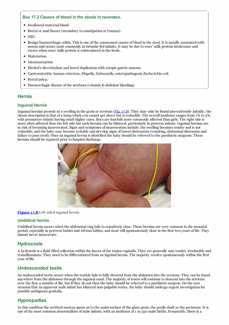
Box 17.2 Causes of blood in the stools in neonates.
Swallowed maternal blood.
Rectal or anal fissure (secondary to constipation or trauma).
NEC.
Benign haemorrhagic colitis. This is one of the commonest causes of blood in the stool. It is usually associated withmucus and occurs most commonly in formula-fed infants. It may be due to cows’ milk protein intolerance andrecurs when cows’ milk protein is reintroduced in the feeds.
Malrotation.
Intussusception.
Meckel’s diverticulum and bowel duplication with ectopic gastric mucosa.
Gastroenteritis: human rotavirus, Shigella, Salmonella, enteropathogenic Escherichia coli.
Rectal polyp.
Haemorrhagic disease of the newborn (vitamin K-deficient bleeding).
Hernia
Inguinal HerniaInguinal hernias present as a swelling in the groin or scrotum (Fig. 17.8). They may only be found intermittently initially; theclassic description is that of a lump which you cannot get above but is reducible. The overall incidence ranges from 1% to 5%,with premature infants having much higher rates. Boys are fourfold more commonly affected than girls. The right side ismore often affected than the left side but such hernias can be bilateral, particularly in preterm infants. Inguinal hernias areat risk of becoming incarcerated. Signs and symptoms of incarceration include: the swelling becomes tender and is notreducible; and the baby may become irritable and develop signs of bowel obstruction (vomiting, abdominal distension andfailure to pass stool). Once an inguinal hernia is identified the baby should be referred to the paediatric surgeons. Thesehernias should be repaired prior to hospital discharge.
Figure 17.8 Left-sided inguinal hernia.
Umbilical herniaUmbilical hernia occurs when the abdominal ring fails to completely close. These hernias are very common in the neonatalperiod, especially in preterm babies and African babies, and most will spontaneously close in the first two years of life. Theyalmost never incarcerate.
HydrocoeleA hydrocele is a fluid-filled collection within the layers of the tunica vaginalis. They are generally non-tender, irreducible andtransilluminate. They need to be differentiated from an inguinal hernia. The majority resolve spontaneously within the firstyear of life.
Undescended testisAn undescended testis occurs when the testicle fails to fully descend from the abdomen into the scrotum. They can be foundanywhere from the abdomen through the inguinal canal. The majority of testes will continue to descend into the scrotumover the first 4 months of life, but if they do not then the baby should be referred to a paediatric surgeon. On the rareoccasion that an apparent male infant has bilateral non-palpable testes, the baby should undergo urgent investigation forpossible ambiguous genitalia.
HypospadiasIn this condition the urethral meatus opens on to the undersurface of the glans penis, the penile shaft or the perineum. It isone of the most common abnormalities of male infants, with an incidence of 1 in 350 male births. Frequently, there is a

dorsal hood to the penis and a ventral curvature of the glans (chordee). Hypospadia may be classified as glandular,penoglandular, penile, penoscrotal or perineal, depending on the site of the urethral opening (Fig. 17.9). Chromosome studiesare indicated with undescended testes and severe penoscrotal or perineal lesions. Mild types are repaired in a one-stageprocedure during the first 6 months, but severe types require several staged operations. The infant must not becircumcised, otherwise definitive surgical treatment will be made more difficult. Epispadias refers to the urethra openingon the dorsal surface of the penis, and this has a worse prognosis.
Figure 17.9 Hypospadius and potential urethra opening sites.
SUMMARY
Normal structural and functional development of the gastrointestinal tract is essential for life. Many of themalformations that affect the gastrointestinal tract are identified on antenatal ultrasound. Appropriate paediatricsurgical management in the newborn period will allow most babies to have an excellent long-term outcome. Preterminfants are unable to coordinate their suck and swallow until 34 weeks’ gestation, and as a result all will requireintragastric feeding. NEC is in an infrequent but significant problem that causes significant morbidity and remains oneof the most significant abdominal complications in preterm babies.
Further reading1. Al-Faleh, K., Anabrees, J., Bassler, D., Al-Kharfi, T. (2014) Probiotics for prevention of necrotizing enterocolitis in
preterm infants. Cochrane Database System Review, 10 (4), CD005496.
2. Chu, A., Hageman, J.R., Caplan, M.S. (2013) Necrotizing enterocolitis: Predictive markers and preventive strategies.NeoReviews, 14 (3), 113–120.
3. Kastenberg, Z.J., Dutta, S. (2013) Ventral abdominal wall defects. NeoReviews, 14 (8), e402–e411.
Now visit www.essentialneonatalmed.com to test yourself on this chapter.

CHAPTER 18 Renal disordersKey topics
Role of amniotic fluid
Renal physiology
Normal urine output
Investigation of renal disease
Presentation of renal disease
Acute kidney injury
Urinary tract infection
Renal masses
Cystic disease of the kidneys
Haematuria
Ectopia vesicae (bladder exstrophy)
IntroductionMany renal disorders are now diagnosed on routine antenatal ultrasound. Occasionally, some of these conditions areincompatible with life, others require active management, and some will need conservative observation only. This chapterdiscusses the basic physiology of the kidney, its role in amniotic fluid, presentation and investigation of renal disease, andspecific conditions which may affect the renal tract.
Role of amniotic fluidAmniotic fluid volume is regulated from a number of fetal pathways, including urine production, lung fluid secretion and fetalswallowing. The fetal kidney is the major contributor to the volume of amniotic fluid. Ultimately, all amniotic fluid is derivedvia the placenta. Some of the roles of amniotic fluid are described in Box 18.1.
Box 18.1 Roles of amniotic fluid.
Amniotic fluid has a number of important roles, including:
Providing space for fetal growth and movement.
Cushioning the umbilical cord against compression.
Protecting the fetus against trauma.
Providing protection against infection.
Maintenance of fetal temperature.
Providing nutrients, hormones and growth factors.
Development of respiratory, gastrointestinal and musculoskeletal systems.
CLINICAL TIPDecreased amniotic fluid volume (e.g. oligohydramnios or anhydramnios) can lead to lung hypoplasia. Severitydepends on the cause, duration and timing of onset, and degree of volume loss. Infants with hypoplastic lungs willpresent with respiratory distress at birth.
Renal physiologyThe first permanent nephrons appear at around 8 weeks’ gestation and nephrogenesis is complete by 36 weeks’ gestation.The increase in renal mass thereafter is predominantly tubular growth. The human has approximately 1 million nephrons.Urine production commences shortly after the first nephrons develop. The fetal kidney produces a relatively large amountof dilute urine; however, the fetus does not depend on the kidney to excrete waste products as the placenta performs thisfunction. Renal function in the newborn depends on both gestational and postnatal age. Renal function rapidly matureswithin the first week after birth.
The newborn baby is largely water. The total body water (TBW) is 75% of body weight at term. The TBW is 80–90% inpreterm infants of 26–30 weeks’ gestation.
The size of the extracellular fluid compartment (ECF) decreases steadily throughout life from approximately 65% of bodyweight at 26 weeks to approximately 40% at term, and 20% by 10 years of age. Adaptation to the ex-utero environmentrequires a rapid loss of excess fluid. The net water and sodium balance is negative in the first few days of life, and this is themost important reason for babies losing weight in the first week of life. Normal weight loss can be up to 10% in term babies,

and 15% in preterm infants.
CLINICAL TIPExcess fluid administration to preterm infants is associated with increased morbidity from PDA, NEC and chronic lungdisease.
Glomerular filtration rateThe newborn infant has a very low glomerular filtration rate (GFR) which is approximately 20% of adult values. Preterminfants have a lower GFR than term infants. After 34 weeks’ gestation, and in response to birth, there is a marked increasein GFR. Although maturation of the GFR is most rapid during the first month of life, it continues into the second year. Themeasurement of serum creatinine is the most convenient index of GFR in infants. Creatinine measured in the first 24 hoursafter birth is not clinically useful as it is more likely to reflect maternal renal function. Plasma urea is also unreliable inneonates as it increases with catabolism even in the presence of normal renal function.
Tubular functionThe concentrating ability of the developing kidney increases throughout gestation and improves rapidly after birth. This isdue partly to elongation of the collecting tubes and partly to a hormonal effect (see below).
Tubular function can most easily be assessed by measuring the fractional excretion of sodium (FES); in the newborn thisshould be less than 2.5%. The formula for calculation of FES is:
Sodium conservationThe fetus has a very poor ability to conserve (reabsorb) sodium. In adults, 80–90% of filtered sodium is reabsorbed in theproximal convoluted tubule. Sodium potassium adenosine triphosphatase (Na+,K+-ATPase) creates the electrical chemicalgradient for transport. Sodium that enters the distal convoluted tube and collecting ducts is reabsorbed under the influenceof aldosterone. Premature babies have decreased Na+,K+-ATPase in the proximal tubule, and therefore higher sodium loadsare delivered to the distal tubules. They also have limited aldosterone responsiveness at the distal tubule (and therefore lesssodium absorption). Maturation of the system appears to be increased by the stress of delivery. Birth causes an increase inthe Na+,K+-ATPase, upregulation of transporter proteins, and increasing responsiveness of the distal tubule to aldosterone.
CLINICAL TIPAntenatal steroids increases the abundance of Na+,K+-ATPase mRNA in both kidneys and lungs and thereforeenhance the maturation of renal tubular transport.
Hormonal functionThe kidney is influenced by a number of hormones.
Antidiuretic hormoneAntidiuretic hormone (ADH) increases water reabsorption from the collecting ducts. It is present from early in fetal life butthe fetal kidney is relatively insensitive to it. After birth the collecting ducts become more sensitive. ADH is active in verypremature infants, and even the most immature infant is capable of concentrating the urine to a remarkable extent withindays of birth.
Renin–aldosteroneRenin levels are higher in newborn infants than in adults and increase in response to sodium loss. However, the adrenal doesnot respond with high aldosterone levels and consequently sodium retention is poor, but matures in response to birth.
Normal urine outputDue to the reduced renal concentrating ability the maximum flow rate is 300 ml kg–1 per day, and the minimum is25 ml kg–1 per day. Over 90% of normal infants pass urine in the first 24 hours of life, and 98% have voided by 48 hoursfrom birth. On day 1 of life, the urine output is approximately 0.5 ml kg–1 per hour, thereafter it is 2–3 ml kg–1 per hour.Neonates often do not completely empty their bladder on voiding.

CLINICAL TIPCare needs to be taken when interpreting potassium values in neonates as:
Haemolysis can falsely elevate (especially if collected via heel prick).
Tissue serum (particularly in oedematous patients) can also falsely elevate.
Delay in processing can elevate.
If the baby has a normal urine output, high potassium levels are unlikely to cause clinical concern.
Investigation of renal disease
UltrasoundThis is the mainstay of investigating the structure of the renal tract both before and after birth (Fig. 18.1). Renal Dopplercan be used to assess blood flow in and out of the kidney.
Figure 18.1 Longitudinal ultrasound view of fetal abdomen showing bilateral renal pelvocalyceal dilatation. Illustrationcourtesy of Dr R. Cincotta.
MAG 3 renogramA MAG 3 (mercaptoacetyltriglycine) renogram is a kinetic scan that involves the intravenous injection of a radioactive tracerwhich is taken up by the kidney, thereby enabling excretion curves to be plotted and obstruction to be visualized. Delay inthe excretion of isotopes from the kidney is suggestive of obstructive uropathy such as pelvi-ureteric junction (PUJ)obstruction. It is also possible to estimate differential kidney function using this scan prior to the excretion phase.
DMSA scanA dimercaptosuccinic acid (DMSA) radionuclide scan is a static scan that delineates renal scarring or dysplasia and allowsbetter estimation of differential renal function. It may show false-positive results if conducted during or soon after anepisode of acute urinary tract infection.
Micturating cystourethrogramThe micturating cystourethrogram (MCUG) is the investigation of choice for vesicoureteric reflux. It involves urethralcatheterization with the injection of radio-opaque dye into the bladder and observation of whether the dye refluxes into theureters on micturition. It is an invasive procedure and prophylactic antibiotics are often recommended before and after theprocedure.
Presentation of renal diseaseMost significant structural renal anomalies are detected antenatally as the result of routine fetal anomaly scanning. Somediagnoses are so severe (e.g. bilateral renal agenesis) that termination of pregnancy or palliative care may be offered to theparents. In other cases, the neonatal team will be notified before delivery of a baby with a renal anomaly and a variety ofinvestigations will be required after birth. Genitourinary disease in the newborn may present in a number of different ways.These are discussed separately.
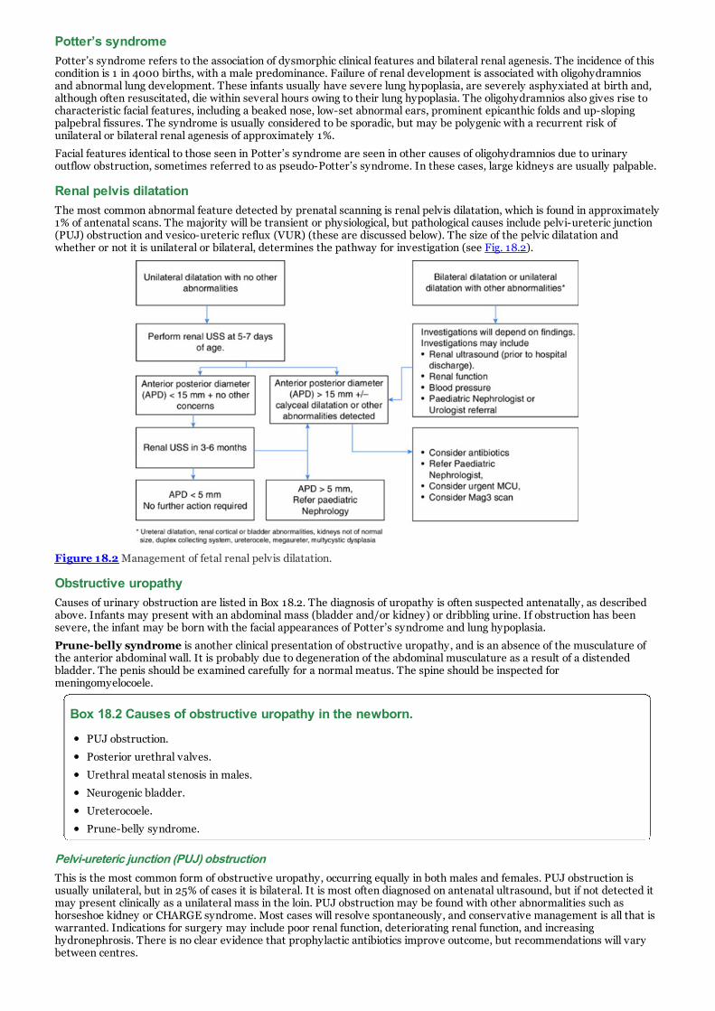
Potter’s syndromePotter’s syndrome refers to the association of dysmorphic clinical features and bilateral renal agenesis. The incidence of thiscondition is 1 in 4000 births, with a male predominance. Failure of renal development is associated with oligohydramniosand abnormal lung development. These infants usually have severe lung hypoplasia, are severely asphyxiated at birth and,although often resuscitated, die within several hours owing to their lung hypoplasia. The oligohydramnios also gives rise tocharacteristic facial features, including a beaked nose, low-set abnormal ears, prominent epicanthic folds and up-slopingpalpebral fissures. The syndrome is usually considered to be sporadic, but may be polygenic with a recurrent risk ofunilateral or bilateral renal agenesis of approximately 1%.
Facial features identical to those seen in Potter’s syndrome are seen in other causes of oligohydramnios due to urinaryoutflow obstruction, sometimes referred to as pseudo-Potter’s syndrome. In these cases, large kidneys are usually palpable.
Renal pelvis dilatationThe most common abnormal feature detected by prenatal scanning is renal pelvis dilatation, which is found in approximately1% of antenatal scans. The majority will be transient or physiological, but pathological causes include pelvi-ureteric junction(PUJ) obstruction and vesico-ureteric reflux (VUR) (these are discussed below). The size of the pelvic dilatation andwhether or not it is unilateral or bilateral, determines the pathway for investigation (see Fig. 18.2).
Figure 18.2 Management of fetal renal pelvis dilatation.
Obstructive uropathyCauses of urinary obstruction are listed in Box 18.2. The diagnosis of uropathy is often suspected antenatally, as describedabove. Infants may present with an abdominal mass (bladder and/or kidney) or dribbling urine. If obstruction has beensevere, the infant may be born with the facial appearances of Potter’s syndrome and lung hypoplasia.
Prune-belly syndrome is another clinical presentation of obstructive uropathy, and is an absence of the musculature ofthe anterior abdominal wall. It is probably due to degeneration of the abdominal musculature as a result of a distendedbladder. The penis should be examined carefully for a normal meatus. The spine should be inspected formeningomyelocoele.
Box 18.2 Causes of obstructive uropathy in the newborn.
PUJ obstruction.
Posterior urethral valves.
Urethral meatal stenosis in males.
Neurogenic bladder.
Ureterocoele.
Prune-belly syndrome.
Pelvi-ureteric junction (PUJ) obstructionThis is the most common form of obstructive uropathy, occurring equally in both males and females. PUJ obstruction isusually unilateral, but in 25% of cases it is bilateral. It is most often diagnosed on antenatal ultrasound, but if not detected itmay present clinically as a unilateral mass in the loin. PUJ obstruction may be found with other abnormalities such ashorseshoe kidney or CHARGE syndrome. Most cases will resolve spontaneously, and conservative management is all that iswarranted. Indications for surgery may include poor renal function, deteriorating renal function, and increasinghydronephrosis. There is no clear evidence that prophylactic antibiotics improve outcome, but recommendations will varybetween centres.

Posterior urethral valvesIn this condition mucosal folds arise within the lumen of the male urethra, producing a valve-like obstruction. Obstruction tourinary flow causes dilatation and hypertrophy of the urethra, bladder, ureter and kidneys. Diagnosis is most often made onantenatal ultrasound. Cases that are not diagnosed antenatally may present clinically with urine dribbling from the penis anda hard muscular bladder felt suprapubically. Severe cases may present with olighydramnios and pulmonary hypoplasia atbirth. Renal dysplasia is often associated with posterior urethral valves, and 20% of patients will eventually progress to end-stage renal failure. Ultrasound will confirm the presence of a hypertrophied bladder and establish the degree of upper tractinvolvement. MCUG is required to confirm the diagnosis and identify any vesicoureteric reflux.
If the lesion is mild, surgical ablation of the valves through a cystoscope is all that is necessary. In more severe cases avesicostomy will be required. Antenatal diagnosis and the insertion of a vesicoamniotic catheter into the fetal bladder areindicated in highly selected cases of severe obstruction.
UreterocoeleThis is a ballooning of the distal terminal ureter into the bladder, which obstructs the drainage of the kidney on that side.They are more common in females and in white babies. A duplex collecting system is frequently associated. Theureterocoele tends to occur on the ureter that drains the upper pole of the kidney. Diagnosis is generally made by antenatalultrasound. MCUG is required to identify vesicoureteric reflux. The ureterocoele should be excised and the ureterreimplanted to avoid urinary reflux.
Acute kidney injuryAcute kidney injury (previously called acute renal failure) may occur as the result of numerous disease processes, many ofwhich are detected on antenatal ultrasound; these can be divided into prerenal, renal and postrenal causes (Table 18.1).Prerenal failure is the commonest variety, and usually occurs with severe respiratory disease or as a postoperativecomplication.
Table 18.1 Causes of renal failure in the neonate.
Prerenal Renal Postrenal
Dehydration Cystic disease Obstructive uropathy (see Box 18.2)
Hypotension due to: Acute tubular necrosis Fungal obstruction
Haemorrhage Cortical necrosis
Infection Disseminated intravascular coagulation
Cardiac dysrhythmia Infection (pyelonephritis)
Asphyxia Haemolysis
Hypothermia Venous thrombosis
Congenital nephrotic syndrome
Drugs (aminoglycosides)
Clinical featuresThe clinical features of acute renal failure are listed in Box 18.3. Severe complications of acute renal failure are listed in Box18.4.
Box 18.3 Clinical signs and symptoms of acute kidney injury in the newborn.
Oliguria (urine output <1.0 ml kg–1 per hour).
Oedema.
Rising serum creatinine and blood urea.
Metabolic acidosis (decreased pH, decreased bicarbonate).
Increased potassium.
Increased phosphate.
Decreased sodium.
Decreased calcium.
Decreased magnesium.
Cardiac dysrhythmias may develop as a result of hyperkalaemia.

Box 18.4 Complications of acute kidney injury.
Hyperkalaemia (>7.5 mmol l–1). Insulin and dextrose may be necessary to drive potassium into the cells. A singledose of IV salbutamol can also briefly reduce serum potassium.
Severe metabolic acidosis (pH <7.20).
Fluid overload with severe oedema and congestive cardiac failure.
Hypertension, usually due to fluid overload.
Severe uraemia with central nervous system (CNS) depression.
Under these circumstances dialysis may be indicated.
Oliguria may be difficult to detect early in the infant’s life, as the passage of urine may have occurred unnoticed or may nothave been recorded. Abdominal ultrasound may also be useful in showing whether there is urine in the bladder. If oliguria issuspected, the insertion of a urinary catheter will allow an accurate determination of urine output.
In oliguric infants there may be difficulty in distinguishing prerenal from renal failure; fractional excretion of sodium (FES)may be used in such cases. In prerenal failure the urine is concentrated and the serum and urinary creatininemeasurements are high. If doubt still exists, a fluid bolus with a dose of furosemide should resolve the issue. The obstructiveuropathy causes of acute renal failure must be excluded in postrenal failure (see Box 18.2).
Pathological examination of the kidneys of infants dying with acute renal failure shows acute tubular necrosis, acute corticalnecrosis or bilateral renal vein thrombosis. With renal vein thrombosis, loin masses are generally palpable and the baby hashaematuria.
ManagementIn established renal failure the underlying cause should be treated wherever possible. Consultation with a paediatricnephrologist is often helpful. The measures listed below are usually sufficient to maintain homeostasis until renal functionrecovers.
Fluid restrictionWater intake should be restricted so that only sensible (e.g. urine output) and insensible losses (e.g. respiration andperspiration) are replaced. These losses are calculated separately. Insensible loss depends on the infant’s birthweight andgestational age. Insensible losses fall quickly in the first few days of life as the stratum corneum layer in the skin matures.Daily weighing is an accurate way of assessing fluid balance.
Protein and potassium restrictionPotassium should not be given unless the infant is hypokalaemic. To avoid endogenous protein breakdown and fluidoverload, a high-carbohydrate infusion (e.g. 15–20% dextrose) should be given. The volume depends on the infant’s output(see above).
Correction of electrolyte imbalanceHyponatraemia is usually due not to sodium depletion but rather to fluid overload. Water should be restricted until this iscorrected.
Metabolic acidosisSodium bicarbonate is carefully administered if the pH falls below 7.25. Care is necessary to avoid hypernatraemia.
Urinary tract infectionThe incidence of symptomatic urinary tract infection in the neonatal period is 1%. The symptoms of urinary tract infection inthe newborn are generally non-specific, with jaundice, vomiting, failure to thrive, temperature instability, lethargy and poorfeeding. Specific symptoms of dysuria, frequency and abdominal discomfort are rarely seen. Urinary tract infections in thenewborn are more common in the male and may be blood-borne, ascending or secondary to an abnormality of the urinarytract. In the neonatal period pyelonephritis is usually due to blood-borne infection. Investigations include an adequate urinespecimen sent for microscopy and culture. Interpretation of urinalysis is provided in Box 18.5.

Box 18.5 Interpretations of neonatal urine results.
Microscopy
A white cell count of more than 30 000 ml–1 suggests infection; a count of less than 10 000 ml–1 is probablynormal.
NB: Some normal newborn infants have up to 30 000 white cells ml–1, even on a bladder tap aspirate of urine.
White cell counts in urine specimens from collecting bags are high and should be interpreted cautiously, and only inconjunction with colony counts.
Culture
A pure growth of more than 100 000 organisms ml–1 indicates a significant infection.
Less than 10 000 ml–1 or mixed organisms suggests contamination.
Counts between 10 000 and 100 000 ml–1 indicate that the urine sample should be repeated.
Any growth of organisms obtained on bladder tap is significant.
The presence of budding hyphae in the urine strongly suggests systemic fungal infection (see Chapter 10).
PRACTICAL PROCEDURE TIPCollection of urine
This may be performed using a ‘clean-catch’ bag method, suprapubic aspiration, or urethral catheter. Suprapubicpuncture is best done once the presence of a full bladder has been confirmed with ultrasound. It is a relatively painfulprocedure and, ideally, should be discouraged. The most reliable way to obtain an uncontaminated sample of urine isthrough urethral catheterization.
Bag collection
A clean-catch bag sample of urine may provide useful information. However, an inexpertly collected sample providesconfusing or misleading information.
1. Cleanse a wide area of the perineum and penis with sterile saline-soaked swabs using a no-touch technique. Infemales the vulva will need to be separated, but in males the foreskin must not be retracted, although it needs tobe cleaned.
2. Wait for the perineum to dry before attaching a sterile urine-collecting bag.
3. Inspect the bag periodically, and as soon as the baby has voided remove the bag and immediately pour the urineinto a universal sterile container.
4. Send the sample to the laboratory immediately. If there is any delay in transporting the sample, refrigerate it.
5. If no urine has been obtained after 1 hour, the skin should be cleansed again and a fresh collecting bag applied.
A suitable alternative to bag collection in babies and small children is to put a pad inside the nappy and, when it is wet,to suck out the urine using a small syringe. This is considered to be as sterile as a bag sample, and is widely practisedin many centres.
Suprapubic aspiration
The indications for this procedure are an inconclusive culture from a bag specimen, or the presence of vulvovaginitisor balanitis when urinary tract infection is suspected.
1. The bladder must be full before commencing the procedure (confirm with ultrasound if possible).
2. A wide area of skin over the lower abdomen is prepared with povidone-iodine solution and alcohol.
3. A 21G needle attached to a 10-ml syringe is used to puncture the skin 1 cm above the pubic symphysis in themidline.
4. The needle is advanced into the bladder, angled slightly upwards to the perpendicular. As the needle is slowlyadvanced gentle suction is applied. The needle usually only needs to be advanced 1.5–2.0 cm below the skinsurface (Fig. 18.3). Ultrasound guidance can be used for insertion of the needle.
5. The urine can then be sent to the laboratory.

Figure 18.3 Suprapubic aspiration of urine from the bladder. The needle should be aimed slightly superiorly in the midlineand 0.5 cm above the pubis.
CLINICAL TIPA ‘clean voided’ bag sample of urine is used only as a screen and is never the basis for a definitive diagnosis. If the bagsample suggests a urinary tract infection, or if the baby is sick and in need of urgent treatment, either a ‘clean catch’,suprapubic aspiration or a urinary catheter sample will be necessary.
TreatmentAntibiotics that are excreted in the urine should be started immediately in an infant for whom there is a strong clinicalsuspicion of urinary tract infection. This is particularly important in pyelonephritis. In the presence of severe or suspectedinfection, therapy is usually commenced with an intravenous aminoglycoside (e.g. gentamicin) and ampicillin. Once culturesand sensitivities are known, any inappropriate antibiotic may be stopped. Co-trimoxazole (Septrin) may be used after thefirst week of life. Other suitable antibiotics include cefalexin (cephalexin), ceftriaxone or cefotaxime (cephotaxime).Treatment is continued for 10 days, when repeat urine microscopy and culture are performed.
Ultrasound examination together with a micturating cystourethrogram (MCU) are indicated in all neonates with provenurinary tract infection. Other investigations include MAG 3 (see page 203) and a DMSA scan, which allows the estimation ofdifferential renal function and delineates renal scarring or dysplasia. Severe reflux may be associated with renal scarringeven in the absence of infection. Specimens of urine should be regularly checked during the first year of life to ensure thatreinfection does not occur.
CLINICAL TIPCare should be taking if prescribing trimethoprim sulfamethoxazole. The sulfonamide can displace bilirubin from itsalbumin-binding site, potentially increasing the amount of free bilirubin available to cross the blood–brain barrier.Therefore, there have been concerns about its safety in neonates.
Renal massesNormal kidneys, especially the right kidney, may be just palpable in the newborn infant. If a renal mass is easily felt, thekidney is probably pathologically enlarged. Differentiation of the mass can be achieved using ultrasound. The causes of arenal mass are listed in Box 18.6.

Box 18.6 Causes of a renal mass in the newborn period.
Hydronephrosis (bilateral).
PUJ obstruction (unilateral).
Cystic disease of the kidneys.
Renal vein thrombosis.
Wilms’ tumour (nephroblastoma).
Adrenal haemorrhage.
Cystic disease of the kidneysThere is a great variety of cystic conditions of the kidney in the newborn. Congenital renal cystic diseases are a geneticallyand clinically diverse group of disorders with the common pathological finding of diffuse bilateral cystic structures withoutdysplasia. Many hereditary malformation syndromes are associated with renal cysts. Renal cystic diseases affecting theneonate are listed in Box 18.7.
Box 18.7 Renal cystic disease affecting neonates.
Cystic dysplastic (multicystic) kidneys.
Autosomal dominant polycystic kidney disease (ADPKD).
Autosomal recessive polycystic kidney disease (ARPKD).
Cysts secondary to obstructive uropathy �(see Table 18.1).
Simple renal cysts.
Autosomal dominant polycystic kidney diseaseAutosomal dominant polycystic kidney disease (ADPKD) is the commonest inherited renal disease, with an incidence of 1 in200 to 1 in 1000. It was originally known as ‘adult-onset PKD’ because of the usual clinical presentation in the third to fifthdecades, although the disease can manifest itself in utero or at any time thereafter. It may be diagnosed on perinatalultrasound with the finding of enlarged or cystic kidneys. Clinically, the disease can present along a spectrum, from thesevere neonatal form with renal failure and Potter’s syndrome, to the asymptomatic unilateral renal cyst found on renalultrasound.
Autosomal recessive polycystic kidney diseaseOriginally known as ‘infantile PKD’ because of its more common perinatal presentation, autosomal recessive polycystickidney disease (ARPKD) can present at any time from in utero to adulthood. It is due to mutations in the PKHD1 gene whichencodes a protein called fibrocystin. It is often diagnosed on antenatal ultrasound with the finding of enlarged kidneys andincreased echogenicity. The cysts are very small (<3 mm) and are due to dilatation of the renal collecting ducts. There willalways be some degree of biliary dysgenesis with periportal fibrosis. ARPKD is associated with oligohydramnios and oftensevere pulmonary hypoplasia. End-stage renal failure is common in early childhood.
Cystic dysplastic (multicystic) kidneysThis is a sporadic condition producing a non-functioning kidney and is usually unilateral, as bilateral cases are fatal. It tendsto occur on the left side and is more common in males. Most cases are identified on antenatal ultrasound. The kidney showsmultiple small and large cysts, and there may be no normal renal tissue identifiable. There is little or no renal function on theaffected side. Hypertension and possible malignancy in the dysplastic kidney are long-term complications. Renal ultrasoundis often performed after birth to confirm the diagnosis and ensure that the other kidney is normal. Conservativemanagement is all that is required in most cases, although occasionally surgical excision is indicated.
Isolated renal cystsThese are a common incidental finding. Patients with isolated cysts will have normal renal function and no renal dysplasia.Long-term follow-up for simple cysts is not required.
HaematuriaIn the first few days of life the presence of red cells in the urine is not uncommon, but after this haematuria is an importantfinding and must be further investigated. Box 18.8 lists causes of haematuria. Investigations include urinalysis, clottingstudies, creatinine and ultrasound examination. Treatment depends on the underlying cause.

Box 18.8 Neonatal renal disorders associated with haematuria.
Acute tubular necrosis.
Infection.
Renal vein thrombosis.
Bleeding disorders.
Cystic renal disease.
Renal calculi.
Tumours.
CLINICAL TIPHaematuria must be differentiated from the presence of urates in the urine, a benign condition that produces pinkstaining of the nappy/diaper.
Ectopia vesicae (bladder exstrophy)This is a complex disorder of the abdominal wall, bladder and pelvis. The mucosa of the bladder herniates through adeficient lower abdominal wall. There is a deficient pelvic floor and wide separation of the pubic symphysis. It is morecommon in male than in female infants. In the male there is total epispadias, undescended testes, and a deficient penis. Inthe female the two halves of the clitoris are separate and the vagina is duplicated.
The surgical management of this complex disorder is extremely difficult and continues over many years. Urinary continenceis rarely achieved.
SUMMARY
Adequate renal function is essential for the development and survival of the neonate. Many renal disorders arediagnosed on routine antenatal ultrasound. Renal disorders have a wide variety of clinical presentations ranging fromnon-survivable pulmonary hypoplasia at birth to being asymptomatic. Appropriate investigation and follow-up isrequired for many of these disorders, the ultimate aim being to preserve normal renal function.
Further reading1. Mistry, K. (2014) Renal and urological diseases of the newborn neonatal acute kidney injury. Current Pediatric
Reviews, 10 (2), 88–94.
2. Poude, A., Afshan, S., Dixit, M. (2016) Congenital anomalies of the kidney and urinary tract. NeoReviews, 17 (1), e18–e27.
Now visit www.essentialneonatalmed.com to test yourself on this chapter.

CHAPTER 19 JaundiceKey topics
Physiology of bilirubin metabolism
Clinical assessment of the jaundiced infant
Unconjugated hyperbilirubinaemia
Conjugated hyperbilirubinaemia
IntroductionJaundice is a yellow discolouration of the skin, sclera and mucous membranes due to the deposition of bilirubin. Neonataljaundice is the most common problem encountered in the newborn. About 50% of all full-term infants and 85% of preterminfants are visibly jaundiced within the first week of life. In most infants the jaundice is physiological as there is nounderlying disease. Physiological jaundice results from increased bilirubin production (due to increased haemoglobin levelsat birth and a shortened red cell lifespan), and decreased bilirubin excretion (due to low concentrations of the hepatocytebinding protein, low activity of glucuronosyl transferase, and increased enterohepatic circulation). Unconjugated bilirubin,which is elevated in the most common forms of neonatal jaundice, can pass through the blood–brain barrier and isneurotoxic. High levels in the brain can cause acute or chronic encephalopathy if not treated appropriately. Conjugatedhyperbilirubinaemia is much less common than unconjugated jaundice in the newborn, but has a much more seriousprognosis.
Physiology of bilirubin metabolism
FetalIn the uterus, the fetal liver is relatively inactive. The placenta and maternal liver metabolize the bilirubin from worn-outred blood cells. The fetus is capable of conjugating bilirubin in small amounts and when haemolysis occurs in utero (as insevere rhesus isoimmunization), bilirubin conjugation increases, and high levels may be measured in the umbilical cordblood.
CLINICAL TIPIf there is excessive fetal red cell haemolysis (e.g. in rhesus haemolytic disease) the placenta and maternal liver maynot be able to deal with the excessive bilirubin load, and the umbilical cord and amniotic fluid will be stained yellow bythe bilirubin pigment produced. In addition, the bone marrow and extramedullary organs of erythropoiesis may notbe able to keep up with the production of red cells, so that the fetus will become anaemic. Hydrops fetalis, a conditionassociated with generalized oedema, pleural effusions, ascites and hepatosplenomegaly, is due to a combination ofanaemia, intrauterine hypoxia, hypoproteinaemia, a low colloid osmotic pressure and congestive heart failure.
NewbornThe metabolism of bilirubin in the newborn is summarized in Figure 19.1. Each of the steps in the metabolism of bile will bediscussed in turn.

Figure 19.1 Summary of neonatal bilirubin metabolism.
Bilirubin productionMost of the daily bilirubin production comes from ageing red blood cells. The red cells are destroyed in thereticuloendothelial (RE) system and the haem is converted to unconjugated bilirubin. Typically, 1 g of haemoglobin willproduce 600 µmol (35 mg) of unconjugated bilirubin. Haemolysis may be increased by maternal drugs such as salicylates,sulphonamides, phenacetin and nitrofurantoin. Some 25% of the daily production of bilirubin comes from sources other thanthe red cells, such as haem protein and free (tissue) haem.
Transport and liver uptakeMost of the unconjugated bilirubin in the blood is bound to serum albumin and is transported to the liver as a boundcomplex. This binding is extremely important and may be altered by many factors. Factors that decrease albumin bindingability include low serum albumin, asphyxia, acidosis, infection, prematurity and hypoglycaemia. In addition, there are manycompetitors for bilirubin-binding sites, and these include:
Non-esterified (free) fatty acids produced by starvation, cold stress or intravenous fat emulsion.
Drugs (sulphonamides, cephalosporins, sodium benzoate (present in diazepam), frusemide and thiazide diuretics).
When bilirubin is bound to albumin it is probably non-toxic, but free unbound unconjugated bilirubin is fat-soluble and canbe transported across the blood–brain barrier and be deposited in certain neurons, causing damage. Hepatocytes lining theliver sinusoids are able to extract unconjugated bilirubin from the blood, and this is then accepted in the liver cell by the Yand Z proteins (ligandins).
Conjugation and excretionThe unconjugated bilirubin is conjugated in the liver and the reaction involves the conversion of insoluble unconjugatedbilirubin to direct-reacting bilirubin (water-soluble). Each molecule of bilirubin is conjugated with two molecules ofglucuronic acid in a reaction catalysed by the enzyme glucuronyltransferase. The conjugated bilirubin is excreted into thebile, and subsequently into the duodenum. In the older child the bilirubin is reduced to stercobilinogen by bacteria in thesmall bowel, but in the newborn with a relatively sterile bowel and poor peristalsis much of the conjugated bilirubin may behydrolysed by glucuronidase back to unconjugated bilirubin. This is then absorbed in the bowel, re-entering the circulationfor further liver metabolism, a process called enterohepatic circulation.
This may be important in pathological situations, and reinforcement of the enterohepatic circulation will increaseunconjugated bilirubin levels in prematurity, small bowel obstruction, functional bowel obstruction and pyloric stenosis.
Clinical assessment of the jaundiced infantJaundice can be detected clinically once the serum bilirubin (SBR) result is above 90 µmol l–1. As jaundice is common it isessential to have a clinical method for determining its severity. Proper lighting (ideally daylight) is important for detectingsubtle levels of jaundice.
An old method of assessing the degree of jaundice was the use of Kramer’s rule (Kramer, 1969). This technique depended onthe blanching of the infant’s skin with the examiner’s finger at standard zones and observing the colour in the blanched area.It is no longer used as it has been proven to be unreliable, particularly in non-Caucasian babies.

CLINICAL TIPA non-invasive method of assessment is the use of transcutaneous bilirubinometry (TcB). It can provide a usefulscreening test in babies 35 weeks or more and more than 24 hours of age. If the TcB suggests a high bilirubin(>250 μmol l–1), a serum bilirubin should be collected. TcB has good correlation with total serum bilirubin, but mayhave limitations in specific situations.
CLINICAL TIPBilirubin in the urine (tested by a ‘stick’ test) indicates that a component of the serum bilirubin is conjugated. Palestools might suggest that the normal passage of bile from the liver into the duodenum is blocked. These are importantclinical signs suggestive of obstructive jaundice. Stool charts are available for parents to describe the stool colouragainst.
ManagementThe first investigation in any jaundiced newborn is to take a careful history. Important considerations in history taking arelisted in Box 19.1. The next step is to perform a complete physical examination. Clinical findings that may help to delineatethe cause are listed in Box 19.2.
Box 19.1 Required history when assessing for causes of neonatal jaundice.
Events in pregnancy, e.g. suspected infection, maternal diseases (e.g. thyroid disorders, diabetes), maternal drugs.
Maternal blood group, and presence of antibodies.
Family history, e.g. ethnic origin, previous jaundiced infant requiring treatment, red cell morphological disorders(spherocytosis, elliptocytosis, stomatocytosis, pynknocytosis), red cell enzyme disorders (thalassaemia, G6PD,pyruvate kinase), thyroid disorders, galactosaemia, cystic fibrosis.
Birth history, e.g. traumatic delivery, condition at birth.
Gestational age and birthweight of the infant.
Current postnatal age of the infant and when baby first became clinically jaundiced.
Current clinical condition of the infant (feeding, stooling, urine output).
Has the baby received any treatment since birth, e.g. phototherapy, medications, etc.?
Box 19.2 Clinical findings that may help to determine the cause and severity of neonataljaundice.
Extravascular blood, e.g. extensive bruising, purpura, petechiae.
Plethora or pallor.
Hepatomegaly.
Splenomegaly.
Evidence of intrauterine infection, e.g. small for gestational age, cataracts, microcephaly.
Evidence of infection, e.g. umbilicus, skin, urine.
Neurological signs: hypertonia, opisthotonus, fits, abnormal eye movements, abnormal cry.
Abdominal distension: associated with bowel obstruction, bowel stasis or hypothyroidism.
In assessing the significance of jaundice in a newborn infant the following guidelines may be useful. Investigations should becarried out under the following circumstances:
Any infant who is visibly jaundiced in the first 24 hours of life.
Any jaundiced infant whose mother has rhesus antibodies.
A preterm infant whose estimated serum bilirubin is greater than 150 µmol l–1.
A term infant whose estimated serum bilirubin exceeds 200 µmol l–1.
Any infant who has the clinical signs of obstructive jaundice (dark urine, pale stools).
Prolonged hyperbilirubinaemia beyond 2 weeks.
First line tests should include:
unconjugated bilirubin;
direct antibody test;
blood group;
full blood count (FBC) and red cell morphology.

Directed tests may include:
infection screen (e.g. TORCH, septicaemia);
glucose-6-phosphate dehydrogenase (G6PD) assay;
thyroid function;
conjugated bilirubin;
liver USS;
erythrocyte galactose uridyl transferase activity if considering galactosaemia.
Once this basic information has been collected, the possible aetiology – and therefore the investigations that should becollected – will often be obvious. Table 19.1 lists the common causes of jaundice and the timing of their presentation.
Table 19.1 Possible causes of jaundice presenting at different times in the neonatal period.
Day(s) Unconjugated jaundice Conjugated jaundice
1 Haemolytic disease assumed until proven otherwise Neonatal hepatitis
Rubella
CMV
Syphilis
2–5 Haemolysis
Physiological
Jaundice of prematurity
Sepsis
Extravascular blood
Polycythaemia
G6PD deficiency
Spherocytosis
As above
5–10 Sepsis
Breast milk jaundice
Galactosaemia
Hypothyroidism
Drugs
As above
10+ Sepsis
Urinary tract infection
Biliary atresia
Neonatal hepatitis
Choledochal cyst
Pyloric stenosis
Repeated total serum bilirubin estimations should be performed in infants with a rapid and early rise of (unconjugated)bilirubin so that treatment for hyperbilirubinaemia can be instituted. Bilirubin in the urine indicates that the conjugatedfraction of bilirubin should be estimated in the laboratory and causes of conjugated hyperbilirubinaemia considered.
Unconjugated hyperbilirubinaemia
CausesThe causes (and timing of onset) of unconjugated hyperbilirubinaemia are shown in Table 19.1. In any baby presenting withjaundice in the first 24 hours of life it is important to exclude a haemolytic cause for the unconjugated hyperbilirubinaemia.The most common causes are ‘physiological jaundice’ and ‘jaundice of prematurity’, and these usually present between day 2and day 5. Breastfeeding infants are more likely to develop jaundice secondary to a lower caloric intake and increasedenterohepatic circulation of bilirubin. The causes of prolonged (lasting >10 days) or late-onset unconjugatedhyperbilirubinaemia are shown in Box 19.3.
CLINICAL TIPJaundice presenting in the first 24 hours of life may be secondary to haemolysis. The important causes are rhesusisoimmunization and ABO incompatibility. These conditions can lead to rapid rises in the bilirubin level, and often needearly and aggressive treatment to prevent complications.

Box 19.3 Causes of prolonged unconjugated hyperbilirubinaemia.
Rhesus and ABO incompatibility.
Hereditary spherocytosis.
Glucose-6-phosphate dehydrogenase deficiency.
Septicaemia and TORCH infection.
Urinary tract infection.
Extravasated blood and excessive bruising.
Twin-to-twin transfusion.
Prematurity.
Dehydration.
Hypothyroidism.
Breast milk jaundice.
Delayed passage of meconium.
Physiological jaundicePhysiological jaundice is a term used by clinicians to describe jaundice for which no underlying cause is identified, and istherefore a diagnosis of exclusion. The major cause is increased bilirubin production (due to increased haemoglobin levels atbirth and a shortened red cell lifespan). Decreased bilirubin excretion (due to low concentrations of the hepatocyte bindingprotein, low activity of glucuronosyl transferase, and increased enterohepatic circulation) also contributes to thedevelopment of physiological jaundice.
InfectionBacterial infections, particularly septicaemia and urinary tract infections, may cause unconjugated hyperbilirubinaemia.Occasionally, severe bacterial infection may cause hepatocellular damage with a conjugated form of jaundice. TORCHinfections may cause either type of hyperbilirubinaemia, but the conjugated form is most frequently seen.
Breastfeeding and jaundiceBreastfeeding-associated jaundice is the term used to refer to the increased bilirubin levels seen during the first weekof life in almost two-thirds of infants who are breastfed. It is probably due to calorie and fluid deprivation in the first fewdays of life and delayed passage of stools, as it can be reduced by an increased frequency of breastfeeding during the firstfew days of life.
Breast milk jaundice is prolonged jaundice that extends until the first 3 months of life. Characteristically, it is a form ofnon-haemolytic, unconjugated hyperbilirubinaemia and should be diagnosed primarily by the exclusion of other aetiologiesin a thriving infant, and by its time course. The enzyme β-glucuronidase has been shown to be elevated in some women’sbreast milk. An excess of this enzyme causes increased enteric absorption of bilirubin, thus increasing the hepatic bilirubinload. In this respect, breast milk jaundice can be thought of as an extension of physiological jaundice, and the greater theconsumption of this enzyme in milk the higher is the concentration of neonatal serum bilirubin. Breast milk jaundice does notrequire any treatment and is not an indication to abandon breastfeeding; however, babies with high bilirubin levels shouldbe kept under review.
Delayed passage of meconiumThis increases the risk of jaundice due to increased enterohepatic absorption of bilirubin.
Gilbert’s syndromeThis is a common cause of unconjugated hyperbilirubinaemia in young adults, but rarely causes problems in the neonatalperiod. It is due to a single gene mutation affecting a hepatic bilirubin enzyme uridine diphosphate glucuronyltransferase(UDPGT), and in the newborn is associated with mild unconjugated hyperbilirubinaemia (<85 µmol l–1). The prognosis isexcellent.
Crigler–Najjar syndromeThis group of conditions has been shown to be due to multiple gene defects, and causes very severe hyperbilirubinaemiapotentially leading to kernicterus. There is little effective treatment other than prolonged phototherapy. Livertransplantation has been successful in some severe cases; in milder cases phenobarbitone may lower the serum bilirubin.
InvestigationsThe initial investigations for jaundice were discussed above. The aim is to identify the infant at risk of acute bilirubinencephalopathy and thereby prevent it occurring by commencing appropriate treatment. Prolonged unconjugatedhyperbilirubinaemia requires additional investigation when present for more than 14 days. Investigations for prolongedunconjugated hyperbilirubinaemia are listed in Box 19.4.
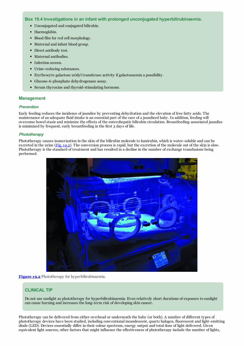
Box 19.4 Investigations in an infant with prolonged unconjugated hyperbilirubinaemia.Unconjugated and conjugated bilirubin.
Haemoglobin.
Blood film for red cell morphology.
Maternal and infant blood group.
Direct antibody test.
Maternal antibodies.
Infection screen.
Urine-reducing substances.
Erythrocyte galactose uridyl transferase activity if galactosaemia a possibility.
Glucose-6-phosphate dehydrogenase assay.
Serum thyroxine and thyroid-stimulating hormone.
Management
PreventionEarly feeding reduces the incidence of jaundice by preventing dehydration and the elevation of free fatty acids. Themaintenance of an adequate fluid intake is an essential part of the care of a jaundiced baby. In addition, feeding willovercome bowel stasis and minimize the effects of the enterohepatic bilirubin circulation. Breastfeeding-associated jaundiceis minimized by frequent, early breastfeeding in the first 3 days of life.
PhototherapyPhototherapy causes isomerization in the skin of the bilirubin molecule to lumirubin, which is water-soluble and can beexcreted in the urine (Fig. 19.2). The conversion process is rapid, but the excretion of the molecule out of the skin is slow.Phototherapy is the standard of treatment and has resulted in a decline in the number of exchange transfusions beingperformed.
Figure 19.2 Phototherapy for hyperbilirubinaemia.
CLINICAL TIP
Do not use sunlight as phototherapy for hyperbilirubinaemia. Even relatively short durations of exposure to sunlightcan cause burning and increases the long-term risk of developing skin cancer.
Phototherapy can be delivered from either overhead or underneath the baby (or both). A number of different types ofphototherapy devices have been studied, including conventional incandescent, quartz halogen, fluorescent and light-emittingdiode (LED). Devices essentially differ in their colour spectrum, energy output and total dose of light delivered. Givenequivalent light sources, other factors that might influence the effectiveness of phototherapy include the number of lights,

the distance from the light source to the infant, the time spent in phototherapy, and the surface area of skin exposed. LEDphototherapy may have an advantage in reducing dehydration.
Various thresholds for commencing phototherapy and for monitoring at-risk infants have been developed by differentorganizations. Commonly used charts are found in the NICE neonatal jaundice guidelines (see Fig. 19.3) and the AmericanAcademy of Pediatrics guideline (see Further reading). A general approach to the management of a jaundiced infant isprovided in Figure 19.4.
Figure 19.3 NICE clinical guideline: treatment threshold for babies with neonatal jaundice ≥38 weeks’ gestation.
Figure 19.4 Approach to the jaundiced infant.
Once therapy has been commenced, serum bilirubin estimates will be necessary to assess the severity of jaundice becausethe skin colour becomes unreliable. In cases of severe haemolytic jaundice, the SBR will need to be repeated 4–6 hours afterinitiating phototherapy, and then every 6–12 hours once it is stable or falling.
Phototherapy can be stopped once the SBR has fallen below the phototherapy threshold (the NICE guideline recommends atleast 50 µmol l–1 below).
A rebound SBR should be collected (12–24 h) after stopping phototherapy. Babies do not necessarily have to remain inhospital for this to be done.
Some complications of phototherapy are listed in Box 19.5.
General care of the baby during phototherapy includes eye protection, monitoring the baby’s temperature and fluid balance,and supporting the parents by educating them and encouraging them to interact with their baby.

Box 19.5 Some complications of phototherapy.
Temperature instability.
Dehydration: regular assessment of the baby’s fluid balance (monitor weight and urine output) and earlymanagement may be required.
Diarrhoea: phototherapy decreases bowel transit time and induces lactose intolerance; both are important causesof diarrhoea and consequent fluid loss.
Retinal damage: the eyes are thought to be vulnerable to phototherapy but this has never been proved. It is,however, a wise precaution to protect the infant’s eyes from the light by a suitable eye shield.
Early discharge and home management of jaundiceThe introduction of early discharge programmes for healthy term infants has resulted in the need for jaundice to bediagnosed and or treated at home. The resurgence of kernicterus is probably also related to the combination of earlydischarge and breastfeeding. This may result in dehydration if the baby feeds poorly and there is a lack of recognition ofsevere hyperbilirubinaemia. The NICE and AAP guidelines can be used to estimate the risk of an infant developing jaundicethat may require treatment. Predicting which infants might require treatment using direct and indirect techniques ofmeasuring serum bilirubin at age 24 hours has been suggested, but more research is required to identify sensitive methods.Home treatment has been facilitated by the introduction since 1990 of the BiliBlanket (MedNow Inc., Eagle, Idaho, USA),which uses a fibre-optic phototherapy system. The infant is placed on a mat containing fibre-optic strands which flood thebody with therapeutic light while the infant is normally clothed.
Exchange transfusionAlthough there are no randomized controlled trials on exchange transfusion versus no treatment, it is generally acceptedthat exchange transfusion is beneficial in the treatment of very severe hyperbilirubinaemia. The exchange transfusionallows the
removal of unconjugated bilirubin;
removal of the antibody causing haemolysis (if present);
replacement of sensitized red cells with cells that cannot be haemolysed as easily;
restoration of blood volume and correction of anaemia; and
provision of free albumin for bilirubin binding.
Exchange transfusions may be required in conjunction with phototherapy for infants with severe jaundice, especially whendue to rhesus isoimmunization. The indications for exchange transfusion depend on the infant’s gestational age, postnatalage and state of health. Metabolic compromise with acidosis predisposes to brain damage due to bilirubin toxicity. The levelat which bilirubin damages the brain is not known, and the indications for exchange transfusion are therefore arbitrary.Different charts are used in different centres to guide treatment. The NICE neonatal jaundice guidelines providerecommendations as to the bilirubin level at which exchange transfusion should be performed for infants of differentgestations. The AAP guidelines also have similar recommendations.
The technique of exchange transfusion is described in Chapter 29. Complications associated with exchange transfusions arelisted in Box 19.6.
Box 19.6 Complications associated with exchange transfusions.
Death (reported mortality rates now less than 3%).
Electrolyte disturbance (hypocalcaemia, hyperkalaemia).
Blood glucose disturbance – initially hyperglycaemia followed by rebound hypoglycaemia, especially in rhesusimmunization.
Infection – viral (CMV, hepatitis B, HIV) or bacterial (Staphylococcus aureus and streptococcus) due tocontaminated blood.
Embolism of either air or thrombosed blood.
Necrotizing enterocolitis (NEC).
Fluid overload or rarely hypovolaemia.
Acidosis, hypoxia, bradycardia, cardiac arrest.
Benign intrahepatic cholestasis in severely growth-restricted infants.
Haemorrhage.

CLINICAL PROCEDUREExchange transfusion is practiced much less frequently these days than formerly. The operator must be fullyacquainted with the technique before embarking on it, as this may lead to serious complications (see Box 19.6). Theblood should be less than 48 hours old, ABO compatible with mother and infant, and rhesus negative. If the baby iscritically ill, blood may be buffered with trishydroxyaminomethane (THAM) 3.5% to correct the pH. The volume to beexchanged is usually 180 ml kg–1 for a double-volume exchange.
Isovolaemic. Blood is removed from the umbilical artery and given via the umbilical vein.
Single catheter. Preferably umbilical vein, but the artery may be used.
Peripheral vessels. A peripheral artery catheter and venous cannula are used.
1. Donor blood is warmed via a heating coil in a water bath. Generally, 10 ml aliquots are used (i.e. remove 10 ml ofinfant’s blood then replace). The first 10 ml aliquot is sent for serum bilirubin (SBR), haematocrit (Hct), fullblood count (FBC), electrolytes, proteins, glucose-6-phosphate dehydrogenase (G6PD), liver enzymes, serology,etc.
2. The heart rate, blood pressure, temperature and respirations should be monitored continuously throughout theexchange transfusion.
3. A careful record is kept of all blood sampled and transfused.
4. At the end of the exchange transfusion, blood is sent to the laboratory for haemoglobin, conjugated andunconjugated bilirubin, electrolytes, calcium, sugar and blood culture.
5. Do not feed the infant for at least 2 hours before and at least 4 hours after exchange transfusion. Instead, givemaintenance IV fluids.
Dilutional exchange transfusion for polycythaemiaThe indications for an isovolaemic dilutional exchange transfusion are:
Venous haematocrit Hct greater than 75% in an asymptomatic baby.
Venous Hct greater than 70% in a symptomatic baby.
In this situation the technique is the same as above but exchange with 0.9% sodium chloride or 4.5% albumin, with an aim ofreducing the haematocrit.
The exchange transfusion should be performed via a blood vessel with a good blood flow (usually an umbilical vein).
The volume to be exchanged can be calculated using the following formula:
Adjunct therapy
ImmunoglobulinIntravenous immunoglobulin (IVIG) given to infants at high risk of severe hyperbilirubinaemia secondary to rhesusisoimmunization is effective in reducing the need for exchange transfusion and the total number of exchange transfusionsrequired. There is no evidence to suggest that it prevents mortality or neurodevelopmental sequelae. IVIG appears to besafe and well tolerated, but supplies are limited. Rare complications of IVIG administration include haemolysis, renal failureand sepsis. More research is required into the long-term effects, which patients are most likely to benefit, dosing, and thetiming of administration.
A number of additional treatment methods for hyperbilirubinaemia have been described (see Box 19.7). There is insufficientevidence to justify their use.

Box 19.7 Previous treatments for hyperbilirubinaemia now NOT recommended.
Agar.
Albumin.
Barbiturates.
Charcoal.
Cholestyramine.
Clofibrate.
D-Penicillamine.
Glycerine.
Manna.
Metalloporphyrins (including tin mesoporphyrin).
Riboflavin.
Traditional Chinese medicine.
Acupuncture.
Homeopathy.
Bilirubin encephalopathy and kernicterusUnconjugated bilirubin can penetrate the blood–brain barrier in newborn babies. Unconjugated bilirubin is potentiallyneurotoxic and can cause both short-term and long-term problems. On postmortem specimens, bilirubin is seen as yellowstaining in the basal ganglia, hippocampus and subthalamic nuclei, and this is referred to as kernicterus.
Acute bilirubin encephalopathy is a spectrum of features due to the acute toxic effects of bilirubin on the brain. It consists oflethargy, irritability, high-pitched cry, abnormal muscle tone (initially hypotonia followed by hypertonia), opisthotonus,fever, apnoea and seizures. This is followed by death in 50% of the affected infants.
Kernicterus refers to the yellow staining of the brain, but historically the term has also been used to describe the chronicform of bilirubin encephalopathy. Chronic bilirubin encephalopathy includes the following signs and symptoms:choreoathetoid cerebral palsy; high-frequency deafness; intellectual impairment; and paralysis of upward gaze (Parinaud’ssign). Preterm infants may manifest more subtle bilirubin brain damage consisting of mild disorders of both motor functionand cognitive function (minimal cerebral dysfunction), without demonstrating any of the acute clinical features of bilirubinencephalopathy. High-tone frequency hearing loss is the commonest feature of the bilirubin encephalopathy syndrome, andis most commonly seen in premature infants.
The levels at which unconjugated bilirubin causes brain damage are not known, and it is probably only free (non-protein-bound) bilirubin that is dangerous, although bound bilirubin has been reported to cross a leaky blood–brain barrier. Acidosis,asphyxia, prematurity and drugs that compete for bilirubin-binding sites predispose infants to kernicterus, possibly byopening the blood–brain barrier to bilirubin molecules, and the indication for exchange transfusion is at a lower level ofunconjugated bilirubin in these babies. Unfortunately, by the time that symptoms of bilirubin encephalopathy appear, braindamage has probably already started.
Conjugated hyperbilirubinaemiaConjugated hyperbilirubinaemia is much less common than unconjugated jaundice in the newborn, but has a much moreserious prognosis. Conjugated forms of neonatal jaundice are due to intra- or extrahepatic obstruction (also calledcholestasis). It usually presents in the second week of life or later, and is associated with greenish skin discoloration, darkbile-stained urine, and pale acholuric stools. Hepatosplenomegaly is commonly present and the infant often fails to thrive.Occasionally, conjugated hyperbilirubinaemia is present at birth as a result of TORCH infections or rhesus isoimmunization.The timing of presentation of causes of conjugated hyperbilirubinaemia are shown in Table 19.1 above.
CausesThe causes of conjugated hyperbilirubinaemia are listed in Table 19.2. Biliary atresia and neonatal hepatitis account for 80%of the cases of conjugated hyperbilirubinaemia in term infants, whereas cholestasis secondary to prolonged parenteralnutrition administration is the most common cause in preterm infants.
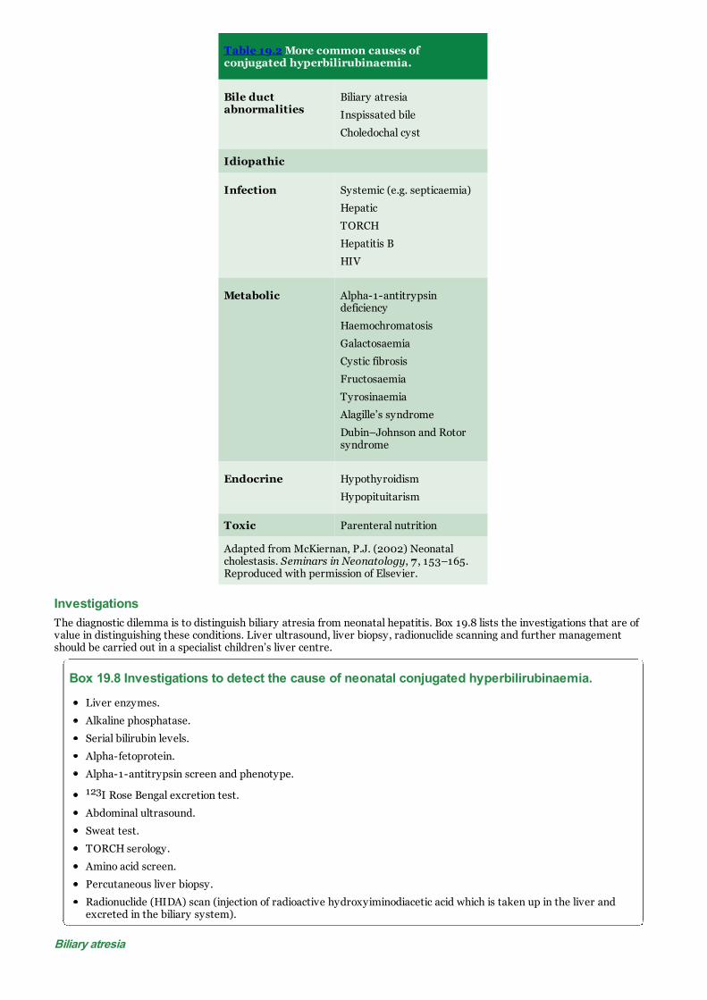
Table 19.2 More common causes ofconjugated hyperbilirubinaemia.
Bile ductabnormalities
Biliary atresia
Inspissated bile
Choledochal cyst
Idiopathic
Infection Systemic (e.g. septicaemia)
Hepatic
TORCH
Hepatitis B
HIV
Metabolic Alpha-1-antitrypsindeficiency
Haemochromatosis
Galactosaemia
Cystic fibrosis
Fructosaemia
Tyrosinaemia
Alagille’s syndrome
Dubin–Johnson and Rotorsyndrome
Endocrine Hypothyroidism
Hypopituitarism
Toxic Parenteral nutrition
Adapted from McKiernan, P.J. (2002) Neonatalcholestasis. Seminars in Neonatology, 7 , 153–165.Reproduced with permission of Elsevier.
InvestigationsThe diagnostic dilemma is to distinguish biliary atresia from neonatal hepatitis. Box 19.8 lists the investigations that are ofvalue in distinguishing these conditions. Liver ultrasound, liver biopsy, radionuclide scanning and further managementshould be carried out in a specialist children’s liver centre.
Box 19.8 Investigations to detect the cause of neonatal conjugated hyperbilirubinaemia.
Liver enzymes.
Alkaline phosphatase.
Serial bilirubin levels.
Alpha-fetoprotein.
Alpha-1-antitrypsin screen and phenotype.
123I Rose Bengal excretion test.
Abdominal ultrasound.
Sweat test.
TORCH serology.
Amino acid screen.
Percutaneous liver biopsy.
Radionuclide (HIDA) scan (injection of radioactive hydroxyiminodiacetic acid which is taken up in the liver andexcreted in the biliary system).
Biliary atresia

At birth, these infants have absent or atretic bile ductules involving the main bile ducts or the main branches of the bileducts. The deeper in the liver substance that the ducts are abnormal, the more severe is the condition. The commonestvariety is extrahepatic biliary atresia. The pathogenesis is unknown. In up to 30% of cases, biliary atresia may be associatedwith other malformations including renal anomalies, heart malformations, asplenia, polysplenia, malrotation and situsinversus. The onset of jaundice may be delayed by up to 4 weeks from birth. It is a surgically operable condition, butbecause progressive obliteration of the bile ducts occurs rapidly with advancing age, early diagnosis is essential for successfultreatment. If surgery is attempted before 60 days of age there is an 80% chance of achieving biliary drainage by theportoenterostomy operation, known as the Kasai procedure. Serum bilirubin falls rapidly after successful surgery, but manychildren develop ascending cholangitis, which is the most serious postoperative complication. Approximately two-thirds ofsurvivors will require liver transplantation by 10 years of age.
Neonatal hepatitisThis is a non-specific condition with a variety of causes, which are discussed below; the prognosis depends on the underlyingcause. In general, approximately one-third of cases deteriorate and develop hepatic cirrhosis, one-third have evidence ofchronic liver disease, and one-third recover fully. The prognosis for idiopathic neonatal hepatitis is good and more than 90%resolve by 1 year of age.
The main causes of neonatal hepatitis are listed in Box 19.9.
Box 19.9 Main causes of neonatal hepatitis.
Infection: most commonly due to TORCH infections contracted in the first trimester, but other viruses may alsoproduce hepatitis. If a mother is positive for hepatitis B, the infant should be protected from infection byimmunization (see Chapter 10).
Metabolic causes: fructosaemia and tyrosinaemia may cause severe neonatal hepatitis. Galactosaemia morecommonly presents with unconjugated hyperbilirubinaemia, but affected infants later develop cholestasis.
Alpha-1-antitrypsin deficiency: this is an autosomal recessive condition and causes a relatively common formof conjugated hyperbilirubinaemia. Only infants who are homozygous for the PiZZ type are at risk of neonatalhepatitis. There is no specific treatment, and up to 50% of affected children improve and may recover fully. Deathdue to liver failure occurs in the other half, and liver transplantation may be life-saving in this group.
Severe intrauterine growth restriction. Especially if associated with chronic fetal hypoxia, can result in abenign intrahepatic cholestasis.
Long-term TPN use (e.g. gastroschisis, short gut syndrome).
Chronic hypoxic–ischaemic damage to liver (i.e. recovery from multiorgan failure but liver failurepersists).
Neonatal haemochromatosis. This rare iron-storage disorder presents as acute liver failure in the fetal orneonatal period. Iron chelation, intravenous immunoglobulin and liver transplantation may improve the outcome.
Inspissated bile syndromeHigh and prolonged levels of unconjugated bilirubin may cause a condition in which the bilirubin produces cholestasis withprogressive conjugated hyperbilirubinaemia. Ultrasound scanning may show sludge in the gallbladder. It is usually a self-limiting condition, but ursodeoxycholic acid (UDCA) may be an effective therapeutic option.
Dubin–Johnson syndromeThis is a rare and benign condition in which the neonate may develop low-grade conjugated and unconjugatedhyperbilirubinaemia.
SUMMARY
Jaundice is the most common clinical condition presenting in the newborn period. In most infants the jaundice isphysiological as there is no underlying disease. Jaundice presenting in the first 24 hours may be due to haemolysis,and always requires investigation. Physiological jaundice, which commonly presents between day 2 and day 5 of life, isa diagnosis of exclusion. Unconjugated bilirubin is neurotoxic in high levels and can cause acute or chronicencephalopathy if not treated appropriately. Conjugated hyperbilirubinaemia is much less common than unconjugatedjaundice in the newborn, but has a much more serious prognosis.
Further reading1. Kramer, L.I. (1969) Advancement of dermal icterus in the jaundiced newborn. American Journal of Diseases of
Childhood, 1969; 118, 454–459.
2. Lauer, B.J., Spector, N.D. (2011) Hyperbilirubinemia in the Newborn. Pediatrics in Review Aug 2011, 32 (8), 341–349; DOI: 10.1542/pir.32-8-341.
3. Mills, J.F., Tudehope, D. (2001) Fibreoptic phototherapy for neonatal jaundice. Cochrane Database System Review2001;1:CD002060.
4. National Institute for Health and Clinical Excellence (NICE) Neonatal Jaundice 2016.http://guidance.nice.org.uk/CG98.

Now visit www.essentialneonatalmed.com to test yourself on this chapter.

CHAPTER 20 Haematological disordersKey topics
Placental transfusion
Anaemia
Hydrops fetalis
Aplasia
Polycythaemia
Bleeding and coagulation disorders
Thrombocytopenia
Haemorrhagic disease of the newborn �(Vitamin K-deficient bleeding)
Disseminated intravascular coagulation (DIC)
Inherited disorders of coagulation
Congenital deficiency of anticoagulant proteins �(hypercoagulable states)
IntroductionHaematological disorders in the fetus and newborn infant arise from the conditions which primarily affect blood constituentssuch as red blood cells (RBCs), haemoglobin and platelets. Some of the conditions are inherited and amenable to prenataldiagnosis or screening at birth. Symptoms in affected infants may start either in utero or after birth. Diagnosis of some ofthe haematological conditions may be complex, requiring interpretation by a paediatric haematologist. Treatment includesprevention and specific therapies. It should be borne in mind that the normal haematological indices in newborns aredifferent from those in older children and vary according to postnatal age.
Placental transfusionThe blood volume and red cell mass at birth and in the neonatal period depend on the volume of the placental transfusionand subsequent readjustments of blood volume.
This occurs within 3 minutes of delivery and can contribute up to 25% of the total neonatal blood volume. This amount willbe increased in the following situations:
Elevated maternal blood pressure.
Use of oxytocic drugs.
Late clamping or milking of the cord.
Infant held in a low, dependent position at birth.
On the other hand, the amount will be reduced by early cord clamping, or holding the infant above the level of the attachedplacenta.
The average blood volume of a newborn infant is 85–90 ml kg–1, but ranges from 75 to 100 ml kg–1. The practice of delay inclamping the umbilical cord or milking the cord from the placenta to the baby may have both advantages and disadvantages.Although this can result in improved blood volume and reduced iron deficiency in childhood, there may be associateddisadvantages too as an inadvertently high red cell mass can result in symptomatic pulmonary plethora andhyperbilirubinaemia. This subject of ‘early’ versus ‘late’ (i.e. physiological) clamping of umbilical cord is still a matter ofdebate. The UK Resuscitation Council recommends delaying cord clamping for 1 minute in uncompromised babies.
AnaemiaAnaemia is usually defined by a haemoglobin (or haematocrit) level and classed as mild if the haemoglobin level (Hb) is 10–12, moderate if it is between 8–10, and severe if it is less than 8 g dl–1. The causes of neonatal anaemia, as shown in Box20.1, broadly fall under the following headings:
Physiological
Haemorrhage
Haemolysis
Hypoplasia or aplasia (diminished production).

Box 20.1 Causes of neonatal anaemia.
Physiological anaemia.
Anaemia of prematurity.
Haemorrhage:
Antepartum haemorrhage
Fetomaternal transfusion
Twin-to-twin transfusion
Neonatal internal haemorrhage
Haemolysis.
Aplasia: Blackfan–Diamond syndrome.
Physiological anaemiaThe full-term infant is born with a haemoglobin concentration in the range 15 to 23.5 g dl–1. The measurement ofhaemoglobin is commonly reported as g dl–1, but in the UK and other countries haemoglobin is expressed as the SI unit ofg l–1. Hence, 23.5g dl–1 would be reported as 235 g l–1 and likewise, 15 g dl–1 as 150 g dl–1.
In the premature infant the haemoglobin level is slightly lower. Initially, there is a slight increase due tohaemoconcentration, but then haemoglobin gradually drops and remains low for most of the first year of life (Fig. 20.1). Thisis known as physiological anaemia.
Figure 20.1 Physiological anaemia. The two graphs show the normal fall in haemoglobin with postnatal age in mature andpremature infants.
Anaemia of prematurityIn the preterm infant physiological anaemia occurs earlier, is more severe and prolonged than in the term infant, and istermed anaemia of prematurity. It is caused by a number of factors.
Lack of erythropoietinAt birth, the infant moves from a relatively hypoxic fetal state to become relatively hyperoxic. This suppresseserythropoietin secretion for the first 7–8 weeks of life. In addition, the bone marrow is probably more resistant to thestimulatory effect of erythropoietin. When this ends, reticulocytes start appearing in the peripheral blood film.
Repeated blood samplingThe preterm infant is often subjected to daily repeated blood sampling for laboratory investigation. This is the commonestcause of anaemia in babies admitted to neonatal units.
Relative haemodilutionThere is an increase in plasma volume over the first months of life and, together with poor red cell production, thehaemoglobin falls. This is referred to as ‘early anaemia’.
Iron deficiencyThe full-term infant is born with sufficient iron stores for the first 4 months of life, but in preterm infants these stores arelow and exhausted more quickly because of the infant’s rapid growth rate. An infant of 1.5 kg at birth has half the iron storesof a 3.0 kg neonate. Iron deficiency mostly causes the ‘late anaemia’ that accounts for low haemoglobin levels after 4 monthsof age, characterized by hypochromic red cells seen on a blood film.

HaemolysisHaemolysis may occur in preterm infants as a result of vitamin E deficiency. As such, administration of vitamin E mayreduce the extent of late anaemia of prematurity but this is not used in practice.
TreatmentA daily dose of elemental iron is associated with a good response in most cases. A routine iron supplementation from an earlyage in premature babies to ‘prevent’ anaemia is now practised universally. There is a debate, however, as to how long thisprophylaxis should continue, and most units prescribe this until babies are established fully on solid diets.
Transfusion (10–20 ml kg–1) may be necessary in premature infants if the haemoglobin falls below 7–8 g dl–1 and the infantis symptomatic. Symptoms include breathlessness with feeds, tachycardia, apnoea and bradycardia or failure to gain weight.However, blood transfusion can also suppress erythropoietin activity, and hence if the infant shows a reticulocyte count ofmore than 5% transfusion should be delayed if possible, depending on the infant’s condition. Premature infants receivingintensive care do end up receiving repeated blood transfusions. In such situations, exposure to multiple blood donors can beavoided by using the same batch blood from a single donor and stored in mini packs. Donor blood should be screened forblood-borne viruses, particularly cytomegalovirus (CMV).
The following formula may be used to calculate the volume of blood to be transfused for an anaemic infant (Hct is thehaematocrit):
The administration of subcutaneous recombinant human erythropoietin to preterm infants has been shown to stimulate redblood cell production, thereby avoiding the need for frequent blood transfusions. However, this treatment has not beenshown to be cost-effective and is not widely used.
HaemorrhageCauses are listed in Box 20.2 and investigations in Box 20.3.
Box 20.2 Causes of haemorrhage.
Haemorrhage before and during delivery from:
Placental – placenta praevia, placental abruption, incision into the placenta during caesarean section.
Fetal–fetomaternal or twin-to-twin transfusion.
Cord – rupture or torn vessels on insertion into the placenta.
Neonatal haemorrhage:
Trauma – bleeding may occur into skull, brain, lung, peritoneum or bowel.
Haemorrhagic disease of the newborn.
Box 20.3 Investigations to determine the cause of haemorrhage.
These will depend on the provisional diagnosis and include:
Haemoglobin and haematocrit.
Kleihauer’s test (to assess the presence of fetal cells in maternal blood, indicating fetomaternal transfusion).
Coagulation studies (indicated if a bleeding diathesis is suspected).
Investigations to determine site of bleeding (e.g. ultrasound, CT or MRI of head or abdomen).
TreatmentAcute blood loss, if significant, is more likely to present with signs of hypovolaemia and shock as the initial manifestationsuch as changes in blood pressure, heart rate, tissue perfusion and urine output. In acute haemorrhage, the haemoglobinmay be normal in the beginning, as enough time has not passed for haemodilution to occur.
Severe haemorrhage may present as a neonatal emergency and require immediate transfusion with blood or bloodsubstitute (such as crystalloid or 4.5% albumin) to prevent irreversible shock. In an emergency, unmatched group Orhesus-negative blood may be used, but formal cross-matching should be done whenever possible. Remember babies rarelydie of anaemia; hypovolaemia is much more damaging.
HaemolysisThe causes of neonatal haemolysis are shown in Box 20.4. Haemolysis is usually associated with unconjugatedhyperbilirubinaemia and reticulocytosis. Causes of haemolytic anaemia can be broadly divided into two groups; immune andnon-immune.

Box 20.4 Causes of immune and non-immune neonatal haemolysis.Immune haemolysis (positive Coombs’ test):
Rhesus incompatibility.
ABO incompatibility.
Minor blood group incompatibility (e.g. Kell, Duffy, Kidd).
Maternal autoimmune diseases (e.g. SLE).
Non-immune haemolysis (negative Coombs’ test):
Congenital infection.
Disseminated intravascular coagulation (DIC).
G6PD deficiency.
Pyruvate kinase deficiency.
Hereditary spherocytosis (HS).
α-Thalassaemia.
Infantile pyknocytosis.
Vitamin E deficiency.
Rhesus haemolytic diseaseThis occurs because the mother’s immune system has been sensitized by rhesus-positive cells from her fetus. Sensitizationmay be due to:
fetomaternal transfusion (during previous delivery or from miscarriage); and/or
rhesus-incompatible transfusions.
The rhesus factor is complex, comprising CDE/cde antigens. The commonest antigen is D, and this accounts for 95% of cases.Approximately 83% of the population are D positive, that is, rhesus positive (Rh +ve). If sensitization occurs, maternalimmunoglobulin G (IgG) crosses the placenta to cause haemolysis of ‘foreign’ fetal erythrocytes. IgG remains present in theneonatal circulation for up to 3 months, and neonatal haemolysis may continue to occur for some weeks after birth.
PreventionRh IgG prophylaxis (anti-D gammaglobulin) is indicated in the management of all non-immunized pregnant women who areRh negative. Current recommendations include the routine administration of IgG at 28 weeks’ gestation to all pregnantwomen who are RhD negative, and within 72 hours of delivery to all rhesus-negative women who give birth to rhesus-positive infants. If antibodies are already present anti-D is not required. The standard dose of 300 µg is sufficient forprotection for up to 30 ml of fetal blood. This provides satisfactory prophylaxis for 99% of all term deliveries.
Anti-D gammaglobulin should also be given to at-risk rhesus-negative women after every sensitizing event, such asmiscarriage, termination of pregnancy or amniocentesis.
Anti-D gammaglobulin is ineffective against non-D rhesus antigen (usually C, E). If a large transfusion of fetal blood occurs,the standard dose of 300 µg kg–1 may be insufficient. In women at high risk, a Kleihauer–Berke smear test can beperformed to quantitate for the fetal blood present. For every 30 ml of fetal blood detected, an additional 300 µg of IgG canbe administered.
Management during pregnancyThis includes routine testing for antibodies, monitoring of antibodies, assessment of bilirubin in amniotic fluid, monitoring offetal well-being and monitoring for signs of fetal complications.
Routine testing. Rhesus-negative women should be screened for rhesus antibodies at their first antenatal visit, and atsubsequent visits according to local guidelines. If antibodies are detected at any of these times, more frequent testing willbe necessary.
Antibodies present. If antibodies are present, then depending on the level and/or whether the titre is rising, anamniocentesis should be performed. Amniocentesis is commonly done at 30–32 weeks, but may be performed earlierdepending on the level of antibodies, and particularly on the history of previous pregnancies. A Kleihauer test is donebefore and after amniocentesis. The timing of delivery will depend on the antibody levels, amniocentesis result andprevious history of affected infants.
Assessment of bilirubin in liquor. Fetal rhesus disease is now a relatively rare condition, and affected pregnanciesshould be managed in recognized regional centres experienced in treating these women and their babies.
Assessment of fetal well-being. High-resolution ultrasound is an essential adjunct to the assessment ofisoimmunized pregnancies. It allows early detection of hydrops, assessment of fetal behaviour (biophysical profile, whichprovides important evidence of fetal well-being), and interventions such as fetal blood samplings and transfusion.Ultrasound is used to assess the amniotic fluid index, hepatomegaly, subcutaneous oedema, ascites and increases inmiddle cerebral artery (MCA) blood flow velocity which goes up as anaemia increases.
Management of the rhesus-immunized infantThe baby should be assessed for maturity, pallor, jaundice, hepatosplenomegaly, oedema, ascites, bruising, heart failure and

respiratory distress. The placenta is examined for the presence of oedema, weighed and sent to the pathology departmentfor confirmation of the diagnosis.
Investigations at birth. Cord blood is taken for grouping, direct Coombs’ test, haemoglobin and platelet count and totalbilirubin estimation. The Coombs’ test is always positive in rhesus incompatibility, unless an intrauterine transfusion withrhesus-negative blood has recently been performed. The more positive the Coombs’ test, the more severely affected theinfant is likely to be.
There are two treatment options: (i) exchange transfusion, which aims to remove the causative IgG antibodies from thebaby circulation; or (ii) administration of immunoglobulin, which blocks the IgG-binding sites on the fetal red cells. These aredescribed in Chapter 19 (Jaundice).
Interval exchange transfusion (see Box 20.5) is done to prevent the serum unconjugated bilirubin reaching apotentially dangerous level (e.g. 450 μmol l–1 in >38 weeks and 350 μmol l–1 at 35 weeks). This is usually carried out as anadjunct to phototherapy, and guideline graphs are useful (see Chapter 19). It is performed far less frequently thanhistorically, due to maternal anti-D prophylaxis, in-utero transfusion of severe cases and better phototherapy technology(see Chapter 19).
Box 20.5 Indications for exchange transfusion.Principal indications for immediate exchange transfusion:
Cord haemoglobin <8 g dl–1 (80 g l–1) (recent in-utero transfusion may lead to falsely reassuring Hb).
Hydrops fetalis.
Indications for early exchange transfusion:
Cord bilirubin >85 µmol l–1 (5 mg per 100 ml).
Cord haemoglobin 8–12 g dl–1 (80–120 g l–1).
Rapidly rising serum bilirubin that crosses the level for exchange transfusion on the charts (see Chapter 19).
A very strongly positive Coombs’ test.
ABO incompatibilityHaemolytic disease caused by ABO incompatibility is now the commonest cause of isoimmune haemolytic anaemia, but isgenerally less severe than that caused by rhesus incompatibility (Box 20.6).
Box 20.6 Complications of rhesus or severe ABO incompatibility.
The complications include:
Kernicterus and bilirubin encephalopathy (see Chapter 19).
Hyaline membrane disease.
Beta-cell hyperplasia of the pancreas, resulting in hypoglycaemia.
Hypoalbuminaemia and lung oedema.
Thrombocytopenia and DIC.
Complications of exchange transfusion (see Chapter 19).
Anaemia. This results from ongoing haemolysis and require monitoring by checking haemoglobin levels andreticulocyte counts. Treatment includes folic acid administration and ‘top-up’ transfusions. Iron therapy is notnecessary unless the infant is born prematurely.
The naturally occurring anti-A or anti-B antibody is of the IgM type, which does not cross the placenta. Approximately 10%of women carry ‘immune’ anti-A or anti-B antibodies of the IgG class. It is in the pregnancies of these women that ABOincompatibility occurs, as the IgG crosses the placenta to haemolyse fetal red cells. Women with blood group O are mostlikely to have anti-A and anti-B IgG agglutinins, and it is this maternal blood group that accounts for the vast majority ofABO incompatibility. This may occur in the first pregnancy, and subsequent pregnancies may be relatively unaffected.
ABO incompatibility generally occurs with the blood group combinations listed in Table 20.1.
Table 20.1 Blood group combinations causing ABO incompatibility.
Mother Infant Frequency
O A or B Common
A B or AB Rare
B A or AB Rare
Clinical featuresThe usual presentation is with jaundice on the first day or two of life. Kernicterus is an unusual complication, and hydrops

fetalis has only occasionally been reported. Unlike rhesus disease, late anaemia is seldom a problem but folic acid isrecommended because of ongoing haemolysis.
InvestigationsABO incompatibility is usually suspected in the presence of maternal blood group O and when the infant is either bloodgroup A or, less commonly, group B.
The direct Coombs’ test on the infant’s blood is usually negative or only weakly positive, but the indirect Coombs’ testmay be positive.
A blood smear from the infant may show features of haemolysis, often with microspherocytes. Spherocytes are rarelyseen in rhesus disease.
If the mother’s serum causes haemolysis of adult A or B cells, this strongly suggests that she carries α or β haemolysins.Maternal blood should be examined for haemolysins.
Immune anti-A or anti-B may be eluted from fetal RBCs or cord blood.
TreatmentThis is as for rhesus haemolytic disease, but intrauterine fetal transfusion is much less likely to be required.
Minor blood group incompatibilitiesRarely, blood group incompatibilities are caused by Duffy, Kell, Kidd and C and E antibodies. They usually present with mildjaundice, but hydrops fetalis has been reported.
Glucose-6-phosphate dehydrogenase deficiencyThis disease is inherited in an X-linked recessive manner and is due to a deficiency of the enzyme glucose-6-phosphatedehydrogenase (G6PD) within the RBCs, thereby rendering the cells more susceptible to haemolysis. More than 100 millionpeople throughout the world, mainly Chinese, southern Mediterranean, black American or black African, have thisabnormality. It usually occurs in males, although the heterozygote female may manifest mild features of the disease. Thereare many variants of this condition, some requiring an oxidizing agent to trigger haemolysis and others that causehaemolysis spontaneously. Some infants with the enzyme deficiency develop jaundice in the newborn period withoutexposure to oxidant drugs, but in other variants of the condition oxidant drugs are required to trigger haemolysis. In lateryears otherwise healthy children may become acutely ill with anaemia when exposed to drugs.
Box 20.7 lists drugs that commonly cause haemolysis in susceptible neonates. Some drugs excreted in breast milk are liableto haemolyse red cells in G6PD-deficient infants, including nitrofurantoin, sulfonamides and sulfasalazine. In addition,respiratory viruses, viral hepatitis and fava beans cause haemolysis in susceptible infants. Routine administration of vitaminK to G6PD-deficient infants is safe.
Box 20.7 Drugs that may cause haemolysis in infants with G6PD deficiency.
Antimalarials (primaquine, quinine).
Nitrofurantoin.
Sulfonamides.
Phenacetin.
Aspirin (acetylsalicylic acid).
Nalidixic acid.
Methylene blue.
Naphthalene.
Vitamin K (large doses).
Chloramphenicol.
Clinical featuresHaemolysis may occur spontaneously or after exposure to infection or drugs. Jaundice and pallor may be the only clinicalsigns. It may be severe and require exchange transfusion. Hepatosplenomegaly is uncommon.
InvestigationsAnaemia with spherocytosis, reticulocytes and crenated red cells is seen. Heinz bodies are another feature of the haemolyticanaemia. A screening test for G6PD deficiency is available and is reliable in infants of Chinese and Mediterranean extraction.In black infants, once haemolysis has occurred, a population of young red cells may remain with normal enzyme activity, andthis makes the screening test unreliable. Black infants and those positive on the screening test should have the enzyme leveldirectly assayed.
TreatmentInfants born into families known to have G6PD deficiency should not be exposed to agents likely to cause haemolysis.Spontaneous haemolysis may occur, and the treatment is as for any cause of unconjugated hyperbilirubinaemia. Anaemiamay require transfusion.
Pyruvate kinase deficiency

This is an autosomal recessive condition affecting glucose metabolism within the red cell membrane. Spontaneoushaemolysis occurs in the neonatal period with anaemia and jaundice. Splenomegaly is always present. Diagnosis is made byenzyme assay.
Hereditary spherocytosisHereditary spherocytosis (HS) is an autosomal dominant condition that may cause neonatal haemolysis. The blood filmshows spherocytes with little splenomegaly initially. ABO incompatibility may present in a similar way, and a family historyof HS is an important diagnostic point. The spherocytes show an increased osmotic fragility, although this may not beapparent in the first few months of life. Severe haemolysis with very high levels of hyperbilirubinaemia may occur suddenly.
ThalassaemiaClassic β-thalassaemia major does not affect neonates because most of the haemoglobin is in the fetal (HbF) form. α-Thalassaemia is a rare and severe condition that causes hydrops and severe anaemia.
Infantile pyknocytosisThis is a rare cause of neonatal haemolysis, and is diagnosed by finding large numbers of small distorted ‘pyknocytes’ in theperipheral blood film. The greater the numbers of pyknocytes, the greater the tendency to haemolysis. This condition maybe due to vitamin E deficiency and is self-limiting.
Hydrops fetalisThis term is used to describe an infant who shows severe and generalized oedema and fluid in at least two visceral cavities(pleural effusions, ascites, pericardial effusions), irrespective of the cause.
CausesThere are many causes of hydrops fetalis which can be broadly categorized as immune and non-immune (Table 20.2).
Table 20.2 Causes of hydrops fetalis.
Immune Non-immune
Severe haemolytic disease of thenewborn (rhesus, ABO, minor bloodgroups).
Severe chronic anaemia in utero: fetomaternal haemorrhage, homozygousα-thalassaemia, chronic fetomaternal transfusion, twin-to-twin transfusion Cardiac failure: severe congenital heart disease, large atrioventricularmalformation (haemangioma), fetal supraventricular tachycardia Hypoproteinaemia: renal disease congenital nephrosis, renal veinthrombosis, congenital hepatitis Intrauterine infections: syphilis, toxoplasmosis, CMV, parvovirus B19a Congenital malformations: cystic adenomatoid malformation of the lung,obstructive uropathy, pulmonary lymphangiectasia Genetic abnormality: trisomy 21, 18 and 13; Turner’s syndrome (45XO);triploidy; Noonan’s syndrome Idiopathicb
aThis usually causes hydrops secondary to severe anaemia.
bThis may account for up to 50% of cases where no obvious cause is found after extensive investigations.
InvestigationsInvestigations are done to determine the cause of the hydrops fetalis and include:
Coombs’ test and full blood count.
Haemoglobin electrophoresis.
Maternal Kleihauer test.
TORCH/syphilis/parvovirus serology.
Placental examination.
Chest radiography.
Detailed ultrasound examination of chest and abdomen.
Echocardiography.
Total serum proteins and serum albumin.
Chromosomal analysis.
Dysmorphology evaluation.
Autopsy.
Treatment

This may include paracentesis of the abdomen and chest, transfusion, intubation and positive-pressure ventilation, plusdiuretic and intravenous albumin therapy. Despite intensive treatment, prognosis for non-immune hydrops remains poor,particularly if there are structural defects or its cause is not known, compared with cases caused by isoimmunization.
AplasiaImpaired RBC production is an unusual cause of anaemia in the newborn. The most common cause is the Diamond–Blackfansyndrome, also known as congenital hypoplastic anaemia.
PolycythaemiaPolycythaemia in the newborn is common and is defined as a venous haematocrit of 65% or more, approximating to ahaemoglobin of 22 g dl–1 (220 g l–1), during the first week of life. Polycythaemia does not mean that the blood ishyperviscous. Blood viscosity depends largely on the packed cell volume (haematocrit), but the deformability of RBCs andthe plasma viscosity may also be significant factors. The relationship between viscosity and haematocrit is linear below ahaematocrit of 60–65%, but increases exponentially above this level. Viscosity is much greater in small vessels than in largevessels.
Polycythaemia should only be diagnosed on a free-flowing venous specimen and not from a heel-prick sample.
The causes of polycythaemia are listed in Box 20.8.
Box 20.8 Causes of neonatal polycythaemia.
Chronic intrauterine hypoxia:
SGA infants
Postmaturity
Excessive transfusion of blood:
Placental transfusion due to delayed clamping
Twin-to-twin transfusion
Maternofetal transfusion
Infants of diabetic mothers.
Down’s syndrome.
Neonatal thyrotoxicosis.
Congenital adrenal hyperplasia.
Beckwith–Wiedemann syndrome.
Clinical featuresThe infant looks plethoric, and polycythaemia may cause problems in a number of organ systems owing to diminished bloodflow through small vessels. The clinical signs associated with polycythaemia/hyperviscosity are illustrated in Figure 20.2.
Figure 20.2 The interrelationship between polycythaemia and hyperviscosity and their contribution towards clinical signs.CNS, central nervous system; GFR, glomerular filtration rate.

ManagementInfants at risk of polycythaemia should have a haematocrit measured on free-flowing venous blood. Babies with ahaematocrit greater than 75%, even if asymptomatic, should have a dilutional exchange transfusion. When symptoms arepresent dilutional exchange should be considered even at a lower haematocrit level (>65–70%).
A dilutional exchange (which should not be confused with exchange transfusion as in rhesus disease) is performed with salineor plasma and is aimed at reducing the haematocrit (Hct) to about 50% by the following formula:
where 90 refers to the blood volume (in ml) per kg. The procedure involves slowly removing the desired volume of blood (inaliquots over 60 minutes) via a UVC or UAC, or peripheral arterial line, whilst infusing the same volume of normal saline orhuman albumin solution.
Bleeding and coagulation disordersThese may be due to thrombocytopenia, clotting factor deficiency, abnormal capillaries, or a combination of these.Coagulation is a complicated process and is relatively less efficient in the newborn (particularly premature infants) than inolder children.
When the vascular endothelium is damaged, specific factors are released that cause platelet aggregation, upon whichthrombus is deposited. This induces fibrin formation, which further induces platelets to be deposited, leading to the creationof a platelet–fibrin syncytium that prevents further bleeding. Thrombin is formed from prothrombin by the action of factorX. Factor X may be activated by either the intrinsic or the extrinsic pathway to precipitate a cascade of clotting factors,culminating in the production of fibrin. Plasminogens act to remove fibrin (fibrinolysis), and in the healthy state this systemis balanced with the clotting mechanism by a series of inhibitors.
Clinical featuresBleeding may be overt, from venepuncture sites or the umbilical stump. Bruising (ecchymoses), purpura or petechialhaemorrhages may be present at birth or develop during the neonatal period.
A careful maternal history should be taken, including a family history of bleeding, drugs (warfarin, aspirin), idiopathicthrombocytopenic purpura (ITP) and recent viral illness.
Neonatal examination includes:
Site of bleeding: examination will determine the origin of bleeding, such as the gastrointestinal tract, umbilicus orcircumcision.
Presence of purpura suggesting thrombocytopenia.
Hepatosplenomegaly suggesting possible: infection (TORCH, bacterial).
Presence of other congenital anomaly, e.g. giant haemangioma, thrombocytopenia with absent radii (TAR) syndrome.
Investigations
Platelet count
A platelet count less than 100 × 109 mm–3 is usually classified as thrombocytopenia. A useful guide to severity is:
50 000–100 000: mild thrombocytopenia (bleeding with surgery).
25 000–50 000: moderate thrombocytopenia (bleeding with minor trauma).
<25 000: severe thrombocytopenia (spontaneous bleeding is likely).
Infants with long-standing thrombocytopenia may have no spontaneous bruising, even with platelet concentrations as low as10 000 mm–3.
Coagulation profile
Bleeding time. This measures the time to stop bleeding after a standard small wound, as from an Autolet device. Theupper limit of normal for this test in the neonate is 3.5 minutes. Prolonged bleeding time is seen in thrombocytopenia,von Willebrand’s disease and disseminated intravascular coagulation (DIC).
Prothrombin time (PT). This assesses the extrinsic clotting pathway (factors II, V, VII and X). It is not markedlyaffected by heparin.
Partial thromboplastin time (APTT). This assesses the intrinsic clotting pathway (most factors except VII andXII), but is prolonged by heparin contamination. It is the most sensitive test for coagulation disturbances.
Thrombin clotting time. This assesses fibrinogen activity and requires calcium for activation.
Fibrin degradation products (FDPs). Fibrin is deposited during coagulation and is simultaneously degraded byplasminogens to FDPs. The presence of increased levels of FDPs or elevated D-dimer indicates that fibrinolysis isoccurring, usually following thrombosis or as part of DIC.
Clotting factor analysis. Specific factors can be assayed individually, but interpretation may be difficult because ofuncertainty as to the normal range in very immature infants.

The normal ranges for some of these tests are shown in Table 20.3.
Table 20.3 Normal results for some of themore commonly used tests of coagulation.
Test Preterm (30–36 weeks)
Term
PT (s) (I, II, V, VII, X) 13–23 13–17
Thrombotest (TT;%) (II,VII, IX, X)
15–50 15–60
APTT (s) (I, II, V, VII,IX, X, XI, XII)
35–100 35–70
Thrombin time (s) 12–24 12–18
Reptilase time (s) 18–30 18–24
Fibrinogen concentration(g l–1)
1.2–3.8 1.5–3.5
ThrombocytopeniaPetechiae and ecchymoses, which may be present at birth or appear after birth, are the characteristic lesions produced byplatelet deficiencies. Whereas bleeding may occur from any site, intracranial haemorrhage is the most devastatingcomplication. Box 20.9 lists the causes of neonatal thrombocytopenia.
Box 20.9 Causes of neonatal thrombocytopenia.
Infection:
Any bacterial infection
TORCH infections
Isoimmune
Maternal disease:
Severe toxaemia of pregnancy
Idiopathic thrombocytopenic purpura (ITP)
Systemic lupus erythematosus
Drug-induced (hydralazine, thiazides)
Neonatal drug exposure:
Thiazide diuretics
Quinine
Sulphonamides
DIC
Thrombocytopenia with absent radii (TAR syndrome)
Giant haemangioma (Kasabach–Merritt syndrome)
Fanconi’s anaemia
Leukaemia
Pancytopenias
Alloimmune thrombocytopeniaThis is analogous to rhesus isoimmunization but is a much rarer condition. A transfusion of fetal A1 antigen-positive plateletsinto the maternal circulation may produce maternal IgG antibodies if the mother is platelet A1 antigen-negative. Themother has a normal numbers of platelets. Thrombocytopenia in the neonate may be severe, but is usually transient. Thedisease presents with intracranial haemorrhage (often fatal) in 20%. In-utero treatment consists of fetal platelet transfusionand regular gammaglobulin therapy. Neonatal treatment is valuable, using platelet transfusions, gammaglobulin andsteroids.
Maternal idiopathic thrombocytopeniaTransplacental maternal antibodies cause thrombocytopenia in the neonate, and the mother will usually havethrombocytopenia. The lower the concentration of maternal platelets, the more severely affected the infant may be.Prenatal administration of corticosteroids or high-dose intravenous IgG (IVIG) has been advocated to reduce the incidenceand severity of neonatal thrombocytopenia, but the response to these treatments is uncertain and unpredictable. The use of

IVIG in the thrombocytopenic neonate may cause the platelet count to increase rapidly.
It has been suggested that delivery by caesarean section should be undertaken in severely thrombocytopenic fetuses toavoid trauma, but recent evidence suggests that intracerebral bleeds may occur even before the onset of labour.
Prednisolone may be given to the severely affected neonate, but the condition is transient, lasting at most 12 weeks.Treatment of severe thrombocytopenia includes platelet transfusion (10 ml kg–1). Serious neonatal haemorrhage does notoccur if the platelet count is above 50 000 mm–3.
Haemorrhagic disease of the newborn (Vitamin K-deficient bleeding)Classic haemorrhagic disease of the newborn is caused by a deficiency of the vitamin K-dependent clotting factors. Itsincidence has reduced considerably since routine supplementation with vitamin K [intramuscular (IM) or by mouth] at birthbecame mandatory. About 2 in 1000 babies who do not receive vitamin K will have some bleeding, and 1 in 10 000 will haveserious intra-cerebral bleeding. Vitamin K is produced by the bacterial flora of the gastrointestinal tract, but as the newborninfant has a sterile bowel at birth there is little production from this source in the first weeks of life.
Clinical featuresSpontaneous bleeding can occur from any site but is usually gastrointestinal (producing haematemesis or melaena),umbilical, or is associated with circumcision. It occurs late in the first week of life, especially in the breastfed infant owing tothe low vitamin K levels in human milk, and nowadays almost only occurs if vitamin K prophylaxis has been missed ordeclined.
Gastrointestinal bleeding in the infant must be differentiated from swallowed maternal blood from antepartumhaemorrhage, episiotomy, or cracked nipples.
The Apt test, which depends on the resistance of haemoglobin F (fetal red cells) to denaturation by sodium hydroxide, washistorically used to distinguish the infant’s blood (predominantly fetal) from maternal blood (adult blood). Most centres willnow perform Hb electrophoresis to identify if it is predominantly fetal or adult haemoglobin to identify the source ofbleeding.
InvestigationsThe diagnosis is confirmed by a prolonged PT but a normal APTT.
TreatmentVitamin K (IM or IV) is given after blood has been obtained for investigations. Whole-blood transfusion (20–30 ml kg–1) willbe indicated for hypovolaemic shock.
Vitamin K prophylaxisRoutine administration of intramuscular vitamin K to all newborn babies will prevent bleeding from vitamin K deficiency.However, concerns about the safety of intramuscular vitamin K, in particular the risk of cancer, were raised in the early1990s, and although there are few data to support this some countries have recommended oral administration of vitamin Kin all healthy full-term infants.
Konakion MM paediatric drops are licensed in the UK for use in healthy neonates of 36 weeks’ gestation and older. Therecommended dosage regimen is a dose orally shortly after birth, a further dose at 4–7 days and, if the baby is exclusivelybreastfed, a third dose should be given at 1 month of age. Failure to give a complete dosage regimen appears to be thereason for the re-emergence of serious late-onset vitamin K deficiency haemorrhage. In rare situations where haemorrhagictendency still persists after administration of vitamin K, one should exclude the possibility of cystic fibrosis and α1-antitrypsin deficiency.
Disseminated intravascular coagulation (DIC)DIC is an acquired coagulation disorder characterized by the intravascular consumption of platelets and clotting factors II, V,VIII and fibrinogen. Widespread intravascular coagulation results from the deposition of thrombi in small vessels and theconsumption of clotting factors, with consequent haemorrhage. DIC is recognized as a complication of an increasing variety ofneonatal conditions (see Box 20.10).
Box 20.10 Neonatal conditions of which DIC is a complication.
Septicaemia.
Severe shock.
Severe perinatal asphyxia.
Hyaline membrane disease in very-low-birthweight (VLBW) infants.
Severe rhesus disease.
TORCH infections.
Hypothermia.
Maternal DIC with a transplacental effect; this occurs secondary to antepartum haemorrhage, a dead twin fetus oramniotic fluid embolism.
InvestigationsBlood film showing haemolysis with fragmented and distorted red cells.
Thrombocytopenia.
Prolonged PT, PTT and thrombin time.
Low fibrinogen.
Increased FDPs.
Not all these features are necessary to make the diagnosis, but the presence of three or more makes DIC very likely.
TreatmentThis is a complex disorder and haematological consultation will often be necessary. Treatment consists of:
Treating the underlying disease process.
Treatment of the haematological abnormality, including exchange transfusion with fresh whole blood and/or replacementof clotting factors with fresh frozen plasma, platelet concentrates and cryoprecipitate. Heparinization is unlikely to be ofany benefit.
Inherited disorders of coagulationThese include haemophilia, Christmas disease and von Willebrand’s disease. Coagulation factors are not transferred from thematernal circulation to the fetus. Severe forms of haemophilia A (factor VIII deficiency) and Christmas disease (factor IXdeficiency, sometimes called haemophilia B) account for the majority of haemorrhagic problems of the newborn caused bycongenital coagulation abnormalities. Bleeding in these X-linked recessive diseases occurs when male infants are subjected tosurgical procedures such as circumcision, or from either birth trauma or routine sampling of capillary blood. The diagnosis isconfirmed by a prolonged APTT, normal PT and decreased factor VIII assay. Factor XI deficiency (haemophilia C) isinherited as an autosomal recessive disease and is not usually diagnosed in the neonatal period. Excessive bleeding typicallyoccurs in the post-traumatic or postoperative settings such as after circumcision. Factor XI deficiency has been described asa common finding in Noonan’s syndrome.
von Willebrand’s disease does not usually present in the neonatal period. However, because von Willebrand factor(vWF) serves as a carrier for factor VIII, those who are symptomatic in early infancy often have signs and symptomsassociated with low factor VIII. Laboratory testing requires determination of vWF antigen and vWF activity and a plateletfunction test in addition to ascertaining factor VIII activity.
ManagementA specific diagnosis may be difficult to make at birth because, in the healthy infant, many of the clotting factor assays arelow. Expert advice should be sought. Treatment is only required in the neonatal period if there is active bleeding or the babyis extremely preterm and at risk of intraventricular haemorrhage (IVH), otherwise anti-factor VIII antibodies (inhibitors)may develop.
Congenital deficiency of anticoagulant proteins (hypercoagulable states)The anticoagulant proteins, protein C, protein S, antithrombin III and heparin cofactor II, are all inherited in an autosomaldominant manner. Homozygous protein C or protein S deficiency causes serious thrombotic events in the postnatal period.The parents, who are usually asymptomatic, are heterozygous for deficiency of the suspected protein. Factor V Leiden, amutation in coagulation factor V that renders it resistant to cleavage by activated protein C, is now the most commonabnormality found in patients with excessive venous thrombosis, such as in cases of neonatal stroke or peripheral ischaemia.Such patients may require anticoagulation such as with low-molecular-weight heparin, especially if they develop life/limb-threatening thrombosis.
LeukocytosisThe normal newborn can have a high white cell count (WCC) ranging from 9 to 30 × 109 l–1, but this usually settles within 2weeks. The neutrophil count typically rises for the first 12–24 hours and then falls.
Causes of neutrophilia include:
Acute bacterial infection.
Maternal chorioamnionitis.
Fungal infection.
Corticosteroid therapy (for BPD).
A left-shift towards higher percentage of immature neutrophils is highly suggestive of infection (see Chapter 10).
Neonatal leukaemiaA very high WCC, with the presence of abnormal blast cells, is suggestive of neonatal leukaemia. However, a blood filmshould be analysed manually because some automated cell counting machines can confuse nucleated RBCs for WCCs. Sincenewborns often have many nucleated RBCs this can lead to a falsely high WCC count.
Neonatal leukaemia is very rare, and unlike in older children, acute myeloid leukaemia (AML) is more common than acutelymphocystic leukaemia (ALL). It often presents with hepatosplenomegaly and cutaneous manifestations (leukaemic skindeposits). It is treated with chemotherapy, but the prognosis is often poor.
Neonatal leukaemia of Down syndrome (NLDS) is an often self-limiting leukaemic picture, with leukocytosis,

thrombocytosis and blast cells and a characteristic skin rash that resembles a staphylococcal infection. NLDS used to bereferred to as transient myeloproliferative disorder (because it usually resolves spontaneously after a few months), but iflife-threatening signs are present it may require treatment with one to five doses of chemotherapy. There is a 33% chanceof later AML in these children.
NeutropeniaNeutropenia is usually defined as a neutrophil count <1.5 × 10⁹ l–1. The most common causes are:
Bacterial sepsis.
NEC.
Congenital viral infection such as CMV.
IUGR, including maternal pre-eclampsia.
Trisomy 21, 13 or 18.
Familial (more common in Afro-Caribbean babies).
Most neutropenia responds to treating the underlying cause. Granulocyte colony-stimulating factor (G-CSF) will increasethe neutrophil count but has not been proven in clinical trials to be of any benefit.
SUMMARY
Several common haematological disorders are known to affect newborn infants. Anaemia can result from a lack ofRBCs and haemoglobin due to diminished production (viz. iron-deficiency anaemia) or excessive destruction(haemolytic anaemias). Excessive blood loss either in utero (such as feto-maternal or twin-to-twin transfusion) and atbirth through accidental bleeding from umbilical cord or trauma to the organs such as liver or skull, is anotherimportant cause for anaemia in newborn infant, and should be suspected even though it is not externally obvious.Various haemoglobinopathies that result from a molecular abnormality of haemoglobulin structure result in conditionssuch as thalassaemias and sickle cell anaemia. Genetic disorders of the RBC membrane (such as congenitalspherocytosis) or RBC metabolism (G6PD deficiency) make the red cells fragile and cause haemolytic episodes.Disorders of platelets or coagulation factors (coagulopathies) resulting in abnormal haemostasis are common, and canbe either a primary defect or secondary to other conditions (consumption coagulopathy), such as in DIC. On the otherhand, a congenital deficiency of certain anticoagulant factors (such as protein-C or -S) may make the bloodhypercoagulable, which predisposes to thrombosis and blockage of important blood vessels such as in kidneys or brain(neonatal stroke). Newborn babies are deficient in vitamin K, which is an essential part of the coagulation process. Asvitamin K deficiency can lead to lethal haemorrhage, it is now global policy to supplement all babies, particularly thosefed on breast milk, with vitamin K. The regime may differ, however, from one unit to another.
Further reading1. Watchko, J.F. (2015) Common haematologic problems in the newborn nursery. Pediatric Clinics of North America, 62
(2), 509–524.
Now visit www.essentialneonatalmed.com to test yourself on this chapter.

CHAPTER 21 Endocrine and metabolic disordersKey topics
Glucose homeostasis and its abnormalities
Disorders of calcium, phosphate and magnesium metabolism
Disorders of magnesium metabolism
Disorders of sodium and potassium metabolism
Endocrine gland disorders
Abnormalities of the adrenal gland
Inborn errors of metabolism
IntroductionSome endocrine and metabolic disorders present acutely in the newborn period and can be life-threatening. They must beincluded in the differential diagnosis of any serious or unexplained illness. Prompt diagnosis and treatment is life-saving. Inother cases, the clinical presentation may not be so acute but if they are undiagnosed and left untreated they can causeserious problems such as growth failure, developmental delay and poor cognitive (intellectual) development. Neonatalscreening for conditions, which can have serious consequences and for which treatment is available, is undertaken in mostdeveloped countries. The investigation and treatment of endocrine and metabolic diseases can be complex and requiresmultidisciplinary specialist input. Some are readily and successfully treated, while for others specific treatment is notavailable. Babies with endocrine and metabolic problems require long-term treatment and follow-up by a specialist team.
Glucose homeostasis and its abnormalitiesThe fetus has a continuous supply of glucose from the mother via the placenta, and consequently fetal blood glucose levelsare the same as the mother’s. At birth the newborn’s blood glucose rapidly falls to approximately 75% of the maternal bloodglucose level and the infant has to switch rapidly to endogenous gluconeogenesis until feeding is established.
Glucose and oxygen are the main metabolic substrates of the mature brain, but in the neonate the brain can use alternativemetabolic fuels such as lactate and ketones. Birth at full term is characterized by a vigorous ketogenic response, but this isimpaired in preterm infants and infants who experience intrauterine growth restriction (IUGR). This is why the brain canfunction normally, or near normally, despite very low levels of blood glucose. Profound neurological compromise andirreversible damage occur if the brain is deprived of glucose and alternative metabolic substrates.
Glucose metabolismFigure 21.1 summarizes the main metabolic pathways involved in gluconeogenesis.
Glycogen production and glycogenolysis. These occur largely in the liver and muscles, but only if liver glycogen isavailable for rapid breakdown to glucose.
Gluconeogenesis. The most important substrates are amino acids (particularly alanine), lactate, pyruvate andglycerol. The points at which these are metabolized are shown in Figure 21.1.
Lipolysis. Glycerol is metabolized from adipose tissue and can be directly utilized in gluconeogenesis metabolism. Otherproducts of lipolysis (fatty acids and triglycerides) are metabolized to ketone bodies, which may be used directly inenergy production, particularly by the brain. Ketone body production is stimulated by infant feeding, particularly bybreast milk.

Figure 21.1 Metabolic pathways involved in gluconeogenesis.
These mechanisms are under the control of the endocrine system and are affected by insulin, glucagon, cortisol and growthhormone. Therefore, so that neonates can regulate blood sugar within the physiological range, they must be endowed withadequate liver glycogen, lipid stores and effective metabolic pathways, including glycogenolysis and gluconeogenesis, as wellas overall endocrine control. Hypoglycaemia will rapidly develop if any of these processes are disturbed.
Measurement of blood glucoseIt is essential to measure blood glucose rapidly and accurately in high-risk infants, and a variety of techniques have beendeveloped to give cotside values. Glucose concentrations in plasma or serum are 10–15% higher than in whole blood, andmany bedside techniques rely on whole-blood methods, whereas the laboratory techniques are more likely to measureserum glucose levels.
CLINICAL TIP
In the neonatal unit strip reagents are most widely used but these are generally unreliable, particularly in thepresence of low blood glucose, which is the range of most importance in the newborn. More recently, portablebiosensing devices for measuring glucose at or near the cotside have been introduced which appear to be morereliable. Usually, low levels of blood sugar recorded in the nursery should be confirmed by sending a specimen to thelaboratory for accurate blood glucose analysis, but these results may not be accurate depending on the method usedand the time interval between sampling and test.
HypoglycaemiaThe definition of hypoglycaemia has been gradually refined over the last 30 years, and higher values for blood sugar are nowaccepted as normal than was the case in the past. Hypoglycaemia is defined as a blood sugar concentration falling below apredetermined reference value, but this is not usually helpful in a clinical setting. A more important question is what thelowest acceptable level is in an individual baby before physiological compromise occurs. As discussed above, neonatalcerebral function may continue normally despite very low levels of blood sugar because an alternative energy substrate isavailable.
Attempts have been made to define the level of blood sugar at which cerebral dysfunction occurs, and then to set‘normoglycaemia’ levels above this. This is based on observational studies in which premature infants (birthweight <1850 g)with blood sugar levels less than 2.6 mmol l–1 were found to be at increased risk of lower neurodevelopmental scores,particularly when blood sugar values were below this figure on repeated occasions. It has also been shown that adeterioration in neurological function (measured by evoked potentials) occurred with a blood sugar level below 2.6 mmol l–1.On the basis of these studies we can define the lower limit of ‘normoglycaemia’ as 2.6 mmol l–1, although levels below this donot necessarily mean that damage will occur. However, we recommend maintaining the blood sugar of preterm infants andterm infants who are at risk of hypoglycaemia (see below) above 2.6 mmol l–1 (40 mg per 100 ml).
Who should be monitored for hypoglycaemia?The majority of babies who are prone to hypoglycaemia can be predicted on the basis of easily identifiable risk factors (Box21.1). Careful monitoring of these babies’ blood sugar will alert the clinician to the onset of hypoglycaemia and enablemanagement aimed to prevent symptomatic hypoglycaemia.

Box 21.1 At-risk infants for hypoglycaemia.
Infants of diabetic mothers.
Infants born small for gestational age (SGA).
Infants born prematurely.
Birth asphyxia.
On rewarming after hypothermia.
Infection.
Polycythaemia.
Beckwith–Wiedemann syndrome.
Infants born large for gestational age (LGA).
In babies at risk of hypoglycaemia, an early and frequent feeding regimen should be instituted with regular blood sugarmonitoring for the first 24 hours. ‘At-risk’ infants should be fed within 2 hours of birth. Breast feeding is recommended witha frequent administration of expressed breast milk. Formula and IV or buccal dextrose may be needed if early breast milk isinsufficient. A suitable regimen would be full-strength formula given 2-hourly, although if the mother is adamant that shewants to feed breast milk only, an intravenous 10% glucose infusion can be used. Small for gestational age (SGA) infantsoften tolerate up to 100 ml kg–1 on day 1 of life. If asymptomatic hypoglycaemia fails to be corrected by early, frequentfeeds, an infusion with 10% dextrose will be necessary.
Blood sugar estimates should be made prior to feeding, as this is the time when the blood sugar is likely to be at its lowestpoint and monitored at regular intervals (6-hourly) until feeding is established. In view of the unreliability of reagent striptests, it may be preferable to undertake more accurate measurement of blood sugar less frequently. More frequentmeasurements are required in the presence of actual hypoglycaemia. When blood sugar levels are satisfactory, thefrequency of heel-prick estimations is reduced before ceasing.
Symptoms of hypoglycaemiaThe symptoms of hypoglycaemia in the newborn can be divided into major and minor (see Box 21.2).
Box 21.2 Major and minor symptoms of hypoglycaemia.
Major: Apnoea, convulsions and coma. Rarely, prolonged hypoglycaemia may cause congestive heart failure orpersistent pulmonary hypertension.
Minor: Jitteriness, irritability, tremors, apathy, cyanotic spells and temperature instability.
It is not possible to confidently diagnose neonatal hypoglycaemia clinically as the symptoms are non-specific andsimilar to those of infection.
CLINICAL TIP
Hypoglycaemia may remain entirely without clinical signs or symptoms, and this is referred to as asymptomatichypoglycaemia. It is unusual for a newborn with hypoglycaemia to have a classic autonomic nervous system response,with sweating, pallor and tachycardia, as occurs in adults. A low blood sugar detected by a stick test should be checkedby a laboratory blood assay for glucose.
Causes of hypoglycaemiaThese can be considered under five major headings, which are summarized in Table 21.1.

Table 21.1 Causes of neonatal hypoglycaemia.
Decreased substrate availability SGA infants (see Chapter 12)
Premature infants
Multiple birth, especially if growth-retarded
Increased glucose utilization Hyperinsulinaemia
Infant of a diabetic mother
Rhesus isoimmunization
Nesidioblastosis (hypertrophy of pancreas)
Islet cell tumour
Beckwith–Wiedemann syndrome
Polycythaemia
Inability to utilize glucose Glycogen storage disease
Galactosaemia
Fructose intolerance
Inborn errors of metabolism
Iatrogenic Inappropriate infusion of glucose
Miscellaneous Birth asphyxia
Endocrine deficiencies (e.g. congenital adrenal hyperplasia)
Hypopituitarism, cortisol insufficiency, amino acid metabolism
ManagementThe major emphasis should be to prevent hypoglycaemia developing, and this is done by recognizing babies who are athigher risk of this condition and giving early and appropriate feeding (see above).
In babies with persistently low glucose levels despite feeding, an IV infusion of 10% dextrose at a rate of 80–100 ml kg–1
per 24 hours should be started. If the blood glucose estimate is still less than 2.6 mmol l–1 (40 mg per 100 ml), the dextroseconcentration may need to be increased to 15%. Up to 12.5% dextrose may be infused via a peripheral IV line, but if higherconcentrations are required a central IV line will be necessary. Where possible, oral feeding should be continued.
Resistant (or persistent) hypoglycaemiaRarely, the above regimen fails to control hypoglycaemia when hyperinsulinaemia should be suspected. Under thesecircumstances, other forms of treatment are necessary to control the hypoglycaemia including:
hydrocortisone;
glucagon;
diazoxide;
somatostatin analogue SMS-201-995 (useful in the short-term management of neonatal hyperinsulinism); and
laparotomy and subtotal pancreatectomy when an insulinoma is strongly suspected.
InvestigationsHypoglycaemia in most infants resolves spontaneously within a few days, but it is sometimes more severe or fails to resolverapidly. Hyperinsulinaemia is suspected clinically when a baby with severe non-ketotic hypoglycaemia requires a glucoseinfusion rate exceeding 10 mg kg–1 min–1 to maintain normoglycaemia. These babies have a very brisk response toglucagon injections, with the blood glucose increasing to >1.7 mmol l–1 above baseline. The definitive diagnosis ofhyperinsulinism is made by measurement of serum insulin (>10 mU ml–1) during an episode of hypoglycaemia(<2.6 mmol l–1). The plasma should be separated and frozen immediately after taking the sample if reliable results are to beobtained. A screen for inborn errors of metabolism may be necessary in some cases. A detailed ultrasound of abdomen andexploratory laparotomy may be necessary in infants suspected of having islet cell tumours. Inborn errors of metabolism orendocrine problems such as hypopituitarism may rarely cause hypoglycaemia, and if these are considered to be a possiblecause, appropriate investigations should be undertaken to diagnose or exclude these conditions.

CLINICAL TIP
Emergency management of hypoglycaemia
The standard management of hypoglycaemia has not changed over time:
Establish intravenous access.
Give 2 ml kg–1 of 10% glucose IV as bolus.
Continue IV infusion containing 10% glucose as maintenance, volume adjusted to maintain blood glucose level of 4–6 mmol l–1.
If IV glucose not available:
Buccal 40% dextrose gel; and/or
Intramuscular injection of glucagon.
PrognosisSevere symptomatic hypoglycaemia carries a very poor prognosis. Affected babies remain at risk of severeneurodevelopmental abnormalities, including cognitive delay, convulsions, generalized hypertonia and microcephaly. Theprevention of symptomatic hypoglycaemia is one of the most important factors in preventing brain damage in the whole ofneonatal medicine.
It is widely believed that infants with asymptomatic hypoglycaemia are not at risk of adverse neurodevelopmental outcome.However, some studies do suggest that neural dysfunction can occur with blood sugar levels below 2.6 mmol l–1, eventhough the baby may not show clinical symptoms. This argument is unresolved.
Specific causes of hypoglycaemia
Infants born to diabetic mothersMaternal diabetes is classified as follows:
Pregestational:
Type 1: the basic cause is beta-cell destruction.
Type 2: this is due to insulin resistance with an insulin secretory defect.
Gestational diabetes.
Infants of diabetic mothers (IDMs) have unique problems and require specialized neonatal care. The prognosis for thediabetic pregnancy depends on the severity of the diabetes and the quality of diabetic control during pregnancy.
The two main factors determining whether maternal diabetes will have an effect on the fetus and baby are the vascularcomplications that the diabetes causes in the mother, and the blood glucose control during pregnancy.
Vascular disease. Mothers with vascular complications as a result of diabetes are much more likely to develophypertension in pregnancy, which may affect fetal growth and well-being.
Glucose control. The outcome of pregnancy in diabetic women also depends on glucose control, both before conceptionand during gestation. Diabetic women should have their diabetes very carefully managed before conception, andcombined care through pregnancy by a physician and obstetrician is essential. The blood sugar should be maintainedbelow 8 mmol l–1 with soluble insulin if necessary, and hypoglycaemia avoided. On this regimen the complications for thefetus are reduced and may be avoided completely. A variety of congenital malformations are particularly common inwomen with diabetes, and the risk appears to depend on the mother’s prepregnancy blood sugar levels.
Clinical features of IDMsComplications to the fetus are likely to occur in diabetic women in whom glucose control has been less than adequate. Thefrequencies of complications in IDM and in gestational diabetic mothers are given in Table 21.2. Complications include:

Table 21.2 Frequency of complications(percentages) in infants of diabetic mothers(IDMs) and infants of gestational diabeticmothers (IGDMs). Complications are relatedto the quality of glucose control inpregnancy.
Complications IDMs(%)
IGDMs(%)
Uneventful course 50 80
RDS 30 10
Hypoglycaemia (asymptomaticand symptomatic)
60 16
Symptomatic hypoglycaemia 20 10
Hypocalcaemia 25 15
Polycythaemia 40 30
Hyperbilirubinaemia 50 25
Congestive heart failure 10 Unknown
Congenital abnormalities 10 3
RDS, respiratory distress syndrome.
Congenital malformations. The most frequent congenital abnormalities in IDM are:
Congenital heart disease, especially ventricular septal defect, transposition of the great vessels, coarctation of theaorta.
Renal vein thrombosis.
Sacral and coccygeal agenesis (caudal regression syndrome).
Left microcolon.
Hypertrophic cardiomyopathy – this mainly affects the intraventricular septum and may cause ventricular outflowobstruction; it is a transient condition that resolves in the first few months of life. Inotropic drugs such as digoxinshould be avoided.
Stillbirth. There is an increased risk of intrauterine fetal death during pregnancy.
Infants born to mothers with diabetic vascular disease are more likely to be SGA.
Macrosomia. Insulin is a major trophic hormone influencing fetal growth, and hyperinsulinaemic fetuses becomemacrosomic. These infants are plethoric, obese and ‘Cushingoid’ in appearance, and have an enlarged heart, liver andspleen (Fig. 21.2). They have excessive fat stores and inhibition of lipolysis and β-oxidation resulting fromhyperinsulinaemia. The large size predisposes to birth-related problems, including:

Figure 21.2 Characteristic appearance of the macrosomic infant of a poorly controlled diabetic mother. Note the right-sided brachial plexus injury (Erb’s palsy), arising from shoulder dystocia.
Birth trauma from cephalopelvic disproportion, difficult instrumental delivery and shoulder dystocia; injuries includeintracranial haemorrhage, fractured bones and nerve palsies.
Birth asphyxia, which may occur in a poorly controlled diabetic pregnancy and may be related to cephalopelvicdisproportion.
Neonatal hypoglycaemia. Chronically elevated maternal glucose levels cause hyperplasia of the islet beta cells in thefetal pancreas with fetal hyperinsulinism. Once the baby is born, the high circulating insulin causes neonatalhypoglycaemia lasting for several days. There are three common patterns:
Transient hypoglycaemia, which lasts 1–4 hours, followed by a spontaneous rise in the blood sugar.
Prolonged initial hypoglycaemia lasting 24–48 hours.
Rarely, there may be a mild initial hypoglycaemia, followed in 12–24 hours by more severe hypoglycaemia, whichmay be symptomatic.
Insulin has an antagonistic effect on surfactant development, and hyperinsulinaemic babies are at much greaterrisk of developing respiratory distress due to surfactant deficiency, retained lung fluid or polycythaemia, even at fullterm.
ManagementCareful control of diabetes during pregnancy decreases many of the complications. Management of the pregnancy involvesobsessional diabetic control, planned delivery in a suitably equipped hospital, examination for congenital abnormalities andscreening for anticipated complications, especially hypoglycaemia.
Prognosis for IDMsThis depends on the quality of glucose control in pregnancy. Published studies give perinatal mortality rates of about 30 per1000 for diabetic pregnancies, but this has improved considerably with counselling during the periconception period and abetter antenatal surveillance of diabetic mothers.
Congenital hyperinsulinismThis is due to a group of disorders in the regulatory function of pancreatic beta cells resulting in unregulated secretion ofinsulin and severe neonatal hypoglycaemia. Specific gene defects have been identified as the cause in 50% of cases. The termnesidioblastosis was previously used to describe congenital hyperinsulinism, but the histological features of it are seen inthe normal pancreas and the term is no longer used. Congenital hyperinsulinism should be considered in infants who requireglucose infusion exceeding 15 mg kg–1 min–1 to prevent hypoglycaemia and confirmed by showing high levels of insulinduring hypoglycaemia. Few ketone bodies are produced during the hypoglycaemic episodes.
Severe resultant hypoglycaemia can be temporarily reversed by glucagon and/or octreotide, an analogue of somatostatin.The main drug used in the long-term management of this condition is diazoxide. Those who fail to respond to medicaltherapy will require surgery. Occasionally, the hyperinsulinism is due to a localized insulinoma, and full excision of this iscurative. More commonly the pancreas is diffusely abnormal, requiring near-total pancreatectomy with removal of up to95% of the pancreas. Advanced techniques such as positron emission tomography (PET) scanning can be used to moreaccurately identify insulinomas for potential surgery.
Beckwith–Wiedemann syndrome

This refers to the association of macroglossia, umbilical hernia (or exomphalos) and macrosomia. The infants almostinvariably show a crease or fissure in their earlobes. There is often hyperinsulinaemia due to beta-cell hyperplasia.Hypoglycaemia occurs in the neonatal period in about one-third of cases. Later malignant disease, for example Wilm’stumour, occurs in about 6% of cases.
Iatrogenic hypoglycaemiaThis occurs most commonly in infants at risk of hypoglycaemia in whom low blood sugar is detected and aggressivetreatment started. Treatment with rapid intravenous injection of concentrated (25% or 50%) dextrose will cause a rapidincrease in blood glucose, and in the presence of hyperinsulinism there may be a rebound hypoglycaemia. When the bloodglucose is next measured, hypoglycaemia is found as a result of this rebound effect, and another rapid infusion ofconcentrated dextrose is given with similar effect.
Rapid or concentrated injections of dextrose are rarely necessary and should be avoided if possible. When absolutelynecessary, they should be followed by a continuous infusion to avoid rebound hypoglycaemia. Effort should be made tomaintain normoglycaemia with enteral feeds.
When insulin is used to treat hyperglycaemia or hyperkalaemia, hypoglycaemia may be induced. Regular blood glucosemeasurements must be performed on all infants receiving insulin.
HyperglycaemiaHyperglycaemia is usually described as a blood sugar concentration greater than 9–10 mmol l–1, at which level glycosuriamay occur. Hyperglycaemia frequently occurs in the preterm infant who is receiving 10% dextrose intravenously, or in anyinfant receiving parenteral nutrition.
Usually, hyperglycaemia responds to a reduction in the glucose concentration or to alterations in the glucose infusion rate.Hyperglycaemia should be considered to be a sign of septicaemia. A full infection screen should always be performed onneonates with high glucose levels or glycosuria. Glycosuria induces an osmotic diuresis and may cause electrolyte imbalance.Very severe hyperglycaemia is rarely seen, but if a baby has a blood sugar level above 20 mmol l–1 then all the baby’sinfusions should be changed to 5% glucose solution and all the solution formulations and pumps that were used on discoveryof the hyperglycaemia should be checked.
Rarely, insulin is required to treat hyperglycaemia. Soluble insulin should be given intravenously as an infusion andincreased as necessary to keep the blood sugar below 9 mmol l–1.
Transient neonatal diabetes mellitusThis is very rare and occurs in severely growth-retarded infants. Non-ketotic hyperglycaemia develops as a result ofinadequate insulin production by the pancreatic beta cells. Treatment is by correction of electrolyte disturbances and theadministration of insulin intravenously. Later, oral hypoglycaemic agents such as sulfonylurea can be substituted for insulinuntil normal pancreatic function develops.
Disorders of calcium, phosphate and magnesium metabolismThe metabolism of these three electrolytes is interrelated and not completely understood.
Calcium. Half the total serum calcium is in ionized form and half is protein-bound. Ionized calcium is the mostphysiologically active. Fetal calcium levels are higher than maternal levels but drop rapidly, reaching a low point at 18–24 hours after birth, largely as a result of calcitonin. This fall in serum calcium stimulates parathormone (PTH), withresultant bone reabsorption and an increase in serum calcium to approximately adult levels.
Phosphate. Most inorganic phosphate is in ionic form (PO43–) or complexed as either HPO4
2– or H2PO4– ions.
Magnesium. Half the total body magnesium is in bone and most of the rest is intracellular. Neonatal levels are higherthan maternal levels. Low levels of magnesium inhibit parathyroid hormone secretion, and hypomagnesaemia iscommonly found together with hypocalcaemia.
Parathormone (PTH). This is secreted from the parathyroids in response to low ionized calcium levels. PTH increasesserum calcium and lowers serum phosphate levels by the following actions:
increasing the reabsorption of calcium and phosphate from bone;
increasing tubular reabsorption of calcium and reduces the reabsorption of phosphate; and
stimulating the production of renal 1,25-dihydroxyvitamin D.
Vitamin D. This compound requires skin, liver and renal metabolism. Oral cholecalciferol is converted in the liver to 25-hydroxyvitamin D and then further metabolized in the kidney to 1,25-dihydroxyvitamin D, which increases the intestinalabsorption of calcium and phosphate. Low maternal vitamin D levels cause the fetus to be born with relatively low levels.
Calcitonin. This hormone is produced in the thyroid and is secreted in response to a high ionized calcium level.Calcitonin reduces serum calcium and phosphate levels.
HypocalcaemiaHypocalcaemia is usually defined as a serum calcium concentration less than 1.8 mmol l–1 (7.5 mg dl–1). It is the ionizedfraction that determines whether symptoms occur, but few laboratories routinely measure ionized calcium. Many neonatalintensive care units (NICUs) have blood gas analysers that measure ionized calcium along with other electrolytes.Furthermore, acidosis causes more calcium to be ionized and alkalosis decreases ionized calcium. For these reasons, infantsmay exhibit few or no symptoms of hypocalcaemia despite low serum calcium levels, provided that ionized calcium isnormal. This is particularly common in hypoproteinaemic states.

Hypocalcaemia may present early or late.
Early hypocalcaemiaThis occurs within the first 72 hours of life, although the reasons for this are not fully understood. It is most likely to occur inthe following conditions:
Prematurity.
Associated with respiratory distress syndrome (RDS).
Birth asphyxia.
IDM.
Neonatal sepsis.
Persistent hypocalcaemia is due to hypoparathyroidism. This is a rare condition and may be inherited in either an X-linkedor an autosomal recessive manner. It also occurs in the DiGeorge syndrome. Hypocalcaemia is associated withhyperphosphataemia and requires lifelong vitamin D treatment.
Late hypocalcaemiaThis is often referred to as neonatal ‘tetany’, and occurs after the first week of life; however, it is now rarely seen withadapted infant milk formulas.
Rarely, late hypocalcaemia is due to maternal hyperparathyroidism. Maternal hypercalcaemia causes fetal hypercalcaemiawith suppression of the fetal parathyroid. This predisposes the infant to hypocalcaemia in the second or third week of life. Itis a transient condition.
Iatrogenic hypocalcaemiaThis may occur following exchange transfusion with citrated blood or as a result of inadequate vitamin D supplementation.
Clinical featuresLow ionized calcium levels produce neuromuscular irritability. Tremors, tetany, jitteriness and convulsions may occur.Seizures may be identical to those due to hypoglycaemia or other cerebral causes. Occasionally, bradycardia and apnoeamay be due to hypocalcaemia.
DiagnosisSerum calcium and magnesium levels should be measured in ‘at-risk’ or symptomatic infants. An electrocardiogram (ECG)will show a prolonged Q-T interval corrected for heart rate.
ManagementAsymptomatic hypocalcaemia. The low serum calcium should be corrected with oral calcium gluconatesupplements or, if there is an intravenous line in situ, by IV calcium gluconate.
Abnormal movements or apnoea due to hypocalcaemia should be corrected by a slow infusion of 10% calciumgluconate over 20 minutes, with ECG monitoring.
Severe and resistant hypocalcaemia, as occurs in congenital hypoparathyroidism, may require vitamin D supplementation inthe form of 1-α-vitamin D.
Some cases of hypocalcaemia will not respond to calcium gluconate infusion and require magnesium sulphate (see below).
PrognosisMost children with neonatal hypocalcaemia recover completely with no adverse neurodevelopmental sequelae (comparehypoglycaemia). Severe enamel dysplasia is seen in the primary dentition of some infants with tetany due to hypocalcaemia.
Metabolic bone disease (rickets or osteopenia of prematurity)This condition, also referred to as osteopenia of prematurity, was formerly known as rickets of prematurity. Rickets impliesa deficiency or abnormality of vitamin D metabolism, which is now known not to be an important factor in metabolic bonedisease; hence, ‘rickets’ is a term that is no longer appropriate. It is now a very rare condition as a result of moreappropriate feeding regimens in very-low-birthweight infants. Copper and zinc deficiencies cause a condition that isindistinguishable from the osteopenia due to phosphate deficiency.
The diagnosis is made on radiography of a long bone (Fig. 21.3). The main feature of this condition is boneundermineralization (osteopenia). Skeletal deformities involving the rib cage or alteration in head shape may occur and, inits most severe form, fractures and frank rickets may be present, but these latter abnormalities are now uncommonly seenradiographically. The most sensitive biochemical abnormality is raised alkaline phosphatase (ALP) – usually more thanthreefold the upper normal range – and repeated ALP testing is used to monitor the progress of this disease.

Figure 21.3 Radiograph of an infant’s forearm and wrist, showing the metaphyseal flaring of neonatal rickets (arrow).
The key to this condition is its prevention. Metabolic bone disease can be avoided by appropriate dietary intake ofphosphate. All enterally fed preterm infants should receive 2 mmol kg–1 per day of phosphate. A baby on full milk feeds ofan adapted low-birthweight formula will receive this intake. Premature babies exclusively fed with breast milk will becomephosphate-deficient, and in these cases supplementation with phosphorus is necessary. This is most easily done with apowdered breast milk fortifier. Additional phosphate supplementation is recommended for the first 6 months of life if thebaby remains on breast milk alone. Babies on total parenteral nutrition also need phosphate supplementation.
The treatment of established metabolic bone disease is to increase phosphate intake so that serum phosphate levels arenormal and phosphorus is excreted in the urine. In some cases additional calcium will also be required. There is no need togive more than 1000 IU per day of vitamin D (cholecalciferol) unless there is evidence of liver disease.
HypercalcaemiaHypercalcaemia is defined as a serum calcium concentration greater than 2.75 mmol l–1 (11 mg dl–1). This is often iatrogenicdue to the excessive use of calcium gluconate in intravenous fluid therapy. Other rare causes include renal failure,inappropriate antidiuretic hormone secretion, hyperparathyroidism, vitamin D intoxication and Williams’ syndrome(idiopathic hypercalcaemia with elfin facies and often supravalvar aortic stenosis or peripheral pulmonary stenosis).
Disorders of magnesium metabolism
HypomagnesaemiaHypomagnesaemia is defined as a serum magnesium concentration less than 0.6 mmol l–1 (1.5 mg dl–1). It is alwaysassociated with hypocalcaemia. Sometimes, hypocalcaemia will not respond to calcium gluconate infusion, but serum calciumlevels rapidly increases following magnesium sulphate injections.
HypermagnesaemiaHypermagnesaemia is defined as a serum magnesium level greater than 1.5 mmol l–1, and it may be associated withhypotonia, bradycardia and apnoea. It may occur as the result of magnesium sulphate administration to the mother forsevere maternal pre-eclampsia or to the baby for pulmonary hypertension.
Disorders of sodium and potassium metabolism
Sodium metabolismThe full-term newborn infant requires 2–3 mmol kg–1 of sodium per day. Preterm infants have greater requirementsbecause of the immaturity of renal tubular reabsorption. Requirements in these infants are on the order of 5–6 mmol kg–1
per day in the first week of life.
Hyponatraemia
The lower limit of normal for serum sodium is 133 mmol l–1, and severe hyponatraemia is defined as a serum sodiumconcentration less than 125 mmol l–1, which may cause apnoea and convulsions. The causes of hyponatraemia are listed inBox 21.3. Care must be taken in the interpretation of serum sodium, as red cell haemolysis in blood samples obtained bysqueezing may lower the apparent serum level.

Box 21.3 Causes of neonatal hyponatraemia.Maternal hyponatraemia following excessive administration of 5% dextrose or oxytocin in labour.
Inadequate sodium intake.
Vomiting and diarrhoea.
Inappropriate excess of intravenous fluids.
Inappropriate antidiuretic hormone secretion.
Congestive cardiac failure (e.g. patent ductus arteriosus).
Diuretic treatment.
Non steroidal anti-inflammatory drugs (NSAIDs) for the treatment of PDA (due to increased intravascularvolume).
Renal failure.
Sepsis.
Cystic fibrosis.
Bowel obstruction.
Congenital adrenal hyperplasia (salt-losing variety).
In the assessment of infants with hyponatraemia, serum potassium, creatinine and osmolality should be measured as well asurinary sodium and osmolality (or specific gravity). A careful fluid input and output chart should be kept for these infants.
CLINICAL TIP
Sodium is essential for normal growth. If a preterm baby has poor growth despite adequate calories, check the serumsodium. If the sodium level is less than 133 mmol l–1, supplementation will be required. If the sodium level is at thelower end of the normal range (133–135 mmol l–1), check the urinary sodium excretion. If this is also low, consideradding sodium supplements to improve growth.
TreatmentTreatment of hyponatraemia depends on the cause. If it is due to haemodilution (inappropriate antidiuretic hormonesecretion or excessive intravenous water) then fluid restriction is the first line of treatment. If it is due to sodium loss, thencareful replacement with hypertonic saline according to the equation:
where 0.5 represents the ‘Na space’, which may vary between 0.3 and 0.7 depending on birth weight, gestation andpostnatal age.
If hyponatraemia is severe (<118 mmol l–1) there is danger of water intoxication causing cerebral oedema, and rapidcorrection (up to 128 mmol l–1) using 3% normal saline is needed followed by slow correction with normal saline infusions.
Hypernatraemia
Hypernatraemia is defined as a serum sodium concentration greater than 150 mmol l–1. Newborn infants, particularly thoseborn preterm, rapidly become dehydrated and hypernatraemic if fluid intake is reduced or abnormal losses occur wherewater loss exceeds sodium loss. There are two main causes of hypernatraemia:
Reduced renal excretion. The newborn kidney is less efficient at excreting excess salt than water, and sohypernatraemia is more likely in very immature infants than in older children.
Excessive water loss. The lack of keratin in the skin of very tiny babies causes excessive transepidermal water loss.Phototherapy and radiant warmers aggravate this loss.
With hypernatraemia the serum osmolality is high and this may be associated with intracerebral haemorrhage. The causesof hypernatraemia are:
Mismanaged intravenous fluids.
Dehydration; delay in establishing oral feeds.
Vomiting and diarrhoea.
Bowel obstruction: necrotizing enterocolitis (NEC).
Osmotic diuresis: hyperglycaemia.
Excessive use of sodium bicarbonate.
Congenital hyperaldosteronism.
Faulty technique in making up formula feeds.
Treatment

The serum sodium is reduced by slow infusion of dextrose solution. Too rapid a reduction in hypernatraemia may bedeleterious, resulting in cerebral fluid shifts and convulsions.
Potassium metabolism
Hyperkalaemia
This is defined as a serum potassium concentration greater than 6.5 mmol l–1, and it may cause life-threatening ventriculardysrrhythmias if levels exceed 7.5 mmol l–1. Spurious hyperkalaemia occurring with haemolysis during blood samplingwhich should not be confused with true hyperkalaemia.
Pathological causes include renal failure, acidosis, shock, hypoxia and blood transfusion. It is particularly likely to occurspontaneously in very ill, premature infants within 72 hours of birth.
TreatmentFirst-line treatment should consist of stopping all potassium-containing fluids (e.g. parenteral nutrition) and maximizingrenal output (e.g. consider furosemide administration). If this still does not work, insulin infusion may be required. Oneneeds to be careful with use of calcium resonium in pre-terms with poor gut function.
Salbutamol infusion has been shown to reduce serum potassium by 1 mmol l–1 in the first hour after starting the infusion,but requires close monitoring because of tendency for rebound phenomenon.
Hypokalaemia
This is defined as a serum potassium concentration less than 2.5 mmol l–1 (depending on laboratory standards). The causesare:
Diuretic treatment.
Alkalosis.
Inadequate intake of potassium.
Vomiting and diarrhoea.
Congenital adrenal hyperplasia.
TreatmentIf the hypokalaemia is due to dietary potassium deficiency, the potassium can be replaced orally by adding it to the milk.The normal requirements are 2–3 mmol kg–1 per day.
Intravenous potassium replacement must only be given in the presence of known and adequate renal function, according tothe formula:
This is given by slow intravenous infusion up to a maximum of 0.5 mmol kg–1 h–1 under ECG control.
Endocrine gland disorders
Disorders of thyroid function
HypothyroidismUntreated hypothyroidism is associated with severe intellectual impairment and as such is an important cause ofsubsequent disability. Because early recognition and effective treatment offer an excellent outcome in the majority of cases,the early detection of congenital hypothyroidism is essential.
The incidence of congenital hypothyroidism in the UK and Australia is approximately 1 in 3500 live-born infants. Causes ofhypothyroidism can be subdivided as shown in Table 21.3.

Table 21.3 Causes of hypothyroidism in the neonate.
Primary, affecting the thyroid gland (90%)
Dysgenesis (75%) Ectopic thyroid (30%)
Absent thyroid (30%)
Hypoplastic thyroid (15%)
Dyshormonogenesis (20%) Multiple causes involving biochemical defects in thyroid gland function
These are usually associated with a goitre at birth and are autosomalrecessive in inheritance
Isoimmune (5%) Occurs as the result of transplacental antibodies and may cause permanentor transient hypothyroidism
Secondary, affecting the pituitary or hypothalamus (10%)
Transient neonatal hypothyroidism This is the cause of 10–20% of all positive screening tests
Excessive use of iodine-containing solutionsfor skin cleansing
Hence not used these days
Screening testsThe aim of screening is to detect all infants with clinically significant congenital hypothyroidism at an early stage in order toeffect appropriate treatment to avoid brain damage. Screening is performed on heel-prick blood obtained at the same timeas the Guthrie test. In the UK and Australia, thyroid-stimulating hormone (TSH) is assayed. Deficiency of circulating thyroidhormone causes the pituitary to release more TSH to stimulate the thyroid. The TSH assay is more sensitive in screeningfor hypothyroidism than the thyroxine assay, but fails to detect the rare cases due to pituitary/hypothalamic failure. It doesnot detect deficiency of thyroid-binding globulin. If TSH levels are high (>40 mU l–1), the infant is urgently referred fordefinitive investigations of thyroid function. A borderline assay (15–40 mU l–1) requires a repeat Guthrie card screen at 28days of life.
Clinical featuresMost infants detected by a screening programme will be clinically normal. The classic signs of severe or untreatedhypothyroidism include poor feeding, constipation, abdominal distension, umbilical hernia, mottled skin, coarse puffy facieswith a large protruding tongue, hypotonia, hypothermia, failure to thrive, persistent jaundice and both growth anddevelopmental delay.
Transient hypothyroidismThis refers to babies who have a positive screening test but whose thyroid function subsequently normalizes withouttreatment. Prenatally acquired causes include maternal thyroid deficiency, antithyroid drugs and thyroid antibodiesacquired transplacentally. In the neonatal period the most common transient abnormality is due to topically applied iodine-containing antiseptics. Elevated TSH levels are related to the surface area swabbed and the frequency of skin swabbing.
InvestigationsOnce a positive screening test has been notified, the following investigations are required:
Serum thyroxine and TSH.
Thyroid autoantibodies in mother and baby.
Radioisotope scan to localize the position of an ectopic or hypoplastic thyroid.
Ultrasound scan of the thyroid gland.
Radiography for bone age – this is delayed in hypothyroidism.
Enzyme assays may be required in the investigation of inborn errors of thyroid metabolism.
TreatmentTreatment consists of L-thyroxine initially. Monitoring of the dose will be by growth assessment, physical appearance andthyroid function tests. Treatment is lifelong in all but transient cases.
PrognosisThe outcome for congenital hypothyroidism depends on the severity of intrauterine hypothyroidism and the delay inestablishing effective treatment after birth. The pretreatment venous thyroxine level is the best predictor of eventual IQ; inthose with high levels, even with effective treatment after birth, mild educational, motor and behavioural problems arelikely at 10 years.
If diagnosis and treatment are delayed until 3–6 months of age, only 50% of treated children achieve an IQ greater than 90.If diagnosis and treatment are commenced by 3 months, 75% will achieve an IQ greater than 90.

Neonatal hyperthyroidismThis is a rare condition that occurs in about 1–10% of infants born to women with Graves’ disease. Women suffering fromthyrotoxicosis (or who have been treated with subtotal thyroidectomy) may have circulating TSH-receptor-stimulatingantibodies that cross the placenta. Neonatal hyperthyroidism secondary to maternal antibodies usually remits after 2–5months when the maternal antibodies have disappeared.
Clinical featuresThe infant is usually growth-restricted and often has a small goitre at birth and develops irritability and diarrhoea, withfailure to gain weight despite feeding well. The most important feature is tachycardia, which may not be present at birth butdevelops rapidly within 48 hours. This may be severe enough to precipitate cardiac failure. In some cases symptoms can bedelayed for up to 6 weeks after birth.
ManagementAll infants born to thyrotoxic women should be carefully monitored for tachycardia for the first 48 hours of life. If symptomsdevelop, propranolol (2 mg kg–1 per day) is the treatment of choice. The condition is self-limiting and it should be possible tostop treatment by 2–3 months.
Abnormalities of the adrenal glandNeonatal adrenal disorders fall into the categories of hyperplasia and hypoplasia.
Congenital adrenal hyperplasiaCongenital adrenal hyperplasia (CAH) is a rare but important autosomal recessive disorder of adrenal function, with anincidence of 1 in 10 000; 95% of cases are due to 21-hydroxylase deficiency. Figure 21.4 shows the metabolic pathway foradrenal hormones. An enzyme block causes failure of cortisol production, which results in central overstimulation of theadrenal by adrenocorticotropic hormone (ACTH). This leads to the overproduction of adrenal hormones produceddownstream of the enzyme block, and it is the effect of this overproduction that produces the clinical features of CAH.
Figure 21.4 A simplified diagram to illustrate the synthesis of adrenal hormones. The asterisk represents the enzyme 17-α-hydroxydehydrogenase.
Clinical presentationDeficiency of 21-hydroxylase presents in one of two ways:
Virilization. This is usually obvious in male infants, who may be born with hypertrophic, pigmented genitalia. Femalevirilization may be missed, as on cursory examination the virilized clitoris may be mistaken for a penis.
Salt-losing. This is due to deficiency of aldosterone leading to loss of sodium and high potassium, and occurs in 75% ofcases. Hyponatraemia does not usually become obvious until the second week of life, and is associated with a high serumpotassium. Symptoms of vomiting and poor feeding precede shock in these babies.
The other forms of CAH are extremely rare but may present with poor virilization and hypertension (Table 21.4).

Table 21.4 Clinical and biochemical features of enzymatic defects in congenital adrenal hyperplasia.
Enzyme defect Virilization Under-virilization
Saltloss
Hypertension Urinary 17-ketosteroids
Plasma 17-OHprogesterone
20/22-Desmolase − + ++ − ↓ ↓
3-β OH steroiddehydrogenase
+ + + − ← Normal or ↑
17-α OH steroiddehydrogenase
− + − + ↓ ↓
21-Hydroxylase + − + − − ← ← ++
11-β-Hydroxylase + − − + ← ←
+ = present; − = absent; arrow = equivocal.
InvestigationsPlasma 17-α-hydroxyprogesterone is markedly raised in the common 21-hydroxylase deficiency type of CAH. Biochemicalabnormalities for the other enzyme defects are shown in Table 21.4. Countries with a high incidence of this condition mayscreen the entire population of newborn babies.
TreatmentEmergency. Intravenous fluid replacement, electrolyte correction and mineralocorticoids (fludrocortisone) andhydrocortisone (three times a day).
Long term. Medical management consists of replacement therapy with fludrocortisone and other corticosteroids,usually under the supervision of a paediatric endocrinologist. Surgical treatment may be required for the enlarged clitorisin virilized females.
Prenatal therapyIt is now possible to determine whether a fetus is affected by 21-hydroxylase deficiency where there is a family history ofthis condition. Diagnosis may be made by identifying the gene mutation on tissue obtained at chorionic villus biopsy, or byassay of hormones in amniotic fluid. Dexamethasone given to the mother from 10 weeks of gestation inhibits ACTHoverstimulation and reduces the extent of fetal virilization. Treatment benefit is controversial but many units still do offerthis.
Adrenal hypoplasiaThis is a rare condition that may arise as primary adrenal failure or secondary to pituitary hypoplasia (as occurs inanencephaly), but may occur in infants with an otherwise normal brain. Adrenal hypoplasia is most often suspected byunrecordable oestriol estimation performed during pregnancy. The infant of any such pregnancy should have adrenalfunction carefully assessed postnatally. There are also X-linked and autosomal recessive forms of adrenal hypoplasia.
At birth, the infant may show hyperpigmentation and hypoglycaemia. Alternatively, symptoms may be delayed until later ininfancy. Severe metabolic collapse with profound hypotension may be the first feature of this condition. Adrenal hypoplasiamay be confused with CAH, but virilization is not seen and salt loss rarely occurs.
Infants with a family history or low oestriols during pregnancy should have short and long Synacthen tests to investigate thecortisol response to stimulation. Treatment involves lifelong replacement with cortisol and possibly aldosterone.
Ambiguous genitaliaThe subject of ambiguous genitalia is complex and one where the neonatologist, the paediatric endocrinologist and thepaediatric surgeon must work together to achieve the optimal physical and psychological result for the child and family.
The neonate with ambiguous genitalia may represent a medical emergency. Assessment and subsequent gender assignmentmust consider the future physical and sexual development of the child.
Parents are informed of the medical concern and the baby is examined in their presence. Terms such as ‘underdeveloped’ or‘overdeveloped’ should be used, and ‘intersex’ and ‘pseudohermaphrodite’ avoided. The naming of the baby should bedelayed until the definitive sex of rearing has been determined.
Clinical assessmentThe following are useful guidelines that should be followed.
Is the baby a female who has been virilized?
Is the baby a male who is undervirilized?
Are gonads palpable in the inguinogenital region? If palpable gonads are present, it is almost certain that they are testes.
A diagnostic flow chart can be based on these observations (Fig. 21.5).

Figure 21.5 Flow diagram showing a scheme for investigating infants with ambiguous genitalia.
Discussion of the various conditions and their individual treatment is beyond the scope of this text. CAH has been discussedabove.
Inborn errors of metabolismThe term ‘inborn error of metabolism’ was introduced by Garrod in 1896 to describe a group of genetically determinedbiochemical disorders caused by specific defects in the structure or function of protein molecules. Some of these have noclinical manifestation, such as histidinaemia, whereas others have major clinical sequelae, for example phenylketonuria(PKU). The majority are inherited as autosomal recessive disorders but some are sex-linked, including Hunter’s syndrome,Lesch–Nyhan disease, Menkes’ syndrome and ornithine transcarbamylase deficiency.
Metabolic disorders may be understood if the hypothetical biochemical reaction shown in Figure 21.6 is considered. Thisreaction may be interfered with at the following sites:
Failure of substance A to cross into cells, e.g. Hartnup’s disease (failure of tryptophan to enter cell).
Deficiency of enzyme a. This may cause disease by the accumulation of precursor A, as happens in galactosaemia with thebuild-up of galactose-1-phosphate.
Alternatively, a deficiency of enzyme a may be associated with the build-up of alternative metabolic products derivedfrom the metabolites of high levels of precursors of A. An example of this is phenylketonuria, with the production of highlevels of phenylketones.
Deficiency of end-metabolic products (C in Fig. 21.6) as occurs in albinism, where there is failure to produce melanin.
Failure of positive feedback control. This occurs when the end product of metabolism is required to switch off a hormone-controlled loop system. CAH is an example of this condition. Failure of cortisol production by an enzyme block results infailure to inhibit ACTH, with consequent adrenal hyperplasia. This is represented by the asterisk in Figure 21.6.
Figure 21.6 Representation of metabolic pathways with a negative feedback loop.
The inborn errors of metabolism may be diagnosed clinically, in either an asymptomatic phase or an acute earlysymptomatic phase. Preferably they are diagnosed by screening programmes for the newborn.
Newborn screeningThere are many important principles inherent in newborn screening. The following criteria should be satisfied:
The disease is a significant health problem.
The disease has a latent or asymptomatic phase.

There is a commonly accepted and successful therapy.
The natural history of the disease is understood.
The test is suitably sensitive (few false negatives) and specific (few false positives).
The screening programme is cost-effective.
The test sample is acceptable to the patient and easily obtained.
Adequate facilities for diagnosis and therapy exist.
There is a commitment to careful follow-up.
The incidences of diseases that have been considered for newborn screening are given in Table 21.5. Screening programmesthat have been adopted in parts of Europe, Australia and the USA include:
Table 21.5 Incidence of diseases considered for newborn screening.
Specific metabolic disease Approximate incidence
Cystic fibrosis 1/2500
Hypothyroidism 1/3500
Phenylketonuria 1/15 000
MCAD deficiency 1/15 000
Hartnup’s disease 1/18 000
Histidinaemia 1/18 000
Galactosaemia 1/30 000
Homocystinuria 1/150 000
Maple syrup urine disease 1/250 000
Phenylketonuria.
Hypothyroidism.
Galactosaemia.
CAH.
MCAD.
Cystic fibrosis. Neonatal screening by elevated immunoreactive trypsin on a dried blood spot detects 90% of caseswith severe disease. Positive cases can then be screened by analysis for the ΔF508 mutation, which accounts for 80% ofcases of cystic fibrosis. Widespread screening for this condition is now available in the UK and Australia. Evidence showsthat those babies with the disease do better if the diagnosis was made early by screening rather than presenting later inlife.
Medium-chain acyl-CoA dehydrogenase (MCAD) deficiency. This condition is as common as PKU and presentswith collapse (implicated in some cases of sudden infant death syndrome; SIDS) and severe hypoglycaemia. If thecondition is recognized, hypoglycaemia can be avoided by careful attention to carbohydrate intake, and the prognosis isexcellent. Screening using a dried blood spot from a newborn infant is possible using tandem mass spectrometry. Theavailability of tandem mass spectrometry in many countries has expanded the range of metabolic disorders that can bescreened on the day 3–5 blood spot.
Presentation of inborn errors of metabolism in an acutely ill childInborn errors of metabolism as a group are very rare and may present in a number of different ways. In the newborn,presentation is usually dramatic and the infant is very ill. Inborn errors of metabolism must always be considered in thedifferential diagnosis of an acutely ill infant when there is no obvious alternative diagnosis.
In considering infants with possible inborn errors of metabolism, particular attention should be paid to the following:
Family history. Most such conditions are inherited as an autosomal recessive disorder, hence consanguinity should beasked about. A family history of unexplained stillbirths or neonatal deaths is important.
Onset of illness related to feeds. Some of the disorders only become manifest when the infant ingests milk (e.g.galactosaemia).
Is there a characteristic smell? Infants with isovaleric acidaemia smell of sweaty feet, and in maple syrup urinedisease the urine smells of maple syrup.
Dysmorphic features. Peroxisomal disorders (e.g. Zellweger’s syndrome) show characteristic facial features.
Cataracts are features of galactosaemia.
DiagnosisAffected infants present in a variety of ways, but the most frequent are:
Encephalopathy. Symptoms include convulsions, apathy, coma and profound hypotonia. Early onset of severe seizures

Encephalopathy. Symptoms include convulsions, apathy, coma and profound hypotonia. Early onset of severe seizuressuggests pyridoxine deficiency and non-ketotic hyperglycinaemia.
Hypoglycaemia. This is seen particularly in the organic acidaemias and type I glycogen storage disease. Ketonuria inthe absence of hypoglycaemia suggests an organic acidaemia.
Acid–base disturbance. This is seen frequently in any sick baby and is usually not related to inborn errors ofmetabolism. Calculation of the anion gap may be helpful:
If the anion gap is greater than 25 mmol l–1 the patient is likely to have a specific organic acidaemia.
Hepatic failure. Rapidly progressive liver disease with rising levels of conjugated bilirubin suggests galactosaemia, α1-antitrypsin deficiency or tyrosinaemia.
InvestigationsIf an inborn error of metabolism is suspected, the following investigations should be undertaken as a matter of urgency:
Amino acid concentrations in blood (urgent chromatography) and urine (freeze all additional urine specimens for moredetailed subsequent examination).
Organic acid analysis.
Plasma ammonia.
Serum lactic acid level.
Urine for ketone estimate.
ManagementDefinitive treatment depends on the precise underlying condition. While awaiting a diagnosis the following managementpoints are important:
Stop all milk feeds.
Treat metabolic acidosis with infusions of sodium bicarbonate.
Prevent catabolism by giving 10–15% dextrose infusions, together with insulin if necessary.
Remove toxic waste products by peritoneal dialysis (see Chapter 18).
If there is hyperammonaemia (>600 μmol l–1), give sodium benzoate.
Phenylketonuria (PKU)PKU is an autosomal recessive condition and, in its classic form, is caused by a deficiency of the enzyme phenylalaninehydroxylase, which converts phenylalanine to tyrosine. Absence of the enzyme results in the accumulation of phenylalanineand its metabolites (Fig. 21.7). In untreated patients the accumulation of phenylalanine and phenylketones produces aclinical picture of neonatal convulsions, later mental impairment, epilepsy and eczema. Affected children usually have fairhair and skin with blue eyes, owing to a relative lack of melanin, which is metabolized downstream from tyrosine.
Figure 21.7 Metabolism of phenylalanine. The broken arrow represents the enzyme defect in phenylketonuria.
Malignant hyperphenylalaninaemia has recently been described and is due to a deficiency of biopterin in the liver. This isrequired as a cofactor for the enzyme phenylalanine hydroxylase.
DiagnosisThis may be suspected from screening tests and confirmed by definitive investigations.
ScreeningScreening for PKU was formerly based on the Guthrie test, which is a bacterial inhibition assay on blood adsorbed ontoblotting paper from a heel-prick sample. The infant needs to be on an adequate milk diet for 48 hours before testing. This isparticularly important in preterm and ill infants. Antibiotic treatment of the infant may inhibit the bacteria that produce theGuthrie reaction. Most screening laboratories now use a radioimmunoassay for phenylalanine.
Definitive diagnosisThis involves recalling the infant for definitive biochemical investigations of blood phenylalanine and tyrosine levels, togetherwith urinary phenylketones.
Treatment
This consists of a diet low in phenylalanine and tryptophan instituted within 20 days of age.
With early treatment the prognosis is good, provided that careful control is maintained. The diet should probably be lifelong,but sustained at least into the early adolescent years. In the rare cases of malignant hyperphenylalaninaemia, treatmentwith biopterin will be necessary.
Such conditions are best managed in specialized metabolic clinics.
Maternal phenylketonuriaUndiagnosed maternal PKU may cause the infant to be brain-damaged owing to the passage of toxic phenylpyruvateproducts across the placenta. All women with seizures or low IQ should be tested at booking in the antenatal clinic forurinary phenylketones by means of a simple stick test (Phenistix, Ames Ltd). Women known to have PKU should be wellcontrolled on their diet before conception in order to prevent neurological compromise of the fetus.
GalactosaemiaThis rare autosomal recessive condition has many variants, but only classic galactosaemia presents early in the neonatalperiod. Classic galactosaemia is due to a deficiency of the enzyme galactose-1-phosphate uridyl transferase. It presents withsevere illness in the first week of life, with vomiting, encephalopathy, jaundice, failure to thrive, cataracts, hepatomegaly anda coagulation disorder. If treatment is not started the infant will die.
DiagnosisIf galactosaemia is suspected clinically, the urine should be tested for reducing substances. If the urine is positive on Clinitesttablet testing, but negative for glucose on a glucose oxidase stick test, then assay of galactose-1-phosphate uridyl transferaseshould be performed. A proportion of children with galactosaemia do not show reducing substances in their urine, and anenzyme assay should be performed if the condition is suspected.
TreatmentThis consists of careful dietary control using galactose-free milk (see Chapter 9).
PrognosisUnlike other inborn errors of metabolism early diagnosis does not appear significantly to improve the outcome ofgalactosaemia, as the fetus has been damaged before birth. Most have significant developmental delay despite adequatedietary management.
ScreeningOutcome is not markedly improved by early diagnosis, which weakens the argument for screening all newborns.
SUMMARY
The successful transition of a fetus from in-utero to ex-utero life requires a number of adaptations, including theestablishment of a normal glucose homeostasis and metabolic processes. Most of the time such adaptations take placewithout any difficulty, but on occasions this process does not happen as usual, resulting in increased mortality andmorbidity. An understanding of the normal physiologic processes controlling these individual adaptations, both inutero and after birth, is necessary to be able to provide a framework for further assessment if needed. Promptrecognition and early treatment is necessary in all affected babies, and this requires a coordinated approach frompaediatrician and other professionals, such as specialists in metabolic and endocrine diseases, because of thecomplexities of such problems. Considering that the harmful effects of many of these conditions can be eitherprevented or at least ameliorated, many countries have introduced neonatal screening programmes for certainconditions, especially if their prevalence is high and they are amenable to treatment.
Further reading1. Ghosh, A., Banerjee, I., Morris, A.A. (2016) Recognition, assessment and management of hypoglycaemia in childhood.
Archives of Diseases in Childhood, 101 , 575–580.
2. Adamkin, D.H. (2017) Neonatal hypoglycemia. Seminars in Fetal and Neonatal Medicine, 22, 36–41.
Now visit www.essentialneonatalmed.com to test yourself on this chapter.

CHAPTER 22 The central nervous systemKey topics
Brain development
Malformations of the central nervous system
Disorders of head size and shape
Intracranial haemorrhage
Periventricular leukomalacia
Neonatal stroke
Hypoxic–ischaemic encephalopathy
Neonatal convulsions
Neonatal hypotonia
IntroductionDevelopment of the brain is not only a complex but also a continuous process starting from very early fetal life, right throughgestation and continuing through the first few years of life. The milestones of central nervous system (CNS) developmentinclude the formation of neural tube and hemispheres, neuronal proliferation, migration and wiring of the brain,synaptogenesis and myelination. The developing brain is also an extremely vulnerable organ and is subject to a wide rangeof insults, both in nature and timing, that may alter its structure and function. Advances in diagnostic techniques such asneuroimaging, genetics and molecular biology have improved our understanding of the mechanism of perinatal and postnatalbrain injury.
Brain developmentDevelopment of the brain is a continuous process starting from very early fetal life, right through gestation and until the endof the first decade of life. The pattern of brain growth and development is illustrated in Figure 22.1.
Figure 22.1 The sequence of brain development.
NeuronogenesisThe first neural tissue appears at about 18 days with the neural crest, from which the neural tube develops. There is a phaseof rapid neuronogenesis occurring from 4 to about 18 weeks of gestation.
DifferentiationThe primitive neural cells differentiate into the different populations of cells, both neurons and glia, that make up the maturebrain. This process occurs mainly from weeks 4 to 18.
ProliferationThere is a vast increase in the number of neuronal cells up to about 18 weeks of gestational life, most of which die as part ofthe neuronal regression process (see below).

Neuronal migrationNeurons are produced deep in the brain and migrate to the cortex and other sites. Final migration of neurons has beenachieved by 25 weeks of gestation.
Neuronal regressionThe process of apoptosis ensures that only neurons that have achieved a functional capacity within the nervous systemsurvive. Most neurons, during the course of migration, do not acquire this function and regress as part of normal braindevelopment.
Synapse developmentFull function in the brain depends on the dendrites of each neuron developing and making many connections (on averageeach neuron is in contact with 10 000 other neurons). These connections are called synapses. Establishing new synapticcontacts is part of the process of development and learning.
MyelinationGlial cells are associated with the process of myelination and there is rapid growth of these cells from 24 weeks as theymigrate to their final sites. Myelination is not complete until the age of about 12 years.
Factors adversely influencing brain growth and development may operate at different times. Community-based studies ofdisabled children, relating the timing of the brain insults to either prenatal, perinatal or postnatal events, show that prenatalinsults account for more disability than perinatal and postnatal causes together. The approximate proportions forneurological handicap in a community are shown in Table 22.1. Table 22.2 lists the relatively common insults that may causeprenatal, perinatal or postnatally acquired disability.
Table 22.1 Estimated timing of events that cause neurological handicap in a community.
Prenatal 70–75%
Perinatal 20%
Premature 60%
Full-term 40%
Postnatal 5–10%
Table 22.2 Causes of neurological disability related tothe timing of onset of the insult.
Prenatal Perinatal Postnatal
Down’s syndrome Birth asphyxia Hypothyroidism
Neural tube defects Intracranialhaemorrhage
Meningitis
Other chromosomaldisorders
Periventricularleukomalacia
Inborn errors ofmetabolism
Viral infection Ototoxic drugs Trauma
Intrauterine growthrestriction
Kernicterus
Toxins and drugs Hypoglycaemia
The brain is not a homogeneous organ. Specific areas of the brain grow at different times and rates. Thus, growth restrictionat any one time (e.g. due to malnutrition) distorts the general growth of the brain and may selectively affect a particulararea. Unlike general body growth, the brain has only one opportunity to develop properly, and thus interference withgrowth at a particular time in development may be irreversible.
Malformations of the central nervous systemAbnormalities of the brain can be classified as malformations (a developmental defect in which the brain was nevernormal) and deformations, where an external insult has affected normal brain development causing an abnormality insubsequent structure.
Malformations of the CNS apparent at birth result from abnormalities in CNS development. These can be divided into twogroups: disorders of dorsal induction and disorders of ventral induction.
Dorsal induction refers to the formation and migration of the neural tube, with subsequent development of theanterior tube into the primitive brain structures. These processes occur during the third and fourth weeks of gestation.Disorders occurring at this time include anencephaly, encephalocoele, myelomeningocoele and meningocoele. These arecollectively known as neural tube defects (NTDs), and the incidence of babies born with these disorders has fallen

markedly over the last years. However, in certain countries the incidence seems to be increasing because of differentattitudes to termination of pregnancy.
Ventral induction refers to development at the ventral end of the neural tube, and particularly cleavage into bilateralhemispheres and ventricles, with thalamic and hypothalamic growth. These processes occur mainly in the fifth and sixthweeks of gestation. The commonest disorder occurring at this time is holoprosencephaly, which may be associatedwith abnormalities in facial development.
Neural tube disorders (NTDs)NTDs in general, and spina bifida in particular, have become much less common in recent years. There are two main reasonsfor this: periconceptual folate supplementation; and antenatal fetal screening. In some women the risk of a baby with NTDsis increased. This includes families with a history of an affected child, or where the mother herself has the condition. Anumber of anticonvulsant drugs administered to the mother (particularly sodium valproate) are associated with aconsiderably increased risk.
It is now known that folic acid is an important substrate for normal early neural tube development, and periconceptualsupplementation of at-risk women with folic acid reduces the incidence of NTDs by approximately 75%. As it is not possibleto know which women are at increased risk until their first baby is born with spina bifida, it is now recommended that allwomen intending to become pregnant take regular folate for 3 months before conception.
The second factor in the falling incidence of NTDs is early fetal ultrasound to detect congenital spine or brain abnormalities.In many developed countries virtually all pregnant women are screened at about 18 weeks of gestation. The detection of aseriously abnormal fetus offers the opportunity for the parents to consider terminating the pregnancy.
AnencephalyIn this condition the forebrain is absent; it occurs as the result of an insult before 24 days’ gestation. With routine fetalscanning and termination of pregnancy this condition is now rarely seen at birth. Anencephaly is incompatible with life andresults in stillbirth or neonatal death.
EncephalocoeleIn this condition there has been failure of midline closure of the skull, usually with herniation of the brain. Up to 80% of casesoccur in the occipital region (Fig. 22.2). This lesion occurs about 28 days after conception. The prognosis depends on theamount of brain in the sac. If the infant is microcephalic with a large encephalocoele, the prognosis is very poor.Neurosurgery is necessary to close the defect.
Figure 22.2 Occipital encephalocoele, prior to surgical repair.
Spina bifidaThis is a developmental failure of fusion of the vertebral column, often with an external protrusion of the meninges and cord.A meningeal sac of cerebrospinal fluid (CSF) with normal underlying spinal cord is referred to as a meningocoele, and ifthere is associated abnormality of the cord it is a myelomeningocoele. The abnormalities produced may be classifiedaccording to their severity.
Spina bifida occultaIn this condition the vertebrae are bifid but there is no meningocoele or myelomeningocoele sac (Fig. 22.3). Thisabnormality is seen in 10% of the population and is usually of no clinical significance. In a small proportion the spinal cordmay be tethered and with growth becomes stretched, causing irreversible neurological signs in the lower limbs and bladder.This condition should be suspected if there are lesions over the midline of the lower back. Such lesions include:
a deep sinus;

a naevus;
a tuft of hair;
a soft fatty swelling referred to as a lipomyelomeningocoele – this is particularly likely to be associated with laterneurological signs.
Figure 22.3 The varieties of spina bifida.
All newborn infants with any of these clinical features should be investigated with real-time ultrasound/MRI scanning and, ifspinal cord tethering is suspected, referred to a neurosurgeon. The prognosis is much better with early repair before theonset of neurological symptoms. In cases where patient has abnormal neurological examination, then consider early MRIspine and referral to neurosurgeon.
Spina bifida cysticaThis includes meningocoeles and meningomyelocoele (see Fig. 22.3). The incidence of this lesion in live-born infants is nowabout 1 in 1000 births, but is considerably higher in some parts of the world.
MeningocoeleThese account for 20% of spina bifida cystica lesions. In this condition there is no herniation of nervous tissue, andconsequently no neurological deficit. There is a risk of meningitis if the sac leaks CSF.
MyelomeningocoeleThese account for 80% of spina bifida cystica lesions. They are associated with herniation of nervous tissue and permanentneurological deficit (Fig. 22.4). The clinical features are listed in Box 22.1.

Figure 22.4 Lumbosacral myelomeningocoele. Note the baby has talipes.
Box 22.1 Clinical features of myelomeningocoele.
The spinal abnormality is obvious at birth; clinical examination should assess the following features:
Site of lesion. About 70% are lumbosacral.
Covering of sac. Usually meninges, but occasionally the sac is ruptured, leading to CSF leakage and consequentrisk of meningitis.
Neurological examination:
motor loss – this is generally lower motor neuron in type and the extent depends on the site of the lesion.
sensory loss – this depends on the position of the lesion, and the level is often asymmetrical.
neurogenic bladder – the patient usually dribbles urine constantly and has a distended expressiblebladder.
patulous anal tone.
Hydrocephalus. Some 90% of infants with myelomeningocoeles have an associated abnormality of the brain andskull base, referred to as the Arnold–Chiari type II malformation. This includes prolapse of the medulla,cerebellum and fourth ventricle through an abnormal foramen magnum into the cervical canal. Not all infantsdevelop hydrocephalus, but most have ventricular dilatation on ultrasound scans. Progressive enlargement of thehead may occur with advancing age.
Orthopaedic abnormalities. These are common and include talipes (see Fig. 22.4), dislocated hips, kyphosis,scoliosis and contractures of the lower limbs.
Miscellaneous abnormalities. These include renal, cardiac and visceral defects and chromosome disorders.
InvestigationsThe neural arches of the vertebrae are poorly mineralized at birth, and spinal radiography is of little value except for theassessment of scoliosis. Ultrasound examination of the spine in the newborn period allows visualization of the spinal cord: if itdoes not move with respiration, this suggests that there may be tethering.
ManagementA careful assessment of the newborn infant by appropriate specialists is necessary before a definite treatment plan can beformulated. Treatment is always discussed with the parents, whose wishes should be considered. Many centres use Lorber’scriteria for conservative treatment. Lorber followed a large number of babies with meningomyelocoele, and identified thefollowing bad prognostic criteria:
Total paralysis of the legs.
Thoracolumbar or thoracolumbosacral lesions.
Severe kyphoscoliosis.
Hydrocephalus at birth.
Other major congenital malformations.
If one or more of these features was present at birth, Lorber recommended conservative management. This consists of

nursing care only, but does not rule out subsequent reappraisal of the need for neurosurgery. Some centres close the skinlesion routinely even if opting for palliative care.
If active treatment is pursued, which it increasingly is, the following approach would be adopted:
Early neurosurgery. Closure of the sac within 24 hours of birth, with assessment by MRI scan for hydrocephalus andinsertion of a ventriculoperitoneal shunt, if indicated.
Orthopaedic assessment and treatment as necessary.
General surgical treatment. Urinary incontinence is a major problem. Girls usually require an ileal conduit, whereasboys may be managed, at least initially, with a penile collecting system or intermittent catheterization. The aim oftreating faecal incontinence is to produce a firm stool to prevent soiling and faecal impaction.
Supportive care. Pressure sores need to be prevented by careful positioning and skin care. Psychological and socialproblems are common and parents need careful support and counselling.
Genetic counselling. This will be necessary for future pregnancies.
Prenatal ManagementThe possibility of fetal surgery has been explored but so far this is experimental approach has been carried out in selectedcentres, in a very small number of cases with varying results, and is not recommended for routine clinical practice because ofits potential hazard to both baby and mother.
Screening for neural tube defects (NTDs)There are two main aspects of screening for NTDs:
Alpha-fetoprotein (AFP). Blood is taken from the mother at 14–18 weeks’ gestation and the level of AFP ismeasured. AFP in the first trimester normally increases with gestational age, and an accurate assessment of the durationof pregnancy is essential in assessing the significance of the AFP level. Those women with high serum levels should haverepeat samples taken 1 week later. Only 10% of pregnancies complicated by a high serum AFP level are associated withNTDs. Multiple pregnancies, exomphalos and other abnormalities may cause high levels. AFP is raised in 90% of cases ofanencephaly, and in most cases of open myelomeningocoele. A skin-covered lesion will not have raised levels.
Ultrasound screening. Various abnormalities are assessed, including careful examination of the lower spine for a skindefect and examination of the skull base for the ‘banana’ sign (a feature of the Arnold–Chiari malformation) or a ‘lemon’sign involving the frontal bones.Fetal MRI scanning. This can establish the degree of spinal cord involvement.
Disorders of ventral induction
HoloprosencephalyThis is a rare condition where there is a failure of midline fusion of the CNS. There may also be midline defects of the eyes,nostrils, lips and palate. Half of these lesions are associated with abnormal chromosomal patterns, most often trisomy 13 or18. The prognosis for normal development is hopeless.
Holoprosencephaly may present as complete alobar forms or as partial forms, referred to as lobar or semilobar, and thediagnosis is made on ultrasound or CT/MRI examination. The appearances of agenesis of the corpus callosum and septo-optic dysplasia may be confused with holoprosencephaly if care is not taken. If septo-optic dysplasia is suspected,assessment of the baby’s vision and hypothalamic/pituitary function should be made.
Disorders of head size and shape
MicrocephalyThis is defined as an occipitofrontal head circumference more than two standard deviations below the mean for the infant’sgestational age. The child may show microcephaly in proportion to weight and length (symmetrical growth restriction) or,more ominously, have a small head but a normally grown body. Microcephaly may be primary or secondary. Primarymicrocephaly can result from disorders of neuronal proliferation (lissencephaly) or cellular migration (such as in Zellweger’ssyndrome). Secondary microcephaly implies normal growth up to a point when a major insult has occurred, after whichbrain growth fails. Table 22.3 lists various causes of microcephaly. The prognosis depends on the underlying cause(congenital, genetic or acquired) and is generally poor except for familial cases.

Table 22.3 Causes of primary and secondary microcephaly.
Primary causes Secondary causes
Familial Intrauterine growth retardation
Autosomal recessive Meningitis
X-linked recessive Hypoglycaemia
Chromosomal Asphyxia
Trisomy 13 Periventricular leukomalacia
Trisomy 18
TORCH infections and Zika virus
Maternal phenylketonuria
Lissencephaly
Fetal alcohol syndrome
Craniostenosis (craniosynostosis)In this condition the skull sutures fuse prematurely resulting in distortion of head shape (Fig. 22.5). The underlying causeshould be diagnosed and treated wherever possible. The prognosis is usually poor and treatment is supportive.
Figure 22.5 Premature suture closure leading to craniostenosis. (a) Scaphocephaly (sagittal suture); (b) turricephaly(coronal suture); (c) plagiocephaly (single lambdoid suture). The dotted line indicates different sutural synostosis, Coronaland lambdoid sutures can be involved on one or both sides, giving different shapes.
The most common form of craniostenosis is premature closure of the sagittal suture, giving a scaphocephalic head shape(boat-shaped; see Fig 6.2). Premature closure of the coronal suture leads to a turricephalic head (towering head), andclosure of one lambdoid suture results in plagiocephaly (oblique head). Positional plagiocephaly also results from the babylying on one side of the head, with resultant distortion in shape. Autosomal dominantly inherited craniofacial deformitieswhich are associated with some other abnormalities include:
Apert’s syndrome (acrocephalosyndactyly).
Crouzon’s syndrome (craniofacial dysostosis).
Carpenter’s syndrome (acrocephalopolysyndactyly).
ManagementSkull plain radiographs may confirm the clinical suspicion, but three-dimensional CT scanning provides a better assessmentof the state of the cranial sutures. An early referral to the craniofacial neurosurgical team would be very helpful in themanagement. Surgery is indicated if premature fusion of the sutures causes raised intracranial pressure. Surgery for purelycosmetic reasons is not indicated, except in very selected cases.
Macrocephaly (large head)Macrocephaly is the term used to describe a large head where the head circumference is more than two standard deviationsabove the mean. It can result from a variety of causes which can be broadly subdivided into:
Enlargement of the skull bones.
Enlargement of brain itself (megacephaly).
Enlargement of CSF spaces (hydrocephalus).
Enlargement of other structures (such as brain neoplasm or intracranial haematoma).

A large head can be an isolated finding and familial, or associated with disorders of growth (Soto’s syndrome). There are anumber of genetic conditions which are also known to be associated with macrocephaly, for example Beckwith–Wiedemannsyndrome, neurofibromatosis type 1 and fragile X syndrome. This should not be confused with the term hydrocephalus.
Mild macrocephaly in older children with normal intelligence, an absence of neurological symptoms and a family history oflarge head is of no clinical concern. However, a child with increasing head size and neurological symptoms may haveintracranial pathology and warrants further investigation.
HydrocephalusHydrocephalus is caused by an imbalance between the production and absorption of CSF, with resultant dilatation of thecerebral ventricles. This can also result from obstruction to CSF circulation.
ClassificationHydrocephalus may be classified as two forms:
Non-obstructive hydrocephalus. In this type there is no interference with CSF flow. The excessive production ofCSF is usually due to a papilloma of the choroid plexus.
Obstructive hydrocephalus (see Fig. 22.6 for the site of obstruction), which can be divided into non-communicatingand communicating forms:
Figure 22.6 Diagram to show intracerebral drainage of cerebrospinal fluid. Reproduced from Levene 1987, with permissionof Churchill Livingstone, Elsevier.
Non-communicating hydrocephalusThere is little or no communication between the ventricles and the subarachnoid space. There are three common sites ofobstruction:
Aqueduct of Sylvius (due to stenosis or atresia).
Occlusion of the foramina of Luschka and Magendie as a result of basal adhesions. This is the commonest site of blockageand is usually due to intraventricular haemorrhage.
Arnold–Chiari type II malformation secondary to spina bifida cystica.
Communicating hydrocephalusCSF can escape from the intracranial system via the foramina of Luschka and Magendie, but cannot be absorbed at thearachnoid granulations situated over the surface of the brain. This is usually due to arachnoiditis following either meningitisor intra-ventricular haemorrhage (IVH).
Clinical featuresHydrocephalus due to congenital abnormalities may be present at birth or diagnosed in utero by ultrasound examination.Follow-up studies have shown that about 50% of babies born with apparently isolated fetal ventriculomegaly (no othercongenital malformation detected) are normal. The prognosis is poor if the ventriculomegaly is associated with spina bifidaor brain abnormality.
Ventricular dilatation occurring after IVH can be detected by serial cranial ultrasound examinations (Fig. 22.7).Ventriculomegaly may be quite advanced before abnormal head growth is noted. A ‘sunset’ appearance of the eyes is a latesign in neonatal hydrocephalus, and may be seen in infants without dilated ventricles.

Figure 22.7 Coronal ultrasound scan showing massive dilatation of both lateral ventricles and the third ventricle.
InvestigationsIt is recommended that all infants with IVH have weekly ultrasound scans to detect ventricular dilatation. Accuratemeasurement of the occipitofrontal circumference (OFC) at weekly intervals is essential in all infants, especially those withintracranial pathology. Normal OFC should increase by 1–2 mm per day.
ManagementThis depends on the underlying cause and the degree of ventriculomegaly.
If treatment of the general condition is thought to be appropriate, then ventriculoperitoneal shunting is the treatment ofchoice. If the infant is unfit for surgery, then temporizing management is necessary by intermittent or continuous drainageof CSF from the ventricles.
Research into the management of posthaemorrhagic hydrocephalus has not suggested that early or aggressive therapy isadvantageous to the baby. The use of acetazolamide and frusemide (furosemide) treatment in established posthaemorrhagichydrocephalus is associated with a worse prognosis than babies not treated with these drugs. In the last 5 years, evaluationof the direct instillation of thrombolytic agents such as streptokinase and urokinase into the ventricles of babies with earlyposthaemorrhagic hydrocephalus and drainage has not been promising for the safe treatment of this condition.
The important factors in the management of posthaemorrhagic ventricular dilatation (PHVD) are probably the extent of theinitial haemorrhage and the intraventricular pressure.
The following management protocol can be followed:
Twice-weekly ultrasound measurement of ventricular size, measured as ventricular width at the level of intraventricularforamen.
If there is progressive ventricular dilatation, then careful measurement of occipitofrontal head circumference should beperformed on alternate days.
Measurement of CSF pressure if:
There is a progressive increase in ventricular width on ultrasound to a point 4 mm above the 97th centile for ventricularindex (Fig. 22.8). Some centres use a ‘ventricular index’ (ratio of ventricular width to intracranial width) to monitor thesize of ventricles.
There is a progressive increase in occipitofrontal head circumference above the 97th centile.
There are symptoms of raised intracranial pressure (apnoea, poor feeding, irritability, etc.).

Figure 22.8 Indication for intervention for significant ventriculomegaly. The lower line is the 97th centile for normalventricular size. The upper line defines ventricular dilatation severe enough to require treatment.
CSF pressure can most safely be measured by lumbar puncture. Pressure is measured either by a fluid manometer ormeasuring the height of a column of CSF in cm H2O. Two important criteria must be applied to these measurements:
There is free flow of CSF. If less than 5 ml of fluid is obtained, it suggests that the obstruction is non-communicating andthat the pressure can only be measured by ventricular tap.
The infant is lying quietly when the pressure measurement is made. If the infant is agitated or crying, the measurementis unreliable. Sometimes, it will be necessary to sedate the infant to obtain accurate measurements.
In infants with non-communicating ventricular dilatation, or those who are very unstable and in whom the handling involvedin lumbar puncture is unacceptable, ventricular tap is the alternative (see Chapter 29).
If the pressure is elevated (>10 cm H2O, >7.5 mmHg), then further treatment is warranted. If the CSF protein is less than
1 g l–1, a ventriculoperitoneal shunt should be inserted. If the protein is in excess of 1 g l–1, a ventricular reservoir or accessdevice will be necessary to prevent the maintenance of raised intracranial pressure. This can later be converted to a shunt.Drug treatment for posthaemorrhagic hydrocephalus should be avoided.
PrognosisComplications of surgery include shunt obstruction, infection (ventriculitis) and the need for shunt revision. The long-termoutlook depends on the cause and severity of the hydrocephalus, the presence of any associated brain parenchymalabnormality seen on scans, and any subsequent complications.
HydranencephalyIn this condition there has been almost total loss of brain substance (encephalomalacia) owing to an early massive insult inutero, such as a vascular insult, infection or trauma. There is usually sparing of the cerebellum and basal ganglia. There is notreatment and the prognosis is fatal.
PorencephalyPorencephaly is a non-specific term given to describe any cystic cavities in the brain, which may or may not communicatewith the lateral ventricles. They can be congenital or acquired following meningitis, intracerebral haemorrhagic venousinfarct or cerebral atrophy. The commonest cause is extension of an IVH into brain parenchyma (an intraparenchymalhaemorrhage; see Fig. 22.10), although a rare familial porencephaly syndrome can occur.
Diagnosis is made by ultrasound or MRI examination.
LissencephalyLissencephaly is a gene-linked, rare brain malformation. This is an abnormality of neuronal migration and may be due to anintrauterine viral infection or vascular or asphyxial insult that occurs in the first half of pregnancy. The brain appearssmooth and is often microcephalic. Lissencephaly is usually associated with dysmorphic features and neonatal convulsionsthat are very difficult to control. Classical and isolated lissencephaly is linked to mutations in LIS1 and XLIS genes. It mayalso be due to other genetic disorder, including Miller–Dieker syndrome and Walker–Warburg syndrome.
Intracranial haemorrhage (ICH)ICH in the newborn is a common finding at autopsy, and 70% of infants who die have evidence of some degree of ICH.Subdural haemorrhage, which 50 years ago was the most common type of ICH, is now rare, and intraventricularhaemorrhage (IVH) has replaced it as the commonest ICH seen in the newborn infant. There are five important types ofneonatal ICH:
Subarachnoid.
Subdural.

Intraventricular haemorrhage (IVH).
Intracerebral haemorrhage (ICH).
Intracerebellar.
Subarachnoid haemorrhage (SAH)Blood in the subarachnoid space is most commonly secondary to IVH, which tracks through the ventricular system.
Primary SAH is a common and usually benign condition, and is seen in both premature and full-term infants. It occurs as aresponse to hypoxia or trauma, and is usually seen as a discrete area over the convexity of the brain.
Clinical featuresSAH is usually asymptomatic, but when symptoms do occur, seizures and apnoea are most commonly seen in full-terminfants on the second day of life. Between seizures, the infant is usually neurologically normal.
DiagnosisUltrasound is very unreliable in detecting SAH, and CT or MRI is much more sensitive for this diagnosis. The CSF is usuallyheavily blood-stained and does not clear in successive tubes.
PrognosisA good prognosis can be given if there are minimal neurological signs in the neonatal period and if the predisposing traumaticor hypoxic injury is mild.
In approximately 90% of cases infants with seizures as the primary manifestation of the haemorrhage are normal on follow-up. Rarely, the patient dies or is left with serious neurological sequelae, such as hydrocephalus, which presents weeks tomonths after the initial insult.
TreatmentThere is no specific treatment. Bleeding disorders should be excluded and, if present, treated appropriately (see Chapter20). Shunting for posthaemorrhagic hydrocephalus may rarely be required.
Subdural haemorrhageSubdural haematoma was formerly relatively common but now rare because of reduced incidence of birth trauma. It isclassically due to rapid changes in head shape during labour and delivery. More frequent use of brain imaging has shownthat clinically insignificant, relatively small subdural haemorrhages are a common feature of babies born by vacuumextraction.
PathogenesisThere are three basic origins of subdural haemorrhage:
Tentorial laceration with rupture of the straight sinus, resulting in an infratentorial haematoma.
Falx cerebri laceration and rupture of the inferior sagittal sinus, giving rise to a haematoma of the longitudinal cerebralfissure.
Rupture of superficial cerebral veins with a subdural haematoma over the temporal lobe – usually unilateral andaccompanied by subarachnoid blood.
Several predisposing factors have been established (see Box 22.2).
Box 22.2 Predisposing factors for subdural haemorrhage.
Rigid birth canal – primipara, elderly multipara, small pelvis.
Infant with large head.
Labour – precipitous or prolonged.
Presentation – breech, foot, face or brow.
Delivery – difficult forceps, difficult rotation.
Vacuum extraction.
Methylmalonic acidaemia and glutaric aciduria, types of inborn error of metabolism.
Clinical featuresSubdural haematomas may present as a recognizable symptom complex including tense fontanelle, hypotonia, lethargy andfacial palsy. If the haemorrhage involves the posterior fossa, then apnoea, irregular sighing respiration, fixed bradycardia,opisthotonus and skew deviation of the eyes may occur. If there is only a minor haemorrhage, the baby may beasymptomatic. Signs of hydrocephalus may develop. Asymptomatic subdural haemorrhage is increasingly recognized byincreased use of MRI.
InvestigationsSubdural haemorrhage over the brain convexity or associated with tentorial tears may be seen on ultrasound, particularly if

large. A midline shift may be the only clue to a convexity subdural collection. A CT/MRI scan is more sensitive for diagnosisthan ultrasound.
TreatmentIn convexity subdural haemorrhage, subdural taps through the anterior fontanelle may be required. Neurosurgicalevacuation of thrombus is rarely required.
Intraventricular haemorrhage (IVH)IVH is usually assumed to have arisen from the rupture of capillaries within the germinal matrix of the caudate nucleus. Thecondition occurs in premature infants. In some infants the bleeding may be massive, with involvement of the cerebralparenchyma (Fig. 22.9). The initial bleeding occurs into the germinal matrix (also called the subependymal plate), which liesover the head of the caudate nucleus. The germinal matrix is present between 24 and 34 weeks of gestation and rapidlyinvolutes after this time. Rupture into the ventricles (hence the term IVH) occurs in 80% of cases of germinal matrixhaemorrhage. Rarely, IVH is seen in full-term infants. Changes in cerebral blood flow probably precipitate the bleeding, andinfants with respiratory distress syndrome (RDS) are most likely to have an unstable cerebral circulation. Conditions arerecognized to be important risk factors associated with onset of IVH are listed in Box 22.3.
Figure 22.9 Post-mortem specimen showing bilateral intraventricular haemorrhage with ventricular dilatation.
IVH can manifest in term infant but usually rare and cause may not be identified.
Box 22.3 Risk factors associated with onset of intraventricular haemorrhage.
Prematurity.
RDS and pneumothorax.
Intermittent positive-pressure ventilation.
Hypercapnia (high PaCO2).
Metabolic acidosis.
Coagulation disorder.
Clinical features
Minimal signs. Approximately 60% of infants with IVH have no major clinical symptoms. Neurological examinationreveals subtle changes in tone, including a tight popliteal angle, and roving eye movements (slow nystagmus) for someweeks after the haemorrhage has occurred.
Intermittent deterioration over a period of days, with increasing signs of apnoea, bradycardia, metabolic acidosisand seizures.
Massive collapse with neurological signs (seizures, coma), shock and anaemia.
DiagnosisReal-time ultrasound is the method of choice. There is no generally agreed method for grading the severity of IVH. The twomost commonly used schemes are outlined below:
The Levene classification

Grade 1: subependymal and/or minimal IVH.
Grade 2: intraventricular clot within the lateral ventricle. (sometimes subdivided into 2a and 2b, depending on whetherthere is dilatation of the ventricle)
Grade 3: intraparenchymal involvement. This is usually unilateral and occurs as a result of venous infarction (Fig.22.10).
Figure 22.10 Coronal ultrasound scan showing massive left-sided IVH with venous infarction of the left parietal lobe withporenchephalic cyst developing (arrow).
The Papile classification:
Grade 1: subependymal germinal matrix haemorrhage.
Grade 2: IVH with no ventricular dilatation.
Grade 3: IVH with ventricle distended by blood.
Grade 4: intraparenchymal haemorrhage.
ComplicationsThere are two main complications of IVH: post-haemorrhagic ventricular dilatation, leading to hydrocephalus; andporencephaly (see above). Affected babies are also prone to neurodevelopmental delay and cerebral palsy, depending on theextent of IVH and associated complications.
TreatmentThere is no specific treatment once IVH has occurred. Careful attention to respiratory management, coagulationdisturbances and blood pressure is important in ill infants in an attempt to reduce the likelihood of IVH.
A variety of drugs have been evaluated to assess their effectiveness in preventing IVH when given shortly after birth(vitamin E, indomethacin and ethamsylate), but there is no convincing evidence that these drugs reduce adverse outcome insurvivors and hence they should not be routinely administered to very premature babies. A more pragmatic approach is toavoid those clinical risk factors (see above) which are known to predispose IVH.
Intracerebral haemorrhageThis is due to venous infarction secondary to IVH or to rebleeding into periventricular ischaemic areas of the brain. Othercauses of primary ICH include:
Coagulation disturbances.
Cerebral artery occlusion.
TORCH infection.
Thalamic haemorrhage.
Arteriovenous malformation (very rare).
Tumour (very rare).
Diagnosis is made by ultrasound examination or CT/MRI.
Intracerebellar haemorrhageThis type of haemorrhage occurs in approximately 1–5% of infants. It may be due to hypoxic insults, a traumatic breechdelivery, or head compression. No treatment is available and cerebellar hypoplasia may develop in surviving infants.

Periventricular leukomalaciaPeriventricular leukomalacia (PVL) literally means softening of the white matter around the ventricles, and if severe is amajor risk factor for the subsequent development of cerebral palsy. It is due to damage to the developing oligodendroglialcells inhabiting the periventricular white matter during the vulnerable period between 26 and 34 weeks of gestation.
CausesIt used to be thought that cerebral underperfusion of the periventricular white matter, as occurs in neonatal hypotension,was the sole cause of PVL because of the potential vascular watershed at an immature gestation, but more modern studieshave shown this proposal to be unlikely. A number of associated factors are correlated with the development of PVL andthese include:
Twin-to-twin transfusion syndrome.
Maternal chorioamnionitis. Infection of the fetal membranes or the fetoplacental unit releases inflammatory proteinscalled cytokines, which are neurotoxic to the immature brain.
Severe fetal asphyxia (e.g. massive placental abruption, cord prolapse).
Complications of severe neonatal lung disease such as tension pneumothorax.
Hypocarbia. Severe and prolonged hypocarbia (low PCO2 <4 kPa) is often associated with the development of PVL andshould be avoided by appropriate blood gas monitoring in babies receiving mechanical ventilation.
Necrotizing enterocolitis (this may be due to cytokine release).
DiagnosisDiagnosis is made by real-time ultrasound, which initially shows areas of echodensity in the white matter; these may resolvespontaneously or progress to cystic degeneration (Fig. 22.11) The prognosis for babies who show persistent echodensitywithout cystic changes (‘flare’) is uncertain. More subtle white matter oedema may be detected on MRI, and this may not beevident on ultrasound, but the clinical significance of these lesions is probably negligible.
Figure 22.11 Cystic periventricular leukomalacia (PVL). (a) Cerebral ultrasound showing PVL; there is bilateralperiventricular ‘flare’ with cysts on the left side (arrowed) which appeared at 14 days of life; (b) T2-weighted MRI scan onthe same patient 6 days later shows extensive bilateral cystic PVL (arrows).
Neonatal strokeThis is a cardiovascular event occurring around the time of birth, with pathological or radiological evidence of focal arterialinfarction. Stroke is the second commonest cause of neonatal seizures and results in cerebral palsy and neurodevelopmentaldelay, mainly affecting the speech. Its exact incidence is difficult to determine, as often the affected babies remainasymptomatic during the newborn period, but is estimated to occur in 1 in 4000 deliveries. The aetiology is multifactorialand summarized in Box 22.4.
Box 22.4 Risk factors for neonatal stroke.
Cardiac disorders.
Haematological disorders characterized by hypercoagulable state thromboembolism (factor V Leiden mutation,protein S and protein C deficiency).
Infection.
Maternal disorders (autoimmune and coagulation disorders).
Vasculopathy.
Diagnosis relies mainly on neuroimaging. MRI provides a better delineation of the lesion (Fig. 22.12). The commonest vesselto be affected is the middle cerebral artery. Neonatal stroke is mostly unilateral, but can be bilateral.

Figure 22.12 Neonatal stroke. MRI scan showing ischaemic infarction of the brain (dark) in the territory of left middlecerebral artery.
Treatment is supportive using anticonvulsants, maintenance of adequate perfusion, and use of thrombolytics such as low-molecular-weight heparin or warfarin. Affected babies require long-term neurodevelopmental surveillance because of a highrisk of developmental delay. Screening of families for prothrombotic disorders can be helpful for counselling.
Hypoxic–ischaemic encephalopathy
Clinical featuresAsphyxia is the simultaneous combination of hypoxia and ischaemia, and the clinical neurological syndrome associated withthis is referred to as hypoxic-ischaemic encephalopathy (HIE).
After the initial resuscitation, the infant may be flaccid, hypotonic and unresponsive. The clinical signs characteristicallyprogress over the first 12–24 hours, and then gradually improve in all but the most severe cases.
The severity of the HIE is ascribed retrospectively to one of three grades: mild, moderate or severe (Table 22.4):
Grade I includes increasing irritability with some degree of hypotonia, together with poor sucking, which recovers by 3days of age. These infants often appear to be ‘hyperalert’, a state in which they seem hungry but feed poorly andrespond vigorously to minimal stimuli. Other causes of irritability, such as hypoglycaemia and infection, must beexcluded.
Grade II includes more marked abnormalities of tone, with marked lethargy and usually a lack of interest in feeding.Seizures often develop between 12 and 24 hours after the insult, but are not severe. These infants classically show adifferential increase in tone, in the neck extensors more than in the neck flexors, and leg tone is greater than that in thearms. Improvement in these symptoms over the first week of life is essential before allocation to this group.
Grade III describes the most severely affected infants. They may initially breathe normally, but rapidly becomecomatose and require ventilatory support. At this time they are profoundly hypotonic and often have multiple seizures,which are frequent and difficult to control. They are often areflexic. Fatalities occur predominantly in this group, andthere may be no improvement prior to death.
Table 22.4 A scheme for the clinical severity of hypoxic–ischaemic encephalopathy.
Grade I (mild) Grade II (moderate) Grade III (severe)
Irritability Lethargy Coma
Hyperalert Seizures Prolonged seizures
Mild hypotonia Marked abnormalities of tone Severe hypotonia
Poor sucking Requirement for tube feeding Failure to maintain spontaneous respiration
Management of encephalopathyTreatment is largely directed towards complications. General measures include
Adequate tissue oxygenation.

Maintenance of optimal environmental temperature.
Treatment of possible infection.
Correction of coagulation disturbances.
Correction of electrolyte imbalance.
Avoidance of hypoglycaemia.
Specific complications include:
Intracranial haemorrhage.
Hypotension.
Seizures.
Cerebral oedema.
Hypotension may lead to further cerebral hypoperfusion, and low systemic blood pressure must be rapidly recognized andeffectively treated with volume and/or inotropic support. The latter may be guided by echocardiographic assessment.Continuous intravascular blood pressure monitoring is the most reliable measurement method.
Seizures are common and may be subtle, at least in their early stages. Their recognition and treatment are of paramountinterest.
Treatment aimed at minimizing cerebral oedema, including fluid restriction (less than usual requirement) is traditional inbirth asphyxia, but there is very little data to prove its beneficial effects.
Cerebral neuroprotectionIt is now recognized that the immature brain is remarkably resistant to the effects of acute brain injury such as hypoxic–ischaemic insult (asphyxia). The acute asphyxial insult may cause some initial neuronal injury, but sets in a train of processof abnormal biochemical events that leads to delayed neuronal death, which may occur over days rather than hours. Thereis no one single route to neuronal death, but rather a whole series of pathways, which may be interconnected. These involvedamage to the cerebral vasculature (in part mediated by macrophages), free radical generation, excessive calcium entry dueto glutamate neurotransmitter overstimulation, and apoptosis. Apoptosis is ‘programmed cell death’, a normal function ofany cell that does not receive survival signs from adjacent cells. This process is energy-requiring and is different fromneuronal necrosis. Apoptosis is a normal process in the developing brain, but insults such as asphyxia may exacerbate theprocess, leading to delayed neuronal loss. Table 22.5 summarizes mechanisms of neural loss to assist the reader inunderstanding the background to the potential therapies.
Table 22.5 Mechanism of neural loss.
Primary intracellularinsult
Reactivereperfusion
Secondary delayedresponse
Na/H2O flux/neuralinstability
+++ – –
Calcium influx +++ + +
Glutamate receptor +++ – +
Free radical ++ – ++
Macrophage ++ ++ +
Apoptosis – – +++
Therapeutic hypothermiaHypothermia is the most promising technique to protect the mature brain following severe perinatal asphyxia. Randomizedcontrol trials and their meta-analysis have shown benefit from cooling the baby’s brain within 6 hours of delivery to 33.5 °Cfor a total of 72 hours. Hypothermia also appears to be a relatively safe technique and is now the standard of care.
It is recommended that active cooling is used only in larger NICUs, though passive cooling of babies with apparent perinatalasphyxia should be started as soon as possible, wherever the baby is born, pending transfer to a cooling centre. It is veryimportant that this is done with continuous rectal temperature monitoring to avoid excessive hypothermia.
Known complications include mild coagulopathy and, rarely, subcutaneous fat necrosis. Rewarming should be undertakenslowly to avoid hypotension. There is some evidence that cooling can offer a window of opportunity to use other agents toprevent secondary neuronal loss. Currently, there is promising research evidence that cooling with coadministration ofinhaled xenon gas shows even greater neuroprotection, but this is still experimental and requires further research. Otherdrugs currently being researched are magnesium sulphate, melatonin and erythopoetin.
Current guidelines for cooling include following criteria:
Gestational age ≥36 weeks with at least one of the following:
Apgar score of ≤5 at 10 minutes after birth.
Continued need for resuscitation at 10 minutes after birth.
Acidosis (pH < 7.00).

Base deficit ≥16 mmol l–1.
Abnormal cerebral function monitoring where available.
PrognosisThe severity of HIE is the best clinical guide to prognosis. Babies with mild (Grade I) encephalopathy have an excellentprognosis; those with moderate encephalopathy have a 25% risk of serious sequelae, including cerebral palsy and mentalretardation. Severe (Grade III) encephalopathy has a poor prognosis, with 80% of infants dying or surviving to be severelyhandicapped, but about 20% of such infants may survive without significant disability. These data are before cooling, whichadds further benefit.
As well as cerebral palsy, mental retardation, epilepsy, deafness, blindness, microcephaly or hydrocephaly may all occur assequelae to perinatal asphyxia. Minor handicaps such as specific learning difficulties, behavioural problems and clumsinessmay not manifest until many years after birth. The best investigations to predict outcome following perinatal asphyxia are:
Electroencephalography (EEG) or cerebral function monitoring (CFM). Very abnormal EEG/CFM traces6 hours after birth indicate a 70% risk of adverse outcome (death or severe disability). The most abnormal traces inmature babies include an isoelectric or very low voltage signal and burst suppression (Fig. 22.13). Completenormalization of the CFM, with a normal voltage and sleep wake cycle by 24 hours, offers a good prognosis.
Doppler assessment of cerebral haemodynamics. Doppler assessment of the anterior or middle cerebral arterieshas also been found to be a good predictor of a bad outcome, but is only reliable at 24 hours after birth. A Pourcelotresistance index (PRI) of <0.55 on Doppler assessment accurately predicts adverse outcome.
MRI. The change that best predicts a bad outcome is abnormality in signal intensity in the posterior limb of the internalcapsule (PLIC) and basal ganglia (Fig. 22.14), with 90% sensitivity and 100% specificity and a positive predictive value of100%.
Figure 22.13 The prognostic values of different forms of aEEG tracings in babies with hypoxic–ischaemic encephalopathy.While the top and middle tracings are mostly indicative of good prognosis, the suppressed amplitude with continuous lowvoltage with seizure activity (burst suppression) as seen in the bottom panel is invariably associated with a worst prognosisin terms of death and neurodisability.

Figure 22.14 Abnormality in the thalamic nuclei (arrows) in a term baby indicating a poor prognosis following acuteintrapartum asphyxia.
Neonatal convulsionsThe terms ‘convulsion’, ‘fit’ and ‘seizure’ are used interchangeably to describe clinically evident, episodic or sometimecontinuous events occurring in the neonatal period and arising from a brain disorder. The incidence of neonatal seizures is 5–8 per 1000 liveborn infants. On many occasions it may be difficult to decide whether movements made by the sick neonateare abnormal, or not. In addition, jitteriness must be distinguished from the infant having convulsions (Table 22.6). A briefvideo recording of episodes can be of great clinical value.
Table 22.6 Important features in distinguishing the jittery infant from one who is having convulsions.
Jitteriness Convulsions
Stimulus provoked Yes No
Predominant movement Rapid, oscillatory Clonic, tonic
Movements cease when limb is held Yes No
Conscious state Awake or asleep Altered
Eye deviation No Yes
Seizure typeThe five basic descriptive seizure types of convulsions in the newborn are subtle, tonic, multifocal clonic, focal clonic andmyoclonic seizures. The generalized tonic clonic seizures do not occur in neonates due to less organized immature brain. Thepreterm infant with a less well-organized immature CNS is more likely to show subtle convulsions.
SubtleThese may be difficult to distinguish from jitteriness. There are a number of recognized types:
Horizontal deviation of the eyes with or without jerking.
Chewing or sucking movements.
Bicycling movements.
Rhythmic or dancing movements of the eyes.
Apnoea, which may be the only feature of seizures.
Complex, purposeless movements.
TonicTonic convulsions are characterized by extensor spasms of the trunk and limbs with opisthotonic posturing. They may occurpredominantly in preterm infants.

Multifocal clonicThese involve a non-ordered progression of clonic movements of the limbs. They occur predominantly in term infants.
Focal clonicWell-localized clonic jerking of a limb or jaw is seen with the focal clonic type. They are sometimes associated with aconvexity haemorrhage (e.g. subdural).
Myoclonic jerksMyoclonic jerks could be focal, multifocal or generalized. Occasional myoclonic jerks may be normal in the newborn (e.g.sleep myoclonus), but multiple myoclonic jerks are usually pathological. They may be difficult to distinguish from jitteriness.Sometimes myoclonic jerks may be confused as hiccups.
Not all abnormal movements in neonates (particularly in premature neonates) are clinical seizures, and differentiating fromnon-seizure activity could be very difficult. A prolonged EEG with video recording of the episodes may help in resolving theclinical difficulty.
Electroconvulsive dissociationWith the introduction of more clinical cotside continuous monitoring of brain electrical function, referred to as ‘cerebralfunction monitoring’ (CFM) or ‘amplitude-integrated EEG’ (aEEG), it has become apparent that the correlation betweenabnormal clinical movements and electrical abnormalities is often poor. The term ‘electroconvulsive dissociation’ (ECD) isused when the patient does not have clinical seizures but the EEG is showing epileptic activity. ECD becomes more apparentafter treatment with certain antiepileptic drugs (see below). CFM or aEEG is increasingly used in neonatal units todetermine whether apparently abnormal movements are due to cortical seizure activity. Figure 22.15 shows an example ofECD. Whether all electroconvulsive seizures require drug treatment is controversial and is discussed below.
Figure 22.15 Trace from a cerebral function monitor. There are frequent electroconvulsive seizures (red arrows). Theclinically evident seizures are marked in the upper panel with black arrows, showing a degree of electroconvulsivedissociation. The blue arrow indicates the onset of seizure activity on the raw EEG panel. The bottom panel shows the rawEEG trace present at the point in time marked by the black arrow on the top panel. This shows the start of a seizure
AetiologyThe major causes of neonatal convulsions depend on the time of onset and whether the infant is term or preterm. Table 22.7lists the more common causes of neonatal seizures and indicates their time of onset.

Table 22.7 Causes of neonatal convulsions indicating whether they occur early or late. The overallfrequency of convulsions is indicated by the number of ‘+’ signs shown.
Time of onset and relative frequency
0–2 days 2–10 days
Asphyxia +++ –
Neonatal/perinatal stroke +++ +
Intracranial haemorrhage ++ +
Hypocalcaemia ++ +
Hypoglycaemia ++ +
Infection + ++
Developmental abnormalities + +
Drug withdrawal +
Inborn errors of metabolism + +
Pyridoxine deficiency ++
Perinatal asphyxiaHypoxic ischaemic encephalopathy due to perinatal asphyxia is the commonest cause of neonatal seizures. Such seizuresusually present on the first day of life as subtle in type, progressing to multifocal clonic and tonic seizures. They usuallyimprove and cease within 4–5 days. Status epilepticus may occur with severe hypoxic–ischaemic encephalopathy.
Intracranial haemorrhageAll types of ICH may present with fits. The infant who convulses as a result of SAH usually appears to be neurologicallynormal between fits. Seizures secondary to cerebral contusion, especially a convexity subdural or subarachnoid collection,may exhibit predominantly focal features. All full-term infants with convulsions should have an ultrasound scan or MRI scanto exclude this as the cause.
InfectionsIntracranial bacterial and non-bacterial infections account for a significant number of neonatal convulsions. The mostcommon infecting organisms are the Gram-negative bacilli, group B β-haemolytic streptococci and the TORCH group.Seizures are less common with brain abscess, a rare cause of brain infection in neonates.
Neonatal abstinence syndromeOver 65% of the babies of opioid-dependent women will show withdrawal symptoms in the first 3–5 days after birth.Symptoms of the neonatal abstinence syndrome include irritability, hypertonia, tremors and hyperactivity in over 70% ofaffected infants. Convulsions occur in less than 10% of cases. Other common symptoms include yawning, snuffliness,sweating, sneezing, diarrhoea, vomiting and poor feeding.
Management of the neonatal abstinence syndrome is discussed in Chapter 4.
Crack cocaine is a commonly abused drug in pregnancy. It may cause fetal cerebral artery infarction, with resultant neonatalconvulsions.
MetabolicDerangements of electrolytes such as hypocalcaemia, hypomagnesaemia, hyponatraemia and hypernatraemia as well ashypoglycaemia and hyperbilirubinaemia may cause convulsions.
Inborn errors of metabolismThese are individually very rare and include maple syrup urine disease, urea cycle defects, organic acidaemias andgalactosaemia. They often present once the baby is on full milk feeds and there may be a family history. Non-ketotichyperglycinaemia is a rare but important cause of neonatal seizures. Screening investigations when inborn errors ofmetabolism are suspected include blood gas urinary amino acids and organic acids, serum lactate and ammonia (see Chapter21 for further details).
Cerebral malformationsA malformation of cortical development is structural abnormalities of the cerebral cortex and this remains a rare cause ofneonatal seizures. The important cortical malformations associated with clinical seizures are focal cortical dysplasia,lissencephaly, polymicrogyria and hetrotropia. Advances in neuroimaging have helped to detect these abnormalities earlyand appropriate management.
Pyridoxine deficiency

This is very rare autosomal recessive disorder but it must be considered where there is a history of very early convulsionsand where the convulsions are resistant to standard anticonvulsant medication. This condition can now be diagnosed bymeasuring α-aminoadipic semialdehyde (AASA) (a specific marker) and pipecolic acid (a non-specific marker) in urine,plasma and CSF, even if the child is receiving treatment. The diagnosis is supported by finding a mutation in the antiquitingene.
Newborn infants with convulsions need a diagnostic evaluation (Box 22.5) which consists of a careful history andexamination and laboratory investigations. It is unusual not to find a cause for the convulsions as idiopathic epilepsy rarely,if ever, commences in the newborn period.
Additional investigations include:
Urinary and serum amino acids.
Urine for organic acids, sulphite.
Serum lactate, ammonia, uric acid and lactate.
TORCH antibody.
CSF (glucose, lactate, amino acids).
EEG with video recording.
Neonatal encephalopathy gene panel.
Galactose-1-phosphate uridyl transferase activity.
Box 22.5 Diagnostic evaluation of an infant with seizures (absence of definite history ofperinatal asphyxia).
History. Family history of neonatal convulsions and neonatal death, maternal drug ingestion, antenatal andintrapartum infections, perinatal asphyxia, birth trauma.
Examination. Developmental anomalies, signs of sepsis and neurological assessment.
Metabolic. Test for hypoglycaemia. Blood is obtained for assay of calcium, magnesium, phosphate and sodiumand blood gas.
Lumbar puncture. Meningitis, haemorrhage.
Septic work-up and TORCH serology. Meningitis, encephalitis.
Cranial ultrasound followed by CT or MRI as indicated. Intracranial haemorrhage, developmentalanomalies of the brain.
TreatmentThe treatment of perinatal and neonatal seizures largely depends on the cause(s) of the seizures.
Specific treatmentThe underlying cause of the convulsion is treated if possible.
HypoglycaemiaIf the infant is hypoglycaemic, 10% dextrose should be given as an intravenous bolus, followed by an infusion.
HypocalcaemiaIf serum calcium is less than 1.5 mmol l–1, the baby is given calcium gluconate intravenously with ECG monitoring. Ifconvulsions are recalcitrant to calcium and the serum magnesium level is low, magnesium sulphate heptahydrate should beadministered by slow intravenous infusion under ECG control.
AsphyxiaThis is discussed fully above.
InfectionThe management of meningitis is discussed in Chapter 10.
Inborn errors of metabolismExchange transfusion may be helpful in some cases, and megavitamin therapy is also recommended.
Neonatal epilepsy syndromeIt is important to identify neonatal specific epilepsies which may not need any treatment. Conditions such as benign familialneonatal seizures and benign non-familial neonatal seizures have typical semiology or clinical courses which help withdiagnosis and prognostication.
Anticonvulsant drug treatmentBefore considering drug treatment, it is necessary to ask whether convulsions are just a marker of existing brain damage or

whether the seizures in their own right cause additional neuronal injury. Recent animal studies support the notion that shortbut frequent seizures do cause both additional structural and functional brain damage. This swings the pendulum towardsmore aggressive management with antiepileptic drugs.
First-line anticonvulsant therapy is with an intravenous phenobarbitone loading dose, with a further half loading dose forpersistent seizures. If the baby continues to have seizures, phenytoin should be given by slow intravenous infusion. Theevidence shows that only 50% seizures are stopped by the use of either drug.
Newer antiepileptic medications, such as levetiracetam and topiramate, have become more widely used in clinical practice.Levetiracetam, although off-label, has been included in the algorithm as second- or third-line management. Lidocaine(lignocaine) by continuous infusion is also used for resistant seizures, but must not be used if phenytoin has been givenpreviously because of the possibility of cardiotoxic effects.
A ketogenic diet can be considered in a very refractory case of neonatal seizures.
Stopping anticonvulsant drugsOnly about 10% of babies with neonatal seizures will have convulsions in the first year of life after discharge from thehospital. It is therefore advisable to stop all anticonvulsant medication in the newborn, provided that the baby is showing noabnormal neurological signs.
PrognosisThe prognosis for infants who have had neonatal convulsions depends on the underlying cause. A summary of the outcomefollowing neonatal convulsions is given in Table 22.8.
Table 22.8 The chance of normal outcome depending on the cause of the neonatal seizure.
Cause of seizures Chance of normal development (%)
Hypoxic–ischaemic encephalopathy 50
Subarachnoid haemorrhage 90
Other intracranial haemorrhage 50
Hypoglycaemia 50
Hypocalcaemia 90
Bacterial meningitis 20–50
Developmental, structural CNS abnormality 0
Idiopathic 75
Neonatal hypotonia (‘floppy infant’)There are a large number of causes of neonatal hypotonia. Hypotonia is a very significant symptom and should never beignored. Generalized floppiness may be due to abnormalities in a variety of anatomical sites:
Brain (asphyxia).
Spinal cord (trauma).
Anterior horn cell (spinal muscular atrophy).
Nerve root (brachial plexus injury) – this causes hypotonia only of the affected limb.
Peripheral nerve (trauma).
Neuromuscular junction (myasthenia gravis).
Muscle (congenital dystrophy).
Clinical featuresThese infants are lethargic, show little movement, and lie in the classic ‘frog’ posture with the hips abducted and externalrotation of the limbs (Fig. 22.16). This posture must be differentiated from the normal posture of a preterm infant.

Figure 22.16 An infant with severe hypotonia, showing the characteristic ‘frog’ posture.
There are two main groups of infants in this category:
Non-paralytic. Weak infant with hypotonia and normal muscles, e.g. cerebral hypoxia. Tendon reflexes are preserved(upper motor neurone disease). This form may progress to cerebral palsy with increased tone.
Paralytic. Weak infant with hypotonia and muscular disease, e.g. spinal muscular atrophy (Werdnig–Hoffmann disease)with absent tendon reflexes (lower motor neurone disease).
The two groups can also be distinguished by the ability of the infant to move his/her limbs against gravity, eitherspontaneously or following a stimulus. The paralytic infant is unable to maintain the posture of an elevated limb, and has apoverty of spontaneous movement.
CausesTable 22.9 lists the causes of paralytic and non-paralytic hypotonia.
Table 22.9 Causes of hypotonia in the newborn.
Paralytic Non-paralytic
Spinal muscular atrophy (Werdnig–Hoffmann) Birth asphyxia
Congenital muscular dystrophy Down’s syndrome
Congenital myopathy Prader–Willi syndrome
Congenital myotonic dystrophy Skeletal and connective tissue disorders
Myasthenia gravis Drugs
Benign congenital hypotonia
Spinal muscular atrophy (Werdnig–Hoffmann disease)This is caused by an abnormality of the anterior horn cells in the spinal cord and is an autosomal recessive disorder. Theinfant presents with profound weakness and hypotonia, although the face is usually striking by its expressiveness becausethe facial muscles are not involved. Congenital contractures are sometimes present. Diagnosis used to be based on musclebiopsy, but new genetic testing is now available which can be done on a blood sample. There is no treatment and the infantdies in the neonatal period or soon after.
Myasthenia gravisSome 90% of cases of neonatal myasthenia gravis occur as a result of maternal disease (specific IgG passing through theplacenta), but are transient and last 6–12 weeks, with complete recovery. Rarely, a congenital form occurs without maternaldisease. Infants show intermittent hypotonia responsive to edrophonium (Tensilon) or neostigmine. Recent studies havedemonstrated the defect at the molecular level, identifying several mutations in the gene encoding of the endplate species ofacetylcholinesterase and acetylcholinesterase receptors.
Congenital muscular dystrophy

A condition of unknown cause but inherited as an autosomal recessive disorder, this may present in the newborn period withgeneralized hypotonia; the infant is often born with severe contractures. The prognosis may be reasonably good, asimprovement in the weakness may occur. Diagnosis is by muscle biopsy.
Congenital myotonic dystrophyThis is a genetic disorder due to chromosomal triplet repeat expansion, but the mother is the affected parent in 90% of casesof neonatal disease. The mothers of all floppy infants should be screened for clinical evidence of myotonia or weakness(inability to close the eyelids tightly and bury the eyelashes, or inability to release grips after clenching the hand). A specificDNA test is now available to diagnose this condition which can be performed on a blood sample. Severely affected infantsrequire ventilatory support and have severe feeding difficulties.
Benign congenital hypotoniaThis is a diagnosis of exclusion and cannot be made in the neonatal period. Prader–Willi syndrome needs to be excluded.
InvestigationsIn all infants with significant weakness or hypotonia the following investigations should be performed:
Creatinine phosphokinase (CPK). This is often high in the first week of life in normal children. Only in blood takenafter 2 weeks of life are high levels of CPK of clinical significance.
Chromosomal analysis and specific DNA testing where available (e.g. RAPSYN protein gene defect that affectsacetyl choline receptors).
Electromyography.
Motor nerve conduction velocities.
Muscle biopsy, if the pathology department has adequate facilities for sophisticated histochemical staining techniques.
MRI has been also used in diagnostic process.
PrognosisThis depends on the underlying cause of the hypotonia. In many cases the prognosis is poor, and for this reason establishinga reliable histological diagnosis is very important.
SUMMARY
Congenital malformation of the CNS accounts for a major proportion of infants born with congenital anomalies, buttheir incidence has declined significantly because of preventive measures (such as folic acid supplementation for spinabifida), better antenatal diagnosis, and the availability of parental choice to terminate pregnancy. Improvements inneuroimaging techniques such as MRI and Doppler techniques, along with neurophysiological tests such as aEEG orCFM, have enhanced the diagnosis and prognostication of CNS lesions. A number of brain-protective strategies suchas head or total body cooling (therapeutic hypothermia) and pharmacological agents seem to show beneficial results,both short term and long term, such as reduced mortality and better neurodevelopment outcomes.
Further reading1. Ferriero, D.M. (2004) Neonatal brain injury. New England Journal of Medicine, 351 (19), 1985–1995.
2. Gressens, P., Huppi, P.S. (2015) Normal and abnormal brain development, in Neonatal-Perinatal Medicine (eds R.Martin, A. Fanaroff, M. Walsh), Elsevier, Philadelphia.
3. Roka, A., Azzopardi, D. (2010) Therapeutic hypothermia for hypoxic-ischaemic encephalopathy. Early HumanDevelopment, 86, 361–367.
Now visit www.essentialneonatalmed.com to test yourself on this chapter.

CHAPTER 23 Neurodevelopmental follow-up and assessment of hearing and visionKey topics
Neurodevelopmental outcome
Hearing impairment
Visual impairment
IntroductionAlthough advances in neonatal–perinatal care have dramatically improved the survival of very premature babies, theyremain at high risk for adverse neurodevelopmental outcomes and therefore a carefully planned follow-up of these babieshas now become an essential part of the neonatal care service provision with an aim to help in:
Early identification of major problems such as cerebral palsy, developmental delay, major hearing or visualimpairment. This in turn should facilitate further diagnostic tests, assessment, and involvement of other relevantprofessionals and agencies.
Screening for other problems (e.g. squint, speech delay, growth failure) and implementation of early remedialmeasures.
Maintenance of optimal health in order to achieve the utmost potential for growth and development.
Support to parents/caregivers regarding the child’s problems, prognosis and the need for consultation with otherspecialists (such as ophthalmologists, paediatric surgeon, orthopaedic surgeon, neurosurgeon, neurologist, psychologistand clinical geneticist) and therapists (such as physiotherapists, speech and language therapists and occupationaltherapists).
Neurodevelopmental outcomeA high proportion of at-risk infants develop transient neurologic abnormalities, including abnormalities of muscle tone suchas hypotonia or hypertonia, hyperexcitability, poor postural control, feeding difficulties and persistence of primitive reflexes.Although most of these transient abnormalities may resolve by the second year of life, it might also be an indicator of laterneurological dysfunction, and such babies require close and longer-term neurodevelopmental surveillance. Further problemscould emerge during the school-age years, such as specific learning difficulties, behavioural problems and attention deficithyperactivity disorder (ADHD), even in the presence of normal intelligence.
Major neurologic disabilities include the following:
Moderate to severe cerebral palsy.
Blindness in both eyes (registered blind).
Severe deafness (requiring hearing aid).
Significant delay in cognitive development.
Intractable seizures (epilepsy).
Cerebral palsyThe term cerebral palsy (CP) refers to a loop of non-progressive, but often changing, motor impairment syndromessecondary to lesions of the developing brain and includes three components:
Abnormalities of tone, reflexes, coordination, and movements.
Delay in motor milestones.
Aberration in primitive reflexes (retention> 6 months).
CP is classified by the number of limbs involved and the severity of the condition (see Box 23.1).
Another way to classify CP is to describe it as ‘non-disabling’ or ‘disabling’. A functional measure, the Gross Motor FunctionClassification System (GMFCS), has been developed to systematically evaluate functional skill in different motor activitiessuch as sitting, walking, jumping and running, and this seems to provide a more stable measure of functional ability overtime.
CP occurs more frequently among babies with extremely low birthweight (LBW) as compared to controls with normalbirthweight. Recognized risk factors include abnormal cranial ultrasound findings, particularly largeintraventricular/periventricular haemorrhage (PV-IVH) and periventricular leukomalacia (PVL), severe respiratorydistress requiring prolonged ventilation, refractory hypotension, postnatal corticosteroid therapy and multiple births. Incontrast, among term infants, in many cases no cause can be found despite extensive investigation.
In some infants, neurological impairment may be transient; the diagnosis of CP in such infants should be delayed until a timewhen motor development is normally established. Similarly, assessment of cognitive function may not be accurate until 3years of age.

Box 23.1 Classification of cerebral palsy.
All four limbs = quadriplegia.
Two limbs (usually the legs) = diplegia.
Both limbs on the same side = hemiplegia.
One limb = monoplegia.
Severity of CP may be classified as:
Mild: impairment interferes with but does not prevent age-appropriate function.
Moderate: walks with assistive device or does not walk, sits independently or with support.
Severe: no ambulation,no sitting even with support.
Clinical risk factors for adverse neurodevelopmental outcomesCertain perinatal and neonatal conditions which are recognized risk factors for adverse neurodevelopmental outcome arelisted in Box 23.2 and Box 23.3.
Box 23.2 Usual risk factors for adverse neurodevelopmental outcomes in very preterm andLBW babies.
Birthweight <750 g or <25 weeks’ gestation.
Perinatal brain injury:
Severe grades of intraventricular periventricular haemorrhage.
Periventricular cystic leucomalacia.
Persistent ventriculomegaly due to cerebral atrophy.
Posthaemorrhagic hydrocephalus requiring shunt.
Neonatal seizures.
Bronchopulmonary dysplasia.
Neonatal meningitis.
Congenital malformation affecting brain.
Chromosomal abnormality.
Box 23.3 Common clinical risk factors for adverse neurodevelopmental outcome in terminfants.
Hypoxic–ischaemic encephalopathy (HIE).
Meningitis/encephalitis.
Neonatal seizures.
Neonatal stroke.
Severe intrauterine growth retardation.
Congenital malformation of brain.
Persistent severe hypoglycaemia.
Bilirubin encephalopathy.
Structured plan for follow-up assessmentListening to the parents/caregivers’ concerns.
Regular anthropometric measurements: weight, length, and head circumference.
Review of systems particularly for any health problem, including feeding.
Assessment of vision and hearing.
Neurological/neurodevelopmental assessment:
Assessment of posture, tone, reflexes, and presence of primitive reflexes.
Assessment of gait and detailed neurological examination in older children.
Achievement of developmental milestones after correcting for prematurity (especially for children less than 24months chronological age in those born extremely preterm).
Structured developmental assessment, such as the Bayley Scale of Infant Development (BSID-II or III) and GriffithsMental Developmental Scales.
Most studies these days use BSID-III, which includes a Mental Developmental Index (MDI), a Psychomotor DevelopmentIndex (PDI), and a Behaviour Rating Scale (BRS) to assess developmental outcomes of high-risk infants in the first 3 years

of life, with high reliability. Bayley scores of 100 represent the mean ± standard deviation of population of normal infantsborn at term. A score of less than 70 (two standard deviations below the mean) is used as evidence of significantdevelopmental delay.
Hearing impairment (deafness)The importance of early diagnosis in hearing-impaired children is obvious, as appropriate sound amplification and theprovision of specialized educational facilities will enable the deaf child to realize his/her maximal potential. Speech in deafchildren is much better if hearing aids are fitted in the first 6 months of life.
Hearing impairment takes one of the following forms:
Conductive. This involves abnormalities of the external auditory meatus, the tympanic membrane or the ossicleswithin the middle ear. This can sometimes be congenital if the meatus has not yet canalized, but more commonly it is dueto infection or serous exudate within the middle ear canal.
Sensorineural. Abnormalities or damage to the cochlea or brainstem nuclei cause ‘nerve’ deafness.
Mixed. Not uncommonly, children with sensorineural deafness may also develop infection, which may further impairhearing as a result of conductive loss.
Screening for hearing in the neonatal periodTwo methods for screening are used: the otoacoustic emissions (OAE) test; and the auditory brainstem response (ABR) test.All newborns in the UK have OAE testing, and those at high risk will go on to have an automated ABR test.
Otoacoustic emissionsThe OAE technology detects the physiological response from the outer hair cells within the cochlea, which are present in98% of normal-hearing babies. As sensorineural hearing impairment is associated with abnormal function of the outer haircells, this is a very sensitive and accurate test for hearing impairment. An automated OAE test is used for routine screeningof all newborn infants.
Auditory brainstem responsesThe ABR technique measures evoked electrical signals detected by surface electrodes from within hearing pathways of thebrain in response to sounds at predetermined frequencies and loudness. ABR tests the whole auditory pathway rather thanjust the cochlea and is more valuable in screening high-risk babies, particularly those who have been subject to neonatalintensive care as they are at more risk of developing retrocochlear hearing impairment (auditory neuropathy).
It is important to be able to refer babies with abnormal responses to neonatal hearing tests for rapid assessment andtreatment with hearing aids if deafness is confirmed. Part of the screening protocol requires appropriate response, follow-upand treatment.
IncidenceDeafness remains a common cause of disability, with approximately 2–3 per 1000 children requiring hearing aids, and aconsiderably larger number having a permanent mild bilateral impairment or significant unilateral deafness.
The prevalence of severe congenital or early hearing impairment is 1.5–2 in 1000 live births, and in 90% of cases this is dueto sensorineural causes. Deafness is, however, much more common in infants who have received intensive care, reaching anincidence of about 30 in 1000, a marked increase compared to the general population.
Although conductive deafness is an uncommon cause of permanent hearing loss, it is a common condition. In a study ofinfants receiving intensive care, up to 25% were found to have evidence of otitis media while in the neonatal unit. Babiesintubated and receiving ventilatory support are most at risk of otitis media. If this condition is recognized and adequatelytreated, it is unlikely to lead to long-term hearing impairment.
AetiologyThere are a large number of causes of sensorineural hearing loss affecting the newborn. Many high-risk babies can berecognized on history taking and physical examination (Box 23.4). The most important are detailed below.

Box 23.4 Indications for routine auditory assessment.
Family history of hearing impairment.
Congenital perinatal infection, particularly rubella, CMV, syphilis.
Anatomical malformations:
Cleft palate.
Ear anomalies.
Syndromes (e.g. Down’s, Treacher–Collins).
Hyperbilirubinaemia:
>340 µmol l–1 (20 mg dl–1) for full-term infants.
>240 µmo l–1 (14 mg dl–1) for infants <1500 g.
Bacterial meningitis.
Severe perinatal asphyxia.
High aminoglycoside serum levels.
Genetic causesWith the reduction in the incidence of rubella embryopathy, inherited causes now account for 50% of all cases of severesensorineural hearing impairment: 80% are due to single gene autosomal recessive disorders and 15% to autosomaldominant disorders. Deafness due to chromosomal abnormalities or syndromic causes is rare. Genetic disorders oftenpresent late and are frequently progressive. The age of onset of hearing loss is not useful in distinguishing environmentalfrom genetic loss.
Syndromic causesThe main syndromes causing hearing impairment are:
Branchio-oto-renal syndrome. Although this is a rare dominant disorder (1:40 000) it is the commonest syndromiccause of deafness. There are branchial clefts, ear abnormalities and renal anomalies.
Pendred’s syndrome (autosomal recessive). Goitre and profound deafness.
Waardenburg’s syndrome (autosomal dominant). White forelock, different-coloured eyes and deafness.
Usher’s syndrome (autosomal recessive). Retinitis pigmentosa leading to blindness and deafness.
Congenital infectionA variety of prenatally acquired viruses can cause permanent deafness.
RubellaThe full-blown syndrome (now very rarely seen) includes microcephaly and cataracts in addition to deafness, but hearingimpairment may be the only abnormality. Active infection may continue well after birth, and deafness that is not apparent inthe newborn period may develop later in childhood.
Cytomegalovirus (CMV)In infants with evidence of CMV infection at birth, 30% will have hearing impairment, and in those with asymptomaticinfection almost 20% will subsequently develop hearing loss. As hearing loss is usually progressive, repeat screening mightbe indicated, and targeted testing at 9–12 months is recommended.
MeningitisDeafness is an important complication of neonatal meningitis, and the organisms E. coli, group B β-haemolyticStreptococcus and Listeria are particularly liable to cause deafness. The infection causes direct inflammatory involvementof inner ear structures, leading to permanent hearing loss. It has been suggested that dexamethasone may reduce thechances of deafness in neonatal meningitis, but as this has not been consistently shown it cannot be recommended as part ofmeningitis treatment.
Bilirubin toxicitySensorineural deafness is part of the clinical spectrum of kernicterus (bilirubin encephalopathy). Indeed, deafness may bethe only neurological manifestation. Neonates whose cochlear nuclei are damaged by hyperbilirubinaemia may pass an OAEscreening test, but will be picked up on an abnormal ABR.
DrugsAminoglycoside antibiotics (gentamicin, amikacin, netilmicin, tobramycin and kanamycin) are potentially ototoxic, but therisk of deafness in neonates treated with these drugs is probably overestimated. Provided that the appropriate dosages andintervals between doses are used together with measurements of trough levels, these drugs are unlikely to contributesignificantly to the numbers of deaf neonates. Trough levels (immediately before the next dose) are used to calculate thedrug regimen. High peak levels indicate a too-high dosage, and high troughs indicate that the interval between dosagesshould be lengthened. Recent studies have suggested that the combined use of aminoglycosides and furosemide increasesthe risk of sensorineural hearing loss in LBW infants. It is now known that ototoxicity related to high gentamicin levels only

occurs in the presence of a specific mitochondrial RNA mutation, which can be tested.
Incubator noiseIt has been suggested that noisy incubators are an important cause of neonatal deafness, but there are few data to supportthis in the present era of relatively quiet incubators. Unnecessary environmental noise should always be avoided in theneonatal nursery (see Chapter 24).
ManagementNow that routine neonatal screening for deafness is widely used and high-risk babies are screened by ABR, a more rapiddiagnosis of deafness is made with more effective intervention. The fitting of hearing aids from a young age is important, anda cochlear implant later in childhood has been shown to be effective for certain children. Because of increased risk forbacterial meningitis, all children with cochlear implants should be monitored and vaccinated specifically againstStreptococcus pneumoniae.
Visual impairmentVisual impairment as a result of prematurity is relatively common, although usually not severe; however, approximately 2%of babies born extremely preterm are registered blind. The most important cause of blindness is retinopathy of prematurity(ROP), but severe brain damage occurring during neonatal/perinatal period can also be associated with cortical visualimpairment, along with other disabilities.
Retinopathy of prematurity (ROP)ROP, formerly known as retrolental fibroplasia, is a relatively common condition affecting the most immature infants.Studies during the 1950s proved a direct association between oxygen dosage and the development of retinal abnormalities,and the controlled use of oxygen reduced the incidence of this condition. More recently, it has been noted that therelationship between oxygen and ROP is more complex, particularly in preterm babies.
The key to the pathophysiology of ROP is abnormality in the expression of the vascular endothelial growth factor (VEGF),which is normally stimulated by relative hypoxia, as occurs in the fetus. Hyperoxia after birth causes VEGF to bedownregulated, with consequent vaso-obliteration and cessation in the growth of new blood vessels within the retina. This inturn leads to hypoxia of the non-perfused retina with secondary local upregulation of VEGF leading to neovascularizationand, in the most severe cases, the development of retinal fibrosis and detachment.
Classification of ROPThe diagnosis of ROP is made clinically by means of indirect ophthalmoscopy after prior pupillary dilatation with 0.5%cyclopentalate eyedrops. The examination is performed by a trained paediatric ophthalmologist, as considerable expertise isrequired. The description of severity of ROP, as given in Box 23.5, follows the recommendations of the internationalclassification of the staging of ROP.
Box 23.5 Severity of retinopathy of prematurity (ROP).
Stage 1 (demarcation line). A thin white line of demarcation in the periphery of the retina separating theavascular retina anteriorly from the vascularized retina posteriorly.
Stage 2 (ridge). The line is more extensive and forms a ridge.
Stage 3 (proliferation). Ridge with vascular proliferation immediately posterior to it (Fig. 23.1).
Stage 4. Retinal detachment – subtotal.
Stage 5. Retinal detachment – total.

Figure 23.1 Stage 3 retinopathy of prematurity (ridging and vascular proliferation) with plus disease (tortuosity ofposterior retinal vessels).
Mild ROP never progresses beyond stage 2 and does not require treatment. Stage 3 or worse is severe and requirestreatment.
Location by zoneThe location of ROP is described by the zone involved. The zones are centred on the optic disc.
Zone 1 . Extends from the optic disc to twice the disc–foveal distance.
Zone 2. From the periphery of the nasal retina in a circle around the anatomical equator.
Zone 3. This is anterior to zone 2 and is present temporally, inferiorly and superiorly, but not in the nasal retina.
ExtentThe extent is recorded in ‘clock hours’ 1–12 around the retinal circumference.
‘Plus’ disease‘Plus’ is added to any stage of ROP if the following signs of activity are seen:
Tortuosity and engorgement of retinal vessels.
Vascular engorgement and rigidity of the iris.
Vitreous haze.
Pupil rigidity.
Threshold diseaseA further concept is ‘threshold’ disease. This refers to the presence of a combination of five continuous clock hours or eightcumulative hours of stage 3 ROP in zones 1 and 2, together with ‘plus’ disease.
More recently, it has been recognized that the most immature infants may develop a more virulent form of ROP referred toas ‘pre-plus disease’ and aggressive posterior ROP. These babies need treatment earlier than more mature infants (seebelow).
IncidenceROP is common in very premature infants; the incidence is 65–70% in babies of birthweight less than 1251 g, but only about5% of these require treatment and 1–2% are blind as a result of severe ROP. In Western countries, ROP accounts for only6% of children with severe visual impairment, usually in the most immature infants. By contrast, in developing countriesROP is the cause of visual impairment in about 40% of children, and often affects larger babies of 1.5–2.0 kg. More severeforms of ROP (stage 3 or worse) occur in about 50% of babies with birthweight 750 g, 30% of babies of 750–999 g, and inonly 18% of babies of 1000–1250 g.
ScreeningScreening for ROP with indirect ophthalmoscopy or a retinal camera is recommended in all babies weighing less than 1501 gor less than 32 weeks’ gestation at birth. In the event of sight-threatening disease developing (such as prethreshold,threshold plus disease or stage 3), it will only occur at or after the baby is 31 weeks of postmenstrual age (PMA; this isgestational age + postnatal age) and after 4 weeks from birth. No new sight-threatening ROP occurs after 46 weeks’ PMA.

Therefore, in order to detect all cases of acute ROP in a timely manner, babies should be screened for signs of potentiallysevere disease starting at 30 weeks if born <27 weeks or in 4–5th week if born 27 + 0 to 31 + 6 weeks’ PMA, and thereafterweekly. If significant disease is not present by 36 weeks’ PMA, screening can be stopped.
PreventionThe PO2 of at-risk infants should be maintained at 6–10 kPa (50–80 mmHg). There has been much speculation as to whatshould be the ideal range for maintaining oxygen saturation target which will save life without causing more ROP. On thebasis of risk/benefit analysis of data from recent studies such as SUPPORT and BOOST trials, it seems that a saturationtarget of 90–95% should provide a safe target limit, without increasing the risk of adverse clinical and visual outcomes.
TreatmentInstead of cryotherapy, which was used in the past, laser therapy has now become more widely used to treat acute ROP.Intraoccular injections of antiVEGF are also used in some centres to treat aggressive ROP. Treatment should be confined tocentres with special expertise in their use.
Criteria for laser therapy for ROP include:
Zone II ‘plus’ disease with stage 2 or 3 ROP.
Zone I ‘plus’ disease with stage 1 or 2 ROP.
Zone I stage 3 ROP.
Cerebral visual impairmentThis refers to an abnormality of the brain that renders the child functionally blind or with a major visual disability where thechild finds it difficult to make visual sense of the world. These children often have major disability acquired in the perinatalperiod, including CP (see Chapter 22).
Causes of cerebral visual impairment include:
Brain malformations.
PVL.
Hypoxic–ischaemic brain injury.
Hydrocephalus.
Meningitis and encephalitis.
CataractsCataracts account for blindness in 20% of children with severe visual handicap. Box 23.6 details the causes of cataracts seenin the neonatal period.
Box 23.6 Causes of cataracts seen in the neonatal period.
Congenital rubella syndrome (see Chapter 10).
Other prenatal infections (CMV and toxoplasma) less commonly.
Prematurity: cataracts may be due to trauma rather than prematurity per se, and are usually transient requiringno treatment.
Galactosaemia (see Chapter 21).
Hypocalcaemia.
Lowe’s syndrome (oculocerebrorenal syndrome): an X-linked condition.
Down’s syndrome: 5–10% of these children have cataracts at birth.
Inherited: usually as an autosomal dominant condition.
Idiopathic: accounts for up to 50% of cases.
DiagnosisCataracts should always be sought at the routine neonatal examination. Normally, on shining a light through anophthalmoscope with power set at ‘0’, directly into the infant’s eyes from a distance of about 18 inches (ca. 45 cm), a ‘redreflex’ (a reddish orange reflection of light) is seen (see Fig. 6.3). A loss of this red reflex or an asymmetry of reflexessuggests cataract, although retinoblastoma and babies of certain ethnic population, such as afro-Caribbean, will give asomewhat similar appearance. If there is any doubt, careful ophthalmic examination should be performed by an experiencedperson.
TreatmentUrgent referral to an ophthalmologist is essential, as the presence of a cataract can affect visual maturation and evenblindness, as well as other complications such as glaucoma. If surgical treatment is necessary, the lens is removed andcontact lenses are placed.
Glaucoma (buphthalmos)Congenital glaucoma occurs in 1 in 10 000 births and is usually bilateral. It may be a familial condition or associated withvascular malformation affecting the face (e.g. Sturge–Weber syndrome). The eye appears large (buphthalmos) in only one-

quarter of infants with congenital glaucoma. The commonest presentation is the observation of a cloudy cornea. Thetreatment is surgical.
Coloboma- this is congenital abnormality of the iris leading to an irregular pupil. It can extend onto the retina where it canimpair visual function. It is one of the features of CHARGE syndrome.
SUMMARY
Advances in neonatal perinatal medicine have led to significant improvements in the survival of very-low-birthweight(VLBW) babies, but a significant proportion of these develop complications which can result in lifelong neurologicmorbidities, including CP, blindness and deafness. Not surprisingly, long-term follow-up of these at-risk babies hasnow become a standard of clinical care and a marker for quality control of the service provided. There are alreadymany reports of neurodevelopmental abnormalities in ex-VLBW infants in early childhood, but increasing evidence isaccumulating of adverse outcomes and special healthcare needs at school age and in young adults. Profound hearingand visual impairments are also major causes of severe disability arising from the neonatal period. The assessment ofboth hearing and vision in infants is usually possible before the child leaves the neonatal unit, and early diagnosis isessential for optimal management of any deficiencies. There are already some established protocols for screeningcertain complications which may lead to their early identification, and it is hoped that early intervention will preventor at least ameliorate their severity.
Further reading1. Bayley, N. (2006) Bayley scales of infant and toddler development: administration manual. Harcourt Assessment,
San Antonio, TX.
2. Beligere, N., Perumalswamy, V., Tandon, M., Mittal, A., Floora, J., Vijayakumar, B., Miller, M.T. (2015) Retinopathy ofprematurity and neurodevelopmental disabilities in premature infants. Seminars in Fetal and Neonatal Medicine, 20(5), 346–353.
3. Lee, K. (2013) Use of Bayley Scales of Infant Development-III by Therapists for Assessing Development and forRecommending Treatment for Infants in NICU Follow-up Clinic. University of Puget Sound. Retrieved 1 February2016.
4. Saigal, S. (2014) Functional outcomes of very premature infants into adulthood Seminars in Fetal and NeonatalMedicine, 19 (2), 125–130.
Now visit www.essentialneonatalmed.com to test yourself on this chapter.

CHAPTER 24 Developmental care and the neonatal environmentKey topics
Thermoregulation
Skin care on the neonatal intensive care unit
Optimizing the neonatal environment
Procedural pain and analgesia
Developmental care
IntroductionHuman newborn babies, especially those born preterm, are exquisitely vulnerable to the environment around them.Humans are born at a relatively more immature stage than many mammals and are dependent on immediate warmth andnutrition being provided by their mother. When this is not possible, for example in the context of a sick infant admitted tothe neonatal intensive care unit (NICU), it is vital that the environment around the baby is controlled to provide theoptimum care and nurture and to avoid causing any harm. The importance of early parental involvement, good positioning,noise reduction and avoiding painful stimuli in facilitating normal development is increasingly being understood. This chapterexplores the key issues.
Thermoregulation
Normal physiologyFull-term infants are able to regulate their own body temperature, but less effectively than older children or adults. Thesmall for gestational age (SGA) infant, and particularly the preterm infant, has greater problems in maintaining bodytemperature. After birth, the core and skin temperature of the term newborn can drop at a rate of approximately 0.1 °C and0.3 C per minute, respectively, unless immediate precautions are taken.
The temperature of an infant will depend on the site at which it is measured. Normal core temperature ranges between36.7 °C and 37.3 °C, and can be measured by a rectal thermometer; however, this is not routine practice and should usuallybe avoided except during therapeutic hypothermia for neuroprotection. Axillary temperature approximates to coretemperature, and is the preferred site for recording temperature in newborn babies. Skin temperature is lower than coretemperature and is recorded by taping a thermistor to the abdomen. Normal abdominal temperature is 35.5–36.5 °C in aterm infant and typically 36.2–37.2 °C in small premature infants. If the difference between abdominal and peripheral (e.g.toe) temperature is large, then the infant is either vasoconstricted to conserve heat (or because of shock or sepsis). If thedifference is small, the infant may be vasodilated and attempting to lose heat. The World Health Organization classifies acore body temperature for newborns of 36–36.4 °C as mild hypothermia, 32–35.9 °C as moderate, and less than 32 °C assevere hypothermia. At these temperatures a special low-reading thermometer will be required to obtain an accuratereading.
Mechanisms of heat lossHeat is lost from the body to the environment in four different ways: conduction; convection; evaporation; and radiation.These mechanisms are illustrated in Figure 24.1, and the ways to minimize their effects in the nursery are described inTable 24.1.
Figure 24.1 Heat loss. (a) By conduction; (b) by convection; (c) by radiation; (d) by evaporation. Reproduced from Warren,I. (2010) Nursing the Neonate, 2nd edition, Wiley Blackwell.

Table 24.1 Mechanisms of heat loss and prevention.
Mechanism Example Prevention
Conduction Heat loss into a cold surface directly under the baby. Heat loss intocold(er) equipment
Warm the Resuscitaire towels priorto delivery
Discard wet towels
Prewarm the incubator
Do not place cold radiographyplates directly in contact with thebaby's skin
Warm hands and stethoscopebefore examining the baby
Kangaroo care – placing babydirectly against a parent's chest
Convection Heat loss directly into currents of air Place newborn preterm infants in aplastic bag or wrap
Do not place the baby near openwindows or air vents
Use closed incubators rather thanopen platforms if possible
Close the doors to the nursery
Close the incubator doors after aprocedure
Evaporation Heat lost when water evaporates from the baby's skin as watervapour
Evaporation of water (and therefore heat loss) from the lungsduring expiration
Place newborn preterm infants in aplastic bag or wrap
Dry newborn term infantsimmediately with warm, dry towels
Humidify the environment (e.g. theincubator)
Humidify and warm ventilationgases
Cover open tissue (e.g. skin burnsor gastroschisis)
Radiation Heat loss (via infrared radiation) to another surface, which is not incontact with the baby or down a concentration gradient viasurrounding air
Maintain the whole nursery at awarm temperature
Use double-walled incubators
Do not come between the baby andan overhead heater
Cover the baby's head (largesurface area) with a hat
Physiological mechanisms to conserve heatWhen exposed to cold the normal baby tries to conserve heat by a variety of means:
Peripheral vasoconstriction.
Increased heat production through increased basal metabolic rate, increased voluntary muscular activity, involuntarymuscular activity – shivering. Shivering is virtually non-existent in preterm infants, and their ability to increasemuscular activity is limited.
Non-shivering thermogenesis. Full-term infants are born with a layer of brown fat, mainly around the neck, between thescapulae and along the aorta. Brown fat can be rapidly metabolized to generate heat. This is under the control of thesympathetic nervous system.
Why is the newborn prone to heat loss?The newborn is particularly susceptible to heat loss for a number of reasons (see Box 24.1).

Box 24.1 Reasons why heat loss is a particular problem for newborn babies.
Babies have (like all small mammals) a relatively large body surface area to weight ratio. A preterm infant's limitedability to flex limbs and trunk increases the exposed surface area for heat loss.
Babies have a limited ability to shiver.
Babies (especially preterm and SGA babies) have little subcutaneous fat.
Babies (especially preterm and IUGR babies) have a deficiency of brown fat and reduced glycogen stores.
Babies are born wet (liquor, meconium, blood), predisposing to evaporation.
Prevention of excessive heat lossThe ways in which heat loss can be prevented in general are described in Table 24.1. Specific issues are described below.
Labour and delivery wardThe provision of warmth and prevention of heat loss is the first essential step in stabilization of the newborn. It comes beforeairway and breathing in all resuscitation algorithms. The baby must either be dried completely with a prewarmed, cleantowels or (if preterm <32 weeks) be placed, still wet, into a large plastic bag which provides a ‘mini-incubator’ around thebaby and prevents evaporative and convective heat loss (see Fig. 24.2). The bag must clearly not cover the baby's head,which should be covered with a hat. Radiant warmers or heated mattresses must be used in conjunction with the plastic bag.Heating gel packs can be used for extreme preterm babies.
Figure 24.2 Demonstration of the use of a plastic wrap and hat to aid thermoregulation in the newborn preterm infant.Reproduced with permission of Dr Wood.
CLINICAL TIP
An ‘emergency preterm delivery box’ containing hats, plastic bags/wraps, heating gel pads and preterm intubationequipment should be kept readily available so that wherever a preterm baby is delivered, the neonatal team caninstitute thermal care immediately.
For a healthy term newborn, skin-to-skin contact immediately after birth is recommended. The infant is thoroughly driedand then placed on the mother's chest and abdomen with a light blanket around them. This reduces radiant and conductiveheat loss, promotes temperature stabilization, and improves bonding and lactation.
NurseryVery low birthweight (VLBW) infants do not have a superficial layer of keratin, and consequently water and heat are lostthrough the permeable skin. Adequate ambient humidity in the incubator prevents this. Infants should be dressed, includinga hat, to reduce radiant and convective heat loss whenever possible. Modern incubators provide a thermoneutral humidifiedenvironment and have warm air ‘curtains’ that minimize convection currents when doors are opened for procedures.
Closed versus open incubatorsBabies are generally provided with a thermoneutral environment by nursing them in either a closed incubator (with doorsand ports to allow access) or on a servo-controlled heated platform, with an overhead radiant heater or heated mattress.The thermoneutral temperature is that at which the baby expends least additional energy maintaining body temperature.The thermoneutral range is much narrower in preterm infants than in older children (Fig. 24.3).

Figure 24.3 (a) Neutral thermal environment during the first week of life, calculated from the measurements. Dewpoint ofthe air 18 °C, flow 10 l min–1; (b) Neutral thermal environment (°C) from day 7 to day 35. Dewpoint of the air 18 °C. flow10 l min–1. Body weight is current weight. Values for body weight >2.0 kg are calculated by extrapolation. Source: SauerPJ, Dane HJ, Visser HK. New standards for neutral thermal environment of healthy very low birthweight infants in weekone of life. Arch Dis Child. 1984 Jan;59(1):18-22. Reproduced with permission of BMJ Publishing Group Ltd.
Closed incubator (see Fig. 24.4a)Closed incubator (see Fig. 24.4a)These reduce heat loss, allow good humidification, and to some extent muffle external noise.In modern incubators the temperature can be set to either ‘air’ or ‘baby’ mode. In air mode a thermistor measures theenvironmental temperature and regulates the heater to maintain air at a constant preset temperature. The baby'stemperature must be checked regularly. The air temperature required will vary depending on the baby's gestation, weight,postnatal age and whether they are dressed or naked (see Fig. 24.2), and may be as high as 35 °C. In baby mode athermistor is attached to the baby, so the heater is servo-controlled to keep the infant's temperature constant. Overheatingcan occur if the thermistor becomes detached from the baby. Both systems require careful monitoring and alarm limits.
Humidification of the ambient air in the incubator is valuable in infants less than 30 weeks and less than 1000 g for the first8–10 days to prevent excessive evaporative water and heat loss until maturation of the stratum corneum layer of the skin.All extremely low birthweight (ELBW) or hypothermic infants (temperature <35 °C) should be nursed in humidifiedincubators. However, such incubators carry the risk of bacterial contamination with Pseudomonas aeruginosa or Serratia,which colonize the water reservoir and may lead to neonatal infection. This risk can be eliminated by changing the waterevery 24 hours and using sterilized water.
Open incubator (platform with radiant warmer) �(see Fig. 24.4c)The infant lies on a mattress under a heating element that delivers radiant heat energy. It has a servo-controlledmechanism with a thermistor on the baby's abdominal wall, and the radiant heater is automatically switched on and off tomaintain body temperature at a preset level (usually 36.5 °C).
The great advantage of this device is that in term infants it gives easy access to the infant for surgical or nursing procedures.It is also useful for rapidly warming cold babies (see below). Its disadvantages are obvious in that heat loss is large andtransepidermal water loss considerable, particularly in the most immature babies. If very preterm babies have to be nursedon an open platform, a thin, clear plastic wrap can reduce insensible water loss, and careful monitoring of fluid andelectrolyte balance is essential. In general, closed incubators are associated with fewer complications than open radiantincubators. There are now hybrid closed incubators which can convert quickly to overhead heated platforms at the push of abutton (Fig. 24.4b).

Figure 24.4 Three designs of intensive care incubator. (a) Closed incubator; (b) hybrid; (c) open platform.
Ventilator humidityIt is important to use adequate humidification and warming in all ventilator circuits as there can be high fluid and heat loss inbabies being ventilated or on high-flow systems such as continuous positive airway pressure (CPAP) or high-flow oxygentherapy.
Neonatal cold injuryProlonged exposure to a cold environment increases oxygen consumption and glucose utilization, and consequently the babyreadily becomes hypoxic and hypoglycaemic. There is peripheral cyanosis with redness of the face, refusal to feed andlethargy, followed by oedema and sclerema (localized hardening of the subcutaneous tissue). Subcutaneous fat necrosis maydevelop. Cold-stressed babies may develop apnoeic spells, worsening of their respiratory distress syndrome, severemetabolic acidosis, hypoglycaemia, pulmonary haemorrhage and intracranial haemorrhage. There is a 60% increase inmortality if preterm babies become significantly hypothermic.
Hypoglycaemia and metabolic acidosis are most likely to occur on rewarming. Restoration of metabolic processes creates ademand for glucose, and this should be anticipated. Regular tests for glucose must be performed. As blood pressure andperfusion improve, the products of anaerobic metabolism are washed out into the circulation, causing a severe metabolicacidosis.
Controlled therapeutic hypothermia to 33.5 °C has been shown to prevent secondary neuronal injury after perinatalasphyxia and is now the standard of care. This is described in Chapter 22.
Skin care on the neonatal intensive care unitThe skin of all newborns is relatively vulnerable, but in preterm babies the protective stratum corneum is not yet developedand there is little protective subcutaneous fat. This leads to massive transepidermal water loss in the first week of life,before the keratin layers mature. Preterm skin is very vulnerable to bruising. Antiseptic lotions containing highconcentrations of alcohol, used as skin prep prior to procedures such as umbilical line insertion, can easily cause chemicalburns. It should be applied with a pre-filled sponge applicator so that fluid does not pool under the baby and can be washedoff with sterile water at the earliest opportunity. Extreme care should be taken when removing adhesive dressings as thismay strip off the superficial layers of skin or, in extreme cases, lead to complete skin loss. Very adhesive tapes (e.g.Elastoplast or zinc oxide tape) should not be used to attach equipment or devices to the skin of very preterm babies.
The use of semipermeable non-adhesive dressings lowers the transepidermal water loss without any increased risk ofcolonization with bacteria, but by using modern well-humidified incubators this should not be necessary. Protecting the skinaround stoma sites (e.g. around ileostomy sites, where loose stool causes dermatitis) is very important.
Emollients are considered to be effective in preventing water loss and protecting skin from cracking and fissuring. Theireffect lasts only for about 3 hours, necessitating repeated application. Some units encourage ‘baby massage’ with simple skinoils in more stable mature babies as part of a package of developmental care. Essential oils should not be used.
CLINICAL TIP
Regular application of moisturizing antiseptic skin washes can help reduce colonization with pathogenic strains ofStaphylococcus aureus.

Optimizing the neonatal environmentThe busy modern neonatal unit can be a noisy, stressful environment with equipment alarms, telephones ringing and brightartificial lighting – very different from the dark, muffled, secure environment of the womb. There is now increasing evidencethat providing a nurturing peaceful environment and avoiding noxious stimuli (pain, excessive noise and interruption of thesleep cycle) can lead to improved developmental outcomes. However, some degree of stimulation is necessary, and onestudy suggested that babies nursed in single rooms may have some impairment of brain development compared with thosein an open nursery. Providing a calm, family friendly environment will also reduce parental stress and anxiety. Stimuli thatmay adversely affect the baby are listed in Table 24.2.
Table 24.2 Adverse stimuli in the neonatal intensive care environment.
Stimulus Strategies to reduce harmful effects
Noise Respond to alarms promptly and reduce volume to the minimum needed.
Ensure doors and pedal-bin lids do not slam closed
Close incubator doors carefully
Keep conversations at a low volume (a wall-mounted sound monitor can help enforce this)
CPAP circuits have been shown to be very noisy; the expiratory piping should vent outside theincubator
Fit the baby with protective earmuffs during MRI scans
Keep background noise <45 dB and peak noise <65 dB. Sound monitors are available.
Light Try to have periods during the day, and especially at night, when the lighting is dimmed
Cover the baby's eyes when switching on an examination light to avoid startling
Use protective eye shades when under intense phototherapy lights
Some visible natural daylight is healthier for staff and parents
Pain Prophylactic analgesia for procedural pain
Discomfort from mechanical ventilation may require sedation
Painful disorders (long bone fractures, osteomyelitis, NEC) require adequate analgesia
Smell Ensure incubators are cleaned regularly and water, clothes and bedding changed daily
Avoid noxious chemicals (e.g. powerful cleaning agents) in the immediate vicinity of the baby
Thermoregulation Ensure there are no draughts near the baby. Use incubators where appropriate.
Close incubator doors
Monitor temperature continuously if baby is in a servo-controlled environment
Overstimulation Normal newborns sleep for most hours of the day. Sick infants and preterm infants must be allowedto rest and achieve a normal sleep–wake pattern
Procedures, examinations and therapies should, wherever possible, be administered at times whenthe baby is awake. They should be clustered together. It is the role of the neonatal nurse to enforcethis
CLINICAL TIP
An automatic sound level monitor can make it easier to educate staff and visitors about maintaining a safe andcomfortable noise level for babies in the nursery.
Procedural pain and analgesiaSick newborn infants often require a number of investigations and practical procedures to be performed, many of which areundoubtedly painful. It is important that adequate analgesia is provided for these. Local anaesthetic, either topical(tetracaine or lidocaine/prilocaine) or injected (lidocaine), paracetamol and opioid analgesics should be considered. Non-pharmacological methods of analgesia are important and have been shown to be remarkable effective; these includeswaddling, skin-to-skin contact with mother, breastfeeding, and oral sucrose. There is clear evidence that even verypreterm infants feel pain. A variety of pain scores have been developed for neonatal practice and these can be used to guidetherapy. A ‘ladder’ of escalating analgesia should be used until pain is adequately treated (pain score returns to normal).Preventing pain is much more effective than treating it once it occurs, but usually both are necessary. Examples of painful

procedures and analgesic strategies are shown in Table 24.3.
Table 24.3 Procedural pain and analgesic strategies.
Analgesia
Procedure Before procedure After procedure
Blood sampling Venepuncture is less painful than heel-prick sampling. Choose the appropriatemethod:
topical anaesthetic (older neonates)
oral sucrose just before procedure
Usually not required
Venous or arterialcannulation (includingPICC line)
Topical anaesthetic and/or oral sucrose Not required if dressing applied to stabilize thecannula
Umbilical venous orarterial catheterization
None required. Ensure retaining stitchesgo through Wharton's jelly and not throughskin
None
Tracheal intubation formechanical ventilation
Premedication with short-actingopiate/sedative and short-acting musclerelaxant
Usually none. If on high pressures or agitatedmay need sedation with opiate infusion (seeChapter 29)
Lumbar puncture Topical anaesthetic for 30 minutes None
Chest drain insertion Local anaesthetic (along tract) None or opiate infusion depending on size/type ofdrain and condition of baby
RoP screening Oral sucrose None
RoP laser treatment General anaesthetic or opiate infusion Paracetamol or opiate infusion
Laparotomy or othersurgery
General anaesthetic Opiate infusion and or epidural anaesthesia
Immunization Oral sucrose Paracetamol (if required)
ROP, retinopathy of prematurity.
Developmental care‘Developmental care’ is a term used to describe the interventions that are used, especially in neonatal units, to promotehealthy long-term development. It is often used to describe an overarching philosophy of humane, family-centred care thataims to promote healthy family attachment, to reduce long-term stress and anxiety for the baby, and to reduce iatrogenicharm such as deformity and abnormal postural development that can arise from prolonged periods of time spent in theintensive care unit. It is important to remember that this often coincides with the period (24–37 weeks) when the brain isundergoing rapid development and neuronal organization, and so interventions that can promote normal neurological andbehavioural development should be used where there is the evidence to support them (see Fig. 24.5).
Figure 24.5 Developmental care within the neonatal nursery.

Kangaroo careHolding and kangaroo care (where the baby is placed vertically against the mother's (or sometimes the father's) nakedchest, covered with a wrap (Fig. 24.6), was originally developed in Bogota, Columbia, to maintain temperature regulation inpreterm infants. Kangaroo care has been shown to provide a number of benefits to both parents and babies. A CochraneReview details the benefits to mothers in terms of sense of competence and some short-term benefits, including a decreasein nosocomial infection and subsequent reduction in respiratory tract infection, and promotion of breastfeeding Allowing 24-hour visiting and providing suitable areas to rest and make drinks on the neonatal nursery unit, and play areas andinformation for siblings, is important to promote good family dynamics and bonding.
Figure 24.6 (a) Kangaroo care. Reproduced with permission of Rady Children’s Hospital – San Diego; (b) Skin-to-skincontact. Reproduced with permission from Neama Firth.
PositioningIt is important that preterm infants or term infants who are very ill or who have neuromuscular problems are not just leftsprawling in the position that gravity decides for them, but are instead ‘nested’ within a soft but supportive environmentwhich keeps their body and limbs in a more physiological flexed position, provides them with boundaries, and allows them todevelop normally, including awareness of self (midline). In addition to this, gentle physiotherapy may be required to keepjoint and limbs mobile. These assessments must be made on an individual basis, usually by skilled neonatal therapists.
StimulationIt is clear that many preterm babies are overstimulated by the environment around them and the procedures carried out onthem. Clustering care to certain times of the day and responding to behavioural clues is vital. ‘Minimal handling’ should beused by staff, but should not discourage ‘positive touch’ by parents. When changing nappies or examining or turning babiesthey should not be startled, but carefully turned, with as much warning and preparation as possible, followed by gentleholding. This is time- and labour-intensive, but important. Equally, mature babies – especially those still on the neonatalunit at several months of age – need an individualized programme of sensory stimulation. Those with long-term artificial(nasogastric or gastrostomy) feeding may require an ‘oral stimulation’ programme to ensure they do not become orophobicand are able to develop normal oral feeding in the future.
CLINICAL TIP
There is some evidence that toys or panels with contrasting colours or black and white stripes placed in the cot canhelp visual cortical development, but these should be used for short periods of time and changed frequently.
Newborn Individualized Developmental Care and Assessment ProgramThe Newborn Individualized Developmental Care and Assessment Program (NIDCAP) is a very individualized programmeof developmental intervention based on repeated periods of observation. It is dynamic, and changes with the needs of thebaby and the family. It uses the concepts of ‘approach’ behaviours, which show the baby is receptive to intervention, and‘avoidance’ behaviours. This programme was developed at the Boston Children's Hospital, but is unfortunately unlikely to bepractical in all healthcare systems as it is very resource-intensive. However, as more evidence of the benefit of suchprogrammes on long-term development emerges it is important that all units adopt a developmental care philosophy,whether it is based on NIDCAP or on those elements of the programme that can be delivered locally.
Family Integrated CareFamily integrated care has been adapted from practice in Estonia, and represents a change in philosophy where the parentsare expected to be the primary care givers for their baby when they are in the neonatal nursery, while the nurses’ role is tosupport and educate the parents. This has been shown to improve breastfeeding rates, reduce nosocomial infection rates,and reduce length of stay. The impact on longer-term neurodevelopment is being assessed. Family integrated care isdiscussed in Chapter 28.

SUMMARY
It is increasingly clear that newborn babies respond to, and are affected by, the environment around them. Comparedwith the womb, even the best-designed modern neonatal nursery is a hostile, noisy and dangerous environment whichis challenging to the immature homeostatic mechanisms of a preterm baby. It is the duty of all neonatal practitionersto minimize the physical and psychological disturbance to the baby during a time of rapid neuronal migration anddevelopment, and to support the whole family in adapting to their circumstances and caring for their new baby inorder to promote the best long-term outcome.
Further reading1. Baley, J., et al. (2015) Skin-to-skin care for term and preterm infants in the neonatal ICU. Pediatrics, 136 (3), 00–00.
2. Chitty, H., Wyllie, J. (2013) Importance of maintaining the newly born temperature in the normal range from deliveryto admission. Seminars in Fetal and Neonatal Medicine, 18 (6), 362–368.
3. Hummel, P., van Dijk, M. (2006) Pain assessment: Current status and challenges. Seminars in Fetal and NeonatalMedicine, 11 (4), 237–245.
4. Leslie, A., Marlow, N. (2006) Non-pharmacological pain relief. Seminars in Fetal and Neonatal Medicine, 11 (4), 246–250.
5. O'Brien, K, et al. (2013) A pilot cohort analytic study of Family Integrated Care in a Canadian neonatal intensive careunit. BMC Pregnancy and Childbirth, 13 (Suppl. 1), S12.
6. Westrup, B. (2005) Newborn Individualized Developmental Care and Assessment Program (NIDCAP). NeoReviews,6 (3), e115–e122.
Now visit www.essentialneonatalmed.com to test yourself on this chapter.

CHAPTER 25 Organization of perinatal servicesKey topics
Organization of perinatal services
Levels of perinatal care
Neonatal networks
IntroductionThe majority of babies are born at or near term and are clinically well after birth. Specialist neonatal care is often providedin specific geographical locations where a critical mass of expertise can be placed. Ideally, pregnant mothers should deliver ata place that can provide the necessary care for their baby. For a variety of reasons, a number of babies will requireemergency transport to a hospital that is capable of providing ongoing management. The care and transport of sick andpremature infants requires specific skills, knowledge and resources.
Organization of perinatal servicesHighly regionalized models for the delivery of perinatal services have developed over time. Perinatal services may provide:
Standard antenatal care.
Antenatal care with maternal fetal medicine (MFM) services.
Planned management of birth.
Care and assessment of well newborn infants.
Neonatal special care.
Neonatal intensive care including care of extremely preterm and extremely low birthweight (ELBW) infants.
Neonatal care of specific surgical and cardiac conditions.
Specialized transport services for infants requiring care at another facility.
Follow-up and ongoing care after discharge from the neonatal service.
The goals of these perinatal services are to improve outcomes with high critical mass and provide cost-effective perinatalcare.
Levels of perinatal careNeonatal intensive care is a coordinated effort by healthcare providers in a defined geographical region to intervene in thereproductive process so as to make available to every neonate a level of medical care commensurate with the perceived riskof neonatal death or serious morbidity. The integration of neonatal and obstetric services into a perinatal programme offersthe best opportunity for prevention and treatment.
A neonatal intensive care unit (NICU) should provide care for all babies born in a district or region, and babies requiringintensive care are referred to the intensive care nursery. Facilities for neonatal surgery and cardiology may only beprovided in some NICUs. When infants no longer require intensive care services they should be transferred back to themost appropriate service closest to their home.
Different countries have different ways of defining the levels of care (see Tables 25.1 and 25.2).
Table 25.1 Levels of neonatal care in the UK.
Special Care BabyUnit (SCBU)
Provide special care but do not aim to provide any continuing high-dependency or intensive care.
This term includes units with or without resident medical staff. The requirement is for one nurseto every four babies
Local Neonatal Unit
(LNU)
Provide high-dependency care and some short-term intensive care as agreed within the network.This requires one trained nurse to two babies
Neonatal IntensiveCare Unit (NICU)
Provide the whole range of medical neonatal care but not necessarily all specialist services such asneonatal surgery. This requires one-to-one specialist nursing per cot

Table 25.2 Levels of neonatal care in Australia and New Zealand.
Level 1hospitals
(50–400 deliveries per annum). These provide services for uncomplicated maternity and newborn patients>36 weeks’ gestation. The mature infant nursery provides basic life supports and receives back transfer fromlevel 2 hospitals
Level 2hospitals
(400–2000 deliveries per annum). These provide services for low- and medium-risk pregnancies and forbabies >32 weeks’ gestation. The special care nursery (SCN) is staffed by neonatal nurses and a paediatricregistrar and consultant, and can provide stabilization of preterm and sick infants prior to neonatal retrieval
Level 3hospitals
(usually >3000 births and >10,000 births in catchment area). These provide services for low-, medium- andhigh-risk obstetrics, have a maternal fetal medicine unit and a full range of ventilation options. The staffinclude neonatal nurses, neonatal registrars and a consultant, and there is access to a full range of paediatricsubspecialties. These units may be located in obstetric hospitals, in general hospitals or in children’s hospitals.There is a nurse:patient ratio of 1:2, or 1:1 for unstable infants
Level 4hospitals
This is a term that is sometimes used to describe services provided to neonates requiring paediatricsubspecialty care (e.g. those with complex metabolic and/or cardiac conditions, and surgical cases)
Tertiary perinatal services have a strong focus on quality improvement, evidence-based practice, risk reduction, clinicalaudit, ethics and research. The types of patient that should be cared for in NICUs are listed in Box 25.1, and the types ofproblems that can be admitted to a high-dependency unit are listed in Box 25.2. Special care is provided for all other babieswho could not reasonably be expected to be looked after at home by their mother. Normal care is care given in a postnatalward, usually by the mother under the supervision of a midwife or doctor, but requiring minimal medical or nursing advice.
Box 25.1 Examples of problems that are defined as requiring neonatal intensive care.
Any baby receiving respiratory support via a tracheal tube and in the first 24 hours after withdrawal of suchsupport.
Any baby receiving non-invasive respiratory support (HHFNC, CPAP, NIPPV) for any part of the day andexpected to require NCPAP for a prolonged period.
Babies who have an umbilical catheter, peripheral arterial line, chest drain.
Any baby less than 1000 grams.
Baby requiring >40% FiO2.
Therapeutic hypothermia.
Presence of a replogle tube, epidural catheter, extraventricular drain, silo for gastroschisis.
Babies requiring major emergency surgery, for the preoperative period and postoperatively for 24 hours.
Babies requiring complex clinical procedures: full exchange transfusion, peritoneal dialysis.
Baby requires infusion of an inotrope, insulin, pulmonary vasodilator or prostaglandin.
Any other very unstable baby considered by the nurse in charge as needing one-to-one nursing.
Box 25.2 Examples of problems that qualify for admission to a high dependency or localneonatal unit.
Babies requiring short-term NCPAP and not fulfilling any of the criteria for intensive care.
Babies over 1000 g current weight and not fulfilling any of the criteria for intensive care.
Babies receiving total parenteral nutrition.
Babies having convulsions.
Babies receiving oxygen therapy and below 1500 g current weight.
Babies requiring treatment for neonatal abstinence syndrome.
Babies requiring specified procedures that do not fulfil any criteria for intensive care:
care of an intra-arterial catheter or chest drain;
partial exchange transfusion;
tracheostomy care until supervised by a parent.
Babies requiring frequent stimulation for severe apnoea.
All maternity units must provide normal care for babies. A district general hospital with a consultant obstetric unit shouldprovide special care facilities, and approximately 6% of infants will require this type of care.

CLINICAL TIP
Regardless of the level of neonatal service provided, staff responsible for looking after babies need to be skilled inneonatal resuscitation, stabilization and examination.
Models of careIdeally, mother and baby should be kept together as much as possible so that models of care for babies with special needs,such as neonatal abstinence syndrome (NAS), infants of diabetic mothers, marginal prematurity (35–36 weeks),intravenous antibiotics and jaundice requiring intensive phototherapy, should be predominantly provided by parents.
Neonatal services should be developed to oversee a continuum of care between the different levels, hospitals andcommunity-based primary health facilities. It is essential that effective communication occurs as babies and their familiestransition between services.
Neonatal networksCollaboration with a regional, national or international neonatal network audits outcomes, provides benchmarking standards,develops clinical indicators, provides standardized guidelines and policies, allows consultation with the referring andreceiving units, and facilitates research through critical mass.
SUMMARY
Neonatal intensive care is now a highly specialized field of medicine. In order to provide a cost-effective critical mass,services are consolidated in specific geographical locations as part of a larger perinatal service. When problems areexpected in a pregnancy, delivery should occur at a place that can provide the necessary care for that particular baby.The safest way for a baby to be transferred to another centre is in utero.
Further reading1. Committee on Fetus and Newborn. (2012) Levels of neonatal care. Pediatrics, 130 (3), 587–597.
2. Tucker, J., Parry, G., Fowlie, P.W., McGuire, W. (2004) Organisation and delivery of perinatal services. BritishMedical Journal, 329, 730.
Now visit www.essentialneonatalmed.com to test yourself on this chapter.

CHAPTER 26 Neonatal transportKey topics
Transport in utero
Preparation for transport
Transport equipment
The role of a neonatal transport service
IntroductionIn infants requiring neonatal intensive care, mortality is significantly lower if birth occurs in a hospital capable of providingthat care rather than in a place that necessitates their transfer to another facility after birth. Therefore, if possible, the birthof a high-risk infant should occur in a hospital capable of providing the necessary care. If birth occurs in a place without therequired facilities, the infant should be stabilized and transferred to an appropriate higher-level centre within the servicenetwork.
Transport in uteroThe ideal time to transfer a potentially sick infant is in utero, if the problem can be anticipated. High-risk pregnancies shouldbe transferred before delivery, and a high-risk fetus should be transferred in utero to a unit with perinatal intensive carefacilities. In all cases there must be consultation with the receiving hospital before transfer. Unfortunately, not all neonatalproblems can be recognized from an at-risk pregnancy, and some women are unwilling to be transported before delivery.
The rationale for transporting sick or low-birthweight (LBW) neonates to special care or intensive care nurseries is based onthe premise that specialized units reduce mortality and improve outcome, and that these advantages outweigh the risk oftransport and physical or social disadvantages for the family. The incidence of intraventricular haemorrhage in infants bornin a referring hospital after in-utero transfer is significantly lower than in a similar group of outborn babies.
The decision to transfer a sick neonate will depend on the expertise of the intensive care nursery, the safety of travel, andthe facilities available at the hospital where the baby was born. Discussion with a neonatal paediatrician may obviate theneed for transport or provide advice on the best methods of transfer. Personnel from the neonatal emergency transportservice can assist with the decision on whether transfer is necessary.
Consideration should be given to the transfer of any infant if the facilities do not exist in the admitting hospital. Commonindications for transfer are listed in Box 26.1.
Box 26.1 Possible indications for neonatal transfer to a NICU.
Gestational age <30 weeks.
Respiratory distress of early onset or persisting more than 4–6 hours.
Oxygen requirement >50% or associated apnoea, meconium aspiration or suspected pneumonia.
Apnoeic episodes.
Convulsions.
Hypoxic–ischaemic encephalopathy requiring hypothermia.
Jaundiced infant in need of exchange transfusion.
Bleeding.
Surgical conditions.
Congenital heart disease.
Severe or multiple congenital abnormalities.
Need for special diagnostic or therapeutic services (e.g. ECMO).
‘Unwell’ infant with lethargy, poor perfusion, �oliguria, etc.
Preparation for transportThe amount of stabilization required before departure depends on the baby’s condition and the rate of progress of thedisease. It is also influenced by the distance to be travelled, the duration of the journey, and the mode of transport.
It is important not to waste time, power, oxygen or air. Always use the nursery or ambulance utilities where possible. Staffmust be aware of the difficulties of detecting and correcting problems in transit, and must ensure appropriate stabilizationbefore transport.
The infant should be resuscitated and his/her condition stabilized prior to transport. While awaiting transfer the referringhospital should provide the following care:
The infant is kept warm (ideally servocontrolled to a skin temperature of 37 °C). Bubble plastic may help to reduce heatloss.

The infant is given sufficient oxygen to maintain oxygen saturation in the range of 91–95% for preterm infants and ≥94%for term infants (target may be lower in certain conditions).
Adequate respiration is ensured (some babies will require additional respiratory support, e.g. humidified high-flow nasalcannula (HHFNC) nasal continuous positive airway pressure (NCPAP) or mechanical ventilation; see below).
Insertion of intravenous access.
Collection of a blood culture and commencement of intravenous antibiotics.
Intravenous dextrose with maintenance of blood glucose above 2.5 mmol l–1.
Insertion of a nasogastric or orogastric tube. The baby is not fed during transport and the stomach is aspirated beforeleaving the referring hospital.
Frequent observations of temperature, heart rate, respiratory rate, blood pressure and blood sugar by reagent stick.
A photograph of the baby for the mother should be taken prior to transport.
Other – specific conditions will require additional treatments on advice from the retrieving centre (e.g. prostaglandin E1(PGE1) infusion if possibility of duct-dependent cyanotic heart disease).
Before the transfer commences the referring hospital should also provide the following:
Completed perinatal data sheet.
Signed parental consent for the infant’s transfer and treatment.
Copies of relevant records and results of tests, including radiographs.
If indicated, specimens should also be transferred with the baby (e.g. maternal blood, placenta, baby blood, etc.).
CLINICAL TIP
In many instances venous access will be required for transfer, but do not attempt to put in an arterial line (peripheralor umbilical) before leaving. This is often best left to experienced operators at the receiving unit.
Important decisions to be made
IntubationOne of the most important decisions that needs to be made before transport is whether or not to intubate and ventilate thebaby. This is influenced by factors relating to the particular baby, such as diagnosis, current condition and likely course, sizeand gestational age, and transport factors, such as the nature of the trip and operational conditions. If in doubt, contact thereceiving neonatologist. Once a baby is intubated, exogenous surfactant administration should occur where indicated. Beforedeparture, it is essential to ensure the endotracheal tube is correctly positioned and safely secured.
Choice of vehicleOn the UK mainland, infants are generally transferred by the road ambulance service. Occasionally, a rotary wing aircraft isused for time critical transports. Fixed-wing aircraft (civil or military) are necessary for transportation overseas or overlonger distances. In Australia there is usually a range of options, including road ambulance (up to 100–150 km), helicopter(up to 250 km) and fixed-wing aircraft (>200 km). The choice of vehicle depends on the availability of vehicles and aircraft,distance, degree of urgency, weather conditions and other factors. Each transfer is considered individually on its merits.
Transport vehiclesTransport vehicles should be dedicated neonatal ambulance vehicles; these are desirable for large services where retrievalsexceed 150 per annum. The vehicles must meet national passenger safety standards, and have adequate seating with safetyrestraints for staff. Adequate lighting and internal climate control are necessary. A fixed positive mounting system isrequired to restrain the transport system. Tolerable noise and vibration levels are an essential prerequisite. Externalcommunication should be available to allow the team to be directly connected to any telephone number at any stage of thetransfer.
Aerial transportTransfer by aeroplane or helicopter has several unique problems.
Any air flight, even in a pressurized cabin, decreases ambient pressure with resultant expansion of gases in body cavities.This is particularly relevant in infants with pneumothorax, lung cysts or trapped gas in the bowel or the peritonealcavity. Pain may occur as a result of expansion of air in the facial sinuses in larger infants.
Inspired oxygen is decreased owing to the rarefied atmosphere.
Noise and vibration may lead to loss of the gag reflex and promote vomiting, with resultant aspiration. The baby shouldbe fitted with ear protectors.
Difficulty with illumination, observation and especially auscultation of the heart and lungs.

CLINICAL TIP
When transporting a ventilated infant in a helicopter, auscultation of the infant becomes almost impossible because ofthe noise levels. Extubation or tube blockage can be particularly difficult to identify. In the event of an acuterespiratory deterioration, a CO2 detector can be used to confirm placement of the endotracheal tube. If no obviouscause is found for the deterioration, consider the possibility of pneumothorax and the need for emergency needlethoracotomy.
Who should accompany the baby?A neonatal nurse experienced in transport should accompany term babies who are not critically ill. A neonatal doctor andnurse should escort any unwell or high-risk baby. It is desirable, but not always practicable, that the mother should betransported with her baby. If the mother is unwell, she should be transferred at a later stage as neonatal retrieval teams donot have the capability to look after an adult as well as the baby.
Transport equipmentThere are a number of basic organizational requirements for providing and organizing neonatal transport equipment andthese are listed in Table 26.1. Emergency equipment required is listed in Table 26.2.
Table 26.1 Basic requirements for neonatal transport.
Quantity ofequipment
A minimum of two sets of equipment are required for any one geographical area to allow for breakdown,maintenance, concurrent calls and twin transportation
Safetyrequirements
The equipment should satisfy all areas of safety regulation and other statutory requirements
Transportincubators
These must provide a neutral thermal environment under a wide range of external temperatures andenvironmental conditions
There must be good lighting and visibility and easy access
Incubators must be capable of operation from a variety of power sources including battery, mains, aircraft(24 V DC) and road vehicle (10–13 V DC)
Most transport systems have an air compressor
Monitoringequipment
Monitoring of temperature, heart rate, inspired oxygen concentration and oxygen partial pressure and/orsaturation is essential
Blood pressure monitoring is desirable End-tidal CO2 monitoring is also useful.
Respiratorysupport
Independent supplies of oxygen and medical air are required to provide controlled inspired oxygenconcentrations from 21% to 100%. Ideally, inspired gases should be heated and humidified
Inhaled nitric oxide is now available for ventilators on transport incubator
A mechanical ventilator able to provide intermittent positive pressure ventilation (IPPV) and continuouspositive airway pressure (CPAP) is required
A suitable hand-operated ventilator system, consisting of bag and mask with manometer, must beavailable as a back-up to mechanical ventilation
Suction Suction equipment must have an independent power supply, and negative pressure must be controlledand adjustable
Oral suction mucus traps must be available as back-up
Infusionpumps
Infusion by a constant-rate pump is required for intravenous and/or intra-arterial infusion
Battery-operated syringe infusion pumps delivering a constant flow are used

Table 26.2 Essential equipment required for neonatal transport.
Equipment for emergencyintubation
Laryngoscope, endotracheal tubes with introducers, tape, CO2 detectors, pre intubationmedications and muscle relaxants
Equipment for emergencyinsertion of a chest tube
Intercostal catheters, Heimlich one-way flutter valve
Equipment for emergencyinsertion of an umbilicalcatheter
3.5 Fr and 5 Fr single-lumen and double-lumen catheters. Sterile surgical instruments forinsertion. Tapes and sutures.
Drugs for resuscitation Adrenaline (epinephrine), normal saline, anticonvulsants, dopamine and dobutamine,bicarbonate, calcium gluconate, surfactant, and prostaglandin E1 (Prostin)
Other miscellaneous items ofequipment
Mobile phones and chargers, Digital camera, paediatric stethoscopes, Portable bloodgas/electrolyte/glucose monitors, paperwork (observation/medication charts/checklists).
The role of a neonatal transport serviceAn integral part of a successful neonatal transport programme is frequent communication with personnel in the referringnursery and call conferencing with sub-specialists. Telemedicine may be very useful in some situations. It is also incrediblyimportant to communicate with the mother and family. The infant should be transferred back to the referring nursery at theearliest possible time.
A coordinated neonatal transport service organizes retrievals and return transfers of infants back to the base hospital, and isalso involved with consultation services and education for outlying practitioners. It provides an active role in thecoordination of all neonatal transport facilities, utilization of perinatal facilities and outreach education.
It plays a pivotal role in the coordination and utilization of perinatal facilities. The selection and standardization of transportfacilities involves liaison with all transport providers. There is also a commitment to audit, research and quality assurance.
Care of parentsThe transport team must be sensitive to the needs of the parents during this stressful period. They must introducethemselves to parents and explain the nature of the transport, treatment, and likely prognosis. Parents are encouraged totouch their baby and are offered a photograph. An information booklet about the tertiary hospital is provided, and informedconsent for transfer and care must be obtained.
Relationships with referring hospital and staffThe retrieval staff must be sensitive in relating to referral hospital personnel: they should never – either directly or byimplication – criticize the referring team’s management. Smooth communication and relationships between referring andreceiving units are essential for effective regionalization of care.
Staff should involve ambulance officers with equipment needs and educate them about the infant’s condition. A debriefingwith referring staff is an essential component of quality management improvement.
Special considerationsIn certain conditions, a number of problems can be anticipated prior to transfer of the baby and these are listed in Table26.3.

Table 26.3 Specific conditions and managements for retrieval.
Diaphragmatic hernia These babies should be intubated and ventilated
Avoid bag-and-mask ventilation
Be prepared for a possible pneumothorax
The stomach must be frequently aspirated via a nasogastric tube
Correct any acidosis before departure and consider paralysis if the babyis hypoxic
Oesophageal atresia with tracheo-oesophagealfistula
Replogle tube should be inserted into the upper pouch and placed oncontinuous suction
Feeding is absolutely contraindicated
Avoid intubation and mechanical ventilation if possible (as it can causesevere abdominal distension and irreversible respiratory compromise insome cases)
Nurse the infant prone with the body elevated to 30° from the horizontal
Exomphalos/gastroschisis/myelomeningocoele The eviscerated lesions should be wrapped in a sterile plastic bag toprevent heat and fluid losses from evaporation and excessive cooling
The infant is nursed on the surface opposite to the lesion
Reposition the bowel if it appears to have impaired blood supply
Assess perfusion and give fluid replacement if necessary
Bowel obstruction Fluid and electrolyte disturbances should be corrected prior to transportand continuous nasogastric suction maintained throughout
The infant should be nursed prone or lying on the right side with thehead up
An indwelling open nasogastric tube is placed in the stomach andaspirated intermittently
An intravenous dextrose infusion is required
Pierre Robin sequence/choanal atresia Ensure adequate airway, both while awake and when asleep
The infant is nursed in the prone position and an oropharyngeal airwaystrapped in place. In some cases a long nasopharyngeal tube is positionedwith tip just above the epiglottis
Continuous observation of respiratory pattern, skin colour and oxygensaturation is required
Duct-dependent cyanotic congenital heartdisease (e.g. pulmonary atresia, TGA withintact septum)
Infants who require a prostaglandin (Prostin, PGE) infusion to maintainthe patency of their ductus arteriosus are at risk of apnoea
If the baby is having apnoea or is at significant risk of apnoea, intubationand mechanical ventilation may be required prior to transfer
SUMMARY
Neonatal intensive care is now a highly specialized field of medicine. In order to provide a cost-effective critical mass,services are consolidated in specific geographical locations as part of a larger perinatal service. When problems areexpected in a pregnancy, delivery should occur at a place that can provide the necessary care for that particular baby.The safest way for a baby to be transferred to another centre is in utero. If delivery occurs in a centre that is notcapable of continuing the care of the baby, then emergency transport to another hospital will be required. The careand transport of sick and premature infants requires specific skills, knowledge and resources. Retrievals should becoordinated through a neonatal transport service that can provide the appropriate advice, staff, equipment andvehicles required.
Further reading1. Insoft, R.M. (Editor), Schwartz, H.P. (Associate Editor), Romito, J. (Special Consultant) (2015) Guidelines for Air and
Ground Transport of Neonatal and Pediatric Patients, 4th edition. American Academy of Pediatrics.

2. Messner, H. (2011) Neonatal transport: A review of the current evidence. Early Human Development, 82, S77.
Now visit www.essentialneonatalmed.com to test yourself on this chapter.

CHAPTER 27 Discharge and �follow-up of high-risk infantsKey topics
Discharge of high-risk infants
Immunization
Specialized follow-up clinics
Follow-up of preterm infants
IntroductionBabies who have required neonatal intensive care are at high risk of long-term neurodevelopmental problems. Parents areoften anxious around the time of discharge from hospital, particularly after a prolonged stay. Careful follow-up of these high-risk infants is essential to enable early identification of problems and the initiation of appropriate treatments. Neonatal careis resource-intensive, and monitoring outcomes is important when distributing health expenditure. Specialized follow-upclinics play an important role in the long-term surveillance of these high-risk infants.
Discharge of high-risk infantsBefore discharge the parents will need advice on feeding, sleeping, nutritional supplementation, introduction ofcomplementary food and immunizations. They will need education about the preventable risk factors for sudden infantdeath syndrome (SIDS; see Chapter 15). An information booklet on these subjects may be issued to the parents. Someparents will also require training in basic resuscitation techniques, depending on the condition of the baby. In certaincircumstances parents will require additional training before discharge (e.g. nasogastric feeding, gastrostomy care,tracheostomy care, etc.).
A full discharge summary should be sent to the infant’s general practitioner and referring paediatrician/obstetrician at thetime of discharge. Parents should have a copy of the discharge summary. The summary should highlight ongoing care atdischarge and need for follow-up. Parents should be advised about the role of the general practitioner in follow-up and theavailability of other services such as a child health nurse or community clinics. Parents may also be provided withinformation on local support groups and other resources.
Rooming inIdeally, the mother and father should ‘room in’ with their baby, or at least sleep in the hospital in close proximity to thebaby for some days and nights before discharge, so that they can gain confidence and learn to care for their previously sickor preterm infant.
Feeding adviceFeeding of the low-birthweight (LBW) infant is discussed in Chapter 9. The frequency of feeds depends on the maturity andneurological integrity of the infant, but 3- to 4-hourly feeds are usually necessary on discharge from the neonatal unit. Themother should be encouraged to demand-feed her baby, but feeds should not be more than 5 hours apart during the day.Artificially fed infants are usually transferred from a preterm formula to a term formula before discharge, althoughtransitional formulas are now available and can be used in the interim. The maternal and child health clinic or the generalpractitioner should follow the growth of preterm and high-risk infants during the first year of life, and plot length, weightand head circumference on appropriate percentile graphs.
Nutritional supplementation
IronTerm infants receive sufficient iron from breast milk until about 6–9 months of age. Although the iron content in breast milkis low (0.5 mg l–1), the absorption is excellent. The term infant being fed on iron-fortified formula receives sufficient ironuntil 6 months, at which time iron-containing foods such as cereals, vegetables, eggs and meat should be started. Thepreterm infant needs supplemental iron from 3 months of age, whether breastfed or not. In some units iron therapy iscommenced in hospital between days 14 and 28 with ferrous sulphate paediatric mixture, and is continued until solids arecommenced.
VitaminsTerm infants probably receive adequate vitamins in their breast milk or vitamin-fortified breast-milk substitutes. However,preterm infants and other ill infants will require extra vitamins, especially D and C. Human milk fortifiers contain vitamins.Preterm infants with inadequate vitamin C intake often have an immaturity of the enzyme phenylalanine hydroxylase andmay develop transient tyrosinaemia. In some units, a multivitamin preparation such as Dalivit, Pentavite or ABIDEC will becommenced in weeks 3–4 of life and continued until 6–12 months. Veiled mothers will often be vitamin D-deficient and theirinfants may require additional vitamin D for the first 12 months of life.
FluorideFluoride drops are recommended in areas where the water supply is not fluoridated. It is important to adhere torecommended dosage schedules, as excessive fluoride may cause tooth staining as a result of fluorosis. Fluoride drops shouldpreferably be given in water rather than milk to permit better absorption.

ImmunizationDetails of the immunization schedules in the UK and Australia are shown in Tables 27.1 and 27.2, respectively. The currentrecommendations advise starting the immunization programme when the infant is 2 months of actual age, rather than age-corrected for prematurity. Some immunizations are contraindicated in certain clinical situations (refer to your localimmunization schedule).
Table 27.1 Immunization schedule in the UK.
Age Immunization (vaccine given)
Birth Hepatitis B and BCG to at-risk groups
2 months DTaP/IPV(polio)/Hib (diphtheria, tetanus, pertussis (whooping cough), polio, andHaemophilus influenzae type b) – all-in-one injection, plus:
PCV (pneumococcal conjugate vaccine) – in a separate injection
MenB (Meningococcal Type B)
Oral Rotavirus
3 months DTaP/IPV(polio)/Hib (2nd dose), plus:
Oral Rotavirus (2nd dose)
4 months DTaP/IPV(polio)/Hib (3rd dose), plus:
MenB (2nd dose)
PCV (2nd dose) – in a separate injection
1 year old Hib/MenC (combined as one injection – 4th dose of Hib and 3rd dose of MenC)plus:
MMR (measles, mumps and rubella – combined as one injection),
MenB (3rd dose)
PCV (3rd dose) – in a separate injection
2–8 years old, including childrenin school years 1,2 and 3
Influenza each year from September
3 years and 4 months DTaP/IPV(polio) (diphtheria, tetanus, pertussis (whooping cough) and polio),plus:MMR (second dose) – in a separate injection
Girls 12–13 years HPV (human papillomavirus) – two doses 6 to 24 months apart. The third dose isgiven about 6 months after the first one
14 years (school year 9) Td/IPV(polio) booster (a combined injection of tetanus, low-dose diphtheria, andpolio)Meningococcal groups A, C, W and Y disease
Adults Influenza each year from September if aged 65 years or overPCV at age65Shingles at age 70

Table 27.2 Immunization schedule in Australia.
Age Immunization (vaccine given)
Birth Hepatitis B, tuberculosis only if high-risk
2 months (canbe given at 6weeks)
DTaP/IPV(polio)/Hib (diphtheria, tetanus, pertussis, and polio, and Haemophilus influenzae �typeb) – all-in-one injection, plus:PCV (pneumococcal conjugate vaccine) – in a separate injectionOralrotavirus
4 months DTaP/IPV(polio)/Hib (2nd dose)
PCV (2nd dose)
Oral rotavirus (2nd dose)
6 months DTaP/IPV(polio)/Hib (3rd dose)
PCV (3rd dose)
Oral rotavirus (3rd dose)
12 months Hib/MenC (combined as one injection – 4th dose of Hib and first dose of MenC) plus:MMR (measles,mumps and rubella – combined as one injection), plus:Infant Pneumococcal (if high-riskgroup)Hepatitis A (if at-risk group)
18 months MMR/VaricellaDTaPHepatitis A (if at-risk group)
24 months Hepatitis A (2nd dose) if high-risk
4 years DTaP/IPV(polio) (diphtheria, tetanus, pertussis, and polio),Pneumococcal if high-risk
Year 7 & 8.Schoolprogram.
Varicella (2nd dose)DTaPHPV (human papillomavirus) – three injections. The second injection isgiven 1–2 months after the first one. The third is given about 6 months after the first one
Adults Influenza and PCV if aged 65 years or over or in a high-risk group
A family history of allergy is not a contraindication, nor is a perinatal or family history of fits, provided that the child has nothad an acute encephalopathic illness within 7 days. Children with evolving neurological conditions may be immunized whenthe condition has become stable. Pertussis vaccine in most countries is now acellular, with much lower complication ratesthan its cellular predecessor.
Special vaccine circumstances
BCG (bacille Calmette–Guérin)In Britain and Australia, BCG is reserved for babies at risk for tuberculosis (TB) and is given by intradermal injection duringthe early neonatal period. Indications include:
All infants living in areas of TB prevalence.
Infants with parents or grandparents born in a country with a high TB prevalence.
Babies born to previously unvaccinated recent immigrants from high-risk countries.
In Australia, Aboriginal neonates are considered to be high risk and BCG immunization is recommended.
Surveys in British children have shown that this vaccine is over 70% effective, with protection lasting at least 15 years.
Hepatitis BInfants born to mothers who are hepatitis B surface antigen (HbsAg) positive should receive passive immunization with100–200 IU of hepatitis B immunoglobulin and active immunization with hepatitis B vaccine as soon after birth as possible.The routine schedule for hepatitis B immunization in the UK and Australia are shown in Tables 27.1 and 27.2, respectively.
Modification to immunization schedule for �preterm infantsGenerally, preterm infants are immunized according to their actual postnatal age and not their corrected age. Someextremely preterm infants are still too unstable to receive diphtheria–pertussis–tetanus (DPT), Hib (Haemophilusinfluenzae type B) and polio at 2 months, and immunization needs to be deferred for some weeks.
CLINICAL TIP
Preterm infants require cardiorespiratory monitoring for 24–48 hours after their first immunization because of thehigh risk of apnoea. They may need to recommence CPAP, or even require a brief period of intubation and mechanicalventilation. Parents should be warned about these risks when consent for immunization is obtained.

Specialized follow-up clinicsIn general it is very difficult to predict outcomes in the newborn period. The most useful predictor clinically remainsgestation at birth. Other factors which help to predict outcome are listed in Box 27.1. Most preterm infants admitted to theneonatal unit grow and develop to be healthy, normal children. Some infants, however, develop significant disability (seeChapter 23). Categories of infant that should be considered for specialized follow-up are listed in Box 27.2.
Infants who are at high risk for adverse outcomes should be followed in specialized multidisciplinary clinics or, at the veryleast, by a paediatrician. The follow-up of high-risk infants is a continuation of neonatal intensive care and an integral part ofit. The benefits of following up high-risk infants in specialized clinics are listed in Box 27.3.
Box 27.1 Factors that increase the risk of adverse outcome in VLBW infants.
Periventricular leukomalacia.
Periventricular haemorrhage.
Posthaemorrhagic hydrocephalus.
Chronic neonatal lung disease.
Retinopathy of prematurity.
Intrauterine growth restriction.
Multiple birth.
Infection.
Encephalopathy and seizures.
Outborn baby.
Lack of antenatal steroids before delivery.
Male sex.
Significant hyperbilirubinaemia.
Hypotension.
Box 27.2 Categories of infants that should be considered for specialized follow-up.
Infants who were mechanically ventilated in the neonatal period, including those infants treated with inhaled nitricoxide.
VLBW infants (<1500 g birthweight).
Infants born <30 weeks’ gestation.
Severe perinatal asphyxia including those requiring therapeutic hypothermia.
Infants with intracranial haemorrhage or periventricular leukomalacia.
Infants with neonatal convulsions.
Infants with meningitis or severe sepsis in the neonatal period.
Infants who have abnormal neurological examination at discharge.
Infants who had major surgery.
Box 27.3 Benefits of specialized clinics in the follow-up of high-risk infants.
Early diagnosis of problems enables early intervention and perhaps a better long-term prognosis.
Evaluation of the perinatal factors that have an adverse effect on outcome, with subsequent modification ofmethods of delivery of perinatal intensive care.
Evaluation of the long-term prognosis of high-risk populations of infants.
Assessment of a cost-benefit analysis for perinatal intensive care.
Most follow-up clinics will involve a multidisciplinary team containing a minimum of paediatricians, physiotherapists andpsychologists. Other important members of the team may include speech therapists, occupational therapists, dieticians,nurses, clinic staff, and research personnel.
Purposes of multidisciplinary follow-up clinicsFollowing high-risk children in a multidisciplinary follow-up clinic has several purposes, including service, audit/qualityassurance, research and education.
Service roleFollow-up is considered an integral part of assuming responsibility for high-risk infants, and provides early identification ofdevelopmental disability, reassurance to parents, psychosocial support and referral for appropriate treatment. There isdebate as to whether the multidisciplinary clinic personnel should provide treatment or merely have an evaluation role infollow-up of high-risk cohorts. The provision of health care by the clinic team gives greatest assurance of both a low attrition

rate and an effective system of healthcare delivery.
Audit roleMultidisciplinary follow-up provides ongoing surveillance of morbidity, enabling an evaluation of the impact of changes inobstetric and neonatal care on quality of survival of VLBW infants. Many advances in clinical practice have emanated fromlongitudinal cohort evaluation.
Research roleFollow-up programmes have a very important role in assessing the impact of perinatal care and monitoring the effect of newtreatments. Almost all new treatments will require an assessment of the long-term developmental outcome before adecision is made as to their safety and efficacy. Ideally, staff should be blinded to the patient’s history, so as to avoid biasedassessments. Unfortunately, follow-up programmes are expensive, require patience and perseverance, take several years tocomplete before research findings are available, and run the risk of becoming outdated because current neonatal practicehas changed.
Educational roleThe clinic provides an excellent multidisciplinary resource for teaching allied health professionals and medical and nurseclinicians about child development and the needs of families of high-risk infants.
Follow-up of preterm infantsIn preterm infants, growth and development should be assessed according to the corrected age:
Data supporting this biological model suggest that, for infants of 28 weeks’ gestation or less, correction should be continuedfor 4 years; in those 29–31 weeks for 2 years; and in those 32–34 weeks for 1 year. In infants of 35 weeks and beyond nocorrection is made for gestational age.
Problems encountered in follow-upCommon problems identified in preterm infants are shown in Table 27.3. Long-term outcomes are discussed in detail inChapter 23.

Table 27.3 Complications in the first year of life in �preterm infants.
Medical
Respiratory Nasal congestion
Exacerbation of bronchopulmonary dysplasia
Recurrent wheezing
SIDS
Cardiac Patent ductus arteriosus
Ventricular septal defect
Ophthalmic Retinopathy of prematurity
Strabismus
Myopia
Auditory Sensorineural hearing loss
Conductive dysfunction
Surgical Inguinal hernia, umbilical hernia, undescended testes, hydrocoele
Growth Failure to achieve genetic growth potential
Gastrointestinal Vomiting, gastro-oesophageal reflux, constipation, colic
Neurological deficits
Major Spastic diplegia/hemiplegia/quadriplegia
Hypotonia, hydrocephalus, microcephaly, moderate to profound global delay
Minor Ataxia, incoordination and clumsiness
Miscellaneous Failure to thrive, behavioural disturbances, emotional disturbances,
GrowthThe use of charts allows accurate assessment of these important parameters of physical growth. As a guide, term infantsusually double their birthweight by 5 months and treble it by 1 year. The head circumference usually grows about 1 cm amonth for the first 6 months, and then 0.5 cm a month for the next 6 months. Most babies increase their length by 20–30 cm �(or 50%) in the first year of life.
The healthy preterm baby who is appropriate for gestational age (AGA) should grow at the same rate as a term infant, withthe same growth patterns as low birthweight infants; that is, regaining birthweight at 2 weeks postnatal age and thenaccelerating, so that by the expected date of delivery the infant will be close to the expected weight of a term infant.
Growth failure often occurs in small for gestational age (SGA) and sick preterm infants, who receive inadequate nutritionand have higher metabolic requirements for the first 4–6 weeks after birth. In this latter group, linear and head growthvirtually cease, and even when adequate nutrition is established growth may remain suboptimal. The SGA infant usuallyloses little, if any, weight in the neonatal period and then rapidly gains weight to reach his/her potential growth percentile.This growth spurt may not be maintained, and a permanent deficit in somatic growth may persist into childhood. Therehave been concerns that, due to fetal programming, overfeeding of the SGA infant can lead to increased metabolic problems(cardiovascular disease, type 2 diabetes, hypertension and obesity) in later life (Barker hypothesis).
CLINICAL TIP
Growth of head circumference is often difficult to interpret in preterm babies. This is partly due to the scaphocephalichead shape. For 3–4 weeks after birth, growth in head circumference is suboptimal at about 0.2 cm per week; thenfollows a period of rapid head growth (catch-up brain growth spurt) at about 1 cm per week for 1–2 months; the headthen grows at the normal rate of 1 cm per month for the first 6 months and then 0.5 cm per month for the rest of thefirst year of life.
Postnatal developmentA wide range of problems have been reported in graduates of the neonatal intensive care nursery. These are discussed inChapter 23. Unfortunately, no standard exists for the reporting of these outcomes, and therefore it can be difficult tointerpret published results. Issues to consider when interpreting published outcome studies on preterm infants are listed in
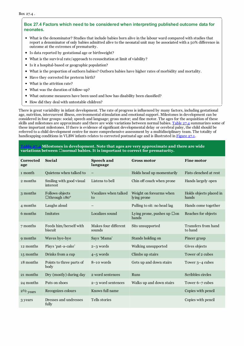
Box 27.4 .
Box 27.4 Factors which need to be considered when interpreting published outcome data forneonates.
What is the denominator? Studies that include babies born alive in the labour ward compared with studies thatreport a denominator of only babies admitted alive to the neonatal unit may be associated with a 50% difference inoutcome at the extremes of prematurity.
Is data reported by gestational age or birthweight?
What is the survival rate/approach to resuscitation at limit of viability?
Is it a hospital-based or geographic population?
What is the proportion of outborn babies? Outborn babies have higher rates of morbidity and mortality.
Have they corrected for preterm birth?
What is the attrition rate?
What was the duration of follow-up?
What outcome measures have been used and how has disability been classified?
How did they deal with untestable children?
There is great variability in infant development. The rate of progress is influenced by many factors, including gestationalage, nutrition, intercurrent illness, environmental stimulation and emotional support. Milestones in development can beconsidered in four groups: social; speech and language; gross motor; and fine motor. The ages for the acquisition of theseskills and milestones are approximate and there are wide variations between normal babies. Table 27.4 summarizes some ofthese important milestones. If there is evidence of significant developmental delay or cerebral palsy, the child should bereferred to a child development centre for more comprehensive assessment by a multidisciplinary team. The totality ofhandicapping conditions in VLBW infants relates to corrected postnatal age and is illustrated in Figure 27.1.
Table 27.4 Milestones in development. Note that ages are very approximate and there are widevariations between �normal babies. It is important to correct for prematurity.
Correctedage
Social Speech andlanguage
Gross motor Fine motor
1 month Quietens when talked to – Holds head up momentarily Fists clenched at rest
2 months Smiling with good visualinterest
Listens to bell Chin off couch when prone Hands largely open
3 months Follows objects�through 180°
Vocalizes when talkedto
Weight on forearms whenlying prone
Holds objects placed inhands
4 months Laughs aloud – Pulling to sit: no head lag Hands come together
6 months Imitates Localizes sound Lying prone, pushes up �onhands
Reaches for objects
7 months Feeds him/herself withbiscuit
Makes four differentsounds
Sits unsupported Transfers from handto hand
9 months Waves bye-bye Says ‘Mama’ Stands holding on Pincer grasp
12 months Plays ‘pat-a-cake’ 2–3 words Walking unsupported Gives objects
15 months Drinks from a cup 4–5 words Climbs up stairs Tower of 2 cubes
18 months Points to three parts ofbody
8–10 words Gets up and down stairs Tower 3–4 cubes
21 months Dry (mostly) during day 2 word sentences Runs Scribbles circles
24 months Puts on shoes 2–3 word sentences Walks up and down stairs Tower 6–7 cubes
2½ years Recognizes colours Knows full name Copies with pencil
3 years Dresses and undressesfully
Tells stories Copies with pencil

Figure 27.1 Corrected postnatal ages at which disabilities become evident in VLBW infants.
Compared with their full-term peers, extremely preterm children tend to achieve lower levels of physical and intellectualperformance. Some of the more commonly reported outcomes are listed in Table 27.5. These problems present at differentages, and this is highlighted in Figure 27.1. However, the majority of preterm infants rate their quality of life as high andthey are less likely to participate in risk-taking behaviours such as heavy alcohol consumption and illicit drug use.
Table 27.5 Adverse developmental outcomes in extremely preterm infants compared with controls.
Intelligent quotient (IQ) Studies consistently find mean IQs for ELBW infants are 0.7–1.0 SDless than term controls
Intellectual impairments Subtle cognitive deficits include lower scores in maths, reading, spelling, visualshort-term memory and executive function independent of IQ
School performance ELBW infants are more likely to be in the lower half of performance in their gradeand are twice as likely to repeat a grade versus term controls
Dyspraxia (clumsiness) Dyspraxia is a common finding in ELBW infants. This is most obvious clinicallywhen using ball skills (kicking and catching) and fine motor control
Attention deficit hyperactivitydisorder and behaviour problems
Premature infants have approximately twice the rate of these problems comparedto the general population
CLINICAL TIPPreterm\infants born close to the end of the school intake year may benefit developmentally by not starting schooluntil the following year.
DefinitionsGreat care must be taken to use the terms ‘handicap’, ‘impairment’ and ‘disability’ correctly, so that comparisons can bemade between different centres. The following are the internationally accepted World Health Organization (WHO)definitions.
Impairment: any loss or abnormality of psychological or anatomical structure or function.
Disability: any restriction or lack of ability (resulting from an impairment) in performing an activity in the manner orwithin the range considered to be normal.
Handicap: a disadvantage for an individual arising from a disability that limits or prevents the fulfilment of a role thatshould be normal for that individual.
SUMMARY
The majority of infants who are discharged from the neonatal intensive care will have normal development and nomajor long-term problems. There is, however, a subgroup that are at particularly high risk and require ongoingsurveillance. Parents will often need significant advice and support in preparation for discharge.
Further reading

1. Carson, C., Redshaw, M., Gray, R., Quigley, M. (2015) Risk of psychological distress in parents of preterm children inthe first year: evidence from the UK Millennium Cohort Study. British Medical Journal Open, 5 (12), e007942.
2. Orton, J., Spittle, A., Doyle, L., Anderson, P., Boyd, R. (2009) Do early intervention programmes improve cognitiveand motor outcomes for preterm infants after discharge? A systematic review. Developmental Medicine and ChildNeurology, 51 (11), 851–859.
Now visit www.essentialneonatalmed.com to test yourself on this chapter.

CHAPTER 28 Parent–infant attachment and support for parents of critically ill infantsKey topics
Parent–infant attachment (bonding)
Care of parents of critically ill infants
Family integrated care
IntroductionFollowing important observations during the 1970s by Klaus and Kennell, significant changes were made to the hospital careof postpartum women. They recognized a ‘sensitive period’ during which bonding and attachment took place, with closecontact in the immediate period necessary for bonding to occur. However, they revised this in the 1980s when a large bodyof professionals raised concern that psychological damage may be caused to families who were unable to have close physicalcontact with their baby immediately after birth due to medical complications of either the mother or the infant. Klaus andKennell revised their early work and recognized that “…early separation did not prevent the formation of a close, intimatebond”.
At the same time, it also became very clear that little attention had been given to the possible effects on parents of the deathof an infant who had had little or no opportunity for life. Since then, there has been considerable literature on perinataldeath, revealing that the care of recently bereaved parents leaves much to be desired. Thus, an understanding of theparent–infant bonding process can help the doctor and other healthcare providers to reduce the devastating effect ofperinatal loss, and also to understand the effects of prematurity and congenital abnormality on a family.
For mothers, a bond is formed quite early in pregnancy, stimulated by hormonal changes, psychological preparation andfantasies about the unborn child. ‘Nesting’ behaviour is manifested by the preparation of a nursery and the purchase of babyclothes. For the father, the attachment process is less recognizable during the pregnancy but heightens with the birth,enhanced by an involvement with the delivery and handling of the baby. A sense of pride and hope for the future ensues.
Parent–infant attachment (bonding)
Steps in attachmentThe actual process by which attachment bonds are formed is unknown. There are thought to be critical stages essential tothe establishment of attachment, and these are listed in Box 28.1. The strength of the attachment during these stages mayvary from one woman to another.
Most research, however, has concentrated on early contact in the immediate postnatal period. Extrapolation from animalresearch has proposed that there is a ‘maternal-sensitive’ or ‘maternal-critical period’, which is the optimal time for a bondof affection to develop between a mother and her infant. Although there is little doubt that the importance of this immediatepostnatal period has been overemphasized in humans, this knowledge had major benefits such as the early establishment ofsuccessful feeding and earlier discharge from hospital. Some mothers are unable to achieve a strong attachment withoutconsistent contact. Failure to bond can result in rejection and resultant problems with neglect and deprivation of nutrition,love and affection or even child abuse.
Parents learn to love their infant at varying times during the pregnancy and after birth. Parents are able to ‘bond’ to babiesthey have adopted, yet there has been no ‘maternal-critical period’. It is apparent that humans differ from other animals intheir patterns of bonding.
Box 28.1 Phases of parent-infant attachment (bonding).
Planning the pregnancy.
Confirming the pregnancy.
Accepting the pregnancy.
Onset of fetal movements (quickening).
Accepting the fetus as an individual.
The birth process.
Seeing the baby.
Touching the baby.
Taking care of the baby.
Attachment after birthAfter birth, a mother initially demonstrates attachment to her baby in several ways:
She is able to establish eye contact with the infant, who is in a state of arousal after birth.
If she is left alone with her naked infant, she may touch each part of the body with her fingertips.
A mother becomes overprotective of her infant in the first few days after delivery and becomes anxious about crying and

minor difficulties. This anxiety may appear excessive to hospital staff and family around her.
Babies may mimic the facial expressions of their parents, e.g. protrude their tongues.
Breastfeeding may be used to comfort and pacify the infant.
Factors that promote attachmentThe parents together plan the pregnancy and attend antenatal educational and physiotherapy classes. The antenatalpreparation of breasts and nipples will assist with subsequent breastfeeding.
The father should support the mother during labour and witness the birth of the baby.
Unless the baby is ill, the mother and baby should be permitted to respond to each other in their own time and manner.Unnecessary separation of infant and mother must be avoided.
The infant should ‘room in’ with the mother for 24 hours of the day, and be taken out to the nursery at night only if themother is ill.
Breastfeeding on demand, even at night, should be actively encouraged. However, if a mother is not successful in herattempts to breastfeed despite skilled help and advice, or does not wish to do so, she must not be made to feel inadequateor guilty. Successful bottle feeding is much better than completely unsuccessful breastfeeding.
Risk factors for failure to produce attachmentMothers who plan their pregnancies have good expectations of the outcome, breastfeed their babies, and rarelysubsequently maltreat them. Some of the risk factors that may have an adverse effect on bonding and render the family ‘atrisk’ for child safety concerns are listed below.
During pregnancyUnsupported pregnancies.
Where the father was unfaithful or deserted the mother during pregnancy.
Frequent pregnancies with excessive workload.
Maternal depression during the pregnancy or a history of previous postnatal depression.
Loss of an emotionally significant person in relation to the pregnancy, e.g. the maternal grandmother, grandfather, aloved sibling, a child of the mother or even a close friend, especially when the mother was somewhat isolated.
Conception during a period of marital conflict.
During labour and deliveryBeing left alone and afraid in the labour ward, or when the mother perceived the staff as unconcerned.
When the birth itself was more painful or prolonged than expected.
When breastfeeding was thrust upon the mother by the staff (it should, however, be encouraged).
When the mother was unable to see the child after delivery, without explanation, or was told that the baby was damaged.
When the mother herself was damaged as a result of the birth.
When the father exhibited more interest in the infant than in his partner.
In the neonatal and postnatal periodPrematurity, with associated separation.
Congenital malformations.
Critically ill infants requiring neonatal intensive care.
Postnatal depression.
When there is a discrepancy between the idealized, perfect baby and the real baby: under these circumstances theparents require careful counselling and support.
Failure of bonding or attachmentWhen bonding fails there is non-acceptance or even rejection of the child. This may result in problems for both the child andthe mother.
Long-term problems in the childChild abuse. Several studies have shown that preterm infants are over-represented among abused children.Approximately 30% of physically abused children are premature, yet the overall incidence of prematurity is only 8%.
Non-organic failure to thrive. Failure to thrive without organic cause is sometimes due to neglect or deprivation.Studies have revealed a fourfold increase among preterm infants and babies who require prolonged hospitalization in thenewborn period.
Temper tantrums, infant colic, feeding problems, sleeplessness and vomiting. Behavioural and feedingdisorders are more common when an affectionate bond has not been formed.
Personality disorders and poor interpersonal skills. Infants who have been deprived of love and affection maysubsequently have emotional and personality disturbances. They may become abusing or neglecting parents as adults,and so the cycle may repeat itself.

Problems in the motherHesitant, clumsy handling of the baby.
Maternal mental health issues such as Anxiety and depression.
Mother states that ‘the baby belongs to the hospital or nursing staff’.
Feelings of inadequacy, disappointment, failure, deprivation and anger.
Mother may complain that the baby does not bond with her.
Care of parents of critically ill infantsThe modern intensive care nursery is a bewildering and frightening place for the parents of a recently delivered prematureor sick infant. These parents often have feelings of extreme frustration, stress, guilt and helplessness, and must becounselled sensitively. The total care of the high-risk neonate must include the parents. The approach to the parents isdiscussed in a chronological manner, from antenatal clinic through to discharge and follow-up.
Antenatal contactWomen with high-risk pregnancies or known fetal abnormality should be introduced, before delivery, to a ‘baby doctor’whom they will probably meet after the baby is born. It is also helpful to the parents if they have the opportunity to visit theneonatal intensive care unit (NICU) before delivery, so that they are aware of the sights that will greet them when they firstcome to see their own baby. They might benefit from receiving an introductory book let describing the nursery care andstaff, and an introduction to a support group for parents of preterm babies or babies with a specific congenital anomaly.
Labour wardWhen the condition of the baby permits, the baby should be given to the mother for suckling and skin-to-skin contact for aslong as possible immediately after delivery. The baby should be dried and warmed before being handled by passed to themother for skin-to-skin care. Even if the baby is critically ill, his/her condition should be explained and the mother shouldsee the baby. Parents often report psychological absence and memory loss during and immediately following the birth oftheir critically ill/preterm baby, and wherever their first contact with their baby occurs it is often characterized by turbulentemotions. The open visiting policy of the nursery should be explained to the parents.
Intensive care nurseryAs close to delivery as possible, the mother and father should come into the nursery to see and touch their baby; however,often the father may visit alone first and ‘report back’ to the mother the infant’s condition. Careful explanations are givenregarding abnormal signs (retractions, bruising, etc.), equipment (monitors, incubators, ventilators) and commonlyperformed procedures (such as umbilical arterial/venous catheter, etc.).
There is every reason for the parents to be intimately involved with their critically ill infant from the outset. The prematureinfant has enough physiological problems already, without adding attachment problems to the list. The parents are informedthat they may visit or telephone the nursery 24 hours a day, and that both parents will be kept informed of the baby’sprogress. Consideration should be given to providing a pamphlet explaining the intensive care nursery and encouraging theparents to become actively involved in their child’s care. A digital photograph or video may help the mother to accept herbaby for the day or two before she is able to visit the nursery.
Parents’ first visit to the intensive care nurseryParents should be greeted and welcomed to the nursery. Again, the equipment should be carefully explained. It is mostimportant for the mother to be able to establish eye contact with her baby and, if appropriate, the goggles used withphototherapy may be removed. Often, the mother will have to look through the porthole to establish an en face positionwith her infant (i.e. align her head with her baby’s). Some parents are very apprehensive of handling, and usually state thattheir baby is too fragile to touch.
The nursing staff must work through this anxiety with the parents and encourage them to touch, fondle and caress theirbaby. Once the parents realize that their baby can actually see them, respond to their voices and can be pacified by them,their attachment grows.
Parents’ subsequent visits to the intensive �care nurseryThe parents should be encouraged to actively participate in their baby’s care. They derive great satisfaction from holdingthe syringe through which milk is being fed, changing nappies, and helping bath the baby. Even though in an incubator, thebaby can usually come out for a cuddle, or skin-to-skin (‘kangaroo’) care, provided that care is taken to maintain bodytemperature and the patency of all attached tubes. This may even be possible when the baby is receiving assistedventilation. This skin-to-skin care should ideally be at least 60 minutes in duration to achieve maximum benefit to bothmother and baby. Guidelines should be available on all NICU regarding skin-to-skin care.
Most mothers are only too happy to express their breast milk for their baby. It is remarkably well tolerated even in verypreterm babies, considering the immaturity of the gastrointestinal tract at that gestational stage, and will decrease theincidence of necrotizing enterocolitis (NEC) and infection. The mother will need a great deal of empathy and support fromstaff members to continue breast expression for the 8–10 weeks before her infant will be able to suckle. The role ofmaternal milk in feeding the very low birthweight (VLBW) infant is discussed in Chapter 9. Breastfeeding (direct from thebreast) should be encouraged while the mother is in hospital, so that the benefits of colostrum may be obtained.
Family integrated careFamily integrated care or ‘FiCare’ as it is sometimes known has recently been introduced in some Canadian, UK andAustralian neonatal units, but was first proposed in Estonia by Dr Adik Levin. It differs from family centred care in that

parents are not just given specific care tasks but are regarded as the central care provider, with nurses used to educate andsupport the parents. Parents are encouraged to be present for most of the day on the neonatal unit, in the way they mightbe on a children’s ward. It has been shown in pilot studies to reduce length of stay and improve weight gain andbreastfeeding rates. A randomised control trial is underway.
Babies as individualsNotes attached to the baby’s bed should be encouraging to the parents, for example ‘Mum is to feed me at 3 pm. I amlooking forward to my first breastfeed. Love Billy’.
Parents are asked to provide the staff with the baby’s name as soon as possible. The baby should be referred to by their firstname, e.g. ‘James’ or ‘Emily’, where possible, and never as ‘it’.
Appropriate decorations, pictures of family and mobiles, which may help parents adjust to a long hospital stay, can beattached to incubators. Parents are encouraged to use the pastoral care personnel of the hospital, or else to bring their ownminister of religion to baptize or administer rites to the baby.
How is my baby doing?The staff should answer this question in a realistic but optimistic way. Never should an unduly pessimistic attitude beconveyed, as this will only encourage detachment from the child. It has been shown that even if the baby dies, the parentsgain from having attached to their child. Their grief is physiological and appropriate. Caution should be exercised whenmaking predictions about outcome, and a problem-orientated approach to the baby should be avoided. It is easy for theparents to become involved with the intricacies of oxygen therapy and oxygen saturation, rather than the baby as a whole.
The social workerSocial workers play an important role in the intensive care nursery. They can provide support, investigating the attitudes ofparents towards the child, and at the same time may uncover social and financial problems. At times, the social worker actsas the ‘case manager’, providing the continuum of care that can be difficult to achieve with nursing and medical rostering.Consideration should be given towards keeping a parental contact chart recording telephone enquiries, visits and the specificinvolvement of parents during these visits. This helps with communication, especially between nursing shifts. Some unitsuse baby diaries or online video links to keep parents informed about their baby’s progress.
Babies transferred from other hospitalsParents of babies transferred from other hospitals into the intensive care nursery have unique problems. Prolonged parent–infant separation is common. If possible, the mother should be transferred along with the baby, but this may separate themother from her husband and other children. If the mother cannot be transferred, e-mailed digital photographs and dailyprogress reports should be provided. Whenever possible, the baby should be transferred back to the referring hospital forrecovery care at the earliest opportunity. Many tertiary perinatal hospitals have temporary accommodation facilities for‘out-of-town’ families of high-risk infants.
Preparation for dischargeEarly discharge for low-birthweight (LBW) babies can safely be practised and may promote attachment. Babies can bedischarged home, provided they are feeding well without cardiorespiratory compromise, have a sustained weight gain,maintain a normal body temperature when fully dressed in an open cot, be apnoea-free for a sufficient duration (check withindividual unit guidelines), the mother is handling competently, and there are no ongoing problems. These days, withoutreach support, babies can go home while still having some tube feeds, if parents have been trained.
SUMMARY
All units involved with the care of sick infants should develop management strategies to promote the attachment ofparents to their sick and preterm infants. A good parent–infant attachment has been shown to produce both short-term and long-term benefits in terms of physical and neurodevelopmental outcomes. Various forms of interventionare being implemented, either on individual basis such as NIDCAP and kangaroo care (see Chapter 24) or moreuniversally such as family centred and family integrated care. As they involve a number of physiological processes, wedo not know which individual item of intervention is most effective. It should also be realized that bonding starts rightat the stage of conception, runs through the entire pregnancy, and extends well into the postnatal period. Therefore,the intensive care nursery should be designed to the needs of parents and babies, and staff should always encourageparental involvement in the care of their babies. Babies who are ill or born preterm are already disadvantaged and arelikely to suffer more if their bonding with parents is not optimal.
Further reading1. Carson, C., Redshaw, M., Gray, R., Quigley, M. (2015) Risk of psychological distress in parents of preterm children in
the first year: evidence from the UK Millennium Cohort Study. British Medical Journal Open, 5 (12), e007942.
2. Crouch, M., Manderson, L. (1995) The social life of bonding theory. Social Science and Medicine, 4 (6), 837–844.
3. Kennell, J., Klaus, M. (1998) Bonding: Recent observations that alter perinatal care. Pediatrics Review, 19 (1), 4–12.
4. Klaus, M., Kennell, J. (1976) Maternal–infant bonding. The CV Mosby Company, St Louis.
5. Klaus, M., Kennell, J. (1982) Parent–infant bonding. The C V Mosby Company, St Louis.
6. Lamb, M.E. (1982) The bonding phenomenon: Misinterpretations and their implications. Journal of Pediatrics, 10 (4),555–557.

Now visit www.essentialneonatalmed.com to test yourself on this chapter.

CHAPTER 29 Ethical issues and decision-making process in the treatment of critically illnewborn infantsKey topics
Principles of ethical reasoning
Decision-making processes
The role of the Institutional Ethics Committee
Withholding and withdrawing life-sustaining treatment
Common neonatal ethical dilemmas
Parents in the decision-making process
IntroductionEthics is the science of morals; the branch of philosophy concerned with human character and conduct. Ethical issues arise inthe interactions of persons that involve the welfare or freedom of humans. They occur when one person or group of personsacts in ways that affect the welfare of another person or group of persons.
In the practice of medicine the best course of action is generally determined by humanist values (intrinsic value of humanlife). The very core of medicine is the respect for human life and the attempts to sustain and improve it. Life-sustainingtreatment decisions for newborn infants are typically made in an environment of scarce resources and where the medical,ethical and personnel implications are complex and often ambiguous. Terminology such as ‘sanctity of life’, ‘quality of life’and ‘ordinary or extraordinary means’ are unduly simplistic and usually unhelpful.
Principles of ethical reasoningFour major principles of ethical reasoning (described below) are particularly relevant to making decisions about newborninfants:
Beneficence.
Non-malificence.
Autonomy.
Equity or distributive justice.
Beneficence (discontinuing futile treatment)The traditional medical ethic is to act in ways that benefit the patient and do no harm. In many cases, however, theinstitution or continuation of treatment aimed at sustaining life is futile. Futile treatment is not likely to prevent death orserious compromise to the patient. There are difficulties in assessing futility, but medical determinations must be made sothat treatments that offer no benefit, and only serve to prolong the dying process, should not be employed.
Non-malificence (burdensome treatment)The primary ethical injunction for the doctor is ‘first, do no harm’ (primum non nocere). In making a decision to withhold orwithdraw life-sustaining medical treatment, the principle of non-malificence would require withholding treatment where itcan be said that it harms the patient. This occurs when the treatment itself is an intolerable burden to the patient.
AutonomyThe patient has the legal right and ethical autonomy to refuse life-sustaining treatment and be allowed to die. The neonatehas never been competent, and therefore decision-making is based on the patient’s ‘best interests’. Generally, the parentshave the authority and responsibility to make decisions on behalf of their baby. The two rationales for giving the parents theresponsibility are ‘bearer of responsibility’ and ‘best advocate’ grounds.
Equity or distributive justiceDoctors have an obligation to distribute benefits and burdens equally and, where differential treatment is given, to explainthe reasons for this based on widely accepted criteria. This principle might mean that in some cases the patient’s bestinterests should not or need not be the sole determining criterion.
Decision-making processesSeveral approaches to decision-making process have been described.
‘Wait until certainty’ approachIn an aggressive treatment environment almost every infant who is thought to have any chance to survive has fulltreatment commenced and continued until it is clear that treatment should be withdrawn. The advantage of this approach isthat it avoids the death of any infant who might have a good outcome, but at the cost of some infants for whom dying mightbe unnecessarily prolonged or who might survive with severe handicaps. This aggressive approach is understandable insocieties where consumer rights, individualism and litigation are prevalent, such as in North American countries, or in

communities with strong religious convictions.
Statistical approachThis approach draws on the accumulated evidence in order to establish categories of patients for whom treatment should bewithheld or withdrawn. This approach seeks to avoid ‘creating’ severely impaired children, even though this may be at theexpense of the deaths of some infants who might have a good outcome. This approach has been widely adopted in theNetherlands and some Scandinavian countries.
Individualized approach (prognostic �decision-making)In this approach treatment is initiated on any infant who has a chance of survival, but the patient is continually assessed todetermine whether this treatment is in the child’s best interests. A determination to withdraw treatment is made earlierthan in the ‘wait until certainty’ approach. For example, applying this approach to a 24-week, 580-g infant with respiratorydistress syndrome (RDS) and refractory hypotension who develops a severe grade IV intraventricular haemorrhage (IVH)may enable the experienced neonatologist to recommend withdrawal of life support and reorientation of care.
Antenatal diagnosisA paradigm shift has occurred in the last two decades in ethical decision-making in perinatal medicine. In previous decades,ethical decision-making usually occurred after the unexpected birth of an infant with a major congenital anomaly. However,most pregnant women now have biochemical and ultrasound screening (such as nuchal fold thickness) in the first trimester,and almost all have ultrasound assessment at 17–19 weeks’ gestation for congenital anomalies. In developed countries mostmajor congenital anomalies are diagnosed antenatally before 20 weeks’ gestation. Parents receive full multidisciplinarycounselling and are supported in their decision-making process. A perinatal clinical care plan is developed, and frequently acase manager or hospice staff member supports the family.
The role of the Institutional �Ethics CommitteeInstitutions and neonatal service providers usually have established general principles and a process for ethical decision-making. The Institutional Ethics Committee (IEC) may have a role to play in treatment decisions on ‘imperilled’ newborninfants. The IEC also has an important proactive role in ethical decision-making for obstetric and fetal patients but less so forcommencing, continuing, withholding or withdrawing treatment in a neonate.
The roles of an IEC can be summarized as follows:
Develop and ratify institutional guidelines.
Act as an advisory and consultative body.
Educate staff.
Disseminate information.
Absolve the attending physician from the decision-making process when necessary.
Resolve differences of opinion.
There are no readily available algorithms that one can follow simply each time an ethical dilemma arises: rather, one mustwork through a complex series of moral, religious, cultural and legal issues to reach an acceptable conclusion.
Good ethics can only be exercised if the medicine practised is correct. Good ethics requires accurate medical facts; not evensound ethical reasoning will rescue a decision based on false assumptions. With the advances in medical technology has comea greater public awareness of neonatal intensive care.
Withholding and withdrawing �life-sustaining treatmentWithholding life-sustaining medical treatment involves a choice to omit a form of treatment that is not considered beneficial,whereas withdrawal involves a choice to remove treatment that has not achieved its full beneficial intent.
Criteria for decision to withhold or withdraw life-sustaining medical treatment are usually based on:
Inevitability of death.
Futility of treatment.
Poor quality of life.
Whatever the reason, a structured decision-making process helps to ensure that appropriate views and preferences aremade explicit. This includes:
Creating an optimal environment for discussion.
Establishing the relevant facts.
Developing consensus between parents and professionals.
Offering a second opinion where appropriate.
Exploring options and choose most appropriate action.
Implementing decisions.
Common neonatal ethical dilemmasMost neonatal ethical dilemmas fall within the following four areas.

Whether to resuscitate at birthUnfortunately, junior doctors are often in the acute situation and may not have the relevant knowledge to make a decisionnot to start resuscitation. Institutional guidelines are necessary to cover all situations. Mistakes can undoubtedly be made inthose first few vital seconds, and if there is any doubt one must err on the conservative side of resuscitation. A decision canbe reversed later on when full facts become available. Currently recommended indications for ‘do not resuscitate’ at birthincludes only a few conditions such as:
Gestational age <23 weeks and/or birthweight <500 g.
Severe central nervous system malformations (e.g. anencephaly, severe holoprosencephaly).
Absence of fetal heartbeat for more than 10 minutes prior to delivery.
Infants with conditions such as suspected chromosomal anomalies (triploidy, trisomy 13 and trisomy 18), perinatal lethalrenal disease and lethal skeletal disorders should probably be stabilised where possible, and then fully assessed andinvestigated so that a rational decision can be made with all relevant information available. The approach to infants with aknown major congenital anomaly is discussed below.
‘How small is too small?’: when not to resuscitate on gestational age or birthweight criteriaHuman nature being what it is, we are straining to push back the frontiers of viability (technological imperative) to improveour statistics, and tend to accept death as failure.
Hospitals should develop guidelines based on their own survival and outcome data keeping in line with their regional andnational trends. An approach could be to attempt resuscitation on all infants over 600 g and/or at least 24 weeks’ gestation,and for selected infants between 500 and 600 g and 23+0 to 23+6 weeks, depending on past obstetric history, the likelihoodof infection, birth trauma and severity, initial asphyxia and parental wishes. Present indications suggest that most infantsless than 500 g and those less than 23 weeks’ gestation have very poor outcome, even with intensive care treatment.
Saying that the neonatologist will attempt full resuscitation and subsequent intensive care for all infants of more than 24weeks’ gestation and/or birthweight greater than 500 g does not mean that the obstetrician must necessarily do everythingpossible for all these pregnancies, as they have two people to consider: not only the neonate but also the mother. At times,there will be a dichotomy in the aggressiveness of care between obstetrician and neonatologist. Good communicationbetween the two specialities, and with the parents, is essential. Parents need to be at the centre of decision-making.
Major congenital malformationsThe majority of malformations are now diagnosed antenatally, often before 20 weeks’ gestation. Occasionally these may notbecome obvious until after birth.
Malformations can be considered under several categories:
Severe but not life-threatening abnormalities. These babies should be given all appropriate medical and nursingcare to promote survival and minimize later disability.
Potentially lethal conditions that may be associated with severe handicap. Examples include:
neural tube defects (myelomeningocoele, hydrocephalus, encephalocoele).
severe antenatal parenchymal brain haemorrhage.Parents are counselled so that they are fully aware of the clinical condition, including the sequelae and themanagement options available. If not diagnosed antenatally, life support measures may be instituted while the infantis fully assessed and all necessary information obtained.
Lethal abnormalities (e.g. anencephaly, trisomy 13 and 18 encephalocoele). The majority of these will have beendiagnosed antenatally. If the parents have decided to continue the pregnancy and the baby is born alive, he/she shouldreceive comfort care only (warm, free from hunger and relieved of pain). No active means are taken to shorten life. Attimes, these babies are discharged home under the parents’ care, sometimes in conjunction with a children’s hospice.Parents and family receive ongoing counselling and support.
CLINICAL TIPWhen a lethal abnormality has been diagnosed in utero and the parents have elected to continue the pregnancy, it isimportant that a clear birth plan is made before delivery. Things to consider include the mode of delivery (vaginal isnormally recommended), resuscitation (what, if any, should be done), pain and symptom management, and dischargehome. Introduction to the palliative care team before the birth may help to facilitate this.
Withdrawal of life supportThe situation regarding possible withdrawal of life supports arises in different circumstances:
Clear-cut cases (e.g. confirmation of bilateral renal agenesis, trisomy 13 or 18, triploidy, irreversible cervical cord injury).To continue ventilation for these lethal conditions would be futile and under the circumstances would constitute‘extraordinary care’.
Irreversible brain death following hypoxic–ischaemic injury.
Preterm infant with progressive bronchopulmonary dysplasia, grade IV IVH with posthaemorrhagic hydrocephalus.
These cases may be clear-cut when the infant can be recognized as dying despite maximal assistance, and death seemsinevitable.
Selective withdrawal of neonatal intensive care

Reasons for considering re-orientation towards palliative care include:
Prognosis extremely poor:
Short-term survival.
Long-term outcome.
Therapy too burdensome.
The decision-making process involves:
Accurate and complete medical facts: subspecialist consultation scientific documentation.
Consultation with hospital ethicist (if available) or independent consultant (second opinion).
In-depth case conference: medical/moral/ethical discussion.
Parents in the decision-making processAlthough parents are usually the best-qualified advocates for their infant, they should not be left to shoulder the burdenentirely; rather, a shared decision should be made.
All ‘proxy decision-makers’ – whether they be parents, physicians or courts of law – must be fully informed and cognizant ofthe relevant facts. Whatever approach is adopted it is vital that the process is made transparent, and that the physiciancommunicates clearly and honestly with parents and other members of the healthcare team.
It is rare for there to be major disagreement between clinical staff caring for the baby and parents, provided that there arecareful and repeated discussions between all parties. Tape-recording of conversations has been shown to be a useful way togive the parents time to reflect on what was said. A major dispute between clinicians and family represents a failure incommunication.
However, in the unusual situation of conflict when parents prefer no active treatment, the wishes of the parents may beover-ridden to sustain life. The reverse situation (i.e. parents wishing the continuation of life support and medical staffwishing to withdraw) occasionally arises and must be handled compassionately.
Circumstances in which parents’ wishes might be overruled, or when parents are incapable ofdecision-makingIt is not the doctor’s role to over-ride the wishes of parents, particularly when their decision is arrived at after carefulconsideration and reflection. Often, religious beliefs have a very strong influence on their decision, and it may be helpful toengage religious advisers in the conversation if the family is happy with this.
Where major dispute arises there are three possible courses of action. The first course is to ask another neonatologist from adifferent hospital to give an independent opinion. Discuss this with the family and ask them whether they will agree to thisas a way forward. Second, in hospitals with clinical ethical committees, the case may be reviewed by them and advice given.
Finally, in many countries the courts become involved in the rare cases where there remains a major disagreement aboutthe continuation of care. This usually arises when the parents feel that care should be continued and the clinical staff feelthat care is not going to save the baby’s life, or where survival will involve significant pain or suffering. The court will make adecision on the basis of an independent assessment of the evidence.
Role of the case conferenceA suggested approach to deal with issues regarding the withdrawal of life support is the case conference. This involves allrelevant staff (medical, nursing, allied health, pastoral care) and parents to work through the complex series of medical,social and ethical issues.
Purpose of conferenceA case conference can serve several purposes:
Ensuring staff are comfortable with the decision.
Enhancing communication between staff.
Developing a future care plan: further information (investigation) time frameworks.
Providing guidance for counselling parents.
Mapping out a process for withdrawal of life support.
Enabling staff to understand the decision-making process.
Giving an opportunity for all opinions to be expressed.
Communication of withdrawal of life supportThe decision to withdraw life support or ‘re-orientate’ care towards palliative care entails several procedures that must befollowed:
The attending physician clearly annotates the medical facts in the chart.
At times, a second neonatologist is consulted and their opinion is documented in the chart.
Rarely, the hospital ethics committee is convened if there is ethical uncertainty.
The decision-making process is annotated in the chart.
The decision is communicated to relevant persons, e.g. charge nurse, director of nursing, medical superintendent.

Cases with unique aspects are documented in detail and archived by the ethics committee.
Care of parentsThe care and support of the parents is a necessary part of the decision-making process and its aftermath.
The parents, preferably together, receive progressive counselling from the neonatologist.
Ongoing support is provided by nursing staff, social workers and pastoral care staff.
Parents are informed of the case conference and are counselled afterwards.
Life and death decisions are made jointly by the neonatologist and the parents – the final decision is parental.
Offer a bereavement review meeting.
SUMMARY
Key concepts underlying ethical care in the neonatal intensive care units involve respecting parental autonomy,applying the ‘best interest of the patient’ philosophy, and minimizing ‘harm’ to the patient. It is also vital to establish asound parent–doctor (professional) relationship, to empower and inform the parents, to respect the parents’ values,culture and religious beliefs, and to apply a family-centred care approach.
Further reading1. DeTora, A.W., Cummings, C.L. (2015) Ethics and the Law: Practical Applications in the Neonatal Intensive Care Unit.
NeoReviews, 16 (7), e384–e392.
2. Nuffield Council on Bioethics (2006) Critical care decisions in fetal and neonatal medicine: Ethical issues. ISBN 1904384 14 5Larcher V, Craig F, Bhogal K, et al. Making decisions to limit treatment in life-limiting and life-threatening conditions in children: a framework for practice Archives of Disease in Childhood 2015;100:s1–s23.
Now visit www.essentialneonatalmed.com to test yourself on this chapter.

CHAPTER 30 End-of-life care and palliative careKey topics
Introduction – why babies die
Unexpected deaths
What is palliative care?
Expected deaths and care planning
Making a care plan
Place of death: hospice versus home versus hospital
Symptom control
Organ donation
Autopsy
Caring for parents – grief and bereavement
Caring for staff
Introduction – why babies dieA birth is an event that is usually associated with joy and excitement. It is preceded by planning, expectations, dreams andhopes. For most people, having a baby brings with it changes in the family structure that affect each family member in adifferent way. The 9 months of the pregnancy are filled with adjustments in role for the mother, father and siblings, and withan awareness that a new life is growing within the mother. Grandparents and members of the extended family share in thepreparations and hopes. In some families there may be ambivalent feelings perhaps because the pregnancy was unplannedor unwanted.
It should also be remembered that some babies die. Whilst the infant and neonatal mortality rates have been steadily falling,the first year of life has by far the highest mortality rate, until old age. Stillbirths are often unexplained but can be due toasphyxia, congenital abnormality or infection. Babies die mostly because of extreme prematurity (46%), congenitalmalformation (22%), intrapartum causes (12%) or infection (7%). Some deaths are very unexpected, but most, especiallydeaths on the neonatal intensive care unit (NICU) can be anticipated and planned for (See box 30.1).
Unexpected deaths (including sudden unexpected postnatal collapse)It is often difficult to define what is meant by an ‘unexpected death’. For example a 23-week infant might well be expectedto die at the point of birth, but if they collapse with necrotizing eneterocolitis (NEC) at 4 weeks of age this may beunexpected. A common definition is a death that was not expected 24 hours beforehand.
About 1 in 20 000 babies in the UK who are born healthy will collapse and die in the first 12 hours of life, and the overallincidence of ‘Sudden Unexplained Postnatal Collapse’ (SUPC) is about 0.05 in 1000 live births. SUPC has been defined asthe unexpected collapse of a term or near-term baby, who was well at birth (Apgar >5) and well enough to go to the normalpost natal ward, but who collapsed unexpectedly within the first week of life, requiring resuscitation, and then goes on toeither die, require intensive care or have encephalopathy. A cause can usually be found in 60% of SUPC cases after autopsyand investigations. A proportion of the others may be due to accidental suffocation, either at the breast or due to co-sleeping.
Unexpected collapse or death on the NICU and SUPC are highly traumatic events for parents and staff. They requiredetailed investigation, including an accurate history of events leading up to the collapse, detailed investigations, including asearch for metabolic or genetic disorders, and often an autopsy by a skilled perinatal pathologist. A guide to the managementof SUPC is available (see Further reading). Staff will need support and debriefing (see below)
What is palliative care?Palliative care is the physical, emotional and spiritual care of a baby or infant who has a life-limiting illness. It does not justinclude end-of-life care, and in many countries the palliative care service provides important respite care for families.Palliative care is a rapidly expanding new speciality in paediatrics, and is now extending into neonatology. Palliative careexperts may be based in a children’s hospice, but many will offer inreach into the hospital and NICU as well as supportingfamilies who wish for their baby to die at home. The focus of palliative care is on improving the quality of life, includingsymptom control, rather than trying to extend the duration of life.
Expected deaths and care planningWhen it is very likely or inevitable that a baby will die, the healthcare team – in conjunction with the parents – may decideto re-orientate or redirect care towards a palliative approach, or to actively withdraw life support measures (see Box 30.1).These difficult decisions, which often throw up difficult ethical decisions, are discussed in Chapter 29.
Expected deaths need to be actively managed, and require as much care and energy from the clinical team – if notmore – than babies who continue to receive full life support. One-to-one nursing ratios will be required to support the familyin preparing for the death of their child.

Box 30.1 Scenarios in which death might be anticipated and planned.
A fetus with a lethal skeletal dysplasia, who may not survive more than a few hours. A plan not to resuscitate atbirth may be agreed.
Congenital abnormality incompatible with long-term survival (e.g. trisomy 13, 18, severe arthrogryposis,anencephaly).
Severe encephalopathy with multi-organ failure, not responding to treatment.
Extreme prematurity with major complications (e.g. a 23-week baby with a massive intra-parenchymal brainhaemorrhage).
Severe chronic lung disease with pulmonary hypertension, unresponsive to treatment and dependent onmechanical ventilation.
Extreme short bowel secondary to severe NEC or volvulus.
Severe neurological disorders such as spinal muscular atrophy (SMA) or central hypoventilation syndrome.
Making a care planOnce it has been decided and agreed that the child has a life-limiting condition, or treatment is being redirected towardsend-of-life care, it can be very useful to agree a formal ‘care plan’ or ‘limitation of treatment agreement’ with the family.This allows the whole team, including community services, paramedics and so forth, to be aware of the plans for the babyand to prevent unnecessary invasive treatment if the child collapses unexpectedly. It also allows parents to take an activeand overt part in the decision making around the care of their child. Limitation of treatment agreements (see Fig. 30.1)cover more than just ‘do not resuscitate’ orders, and can allow targeted intervention (e.g. clearing airway, suction,administration of oxygen) without invasive steps such as intubation and ventilation. Care plans cover the whole palliativecare process, including the parents’ wishes on what treatment should continue, where the care should take place, where theparents would like the child to be when they die (often at home, with support, or in a hospice or in quieter area of the NICU),or where they would like to be with the child after death (many hospices offer cooled bedrooms where the family may staywith or visit the baby after death and before the funeral).
Figure 30.1 A typical Limitation of Treatment Agreement (LOTA) agreement.
Place of death: hospice versus home versus hospital

Where the family wish their child to die is a very individual choice, and will depend to a large extent on the level of intensivecare the child is receiving, the predicted life expectancy after redirecting care, and their home circumstances or culturalbeliefs. Many parents of children with lethal trisomies such as Edward’s syndrome (trisomy 18) or Patau’s syndrome(trisomy 13) can enjoy many weeks or months at home with their child. Children receiving mechanical ventilation prior todeath may be extubated in a side room or parental bedroom on the NICU with the parents present, or extubated aftertransfer to the hospice or home. This is often referred to as ‘compassionate extubation’. Some parents would prefer the babyto be extubated on the NICU and then given to them to hold, without all the tubes and wires, in a quiet, private space.Culturally, some parents may not want to be with their baby at the moment of death, and in these circumstances a nurse orother staff member will normally stay with the baby.
Symptom controlOnce palliative care has been agreed it is very important to try to minimize pain and distress for the baby. This mightinclude a plan to reduce or stop altogether, and blood tests and heel-pricks for blood gas analysis. The degree of physiologicalmonitoring may be reduced or stopped. If the baby is experiencing respiratory distress, or has a painful condition (e.g.severe NEC or skin lesions), then all efforts should be made to address symptom control using whatever medication isrequired, by the least invasive route. Examples are shown in Table 30.1. It is reasonable to give opiates in the knowledgethat they may cause respiratory depression and may thereby shorten life, as long as this is not the main purpose of themedication and it is being given to relieve distress or pain. In practice, if the child is in pain morphine is a safe drug that willnot cause respiratory depression once tolerated.
Table 30.1 Examples of symptom control in neonatal palliative care.
Intervention Rationale
Anticonvulsants Seizures can be distressing for the baby and the parents
Opiate infusion Relief of pain and respiratory distress. Subcutaneous infusion of diamorphine or fentanyl allowsdelivery without large volumes and without the need for maintaining intravenous access.
Benzodiazepines(buccal, oral,rectal)
Seizure control, relief of anxiety or distress and sedation
Oxygen This may seem counter-intuitive but prolonged hypoxia can make babies very irritable and agitated.
Nasogastric ornaso-jejunal feeds
Giving a small volume of milk either orally, if tolerated, or via a tube, can relieve symptoms ofhunger and distress without unnecessarily prolonging life or aiming for growth.
Antibiotics Infections may cause pain or discomfort.
Organ donationMany parents will obtain comfort from knowing that their child’s organs helped another individual to survive. There alsoremains a shortage of donor organs for children with end-stage organ failure. Until recently, brainstem death testing was notpermitted in the UK in neonates, but since 2015 it is now permissible from 37 weeks’ gestation, so ‘death by neurologicalcriteria’ (DND) can allow organ retrieval with a beating heart in certain selected cases. However, ‘brainstem death’ isrelatively unusual in neonates. Donation after cardiac death (DCD) is a relatively new practice that allows – if the parentsare in agreement – organs such as the kidneys and liver to be retrieved within 10 minutes of cardiac arrest. Mechanicalventilation is stopped while the parents are with the baby, who must remain on an ECG monitor. When there has beenasystole for 5 minutes the baby is then taken to theatre for organ retrieval. Both neonatal kidneys can now be anastomosed‘en-bloc’ to the iliac vessels of an adult patient to provide sufficient renal function. Donation of organs after DCD may also beconsidered in babies with anencephaly. Finally, in most babies some tissues such as heart valves can be donated severalhours after death if parents agree. A specialist nurse for organ donation can be especially useful in taking the parentsthrough the process of consenting for organ donation in a compassionate and knowledgeable manner.
AutopsyAn autopsy should almost always be offered, even if the cause of death is thought to be known. Autopsy findings differ fromthe clinical diagnosis in 10–32% of cases. Autopsy may be a legal requirement when there has been an unexpected death ora death soon after surgery, or if unnatural causes are thought likely. If parents decline a full autopsy, as many do, usefulinformation can be obtained by targeted autopsy (e.g. just examining the lungs but not the brain, or targeted tissuebiopsies). Genetic testing, post-mortem imaging (including MRI) and peri-mortem skin and muscle biopsies can also beuseful in selected cases. A full autopsy involves photographs and X-rays of the body, a full external and internal examinationof the organs, and histological analysis of tissue samples. Explicit additional consent must be taken if samples are to beretained for teaching or research. After the autopsy the organs are returned to the body and it is sutured so that, whendressed, the parents can see their baby again if they wish.
Caring for parents – grief and bereavementThe news that their baby is dying or has died often comes as an unexpected catastrophe. For parents who have experiencedmiscarriage or a stillbirth, the fact that they have never known the baby can make their loss seem even more unreal anddifficult to accept. When the baby survives for a short time the parents have had an opportunity to experience their child asa living being.

Some of the common characteristics of normal grief are outlined in Box 30.2. The stages of grief are not necessarily in thisorder, and occupy a variable period of time. It has been suggested that a normal grief reaction lasts 6–9 months, althoughthe most intensive phase lasts from 1 to 6 weeks. Grief reactions can also be delayed and often will reoccur on anniversaries.
Box 30.2 Stages of grief.
‘Shock’.
Emotional release.
Utter depression, loneliness and isolation.
Physical symptoms, e.g. choking, dyspnoea, empty feeling, weakness, fatigue, insomnia, loss of appetite.
Panic – about their own worth and the safety of other children.
Guilt.
Anger.
Inability to return to normal activities.
Overcoming grief.
Readjustment of life.
Parents will need a lot of emotional and practical support as their baby dies. This is often provided by experienced neonatalnurses. Nurses will help the family gather mementos (e.g. identity wrist bands, locks of hair, footprints) and the family canbe provided with a ‘memory box’ (see Fig. 30.2). Bereavement photography services are available who will help takecompassionate photographs of the baby with the parents after death.
Figure 30.2 Memory box. Source: Tom Lissauer, Avroy A. Fanaroff, Lawrence Miall, Jonathan Fanaroff. Neonatology at aGlance, 3rd Edition August 2015, ©2014, Wiley-Blackwell. Reproduced with permission of John Wiley & Sons.
Some units have specialist bereavement nurses or counselling/psychology support. Hospices are often able to offerbereavement support for some time after the death. Some parents will need medical support from their own family doctorto deal with symptoms such as insomnia or extreme anxiety. Charitable organizations (e.g. SANDS – the Stillbirth AndNeonatal Death Society, in the UK) are able to offer support and advice. Older siblings will need to be supported sensitively.It is often good if they can see the baby before they die. Finally, when a baby dies it is important to notify the communityhealth IT system to prevent appointments being sent for immunizations and so forth, which may cause distress.
It is common practice to invite bereaved parents back to the hospital, or to another place, to allow them to ask questionsabout the care their baby received, to hear the results of any outstanding investigations, or to go through the autopsyfindings.
Caring for staffMost neonatal staff enjoy saving lives and helping treat babies so they get better, but supporting a family throughbereavement can in itself be very rewarding. Watching a child die, and supporting the family at close quarters, can also bevery stressful and sometimes upsetting, especially for less-experienced staff. It is important that staff are able to share theirexperiences in a safe environment, and many larger neonatal units have psychologists to support staff. When a child diesunexpectedly it is important to arrange a ‘debrief’ meeting to go through the events and reassure staff of their actions. Thisshould usually take place as soon as possible after the event.

summary
Palliative care is not just about end-of-life care, but is about providing good holistic care to the family and goodsymptom control for the child. The focus is on quality of life, not duration of life. Early referral should be consideredfor any child with a life-limiting diagnosis. Palliative care teams are increasingly involved early in the care planningprocess, including before birth if there is an antenatal diagnosis of a potentially life-limiting disorder. Limitation oftreatment agreements can help clarify how much treatment is appropriate, and how much the family want. Good careshould not stop with the death of the child – bereavement care is a crucial part of the holistic care of the whole family,and some of the best neonatal teams now have bereavement support available for families and psychological supportavailable for staff.
Further reading1. Becher, J.-C., Bhushan, S.S., Lyon, A.J. (2012) Unexpected collapse in apparently healthy newborns – a prospective
national study of a missing cohort of neonatal deaths and near-death events. Archives of Diseases in Childhood: Fetaland Neonatal Edition, 97 (1), F30–F34.
2. British Association of Perinatal Medicine: Palliative care (supportive and end of life care). A framework for clinicalpractice in Perinatal medicine. Available at:http://www.bapm.org/publications/documents/guidelines/Palliative_Care_Report_final_%20Aug10.pdf
3. English, S. (2012) Palliative care and end-of-life decisions. Seminars in Fetal and Neonatal Medicine, 18 (2), 67.
4. Larcher, V., et al. on behalf of the Royal College of Paediatrics and Child Health (2015) Making decisions to limittreatment in life-limiting and life-threatening conditions in children: a framework for practice. Archives of Diseases inChildhood, 100, s1–s23.
5. Mancini, A., Uthaya, S., Beardsley, C., Wood, D., Modi, N. (2014) Practical guidance for the management of palliativecare on neonatal units: 1st edition. Available at: http://www.uk-sands.org/sites/default/files/NICU-Palliative-Care-Feb-2014.pdf
Now visit www.essentialneonatalmed.com to test yourself on this chapter.

IndexAAase's syndrome
abdomen
abdominal
circumference
distension
wall defects
abnormal movements
ABO incompatibility
abrasions
abruptio placentae
absent radius
abstinence syndrome, neonatal
acetazolamide
achondroplasia
acid–base balance
acidosis
blood
metabolic
respiratory
acid–base disturbance
acute kidney injury
acute life-threatening events (ALTEs)
adjunctive therapy
adrenal gland
abnormalities of
injuries
adrenal hormones synthesis, illustration
adrenal hypoplasia
adrenaline
adrenergic receptors
aganglionosis
alcohol
congenital malformations
withdrawal
aldosterone
alkalosis
metabolic
respiratory
alpha-fetoprotein (AFP)
ambiguous genitalia
amelia
amikacin
aminoglycoside antibiotics
aminophylline
amniocentesis
amniotic band amputations
amniotic cavity

amniotic fluid
role of
volume
amplitude integrated EEG (aEEG)
anaemia
early
exchange transfusion
late
physiological
of prematurity
anaerobic metabolism
analgesia, mechanical ventilation
analgesia
anencephaly
aneuploidy
anorectal malformations
anticholinesterase (neostigmine)
anticoagulant proteins
anticonvulsant drug treatment
anti-D gammaglobulin
antidiuretic hormone (ADH)
anus
aortic stenosis
Apert's syndrome
apex beat
Apgar score
aplasia
apnoea
aetiology
causes of
investigations
management
monitoring
primary
prognosis
terminal
treatment
types of
ventilatory support
apoptosis
Apt test
arms, total paralysis
Arnold–Chiari malformation
arterial blood, oxygen tension
arthrogryposis multiplex
artificial feeding/formulas
cows' milk-based formulas
elemental formulas
for malabsorptive conditions
preparation of
techniques of
whey-dominant formulas

asphyxia, perinatal
Apgar score
causes of
clinical evidence of
definition
fetal/neonatal responses to
medico-legal evidence of
physiological effect of
postresuscitation care
prevention of
resuscitation
sequelae of birth
SGA infant
assist/control ventilation (A/C)
assisted reproductive technology (ART)
asymptomatic hypocalcaemia
atopic dermatitis
auditory brainstem responses
audit perinatal and neonatal
auscultation
autopsy
autosomal dominant inheritance
autosomal dominant polycystic kidney disease (ADPKD)
autosomal recessive inheritance
autosomal recessive polycystic kidney disease
axonotmesis
Bback examination
Barker hypothesis
Barlow's (dislocation) test
Bayley Scale of Infant Development (BSID-II/-III)
BCG vaccination
Beckwith–Wiedemann syndrome
behavioural disorders
beneficence principle
benign congenital hypotonia
bile-stained vomiting
bilevel pressure support
biliary atresia
bilirubin
assessment in liquor
conjugated
encephalopathy
liver uptake
metabolism
molecule
production
serum
transport
unconjugated
biophysical profile
biparietal diameter (BPD)

birth
circulatory maladaptation at
infant at, assessment of
neonatal depression at
physiological changes at
stabilization at
birth depression
birth injuries
bone and joint injuries
organ injuries
peripheral nerve injuries
risk factors for
of scalp, skull and brain
soft-tissue injuries
sustained in NICU
types of
birthmarks
birth trauma. See birth injuries
birthweight
bladder exstrophy
blastocyst
blistering
blood acidosis
blood gas monitoring
blood pressure
diastolic
mean arterial
normal range
preterm infant
suprasystemic pulmonary
systolic
blood sampling, fetal
blood transfusion
anaemia
haemorrhage
inborn errors of metabolism
rhesus haemolytic disease
see also exchange transfusion
B lymphocytes
bowel motility
bowel obstruction
brain development
brain, injuries to
branchio-oto-renal syndrome
breast engorgement

breastfeeding
advice
benefits of
contraindications
jaundice
nutritional aspects
physiology of lactation
problems
techniques
breast milk
anti-infective properties of
bank
changing
drugs in, excretion of
engorgement
insufficient
and intelligence
successful, steps to
variations in
breast milk jaundice
breathing
patterns
physiology
breathing movements, fetal
breath sounds
breech presentation, preterm
bronchopulmonary dysplasia (BPD)
aetiology
chest radiograph
clinical features
management
prognosis
radiology
bruising
to foot
testicular
bullous disorders
buphthalmos
burn, chemical
Ccaesarean section
caffeine citrate
Candida albicans
candida vesicles
candidiasis
systemic
capillary malformations
captopril
caput succedaneum

carbohydrate
absorption
modification
total parenteral nutrition
carbon dioxide transport
carbonic anhydrase
cardiac assessment
cardiac catheterization
cardiac compressions
cardiac massage, external
cardiac output
cardiomyopathy
cardiotocography (CTG)
fetal heart rate accelerations
heart rate followed by bradycardia
interpretations of
intrapartum
late decelerations
loss of beat-to-beat variability
cardiovascular disorders
cardiovascular physiology
circulatory maladaptation at birth
congenital heart disease
congestive heart failure
cyanotic heart disease
dysrrhythmias
electrocardiograph
hypertension
hypotension
investigations
obstructive lesions
patent ductus arteriosus
perinatal asphyxia
ventricular septal defect
carpenter's syndrome
case conference, critically ill infants
casein-dominated formulas
caudal regression syndrome
central apnoea
cephalhaematoma
cerebral function monitoring (CFM)
cerebral hypoxia
cerebral neuroprotection
cerebral palsy
in twins
cerebral visual impairment
chemical burn

central nervous system malformations
anencephaly
encephalocoele
hydranencephaly
lissencephaly
neural tube disorders
porencephaly
spina bifida
ventral induction disorders
see also hydrocephalus
chest
examination of
radiography
shape and size
small
chignon
child abuse
Chlamydia trachomatis
choanal atresia
cholestatic jaundice
chordee
chorion
chorionic villus sampling
chromosomes
addition of
analysis
changes in number
deletion of
disorders
patterns
translocation
chronic lung disease (CLD)
conditions causing
circulation, fetal
circulatory maladaptation at birth
clavicle fracture
cleft hand
cleft lip
cleft palate
clinodactyly
clitoris
coagulase-negative staphylococci
coagulation disorders
coarctation of aorta (CoA)
cocaine
cochlear implants
cold injury neonatal
colic, infant
collapse, postnatal
collodion baby
colonic atresia
colony-stimulating factors

congenital abnormalities of hips and limbs
arthrogryposis multiplex
developmental dysplasia of the hip
dwarfism
limb malformations
skeletal dysplasia
congenital adrenal hyperplasia (CAH)
enzymatic defects
congenital anomalies
associated with teratogens
classification of
deformations
malformations
congenital ascites
congenital deformations
risk factors for
congenital diaphragmatic hernia (CDH)
congenital heart block
congenital heart disease (CHD)
aetiology
cyanotic. See cyanotic heart disease
echocardiography
mode of presentation
prostaglandins for
congenital hyperinsulinism
congenital lobar emphysema
congenital malformations
causes of
chemicals
incidence of
irradiation
maternal disease
SGA infant
twins
congenital muscular dystrophy
congenital myotonic dystrophy
congenital pulmonary airway malformation (CPAM)
congenital rubella syndrome
conjoined twins
conjugated hyperbilirubinaemia
causes of
investigations
conjunctival swabs
conjunctivitis
conscious level
constipation
continuous positive airway pressure (CPAP)
complications
contraindications for
indications for
respiratory failure
convulsions

cord entanglement
cordocentesis
cornea
cortical visual impairment
crack cocaine
cradle cap
craniostenosis/craniosynostosis
craniosynostosis
craniotabes
creatinine phosphokinase (CPK)
Crigler–Najjar syndrome
crown–rump length (CRL)
cutis aplasia
cutis laxa
cutis marmorata
cyanotic heart disease
Ebstein's anomaly
pulmonary atresia
pulmonary stenosis
tetralogy of fallot
total anomalous pulmonary venous drainage
transposition of the great arteries
tricuspid atresia
cystic dysplastic (multicystic) kidneys
cystic fibrosis
cystic hygroma
cystic periventricular leukomalacia (PVL)
cytomegalovirus (CMV)
cytotrophoblast
Ddeformation sequence
dehydration
delayed passage of meconium
developmental dysplasia of the hip (DDH)
examination of hip
management
risk factors for
ultrasound
dexamethasone
diabetes mellitus
Diamond–Blackfan syndrome
diarrhoea
dimercaptosuccinic acid (DMSA) radionuclide scan
dipalmitoyl phosphatidylcholine (DPPC)
disappearing twin phenomenon
disseminated intravascular coagulation (DIC)
distal obstruction
distributive justice principle
divarication of rectus muscles
diving seal reflex
dobutamine
dopamine

Doppler assessment of cerebral haemodynamics
Doppler flow velocity analysis
Down's syndrome
screening for
doxapram
drugs, congenital anomalies
Dubin–Johnson syndrome
ductus arteriosus
patent
ductus venosus
duodenal obstruction
causes of
diagnosis
treatment
dwarfism, neonatal
dysmorphic neonate
dysrrhythmias
Eearly-onset infections
ears
Ebstein's anomaly
ecchymoses
echocardiography
ectodermal dysplasia
ectopia vesicae
ectromelia
eczema
Edrophonium
Ehlers–Danlos syndrome
electrocardiogram (ECG), fetal
electrocardiography (ECG)
electroconvulsive dissociation (ECD)
electroencephalography (EEG)
electrolyte imbalance
elemental formulas
encephalocoele
encephalopathy
end-diastolic flow (EDF)
endocrine gland disorders, hypothyroidism
endotracheal tube
end-tidal carbon dioxide monitors
energy requirements
epidermal naevi
epidermolysis bullosa
epidermolysis bullosa simplex
epigenetics
epilepsy, maternal
epiphyses, separation of
epispadias
Epstein's pearls
Erb's palsy
erythema

erythema toxicum
erythropoietin
Escherichia coli
ethamsylate
European Society for Pediatric Gastro-enterology, Hepatology and Nutrition (ESPGHAN)
exchange transfusion, dilutional
exomphalos
expressed breast milk (EBM)
extended perinatal mortality rate (EPMR)
extracorporeal membrane oxygenation (ECMO)
extraembryonic mesoderm
extravasation injury
extremely low birthweight (ELBW) infants
fungal prophylaxis
extremely preterm delivery
extremities, examination of
lower limbs
movements
upper limbs
eyelids
Ffacial features
facial nerve palsy
failure to thrive
Fanconi's syndrome
fat necrosis, subcutaneous
fats
absorption
intake
modification
total parenteral nutrition
feeding
artificial. See also breastfeeding; nutrition
continuous
disorders
constipation
diarrhoea
failure to thrive
infant colic
pyloric stenosis
vomiting
LBW infant
preterm infant
trophic
very-low-birthweight infant
feet
femoral retroversion
femur
fracture
length
fertilization, physiology of

fetal alcohol spectrum disorder (FASD)
drug abuse
four-digit diagnostic code for
infant with features of
fetal assessment
fetal growth
maturity
timeline for
well-being
fetal compromise
causes of
physiological changes at birth
fetal distress
fetal electrocardiogram
fetal heart rate (FHR)
fetal size, ultrasound
fetal weight, estimation of
fetotoxin
fetus
breathing movements
circulation
growth assessment
heart rate monitoring
homeostasis
imaging of
lung development
monitoring during labour
well-being assessment. See also fetal assessment
fibrin degradation products (FDPs)
fingers
fluconazole
fluid restriction
fluorescence in-situ hybridization (FISH)
fluoride
folate, preconceptual
follow-on formulas
fontanelles
foramen ovale,
foreskin
fractures
clavicle
femur
multiple
skull
functional residual capacity (FRC)
fundal height measurement
fungal prophylaxis
funisitis
furosemide
Ggastric acid secretion
gastritis

gastroenteritis
gastrointestinal bleeding
gastrointestinal (GI) tract
abdominal wall defects
development of
malformations
necrotizing enterocolitis
obstruction
rectal bleeding
short bowel syndrome
gastro-oesophageal reflux disease (GORD)
gastroschisis
gene structure
genetic disorders
approach to dysmorphic neonate
autosomal disorders
chromosome
investigations
multifactorial inheritance
prenatal diagnosis
prevention of congenital abnormalities
single-gene defects
X-linked disorders
genetic variation
DNA sequence, changes in
multifactorial changes
structural changes
genitalia
gentamicin
genu recurvatum
gestational age
Giemsa stain
Gilbert's syndrome
glaucoma
glial cells
glomerular filtration rate (GFR)
gluconeogenesis
glucose
homeostasis
metabolism
goitre
granulocyte colony-stimulating factor (G-CSF)
granulomata, umbilical
Graves' disease
Griffiths Mental Developmental Scale
Group B β-haemolytic streptococci (GBS) infection
growth
discordant
fetal
SGA infant
gut perfusion
H

haemangiomas
haematuria
renal disorders associated with
haemodilution
haemoglobin
haemolysis
Haemophilus influenzae
haemorrhage
adrenal
intracranial
intraventricular
pulmonary
subaponeurotic
testicular
harlequin colour change
harlequin icthyosis
head
anatomic location of injuries to
examination of
growth of
head circumference (HC)
heart
hypoplastic left
position
shape
size
heart murmur
heart rate
accelerations
monitoring
hemimelia
hepatitis B
hepatitis C
hepatitis, neonatal
Hering–Breuer deflation reflex
hernia
inguinal
umbilical
herpes congenital vesicles
herpes simplex infection
high-flow humidified oxygen therapy (HFNT)
high-flow nasal cannulae (HFNC)
high-frequency jet ventilation (HFJV)
high-frequency oscillatory ventilation (HFOV)
high-frequency ventilation (HFV)

high-risk infants
developmental care for
developmental outcomes
discharge
feeding advice
follow-up
growth
immunization
nutritional supplementation
postnatal development
rooming in
specialized follow-up clinics
highly active antiretroviral therapy (HAART)
hip adductor tone
Hirschsprung's disease
HIV infection
holoprosencephaly
homeostasis, fetal
human chorionic gonadotropin (hCG)
human chorionic thyrotropin (hCT)
human milk fortifiers (HMFs)
humerus fracture
humidified high-flow nasal cannula (HHFNC)
Hunter's syndrome
hydranencephaly
hydrocele
hydrocephalus
classification
clinical features
investigations
management
prognosis
hydrocoele
hydrolysates
hydrometrocolpos
hydrops fetalis
causes of
treatment
hymen
hyperbilirubinaemia
convulsions
deafness
hypercalcaemia
hyperglycaemia
hyperinsulinaemia
hyperinsulinism
congenital
fetal
hypernatraemia
hyperpigmented birthmarks
hypertension
hyperthyroidism maternal

hypertonic baby
hypertrophic cardiomyopathy
hypertrophic obstructive cardiomyopathy (HOCM)
hypocalcaemia
defined
early
late
iatrogenic
asymptomatic
hypoglycaemia
at-risk infants
causes
convulsions
iatrogenic
inborn errors of metabolism
investigations
management
maternal diabetes
prognosis
resistant
resistant/persistent
SGA infant
symptoms of
warming
hyponatraemia
hypopigmented
birthmarks
congenital patches
hypopituitarism
hypoplastic left heart
hypospadias
hypotension
causes of
management of
hypotonia
hypotonic baby
hypoxic ischaemic encephalopathy (HIE)
cerebral neuroprotection
clinical features
management
prognosis
hypoxic–ischaemic injury, perinatal
Iiatrogenic hypoglycaemia
ichthyosis vulgaris
ichthyotic disorders
IgG antibodies
ileal atresia
immune system
components of
non-specific immunity
specific immunity

immunity
non-specific
specific
impetigo neonatorum
implantation, physiology of
inborn errors of metabolism
infant, assessment at birth
infant death
infantile larynx
infants of diabetic mothers (IDMs)
infections
congenital
intrapartum (early-onset)
maternal
maternal, teratogenic effects
postnatal (late-onset)
risk factors
SGA infant
of skin and subcutaneous tissues
susceptibility to
TORCH
twins
upper respiratory tract
inferior vena cava (IVC)
inguinal hernias
inhaled nitric oxide (iNO) therapy
inotropic agents
inspissated bile syndrome
insulin
intensive care management
intensive care nursery
critically ill infants
discharge preparation
parent visit
skin care
social worker
transferred babies,
intermittent mandatory ventilation (IMV)
intestinal obstruction
clinical features
investigations
management
types of
intracerebellar haemorrhage
intracerebral haemorrhage
intracranial haemorrhage

intrauterine growth restriction (IUGR)
causes of
classification
developmental outcome
early-onset
head growth
investigation of
later-onset
placental disorders
problems
single umbilical artery
intravenous immunoglobulin (IVIG)
intraventricular haemorrhage
hydrocephalus
preterm infant
intubation
stages of
iris
iron
supplementation
irradiation
congenital anomalies
Jjaundice
assessment
breastfeeding
breast milk
causes of
cholestatic
home management
physiological
preterm infant
jaw
jejunal atresia
jitteriness
joints, dislocation of
Kkangaroo care
kernicterus
kidneys
cystic disease
hormonal function
injuries
Klippel–Feil syndrome
Klumpke's paralysis
knees
Llabial adhesions
labial fusion
labia majora

labour
fetal monitoring during
fetal response
labour ward, critically ill infants
lacerations
lactation
establishment of
factor in failure of
hormonal maintenance of
physiology of
lactoferrin
lactose intolerance (LI)
laryngeal nerve palsy, recurrent
laryngomalacia
laryngoscopy
late infant deaths
late-onset infection
late preterm delivery
left ventricular hypertrophy (LVH)
Lesch–Nyhan disease
let-down reflex
leukaemia, neonatal
leukocytosis
lidocaine
limbs
congenital abnormalities of
lower
malformations
upper
lipaemia
lipolysis
lips, examination of
lissencephaly
Listeria monocytogenes
liver
injuries to
long aganglionic segment
long-chain polyunsaturated fatty acids (LC-PUFAs)
low birthweight (LBW) infant
feeding
formulas
management
prevention of
supportive care
lower limbs
lumbar puncture
lung function tests

lungs
compliance
development
fat accumulation in
surfactants
vascularity of
volumes
Mmacrocephaly
macronutrients
macrosomia
magnesium metabolism
hypomagnesaemia
hypermagnesaemia
magnesium sulphate
magnetic resonance imaging (MRI)
fetal
MAG 3 (mercaptoacetyltriglycine) renogram
malignant hyperphenylalaninaemia
malpresentation, twins
malrotation, bowel
maple syrup urine disease
maternal blood screening
maternal chorioamnionitis
maternal hyponatraemia
maturity assessment, fetal
mean arterial blood pressure (MABP)
mechanical ventilation
aims of
analgesia
classification
complications
indications for
neurally adjusted ventilatory assist
principles of
refractory respiratory failure, alternative strategies
sedation
troubleshooting
ventilator-induced lung injury
ventilatory modes and modalities
meconium aspiration syndrome (MAS)
aetiology
chest radiographs
clinical features
pathogenesis
prophylactic management
treatment
meconium ileus
meconium plug syndrome
medium-chain acyl-CoA dehydrogenase (MCAD) deficiency
melanocytic naevus, congenital

meningitis
causes of
investigations
lumbar puncture
management of
outcomes
Menkes's syndrome
metabolic
acidosis
alkalosis
bone disease
disorders
disturbances
pathways, representation of
methylxanthines
microarray testing
microcephaly
microcolon
micrognathia
nasopharyngeal tube for
micturating cystourethrogram (MCUG)
middle cerebral artery (MCA)
Miller–Dieker syndrome
milrinone
minerals
requirements
total parenteral nutrition
minimal handling
mixed apnoea
Mobius syndrome
moderately preterm delivery
Mongolian blue spots
monitoring
blood gas
preterm infant
monosomy
monozygotic twins
Moro reflex
morula
moulding, skull
mouth
mouth, examination of
mucoid vaginal discharge
mucous membranes
multifactorial inheritance
multiple births
assisted reproductive technologies
classification of
complications of
incidence of
parental counselling
multiple fractures

multiple pregnancy. See multiple births
multivitamin supplementation
mutagens
myaesthenia, transient neonatal
myasthenia gravis
myelination
myelomeningocoele
myoclonic jerks
myotonic dystrophy
Nnaloxone
Napkin psoriasis
nasal mask
nasal prongs
nasopharyngeal lesions
nasopharyngeal tube, for micrognathia
neck
cystic hygroma of
teratoma of
neck tone
necrotizing enterocolitis
clinical features
complications
development of
incidence
investigations
pathogenesis
predisposing factors for
preterm infant
prevention
radiology
treatment
Neisseria gonorrhoeae
neonatal abstinence syndrome (NAS)
neonatal audit
neonatal death
neonatal death rate
neonatal depression
at birth
resuscitation of
neonatal hyperthyroidism
neonatal hypoglycaemia
neonatal intensive care unit (NICU)
injuries sustained in
supportive care on
neonatal leukaemia, Down syndrome (NLDS)
neonatal polycythaemia, causes of
neonatal screening

neonatal transport
aerial
equipment
preparation for
vehicles
nerve injuries, peripheral
nesidioblastosis
neurally adjusted ventilatory assist (NAVA)
neural tube defects (NTDs), screening for
neurodevelopmental outcome
neurodevelopmental outcomes, in twins
neurofibromatosis
neuronal migration
neuronal regression
neuronogenesis
neuropraxia
neuroprotection, cerebral
neurotmesis
neutropenia
newborn examination
abdomen
appearance, general
approach to
back
cardiovascular
chest
colour
communication with parents
congenital abnormalities of hips and limbs
cry
ears
extremities
eyes
facial features
genitalia
head
lower limbs
mouth
neck
nose
posture
as screening test
skin
umbilicus
upper limbs
Newborn Individualized Developmental Care and Assessment Program (NIDCAP)
Newborn Infant Physical Examination (NIPE)
Nikolsky's sign
nipples
cracked
inverted/retracted
nitrogen washout test

non-esterified (free) fatty acids
non-invasive/nasal intermittent positive pressure ventilation (NIPPV)
non-ketotic hyperglycaemia
non-ketotic hyperglycinaemia
non steroidal anti-inflammatory drugs (NSAIDs)
non-stress test
noradrenaline (norepinephrine)
normoglycaemia
nose
nucleotides
nutrition
nutritional requirements
Oobstetric brachial palsy (OBP)
obstructive apnoea
obstructive uropathy
oesophageal atresia
oestriol
omphalitis
omphalocoele
organic acidaemias
organ perfusion
ornithine transcarbamylase deficiency
oropharyngeal lesions
Ortolani's (reduction) test
osteogenesis imperfecta
otoacoustic emissions
ototoxicity
oxygen
respiratory distress syndrome (RDS)
transcutaneous
transport
oxygenation index (OI)
oxygen therapy
preterm infant
respiratory distress
respiratory failure
Ppain, in newborn infants
palate
palliative care
organ donation
symptom control
palmar grasp
paracetamol
parental counselling
parenteral nutrition
paronychia
partial thromboplastin time (APTT)
patent ductus arteriosus
pelvi-ureteric junction (PUJ) obstruction

percutaneous intravenous central catheter (PICC)
perinatal asphyxia. See asphyxia, perinatal
perinatal audit
perinatal death
classification of
factors affecting
prevention of
perinatal mortality rate (PMR)
changing trends of
factors affecting
Perinatal Society of Australia and New Zealand (PSANZ)
peripheral nerve injuries
periventricular leukomalacia
persistent pulmonary hypertension of newborn (PPHN)
aetiology
clinical features
diagnostic criteria for
management
pulmonary artery pressure in
phenobarbitone
phenylalanine, metabolism of
phenylketonuria (PKU)
phocomelia
phototherapy
phrenic nerve injury
physiological jaundice
Pendred's syndrome
Pierre Robin syndrome
ping-pong ball skull
pink nappies
placenta
blood perfusion
functions
immunity
metabolism
multiple births
transport
placental transfusion
placenta praevia
plagiocephaly
plasminogens
plus disease
pneumomediastinum
pneumonia
aetiology
clinical features
radiology
treatment
ventilator-associated
pneumothorax
polycythaemia
polydactyly

polymerase chain reaction (PCR)
polyploidy
porencephaly
positioning infants
positive end-expiratory pressure (PEEP)
posterior urethral valves
postnatal collapse
postnatal development of high-risk infants
postneonatal death rate
post-resuscitation care, of asphyxiated infant
post-term delivery
potassium restriction
Potter's syndrome
Pourcelot resistance index (PRI)
Prader–Willi syndrome
pregnancy
cocaine use in
monitoring during
multiple
opiate use in
screening during
smoking in
prematurity
retinopathy of
twins
pre-plus disease
pressure-controlled ventilation
pressure-regulated volume-control ventilation (PRVC)
pressure support ventilation (PSV)
preterm delivery
at margins of viability
outcomes
prediction
survival
threatened, approach to
preterm formulas
preterm infant
discharge, preparation for
feeding
gestational age
golden hour' management
intravenous fluids for
iron supplement
mineral intake
monitoring
oxygen therapy
problems
stabilization at birth
supportive care
thermoregulation
transportation

preterm labour
clinical management of
mode of delivery in
risk factors for
prilocaine
probiotics
proportional assist ventilation (PAV)
prostaglandins
protein
in human milk and cows' milk
modification
restriction
total parenteral nutrition
prothrombin time (PT)
Prune-belly syndrome
PSANZ-Neonatal Death Classification (PSANZ-NDC)
pulmonary air leaks
clinical features
diagnosis
disorders categorized as
management
pathophysiology
pneumomediastinum
pulmonary atresia
pulmonary function tests
pulmonary interstitial emphysema (PIE)
pulmonary stenosis
pulmonary surfactants
pulmonary vasodilators
pulse oximetry
pupil
pyloric stenosis
pyridoxine deficiency
pyrimethamine
QQRS axis
QT interval
Rradial nerve paralysis
recessive X-linked ichthyosis
rectal bleeding
recurrent laryngeal nerve palsy
reflex apnoea
reflexes, primitive
renal cystic disease
renal cysts

renal disorders
acute kidney injury
associated with haematuria
investigations
presentation of
urinary tract infection
urine output
renal function
renal mass
renal pelvis dilatation
renal physiology
renal tubular function
renin
respiratory acidosis
respiratory alkalosis
respiratory disorders
chronic lung disease
congenital lobar emphysema
congenital pulmonary airway malformation
diaphragmatic hernia, congenital
oesophageal atresia
pneumonia
pulmonary air leaks
pulmonary haemorrhage
pulmonary hypoplasia
respiratory distress
artificial respiratory support
causes of
diagnosis
fluids
investigations
oxygen therapy
SGA infant
supportive care
treatment
respiratory distress syndrome (RDS)
aetiology
clinical features
complications
gestational age and
incidence of
laboratory abnormalities
mechanical ventilation, indications for
pathogenesis
predisposing factors to
preterm infant
prognosis
radiology
surfactant replacement therapy
treatment

respiratory failure
causes of
treatment of
respiratory function, assessment of
respiratory physiology
respiratory system, perinatal asphyxia
resuscitation
advanced
algorithm for
apnoea
assessment during
delayed cord clamping
drugs in
equipment on resuscitaire
of infant with moderate depression
open airway, establishing
perinatal asphyxia
preparation
stopping
retinopathy of prematurity (ROP)
rhesus isoimmunization
rickets
right ventricular hypertrophy (RVH)
rooting reflex
rubella
Ssacrum
sagittal synostosis
saliva
saturation screening
scalded skin syndrome
scalp
cutis aplasia of
injuries to
scaphocephaly
sciatic nerve injury
scoliosis
scrotum
seborrhoeic dermatitis
sedation, mechanical ventilation
seizures
shivering
short bowel syndrome
shunts
skeletal dysplasia
skin appearance
skin disorders
birthmarks
blistering/bullous
ichthyotic disorders
skin infections
skin perfusion, poor

skin tags
hymenal
preauricular
skull
fractures
injuries to
shape
sleepy babies
small for gestational age (SGA) infant
classification
developmental outcome
feeding
problems
sodium bicarbonate
sodium metabolism
sodium valproate
Soto's syndrome
sound level monitors
spina bifida
spinal cord injury
spinal muscular atrophy
spine
spiramycin
spleen
injuries to
spontaneous bleeding
Staphylococcus aureus
starter formulas
stenosis
aortic
pulmonary
pyloric
subglottic
sterilization
sternomastoid tumour
stillbirth rate
stimulation of infants
Streptococcus pneumoniae
stridor
subglottic stenosis
substance abuse
substance-misusing mothers
infants of
suck and gag reflex
sudden and unexpected infant death (SUID)
sudden infant death syndrome (SIDS)
sulfadoxine
supraventricular tachycardia (SVT)
surfactant replacement therapy
surfactants, pulmonary
swallowing
symbrachydactyly

synapse development
synapses
synchronized intermittent mandatory ventilation (SIMV)
syncytiotrophoblast
syndactyly
synostosis, sagittal
syphilis
systemic lupus erythematosus (SLE), maternal
Ttalipes calcaneovalgus
talipes equinovarus
teeth, natal
teratogens
testes
injuries
undescended
testicular torsion
tetracaine
thermal instability, SGA infant
thermoneutral environment
thermoregulation
preterm infant
thickening agents
threshold disease
thrombocytopenia, maternal idiopathic
thrombocytopenia
alloimmune
causes of
thyroid-stimulating hormone (TSH)
time constant
tissue oxygenation
tobramycin
topiramate
total anomalous pulmonary venous drainage
total parenteral nutrition (TPN)
toxic epidermal necrolysis
toxoplasmosis
trace elements
daily enteral intake of
requirements of
total parenteral nutrition
tracheal obstruction
tracheo-oesophageal fistula
variants of
transcutaneous oxygen
transient hypothyroidism
transient neonatal pustular melanosis
transient neurological abnormalities
transport equipment
transposition of the great arteries (TGA)
investigations
treatment

treponemal tests
tricuspid atresia
trishydroxyaminomethane (THAM)
trisomy
trophic feeding
trophoblast
truncal tone
Turner's syndrome
twin anaemia polycythaemia sequence (TAPS)
twin peak sign
twins
conjoined
cord entanglement
feeding
monozygotic
neurodevelopmental outcomes
pregnancy complications
prematurity
twin-to-twin transfusion syndrome (TTTS)
Uultrasound
fetal growth monitoring
fetal size
umbilical artery
blood flow in
Doppler flow velocity analysis
single
umbilical hernia
umbilical vessels
umbilicus
unconjugated hyperbilirubinaemia
adjunct therapy
causes
exchange transfusion
investigations
management
phototherapy
upper airway obstruction
choanal atresia
clinical features
laryngomalacia
pharyngeal obstruction
subglottic stenosis
tracheal obstruction
upper respiratory tract infections
urea cycle defects
ureterocoele
urethra
uridine diphosphate glucuronyltransferase (UDPGT)
urinary tract infection
urine output
reduced

Usher's syndrome
Vvagina
mucoid discharge
withdrawal bleeding
varicella virus
vascular disease
vascular malformations skin
ventilated infants, management of
ventilation
assist/control
cardiac compressions and
face masks for
intermittent mandatory
mask inflation
mechanical
positive-pressure
pressure-controlled
pressure support
proportional assist
volume-controlled
volume-guarantee
ventilator-associated pneumonia (VAP)
ventilator humidity
ventilator-induced lung injury (VILI)
ventricular dilatation
ventricular fibrillation
ventricular septal defect
ventricular tachycardia (VT)
vertebral anomalies
very-low-birthweight infant
feeding
virilization
visual impairment
vitamin C
vitamin D
vitamin E
vitamin K-deficient bleeding
haemorrhagic disease of newborn
prophylaxis
vitamins
fat-soluble
preterm infant
supplementation
water-soluble
vitialipid
volume-assured pressure support ventilation (VAPS)
volume-guarantee ventilation (VG)
volume replacement
vomiting
bile-stained
von Willebrand's disease

vulva
WWaardenburg's–syndrome
Walker–Warburg syndrome
warfarin
Werdnig–Hoffmann disease
Wharton's jelly
whey-dominant formulas
Xxenon gas, inhaled
X-linked dominant inheritance
X-linked recessive disorders
ZZellweger's syndrome
Zika virus
zygosity

WILEY END USER LICENSE AGREEMENTGo to www.wiley.com/go/eula to access Wiley’s ebook EULA.
Copyright © 2022 FDOKUMEN


![[Neonatal screening program at the university hospital of the Ribeirão Preto School of Medicine, São Paulo University, Brazil]](https://static.fdokumen.com/doc/165x107/6326959cafbf9788c905dbe3/neonatal-screening-program-at-the-university-hospital-of-the-ribeirao-preto-school.jpg)