Interaction of recombinant and natural soluble CD5 forms with an alternative cell surface ligand
11
0014-2980/99/0707-2119$17.50 + .50/0 © WILEY-VCH Verlag GmbH, D-69451 Weinheim, 1999 Interaction of recombinant and natural soluble CD5 forms with an alternative cell surface ligand Javier Calvo 1 , Lourdes Places 1 , Olga Padilla 1 , Josep M. Vil ` a 1 , Jordi Vives 1 , Michael A. Bowen 2 and Francisco Lozano 1 1 Servei d’Immunologia, Institut D’Investigacions Biom ` ediques August Pi i Sunyer (IDIBAPS), Hospital Cl´ ınic, Barcelona, Spain 2 Immunology and Inflammation Department, Bristol Myers-Squibb Pharmaceutical Research Institute, Princeton, USA CD5, a member of the scavenger receptor cysteine-rich (SRCR) receptor family, plays a role in the thymocyte maturation, T cell activation and T cell-antigen-presenting cell interactions. To date only CD5 ligands (CD5L) compatible with a T-B co-stimulatory role have been described (CD72, gp40-80 and IgV H framework region) so the existence of alternative CD5L involved in other aspects of T cell biology warrants further exploration. Here we characterize the cell binding properties of a recombinant soluble human CD5 extracellular domain glyco- protein (rsCD5). In contrast to previously characterized ligands, this molecule binds to a broadly distributed cell surface receptor expressed on monocytes, lymphocytes and various cell lines of lymphoid, myelomonocytic and epithelial origin. The cell binding of rsCD5 is divalent cation independent and inhibited by high molar concentrations of certain monosac- charides. Both human CD5 Ig fusion proteins and a natural soluble CD5 form (present in human serum and resulting from proteolytic cleavage following lymphocyte activation) reproduce the cell binding pattern of rsCD5 and block its binding in a competitive form. The involvement of the most N-terminal CD5 SRCR domains (D1 and D2) in binding is deduced from competition cell binding assays with CD5 Ig fusion proteins. These results imply a novel CD5/CD5L interaction model recalling some aspects of the interaction of CD6 with activated leukocyte cell adhesion molecule (ALCAM). Key words: Soluble CD5 / CD5 ligand / SRCR domain Received 28/2/99 Accepted 1/4/99 [I 19310] Abbreviations: SRCR: Scavenger receptor cysteine- rich ALCAM: Activated leukocyte cell adhesion mole- cule rs: Recombinant soluble ns: Naturally occurring sol- uble TEC: Thymic epithelial cell 1 Introduction CD5 is a 67-kDa membrane glycoprotein found on thy- mocytes, mature T lymphocytes, B1a normal B cell sub- set and chronic B cell lymphocytic leukemias [1]. CD5 belongs to the ancient receptor family with extracellular scavenger receptor cysteine-rich (SRCR) domains [2, 3]. This family includes both cell surface and secreted pro- teins containing one or more SRCR domains. Many of these proteins act during immune system development and in the regulation of innate and specific immune responses [3]. CD5 and CD6 are the most closely related members of the SRCR group B [3] characterized by SRCR domains encoded by a single exon which nor- mally contains eight cysteine residues. Both receptors are type I membrane proteins with three extracellular SRCR domains, they share a similar pattern of cellular expression and are genetically linked in human chromo- some 11 [4]. CD6 has been shown to bind to CD166 acti- vated leukocyte cell adhesion molecule (ALCAM), a broadly distributed cell surface receptor expressed by activated leukocytes, thymic epithelial cells, a number of tumor cell lines and neurons [5, 6]. The ALCAM extracel- lular region is comprised of five Ig-like domains, two N- terminal variable (V)-like domains and three constant (C)- like domains. Studies with truncated forms of CD6 and ALCAM have shown that the interaction between them is mediated by the membrane-proximal SRCR domain of CD6 and the N-terminal Ig-like domain of ALCAM [7, 8]. A novel human member of the SRCR-group B known as Sp [9] is a secreted protein produced in lymphoid tis- sues and shows identical extracellular domain organiza- tion as both CD5 and CD6. Sp is capable of binding to peripheral monocytes but not T or B lymphocytes, impli- cating it in monocyte function [9]. Eur. J. Immunol. 1999. 29: 2119–2129 Alternative CD5L receptor 2119
-
Upload
universidaddelvallecolombia -
Category
Documents
-
view
0 -
download
0
Transcript of Interaction of recombinant and natural soluble CD5 forms with an alternative cell surface ligand

0014-2980/99/0707-2119$17.50+.50/0© WILEY-VCH Verlag GmbH, D-69451 Weinheim, 1999
Interaction of recombinant and natural soluble CD5forms with an alternative cell surface ligand
Javier Calvo1, Lourdes Places1, Olga Padilla1, Josep M. Vila1, Jordi Vives1, Michael A.Bowen2 and Francisco Lozano1
1 Servei d’Immunologia, Institut D’Investigacions Biomediques August Pi i Sunyer (IDIBAPS),Hospital Clınic, Barcelona, Spain
2 Immunology and Inflammation Department, Bristol Myers-Squibb Pharmaceutical ResearchInstitute, Princeton, USA
CD5, a member of the scavenger receptor cysteine-rich (SRCR) receptor family, plays a rolein the thymocyte maturation, T cell activation and T cell-antigen-presenting cell interactions.To date only CD5 ligands (CD5L) compatible with a T-B co-stimulatory role have beendescribed (CD72, gp40-80 and IgVH framework region) so the existence of alternative CD5Linvolved in other aspects of T cell biology warrants further exploration. Here we characterizethe cell binding properties of a recombinant soluble human CD5 extracellular domain glyco-protein (rsCD5). In contrast to previously characterized ligands, this molecule binds to abroadly distributed cell surface receptor expressed on monocytes, lymphocytes and variouscell lines of lymphoid, myelomonocytic and epithelial origin. The cell binding of rsCD5 isdivalent cation independent and inhibited by high molar concentrations of certain monosac-charides. Both human CD5 Ig fusion proteins and a natural soluble CD5 form (present inhuman serum and resulting from proteolytic cleavage following lymphocyte activation)reproduce the cell binding pattern of rsCD5 and block its binding in a competitive form. Theinvolvement of the most N-terminal CD5 SRCR domains (D1 and D2) in binding is deducedfrom competition cell binding assays with CD5 Ig fusion proteins. These results imply a novelCD5/CD5L interaction model recalling some aspects of the interaction of CD6 with activatedleukocyte cell adhesion molecule (ALCAM).
Key words: Soluble CD5 / CD5 ligand / SRCR domain
Received 28/2/99Accepted 1/4/99
[I 19310]
Abbreviations: SRCR: Scavenger receptor cysteine-rich ALCAM: Activated leukocyte cell adhesion mole-cule rs: Recombinant soluble ns: Naturally occurring sol-uble TEC: Thymic epithelial cell
1 Introduction
CD5 is a 67-kDa membrane glycoprotein found on thy-mocytes, mature T lymphocytes, B1a normal B cell sub-set and chronic B cell lymphocytic leukemias [1]. CD5belongs to the ancient receptor family with extracellularscavenger receptor cysteine-rich (SRCR) domains [2, 3].This family includes both cell surface and secreted pro-teins containing one or more SRCR domains. Many ofthese proteins act during immune system developmentand in the regulation of innate and specific immuneresponses [3]. CD5 and CD6 are the most closely relatedmembers of the SRCR group B [3] characterized bySRCR domains encoded by a single exon which nor-
mally contains eight cysteine residues. Both receptorsare type I membrane proteins with three extracellularSRCR domains, they share a similar pattern of cellularexpression and are genetically linked in human chromo-some 11 [4]. CD6 has been shown to bind to CD166 acti-vated leukocyte cell adhesion molecule (ALCAM), abroadly distributed cell surface receptor expressed byactivated leukocytes, thymic epithelial cells, a number oftumor cell lines and neurons [5, 6]. The ALCAM extracel-lular region is comprised of five Ig-like domains, two N-terminal variable (V)-like domains and three constant (C)-like domains. Studies with truncated forms of CD6 andALCAM have shown that the interaction between them ismediated by the membrane-proximal SRCR domain ofCD6 and the N-terminal Ig-like domain of ALCAM [7, 8].A novel human member of the SRCR-group B known asSp § [9] is a secreted protein produced in lymphoid tis-sues and shows identical extracellular domain organiza-tion as both CD5 and CD6. Sp § is capable of binding toperipheral monocytes but not T or B lymphocytes, impli-cating it in monocyte function [9].
Eur. J. Immunol. 1999. 29: 2119–2129 Alternative CD5L receptor 2119
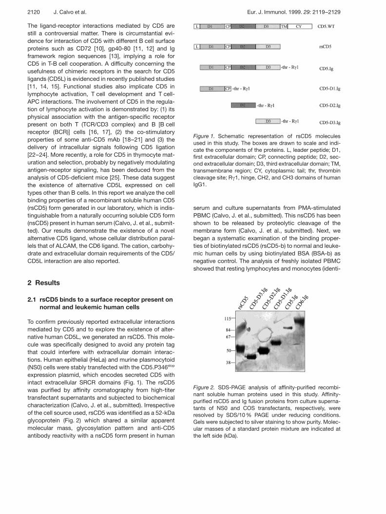
Figure 1. Schematic representation of rsCD5 moleculesused in this study. The boxes are drawn to scale and indi-cate the components of the proteins. L, leader peptide; D1,first extracellular domain; CP, connecting peptide; D2, sec-ond extracellular domain; D3, third extracellular domain; TM,transmembrane region; CY, cytoplasmic tail; thr, thrombincleavage site; R + 1, hinge, CH2, and CH3 domains of humanIgG1.
Figure 2. SDS-PAGE analysis of affinity-purified recombi-nant soluble human proteins used in this study. Affinity-purified rsCD5 and Ig fusion proteins from culture superna-tants of NS0 and COS transfectants, respectively, wereresolved by SDS/10 % PAGE under reducing conditions.Gels were subjected to silver staining to show purity. Molec-ular masses of a standard protein mixture are indicated atthe left side (kDa).
The ligand-receptor interactions mediated by CD5 arestill a controversial matter. There is circumstantial evi-dence for interaction of CD5 with different B cell surfaceproteins such as CD72 [10], gp40-80 [11, 12] and Igframework region sequences [13], implying a role forCD5 in T-B cell cooperation. A difficulty concerning theusefulness of chimeric receptors in the search for CD5ligands (CD5L) is evidenced in recently published studies[11, 14, 15]. Functional studies also implicate CD5 inlymphocyte activation, T cell development and T cell-APC interactions. The involvement of CD5 in the regula-tion of lymphocyte activation is demonstrated by: (1) itsphysical association with the antigen-specific receptorpresent on both T (TCR/CD3 complex) and B [B cellreceptor (BCR)] cells [16, 17], (2) the co-stimulatoryproperties of some anti-CD5 mAb [18–21] and (3) thedelivery of intracellular signals following CD5 ligation[22–24]. More recently, a role for CD5 in thymocyte mat-uration and selection, probably by negatively modulatingantigen-receptor signaling, has been deduced from theanalysis of CD5-deficient mice [25]. These data suggestthe existence of alternative CD5L expressed on celltypes other than B cells. In this report we analyze the cellbinding properties of a recombinant soluble human CD5(rsCD5) form generated in our laboratory, which is indis-tinguishable from a naturally occurring soluble CD5 form(nsCD5) present in human serum (Calvo, J. et al., submit-ted). Our results demonstrate the existence of a novelalternative CD5 ligand, whose cellular distribution paral-lels that of ALCAM, the CD6 ligand. The cation, carbohy-drate and extracellular domain requirements of the CD5/CD5L interaction are also reported.
2 Results
2.1 rsCD5 binds to a surface receptor present onnormal and leukemic human cells
To confirm previously reported extracellular interactionsmediated by CD5 and to explore the existence of alter-native human CD5L, we generated an rsCD5. This mole-cule was specifically designed to avoid any protein tagthat could interfere with extracellular domain interac-tions. Human epithelial (HeLa) and murine plasmocytoid(NS0) cells were stably transfected with the CD5.P346stop
expression plasmid, which encodes secreted CD5 withintact extracellular SRCR domains (Fig. 1). The rsCD5was purified by affinity cromatography from high-titertransfectant supernatants and subjected to biochemicalcharacterization (Calvo, J. et al., submitted). Irrespectiveof the cell source used, rsCD5 was identified as a 52-kDaglycoprotein (Fig. 2) which shared a similar apparentmolecular mass, glycosylation pattern and anti-CD5antibody reactivity with a nsCD5 form present in human
serum and culture supernatants from PMA-stimulatedPBMC (Calvo, J. et al., submitted). This nsCD5 has beenshown to be released by proteolytic cleavage of themembrane form (Calvo, J. et al., submitted). Next, webegan a systematic examination of the binding proper-ties of biotinylated rsCD5 (rsCD5-b) to normal and leuke-mic human cells by using biotinylated BSA (BSA-b) asnegative control. The analysis of freshly isolated PBMCshowed that resting lymphocytes and monocytes (identi-
2120 J. Calvo et al. Eur. J. Immunol. 1999. 29: 2119–2129

Figure 3. Binding of rsCD5 to normal and tumor cells. Cell-bound rsCD5-b was developed with streptavidin-Tri-Colorand analyzed by flow cytometry (filled histograms). BSA-bwas used as negative control (dotted line). Histograms wereoverlaid and subjected to Kolmogorov-Smirnov test. Resultswere categorized based on the percentage of positive cellswhich are included in the non-overlapping areas: high,G 60 %; medium, 30–60 %; low, 10–30 %; and negative,X 10 %. Relative cell number is indicated by the (y) axis, and
fluorescence intensity by the (x) axis.
fied by their characteristic forward and side scatter) bindto rsCD5-b, although with a relative intermediate level ofexpression (Fig. 3). Contrary to that described for gp40-80 [11], the stimulation of PBMC with CD3 plus CD28mAb (10 ? g/ml each) for a 24–96-h time period did notinduce increases in fluorescence intensity with regard tounstimulated cells (data not shown). Similarly the incuba-tion of PBMC for the same period of time with 1000 U/mlIL-4 plus 50 ng/ml GM-CSF, which is known to inducethe differentiation of monocytes into dendritic cells [26],did not result in increased binding of rsCD5 as comparedto unstimulated cells (data not shown). Thymocytes andpurified tonsil B lymphocytes did not show significantbinding to rsCD5-b (Fig. 3). On the contrary, rsCD5-bbound to various tumor cell lines of lymphocytic (T and
B), myelomonocytic and non-hematopoietic origin(Fig. 3). The monocytic and erythromyelomonocyticU937 and K562 cell lines, respectively, exhibited thebrightest fluorescence intensities. The myelocytic THP-1and pro-myelocytic HL60 cell lines displayed low andnegative fluorescence intensity, respectively. The rsCD5binding was low among most of the T cell lines studied(HUT78, Jurkat, HSB-2 and Molt-4), being negative forHPB-ALL and intermediate for CEM cells. The plasmo-cytoid U266 and EBV-transformed Daudi B cell lines dis-played intermediate expression levels, with Namalwaand Raji being negative. The human epithelial cell lineHeLa and HEP-2 gave high and low expression, respec-tively. Interestingly, a human thymic epithelial cell (TEC)line [27] displayed high expression. Taken together, ourresults indicate that rsCD5 binds to a broadly expressedcell surface CD5L. It is worth mentioning that the cellbinding pattern of rsCD5 was not cell source dependent.Purified rsCD5 from both the murine NS0 and the humanHeLa transfectants displayed identical cellular reactivity(data not shown). This indicates that post-translationalmodifications introduced by murine or human cells onrsCD5 do not affect its cell binding characteristics.
2.2 Cell binding characteristics of nsCD5 andCD5.Ig proteins
To determine whether the cell binding characteristicsdisplayed were peculiar to rsCD5, we also investigatedthe cell binding properties of natural or other recombi-nant soluble forms of CD5. They include a CD5 Ig fusionprotein as well as nsCD5 present in human serum and insupernatants from in vitro PMA-stimulated PBMC(Calvo, J. et al., submitted). The human CD5 Ig fusionprotein has been successfully used to identify a novelCD5L, distinct from CD72, present on activated murine Band T cells [11] and on murine B cells [12], and for whichno human counterpart has been reported so far. In ourhands CD5 Ig displayed a cell binding pattern similar tothat described above for rsCD5. Fig. 4A illustrates theCD5 Ig binding results obtained for three representativehuman cell lines, two positive (U937, K562) and one neg-ative (HL60). As a control, we used a human CD6-Igfusion protein which binds ALCAM, a surface receptorexpressed on HL60, but not U937 and K562 cells [6]. Evi-dence for recognition of the same cell surface receptorby rsCD5 and CD5.Ig came from competition bindingassays. As shown in Fig. 4B, the binding of rsCD5-b toU937 was blocked by unlabeled CD5.Ig, but not byCD6.Ig or ALCAM.Ig (at 10 ? g/100 ? l each). Identicalresults were obtained when similar assays were per-formed on K562 cells (Fig. 4B). The blocking effects ofCD5.Ig were dose dependent and saturable (at 20 ? g/100 ? l), as illustrated by Fig. 4C. Taken together, these
Eur. J. Immunol. 1999. 29: 2119–2129 Alternative CD5L receptor 2121

Figure 4. Specific binding of Ig fusion proteins to humancell lines. (A) Binding of CD5 (filled histograms) or CD6 (solidline histograms) fusion proteins to U937, K562 and HL60human cell lines. Bound proteins were developed with FITC-conjugated goat anti-human IgG serum and were analyzedby flow cytometry. (B) U937 and K562 cells were incubatedwith suboptimal doses of rsCD5-b alone (solid line) or in thepresence of unlabeled CD5-Ig (+ CD5-Ig), CD6-Ig (+ CD6-Ig)or ALCAM-Ig (+ ALCAM-Ig) (filled histograms), at 10 ? g/100 ? l each. Streptavidin-Tri-Color was added to developbound rsCD5-b, which was analyzed by flow cytometry.BSA-b was used as negative control (dotted line). (C) Dose-dependent inhibition of rsCD5-b binding to U937 cells withCD5.Ig. Results are expressed as percent inhibition (y axis).The percentage of cells stained by rsCD5-b in the absenceof unlabeled CD5.Ig was considered as 0 % inhibition. The100 % inhibition was reached when the number of stainedcells equaled the number of cells stained with BSA-b. Theamount of CD5.Ig protein (in ? g/100 ? l) is indicated by the(x) axis.
Figure 5. Binding of nsCD5 to human cells. (A) Cell-boundnsCD5-b was developed with streptavidin-Tri-Color andanalyzed by flow cytometry (filled histograms). BSA-b wasused as negative control (dotted line). (B) Suboptimal dosesof nsCD5-b and (C) rsCD5-b were used to stain U937 cellspreincubated for 15 min alone (filled histogram) or in thepresence of 0.5 ? g/100 ? l unlabeled rsCD5 (solid line).Streptavidin-Tri-Color was added to develop bound nsCD5-b or rsCD5.b, which was analyzed by flow cytometry. BSA-bwas used as negative control (dotted line).
results argue for the specificity of the cell interactionmediated by both rsCD5 and CD5.Ig.
The analysis of nsCD5-b purified from PMA-stimulatedPBMC culture supernatants also showed that its cellbinding pattern was similar to rsCD5. Fig. 5A shows theresults obtained with three representative cell lines. Asshown in Fig. 5B, the binding of nonsaturating amountsof nsCD5-b to U937 cells was significantly inhibited bypreincubation of cells with unlabeled rsCD5. The inhibi-tion reached values of 50 %, as measured by reductionin the mean fluorescence intensity channel and was simi-lar to the inhibition achieved with unlabeled rsCD5(Fig. 5C). Higher inhibitions could not be achieved due tothe limiting amounts of rsCD5 purified from culturesupernatants (the approximate yield was 1 mg per 20 lculture supernatant) and the relatively low concentra-tions of rsCD5 present in the competition binding assays(approximately 5 ? g/ml). No inhibition was observed fol-lowing cell preincubation with unlabeled purified unre-lated proteins such as BSA and hemoglobin (Hb) (datanot shown). These results confirm the close similaritybetween nsCD5 and rsCD5 forms, not only at the bio-chemical (molecular mass, N-glycosylation, antibodyreactivity) (Calvo, J. et al., submitted) but at cell bindinglevels.
2122 J. Calvo et al. Eur. J. Immunol. 1999. 29: 2119–2129

Figure 6. Divalent cation and carbohydrate requirements ofrsCD5 binding. (A) U937 cells were incubated with rsCD5-bin PBS containing 2 % FCS (solid line) or HBSS plus 5 mMEDTA (filled histogram) and bound protein developed withstreptavidin-Tri-Color. Binding of BSA-b in PBS was used asnegative control (dotted line). Relative cell number is indi-cated by the (y) axis, and fluorescence intensity by the(x) axis. (B) Binding of rsCD5-b was analyzed on U937 cellsin the presence of D-fructose, D-mannose, N-acetyl-glucosamine, N-acetyl-galactosamine and D-glucose at theindicated concentrations. Results are expressed as percentof inhibition, measured by comparing the percent of stainedcells in the presence or absence of monosaccharides.
2.3 Characteristics of the rsCD5/CD5Linteraction
The properties of the binding of rsCD5 to CD5L was fur-ther characterized on the U937 monocytic cell line,which shows high CD5L surface expression. SinceCD72, a member of the C-type lectin family [28], hasbeen reported to bind CD5, we investigated the divalentcation and the carbohydrate requirements of the rsCD5interaction reported here. The influence of divalent cat-ions on rsCD5 binding was explored by performing cellbinding assays in HBSS medium, which lacks Ca2+ andMg2+. The binding of rsCD5 was unaffected by theabsence of divalent cations. Identical results wereobserved when HBSS was supplemented with either5 mM EDTA (Fig. 6A) or EGTA (not shown), thus confirm-ing the cation independence of rsCD5 cell binding. Thepossible carbohydrate interference on rsCD5 cell bind-ing was examined by performing binding assays in thepresence of different concentrations (0.1–1000 mM) ofseveral monosaccharides (D-glucose, D-fructose, D-mannose, N-acetyl-glucosamine and N-acetyl-galactosamine). As shown in Fig. 6B, we detected signif-icant inhibitory effects (above 50 % inhibition) only whenhigh molar concentrations (0.1–1 M) of D-fructose and D-mannose were used, with D-fructose being the mostpotent inhibitor. The percent of inhibition induced by D-glucose, N-acetyl-glucosamine and N-acetyl-galactosamine was always below 40 %. At relative lowmolar concentrations (0.1–10 mM), none of the carbohy-drates tested showed significant inhibitory effects (below25 %). These results indicate the relatively poor contribu-tion of the carbohydrates analyzed on rsCD5 binding,compared to the receptor-ligand interactions mediatedby C-type lectin molecules [28, 29].
The specificity of the rsCD5-CD5L interaction describedherein was also explored by competition binding experi-ments with mAb against different cell surface antigensexpressed on U937 cells. This includes mAb specific forHLA class II, CD1a, CD43, CD45, CD50, CD69, CD72and CD100 molecules. U937 cells were preincubatedwith saturating amounts of affinity-purified mAb beforestaining with rsCD5-b. None of the mAb used displayedinhibitory effects on the rsCD5 binding. Fig. 7A (top) illus-trates the results obtained with two representative mAb(CD45 and CD72). Interestingly, we used several CD72mAb, one of which (BU40) has been previously reportedto block the interaction of a membrane form of humanCD5 with the B cell surface antigen CD72 [10]. Toexclude CD72 as the CD5L binding to rsCD5, we per-formed cell binding studies on the P815-CD72+ trans-fectant. While this cell expresses high surface levels ofCD72, as measured by reactivity with anti-CD72 mAb(data not shown), it was unable to bind rsCD5 (Fig. 3).
This result agrees with the differences found between thepattern of cellular distribution reported for CD72 [10] andthe CD5L described here, and also demonstrates thatrsCD5 does not bind to CD72.
2.4 mAb competition-binding assays
Competition-binding experiments were also performedwith different CD5 mAb (Cris-1, Leu-1, UCTH2, NUT-PAN, BL1a, F101-15, F145-GF3, OKT1 and 83-C4). Inthese assays, affinity-purified CD5 mAb were preincu-bated with rsCD5-b before staining of U937 cells. Noneof the mAb tested were able to block the binding ofrsCD5 to U937. Representative results with two mAb
Eur. J. Immunol. 1999. 29: 2119–2129 Alternative CD5L receptor 2123
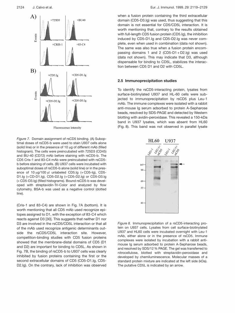
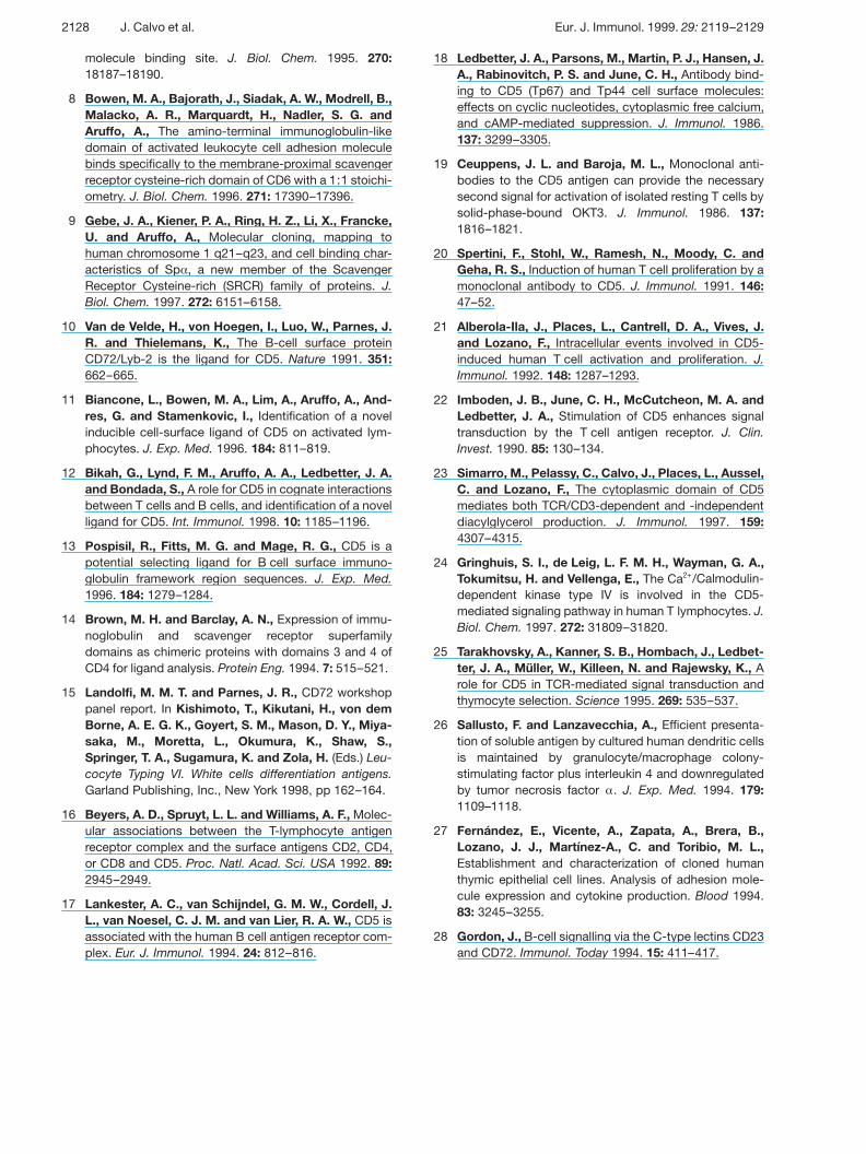
Figure 7. Domain assignment of rsCD5 binding. (A) Subop-timal doses of rsCD5-b were used to stain U937 cells alone(solid line) or in the presence of 10 ? g of different mAb (filledhistogram). The cells were preincubated with 725D3 (CD45)and BU-40 (CD72) mAb before staining with rsCD5-b. TheCD5 Cris-1 and 83-C4 mAb were preincubated with rsCD5-b before staining of cells. (B) U937 cells were incubated withsuboptimal doses of rsCD5-b alone (solid line) or in the pres-ence of 10 ? g/100 ? l unlabeled CD5.Ig (+ CD5-Ig), CD5-D1.Ig (+ CD-D1.Ig), CD5-D2.Ig (+ CD5-D2.Ig) or CD5-D3.Ig(+ CD5-D3.Ig) (filled histograms). Bound rsCD5-b was devel-oped with streptavidin-Tri-Color and analyzed by flowcytometry. BSA-b was used as a negative control (dottedline).
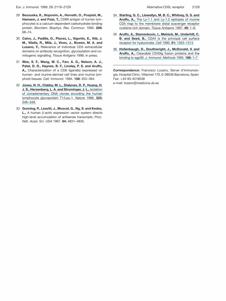
Figure 8. Immunoprecipitation of a rsCD5-interacting pro-tein on U937 cells. Lysates from cell surface-biotinylatedU937 and HL60 cells were incubated overnight with Leu-1mAb, either alone or in the presence of rsCD5. Immunecomplexes were isolated by incubation with a rabbit anti-mouse Ig serum adsorbed to protein A-Sepharose beads,and resolved by SDS/12 % PAGE. The gel was transferred tonitrocellulose, blotted with streptavidin-peroxidase anddeveloped by chemiluminescence. Molecular masses of astandard protein mixture are indicated at the left side (kDa).The putative CD5L is indicated by an arrow.
(Cris-1 and 83-C4) are shown in Fig. 7A (bottom). It isworth mentioning that all CD5 mAb used recognize epi-topes assigned to D1, with the exception of 83-C4 whichreacts against D3 [30]. This suggests that neither D1 norD3 are involved in the rsCD5/CD5L interaction or that allof the mAb used recognize antigenic determinants out-side the rsCD5/CD5L interaction site. However,competition-binding studies with CD5 fusion proteinsshowed that the membrane-distal domains of CD5 (D1and D2) are important for binding to CD5L. As shown inFig. 7B, the binding of rsCD5-b to U937 cells was clearlyinhibited by fusion proteins containing the first or thesecond extracellular domains of CD5 (CD5-D1.Ig, CD5-D2.Ig). On the contrary, lack of inhibition was observed
when a fusion protein containing the third extracellulardomain (CD5-D3.Ig) was used, thus suggesting that thisdomain is not essential for CD5/CD5L interaction. It isworth mentioning that, contrary to the results obtainedwith full-length CD5 fusion protein (CD5.Ig), the inhibitioninduced by CD5-D1.Ig and CD5-D2.Ig was never com-plete, even when used in combination (data not shown).The same was also true when a fusion protein encom-passing domains 1 and 2 (CD5-D1 + D2.Ig) was used(data not shown). This may indicate that D3, althoughdispensable for binding to CD5L, stabilizes the interac-tion between CD5 D1 and D2 with CD5L.
2.5 Immunoprecipitation studies
To identify the rsCD5-interacting protein, lysates fromsurface-biotinylated U937 and HL-60 cells were sub-jected to immunoprecipitation by rsCD5 plus Leu-1mAb. The immune complexes were isolated with a rabbitanti-mouse Ig serum adsorbed to protein A-Sepharosebeads, resolved by SDS-PAGE and detected by Westernblotting with avidin-peroxidase. This revealed a 150-kDaband in U937 lysates, which was absent from HL60(Fig. 8). This band was not observed in parallel lysate
2124 J. Calvo et al. Eur. J. Immunol. 1999. 29: 2119–2129

samples immunoprecipitated with a rabbit anti-mouse Igserum adsorbed to protein A-Sepharose beads in com-bination with Leu-1 mAb. However, under the same con-ditions, CD5 fusion proteins failed to immunoprecipitatethe 150-kDa band. Similarly, attempts at cross-linkingrsCD5 to 125I surface-labeled proteins with 3,3' ditiobissulfosuccinimidyl proprionate (DTSSP), which have beensuccessfully used to identify activated leukocyte celladhesion molecule (ALCAM) [6], did not result in theimmunoprecipitation of the 150-kDa band (data notshown). Since these data do not prove that the 150-kDaspecies is itself the CD5L, elucidation of the identity ofCD5L will require further investigation.
3 Discussion
In the present work, we provide evidence of the exis-tence of a novel ligand of human CD5 (CD5L) by exploit-ing the cell binding properties of recombinant (rsCD5,CD5.Ig) and natural (nsCD5) soluble human CD5 mole-cules. The novel CD5L reported here is a broadlyexpressed cell surface protein, which is found on periph-eral blood lymphocytes and monocytes as well as onseveral lymphocytic (T and B), myelomonocytic and epi-thelial cell lines. This observation indicates that CD5/CD5L interactions may play a role not only in T-B cell co-stimulation but also in other cell-mediated interactionsinvolving lymphocytes and other APC such as mono-cytes and epithelial cells.
The CD5L identified in the present study appears to bedistinct from those previously reported (CD72, gp40–80and framework VHa2 region) [10–13]. CD72 is a 42-kDatype II glycoprotein belonging to a family of Ca2+-dependent C-type lectins [28], which is constitutivelyexpressed on all cells of B lymphoid lineage, with theexception of plasma cells. We find that rsCD5 is unableto bind purified tonsillar B cells and binds to only two ofthe B cell lines analyzed (one of them, U266, of plasmo-cytoid origin). We also find lack of binding of rsCD5 to aCD72+ transfectant and lack of blocking effects of CD72mAb. The same results were obtained using rsCD5 frommouse (NS0) or human (HeLa) sources, arguing againstspecies-specific differences (e.g. glycosylation) asresponsible for the lack of binding to CD72. Theseresults make our rsCD5 similar to previously reportedchimeric CD5 proteins (CD5D1-CD4D3 + 4 and CD5.Ig)which are unable to bind to CD72 [11, 14, 15].
Recently, a novel inducible cell surface CD5L of40–80 kDa has been reported on murine B splenocytes(transiently expressed 1–3 days after stimulation of sple-nocytes with CD3 plus CD28 mAb) and murine T cellclones (10–12 days after stimulation with antigen in the
presence of APC and IL-2) [11, 12]. This has been dem-onstrated by the use of a human CD5-receptor globin(CD5.Ig) composed of the extracellular region of humanCD5 fused to the constant region of human IgG1, andpurified from CHO cells (the same was not true for COScell-derived CD5.Ig). Although an identical CD5.Ig fusionprotein was used in this study and no human equivalenthas been reported so far, several facts argue against thepossibility that the CD5L identified herein corresponds togp40–80: (1) constitutive expression by lymphocytesand monocytes, (2) broad cellular distribution, (3) lack ofinducibility following stimulation with CD3 plus CD28mAb. Similarly, our observations also argue against thepossibility that rsCD5 binds to human framework VH
regions, as recently proposed for rabbits [13]. This wouldbe possible only in the case that rsCD5 binds the frame-work region of a restricted VH family expressed by two ofthe four B cell lines analyzed in our study.
A relevant aspect of the CD5/CD5L interaction reportedhere is that it recalls some characteristics of the CD6/ALCAM interaction. Both interactions are divalent cationindependent and seem to be mediated by a similarlybroad distributed ligand. A detailed examination of theircell reactivity pattern shows that both CD5L and ALCAMare similarly expressed on peripheral monocytes andlymphocytes, as well as on several cell lines of lympho-cytic (T and B), monocytic and epithelial origin [6, 31].Interestingly, both CD5L and ALCAM are expressed onthymic epithelial cells, suggesting that CD5 and CD6may have a role in T cell development and maturation.Since both CD5 and CD6 are early T cell markers, theymay play an important role in T cell maturation by medi-ating interactions between thymocytes and thymic epi-thelial cells or other resident APC. This is consistent withprevious data showing that CD5 deficiency influencesthe development and selection of thymocytes in TCR-transgenic mice [25]. Our data also support a model inwhich the extracellular region of CD5 represents a versa-tile platform mediating different types of molecular inter-actions. The most N-terminal domains (D1 and D2)would bind to a broadly distributed ligand reported hereand the most membrane-proximal domains would inter-act with the other ligands (e.g. CD72, gp40-80 or VH
framework). An intriguing aspect of the presentlyreported ligands for CD5 and CD6 is that they fall intodistinct families. CD5 and CD6 are the closest membersof the SRCR family. Both have a similar extracellulardomain organization, tissue distribution and co-stimulatory activity in T cells. These observations,together with the finding that CD5 and CD6 genes areclosely located on human chromosome 11, suggest thatthey are likely to have arisen from duplication of a com-mon ancestral gene [4]. While CD6 binds to a ALCAM, amember of the Ig superfamily, CD5 was reported to bind
Eur. J. Immunol. 1999. 29: 2119–2129 Alternative CD5L receptor 2125

CD72, a protein homologous to C-type lectins [28]. Thiswould mean that two highly related genes, CD5 andCD6, have evolved to bind to different types of ligands.Further characterization and cloning of CD5L 150 willreveal if CD5 and CD6 bind to similar pairs of ligands.
In summary, our results indicate the existence of a novelalternative ligand for CD5. Although elucidation of theidentity of this CD5L deserves further investigation, thereported CD5/CD5L interaction is reminiscent of thatof CD6 and ALCAM and opens new pathways toexplore CD5 function in T/B cell maturation and cellactivation.
4 Materials and methods
4.1 Cells
All the human cell lines used in this study were from theAmerican Tissue Culture Collection and were cultured inRPMI 1640 medium (Life Technologies, Gaithersburg, MD)supplemented with 100 ? g/ml gentamycin and 10 % FCS.The murine B cell line NS0 was a gift from C. Milstein (MRC,Cambridge, GB). The murine P815 transfectant expressinghuman CD72 was kindly provided by Dr. Lewis L. Lanier(DNAX Research Institute, Palo Alto, CA). PBMC fromhealthy donors were isolated by density centrifugation gra-dients on Ficoll-Hypaque (Pharmacia Biotech, Uppsala,Sweden). Purified B cells were obtained from tonsillar cellsuspensions treated by two rounds of rabbit complement-mediated lysis with mAb to CD3, CD4, CD8 and CD14. Thy-mocytes were obtained from thymus specimens of childrenundergoing cardiac surgery. The human TEC [27] was a kindgift from M. L. Toribio (CBM, Madrid).
4.2 Monoclonal antibodies
The F145-1G3 (CD5) mAb was provided by D. Carriere(Sanofi Recherche, Montpellier). The CD1a (33.7 and NA1/34) mAb were provided by C. Milstein. The Leu-1 (CD5;IgG2a), OKT3 (CD3) and CH/4 (CD69) mAb were from acommercial source (Becton Dickinson, Ortho Diagnosticsand Caltag Laboratories, respectively). The Cris-1 (CD5;IgG2a), 83-C4 (CD5; IgG1), 152-2E10 (CD28; IgG1), 843C1(CD43; IgG1), 725D3 (CD45; IgG2a), 1011D2 (CD50; IgG1),1331C6 (CD100; IgM) and 312B1 (HLA class II; IgG2b) mAbwere produced in our laboratory. The BU.40 (CD72; IgG2a)mAb was kindly provided by D. Hardie (University of Birmin-gham). mAb from the CD72 and CD5 panel of the 5th and6th International Workshops on Leukocyte Antigens wereprovided by P. Engel and K. Okumura, respectively. mAbwere affinity-purified using a 5-ml HiTrapTM Protein G col-umn and then coupled to CNBr-activated Sepharose 4B fol-lowing the manufacturer’s instructions (Pharmacia BiotechAB). EZ-LinkTM Sulfo-NHS-LC-Biotin (Pierce, Rockford, IL)
was used to label purified mAb following the manufacturer’sinstructions.
4.3 Expression of rsCD5 molecules
Construction for expression of rsCD5 (CD5.P346stop) wasdone by EcoRI/KpnI cloning of two previously reportedcDNA clones, pT1-1 and pT1-2 [32], into the EcoRI site of amodified version of the pH g APr-1-neo expression vector[33]. The EcoRI/EcoRI fragment from pT1-2 cloned intoM13mp18 was first subjected to oligonucleotide-directedin vitro mutagenesis (Kunkel’s method) to introduce a pre-mature stop codon at P346, which in turn created a newMseI site. The nucleotide changes were checked by double-stranded DNA sequencing and plasmids purified by cesiumchloride density gradients. For stable transfection, 12 × 106
cells were resuspended with 0.2 ml RPMI 1640 mediumsupplemented with 10 % FCS and electroporated in a 0.4-cm gap curvette with 10 ? g NdeI-linearized expression vec-tor. The power settings were 0.2 kV and 960 ? F (Gene Pulserapparatus, Bio-Rad, Richmond, CA). The pulsed cells wereplated at 105 cells/well in flat-bottom 96-well microtiterplates, and 48 h later cells were selected for neor with2 mg/ml G418 (Life Technologies). Drug-resistant coloniesproducing rsCD5 were screened and selected for furtherexpansion by testing culture supernatants with a CD5-specific ELISA (Calvo, J. et al., submitted).
4.4 Production, purification and biochemical analysisof nsCD5 and rsCD5
The nsCD5 was purified from 96-h culture supernatants ofPMA-stimulated PBMC. The rsCD5 was from culture super-natants of NS0 and HeLa stable CD5.P346stop transfectants.For purification purposes, all the samples were subjected toprecipitation with 70 % ammonium sulfate as a concentra-tion step. After extensive dialysis against PBS/0.02 %sodium azide, the samples were affinity-purified by passingthrough a Cris-1 mAb-coupled CNBr-activated Sepharose4B column. The column was extensively washed with PBS/0.5 M NaCl/1 % NP40 and bound material eluted with PBS/4 M MgCl2 in 1-ml fractions. Elution was monitored byabsorbance at 280 nm to identify the peak fractions, whichwere pooled and dialyzed against PBS containing 0.02 %sodium azide. Elution fractions were tested by ELISA (Calvo,J. et al., submitted) and Western blotting (see above). Puritywas assessed by silver staining of SDS/PAGE gels. PurifiedrsCD5 and nsCD5 were biotinylated with EZ-LinkTM Sulfo-NHS-LC-Biotin (Pierce) following the manufacturer’s instruc-tions.
4.5 Expression of human soluble Ig fusion proteins
The construction of human ALCAM.Ig and CD6.Ig has beenreported elsewhere [6]. Recombinant human CD5.Ig fusion
2126 J. Calvo et al. Eur. J. Immunol. 1999. 29: 2119–2129

proteins similar to those described for mouse CD5 [34] wereprepared and characterized. Plasmids encoding solublehuman CD5.Ig fusion proteins were produced by methodspreviously described [8]. Briefly, CD5.Ig was made by clon-ing the cDNA of the extracellular region of CD5 into anexpression plasmid containing genomic DNA of the hinge,CH2, and CH3 domains of human IgG1 [35]. Complemen-tary DNA fragments encoding individual or multiple domainsof human CD5 were obtained by PCR and cloned into anexpression plasmid containing a cDNA fragment encoding athrombin cleavage site followed by the hinge, CH2, and CH3domains of human IgG1 [36]. The fusion proteins containedthe following residues of the mature CD5 peptide: CD5.Ig,R1-P348; CD5-D1.Ig, R1-R134; CD5-D2.Ig, L135-Q251;CD5-D3.Ig, V250-P348. Proteins were produced by tran-sient expression in COS cells and purified by protein A-Sepharose affinity chromatography.
4.6 Cell binding experiments and flow cytometryanalysis
For cell binding experiments, 105 cells were incubated on icefor 30 min in 100 ? l stain buffer (PBS/2 % FCS/10 % humanAB serum/0.05 % sodium azide) containing 10 ? g/ml bioti-nylated rsCD5 or nsCD5. BSA-b or Hb-b were used as neg-ative fluorescence controls and all incubations were made inthe presence of 10 % human AB serum to avoid nonspecificbinding. After washing with PBS/2 % FCS/0.02 % sodiumazide, cells were incubated for 30 min on ice with streptavi-din Tri-color (Caltag, Laboratories) and washed again. Forcompetition-binding experiments, cells were preincubatedfor 15 min on ice with unlabeled rsCD5, Ig fusion proteins ormAb specific for different leukocyte surface moleculesexpressed on U937, prior to addition of non-saturatingamounts of biotinylated nsCD5 or rsCD5. When anti-CD5mAb were used in competition-binding experiments, theywere preincubated for 15 min on ice with rsCD5-b, and thenadded to U937 cells. Assays designed to test the direct cellbinding of Ig fusion proteins were performed in the absenceof human AB serum in the stain buffer and by using a FITC-conjugated goat anti-human Ig serum (Dako A/S, Denmark).Cell fluorescence intensity was analyzed by flow cytometryon a FACStar Plus (Becton Dickinson, Mountain View, CA).
4.7 Immunoprecipitation studies
Cell surface biotinylation of 10 × 106 U937 and HL60 cellswas performed at room temperature for 30 min, by resus-pending cells at 25 × 106 cells/ml in PBS pH 8.0 containing0.5 mg EZ-LinkTM Sulfo-NHS-LC-Biotin. After three washeswith ice-cold PBS, cells were disrupted for 10 min with ice-cold 1 ml lysis buffer (150 mM NaCl, 1 % NP40, 10 mM Tris,2 mM EDTA, 1 mM NaF, 1 mM phenylmethylsulfonyl fluoride(PMSF), 1 ? g/ml aprotinin, 1 ? g/ml leupeptin) and centri-fuged at 10 000 × g for 5 min. Solubilized proteins wereincubated overnight at 4 °C with 100 ? g rsCD5 plus 10 ? g
Leu-1 mAb and 40 ? l of packed protein-A Sepharose beadsadsorbed to a rabbit anti-mouse Ig serum (Dako). Immunecomplexes were washed five times in lysis buffer and ana-lyzed by SDS/12 % PAGE under reducing conditions. Biotin-labeled proteins were electrotransferred to a nitrocellulosemembrane and developed with peroxidase-labeled strepta-vidin (Boehringer Mannheim) by the enhanced chemilumi-nescence (ECL) system (Amersham).
Acknowledgements: The authors wish to thank R. Vilella,D. Carriere, L. L. Lanier, C. Milstein, D. Hardie, P. Engel andM. L. Toribio for kindly providing valuable reagents; J. Milaand J. Barcelo for technical assistance; and M. Isamat, J.Perez-Villar and K. S. Campbell for critically reviewing themanuscript. This work was supported by Fondo de Investi-gacion Sanitaria (FIS 95/1102, FIS 97/0669) and ComisionInterministerial de Ciencia y Tecnologıa (SAF 98/045). J. C.,O. P. and J. M. V. are recipients of fellowships from Funda-cion LAIR, CIRIT (1998FI/839) and IDIBAPS (330017),respectively.
5 References
1 Hardy, R. R. and Hayakawa, K., CD5 B cells, a fetalB cell lineage. Adv. Immunol. 1994. 55: 297–339.
2 Resnick, D., Pearson, A. and Krieger, M., The SRCRsuperfamily: a family reminiscent of the Ig superfamily.Trends Biochem. Sci. 1994. 19: 5–8.
3 Aruffo, A., Bowen, M. A., Patel, D. D., Haynes, B. F.,Starling, G. C., Gebe, J. A. and Bajorath, J., CD6-ligand interactions: a paradigm for SRCR domain func-tion? Immunol. Today 1997. 18: 498–504.
4 Bowen, M. A., Whitney, G. S., Neubauer, M., Starling,G. C., Palmer, D., Zhang, J., Nowak, N. J., Shows, T.B. and Aruffo, A., Structure and chromosomal locationof the human CD6 gene. Detection of five human CD6isoforms. J. Immunol. 1997. 158: 1149–1156.
5 Patel, D. D., Wee, S. F., Whichard, L. P., Bowen, M. A.,Pesando, J. M., Aruffo, A. and Haynes, B. F., Identifi-cation and characterization of a 100-kDa ligand for CD6on human thymic epithelial cells. J. Exp. Med. 1995.181: 1563–1568.
6 Bowen, M. A., Patel, D. D., Li, X., Modrell, B.,Malacko, A. R., Wang, W. C., Marquardt, H., Neu-bauer, M., Pesando, J. M., Francke, U., Haynes, B. F.and Aruffo, A., Cloning, mapping, and characterizationof activated leukocyte-cell adhesion molecule (ALCAM),a CD6 ligand. J. Exp. Med. 1995. 181: 2213–2220.
7 Whitney, G. S., Starling, G. C., Bowen, M. A., Modrell,B., Siadak, A. W. and Aruffo, A., The membrane-proximal scavenger receptor cysteine-rich domain ofCD6 contains the activated leukocyte cell adhesion
Eur. J. Immunol. 1999. 29: 2119–2129 Alternative CD5L receptor 2127
molecule binding site. J. Biol. Chem. 1995. 270:18187–18190.
8 Bowen, M. A., Bajorath, J., Siadak, A. W., Modrell, B.,Malacko, A. R., Marquardt, H., Nadler, S. G. andAruffo, A., The amino-terminal immunoglobulin-likedomain of activated leukocyte cell adhesion moleculebinds specifically to the membrane-proximal scavengerreceptor cysteine-rich domain of CD6 with a 1:1 stoichi-ometry. J. Biol. Chem. 1996. 271: 17390–17396.
9 Gebe, J. A., Kiener, P. A., Ring, H. Z., Li, X., Francke,U. and Aruffo, A., Molecular cloning, mapping tohuman chromosome 1 q21–q23, and cell binding char-acteristics of Sp § , a new member of the ScavengerReceptor Cysteine-rich (SRCR) family of proteins. J.Biol. Chem. 1997. 272: 6151–6158.
10 Van de Velde, H., von Hoegen, I., Luo, W., Parnes, J.R. and Thielemans, K., The B-cell surface proteinCD72/Lyb-2 is the ligand for CD5. Nature 1991. 351:662–665.
11 Biancone, L., Bowen, M. A., Lim, A., Aruffo, A., And-res, G. and Stamenkovic, I., Identification of a novelinducible cell-surface ligand of CD5 on activated lym-phocytes. J. Exp. Med. 1996. 184: 811–819.
12 Bikah, G., Lynd, F. M., Aruffo, A. A., Ledbetter, J. A.and Bondada, S., A role for CD5 in cognate interactionsbetween T cells and B cells, and identification of a novelligand for CD5. Int. Immunol. 1998. 10: 1185–1196.
13 Pospisil, R., Fitts, M. G. and Mage, R. G., CD5 is apotential selecting ligand for B cell surface immuno-globulin framework region sequences. J. Exp. Med.1996. 184: 1279–1284.
14 Brown, M. H. and Barclay, A. N., Expression of immu-noglobulin and scavenger receptor superfamilydomains as chimeric proteins with domains 3 and 4 ofCD4 for ligand analysis. Protein Eng. 1994. 7: 515–521.
15 Landolfi, M. M. T. and Parnes, J. R., CD72 workshoppanel report. In Kishimoto, T., Kikutani, H., von demBorne, A. E. G. K., Goyert, S. M., Mason, D. Y., Miya-saka, M., Moretta, L., Okumura, K., Shaw, S.,Springer, T. A., Sugamura, K. and Zola, H. (Eds.) Leu-cocyte Typing VI. White cells differentiation antigens.Garland Publishing, Inc., New York 1998, pp 162–164.
16 Beyers, A. D., Spruyt, L. L. and Williams, A. F., Molec-ular associations between the T-lymphocyte antigenreceptor complex and the surface antigens CD2, CD4,or CD8 and CD5. Proc. Natl. Acad. Sci. USA 1992. 89:2945–2949.
17 Lankester, A. C., van Schijndel, G. M. W., Cordell, J.L., van Noesel, C. J. M. and van Lier, R. A. W., CD5 isassociated with the human B cell antigen receptor com-plex. Eur. J. Immunol. 1994. 24: 812–816.
18 Ledbetter, J. A., Parsons, M., Martin, P. J., Hansen, J.A., Rabinovitch, P. S. and June, C. H., Antibody bind-ing to CD5 (Tp67) and Tp44 cell surface molecules:effects on cyclic nucleotides, cytoplasmic free calcium,and cAMP-mediated suppression. J. Immunol. 1986.137: 3299–3305.
19 Ceuppens, J. L. and Baroja, M. L., Monoclonal anti-bodies to the CD5 antigen can provide the necessarysecond signal for activation of isolated resting T cells bysolid-phase-bound OKT3. J. Immunol. 1986. 137:1816–1821.
20 Spertini, F., Stohl, W., Ramesh, N., Moody, C. andGeha, R. S., Induction of human T cell proliferation by amonoclonal antibody to CD5. J. Immunol. 1991. 146:47–52.
21 Alberola-Ila, J., Places, L., Cantrell, D. A., Vives, J.and Lozano, F., Intracellular events involved in CD5-induced human T cell activation and proliferation. J.Immunol. 1992. 148: 1287–1293.
22 Imboden, J. B., June, C. H., McCutcheon, M. A. andLedbetter, J. A., Stimulation of CD5 enhances signaltransduction by the T cell antigen receptor. J. Clin.Invest. 1990. 85: 130–134.
23 Simarro, M., Pelassy, C., Calvo, J., Places, L., Aussel,C. and Lozano, F., The cytoplasmic domain of CD5mediates both TCR/CD3-dependent and -independentdiacylglycerol production. J. Immunol. 1997. 159:4307–4315.
24 Gringhuis, S. I., de Leig, L. F. M. H., Wayman, G. A.,Tokumitsu, H. and Vellenga, E., The Ca2+/Calmodulin-dependent kinase type IV is involved in the CD5-mediated signaling pathway in human T lymphocytes. J.Biol. Chem. 1997. 272: 31809–31820.
25 Tarakhovsky, A., Kanner, S. B., Hombach, J., Ledbet-ter, J. A., Müller, W., Killeen, N. and Rajewsky, K., Arole for CD5 in TCR-mediated signal transduction andthymocyte selection. Science 1995. 269: 535–537.
26 Sallusto, F. and Lanzavecchia, A., Efficient presenta-tion of soluble antigen by cultured human dendritic cellsis maintained by granulocyte/macrophage colony-stimulating factor plus interleukin 4 and downregulatedby tumor necrosis factor § . J. Exp. Med. 1994. 179:1109–1118.
27 Fernandez, E., Vicente, A., Zapata, A., Brera, B.,Lozano, J. J., Martınez-A., C. and Toribio, M. L.,Establishment and characterization of cloned humanthymic epithelial cell lines. Analysis of adhesion mole-cule expression and cytokine production. Blood 1994.83: 3245–3255.
28 Gordon, J., B-cell signalling via the C-type lectins CD23and CD72. Immunol. Today 1994. 15: 411–417.
2128 J. Calvo et al. Eur. J. Immunol. 1999. 29: 2119–2129
29 Bezouska, K., Nopovim, A., Horvath, O., Pospisil, M.,Hamann, J. and Feizi, T., CD69 antigen of human lym-phocytes is a calcium-dependent carbohydrate-bindingprotein. Biochem. Biophys. Res. Commun. 1995. 208:68–74.
30 Calvo, J., Padilla, O., Places, L., Vigorito, E., Vila, J.M., Vilella, R., Mila, J., Vives, J., Bowen, M. A. andLozano, F., Relevance of individual CD5 extracellulardomains on antibody recognition, glycosylation and co-mitogenic signalling. Tissue Antigens 1999, in press.
31 Wee, S. F., Wang, W. C., Farr, A. G., Nelson, A. J.,Patel, D. D., Haynes, B. F., Linsley, P. S. and Aruffo,A., Characterization of a CD6 ligand(s) expressed onhuman- and murine-derived cell lines and murine lym-phoid tissues. Cell. Immunol. 1994. 158: 353–364.
32 Jones, N. H., Clabby, M. L., Dialynas, D. P., Huang, H.J. S., Herzenberg, L. A. and Strominger, J. L., Isolationof complementary DNA clones encoding the humanlymphocyte glycoprotein T1/Leu-1. Nature 1986. 323:346–349.
33 Gunning, P., Leavitt, J., Muscat, G., Ng, S. and Kedes,L., A human g -actin expression vector system directshigh-level accumulation of antisense transcripts. Proc.Natl. Acad. Sci. USA 1987. 84: 4831–4835.
34 Starling, G. C., Llewellyn, M. B. C., Whitney, G. S. andAruffo, A., The Ly-1.1 and Ly-1.2 epitopes of murineCD5 map to the membrane distal scavenger receptorcysteine-rich domain. Tissue Antigens 1997. 49: 1–6.
35 Aruffo, A., Stamenkovic, I., Melnick, M., Underhill, C.B. and Seed, B., CD44 is the principal cell surfacereceptor for hyaluronate. Cell 1990. 61: 1303–1313.
36 Hollenbaugh, D., Douthwright, J., McDonald, V. andAruffo, A., Cleavable CD40Ig fusion proteins and thebinding to sgp39. J. Immunol. Methods 1995. 188: 1–7.
Correspondence: Francisco Lozano, Servei d’Immunolo-gia, Hospital Clınic, Villarroel 170, E-08036 Barcelona, SpainFax: +34-93-4518038e-mail: lozano — medicina.ub.es
Eur. J. Immunol. 1999. 29: 2119–2129 Alternative CD5L receptor 2129