First experimental identification of Ras-inhibitor binding interface using a water-soluble Ras...
6
First experimental identification of Ras-inhibitor binding interface using a water-soluble Ras ligand Alessandro Palmioli a , Elena Sacco a , Sherwin Abraham c , Celestine J. Thomas b , Alessandro Di Domizio a , Luca De Gioia a , Vadim Gaponenko c , Marco Vanoni a , Francesco Peri a, * a Department of Biotechnology and Biosciences, University of Milano-Bicocca, Piazza della Scienza, 2, 20126 Milano, Italy b Center for Biomolecular Structure and Dynamics (CBSD), Chem-218, 32 Campus Dr, University of Montana, Missoula, MT 59812, United States c Department of Biochemistry and Molecular Genetics University of Illinois at Chicago, 900 S. Ashland Ave. Chicago, IL 60607, United States article info Article history: Received 10 March 2009 Revised 26 May 2009 Accepted 27 May 2009 Available online 30 May 2009 Keywords: Ras protein Sugars Synthesis Anticancer agents NMR Molecular modeling abstract By combining in the same molecule Ras-interacting aromatic moieties and a sugar, we prepared a water- soluble Ras ligand that binds Ras and inhibits guanine nucleotide exchange. With this compound it was possible to determine experimentally by a 15 N-edited HSQC NMR experiment the ligand-Ras binding interface. Ó 2009 Elsevier Ltd. All rights reserved. Ras, a key member in the super-family of small GTPases, is an essential component of signal transduction pathways that regulate cell growth, proliferation, differentiation and apoptosis. 1–3 Operat- ing as molecular switches, Ras GTPases, assisted by guanine exchange factors (GEFs), undergo nucleotide exchange, allowing them to rapidly cycle from the inactive GDP 4 bound state to the activated GTP bound state. The active Ras, aided by GTPase activat- ing proteins (GAPs), hydrolyses the bound GTP to GDP and returns to the inactive GDP bound state. 5,6 Alteration of this deactivating reaction is a common biochemical defect associated with onco- genic Ras mutants. 7–9 Oncogenic Ras mutants exhibit decreased hydrolytic efficiency due to reduced affinities towards GAP proteins. 10 Identifying small drug molecules that selectively interact with oncogenic Ras proteins and inhibit constitutive Ras activation is a challenging and a still largely unsolved task. The prolonged life- times of activated oncogenic Ras mutants can, in principle, be reduced by the action of drugs that are capable of converting the oncogenic states to behave like normal phenotypes. This can be achieved by molecules that either inhibit the process of GEF-cata- lyzed GTP loading on inactive Ras GDP 11,12 or by simply accelerat- ing the GTP hydrolysis in the active Ras GTP complex. 13–15 While several efforts have been made in developing agents that acceler- ate the rate of GTP hydrolysis (self-hydrolyzing GTP analogues), very limited strategies directed at inhibiting GEF-catalyzed nucle- otide exchange have been undertaken. To address this issue, our group has been actively engaged in developing sugar-derived ligands that can inhibit GEF-promoted guanine nucleotide exchange on H-Ras. Several generations of compounds, composed of a sugar moiety or a linear spacer to which aromatic pharmaco- phore groups are covalently linked, were synthesized and evalu- ated for Ras inhibitory activity. 16–20 These small organic molecules (whose MW ranges from 300 to 500 Da) bind to H- Ras GDP with micromolar affinity and inhibit GEF-Ras interaction thereby preventing guanine nucleotide exchange. All of these inhibitors seem to recognize a common binding site on Ras. In sil- ico docking studies indicated that amino acid residues Y96, G60, Q61 and E62 of H-Ras exhibit strong interactions with these inhib- itors. Hence a binding interface that partially overlaps with the Switch II region (residues 60–76) was identified to be a plausible binding pocket. 17,18 Our earlier experiments using Surface Plasmon Resonance (SPR) studies indicated that arabinose and glucose-de- rived compounds interfere with the interaction of GEF molecules with H-Ras, reinforcing our hypothesis that these inhibitors bind to a site proximal to the Switch II region. 20 However, the exact mode of action of these nucleotide exchange inhibitors is currently unknown since no structure of an inhibitor bound Ras GDP 0960-894X/$ - see front matter Ó 2009 Elsevier Ltd. All rights reserved. doi:10.1016/j.bmcl.2009.05.107 * Corresponding author. E-mail address: [email protected] (F. Peri). Bioorganic & Medicinal Chemistry Letters 19 (2009) 4217–4222 Contents lists available at ScienceDirect Bioorganic & Medicinal Chemistry Letters journal homepage: www.elsevier.com/locate/bmcl
Transcript of First experimental identification of Ras-inhibitor binding interface using a water-soluble Ras...

Bioorganic & Medicinal Chemistry Letters 19 (2009) 4217–4222
Contents lists available at ScienceDirect
Bioorganic & Medicinal Chemistry Letters
journal homepage: www.elsevier .com/ locate/bmcl
First experimental identification of Ras-inhibitor binding interface using awater-soluble Ras ligand
Alessandro Palmioli a, Elena Sacco a, Sherwin Abraham c, Celestine J. Thomas b, Alessandro Di Domizio a,Luca De Gioia a, Vadim Gaponenko c, Marco Vanoni a, Francesco Peri a,*
a Department of Biotechnology and Biosciences, University of Milano-Bicocca, Piazza della Scienza, 2, 20126 Milano, Italyb Center for Biomolecular Structure and Dynamics (CBSD), Chem-218, 32 Campus Dr, University of Montana, Missoula, MT 59812, United Statesc Department of Biochemistry and Molecular Genetics University of Illinois at Chicago, 900 S. Ashland Ave. Chicago, IL 60607, United States
a r t i c l e i n f o a b s t r a c t
Article history:Received 10 March 2009Revised 26 May 2009Accepted 27 May 2009Available online 30 May 2009
Keywords:Ras proteinSugarsSynthesisAnticancer agentsNMRMolecular modeling
0960-894X/$ - see front matter � 2009 Elsevier Ltd. Adoi:10.1016/j.bmcl.2009.05.107
* Corresponding author.E-mail address: [email protected] (F. Peri).
By combining in the same molecule Ras-interacting aromatic moieties and a sugar, we prepared a water-soluble Ras ligand that binds Ras and inhibits guanine nucleotide exchange. With this compound it waspossible to determine experimentally by a 15N-edited HSQC NMR experiment the ligand-Ras bindinginterface.
� 2009 Elsevier Ltd. All rights reserved.
Ras, a key member in the super-family of small GTPases, is anessential component of signal transduction pathways that regulatecell growth, proliferation, differentiation and apoptosis.1–3 Operat-ing as molecular switches, Ras GTPases, assisted by guanineexchange factors (GEFs), undergo nucleotide exchange, allowingthem to rapidly cycle from the inactive GDP4 bound state to theactivated GTP bound state. The active Ras, aided by GTPase activat-ing proteins (GAPs), hydrolyses the bound GTP to GDP and returnsto the inactive GDP bound state.5,6 Alteration of this deactivatingreaction is a common biochemical defect associated with onco-genic Ras mutants.7–9 Oncogenic Ras mutants exhibit decreasedhydrolytic efficiency due to reduced affinities towards GAPproteins.10
Identifying small drug molecules that selectively interact withoncogenic Ras proteins and inhibit constitutive Ras activation is achallenging and a still largely unsolved task. The prolonged life-times of activated oncogenic Ras mutants can, in principle, bereduced by the action of drugs that are capable of converting theoncogenic states to behave like normal phenotypes. This can beachieved by molecules that either inhibit the process of GEF-cata-lyzed GTP loading on inactive Ras�GDP11,12 or by simply accelerat-ing the GTP hydrolysis in the active Ras�GTP complex.13–15 While
ll rights reserved.
several efforts have been made in developing agents that acceler-ate the rate of GTP hydrolysis (self-hydrolyzing GTP analogues),very limited strategies directed at inhibiting GEF-catalyzed nucle-otide exchange have been undertaken. To address this issue, ourgroup has been actively engaged in developing sugar-derivedligands that can inhibit GEF-promoted guanine nucleotideexchange on H-Ras. Several generations of compounds, composedof a sugar moiety or a linear spacer to which aromatic pharmaco-phore groups are covalently linked, were synthesized and evalu-ated for Ras inhibitory activity.16–20 These small organicmolecules (whose MW ranges from 300 to 500 Da) bind to H-Ras�GDP with micromolar affinity and inhibit GEF-Ras interactionthereby preventing guanine nucleotide exchange. All of theseinhibitors seem to recognize a common binding site on Ras. In sil-ico docking studies indicated that amino acid residues Y96, G60,Q61 and E62 of H-Ras exhibit strong interactions with these inhib-itors. Hence a binding interface that partially overlaps with theSwitch II region (residues 60–76) was identified to be a plausiblebinding pocket.17,18 Our earlier experiments using Surface PlasmonResonance (SPR) studies indicated that arabinose and glucose-de-rived compounds interfere with the interaction of GEF moleculeswith H-Ras, reinforcing our hypothesis that these inhibitors bindto a site proximal to the Switch II region.20 However, the exactmode of action of these nucleotide exchange inhibitors is currentlyunknown since no structure of an inhibitor bound Ras�GDP

4218 A. Palmioli et al. / Bioorg. Med. Chem. Lett. 19 (2009) 4217–4222
complex is available. The low aqueous solubility of thesecompounds hampered meaningful NMR and crystallographystudies and therefore most insights were derived only fromcomputational analysis. Indeed numerous efforts by our group toco-crystallize the hydroxylamine-containing inhibitors with Raswere unsuccessful.21 The extremely low water solubility of thesecompounds necessitated the addition of at least 10% methanol thatresulted in aggregation and precipitation of H-Ras, leading to ageneralized shift of cross-peaks in 2D NMR spectra.
O
OH
HOHO OH
N
O
HO OH
O
OH
HOHO OH S
N
HOHN
O
O
O
1 SCH-54292
Structures of compounds SCH-54292 and 1.
In this Letter we present the first experimental characterizationof the H-Ras binding site using a rationally designed water-solubleH-Ras ligand. The design of compound 1 was inspired by the N-glu-cosylated sulphonamide SCH-54292 previously synthesized bySchering Plough laboratories and used in NMR experiments toevaluate binding to H-Ras.12 We re-synthesized compound SCH-54292 and found it to be almost completely insoluble at theconcentrations required for NMR-based binding experiments.Compound 1 was tailored specifically to overcome this insolubilityissue associated with previously developed H-Ras inhibitors. Ourmolecule is composed of an O-Benzyl-N-(3,4-dihydroxybenzyl)hydroxylamine moiety that is N-glycosylated with D-glucose. Toensure water-solubility, the hydrophylic sugar and dihydroxyben-zyl moieties were included in the design of 1, and the hydrophobicnaphthyl of SCH-54292 was replaced with a benzyl group. The keystep in the synthesis of 1 is the chemoselective glucosylation of theO-benzyl-N-(3,4-dihydroxybenzyl) hydroxylamine 3 (Scheme 1).
Our group pioneered the chemoselective formation of glycosidicbonds between reducing sugars and a N,O-disubstituted hydroxyl-amines to form ‘neoglycosides’ and this method is now extensivelyused by other research groups for the preparation of libraries ofdifferently glycosylated bioactive compounds ranging fromneoglycopeptides, neooligosacchardies to neoglycosteroids andantibiotics.22–27 The notable advantage that this approach
O H
OHOH
O NH2
+NO
a
OHOH
b
2
Scheme 1. Chemoselective synthesis of compound 1. Reagents and conditions: (a) dry pyat pH 4.5, 80%.
provides, over traditional chemical glycosylation reactions, is theuse of unprotected and non-activated reducing sugar moieties asglycosyl donors. Additionally, the final neoglycoside product isobtained stereoselectively (at least in the case of glucose, galactoseand glucosamine-N-acetyl) in the b-anomeric configuration. Thishighly convergent and efficient synthetic strategy can be effec-tively employed for the preparation of an array of potential Rasligands, in which different reducing mono- or oligosaccarides arecombined with N,O-disubstituted hydroxylamines through a b-Nglycosidic linkage.
For the synthesis of compound 1, commercially available 3,4-dihydroxybenzaldehyde was reacted with O-benzylhydroxylaminehydrochloride in pyridine at rt, 12 h, affording the correspondingO-benzyl oxime 2 with 98% yield. The oxime was reduced to the4-[(benzyloxyamino)methyl]benzene-1,2-diol 3 by treatment withNaCNBH3 in acetic acid at r.t. for 2 h (80% yield). The hydroxyl-amine 3 and D-glucose were then reacted in DMF/AcOH/aqueousacetate buffer pH 4.5 (1:1:1) at 50 �C overnight affording com-pound 1 in 80% yield.
In contrast to all Ras inhibitors (including SCH-54292) synthe-sized and tested by our group, compound 1 was clearly soluble inwater or TRIS buffers to a concentration of several mM, and was sta-ble at neutral pH for months (as assessed by TLC and HPLC analyses).
Isothermal titration calorimetric (ITC) studies were performedto quantify the affinity of compound 1 towards H-Ras (1–166). Thismolecule was able to bind to H-Ras with an affinity of 37 lM andwith an enthalpy of binding of �1964 ± 40 cal/mol (error was cal-culated on the basis of three independent titrations).
Compound 1 exhibited exothermic heats in ITC profile. Interest-ingly the stoichiometric value of the compound 1: H-Ras interac-tion was 1:1 (equimolar). Figure 1S (Supplementary data)indicates the binding to H-Ras to compound 1.
Compound 1 was then tested for its ability to bind H-Ras�GDP(1–166) by NMR experiments in solution. The titration of com-pound 1 into a solution of 15N H-Ras (1–166) was followed by15N-edited HSQC experiments that allowed delineation of theRas–ligand binding interface. A series of spectra was collected atdifferent Ras:ligand molar ratios. The results of these experimentsare depicted in Figure 1. The residues exhibiting statistically signif-icant chemical shift perturbations are marked on the graph. Upontitration with 1, several cross-peak chemical shifts changed, whilemost signals did not show significant perturbations (Fig. 1A). Thechanges in chemical shifts were concentration-dependent asshown in Figure 1B–D. The presence of fast exchange on theNMR time-scale evident in Figure 1B–D is in agreement with lowmicromolar binding affinity of 1 for H-Ras�GDP as observed inITC studies. The statistically significant chemical shift perturba-tions were observed in the b-3 strand (residues I55, T58, A59,and Y64) of the central b-sheet and a-2 helix (residues M67, R68,
O
OH
HOHO OH
OH
c
NHO
OHOH
O
OH
HOHO OH
N
O
HO OH
13
ridine, 98%; (b) NaCNBH3, glacial AcOH, 80%; (c) DMF/AcOH/aqueous acetate buffer

Figure 1. NMR titration of H-Ras�GDP 1–166 with compound 1. (A) 15N HSQC spectra of H-Ras�GDP 1–166 with 0 (blue) and 20 M equiv of compound 1 (red) aresuperimposed. Examples of chemical shift perturbations are marked. Spectral inserts for Y64 (B), R68 (C), and M72 (D) show addition of 0 (blue), 1, 2, 5, 10, and 20(red) M equiv of compound 1. The arrows mark the direction of chemical shift changes. (E) normalized HN chemical shift perturbations. The boxes and arrows above thegraph represent helices and sheets in H-Ras�GDP 1–166.28 The horizontal line marks the average value of chemical shift perturbations plus one standard deviation.
A. Palmioli et al. / Bioorg. Med. Chem. Lett. 19 (2009) 4217–4222 4219
Y71, M72, R73, T74 and G75) as shown in Figure 1E. Residues K5and V103 are adjacent to the b-3/a-2 region and complete a
continuous binding site of the drug lead compound on the catalyticdomain of H-Ras�GDP.

Figure 2. (A) Contact maps and (B) 3D representations of the two most representative lowest-energy protein–ligand complexes (termed as A and B) as obtained from MDcalculations starting from the best docking results of compound 1.
4220 A. Palmioli et al. / Bioorg. Med. Chem. Lett. 19 (2009) 4217–4222
The crosspeak shifts observed in HSQC experiments are causedby a variation in the electromagnetic environment of the aminoacids. This may indicate direct binding with ligand or structuralrearrangement of the protein due to binding. In the case of residuesE143 and K147 that do not belong to the continuous binding site,the chemical shift variations outlined in Figure 1E may be due toa remote or propagated structural change of Ras induced by ligandbinding.
The NMR binding data were complemented by moleculardynamic (MD) and docking experiments. Assuming the cleft in thevicinity of the Switch II region of H-Ras as the receptor binding site,we docked compound 1 into the rigid structure of humanH-Ras derived from crystallographic coordinates (PDB code: 4q21).Two principal clusters of ligand arrangements were obtained anddocking energies of the two lowest energy protein-ligand complexesare �6.41 and �5.91 kcal/mol, respectively (Fig. 2).
Explicit solvent MD simulations were then carried out startingfrom the best protein-ligand complexes obtained by docking calcu-lations, with the aim of refining and improving docking resultsincluding solvent effects and accounting for induced fit.29 TheMD refined structures, obtained starting from cluster A and Bgeometries, were then analyzed, with the aim of highlightingprotein residues for which different chemical shifts are expectedto deviate from the unbound (H-Ras�GDP) to the bound (H-Ras�GDP�1) structures. In particular, protein residues which drasti-cally change their chemical environment are listed in Table 1S(Supplementary data).
In order to correlate binding data with biochemical activity, theinhibitory effect of compound 1 on the GEF-catalyzed exchange ofH-Ras-bound GDP was tested. The catalytic domain of Ras-specificGEF RasGRF1 was used to promote exchange as described earlier.30
Exchange assays were performed in the presence of increasingconcentrations of compound 1 (0, 50, 100, 250 and 500 lM)(Fig. 3A). The initial rate of each exchange reaction, calculated asdescribed in Supplementary data was plotted as a function ofcompound concentration (dose–response curve, Fig. 3B). It is evi-dent that at 100 lM compound 1 induce about a fifty percent ofinhibition of the GEF-catalyzed exchange rate on H-Ras. CircularDichroism experiments (data not shown) demonstrated that bind-ing of compound 1 to Ras does not induce protein denaturation nordamages the protein structure.
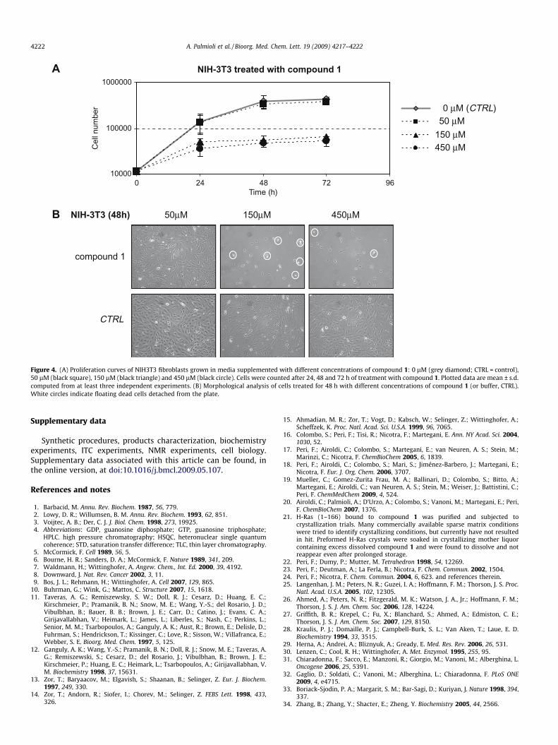
To verify the activity of 1 on cells, experiments that monitor therate of proliferation were performed using NIH3T3 mouse fibro-blasts. Cells were plated at a density of 3000 cells/cm2 and left toadhere onto plastic surface for 18 h. Then cells were treated with1 (at concentrations 0, 50, 150 and 450 lM) supplemented in thegrowth medium, and counted at 24, 48 and 72 h after treatment.The obtained growth curves (Fig. 4A) and morphological analysisof treated cells (Fig. 4B) demonstrate that compound 1 at150 lM effectively inhibits cell proliferation. In fact the numberof cells treated with 150 lM and 450 lM 1 at 24 h from treatmentis lower than control and does not increase later in time. Accord-ingly, in the culture medium of treated cells (Panel B) it is possibleto appreciate many dead cells in suspension (refrangent cellscircled in white).31,32
The data presented here point out that compound 1 binds toH-Ras with low affinity but it is capable of inhibiting GEF-mediatednucleotide exchange. The ITC experiments conclude thatcompound 1 has a lM affinity towards H-Ras�GDP and that 1:1Ras–ligand complex is formed.
NMR and MD based experimental results converge to indicatethat certain H-Ras residues are critical for ligand binding. These in-

010
2030
4050
6070
8090
100
0 500 1000 1500 2000 2500 3000 3500 4000
Rel
ativ
e R
asm
antG
DP
load
ing
(%)
Res
idua
l exc
hang
e ac
tivity
(%)
A
B
50 µM0 µM (CTRL)
100 µM
250 µM
500 µM
0
20
40
60
80
100
120
0 100 200 300 400 500 600
Time (s)
[ Compound 1 ]
Figure 3. Effect of compound 1 on GEF-catalyzed nucleotide exchange of H-Ras. (A) Stimulation of the GDP to mantGDP exchange reaction on H-Ras determined adding to0.25 lM H-Ras�GDP, 1.25 mM mantGDP, 0.0625 lM RasGRF1 and different concentrations (0, 50, 100, 250 and 500 lM) of compound 1. Fluorescence measurements weremade with a Perkin–Elmer LS-45 luminescence spectrometer, monitoring the fluorescence each second for at least 1500 sec. For graphical reason in figure only a point every15 has been plotted. Each experimental curve was fitted to a non linear ‘growth-sigmoidal Hill’ curve (n = 1) with OriginPro 8.0 software, reported in graph as a thin line. Inthe graph the maximum value of relative fluorescence (100 on Y-axis) represent the fully charged Ras status obtained as plateau of an exchange curve without compound. Foreach reaction the initial exchange rate was calculated as first derivative at time 0 with OriginPro 8.0 software. (B) Dose–response curves of compound 1 on GEF-catalyzedexchange of Ras. The initial exchange rate of each exchange reaction (mean of at least three independent experiments) was plotted as a function of the compoundconcentration (dose–response curves). The exchange rate of control reaction (performed without compound) was normalized to 100.
A. Palmioli et al. / Bioorg. Med. Chem. Lett. 19 (2009) 4217–4222 4221
clude amino acids T58, A59 and Y64 belonging to the b-3 strandand R68, M72 on the a-2 helix. The observed binding site coversa region that is essential for Ras interactions with the Ras–GEFdomain of RasGRF1, homologous to SOS, as described by the crystalstructure of the Ras–SOS complex.33 Therefore, it is possible thatcompound 1 interferes with H-Ras–GEF interaction, as previouslyoutlined using SPR experiments.20 The affinity of binding to H-Ras as calculated by ITC experiments (Fig. 1S) is in the same orderof magnitude of the affinity between Ras�GDP and GEF.34 Thismeans that compound 1, although having low affinity for Ras,can compete effectively with GEF for Ras binding thus inhibitingRas activation.
In conclusion, because of the favorable solubility of thedesigned ligand, we were able to determine experimentally theRas-inhibitor binding site as well as fundamental thermodynamicbinding parameters. The involvement of sections of the Switch IIregion in inhibitor binding was previously hypothesized by us onthe base of NMR STD experiments and is now conclusively demon-strated in this report. We are currently investigating whetherglobal or allosteric conformational changes can occur upon
inhibitor binding to H-Ras, and how these may affect Ras-modu-lated downstream signaling events.
We consider the development of soluble inhibitors againstmutant Ras super-family of GTPases as one of the strongest routesto provide clinically effective cancer therapeutics. We propose thatour design and evaluation of small molecule inhibitors, such ascompound 1, may serve as templates on which future pharmaco-logically relevant drug design studies can be attempted.
Acknowledgements
The authors wish to thank Annalisa D’urzo for technical assis-tance. Dr. C. J. Thomas would like to thank Dr. Stephen R Sprang(SRS), Director of CBSD, University of Montana for his support inboth space and instruments towards this project and also for thesalary support from National Institutes of Health GrantsDK46371. Dr. V. Gaponenko acknowledges the generous supportfrom the American Cancer Society Grant RGS-09-057-01-GMC.Professor M. Vanoni acknowledges Creabilis and MIUR-FAR forfinancial support.
0 µM (CTRL)50 µM
150 µM450 µM
compound 1
CTRL
450µM150µM50µMNIH-3T3 (48h)
NIH-3T3 treated with compound 1
10000
100000
1000000
0 24 48 72 96Time (h)
Cel
l num
ber
A
B
Figure 4. (A) Proliferation curves of NIH3T3 fibroblasts grown in media supplemented with different concentrations of compound 1: 0 lM (grey diamond; CTRL = control),50 lM (black square), 150 lM (black triangle) and 450 lM (black circle). Cells were counted after 24, 48 and 72 h of treatment with compound 1. Plotted data are mean ± s.d.computed from at least three independent experiments. (B) Morphological analysis of cells treated for 48 h with different concentrations of compound 1 (or buffer, CTRL).White circles indicate floating dead cells detached from the plate.
4222 A. Palmioli et al. / Bioorg. Med. Chem. Lett. 19 (2009) 4217–4222
Supplementary data
Synthetic procedures, products characterization, biochemistryexperiments, ITC experiments, NMR experiments, cell biology.Supplementary data associated with this article can be found, inthe online version, at doi:10.1016/j.bmcl.2009.05.107.
References and notes
1. Barbacid, M. Annu. Rev. Biochem. 1987, 56, 779.2. Lowy, D. R.; Willumsen, B. M. Annu. Rev. Biochem. 1993, 62, 851.3. Voijtec, A. B.; Der, C. J. J. Biol. Chem. 1998, 273, 19925.4. Abbreviations: GDP, guanosine diphosphate; GTP, guanosine triphosphate;
HPLC. high pressure chromatography; HSQC, heteronuclear single quantumcoherence; STD, saturation transfer difference; TLC, thin layer chromatography.
5. McCormick, F. Cell 1989, 56, 5.6. Bourne, H. R.; Sanders, D. A.; McCormick, F. Nature 1989, 341, 209.7. Waldmann, H.; Wittinghofer, A. Angew. Chem., Int. Ed. 2000, 39, 4192.8. Downward, J. Nat. Rev. Cancer 2002, 3, 11.9. Bos, J. L.; Rehmann, H.; Wittinghofer, A. Cell 2007, 129, 865.
10. Buhrman, G.; Wink, G.; Mattos, C. Structure 2007, 15, 1618.11. Taveras, A. G.; Remiszewsky, S. W.; Doll, R. J.; Cesarz, D.; Huang, E. C.;
Kirschmeier, P.; Pramanik, B. N.; Snow, M. E.; Wang, Y.-S.; del Rosario, J. D.;Vibulbhan, B.; Bauer, B. B.; Brown, J. E.; Carr, D.; Catino, J.; Evans, C. A.;Girijavallabhan, V.; Heimark, L.; James, L.; Liberles, S.; Nash, C.; Perkins, L.;Senior, M. M.; Tsarbopoulos, A.; Ganguly, A. K.; Aust, R.; Brown, E.; Delisle, D.;Fuhrman, S.; Hendrickson, T.; Kissinger, C.; Love, R.; Sisson, W.; Villafranca, E.;Webber, S. E. Bioorg. Med. Chem. 1997, 5, 125.
12. Ganguly, A. K.; Wang, Y.-S.; Pramanik, B. N.; Doll, R. J.; Snow, M. E.; Taveras, A.G.; Remiszewski, S.; Cesarz, D.; del Rosario, J.; Vibulbhan, B.; Brown, J. E.;Kirschmeier, P.; Huang, E. C.; Heimark, L.; Tsarbopoulos, A.; Girijavallabhan, V.M. Biochemistry 1998, 37, 15631.
13. Zor, T.; Baryaacov, M.; Elgavish, S.; Shaanan, B.; Selinger, Z. Eur. J. Biochem.1997, 249, 330.
14. Zor, T.; Andorn, R.; Siofer, I.; Chorev, M.; Selinger, Z. FEBS Lett. 1998, 433,326.
15. Ahmadian, M. R.; Zor, T.; Vogt, D.; Kabsch, W.; Selinger, Z.; Wittinghofer, A.;Scheffzek, K. Proc. Natl. Acad. Sci. U.S.A. 1999, 96, 7065.
16. Colombo, S.; Peri, F.; Tisi, R.; Nicotra, F.; Martegani, E. Ann. NY Acad. Sci. 2004,1030, 52.
17. Peri, F.; Airoldi, C.; Colombo, S.; Martegani, E.; van Neuren, A. S.; Stein, M.;Marinzi, C.; Nicotra, F. ChemBioChem 2005, 6, 1839.
18. Peri, F.; Airoldi, C.; Colombo, S.; Mari, S.; Jiménez-Barbero, J.; Martegani, E.;Nicotra, F. Eur. J. Org. Chem. 2006, 3707.
19. Mueller, C.; Gomez-Zurita Frau, M. A.; Ballinari, D.; Colombo, S.; Bitto, A.;Martegani, E.; Airoldi, C.; van Neuren, A. S.; Stein, M.; Weiser, J.; Battistini, C.;Peri, F. ChemMedChem 2009, 4, 524.
20. Airoldi, C.; Palmioli, A.; D’Urzo, A.; Colombo, S.; Vanoni, M.; Martegani, E.; Peri,F. ChemBioChem 2007, 1376.
21. H-Ras (1–166) bound to compound 1 was purified and subjected tocrystallization trials. Many commercially available sparse matrix conditionswere tried to identify crystallizing conditions, but currently have not resultedin hit. Preformed H-Ras crystals were soaked in crystallizing mother liquorcontaining excess dissolved compound 1 and were found to dissolve and notreappear even after prolonged storage.
22. Peri, F.; Dumy, P.; Mutter, M. Tetrahedron 1998, 54, 12269.23. Peri, F.; Deutman, A.; La Ferla, B.; Nicotra, F. Chem. Commun. 2002, 1504.24. Peri, F.; Nicotra, F. Chem. Commun. 2004, 6, 623. and references therein.25. Langenhan, J. M.; Peters, N. R.; Guzei, I. A.; Hoffmann, F. M.; Thorson, J. S. Proc.
Natl. Acad. U.S.A. 2005, 102, 12305.26. Ahmed, A.; Peters, N. R.; Fitzgerald, M. K.; Watson, J. A., Jr.; Hoffmann, F. M.;
Thorson, J. S. J. Am. Chem. Soc. 2006, 128, 14224.27. Griffith, B. R.; Krepel, C.; Fu, X.; Blanchard, S.; Ahmed, A.; Edmiston, C. E.;
Thorson, J. S. J. Am. Chem. Soc. 2007, 129, 8150.28. Kraulis, P. J.; Domaille, P. J.; Campbell-Burk, S. L.; Van Aken, T.; Laue, E. D.
Biochemistry 1994, 33, 3515.29. Herna, A.; Andrei, A.; Bliznyuk, A.; Gready, E. Med. Res. Rev. 2006, 26, 531.30. Lenzen, C.; Cool, R. H.; Wittinghofer, A. Met. Enzymol. 1995, 255, 95.31. Chiaradonna, F.; Sacco, E.; Manzoni, R.; Giorgio, M.; Vanoni, M.; Alberghina, L.
Oncogene 2006, 25, 5391.32. Gaglio, D.; Soldati, C.; Vanoni, M.; Alberghina, L.; Chiaradonna, F. PLoS ONE
2009, 4, e4715.33. Boriack-Sjodin, P. A.; Margarit, S. M.; Bar-Sagi, D.; Kuriyan, J. Nature 1998, 394,
337.34. Zhang, B.; Zhang, Y.; Shacter, E.; Zheng, Y. Biochemistry 2005, 44, 2566.