Rho family proteins and Ras transformation - Nature
24
Rho family proteins and Ras transformation: the RHOad less traveled gets congested Irene M Zohn 1 , Sharon L Campbell 2,3 , Roya Khosravi-Far 4 , Kent L Rossman 2 and Channing J Der 1,3,5 1 Department of Pharmacology, 2 Department of Biochemistry and Biophysics, 3 Lineberger Comprehensive Cancer Center, 4 Department of Biology, MIT, Cambridge, Massachusetts 02139; 5 Curriculum in Genetics and Molecular Biology, University of North Carolina, Chapel Hill, North Carolina 27599, USA The Rho family of small GTPases has attracted considerable research interest over the past 5 years. During this time, we have witnessed a remarkable increase in our knowledge of the biochemistry and biology of these Ras-related proteins. Thus, Rho family proteins have begun to rival, if not overshadow, interest in their more celebrated cousins, the Ras oncogene proteins. The fascination in Rho family proteins is fueled primarily by two major observations. First, like Ras, Rho family proteins serve as guanine nucleotide- regulated binary switches that control signaling pathways that in turn regulate diverse cellular processes. Rho family proteins are key components in cellular processes that control the organization of the actin cytoskeleton, activate kinase cascades, regulate gene expression, regulate membrane tracking, promote growth trans- formation and induce apoptosis. Second, at least five Rho family proteins have been implicated as critical regulators of oncogenic Ras transformation. Thus, it is suspected that Rho family proteins contribute signifi- cantly to the aberrant growth properties of Ras- transformed cells. Rho family proteins are also critical mediators of the transforming actions of other trans- forming proteins and include Dbl family oncogene proteins, G protein-coupled receptors and G protein a subunits. Thus, Rho family proteins may be key components for the transforming actions of diverse oncogene proteins. Major aims of Rho family protein studies are to define the molecular mechanism by which Rho family proteins regulate such a diverse spectrum of cellular behavior. These eorts may reveal novel targets for the development of anti-Ras and anti-cancer drugs. Keywords: Ras superfamily; signal transduction; actin cytoskeleton; eectors An important theme that has emerged during the past 5 years is that Ras transformation is mediated by signaling activities that are much more complex than originally envisioned (Marshall, 1996; Khosravi-Far et al., 1997; Campbell et al., 1998). Considerable biological, biochemical and genetic evidence support the importance of the Raf serine/threonine kinase a key eector of Ras function. However, the evidence that Ras has a life beyond simply activating Raf is strong and continues to mount. Instead, our current model proposes that Ras employs a spectrum of functionally diverse downstream eectors to cause its diverse actions on cell proliferation, dierentiation and apoptosis. Among these Raf-independent signaling pathways are those that connect Ras with specific members of the Rho family of small GTPases. Since Rho family proteins are regulators of actin organiza- tion, gene expression, and cell cycle progression, it is likely that Rho family proteins will contribute significantly to the actions of oncogenic Ras. The Rho family of proteins have been the subject of a number of excellent reviews (Symons, 1996; Narumiya, 1996; Van Aelst and D’Souza-Schorey, 1997). There- fore, in this review we will emphasize the involvement of Rho family proteins in the regulation of cell proliferation and Ras transformation. Despite our wealth of knowledge on these small GTPases, we are clearly in the early days of comprehending the complex involvement of these small GTPases in regulating normal and neoplastic cell biology. More excitement is certain to follow as we continue to develop and revise, or discard, our current models and concepts. Rho family proteins are members of the Ras superfamily of small GTPases The Ras superfamily of small (20 – 25 kDa) GTPases (480 mammalian members) can be categorized into at least nine distinct branches. These include the Ras, Rab, Rho, Ran, Rheb, Rad/Gem, Rin/Rit and Arf families. Rho family proteins constitute one of the three major branches of the Ras superfamily and its members share approximately 30% amino acid identity with the four Ras proteins (Chardin, 1993). Presently, at least 14 mammalian Rho family proteins have been identified: RhoA, RhoB, RhoC, RhoD, RhoE/Rnd3, Rnd1/Rho6, Rnd2/Rho7, RhoG, Rac1, Rac2, Rac3, Cdc42, TC10 and TTF that share significant (ranging from 50 – 90%) amino acid identity with each other (Ridley, 1996) (Figure 1a). Two mammalian isoforms of Cdc42 have been identified from brain (B) or placenta (P). Much of our knowledge of Rho family protein function has been derived primarily from the studies of Rac1, RhoA and Cdc42 and each exhibits distinct cellular functions. A recent report indicates that RhoD possesses functions distinct from these three proteins (Murphy et al., 1996). Sequence comparisons suggest that RhoE and TTF may also share functional relationships, whereas TC10 and RhoG are anticipated to be most related in function to Cdc42 and Rac1, respectively (Figure 1b). Recent observations that Rho/ Rnd3 or Rnd1 causes a disruption of stress fibers Correspondence: CJ Der Oncogene (1998) 17, 1415 – 1438 1998 Stockton Press All rights reserved 0950 – 9232/98 $12.00 http://www.stockton-press.co.uk/onc
-
Upload
khangminh22 -
Category
Documents
-
view
0 -
download
0
Transcript of Rho family proteins and Ras transformation - Nature

Rho family proteins and Ras transformation: the RHOad less traveled getscongested
Irene M Zohn1, Sharon L Campbell2,3, Roya Khosravi-Far4, Kent L Rossman2 andChanning J Der1,3,5
1Department of Pharmacology, 2Department of Biochemistry and Biophysics, 3Lineberger Comprehensive Cancer Center,4Department of Biology, MIT, Cambridge, Massachusetts 02139; 5Curriculum in Genetics and Molecular Biology, University ofNorth Carolina, Chapel Hill, North Carolina 27599, USA
The Rho family of small GTPases has attractedconsiderable research interest over the past 5 years.During this time, we have witnessed a remarkableincrease in our knowledge of the biochemistry andbiology of these Ras-related proteins. Thus, Rho familyproteins have begun to rival, if not overshadow, interestin their more celebrated cousins, the Ras oncogeneproteins. The fascination in Rho family proteins is fueledprimarily by two major observations. First, like Ras,Rho family proteins serve as guanine nucleotide-regulated binary switches that control signaling pathwaysthat in turn regulate diverse cellular processes. Rhofamily proteins are key components in cellular processesthat control the organization of the actin cytoskeleton,activate kinase cascades, regulate gene expression,regulate membrane tra�cking, promote growth trans-formation and induce apoptosis. Second, at least ®ve Rhofamily proteins have been implicated as criticalregulators of oncogenic Ras transformation. Thus, it issuspected that Rho family proteins contribute signi®-cantly to the aberrant growth properties of Ras-transformed cells. Rho family proteins are also criticalmediators of the transforming actions of other trans-forming proteins and include Dbl family oncogeneproteins, G protein-coupled receptors and G protein asubunits. Thus, Rho family proteins may be keycomponents for the transforming actions of diverseoncogene proteins. Major aims of Rho family proteinstudies are to de®ne the molecular mechanism by whichRho family proteins regulate such a diverse spectrum ofcellular behavior. These e�orts may reveal novel targetsfor the development of anti-Ras and anti-cancer drugs.
Keywords: Ras superfamily; signal transduction; actincytoskeleton; e�ectors
An important theme that has emerged during the past5 years is that Ras transformation is mediated bysignaling activities that are much more complex thanoriginally envisioned (Marshall, 1996; Khosravi-Far etal., 1997; Campbell et al., 1998). Considerablebiological, biochemical and genetic evidence supportthe importance of the Raf serine/threonine kinase a keye�ector of Ras function. However, the evidence thatRas has a life beyond simply activating Raf is strongand continues to mount. Instead, our current modelproposes that Ras employs a spectrum of functionally
diverse downstream e�ectors to cause its diverseactions on cell proliferation, di�erentiation andapoptosis. Among these Raf-independent signalingpathways are those that connect Ras with speci®cmembers of the Rho family of small GTPases. SinceRho family proteins are regulators of actin organiza-tion, gene expression, and cell cycle progression, it islikely that Rho family proteins will contributesigni®cantly to the actions of oncogenic Ras. TheRho family of proteins have been the subject of anumber of excellent reviews (Symons, 1996; Narumiya,1996; Van Aelst and D'Souza-Schorey, 1997). There-fore, in this review we will emphasize the involvementof Rho family proteins in the regulation of cellproliferation and Ras transformation. Despite ourwealth of knowledge on these small GTPases, we areclearly in the early days of comprehending the complexinvolvement of these small GTPases in regulatingnormal and neoplastic cell biology. More excitementis certain to follow as we continue to develop andrevise, or discard, our current models and concepts.
Rho family proteins are members of the Ras superfamilyof small GTPases
The Ras superfamily of small (20 ± 25 kDa) GTPases(480 mammalian members) can be categorized into atleast nine distinct branches. These include the Ras,Rab, Rho, Ran, Rheb, Rad/Gem, Rin/Rit and Arffamilies. Rho family proteins constitute one of the threemajor branches of the Ras superfamily and its membersshare approximately 30% amino acid identity with thefour Ras proteins (Chardin, 1993). Presently, at least 14mammalian Rho family proteins have been identi®ed:RhoA, RhoB, RhoC, RhoD, RhoE/Rnd3, Rnd1/Rho6,Rnd2/Rho7, RhoG, Rac1, Rac2, Rac3, Cdc42, TC10and TTF that share signi®cant (ranging from 50 ± 90%)amino acid identity with each other (Ridley, 1996)(Figure 1a). Two mammalian isoforms of Cdc42 havebeen identi®ed from brain (B) or placenta (P).
Much of our knowledge of Rho family proteinfunction has been derived primarily from the studiesof Rac1, RhoA and Cdc42 and each exhibits distinctcellular functions. A recent report indicates that RhoDpossesses functions distinct from these three proteins(Murphy et al., 1996). Sequence comparisons suggestthat RhoE and TTF may also share functionalrelationships, whereas TC10 and RhoG are anticipatedto be most related in function to Cdc42 and Rac1,respectively (Figure 1b). Recent observations that Rho/Rnd3 or Rnd1 causes a disruption of stress ®bersCorrespondence: CJ Der
Oncogene (1998) 17, 1415 ± 1438 1998 Stockton Press All rights reserved 0950 ± 9232/98 $12.00
http://www.stockton-press.co.uk/onc

support a distinct function for RhoE and relatedproteins (Nobes et al., 1998; Guasch et al., 1998).Furthermore, RhoE and related proteins possesssubstitutions at positions corresponding to Rasresidues 12, 13, 59, 61 ± 63 and 66 ± 67 that in¯uenceboth intrinsic and GAP-mediated GTP hydrolysis inRas, and consequently, they exist in a predominantlyGTP-bound state. Thus, sequence and functionaldi�erences allow the de®nition of at least six distinctRho subfamilies: (1) Cdc42 and TC10; (2) Rac1, Rac2,Rac3 and RhoG; (3) RhoA, RhoB and RhoC; (4)RhoE, Rnd1/Rho6 and Rnd2/Rho7; and (5) RhoD andTTF. However, a preliminary report that a dominantnegative mutant of RhoG did not impair formation ofmembrane ru�es, ®lopodia or stress ®bers suggests thatRhoG function may be distinct from both Cdc42 andRac (Roux et al., 1997). Thus, further subdivision of theRho family, as well as the identi®cation of additional
members, are likely to occur in the future. Finally,although RhoA, RhoB and RhoC are likely to sharecommon functions in regulating stress ®ber formation(Ridley and Hall, 1992), they di�er in subcellularlocation (Adamson et al., 1992), regulation of expres-sion (Jahner and Hunter, 1991; Fritz et al., 1995) andposttranslational lipid modi®cation (Adamson et al.,1992; Lebowitz et al., 1995). Thus, even closely relatedmembers are likely to exhibit unique roles as well.
Like Ras, Rho family proteins function as GTP/GDP regulated switches that cycle between activeGTP- and an inactive GDP-bound forms (Figure 2)(Ridley, 1996). This cycle is regulated by three distinctclasses of regulatory proteins. First, Rho guaninenucleotide exchange factors (GEFs; also referred toas Dbl homology proteins) serve as activators andstimulate the replacement of GDP by GTP (Whiteheadet al., 1997). To date, over 20 Dbl homology proteins
a
Rho family of small GTPases and transformationI Zohn et al
1416

have been identi®ed, and the majority were initiallydiscovered as transforming proteins in NIH3T3 focus-formation assays (e.g., Dbl, Vav) (Figure 3). Second, atleast 16 Rho GTPase activating proteins (GAPs) havealso been identi®ed that serve as negative regulators ofRho family protein function by stimulating theirintrinsic GTPase activities (Cerione and Zheng, 1996).Some GAPs show preferential stimulation of speci®cRho family proteins.
A third class of Rho regulators are the Rho GDP-dissociation inhibitory factors (RhoGDIs). The ®rstRhoGDI was originally identi®ed as an inhibitor ofRac GDP dissociation (Ueda et al., 1990) andsubsequently, of Cdc42 and Rac as well (Abo et al.,1991; Leonard et al., 1992). This ubiquitouslyexpressed, cytosolic protein, was later shown tointerfere with both intrinsic and GAP-stimulatedGTP hydrolysis of Rac and Cdc42 (Chuang et al.,1993; Hart et al., 1992). Thus, RhoGDI can perturbRho GDP/GTP cycling via two distinct mechanisms.Finally, RhoGDIs also serve an important role inregulating the association of Rho family proteins withmembranes (Takai et al., 1995). A cytosolic complexwith RhoGDI is disrupted during Rho membranetranslocation and activation by GEFs. Two additionalRhoGDIs include the hematopoietic cell speci®c D4-GDI/LY-GDI (Lelias et al., 1993; Scherle and Staudt,1993) and RhoGDIg/RhoGDI-3, which is preferen-tially expressed in brain, pancreas and other tissues(Adra et al., 1997; Zalcman et al., 1996).
Several regulators of Rho GDP/GTP cycling appearto function as multifunctional regulators. For example,BCR and ABR each possess distinct catalytic domainsthat serve as both Rho GEFs and GAPs (Chuang etal., 1995) (Figure 3). In addition, some Rho GAPs mayalso serve as downstream e�ectors (e.g., N-chimaerinand the p85 subunit of phosphoinositide-3-OH kinase(PI3K)) (Kozma et al., 1996; Zheng et al., 1994a).Although RasGRFs and SOS have been shown tofunction as Ras GEFs, the presence of tandem DH/PHdomains in these molecules suggests that they maypossess a Rho family GEF function which is distinctfrom their Ras GEF function (Nimnual et al., 1998;Fan et al., 1998). Finally, one Dbl family protein (Trio)possesses two distinct DH domains and each showsdistinct GEF activities.
Like Ras, Rho family GTPases share high sequencesimilarity, especially in the core GTP bindingsequences, and diverge primarily at their COOH-termini. The x-ray and NMR structures of truncatedforms of Rac1-GMPPNP, Cdc42-GDP, G14V RhoA-GDP and RhoA-GTPgS have recently been solved(Hirshberg et al., 1997; Feltham et al., 1997; Ihara etal., 1998; Rittinger et al., 1997; Wei et al., 1997). LikeRas these Rho family GTPases share a common a/bfold with the core GTP-binding domain as aconserved structural unit. In contrast to Ras, theRho subfamily contains an insert of 13 amino acids ina region between b5 and a4 of H-Ras. One exception,however, is TTF, which contains a smaller insert
b
Figure 1 Sequence comparison of Rho family proteins. (a) Todate, at least 14 distinct mammalian Rho family proteins havebeen described. A multiple sequence alignment of human Rhofamily proteins and H-Ras was generated using the dynamicalgorithm alignment program ClustalW (Thompson et al., 1994).A pro®le alignment of the Rac1 primary sequence containingRac1 secondary structural elements and the remaining Rho familyproteins was initially constructed. The primary sequence of H-Raswas then added before the ®nal multiple sequence alignment wasproduced. All alignments were carried out using PAM seriesprotein weight matrices. The ClustalW multiple sequencealignment was shaded using Boxshade 3.2 (Thompson et al.,1994). Residues that are conserved in greater than 50% ofsequences at a given sequence position are colored blue, whileresidues that are similar are colored yellow. (b) The sequencealignment described above was used to construct the Rhodendrogram. The branch lengths in the dendrogram areproportional to the estimated divergence along each branch.Rnd3/Rho8 di�ers from RhoE only in having an additional 15amino acid NH2-terminal extension (Nobes et al., 1998)
Rho family of small GTPases and transformationI Zohn et al
1417

consisting of 7 amino acids. In Rac1 and RhoA, theinsert consists of two alpha helices followed by anextended loop. The insert represents a highly chargedsurface, is mobile and exposed. However, Cdc42contains only one helix in the insert. Moreover, theinsert appears to form a compact loop structure that
forms contacts with the loop between b4 and a3. It isnot clear whether these di�erences represent differ-ences between X-ray and NMR methods or whetherthe fact that the structure of Rac1 was solved on aF87S mutant that may alter the structure and contactsbetween the insert and the b4/a3 loop. NMR studiesindicate that the conformation of the insert is notsensitive to the binding of GTP versus GDP.
Like Ras, the conformation of the switch I and switchII domains is sensitive to the binding of GTP versusGDP and both appear to be dynamic structures.However, in contrast to Rac1, helix two in the switchII domain of Cdc42 is absent. Interestingly, NMRchemical shift mapping of Cdc42-GDP and Cdc42-GMPPCP suggest the existence of an additional switchregion comprised of b4/a3 and a3 (Feltham et al., 1997).
Rho family proteins are regulators of diverse cellularprocesses
Rho family proteins have been implicated in theregulation of a diverse and extensive spectrum ofcellular processes. Most prominent among these aretheir e�ects on the organization of the actincytoskeleton. Rho proteins also initiate signalingcascades that cause activation of a variety oftranscription factors. Hence, Rho-mediated changesin the actin organization or in gene expression mayregulate many of the cellular processes associated withRho protein function. These include regulation of cellshape, cell attachment, cell motility and invasion, cell-cell interactions, cell proliferation, di�erentiation andapoptosis. Below, we summarize our current knowl-edge regarding the involvement of Rho proteins incontrolling speci®c cellular processes.
Rho proteins are regulators of actin cytoskeletalorganization
The best characterized function of Rho family proteinsinvolves their regulation of speci®c ®lamentous F-actinorganization. Actin ®laments are components of one ofthe three major cytoskeletal protein networks thatdetermine cell shape, movement and regulate cellularprocesses: actin ®laments, microtubules and intermedi-ate ®laments. The actin cytoskeleton is a highlydynamic cytoplasmic structure that is reshaped andreformed in response to diverse extracellular signals.
In ®broblasts, polymerized actin is assembled into avariety of distinct structures. Lamellipodia are curtain-like extensions that consist of thin protrusive actinsheets. Membrane ru�es represent lamellipodia thathave lifted from the substratum at the leading edge ofcells. Actin stress ®bers consist of actin bundles thattraverse the cell and promote cell attachment to theextracellular matrix via focal adhesions. Focal adhe-sions consist of integrins and cytoplasmic proteins suchas vinculin and talin. Filopodia are thin, ®nger-likecytoplasmic extensions that contain tight actin bundlesand may be involved in the recognition of theextracellular environment (Nobes and Hall, 1995).Speci®c Rho family proteins are regulators of distinctchanges in these actin-based structures, as well asothers (e.g., tight junctions in polarized epithelial cells)(Nusrat et al., 1995), in ®broblasts and other cell types.
Figure 3 Dbl family proteins are guanine nucleotide exchangefactors and activators of Rho family proteins. Presently, at least22 mammalian Dbl family proteins have been described (reviewedin Whitehead et al., 1997); Fam et al., 1997; Schuebel et al., 1996;Henske et al., 1995; Gebbink et al., 1997; Alam et al., 1997). Allshare a Dbl homology (DH) followed by a pleckstrin homology(PH) domain. Beyond the tamdem DH/PH domains, each Dblfamily protein possesses distinct catalytic (Ras GEF, Rho GAP)or protein-protein or protein-lipid interaction motifs [Srchomology 2 and 3 (SH2 and SH3), cysteine-rich zinc-bindingdomains (CRDs)]. A Rho GEF activity has been de®ned formost, but not all, members of this family. Whereas some exhibitvery speci®c activity, others serve as activators of multiple Rhofamily proteins. For example, FGD1 is an activator of Cdc42,while Vav can activate Rac1, RhoA and Cdc42
Figure 2 Regulators of Rho family protein GDP/GTP cycling.Like Ras, Rho family proteins function as GDP/GTP-regulatedbinary switches. A variety of extracellular stimuli regulate Rhofamily protein GDP/GTP cycling, through modulation of RhoGEF, GAP or GDI function. Activated Rho family proteinsexhibit diverse cellular functions that include regulation of actinorganization, gene expression and cell cycle progression
Rho family of small GTPases and transformationI Zohn et al
1418

The ®rst clues that Rho family proteins regulateactin organization were provided by studies using C3toxin from Clostridium botulinum, an inhibitor ofRhoA, B and C function (Chardin et al., 1989) andby microinjection analyses using mutant Rho familyproteins (Paterson et al., 1990; Ridley et al., 1992;Ridley and Hall, 1992; Nobes and Hall, 1995). Thesestudies demonstrated that speci®c Rho family proteinsare components of signaling pathways that induceunique morphological changes involving rearrange-ments of F-actin. RhoA, Rac1 and Cdc42 controldistinct changes in the actin cytoskeleton and distinctcellular structures. Furthermore, they can act inconcert in cascades that link their activities. Constitu-tively activated Cdc42 caused induction of ®lopodia inSwiss 3T3 ®broblasts as well as the activation of Rac(Nobes and Hall, 1995; Kozma et al., 1995). ActivatedRac1 in turn caused the induction of lamellipodia andmembrane ru�ing, and the activation of Rho (Ridleyet al., 1992). Activated RhoA caused the formation ofstress ®bers and focal adhesions (Ridley and Hall,1992). Both Rac and Cdc42 also regulate formation offocal complexes distinct from those caused by RhoA.A second cascade of small GTPases involves the abilityof oncogenic Ras to activate Rac and subsequentlyRho (Ridley et al., 1992). GTPase cascades that involve
Rho family proteins have also been described in theyeasts S. cerevisiae and S. pombe (reviewed in Chantand Stowers, 1995).
Although a Cdc424Rac4Rho cascade has alsobeen seen in other cell types (Allen et al., 1997),variations on this theme, involving separate pathwaysor feedback loops, have also been observed. Forexample, Cdc42 was shown to inhibit, rather thanpromote, stress ®ber formation in other cell types(Kozma et al., 1995; Qiu et al., 1997). Activated Racdid not induce actin stress ®ber formation in MDCKepithelial cells (Ridley et al., 1995). Inactivation of Rhocaused activation of Cdc42 and Rac in N1E-115neuroblastoma cells (Kozma et al., 1997). Theseobservations, when taken together with the fact thateach Rho family can be activated by distinct signals,re¯ect the versatility of Rho family proteins inorchestrating di�erent cellular processes in di�erentsituations and cell types.
Finally, the precise links between these GTPasecascades have not been established. However, based onthe involvement of Dbl family-related proteins (CDC24)in the activation of Cdc42 in yeast pathways (Zheng etal., 1994b), it is logical that Dbl family proteins willserve as intermediates between GTPases. GAPs andGDIs may also facilitate these GTPase cascades.
Figure 4 Rho family proteins are regulators of signaling pathways that regulate the organization of the actin cytoskeleton. Cdc42,Rac1 and RhoA function as downstream components of signaling pathways initiated by ligand-stimulated G protein-coupledserpentine receptors (SRs), receptor tyrosine kinases (RTKs) or integrin receptors (IRs). Cdc42, Rac1 and RhoA each modulatedistinct changes in actin organization. At least two GTPase cascades have been identi®ed that regulate actin reorganization. Oneinvolves oncogenic Ras activation of Rac1, then Rac1 activation of RhoA. A second involves Cdc42 activation of Rac1 and RhoA.These hierarchies of small GTPases may vary in di�erent cell types
Rho family of small GTPases and transformationI Zohn et al
1419

Like Ras, Rho family proteins also serve as GDP/GTP-regulated relay switches that transmit extra-cellular ligand-mediated signals that promote changesin actin structures (Figure 4). It had been observedpreviously that lysophosphatidic acid (LPA), bombe-sin and bradykinin each bind to GPCRs to activatethe formation of ®lopodia, membrane ru�es andstress ®bers. Rho family proteins were subsequentlyimplicated in these processes (Ridley et al., 1992;Ridley and Hall, 1992). For example, LPA stimula-tion of actin stress ®bers formation and theassembly of focal adhesion complexes in Swiss 3T3cells was blocked by C3 toxin (Ridley and Hall,1992).
PDGF stimulation of its receptor, a transmembranetyrosine kinase, causes membrane ru�ing which wasblocked by dominant negative Rac (Ridley et al.,1992). Dominant negative Cdc42 blocked the forma-tion of ®lopodia induced by bradykinin (Nobes andHall, 1995). Finally, insulin- or hepatocyte growthfactor-induced membrane ru�ing in KB humanepidermoid carcinoma cells was dependent on Rac orRho, respectively (Nishiyama et al., 1994). There isevidence that PI3K may link PDGF- and insulin-mediated signaling to Rac activation and induction of
membrane ru�ing (Kotani et al., 1994; Wennstrom etal., 1994; Nobes and Hall, 1995).
In general, speci®c Rho family proteins show thesame e�ects on actin organization in di�erent cell types.However, some cell type di�erences have been observed.For example, Rac and Rho cause distinct membraneru�ing response in KB human epithelial carcinoma celllines (Nishiyama et al., 1994). Whereas Rho causesneurite retraction, Rac causes neurite extension in NIE-115 neuroblastoma cells. Other members of the Rhofamily are also regulators of additional actin cytoske-letal rearrangements and cellular processes. Forexample, transient expression of activated RhoD in avariety of cell types caused rearrangements of the actincytoskeleton and cell surface and involved the forma-tion of long thin F-actin containing membraneprocesses together with the disassembly of stress ®bersand focal adhesions. These cytoskeletal changescorresponded with regulated endosome motility anddistribution (Murphy et al., 1996). RhoG has beenreported to cause a Rac-dependent membrane ru�ing inSwiss 3T3 cells (Roux et al., 1997). Finally, activatedRnd1 and RhoE/Rnd3 has been reported to cause adisruption of stress ®bers (Nobes et al., 1998; Guasch etal., 1998).
Figure 5 Rho family proteins are regulators of mitogen-activated protein kinase cascades. At least three distinct MAPK moduleshave been identi®ed and involve a cascade of a MAPK kinase kinase (MAPKKK; also MEKKs), a MAPK kinase (MAPKK; alsoMKKs) and a MAPK. Rac and Cdc42 have been shown to be activators of pathways that lead to the activation of the JNK andp38 MAPK modules (reviewed in Vojtek and Cooper, 1995; Treisman, 1996). Although several kinases have been implicated asRac/Cdc42 e�ectors (e.g., PAKs, MLKs, MEKKs), the precise link(s) between Rac and Cdc42 and these cascades is presently notknown. Ras activates ERKs via a Raf-dependent pathway, and JNKs and p38 MAPKs via a Raf-dependent pathway that involvesRac/Cdc42. Activated MAPKs in turn regulate a variety of substrates that include nuclear transcription factors (reviewed inTreisman, 1996); (Wang and Ron, 1996; Zervos et al., 1995). Note that common transcription factor targets are shared by thedi�erent MAPKs
Rho family of small GTPases and transformationI Zohn et al
1420

Rho family proteins are also associated withprocesses that involve the actin cytoskeleton in otherorganisms. The regulation of cytoskeletal organizationby Rho family members is evident from geneticsstudies in yeast and Drosophila. Yeast Cdc42 hasbeen demonstrated to coordinate polarization of theactin cytoskeleton during cell division by budding, andinvolves a cascade of other small GTPases (BUD1/RSR1 and RHO proteins) (reviewed in Chant andStowers, 1995). In Drosophila, Drac1 is required toassemble actin at adherens junctions of the wing discepithelium, while Dcdc42 is involved in the regulationof polarized cell shape during various stages of wingdisc development (Eaton et al., 1995). Studies have alsoimplicated a requirement for Rho family proteinsduring cytokinesis in sand dollar (Mabuchi et al.,1993) Xenopus (Kishi et al., 1993; Drechsel et al.,1996), Dictyostelium (Larochelle et al., 1996) andmammalian cells (Madaule et al., 1998).
Rho family proteins are regulators of gene expression
In addition to their involvement in regulation ofcytoskeletal organization, it has been shown that Rhofamily proteins regulate protein kinase cascades thatcontrol the activity of a variety of nuclear transcriptionfactors (Figure 5). To date, at least three distinctmitogen-activated protein kinase (MAPK) cascadeshave been identi®ed in mammalian cells and more arelikely to be discovered (Treisman, 1996). The p42 andp44 extracellular signal regulated kinases (ERKs; alsocalled p42 and p44 MAPKs), Jun NH2-terminal kinases(JNKs) and p38/HOG kinases are distinct kinasecascades activated by distinct small GTPases. ERKs
are activated by many mitogenic stimuli, whereas JNKand p38 are more commonly activated by cellular stress(heat shock, ionizing radiation, etc.) and in¯ammatorycytokines.
These cascades involve kinase modules where aMAPK kinase kinase (or MEKK) causes activation ofa MAPK kinase (or MKK), which in turn activates aMAPK. The activated MAPKs have a variety oftargets and include various Ets domain, bZIP andMADS box containing transcription factors reviewedin Treisman, 1996). Although we have depicted eachMAPK module as a separate linear array, there is crosstalk between these cascades, as well as branch pointsand feedback loops within each module emphasizingthe complex nature of signaling pathways.
Constitutively activated Rac1 and Cdc42, but notRhoA, are activators of JNKs (also called stress-activated protein kinases, or SAPKs) and p38/HOGs,but not the p42 and p44 ERKs (Coso et al., 1995;Minden et al., 1995; Olson et al., 1995; Bagrodia et al.,1995a; Zhang et al., 1995). However, RhoA canactivate JNK in some cells (Teramoto et al., 1996b)and RhoG is also an activator of JNK, but not ERK(Roux et al., 1997). JNK in turn activates the ATF-2and Jun nuclear transcription factors. ATF-2 and Juncan dimerize with other transcription factors tostimulate transcription from promoters containingAP-1 and related DNA sequences (e.g., the c-junpromoter) (Figure 6) (Karin, 1995). Although Rac,RhoA and Cdc42 are not activators of ERKs, it hasbeen observed that they can indirectly modulate ERKactivity in 293 cells (Luttrell et al., 1997). Dominantnegative Rac2 was found to impair Ras activation ofERK2, whereas activated RhoA, Rac2 and Cdc42
Figure 6 Rho family proteins are regulators of genes that control cell growth, di�erentiation and apoptosis. Rho family proteinsregulate the activity of genes that regulate cell proliferation and apoptosis (reviewed in Treisman, 1996). RhoA, Rac1 and Cdc42activate SRF (Hill and Treisman, 1995), which forms a complex with ternary complex factors (e.g., Elk-1) at the serum responseDNA element found in the promoter sequences of c-fos and other early response genes. RhoA and Rac/Cdc42 are believed toactivate SRF through distinct pathways. Rac1/Cdc42 (and RhoA in some cells) activate JNK, which in turn can activate the Jun,ATF-2 and Elk-1 nuclear transcription factors. Activated c-Jun:ATF-2 heterodimers stimulate AP-1 like DNA elements in variouspromoters that regulate c-jun and other genes. Rac1 activates NF-kB by as yet unknown pathways that involve production ofsuperoxide and phosphorylation and inactivation of IkB (Perona et al., 1997). Among these NF-kB-responsive genes are those thathave an anti-apoptotic function (Baichwal and Baeuerle, 1997). Transduced and mutated versions of cellular Fos, Jun and NF-kBsequences were identi®ed as potent oncogene proteins responsible for the highly oncogenic properties of retroviruses (Bishop, 1991).Thus, these Rho family mediated changes in gene expression are likely to contribute to the growth promoting actions of these smallGTPases
Rho family of small GTPases and transformationI Zohn et al
1421

synergistically enhanced Raf activation of ERK2. HowRho family proteins may modulate ERK2 activation isnot presently known. Furthermore, this crosstalk maynot occur in all cell types, since we have not observedthis in NIH3T3 cells (Khosravi-Far et al., 1995).Oncogenic Ras activates ERKs via Raf and JNK andp38 via a Raf-independent pathway(s) (Minden et al.,1994; Olson et al., 1995; Clark et al., 1997) that arelikely to involve Rac/Cdc42.
RhoA, Rac1 and Cdc42 have been shown toactivate the transcription factor SRF (serum responsefactor) by as yet unde®ned signaling pathways(Whitmarsh et al., 1995). SRF cooperates withternary complex factors (TCFs; Elk-1 and SAP1/2)and the serum response DNA elements found incertain promoters such as the c-fos promoter andmany other growth factor-regulated promoters(Marais et al., 1993). TCFs are activated by theRaf4MEK4MAPK pathway (Figure 6). Interest-ingly, Rac and Cdc42 activation of SRF is notdependent on Rho indicating that Rho familyproteins utilize distinct pathways to activate SRF(Hill et al., 1995). Thus, the Cdc424Rac4Rhocascade that regulates actin structure is clearlydistinct from pathways that regulate SRF. Again,cross-talk at the level of transcription factors is alsoseen. For example, Elk-1 can also be activated byJNK and p38.
Rho family proteins have also been implicated in theregulation of NF-kB. NF-kB recognizes DNA elementsfound in a wide variety of promoters (Baeuerle and
Baltimore, 1996). In one study, Finkel and colleaguesfound that activated Rac1, but not Cdc42, stronglystimulated NF-kB in HeLa cells (Sulciner et al., 1996).Interleukin 1 b-stimulated NF-kB was also dependenton Rac function. However, a second study found thatRac1, RhoA and Cdc42 stimulated NF-kB by amechanism that involves phosphorylation of IkB topromote NF-kB translocation to the nucleus (Perona etal., 1997). Consistent with this second study, we havefound that a variety of Dbl family proteins that are notactivators of Rac (e.g., Dbl and Dbs) also activate NF-kB (Westwick et al., 1998).
Ras activation of JNK and NF-kB has been shownto be mediated, in part, by activation of Rac (Mindenet al., 1995; Sulciner et al., 1996). Rac1(12V) activationof NF-kB was found to be independent of JNKactivation. Instead, Rac1 stimulated increased produc-tion of superoxides and other reactive oxygen species(ROS), and inhibitors of their production alsoinhibited NFkB activation (Baeuerle and Baltimore,1996). Inhibition of either ROS production (Irani et al.,1997) or NF-kB activation (Finco et al., 1997) has beenshown to block oncogenic Ras transformation.Furthermore, since NFkB inhibition caused Ras-transformed NIH3T3 cells to undergo apoptosis(Mayo et al., 1997), the role of NFkB activation inRas, and possibly Rac, transformation may be to causeupregulation of as yet to be identi®ed anti-apoptoticgenes. An anti-apoptotic role for NFkB has beendescribed for other extracellular stimuli (reviewed inBaichwal and Baeuerle, 1997). Whether this is themechanism by which superoxides promote Rastransformation remains to be determined.
Rho family proteins regulate cell proliferation
Some early indications that Rho family proteins mayregulate cell growth include observations that expres-sion of speci®c Rho family proteins is regulated bygrowth factor stimulation. RhoB was identi®ed as animmediate-early response gene induced by receptortyrosine kinase activation (EGF and PDGF stimula-tion), by nonreceptor tyrosine kinases (viral Src andFps), UV irradiation, and by DNA damaging agents(cisplatin and N-methyl-N-nitrosurea) (Jahner andHunter, 1991; Fritz et al., 1995). RhoB was alsotransiently induced by EGF or TGFa in humanmammary epithelial cells and was overexpressed inhuman breast cancer tissues and cell lines (de Cremouxet al., 1994; Linares-Cruz et al., 1994). RhoG geneexpression was also found to be growth-inducible(Vincent et al., 1992). The hematopoietic cell-speci®cRac2 was induced in T cells by phytohemagglutinin Agrowth stimulation (Reibel et al., 1991).
Rho family proteins have also been implicated in cellcycle regulation. RhoA, Rac1 and Cdc42 were shownto be requirement for cell cycle progression through theG1 phase of the cell cycle (Yamamoto et al., 1993;Olson et al., 1995). For example, C3 toxin treatmentand inhibition of Rho function caused Swiss 3T3 cellsto accumulate in the G1 phase of the cell cycle andgrowth inhibition (Yamamoto et al., 1993). Further-more, microinjection of constitutively activated mu-tants of Ras, RhoA, Rac1 and Cdc42 caused G1progression and stimulation of DNA synthesis inquiescent Swiss 3T3 ®broblasts, whereas the dominant
Figure 7 Oncogenic Ras causes transformation by activation ofRaf-independent pathways that cause activation of speci®c Rhofamily proteins. The ability of dominant negative mutants ofRhoA, RhoB, Rac1, Cdc42 and RhoG to block oncogenic Rastransforming activity demonstrates the requirement for speci®cRho family proteins in mediating, in part, Ras transformation.Prevailing evidence suggests that each Rho family protein mayfunction in distinct pathways and that the Cdc424Rac4Rhohierarchy seen for actin reorganization in Swiss 3T3 cells may notoccur for Ras transformation of rodent ®broblasts. Rho familyproteins contribute to multiple aspects of the transformed andtumorigenic phenotype of Ras-transformed cells
Rho family of small GTPases and transformationI Zohn et al
1422

negative mutant counterparts blocked serum-stimu-lated DNA synthesis (Olson et al., 1995). Themechanism by which Ras and Rho proteins regulatecell cycle progression is not yet understood but it isthought to involve stimulation of D-type cyclins whichare stimulated by Ras and AP1 proteins (Albanese etal., 1995). We recently showed that Rac1 and RhoAstimulated transcription of cyclin D1 (Westwick et al.,1997). Rho proteins have also been implicated in thedegradation of the cyclin-dependent kinase inhibitorp27Kip1, to facilitate entry into S phase (Hirai A et al.,1997). Thus, it is possible that Rho family proteins aremediators of Ras-induced cell cycle progression.
The involvement of BCR, a bifunctional Rho GAPand GEF, as the translocation partner of the Abltyrosine kinase in Philadelphia Chromosome-positivechronic myelogenous leukemias implicated deregulatedRho protein function in human leukemias. However,whether the BCR sequences present in the p185 orp210 BCR-Abl chimeric proteins contribute to theirfunction as oncoproteins has not been established.Also, although transcripts of the reciprocal Abl-BCRfusion have been detected, whether it encodes afunctional protein has not been described. Finally,one Rho family protein (TTF) was rearranged by at(3 : 4) chromosome translocation in large cell non-Hodgkin's lymphomas (Dallery et al., 1995). Whatfunction the hematopoietic cell-speci®c TTF proteinmay have, and whether its loss of function is importantfor lymphoma development, has not been addressed.
Direct evidence for the importance of Rho familyproteins in the regulation of cell growth came fromstudies to determine if constitutively activated mutantsof various Rho family proteins could cause transforma-tion of rodent ®broblasts. The ®rst evaluation of thispossibility did identify a transforming activity for RhoA(Avraham and Weinberg, 1989). However, transforma-tion of NIH3T3 cells was associated with wild type,rather than activated, RhoA protein. Lacal andcolleagues showed that both wild type and constitu-tively activated Aplysia Rho caused tumorigenictransformation of NIH3T3 cells (Perona et al., 1993).
Expression of activated mutants of human Rac1,RhoA or RhoB alone have been shown to causetumorigenic transformation of NIH3T3 and Rat-1rodent ®broblasts (Qiu et al., 1995a, b; Khosravi-Faret al., 1995; Prendergast et al., 1995). Constitutivelyactivated Cdc42 has been shown to cause partialtransformation of Rat-1 cells (Qiu et al., 1997).Cdc42(12V)-expressing cells formed colonies in softagar and tumors in nude mice, but did not showreduced requirement for serum or loss of density-dependent growth in culture. We have found thatCdc42(12V) is growth inhibitory in NIH3T3 cells, butcan cooperate with activated Raf to cause focus-formation in NIH3T3 assays (Whitehead et al., 1998).Interestingly, Cdc42, but not RhoA or Rac1, has beenshown to cause G1 arrest by a p38-dependentmechanism in NIH3T3 cells (Molna r et al., 1997).
A recent report also found that overexpression ofconstitutively activated, GAP-insentive Cdc42(61L)was growth inhibitory in NIH3T3 cells. In contrast,they showed that a mutant of Cdc42 (F28L), whichexhibited enhanced GDP/GTP cycling could causegrowth transformation of NIH3T3 cells (growth inlow serum and soft agar) (Lin et al., 1997) These
observations suggested that increased formation of theactive GTP-bound protein, together with acceleratedGDP/GTP cycling, may both be required to promoteCdc42 transformation. Enhanced GDP/GTP exchange,rather than impairment in GAP-stimulated GTPhydrolysis, has also been shown to activate thetransforming potential of Ras proteins (Walter et al.,1986; Feig and Cooper, 1988; Der et al., 1988;Reinstein et al., 1991).
A growth-regulatory function for other Rho familyproteins is also likely. For example, activatedRhoG(12V) showed only limited growth transforma-tion of NIH3T3 cells (Roux et al., 1997). However, itshowed strong synergistic focus-forming activity whenco-expressed with activated mutants of RhoA, Cdc42or Rac1. Whether the aberrant upregulation of otherRho family proteins also promote growth transforma-tion remains to be determined. Additionally, whetherRho family proteins can cause growth transformationof epithelial and other cell types has not beenaddressed.
Rho family proteins promote cell motility and invasion
Rac1, RhoA and Cdc42 function have also been shownto promote cell motility and invasion. Rho proteinfunction was found to be required for crawlingmovements of NIH3T3 cells on a ¯at surface(Takaishi et al., 1993) and for hepatocyte growthfactor induced motility of mouse karatinocytes(Takaishi et al., 1994) or MDCK epithelial cells(Ridley et al., 1995). Similarly, C3 treatment showedthat Rho is required for LPA-induced invasion ofhepatoma cells through a mesothelial cell monolayer(Yoshioka et al., 1995; Wang and Ron, 1996). C3treatment inhibited the invasive of three T celllymphoma cell lines through a monolayer ofC3H10T1/2 cells (BW-O-Lil, BW-19 and CCRF-CEM). Invasion in this in vitro assay was shown tocorrelate with the ability or a cell line to induceexperimental metastasis formation in mice (Verschue-ren et al., 1997). Collard and colleagues showed thatactivated Rac1(12V) and the Tiam1 Rac GEF, but notactivated RhoA(14V), induced invasion of T lympho-ma cells in the same ®broblast invasion assay (Habetset al., 1994; Michiels et al., 1995). Tiam1 alsopromoted metastasis in nude mice. Finally, aberrantRac1 or Cdc42 function increased the motility andinvasion in vitro of T47D human breast epithelial cells(Keely et al., 1997). Both the increased motility andinvasion were blocked by inhibitors of PI3K andconstitutively activated PI3K alone was su�cient tocause these alterations. Thus, PI3K may be a keye�ector in mediating Rac- and Cdc42-mediatedinvasion. When taken together with the involvementof Rho proteins in mediating cell attachment and cell-cell interactions, Rho regulation of motility andinvasion supports the possibility that aberrant func-tion of Rho family proteins promote the invasive andmetastatic properties of human tumor cells.
Dbl family proteins: Rho family GEFs and transformingproteins
Finally, the transforming activities associated withmany Dbl family proteins also provide evidence that
Rho family of small GTPases and transformationI Zohn et al
1423
upregulation of Rho family protein function can causegrowth deregulation (reviewed in Whitehead et al.,1997). Dbl family proteins are presumed to causetransformation by causing constitutive upregulation ofRho protein function. Consistent with this, Dbl familyproteins cause the same changes in actin organizationand gene expression as seen with Rho family proteins(Westwick et al., 1997b).
All Dbl family proteins share a domain ofapproximately 180 amino acids that has beendesignated the Dbl homology (DH) domain. Inaddition, all family members possess a second, shareddomain designated the pleckstrin homology (PH)domain of approximately 100 amino acids (Figure 3).The PH domain is invariably located immediatelyCOOH-terminal to the DH domain and this invarianttopography suggests a functional interdependencebetween these two structural modules. The DHdomain provides the GEF activity and each Dblfamily protein may activate a speci®c or several Rhofamily proteins. Whether the DH domains of somefamily members actually serve as Rho GEFs remainsto be determined (e.g., SOS, RasGRF, BCR).
PH domains are also found in many non-Dbl familyproteins that comprise a large, diverse group ofproteins that are involved in cell signaling orcytoskeletal functions that requires association withcell membranes (Lemmon et al., 1996). PH domainscan function as protein-protein or protein-lipidinteraction motifs and it is generally believed thatthey serve to recruit proteins to the cell surface by lipidbinding. Thus, it is speculated that the PH domain mayserve two roles in Dbl family protein function. First, itmay promote membrane translocation. This function issupported by the observation of Whitehead andcolleagues that addition of a plasma membranetargeting sequence restored the loss of PH functionand recreated a transforming version of Lfc (White-head et al., 1996). Second, the PH domain may serve asan intramolecular regulator of the DH domain. Thus,upstream signaling pathways that cause activation ofbeta-gamma subunits of G proteins or upregulation ofphosphatidylinositol lipids may regulate Dbl familyprotein function via interaction with the PH domain.How Dbl family proteins link extracellular signals withRho family proteins remains a poorly understood area.Three recent studies showing that speci®c tyrosinekinases cause phosphorylation of Vav to stimulate itsGEF activity de®ne one mechanism where extracellularsignals cause, via a Dbl family protein, activation ofRho family proteins (Crespo et al., 1997; Han et al.,1997; Teramoto et al., 1997).
Ras activation of a GTPase cascade: an involvement ofRho family proteins in transformation
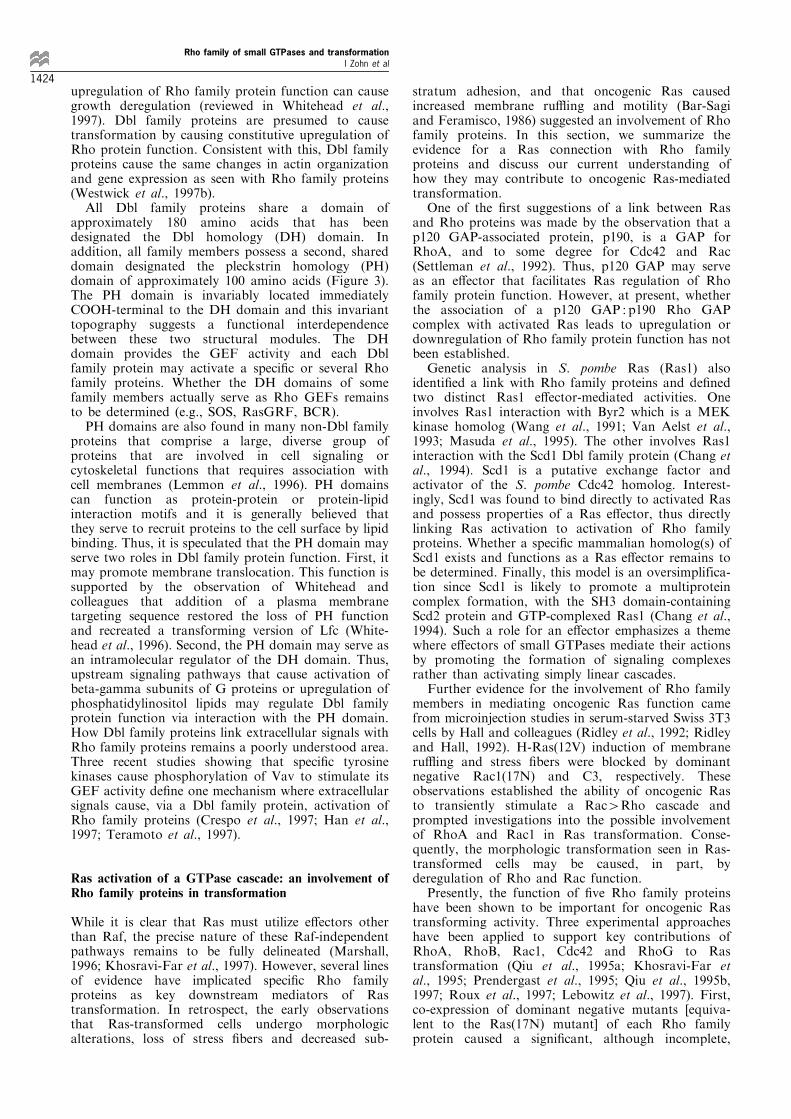
While it is clear that Ras must utilize e�ectors otherthan Raf, the precise nature of these Raf-independentpathways remains to be fully delineated (Marshall,1996; Khosravi-Far et al., 1997). However, several linesof evidence have implicated speci®c Rho familyproteins as key downstream mediators of Rastransformation. In retrospect, the early observationsthat Ras-transformed cells undergo morphologicalterations, loss of stress ®bers and decreased sub-
stratum adhesion, and that oncogenic Ras causedincreased membrane ru�ing and motility (Bar-Sagiand Feramisco, 1986) suggested an involvement of Rhofamily proteins. In this section, we summarize theevidence for a Ras connection with Rho familyproteins and discuss our current understanding ofhow they may contribute to oncogenic Ras-mediatedtransformation.
One of the ®rst suggestions of a link between Rasand Rho proteins was made by the observation that ap120 GAP-associated protein, p190, is a GAP forRhoA, and to some degree for Cdc42 and Rac(Settleman et al., 1992). Thus, p120 GAP may serveas an e�ector that facilitates Ras regulation of Rhofamily protein function. However, at present, whetherthe association of a p120 GAP : p190 Rho GAPcomplex with activated Ras leads to upregulation ordownregulation of Rho family protein function has notbeen established.
Genetic analysis in S. pombe Ras (Ras1) alsoidenti®ed a link with Rho family proteins and de®nedtwo distinct Ras1 e�ector-mediated activities. Oneinvolves Ras1 interaction with Byr2 which is a MEKkinase homolog (Wang et al., 1991; Van Aelst et al.,1993; Masuda et al., 1995). The other involves Ras1interaction with the Scd1 Dbl family protein (Chang etal., 1994). Scd1 is a putative exchange factor andactivator of the S. pombe Cdc42 homolog. Interest-ingly, Scd1 was found to bind directly to activated Rasand possess properties of a Ras e�ector, thus directlylinking Ras activation to activation of Rho familyproteins. Whether a speci®c mammalian homolog(s) ofScd1 exists and functions as a Ras e�ector remains tobe determined. Finally, this model is an oversimplifica-tion since Scd1 is likely to promote a multiproteincomplex formation, with the SH3 domain-containingScd2 protein and GTP-complexed Ras1 (Chang et al.,1994). Such a role for an e�ector emphasizes a themewhere e�ectors of small GTPases mediate their actionsby promoting the formation of signaling complexesrather than activating simply linear cascades.
Further evidence for the involvement of Rho familymembers in mediating oncogenic Ras function camefrom microinjection studies in serum-starved Swiss 3T3cells by Hall and colleagues (Ridley et al., 1992; Ridleyand Hall, 1992). H-Ras(12V) induction of membraneru�ing and stress ®bers were blocked by dominantnegative Rac1(17N) and C3, respectively. Theseobservations established the ability of oncogenic Rasto transiently stimulate a Rac4Rho cascade andprompted investigations into the possible involvementof RhoA and Rac1 in Ras transformation. Conse-quently, the morphologic transformation seen in Ras-transformed cells may be caused, in part, byderegulation of Rho and Rac function.
Presently, the function of ®ve Rho family proteinshave been shown to be important for oncogenic Rastransforming activity. Three experimental approacheshave been applied to support key contributions ofRhoA, RhoB, Rac1, Cdc42 and RhoG to Rastransformation (Qiu et al., 1995a; Khosravi-Far etal., 1995; Prendergast et al., 1995; Qiu et al., 1995b,1997; Roux et al., 1997; Lebowitz et al., 1997). First,co-expression of dominant negative mutants [equiva-lent to the Ras(17N) mutant] of each Rho familyprotein caused a signi®cant, although incomplete,
Rho family of small GTPases and transformationI Zohn et al
1424

reduction in oncogenic Ras focus-forming activitywhen analysed in rodent ®broblasts. Second, co-expression of constitutively activated versions of eachRho family GTPase with activated Raf-1 mutantsshowed cooperative and synergistic focus-formingactivity. Finally, constitutively activated mutantscaused growth transformation of NIH3T3 or Rat1rodent ®broblasts. Taken together, these observationssupport a model where oncogenic Ras causes consti-tutive activation of speci®c Rho family proteins whichin turn activate a spectrum of functions that contributeto full Ras transforming activity. However, it should beemphasized that a demonstration that Ras-transformedcells exhibit constitutively elevated GTP-bound levelsof a speci®c Rho family protein has not beendetermined. Thus, which, if any, of the known Rhofamily proteins are clearly targeted by oncogenic Rasremains to be established.
The evaluation of e�ector domain mutant ofoncogenic H-Ras also supports the contribution ofRho family proteins to Ras transformation (White etal., 1995; Joneson et al., 1996b; Khosravi-Far et al.,1996). Speci®cally, two mutants with single amino acidsubstitutions (37G and 40C) lost the ability to bind andactivate Raf and the ERK pathway. Despite thisdefect, both mutant proteins retained the ability tocause full transformation of NIH3T3 cells (Khosravi-Far et al., 1996). However, the transformed phenotypecaused by Ras(12V, 37G) or Ras(12V, 40C) wasdistinct from that caused by activated Ras or Raf,and instead, was indistinguishable from the trans-formed phenotype caused by activated Rac or Rhoproteins. These observations lend further support for aRaf-independent connection between Ras and Rhofamily proteins that contributes to cellular transforma-tion.
The evaluation of Ras-transformed rodent fibro-blasts that stably co-expressed either dominantactivated or negative Rho family proteins hasprovided some clues regarding the contribution thesesmall GTPases to the Ras transformed phenotype. Forexample, coexpression of activated Rac1 or RhoAcaused further morphologic transformation and de-creased attachment to the plastic substratum of Ras-transformed NIH3T3 cells (Khosravi-Far et al., 1995).These cells also formed colonies in soft agar thatsuggested increased motility and invasive properties. Incontrast, co-expression of dominant negative Rac1 orRhoA caused a morphologic reversion of Ras-transformed NIH3T3 cells (Khosravi-Far et al.,1995). Taken together, these observations support thepossibility that Ras causes morphologic transforma-tion, in part, by upregulation of Rac1 or RhoAfunction. However, the observations that Rac1- andRhoA-transformed NIH3T3 cells do not undergo anysigni®cant morphologic transformation, and retainwell-organized stress ®bers and focal adhesions, arguethat aberrant Rac or Rho function is not responsiblefor that altered morphology and loss of stress ®bers.Instead, since Raf- and MEK-transformed NIH3T3cells also undergo morphologic transformation, itsuggests that the Raf4MEK4ERK pathway pro-motes morphologic transformation.
How the transient changes in actin organizationcaused by Rho family proteins relate to long termchanges is also not clear. Ras-transformed NIH3T3
cells do possess enhanced membrane ru�ing andpinocytosis, consistent with persistent Rac activation.However, the loss, rather than enhancement, of stress®bers and focal adhesions, are not consistent withpersistent Rho activation.
Several observations suggest that each Rho familyprotein contributes a distinct aspect of Ras transfor-mation. First, constitutively activated mutants of eachRho family protein caused a distinct transformedphenotype in NIH3T3 cells. Second, co-expression ofdi�erent combinations of activated mutants showedcooperative focus-forming activity, suggesting thatthey activate distinct pathways (Roux et al., 1997).Interestingly, the greatest cooperative focus-formingactivity was seen with co-expression of activatedCdc42(12V) and Rac1(12V). Third, the characteriza-tion of Rat1 ®broblasts coexpressing H-Ras(12V) andeither dominant negative Rac1 or Cdc42 argued thateach Rho family protein played distinct roles in Rastransformation (Qiu et al., 1997). Dominant negativeCdc42(17N) blocked the soft agar growth and causemorphologic reversion of Ras-transformed Rat-1cells. However, low serum growth was retained. Incontrast, Rac1(17N) reverted low serum growth andcolony formation in soft agar, but not morphologictransformation. Hence, it was proposed that Cdc42and Rac play distinct roles in Ras transformation,with Cdc42 mediating morphological transformationand Rac1 promoting serum-independent growth.Finally, distinct contributions of each Rho familyprotein to Ras transformation is also suggested bythe observation that dominant negative RhoA andCdc42, but not Rac1, blocked Raf transformingactivity. These ®ndings also suggest that theCdc424Rac4Rho cascade for actin reorganizationmay not apply for their association with oncogenicRas transformation. Finally, since dominant negativeRac1 impaired RhoG-mediated transformation, it wasproposed that Rac1 functioned downstream ofRhoG.
A search for the missing link that connects Ras with Rhofamily proteins
Although how Ras may connect with Rho familyproteins is not clearly understood at present, theprevailing evidence support the linkage via a Raf-independent pathway. First, dominant negative mu-tants of Rac1, RhoA, Cdc42 and RhoG showedcomplete, or preferential, inhibition of Ras versus Raffocus-forming activity. Second, the potent cooperativefocus-forming activity seen when activated Raf andRho family proteins are co-expressed suggests activa-tion of distinct signaling pathways. Third, activatedRaf does not cause direct activation of JNK, indicatingthat Rac, Cdc42 and RhoG are not downstreamcomponents of this pathway.
The studies presented above clearly establish acritical role for Rho proteins in mediating Ras-induced cytoskeletal alterations, cellular transforma-tion, gene expression and cell cycle progression.However, the components that transmit the signalfrom activated Ras to Rho proteins remain to bedetermined. Evidence that support the involvement ofknown Ras-binding proteins as candidate e�ectors that
Rho family of small GTPases and transformationI Zohn et al
1425
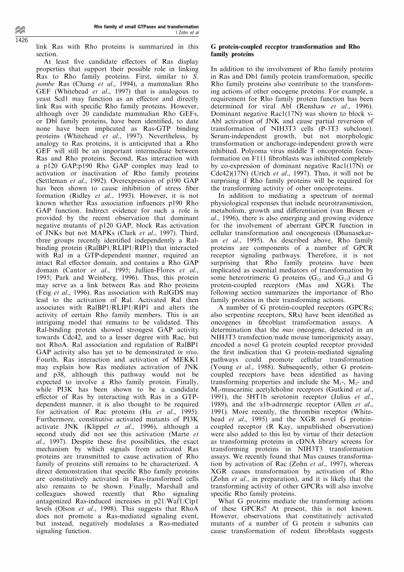
link Ras with Rho proteins is summarized in thissection.
At least ®ve candidate e�ectors of Ras displayproperties that support their possible role in linkingRas to Rho family proteins. First, similar to S.pombe Ras (Chang et al., 1994), a mammalian RhoGEF (Whitehead et al., 1997) that is analogous toyeast Scd1 may function as an e�ector and directlylink Ras with speci®c Rho family proteins. However,although over 20 candidate mammalian Rho GEFs,or Dbl family proteins, have been identi®ed, to datenone have been implicated as Ras-GTP bindingproteins (Whitehead et al., 1997). Nevertheless, byanalogy to Ras proteins, it is anticipated that a RhoGEF will still be an important intermediate betweenRas and Rho proteins. Second, Ras interaction witha p120 GAP:p190 Rho GAP complex may lead toactivation or inactivation of Rho family proteins(Settleman et al., 1992). Overexpression of p190 GAPhas been shown to cause inhibition of stress ®berformation (Ridley et al., 1993). However, it is notknown whether Ras association in¯uences p190 RhoGAP function. Indirect evidence for such a role isprovided by the recent observation that dominantnegative mutants of p120 GAP, block Ras activationof JNKs but not MAPKs (Clark et al., 1997). Third,three groups recently identi®ed independently a Ral-binding protein (RalBP1/RLIP1/RIP1) that interactedwith Ral in a GTP-dependent manner, required anintact Ral e�ector domain, and contains a Rho GAPdomain (Cantor et al., 1995; Jullien-Flores et al.,1995; Park and Weinberg, 1996). Thus, this proteinmay serve as a link between Ras and Rho proteins(Feig et al., 1996). Ras association with RalGDS maylead to the activation of Ral. Activated Ral thenassociates with RalBP1/RLIP1/RIP1 and alters theactivity of certain Rho family members. This is anintriguing model that remains to be validated. ThisRal-binding protein showed strongest GAP activitytowards Cdc42, and to a lesser degree with Rac, butnot RhoA. Ral association and regulation of RalBP1GAP activity also has yet to be demonstrated in vivo.Fourth, Ras interaction and activation of MEKK1may explain how Ras mediates activation of JNKand p38, although this pathway would not beexpected to involve a Rho family protein. Finally,while PI3K has been shown to be a candidatee�ector of Ras by interacting with Ras in a GTP-dependent manner, it is also thought to be requiredfor activation of Rac proteins (Hu et al., 1995).Furthermore, constitutive activated mutants of PI3Kactivate JNK (Klippel et al., 1996), although asecond study did not see this activation (Marte etal., 1997). Despite these ®ve possibilities, the exactmechanism by which signals from activated Rasproteins are transmitted to cause activation of Rhofamily of proteins still remains to be characterized. Adirect demonstration that speci®c Rho family proteinsare constitutively activated in Ras-transformed cellsalso remains to be shown. Finally, Marshall andcolleagues showed recently that Rho signalingantagonized Ras-induced increases in p21/Waf1/Cip1levels (Olson et al., 1998). This suggests that RhoAdoes not promote a Ras-mediated signaling event,but instead, negatively modulates a Ras-mediatedsignaling function.
G protein-coupled receptor transformation and Rhofamily proteins
In addition to the involvement of Rho family proteinsin Ras and Db1 family protein transformation, speci®cRho family proteins also contribute to the transform-ing actions of other oncogene proteins. For example, arequirement for Rho family protein function has beendetermined for viral Abl (Renshaw et al., 1996).Dominant negative Rac1(17N) was shown to block v-Abl activation of JNK and cause partial reversion oftransformation of NIH3T3 cells (P-3T3 subclone).Serum-independent growth, but not morphologictransformation or anchorage-independent growth wereinhibited. Polyoma virus middle T oncoprotein focus-formation on F111 ®broblasts was inhibited completelyby co-expression of dominant negative Rac1(17N) orCdc42)(17N) (Urich et al., 1997). Thus, it will not besurprising if Rho family proteins will be required forthe transforming activity of other oncoproteins.
In addition to mediating a spectrum of normalphysiological responses that include neurotransmission,metabolism, growth and di�erentiation (van Biesen etal., 1996), there is also emerging and growing evidencefor the involvement of aberrant GPCR function incellular transformation and oncogenesis (Dhanasekar-an et al., 1995). As described above, Rho familyproteins are components of a number of GPCRreceptor signaling pathways. Therefore, it is notsurprising that Rho family proteins have beenimplicated as essential mediators of transformation bysome heterotrimeric G proteins (G12 and G13) and Gprotein-coupled receptors (Mas and XGR). Thefollowing section summarizes the importance of Rhofamily proteins in their transforming actions.
A number of G protein-coupled receptors (GPCRs;also serpentine receptors, SRs) have been identi®ed asoncogenes in ®broblast transformation assays. Adetermination that the mas oncogene, detected in anNIH3T3 transfection/nude mouse tumorigenicity assay,encoded a novel G protein coupled receptor providedthe ®rst indication that G protein-mediated signalingpathways could promote cellular transformation(Young et al., 1988). Subsequently, other G protein-coupled receptors have been identi®ed as havingtransforming properties and include the M1-, M2- andM3-muscarinic acetylcholine receptors (Gutkind et al.,1991), the 5HT1b serotonin receptor (Julius et al.,1989), and the a1b-adrenergic receptor (Allen et al.,1991). More recently, the thrombin receptor (White-head et al., 1995) and the XGR novel G protein-coupled receptor (R Kay, unpublished observation)were also added to this list by virtue of their detectionas transforming proteins in cDNA library screens fortransforming proteins in NIH3T3 transformationassays. We recently found that Mas causes transforma-tion by activation of Rac (Zohn et al., 1997), whereasXGR causes transformation by activation of Rho(Zohn et al., in preparation), and it is likely that thetransforming activity of other GPCRs will also involvespeci®c Rho family proteins.
What G proteins mediate the transforming actionsof these GPCRs? At present, this is not known.However, observations that constitutively activatedmutants of a number of G protein a subunits cancause transformation of rodent ®broblasts suggests
Rho family of small GTPases and transformationI Zohn et al
1426

candidates for this role. In particular, constitutivelyactivated mutants of Ga12 and the related Ga13 haveexhibited the strongest transforming potential and cancause tumorigenic transformation of rodent ®broblasts(Xu et al., 1993; Vara Prasad et al., 1994; Chan et al.,1993; Jiang et al., 1993; Voyno-Yasenetskaya et al.,1994). Similarly, Gaq has been shown to transformNIH3T3 cells (Kalinec et al., 1992; De Vivo et al.,1992). However, Gaq can also be cytotoxic (Kalinec etal., 1992; Wu et al., 1992), and induces apoptosis(Althoefer et al., 1997). Since the signaling andtransforming activity of Ga12 and Ga13 have beenshown to involve Rac or Rho function, they representlogical connectors between transforming GPCRs andRho family proteins. For example, Ga12 transformingactivity was shown to be inhibited by dominantnegative RhoA and Rac1, but not Cdc42 (Tolkachevaet al., 1997). Microinjection analyses of activated Ga12
and Ga13 in Swiss 3T3 cells caused the induction ofstress ®bers (Buhl et al., 1995; Hooley et al., 1996) andboth have been shown to activate JNK (Prasad et al.,1995; Collins et al., 1996). Recently, the demonstrationthat Ga13 can interaction with and activate the Lsc/p115 RhoGEF Dbl family protein, a Rho GEF, de®nesat least one possible link between GPCRs and Rho(Hart et al., 1998).
In summary, the transforming actions of diverseoncoproteins, ranging from Ras to G protein-coupledreceptors, require the function of speci®c Rho familyproteins. Thus, it is likely that speci®c Rho familyproteins will play key supportive roles in humancarcinogenesis. Whether mutational activation, orabberant overexpression, of speci®c Rho familyproteins alone occur in human cancers will be animportant goal of future studies.
Rho family proteins mediate their actions throughinteraction with multiple e�ectors
Clearly, Rho family protein-mediated changes in actincytoskeletal organization, gene expression and regula-tin of cell cycle progression are all likely to contributeto the aberrant growth phenotype of Ras-transformedcells. However, what aspects of their function,mediated by which e�ectors, contribute to Rastransformation are presently not understood. Oneapproach for addressing this question has been todetermine what promotes the transforming activity ofspeci®c Rho family proteins. This process is compli-cated by the multitude of functions attributed to eachRho family protein and the fact that, like Ras, eachmediates its actions through a plethora of e�ectortargets. As with Ras, it is suspected that no one speci®ce�ector will be important for the growth-promotingactions of a speci®c Rho family protein.
The roster of candidate e�ectors for Rac1, RhoAand Cdc42 is extensive and continues to expand.Whereas the discovery of binding partners has beenrapid, progress in de®ning their contributions to Rhofamily protein function has been very limited. In thefollowing section, we provide a brief overview of Rhofamily binding proteins and possible functions. We alsosummarize observations from recent studies usinge�ector domain mutants of Rac1 and Cdc42 thatbegin to de®ne the precise function of speci®c Rho
family GTPase e�ectors. Finally, we conclude with adiscussion of how a spectrum of structurally andfunctionally diverse proteins all share the commonproperty of serving as e�ectors of a particular Rhofamily protein.
We have categorized Rho family protein bindingpartners into two general groups based on their bindingspeci®cities: (1) those that bind Rac and/or Cdc42, butnot Rho and (2) those that bind Rho. However,exceptions exist and include proteins that bind Rhoand Rac/Cdc42. It should be emphasized that many ofthese analyses have been limited to the analyses ofinteraction with Rac1, RhoA or Cdc42. Therefore,possible e�ector interactions with other Rho familymembers may complicate these simple classi®cations.
Many of the binding analyses have been performedin vitro or by using yeast two-hybrid binding analyses.Thus, whether they represent physiologically-relevantinteractions remain to be determined for many of theseproteins. What properties are required to validate ane�ector? First, the ability of a particular small GTPaseto bind to an isolated domain in vitro may bemisleading. Therefore, can interaction with the fulllength protein needs to be established in vivo? Second,does association lead to regulation, positive ornegative, of e�ector function? Third, does overexpres-sion of e�ector function mimic any aspect of thefunction of its corresponding GTPase provides?
A plethora of candidate e�ectors of Rac and Cdc42
Unlike the situation with candidate e�ectors of Ras, atleast three clear consensus binding sequences for Rhofamily binding proteins have been identi®ed for at leastsome of these proteins. Hall and colleagues havedescribed a minimal region of 16 amino acids requiredfor binding of Cdc42 and/or Rac, designated theCdc42/Rac Interactive Binding (CRIB) motif (Burbeloet al., 1995) (Figure 8a). Using this motif in a search ofthe GenBank data base, they identi®ed more than 25proteins from a wide variety of eukaryotic species thatcontain the CRIB motif. This motif is found in knownCdc42/Rac-binding proteins, such as the three p21(Cdc42/Rac) activated kinase (PAKs) isoforms and thetwo ACK nonreceptor tyrosine kinases. Other mam-malian CRIB motif-containing proteins include WASP,several mixed lineage kinases (MLK2, MLK3), and anuncharacterized human protein MSE55. Furtheranalyses demonstrated that all showed GTP-depen-dent interaction with Cdc42 and/or Rac1. Therefore,the CRIB motif may de®ne a GTP-dependentinteraction site found in a subset of Rac and/orCdc42 e�ector molecules.
Generally, Rac and Cdc42 share a number ofcommon binding partners which do not bind RhoA,which may be related to the higher sequence homologybetween Rac and Cdc42 compared to RhoA. However,some Rac-speci®c- (e.g., p67phox and POR-1) and someRac/Cdc42-binding proteins have been identi®ed thatlack the CRIB motif (e.g., pp70 S6 kinase, the p85subunit of PI3K, IQGAPs). Clearly, Rac-bindingmotifs other than the CRIB motif will be found.
Among the Rac/Cdc42-binding proteins that havebeen identi®ed, the three mammalian PAK serine/threonine kinase isoforms (rat a-PAK/human hPAK-1,rat p65 b-PAK/mouse mPAK3 and p62 g-PAK/human
Rho family of small GTPases and transformationI Zohn et al
1427

h-PAK2) have attracted the most interest (Manser etal., 1994, 1995; Teo et al., 1995; Bagrodia et al., 1995b;Martin et al., 1995). By virtue of their strikinghomology with the S. cerevisiae protein Ste20, whichis implicated in G protein-associated pheromonesignaling to a MAPK cascade, PAKs have beenconsidered likely candidates for e�ectors that promoteRac/Cdc42 activation of the JNK MAPK cascade(Simon et al., 1995; Zhao et al., 1995). Support for thispossibility was provided by experiments using Xenopusoocyte extracts, where Ste20, a related protein from S.pombe (Shk1) and PAK were shown to activate JNK/SAPK (Polverino et al., 1995). Additionally, theintrinsic kinase activity of PAKs was activated byRac/Cdc42-binding (Manser et al., 1994), and over-expression or constitutive activation of PAK showedenhanced activation of JNK or p38 (Zhang et al., 1995;Frost et al., 1996). Furthermore, dominant negativemutants of PAK inhibited Rac1 activation of JNK/p38(Zhang et al., 1995) or of the c-Jun-responsive elementin the collagenase promoter (Osada et al., 1997).
However, other evidence argues that PAKs are note�ectors for Rac/Cdc42 activation of JNK. First,Westwick et al., recently identi®ed Rac e�ector domainmutants that no longer bound to, or activated, PAK,yet they retained the ability to activate JNK and p38(Westwick et al., 1997). This demonstrated that PAKwas not required for Rac activation, of JNK. Second,Gutkind and colleagues suggested that MLK3 linkedRac/Cdc42 to JNK (Teramoto et al., 1996a). MLK3(also known as PTK1 or SPRK) is a member of afamily of related kinases (including MLK1, MLK2/MST, and MUK/DLK/ZPK) of unknown function.MLK3 RNA is expressed in most tissues and cell lines(Ezoe et al., 1994; Ing et al., 1994). These investigatorsfound that MLK3 associated with activated Rac1 andCdc42, but not RhoA, in vivo, and co-expression ofMLK3 (and not PAK) caused a synergistic enhance-ment of Rac1 and Cdc42 activation of JNK (Teramotoet al., 1996a). Overexpression of MLK3 (or MLK2)alone caused activation of JNK, but not p38 or ERK(Tibbles et al., 1996; Hirai S-I et al., 1997). Thus,MLK2/3, rather than PAKs, may serve as Rac/Cdc42e�ectors that lead to activation of JNK. However, Halland colleagues described evidence that MLK2/MLK3were not e�ectors for Rac activation of JNK (Nagata etal., 1998).
MEKK1 and MEKK4 represent additional candi-dates for the MAPKKK that links Rac/Cdc42 withJNK (Gerwins et al., 1997; Fanger et al., 1997).MEKK1 was found to interact with Rac and Cdc42,but not RhoA, in a GTP-dependent fashion in vitro. Incontrast, MEKK4 associated with both GDP- andGTP-bound Rac1 and Cdc42 in vitro. MEKK4, butnot MEKK1, contains a modi®ed CRIB motif. Kinase-inactive mutants of MEKK1 and MEKK4 bothblocked Cdc42 and Rac activation of JNK, and bothwere activators of JNK, but not p38, in COS cells. Incontrast, a second study evaluating a human homologof MEKK4 suggested that it was not a mediator ofRac or Cdc42 activation of JNK and furthermore, thatit was an activator of primarily p38, rather than JNK(Takekawa et al., 1997). The basis for thesediscrepancies is presently not clear.
Rac e�ector domain mutant studies also eliminatedPAK as the e�ector for directing Rac activation of
SRF or for transformation of NIH3T3 cells (Westwicket al., 1997a) and well as for induction of lamellipodia(Lamarche et al., 1996; Joneson et al., 1996a; Westwicket al., 1997). Instead, a correlation was found thatimplicated PAK as a possible e�ector for mediatingRac stimulation of transcription from the cyclin D1
promoter (Westwick et al., 1997). However, Lamarcheet al. (1996) found that PAK was dispensable for Rac-mediated progression through G1. Thus, Rac upregu-lation of cyclin D1 alone may not be su�cient for itsregulation of cell cycle progression. Furthermore, theRac-induced pathways leading to the regulation oflamellipodia, activation of JNK, SRF and cyclin D1
appear to be mediated by distinct e�ectors (Westwicket al., 1997). Which, if any, of the known Racfunctions are important for Rac transformation, andhence, Ras transformation, is presently unresolved.Finally, RhoB activation of SRE was retained in anonprenylated mutant that was impaired in transform-ing activity, indicating that SRE activation alone is notsu�cient for transformation (Lebowitz et al., 1997).
E�ector mutants of Rac and Cdc42 have alsouncoupled JNK activation from regulation of cytoske-letal changes and G1 cell cycle progression. Y40Cmutants of Rac and Cdc42 showed impaired binding toPAK and other CRIB motif-containing proteins (e.g.,WASP), but Rac1(40C) did retain interaction with thenon-CRIB motif-containing proteins p67PHOX or p160ROCK. However, since the Y40C mutants of Rac1 andCdc42 retained wild type abilities to induce actinreorganization, CRIB motif-containing e�ectors arenot likely to be mediators of actin reorganization norare they involved in linking Cdc42 to Rac or Rac toRho. Activated PAK can cause changes in actin
a
Rho family of small GTPases and transformationI Zohn et al
1428

organization, but in a way distinct from those causedby Rac1 or Cdc42 (Manser et al., 1997; Sells et al.,1997). Furthermore, since Y40C mutants of both Racand Cdc42 also retained the ability to stimulate DNAsynthesis, CRIB motif-containing e�ectors may not be
involved in regulating Rac- or Cdc42-induced G1progression. Finally, since a F37A mutant ofRac1(61L) showed impaired p160 ROCK binding,and lost the ability to stimulate lamellipodia andDNA synthesis, it supports a role for ROCK as an
b
c
Figure 8 Consensus binding sequences de®ne di�erent classes of Rac/Cdc42 and Rho binding proteins. (a) The Cdc42/RacInteractive Binding (CRIB) motif (Burbelo et al., 1995) is shared by some, but not all, Rac and/or CDC42-binding proteins. CRIBsequences of MEKK4 (Gerwins et al., 1997) and Ack-2 (Yang and Cerione, 1997) and those previously reported by Burbelo et al.(1995) were aligned using ClustalW and shaded using Boxshade 3.2 (Thompson et al., 1994). Residues that are conserved in greaterthan 50% of sequences at a given sequence position are colored blue, while residues that are similar are colored yellow. R, rat; M,mouse; H, human; Sc, S. cerevisiae, Sp, S. pombe; C, C. elegans; B, bovine; D, Drosophila; As, Ascaris. The domain structure ofseveral known RhoA-binding proteins can be grouped into two distinct classes that share sequence similarity in their minimal Rhobinding sequences. (b) The class I Rho e�ector binding motif (REM-1) is shared by the PKN/PRK1 and PRK2 serine/threoninekinases and a novel protein that lack kinase domains (Rhophilin). Shown for PKN/PRK2 are: three predicted homologous coiled-coil (CC) domains referred to as homologous regions 1a, 1b and 1c (HR1a, HR1b, HR1c), (2) a Vo/HR2 domain that showshomology between PKN and PRK2 and highly similar to sequences surrounding the NH2-terminal Vo and pseudosubstrate sites ofPCKe and PKCZ and (3) the COOH-terminal kinase domain. The Rho binding sequence (shaded region) is located in the NH2-terminal CC domain of PKN/PRK2 and Rhophilin. The multiple sequence alignment of class I REM shows a highly conservedsequence followed by a heptad repeat and are indicated by a solid square (&). The ®rst position of the heptad repeat is notpredicted to be present in Rhophilin. Strictly conserved residues are indicated with an asterisk (*) and positions of very high aminoacid similarity are indicated by a period (.); (c) Proteins containing the class II REM-2 motif include Rho-kinase, p160 ROCK andROKa/b. These related kinases possess an NH2-terminal kinase domain, followed by a predicted large central coiled-coil (CC)domain and a pleckstrin homology (PH) domain that encompasses a cysteine-rich domain (CRD). The Rho binding sequence(shaded box) is located COOH-terminus of the central CC domain. The multiple sequence alignment of Rem-2 motifs contain aregion of strict sequence conservation and are indicated by an asterisk (*). Citron shows a similar domain structure to Rho-kinase,p160 ROCK and ROKa/b. The NH2-terminal half of the protein contains a large (*875 residues) predicted coiled-coil (CC)domain. Like the other class II proteins, the CC domain is followed by CRD and PH domains. Citron also contains a proline-richsite (PR) that is a putative SH3 domain binding motif. The putative Rho-binding sequence (shaded box; ) is found in theextreme COOH-terminus of the CC domain. Apart from similarities in domain structure, no strong sequence homology wasobserved between the Citron and the Rem-1 or Rem-2 motif-containing proteins
Rho family of small GTPases and transformationI Zohn et al
1429

e�ector for these two Rac functions. Further mutagen-esis of Rac and Cdc42 may identify mutants that willde®ne the contribution of other e�ectors to thefunction of Rac and Cdc42.
A role for PAK in Ras and Rac transformation hasbeen suggested by two di�erent groups. First, Fieldand colleagues showed that kinase-de®cient mutants ofPAK could block oncogenic Ras transformation (Tanget al., 1997). However, since mutation of the CRIBmotif did not abolish this inhibition of Ras transforma-tion. The inhibition was not likely to be due toformation of nonproductive complexes with Rac orCdc42. Interestingly, this antagonism of Ras was seenin Rat1, but not NIH3T3 cells. A second study showedthat expression of the CRIB motif-containing fragmentof PAK could reverse both Ras and Rac1 transformingactivity in NIH3T3 cells (Osada et al., 1997). Since thismutant PAK also blocked Ras and Rac activation of ac-Jun-responsive promoter element (TRE), it wasproposed that titration of Rac and/or Cdc42 wasresponsible for inhibition of Ras transformation.
The 70 kDa S6 kinase (p70S6K) also appears to be animportant e�ector of Rac and Cdc42 function. p70S6K isactivated by diverse mitogenic stimuli, includinggrowth factors, cytokines and activated oncogenes(Chou and Blenis, 1995). Present evidence supportsan important role for p70S6K in the progression of cellsfrom the G1 to S phase of the cell cycle. Chou andBlenis (1996) showed that catalytically inactive p70S6K
interacted with GTP-complexed Rac1 and Cdc42, butnot RhoA, in vitro (Chou and Blenis, 1996).Constitutively activated mutants of Rac1 and Cdc42,but not RhoA, stimulated p70S6K activity in vivo.E�ector domain mutations abolished this activity.Dominant negative Cdc42(17N) or Rac1(17N)blocked EGF- and PDGF-induced activation ofpp70S6K. Similar to the Ras:Raf interaction, Cdc42association with p70S6K in vitro alone did not activateits kinase activity. Thus, Cdc42/Rac interaction withp70S6K must facilitate subsequent events that lead tofull kinase activation in vivo. It is presently not knownwhat aspect of Rac function is mediated by p70S6K.
Rac1 and Cdc42 have been shown to complex thep85 subunit of PI3K in vitro and to co-precipitate withPI3K activity in vitro and in vivo (Zheng et al., 1994a;Tolias et al., 1995). No CRIB motif is present in thep85 subunit. This interaction was found to be GTP-dependent and a Cdc42(35A) e�ector domain mutantshowed impaired p85 binding (Zheng et al., 1994a).Cdc42 stimulated the activity of immunoprecipitatedPI3K 2 ± 4-fold. Thus, although the sequence homologyof p85 with other Rho GAPs (e.g., BCR, N-chimaerinand p190 Rho GAP) suggested that it acted as anegative regulator of Rho function, no GAP functionhas been demonstrated for p85. Instead, this GAP-related domain may serve to promote PI3K associationwith Rac/Cdc42 as an e�ector in regulating reorganiza-tion of the actin cytoskeleton (Zheng et al., 1994a).Interestingly, another Rho GAP (N-chimaerin) hasalso been shown to exhibit properties of a Rac/Cdc42e�ector that mediates actin cytoskeletal events (Kozmaet al., 1996).
PI3K has also been implicated as an e�ector of Ras.However, it is the p110 catalytic subunit thatcomplexes with Ras-GTP (Rodriguez-Viciana et al.,1994, 1996). PI3K functions as a lipid kinase and its
phosphorylated phosphoinositides can interact andregulate a wide variety of protein targets, including anumber of proteins that regulate actin assembly(reviewed in Toker and Cantley, 1997). Since theoncogene responsible for the transforming activity ofthe avian sarcoma virus 16 in chicken embryo®broblasts encodes the p100 catalytic subunit chickenPI3K (Chang et al., 1997), upregulation of PI3Kactivity may contribute to Ras and Rac growthtransformation.
Two Rac-binding proteins have been implicated inmediating Rac regulation of the actin cytoskeletalorganization. In one study, Van Aelst and colleaguesused yeast two-hybrid screening and identi®ed apartner of Rac1(POR1) protein that interacted withGTP-complexed Rac1, but not Cdc42, RhoA or H-Ras(Van Aelst et al., 1996). The 34 kDa POR1 proteinshowed no signi®cant sequence identity with anyknown proteins and no conserved motifs besides thepresence of a putative leucine zipper motif. TheRac1(35A) e�ector domain mutant that is impairedin membrane ru�ing also showed impaired binding toPOR1. Truncated POR1 inhibited Rac1(12V)-inducedmembrane ru�ing, whereas full length POR1 acted tosynergistically enhance activated H-Ras(12V) mem-brane ru�ing activity. However, no synergisticinteraction was seen between Rac1 and POR1.Finally, POR1 may also serve as an e�ector foranother small GTPase ARF6 and mediate relatedchanges in actin cytoskeletal organization in CHO cells(D'Souza-Schorey et al., 1997). ARF6 also regulatesendocytic tra�cking. Thus, together with RhoD, itprovides further functional links between actincytoskeleton reorganization and membrane tra�cking.
In another study, yeast two-hybrid library screeningwith activated RhoA(63L) isolated a previouslyidenti®ed RhoA-binding protein, p160ROCK (Ishizaki etal., 1996), that also interacts with activated forms ofRac1 and Cdc42, but not R-Ras (Lamarche et al.,1996). Since the Rac1(37A) e�ector domain mutant nolonger interacted with ROCK and failed to inducelamellipodia or G1 progression, it suggested thatROCK may mediate these two activities of Rac1.
The Wiskott ±Aldrich syndrome (WAS) protein(WASP) contains the CRIB motif and was identi®edas a Cdc42-binding protein that bound weakly to Rac1,but not RhoA (Symons et al., 1996; AspenstroÈ m et al.,1996; Kolluri et al., 1996). The WASP gene is mutatedin WAS patients. These WAS-a�ected patients possessan X-linked recessive disorder characterized bythrombocytopenia, which is characterized by recurrentinfections due to T and B cell function and eczema.Abnormalities seen in T and B cells suggested defects inthe organization of the actin cytoskeleton (Kirchhausenand Rosen, 1996). WASP overexpression caused theformation of WASP clusters that were highly enrichedin polymerized actin in porcine aortic endothelial cellsand clustering was inhibited by dominant negativeCdc42(17N), but not Rac1(17N) (Symons et al., 1996).Additionally, co-expression of WASP inhibitedCdc42(12V) and Rac1(12V) induced changes in actincytoskeleton organization. Furthermore, a region ofhomology between WASP and other proteins (VASPand Mena) that are involved in the organization of actincytoskeleton and control of micro ®lament dynamicshas been identi®ed (Gertler et al., 1996). Interestingly,
Rho family of small GTPases and transformationI Zohn et al
1430

this domain in VASP and Mena has been shown tointeract directly with components of focal adhesions.These results implicate WASP as an e�ector thatmediates the activity of Cdc42/Rac in F-actin poly-merization (Symons et al., 1996) and reduced WASPexpression may contribute to the cytoskeletal abnorm-alities seen in WAS a�ected males (Kolluri et al., 1996).Genetic evidence supporting this link comes fromanalyses of the yeast counterpart of WASP, designatedBee1p. Bee1p is a component of the cortical actincytoskeleton and plays an essential role in theorganization of actin ®laments (Li, 1997). Bee1 nullcells possess altered actin organization and are defectivein budding and cytokinesis. However, since Bee1 lacks aCRIB motif, its functions may be distinct from those ofhuman WASP. Finally, since a Cdc42 e�ector mutantthat lacked WASP binding still stimulated lamellipodiaformation, WASP is clearly not the e�ector to mediatethis Cdc42 e�ect (Lamarche et al., 1996).
Interestingly, WASP is also recognized by the Nckadaptor protein, via its Src homology 3 domains(Rivero-Lezcano et al., 1995; Quilliam et al., 1996),and Nck also complexes with other Rho family bindingproteins (PAK1, PAK3 and PRK2) (Bagrodia et al.,1995b; Quilliam et al., 1996; Bokoch et al., 1996;Galisteo et al., 1996). Nck associates with activatedreceptor tyrosine kinases (Li et al., 1992; Lee et al.,1993). Therefore, it may serve as an adaptor that linksextracellular stimuli to Rho family proteins.
IQGAP1, and the closely-related IQGAP2 (62%identity), have been shown to bind Rac1 and Cdc42,but not RhoA (Brill et al., 1996; Kuroda et al., 1996).No CRIB motif is present in these proteins. IQGAPwas identi®ed originally as a human protein thatharbored potential calmodulin binding IQ motifsupstream of a sequence related to the catalytic domainof Ras GAPs (Weissbach et al., 1994), and was isolatedindependently by a�nity chromatography analyses forGTP-dependent Cdc42-binding proteins (Kuroda et al.,1996). Whereas IQGAP1 RNA was found highlyexpressed in lung, kidney and placenta, IQGAP2 wasobserved to be expressed predominantly in the liver(Brill et al., 1996). Although both proteins show stronghomology with the Ras GAP of S. pombe, nostimulation of Ras, Rac1 or Cdc42 GTPase activityhas been detected (Brill et al., 1996; Kuroda et al.,1996). Since IQGAPs harbor a potential actin bindingdomain, and were found to be accumulated at insulin-or Rac1-induced membrane ru�ing areas, it waspostulated that IQGAPs may serve as e�ectors thatmediate the actin cytoskeletal events of Cdc42 andRac1 (Kuroda et al., 1996). Consistent with thispossibility, it was found that Cdc42 and IQGAP1formed a stable complex in vivo along with F-actin(Erickson et al., 1997). EGF stimulation also promotedformation of Cdc42:IQGAP1 complexes in COS cells,suggesting that growth factor stimulation may promotean IQGAP1-mediated link between Cdc42 and theactin cytoskeleton. Finally, additional modes ofIQGAP regulation are suggested by evidence thatcalmodulin and calcium caused disruption ofCdc42:IQGAP1 interaction in vitro (Joyal et al., 1997).
Other candidate e�ectors of Rac include ROKa, apreviously described RhoA-binding protein (Leung etal., 1996b), POR-2, a protein of unknown function(Van Aelst et al., 1996), and tubulin (Best et al., 1996).
Citron is a protein of unknown function that wasidenti®ed as a RhoC-binding protein in a yeast two-hybrid library screen, and found to bind to Rac1, butnot Cdc42, in vitro (Madaule et al., 1995). Theinteraction of Citron with two functionally distinctsmall GTPases does not necessarily indicate a commonfunction for Citron. It is possible that interaction withRac and Rho may cause distinct subcellular locationsof Citron, or form distinct, signaling complexes, tocause distinct downstream events. Narumiya andcolleagues have recently described a splice variant ofCitron that contains an N-terminal Ser/Thr proteinkinase domain that has *50% sequence identity to thekinase domains of ROCK, ROK and MDK that arediscussed below (Madaule et al., 1998). DesignatedCitron-K, this widely expressed form may mediate Rhoregulation of cytokinesis. Messenger RNA encodingthe previously discovered Citron variant (designatedCitron-N) was detected only in brain tissue.
The 120 kDa ACK tyrosine kinase (ACK-1)speci®cally interacts with Cdc42 and not Rac orRhoA. In addition to its kinase domain, ACK alsohas an SH3 domain, a CRIB motif, and a proline-richC-terminus which may represent putative SH3 domainbinding sites. To date, little is known regarding the roleof ACK-1 as a Cdc42 e�ector. Recently, a 96 kDaACK-2 nonreceptor tyrosine kinase that shares thesame structural features with ACK and exhibits speci®cinteraction with GTP-bound Cdc42 was identi®ed(Yang and Cerione, 1997). The reduced size of ACK-2 is due primarily to the absence of sequences relatedto those present in the N- and C- terminal regions ofACK-1. ACK-2 transcripts were detected in a widevariety of human tissues, with highest levels seen inbrain and skeletal muscle tissue. Co-expression ofCdc42(61L) and ACK-2 in COS cells caused increasedACK-2 autophosphorylation. Furthermore, the copre-cipitation of the two proteins supports that possibilitythat ACK-2 is a physiologically relevant e�ector forCdc42. ACK-2 autophosphorylation was stimulated byattachment as well as by EGF or bradykininstimulation. Thus, ACK-2 may mediate the actions ofCdc42 in response to diverse extracellular stimuli.
The yeast two-hybrid screening of an Epstein ± Barrvirus-transformed B-cell library that identi®ed WASPalso identi®ed a second Cdc42-binding protein(AspenstroÈ m, 1997). Designated CIP4, this 545 aminoacid protein lacks a CRIB motif and shows sequencehomology with a number of proteins that serve asregulators of actin organization (e.g., RhoGAP).Binding to Cdc42 was GTP-dependent, and nobinding was seen to Rac1-GTP. Overexpression ofCIP4 caused a reduction in stress ®ber formation andco-expression of Cdc42(61L) caused a clustering ofCIP4. Thus, CIP4 may interact with Cdc42 in vivo toregulate actin organization although it does not appearto mediate Cdc42 induction of ®lopodia.
In summary, there exists multiple candidate e�ectorsof Rac and/or Cdc42 function and more are likely tobe identi®ed. The existence of Rac/Cdc42-bindingproteins that lack the CRIB motif indicates that atleast two, if not more, Rac- and/or Cdc42-bindingmotifs will be identi®ed. Currently, it is not clear,which, if any, of these Rac-binding proteins arephysiologically relevant e�ectors. Furthermore, whiche�ectors mediate Rac/Cdc42 regulation of actin
Rho family of small GTPases and transformationI Zohn et al
1431

organization, gene expression and regulation of cellproliferation and invasion remain to be characterized.With regards to transformation, our recent evaluationof Rac e�ector domain mutants failed to implicate anyknown Rac1 function with transforming potential(Westwick et al., 1997a). One interpretation is thatmultiple pathways promote Rac growth regulation andthat no one pathway alone is necessary. Alternatively,it suggests that some as yet undiscovered Rac functionis responsible for Rac-mediated transformation.Regardless of which is the correct scenario, it remainsto be de®ned what Rac function(s) is required topromote oncogenic Ras transformation. Finally,despite their signi®cant number of shared e�ectors, itis clear that Rac1 and Cdc42 are functionally distinctproteins. For example, human Cdc42, but not Rac1 orRac2, can restore a Cdc42Sc defect in S. cerevisiae(Shinjo et al., 1990). Furthermore, as described above,the transforming functions of Rac1 and Cdc42 arequite distinct.
A plethora of candidate e�ectors of Rho
A growing number of Rho-binding proteins have alsobeen identi®ed. Two distinct Rho-binding sequencemotifs have also been described for a subset of Rho-binding proteins (Reid et al., 1996; Fujisawa et al.,1996). Both Rho e�ector binding motifs are distinctfrom the CRIB motif and are associated with proteinsthat contain predicted coiled-coil (CC) domains (Cohenand Parry, 1994). These CC domains may provide thebasis for the multimerization of these proteins withRho and other signaling molecules in a complex withRho.
Two closely related serine/threonine kinases thatshow strong sequence similarity to the kinase domainof PKCs (PKN/PRK1 and PRK2), and two novelproteins lacking kinase domains (Rhotekin andRhophilin), appear to share a distinct Rho e�ectorbinding motif (designated class I, REM-1) (Figure 8b).PRK1 and PRK2 (58% amino acid identity) wereidenti®ed originally by a PCR-mediated approach toidentify sequences that encoded novel PKC-relatedhuman proteins (Palmer et al., 1995). The encoded120 ± 130 kDa proteins identi®ed by this approachcontain an NH2-terminal basic sequence encompassingGAXN (a putative pseudosubstrate domain), a REM-1Rho-binding sequence, followed by amphipathic helixand leucine zipper domains, and a COOH-terminalkinase domain (Figure 8b). Unlike PKC, neither areregulated by phorbol esters or calcium. Both PRK1and PRK2 show broad tissue expression.
The 71 kDa Rhophillin protein also contains anNH2-terminal Rem-1 binding motif. However, it lacksa kinase domain or any other known catalyticsequences. Aside from the NH2-terminal Rem-1motif, two proline-rich motifs and putative SH3-binding domains are present in the COOH-terminus.Thus, Rho-binding proteins with class I binding motifsalso typically contain other putative protein-protein orprotein-lipid binding motifs, suggesting that thesee�ectors may serve to promote targeting and/ormulti-protein complex formation with Rho proteins.
PRK1 was identi®ed independently by a�nitychromatography analysis for RhoA-binding proteinsand designated protein kinase N (PKN) (Watanabe et
al., 1996). Interestingly, LPA resulted in a Rho-dependent activation of PKN/PRK1 supporting a rolefor this kinase in mediating Rho function. Theobservation that staurosporine, a potent inhibitor ofPKC (and possibly PKN/PRK1), blocked formation offocal adhesions suggests that PKN may indeed mediatethis action of Rho proteins. Additionally, PKN hasbeen shown to interact with and phosphorylate thehead-rod domain of neuro®lament proteins suggestingits role in the regulation of neuro®lament proteinassembly (MoÈ sch et al., 1996). Hence, PKN may be apotential mediator of the growth factor-inducedstimulation of intermediate ®laments.
PRK2 was independently isolated by two groupssearching for Nck- or RhoA-interacting proteins. First,Quilliam et al. (1996) isolated PRK2 using anexpression library screen for proteins that bound tothe SH3 domains of the Nck SH2/SH3 adaptor protein(Quilliam et al., 1996). It was also shown to bind RhoA,but not Rac1 or Cdc42. In vitro and co-expression ofPRK2 with activated RhoA caused synergistic enhance-ment of RhoA-dependent activation of SRF expression.Vincent and Settleman (1997) puri®ed PRK2 (andPKN) by a�nity chromatography with RhoA fromrat liver extracts. However, they found that COS cell-expressed PRK2 interacted with GTP-bound Rac1, butinteracted with, and activated, both RhoA-GTP andRhoA-GDP. They also showed that a kinase-defectivemutant of PRK2 caused a disruption in actin stress®bers in NIH3T3 cells. Thus, they suggested that PRK2may be a common e�ector of both Rac and Rho. Thereason for the di�erences seen in these two studies is notknown. Finally, PKN/PRK1 and PRK2 were alsoidenti®ed independently as liver protease-activatedkinases 1 and 2, respectively (Yu et al., 1997).
Rhophilin, and to a lesser degree Rhotekin, shareNH2-terminal sequence homology with the PKN/PRK1and PRK2 Rem-1 motif. Rhotekin was identi®ed byyeast two-hybrid screening for RhoC-binding proteins(Reid et al., 1996). It also interacted with GTP-complexed RhoA and RhoB, but not Rac1 or Cdc42.Rhophilin was isolated by yeast two-hybrid screeningof a mouse embryo cDNA library for RhoA-bindingproteins (Watanabe et al., 1996). Rhophilin showedstrong GTP-dependent interaction with RhoA, littlewith RhoC, and less with RhoB. It also shares aputative NH2-terminal CC with PKN/PRK1 andPRK2.
A family of novel serine/threonine kinases share adistinct, second Rho e�ector binding motif (designatedclass II/REM-2) (Figure 8c). ROKa/Rho-kinase(Leung et al., 1996b) and p160ROCK/ROKb/ROCK-II(Leung et al., 1996a; Ishizaki et al., 1996) share anNH2-terminal kinase domain that has strong sequenceidentity with the kinase domain of myotonic dystrophykinase (MDK) (Ishizaki et al., 1996), a central putativeCC domain and a COOH-terminal PH domain (Mayeret al., 1993) that is split by a cysteine-rich, zinc-dependent folding domain. Interestingly, the Rho/Rac1binding protein Citron (Madaule et al., 1995) containsa related NH2-terminal CC domain that includes theRho-binding domain, and a related kinase domain(Madaule et al., 1998). However, the Rho-bindingsequence exhibits weaker sequence homology to theRem-2 motif present in these kinases. Hence, Citronmay possess a Rho-binding element distinct from the
Rho family of small GTPases and transformationI Zohn et al
1432

class II motif. Both Citron-K and Citron-N alsocontain a COOH-terminal cysteine-rich motif, a PHdomain and a proline-rich, putative SH3-binding motif.Thus, like other Ras and Rho family e�ectors, Citronmay also function as a sca�olding protein for Rho.
ROKa was identi®ed in expression screening forRhoA-binding proteins serine/threonine kinase (Leunget al., 1996b). ROKa showed a cytoplasmic locationand co-expression of RhoA(14V) caused an increasedassociation of ROKa with the pellet fraction, suggest-ing that RhoA promoted its translocation to mem-branes. Injection of DNA encoding full length ROKacaused the formation of stress ®bers and focaladhesions in HeLa cells (Leung et al., 1996a). Aclosely related protein with 64% overall identity toROKa and 90% identity to the ROKa kinase domain,was subsequently isolated (ROKb; also ROCK-II andp160 ROCK). In vitro binding analyses using puri®edrecombinant proteins showed that both ROKa andROKb bound RhoA, RhoB and RhoC, but not Rac1or Cdc42 (Leung et al., 1996a), although subsequentyeast two-hybrid analysis also showed interaction withRac1 (Joneson et al., 1996a) and Cdc42 (Lamarche etal., 1996).
Narumiya and colleagues identi®ed p160ROCK (a Rho-associated CC domain-containing protein kinase) as anovel RhoA-binding protein serine/threonine kinasethat showed 44% identity with the kinase domain ofMDK (Ishizaki et al., 1996). RhoA was shown toassociate with p160ROCK in vivo and this associationpromoted weak enhancement of its autophosphorylat-ing activity. Finally, Kaibuchi and colleagues identi®edthe p164 Rho-kinase by a�nity chromatographyanalyses for RhoA-GTP binding proteins (Matsui etal., 1996). Rho-kinase is likely a splice variant of ROKa,since they di�er only at their NH2-termini, with Rho-kinase containing an additional nine amino acids. Sincethe kinase activities of these proteins showed onlylimited stimulation by association with RhoA in vitro, itis likely that RhoA binding may facilitate other eventsthat lead to full kinase activation.
Another RhoA-GTP binding protein, myosin lightchain (MLC) phosphatase, together with Rho kinase,provides a mechanism for RhoA regulation of stress®ber formation (Kimura et al., 1996). The myosinbinding subunit (MBS) of MLC phosphatase wasidenti®ed as a RhoA-binding protein by a�nitychromatography analysis of bovine brain membraneextracts. RhoA binding and activation of Rho kinasestimulates Rho kinase phosphorylation of MBS. Thisresults in the inactivation of myosin phosphataseactivity, leading to activation of myosin through anet increase in MLC phosphorylation by other proteinkinases. MLC phosphorylation has been shown toinduce a conformational change in myosin, whichincreases its binding to actin ®laments, causingultimately the formation of stress ®bers (Chrzanows-ka-Wodnicka and Burridge, 1996). Rho-stimulatedcontraction of ®broblasts was blocked by the MLCkinase inhibitor (KT5926), resulting in decreased MLCphosphorylation and loss of stress ®bers and focaladhesions. Additionally, Rho kinase can also phos-phorylate MLC at the same sites that are phosphory-lated by MLC kinase (Amano et al., 1996). Finally,current evidence indicates that RhoA causes stress®bers and focal adhesion through distinct pathways
(Chrzanowska-Wodnicka and Burridge, 1996). TheCOOH-terminal sequences of MBS that bind to RhoAshare structural similarity (polybasic region followedby leucine zipper-like motif) with PKN/PRK1.However no detectable Rem-2 motif was observed(Kimura et al., 1996).
Another e�ector that may link Rho with the actincytoskeleton involves members of a formin-relatedfamily of proteins that include the S. cerevisiaeBni1p, the Drosophila Diaphanous and Cappuchinoproteins, and the mammalian Bnip1 homologp140mDia (Castrillon and Wasserman, 1994; Emmonset al., 1995; Peterson et al., 1995; Marhoul and Adams,1995). Bni1p and the related Bnr1p regulate the e�ectsof Rho proteins on actin organization. p140mDia caninteract with mammalian RhoA-GTP and pro®lin.Furthermore, p140mDia, pro®lin and RhoA are co-localized in a Rho-dependent structures such asmembrane ru�es in motile cells, cleavage furrows inmitotic cells, and phagocytic cups induced by®bronectin (Watanabe et al., 1997). Thus, p140mDiamay mediate actin assembly via a pro®lin-mediatedmechanism.
Since Rhotekin and Rhophillin lack known catalyticfunctions, essentially nothing has been describedregarding their functions. At least three possiblemechanisms for the function of these proteins can beenvisioned. They may either be directly involved inmediating some aspect of Rho function that does notrequire a catalytic function by promoting theirtranslocation to various components of cytoskeleton,or they may somehow negatively regulate the functionof Rho proteins by forming an inactive complex andpreventing the accessibility to other downstreame�ectors. Another possible function is that they mayact as sca�olding proteins that regulate the coordinateactivities of Rho proteins and their downstreame�ectors in a multimeric structure.
Observations that stimulation by G protein-coupledreceptor agonists that activate Rho (LPA andthrombin), or C3 treatment to block Rho function,of undi�erentiated neoronal cells (N1E-115, NG108-15and PC12 cells) have implicated Rho proteins incausing growth cone collapse, retraction of developingneurites, and transient rounding of the cell body(Mackay et al., 1995; Luo et al., 1996). One recentlyidenti®ed RhoA-binding protein, designated p116RIP,showed the same activity as C3 or dominant negativeRhoA when overexpressed in N1E-115 cells. Althoughp116RIP showed preferential binding to RhoA-GTP inyeast two-hybrid binding analyses, in vitro bindinganalyses showed equivalent binding to either GDP- orGTP-bound GST-RhoA. p116RIP is a novel protein thatcontains a PH domain, two putative SH3 domain-binding sites and a CC region. It was identi®ed in ayeast two-hybrid screen for RhoA-binding proteins,together with a novel RhoGDI (RIP1-RhoGDI2), anovel Db1 family protein (RhoGEF), and anotherRhoA-GTP binding protein (RIP4) (Gebbink et al.,1997).
Structural requirements for Rho family protein:e�ectorinteractions
The identi®cation of multiple binding proteins stronglysuggests that Rho family proteins utilize multiple
Rho family of small GTPases and transformationI Zohn et al
1433

e�ector-mediated pathways to cause its diverse array ofcellular e�ects. Like Ras, the regions of Rho familyproteins important for e�ector interaction also appearto be complex. The regions analogous to the core Rase�ector sequence (Ras residues 32 ± 40), and ¯ankingresidues are clearly important for all Rho familyprotein interactions with e�ectors. Mutagenesis andstructural analyses (Hirshberg et al., 1997) suggest atleast two additional, distinct, regions are also likely tomediate interaction with e�ectors.
There is some evidence that the Rho insert domainmay be important for interaction with at least somee�ectors (Freeman et al., 1996). Mutations in the Rac1caused a marked decrease in superoxide productionwithout perturbing p67PHOX binding, indicating that itwas not involved directly in e�ector binding (Nisimotoet al., 1997). Instead, it was proposed that it mayinteract with other components of the NADPH oxidasein neutrophils that was important for activity. In asecond study, a Cdc42 mutant protein, where the insertsequence was deleted by replacing it with thecorresponding region from H-Ras, retained the abilityto activate PAK-3 in vivo and to bind other CRIBmotif-containing proteins in vitro (ACK and WASP)(Wu et al., 1997). However, a reduced a�nity forinteraction from IQGAP1 was seen. Furthermore,while neither GEF or GAP activities were altered,RhoGDI inhibition of GDP dissociation was impairedin the insert mutant of Cdc42. Mutation of the insertdomains of Rac1 (Joneson and Bar-Sagi, 1998), Cdc42(Wu et al., 1998), or RhoA (Quilliam LA, unpublishedobservation) perturb their growth-promoting and/ortransforming activites. Thus, de®ning the e�ectortargets that interact with their respective insertdomains will delineate the signaling pathways thatpromote cellular transformation.
Studies with chimeric proteins between Rac1 andRhoA identi®ed a second, COOH-terminal region ofRac1 (residues 143 ± 175) important for Rac e�ectorfunction. Hall and colleagues showed that both aminoand carboxyl terminal regions of Rac1 were requiredfor PAK and p67PHOX binding, as well as for Rac-induced lamellipodia formation (Diekmann et al.,1995). In agreement with their observations, we alsoobserved that the Rac73Rho chimera did not bind PAKor induce lamellipodia, but instead induced stress ®berformation (Westwick et al., 1997). Furthermore, weobserved a loss of JNK activation, suggesting that thee�ector required for JNK activation also requiresinteraction with COOH terminal Rac sequences.Thus, like Ras, the e�ector-interacting sequences ofRho family proteins are also composed of multiple,distinct sequences.
The importance of sequences beyond switch I and IIin interaction with e�ectors underlines an emerging
concept that some small GTPase:e�ector interactionsare not GTP-dependent (e.g., PI5K). Instead, somemay be GDP-dependent, others may associate witheither GDP or GTP, yet activation may still be GTP-dependent.
Future directions
Despite the rapid accumulation of information on Rhofamily proteins, it is clear that we are only beginning toappreciate the complex nature of their biology andbiochemistry. Many questions remain. Will thediversity of cellular processes that involve Rho familyproteins continue to increase? Yes, the alreadyoutdated nature of this review veri®es that this willbe the case. The continued unearthing of yet more Dblfamily proteins argues that the diversity of cellularprocesses that activate Rho family proteins willcontinue to increase. More Rho family proteins willalso be discovered. Thus, the delineation of additionalRho-mediated functions promises to be a major themefor the coming years. Ample evidence from experi-mental cell systems implicate Rho family proteins ascritical components of oncogenic Ras transformation.Do Rho family proteins play such a role in humancancer cells? If so, are Rho-mediated pathwaysimportant targets for the development of anti-cancerdrugs? What speci®c aspects of Ras transformation arecontributed by Rho family proteins?
Understanding the mechanisms of Rho familyprotein signaling, both upstream and downstream,remains limited. How extracellular signals causeactivation of Rho family proteins remains a poorlyundestood area. Will Db1 family proteins provide thislink? The roster of Rho family binding proteins willcontinue to expand. Which are physiologically relevante�ectors? What is the role of each e�ector and how ise�ector utilization is controlled? Our understanding ofRho family signal transduction will continue to evolve.Complex interrelated networks, rather than simplylinear pathways, will likely be de®ned. Cell typedi�erences will continue to add further complexity. Insummary, the only thing that seems certain is thatevidence will continue to mount, establishing Rhofamily proteins as key regulators of many cellularprocesses in normal and neoplastic cells.
AcknowledgementsWe thank Jennifer Parrish for excellent assistance in thepreparation of the text, references and ®gures. Our studiesare supported by NIH grants to CJD (CA42978, CA52072,CA55008 and CA67771) and SC (CA64569 and CA70308).
References
Abo A, Pick E, Hall A, Totty N, Teahan CG and Segal AW.(1991). Nature, 353, 668 ± 670.
Adamson P, Marshall CJ, Hall A and Tilbrook PA. (1992). J.Biol. Chem., 267, 20033 ± 20038.
Adra CN, Manor D, Ko JL, Zhu S, Horiuchi T, Van Aelst L,Cerione RA and Lim B. (1997). Proc. Natl. Acad. Sci.USA, 94, 4279 ± 4284.
Alam MR, Johnson RC, Darlington DN, Hand TA, MainsRE and Eipper BA. (1997). J. Biol. Chem., 272, 12667 ±12675.
Albanese C, Johnson J, Watanabe G, Eklund N, Vu D,Arnold A and Pestell RG. (1995). J. Biol. Chem., 270,23589 ± 23597.
Rho family of small GTPases and transformationI Zohn et al
1434

Allen LF, Lefkowitz RJ, Caron MG and Cotecchia S. (1991).Proc. Natl. Acad. Sci. USA, 88, 11354 ± 11358.
Allen WE, Jones GE, Pollard JW and Ridley AJ. (1997). J.Cell Sci., 110, 707 ± 720.
Althoefer H, Eversole-Cire P and Simon MI. (1997). J. Biol.Chem., 272, 24380 ± 24386.
Amano M, Ito M, Kimura K, Fukata Y, Chihara K, NakanoT, Matsuura Y and Kaibuchi K. (1996). J. Biol. Chem.,271, 20246 ± 20249.
AspenstroÈ m, P, Lindberg U and Hall A. (1996). Curr. Biol.,6, 70 ± 75.
AspenstroÈ m P. (1997). Curr. Biol., 7, 479 ± 487.Avraham H and Weinberg RA. (1989). Mol. Cell. Biol., 9,
2058 ± 2066.Baeuerle PA and Baltimore D. (1996). Cell, 87, 13 ± 20.Bagrodia S, De rijard B, Davis RJ and Cerione RA. (1995a).
J. Biol. Chem., 270, 27995 ± 27998.Bagrodia S, Taylor SJ, Creasy CL, Cherno� J and Cerione
RA. (1995b). J. Biol. Chem., 270, 22731 ± 22737.Baichwal VR and Baeuerle PA. (1997). Curr. Biol., 7, R94 ±
R96.Bar-Sagi D and Feramisco JR. (1986). Science, 233, 1061 ±
1068.Best A, Ahmed S, Kozma R and Lim L. (1996). J. Biol.
Chem., 271, 3756 ± 3762.Bishop JM. (1991). Cell, 64, 235 ± 248.Bokoch GM, Wang Y, Bohl BP, Sells MA, Quilliam LA and
Knaus UG. (1996). J. Biol. Chem., 271, 25745 ± 25749.Brill S, Li S, Lyman CW, Church DM, Wasmuth JJ,
Weissbach L, Bernards A and Snijders AJ. (1996). Mol.Cell. Biol., 16, 4869 ± 4878.
Buhl AM, Johnson NL, Dhanasekaran N and Johnson GL.(1995). J. Biol. Chem., 270, 24631 ± 24634.
Burbelo PD, Drechsel D and Hall A. (1995). J. Biol. Chem.,270, 29071 ± 29074.
Campbell SL, Khosravi-Far R, Rossman KL, Clark GJ andDer CJ. (1998). Oncogene, in press.
Cantor SB, Urano T and Feig LA. (1995). Mol. Cell. Biol.,15, 4578 ± 4584.
Castrillon DH and Wasserman SA. (1994). Development,120, 3367 ± 3377.
Cerione RA and Zheng Y. (1996). Curr. Op. Cell Biol., 8,216 ± 222.
Chan AM-L, Fleming TP, McGovern ES, Chedid M, Miki Tand Aaronson SA. (1993). Mol. Cell. Biol., 13, 762 ± 768.
Chang EC, Barr M, Wang Y, Jung V, Xu H-P and WiglerMH. (1994). Cell, 79, 131 ± 141.
Chang HW, Aoki M, Fruman D, Auger KR, Bellacosa A,Tsichlis PN, Cantley LC, Roberts TM and Vogt PK.(1997). Science, 276, 1848 ± 1850.
Chant J and Stowers L. (1995). Cell, 81, 1 ± 4.Chardin P, Boquet P, Madaule P, Popo� MR, Rubin EJ and
Gill D. (1989). EMBO J., 8, 1087 ± 1092.Chardin P. (1993). GTPases in biology I, Dickey BF and
Birnbaumer L (Eds). Springer-Verlag: Heidelberg, pp159 ± 176.
Chou MM and Blenis J. (1995). Curr. Opin. Cell Biol., 7,806 ± 814.
Chou MM and Blenis J. (1996). Cell, 85, 573 ± 583.Chrzanowska-Wodnicka M and Burridge K. (1996). J. Cell
Biol., 6, 1403 ± 1415.Chuang T-H, Xu X, Kaartinen V, Heisterkamp N, Gro�en J
and Bokoch GM. (1995). Proc. Natl. Acad. Sci. USA, 92,10282 ± 10286.
Chuang TH, Xu X, Knaus UG, Hart MJ and Bokoch GM.(1993). J. Biol. Chem., 268, 775 ± 778.
Clark GJ, Westwick JK and Der CJ. (1997). J. Biol. Chem.,272, 1677 ± 1681.
Cohen C and Parry DAD. (1994). Science, 263, 488 ± 489.Collins LR, Minden A, Karin M and Brown JH. (1996). J.
Biol. Chem., 271, 17349 ± 17353.
Coso OA, Chiariello M, Yu J-C, Teramoto H, Crespo P, XuN, Miki T and Gutkind JS. (1995). Cell, 81, 1137 ± 1146.
Crespo P, Schuebel KE, Ostrom AA, Gutkind JS andBustelo XR. (1997). Nature, 385, 169 ± 172.
D'Souza-Schorey C, Boshans R, McDonough M, Stahl PDand Van Aelst L. (1997). EMBO J., in press.
Dallery E, Galiegue-Zouitina S, Collyn-d'Hooghe M, QuiefS, Denis C, Hildebrand MP, Lantoine D, Deweindt C,Tilly H and Bastard C. (1995). Oncogene, 10, 2171 ± 2178.
de Cremoux P, Gauville C, Closson V, Linares G, Calvo F,Tavitian A and Olofsson B. (1994). Int. J. Cancer, 59,408 ± 415.
De Vivo M, Chen J, Codina J and Iyengar R. (1992). J. Biol.Chem., 267, 18263 ± 18266.
Der CJ, Weissman B and MacDonald MJ. (1988). Oncogene,3, 105 ± 112.
Dhanasekaran N, Heasley LE and Johnson GL. (1995).Endocrine Rev., 16, 259 ± 270.
Diekmann D, Nobes CD, Burbelo PD, Abo A and Hall A.(1995). EMBO J., 14, 5338 ± 5349.
Drechsel DN, Hyman AA, Hall A and Glotzer M. (1996).Curr. Biol., 7, 12 ± 23.
Eaton S, Auvinen P, Luo L, Jan YN and Simons K. (1995). J.Cell Biol., 131, 151 ± 164.
Emmons S, Phan H, Calley J, Chen W, James B andManseau L. (1995). Genes Dev., 9, 2482 ± 2494.
Erickson JW, Cerione RA and Hart MJ. (1997). J. Biol.Chem., 272, 23443 ± 24447.
Ezoe K, Lee S-T, Strunk KM and Spritz RA. (1994).Oncogene, 9, 935 ± 938.
Fam NP, Fan W-T, Wang Z, Zhang L-J, Chen H and MoranMF. (1997). Mol. Cell. Biol., 17, 1396 ± 1406.
Fan W-T, Koch A, de Hoog CL, Fam NP and Moran MF.(1998). Curr. Biol., 8, 935 ± 938.
Fanger GR, Johnson NL and Johnson GL. (1997). EMBOJ., 16, 4961 ± 4972.
Feig LA, Urano T and Cantor S. (1996). Trends Biochem.Sci., 21, 438 ± 441.
Feig LA and Cooper GA. (1988). Mol. Cell. Biol., 8, 2472 ±2478.
Feltham JL, Dotsch V, Raza S, Manor D, Cerione RA,Sutcli�e MJ, Wagner G and Oswald RE. (1997).Biochemistry, 36, 8755 ± 8766.
Finco TS, Westwick JK, Norris JL, Beg AA, Der CJ andBaldwin Jr AS. (1997). J. Biol. Chem., 272, 24113 ± 24116.
Freeman JL, Abo A and Lambeth JD. (1996). J. Biol. Chem.,271, 19794 ± 19801.
Fritz G, Kaina B and Aktories K. (1995). J. Biol. Chem., 270,25172 ± 25177.
Frost JA, Xu S, Hutchison MR, Marcus S and Cobb MH.(1996). Mol. Cell. Biol., 16, 3707 ± 3713.
Fujisawa K, Fujita A, Ishizaki T, Saito Y and Narumiya S.(1996). J. Biol. Chem., 271, 23022 ± 23028.
Galisteo ML, Cherno� J, Su Y-C, Skolnik EY andSchlessinger J. (1996). J. Biol. Chem., 271, 20997 ± 21000.
Gebbink MFBG, Kranenburg O, Poland M, vanHorckFPG, Houssa B and Moolenaar WH. (1997). J. CellBiol., 137, 1603 ± 1613.
Gertler FB, Niebuhr K, Reinhard M, Wehland J and SorianoP. (1996). Cell, 87, 227 ± 239.
Gerwins P, Blank JL and Johnson GL. (1997). J. Biol.Chem., 272, 8288 ± 8295.
Guasch RM, Scambler P, Jones GE and Ridley AJ. (1998).Mol. Cell. Biol., 18, 4761 ± 4771.
Gutkind JS, Novotny EA, Brann MR and Robbins KC.(1991). Proc. Natl. Acad. Sci. USA, 88, 4703 ± 4707.
Habets GGM, Scholtes EHM, Zuydgeest D, van derKammen RA, Stam JC, Berns A and Collard JG. (1994).Cell, 77, 537 ± 549.
Rho family of small GTPases and transformationI Zohn et al
1435

Han J, Das B, Wei W, Van Aelst L, Mosteller RD, Khosravi-Far R, Westwick JK, Der CJ and Broek D. (1997). Mol.Cell. Biol., 17, 1346 ± 1353.
Hart MJ, Maru Y, Leonard D, Witte ON, Evans T andCerione RA. (1992). Science, 258, 812 ± 815.
Hart MJ, Jian X, Kozasa T, Roscoe W, Singer WD, GilmanAG, Sternweiss PC and Bollag G. (1998). Science, 280,2112 ± 2114.
Henske EP, Short MP, Jozwiak S, Bovey CM, Ramlakhan S,Haines JL and Kwiatkowski DJ. (1995). Ann. Hum.Genet., 59, 25 ± 37.
Hill CS, Wynne J and Treisman R. (1995). Cell, 81, 1159 ±1170.
Hill CS and Treisman R. (1995). EMBO J., 14, 5037 ± 5047.Hirai A, Nakamura S, Noguchi Y, Yasuda T, Kitagawa M,
Tatsuno I, Oeda T, Tahara K, Terano T, Narumiya S,Kohn LD and Saito Y. (1997). J. Biol. Chem., 272, 13 ± 16.
Hirai S-I, Katoh M, Terada M, Kyriakis JM, Zon LI, RanaA, Avruch J and Ohno S. (1997). J. Biol. Chem., 272,15167 ± 15173.
Hirshberg M, Stockley RW, Dodson G and Webb MR.(1997). Nat. Struct. Biol., 4, 147 ± 152.
Hooley R, Yu C-Y, Symons M and Barber DL. (1996). J.Biol. Chem., 271, 6152 ± 6158.
Hu Q, Klippel A, Muslin AJ, Fantl WJ and Williams LT.(1995). Science, 268, 100 ± 102.
Ihara K, Muraguchi S, Kato M, Shimizu T, Shirakawa M,Kuroda S, Kaibuchi K and Hakoshima T. (1998). J. Biol.Chem., 273, 9656 ± 9666.
Ing YL, Leung IWL, Heng HHQ, Tsui L-C and Lassam NJ.(1994). Oncogene, 9, 1745 ± 1750.
Irani K, Xia Y, Zweier JL, Sollott SJ, Der CJ, Fearon ER,Sundaresan M, Finkel T and Goldschmidt-Clermont PJ.(1997). Science, 275, 1649 ± 1652.
Ishizaki T, Maekawa M, Fujisawa K, Okawa K, IwamatsuA, Fujita A, Watanabe N, Saito Y, Kakizuka A, Morii Nand Narumiya S. (1996). EMBO J, 15, 1885 ± 1893.
Jahner D and Hunter T. (1991). Mol. Cell. Biol., 11, 3682 ±3690.
Jiang H, Wu D and Simon MI. (1993). FEBS Lett., 330,319 ± 322.
Joneson T, McDonough M, Bar-Sagi D and Van Aelst L.(1996a). Science, 274, 1374 ± 1377.
Joneson T, White MA, Wigler MH and Bar-Sagi D. (1996b).Science, 271, 810 ± 812.
Joneson T and Bar-Sagi D. (1998). J. Biol. Chem., 273,17991 ± 17994
Joyal JL, Annan RS, Ho Y-D, Huddleston ME, Carr SA,Hart MJ and Sacks DB. (1997). J. Biol. Chem., 272,15419 ± 15425.
Julius D, Livelli TJ, Jessell TM and Axel R. (1989). Science,244, 1057 ± 1062.
Jullien-Flores V, Dorseuil O, Romero F, Letourneur F,Saragosti S, Berger R, Tavitian A, Gacon G and CamonisJH. (1995). J. Biol. Chem., 270, 22473 ± 22477.
Kalinec G, Nazarali AJ, Hermouet S, Xu N and Gutkind JS.(1992). Mol. Cell. Biol., 12, 4687 ± 4693.
Karin M. (1995). J. Biol. Chem., 270, 16483 ± 16486.Keely PJ, Westwick JK, Whitehead IP, Der CJ and Parise
LV. (1997). Nature, 360, 632 ± 636.Khosravi-Far R, Solski PA, Kinch MS, Burridge K and Der
CJ. (1995). Mol. Cell. Biol., 15, 6443 ± 6453.Khosravi-Far R, White MA, Westwick JK, Solski PA,
Chrzanowska-Wodnicka M, Van Aelst L, Wigler MHand Der CJ. (1996). Mol. Cell. Biol., 16, 3923 ± 3933.
Khosravi-Far R, Campbell S, Rossman KL and Der CJ.(1997). Adv. Cancer Res., 72, 57 ± 107.
Kimura K, Ito M, Amano M, Chihara K, Fukata Y,Nakafuku M, Yamamori B, Geng J, Nakano T, OkawaK, Iwamatsu A and Kaibuchi K. (1996). Science, 273,245 ± 248.
Kirchhausen T and Rosen FS. (1996). Curr. Biol., 6, 676 ±678.
Kishi K, Sasaki T, Kuroda S, Itoh T and Takai Y. (1993). J.Cell Biol., 120, 1187 ± 1195.
Klippel A, Reinhard C, Kavanaugh M, Apell G, EscobedoM-A and Williams LT. (1996). Mol. Cell. Biol., 16, 4117 ±4127.
Kolluri R, Tolias KF, Carpenter CL, Rosen RS andKirchhausen T. (1996). Proc. Natl. Acad. Sci. USA, 93,5615 ± 5618.
Kotani K, Yonezawa K, Hara K, Ueda H, Kitamura Y,Sokaue H, Ando A, Chavanieu A, Calas B and GrigorescuF. (1994). EMBO J., 13, 2313 ± 2321.
Kozma R, Ahmed S, Best A and Lim L. (1995). Mol. Cell.Biol., 15, 1942 ± 1952.
Kozma R, Ahmed S, Best A and Lim L. (1996). Mol. Cell.Biol., 16, 5069 ± 5080.
Kozma R, Sarner S, Ahmed S and Lim L. (1997). Mol. Cell.Biol., 17, 1201 ± 1211.
Kuroda S, Fukata M, Kobayashi K, Nakafuku M, NomuraN, Iwamatsu A and Kaibuchi K. (1996). J. Biol. Chem.,271, 23363 ± 23367.
Lamarche N, Tapon N, Stowers L, Burbelo PD, AspenstromP, Bridges T, Chant J and Hall A. (1996). Cell, 87, 519 ±529.
Larochelle DA, Vithalani KK and De Lozanne A. (1996). J.Cell Biol., 133, 1321 ± 1329.
Lebowitz PF, Davide JP and Prendergast GC. (1995). Mol.Cell. Biol., 15, 6613 ± 6622.
Lebowitz PF, Du W and Prendergast GC. (1997). J. Biol.Chem., 272, 16093 ± 16095.
Lee C-H, Li W, Nishimura R, Zhou M, Batzer AG, Myers JrMG, White MF, Schlessinger J and Skolnik EY. (1993).Proc. Natl. Acad. Sci. USA, 90, 11713 ± 11717.
Lelias JM, Adra CN, Wulf GM, Guillemot JC, Khagad M,Caput D and Lim B. (1993). Proc. Natl. Acad. Sci. USA,90, 1479 ± 1483.
Lemmon MA, Ferguson KM and Schlessinger J. (1996).Cell, 85, 621 ± 624.
Leonard D, Hart MJ, Platko JV, Eva A, Henzel W, Evans Tand Cerione RA. (1992). J. Biol. Chem., 267, 22860 ±22868.
Leung T, Chen X-Q, Manser E and Lim L. (1996a). Mol.Cell. Biol., 16, 5313 ± 5327.
Leung T, Manser E, Tan L and Lim L. (1996b). J. Biol.Chem., 270, 29051 ± 29054.
Li R. (1997). J. Cell Biol., 136, 649 ± 658.Li W, Hu P, Skolnik EY, Ullrich A and Schlessinger J.
(1992). Mol. Cell. Biol., 12, 5824 ± 5833.Lin R, Bagrodia S, Cerione R and Manor D. (1997). Curr.
Biol., 7, 794 ± 797.Linares-Cruz G, Rigaut JP, Vassy J, De Oliveira TC, de
Cremoux P, Olofsson B and Calvo F. (1994). J. Microsc.,173, 27 ± 38.
Luo L, Hensch TK, Ackerman L, Barbel S, Jan LY and JanYN. (1996). Nature, 379, 837 ± 840.
Luttrell LM, Della Rocca GJ, van Biesen T, Luttrell DK andLefkowitz RJ. (1997). J. Biol. Chem., 272, 4637 ± 4644.
Mabuchi I, Hamaguchi Y, Fujimoto H, Morii N, MishimaMand Narumiya S. (1993). Zygote, 1, 325 ± 331.
Mackay DJ, Nobes CD and Hall A. (1995). Trends Neurosci.,18, 496 ± 501.
Madaule P, Furuyashiki T, Reid T, Watanabe G, Morii Nand Narumiya S. (1995). FEBS Lett., 377, 243 ± 248.
Madaule P, Eda M, Watanabe N, Fujisawa K, Matsuoka T,Bito H, Ishzahi T and Narumiya S. (1998). Nature, 394,491 ± 494.
Manser E, Leung T, Salihuddin H, Zhao Z-S and Lim L.(1994). Nature, 367, 40 ± 46.
Manser E, Chong C, Zhao Z-S, Leung T, Michael G, Hall Cand Lim L. (1995). J. Biol. Chem., 270, 25070 ± 25078.
Rho family of small GTPases and transformationI Zohn et al
1436
Manser E, Huang H-Y, Loo T-H, Chen X-Q, Dong J-M,Leung T and Lim L. (1997). Mol. Cell. Biol., 17, 1129 ±1143.
Marais R, Wynne J and Treisman R. (1993). Cell, 73, 381 ±393.
Marhoul JF and Adams TH. (1995). Genet., 139, 537 ± 547.Marshall CJ. (1996). Curr. Opin. Cell Biol., 8, 197 ± 204.Marte BM, Rodriguez-Viciana P, WennstroÈ m S, Warne PH
and Downward J. (1997). Curr. Biol., 7, 63 ± 71.Martin GA, Bollag G, McCormick F and Abo A. (1995).
EMBO J., 14, 1970 ± 1978.Masuda T, Kariya K-I, Shinkai M, Okada T and Kataoka T.
(1995). J. Biol. Chem., 270, 1979 ± 1982.Matsui T, Amano M, Yamamoto T, Chihara K, Nakafuku
M, Ito M, Nakano T, Okawa K, Iwamatsu A andKaibuchi K. (1996). EMBO J., 15, 2208 ± 2216.
Mayer BJ, Ren R, Clark KL and Baltimore D. (1993). Cell,73, 629 ± 630.
Mayo MW, Wang C-Y, Cogswell PC, Rogers-Graham KS,Lowe SW, Der CJ and Baldwin Jr AS. (1997). Science,278, 1812 ± 1815.
Michiels F, Habets GG, Stam JC, van der Kammen RA andCollard JG. (1995). Nature, 375, 338 ± 340.
Minden A, Lin A, McMahon M, Lange-Carter C, DerijardB, Davis RJ, Johnson GL and Karin M. (1994). Science,266, 1719 ± 1723.
Minden A, Lin A, Claret F-X, Abo A and Karin M. (1995).Cell, 81, 1147 ± 1157.
Molna r A, Theodoras AM, Zon LI and Kyriakis JM. (1997).J. Biol. Chem., 272, 13229 ± 13235.
MoÈ sch H-U, Roberts RL and Fink GR. (1996). Proc. Natl.Acad. Sci. USA, 93, 5352 ± 5356.
Murphy C, Sa�rich R, Grummt M, Gournier H, Rybin V,Rubino M, Auvinen P, LuÈ tcke A, Parton RG and ZerialM. (1996). Nature, 384, 427 ± 432.
Narumiya S. (1996). J. Biochem., 120, 215 ± 228.Nimnual AS, Yatsula BA and Bar-Sagi D. (1998). Science,
279, 560 ± 563.Nishiyama T, Sasaki T, Takaishi K, Kato M, Yaku H, Araki
K, Matsuura Y and Takai Y. (1994). Mol. Cell. Biol., 14,2447 ± 2456.
Nisimoto Y, Freeman JLR, Motalebi SA, Hirshberg M andLambeth JD. (1997). J. Biol. Chem., 272, 18834 ± 18841.
Nobes CD and Hall A. (1995). Cell, 81, 53 ± 62.Nobes CD, Lauritzen I, Mattei M-G, Paris S, Hall A and
Chardin P. (1998). J. Cell. Biol., 141, 187 ± 197.Nusrat A, Giry M, Turner JR, Colgan SP, Parkos CA,
Carnes D, Lemichez E, Boquet P and Madara JL. (1995).Proc. Natl. Acad. Sci. USA, 92, 10629 ± 10633.
Olson MF, Ashworth A and Hall A. (1995). Science, 269,1270 ± 1272.
Osada S-I, Izawa M, Koyama T, Hirai S-I and Ohno S.(1997). FEBS Lett., 404, 227 ± 233.
Palmer RH, Ridden J and Parker PJ. (1995). Eur. J.Biochem., 227, 344 ± 351.
Park S-H and Weinberg RA. (1996). Oncogene, 11, 2349 ±2355.
Paterson HF, Self AJ, Garrett MD, Just I, Aktories K andHall A. (1990). J. Cell Biol., 111, 1001 ± 1007.
Perona R, Esteve P, Jime nez B, Ballestero RP, Ramo n yCajal S and Lacal JC. (1993). Oncogene, 8, 1285 ± 1292.
Perona R, Montaner S, Saniger L, Sa nchez-Pe rez I, Bravo Rand Lacal JC. (1997). Genes Dev., 11, 463 ± 475.
Peterson J, Weilguny D, Egel R and Nielsen O. (1995). Mol.Cell. Biol., 15, 3697 ± 3707.
Polverino A, Frost J, Yang P, Hutchinson M, Neiman AM,Cobb MH and Marcus S. (1995). J. Biol. Chem., 270,26067 ± 26070.
Prasad MVVSV, Dermott JM, Heasley LE, Johnson GL andDhanasekaran N. (1995). J. Biol. Chem., 270, 18655 ±18659.
Prendergast GC, Khosravi-Far R, Solski PA, Kurzawa H,Lebowitz PF and Der CJ. (1995). Oncogene, 10, 2289 ±2296.
Qiu R-G, Chen J, Kirn D, McCormick F and Symons M.(1995a). Nature, 374, 457 ± 459.
Qiu R-G, Chen J, McCormick F and Symons M. (1995b).Proc. Natl. Acad. Sci. USA, 92, 11781 ± 11785.
Qiu R-G, Abo A, McCormick F and Symons M. (1997).Mol.Cell. Biol., 17, 3449 ± 3458.
Quilliam LA, Lambert QT, Westwick JK, Mickelson-YoungLA, Sparks AB, Kay BK, Jenkins NA, Gilbert DJ,Copeland NG and Der CJ. (1996). J. Biol. Chem., 271,28772 ± 28776.
Reibel L, Dorseuil O, Stancou R, Bertoglio J and Gacon G.(1991). Biochem. Biophys. Res. Commun., 175, 451 ± 458.
Reid T, Furuyashiki T, Ishizaki T, Watanabe G, WatanabeN, Fujisawa K, Morii N, Madaule P and Narumiya S.(1996). J. Biol. Chem., 271, 13556 ± 13560.
Reinstein J, Schlichting I, Frech M, Goody RS andWittinghofer A. (1991). J. Biol. Chem., 266, 17700 ± 17706.
Renshaw MW, Lea-Chou E and Wang JY. (1996). Curr.Biol., 6, 76 ± 83.
Ridley AJ, Paterson HF, Johnston CL, Diekmann D andHall A. (1992). Cell, 70, 401 ± 410.
Ridley AJ, Self AJ, Kasmi F, Paterson HF, Hall A, MarshallCJ and Ellis C. (1993). EMBO J., 12, 5151 ± 5160.
Ridley AJ, Comoglio PM and Hall A. (1995). Mol. Cell.Biol., 15, 1110 ± 1122.
Ridley AJ. (1996). Curr. Biol., 6, 1256 ± 1264.Ridley AJ and Hall A. (1992). Cell, 70, 389 ± 399.Rittinger K, Walker PA, Eccleston JF, Smerdon SJ and
Gamblin SJ (1997). Nature, 389, 758 ± 762.Rivero-Lezcano OM, Marcilla A, Sameshima JH and
Robbins KC. (1995). Mol. Cell. Biol., 15, 5725 ± 5731.Rodriguez-Viciana P, Warne PH, Dhand R, Vanhaesebroeck
B, Gout I, Fry MJ, Water®eld MD and Downward J.(1994). Nature, 370, 527 ± 532.
Rodriguez-Viciana P, Warne PH, Vanhaesebroeck B,Water®eld MD and Downward J. (1996). EMBO J., 15,2442 ± 2451.
Roux P, Gauthier-RouvieÁ re C, Doucet-Brutin S and Fort P.(1997). Curr. Biol., 7, 629 ± 637.
Scherle P and Staudt LM. (1993). Proc. Natl. Acad. Sci.USA, 90, 7568 ± 7572.
Schuebel KE, Bustelo XR, Nielsen DA, Song B-J, BarbacidM,GoldmanD and Lee IJ. (1996).Oncogene, 13, 363 ± 371.
Sells MA, Knaus UG, Bagrodia S, Ambrose DM, BokochGM and Cherno� J. (1997). Curr. Biol., 7, 202 ± 210.
Settleman J, Narasimhan V, Foster LC and Weinberg RA.(1992). Cell, 69, 539 ± 549.
Shinjo K, Koland JG, Hart MJ, Narasimhan V, Johnson DI,Evans T and Cerione RA. (1990). Proc. Natl. Acad. Sci.USA, 87, 9853 ± 9857.
Simon M-N, De Virgilio C, Souza B, Pringle JR, Abo A andReed SI. (1995). Nature, 376, 702 ± 705.
Sulciner DJ, Irani K, Yu Z-X, Ferrans VJ, Goldschmidt-Clermont P and Finkel T. (1996). Mol. Cell. Biol., 16,7115 ± 7121.
Symons M. (1996). Trends Biochem. Sci., 21, 178 ± 181.Symons M, Derry JMJ, Karlak B, Jiang S, Lemahieu V,
McCormick F, Francke U and Abo A. (1996). Cell, 84,723 ± 734.
Takai Y, Sasaki T, Tanaka K and Nakanishi H. (1995).TIBS, 20, 227 ± 231.
Takaishi K, Kikuchi A, Kuroda S, Kotani K, Sasaki T andTakai Y. (1993). Mol. Cell. Biol., 13, 72 ± 79.
Takaishi K, Sasaki T, Kato M, Yamochi W, Kuroda S,Nakamura T, Takeichi M and Takai Y. (1994). Oncogene,5, 273 ± 278.
Takekawa M, Posas F and Saito H. (1997). EMBO J., 16,4973 ± 4982.
Rho family of small GTPases and transformationI Zohn et al
1437

Tang Y, Chen Z, Ambrose D, Liu J, Gibbs JB, Cherno� Jand Field J. (1997). Mol. Cell. Biol., 17, 4454 ± 4464.
Tapon N, Nagata K, Lamarche N and Hall A. (1998).EMBO J., 17, 1395 ± 1404.
Teo M, Manser E and Lim L. (1995). J. Biol. Chem., 270,26690 ± 26697.
Teramoto H, Coso OA, Miyata H, Miki T and Gutkind JS.(1996a). J. Biol. Chem., 271, 27225 ± 27228.
Teramoto H, Crespo P, Coso OA, Igishi T, Xu N andGutkind JS. (1996b). J. Biol. Chem., 271, 25731 ± 25734.
Teramoto H, Salem P, Robbins KC, Bustelo XR andGutkind JS. (1997). J. Biol. Chem., 272, 10751 ± 10755.
Thompson JD, Higgins DG and Gibson TJ. (1994). Nucl.Acids Res., 22, 4673 ± 4680.
Tibbles LA, Ing YL, Kiefer F, Chan J, Iscove N, WoodgettJR and Lassam NJ. (1996). EMBO J., 15, 7026 ± 7035.
Toker A and Cantley LC. (1997). Nature, 387, 673 ± 676.Tolias KF, Cantley LC and Carpenter CL. (1995). J. Biol.
Chem., 270, 17656 ± 17659.Tolkacheva T, Feuer B, Lorenzi MV, Saez R and Chan AM.
(1997). Oncogene, 15, 727 ± 735.Treisman R. (1996). Curr. Opin. Cell Biol., 8, 205 ± 215.Ueda T, Kikuchi A, Ohga N, Yamamoto J and Takai Y.
(1990). J. Biol. Chem., 265, 9373 ± 9380.Urich M, Senften M, Shaw PE and Ballmer-Hofer K. (1997).
Oncogene, 14, 1235 ± 1241.Van Aelst L, Barr M, Marcus S, Polverino A and Wigler M.
(1993). Proc. Natl. Acad. Sci. USA, 90, 6213 ± 6217.Van Aelst L, Joneson T and Bar-Sagi D. (1996). EMBO J.,
15, 3778 ± 3786.Van Aelst L and D'Souza-Schorey C. (1997). Genes Dev., 11,
2295 ± 2322.van Biesen T, Luttrell LM, Hawes BE and Lefkowitz RJ.
(1996). Endocrine Rev., 17, 698 ± 714.Vara Prasad MVVS, Shore SK and Dhanasekaran N. (1994).
Oncogene, 9, 2425 ± 2429.Verschueren H, De Baetselier P, De Braekeleer J, Dewit J,
Aktories K and Just I. (1997). Eur. J. Cell Biol., 73, 182 ±187.
Vincent S, Jeanteur P and Fort P. (1992).Mol. Cell. Biol., 12,3138 ± 3148.
Vincent S and Settleman J. (1997). Mol. Cell. Biol., 17,2247 ± 2256.
Vojtek AB and Cooper JA. (1995). Cell, 82, 527 ± 529.Voyno-Yasenetskaya TA, Pace AM and Bourne HR. (1994).
Oncogene, 9, 2559 ± 2565.Walter M, Clark SG and Levinson AD. (1986). Science, 233,
649 ± 652.Wang XZ and Ron D. (1996). Science, 272, 1347 ± 1349.Wang Y, Xu H-P, Riggs M, Rodgers L andWigler M. (1991).
Mol. Cell. Biol., 11, 3554 ± 3563.Watanabe G, Saito Y, Madaule P, Ishizaki T, Fujisawa K,
Morii N, Mukai H, Ono Y, Kakizuka A and Narumiya S.(1996). Science, 271, 645 ± 648.
Watanabe N, Madaule P, Reid T, Ishizaki T, Watanabe G,Kakizaki T, Saito Y, Nakao K, Jockusch BM andNarumiya S. (1997). EMBO J., 16, 3044 ± 3056.
Wei Y, Zhang Y, Derewenda U, Liu X, Minor W, NakamotoRK, Somlyo AV, Somlyo AP and Derewenda ZS. (1997).Nature Struct. Biol., 4, 699 ± 703.
Weissbach L, Settleman J, Kalady MF, Snijders AJ, MurthyAE, Yan Y-X and Bernards A. (1994). J. Biol. Chem., 269,20517 ± 20521.
Wennstrom S, Hawkins P, Cooke F, Hara K, Yonezawa K,Kasuga M, Jackson T, Claesson WL and Stephens L.(1994). Curr. Biol., 4, 385 ± 393.
Westwick JK, Lambert QT, Clark GJ, Symons M, Van AelstL, Pestell RG and Der CJ. (1997). Mol. Cell. Biol., 17,1324 ± 1335.
Westwick JK, Lambert QT, Kay R, Der CJ and WhiteheadIP. (1998). J. Biol. Chem., 273, 16739 ± 16747.
White MA, Nicolette C, Minden A, Polverino A, Van AelstL, Karin M and Wigler MH. (1995). Cell, 80, 533 ± 541.
Whitehead I, Kirk H and Kay R. (1995). Oncogene, 10, 713 ±721.
Whitehead IP, Khosravi-Far R, Kirk H, Trigo-Gonzalez G,Der CJ and Kay R. (1996). J. Biol. Chem., 271, 18643 ±18650.
Whitehead IP, Campbell S, Rossman KL and Der CJ. (1997).Biochem. Biophys. Acta., 1332, F1 ± F23.
Whitehead IP, Abe K, Gorski JL and Der CJ. (1998). Mol.Cell. Biol., 18, 4689 ± 4697.
Whitmarsh AJ, Shore P, Sharrocks AD and Davis RJ.(1995). Science, 269, 403 ± 407.
Wu D, Lee CH, Rhee SG and Simon MI. (1992). J. Biol.Chem., 267, 1811 ± 1817.
Wu W-J, Leonard DA, Cerione RA and Manor D. (1997). J.Biol. Chem., 272, 26153 ± 26158.
Wu WJ, Lin R, Cerione RA and Manor D. (1998). J. Biol.Chem., 273, 16655 ± 16558.
Xu N, Bradley L, Ambdukar I and Gutkind JS. (1993). Proc.Natl. Acad. Sci. USA, 90, 6741 ± 6745.
YamamotoM,Marui N, Sakai T, Morii N, Kozaki S, Ikai K,Imamura S and Narumiya S. (1993). Oncogene, 8, 1449 ±1455.
Yang W and Cerione RA. (1997). J. Biol. Chem., 272,24819 ± 24824.
Yoshioka K, Imamura F, Shinkai K, Miyoshi J, Ogawa H,Mukai M, Komagome R and Akedo H. (1995). FEBSLett., 372, 25 ± 28.
Young D, O'Neill K, Jessell T and Wigler M. (1988). Proc.Natl. Acad. Sci. USA, 85, 5339 ± 5342.
Yu W, Liu J, Morrice NA and Wettenhall REH. (1997). J.Biol. Chem., 272, 10030 ± 10034.
Zalcman G, Closson V, Camonis J, Honore N, Rousseau-Merck M-F, Tavitian A and Olofsson B. (1996). J. Biol.Chem., 271, 30366 ± 30374.
Zervos AS, Faccio L, Gatto JP, Kyriakis JM and Brent R.(1995). Proc. Natl. Acad. Sci. USA, 92, 10531 ± 10534.
Zhang S, Han J, Sells MA, Cherno� J, Knaus UG, UlevitchRJ and Bokoch GM. (1995). J. Biol. Chem., 270, 23934 ±23936.
Zhao Z-s, Leung T, Manser E and Lim L. (1995). Mol. Cell.Biol., 15, 5246 ± 5257.
Zheng Y, Bagrodia S and Cerione RA. (1994a). J. Biol.Chem., 269, 18727 ± 18730.
Zheng Y, Cerione R and Bender A. (1994b). J. Biol. Chem.,269, 2369 ± 2372.
Zohn IM, Symons M, Chrzanowska-Wodnicka M, West-wick JK and Der CJ. (1998). Mol. Cell. Biol., 18, 1225 ±1235.
Rho family of small GTPases and transformationI Zohn et al
1438