Recombinant Supercharged Polypeptides Restore and Improve Biolubrication
Upload
independentCategory
view
2download
0
BacMam System for High-Level Expression of RecombinantSoluble and Membrane Glycoproteins for Structural Studies
Abhiram Dukkipatia,c, Hyun Ho Parka, Deepa Waghraya, Suzanne Fischera, and K.Christopher Garciaa,b,*
aDepartments of Molecular and Cellular Physiology and Structural Biology 279 Campus Drive, StanfordUniversity School of Medicine, Stanford, CA 94305, USA
bHoward Hughes Medical Institute, 4000 Jones Bridge Road, Chevy Chase, MD 20815, USA
AbstractBaculovirus mediated gene transduction of mammalian cells (BacMam) is an emerging techniquefor rapid recombinant protein expression in mammalian cells. Towards this, we constructed twobaculovirus transfer vectors that incorporate several mammalian transcriptional regulatory elementsnecessary for high level protein expression in mammalian cells. Using these vectors, we show thatthe BacMam system in combination with the 293 GnTI− cell line can be used for production ofmilligram quantities of soluble glycoproteins. Moreover, for crystallization trials, the purifiedglycoproteins are sensitive to EndoH treatment resulting in a loss of the bulk of the attached N-linkedglycosylation. In addition, we also show that a combination of the BacMam system and 293GnTI−cell line can be used for producing milligram quantities of a GPCR-protein ligand complex suitablefor crystallization trials.
KeywordsBacMam; 293 cells; GPCR; Parathyroid hormone receptor; Frizzled; US28; chemokine; Fractalkine;purification; crystallization
IntroductionThe baculovirus system for insect cells is one of the most commonly used methods forexpression of recombinant proteins for structural studies. Although this system has been usedvery successfully, there are still many cases of mammalian proteins that are refractory toexpression in insect cells, and require a mammalian expression system. This could be due tothe fact that insect cells are not natural hosts for mammalian proteins and therefore are unableto process these proteins into their correct functional conformations leading to low proteinyields and/or non-functional proteins. Over a decade ago, it was demonstrated very elegantlyby several groups that a number of mammalian cell lines are susceptible to transduction bybaculovirus. Moreover, by the inclusion of mammalian transcriptional units within thebaculovirus DNA, several mammalian cells grown as adherent cultures were shown to have
*Corresponding author. Fax: +1 650 725 8021, E-mail address: [email protected] Address: Structural Genomics Consortium, University of Oxford, ORCRB, Roosevelt Drive, Oxford, OX3 7DQ, UKPublisher's Disclaimer: This is a PDF file of an unedited manuscript that has been accepted for publication. As a service to our customerswe are providing this early version of the manuscript. The manuscript will undergo copyediting, typesetting, and review of the resultingproof before it is published in its final citable form. Please note that during the production process errors may be discovered which couldaffect the content, and all legal disclaimers that apply to the journal pertain.
Published as: Protein Expr Purif. 2008 December ; 62(2): 160–170.
HH
MI Author M
anuscriptH
HM
I Author Manuscript
HH
MI Author M
anuscript
the ability to recognize these transcriptional units when transduced by baculovirus [1–4]. Thisin turn leads to the very attractive option of using baculovirus mediated gene transduction forrecombinant protein production in mammalian cells (now termed as the BacMam system).Given that many structural biology labs are adept at the use of baculovirus, the BacMam systemoffers a relatively easy seque into mammalian cells using pre-existing methodologies withinthe laboratory.
Although very attractive in theory, there were several limitations that precluded this approachfrom a structural biology perspective. One of the main reasons was that in order to produce theamounts of recombinant proteins needed for structural studies on a routine basis with minimalmanual labor and maximal cost-effectiveness, one has to have the ability to grow and maintainmammalian cells such as 293S and CHO cells as high density suspension cultures. This wasnot feasible until recently due to lack of cell culture media that could promote long-term high-density suspension cultures of mammalian cells. However, with the recent introduction ofseveral commercially available chemically defined media, it is now possible to do so.
The BacMam system has evolved rapidly over the last couple of years. It is currently beingused by the pharmaceutical industry in a high-throughput setting for rapid protein productionin mammalian cells for the purpose of drug screening [5]. However, there has been only onereport where the BacMam system was used for producing recombinant proteins in quantitiesthat are sufficient to support protein crystallization trials [6]. The authors were successful inbeing able to produce and purify functional mammalian proteases in the range of severalmilligrams per liter of culture.
In this report we demonstrate that the BacMam system can be used for producing soluble ligandbinding domains of receptor glycoproteins in the range of several milligrams per liter of culture.Moreover by using the 293 GnTI− cell line [7] for protein production, we further show that forthe purpose of crystallization trials, the proteins can be deglycosylated very efficiently undernon-denaturing conditions. To demonstrate the ability of this system for purification ofrecombinant membrane proteins, we describe a simple strategy for the high-level expressionand purification of a G protein-coupled receptor (GPCR)1 in complex with its protein ligandusing the BacMam system. The purified GPCR-ligand complex is suitable for crystallizationstudies.
It is hoped that the results from this and the previous report [6] would help in persuadingmembers of the structural biology community to explore this technically simple and robustsystem as part of their expression systems for routine high level protein production. While wefind that neither baculovirus nor mammalian cells are uniformly successful at solving anyexpression problem, we can generally express the vast majority of proteins in one system orthe other. This requires that both systems are simultaneously accessible within one laboratory.The advantage of BacMam is that it enables one laboratory to easily use both insect-based andmammalian cell lines for expression, through the core technology of vector delivery throughthe baculovirus particle. In this fashion, both expression systems can be used side-by-side ina synergistic fashion.
Materials and metodsThe 293 GnTI− cell line was kindly provided by Prof H.G. Khorana (MIT, Cambridge, MA,USA) and Dr Philip J Reeves (MIT, Cambridge, MA, USA). pVL1393 vector was from BD
1Abbreviations used: GPCR, G protein-coupled receptor; DDM, n-dodecyl-β-D-maltoside; CHS, cholesteryl hemisuccinate tris salt; Sf9,Spodoptera Frugiperda; FPLC, fast protein liquid chromatography; A280, absorbance at 280 nanometer wavelength; WPRE, Woodchuckhepatitis virus post-transcriptional regulatory element; GlcNAc, N-acetylglucosamine; PTH, parathyroid hormone; FCS, foetal calfserum; GnTI, N-acetylglucosaminyltransferase-I; MWCO, molecular weight cut-off; PTH, parathyroid hormone
Dukkipati et al. Page 2
Protein Expr Purif. Author manuscript; available in PMC 2009 June 1.
HH
MI Author M
anuscriptH
HM
I Author Manuscript
HH
MI Author M
anuscript
Biosciences (San Jose, CA). pCI vector was from Promega (Madison, WI). Sapphire linearizedbaculovirus DNA was from Orbigen (San Diego, CA). Pro293s-CDM was from Cambrex (EastRutherford, NJ). DMEM, SF900 media, Cellfectin, Lipofectamine Penicillin/Streptomycin,Gentamycin, L-glutamine and Glutamax were from Invitrogen (Carlsbad, CA). All molecularbiology reagents, EndoHf and PNGaseF were obtained from NEB (Ipswich, MA). EndoHf isa fusion protein between maltose binding protein and EndoH. Primers for PCR weresynthesized by Operon (Huntsville, AL). Custom ordered double-stranded DNA wassynthesized by Genscript (Piscataway, NJ). Sodium butyrate and Protein A-Sepharose werefrom Sigma (St. Louis, MO). The anti penta-his antibody was from Qiagen (Valencia, CA) andHRP-conjugated polyclonal rabbit anti-mouse antibody was from DAKO (Carpinteria, CA).PreScission 3C protease was from GE Healthcare (Piscataway, NJ) and pre-cast 12% SDS-PAGE gels were from Bio-Rad (Hercules, CA).
Construction of expression vectors pVLAD7 and pVLAD6pCI vector was digested with BglII and BamHI and the 1kb fragment was gel purified. Thisfragment was ligated into pVL1393 that was digested with BglII and BamHI and transformedinto DH5α competent cells. A positive clone with the insert in the right orientation wasidentified by restriction digest analysis of mini-prep DNA and was labeled as pVLAD1.pVLAD1 was digested with NotI and BamHI and ligated with Fragment A (Figure 1A).Fragment A was a custom synthesized double stranded DNA fragment coding for 5’-NotI–[3Cprotease site]-[Glycine-Serine]-[hIgG1 Fc tag]-[6-His tag]-[Stop Codon]-[WPRE]-[SV40Late poly A]-BamHI-3’. The ligation mixture was transformed into DH5α competent cells anda clone containing the insert was identified by restriction digest analysis of mini-prep DNA.This final vector was labeled as pVLAD7. In another version, the Fc tag was replaced by aProtein C tag and the 6-His tag was replaced by a 8-His tag. This version of the vector waslabeled as pVLAD6. The nucleotide sequence coding for the extra-cellular domain of thehuman parathyroid hormone receptor 1 (corresponding to amino acids 1–187, including thesignal sequence) was PCR amplified and cloned into pVLAD7 (PTHR1ECD-pVLAD7) as anEcoRI/NotI fragment. A Kozak’s consensus sequence (CCACC) was included before theinitiating Methionine codon in the forward PCR primer. A chosen clone was sequenced toensure the absence of any unwanted mutations.
Sf9 transfection and virus amplificationSf9 cells were maintained as suspension cultures at 27°C in SF900 media supplemented with10% heat-inactivated FCS, 2mM L-glutamine and 10 µg/ml Gentamycin. For Sf9 transfection,0.5×106 cells were plated out per well in a 6-well plate. Twelve hours later, the cells werewashed twice with serum and antibiotic free SF900 media. The PTHR1ECD-pVLAD7construct was co-transfected with linearized baculovirus DNA using Cellfectin according tothe manufacturers recommendations. The primary virus was harvested after 4 days and usedfor amplification. For viral amplification, 500 ml of Sf9 cells in suspension (2×10⁶ cells/ml)were infected with 500µl of primary virus. Cells were diluted down to 2×10⁶ cells/ml the nextday with serum free SF900 media supplemented with 2mM L-glutamine. The amplified viruswas harvested 4 days later by spinning down the cells at 1000 g and collecting the supernatant.Viral stocks were stored at 4°C in the dark.
Growth of 293 GnTI− cells in suspension and viral transductionAdherent 293 GnTI− cells were grown in a humidified incubator (37°C, 5% CO2) in DMEMmedia supplemented with 2mM L-glutamine and 10% heat-inactivated FCS. When cells fromtwo T-75 flasks reached 80% confluency, they were sloughed from the flasks using Pro 293s-CDM media supplemented with 0.1% FCS and 4mM Glutamax. The cells were pooledtogether, made up to 25 ml, transferred to a 125 ml square flask and placed on an orbital shaker
Dukkipati et al. Page 3
Protein Expr Purif. Author manuscript; available in PMC 2009 June 1.
HH
MI Author M
anuscriptH
HM
I Author Manuscript
HH
MI Author M
anuscript
(1 inch diameter of rotation) that was placed within the incubator. The cap was opened by ahalf turn, taped down and the speed of the shaker was set to 130 rpm [8]. After 24 hours, thecells were diluted down to 0.5×10⁶ cells/ml. Typically, after each time the cells are diluteddown, they exhibit a lag time of ~24 hours before they resume growth and start doubling every24 hours. The suspension adapted cells were routinely maintained between 0.5–2×10⁶ cells/ml during passaging. The maximum cell density we achieve under these growth conditions is~ 4×10⁶ cells/ml with greater than 90% cell viability. The culture volume was maintained at25 ml in a 125 ml bottle. For scaling up, cells from four 125 ml bottles (2×10⁶ cells/ml) weretransferred to a 1L square flask, volume made upto 400ml and the cells were further grown ina 37°C warm room under identical agitation conditions. The growth profile of cells in a warmroom is very similar to that in a 5% CO2, 37°C humidified incubator (see above). When thecell density reached 2×10⁶ cells/ml, the cells were further split into 2L square bottles (1L cultureper bottle) until the desired culture volume was reached. Cell density was measured using ahaemocytometer and cell viability was measured by the Trypan blue dye exclusion method.
For viral transduction and protein expression, the appropriate volume of virus was added whenthe cells reached a density of 2×10⁶ cells/ml. Sodium butyrate was added to a finalconcentration of 10mM and the cells were left shaking for 72 hours in the warm room. Fortransient transfections of adherent 293 GnTI− cells on a small scale, 1×10⁶ cells/well in a 6well plate were transfected with 2µg of plasmid DNA using Lipofectamine according to themanufacturer’s recommendations. The media was collected 60 hours post-transfection andsubjected to Western blot analysis using the anti-his antibody (see below).
Protein purification using the Fc tagFor large-scale protein purification of secreted proteins containing the Fc tag, the transduced293 GnTI− cells were spun down at 1000g and the supernatant was filtered through a 0.45 µmmembrane. Two ml of protein A-Sepharose beads were packed into a column, the column wasconnected to a peristaltic pump and the beads were equilibrated with twenty bed volumes ofHBS (20 mM HEPES, 150 mM NaCl, pH 7.1) at a flow rate of 1 ml/min. The filtered mediawas pumped over the column at a flow rate of 1 ml/min. The beads were washed with ten bedvolumes of HBS and the protein was eluted with two bed volumes of 2M Arginine (pH 4.3)[9] at a flow rate of 0.5 ml/min. The beads were washed with HBS and stored in 20% ethanolfor reuse. The protein containing fractions were immediately neutralized with one-tenthvolume of 1M Tris.HCl, (pH 8.0), pooled and buffer exchanged into HBS using a PD-10desalting column. Protein concentration was quantitated by measuring A280 of the purifiedsample in HBS and this was used to calculate the expression level per liter of culture (assumingA280 of 1 mg/ml=1.6).
Protein purification using the 8-His tagFor large-scale protein purification of secreted proteins containing the 8-His tag tag, thetransduced 293 GnTI− cells were spun down at 1000g and the supernatant was filtered througha 0.45 µm membrane. The filtered media was concentrated down using a tangential flowconcentrator and buffer exchanged into HBS. The media was incubated overnight with Ni-NTA agarose beads in the presence of 20mM immidazole. The beads were washed in a sinteredglass filter with wash buffer (HBS+20mM immidazole), packed into a column and the boundprotein was eluted with elution buffer (HBS+200mM immidazole).
Protein deglycosylation and 3C protease digestionFor preparative scale EndoHf digestion under non-denaturing conditions, the purified proteinwas incubated with 50K units of EndoHf at room temperature for 12 hours. SDS-PAGE gelanalysis was used to verify deglycosylation. For 3C protease digestion, the sample wasincubated with 80 units of Precission 3C protease for 12 hours at 4°C. For analytical
Dukkipati et al. Page 4
Protein Expr Purif. Author manuscript; available in PMC 2009 June 1.
HH
MI Author M
anuscriptH
HM
I Author Manuscript
HH
MI Author M
anuscript
deglycosylation experiments with PNGaseF under denaturing conditions, approximately 5µgof protein sample was incubated with the manufacturer supplied denaturing buffer at 95°C for10 mins. The sample was cooled before adding the manufacturer supplied reaction buffer andNP-40 (final concentration 1%). 500 units of PNGaseF was then added and the sample wasincubated at 37°C for 1 hour. Samples were analyzed by SDS-PAGE.
Gel filtrationIn the case of Fc tagged proteins, the deglycosylated and 3C protease digested sample wasincubated with the required volume of Protein A- resin and Glutathione Sepharose (GEHealthcare). The beads were spun down at 1000 rpm, the supernatant was collected and thebeads were washed once with HBS. The supernatant and the wash solution were pooledtogether and injected in 500µl aliquots over a Superdex 200(10/30) gel filtration columnconnected to an AKTA FPLC purifier (GE Healthcare) and pre-equilibrated in HBS. The flowrate was maintained at 0.5 ml/min and 0.5 ml fractions were collected. Protein elution wasmonitored by measuring A280 and protein containing fractions were analyzed by SDS-PAGE.
Purification of US28-Fractalkine complexThe N-terminal chemokine domain of Fractalkine (residues 1–99, including the signalsequence) was PCR amplified, cloned into pVLAD7 and sequenced. Recombinant baculoviruswere generated as described above. One liter of 293GnTI− cells was transduced with 100 mlof Fractalkine-pVLAD7 baculovirus and supplemented with 10mM sodium butyrate. Theconditioned media was harvested 72 hours later, filtered and incubated overnight with 5ml ofprotein A-Sepharose in a batch mode. The beads were collected by vaccum filtration andwashed with HBS and stored at 4°C in HBS with protease inhibitors till further use (usuallywithin 24–36 hours). Based on small-scale elutions, we estimtate a density of ~2 mg ofFractalkine-Fc per ml of Protein A-Sepharose.
The US28 coding sequence (Genbank accession no AY174271) was PCR amplified to includea C-terminal 1D4 epitope tag followed by a stop codon and cloned into pVLAD6 (see above)as an EcoRI fragment. A clone containing the insert in the right orientation was identified byrestriction digest analysis of mini-prep DNA and sequenced. This clone was labeled asUS28-1D4-pVLAD6. Note that the presence of a stop codon immediately after the 1D4 epitopeprevents translation beyond the 1D4 epitope into the 3C site. A Kozak’s consensus sequence(CCACC) before the first ATG codon was included in the forward PCR primer. Recombinantbaculovirus for US28-1D4-pVLAD6 were generated as described above. Two liters of 293GnTI− cells (~1.5×10⁶ cells/ml) were transduced with 200 ml of US28-1D4-pVLAD6baculovirus and sodium butyrate was added to a final concentration of 10 mM. The cells wereharvested 30 hours later by spinning down at 1000g and washed with HBS (supplemented with5mM EDTA, pH 8.0). All steps from here on were carried out at 4°C. The washed cells werere-suspended in 200 ml of lysis buffer, (20mM Tris.HCl, pH 8.0, 5mM EDTA), incubated onice for 30 mins and dounce homogenized (20 strokes using a tight fitting pestle). The unlysedcells and cell debris were spun down at 1000g, supernatant collected and the pellet resuspendedin 50 ml of lysis buffer and redounced. The suspension was centrifuged again at 1000g, thesupernatants pooled together and spun at 48,000g for 1 hour. The crude membrane pellet wassolubilized in 200 ml of solubilization buffer (20mM HEPES, pH 7.1, 200mM NaCl, 10%glycerol, 0.5% DDM, 0.1% CHS, complete EDTA free protease inhibitor tablets) for 2 hoursby gentle end over end mixing followed by centrifugation at 48,000g for 1 hour. The clarifiedsupernatant was collected and batch incubated with 5ml of protein A-Sepharose bound toFractalkine-Fc (see above) by gentle end over end mixing for 36 hours. The beads were packedinto a column, connected to a peristaltic pump and washed at a flow-rate of 1ml/min with 20ml of wash buffer (20mM HEPES, pH 7.1, 200mM NaCl, 10% glycerol, 0.02% DDM/ 0.004%CHS). Protein was eluted with 3 bed volumes of elution buffer (100mM Glycine, pH 3.1,
Dukkipati et al. Page 5
Protein Expr Purif. Author manuscript; available in PMC 2009 June 1.
HH
MI Author M
anuscriptH
HM
I Author Manuscript
HH
MI Author M
anuscript
200mM NaCl, 10% glycerol, 0.02% DDM/0.004% CHS) at the same flow rate. The collectedfractions (8 ml each fraction) were neutralized immediately with one-tenth volume ofneutralization buffer (1M Tris.HCl, pH 8.0, 200mM NaCl, 10% glycerol. 0.02% DDM/0.004%CHS). The resin was washed with 50 bed volumes of wash buffer and stored in wash bufferwith 0.02% sodium azide. Protein containing fractions were identified by SDS-PAGE analysis,pooled together and incubated overnight with 50 units of 3C protease. The sample was depletedof free Fc and any undigested Fractalkine-Fc by passing through a 1 ml protein A-Sepharosecolumn. The flow-through was concentrated down to 0.5 ml using a 50 kDa MWCO centrifugalconcentrator and further purified over a Superose 6(10/30) gel filtration column (GEHealthcare) connected to a BioLogic Duo flow FPLC system (Bio-Rad) and equilibrated ingel filtration buffer [20 mM HEPES, pH 7.1, 150 mM NaCl, 10% glycerol, 0.02% DDM(Anagrade)/0.004% CHS]. Flow rate was maintained at 0.3 ml/min and 0.5 ml fractions werecollected. Protein elution was monitored by measuring A280 (0.5 cm path-length UV detector)and protein containing fractions were analyzed by SDS-PAGE.
SDS-PAGE and Western blottingFor SDS-PAGE analysis, samples were diluted 1:1 with reducing or non-reducing Laemmlisample buffer. Samples containing reducing buffer were heated to 95°C for 3 mins beforeloading onto 12% SDS-PAGE gels. Samples containing US28-1D4 were diluted 1:1 withsample buffer incubated at room temp for 10 mins and loaded onto 12% SDS-PAGE gels. Thegels were stained with Coomassie blue. For Western blotting, protein was transferred ontoPVDF membranes for 1 h at 350 mA in ice-cold transfer buffer (25 mM Tris, 192 mM Glycineand 10 % methanol). The blot was incubated for 30 min in TBST (20 mM Tris.HCl, pH 7.4,150 mM NaCl, 0.2% Tween) containing 5% non-fat dry milk powder (Carnation) and 1µg/mlof anti-penta His antibody (or 100 ng/ml of 1D4 antibody). The blot was washed for 10 minwith TBST and incubated with 200 ng/ml of an HRP-conjugated polyclonal rabbit anti-mousesecondary antibody in TBST for 30 min. The blot was washed thrice for 10 min each withTBST and developed using an enhanced chemiluminiscent detection kit (GE Healthcare).
Results293GnTI− cell line
The 293 GnTI− cell line developed by Khorana and collegues is a HEK293S derived cell linethat lacks the capability to process N-linked glycans on glycoproteins from the high mannoseto the complex mature form due to inactivation of both alleles of the GnTI gene [7]. Theresulting N-linked high mannose glycans are homogenous. This feature of the cell line makesit extremely attractive for recombinant protein production from a structural biology perspectivesince one of the major obstacle to obtaining high resolution diffraction quality crystals ofglycoproteins expressed and purified from eukaryotic hosts is the heterogenous nature of theattached N-linked glycans. In addition to the homogeneity, the high mannose glycans aresusceptible to EndoH enzyme treatment. EndoH is a glycosidase that cleaves within thechitobiose core of high mannose glycans in N-linked glycoproteins. The enzyme cleaves thelinkage between the 1st and 2nd GlcNAc of the high mannose glycan moiety. Thus, the EndoHtreated glycoprotein from 293GnTI− cells in principal should be devoid of the bulk of N-linkedglycosylation while at the same time the single GlcNAc’s still attached to the Asparagine/swould be presumably sufficient for maintaining protein solubility and activity.
We are able to maintain long-term suspension growth of this cell line in Pro293s-CDM mediain the presence of 0.1% serum. Although the stock cells are passaged in suspension in a CO2incubator, the dual buffering capability of Pro293s-CDM allows us to grow large scale cultures(>400ml) in a regular 37°C warm room environment. This relieves a lot of the space constraintsfor large-scale cultures. In our experience, there is no significant difference in growth rates
Dukkipati et al. Page 6
Protein Expr Purif. Author manuscript; available in PMC 2009 June 1.
HH
MI Author M
anuscriptH
HM
I Author Manuscript
HH
MI Author M
anuscript

between suspension cultures passaged in a CO2 incubator and those that have been passagedin the warm room as long as the cells are passaged every 3 days.
pVLAD7 and pVLAD6 baculovirus transfer vectorFor the purpose of baculovirus mediated gene transduction of mammalian cells, we constructedtwo pVL1393 derived vectors, pVLAD7 and pVLAD6 (Figure 1A and B). Both vectorsincorporate several elements necessary for mammalian transcription initiation (CMVpromoter), transcription termination (SV40 poly A late signal), RNA splicing (syntheticintron), mRNA processing, stability and export to the cytoplasm for translation (WPRE andsynthetic intron) [Figure 1A and B]. The CMV promoter is one of the strongest promoter thatis constitutively active in a number of mammalian cell lines while the SV40 poly A late signalis a strong transcription termination signal. A combination of these two elements should ensurethat the recombinant cDNA is constitutively transcribed while at the same time transcriptiontermination is tightly regulated. The effect of splicing of the nascent RNA transcript on proteinexpression from transfected cDNA’s in mammalian cells has been extensively studied over thelast two decades. The general outcome from these studies points to the phenomenon ofincreased recombinant protein expression when a splicing element is included 5’ to the codingsequence of the cDNA [10, 11]. For robust protein expression, the mRNA needs to be furtherprocessed and exported from the nucleus to the cytoplasm in a stable form that is recognizedby the ribosomal translation machinery. These processes are enhanced by the Woodchuckhepatitis virus post-transcriptional regulatory element [WPRE] (and also to a certain extent bythe process of splicing of the nascent RNA) [12]. To summarize, we have included in pVLAD7and pVLAD6 several features necessary for robust recombinant gene transcription and mRNAprocessing in mammalian cells. For the purpose of protein purification, the pVLAD7 vectorencodes downstream of the multiple cloning site for a 3C protease site followed by the humanIgG1 Fc tag (amino acids Asp104 to Lys330 of human IgG1 heavy chain) and a 6-His tag(Figure 1A). In the case of pVLAD6, the Fc fragment was replaced by the Protein C epitopeand the 6-His tag was replaced with an 8-His tag (Figure 1B). For cloning purposes, the cDNAis cloned in frame with the 3C protease sequence in both vectors (Figure 1C). Cleavage of therecombinant fusion protein with 3C protease permits separation of the purification tag fromthe native protein.
As a proof of principle that a combination of the BacMam system and 293GnTI− cells couldprovide sufficient amounts of deglycosylated recombinant soluble glycoproteins for structuralstudies, we describe here the purification of the extra-cellular ligand binding domain of a classB GPCR, the Parathyroid hormone receptor 1 (PTHR1). This receptor mediates most of thephysiological role of PTH in mineral ion homeostasis, particularly in bones and kidneys [13].Previous reports have shown that the ~170 amino acid extra-cellular domain (excluding thesignal sequence) confers ligand binding activity and moreover glycosylation of the receptor atthe 4 potential sites in the extra-cellular domain is not necessary for ligand binding [14–16].
The extra-cellular domain of PTHR1 (PTHR1ECD) including the native signal sequence wasPCR amplified and cloned into pVLAD7. Before undertaking large-scale baculovirusproduction, in order to determine whether this protein if at all could be expressed in 293GnTI− cells, the PTHR1ECD-pVLAD7 vector was transiently transfected on a small-scale intoadherent 293GnTI− cells. A Western blot analysis of the conditioned media after 60 hoursshows that PTHR1ECD-Fc is indeed synthesized and secreted into the culture medium (Figure2A) with an apparent molecular weight of 60 kDa on an SDS-PAGE gel. After confirmingprotein expression by transient transfection of 293 cells on a small-scale, recombinantbaculovirus for PTHR1ECD-pVLAD7 were produced and amplified as described in materialsand methods.
Dukkipati et al. Page 7
Protein Expr Purif. Author manuscript; available in PMC 2009 June 1.
HH
MI Author M
anuscriptH
HM
I Author Manuscript
HH
MI Author M
anuscript
Optimization of transduction conditionsInitial small-scale analytical experiments with 1 ml cultures in 12 well-plates were performedto optimize transduction conditions of suspension adapted 293GnTI− cells with the pVLAD7recombinant baculovirus. The parameters that were examined were volume of virus fortransduction, time course of protein expression and effect of histone deacetylase inhibitors onprotein expression. We show here the results for the PTHR1ECD-pVLAD7 baculovirus. Theresults obtained with pVLAD7 and pVLAD6 baculoviruses for most other proteins arequalitatively similar to those of PTHR1ECD-pVLAD7.
The first parameter that was optimized was the volume of virus to be added to the cells to detectprotein expression. As shown in figure 2A, lanes 1–5, protein expression is detected when 5µl of virus is added to 1 ml of cells and the expression level increases with increasing volumesof virus added (Figure 2B, lanes 2–5). We typically do not use a ratio greater than 1:10 of virusadded to culture volume as we noticed that cells tend to aggregate at higher ratios. To furtherboost protein expression, we examined the effect of sodium butyrate, a histone deacetylaseinhibitor, on protein expression. As can be clearly seen in Figure 2B, lanes 6–10, addition of10mM butyrate to the cultures results in significantly higher protein expression at all virus toculture ratios tested (Figure 2B, compare lanes 2–5 vs 6–9). A time course of protein expressionanalysis by Western blotting of the butyrate supplemented media is shown in Figure 2C. A 25ml suspension culture of 293 GnTI− cells (1×10⁶ cells/ml) was transduced with 2.5 ml ofPTHR1ECD-pVLAD7 baculovirus and supplemented with 10mM sodium butyrate. Smallaliquots of the media was harvested at the indicated time points (Figure 2C) after initial virusaddition and analyzed by immunoblot analysis with an anti-penta his antibody. The resultindicates that protein accumulation in the media peaks at 72 hours after virus addition. Finally,we compared time course of protein expression of cultures maintained in a 37°C warm roomversus in a 37°C, 5% CO2 humidified incubator (Figure 2D). A 25 ml suspension culture ofcells (1×10⁶ cells/ml) that had been maintained for 3 passages in a 37°C warm roomenvironment without any CO2 regulation was transduced with 2.5 ml of PTHR1ECD-pVLAD7baculovirus and supplemented with 10mM butyrate. For comparison sake, a parallel culturethat had been maintained in a regular humidified CO2 regulated cell-culture incubator was alsotransduced with the PTHR1ECD-pVLAD7 baculovirus and supplemented with butyrate. Bothtransduced cultures were shaken under identical agitation conditions in their respectiveculturing environment. Media was harvested at the indicated time points after virus additionand analyzed for protein expresssion by immunoblot analysis with an anti-penta his antibody.The results indicate that there is a slight lag in the onset of protein expression when culturesare maintained in a warm room environment. However, at the end of the 72 hour incubationperiod, protein expression levels are similar in both cases as judged by Western blot analysis.
We have also determined that the optimal cell density for viral transduction and proteinexpression is 1–2×10⁶ cells/ml (data not shown). When transduction is performed at highercell densities, there is significantly reduced protein expression and this could have to do witha shift in cell metabolism or a change in the culture media composition (such as lactateaccumulation or change in glucose concentration). In addition, amongst the various syntheticchemically defined media tested, we have found that the 293GnTI− cells grow most robustlyin suspension with significantly higher protein expression levels in Pro293s-CDM (data notshown).
Thus a large-scale viral transduction and protein expression involves, adding the amplifiedrecombinant baculovirus to the culture at a particular virus to culture ratio determined fromsmall-scale titrations. It is critical that for maximal protein expression, the cell density at thetime of virus addition be 1–2×10⁶ cells/ml. The culture is further supplemented with 10mMbutyrate and incubated in a 37°C warm room for 72 hours before harvesting the media.
Dukkipati et al. Page 8
Protein Expr Purif. Author manuscript; available in PMC 2009 June 1.
HH
MI Author M
anuscriptH
HM
I Author Manuscript
HH
MI Author M
anuscript

Large-scale purification of PTHR1ECDOne liter of 293GnTI− cells were transduced with 100 ml of baculovirus for PTHR1ECD-pVLAD7 and sodium butyrate was added to a final concentration of 10mM. The cells wereincubated shaking in a warm room at 37°C. The supernatant was harvested 72 hours later andthe Fc fusion protein was purified using protein A-Sepharose (see materials and methods). Asample of the purified protein analyzed by SDS-PAGE under reducing and non-reducingconditions is shown in Figure 3A. Majority of the PTHR1ECD-Fc fusion protein migrates withan apparent molecular weight of 60 kDa (Figure 3A, lane1) and as a disulfide linked dimer of~120 kDa (Figure 3A, lane 2) under reducing and non-reducing conditions respectively. Thisis consistent with our expectation that in solution, the PTHR1ECD domain contains no inter-molecular disulfides while the Fc domain exists as an inter-molecular disulfide linked dimer.The theoretical molecular weight of the mature fusion construct without taking into accountany post-translational modifications is 48 kDa. The total protein yield is 14 mg per liter with>90% purity of the PTHR1ECD-Fc recombinant protein.
The purified protein was treated with EndoHf under non-denaturing conditions (see materialsand methods) resulting in a drop in the molecular weight of the protein to 51 kDa and 100 kDaunder reducing and non-reducing conditions respectively (Figure 3B). The PTHR1ECD-Fcfusion possesses 5 potential N-linked glycosylation sites (4 in PTHR1ECD and 1 in the Fcdomain). It has been shown in previous work that some if not all 4 potential glycosylation sitesin PTHR1ECD are utilized in 293 cells and insect cells. An increase in incubation time, amountof enzyme used or increasing the incubation temperature to 37°C does not result in any furthershift (data not shown). It should be noted that the EndoHf treated sample migrates as a sharpband with a trailing smear suggesting that the sample either possesses N-linked glycosylationsites that are resistant to EndoHf treatment under non-denaturing conditions or the sample haspost-translational modifications other than N-linked glycosylation. This is further clarifiedupon cleavage of the sample with 3C protease thus separating the PTHR1ECD from the Fcdomain. Treatment with 3C protease converts the 51 kDa fusion protein into two main bands,a 29 kDa band corresponding to the Fc moiety and a 22 kDa band corresponding to PTHR1ECD(Figure 3C). The Fc band migrates as a sharp homogenous band while the PTHR1ECD migratesas a sharp band with a trailing smear and also an additional fuzzy band migrating just belowthe Fc under reducing conditions. To verify whether the heterogeneity in PTHR1ECD was dueto incomplete deglycosylation by EndoHf, the sample was treated with PNGaseF underdenaturing conditions. If the heterogeneity was due to N-linked glycosylation, we would expectthat treatment with PNGaseF under denaturing conditions would convert the heterogenousforms of PTHR1ECD into one form. However, as can be clearly seen in Figure 3D, treatmentwith PNGaseF does not result in any change in the migration pattern of the heterogenous formsof PTHR1ECD. This result leads us to conclude that additional post-translational modificationssuch as O-linked glycosylation contribute to the heterogeneity of PTHR1ECD and the sampleis deglycosylated upon treatment with EndoHf under non-denaturing conditions.
The deglycosylated sample was depleted of free Fc, undigested PTHR1ECD-Fc and 3Cprotease as described in materials and methods and further purified over a gel filtration column(Figure 3E). The PTHR1ECD elutes from the gel filtration column as a well-defined peak at16 ml with the trailing smear eluting slightly earlier compared to the main band of PTHR1ECDas judged by SDS-PAGE analysis (Figure 3E inset). Based on molecular weight calibration ofthe gel filtration column using protein standards of known molecular weight, the elution profileof PTHR1ECD suggests that it is monomeric in solution in the absence of ligand.
Previous attempts at expressing PTHR1ECD in insect cells resulted in expression levels of 0.6mg/ml [16]. Use of the BACMAM system results in significantly higher yields of 14 mg/literof the PTHR1ECD-Fc fusion (corresponding to ~6 mg/liter of PTHR1ECD) and moreover byusing the 293GnTI− cells, we were able to quantitatively deglycosylate bulk of the N-linked
Dukkipati et al. Page 9
Protein Expr Purif. Author manuscript; available in PMC 2009 June 1.
HH
MI Author M
anuscriptH
HM
I Author Manuscript
HH
MI Author M
anuscript
glycosylation present on PTHR1ECD which is an added advantage in crystallization attemptsof this molecule.
Purification and crystallization of the mouse Frizzled-4 receptor ligand binding domainThe Frizzled family of proteins are receptors for secreted Wnt proteins and other ligands. Thesereceptors belong to the GPCR superfamily and play an important role in regulation of cellpolarity, formation of neural synapses and the regulation of proliferation and several otherprocesses in the developing and adult organisms [17]. All members of this family share severalconserved features, including an extracellular ligand binding Cysteine rich domain and sevenhydrophobic transmembrane segments. One member of this family, human Frizzled-4, hasbeen implicated in familial exudative vitreoretinopathv [18]. In order to begin understandingthe molecular basis for ligand recognition by mammalian Frizzled-4, we expressed, purifiedand crystallized the ligand binding domain of mouse Frizzled-4 using the BacMam system.
The mouse Frizzled-4 receptor extracellular Cysteine rich ligand binding domain (mFz4 ECD)corresponding to residues 1–162 (including the signal sequence) was cloned into pVLAD6.Recombinant baculovirus were generated and the secreted protein was purified from 500 mlof media from a transduced 293GnTI− culture using Ni-NTA beads as described in materialsand methods. The purified protein runs as a doublet between 21 and 24 kD (Figure 4A) andthe approximate yield of mFz4 ECD is 1 mg/L. Further treatment under non-denaturingconditions with EndoHf collapses this doublet into a single band that migrates at ~21 kDa(Figure 4B, lane 3). The deglycosylayed sample was further purified over a Superdex 75(10/30)gel filtration column. The mFz4 ECD elutes from the column in a peak centered around 14.02ml (Figures 4D) and an SDS-PAGE analysis shows that the purified protein is >90% pure(Figure 4D inset, lanes 4–7). The purified protein from the gel filtration column wasconcentrated down to 3 mg/ml for crystallization trials using the hanging drop method. Crystalswere obtained after 2 days in 1M Sodium Malonate, 0.1% Jeffamine and 100mM HEPES, pH6.8 (Figure 4E). Crystals were harvested after 7 days and flash frozen using 20% glycerol asa cryoprotectant. These crystals diffracted to a resolution of 2.1 Å (Figure 4E, inset) at asynchrotron light source enabling us to solve the structure of mFz4 ECD, the details of whichwill be published elsewhere (manuscript in preparation).
In order to determine whether the expressed mFz4 ECD is capable of binding it cognate ligandWnt 5a, analytical pull down experiments were performed. For these experiments, a versionof Wnt 5a that has a C-terminal Protein C epitope and lacking a 8-His tag was used. mFz4 ECDwhen expressed by itself is predominantly found in the cell culture supernatant whereas Wnt5a expressed by itself is found in the soluble detergent extract of the cell pellet (Figure 4C,lanes 1 and 2) as determined by Western blot using an antibody directed against the Protein Cepitope. Moreover, Wnt 5a cannot be purified from this detergent extract using Ni-NTA beads(Figure 4C, lane 3), which is consistent with this construct lacking a 8-His tag. However whenpVLAD6 mFz4 ECD and Wnt 5a are co-expressed and the soluble detergent extract of the cellpellet was subjected to pull-down assays using Ni-NTA beads, it can be clearly seen that mFz4ECD can be purified from this extract and in addition, Wnt 5a also co-purifies (Figure 4C, lane4). This result indicates that mFz4 ECD is bioactive.
Purification of a GPCR- protein ligand complexUS28 is a constitutively active HCMV encoded GPCR that displays promiscuous binding withmembers of the CC-class of chemokines [19,20]. US28 also binds the membrane boundCX3CL1 chemokine (also known as Fractalkine) with a sub nanomolar affinity [21].Fractalkine consists of an N-terminal chemokine domain that is separated from thetransmembrane region by a mucin-like stalk [22]. Numerous biochemical studies havedemonstrated that just the N-terminal chemokine domain of Fractalkine is sufficient to bind
Dukkipati et al. Page 10
Protein Expr Purif. Author manuscript; available in PMC 2009 June 1.
HH
MI Author M
anuscriptH
HM
I Author Manuscript
HH
MI Author M
anuscript
US28 as well as its endogenous receptor CX3CR1 in heterologous expression systems [21,23]. US28 has been implicated in helping the virus hijack the infected host cellulartranscriptional machinery, promote cell to cell fusion, cell migration and also help infectedcells evade detection by the immune system by virtue of its ability to “scavenge” a broad rangeof CC-chemokines such as CCL5 (RANTES) and CCL3 (MIP-1α). Moreover, US28 has alsobeen show to be a co-receptor for HIV entry [24]. Thus an understanding of the molecular basisfor the interaction of US28 with its ligands is important from a physiological as well as drugdevelopment point of view. From here on, the term Fractalkine refers to the N-terminalchemokine domain of full-length Fractalkine.
Our success in purifying milligram quantities of soluble secreted proteins using the BacMamsystem prompted us to examine whether this system could be used for purification of functionalmembrane proteins such as GPCRs for structural studies. We decided to focus on GPCRs thathave protein ligands. There were two main reasons for doing so. The first being that in the caseof GPCRs which have protein ligands, we can express and purify the ligands as Fc fusionmolecules using the BacMam system. The second reason being we can use the protein ligand-Fc fusion bound to protein A-Sepharose as a ligand affinity column for a single step purificationof functional GPCRs from the solubilized membranes. Here we provide evidence for thefeasibility of such a strategy by describing the purification of the viral chemokine GPCR US28in complex with Fractalkine.
Western blot analysis of conditioned media from large-scale cultures transduced withFractalkine-pVLAD7 baculovirus shows the presence of two major immunoreactive bandswhen the sample is reduced and heated to 95°C in sample buffer before loading onto the SDS-PAGE gel (Figure 5A, lane 1). The band migrating at ~45 kDa corresponds to full-lengthFractalkine-Fc while the band migrating at ~35 kDa corresponds to the Fc domain due toproteolysis between the Fractalkine and Fc domains during cell culture. Surprisingly, when thesample preparation conditions are changed to just reducing conditions in sample buffer withoutheating to 95°C, three immunoreactive bands are observed (Figure 5A, lane 2). The bandmigrating at 50 kDa corresponds to the Fc domain while the full-length Fractalkine-Fc nowruns as two bands at ~ 60 and 70 kDa bands. This anomalous migration of full-lengthFractalkine-Fc is most probably due to incomplete denaturation of the chemokine domain whenthe sample is not heated before SDS-PAGE. Note that from here on, all samples for SDS-PAGEgels are prepared under reducing conditions in sample buffer without heating to 95°C. This isnecessary for the reason that in the experiments described below, boiling the sample prior toSDS-PAGE results in aggregation of US28-1D4 preventing it from entering the gel. This is acommon phenomenon observed for other chemokine GPCRs too [25, 26].
For the purpose of US28-1D4 purification, a ligand affinity column composed of Fractalkine-Fc bound to protein A-Sepharose was prepared as detailed in the materials and methods. Weprefer to use a batch mode of binding Fractalkine-Fc to protein A-Sepharose rather than acolumn format (as in the case of PTHR1ECD-Fc) due to the fact that in a batch mode, onewould expect a more uniformly distributed binding of the protein to the resin in contrast towhen the protein is bound to the resin in a column format. Based on protein elution andquantification from small-scale elutions, we estimate a protein density of ~2 mg of Fractalkine-Fc/ml of resin.
For expression of US28-1D4, two liters of 293 GnT1− cells were transduced with 200 ml ofUS28-1D4-pVLAD6 baculovirus. The cells were harvested 30 hours later and the membranesprepared as described. The membranes were solubilized with a combination of DDM/CHS for2 hours and the clarified soluble supernatant was batch incubated with the Fractalkine-Fc boundto protein A-Sepharose beads (see above). After washing the beads, protein was eluted fromthe protein A column using a low pH buffer (see materials and methods). An SDS-PAGE
Dukkipati et al. Page 11
Protein Expr Purif. Author manuscript; available in PMC 2009 June 1.
HH
MI Author M
anuscriptH
HM
I Author Manuscript
HH
MI Author M
anuscript
analysis of the eluted protein indicates the presence of two major protein bands in the elutionfraction (Figure 5B, lane 4). The major overloaded band migrating as a doublet between 55–70 kDa corresponds to full-length Fractalkine-Fc, which is consistent with the previouslydescribed Western blot analysis of the conditioned media (Figure 5A, lane 2). In addition, theother major protein band that migrates as a doublet between 42–50 kDa corresponds toUS28-1D4. The identity of this band was confirmed by a Western blot analysis with the 1D4antibody (data not shown). Based on the intensity of Commassie staining, we estimate a totalyield of ~2 mg of US28-1D4 from a two liter culture.
The eluted protein was treated with 3C protease to cleave the linkage between Fractalkine andthe Fc domain. SDS-PAGE analysis of the 3C treated sample shows a marked decrease in theintensity of the Fractalkine-Fc bands at ~60 and 70 kDa and at the same time the appearanceof three new bands (Figure 5C, compare lanes 1 and 2). The band migrating at ~50 kDAcorresponds to Fc and the bands migrating at ~14 and 8 kDa corresponds to Fractalkine. Asmentioned above, Fractalkine runs as two bands on SDS-PAGE gels under these samplepreparation conditions most probably due to incomplete denaturation. The 3C treated samplewas further passed over a protein A-agarose column and an SDS-PAGE analysis of the flow-through from the column clearly shows that the sample is depleted of the Fc domain and alsoany remaining undigested Fractalkine-Fc, leaving mainly US28-1D4 and Fractalkine in theflow-through solution (Figure 5C, compare lanes 2 and 3).
The flow-through sample was concentrated down to 0.5 ml using a 50 kDa MWCOconcentrator and applied to a Superose 6(10/30) gel filtration column. Protein elution wasmonitored using A280 (0.5 cm path length UV detector). The applied sample elutes from thecolumn in two major peaks (Figure 5D). The first peak centered around 7.42 ml (fraction 2)corresponds to the void volume of the column and a sample from the collected fraction 2 whenrun on a gel indicates the presence of background aggregated proteins (Figure 5D inset). Thesecond major peak centered around 14.8 ml (fraction 17) corresponds to a molecular weightof 230 kDa based on comparison with the elution volumes of known molecular weightstandards used for column calibration. The collected sample fractions 12–22 when analyzedby SDS-PAGE clearly shows the co-elution of US28-1D4 doublet and Fractalkine across thispeak with the staining intensity of both US28-1D4 and Fractalkine being the highest in fraction17 (Figure 5D inset). This result indicates that US28-1D4 and Fractalkine form a stable,monodispersed complex and based on the elution position of the complex on the Superose 6(10/30) column, we conclude that the US28-1D4/Fractalkine complex exists as a dimer underthese purification conditions. The final yield of the US28-1D4/Fractalkine complex from thegel filtration column is ~1 mg. For crystallization trials, fractions 15–19 were pooled togetherand concentrated down to a final protein concentration of ~10 mg/ml using a 100 kDa MWCOcentrifugal concentrator. An aliquot of the concentrated sample analyzed by SDS-PAGE showsthat the final purity of the sample is >90% (Figure 5E). In addition to the main US28-1D4 bandrunning at 42–50 kDa and Fractalkine migrating at 14 kDa, a small amount of undissociatedreceptor dimer can also be seen running at ~97 kDa. Current nanovolumetric proteincrystallization technologies permit screening of approximately 600 crystallization conditionsof the sample from a single two-liter prep. We note the heterogenous nature of the purifiedUS28-1D4 band on an SDS-PAGE gel and this is due to post-translational modifications at themultiple potential O-linked glycosylation sites, single Tyrosine sulfation site in the N-terminusand multiple Serine phosphorylation sites in the C-terminus of the receptor (data not shown).The presence of O-linked glycosylation and Tyrosine sulfation on chemokine receptors hasbeen shown to be important for high affinity ligand binding [27, 28]. In addition, US28 hasbeen shown to be constitutively phosphorylated in other recombinant expression systems[29].
Dukkipati et al. Page 12
Protein Expr Purif. Author manuscript; available in PMC 2009 June 1.
HH
MI Author M
anuscriptH
HM
I Author Manuscript
HH
MI Author M
anuscript
DiscussionRecombinant protein expression in mammalian cells for the purpose of structural studies hasbeen lagging behind other more commonly used systems such as E.Coli and the baculovirussystem for insect cells. This can be mainly attributed to the rather time consuming and laboriousprocess of generation, selection and maintanance of stable cell lines expressing high levels ofthe recombinant protein. Recently, PEI mediated transient transfection of mammalian cells hasbeen used as a means for expressing recombinant proteins at the milligram level [30]. However,PEI mediated transfection suffers from the drawbacks that large quantities (in the order ofmilligrams) of high purity sterile plasmid DNA is required for large scale cultures and also anumber of manual handling steps are involved during the transfection process of adherent aswell as suspension cells. This could potentially give rise to large variations in transfectionefficiencies and protein expression levels when the culture volumes are large.
An attractive alternative to this would be the use of baculovirus mediated protein expressionin mammalian cells. We have constructed pVLAD7 and pVLAD6, novel baculovirus transfervectors that incorporate several mammalian transcriptional elements necessary for thispurpose. The whole process of transfection of Sf9 cells with only 2 µg of plasmid DNA, virusamplification and large-scale transduction and protein purification from 293 GnTI− cells takesapproximately two weeks, which is comparable to the time frame of using the baculovirussystem in insect cells. Plasmid DNA transfection into Sf9 cells is done on a very small scalethus enabling very consistent transfection efficiencies. There is very little human interventioninvolved during the course of virus amplification, virus transduction and protein expression.Most of the human effort is directed towards the relatively simple task of passaging cells insuspension every 3 days. We can grow large-scale cultures of these mammalian cells in a warmroom to a relatively high density thus obviating the need for expensive multiple cell cultureincubators. Once a large volume of the recombinant baculovirus has been generated, it can bestored and used multiple times over at least a 6-month period of time. All these factors accountfor a very high consistency of expression levels from prep to prep for large-scale cultures. Overthe course of this study, the BacMam system in combination with the 293GnTI− cell line hasbeen used successfully in our lab for expression and purification of a number of functionalsoluble glycoproteins that were previously refractory to expression or misfolded in otherexpression systems (unpublished data). In addition to the high expression levels, use of the293GnTI− cells permits a very convenient way for deglycosylating bulk of the N-linkedglycosylation on the purified glycoproteins. We have also shown that the BacMam system inconjunction with the 293 GnTI− cell line can be used for the purification of milligram quantitiesof a functional GPCR for structural studies.
In summary, we present results showing that any lab currently using baculovirus infection ofinsect cells, such as Sf9 and Hi5, can seamlessly incorporate mammalian cell expressionmethodologies since both baculovirus and BacMam both rely on the central methods of virusproduction, amplification, and titering, for which we have described highly streamlinedmethods here.
AcknowledgementsWe are grateful to Dr. Frederick M. Boyce (Massachusetts General Hospital, Boston) for his advice during the initialstages of this work. This work was supported by grants from HHMI and W.M Keck foundation to KCG.
References1. Boyce FM, Bucher NL. Baculovirus-mediated gene transfer into mammalian cells. Proc Natl. Acad.
Sci. U S A 1996;93:2348–2352. [PubMed: 8637876]
Dukkipati et al. Page 13
Protein Expr Purif. Author manuscript; available in PMC 2009 June 1.
HH
MI Author M
anuscriptH
HM
I Author Manuscript
HH
MI Author M
anuscript

2. Condreay JP, Witherspoon SM, Clay WC, Kost TA. Transient and stable gene expression inmammalian cells transduced with a recombinant baculovirus vector, Proc. Natl. Acad. Sci. U S A1999;96:127–132.
3. Hofmann C, Sandig V, Jennings G, Rudolph M, Schlag P, Strauss M. Efficient gene transfer into humanhepatocytes by baculovirus vectors. Proc. Natl. Acad. Sci. U S A 1992;92:10099–10103. [PubMed:7479733]
4. Shoji I, Aizaki H, Tani H, Ishii K, Chiba T, Saito I, Miyamura T, Matsuura Y. Efficient gene transferinto various mammalian cells, including non-hepatic cells, by baculovirus vectors. J. Gen. Virol1997;78(Pt 10):2657–2664. [PubMed: 9349488]
5. Kost TA, Condreay JP, Ames RS, Rees S, Romanos MA. Implementation of BacMam virus genedelivery technology in a drug discovery setting. Drug Discov. Today 2007;12:396–403. [PubMed:17467576]
6. Scott MJ, Modha SS, Rhodes AD, Broadway NM, Hardwicke PI, Zhao HJ, Kennedy-Wilson KM,Sweitzer SM, Martin SL. Efficient expression of secreted proteases via recombinant BacMam virus.Protein Expr. Purif 2007;52:104–116. [PubMed: 17129735]
7. Reeves PJ, Callewaert N, Contreras R, Khorana HG. Structure and function in rhodopsin: high-levelexpression of rhodopsin with restricted and homogeneous N-glycosylation by a tetracycline-inducibleN-acetylglucosaminyltransferase I-negative HEK293S stable mammalian cell line. Proc Natl. Acad.Sci. U S A 2002;99:13419–13424. [PubMed: 12370423]
8. Muller N, Girard P, Hacker DL, Jordan M, Wurm FM. Orbital shaker technology for the cultivationof mammalian cells in suspension. Biotechnol. Bioeng 2005;89:400–406. [PubMed: 15619325]
9. Arakawa T, Philo JS, Tsumoto K, Yumioka R, Ejima D. Elution of antibodies from a Protein-A columnby aqueous arginine solutions. Protein Expr. Purif 2004;36:244–248. [PubMed: 15249046]
10. Buchman AR, Berg P. Comparison of intron-dependent and intron-independent gene expression. Mol.Cell. Biol 1988;8:4395–4405. [PubMed: 3185553]
11. Huang MT, Gorman CM. Intervening sequences increase efficiency of RNA 3' processing andaccumulation of cytoplasmic RNA. Nucleic Acids Res 1990;18:937–947. [PubMed: 1690394]
12. Zufferey R, Donello JE, Trono D, Hope TJ. Woodchuck hepatitis virus posttranscriptional regulatoryelement enhances expression of transgenes delivered by retroviral vectors. J. Virol 1999;73:2886–2892. [PubMed: 10074136]
13. Potts JT. Parathyroid hormone: past and present. J. Endocrinol 2005;187:311–325. [PubMed:16423810]
14. Bisello A, Greenberg Z, Behar V, Rosenblatt M, Suva LJ, Chorev M. Role of glycosylation inexpression and function of the human parathyroid hormone/parathyroid hormone-related proteinreceptor. Biochemistry 1996;35:15890–15895. [PubMed: 8961954]
15. Grauschopf U, Lilie H, Honold K, Wozny M, Reusch D, Esswein A, Schafer W, Rucknagel KP,Rudolph R. The N-terminal fragment of human parathyroid hormone receptor 1 constitutes a hormonebinding domain and reveals a distinct disulfide pattern. Biochemistry 2000;39:8878–8887. [PubMed:10913300]
16. Monaghan P, Woznica I, Moza B, Sundberg EJ, Rosenblatt M. Recombinant expression andpurification of the N-terminal extracellular domain of the parathyroid hormone receptor. ProteinExpr. Purif 2007;54:87–93. [PubMed: 17448676]
17. Huang HC, Klein PS. The Frizzled family: receptors for multiple signal transduction pathways.Genome Biol 2004;5:234. [PubMed: 15239825]
18. Robitaille J, MacDonald ML, Kaykas A, Sheldahl LC, Zeisler J, Dube MP, Zhang LH, Singaraja RR,Guernsey DL, Zheng B, Siebert LF, Hoskin-Mott A, Trese MT, Pimstone SN, Shastry BS, MoonRT, Hayden MR, Goldberg YP, Samuels ME. Mutant frizzled-4 disrupts retinal angiogenesis infamilial exudative vitreoretinopathy. Nat. Genet 2002;32:326–330. [PubMed: 12172548]
19. Gao JL, Murphy PM. Human cytomegalovirus open reading frame US28 encodes a functional betachemokine receptor. J. Biol. Chem 1994;269:28539–28542. [PubMed: 7961796]
20. Kuhn DE, Beall CJ, Kolattukudy PE. The cytomegalovirus US28 protein binds multiple CCchemokines with high affinity. Biochem. Biophys. Res. Commun 1995;211:325–330. [PubMed:7540006]
Dukkipati et al. Page 14
Protein Expr Purif. Author manuscript; available in PMC 2009 June 1.
HH
MI Author M
anuscriptH
HM
I Author Manuscript
HH
MI Author M
anuscript
21. Kledal TN, Rosenkilde MM, Schwartz TW. Selective recognition of the membrane-bound CX3Cchemokine, fractalkine, by the human cytomegalovirus-encoded broad-spectrum receptor US28.FEBS Lett 1998;441:209–214. [PubMed: 9883886]
22. Bazan JF, Bacon KB, Hardiman G, Wang W, Soo K, Rossi D, Greaves DR, Zlotnik A, Schall TJ. Anew class of membrane-bound chemokine with a CX3C motif. Nature 1997;385:640–644. [PubMed:9024663]
23. Mizoue LS, Sullivan SK, King DS, Kledal TN, Schwartz TW, Bacon KB, Handel TM. Moleculardeterminants of receptor binding and signaling by the CX3C chemokine fractalkine. J. Biol. Chem2001;276:33906–33914. [PubMed: 11432858]
24. Pleskoff O, Treboute C, Brelot A, Heveker N, Seman M, Alizon M. Identification of a chemokinereceptor encoded by human cytomegalovirus as a cofactor for HIV-1 entry. Science 1997;276:1874–1878. [PubMed: 9188536]
25. Blackburn PE, Simpson CV, Nibbs RJ, O'Hara M, Booth R, Poulos J, Isaacs NW, Graham GJ.Purification and biochemical characterization of the D6 chemokine receptor. Biochem. J2004;379:263–272. [PubMed: 14723600]
26. Dukkipati A, Vaclavikova J, Waghray D, Garcia KC. In vitro reconstitution and preparativepurification of complexes between the chemokine receptor CXCR4 and its ligands SDF-1alpha,gp120-CD4 and AMD3100. Protein Expr. Purif 2006;50:203–214. [PubMed: 16962791]
27. Bannert N, Craig S, Farzan M, Sogah D, Santo NV, Choe H, Sodroski J. Sialylated O-glycans andsulfated tyrosines in the NH2-terminal domain of CC chemokine receptor 5 contribute to high affinitybinding of chemokines. J. Exp. Med 2001;194:1661–1673. [PubMed: 11733580]
28. Farzan M, Babcock GJ, Vasilieva N, Wright PL, Kiprilov E, Mirzabekov T, Choe H. The role of post-translational modifications of the CXCR4 amino terminus in stromal-derived factor 1 alphaassociation and HIV-1 entry. J. Biol. Chem 2002;277:29484–29489. [PubMed: 12034737]
29. Mokros T, Rehm A, Droese J, Oppermann M, Lipp M, Hopken UE. Surface expression andendocytosis of the human cytomegalovirus-encoded chemokine receptor US28 is regulated byagonist-independent phosphorylation. J. Biol. Chem 2002;277:45122–45128. [PubMed: 12244063]
30. Geisse S, Jordan M, Wurm FM. Large-scale transient expression of therapeutic proteins in mammaliancells. Methods Mol. Biol 2005;308:87–98. [PubMed: 16082028]
Dukkipati et al. Page 15
Protein Expr Purif. Author manuscript; available in PMC 2009 June 1.
HH
MI Author M
anuscriptH
HM
I Author Manuscript
HH
MI Author M
anuscript
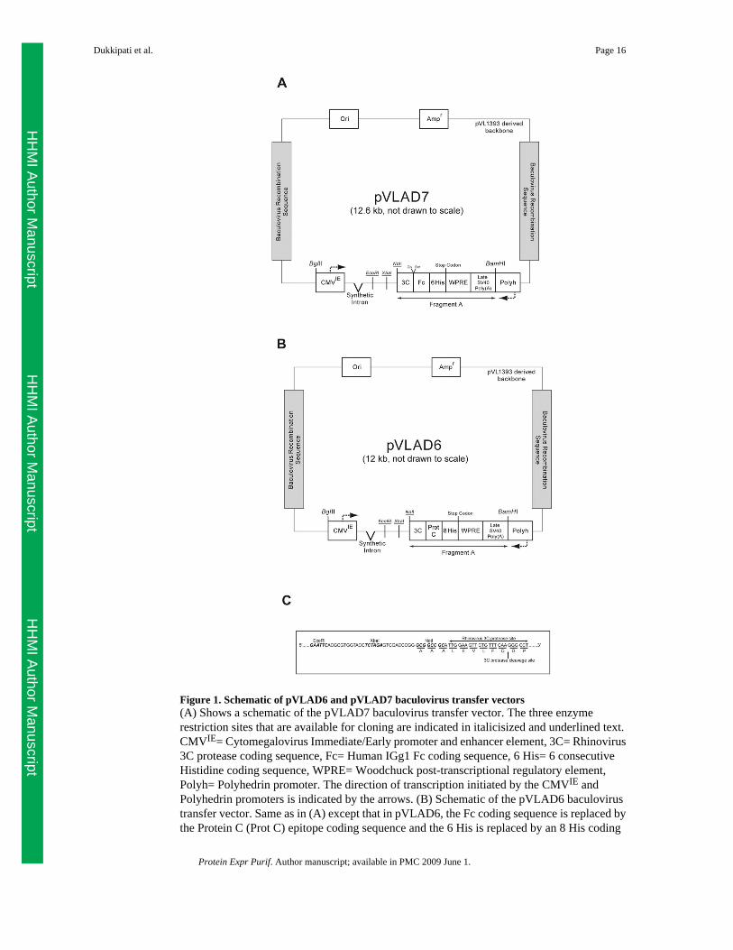
Figure 1. Schematic of pVLAD6 and pVLAD7 baculovirus transfer vectors(A) Shows a schematic of the pVLAD7 baculovirus transfer vector. The three enzymerestriction sites that are available for cloning are indicated in italicisized and underlined text.CMVIE= Cytomegalovirus Immediate/Early promoter and enhancer element, 3C= Rhinovirus3C protease coding sequence, Fc= Human IGg1 Fc coding sequence, 6 His= 6 consecutiveHistidine coding sequence, WPRE= Woodchuck post-transcriptional regulatory element,Polyh= Polyhedrin promoter. The direction of transcription initiated by the CMVIE andPolyhedrin promoters is indicated by the arrows. (B) Schematic of the pVLAD6 baculovirustransfer vector. Same as in (A) except that in pVLAD6, the Fc coding sequence is replaced bythe Protein C (Prot C) epitope coding sequence and the 6 His is replaced by an 8 His coding
Dukkipati et al. Page 16
Protein Expr Purif. Author manuscript; available in PMC 2009 June 1.
HH
MI Author M
anuscriptH
HM
I Author Manuscript
HH
MI Author M
anuscript

sequence. (C) Nucleotide sequence corresponding to the region between the EcoRI site and the3C protease site in pVLAD6 and pVLAD7 is shown.
Dukkipati et al. Page 17
Protein Expr Purif. Author manuscript; available in PMC 2009 June 1.
HH
MI Author M
anuscriptH
HM
I Author Manuscript
HH
MI Author M
anuscript
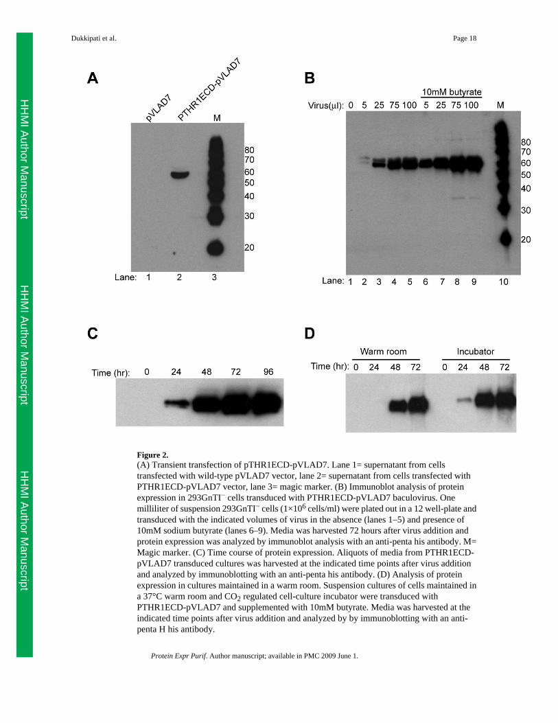
Figure 2.(A) Transient transfection of pTHR1ECD-pVLAD7. Lane 1= supernatant from cellstransfected with wild-type pVLAD7 vector, lane 2= supernatant from cells transfected withPTHR1ECD-pVLAD7 vector, lane 3= magic marker. (B) Immunoblot analysis of proteinexpression in 293GnTI− cells transduced with PTHR1ECD-pVLAD7 baculovirus. Onemilliliter of suspension 293GnTI− cells (1×10⁶ cells/ml) were plated out in a 12 well-plate andtransduced with the indicated volumes of virus in the absence (lanes 1–5) and presence of10mM sodium butyrate (lanes 6–9). Media was harvested 72 hours after virus addition andprotein expression was analyzed by immunoblot analysis with an anti-penta his antibody. M=Magic marker. (C) Time course of protein expression. Aliquots of media from PTHR1ECD-pVLAD7 transduced cultures was harvested at the indicated time points after virus additionand analyzed by immunoblotting with an anti-penta his antibody. (D) Analysis of proteinexpression in cultures maintained in a warm room. Suspension cultures of cells maintained ina 37°C warm room and CO2 regulated cell-culture incubator were transduced withPTHR1ECD-pVLAD7 and supplemented with 10mM butyrate. Media was harvested at theindicated time points after virus addition and analyzed by by immunoblotting with an anti-penta H his antibody.
Dukkipati et al. Page 18
Protein Expr Purif. Author manuscript; available in PMC 2009 June 1.
HH
MI Author M
anuscriptH
HM
I Author Manuscript
HH
MI Author M
anuscript

Figure 3. PTHR1ECD purification(A) Purification of PTHR1ECD-Fc. Lane 1 shows a sample of the elution from the protein Acolumn. The sample was prepared under reducing conditions for SDS-PAGE. Lane 2, same asin lane 1 except that the sample was prepared under non-reducing conditions for SDS-PAGE.(B) EndoHf treatment of PTHR1ECD-Fc. The purified PTHR1ECD-Fc fusion protein wastreated with EndoHf under non-denaturing conditions and analyzed by SDS-PAGE. Lanes 1and 2 show the untreated and EndoHf treated samples respectively prepared under reducingconditions for SDS-PAGE. The band migrating at 66 kDa in lane 2 corresponds to EndoHf.Lanes 3 and 4 are identical to samples in lanes 1 and 2 respectively except that they wereprepared under non-reducing conditions for SDS-PAGE. (C) Lane 1 shows a sample of theEndoHf treated fusion protein and lane 2 shows a sample that was further treated with 3Cprotease. The positions of free Fc and PTHR1ECD in lane 2 are indicated. The band migratingat 48 kDa in lane 2 corresponds to 3C protease. The samples in lane 1 and 2 were preparedunder reducing conditions for SDS-PAGE. Samples in lanes 3 and 4 are identical to samplesin lanes 1 and 2 except that they were prepared under non-reducing conditions for SDS-PAGE.(D) Lane 1 shows a sample of the EndoHf and 3C treated sample. Lane 2 shows a sample thatwas further treated with PNGase under denaturing conditions. Lane 3 shows a sample ofPNGase F only. (E) Size exclusion chromatographic purification of PTHR1ECD. The
Dukkipati et al. Page 19
Protein Expr Purif. Author manuscript; available in PMC 2009 June 1.
HH
MI Author M
anuscriptH
HM
I Author Manuscript
HH
MI Author M
anuscript
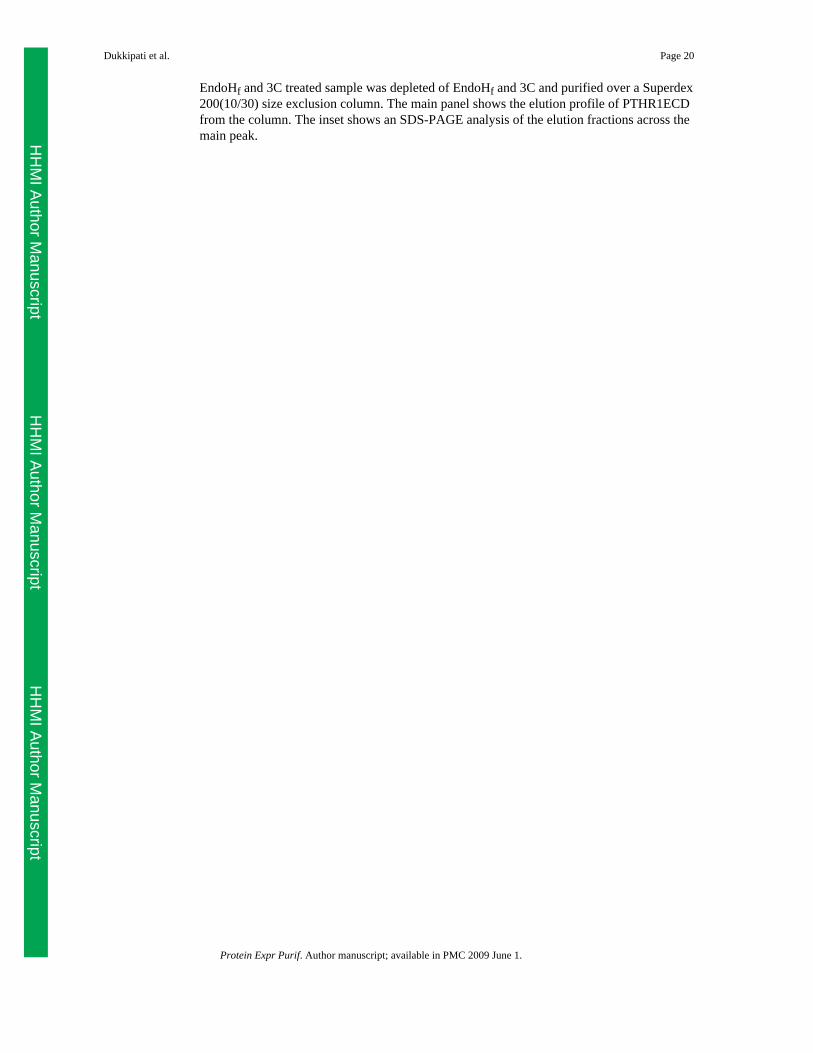
EndoHf and 3C treated sample was depleted of EndoHf and 3C and purified over a Superdex200(10/30) size exclusion column. The main panel shows the elution profile of PTHR1ECDfrom the column. The inset shows an SDS-PAGE analysis of the elution fractions across themain peak.
Dukkipati et al. Page 20
Protein Expr Purif. Author manuscript; available in PMC 2009 June 1.
HH
MI Author M
anuscriptH
HM
I Author Manuscript
HH
MI Author M
anuscript
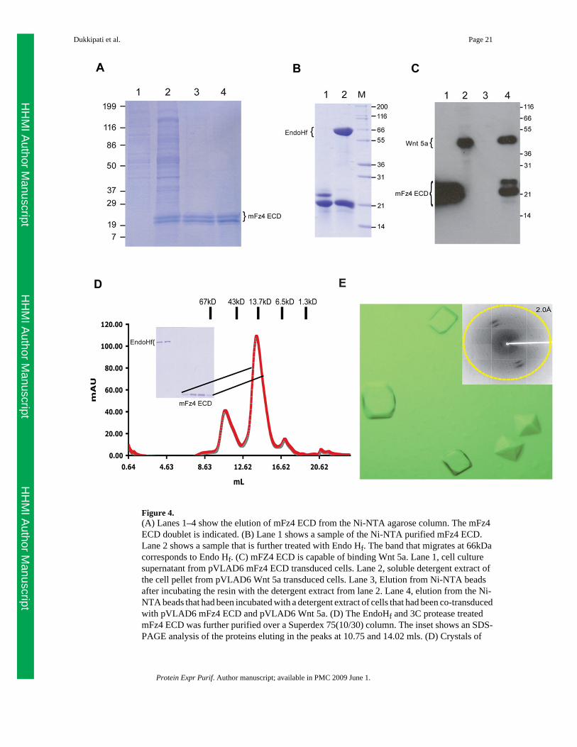
Figure 4.(A) Lanes 1–4 show the elution of mFz4 ECD from the Ni-NTA agarose column. The mFz4ECD doublet is indicated. (B) Lane 1 shows a sample of the Ni-NTA purified mFz4 ECD.Lane 2 shows a sample that is further treated with Endo Hf. The band that migrates at 66kDacorresponds to Endo Hf. (C) mFZ4 ECD is capable of binding Wnt 5a. Lane 1, cell culturesupernatant from pVLAD6 mFz4 ECD transduced cells. Lane 2, soluble detergent extract ofthe cell pellet from pVLAD6 Wnt 5a transduced cells. Lane 3, Elution from Ni-NTA beadsafter incubating the resin with the detergent extract from lane 2. Lane 4, elution from the Ni-NTA beads that had been incubated with a detergent extract of cells that had been co-transducedwith pVLAD6 mFz4 ECD and pVLAD6 Wnt 5a. (D) The EndoHf and 3C protease treatedmFz4 ECD was further purified over a Superdex 75(10/30) column. The inset shows an SDS-PAGE analysis of the proteins eluting in the peaks at 10.75 and 14.02 mls. (D) Crystals of
Dukkipati et al. Page 21
Protein Expr Purif. Author manuscript; available in PMC 2009 June 1.
HH
MI Author M
anuscriptH
HM
I Author Manuscript
HH
MI Author M
anuscript
mFz4 ECD are shown. The inset shows the diffraction pattern of a crystal at a synchrotron lightsource.
Dukkipati et al. Page 22
Protein Expr Purif. Author manuscript; available in PMC 2009 June 1.
HH
MI Author M
anuscriptH
HM
I Author Manuscript
HH
MI Author M
anuscript

Figure 5. Purification of US28-Fractalkine complex(A) Immunoblot analysis of Fractalkine-Fc under different sample preparation conditions.Aliquots of media from a culture of 293GnTI− cells transduced with baculovirus forFractalkine-pVLAD7 were prepared under reducing conditions and heated to 95°C (lane 1) ornot heated (lane 2) before SDS-PAGE and immunodetection. The bands corresponding to freeFc and Fractalkine-Fc in lane 1 are indicated. M= magic marker. (B) Ligand affinitypurification of US28-1D4. Lane 1 shows the solubilized membranes before incubation withthe Fractinlkine-Fc/Protein A resin. Lanes 2 and 3 show a sample from the unbound fractionand the last wash fraction respectively. Lane 4 shows a sample from the main elution fraction.The bands corresponding to Fractalkine-Fc and US28-1D4 in lane 4 are indicated. (C) 3C
Dukkipati et al. Page 23
Protein Expr Purif. Author manuscript; available in PMC 2009 June 1.
HH
MI Author M
anuscriptH
HM
I Author Manuscript
HH
MI Author M
anuscript
digestion of Fractalkine-Fc. The eluted sample shown in lane 1 was treated with 3C protease.A sample of the 3C treated material is shown in lane 2. The 3C treated material was passedthrough a protein A-Sepharose column. Lane 3 shows the flow-through from the protein Acolumn. (D) Size exclusion chromatographic purification of US28-1D4/Fractalkine complex.The sample from lane 3 in (C) was concentrated and injected over a Superose 6(10/30) sizeexclusion column. The main panel shows the elution profile from the Superose column. Theinset shows an SDS-PAGE analysis of the peak fractions at 7.42 ml and 14.8 ml. (E)Concentrated sample used for crystallization trials is shown.
Dukkipati et al. Page 24
Protein Expr Purif. Author manuscript; available in PMC 2009 June 1.
HH
MI Author M
anuscriptH
HM
I Author Manuscript
HH
MI Author M
anuscript
Copyright © 2022 FDOKUMEN