Sarcoma in the thymus of juvenile meagre Argyrosomus regius reared in an intensive system
Upload
khangminh22Category
view
1download
0
Recombinant gonadotropins in meagre
(Argyrosomus regius): In-vivo effect on sexually
undifferentiated fish.
Alumno: Álvaro González Cid
Tutor IRTA: Neil Duncan
Tutor UCV: Ignacio Giménez Nebot
Año académico: 2020-2021

Recombinant gonadotropins in meagre (Argyrosomus
regius): In-vivo effect on sexually undifferentiated fish.

Recombinant gonadotropins in meagre (Argyrosomus
regius): In-vivo effect on sexually undifferentiated fish.
Contents Abstract ......................................................................................................................................... 1
Resumen ....................................................................................................................................... 2
1. Introduction .......................................................................................................................... 3
1.1. Meagre (A. regius) biology and perspectives in aquaculture ..................................... 3
1.2. Fish gonadotropins and their receptors ...................................................................... 5
1.3. Steroidogenesis in teleosts .......................................................................................... 8
1.4. Oogenesis and spermatogenesis in teleosts .............................................................. 10
1.5. Fish recombinant gonadotropins ............................................................................... 14
2. Objectives ........................................................................................................................... 24
2.1. General objective ........................................................................................................ 24
2.2. Specific objectives ...................................................................................................... 24
3. Material and methods ........................................................................................................ 25
3.1. Study animals and maintenance ................................................................................ 25
3.2. Recombinant Gths production of A. regius ............................................................... 26
3.3. Experimental setup, fish manipulation and samplings ............................................. 27
3.4. Plasma steroid analysis .............................................................................................. 28
3.5. Histological preparation ............................................................................................. 28
3.6. Statistical analysis ....................................................................................................... 30
4. Results ................................................................................................................................. 31
4.1. Gonadosomatic and hepatosomatic index ................................................................ 31
4.2. Estradiol ...................................................................................................................... 32
4.3. Histological observations ........................................................................................... 33
4.4. Sexual differentiation ................................................................................................. 36
5. Discussion ........................................................................................................................... 37
5.1. Gonadosomatic index ................................................................................................. 37
5.2. Estradiol ...................................................................................................................... 37
5.3. Histological observations ........................................................................................... 38
5.4. Sexual differentiation ................................................................................................. 39
6. Conclusion ........................................................................................................................... 45
7. References .......................................................................................................................... 46

Recombinant gonadotropins in meagre (Argyrosomus
regius): In-vivo effect on sexually undifferentiated fish.

Recombinant gonadotropins in meagre (Argyrosomus
regius): In-vivo effect on sexually undifferentiated fish.
1
Abstract
Meagre (Argyrosomus regius) is a teleost fish that has experienced an increase in Mediterranean
aquaculture production in recent years. To improve productivity, it is necessary to establish a
genetic selection program, but the generational interval in meagre is very high (2 and 3 years in
males and females respectively). The objective of this survey is to evaluate the effect of specific
A. regius single-chain recombinant gonadotropins (rGths) in prepubertal and sexually
undifferentiated meagre. For this, 8 treatment groups were established, in which doses of 6, 12
or 18 µg / kg of rFSH or rLH were injected weekly for 3 weeks. Results did not show significant
differences in GSI, but they did in plasma E2. Furthermore, early gonadal development was
induced with the appearance of oocytes and spermatids in localized regions, while 100% of the
fish in the saline group remained sexually undifferentiated. Our results on sexual differentiation
show that with rGths therapy 1) males sexually differentiate earlier than females; 2) there is a
higher proportion of males; 3) there are intersex fish. This is the first report of the presence of
intersex fish and a male skewed ratio in this species. In conclusion, rGths therapy represent a
potential solution to reduce the generation interval in prepubertal meagre, but more studies are
needed to know how its administration affects during the period of sexual differentiation.
Keywords: Argyrosomus regius, meagre, prepubertal, recombinant gonadotropins, intersex,
sexual differentiation.

Recombinant gonadotropins in meagre (Argyrosomus
regius): In-vivo effect on sexually undifferentiated fish.
2
Resumen
La corvina (Argyrosomus regius) es un pez teleósteo que ha experimentado un incremento en la
producción acuícola mediterránea en los últimos años. Para mejorar la productividad es
necesario establecer un programa de selección genética, pero el intervalo generacional en la
corvina es muy alto (2 y 3 años en machos y hembras respectivamente). El objetivo de este
estudio es evaluar el efecto de las gonadotropinas recombinantes (rGths) de cadena simple
específicas de A. regius en corvina juvenil y sexualmente indiferenciada. Para ello se
establecieron 8 grupos de tratamiento, en los que se inyectaron semanalmente durante 3
semanas dosis de 6, 12 o 18 µg / kg de rFSH o rLH. Los resultados no mostraron diferencias
significativas en IGS, pero sí en E2 en plasma. Además, se indujo un desarrollo gonadal temprano
con la aparición de ovocitos y espermátidas en regiones localizadas, mientras que el 100% de
los peces del grupo salino permanecieron sexualmente indiferenciados. Nuestros resultados
sobre diferenciación sexual demuestran que con un tratamiento de rGths, 1) los machos
diferencian sexualmente antes que las hembras; 2) hay una mayor proporción de machos; 3)
hay peces intersexuales. Este es la primera vez que se publica la presencia de peces intersexuales
y una mayor proporción poblacional de machos en esta especie. En conclusión, un tratamiento
con rGths representa una posible solución para reducir el intervalo generacional en la corvina
juvenil, pero se necesitan más estudios para conocer cómo afecta su administración durante el
período de diferenciación sexual.
Palabras clave: Argyrosomus regius, corvina, juvenil, gonadotropinas recombinantes,
intersexual, diferenciación sexual.

Recombinant gonadotropins in meagre (Argyrosomus
regius): In-vivo effect on sexually undifferentiated fish.
3
1. Introduction
1.1. Meagre (A. regius) biology and perspectives in aquaculture
Meagre (Argyrosomus regius) (Asso, 1801) is a marine and migratory teleost fish species which
belongs to Sciaenidae family that can be found in the Mediterranean and Black Sea, and along
the Eastern coast of the Atlantic Ocean (Kružić et al., 2016; Ramos-Júdez et al., 2019). This fish
species is morphologically characterized by having a large head with small eyes, as well as large
ctenoid scales with a grey colour. It has a terminal mouth with a yellow internal colour and a
multitude of small sharp teeth. The lateral line is easily identifiable by the black spots it presents
(Fig.1) (García, 2012). In addition, it has a large swim bladder with which it produces drumming
sounds like other fish of the Sciaenidae family, which is why they are commonly referred to as
"croakers" or "drums" (Duncan et al., 2013).
Fig.1: Anesthetized meagre (Argyrosomus regius). (Photo by Álvaro González).
This is a gonochoristic species that reaches sexual differentiation between 10 and 12 months of
age, although it begins to differentiate at 5 months (Schiavone et al., 2012). Females are
asynchronous (Gil et al., 2013) or group-synchronous spawners (Duncan et al., 2013) and in most
cases fail to undergo maturation, ovulation and spawning in captivity (Duncan et al., 2018).
Males have an unrestricted, cystic and lobular type testis (Gil et al., 2013) and undergo
spermatogenesis and spermiogenesis, but often produce low volume with reduced quality of
sperm (Duncan et al., 2012; Ramos-Júdez et al., 2019). For this reason, the use of synthetic
agonist of gonadotropin-releasing hormone (GnRHa) is necessary to induce final oocyte
maturation (FOM), ovulation and spawning in females, and to ensure adequate milt production
in males (Fakriadis et al., 2020). Vitellogenesis begins in March, and oocytes reach the fully
vitellogenic stage between April and June; the spermiating period, when fluid sperm can easily
be extracted is between March and June (Mylonas et al., 2013) The sex-ratio population is 1:1
(Gil et al., 2013).

Recombinant gonadotropins in meagre (Argyrosomus
regius): In-vivo effect on sexually undifferentiated fish.
4
The meagre has had an important increase in Mediterranean aquaculture in recent years for its
good feed conversion ratio (FCR) (0,9-1,2) (Monfort, 2010) (1,8) (Duncan et al., 2013), fast
growth rates reaching 1 kg per year, large size (above 2 m and 50 kg), capability to withstand
diverse environmental conditions (Monfort, 2010; Duncan et al., 2013; Kružić et al., 2016), high
resilience to stress factors (Monfort, 2010), low fat content (1,06 %) and of high nutritional value
(Grigorakis et al., 2011) and high fecundity with GnRHa treatments, achieving up to 1.415.000
eggs per kg (Duncan et al., 2012; Mylonas et al., 2016).
For these reasons, A. regius Mediterranean production increased 6.6% in 2018 (APROMAR,
2019) and 10,5% in 2019 (Fig.2), with the main producing countries being Egypt (32.000 t), Spain
(3.650 t), Turkey (2.600 t) and Greece (1.800 t) (APROMAR, 2020); while the global fisheries
production ranges from 5.000 t to 10.000 t per year (Monfort, 2010). In Spain there was an
increase of 44,9% in 2019 compared to the previous year (APROMAR, 2020).
Fig.2: Evolution of the Mediterranean production of meagre (A. regius) in tons, through aquaculture (light
blue) and fishing (dark blue), in the period 1996 - 2018 (APROMAR, 2020).
Selective breeding programmes enable an increase in production due to a higher growth rate, a
reduction of production costs as a result of an improved FCR, a reduction in stress and mortality
with better domestication and adaptivity to captivity, a raise in disease resistance and a better
flesh quality (Gjedrem, 2000; Houston et al., 2020). A key factor of this process is the length of
the generation interval (Houston et al., 2020); in A. regius sexual maturity is reached at two
years old at 0.92 ± 0.08 kg in males, and three years old at 1.61 ± 0.09 kg in females (Schiavone
Recombinant gonadotropins in meagre (Argyrosomus
regius): In-vivo effect on sexually undifferentiated fish.
5
et al., 2012). One method that is being researched to reduce this generation interval is the
isolation of the germ cells of the objective species at an early life stage and transplantation of
these cells in a surrogate species with a shorter generation time (surrogate broodstock)
(Houston et al., 2020). Nevertheless, some species possess low concentrations of germline stem
cells in their genital glands and the efficiency of transplantation of germs cells can vary with the
season (Yoshizaki and Yazawa, 2019; Goto and Saito, 2019).
Another innovative alternative to reduce the generation interval could be the use of
recombinant gonadotropins (rGths) to stimulate spermatogenesis and oogenesis in immature
prepubertal fish. The role of Gths in reproduction has been examined as a hormone therapy to
treat infertility and other reproduction-related problems (Molés et al., 2020).
1.2. Fish gonadotropins and their receptors
Reproduction in vertebrates is controlled by the brain-pituitary-gonad (BPG) axis (Burow et al.,
2019), which in turn is controlled by environmental factors (Fig.3) (Migaud et al., 2010). Within
this system, the pituitary gonadotropins (Gths) and their receptors are the key to conveying the
hormonal signals released by the BPG axis. Gths are two heterodimeric glycoproteins composed
of a common alpha subunit and a specific beta subunit linked non-covalently (Schulz et al., 2001;
Levavi-Sivan et al., 2010). They are synthesized and regulated by the pituitary gland through the
main neuropeptide gonadotropin-releasing hormone (GnRH), in addition to others such as
kisspeptines, dopamine (DA), gonadotropin-inhibitory hormone (GnIH), neuropeptide Y (NPY),
serotonine, leptin, glutamate, ϒ-aminobutyric acid (GABA), norepinephrine (NA), secretoneurin,
ghrelin and pituitary adenilate cyclase-activating peptide (PACAP) (Fig.3). These hypothalamic
neurohormones act on the cells of the anterior lobe of the adenohypophysis (Levavi-Sivan et al.,
2010; Carrillo et al., 2012; Paullada-Salmerón et al., 2016). Gths can stimulate sexual steroids
secretion (Fig.3), which in turn positively or negatively regulate gonadotropin secretion
depending on the stage of gonadal development. Estrogens and aromatizable androgens
stimulate β-LH synthesis in juveniles of various teleost species, while an increase in 17β-estradiol
(E2) or testosterone (T) inhibits β-FSH synthesis in rainbow trout (Oncorhynchus mykiss) (Yaron
and Levavi -Sivan, 2011), coho salmon (Oncorhynchus kisutch) (Dickey and Swanson, 1998) and
Atlantic salmon (Salmo salar) (Borg et al., 1998). Otherwise, progestins stimulate β-FSH
synthesis in zebrafish (Danio rerio) (Wang et al., 2016). In addition, it has been observed that in
in-vitro studies, the gonadal peptides activin A and B stimulate β-FSH synthesis and reduce β-LH

Recombinant gonadotropins in meagre (Argyrosomus
regius): In-vivo effect on sexually undifferentiated fish.
6
synthesis, although in turn, they are regulated by follistatin and activin-binding protein (Levavi-
Sivan et al., 2010; Yaron and Levavi-Sivan, 2011).
Fig.3: Representative diagram of the brain pituitary-gonad (BPG) axis that controls the reproductive
process in fish. GnRH, gonadotropin-releasing hormone; GnIH, gonadotropin-inhibitory hormone; NPY,
neuropeptide Y; GABA, ϒ-aminobutyric acid; PACAP, pituitary adenilate cyclase-activating peptide; NA,
norepinephrine/noradrenaline; DA, dopamine; E2, 17β-estradiol; T, testosterone. By Álvaro González.
Follicle stimulating hormone (FSH) stimulates the early stages of gametogenesis, including
spermatogenesis in males and oogenesis in females through the synthesis of 17β-estradiol (E2)
and 11-ketotestosterone (11-KT) (Melamed et al., 1998) via the membrane-bound receptors
Fshra, which belongs to the family of G-protein-coupled receptors (GPCRs). In males, Fshra can
be expressed in Sertoli and Leydig somatic cells; and in females in the follicular somatic cells of
Gths
GnRH GnIH
DA
Kisspeptines
NPY
Serotonine
Leptin
Glutamate
GABA
Ghrelin
PACAP
NA
E2
E2
T
E2
Secreted in brain
Secreted in pituitary
Secreted in gonad
Stimulation
Inhibition
Stimulation/inhibition
Gonadal steroids
Environmental factors
Eyes and pineal complex
(melatonin)

Recombinant gonadotropins in meagre (Argyrosomus
regius): In-vivo effect on sexually undifferentiated fish.
7
granulosa and theca as well as in connective tissue (Yaron and Levavi-Sivan, 2011; Chauvigné et
al., 2012; Ozaki et al., 2019).
Luteinizing hormone (LH) is related to the final oocyte maturation (FOM) and ovulation in
females (Kwok et al., 2005), and the later stages of germ cell development and the
differentiation of spermatids to spermatozoa in males (Chauvigné et al., 2012) by the production
of maturation induction steroid (MIS) (Nagahama, 2008) through Lhcgrba membrane-bound
receptors (GPCRs) activation. In males it is expressed in spermatids and in Leydig cells (Yaron
and Levavi-Sivan, 2011; Chauvigné et al., 2012). García-Lópes et al., (2010) described the
presence of Lhcgrba in Sertoli cells in zebrafish (D. rerio), being the first report of the presence
of these receptors in Sertoli cells in vertebrates. In females, Lhcgrba are found in granulosa cells
(Yaron and Levavi-Sivan, 2011). It has been observed that in some teleosts Fshra also respond
to LH (Kobayashi et al., 2008; García-Lópes et al., 2010; Burow et al., 2019; Rajakumar and
Senthilkumaran, 2020), but when there are peaks of this hormone as in the spawning season
(Schulz et al., 2010). In rainbow trout (O. mykiss), Lhcgrba also responded to supraphysiological
purified FSH, although at concentrations five times higher than LH (Sambroni et al., 2007). Both
receptors can be expressed in non-gonadal tissues such as brain, gills, eyes, intestine, kidney or
liver (Kwok et al., 2005; Kobayashi et al., 2008; Burow et al., 2019).
In females, Fshra mRNA expression (fshra) is detectable at low levels in immature ovaries. Its
expression increases during vitellogenesis reaching its maximum expression in the
midvitellogenic stage and decreasing in the postovulatory follicle state. The expression of
lhcgrba is detectable in the previtellogenesis stage, although there is an increase in transcription
with the onset of vitellogenesis, reaching the peak of expression at the full-grown stage (Kwok
et al., 2005; Kobayashi et al., 2008; Carrillo et al., 2012) or maturation / ovulation (Sambroni et
al., 2007; Rocha et al., 2009). In males, the expression of fshra and lhcgrba was found in all testis
stage, from immature to fully mature, although their expression increased during the transition
to fully mature stage (spermiation) (Kusakabe, 2006; Sambroni et al., 2007; Maugars and
Schmitz, 2007; Rocha et al., 2009; García-Lópes et al., 2009; Burow et al., 2019). In contrast, in
Shortfinned eel (Anguilla australis) with hCG-induced spermatogenesis, fshra expression
increased in early stages, while lhcgrba increased since spermatids were observed (Ozaki et al.,
2019). In Nile tilapia (O. niloticus) there is fshra and lhcgrba during sexual differentiation period
(Yan et al., 2012).

Recombinant gonadotropins in meagre (Argyrosomus
regius): In-vivo effect on sexually undifferentiated fish.
8
1.3. Steroidogenesis in teleosts
Sex steroids stimulate gametogenesis and are related to sex differentiation in different
vertebrate taxa (Nakamura, 2010; Morohashi et al., 2013). Sexual steroidogenesis is regulated
by Gths, and begins with the transport of cholesterol to the mitochondria by the steroidogenic
acute regulatory protein (StAR), where it is converted to pregnenolone by the cholesterol side-
chain cleavage enzyme (P450scc / Cyp11a1). Pregnenolone is the precursor of various steroids,
including E2, 11-KT, T, progestogens, and corticosteroids (Rajakumar and Senthilkumaran,
2020).
In females, gonadal steroidogenesis is explained according to the two-cell model described by
Young et al. (1986), in which FSH-stimulated theca cells synthesize precursors for their
subsequent transformation into estrogens and progestogens in granulosa cells when stimulated
with LH (Fig.4). Nevertheless, LH and FSH have similar potencies in stimulating E2 production
during early phases of oogenesis; yet, LH is more potent during later stages of the process (Cerdá
et al., 2007). First, pregnenolone is transformed into progesterone in theca cells by the enzyme
3β-hydroxy-steroid dehydrogenase (3β-HSD). Progesterone is converted back in theca cells to
17α-hydroxy-progesterone by the action of the enzyme 17-hydroxylase (P450c17 / Cyp17). The
synthesis of 3β-HSD is stimulated by FSH and LH (Nakamura et al., 2003).
For E2 synthesis, 17α-hydroxy-progesterone is transformed into androstenedione by the
enzyme P450c17 / Cyp17, which in turn is converted into T by the activity of the enzyme 17β-
hydroxy-steroid dehydrogenase (17β-HSD). Finally, T can be catalyzed to E2 by the enzyme
cytochrome P450 aromatase (P450arom / Cyp19a) in granulosa cells (Nagahama et al., 1994;
Young et al., 2005; Nakamura et al., 2005; Nagahama et al., 2008; Carrillo et al., 2012). The
synthesis of P450arom / Cyp19a is stimulated by FSH (Rocha et al., 2009), LH and insulin growth
factor I (IGF-I) (Kagawa et al., 2003; Nakamura et al., 2003; Montserrat et al., 2004). Its
expression in the ovary increases during vitellogenesis (Chang et al., 2005).
For progestogens synthesis, 17α-hydroxy-progesterone crosses the basement membrane and is
converted in granulosa cells to MIS by the enzyme 20β-hydroxy-steroid dehydrogenase (20β-
HSD). This enzyme is synthesized in granulosa cells in response to LH. Two progestins have been
identified that act as MIS in teleosts, 17α, 20β-dihydroxy-4-pregnen-3-one (DHP; 20βP) and 17,
20β, 21-trihydroxy-4-pregnen- 3-one (20βS) (Young et al., 1986; Nagahama et al., 1994; Thomas
et al., 2001; Tanaka et al., 2002; Carrillo et al., 2012); although any steroid capable of breaking
down the germ vesicle (GBVD) such as 11-deoxycorticosterone (DOC) or T are also considered
as MIS (Nagahama et al., 2008). In Sciaenidae family, 20βS was shown to act as MIS (Peter and
Yu, 1997; Senthilkumaran et al., 2002; Fakriadis et al., 2020).

Recombinant gonadotropins in meagre (Argyrosomus
regius): In-vivo effect on sexually undifferentiated fish.
9
Therefore, E2 is produced during oocyte growth, and the progestins (DHP or 20βS) during
maturation. The change in the steroidogenic pathway occurs in fish ovarian follicles immediately
prior to oocyte maturation (Nagahama and Yamashita, 2008). This change could occur due to
the cellular levels of the enzymes P450c17 and 3β-HSD, so that when P450c17 is dominant, the
synthesis of E2 is favoured and vice versa (Sakai et al., 1994; Nakamura et al., 2005).
Fig.4: Representative diagram of the two-cell model described by Young et al., (1986) for female gonadal
steroidogenesis. P450scc, P450 side-chain cleavage; P450c17, 17-hydroxylase/C17-C20-lyase; 3β-HSD, 3β-
hydroxysteroid dehydrogenase; 17β-HSD, 17β-hydroxysteroid dehydrogenase; 20β-HSD, 20β-
hydroxysteroid dehydrogenase; P450arom, P450 aromatase (Luzbens et al., 2010).
In males, gonadal steroidogenesis is not as well described as in females. The steroids required
for spermatogenesis are E2, 11-KT, and DHP / 20βS. The P450arom / Cyp19a enzyme is
expressed in the testis (Blázquez and Piferrer, 2004; Chang et al., 2005), allowing the synthesis
of E2 from T.
For androgens synthesis (11-KT and T), FSH and LH are equipotent in stimulating its production
by upregulating expression of steroidogenic enzyme genes (Suzuki et al., 2019). However, LH is
more potent in stimulating DHP / 20βS (Planas and Swanson, 1995; Kazeto et al., 2008). For the
synthesis of 11-KT, androstenedione is converted into 11β-hydroxyandrostenedione (11β-OHA)
by the enzyme 11β-hydroxylase (P45011β / Cyp11b) (Rajakumar and Senthilkumaran, 2015).
Then, 17β-HSD converts 11β-OHA on 11β-hydroxytestosterone (11β-OHT). Furthermore, T can
be converted to 11β-OHT by P45011β / Cyp11b (Kusakabe et al., 2003). Finally, the enzyme 11-

Recombinant gonadotropins in meagre (Argyrosomus
regius): In-vivo effect on sexually undifferentiated fish.
10
beta-hydroxysteroid-dehydrogenase (11β-HSD) converts 11β-OHT into 11-KT (Fig.5) (Kusakabe
et al., 2003; Ozaki et al., 2006; Suzuki et al., 2020). The steroid 11-KT stimulates its own
production based on positive feedback, it enhanced cyp11b expression in testis (Ozaki et al.,
2019). Furthermore, an increase in 11-KT following hCG injection elevated testicular fshra mRNA
levels augmenting FSH sensitivity in the testis (Ozaki et al., 2016). Progestin production is
catalyzed by 20β-HSD from 17α-hydroxy-progesterone in Leydig cells, spermatogonia and
spermatocytes (Sreenivasulu et al., 2012). The DHP / 20βS stimulates the synthesis of 11-KT by
increasing the expression of 11β-HSD (Ozaki et al., 2006).
Fig.5: Simplified pathways of biosynthesis of sex steroids and glucocorticoids in fish. CYP11A1, cholesterol
side-chain cleavage enzyme (P450scc); 3β-HSD, 3β-hydroxysteroid dehydrogenase; 17β-HSD, 17β-
hydroxysteroid dehydrogenase; CYP19, P450 aromatase (P450arom); CYP17, 17-hydroxylase/C17-C20-
lyase (P450c17); 11β-HSD, 11-beta-hydroxysteroid-dehydrogenase, CYP11B, 11β-hydroxylase (P45011β)
(Fernandino et al., 2013).
1.4. Oogenesis and spermatogenesis in teleosts
Spermatogenesis is the process during which immature diploid spermatogonia develop into
mature, fertile haploid spermatozoa (Ozaki et al., 2019). In spermatogenesis there is a self-
renewal of spermatogonia stimulated by E2 released by Leydig cells. The steroid E2 interacts
with its receptors on Sertoli cells (Miura et al., 1999), which synthesize spermatogonial stem-
cell renewal factor (Gonadal soma-derived growth factor; GSDF) (Fig.6) (Satawari et al., 2007;
Yaron and Levavi -Sivan, 2011). Undifferentiated A spermatogonia (StgAund) have the capacity

Recombinant gonadotropins in meagre (Argyrosomus
regius): In-vivo effect on sexually undifferentiated fish.
11
for self-renewal, however, it is not clear whether differentiated A spermatogonia (StgAdiff) in
teleosts have the capacity to return to a more undifferentiated state of development (StgAund)
(Lacerda et al., 2014) ; although Huckins (1971) showed that in mammals they have this capacity.
The secretion of 11-KT in Leydig cells stimulates in Sertoli cells the synthesis of mediators that
regulate the proliferation of more differentiated spermatogonia cells (spermatogonia B; StgB)
for their subsequent entry into meiosis (Schulz et al., 2010; Lacerda et al., 2014). Activin B and
insulin growth factor I (IGF-I) are the mediators that stimulate differentiation to StgB, while anti-
Müllerian hormone (AMH) and eSRS21 inhibit it (Halm et al., 2007; Carrillo et al., 2012).
Induction of meiosis (formation of spermatocytes to develop into spermatids) is stimulated by
progestins (Miura et al., 2006; Yaron and Levavi-Sivan, 2011). Sperm maturation (capability of
motility and fertilization) is also produced by DHP, which increases the pH of seminal plasma by
increasing the cAMP content in sperm after activation of carbonic anhydrase (CA / eSR22) (Fig.6)
(Miura and Miura, 2003). Spermiation (sperm are released into the lumen of the seminiferous
tubule) is induced by DHP and 11-KT (Miura et al., 2006; Schulz et al., 2010).
Fig.6: Endocrine mechanisms regulating spermatogenesis in the Japanese eel (Anguilla japonica). 11-KT,
11-ketotestosterone; AMH, a peptide homologous to anti-Müllerian hormone; DHP, 17α,20β-dihydroxy-
4-pregnen-3-one; IGF-I, insulin-like growth factor I. Cells at the upper right corner depict the germ cells as
the source of DHP acting upon themselves in a paracrine or autocrine manner. Modified from Yaron and
Levavi-Sivan (2011).

Recombinant gonadotropins in meagre (Argyrosomus
regius): In-vivo effect on sexually undifferentiated fish.
12
Oogenesis is the formation, development and maturation of the female gamete and ovum
(Arukwe and Goksøyr, 2003). In oogenesis there is mitotic renewal of oogonia stimulated by E2,
while the induction of meiosis is stimulated by progestogens (Fig.7) (Miura et al., 2007; Luzbens
et al., 2010). The expression of IGF-I has also been observed in oocytes and somatic cells at the
onset of meiosis in Nile tilapia (Oreochromis niloticus) (Berishivili et al., 2006). The role of FSH in
regulating the accumulation of cortical alveoli is currently unknown, however, the synthesis of
cortical alveoli is related to an increase in plasma and pituitary FSH, Fshra, plasma E2, cyp19a
mRNA, AMH, StAR mRNA and GSDF (Kwok et al., 2005; Rodríguez-Marí et al., 2005; Campbell et
al., 2006). In the oocyte lipidation there is an accumulation of lipids from the precursor triacyl
glyceride (TAG)-rich serum lipoprotein such as very-low-density lipoprotein (VLDL) (Hiramatsu
et al., 2015). In rainbow trout (O. mykiss) VLDL is sufficient to induce lipidation, while in
Shortfinned eel (A. australis) and Japanese eel (A. japonica) VLDL and 11-KT are required
(Lokman et al., 2007; Endo et al., 2011).
Fig.7: Stages of fish oogenesis and their endocrine regulation. DHP, 17α,20β-dihydroxy-4-pregnen-3-one;
20βS, 17,20β, 21-trihydroxy-4-pregnen- 3-one; GVBD, germinal vesicle breakdown. Modified from Yaron
and Levavi-Sivan (2011).
Vitellogenesis begins with the synthesis of vitellogenin, a phosphoglycoprotein synthesized in
the liver and regulated mainly by E2 (Fig.8) (Miura et al., 2007). After their release and transport
into blood plasma, vitalogenins (Vtg) are incorporated into the oocyte through the Vtg receptor
(Vtg-R) in the ovary, resulting in the formation of yolky eggs from oocytes (Cohen and Smith
DHP/20βS DHP/20βS

Recombinant gonadotropins in meagre (Argyrosomus
regius): In-vivo effect on sexually undifferentiated fish.
13
2014). Yolk is a source of nutrients for embryogenesis and facilitates hydration in buoyant eggs
(Kwon et al., 2001).
The final maturation of the oocyte (FOM) is related to an increase in the synthesis of LH, Lhcgrba
and MIS. MIS activates its receptors on the oocyte surface (GPCRs) reducing the levels of cAMP
and protein kinase A (PKA), which in turn activates the maturation promoting factor (MPF)
(Fig.8) (Nagahama et al., 1994; Nagahama et al., 2008; Luzbens et al., 2010). The MPF is activated
by phosphorylation of the Cdc2 subunit on threonine 161 (T161) by cdk-activating kinase (CAK).
Phosphorylation occurs when Cdc2 is bound to cyclin B, and induces GVBD and the continuation
of meiosis (until now the oocyte was arrested in metaphase II by the activity of the Mos complex)
(Nagahama et al., 2008; Carrillo et al., 2012). During ovulation, the oocyte is released from its
follicle into the ovarian cavity or into the abdominal cavity (Luzbens et al., 2010). It is induced
by MIS (Nagahama and Yamahita, 2008) and prostaglandins (PGs) (Fig.8) (Peter and Yu, 1997;
Takahashi et al., 2013). In the hydration of the oocyte in pelagophil teleosts, water enters
through the aquaporins due to the increase in osmotic pressure. This osmotic pressure is
generated by free ions (K+, Cl-, NH4+…) and free amino acids (FAAs) pool, product of the hydrolysis
of vitalogenins (Finn et al., 2002; Cerdá et al., 2007).
Fig.8: (a) an overview of the endocrine chain, brain–pituitary–gonadal axis (BPG axis) in model female fish
during the vitellogenic phase. (b) An overview of the BPG axis during final oocyte maturation and
ovulation. DA, dopamine; GnRH, gonadotropin-releasing hormone; MIS, maturation induction steroid;
DHP, 17α,20β-dihydroxy-4-pregnen-3-one; 20βS, 17,20β, 21-trihydroxy-4-pregnen- 3-one; MPF,
maturation promoting factor. Modified from Yaron and Levavi-Sivan (2011).
MIS/PGs

Recombinant gonadotropins in meagre (Argyrosomus
regius): In-vivo effect on sexually undifferentiated fish.
14
1.5. Fish recombinant gonadotropins
Recombinant hormones (rGths) have the advantage that they guarantee no cross-
contamination with other related glycoproteins and can be produced continuously without
depending on the purification of the native hormones from fish pituitary glands, which is costly
and time-consuming process (Levavi-Sivan et al., 2010). For the production of rGTHs, the
isolated cDNA is cloned in expression vectors (plasmids or viruses) that are subcloned in
heterologous prokaryotic or eukaryotic systems (Molés et al., 2020). There are several
expression systems such as Escherichia coli (Hew et al., 1989; Cao et al., 2009), the yeast Pichia
pastoris (Kamei et al., 2003; Kasuto and Levavi-Sivan, 2005; Sanchís-Benlloch et al., 2017;
Nocillado et al., 2019), Drosophila S2 cells (Zmora et al., 2007; Kazeto et al., 2008), Chinese
hamster ovary (CHO) cells (Chauvigné et al., 2012; Chauvigné et al., 2017; Ramos-Júdez et al.,
2019), silkworm larvae (Hayakawa et al., 2009; Kobayashi et al., 2010), the soil amoeba
Dictyostelium discoideum (Vischer, 2003), Sf9 insect cells (Cui et al., 2007; Molés et al., 2011;
Morita et al., 2004) and FreeStyle 293-F cells (Kazeto et al., 2019; Suzuki et al., 2019). The choice
of the expression system depends on cost, hormone secretion, correctly folding and
glycosylation, which is essential for the biological activity of the rGths (Levavi-Sivan et al., 2010).
Baculovirus-infected insect cells produce higher amounts of rGths (Cui et al., 2007). However,
post-transcriptional modification, as in P. pastoris, does not allow the production of complex
products with terminal sialic acids, generating a reduction in the half-life of the protein (Levavi-
Sivan et al., 2008). CHO cells are able to produce glycosylated proteins with terminal sialic acids,
which makes them excellent for in vivo administration (Molés et al., 2020), however CHO cells
are expensive expression systems too (Cui et al., 2007). Silkworm larvae, soil amoeba, fish
embryos or P. pastoris are less expensive (Morita et al., 2004; Levavi-Sivan et al., 2010).
Prokaryotic expression systems (E. coli) are also less expensive, but they are not capable of
performing N-glycosylations (Molés et al., 2020).
For the expression of the dimer there are two possibilities: a) transfecting a single plasmid with
a cDNA encoding both subunits joined by a linker (single-chain) (Cui et al., 2007), which is usually
composed of histidine amino acids (Aizen et al., 2007a), the carboxy-terminal peptide (CTP) from
the human (hCG) or equine chorionic gonadotropin (eCG) (Morita et al., 2004; Chauvigné et al.,
2017), three Gly-Ser pairs (or other variations of Gly- Ser) (Palma et al., 2019; Kazeto et al., 2019)
or a synthetic N-linked glycosylation sequence (NCS) (Kim et al., 2012); b) co-transfecting two
expression plasmids, one with the alpha subunit and the other with the beta subunit (Kobayashi
et al., 2006). However, the fusion of the two monomers without a linker or the expression of the

Recombinant gonadotropins in meagre (Argyrosomus
regius): In-vivo effect on sexually undifferentiated fish.
15
two monomers separately usually generates misfolding, low yield in protein production or
impaired bioactivity (Molés et al., 2020).
The rGths have been successfully used in various fish species. Until now, at the steroidogenic
level it has been tested in 18 species (Table 1), while at the level of gonadal development it has
been tested in 16 species (Table 2).

16
Table 1: Review of publications where rGths have been used to stimulate steroid secretion. All significant data are compared to control groups unless otherwise indicated.
The best results obtained from all the treatments used in each survey are shown. Recombinant hormones are species specific unless otherwise indicated. Data is displayed
in chronological order of publication. E2, 17β-estradiol; T, testosterone; 11-KT, 11-ketotestosterone; OHA, 11β-hydroxyandrostenedione; SGv, vitellogenic stage of
secondary growth; OM, maturation stage.
Species Sex rGth Subunit construct Expression system
Expression vector
Steroidogenesis Reference
Japanese eel (Anguilla japonica)
Male rFSH Single-chain Yeast (Pichia pastoris)
pPIC9K Stimulation of E2 and 11-KT secretion while control did not stimulate steroid production (in-vitro)
Kamei et al., 2003
African catfish (Clarias gariepinus)
Male rGths Alone Amoeba (Dictyostelium discoideum)
MB12n Stimulation of OHA (in-vitro) Vischer, 2003
Nile tilapia (Oreochromis niloticus)
Male rLH Single-chain/alone (just βLH)
Yeast (Pichia pastoris)
pGEM-T Easy Vector and pPIC9K
Single-chain gonadotropin showed a stimulation of 11-KT secretion (in-vitro) βLH did not show any stimulation of 11-KT secretion (in-vitro)
Kasuto and Levavi-Sivan, 2005
Japanese eel (Anguilla japonica)
Female rFSH Single-chain Yeast (Pichia pastoris)
pPIC9K Significant stimulation of E2 and T secretion (in-vitro)
Kamei et al., 2006
Goldfish (Carrassius auratus)
Both rGths Single-chain silkworm (Bombyx mori) larvae
Pyng and baculovirus
Significant increase in plasma concentration of E2 and T in females and males respectively
Kobayashi et al., 2006
Orange-spotted grouper (Epinephelus coioides)
Female protogynous
rLH Alone Sf9 insect cells pFastBacDual bacmid transfer vector and baculovirus
Significant increase in E2 and T secretion compared with hCG treatment group (in vitro)
Cui et al., 2007
Rainbow trout (Oncorhynchus mykiss)
Female rGths (Brachymystax lenok)
Single-chain silkworm (Bombyx mori) larvae
pYNG and baculovirus
Stimulation of E2 and T secretion (in-vitro) No significant differences in E2 and T stimulation (in-vivo)
Ko et al., 2007

Recombinant gonadotropins in meagre (Argyrosomus regius): In-vivo effect on sexually undifferentiated fish.
17
Species Sex rGth Subunit construct Expression system
Expression vector
Steroidogenesis Reference
Nile tilapia (Oreochromis niloticus)
Both rFSH Single-chain Yeast (Pichia pastoris)
pPIC9K Stimulation of E2 and 11-KT secretion in ovarian and testes respectively (in-vitro)
Aizen et al., 2007b
Channel catfish (Ictalurus punctatus)
Both rGTHs Single-chain Drosophila S2 cell line
pMT Stimulation of E2 and OHA secretion in ovary and testes respectively (in-vitro)
Zmora et al., 2007
Japanese eel (Anguilla japonica)
Male rGths (Carrassius auratus)
Single-chain silkworm (Bombyx mori) larvae
pYNG and baculovirus
Significant increase in plasma 11-KT concentration Hayakawa et al., 2008a
Japanese eel (Anguilla japonica)
Female rGths (Brachymystax lenok)
- - - Significant increase in plasma E2 and T concentration Kim et al., 2008
Japanese eel (Anguilla japonica)
Male rGths (Carrassius auratus)
Single-chain silkworm (Bombyx mori) larvae
pYNG and baculovirus
No significant differences in plasma 11-KT concentration Hayakawa et al., 2008b
Japanese eel (Anguilla japonica)
Male rGths Alone Drosophila S2 cell line
pMT/V5-His Significant increase in 11-KT secretion (in vitro) Kazeto et al., 2008
Zebrafish (Danio rerio)
Male rGths - - - Stimulation of 11-KT and OHA secretion (in-vitro) Significant increase in plasma concentration of 11-KT
García-Lópes et al., 2010
Flatfish Senegalese sole (Solea senegalensis)
Male rGths Single-chain CHO cells pcDNA3 Stimulation of T and 11-KT secretion (in-vitro) Significant increase in plasma T and 11-KT concentration
Chauvigné et al., 2012
Cinnamon clownfish (Amphiprion melanopus)
Both rGths Single-chain Escherichia coli pYNG Significant increase concentration of E2 (in vitro) Kim et al., 2012
Orange-spotted grouper (Epinephelus coioides)
Female protogynous
rFSH Single-chain/alone Yeast (Pichia pastoris)
pPICZαA Single-chain and alone rFSH showed stimulation of E2 and T (in-vitro) Single-chain and alone rFSH showed a significant increase in plasma E2 and T concentration
Chen et al., 2012a
Carp (Cyprinus carpio) Female rGths (Oreochromis niloticus)
Single-chain silkworm (Bombyx mori) larvae
pYNG and baculovirus
Very low stimulation of E2 in early SGv stage oocytes (in-vitro) No significant stimulation of E2 in mid SGv stage oocytes Significant increase of E2 in post SGv stage oocytes
Aizen et al., 2012

Recombinant gonadotropins in meagre (Argyrosomus regius): In-vivo effect on sexually undifferentiated fish.
18
Species Sex rGth Subunit construct Expression system
Expression vector
Steroidogenesis Reference
Significant increase of E2 in OM stage oocytes No significant stimulation of DHP in early and mid SGv stage oocytes Significant increase of DHP in post SGv stage and OM stage oocytes
Carp (Cyprinus carpio) Female rGths (Brachymistax lenok)
Single-chain silkworm (Bombyx mori) larvae
pYNG and baculovirus
Very low stimulation of E2 in early SGv oocytes (in-vitro) No significant stimulation of E2 in mid SGv stage oocytes Significant increase of E2 in post SGv stage oocytes No significant stimulation of E2 in OM stage oocytes No significant stimulation of DHP in early, mid and post SGv stage, and OM stage oocytes
Aizen et al., 2012
Carp (Cyprinus carpio) Female rGths (Anguilla japonica)
Single-chain silkworm (Bombyx mori) larvae
pYNG and baculovirus
Very low stimulation of E2 in early SGv stage oocytes (in-vitro) Stimulation of E2 in mid SGv stage oocytes Significant increase of E2 in post SGv stage oocytes No significant stimulation of E2 in OM stage oocytes No significant stimulation of DHP in early and mid SGv stage oocytes Significant increase of DHP in post SGv stage and OM stage oocytes
Aizen et al., 2012
European Sea Bass (Dicentrarchus labrax)
Male rFSG Single-chain CHO cells pGEM-T Easy Vector
Significant increase in plasma 11-KT concentration Mazón et al., 2013
Shortfinned eel (Anguilla australis)
Both rFSH Alone Drosophila S2 cell line
pMT/V5-His No significant differences in stimulation of E2, 11-KT, OHA, T and DHP (in-vitro)
Reid et al., 2013
Russian sturgeon (Acipenser gueldenstaedtii)
Both rGths Single-chain Yeast (Pichia pastoris)
pPIC9K Stimulation of E2 and 11-kt secretion in ovarian and testes respectively (in-vitro)
Yom-Din et al., 2016
Carp (Cyprinus carpio) Female rLH Single-chain Yeast (Pichia pastoris)
pPIC9K Significant increase in plasma concentration of E2 and DHP Aizen et al., 2016
European eel (Anguilla anguilla)
Male rGths Single-chain CHO cells - Significant increase in plasma 11-KT and T concentration compared to the initial levels
Peñaranda et al., 2017
Yellowtail kingfish (Seriola lalandi)
Both rFSH Single-chain Yeast (Pichia pastoris)
pPIC9K Significant increase of E2 and 11-KT concentration compared to the control group in ovarian and testes respectively (in-vitro)
Sanchís-Benlloch et al., 2017

Recombinant gonadotropins in meagre (Argyrosomus regius): In-vivo effect on sexually undifferentiated fish.
19
Species Sex rGth Subunit construct Expression system
Expression vector
Steroidogenesis Reference
Significant increase in plasma E2 concentration in females and significant decrease in plasma concentration of 11-KT in males
Flatfish Senegalese sole (Solea senegalensis)
Male rGths Single-chain CHO cells pcDNA3 Significant increase in plasma 11-KT concentration Chauvigné et al., 2017
Spotted scat (Scatophagus argus)
Both rGths Single-chain Escherichia coli pMD18-T Significant increase in plasma concentration of E2 and 11-KT in females and males respectively
Zhang et al., 2018
Japanese eel (Anguilla japonica)
Male rGths Single-chain FreeStyle 293-F cells
pCAGGS Significant increase in 11-KT secretion (in vitro) Suzuki et al., 2019
Brown-marbled grouper (Epinephelus fuscoguttatus)
Female protogynous
rFSH (Epinephelus lanceolatus)
Single-chain Yeast (Pichia pastoris)
pPIC9K Significant increase in plasma concentration of E2 and T Palma et al., 2019
Flathead grey mullet (Mugil cephalus)
Both rGths Single-chain CHO cells pGEM-T Easy vector
Significant increase in plasma concentration of E2 and 11-KT in females and males respectively
Ramos-Júdez et al., 2020

Recombinant gonadotropins in meagre (Argyrosomus regius): In-vivo effect on sexually undifferentiated fish.
20
Table 2: Review of publications in which rGths are used to stimulate gonadal development. All significant data are compared to control groups unless otherwise indicated.
The best results obtained from all the treatments used in each survey are shown. Recombinant hormones are species specific unless otherwise indicated. Data is displayed
in chronological order of publication. The terminology used for the germ cells maturation stages has been homogenized according to the terminology used in this survey.
StgA, type A; StgB, type B spermatogonia; Spc, spermatocytes; Spd, spermatids; Spz, spermatozoa; PGcn, oocyte at primary growth of chromatin nucleolar stage; PGps,
oocyte at perinucleolar stage of primary growth; SGca, cortical alveoli stage of secondary growth; SGv, vitellogenic stage of secondary growth; GVBD, germinal vesicle
breakdown. Alone refers to the fact that the two subunits are not expressed together by a linker (single-chain).
Species Sex rGth Subunit construct Expression system
Expression vector
Gonadal development stimulation Reference
Japanese eel (Anguilla japonica)
Male rFSH Single-chain Yeast (Pichia pastoris)
pPIC9K Increased in germ cell proliferation with the appearance of StgB, spc and spd; while control group just had StgA (in-vitro)
Kamei et al., 2003
Goldfish (Carrassius auratus)
Both rGths Single-chain silkworm (Bombyx mori) larvae
pYNG and baculovirus
Females were in vitellogenesis, and ovulation did not occur Induction of milt production while control group did not
Kobayashi et al., 2006
Bitterling (Rhodeus ocellatus ocellatus)
Female rGths (Carrassius auratus)
Single-chain silkworm (Bombyx mori) larvae
pYNG and baculovirus
Induced ovulation Kobayashi et al., 2006
Rainbow trout (Oncorhynchus mykiss)
Female rGths (Brachymystax lenok)
Single-chain silkworm (Bombyx mori) larvae
pYNG and baculovirus
Significant increase in GSI and follicular diameters Oocytes at early SGca stage were presented in treated females while control females presented oocytes at PGcn and PGps stages
Ko et al., 2007
Rainbow trout (Oncorhynchus mykiss)
Female rGths (Brachymystax lenok)
Single-chain silkworm (Bombyx mori) larvae
pYNG and baculovirus
No significant differences in GSI Significant increase in follicular diameters There was no induction of ovulation
Park et al., 2007
Goldfish (Carrassius auratus)
Male rGths (Brachymystax lenok)
Single-chain silkworm (Bombyx mori) larvae
pYNG and baculovirus
Induced milt production Ko et al., 2007
Japanese eel (Anguilla japonica)
Male rFSH Single-chain Yeast (Pichia pastoris)
pPIC9K More percentage of cysts of late StgB than control group (in-vitro)
Ohta et al., 2007
Japanese eel (Anguilla japonica)
Male rGths (Carrassius auratus)
Single-chain silkworm (Bombyx mori) larvae
pYNG and baculovirus
Significant increase in GSI Induced spermatogenesis, with the appearance cysts containing spz, while control group had StgB
Hayakawa et al., 2008a

Recombinant gonadotropins in meagre (Argyrosomus regius): In-vivo effect on sexually undifferentiated fish.
21
Species Sex rGth Subunit construct Expression system
Expression vector
Gonadal development stimulation Reference
Induced milt production while control group did not
Japanese eel (Anguilla japonica)
Male rGths (Carrassius auratus)
Single-chain silkworm (Bombyx mori) larvae
pYNG and baculovirus
No significant differences in GSI Induced spermatogenesis, with the appearance cysts containing spz, while control group had StgA There was no induction of milt production
Hayakawa et al., 2008b
Japanese eel (Anguilla japonica)
Female rGths (Brachymystax lenok)
- - - Significant increase in GSI Significant increase in follicular diameters
Kim et al., 2008
Japanese eel (Anguilla japonica)
Both rGths Alone Drosophila S2 cell line
pMT/V5-His No significant differences in GSI Oocytes at SGv stage were presented in treated females while control females presented oocytes at SGca stage Entire process of spermatogenesis was presented in treated group while control group presented StgA or StgB (in-vitro) Late StgB were presented in treated males while control males presented germ cells before proliferation
Kazeto et al., 2008
Goldfish (Carrassius auratus)
Male rGths Single-chain silkworm (Bombyx mori) larvae
pYNG and baculovirus
Induced milt production Hayakawa et al., 2008b
Bitterling (Rhodeus ocellatus ocellatus)
Female rGths (Carrassius auratus)
Single-chain silkworm (Bombyx mori) larvae
pYNG and baculovirus
Induced ovulation Hayakawa et al., 2008b
Japanese eel (Anguilla japonica)
Both rGths Single-chain silkworm (Bombyx mori) larvae
pYNG and baculovirus
Significant increase in GSI Induction of GVBD of oocytes (in-vitro) Induced spermatogenesis with the appearance cysts containing spz, while control group had StgA
Kobayashi et al., 2010
Orange-spotted grouper (Epinephelus coioides)
Female protogynous
rFSH Single-chain/alone Yeast (Pichia pastoris)
pPICZαA Single-chain and alone rFSH stimulated early ovarian development with the appearance of oocytes at PGcn and PGps, while control group had no oocytes
Chen et al., 2012a
European Sea Bass (Dicentrarchus labrax)
Male rFSH Single-chain CHO cells pGEM-T Easy Vector
Increased in germ cell proliferation and cysts of spc and spd were observed, while control group presented just StgA. Furthermore, the treated groups had a Sertoli cell proliferation.
Mazón et al., 2013
Medaka (Oryzias latipesr)
Female rLH Single-chain CHO cells pEB Multi-Neo vector
Induced GVBD of oocytes (in-vitro) Ogiwara et al., 2013

Recombinant gonadotropins in meagre (Argyrosomus regius): In-vivo effect on sexually undifferentiated fish.
22
Species Sex rGth Subunit construct Expression system
Expression vector
Gonadal development stimulation Reference
Japanese eel (Anguilla japonica)
rGths Single chain CHO cells pcDNA3.1 Induction of GVBD of oocytes (in-vitro)
Kim et al., 2016
Medaka (Oryzias latipesr)
Female rLH - - - Induced ovulation (in-vitro) Takahashi et al., 2013
Zebrafish (Danio rerio)
Male rGths Single-chain Drosophila S2 cell line
pMT 2-fold increase of the mitotic index of StgA Nóbrega et al., 2015
Carp (Cyprinus carpio) Female rLH Single-chain Yeast (Pichia pastoris)
pPIC9K Induced spawning while control group did not Aizen et al., 2016
European eel (Anguilla anguilla)
Male rGths Single-chain CHO cells - Significant increase in GSI Induced spermiation in 80% of eels
Peñaranda et al., 2017
Yellowtail kingfish (Seriola lalandi)
Female rFSH Single-chain Yeast (Pichia pastoris)
pPIC9K No significant differences in GSI Oocytes at early SGca were presented in treated females while control females presented oocytes at PGcn and PGps Spz in lumen of the lobules were presented just in treated males
Sanchís-Benlloch et al., 2017
Flatfish Senegalese sole (Solea senegalensis)
Male rGths Single-chain CHO cells pcDNA3 Significant increase in GSI Enhanced spermatogenesis, reducing the number of Stg and progressively increasing the number of spc, spd and spz compared to the control group. Furthermore, the treated groups had a Leydig cell proliferation. Increased sperm production up to 7 times
Chauvigné et al., 2017
Spotted scat (Scatophagus argus)
Both rGths Single-chain Escherichia coli
pMD18-T Late SGv oocytes were presented in treated females while control fish presented early SGv oocytes. Male fish presented more mature spd than control group.
Zhang et al., 2018
Brown-marbled grouper (Epinephelus fuscoguttatus)
Female protogynous
rFSH (Epinephelus lanceolatus)
Single-chain Yeast (Pichia pastoris)
pPIC9K No significant changes in GSI Oocytes at early SGca stage were presented in treated fish while control fish presented oocytes at PGcn and PGps (8 weeks of treatment) Stg were presented in treated fish while control fish presented oocytes at SGca stage (38 weeks of treatment)
Palma et al., 2019
Shortfinned-eel (Anguilla australis)
Female rFSH (Anguilla japonica)
Single-chain FreeStyle 293-F cells
pCAGGS Significant increase in GSI Significant increase in follicular diameters Oocytes at SGca stage were presented in treated fish while just a few of oocyte at SGca stage were presented, mostly PGcn and PGps stage
Tuan Nguyen et al., 2020

Recombinant gonadotropins in meagre (Argyrosomus regius): In-vivo effect on sexually undifferentiated fish.
23
Species Sex rGth Subunit construct Expression system
Expression vector
Gonadal development stimulation Reference
Flathead grey mullet (Mugil cephalus)
Both rGths Single-chain CHO cells pGEM-T Easy vector
Induced the entire process of gametogenesis in sexually immature male and females producing viable larvae. While control groups remained arrested as immature fish
Ramos-Júdez et al., 2020

24
2. Objectives
2.1. General objective
Study the effect of different single-chain recombinant gonadotropin-based hormones therapies
(rFSH or rLH) produced in CHO cells heterologous system in prepubertal and sexually
undifferentiated meagre (A. regius).
2.2. Specific objectives
- Evaluate the effect of rGths therapy on the gonadosomatic index (GSI).
- Evaluate the effect of rGths therapy on the synthesis of estradiol (E2).
- Evaluate the effect of rGths therapy on gametogenesis at histological level.
- Evaluate the effect of rGths therapy on sexual differentiation.
Recombinant gonadotropins in meagre (Argyrosomus
regius): In-vivo effect on sexually undifferentiated fish.
25
3. Material and methods
The present experimental study has been approved by IRTA's Ethics Committee for Animal
Experimentation and the Animal Experimentation Commission from the Local Government (Dpt.
de Territori i Sostenibilitat from the Generalitat de Catalunya). The study was conducted in
accordance with the European Union, Spanish and Catalan legislation for experimental animal
protection (European Directive 2010/63/EU of 22 September on the protection of animals used
for scientific purposes; Spanish Royal Decree 53/2013 of February 1st on the protection of
animals used for experimentation or other scientific purposes; Boletín Oficial del Estado (BOE),
2013; Catalan Law 5/1995 of June 21th, for protection of animals used for experimentation or
other scientific purposes and Catalan Decree 214/1997 of July 30th for the regulation of the use
of animals for the experimentation or other scientific purposes).
3.1. Study animals and maintenance
A total of 89 juvenile meagre fish (captivity reared fish) with approximately 10-months old of
222 ± 36 g (mean ± SD) were obtained from the research centre Estação Piloto de Piscicultura
de Olhão (EPPO) / Aquaculture Research Station (Olhão, Portugal), which is part of the Instituto
Portugués do Mar e da Atmosfera / Portuguese institute for the Ocean and Atmosphere (IPMA)
(Lisboa, Portugal). The meagre arrived in IRTA research facilities at Sant Carles de la Rápita
(Tarragona, Spain) on the 26th November 2020. Each fish was implanted with a PIT tag (Trovan,
Spain) for identification. Fish were fed to satiety five days a week with commercial feed
(BroodFeed, Sparos, Portugal) and were kept under natural photoperiod. The mean
temperature during the experiment was 16,1 ± 0,4°C based on previous experiments with
meagre (Duncan et al., 2013).
Juvenile fish were held in rectangular 10 m³ fiber glass tank (3 m × 3 m × 1.5 m depth) with a
biomass of 15,1 kg connected to a recirculation system (IRTAmar®).
Tanks were covered with a strong top net (multi-monofilament knotless nylon netting with 30
mm mesh suitable for seine fishing, Badinotti, Milan, Italy) as descrived by Duncan et al. (2013)
to avoid any impact damage as a result of meagre’s jump.

Recombinant gonadotropins in meagre (Argyrosomus
regius): In-vivo effect on sexually undifferentiated fish.
26
3.2. Recombinant Gths production of A. regius
Argyrosomus regius single-chain recombinant gonadotropins were made by Rara Avis Biotec S.L.
(Valencia, Spain). The pituitary gland was removed from a sacrificed fish and RNA was purified;
subsequently, the alpha subunit of FSH and beta subunits of FSH and LH were sequenced,
synthesizing the cDNA as a single-chain with the entire coding sequence of meagre βFSH or βLH
followed by six His residues, the carboxyl-terminal 28 amino acid peptide hCGβ as a linker and
the α subunit previously sequenced; rFSH` and rLH` had the same structure except the linker,
which has a sequence that is protected intellectual property (Rara Avis Biotec S.L., Valencia,
Spain) (Fig.9). The sequences were subcloned into the expression vector pcDNA3.1, which was
transfected into CHO cells. For recombinant gonadotropins production, clones were cultured for
8 days at 37°C in 225 cm3 flasks with 5% CO2 and Dulbecco modified Eagle medium (DMEM).
After that, the medium was centrifuged at 15.000 rpm for 15 min and the supernatant
(containing the recombinant gonadotropins) was purified by a chromatography nickel column
for their affinity for histidine tags. Purified recombinant gonadotropins were released from the
column with imidazole (the final concentration of imidazole was reduced with successive washes
with phosphate-buffered saline). During this process rGTHs were concentrated to 12 µg/mL and
stored in 1 mL vials at -80°C.
Fig.9: Schematic representation of the structure of the single chain recombinant gonadotropins used in
this study. HisR, histidine residues; CTP, carboxyl-terminal 28 amino acid peptide hCGβ (linker), and ?,
protected linker. By Álvaro González.
β-FSH subunit α-Gth subunit HisR CTP HisR HisR HisR HisR HisR
β-LH subunit α-Gth subunit HisR CTP HisR HisR HisR HisR HisR
NH2
NH2 COOH
COOH
β-FSH subunit α-Gth subunit HisR ? HisR HisR HisR HisR HisR
β-LH subunit α-Gth subunit HisR HisR HisR HisR HisR HisR
NH2
NH2 COOH
COOH
?
rFSH
rFSH`
rLH
rLH`

Recombinant gonadotropins in meagre (Argyrosomus
regius): In-vivo effect on sexually undifferentiated fish.
27
3.3. Experimental setup, fish manipulation and samplings
The experiment to investigate the effect of rFSH and rLH on immature fish was performed over
3 weeks, from 15th of February to 8th of March. Ten groups of fish were stablished. All groups
were made up of 8 fish, except for group 1 (21 fish) and groups 9 and 10, wich were made up of
6 fish. All groups were injected intramusculary (constant concentration throughout the
experiment). Group 1 (control) was sacrificed on the first day to know the initial gonadal
development. Group 2 (saline) was injected with saline serum (ERN S.A.). Groups 3, 4 and 5
received rFSH doses of 6, 12 and 18 µg kg-1 respectively. Groups 6, 7 and 8 received rLH doses of
6, 12 and 18 µg kg-1 respectively. Group 9 (rFSH`) and group 10 (rLH`) included a new protected
linker that differentiated these rGths from the rGths used in groups 3 – 8, which used a
previously proven linker. Throughout the experiment, 8 blood samples were drawn and 3 doses
of hormone/ saline serum were injected in all groups (Fig.10).
Fig.10: Shematic representation of the experimental set up. The effect of different doses of rGths (rFSH
or rLH) on steroid production and gonadal development was tested over 3 weeks. The rGths treatments
were administered once a week. The effect on the level of 17β-estradiol was measured on the first day of
the experiment (control) and three days after the third treatment The effect on gonadal development
(oogenesis and spermatogenesis) was tested at the end of the third week. By Álvaro González.
Before any manipulation for blood extraction or hormone administration fish were
anaesthetised with 90 mg L-1 tricaine methanesulfonate (MS-222; Sigma Aldrich) as in previous
studies (Duncan et al., 2013; Chauvigné et al., 2017; Ramos-Júdez et al., 2020). Blood samples
(400 µL) were collected from caudal vein with a syringe previously coated with lithium heparin
(Deltalab S.L., Tarragona) to prevent coagulation during blood extraction. On days when blood

Recombinant gonadotropins in meagre (Argyrosomus
regius): In-vivo effect on sexually undifferentiated fish.
28
samples were taken and rGths administered the blood samples were taken immediately before
hormone administration. The blood was placed into a 1,5 mL eppendorf containing 5 μL lithium
heparin, centrifuged at 3000 rpm for 5,5 min at 4˚C; and plasma was aliquoted 3 times (replicas
for sex steroid hormones analysis) and stored at -80˚C.
Fish were sacrificed by anaesthetic overdose (180 mg L-1 MS-222; Sigma Aldrich) followed by
bled out and pithing to destroy the brain according to Directive 2010/63/EU guidelines. Gonads
and liver were weighed to calculate for gonadosomatic index (GSI) and hepatosomatic index
(HSI) (gonad and liver weight respectively divided by total body weight, expressed in
porcentage).
3.4. Plasma steroid analysis
Levels of E2 were determined by commercial enzyme inmunosorbent assay (EIA, Cayman
chemical company, USA) in all prepubertal fish. Before this analysis, free steroids were extracted
from plasma with methanol. Firstly, the plasma samples were thawed at room temperature.
Then, 100 µL of plasma was extracted and mixed with 500 µL of methanol (previously cooled to
4˚C), subsequently centrifuged at 6000 rpm and 4˚C for 10 minutes. The supernatant was
pipetted and the process was repeated with 250 µL of methanol. The resulting supernatant
(about 750 µL) was allowed to incubate in the oven overnight at 37˚C. The pellet resulting from
evaporation (steroids) was resuspended in 0,5 mL of Elisa buffer (1:5 dilution).
3.5. Histological preparation
Ovarian, testis and undifferentiated gonads biopsy samples were fixed in bouin`s fluid for 24
hours at room temperature and mantained in ethanol 70% until being dehydrated through
ethanol series. Dehydrated samples were oriented in the molds and embedded in paraffin.
Immature fish samples were oriented to obtain a longitudinal section. Histological sections (3
µm) (RM 2155, Leica) were stained with hematoxylin and eosin (Casa Álvarez, Spain). Gonadal
tissue structure was observed under a light microscope (Leica DMLB, Houston, USA).
Germ cells developmental stage was classified according to the relative size, appearance of
structures and morphological changes. The gonadal development of prepubertal meagres was
established by evaluating the presence of germ cells in six random optical areas at 20x
magnification, although a maximum gonadal development was also established by evaluating
the entire gonad due the low presence of sex specific germ cell stages of development.

Recombinant gonadotropins in meagre (Argyrosomus
regius): In-vivo effect on sexually undifferentiated fish.
29
Female germs cells were classified as: chromatin nucleolar stage of primary growth (PGcn)
characterized by small oocytes with a large single nucleolus surrounded by a thin layer of
citoplasm; perinuclear stage of primary growth (PGps) with a bigger oocyte due to the
enlargement of the nucleus and the appearance of multiple nucleoli; cortical alveoli stage of
secondary growth (SGca) characterized by the presence of small lipid droplets and cortical
alveoli at the periphery of the cytoplasm; vitellogenic stage of secondary growth (SGv) with the
initiation of the vitellogenesis and the presence of yolk globules; mature stage (OM)
characterized by the coalescence of the lipid droplets and the yolk globules, the peripheral
migration of the germinal vesicle (nucleus) and the dissolution of its membrane, and the
hydration of the oocyte; ovulation stage (OV) when there is a unique large yolk globule and the
size is maximum (West, 1990; Ramos-Júdez et al., 2020).
Male germs cells were classified as: type A undifferentiated spermatogonia (StgAund)
characterized by being the largest male germ cells and having a large nucleus in adittion to one
or two nucleoli; type A differentiated spermatogonia (StgAdiff), smaller than StgAund and present
in cysts in groups of 2 to 8 germs cells linked by cytoplasmic bridges; type B spermatogonia (StgB)
with a smaller size than the previous cells and present in cysts in groups of 16 or more germs
cells; spermatocyte (spc) characterized by an increase in cell and nucleus size with respect to
StgB due to entry into meiosis (condensing chromosomes), moreover an increase in germs cells
per cyst; spermatid (spd) with a significant reduction in cell size and increase of germs cells per
cyst; and spermatozoa (spz) with the presence of a flagellum and reduction in cell size (Leal et
al., 2009; Schulz et al., 2010; Lacerda et al., 2014).
Gonad development was determined according to the criteria established by Gil et al., (2013)
for wild specimens, adapted to the germ cell classification used in this study. Seven stages of
development were described based on histological examination: a) Stage I (incompletely
differentiated) when female fish present oogonia, PGcn and PGps. Males lack a well-defined
tubular system with numerous espermatogonia b) Stage II (differentiated immature) in which
female fish present oogonia, PGcn and PGps too, while male fish has numerous tubules filled
with spermatogonia and some spermatogenic cysts in all developmental stages; c) Stage III
(developing) with very few oogonia but abundant PGcn and PGps; SGca can be observe too.
Spermatogenesis activity is generalised with numerous StgA and StgB, and spz present in some
tubules; d) Stage IV (ripening) when there are oocytes at all stages of development, but
postovulatory follicules are not seen; vitellogenic oocytes remain low (<30%). Males present
cysts at all stages of development with spz in the majority of tubules, but not in all; e) Stage V
(running) represent females with oocytes at all stages of development and postovulatory

Recombinant gonadotropins in meagre (Argyrosomus
regius): In-vivo effect on sexually undifferentiated fish.
30
follicules. Males present enlarged tubules and the sperm duct (vas deferens) full of spz; f) Stage
VI (spent) with SGv, atretic and postovulatory follicules. Males have tubules and sperm duct full
of spz; f) Stage VII (recovering) present oogonia, PGcn, PGps, corpus albicans (from atretic
oocytes and postovulatory follicules), no SGv are present. Tubules of testes are full of StgA and
StgB with residual spz at the lumen of tubules and sperm duct; f) Stage VIII (resting) represent
females with oogonia, PGcn and PGps, and males with tubules full of StgA and StgB indicating
the beginning of the resting period.
3.6. Statistical analysis
Shapiro-Wilk and Levene tests were used to check the normality of data distribution and
variance homogeneity, respectively. A one-way repeated-measures analysis of variance
(ANOVA) followed by the Holm-Sidak test for pairwise comparisons was used to compare the
GSI and HSI between the immature meagre treatment groups. Differences in E2 secretion
between the beginning and the end of the experiment and between treatments, a Two Way
repeated measures ANOVA followed by the Holm-Sidak test were performed to compare plasma
E2 concentration amongst treatments at the beginning and end of the experiment. The Chi-
square test was used to compare sex-ratio of immature meagres amongst the treatment groups.
Analyses were performed using SigmaPlot version 12.0 (Systat Software Inc., Richmond, CA,
USA).

Recombinant gonadotropins in meagre (Argyrosomus
regius): In-vivo effect on sexually undifferentiated fish.
31
4. Results
4.1. Gonadosomatic and hepatosomatic index
After three weeks of treatment with weekly intramuscular injections, no significant differences
(P < 0.05) were found in GSI between group 2 (Saline) and the rest of the treatment groups.
Slight differences, although significant (P < 0.05), were found between group 5 (FSH 18) and
group 10 (LH` 12) (Fig.11). At HSI level, no significant differences were found between treatment
groups (Fig.12).
Fig.11: Effect of rGths treatments on GSI of immature meagre (A. regius) of 220 grams. Data are the mean
± SD. “a” denote significant difference between treatment groups and saline group. “b” and “c” denote
significant differences among treatment groups. P < 0.05.
Fig.12: Effect of rGths treatments on HSI of immature meagre (A. regius) of 222 ± 36 grams. Data are the
mean ± SD.
ab a
a a
ac
a
a a a
Recombinant gonadotropins in meagre (Argyrosomus
regius): In-vivo effect on sexually undifferentiated fish.
32
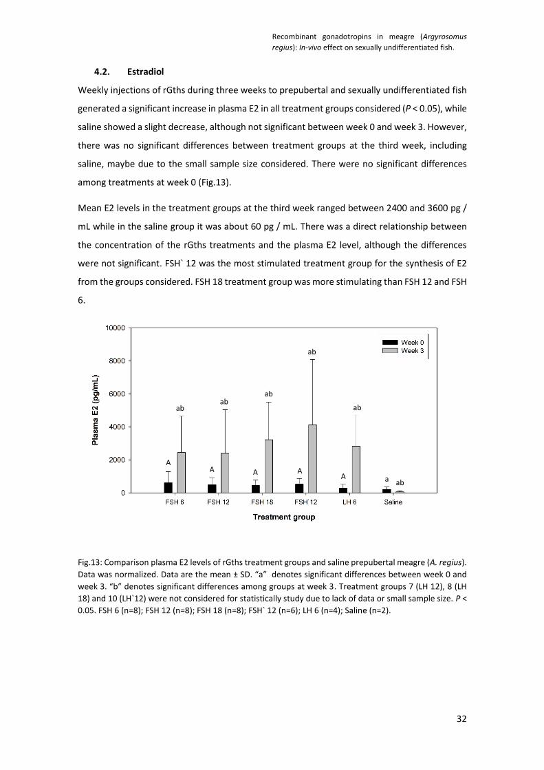
4.2. Estradiol
Weekly injections of rGths during three weeks to prepubertal and sexually undifferentiated fish
generated a significant increase in plasma E2 in all treatment groups considered (P < 0.05), while
saline showed a slight decrease, although not significant between week 0 and week 3. However,
there was no significant differences between treatment groups at the third week, including
saline, maybe due to the small sample size considered. There were no significant differences
among treatments at week 0 (Fig.13).
Mean E2 levels in the treatment groups at the third week ranged between 2400 and 3600 pg /
mL while in the saline group it was about 60 pg / mL. There was a direct relationship between
the concentration of the rGths treatments and the plasma E2 level, although the differences
were not significant. FSH` 12 was the most stimulated treatment group for the synthesis of E2
from the groups considered. FSH 18 treatment group was more stimulating than FSH 12 and FSH
6.
Fig.13: Comparison plasma E2 levels of rGths treatment groups and saline prepubertal meagre (A. regius).
Data was normalized. Data are the mean ± SD. “a” denotes significant differences between week 0 and
week 3. “b” denotes significant differences among groups at week 3. Treatment groups 7 (LH 12), 8 (LH
18) and 10 (LH`12) were not considered for statistically study due to lack of data or small sample size. P <
0.05. FSH 6 (n=8); FSH 12 (n=8); FSH 18 (n=8); FSH` 12 (n=6); LH 6 (n=4); Saline (n=2).
ab ab
ab
ab
ab
ab a
A A A A
A

Recombinant gonadotropins in meagre (Argyrosomus
regius): In-vivo effect on sexually undifferentiated fish.
33
4.3. Histological observations
In general, the gonad had somatic tissue on the outside and germ cells in the middle, with a
space for the duct in the area of germ cells. The duct indicated sexual differentiation had
initiated, but actually gonads had not completed differentiation. Moreover, most cells were
completely undifferentiated either somatic cells or germ cells that could not be identified as
oogonia or spermatogonia (Fig. 15A and B). Amongst the germ cell were isolated cells that had
differentiated into either oocytes or spermatogonia B. These few cell we searched for to define
male or female, however, differentiated had just initiated and was far from complete.
Recombinant gonadotropins short-term therapy induced early gonad development with the
appearance of oocytes at PGcn and PGps stage (Fig.15B and E), and the increase in the
percentage of fish that presented spc and StgB compared to the control and saline groups.
Group 1 (control) and group 2 (saline) did not show any oocyte development, while treatments
with rFSH, rFSH` and rLH showed oocytes; specifically group 3 (FSH 6; 12.5%; n = 8), group 4 (FSH
12; 12.5%; n = 8), group 7 (LH 12, 37.5%; n = 8) , group 8 (LH 18; 12.5%; n = 8) and group 9 (LH`
12; 16.7%; n = 6) (Fig.14E). At level of male germ cells development, all treatment groups showed
StgB (Fig.14B) (Fig.15E), and spc (Fig.14C) (Fig.15D) while saline did not. Treatment groups 3 (FSH
6), 4 (FSH 12), 6 (LH 6) and 10 (LH`12) showed one fish with spd each, however, control group
showed two fish with spd (n = 17) (Fig.14D) (Fig.15C) (4 fish from the control group were
discarded due to the small amount of gonadal tissue). Fish with all completely undifferentiated
gonadal tissue were found in group 1 (control; 88%), group 2 (saline; 100%), group 4 (FSH 12;
62,5%) and group 8 (LH 18; 62,5%) (Fig.14A).
The most satisfactory treatment to induce gonadal development was FSH 6 followed by FSH` 12
and LH 12. FSH 6 treatment group showed 75% of the fish (n = 8) with some germ cells in the
stages of spc, spd or oocytes. FSH 12, FSH 18 and LH 18 treatments showed less ovarian and
testis development, each only had 37.5% of fish (n = 8) in these stages of germinal development.
FSH` 12 treatment group (n = 6) presented 66.7% of fish with a development of spc, spd or
oocyte; while the LH 12 treatment group (n = 8) accounted for 50% of the fish. Treatments with
LH 6 (n = 8) and LH` 12 (n = 6) only showed one fish with some of the commented gonadal states.
Germ cell development was highly localized, most of the gonadal tissue did not have any
development and all fish were classified as Stage I (incompletely differentiated). In fact, all of
them showed a large amount of Embryonic germ stem cells (EGSC) and connective tissue (CT)
(Fig.15A,B and E). Actually, in groups 1 (control), 2 (saline), 6 (LH 6) and 10 (LH` 12) only EGCS,

Recombinant gonadotropins in meagre (Argyrosomus
regius): In-vivo effect on sexually undifferentiated fish.
34
oogonia or spermatogonia were found in the first six fields observed. The rest of the group
showed more advanced stages of development.
Fig.14: Comparison of the relative frequency of fish per treatment group that presented the
developmental stage of A) Oogonia or spermatogonia; B) Spermatogonia B; C) Spermatocyte; D)
Spermatid and E) Oocyte at chromatin nucleolar stage (PGcn) and perinucleolar stage (PGps). Control
(n=17); Saline (n=8); FSH 6 (n=8); FSH 12 (n=8); FSH 18 (n=8); LH 6 (n=8); LH 12 (n=8); LH 18 (n=8); FSH` 12
(n=6); LH` 12 (n=6).
A
E
D C
B

Recombinant gonadotropins in meagre (Argyrosomus
regius): In-vivo effect on sexually undifferentiated fish.
35
Fig.15: Representative photomicrographs of histological sections of immature meagre stained with hematoxylin and eosin after three weeks of treatment. A) sexually undifferentiated fish; B) female fish; C) male fish; D) male fish; E) female fish; F) intersex fish. EGCS, Embryonic Germ Stem Cells; Oog/Stg, Oogonia or spermatogonia; Oog, oogonia; StgAund, type A undifferentiated spermatogonia; StgB, type B spermatogonia; Spc, spermatocytes; Spd, spermatids; PGps, oocyte at perinucleolar stage of primary growth; Ct, connective tissue.
Spc Spd
PGps
PGps
PGps
PGps
PGps
StgB
Oog/StgAund
Ct Ct
Ct
EGSCs
EGSCs
EGSCs
StgA
StgA
Oog/StgAund
Oog
Oog
A
E F
D C
B

Recombinant gonadotropins in meagre (Argyrosomus
regius): In-vivo effect on sexually undifferentiated fish.
36
4.4. Sexual differentiation
Fish were sexed at cellular level because no difference was found in the morphology of the
ovaries and testes in this stage of sexual differentiation, so fish that presented oocytes were
classified as female, those that presented StgB or higher development stages were classified as
males, those with both developed stages were classified as intersex, and those with all germ
cells in a developmental stage lower than StgB or oocyte were classified as sexually
undifferentiated.
Gonadotropin treatment significantly altered the sex ratio, increasing the proportion of males
and developing intersex fish (P < 0.05). The only treatment group that presented females was
LH 12. All fish in saline treatment group and 88% of fish in control group (n = 17) were sexually
undifferentiated due to low gonadal development. However, in all treatment groups, except LH
12, no females were found, although oocytes were found (Fig.16). Fish that presented oocytes
from the treatment groups FSH 6, FSH 12, FSH` 12 and LH 18 also presented male germ cells
(StgB, spc or spd) (Fig.15F). The recombinant hormone that showed the most intersex fish was
FSH (n = 30), with 10% fish; specifically, one fish from each treatment of FSH 6, FSH 12 and FSH`
12 (Fig. 16).
Fig.16: Comparison of the relative frequency of fish per treatment group that were classified as sexually
undifferentiated, male, female or intersex based on the development of germ cells in the histological
sections.
Recombinant gonadotropins in meagre (Argyrosomus
regius): In-vivo effect on sexually undifferentiated fish.
37
5. Discussion
The present survey shows that the rGths produced by a CHO system are biologically active and
their half-life is long enough to induce in vivo effects in prepubertal and sexually undifferentiated
meagre (A. regius). Both gonadotropins, rFSH and rLH, were able to stimulate E2 secretion, the
onset of meiosis in female germ cells, and the onset and resumption of meiosis in male germ
cells. These findings demonstrate that there is fshra and lhcgrba expression in gonadal tissue at
this stage of development, and single-chain rGths therapy could be a useful tool to induce
gametogenesis in prepubertal fish.
5.1. Gonadosomatic index
In our study, no treatment group showed a significant increase in GSI. In contrast, recombinant
gonadotropins therapy has increased it in various fish species, such as Japanese eel (A. japonica)
(Kobayashi et al., 2010), European eel (A. anguilla) (Peñaranda et al., 2017) or Shortfinned eel
(A. australis) (Tuan-Nguyen et al., 2020). However, the effectiveness of treatments on the gonad
growth depends on the concentration of recombinant hormone, the initial stage of development
or the duration of the experiment. In the Tuan-Nguyen et al. (2020) survey, only the group
treated with the dose with the highest concentration of rFSH (500 µg / kg) for 3 weeks showed
significant differences; while the doses of 20 µg / kg and 100 µg / kg did not show significant
differences. Moreover, in contrast with our survey, all of them were sexually differentiated.
Nevertheless, in our study, the treatment with the best histological result was the one with the
lowest concentration of rFSH (6 µg / kg). This data may indicate that the doses used were correct,
but fish were too young to response at GSI level.
5.2. Estradiol
Both FSH and LH were able to stimulate E2 secretion. All of our treatment groups showed
significant differences in plasma E2 secretion between week 0 and week 3, while saline did not.
The initial stage of development of fish was not enough to stimulate an increase in gonadal size,
but actually it was enough to stimulate the synthesis of plasma E2; doses with less concentration
are enough to stimulate its secretion. Similar results were obtained in Shortfinned eel (A.
australis), in which 100 µg / kg rFSH treatment for 3 weeks did not show an increase in GSI, but
showed an increase in E2 (Tuan-Nguyen et al., 2020).

Recombinant gonadotropins in meagre (Argyrosomus
regius): In-vivo effect on sexually undifferentiated fish.
38
5.3. Histological observations
Data demonstrate that rGths treatments were effective in inducing early gonad development,
although not enough to differentiate all germ cells. In females, gonad development was similar
to that obtained with rFSH in Orange-spotted grouper (E. coioides), where control group resulted
in an undifferentiated ovary, while treatment group in an ovary with oocytes in PGcn and PGps
(Chen et al., 2012a). Both surveys studied the effects of rGths treatment on fish that had not
reached sexual maturity (prepubertal or juvenile fish). However, the Orange-spotted grouper (E.
coioides) is a protogyny hermaphroditic fish (sexually differentiated) (Liu and Mitcheson., 2009)
(Chen et al., 2012a), while meagre (A. regius) is a gonochoristic fish that was at sexual
differentiation period. Otherwise, in our experiment, males had a greater development of germ
cells than females, with the resumption of meiosis and development of spermatids in some
cases, while females initiated meiosis but did not reach SGca stage. In flathead grey mullet (M.
cephalus) rFSH and rLH therapy induced the entire process of gametogenesis, however, 11 and
5 weeks of treatment for females and males respectively and the combination of both
gonadotropins were necessary. Furthermore, the initial stage of development in female germ
cells were PGps or SGca (Ramos-Júdez et al., 2020). In giant grouper (E. fuscoguttatus) after 8
weeks of treatment with a weekly injection of rFSH at 100 µg / kg, the oocyte in PGps stage
developed to SGca (Palma et al., 2019). In Japanese eel (A. japonica) 5 weeks of treatment with
a weekly injection of rFSH or rLH were able to develop spermatogonia into spermatids and
spermatozoa respectively (Hayakawa et al., 2008). In Shortfinned eel (A. australis) the duration
of the experiment was 3 weeks like in our survey; and weekly injections of rFSH developed
oocytes to SGca from PGps, although there were some oocytes at SGca stage in initial control
group (Tuan-Nguyen et al., 2020). This data demonstrates that at histological level there are
differences depending on the initial stage of development and the duration of the treatment.
Considering our results at plasma E2 level and at histological level with the onset of meiosis in
germ cells of males and females, we can suggest that i) there was expression of fshra and lhcgrba
in gonads during the period of sexual differentiation, ii) rFSH and rLH, both, were able to
stimulate the synthesis of 11-KT, iii) rFSH and rLH, both, were able to stimulate the synthesis of
progestin 20βS.
i) There was fshra and lhcgrba expression in meagre (A. regius) tissue during the period of
sexual differentiation, due to greater gonadal development and E2 secretion in the
treatment groups than in the control and saline groups. In fact, in control and saline

Recombinant gonadotropins in meagre (Argyrosomus
regius): In-vivo effect on sexually undifferentiated fish.
39
groups the gonad in most cases remained undifferentiated and with a lower level of
plasma E2, although not significant. This is in agreement with Baron et al. (2005), where
there was expression of lhcgrba in female rainbow trout (O. mykiss) during the period
of sexual differentiation; or as in the medaka (O. latipes), where fshra and lhcgrba
expression was found in all testis stage (Burow et al., 2019). Moreover, in Nile tilapia (O.
niloticus) there is fshra and lhcgrba in undifferentiated gonads during sexual
differentiation period (Yan et al., 2012).
ii) Studies in Japanese huchen (Hucho perryi) (Amer et al., 2001), Japanese eel (A. japonica)
(Miura et al., 2006), Atlantic salmon (S. salar) (Chen et al., 2012b) or Nile tilapia (O.
niloticus) (Liu et al., 2014) suggest that MIS 20βP induces entry into meiosis of germ
cells. In our results, females and males treated with rFSH and rLH initiated meiosis,
suggesting that both rGths were able to activate Lhcgrba for the synthesis of MIS 20βS.
This was in accordance with the results obtained by Burow et al. (2019), where both
rGths activated the Fshra and Lhcgrba receptors in medaka (O. latipes).
iii) Otherwise, according to Miura and Miura (2003), 11-KT stimulates the differentiation of
spermatogonia priors to initiation of meiosis. Our results suggest that both rFSH and rLH
were able to stimulate the synthesis of 11-KT as it was reported before (Suzuki et al.,
2019).
5.4. Sexual differentiation
About sexual differentiation, our histological observations demonstrate that with recombinant
gonadotropin treatment: 1) the onset on meiosis in female germ cells occurs at the same time
as de appearance of the ovarian cavity 2) males sexually differentiate earlier than females; 3)
there is a higher proportion of males during the stage of sexual differentiation, 4) there are
intersex fish during the stage of sexual differentiation. This is the first report of a male skewed
ratio and the presence of intersex fish in meagre during the stage of sexual differentiation
In gonochoristic teleost fish such as meagre (A. regius), the first indicator of sexual identification
is made according to the ovarian ontogeny. It depends on whether the ovarian lumen / ovarian
cavity develops earlier or not than the entry into meiosis of the oogonia with the consequent
development of the oocytes (Nakamura et al., 1998). In Olive flounder (Paralichthys olivaceus),
Southern flounder (P. lethostigma), Nile tilapia (Oreochromis niloticus) or Blue tilapia (O.
aureus), meiotic division occurs after the formation of the ovarian lumen, thus, it is used as the
earliest recognizable sign of female differentiation (Liu and Mitcheson, 2009). In European eel

Recombinant gonadotropins in meagre (Argyrosomus
regius): In-vivo effect on sexually undifferentiated fish.
40
(A. anguilla), Silver-stripe round herring (Spratelloides gracilis) or Cichlasoma dimerus, meiotic
division occurs before the formation of the ovarian lumen, so the appearance of oocytes is the
earliest recognizable sign of female differentiation (Colombo and Grandi, 1995; Hatakeyama et
al., 2005; Meijide et al., 2005). In Fathead minnow (Pimephales promelas), Viviparous eelput
(Zoarces viviparus) or Roach (Rutilus rutilus), meiotic division of the oogonia and the formation
of the ovarian lumen occurs at the same time (Van Aerle et al., 2004; Rasmussen et al., 2006;
Paull et al., 2008). According to our results, in meagre (A. regius), meiosis of the oogonia occurs
at the same time as the appearance of the ovarian lumen, as described by Schiavone et al.
(2012). For this reason, oocyte development was used as a classification criterion for females.
Moreover, according to Schiavone et al. (2012), females sexually differentiate earlier than males,
and according to Gil et al. (2013), wild meagre has a population sex ratio of 1: 1. Both ideas are
in disagreement with our results, in wich male sexually differentiate earlier than females and
there is a male skewed ratio. The control group presented 2 males and 15 undifferentiated fish,
the LH` 12 treatment group showed 1 male and 6 undifferentiated fish, the LH 6 treatment group
showed 2 males and 6 undifferentiated fish, and the FSH 18 treated group had 3 males and 5
undifferentiated fish. In addition, the rest of the treatments had males in a higher percentage
than intersex fish. The only group that presented females was the one treated with LH 12, which
showed 3 females, 1 male and 4 undifferentiated fish (Fig.16). We must consider that the
population sample of Gil et al., (2013) was composed of completely sexually differentiated and
wild individuals.
Sex determination is defined as the forces that determine whether a fish will become a male or
a female, whereas sex differentiation refers to molecular and cellular processes that make a
bipotential gonadal primordium develop into a testis or ovary after sex has been determined
(Devlin and Nagahama, 2002). The sex determination mechanisms in vertebrates include
Genotypic Sex Determination (GSD) and Environmental Sex determination (ESD), or a
combination of both (Nakamura et al., 1998; Sandra and Norma, 2009; Navarro-Martín et al.,
2011; Fernandino et al., 2013). Sex differentiation in egg-laying vertebrates is a hormone-
dependent process that involves complex interactions among a large network of genes (Baron
et al., 2005), such as cyp19a or hsd11b. Exposure to exogenous hormones and endocrine
disruptors throughout sex differentiation can alter the expression of these genes, causing
alterations in the sex differentiation process that ultimately leads to reproductive abnormalities,
including intersex (Abdel-Moneim et al. 2015). There are two hypotheses about the influence of
steroids on the gonad, the first one maintains that the ovaries develop due to the action of
estrogens, while the testes develop due to the action of androgens (Nakamura, 2010). The other

Recombinant gonadotropins in meagre (Argyrosomus
regius): In-vivo effect on sexually undifferentiated fish.
41
hypothesis proposes that the testes develop by inaction of Cyp19a / P450arom (Guiguen et al.,
2010). In any case, the balance between E2 and 11-KT have a key role on sexual differentiation
processes (Sandra and Norma, 2009; Navarro-Martín et al., 2011; Bahamonde et al., 2013). Our
results support the first hypothesis because treatment groups showed a male skewed ratio and
an increase in E2 secretion, although not significant. Moreover, according to Kagawa et al.
(2003), in Red seabream (Pagrus major), LH but not FSH, stimulated both aromatase activity and
P450arom gene expression. This could explain why our treatment group 7 (LH 12) was the only
one that presented females (37.5%; n = 8); however more data about E2 production in treatment
groups would be necessary.
Intersex is defined as the presence of male and female gonadal tissue in a gonochoristic species
(Bahamonde et al., 2013). The presence of intersex fish during the stage of sexual differentiation
can have four main reasons. The first is that the development of juvenile intersex is a passive
phenomenon and perhaps it is an advantage to achieve a reproductive partner in severe
ecological conditions, since it temporarily maintains bipotentiality to develop in ovary or testis
(Tricas and Hiramoto, 1989; Sandra and Norma, 2009). The second possible cause is that there
is a “basal rate” of intersex in the meagre species although it is rarely (Grim et al., 2007; Abdel-
Moneim et al., 2015). The third possible cause is that all the gonads develop initially as ovaries
and that later half of them degenerate giving rise to testes (Juvenile hermaphroditism) (Deblin
and Nagahama, 2002). The fourth possible cause is that some environmental factor has altered
the normal development of sexual differentiation in this survey. Since the development of
intersex fish during sexual differentiation and during adulthood has not been previously
documented in meagre (A. regius) (Schiavone et al., 2012; Gil et al., 2013), and that the third
possible cause is in disagreement with our results and those of previous studies, it is most likely
that it is the fourth cause. Environmental factors include: a) pollution and exogenous steroids
(endocrine-disrupting chemicals), b) temperature and other physical variables, and c) social
interactions (Deblin and Nagahama, 2002). Although we should include d) recombinant
gonadotropins treatment as a potential cause of intersex and male skewed sex ratio.
a. The development of intersex fish as a consequence of endocrine-disrupting chemicals
(EDCs) has been widely studied in freshwater and marine fish (Kavanagh et al. 2004;
Barucca et al., 2006; Blazer et al., 2007; Vajda et al. 2008; Diaz de Cerio et al., 2012;
Rochman et al., 2014; Bahamonde et al., 2014). However, prepubertal fish in our study
do not come from the marine environment, so they should not have been affected by
EDCs. Furthermore, they have not received a diet with a rich source of phytoestrogens

Recombinant gonadotropins in meagre (Argyrosomus
regius): In-vivo effect on sexually undifferentiated fish.
42
or any compound with any estrogenic or androgenic activity that can alter the process
of sexual differentiation (Grim et al., 2007; Rzepkowska et al., 2014).
b. Temperature is probably the most studied factor in ESD. It has been observed that in
species with Temperature Sex Determination (TSD), high temperatures during the
sexual differentiation process are associated with masculinization processes,
intermediate ones with mixed-sex populations, and low temperatures with feminization
processes (Strüssmann et al., 1997; Sandra and Norma, 2009). According to Fernandino
et al. (2012), thermal stress of high temperatures during the period of sexual
differentiation in TSD species generates an increase in cortisol levels, which in turn
induces an increase in hsd11b levels (codes for the enzyme 11β-hsd), leading to a higher
synthesis of 11-KT and a higher sex ratio of males. High temperatures also increase the
methylation levels of the cyp19a promoter region in European seabass (D. labrax),
which were found to be sexspecific and highly influenced by changes in temperature
during early life stages, causing signs of masculinization as a result of the down-
regulation of the expression of this gene (Navarro-Martín et al., 2011). In medaka (O.
latipes), which is a GSD species with XX / XY sex determination, it has been observed
that a high temperature treatment or a cortisol treatment at intermediate temperatures
during the period of sexual differentiation also increases the sex ratio of males,
suppresses the expression of fshra and female-type proliferation of germ cells (by
upregulating of amh) (Selim et al., 2009; Hayashi et al., 2010). In Japanese flounder
(Paralichthys olivaceus) (GSD with XX / XY sex determination) and Silverside
(Odontesthes bonariensis) (TSD), a high temperature treatment during sex
differentiation period stimulates the synthesis of cortisol, which inhibits the expression
of cyp19a, increasing the sex ratio of males (Hattori et al., 2009; Yamaguchi et al., 2010;
Guiguen et al., 2010; Yang et al., 2020). Therefore, an increase in cortisol levels is related
to masculinization processes due to cyp19a downregulation, increased synthesis of 11-
KT, reducing the number of PGC cells (Goikoetxea et al., 2017; Miller et al., 2018), or
downregulation of fshra.
Further studies are necessary to elucidate the sex determination mechanism in meagre
(A. regius). In any case, our fish were kept around 16°C, which should not be too high
temperature to induce strong thermal stress, compared to the average water
temperature between the months of February and May in the Western Mediterranean
and the Eastern Atlantic (data not shown) (MITMA, 2021).

Recombinant gonadotropins in meagre (Argyrosomus
regius): In-vivo effect on sexually undifferentiated fish.
43
c. Population density is one of the social factors that affect sexual differentiation
(Fernandino et al., 2013). In Japanese eel (A. japonica) or European seabass (D. labrax),
high population densities in captivity were related to the male-biased sex ratio (Davey
and Jellyman, 2005; Saillant et al., 2006), and an increase in cortisol levels (Chiba et al.,
2002 cited in Fernandino et al., 2013). Prepubertal fish in our study were fished from
the tank eight times in three weeks, and introduced into 300 dm3 tanks with a
population density of 50.3 kg m-3. In addition, in each of these periods, blood extraction
on hormone injection were performed. Is a possibility that intensive sampling during
three weeks of the sexual differentiation period could increase cortisol levels and induce
masculinization.
d. Never before rGths have been injected into sexually undifferentiated fish, so little is
known about how they can affect sexual differentiation of gonochoristic fish. In rainbow
trout (O. mykiss) and Nile tilapia (O. niloticus) during the stage of sexual differentiation
there is expression of βFSH and βLH (Baron et al., 2005; Yan et al., 2012), however, it is
possible that the increase in the level of gonadotropins by supraphysiological doses
during this period could affect the expression of one or more genes related to sexual
differentiation.
Sox transcriptional factors are characterized as Sry-related high-mobility group (HMG)
box proteins. Sox genes have been reported to be involved in sex determination and
differentiation, formation of neuronal system, gonad, eye, pancreas, and cartilage (Wei
et al., 2016). Sox30 has been proven to be critical for mammalian and teleostean
spermatogenesis. There is expression of sox30 in testes and ovary of carp (C. carpio)
(Arumugam and Balasubramanian, 2020) and Nile tilapia (O. niloticus) (Han et al., 2010;
Wei et al., 2016). In fact, in carp (C. carpio), sox30 expression is high during
spermatogenesis and spawning / spermiation (Arumugam and Balasubramanian, 2020).
In Nile tilapia (O. niloticus), sox30 expression was reported during sexual determination
and differentiation period in ovary and testes (Han et al., 2010). These data support that
Sox30 plays a key role in gonadal differentiation and testes development (Han et al.,
2010; Wei et al., 2016; Arumugam and Balasubramanian, 2020). Furthermore, sox30
expression is influenced by gonadotropins. There is a substantial increase in sox30
mRNA transcripts post-hCG induction, and hCG is proven to bind Lhcgrba (Arumugam
and Balasubramanian, 2020).
Taken together these results, we speculate that the increase in gonadotropin levels
during the period of sexual differentiation with respect to basal secretion could increase

Recombinant gonadotropins in meagre (Argyrosomus
regius): In-vivo effect on sexually undifferentiated fish.
44
sox30 expression and alter sexual differentiation, however, there are no results that
support this hypothesis. There are also other genes related to sexual differentiation such
as sox3 (Takehana et al., 2014).
Further studies are necessary to elucidate the possible alteration in gene expression by the
administration of gonadotropins during the period of sexual differentiation.

Recombinant gonadotropins in meagre (Argyrosomus
regius): In-vivo effect on sexually undifferentiated fish.
45
6. Conclusion
1. Weekly intramuscular injections of specific Argyrosomus regius single-chain rGths for 3
weeks in prepubertal and sexually undifferentiated meagre (A. regius) are not able to
stimulate a significant increase in GSI comparing with saline group.
2. Both rFSH and rLH are able to stimulate E2 secretion.
3. Both FSH and LH are able to stimulate early gonadal development with the onset of
meiosis in female germ cells, and the onset and resumption of meiosis in male germ
cells.
4. Our histological observations in sexual differentiation demonstrate that with
recombinant gonadotropin treatment 1) the onset of meiosis in female germ cells occurs
at the same time as de appearance of the ovarian cavity 2) males sexually differentiate
earlier than females; 3) there is a higher proportion of males during the stage of sexual
differentiation, 4) there are intersex fish during the stage of sexual differentiation.
Further studies are necessary to elucidate what kind of sex determination mechanisms has the
meagre, how the administration of exogenous gonadotropins can influence sexual
differentiation during this period and, to evaluate the effect of rGths treatment with a longer
therapy and in older fish.

Recombinant gonadotropins in meagre (Argyrosomus
regius): In-vivo effect on sexually undifferentiated fish.
46
7. References
- Abdel-moneim, A., Coulter, D. P., Mahapatra, C. T., Sepúlveda, M. S. (2015). “Intersex in
fishes and amphibians: population implications, prevalence, mechanisms and molecular
biomarkers”. Journal of Applied Toxicology, 35(11), 1228–1240. doi:10.1002/jat.3204
- Aizen, J., Kasuto, H., Levavi-Sivan, B. (2007a). “Development of specific enzyme-linked
immunosorbent assay for determining LH and FSH levels in tilapia, using recombinant
gonadotropins”. General and Comparative Endocrinology, 153(1-3), 323–
332. doi:10.1016/j.ygcen.2007.04.004
- Aizen, J., Kasuto, H., Golan, M., Zakay, H., Levavi-Sivan, B. (2007b). “Tilapia Follicle-
Stimulating Hormone (FSH): Immunochemistry, Stimulation by Gonadotropin-Releasing
Hormone, and Effect of Biologically Active Recombinant FSH on Steroid Secretion”.
Biology of Reproduction, 76(4), 692–700. doi:10.1095/biolreprod.106.055822
- Aizen, J., Kobayashi, M., Selicharova, I., Sohn, Y. C., Yoshizaki, G., Levavi-Sivan, B.
(2012). Steroidogenic response of carp ovaries to piscine FSH and LH depends on the
reproductive phase”. General and Comparative Endocrinology, 178(1), 28–
36. doi:10.1016/j.ygcen.2012.04.002
- Aizen, J., Hollander-Cohen, L., Shpilman, M., Levavi-Sivan, B. (2016). “Biologically active
recombinant carp LH as a spawning-inducing agent for carp”. Journal of Endocrinology,
232(3), 391–402. doi:10.1530/joe-16-0435
- Amer, M. A., Miura, T., Miura, C., Yamauchi, K. (2001). “Involvement of Sex Steroid
Hormones in the Early Stages of Spermatogenesis in Japanese Huchen (Hucho perryi )”.
Biology of Reproduction, 65(4), 1057–1066. doi:10.1095/biolreprod65.4.1057
- APROMAR, (2019). “La acuicultura en España 2019”. In APROMAR [Online], available in
http://apromar.es/sites/default/files/2019/InformeAcui/APROMAR%20Informe%20AC
UICULTURA%202019%20v-1-2.pdf [accesed on May 2, 2021]
- APROMAR, (2020). “La acuicultura en España 2020”. In APROMAR [Online], available in
http://www.apromar.es/sites/default/files/2020/Informe%20Acuicultura%20Espa%C3
%B1a%202020_APROMAR.pdf [accesed on May 2, 2021]
- Arukwe, A., Goksøyr, A. (2003). “Eggshell and egg yolk proteins in fish: hepatic proteins
for the next generation: oogenetic, population, and evolutionary implications of
endocrine disruption”. Comparative Hepatology, 2(1), 4. doi:10.1186/1476-5926-2-4
- Arumugam, A., Balasubramanian, S. (2020). “Role of sox30 in regulating testicular
steroidogenesis of common carp”. The Journal of Steroid Biochemistry and Molecular
Biology, 105769. doi:10.1016/j.jsbmb.2020.1057 69

Recombinant gonadotropins in meagre (Argyrosomus
regius): In-vivo effect on sexually undifferentiated fish.
47
- Bahamonde, P. A., Munkittrick, K. R., Martyniuk, C. J. (2013). “Intersex in teleost fish:
Are we distinguishing endocrine disruption from natural phenomena?” General and
Comparative Endocrinology, 192, 25–35. doi:10.1016/j.ygcen.2013.04.005
- Bahamonde, P. A., Tetreault, G. R., McMaster, M. E., Servos, M. R., Martyniuk, C. J.,
Munkittrick, K. R. (2014). “Molecular signatures in rainbow darter (Etheostoma
caeruleum) inhabiting an urbanized river reach receiving wastewater effluents”. Aquatic
Toxicology, 148, 211–220. doi:10.1016/j.aquatox.2014.01.010
- Baron, D., Houlgatte, R., Fostier, A., Guiguen, Y. (2005). “Large-Scale Temporal Gene
Expression Profiling During Gonadal Differentiation and Early Gametogenesis in
Rainbow Trout”. Biology of Reproduction, 73(5), 959–966.
doi:10.1095/biolreprod.105.041830
- Barucca, M., Canapa, A., Olmo, E., Regoli, F. (2006). “Analysis of vitellogenin gene
induction as a valuable biomarker of estrogenic exposure in various Mediterranean fish
species”. Environmental Research, 101(1), 68–73. doi:10.1016/j.envres.2005.07.002
- Berishvili, G., D’Cotta, H., Baroiller, J.F., Segner, H., Reinecke, M. (2006). “Differential
expression of IGF-I mRNA and peptide in the male and female gonad during early
development of a bony fish, the tilapia Oreochromis niloticus”. General and Comparative
Endocrinology, 146(3), 204–210. doi:10.1016/j.ygcen.2005.11.008
- Blazer, V. S., Iwanowicz, L. R., Iwanowicz, D. D., Smith, D. R., Young, J. A., Hedrick, J. D.,
Foster, S.W., Reeser, S. J. (2007). “Intersex (Testicular Oocytes) in Smallmouth Bass from
the Potomac River and Selected Nearby Drainages”. Journal of Aquatic Animal Health,
19(4), 242–253. doi:10.1577/h07-031.1
- Blázquez, M., Piferrer, F. (2004). “Cloning, sequence analysis, tissue distribution, and
sex-specific expression of the neural form of P450 aromatase in juvenile sea bass
(Dicentrarchus labrax)”. Molecular and Cellular Endocrinology, 219(1-2), 83–
94. doi:10.1016/j.mce.2004.01.006
- Burow, S., Mizrahi, N., Maugars, G., von Krogh, K., Nourizadeh-Lillabadi, R., Hollander-
Cohen, L., Shpilman, M., Atre, I., Weltzien, F.A., Levavi-Sivan, B.
(2019). “Characterization of gonadotropin receptors Fshr and Lhr in Japanese medaka,
Oryzias latipes”. General and Comparative Endocrinology,
113276. doi:10.1016/j.ygcen.2019.113276
- Campbell, B., Dickey, J., Beckman, B., Young, G., Pierce, A., Fukada, H., Swanson, P.
(2006). “Previtellogenic Oocyte Growth in Salmon: Relationships among Body Growth,
Plasma Insulin-Like Growth Factor-1, Estradiol-17beta, Follicle-Stimulating Hormone
and Expression of Ovarian Genes for Insulin-Like Growth Factors, Steroidogenic-Acute

Recombinant gonadotropins in meagre (Argyrosomus
regius): In-vivo effect on sexually undifferentiated fish.
48
Regulatory Protein and Receptors for Gonadotropins, Growth Hormone, and
Somatolactin”. Biology of Reproduction, 75(1), 34–
44. doi:10.1095/biolreprod.105.049494
- Cao, H., Zhou, L., Zhang, Y.Z., Wei, Q.W., Chen, X.H., Gui, J.F. (2009). “Molecular
characterization of Chinese sturgeon gonadotropins and cellular distribution in
pituitaries of mature and immature individuals”. Molecular and Cellular Endocrinology,
303(1-2), 34–42. doi:10.1016/j.mce.2009.01.015
- Carrillo, M. A., Muñoz, J. A., Zanuy, S., Rocha, A., Molés, G., Bayarri, M., Piferrer, F.,
Fernández-Palacios, H., Izquierdo, M., Cerdá, J., Herráez, M., Navas, J., Cañavate, J.,
Gracia, V. Valdebenito, I. (2012). La reproducción de los peces: aspectos básicos y sus
aplicaciones en acuicultura. Madrid: Fundación Observatorio Español de Acuicultura.
- Cerdà, J., Fabra, M., Raldúa, D., (2007). Physiological and molecular basis of fish oocyte
hydration. In: Babin, P.J., Cerdà, J., Lubzens, E. (Eds.), The Fish Oocyte: From Basic Studies
to Biotechnological Applications. Springer, Dordrecht, pp. 349–396.
- Chang, X., Kobayashi, T., Senthilkumaran, B., Kobayashi-Kajura, H., Sudhakumari, C. C.,
Nagahama, Y. (2005). “Two types of aromatase with different encoding genes, tissue
distribution and developmental expression in Nile tilapia (Oreochromis niloticus)”.
General and Comparative Endocrinology, 141(2), 101–
115. doi:10.1016/j.ygcen.2004.11.020
- Chauvigné, F., Verdura, S., Mazón, M. J., Duncan, N., Zanuy, S., Gómez, A., Cerdà, J.
(2012). “Follicle-Stimulating Hormone and Luteinizing Hormone Mediate the
Androgenic Pathway in Leydig Cells of an Evolutionary Advanced Teleost”. Biology of
Reproduction, 87(2). doi:10.1095/biolreprod.112.100784
- Chauvigné, F., Ollé, J., González, W., Duncan, N., Giménez, I., Cerdà, J. (2017). “Toward
developing recombinant gonadotropin-based hormone therapies for increasing fertility
in the flatfish Senegalese sole”. PLoS ONE 12(3). doi: 10.1371/journal.pone.0174387
- Chen, J., Zhang, Y., Tang, Z., Mao, J., Kuang, Z., Qin, C., Li, W. (2012a). “Production of
recombinant orange-spotted grouper (Epinephelus coioides) follicle-stimulating
hormone (FSH) in single-chain form and dimer form by Pichia pastoris and their
biological activities”. General and Comparative Endocrinology, 178(2), 237–
249. doi:10.1016/j.ygcen.2012.05.009
- Chen, S. X., Bogerd, J., Andersson, E., Almeida, F. F. L., Taranger, G. L., Schulz, R. W.
(2012b). “Cloning, pharmacological characterization, and expression analysis of Atlantic
salmon (Salmo salar) nuclear progesterone receptor”. Reproduction, 141(4), 491–500.
doi:10.1530/rep-10-0224

Recombinant gonadotropins in meagre (Argyrosomus
regius): In-vivo effect on sexually undifferentiated fish.
49
- Cohen, A., Smith, Y. (2014). “Estrogen Regulation of microRNAs, Target Genes, and
microRNA Expression Associated with Vitellogenesis in the Zebrafish”. Zebrafish, 11(5),
462–478. doi:10.1089/zeb.2013.0873
- Colombo, G., Grandi, G. (1995). “Sex differentiation in the European eel: histological
analysis of the effects of sex steroids on the gonad”. Journal of Fish Biology, 47(3), 394–
413. doi:10.1111/j.1095-8649.1995.tb01909.x
- Cui, M., Li, W., Liu, W., Yang, K., Pang, Y., Haoran, L. (2007). “Production of Recombinant
Orange-Spotted Grouper (Epinephelus coioides) Luteinizing Hormone in Insect Cells by
the Baculovirus Expression System and Its Biological Effect”. Biology of Reproduction,
76(1), 74–84. doi:10.1095/biolreprod.105.050484
- Davey, A. J. H., Jellyman, D. J. (2005). “Sex Determination in Freshwater Eels and
Management Options for Manipulation of Sex”. Reviews in Fish Biology and Fisheries,
15(1-2), 37–52. doi:10.1007/s11160-005-7431-x
- Devlin, R. H., Nagahama, Y. (2002). “Sex determination and sex differentiation in fish: an
overview of genetic, physiological, and environmental influences”. Aquaculture, 208(3-
4), 191–364. doi:10.1016/s0044-8486(02)00057-1
- Diaz de Cerio, O., Rojo-Bartolomé, I., Bizarro, C., Ortiz-Zarragoitia, M., Cancio, I.
(2012). “5S rRNA and Accompanying Proteins in Gonads: Powerful Markers to Identify
Sex and Reproductive Endocrine Disruption in Fish”. Environmental Science and
Technology, 46(14), 7763–7771. doi:10.1021/es301132b
- Duncan, N., Estévez, A., Porta, J., Carazo, I., Norambuena, F., Aguilera, C., Gairin, I., Bucci,
F., Valles, R., Mylonas, C. C. (2012). “Reproductive development, GnRHa-induced
spawning and egg quality of wild meagre (Argyrosomus regius) acclimatised to
captivity”. Fish Physiology and Biochemistry, 38(5), 1273–1286. doi:10.1007/s10695-
012-9615-3
- Duncan, N. J., Estévez, A., Fernández-Palacios, H., Gairin, I., Hernández-Cruz, C. M., Roo,
J., Schuchardt, D., Vallés, R. (2013). “Aquaculture production of meagre (Argyrosomus
regius): hatchery techniques, ongrowing and market”. Advances in Aquaculture
Hatchery Technology, 519–541. doi:10.1533/9780857097460.3.519
- Duncan, N. J., Mylonas, C. C., Milton Sullon, E., Karamanlidis, D., França Nogueira, M. C.,
Ibarra-Zatarain, Z., Chiumento, M., Aviles-Carrillo, R. O. (2018). “Paired spawning with
male rotation of meagre Argyrosomus regius using GnRHa injections, as a method for
producing multiple families for breeding selection programs”. Aquaculture, 495, 506–
512. doi:10.1016/j.aquaculture.2018.06.017

Recombinant gonadotropins in meagre (Argyrosomus
regius): In-vivo effect on sexually undifferentiated fish.
50
- Endo, T., Todo, T., Lokman, P. M., Kudo, H., Ijiri, S., Adachi, S., Yamauchi, K.
(2011). “Androgens and Very Low Density Lipoprotein Are Essential for the Growth of
Previtellogenic Oocytes from Japanese Eel, Anguilla japonica, In Vitro”. Biology of
Reproduction, 84(4), 816–825. doi:10.1095/biolreprod.110.087163
- Fakriadis, I., Zanatta, E. M., Fleck, R. P. D. S., Sena Mateo, D. L., Papadaki, M., Mylonas,
C. C. (2020). “Endocrine regulation of long-term enhancement of spermiation in meagre
(Argyrosomus regius) with GnRHa controlled-delivery systems”. General and
Comparative Endocrinology, 297, 113549. doi:10.1016/j.ygcen.2020.113549
- Fernandino, J. I., Hattori, R. S., Kishii, A., Strüssmann, C. A., Somoza, G. M. (2012). “The
Cortisol and Androgen Pathways Cross Talk in High Temperature-Induced
Masculinization: The 11β-Hydroxysteroid Dehydrogenase as a Key Enzyme”.
Endocrinology, 153(12), 6003–6011. doi:10.1210/en.2012-1517
- Fernandino, J. I., Hattori, R. S., Moreno Acosta, O. D., Strüssmann, C. A., Somoza, G. M.
(2013). “Environmental stress-induced testis differentiation: Androgen as a by-product
of cortisol inactivation”. General and Comparative Endocrinology, 192, 36–
44. doi:10.1016/j.ygcen.2013.05.024
- Finn, R.N., Ostby, G.C., Norberg, B., Fyhn, H.J., (2002). “In vivo oocyte hydration in
Atlantic halibut (Hippoglossus hipoglossus): proteolytic liberation of free aminoacids,
and ion transport, are driving forces for osmotic water influx”. J. Exp. Biol. 205, 211–224.
doi:10.1242/jeb.205.2.211
- García, S., (2012). Investigaciones aplicables al desarrollo de producción intensiva de
corvina (Argyrosomus regius). Doctoral thesis. Granada. Universidad de Granada.
- García-López, Á., Bogerd, J., Granneman, J. C. M., van Dijk, W., Trant, J. M., Taranger, G.
L., Schulz, R. W. (2009). “Leydig Cells Express Follicle-Stimulating Hormone Receptors in
African Catfish”. Endocrinology, 150(1), 357–365. doi:10.1210/en.2008-0447
- García-López, Á., de Jonge, H., Nóbrega, R. H., de Waal, P. P., van Dijk, W., Hemrika, W.,
Taranger, G.L., Bogerd, J., Schulz, R. W. (2010). “Studies in Zebrafish Reveal Unusual
Cellular Expression Patterns of Gonadotropin Receptor Messenger Ribonucleic Acids in
the Testis and Unexpected Functional Differentiation of the Gonadotropins”.
Endocrinology, 151(5), 2349–2360. doi:10.1210/en.2009-1227
- Gil, M. D. M., Grau, A., Basilone, G., Ferreri, R., Palmer, M. (2013). “Reproductive
strategy and fecundity of meagre Argyrosomus regius Asso, 1801 (Pisces: Sciaenidae):
implications for restocking programs”. Scientia Marina, 77(1), 105–
118. doi:10.3989/scimar.03688.28a

Recombinant gonadotropins in meagre (Argyrosomus
regius): In-vivo effect on sexually undifferentiated fish.
51
- Gjedrem, T. (2000). “Genetic improvement of cold-water fish species”. Aquaculture
Research, 31(1), 25–33. doi:10.1046/j.1365-2109.2000.00389.x
- Goikoetxea, A., Todd, E. V., Gemmell, N. J. (2017). “Stress and sex: does cortisol mediate
sex change in fish?” Reproduction, 154(6), R149–R160. doi:10.1530/rep-17-0408
- Goto, R., Saito, T. (2019). “A state-of-the-art review of surrogate propagation in fish”.
Theriogenology, 133, 216–227. doi:10.1016/j.theriogenology.2019.03.032
- Grigorakis, K., Fountoulaki, E., Vasilaki, A., Mittakos, I., Nathanailides, C. (2011). “Lipid
quality and filleting yield of reared meagre (Argyrosomus regius)”. International Journal
of Food Science and Technology, 46(4), 711–716. doi:10.1111/j.1365-
2621.2010.02537.x
- Grim, K. C., Wolfe, M., Hawkins, W., Johnson, R., Wolf, J. (2007). “intersex in japanese
medaka (Oryzias latipes) used as negative controls in toxicologic bioassays: a review of
54 cases from 41 studies. Environmental Toxicology and Chemistry, 26(8),
1636. doi:10.1897/06-494r.1
- Guiguen, Y., Fostier, A., Piferrer, F., Chang, C.F. (2010). “Ovarian aromatase and
estrogens: A pivotal role for gonadal sex differentiation and sex change in fish”. General
and Comparative Endocrinology, 165(3), 352–366. doi:10.1016/j.ygcen.2009.03.002
- Halm, S., Rocha, A., Miura, T., Prat, F., Zanuy, S. (2007). “Anti-Müllerian hormone
(AMH/AMH) in the European sea bass: Its gene structure, regulatory elements, and the
expression of alternatively-spliced isoforms”. Gene, 388(1-2), 148–
158. doi:10.1016/j.gene.2006.10.018
- Han, F., Wang, Z., Wu, F., Liu, Z., Huang, B., Wang, D. (2010). “Characterization,
phylogeny, alternative splicing and expression of Sox30 gene”. BMC Molecular Biology,
11(1), 98. doi:10.1186/1471-2199-11-98
- Hatakeyama, R., Shirafuji, N., Nishimura, D., Kawamura, T., Watanabe, Y.
(2005). “Gonadal development in early life stages of Spratelloides gracilis”. Fisheries
Science, 71(6), 1201–1208. doi:10.1111/j.1444-2906.2005.01084.x
- Hattori, R. S., Fernandino, J. I., Kishii, A., Kimura, H., Kinno, T., Oura, M., Somoza, G.M.,
Yokota, M., Strüssman, C., Watanabe, S. (2009). “Cortisol-Induced Masculinization:
Does Thermal Stress Affect Gonadal Fate in Pejerrey, a Teleost Fish with Temperature-
Dependent Sex Determination?” PLoS ONE, 4(8),
e6548. doi:10.1371/journal.pone.0006548
- Hayakawa, Y., Nagaya, H., Kaki, H., Hotta, K., Kobayashi, M. (2008a). “Induction of
spermatogenesis in Japanese eel by recombinant goldfish gonadotropins”. Fisheries
Science, 75(1), 137–144. doi:10.1007/s12562-008-0007-7

Recombinant gonadotropins in meagre (Argyrosomus
regius): In-vivo effect on sexually undifferentiated fish.
52
- Hayakawa, Y., Morita, T., Kitamura, W., Kanda, S., Banba, A., Nagaya, H., Hotta, K., Sohn,
Y.C., Yoshizaki, G., Kobayashi, M. (2008b). “Biological activities of single-chain goldfish
follicle-stimulating hormone and luteinizing hormone”. Aquaculture, 274(2-4), 408–415.
doi:10.1016/j.aquaculture.2007.12.002
- Hayakawa, Y., Nagaya, H., Kaki, H., Hotta, K., Kobayashi, M. (2009). “Induction of
spermatogenesis in Japanese eel by recombinant goldfish gonadotropins”. Fisheries
Science, 75(1), 137–144. doi:10.1007/s12562-008-0007-7
- Hayashi, Y., Kobira, H., Yamaguchi, T., Shiraishi, E., Yazawa, T., Hirai, T., Kamei, Y., Kitano,
T. (2010). “High temperature causes masculinization of genetically female medaka by
elevation of cortisol”. Molecular Reproduction and Development, 77(8), 679–
686. doi:10.1002/mrd.21203
- Hew, C. L., Trinh, K. Y., Du, S. J., Song, S. (1989). “Molecular cloning and expression of
salmon pituitary hormones”. Fish Physiology and Biochemistry, 7(1-6), 375–
380. doi:10.1007/bf00004731
- Hiramatsu, N., Todo, T., Sullivan, C. V., Schilling, J., Reading, B. J., Matsubara, T., Ryu,
Y.W., Mizuta, H., Luo, W., Nishimiya, O., Wu, M., Mushirobira, Y., Yilmaz, O., Hara, A.
(2015). “Ovarian yolk formation in fishes: Molecular mechanisms underlying formation
of lipid droplets and vitellogenin-derived yolk proteins”. General and Comparative
Endocrinology, 221, 9–15. doi:10.1016/j.ygcen.2015.01.025
- Houston, R. D., Bean, T. P., Macqueen, D. J., Gundappa, M. K., Jin, Y. H., Jenkins, T. L.,
Selly, S.C., Martin, S. A. M., Stevens, J. R., Santos, E. M., Davie, A., Robledo, D.
(2020). “Harnessing genomics to fast-track genetic improvement in aquaculture”.
Nature Reviews Genetics. doi:10.1038/s41576-020-0227-y
- Huckins, C. (1971). “The spermatogonial stem cell population in adult rats. I. Their
morphology, proliferation and maturation”. The Anatomical Record, 169(3), 533–
557. doi:10.1002/ar.1091690306
- Kagawa, H., Gen, K., Okuzawa, K., Tanaka, H. (2003). “Effects of Luteinizing Hormone
and Follicle-Stimulating Hormone and Insulin-Like Growth Factor-I on Aromatase
Activity and P450 Aromatase Gene Expression in the Ovarian Follicles of Red Seabream,
Pagrus major”. Biology of Reproduction, 68(5), 1562–1568.
doi:10.1095/biolreprod.102.008219
- Kamei, H., Kaneko, T., Aida, K. (2003). “In vivo gonadotropic effects of recombinant
Japanese eel follicle-stimulating hormone”. Aquaculture, 261(2), 771–
775. doi:10.1016/j.aquaculture.2006.08.039
Recombinant gonadotropins in meagre (Argyrosomus
regius): In-vivo effect on sexually undifferentiated fish.
53
- Kamei, H., Kaneko, T., Aida, K. (2006). “Steroidogenic activities of follicle-stimulating
hormone in the ovary of Japanese eel, Anguilla japonica”. General and Comparative
Endocrinology, 146(2), 83–90. doi:10.1016/j.ygcen.2005.09.019
- Kasuto, H., Levavi-Sivan, B. (2005). “Production of biologically active tethered tilapia
LHβα by the methylotrophic yeast Pichia pastoris”. General and Comparative
Endocrinology, 140(3), 222–232. doi:10.1016/j.ygcen.2004.10.016
- Kavanagh, R. J., Balch, G. C., Kiparissis, Y., Niimi, A. J., Sherry, J., Tinson, C., Metcalfe, C.
D. (2004). “Endocrine Disruption and Altered Gonadal Development in White Perch
(Morone americana) from the Lower Great Lakes Region”. Environmental Health
Perspectives, 112(8), 898–902. doi:10.1289/ehp.6514
- Kazeto, Y., Kohara, M., Miura, T., Miura, C., Yamaguchi, S., Trant, J. M., Adachi, S.,
Yamauchi, K. (2008). “Japanese Eel Follicle-Stimulating Hormone (Fsh) and Luteinizing
Hormone (Lh): Production of Biologically Active Recombinant Fsh and Lh by Drosophila
S2 Cells and Their Differential Actions on the Reproductive Biology”. Biology of
Reproduction, 79(5), 938–946. doi:10.1095/biolreprod.108.070052
- Kazeto, Y., Tanaka, T., Suzuki, H., Ozaki, Y., Fukada, H., Gen, K. (2019). “Development
and validation of enzyme-linked immunosorbent assays specific for follicle-stimulating
hormone and luteinizing hormone in Japanese eel”. Fisheries Science, 85, 839-
837. doi:10.1007/s12562-019-01338-8
- Kim, D.J., Park, W., Sohn, Y.C., Bae, J.Y., Yoon, S.J., Son, M.H., Kobayashi, M., Han, C.H.
(2008). “Maturation induction by Manchurian Trout Recombinant Gonadotropin
Hormone (mt-rGTH) in female eel, Anguilla japonica”. Dev. Reprod. (3), 261.266.
- Kim, N. N., Habibi, H. R., Lee, J., Choi, C. Y. (2012).” Effects of recombinant gonadotropin
hormones on the expression of vitellogenin, gonadotropin subunits and gonadotropin
receptors in cinnamon clownfish, Amphiprion melanopus”. Comparative Biochemistry
and Physiology Part B: Biochemistry and Molecular Biology, 162(4), 73–
80. doi:10.1016/j.cbpb.2012.04.006
- Kim, D.J., Park, C.W., Kim, D.W., Park, H.K., Byambaragchaa, M., Lee, N.-S., Hong, S.M.,
Seo, M.Y., Kang, M.H., Min, K.S. (2016). “Production and characterization of monoclonal
antibodies against recombinant tethered follicle-stimulating hormone from Japanese
eel Anguilla japonica”. General and Comparative Endocrinology, 233, 8–
15. doi:10.1016/j.ygcen.2016.04.030
- Ko, H., Park, W., Kim, D.-J., Kobayashi, M., Sohn, Y. C. (2007). “Biological activities of
recombinant Manchurian trout FSH and LH: their receptor specificity, steroidogenic and
Recombinant gonadotropins in meagre (Argyrosomus
regius): In-vivo effect on sexually undifferentiated fish.
54
vitellogenic potencies”. Journal of Molecular Endocrinology, 38(1), 99–
111. doi:10.1677/jme.1.02163
- Kobayashi, M., Morita, T., Ikeguchi, K., Yoshizaki, G., Suzuki, T., Watabe, S. (2006). “In
vivo biological activity of recombinant goldfish gonadotropins produced by baculovirus
in silkworm larvae”. Aquaculture, 256(1-4), 433–
442. doi:10.1016/j.aquaculture.2006.01.016
- Kobayashi, T., Pakarinen, P., Torgersen, J., Huhtaniemi, I., Andersen, Ø. (2008). “The
gonadotropin receptors FSH-R and LH-R of Atlantic halibut (Hippoglossus hippoglossus).
Differential follicle expression and asynchronous oogenesis”. General and Comparative
Endocrinology, 156(3), 595–602. doi:10.1016/j.ygcen.2008.02.010
- Kobayashi, M., Hayakawa, Y., Park, W., Banba, A., Yoshizaki, G., Kumamaru, K.,Kagawa,
H., Kaki, H., Nagaya, H., Sohn, Y. C. (2010). “Production of recombinant Japanese eel
gonadotropins by baculovirus in silkworm larvae”. General and Comparative
Endocrinology, 167(3), 379–386. doi:10.1016/j.ygcen.2010.01.003
- Kružić, N., Mustać, B., Župan, I., Čolak, S. (2016). “Meagre (Argyrosomus regius Asso,
1801) aquaculture in Croatia”. Journal of Fisheries 74, 14-19. doi: 10.1515/cjf-2016-0003
- Kusakabe, M., Nakamura, I., Young, G. (2003). “11β-Hydroxysteroid Dehydrogenase
Complementary Deoxyribonucleic Acid in Rainbow Trout: Cloning, Sites of Expression,
and Seasonal Changes in Gonads”. Endocrinology, 144(6), 2534–
2545. doi:10.1210/en.2002-220446
- Kusakabe, M. (2006). “Changes in mRNAs encoding steroidogenic acute regulatory
protein, steroidogenic enzymes and receptors for gonadotropins during
spermatogenesis in rainbow trout testes”. Journal of Endocrinology, 189(3), 541–
554. doi:10.1677/joe.1.06684
- Kwok, H.F., So, W.K., Wang, Y., Ge, W. (2005). “Zebrafish Gonadotropins and Their
Receptors: I. Cloning and Characterization of Zebrafish Follicle-Stimulating Hormone and
Luteinizing Hormone Receptors Evidence for Their Distinct Functions in Follicle
Development”. Biology of Reproduction, 72(6), 1370–
1381. doi:10.1095/biolreprod.104.038190
- Kwon, J.Y., Prat, F., Randall, C., Tyler, C.R. (2001). “Molecular Characterization of
Putative Yolk Processing Enzymes and Their Expression During Oogenesis and
Embryogenesis in Rainbow Trout (Oncorhynchus mykiss)”. Biology of Reproduction,
65(6), 1701–1709. doi:10.1095/biolreprod65.6.1701

Recombinant gonadotropins in meagre (Argyrosomus
regius): In-vivo effect on sexually undifferentiated fish.
55
- Lacerda, S. M. d., Costa, G. M. J., de França, L. R. (2014). “Biology and identity of fish
spermatogonial stem cell”. General and Comparative Endocrinology, 207, 56–
65. doi:10.1016/j.ygcen.2014.06.018
- Leal, M. C., Cardoso, E. R., Nóbrega, R. H., Batlouni, S. R., Bogerd, J., França, L. R., Schulz,
R. W. (2009). “Histological and Stereological Evaluation of Zebrafish (Danio rerio)
Spermatogenesis with an Emphasis on Spermatogonial Generations”. Biology of
Reproduction, 81(1), 177–187. doi:10.1095/biolreprod.109.076299
- Levavi-Sivan, B., Golan, M., Aizen, J., Elizur, A. (2008). “Fish recombinant
gonadotropins”. Cybium. 32 (2), 17–21. doi: 10.26028/cybium/2008-322SP-003
- Levavi-Sivan, B., Bogerd, J., Mañanós, E. L., Gómez, A., Lareyre, J. J. (2010). “Perspectives
on fish gonadotropins and their receptors”. General and Comparative Endocrinology,
165(3), 412–437. doi:10.1016/j.ygcen.2009.07.019
- Liu, M., de Mitcheson, Y. S. (2009). “Gonad development during sexual differentiation
in hatchery-produced orange-spotted grouper (Epinephelus coioides) and humpback
grouper (Cromileptes altivel is) (Pisces: Serranidae, Epinephelinae)”. Aquaculture, 287(1-
2), 191–202. doi:10.1016/j.aquaculture.2008.10.027
- Liu, G., Luo, F., Song, Q., Wu, L., Qiu, Y., Shi, H., Wang, D., Zhou, L. (2014). “Blocking of
progestin action disrupts spermatogenesis in Nile tilapia (Oreochromis niloticus)”.
Journal of Molecular Endocrinology, 53(1), 57–70. doi:10.1530/jme-13-0300
- Lokman, P. M., George, K. A. N., Divers, S. L., Algie, M., Young, G. (2007). “11-
Ketotestosterone and IGF-I increase the size of previtellogenic oocytes from shortfinned
eel, Anguilla australis, in vitro”. Reproduction, 133(5), 955–967. doi:10.1530/rep-06-
0229
- Lubzens, E., Young, G., Bobe, J., Cerdà, J. (2010). “Oogenesis in teleosts: How fish eggs
are formed”. General and Comparative Endocrinology, 165(3), 367–
389. doi:10.1016/j.ygcen.2009.05.022
- Maugars, G., Schmitz, M. (2007). “Expression of gonadotropin and gonadotropin
receptor genes during early sexual maturation in male Atlantic salmon”. Molecular
Reproduction and Development, 75(2), 403–413. doi:10.1002/mrd.20767
- Mazón, M. J., Gómez, A., Yilmaz, O., Carrillo, M., Zanuy, S. (2013). “Administration of
Follicle-Stimulating Hormone In Vivo Triggers Testicular Recrudescence of Juvenile
European Sea Bass (Dicentrarchus labrax)”. Biology of Reproduction, 90(1).
doi:10.1095/biolreprod.113.110569
- Meijide, F. J., Nostro, F. L. L., Guerrero, G. A. (2005). “Gonadal development and sex
differentiation in the cichlid fish Cichlasoma dimerus (Teleostei, perciformes): A light-

Recombinant gonadotropins in meagre (Argyrosomus
regius): In-vivo effect on sexually undifferentiated fish.
56
and electron-microscopic study”. Journal of Morphology, 264(2), 191–
210. doi:10.1002/jmor.10329
- Melamed, P., Rosenfeld, H., Elizur, A., Yaron, Z. (1998). “Endocrine regulation of
gonadotropin and growth hormone gene transcription in fish. Comparative
Biochemistry and Physiology Part C: Pharmacology”, Toxicology and Endocrinology,
119(3), 325–338. doi:10.1016/s0742-8413(98)00021-8
- Migaud, H., Davie, A., Taylor, J. F. (2010). “Current knowledge on the
photoneuroendocrine regulation of reproduction in temperate fish species”. Journal of
Fish Biology, 76(1), 27–68. doi:10.1111/j.1095-8649.2009.02500.x
- Miller, K. A., Kenter, L. W., Breton, T. S., Berlinsky, D. L. (2018). “The effects of stress,
cortisol administration and cortisol inhibition on black sea bass (Centropristis striata) sex
differentiation”. Comparative Biochemistry and Physiology.
doi:10.1016/j.cbpa.2018.10.009
- Ministerio de Transportes, Movilidad y Agenda Urbana, (2021). “Puertos del Estado” in
Home, Oceanography, Predicción de oleaje, nivel del mar and Boyas y mareografos
[Online], available in http://www.puertos.es/en-us/oceanografia/Pages/portus.aspx
[accesed on May 22, 2021]
- Miura, T., Miura, C., Ohta, T., Nader, M. R., Todo, T., Yamauchi, K. (1999). “Estradiol-17β
Stimulates the Renewal of Spermatogonial Stem Cells in Males”. Biochemical and
Biophysical Research Communications, 264(1), 230–234. doi:10.1006/bbrc.1999.1494
- Miura, T., Miura, C. I. (2003). “Molecular control mechanisms of fish spermatogenesis”.
Fish Physiology and Biochemistry, 28(1-4), 181–
186. doi:10.1023/b:fish.0000030522.71779.47
- Miura, T., Higuchi, M., Ozaki, Y., Ohta, T., & Miura, C. (2006). “Progestin is an essential
factor for the initiation of the meiosis in spermatogenetic cells of the eel”. Proceedings
of the National Academy of Sciences, 103(19), 7333-
7338. doi:10.1073/pnas.0508419103
- Miura, C., Higashino, T., Miura, T. (2007). “A Progestin and an Estrogen Regulate Early
Stages of Oogenesis in Fish”. Biology of Reproduction, 77(5), 822–
828. doi:10.1095/biolreprod.107.061408
- Molés, G., Zanuy, S., Muñoz, I., Crespo, B., Martínez, I., Mañanós, E., Gómez, A.
(2011). “Receptor Specificity and Functional Comparison of Recombinant Sea Bass
(Dicentrarchus labrax) Gonadotropins (Fsh and Lh) Produced in Different Host Systems”.
Biology of Reproduction, 84(6), 1171–1181. doi:10.1095/biolreprod.110.086470

Recombinant gonadotropins in meagre (Argyrosomus
regius): In-vivo effect on sexually undifferentiated fish.
57
- Molés, G., Hausken, K., Carrillo, M., Zanuy, S., Levavi-Sivan, B., Gómez, A. (2020).
“Generation and use of recombinant gonadotropins in fish”. General and Comparative
Endocrinology, 299. doi.org/10.1016/j.ygcen.2020.113555
- Monfort, M.C. (2010) Present market situation and prospects of meagre (Argyrosomus
regius), as an emerging species in Mediterranean aquaculture. Studies and Reviews,
General Fisheries Commission for the Mediterranean No. 89. Roma: FAO.
- Montserrat, N., González, A., Méndez, E., Piferrer, F., Planas, J. V. (2004). “Effects of
follicle stimulating hormone on estradiol-17β production and P-450 aromatase (CYP19)
activity and mRNA expression in brown trout vitellogenic ovarian follicles in vitro”.
General and Comparative Endocrinology, 137(2), 123–
131. doi:10.1016/j.ygcen.2004.02.011
- Morita, T., Yoshizaki, G., Kobayashi, M., Watabe, S., Takeuchi, T. (2004). “Fish eggs as
bioreactors: the production of bioactive luteinizing hormone in transgenic trout
embryos”. Transgenic Research, 13(6), 551–557. doi:10.1007/s11248-004-2824-5
- Morohashi, K., Baba, T., Tanaka, M. (2013). “Steroid Hormones and the Development of
Reproductive Organs”. Sexual Development, 7(1-3), 61–79. doi:10.1159/000342272
- Mylonas, C. C., Mitrizakis, N., Papadaki, M., Sigelaki, I. (2013). “Reproduction of
hatchery-produced meagre Argyrosomus regius in captivity. Description of the annual
reproductive cycle”. Aquaculture, 414-415, 309–317.
doi:10.1016/j.aquaculture.2013.09.009
- Mylonas, C. C., Salone, S., Biglino, T., de Mello, P. H., Fakriadis, I., Sigelaki, I., Duncan, N.
(2016). “Enhancement of oogenesis/spermatogenesis in meagre Argyrosomus regius
using a combination of temperature control and GnRHa treatments”. Aquaculture, 464,
323–330. doi:10.1016/j.aquaculture.2016.07.006
- Nagahama, Y., Yoshikuni, M., Yamashita, M., Tanaka, M. (1994). “Regulation of Oocyte
Maturation in Fish”. Molecular Endocrinology of Fish, 393–439. doi:10.1016/s1546-
5098(08)60074-6
- Nagahama, Y., Yamashita, M. (2008). “Regulation of oocyte maturation in fish”.
Development, Growth and Differentiation, 50, 195–219. doi:10.1111/j.1440-
169x.2008.01019.x
- Nakamura, M., Kobayashi, T., Chang, X.-T., Nagahama, Y. (1998). “Gonadal sex
differentiation in teleost fish”. The Journal of Experimental Zoology, 281(5), 362–372.
doi:10.1002/(sici)1097-010x(19980801)281:5<362::aid-jez3>3.0.co;2-m

Recombinant gonadotropins in meagre (Argyrosomus
regius): In-vivo effect on sexually undifferentiated fish.
58
- Nakamura, I., Kusakabe, M., Young, G. (2003). “Regulation of steriodogenic enzyme
mRNAs in rainbow trout (Oncorhynchus mykiss) ovarian follicles in vitro”. Fish Physiology
and Biochemistry, 28(1-4), 355–356. doi:10.1023/b:fish.0000030586.74085.0f
- Nakamura, I., Evans, J. C., Kusakabe, M., Nagahama, Y., Young, G. (2005). “Changes in
steroidogenic enzyme and steroidogenic acute regulatory protein messenger RNAs in
ovarian follicles during ovarian development of rainbow trout (Oncorhynchus mykiss)”.
General and Comparative Endocrinology, 144(3), 224–
231. doi:10.1016/j.ygcen.2005.06.004
- Nakamura, M. (2010). “The mechanism of sex determination in vertebrates-are sex
steroids the key-factor?” Journal of Experimental Zoology Part A: Ecological Genetics
and Physiology, 313A(7), 381–398. doi:10.1002/jez.616
- Navarro-Martín, L., Viñas, J., Ribas, L., Díaz, N., Gutiérrez, A., Di Croce, L., Piferrer, F.
(2011). “DNA Methylation of the Gonadal Aromatase (cyp19a) Promoter Is Involved in
Temperature-Dependent Sex Ratio Shifts in the European Sea Bass”. PLoS Genetics,
7(12), e1002447. doi:10.1371/journal.pgen.1002447
- Nóbrega, R. H., Morais, R. D. V. de S., Crespo, D., de Waal, P. P., de França, L. R., Schulz,
R. W., Bogerd, J. (2015). “Fsh Stimulates Spermatogonial Proliferation and
Differentiation in Zebrafish via Igf3”. Endocrinology, 156(10), 3804–3817.
doi:10.1210/en.2015-1157
- Nocillado, J., Palma, P., Fielder, S., Zanardini, M., Dennis, L. P., Elizur, A.
(2019). “Development of specific enzyme-linked immunosorbent assay for yellowtail
kingfish (Seriola lalandi) follicle stimulating hormone using recombinant
gonadotropins”. General and Comparative Endocrinology, 282,
113208. doi:10.1016/j.ygcen.2019.113208
- Ogiwara, K., Fujimori, C., Rajapakse, S., Takahashi, T. (2013). “Characterization of
Luteinizing Hormone and Luteinizing Hormone Receptor and Their Indispensable Role in
the Ovulatory Process of the Medaka”. PLoS ONE, 8(1),
e54482. doi:10.1371/journal.pone.0054482
- Ohta, T., Miyake, H., Miura, C., Kamei, H., Aida, K., Miura, T. (2007). “Follicle-Stimulating
Hormone Induces Spermatogenesis Mediated by Androgen Production in Japanese Eel,
Anguilla japonica”. Biology of Reproduction, 77(6), 970–
977. doi:10.1095/biolreprod.107.062299
- Ozaki, Y., Higuchi, M., Miura, C., Yamaguchi, S., Tozawa, Y., Miura, T. (2006). “Roles of
11β-Hydroxysteroid Dehydrogenase in Fish Spermatogenesis”. Endocrinology, 147(11),
5139–5146. doi:10.1210/en.2006-0391

Recombinant gonadotropins in meagre (Argyrosomus
regius): In-vivo effect on sexually undifferentiated fish.
59
- Ozaki, Y., Damsteegt, E.L., Setiawan, A.N., Miura, T., Lokman, P.M. (2019). “Expressional
regulation of gonadotropin receptor genes and androgen receptor genes in the eel
testis”. General and Comparative Endocrinology. 280, 123–133.
- Palma, P., Nocillado, J., Superio, J., Ayson, E. G. D. J., Ayson, F., Bar, I., Elizur, A. (2019).
“Gonadal response of juvenile protogynous grouper (Epinephelus fuscoguttatus) to
long-term recombinant follicle-stimulating hormone administration”. Biology of
reproduction, 100(3), 798-809. doi.10.1093/biolre/ioy228
- Park, W. D., Ko, H. Y., Kim, D. J., Lee, C. H., Sohn, Y. C. (2007). ”Effects of Manchurian
Trout Gonadotropins on Sexual Maturation in Female Rainbow Trout”. Journal of
Aquaculture, 20(3), 154-159.
- Paull, G. C., Lange, A., Henshaw, A. C., Tyler, C. R. (2008). “Ontogeny of sexual
development in the roach (Rutilus rutilus) and its interrelationships with growth and
age”. Journal of Morphology, 269(7), 884–895. doi:10.1002/jmor.10641
- Paullada-Salmerón, J. A., Cowan, M., Aliaga-Guerrero, M., López-Olmeda, J. F.,
Mañanós, E. L., Zanuy, S., Muñoz-Cueto, J. A. (2016). “Testicular Steroidogenesis and
Locomotor Activity Are Regulated by Gonadotropin-Inhibitory Hormone in Male
European Sea Bass”. PLOS ONE, 11(10), e0165494.
- Peñaranda, D. S., Gallego, V., Rozenfeld, C., Herranz-Jusdado, J. G., Pérez, L., Gómez, A.,
Giménez, I., Asturiano, J. F. (2017). “Using specific recombinant gonadotropins to induce
spermatogenesis and spermiation in the European eel ( Anguilla )”. Theriogenology, 107,
6–20. doi:10.1016/j.theriogenology.2017.11.002
- Peter, R. E., Yu, K. L. (1997). “Neuroendocrine regulation of ovulation in fishes: basic and
applied aspects”. Reviews in Fish Biology and Fisheries, 7(2), 173–197.
doi:10.1023/a:1018431610220
- Rajakumar, A., Senthilkumaran, B. (2015). “Dynamic expression of 11β-hydroxylase
during testicular development, recrudescence and after hCG induction, in vivo and in
vitro in catfish, Clarias batrachus”. General and Comparative Endocrinology, 211, 69–
80. doi:10.1016/j.ygcen.2014.11.010
- Rajakumar, A., Senthilkumaran, B. (2020). “Steroidogenesis and its regulation in teleost-
a review”. Fish Physiology and Biochemistry. doi:10.1007/s10695-019-00752-0
- Ramos-Júdez, S., González, W., Dutto, G., Mylonas, C. C., Fauvel, C., Duncan, N.
(2019). “Gamete quality and management for in vitro fertilisation in meagre
(Argyrosomus regius)”. Aquaculture, 509, 227–235. doi:
10.1016/j.aquaculture.2019.05.033
Recombinant gonadotropins in meagre (Argyrosomus
regius): In-vivo effect on sexually undifferentiated fish.
60
- Ramos-Júdez, S., Chauvigné, S., González-López, W.A., Rosenfeld, H., Cerdá, J., Jiménez,
I., Duncan, N. (2020). “Providing recombinant gonadotropin-based therapies that induce
complete gametogenesis and produce viable larvae from an immature teleost, flathead
grey mullet (Mugil cephalus)”.
- Rasmussen, T. H., Jespersen, Å., Korsgaard, B. (2006). “Gonadal morphogenesis and sex
differentiation in intraovarian embryos of the viviparous fish Zoarces viviparus
(Teleostei, Perciformes, Zoarcidae): A histological and ultrastructural study”. Journal of
Morphology, 267(9), 1032–1047. doi:10.1002/jmor.10453
- Rocha, A., Zanuy, S., Carrillo, M., Gómez, A. (2009). “Seasonal changes in gonadal
expression of gonadotropin receptors, steroidogenic acute regulatory protein and
steroidogenic enzymes in the European sea bass”. General and Comparative
Endocrinology, 162(3), 265–275. doi:10.1016/j.ygcen.2009.03.023
- Rochman, C. M., Kurobe, T., Flores, I., Teh, S. J. (2014). “Early warning signs of endocrine
disruption in adult fish from the ingestion of polyethylene with and without sorbed
chemical pollutants from the marine environment”. Science of The Total Environment,
493, 656–661. doi:10.1016/j.scitotenv.2014.06.051
- Rodríguez-Marí, A., Yan, Y.L., BreMiller, R. A., Wilson, C., Cañestro, C., Postlethwait, J. H.
(2005). “Characterization and expression pattern of zebrafish anti-Müllerian hormone
(amh) relative to sox9a, sox9b, and cyp19a1a, during gonad development”. Gene
Expression Patterns, 5(5), 655–667. doi:10.1016/j.modgep.2005.02.008
- Rzepkowska, M., Ostaszewska, T., Gibala, M., Roszko, M. L. (2014). “Intersex Gonad
Differentiation in Cultured Russian (Acipenser gueldenstaedtii) and Siberian (Acipenser
baerii) Sturgeon”. Biology of Reproduction, 90(2). doi:10.1095/biolreprod.113.112813
- Saillant, E., Chatain, B., Menu, B., Fauvel, C., Vidal, M. O., Fostier, A. (2006). “Sexual
differentiation and juvenile intersexuality in the European sea bass (Dicentrarchus
labrax)”. Journal of Zoology, 260(1), 53–63. doi:10.1017/s0952836903003467
- Sakai, N., Tanaka, M., Takahashi, M., Fukada, S., Ian Mason, J., Nagahama, Y.
(1994). “Ovarian 3,β-hydroxysteroid dehydrogenase/Δ5-4-isomerase of rainbow trout:
Its cDNA cloning and properties of the enzyme expressed in a mammalian cell”. FEBS
Letters, 350(2-3), 309–313. doi:10.1016/0014-5793(94)00795-0
- Sambroni, E., Le Gac, F., Breton, B., Lareyre, J.J. (2007). “Functional specificity of the
rainbow trout (Oncorhynchus mykiss) gonadotropin receptors as assayed in a
mammalian cell line”. Journal of Endocrinology, 195(2), 213–228. doi:10.1677/joe-06-
0122

Recombinant gonadotropins in meagre (Argyrosomus
regius): In-vivo effect on sexually undifferentiated fish.
61
- Sanchís-Benlloch, P. J., Nocillado, J., Ladisa, C., Aizen, J., Miller, A., Shpilman, M., Levavi-
Shivan, B., Ventura, T., Elizur, A. (2017). “In-vitro and in-vivo biological activity of
recombinant yellowtail kingfish (Seriola lalandi) follicle stimulating hormone”. General
and Comparative Endocrinology, 241, 41–49. doi:10.1016/j.ygcen.2016.03.001
- Sandra, G. E., Norma, M. M. (2009). “Sexual determination and differentiation in teleost
fish”. Reviews in Fish Biology and Fisheries, 20(1), 101–121. doi:10.1007/s11160-009-
9123-4
- Sawatari, E., Shikina, S., Takeuchi, T., & Yoshizaki, G. (2007). “A novel transforming
growth factor-β superfamily member expressed in gonadal somatic cells enhances
primordial germ cell and spermatogonial proliferation in rainbow trout (Oncorhynchus
mykiss)”. Developmental Biology, 301(1), 266–275. doi:10.1016/j.ydbio.2006.10.001
- Schiavone, R., Zilli, L., Storelli, C., Vilella, S. (2012). “Changes in hormonal profile, gonads
and sperm quality of Argyrosomus regius (Pisces, Scianidae) during the first sexual
differentiation and maturation”. Theriogenology, 77(5), 888–
898. doi:10.1016/j.theriogenology.2011.09.014
- Schulz, R. W., Vischer, H. F., Cavaco, J. E. B., Santos, E. M., Tyler, C. R., Goos, H. J. T.,
Bogerd, J. (2001). “Gonadotropins, their receptors, and the regulation of testicular
functions in fish”. Comparative Biochemistry and Physiology Part B: Biochemistry and
Molecular Biology, 129(2-3), 407–417. doi:10.1016/s1096-4959(01)00339-6
- Schulz, R. W., de França, L. R., Lareyre, J.J., LeGac, F., Chiarini-Garcia, H., Nobrega, R. H.,
Miura, T. (2010). “Spermatogenesis in fish”. General and Comparative Endocrinology,
165(3), 390–411. doi:10.1016/j.ygcen.2009.02.013
- Selim, K. M., Shinomiya, A., Otake, H., Hamaguchi, S., Sakaizumi, M. (2009). “Effects of
high temperature on sex differentiation and germ cell population in medaka, Oryzias
latipes”. Aquaculture, 289(3-4), 340–349. doi:10.1016/j.aquaculture.2008.12.019
- Senthilkumaran, B., Sudhakumari, C.C., Chang, X.T., Kobayashi, T., Oba, Y., Guan, G.,
Yoshiura, Y., Yoshikuni, M., Nagahama, Y. (2002). “Ovarian Carbonyl Reductase-Like
20β-Hydroxysteroid Dehydrogenase Shows Distinct Surge in Messenger RNA Expression
During Natural and Gonadotropin-Induced Meiotic Maturation in Nile Tilapia”. Biology
of Reproduction, 67(4), 1080–1086. doi:10.1095/biolreprod67.4.1080
- Sreenivasulu, G., Senthilkumaran, B., Sridevi, P., Rajakumar, A., Rasheeda, M. K.
(2012). “Expression and immunolocalization of 20β-hydroxysteroid dehydrogenase
during testicular cycle and after hCG induction, in vivo in the catfish, Clarias gariepinus”.
General and Comparative Endocrinology, 175(1), 48–
54. doi:10.1016/j.ygcen.2011.09.002

Recombinant gonadotropins in meagre (Argyrosomus
regius): In-vivo effect on sexually undifferentiated fish.
62
- Strüssmann, C. A., Saito, T., Usui, M., Yamada, H., Takashima, F. (1997). “Thermal
thresholds and critical period of thermolabile sex determination in two atherinid fishes,
Odontesthes bonariensis and Patagonina hatcheri”. The Journal of Experimental
Zoology, 278(3), 167–177. doi:10.1002/(sici)1097-010x(19970615)278:3<167::aid-
jez6>3.0.co;2-m
- Suzuki, H., Kazeto, Y., Gen, K., Ozaki, Y. (2019). “Functional analysis of recombinant
single-chain Japanese eel Fsh and Lh produced in FreeStyle 293-F cell lines: binding
specificities to their receptors and differential efficacy on testicular steroidogenesis”.
General and Comparative Endocrinology, 113241. doi:10.1016/j.ygcen.2019.113241
- Suzuki, H., Ozaki, Y., Ijiri, S., Gen, K., Kazeto, Y. (2020). “17β-hydroxysteroid
dehydrogenase type 12a responsible for testicular 11-ketotestosterone synthesis in the
Japanese eel, Anguilla japonica”. The Journal of Steroid Biochemistry and Molecular
Biology, 105550. doi:10.1016/j.jsbmb.2019.105550
- Takahashi, T., Fujimori, C., Hagiwara, A., Ogiwara, K. (2013). “Recent Advances in the
Understanding of Teleost Medaka Ovulation: The Roles of Proteases and
Prostaglandins”. Zoological Science, 30(4), 239–247. doi:10.2108/zsj.30.239
- Takehana, Y., Matsuda, M., Myosho, T., Suster, M. L., Kawakami, K., Shin-I, T., Kohara,
Y., Kuroki, Y., Toyoda, A., Fujiyama, A., Hamaguchi, S., Sakaizumi, M., Naruse, K. (2014).
“Co-option of Sox3 as the male-determining factor on the Y chromosome in the fish
Oryzias dancena”. Nature Communications, 5(1). doi:10.1038/ncomms5157
- Tanaka, M., Nakajin, S., Kobayashi, D., Fukada, S., Guan, G., Todo, T., Senthilkumaran,
B., Nagahama, Y. (2002). “Teleost Ovarian Carbonyl Reductase-Like 20β-Hydroxysteroid
Dehydrogenase: Potential Role in the Production of Maturation-Inducing Hormone
During Final Oocyte Maturation”. Biology of Reproduction, 66(5), 1498–
1504. doi:10.1095/biolreprod66.5.1498
- Thomas, P., Pinter, J., Das, S. (2001). “Upregulation of the Maturation-Inducing Steroid
Membrane Receptor in Spotted Seatrout Ovaries by Gonadotropin During Oocyte
Maturation and Its Physiological Significance”. Biology of Reproduction, 64(1), 21–
29. doi:10.1095/biolreprod64.1.21
- Tricas, T. C., Hiramoto, J. T. (1989). “Sexual differentiation, gonad development, and
spawning seasonality of the Hawaiian butterflyfish, Chaetodon multicinctus”.
Environmental Biology of Fishes, 25(1-3), 111–124. doi:10.1007/bf00002205
- Tuan Nguyen, A., Chia, J. H. Z., Kazeto, Y., Wylie, M. J., Mark Lokman, P. (2020). Induction
of oocyte development in previtellogenic eel, Anguilla australis. General and
Comparative Endocrinology, (291) 113404. doi:10.1016/j.ygcen.2020.113404

Recombinant gonadotropins in meagre (Argyrosomus
regius): In-vivo effect on sexually undifferentiated fish.
63
- Vajda, A. M., Barber, L. B., Gray, J. L., Lopez, E. M., Woodling, J. D., Norris, D. O. (2008).
“Reproductive Disruption in Fish Downstream from an Estrogenic Wastewater Effluent”.
Environmental Science and Technology, 42(9), 3407–3414. doi:10.1021/es0720661
- Van Aerle, R., Runnalls, T. J., Tyler, C. R. (2004). “Ontogeny of gonadal sex development
relative to growth in fathead minnow”. Journal of Fish Biology, 64(2), 355–369.
doi:10.1111/j.0022-1112.2004.00296.x
- Vischer, H. (2003). “Both recombinant African catfish LH and FSH are able to activate the
African catfish FSH receptor”. Journal of Molecular Endocrinology, 31(1), 133–
140. doi:10.1677/jme.0.0310133
- Wang, C., Liu, D., Chen, W., Ge, W., Hong, W., Zhu, Y., Chen, S. X. (2016). “Progestin
increases the expression of gonadotropins in pituitaries of male zebrafish”. Journal of
Endocrinology, 230(1), 143–156. doi:10.1530/joe-16-0073
- Wei, L., Yang, C., Tao, W., Wang, D. (2016). “Genome-Wide Identification and
Transcriptome-Based Expression Profiling of the Sox Gene Family in the Nile Tilapia
(Oreochromis niloticus)”. International Journal of Molecular Sciences, 17(3),
270. doi:10.3390/ijms17030270
- West, G. (1990). “Methods of Assessing Ovarian development in Fishes: a Review”.
Marine and Freshwater Research, 41(2), 199. doi:10.1071/mf9900199
- Yamaguchi, T., Yoshinaga, N., Yazawa, T., Gen, K., Kitano, T. (2010). “Cortisol Is Involved
in Temperature-Dependent Sex Determination in the Japanese Flounder”.
Endocrinology, 151(8), 3900–3908. doi:10.1210/en.2010-0228
- Yan, H., Ijiri, S., Wu, Q., Kobayashi, T., Li, S., Nakaseko, T., Adachi, S., Nagahama, Y.
(2012). “Expression Patterns of Gonadotropin Hormones and Their Receptors During
Early Sexual Differentiation in Nile Tilapia Oreochromis niloticus”. Biology of
Reproduction, 87(5). doi:10.1095/biolreprod.112.101220
- Yang, Y., Liu, Q., Xiao, Y., Xu, S., Wang, X., Yang, J., Song, Z., You, F., Li, J. (2020). “Effects
of environmental stress (sex steroids and heat) during sex differentiation in Japanese
flounder (Paralichthys olivaceus): insight from germ cell proliferation and gsdf-amh-
cyp19a1a expression”. Aquaculture, 734536. doi:10.1016/j.aquaculture.2019.734536
- Yaron, Z.; Levavi-Sivan, B. (2011). “Endocrine regulation of fish
reproduction”. Encyclopedia of fish physiology: from genome to environment, 2, 1500-
1508.
- Yom-Din, S., Hollander-Cohen, L., Aizen, J., Boehm, B., Shpilman, M., Golan, M.,Hurvitz,
A., Degani, G., Levavi-Sivan, B. (2016). “Gonadotropins in the Russian Sturgeon: Their

Recombinant gonadotropins in meagre (Argyrosomus
regius): In-vivo effect on sexually undifferentiated fish.
64
Role in Steroid Secretion and the Effect of Hormonal Treatment on Their Secretion”.
PLOS ONE, 11(9), e0162344. doi:10.1371/journal.pone.0162344
- Yoshizaki, G., Yazawa, R. (2019). “Application of surrogate broodstock technology in
aquaculture”. Fisheries Science, 85(3), 429–437. doi:10.1007/s12562-019-01299-y
- Young, G., Adachi, S., Nagahama, Y. (1986). “Role of ovarian thecal and granulosa layers
in gonadotropin-induced synthesis of a salmonid maturation-inducing substance
(17α,20β-dihydroxy-4-pregnen-3-one)”. Developmental Biology, 118(1), 1–
8. doi:10.1016/0012-1606(86)90067-9
- Young, G., Kusakabe, M., Nakamura, I., Lokman, P. M., Goetz, F. W. (2005). “Gonadal
steroidogenesis in teleost fish”. Molecular aspects of fish and marine biology, 2, 155-
223.
- Zhang, G., Wang, W., Su, M., Zhang, J. (2018). “Effects of recombinant gonadotropin
hormones on the gonadal maturation in the spotted scat, Scatophagus argus”.
Aquaculture, 483, 263–272. doi:10.1016/j.aquaculture.2017.10.017
- Zmora, N., Kazeto, Y., Kumar, R. S., Schulz, R. W., Trant, J. M. (2007). “Production of
recombinant channel catfish (Ictalurus punctatus) FSH and LH in S2 Drosophila cell line
and an indication of their different actions”. Journal of Endocrinology, 194(2), 407–
416. doi:10.1677/joe-07-0171
Copyright © 2022 FDOKUMEN