Sarcoma in the thymus of juvenile meagre Argyrosomus regius reared in an intensive system
9
DISEASES OF AQUATIC ORGANISMS Dis Aquat Org Vol. 102: 119–127, 2012 doi: 10.3354/dao02545 Published December 27 INTRODUCTION In fish, as in mammals, the thymus plays an important immunological role for the production and development of T lymphocytes involved in the host recognition of antigens. The thymus is a paired gland localised bilaterally under the operculum, posterior to the gill arches, and is composed pri- marily of lymphocytic cells supported by a fine con- nective tissue stroma and normally surrounded by a thin capsule of connective tissue and external epithelium. The size and growth of the thymus are dependent on season, hormones, sexual maturity, age and the presence of stressors (Chilmonczyk 1982). Com- monly this organ becomes more flattened or invo- luted in response to various physiological or external stimuli, but it is not clear if the thymus involutes in all fish. This is in contrast with mammals, where age- related involution is normal. The thymus is easily exposed to pathogens or pollutants because of its superficial location (Chilmonczyk 1982) when com- pared with other haematopoietic organs (e.g. spleen and pronephros). Several factors can cause abnormal development and pathology of the thymus, including environmen- tal stressors (Chilmonczyk 1982) and parasites (e.g. myxosporeans) (Honma & Tamura 1976). Bacterial and viral infections have not been implicated with abnormal development or pathology of the thymus (Chilmonczyk 1982). However, the neoplastic lesions lymphoma and lymphosarcoma affecting the organ have been described (Dawe & Berard 1971, Papas et al. 1976, Okihiro & Hinton 1989). © Inter-Research 2012 · www.int-res.com *Email: [email protected] Sarcoma in the thymus of juvenile meagre Argyrosomus regius reared in an intensive system F. Soares 1, *, A. Leitão 1 , M. Moreira 1 , J. Teixeira de Sousa 1 , A. C. Almeida 1 , M. Barata 1 , S. W. Feist 2 , P. Pousão-Ferreira 1 , L. Ribeiro 1 1 IPMA, Av. 5 de Outubro s/n 8700-305 Olhão, Portugal 2 Cefas Weymouth Laboratory, Weymouth, Dorset DT4 8UB, UK ABSTRACT: Juvenile meagre Argyrosomus regius (Asso, 1809) maintained in experimental con- ditions developed lateral and/or bilateral circular-shaped sarcoma within the opercular cavity. The sarcoma was dense, reddish and its growth from the branchial arch exerted pressure on the operculum forcing it to open. Histologically, the neoplasm exhibited marked proliferation of mes- enchymal connective tissue composed largely of fusiform cells, which developed in a solid pattern accompanied by abundant mononuclear cell types. Multifocal areas of discrete necrosis were also observed, compatible with a sarcomatous proliferation. The immunological parameters analysed suggested an inflammatory response. No bacteria were isolated from the hematopoietic organs. However, Vibrio species, components of the normal seawater flora, were isolated from the tumour, which may have had a role in eliciting the immune response. No evidence of viral pathogens was found by electron microscopy. In order to look for cytogenetic alterations often linked to sarcomas, the diploid number and karyotype of this species were determined for the first time. An increase in the aneuploidy level was observed in sarcoma cell metaphase stages compared to other tissues. The aetiology of this tumour remains unknown. KEY WORDS: Neoplasm · Bacteria · Blood parameters · Karyotype · Chromosomal abnormalities Resale or republication not permitted without written consent of the publisher OPEN PEN ACCESS CCESS
-
Upload
independent -
Category
Documents
-
view
2 -
download
0
Transcript of Sarcoma in the thymus of juvenile meagre Argyrosomus regius reared in an intensive system

DISEASES OF AQUATIC ORGANISMSDis Aquat Org
Vol. 102: 119–127, 2012doi: 10.3354/dao02545
Published December 27
INTRODUCTION
In fish, as in mammals, the thymus plays animportant immunological role for the productionand development of T lymphocytes involved in thehost recognition of antigens. The thymus is a pairedgland localised bilaterally under the operculum,posterior to the gill arches, and is composed pri -marily of lymphocytic cells supported by a fine con-nective tissue stroma and normally surrounded by athin capsule of connective tissue and externalepithelium.
The size and growth of the thymus are dependenton season, hormones, sexual maturity, age and thepresence of stressors (Chilmonczyk 1982). Com-monly this organ becomes more flattened or invo-luted in response to various physiological or external
stimuli, but it is not clear if the thymus involutes in allfish. This is in contrast with mammals, where age-related involution is normal. The thymus is easilyexposed to pathogens or pollutants because of itssuperficial location (Chilmonczyk 1982) when com-pared with other haematopoietic organs (e.g. spleenand pronephros).
Several factors can cause abnormal developmentand pathology of the thymus, including environmen-tal stressors (Chilmonczyk 1982) and parasites (e.g.myxosporeans) (Honma & Tamura 1976). Bacterialand viral infections have not been implicated withabnormal development or pathology of the thymus(Chilmonczyk 1982). However, the neoplastic lesionslymphoma and lymphosarcoma affecting the organhave been described (Dawe & Berard 1971, Papas etal. 1976, Okihiro & Hinton 1989).
© Inter-Research 2012 · www.int-res.com*Email: [email protected]
Sarcoma in the thymus of juvenile meagre Argyrosomus regius reared in an intensive system
F. Soares1,*, A. Leitão1, M. Moreira1, J. Teixeira de Sousa1, A. C. Almeida1, M. Barata1, S. W. Feist2, P. Pousão-Ferreira1, L. Ribeiro1
1IPMA, Av. 5 de Outubro s/n 8700-305 Olhão, Portugal2Cefas Weymouth Laboratory, Weymouth, Dorset DT4 8UB, UK
ABSTRACT: Juvenile meagre Argyrosomus regius (Asso, 1809) maintained in experimental con-ditions developed lateral and/or bilateral circular-shaped sarcoma within the opercular cavity.The sarcoma was dense, reddish and its growth from the branchial arch exerted pressure on theoperculum forcing it to open. Histologically, the neoplasm exhibited marked proliferation of mes-enchymal connective tissue composed largely of fusiform cells, which developed in a solid patternaccompanied by abundant mononuclear cell types. Multifocal areas of discrete necrosis were alsoobserved, compatible with a sarcomatous proliferation. The immunological parameters analysedsuggested an inflammatory response. No bacteria were isolated from the hematopoietic organs.However, Vibrio species, components of the normal seawater flora, were isolated from the tumour,which may have had a role in eliciting the immune response. No evidence of viral pathogens wasfound by electron microscopy. In order to look for cytogenetic alterations often linked to sarcomas,the diploid number and karyotype of this species were determined for the first time. An increasein the aneuploidy level was observed in sarcoma cell metaphase stages compared to other tissues.The aetiology of this tumour remains unknown.
KEY WORDS: Neoplasm · Bacteria · Blood parameters · Karyotype · Chromosomal abnormalities
Resale or republication not permitted without written consent of the publisher
OPENPEN ACCESSCCESS

Dis Aquat Org 102: 119–127, 2012
In fish, neoplasia of haematopoietic origin, most fre-quently from lymphoid tissues (Harshbarger 1977),has been reported for several species of freshwaterfish (Chilmonczyk 1982). The occurrence of thymiclymphoma has been reported for muskellunge Esoxmasquinongy Mitchill, 1824, northern pike Esoxlucius Linnaeus, 1758 (Mulcahy 1970, Sonstegard1975, Wolf 1988), Atlantic salmon Salmo salar (Lin-naeus, 1758) (Roald & Hastein 1979), Japanese meda -ka Oryzias latipes (Temminck & Schlegel, 1846) (Oki-hiro & Hinton 1989, Battalora et al. 1990, Hayashi etal. 2008), channel catfish Ictalurus punctatus (Rafi -nesque, 1818) (Chen et al. 1985) and rainbow troutOncorhynchus mykiss (Walbaum, 1792) (McArdle &Roberts 1974, Bernstein 1984, Warr et al. 1984). Theorigin of these neoplastic lesions is often unknown,and usually there is no associated mortality. However,the occurrence of lymphosarcoma on wild Esox spp.,caused by a retrovirus-like agent (Papas et al. 1976),has assumed epizootic proportions (Mulcahy 1970,Papas et al. 1976, Sonstegard 1976, Thompson 1982).Bernstein (1984) also found a Type C virus in pikelymphoid tumours. Plasmacytoid leukemia caused bya retroviral agent (Kent et al. 1990, Eaton & Kent 1992,Eaton et al. 1993, Kent & Dawe 1993) has caused massmortalities in cultured chinook salmon Oncorhynchustsha wy tscha (Walbaum, 1792) (Kent et al. 1990).
Changes in haematological parameters have beenwidely used for assessing the health status of fish(Austin et al. 1993, Lamas et al. 1994) and thymictumours in humans (Johns & Reinhardt 2009).Lysozyme, or muramidase, is a glycosidic enzymeproduced by leucocytes (neutrophils and macro-phages) involved in the non-specific immunedefence mechanisms of fishes (Tort et al. 2003).Lysozyme acts as an acute-phase protein, beingreleased not only in response to bacterial antigensbut also to other alarm situations such as stress(Demers & Bayne 1997). Consequently, the level oractivity of this enzyme has been widely used as anindex to evaluate fish defensive capacity, towardsdisease and/or any other challenging situation (Bow-den et al. 2004, Wu et al. 2007, Costas et al. 2011).The nitroblue tetrazolium (NBT) assay is mostly usedto measure the oxidative radical production byleukocytes in defence against pathogens (Jeney &Anderson 1993, Jeney et al. 1997, Castro et al. 1999,Cook et al. 2003, Sahoo et al. 2005). Sarcomas consti-tute a heterogeneous group of rare tumours thatpresent a remarkably high incidence of specific andprimary chromosomal aberrations (Bridge 2008), andcytogenetic analysis can be used for the precise clas-sification of certain tumours.
In a previous note, Soares et al. (2011) reported that3.6% of meagre Argyrosomus regius (Asso, 1801) ju-veniles maintained in experimental conditions devel-oped a sarcomatous proliferation beneath the oper-cula. No mortality was associated with the presence ofthese lesions; still, signs of tissue inflammation wereevidenced using histological techniques. Therefore,the objective of this study was to provide additionalinformation on the sarcoma, on the histopathologyand on the ultrastructure of the tumour. Haematologyand possible bacterial and viral aetiology of affectedfish were also investigated. Moreover, both diploidnumber and karyotype composition were determinedfor the first time in this species. A comparison be -tween the tumour and other tissues from affected fishas well as tissues from control individuals was made,and specific cytogenetic alterations were identified.
This study provides new information on the possi-ble aetiology of the neoplastic lesions in meagre andcontributes to the (general) knowledge in compara-tive pathology of thymic lesions.
MATERIALS AND METHODS
Animals
This study was carried out over 3 mo at the Aqua-culture Research Centre facilities of IPMA (Portugal).Fish were reared at a density of 5.5 kg m−3 and werefed with a commercial diet twice a day ad libitum.
Of a total of 972 fish, 35 fish were found to be af-fected with thymic sarcoma. Hereafter this group willbe referred to as the ‘sarcoma group’. The total num-ber of fish with sarcoma was used for total weight andlength measurements, with 10 fish sacrificed for mor-phological, histological, microbiological and bloodanalyses, and 10, for cytogenetic parameters.
Fish from 2 tanks in which sarcoma was notdetected were used as controls and designated the‘control group’. For total weight and length measure-ments 142 fish were used, with 10 fish sacrificedspecifically for blood analyses.
Analytical procedures
Fish were anesthetized in a solution of 2-phen oxy -ethanol (ethylene glycol monophenyl ether), and bloodwas taken immediately by caudal vein puncture witha 1 ml heparinized syringe for the determination ofblood parameters. Collection of blood samples wascompleted within 5 min of capturing the fish to mini-
120

Soares et al.: Thymus sarcoma in reared juvenile meagre
mize handling stress. Afterwards, fish were eutha-nized by cutting the spinal cord immediately posteriorto the head. Thymic sarcoma tissue was sampled formorphological, bacteriological, histological and ultra-structural analyses. Lesions were photographed,measured and dissected from adjacent tissues.
Histology
Following fixation for a minimum of 24 h in Bouin’sfixative, tissues were transferred to 70% industrialmethylated spirit (IMS) and processed to wax blocksusing an automatic vacuum infiltration tissue proces-sor (Vision Biosystems Peloris tissue processor). Sec-tions were cut at 3 to 5 µm and stained with haema-toxylin and eosin (H&E) in an automatic tissuestainer. Tissues were examined on a Nikon E800microscope using bright-field illumination for thepresence of pathogens and pathological changes.Representative images were captured using LUCIA™screen measurement system. Stained sections weredeposited in the Registry of Aquatic Pathology (RAP)at Cefas Weymouth Laboratory, UK.
Electron microscopy
Small samples of tissue fixed with neutral bufferedformalin were carefully removed from blocks oftumour tissue and rinsed 3 times in 0.1 M sodiumcacodylate buffer, each rinse for 30 min. The sampleswere then fixed in 1% osmium tetroxide in 0.1 Msodium cacodylate buffer (pH 7.4) for 1 h and rinsedin the same buffer before dehydration through agraded acetone series. They were then embedded inAgar 100 epoxy resin (Agar Scientific, Agar 100 pre-mix kit, medium) and polymerised overnight at 60°C.Semi-thin sections (1 to 2 µm) were stained with tolu-idine blue for viewing by light microscopy, andselected regions were identified for ultrastructuralstudy. Ultrathin sections (70 to 90 nm) were obtainedand stained with uranyl acetate and Reynold’s leadcitrate. Grids were examined on a JEOL JEM 1210transmission electron microscope, and digital imageswere captured using a Gatan Erlangshen ES500Wcamera and Gatan Digital Micrograph™ software.
Bacterial sampling, isolation and identification
Samples were collected from 10 meagre with thymicsarcoma and 10 meagre from the control group. They
were opened aseptically, and the internal organs offish and sarcoma were routinely sampled for bacteri-ological analyses. Anterior kidney, liver, spleen andthymus sarcoma were cultured on Tryptic Soy Agar(TSA, OXOID®) supplemented with 1.5% NaCl andthiosulfate citrate bile salts sucrose agar (TCBS,OXOID®), and the plates were incubated at 24°C forup to 72 h.
Pure cultures of the isolates, obtained by plating onTSA were identified by physiological and biochemi-cal characterisation as described by Smibert & Krieg(1981), Holt (1994) and Buller (2004).
The following tests were performed: Gram-nega-tive test, cell morphology and motility (microscopicalobservation in a Zeiss® Axiostar Plus microscope);luminescence; cytochrome-oxidase (OXOID®-BR64Asticks oxidase); catalase; O-F test (OF basal mediumDifco®, with D-glucose [Merck®] as added sugar);growth on TCBS agar (OXOID®) for Vibrionaceaedetermination; swarming on TSA-SW; growth in 0, 5and 10% NaCl; gas production from glucose (triplesugar iron agar, Merck®); dihydrogen sulfide (H2S)production (triple sugar iron agar, Merck®); argininedihydrolase; decarboxylation of lysine and ornithine;nitrate reduction (nitrate broth, Merck®); sucrose,dextrose and lactose fermentation (triple sugar ironagar, Merck®); and the extracellular enzymatic activ-ities of gelatinase, lipase and amylase. All tests wereincubated at 24°C. Drug sensitivity of the isolateswas determined on Mueller-Hinton agar (OXOID®,supplemented with 1.5% of sodium chloride) by thedisc diffusion method using the following chemo -therapeutic agents (µg disc−1): Ampicillin (10 µl,OXOID®), novobiocin (30 µl, OXOID®) and thevibrio static agent O129 (10 µl, OXOID®). Anti bio -gram readings were performed 48 h after incubationat 24°C.
Blood sample preparation
Blood samples were collected from 20 meagre fromboth groups. Two different aliquots of blood wereused for different analyses. The first aliquot of bloodwas used for NBT determination as described by An-derson (2004) for measuring the activity of circulatingneutrophils. The second aliquot was transferred to aplate and was used for haematological determination.Blood cells were fixed with methyl alcohol, air driedand stained with Giemsa. The number of leucocytesand erythrocytes were counted microscopically.
A blood sample portion was transferred to micro-haematocrit-tubes (plain capillary tubes, 75 mm;
121

Dis Aquat Org 102: 119–127, 2012
Super Rior) upon centrifugation (5000 × g, 5 min)with a haematocrit centrifuge (EBA 21 Hettich) todetermine the haematocrit value.
The remaining blood was centrifuged for 10 min at2500 × g, and the plasma was stored at −20°C untilanalysis.
Lysozyme activity
Lysozyme activity was assayed using the turbido-metric assay, based on the methods described by Ellis(1990) and modified by Costas et al. (2011). Briefly,lysozyme of chicken egg white (Fluka) was seriallydiluted with 0.05 M phosphate buffer (pH 6.2) andused as the standard solution. Micrococcus lysodeik-ticus (0.5 mg ml−1 of 0.05 M phosphate bufferedsaline [PBS]; pH 6.2) was used as the substrate.Diluted lysozyme (15 µl; standard solution) or 15 µlfish plasma was added to a 175 µl suspension of M.lysodeikticus (Sigma-M). The reaction was carriedout at 25°C, at 450 nm, for 5 min, with readings takenevery 30 s. The amount of lysozyme in the samplewas calculated using the formula of the standardcurve.
Cytogenetic analysis
Fish were injected in the intraperitoneal regionwith 0.025% colchicine solution (2 µl g−1 bodyweight) 75 min before sacrifice. Cephalic kidney,spleen and thymus sarcoma were removed and dis-sociated directly in 0.9% sodium citrate, and werekept 20 min at 37°C for hypotonic treatment. After-wards the suspension was centrifuged, and the pelletwas subsequently fixed in a freshly prepared mixtureof absolute alcohol−acetic acid (3:1).
Chromosomal slides were performed following theair-drying technique of Thiriot-Quiévreux & Ayraud(1982) and stained for 10 min with Giemsa (4%, pH6.8). Chromosome counts were made directly onimages of Giemsa-stained metaphases acquired witha digital camera (Nikon DSFi 1) coupled to a lightmicroscope (Nikon Eclipse 80i). Digital images wereprocessed with Adobe Photoshop (Version CS5)using functions affecting the whole of the image only.
Karyotypes of diploid and aneuploid metaphaseswere performed taking into consideration standardmeasurements of chromosome pairs (measurementsof size and centromeric index); terminology relatingto centromere position followed that of Levan et al.(1964).
RESULTS
Meagre Argyrosous regius specimens with sarco-mas showed a significantly lower weight and condi-tion factor (K) (p < 0.05) than fish without sarcomas(Table 1), but mean lengths were identical betweenthe 2 meagre groups.
Sarcomas were located in the upper quadrant ofeach branchial chamber, just under the operculum(Fig. 1). In this study, 97.1% of fish exhibited bilateralsarcoma-like structures; no side preference wasobserved for the appearance of this structure. Sar-coma diameter ranged from 0.8 ± 0.4 to 1.1 ± 0.3 cm;smaller sarcomas exhibited an oedematous aspect,whereas larger sarcomas had a circular/round shape.The colour was pale red and/or with haemorrhagicspots.
Histologically, thymocyte-like cells were present insmall multifocal regions and were interspersedthroughout the neoplastic tissue (which was pre -
122
Sarcoma group Control group
Weight (g) 85.7 ± 11.25b 91.7 ± 13.92a
Mean total length (cm) 20.9 ± 0.92 21.2 ± 1.21 Sarcoma diameter, 0.8 ± 0.4 / – min./max. (cm) 1.1 ± 0.3
Haematocrit 18.7 ± 3.34b 22.4 ± 3.55a
Leucocytes (Giemsa) 8.7 ± 0.05a 4.14 ± 0.02b
NBT-positive cells (%) 0.15 ± 0.13 0.04 ± 0.01
Table 1. Argyrosomus regius. Meagre length, weight, sarcomadiameter, haematocrit, number of leukocytes and nitro bluetetrazolium (NBT)-positive cells in fish from sarcoma andcontrol groups. Different superscripts indicate differences
between groups
Fig. 1. Argyrosomus regius. Meagre thymic sarcoma attached posterior to the gill arches (arrow)

Soares et al.: Thymus sarcoma in reared juvenile meagre
dominant), surrounded by a prominent marginallayer of hyperplastic epithelial cells composing themucosal epithelium (Fig. 2). The tumour itself wascomposed of a solid mass of pleomorphic, generallyfusiform cells, with lightly staining cytoplasm con-taining small vacuoles, and round to oval nuclei thatwere slightly irregular in shape and contained a cen-tral nucleolus. These cells developed into a pattern ofgrowth that was solid and dense in cellularity, withmoderate vascularization (Fig. 3) and exhibiting mul-tifocal congestion. In some areas these cells wereaccompanied by an intense and diffuse infiltration ofresidual thymocytes/lymphocytes. Areas of necrosis,including granulomatous-like lesions compatible
with sarcomatous proliferation, were also observed.Areas of intense proliferation of fusiform cells sug-gestive of a mesenchymal origin, which grew in asolid pattern accompanied by an abundant numberof mononuclear-like cells, were also present. The histological picture is compatible with sarcomatousproliferation, associated with a secondary chronicinflammatory response.
Ultrastructural examination of the tumour revealedthe presence of a mixed population of cells, oftenlymphocytic in nature. Occasional putative granulo-cyte cells were also observed. Larger fusiform tumourcells were predominant, often with round to ovalnuclei containing sparse heterochromatin and a pro -minent nucleolus. Necrotic cells were distributedthroughout the tissue in low numbers. Viral assemblysites and virions were not detected in the sectionsexamined.
Bacteria were only isolated from 4 thymus speci-mens of the sarcoma group, and they were found inlow numbers. Different Vibrionaceae species (Vibrioalginolyticus, V. vulnificus, V. proteolyticus, V. fis-cheri and V. mediterranei) were identified, with onlyV. alginolyticus being found in all tumours examined.No bacterial growth was observed in any of the otheranalysed organs (anterior kidney, liver and spleen),either in the control or sarcoma group. Meagre fromthe sarcoma group exhibited a lower number of ery-throcytes (p < 0.05) than the control group, based onhaematocrit and counts from Giemsa-stained smears.However, this difference was less clear using theNBT technique (Table 1). The number of leucocyteswas significantly higher in meagre from the sarcomagroup than the control group, using counts fromGiemsa-stained smears (p > 0.05). Lysozyme activitytested in undiluted meagre plasma, from both thesarcoma and control groups, was low and extremelyvariable, even negative sometimes. As a control,undiluted Senegalese sole Solea senegalensis Kaup,1858 plasma was analysed and showed 8.1 ± 0.7 Uml−1 of lysozyme activity.
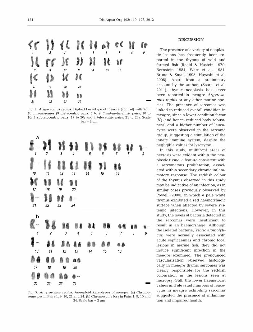
The diploid number of 2n = 48 and the karyotype,which comprised 9 metacentric (Pairs 1 to 9), 7 sub-metacentric (Pairs 10 to 16), 4 subtelocentric (Pairs 17to 20) and 4 telocentric chromosome pairs (Pairs 21 to24), were also established for the first time (Fig. 4). Apercentage of hypodiploid metaphases was observedin chromosomal preparations of the sarcoma; thiscontrasts with preparations of the kidney and spleen,in which all observed metaphases were diploid. Fromanalyses of 6 aneuploid karyotypes, 4 out of the 24chromosome pairs seemed to be more prone to chro-mosome loss (Pairs 1, 9, 10 and 24) (Fig. 5).
123
Fig. 2. Argyrosomus regius. Perpendicular section throughthe sarcoma, showing a layer of hyperplastic epithelium (H)and the underlying neoplastic tissue with a necrotic focus(arrows), haematoxylin and eosin (H&E). Scale bar = 100 µm
Fig. 3. Argyrosomus regius. Section showing prominent vas-cularisation of the neoplasm and the random orientation ofthe cellular architecture, haematoxylin and eosin (H&E).
Scale bar = 50 µm
Dis Aquat Org 102: 119–127, 2012
DISCUSSION
The presence of a variety of neoplas-tic lesions has frequently been re -ported in the thymus of wild andfarmed fish (Roald & Hastein 1979,Bernstein 1984, Warr et al. 1984,Bruno & Smail 1998, Hayashi et al.2008). Apart from a preliminaryaccount by the authors (Soares et al.2011), thymic neoplasia has neverbeen reported in meagre Argyroso-mus regius or any other marine spe-cies. The presence of sarcomas waslinked to reduced overall condition inmeagre, since a lower condition factor(K) (and hence, reduced body robust-ness) and a higher number of leuco-cytes were observed in the sarcomagroup, suggesting a stimulation of theinnate immune system, despite thenegligible values for lysozyme.
In this study, multifocal areas ofnecrosis were evident within the neo-plastic tissue, a feature consistent witha sarcomatous proliferation, associ-ated with a secondary chronic inflam-matory response. The reddish colourof the thymus observed in this studymay be indicative of an infection, as insimilar cases previously observed byPowell (2000), in which a pale whitethymus exhibited a red haemorrhagicsurface when affected by severe sys-temic infections. However, in thisstudy, the levels of bacteria detected inthe sarcomas were insufficient toresult in an haemorrhage. Althoughthe isolated bacteria, Vibrio alginolyti-cus, were normally associated withacute septicaemias and chronic focallesions in marine fish, they did notinduce significant infection in themeagre examined. The pronouncedvascularization observed histologi-cally in meagre thymic sarcomas wasclearly responsible for the reddishcolouration in the lesions seen atnecropsy. Still, the lower haematocritvalues and elevated numbers of leuco-cytes in meagre exhibiting sarcomassuggested the presence of inflamma-tion and impaired health.
124
Fig. 4. Argyrosomus regius. Diploid karyotype of meagre (control) with 2n =48 chromosomes (9 metacentric pairs, 1 to 9; 7 submetacentric pairs, 10 to16; 4 subtelocentric pairs, 17 to 20; and 4 telocentric pairs, 21 to 24). Scale
bar = 2 µm
Fig. 5. Argyrosomus regius. Aneuploid karyotypes of meagre. (a) Chromo-some loss in Pairs 1, 9, 10, 21 and 24. (b) Chromosome loss in Pairs 1, 9, 10 and
24. Scale bar = 2 µm

Soares et al.: Thymus sarcoma in reared juvenile meagre
Normally in teleosts, only a thin layer of mucosalepithelial cells separates the thymus from the exter-nal environment (O’Neil 1989). However, in the cur-rent case, the epithelial layer was hyperplastic andapparently intact, thereby providing additional pro-tection to the underlying tissue. In the current study,as well as in that undertaken by Bruno & Smail(1998), there was no evidence of septicaemia and nobacteria were isolated in other haematopoieticorgans. Moreover, no mortality was associated withthymic sarcomas, and affected fish displayed normalswimming and eating behaviour (Soares et al. 2011).
Elevated lysozyme activity has been recorded inmarine fish species, following infection, environmen-tal, or nutritional stress (Saurabh & Sahoo 2008,Estévez et al. 2011). In the present study, lysozymeactivity was almost absent in the plasma of meagrefrom both sarcoma and control groups. A similarresponse was observed for cod Gadus morhua whenexposed to stressors (Magnadóttir et al. 1999). How-ever, lysozyme activities were observed in the serumof meagre juveniles exposed to nutritional stress(Estévez et al. 2011). The different responses oflysozyme activity in both studies of meagre suggestdifferences in the activity of the biological fluids usedor different mechanisms of response towards differ-ent stressors.
NBT reduction is a simple assay widely used todemonstrate the production of superoxide anion fromphagocytes. The superoxide anion and the hydroxylfree radical are reactive oxygen species, which arehighly bactericidal (Ellis 1999). The fact that bacteriawere only isolated in some individuals from the sar-coma group, increasing the variability within thegroup, likely explains the absence of significant dif-ferences in activated leucocytes between the meagresarcoma and control groups. These findings suggestthat only non-specific activation of immune defencesoccurred in sarcoma-affected meagre.
Poorly differentiated sarcomas represent a signifi-cant challenge to pathologists; consequently, de -scriptions of characteristic chromosomal aberrationsare especially useful in these cases (Bridge 2008).The cytogenetic characterisation of sarcoma cells inthis study revealed a considerable percentage ofaneuploid metaphases (not observed in the otherstudied tissues).
In addition, in karyotypes, some of these meta -phases showed that some chromosomal pairs appearto be more prone to loss than others. Such non- random chromosome losses in aneuploid situationshave previously been observed in humans and plants(Cheng & Murata 2002, Takeuchi et al. 2009). Indeed,
certain chromosome pairs are lost more often thanexpected under the assumption of non-random seg-regation. Moreover, chromosome changes in humancancer cells appear to evolve by non-random lossesand/or gains of particular homologues or groups (e.g.Cigudosa et al. 1998).
However, in order to verify whether the chromoso-mal loss observed in this study does indeed reflectdifferential chromosomal susceptibility and to checkfor structural chromosomal aberrations, the applica-tion of differential chromosomal banding techniquesto aneuploid karyotypes of sarcoma-affected Argyro-somus regius is essential. The precise identificationof characteristic structural and numeric cytogeneticalterations can also guide future molecular studiesinto the nature of the genes involved in the neo -plastic transformation of meagre. Such informationwould be of value in understanding the pathogenesisof similar tumours in other animals, including man.
Acknowledgements. This work was partially supported bythe Project AQUACOR (PROMAR 31-03-05FEP-003) andthe FCT-‘Ciência 2007 program’ and FCT-‘Ciência 2008program’. Support from the UK’s Department for the Envi-ronment, Fisheries and Rural Affairs, Contract FB002 (toS.W.F.) is gratefully acknowledged.
LITERATURE CITED
Anderson DP (2004) Immunostimulants, vaccines, and envi-ronmental stressors in aquaculture: NBT assays to showneutrophil activity by these immunomodulators. In: Suárez LE, Marie DR, López MGN, Villarreal D, ScholzU, González M (eds) Avances en Nutrición Acuícola VII.Mem VII Simp Intern Nutric Acuí, Sonora/México. 16–19November, 2004. Hermosillo, Sonora, p 320–328
Austin B, Stobie M, Robertson PAW, Glass HG, Stark JR,Mudaris M (1993) Vibrio alginolyticus: the cause of gilldisease leading to progressive low-level mortalitiesamong juvenile turbot, Scophthalmus maximus L., in aScottish aquarium. J Fish Dis 16: 277−290
Battalora MS, Hawkins WE, Walker WW, Overstreet RM(1990) Occurrence of thymic lymphoma in carcinogene-sis bioassay specimens of the Japanese medaka (Oryziaslatipes). Cancer Res 50: 5675−5678
Bernstein JW (1984) Leukaemic lymphosarcoma in a hatch-ery-reared rainbow trout, Salmo gairdneri Richardson.J Fish Dis 7: 83−86
Bowden TJ, Butler R, Bricknell IR (2004) Seasonal variationof serum lysozyme levels in Atlantic halibut (Hippoglos-sus hippoglossus L.). Fish Shellfish Immunol 17: 129−135
Bridge JA (2008) Contribution of cytogenetics to the man-agement of poorly differentiated sarcomas. UltrastructPathol 32: 63−71
Bruno DW, Smail DA (1998) Lymphoma in farmed rainbowtrout, Oncorhynchus mykiss (Walbaum): a case study.J Fish Dis 21: 381−386
Buller NB (2004) Bacteria from fish and other aquatic ani-
125

Dis Aquat Org 102: 119–127, 2012
mals: a practical identification manual, 1st edn. CABIPublishing, Wallingford
Castro R, Couso N, Obach A, Lamas J (1999) Effect of differ-ent β-glucans on the respiratory burst of turbot (Psettamaxima) and gilthead seabream (Sparus aurata) phago-cytes. Fish Shellfish Immunol 9: 529−541
Chen HHC, Brittelli MR, Muska CF (1985) Two cases of lym-phosarcoma in channel catfish exposed to N-methyl-N’-nitro-N-nitrosoguanidine. J Natl Cancer Inst 74: 933−939
Cheng ZJ, Murata M (2002) Loss of chromosomes 2R and5RS in octoploid triticale selected for agronomic traits.Genes Genet Syst 77: 23−29
Chilmonczyk S (1982) Rainbow trout lymphoid organs: cel-lular effects of corticosteroids and anti-thymocyte serum.Dev Comp Immunol 6: 271−280
Cigudosa JC, Rao PH, Calasanz MJ, Odero MD, Michaeli J,Jhanwar SC, Chaganti RSK (1998) Characterization ofnonrandom chromosomal gains and losses in multiplemyeloma by comparative genomic hybridization. Blood91: 3007−3010
Cook MT, Hayball P, Hutchinson W, Nowak B, Hayball J(2003) Administration of a commercial immunostimulantpreparation, Eco Ativa™ as a feed supplement enhancesmacrophage respiratory burst and the growth rate ofsnapper (Pagrus auratus, Sparidae (Bloch and Schnei-der)) in winter. Fish Shellfish Immunol 14: 333−345
Costas B, Conceição LE, Aragão C, Martos JA, Ruiz-JaraboI, Mancera JM, Afonso A (2011) Physiological responsesof Senegalese sole (Solea senegalensis Kaup, 1858) afterstress challenge: effects on non-specific immune param-eters, plasma free amino acids and energy metabolism.Aquaculture 316: 68−76
Dawe CJ, Berard CW (1971) Workshop on comparativepathology of haematopoietic and lymphoreticular neo-plasms. J Natl Cancer Inst 47: 1365−1370
Demers NE, Bayne CJ (1997) The immediate effects of stresson hormones and plasma lysozyme in rainbow trout. DevComp Immunol 21: 363−373
Eaton WD, Kent ML (1992) A retrovirus in chinook salmon(Oncorhynchus tshawytscha) in plasmacytoid leukemiaand evidence for the etiology of the disease. Cancer Res52: 6496−6500
Eaton WD, Folkins B, Bagshaw J, Traxler G, Kent ML (1993)Isolation of a retrovirus from two fish cell lines developedfrom chinook salmon (Oncorhynchus tshawytscha) withplasmacytoid leukemia. J Gen Virol 74: 2299−2302
Ellis AE (1990) Lysozyme assays. In: Stolen JS, Fletcher TC,Anderson DP, Roberson BS, van Muiswinkel WB (eds)Techniques in fish immunology. SOS Publications, FairHaven, NJ, p 101−103
Ellis AE (1999) Immunity to bacteria in fish. Fish ShellfishImmunol 9: 291−308
Estévez A, Trevino L, Kotzamanis Y, Karacostas I, Tort L,Gisbert E (2011) Effects of different levels of plant pro-teins on the ongrowing of meagre (Argyrosomus regius)juveniles at low temperatures. Aquacult Nutr 17: E572−E582
Harshbarger JC (1977) Role of the registry of tumors inlower animals in the study of environmental carcinogen-esis in aquatic animals. Ann NY Acad Sci 298: 280−289
Hayashi S, Furukawa S, Obe M, Usuda K, Ogawa I,Miyamoto Y (2008) Lymphoma in Japanese killifish.J Toxicol Pathol 21: 115−117
Holt JG (1994) Bergey’s manual of determinative bacteriol-ogy, 9th edn. Williams & Wilkins, Baltimore, MD
Honma Y, Tamura E (1976) Histological observations on thesporadic diseases found in the branchial region of fishes.Ann Rep Sado Mar Biol Stn Niigata Univ 6: 9−16
Jeney G, Anderson D (1993) Glucan injection or bath expo-sure given alone or in combination with a bacterinenhance the non-specific defence mechanisms in rain-bow trout (Oncorhynchus mykiss). Aquaculture 116: 315−329
Jeney G, Galeotti M, Volpatti D, Jeney Z, Anderson D (1997)Prevention of stress in rainbow trout (Oncorhynchusmykiss) fed diets containing different doses of glucan.Aquaculture 154: 1−15
Johns RH, Reinhardt AK (2009) Association between thy-moma and persistent hypothermia: a case report. J MedCase Rep 3: 73
Kent ML, Dawe SC (1993) Further evidence for a viral etiol-ogy in plasmacytoid leukemia of chinook salmon Onco-rhynchus tshawytscha. Dis Aquat Org 15: 115−121
Kent ML, Groff JM, Traxler GS, Zinkl JG, Bagshaw JW(1990) Plasmacytoid leukemia in seawater reared chi-nook salmon Oncorhynchus tshawytscha. Dis Aquat Org8: 199−209
Lamas J, Santos Y, Bruno D, Toranzo AE, Anadon R (1994) Acomparison of pathological changes caused by Vibrioanguillarum and its extracellular products in rainbowtrout (Oncorhynchus mykiss). Fish Pathol 29: 79−89
Levan A, Fredga K, Sandberg AA (1964) Nomenclature forcentromere position in chromosomes. Hereditas 52: 201−220
Magnadóttir B, Jónsdóttir H, Helgason S, Björnsson B, Jør-gensen T, Pilström L (1999) Humoral immune parametersin Atlantic cod (Gadus morhua L.) II: the effects of sizeand gender under different environmental conditions.Comp Biochem Physiol B 122:181–188
McArdle JF, Roberts RJ (1974) Bilateral hyperplasia of thethymus in rainbow trout (Salmo gairdneri). J Fish ResBoard Can 31: 1537−1539
Mulcahy MF (1970) The thymus glands and lymphosar-coma in the pike Esox zucius L. (Pisces; Esocidae) inIreland. In: Dutcher RM (ed) Comparative leukemiaresearch 1969. Bibl. Haematol. 36, Karger, Basel,p 600−609
Okihiro MS, Hinton DE (1989) Lymphoma in the Japanesemedaka Oryiias latipes. Dis Aquat Org 7: 79−87
O'Neil JG (1989) Thymic development in two species ofmarine teleost; an Antarctic silverfish, Pleuragrammaant-arcticum boulenger, and a warmer-water sea bass,Dicentrarchus labrax (Linnaeus). Proc NIPR Symp PolarBiol 2:83–93
Papas TS, Dahlberg JE, Sonstegard RA (1976) Type C virusin lymphosarcoma in northern pike (Esox lucius). Nature261: 506−508
Powell DB (2000) Immune system. In: Ostrander GK (ed)The laboratory fish. Academic, London, p 441–449
Roald SO, Hastein T (1979) Lymphosarcoma in an Atlanticsalmon Salmo salar L. J Fish Dis 2: 249−251
Sahoo P, Kumari J, Mishra B (2005) Non-specific immuneresponses in juveniles of Indian major carps. J ApplIchthyol 21: 151−155
Saurabh S, Sahoo PK (2008) Lysozyme: an importantdefence molecule of fish innate immune system.Aquacult Res 39: 223−239
Smibert RM, Krieg NR (1981) General characterization. In: Gerhardt P (ed) Manual of methods for general bacteriol-ogy. ASM Press, Washington, DC, p 409−443
126

Soares et al.: Thymus sarcoma in reared juvenile meagre
Soares F, Ribeiro L, Barata M, Pousão-Ferreira P (2011) Sar-coma in the thymus of meagre juveniles (Argyrosomusregius). Bull Eur Assoc Fish Pathol 31: 189–192
Sonstegard RA (1975) Lymphosarcoma in muskellunge(Esox masquinongy). In: Ribelin WE, Migaki G (eds) Thepathology of fishes. The University of Wisconsin Press,Madison, WI, p 907−924
Sonstegard RA (1976) Studies of the etiology and epizootiol-ogy of lymphosarcoma in Esox (Esox lucius L. and Esoxmasquinongy). Prog Exp Tumor Res 20: 141−155
Takeuchi M, Takeuchi K, Ozawa Y, Kohara A, Mizusawa H(2009) Aneuploidy in immortalized human mesenchymalstem cells with non-random loss of chromosome 13 inculture. In Vitro Cell Dev Biol Anim 45: 290−299
Thiriot-Quiévreux C, Ayraud N (1982) Les caryotipes dequelques espèces de bivalves et de gastéropodes marins.Mar Biol 70: 165−172
Thompson JS (1982) An epizootic of lymphoma in northernpike, Esox lucius L., from the Aland Islands of Finland.J Fish Dis 5: 1−11
Tort L, Balasch JC, Mackenzie S (2003) Fish immune system: a crossroads between innate and adaptive responses.Immunología 22: 277−286
Warr GW, Griffin BR, Anderson DP, McAllister PE, Lidgerd-ing B, Smith CE (1984) A lymphosarcoma of thymic ori-gin in the rainbow trout, Salmo gairdneri Richardson.J Fish Dis 7: 73−82
Wolf K (1988) Esocid lymphosarcoma. In: Fish viruses andfish viral diseases. Cornell University Press, Ithaca,NY
Wu SM, Shih MJ, Ho YC (2007) Toxicological stressresponse and cadmium distribution in hybrid tilapia(Oreochromis sp.) upon cadmium exposure. CompBiochem Physiol C Toxicol Pharmacol 145: 218−226
127
Editorial responsibility: Thomas Lang, Cuxhaven, Germany
Submitted: February 20, 2012; Accepted: October 19, 2012Proofs received from author(s): December 14, 2012