![[Ovulation induction by pulsatile GnRH therapy in 2014: literature review and synthesis of current practice]](https://static.fdokumen.com/doc/165x107/6333c99c28cb31ef600d6b7b/ovulation-induction-by-pulsatile-gnrh-therapy-in-2014-literature-review-and-synthesis.jpg)
Effects of thymulin and GnRH on the release of gonadotropins by in vitro pituitary cells obtained...
10
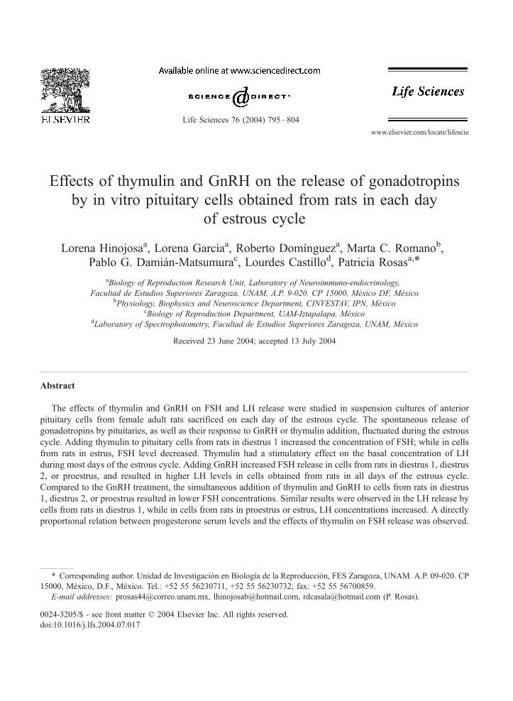
Effects of thymulin and GnRH on the release of gonadotropins by in vitro pituitary cells obtained from rats in each day of estrous cycle Lorena Hinojosa a , Lorena Garcı ´a a , Roberto Domı ´nguez a , Marta C. Romano b , Pablo G. Damia ´n-Matsumura c , Lourdes Castillo d , Patricia Rosas a, * a Biology of Reproduction Research Unit, Laboratory of Neuroimmuno-endocrinology, Facultad de Estudios Superiores Zaragoza, UNAM, A.P. 9-020. CP 15000, Me ´xico DF, Me ´xico b Physiology, Biophysics and Neuroscience Department, CINVESTAV, IPN, Me ´xico c Biology of Reproduction Department, UAM-Iztapalapa, Me ´xico d Laboratory of Spectrophotometry, Facultad de Estudios Superiores Zaragoza, UNAM, Me ´xico Received 23 June 2004; accepted 13 July 2004 Abstract The effects of thymulin and GnRH on FSH and LH release were studied in suspension cultures of anterior pituitary cells from female adult rats sacrificed on each day of the estrous cycle. The spontaneous release of gonadotropins by pituitaries, as well as their response to GnRH or thymulin addition, fluctuated during the estrous cycle. Adding thymulin to pituitary cells from rats in diestrus 1 increased the concentration of FSH; while in cells from rats in estrus, FSH level decreased. Thymulin had a stimulatory effect on the basal concentration of LH during most days of the estrous cycle. Adding GnRH increased FSH release in cells from rats in diestrus 1, diestrus 2, or proestrus, and resulted in higher LH levels in cells obtained from rats in all days of the estrous cycle. Compared to the GnRH treatment, the simultaneous addition of thymulin and GnRH to cells from rats in diestrus 1, diestrus 2, or proestrus resulted in lower FSH concentrations. Similar results were observed in the LH release by cells from rats in diestrus 1, while in cells from rats in proestrus or estrus, LH concentrations increased. A directly proportional relation between progesterone serum levels and the effects of thymulin on FSH release was observed. 0024-3205/$ - see front matter D 2004 Elsevier Inc. All rights reserved. doi:10.1016/j.lfs.2004.07.017 * Corresponding author. Unidad de Investigacio ´ n en Biologı ´a de la Reproduccio ´n, FES Zaragoza, UNAM. A.P. 09-020. CP 15000, Me ´xico, D.F., Me ´xico. Tel.: +52 55 56230711, +52 55 56230732; fax: +52 55 56700859. E-mail addresses: [email protected], [email protected], [email protected] (P. Rosas). Life Sciences 76 (2004) 795 – 804 www.elsevier.com/locate/lifescie
-
Upload
independent -
Category
Documents
-
view
4 -
download
0
Transcript of Effects of thymulin and GnRH on the release of gonadotropins by in vitro pituitary cells obtained...
Life Sciences 76 (2004) 795–804
www.elsevier.com/locate/lifescie
Effects of thymulin and GnRH on the release of gonadotropins
by in vitro pituitary cells obtained from rats in each day
of estrous cycle
Lorena Hinojosaa, Lorena Garcıaa, Roberto Domıngueza, Marta C. Romanob,
Pablo G. Damian-Matsumurac, Lourdes Castillod, Patricia Rosasa,*
aBiology of Reproduction Research Unit, Laboratory of Neuroimmuno-endocrinology,
Facultad de Estudios Superiores Zaragoza, UNAM, A.P. 9-020. CP 15000, Mexico DF, MexicobPhysiology, Biophysics and Neuroscience Department, CINVESTAV, IPN, Mexico
cBiology of Reproduction Department, UAM-Iztapalapa, MexicodLaboratory of Spectrophotometry, Facultad de Estudios Superiores Zaragoza, UNAM, Mexico
Received 23 June 2004; accepted 13 July 2004
Abstract
The effects of thymulin and GnRH on FSH and LH release were studied in suspension cultures of anterior
pituitary cells from female adult rats sacrificed on each day of the estrous cycle. The spontaneous release of
gonadotropins by pituitaries, as well as their response to GnRH or thymulin addition, fluctuated during the estrous
cycle. Adding thymulin to pituitary cells from rats in diestrus 1 increased the concentration of FSH; while in cells
from rats in estrus, FSH level decreased. Thymulin had a stimulatory effect on the basal concentration of LH
during most days of the estrous cycle. Adding GnRH increased FSH release in cells from rats in diestrus 1, diestrus
2, or proestrus, and resulted in higher LH levels in cells obtained from rats in all days of the estrous cycle.
Compared to the GnRH treatment, the simultaneous addition of thymulin and GnRH to cells from rats in diestrus
1, diestrus 2, or proestrus resulted in lower FSH concentrations. Similar results were observed in the LH release by
cells from rats in diestrus 1, while in cells from rats in proestrus or estrus, LH concentrations increased. A directly
proportional relation between progesterone serum levels and the effects of thymulin on FSH release was observed.
0024-3205/$ -
doi:10.1016/j.lf
* Correspon
15000, Mexico
E-mail addr
see front matter D 2004 Elsevier Inc. All rights reserved.
s.2004.07.017
ding author. Unidad de Investigacion en Biologıa de la Reproduccion, FES Zaragoza, UNAM. A.P. 09-020. CP
, D.F., Mexico. Tel.: +52 55 56230711, +52 55 56230732; fax: +52 55 56700859.
esses: [email protected], [email protected], [email protected] (P. Rosas).

L. Hinojosa et al. / Life Sciences 76 (2004) 795–804796
These data suggest that thymulin plays a dual role in the release of gonadotropins, and that its effects depend on the
hormonal status of the donor’s pituitary.
D 2004 Elsevier Inc. All rights reserved.
Keywords: Thymulin; Pituitary cells; FSH; LH; GnRH; Estrous cycle
Introduction
Based on published results on the effects of neonatal or infantile thymectomy (Besedovsky and
Sorkin, 1974; Michael et al., 1980; Kosiewicz and Michael, 1990; Garcıa et al., 2000), the congenital
absence of the thymus (Rebar et al., 1981b), and the effects of thymic peptides on in vivo and in vitro
models (Rebar et al., 1981a; Michael, 1983; Mendoza and Romano, 1989; Mendoza et al., 1995;
Hinojosa et al., 1999), the participation of the thymus in regulating the hypothalamic-pituitary-ovary axis
is widely accepted.
Thymulin is a nonapeptide (pyro-Glu-Ala-Lys-Ser-Gln-Gly-Gly-Ser-Asn-Oh) exclusively synthesized
by the thymic epithelium, since it is not detected in athymic nude mice (nu/nu) or in thymectomized
mice. In both models, grafting the thymus or epithelial thymic cells results in the detection of thymulin in
serum (Bach et al., 1977; Dardenne et al., 1984; Safieh et al., 1990). Injecting equine chorionic
gonadotropin (eCG) to 20-day old mice does not induce ovulation; however, thymulin treatment before
eCG injection results in an increase of both, ovulation rate and ovarian weight (Hinojosa et al., 1999).
Infantile thymectomy in mice results in lower serum level of estrogen during the pre-pubertal phase and
in a lower ovulatory response to gonadotropin stimulus; in turn, injecting thymulin immediately after
surgery resulted in normal estrogen level and normal ovulation rates (Garcıa et al., 2000).
In vitro studies show that in both, male and female gonads, thymulin modulates steroidogenesis
(Ledwitz-Rigby and Scheid, 1990; Wise, 1998). In in vitro conditions, the addition of thymulin to
pituitary tissue obtained from adult male rats stimulates the release of luteinizing hormone (LH) in a
dose-related way; similar to the responses obtained by adding gonadotropin-releasing hormone (GnRH)
(Zaidi et al., 1988; Hadley et al., 1997). Comparable results were observed on the release of follicle
stimulating hormone (FSH) and LH from perifusates pituitary cells, or stationary cell cultures obtained
from female rats (Brown et al., 2000).
Taken together, the results reported to date suggest that in in vitro conditions, thymulin stimulates the
release of FSH and LH. However, previous studies have been performed without considering the
plausible hormonal influences arising from the different days of the estrous cycle. The present study was
designed to establish if thymulin’s effects on FSH and LH release depend on the hormonal status of the
pituitary donor.
Materials and methods
Animals
A total of 160-adult (3 months old) female Wistar rats were used in this study. Animals were housed
under controlled light conditions (light on from 05:00 to 19:00 h) and had free access to food and water.
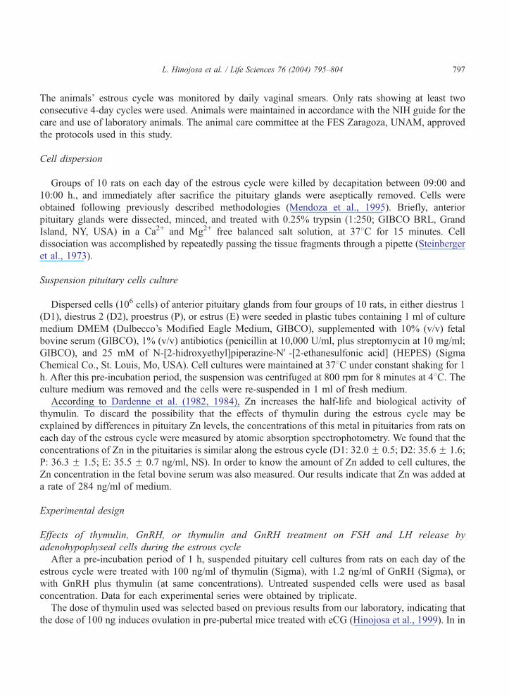
L. Hinojosa et al. / Life Sciences 76 (2004) 795–804 797
The animals’ estrous cycle was monitored by daily vaginal smears. Only rats showing at least two
consecutive 4-day cycles were used. Animals were maintained in accordance with the NIH guide for the
care and use of laboratory animals. The animal care committee at the FES Zaragoza, UNAM, approved
the protocols used in this study.
Cell dispersion
Groups of 10 rats on each day of the estrous cycle were killed by decapitation between 09:00 and
10:00 h., and immediately after sacrifice the pituitary glands were aseptically removed. Cells were
obtained following previously described methodologies (Mendoza et al., 1995). Briefly, anterior
pituitary glands were dissected, minced, and treated with 0.25% trypsin (1:250; GIBCO BRL, Grand
Island, NY, USA) in a Ca2+ and Mg2+ free balanced salt solution, at 378C for 15 minutes. Cell
dissociation was accomplished by repeatedly passing the tissue fragments through a pipette (Steinberger
et al., 1973).
Suspension pituitary cells culture
Dispersed cells (106 cells) of anterior pituitary glands from four groups of 10 rats, in either diestrus 1
(D1), diestrus 2 (D2), proestrus (P), or estrus (E) were seeded in plastic tubes containing 1 ml of culture
medium DMEM (Dulbecco’s Modified Eagle Medium, GIBCO), supplemented with 10% (v/v) fetal
bovine serum (GIBCO), 1% (v/v) antibiotics (penicillin at 10,000 U/ml, plus streptomycin at 10 mg/ml;
GIBCO), and 25 mM of N-[2-hidroxyethyl]piperazine-NV-[2-ethanesulfonic acid] (HEPES) (Sigma
Chemical Co., St. Louis, Mo, USA). Cell cultures were maintained at 378C under constant shaking for 1
h. After this pre-incubation period, the suspension was centrifuged at 800 rpm for 8 minutes at 48C. Theculture medium was removed and the cells were re-suspended in 1 ml of fresh medium.
According to Dardenne et al. (1982, 1984), Zn increases the half-life and biological activity of
thymulin. To discard the possibility that the effects of thymulin during the estrous cycle may be
explained by differences in pituitary Zn levels, the concentrations of this metal in pituitaries from rats on
each day of the estrous cycle were measured by atomic absorption spectrophotometry. We found that the
concentrations of Zn in the pituitaries is similar along the estrous cycle (D1: 32.0F 0.5; D2: 35.6 F 1.6;
P: 36.3 F 1.5; E: 35.5 F 0.7 ng/ml, NS). In order to know the amount of Zn added to cell cultures, the
Zn concentration in the fetal bovine serum was also measured. Our results indicate that Zn was added at
a rate of 284 ng/ml of medium.
Experimental design
Effects of thymulin, GnRH, or thymulin and GnRH treatment on FSH and LH release by
adenohypophyseal cells during the estrous cycle
After a pre-incubation period of 1 h, suspended pituitary cell cultures from rats on each day of the
estrous cycle were treated with 100 ng/ml of thymulin (Sigma), with 1.2 ng/ml of GnRH (Sigma), or
with GnRH plus thymulin (at same concentrations). Untreated suspended cells were used as basal
concentration. Data for each experimental series were obtained by triplicate.
The dose of thymulin used was selected based on previous results from our laboratory, indicating that
the dose of 100 ng induces ovulation in pre-pubertal mice treated with eCG (Hinojosa et al., 1999). In in
L. Hinojosa et al. / Life Sciences 76 (2004) 795–804798
vitro conditions the dose of GnRH used, 1.2 ng/ml, stimulates the release of FSH and LH (Mendoza and
Romano, 1989; Mendoza et al., 1995; Hinojosa et al., 2002).
All cell cultures were maintained at 378C under constant shaking for 1 h. After incubation, the
suspension was centrifuged at 800 rpm for 8 minutes at 48C. Supernatants were collected and frozen at
�208C, until FSH and LH were measured by radioimmunoassay (RIA).
Radioimmunoassay
The blood from the trunk from six randomly selected animals on each day of the estrous cycle was
collected, allowed to coagulate at room temperature for 1 h, and centrifuged at 3000 rpm. The serum was
kept at –208C, until FSH, LH, progesterone, and 17h-estradiol concentrations were measured by RIA.
FSH and LH levels in serum and medium were measured by the double antibody RIA technique,
employing reagents and protocols kindly supplied by the NIADDK National Pituitary Program
(Bethesda, MD, USA). Intra- and inter-assay variations were in the order of 5.1% and 6.5% for LH, and
4% and 7.9% for FSH. All samples were assayed in duplicates. The results are expressed in terms of
NIADDK standards RP-2.
Serum concentrations of progesterone and 17h-estradiol were measured by RIA using the solid-phase
iodine-125 technique (Kubasik et al., 1984), with kits purchased from Coat-A-Count, following the
methodology described by Diagnostic Products Corporation (Los Angeles, CA, USA). This procedure’s
sensitivity to measure progesterone and 17h-estradiol was 0.02 ng/ml and 8 pg/ml, respectively. Intra-
and inter-assay coefficients of variation for progesterone were 5.3% and 9.8%, respectively; and for 17h-estradiol, 6.9% and 10.8%, respectively. All samples were assayed in duplicates.
Statistical analysis
Hormone levels in serum were analyzed by ANOVA followed by Tukey’s test. Gonadotropins
concentration in the culture medium was analyzed by Kruskal-Wallis ANOVA test, followed by Dunn’s
multiple comparisons test. Differences between two groups were compared by Mann-Whitney U-test. A
probability of less than 5% was considered significant. The correlation between progesterone and
estradiol serum concentrations and the effects of thymulin or GnRH or GnRH + thymulin on FSH and
LH release by pituitary cells from rats on each day of the estrous cycle was analyzed. A correlation (r2)
was considered significant if greater than 0.85.
Results
Gonadotropins and steroid serum levels
During the morning, FSH serum levels were higher in P and E than in D2. LH concentrations were
similar during the estrous cycle. In D2 and P, progesterone serum levels were lower than in D1, while the
highest estradiol serum level was observed in animals sacrificed on P (Table 1).
Basal and stimulated levels of FSH and LH
Anterior pituitary cells from rats in E released more FSH than cells obtained in other days of the estrous
cycle. GnRH treatment increased FSH release in pituitary cells from rats in D1, D2, and P (Table 2).

Table 1
Mean F s.e.m. of FSH, LH, progesterone and 17h-estradiol serum concentations from rats in the morning of each day of
estrous cycle
Group n FSH (ng/ml) LH (ng/ml) Progesterone (ng/ml) 17h-estradiol (pg/ml)
Diestrus 1 6 5.6 F 0.1 0.5 F 0.1 16.4 F 2.0 39.7 F 1.4
Diestrus 2 6 5.1 F 0.4 0.5 F 0.0 9.4 F 1.6* 46.0 F 2.5
Proestrus 6 7.5 F 0.7& 0.7 F 0.1 8.5 F 1.5* 53.2 F 3.1*
Estrus 6 7.1 F 0.5& 0.6 F 0.2 10.5 F 1.7 38.8 F 1.6%
* p b 0.05 vs. diestrus 1 group (ANOVA followed by Tukey’s test).& p b 0.05 vs. diestrus 2 group (ANOVA followed by Tukey’s test).% p b 0.05 vs. proestrus group (ANOVA followed by Tukey’s test).
L. Hinojosa et al. / Life Sciences 76 (2004) 795–804 799
The basal release of LH by pituitary cells from rats in P was higher than in cells from rats in D1 or D2,
while the concentration observed in E was lower than in D1 (Table 2).
GnRH addition to pituitary cell cultures resulted in an increase of LH release in all days of the estrous
cycle and the highest influence on LH release was observed in cells collected on D2 and P (Table 2).
Effects of thymulin or thymulin and GnRH treatment on FSH and LH release by adenohypophyseal cells
during the estrous cycle
The addition of thymulin to pituitaries from rats in D1 resulted in a significant increase of FSH
release, while an opposite effect was observed in pituitaries from rats in E (Fig. 1A).
Compared to GnRH stimulated cells, adding thymulin and GnRH to pituitaries from rats in D1, D2, or
P resulted in lower FSH concentrations (Fig. 1B).
Thymulin addition to cells from rats in D1, D2, or E resulted in LH concentrations increase, but not in
cells obtained in P (Fig. 2A). Compared to GnRH treated cells, adding thymulin and GnRH to pituitary
cells from rats in D1 resulted in lower LH release; while a significant increase in the release of this
hormone was observed in cells from rats in P or E (Fig. 2B).
The following correlation coefficients (r2) were calculated: Progesterone serum levels and FSH
release stimulated by thymulin, the r2 = 0.90; progesterone serum levels and LH release stimulated
Table 2
Mean F s.e.m. of the FSH and LH release by anterior pituitary cells obtained from rats on each day of estrous cycle
Group FSH (ng/ml) LH (ng/ml)
n Basal n GnRH n Basal n GnRH
Diestrus 1 12 15.6 F 1.6 12 29.4 F 2.1e 12 75.5 F 3.4 12 92.9 F 3.3e
Diestrus 2 12 19.2 F 2.1 12 36.5 F 4.6e 12 90.2 F 5.5 12 138.8 F 4.0*,e
Proestrus 12 23.9 F 1.2 12 39.4 F 3.6e 12 130.0 F 10.9*,& 12 148.1 F 5.5*,e
Estrus 12 43.9 F 3.1*,&,% 12 32.4 F 3.7 12 96.4 F 3.9* 12 123.4 F 12.2e
After a 1 h pre-incubation, cells were incubated for 1 h without treatment (basal) or with 1.2 ng/ml of GnRH.
* p b 0.05 vs. diestrus 1 group (Kruskal-Wallis followed by Dunn’s test).& p b 0.05 vs. diestrus 2 group (Kruskal-Wallis followed by Dunn’s test).% p b 0.05 vs. proestrus group (Kruskal-Wallis followed by Dunn’s test).e p b 0.05 vs. basal group (Mann-Whitney U-test).

Fig. 1. Mean F s.e.m. of FSH release by anterior pituitary cells obtained from rats on each day of estrous cycle. Cells were pre-
incubated for 1 h. The culture medium was removed and the cells were re-suspended in fresh medium [basal], treated with
thymulin (100 ng/ml) [panel A], with GnRH (1.2 ng/ml), or with GnRH + thymulin [panel B]. The cells were incubated for 1 h.
Each bar represents four experimental series and three replicates for each series (n = 12). *p b 0.05 vs. basal group (Mann-
Whitney U-test); w p b 0.05 vs. GnRH group (Mann-Whitney U-test).
L. Hinojosa et al. / Life Sciences 76 (2004) 795–804800
by GnRH, the r2 = 0.96; and between estradiol serum levels and FSH release stimulated by GnRH,
the r2 = 0.86.
Discussion
Present results indicate that thymulin’s effect on FSH and LH release by anterior pituitary cells from
female rats varies during the estrous cycle, suggesting that the influence of thymulin depends on the
hormonal status of the donor’s pituitary.
Haisenleder et al. (1994) showed that the mRNA concentration for FSHh and LHh vary during the
estrous cycle, and established that LHh mRNA levels show two peaks, one at 08:00 of D2, and a second
at 17:00 of P. In turn, FSHh mRNA levels show a single peak at 02:00 on the day of E. Such changes in
mRNAs concentrations are not totally parallel with FSH and LH serum concentrations changes.
The present study evaluates the changes in the gonadotropes’ ability to release hormones, as no time
for new hormone synthesis was allowed. The pattern of FSH and LH release to the blood does not
parallel to the spontaneous release of these hormones to the culture medium after 1 h of incubation (in

Fig. 2. Mean F s.e.m. of LH release by anterior pituitary cells obtained from rats on each day of estrous cycle. Cells were pre-
incubated for 1 h. The culture medium was removed and the cells were re-suspended in fresh medium [basal], treated with
thymulin (100 ng/ml) [panel A], with GnRH (1.2 ng/ml), or with GnRH+thymulin [panel B]. The cells were incubated for 1 h.
Each bar represents four experimental series and three replicates for each series (n = 12). *p b 0.05 vs. basal group (Mann-
Whitney U-test); w p b 0.05 vs. GnRH group (Mann-Whitney U-test).
L. Hinojosa et al. / Life Sciences 76 (2004) 795–804 801
serum, the concentration of FSH is 10 fold higher than LH, while in the culture medium LH values are
2–5 fold higher).
These results could be explained by three distinct, non-exclusive, ways: the lack of gonadal hormones
regulating gonadotropes secretion activity (Kamel et al., 1987; Fallest and Schwartz, 1991; Blake, 1998;
Fowler et al., 2003); the existence of hypothalamic inhibiting factors (Hwan and Freeman, 1987; De la
Lastra and Leal, 1989; Blake, 1998); or an intrinsic regulatory system acting through autocrine and
paracrine signals (Kitaoka et al., 1988; Bohnsack et al., 2000). It is also possible that the differences in
gonadotropins secretion observed in in vitro studies reflect the presence of factors secreted by pituitary
that directly inhibiting basal FSH secretion, such as inhibin (Schwall, 1998).
It is well established that an ordered sequence of hormonal events occur throughout the estrous cycle,
as the pituitary gland is cyclically exposed to diverse feedbacks mechanisms regulating gonadotropin
secretion (Blake, 1998; Bohnsack et al., 2000). During D2 and P, higher levels of GnRH receptors in
pituitary cells are found (Clayton et al., 1980; Savoy-Moore et al., 1980; Marian et al., 1981). The results
obtained in the present study are consistent with the above-mentioned results, since LH release to the
culture medium by cells in D2 and P reached its highest values. Then, the effects of GnRH on pituitary
cells’ release of FSH or LH depends on the priming conditions of the hormonal milieu characteristic of

L. Hinojosa et al. / Life Sciences 76 (2004) 795–804802
each day of the estrous cycle. This idea is supported by the results obtained in the present study: a
directly proportional relation between estradiol serum levels and GnRH effects on FSH release; and a
proportionally inverse relation between progesterone serum levels and LH release in GnRH stimulated
pituitaries.
Our results suggest that thymulin has a dual effect on gonadotropins release. For LH release, the effect
was stimulatory during the estrous cycle; and for FSH release, the effects were stimulatory in D1 and
inhibitory in E. Because the correlation analysis showed a directly proportional relation between
progesterone serum levels and the effects of thymulin on FSH release, we suggest that progesterone
modulates the cells’ reactivity to thymulin.
Brown et al. (2000) showed that in pituitary cells from female rats (regardless of the estrous cycle day
they were in) incubated for 40 min, thymulin stimulates the release of FSH and LH; and that thymulin
plus GnRH treatment has a synergistic effect on LH release, and an additive effect on FSH release. In the
study, Brown et al. suggest that thymulin and GnRH do no share proximal steps of transduction
pathways. Present results show that when GnRH and thymulin coexist in the culture medium, the action
of thymulin can either inhibit or stimulate GnRH effects, depending on which day of the estrous cycle
the pituitary donor is in. This suggests that thymulin is capable of regulating the response of
gonadotropes to GnRH on gonadotropin secretion in different ways.
Interactions between the nervous, immune, and endocrine systems require a complex communi-
cation network. This network is composed of many hormones, including thymulin, that participate as
messengers between the brain, pituitary, peripheral nervous system, immune cells, and gonads. Taken
together, the variability between GnRH and thymulin interactions during the estrous cycle on FSH and
LH release suggests that the effects of thymulin are influenced by paracrine interactions, known to
vary during the estrous cycle, and that thymulin acts as a fine modulator of GnRH actions on
gonadotropes.
Acknowledgments
The authors wish to acknowledge the technical assistance expertise of Mario A. Altamirano Lozano
PhD. and Carolina Miranda Brito. We would also like to thank the statistical advise of Isaıas H. Salgado
Ugarte PhD, J. Miguel Betancourt Rule PhD, and Xavier Chiappa Carrara PhD; and Alvaro Domınguez
Gonzalez M.Sc for the English revision.
This work was supported by DGAPA-PAPIIT IN217301, PAEP 101312, 101313 and CONACyT.
References
Bach, J.-F., Dardenne, M., Pleau, J.M., 1977. Biochemical characterisation of a serum thymic factor. Nature 266 (3), 55–57.
Besedovsky, H.O., Sorkin, E., 1974. Thymus involvement in female sexual maturation. Nature 249, 356–358.
Blake, C.A., 1998. Gonadotropin secretion, control of. In: Knobil, E., Neill, J.D. (Eds.), Encyclopedia of Reproduction.
Academic Press, San Diego, pp. 528–552.
Bohnsack, B.L., Szabo, M., Kilen, S.M., Tam, D.H.Y., Schwartz, N.B., 2000. Follistatin suppresses steroid-enhanced follicle-
stimulating hormone release in vitro in rats. Biology of Reproduction 62 (3), 636–641.
Brown, O.A., Sosa, Y.E., Dardenne, M., Pleau, J.M., Goya, R.G., 2000. Studies on the gonadotropin-releasing activity of
thymulin: changes with age. Journal of Gerontology: Biological Sciences 55A (4), B170–B176.

L. Hinojosa et al. / Life Sciences 76 (2004) 795–804 803
Clayton, R.N., Solano, A.R., Garcia-Vela, A., Dufau, M.L., Catt, K.J., 1980. Regulation of pituitary receptors for gonadotropin-
releasing hormone during the rat estrous cycle. Endocrinology 107 (3), 699–706.
Dardenne, M., Pleau, J.M., Nabarra, B., Lefrancier, P., Derrien, M., Choay, J., Bach, J.F., 1982. Contribution of zinc and other
metals to the biological activity of the serum thymic factor. Proceedings of the National Academy of Sciences USA 79,
5370–5373.
Dardenne, M., Savino, W., Gastinel, L., Bach, J.F., 1984. Thymulin. New biochemical aspects. In: Goldstein, A.L. (Ed.),
Thymic Hormones and Lymphokines. Basic Chemistry and Clinical Applications. Plenum Press, New York, pp. 37–42.
De la Lastra, M., Leal, J., 1989. Factor hipotalamico inhibidor de la secrecion de hormona luteinizante: relacion con fragmentos
1–5 de hormona liberadora de LH. Archivos de Biologıa y Medicina Experimentales 22 (1), 53–59.
Fallest, P.C., Schwartz, N.B., 1991. Acute inhibitory effects of 17h-estradiol are observed on gonadotropin secretion from
perifused pituitary fragments of metestrous, but not proestrous, rats. Endocrinology 128 (1), 273–279.
Fowler, P.A., Sorsa-Leslie, T., Harris, W., Mason, H.D., 2003. Ovarian gonadotrophin surge-attenuating factor (GnSAF): where
are we after 20 years of research? Reproduction 126, 689–699.
Garcıa, L., Hinojosa, L., Domınguez, R., Chavira, R., Rosas, P., 2000. Effects of infantile thymectomy on ovarian functions and
gonadotrophin-induced ovulation in prepubertal mice: role of thymulin. Journal of Endocrinology 166 (2), 381–387.
Hadley, A.J., Rantle, C.M., Buckingham, J.C., 1997. Thymulin stimulates corticotrophin release and cyclic nucleotide
formation in the rat anterior pituitary gland. Neuroimmunomodulation 4 (2), 62–69.
Haisenleder, D.J., Dalkin, A.C., Marshall, J.C., 1994. Regulation of gonadotropin gene expression. In: Knobil, E., Neill, J.D.
(Eds.), The Physiology of Reproduction. Raven Press, New York, pp. 1793–1813.
Hinojosa, L., Chavira, R., Domınguez, R., Rosas, P., 1999. Effects of thymulin on spontaneous puberty and gonadotrophin-
induced ovulation in prepubertal normal and hypothymic mice. Journal of Endocrinology 163 (2), 255–260.
Hinojosa, L., Garcıa, L., Quiroz, U., Chavira, R., Romano, M.C., Domınguez, R., Rosas, P., 2002. Sex differences in FSH and
LH secretion by the pituitary treated in vitro with thymulin. The Thirty Fifth Annual Meeting of the Society for the Study of
Reproduction, Baltimore, Maryland, July 28–31, 2002. Biology of Reproduction 66 (272), pp. 306–307.
Hwan, J.C., Freeman, M.E., 1987. A physiological role for luteinizing hormone release-inhibiting factor of hypothalamic origin.
Endocrinology 121 (3), 1099–1103.
Kamel, F., Balz, J.A., Kubajak, C.L., Schneider, V.A., 1987. Gonadal steroids modulate pulsatile luteinizing hormone secretion
by perifused rat anterior pituitary cells. Endocrinology 120 (4), 1651–1657.
Kitaoka, M., Kojima, I., Ogata, E., 1988. Activin-A: a modulator of multiple types of anterior pituitary cells. Biochemical and
Biophysical Research Communications 157 (1), 48–54.
Kosiewicz, M.M., Michael, S.D., 1990. Neonatal thymectomy affects follicle populations before the onset of autoimmune
oophoritis in B6A mice. Journals of Reproduction and Fertility 88 (2), 427–440.
Kubasik, N.P., Hallauer, G.D., Brodows, R.G., 1984. Evaluation of a direct solid-phase radioimmunoassay for progesterone,
useful for monitoring luteal funtion. Clinical Chemical 30, 284–286.
Ledwitz-Rigby, F., Scheid, P.G., 1990. Thymulin (serum thymic factor) modulation of porcine granulosa cell responsiveness to
gonadotropins in vitro. The Eight Ovarian Workshop. Regulatory Process and Gene Expression in the Ovary, Maryville,
Tennessee, July 12–14, pp. 473–478.
Marian, J., Cooper, R.L., Conn, P.M., 1981. Regulation of the rat pituitary gonadotropin-releasing hormone receptor. Molecular
Pharmacology 19 (3), 339–405.
Mendoza, M.E., Romano, M.C., 1989. Prepubertal rat thymus secretes a factor that modulates gonadotropin secretion in
cultured rat pituitary cells. Thymus 14 (4), 233–242.
Mendoza, M.E., Martin, D., Candelaria, P.G., Romano, M.C., 1995. Evidence that secretory products of the reticulo-epithelial
cells of the rat thymus modulate the secretion of gonadotrophins by rat pituitary cells in culture. Journal of Reproductive
Immunology 28 (3), 203–215.
Michael, S.D., 1983. Interactions of the thymus and the ovary. In: Greenwald, G.S., Terranova, P.F. (Eds.), Factors Regulating
Ovarian Function. Raven Press, New York, pp. 445–464.
Michael, S.D., Taguchi, O., Nishizuka, Y., 1980. Effect of neonatal thymectomy on ovarian development and plasma LH, FSH,
GH and PRL in the mouse. Biology of Reproduction 22 (2), 343–350.
Rebar, R.W., Miyake, A., Low, T.L.K., Goldstein, A.L., 1981a. Thymosin stimulates secretion of luteinizing hormone-releasing
factor. Science 214 (6), 669–671.
Rebar, R.W., Morandini, I.C., Erickson, G.F., Petze, J.E., 1981b. The hormonal basis of reproductive defects in athymic mice:
diminished gonadotropin concentrations in prepubertal females. Endocrinology 108 (1), 120–126.

L. Hinojosa et al. / Life Sciences 76 (2004) 795–804804
Safieh, B., Kendall, M.D., Norman, J.C., Metreau, E., Dardenne, M., Bach, J.F., Pleau, J.M., 1990. A new radioimmunoassay
for the thymic peptide thymulin, and its application for measuring thymulin in blood samples. Journal of Immunological
Methods 127 (2), 255–262.
Savoy-Moore, R.T., Schwartz, N.B., Duncan, J.A., Marshall, J.C., 1980. Pituitary gonadotropin-releasing hormone receptors
during the rat estrous cycle. Science 209 (4459), 942–944.
Schwall, R.H., 1998. Inhibin. In: Knobil, E., Neill, J.D. (Eds.), Encyclopedia of Reproduction. Academic Press, San Diego,
pp. 832–839.
Steinberger, A., Chrowdhury, M., Steinberger, E., 1973. Effect of repeated replenishment of hypothalamic extract on LH and
FSH secretion in monolayer cultures of rat anterior pituitary cells. Endocrinology 92 (1), 12–17.
Wise, T., 1998. In vitro and in vivo effects of thymulin on rat testicular steroid synthesis. The Journal of Steroid Biochemistry
and Molecular Biology 66 (3), 129–135.
Zaidi, S.A.A., Kendall, M.D., Gillham, B., Jones, M.T., 1988. The release of luteinizing hormone from pituitaries perifused
with thymic extracts. Thymus 12 (4), 253–264.