Targeted Expression of a Dominant-Negative Fibroblast Growth Factor (FGF) Receptor in...
12
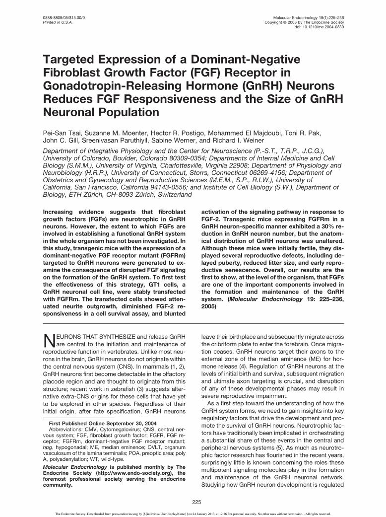
Targeted Expression of a Dominant-Negative Fibroblast Growth Factor (FGF) Receptor in Gonadotropin-Releasing Hormone (GnRH) Neurons Reduces FGF Responsiveness and the Size of GnRH Neuronal Population Pei-San Tsai, Suzanne M. Moenter, Hector R. Postigo, Mohammed El Majdoubi, Toni R. Pak, John C. Gill, Sreenivasan Paruthiyil, Sabine Werner, and Richard I. Weiner Department of Integrative Physiology and the Center for Neuroscience (P.-S.T., T.R.P., J.C.G.), University of Colorado, Boulder, Colorado 80309-0354; Departments of Internal Medicine and Cell Biology (S.M.M.), University of Virginia, Charlottesville, Virginia 22908; Department of Physiology and Neurobiology (H.R.P.), University of Connecticut, Storrs, Connecticut 06269-4156; Department of Obstetrics and Gynecology and Reproductive Sciences (M.E.M., S.P., R.I.W.), University of California, San Francisco, California 94143-0556; and Institute of Cell Biology (S.W.), Department of Biology, ETH Zu ¨ rich, CH-8093 Zu ¨ rich, Switzerland Increasing evidence suggests that fibroblast growth factors (FGFs) are neurotrophic in GnRH neurons. However, the extent to which FGFs are involved in establishing a functional GnRH system in the whole organism has not been investigated. In this study, transgenic mice with the expression of a dominant-negative FGF receptor mutant (FGFRm) targeted to GnRH neurons were generated to ex- amine the consequence of disrupted FGF signaling on the formation of the GnRH system. To first test the effectiveness of this strategy, GT1 cells, a GnRH neuronal cell line, were stably transfected with FGFRm. The transfected cells showed atten- uated neurite outgrowth, diminished FGF-2 re- sponsiveness in a cell survival assay, and blunted activation of the signaling pathway in response to FGF-2. Transgenic mice expressing FGFRm in a GnRH neuron-specific manner exhibited a 30% re- duction in GnRH neuron number, but the anatom- ical distribution of GnRH neurons was unaltered. Although these mice were initially fertile, they dis- played several reproductive defects, including de- layed puberty, reduced litter size, and early repro- ductive senescence. Overall, our results are the first to show, at the level of the organism, that FGFs are one of the important components involved in the formation and maintenance of the GnRH system. (Molecular Endocrinology 19: 225–236, 2005) N EURONS THAT SYNTHESIZE and release GnRH are central to the initiation and maintenance of reproductive function in vertebrates. Unlike most neu- rons in the brain, GnRH neurons do not originate within the central nervous system (CNS). In mammals (1, 2), GnRH neurons first become detectable in the olfactory placode region and are thought to originate from this structure; recent work in zebrafish (3) suggests alter- native extra-CNS origins for these cells that have yet to be explored in other species. Regardless of their initial origin, after fate specification, GnRH neurons leave their birthplace and subsequently migrate across the cribriform plate to enter the forebrain. Once migra- tion ceases, GnRH neurons target their axons to the external zone of the median eminence (ME) for hor- mone release (4). Regulation of GnRH neurons at the levels of initial birth and survival, subsequent migration and ultimate axon targeting is crucial, and disruption of any of these developmental phases may result in severe reproductive impairment. As a first step toward the understanding of how the GnRH system forms, we need to gain insights into key regulatory factors that drive the development and pro- mote the survival of GnRH neurons. Neurotrophic fac- tors have traditionally been implicated in orchestrating a substantial share of these events in the central and peripheral nervous systems (5). As much as neurotro- phic factor research has flourished in the recent years, surprisingly little is known concerning the roles these multipotent signaling molecules play in the formation and maintenance of the GnRH neuronal network. Studying how GnRH neuron development is regulated First Published Online September 30, 2004 Abbreviations: CMV, Cytomegalovirus; CNS, central ner- vous system; FGF, fibroblast growth factor; FGFR, FGF re- ceptor; FGFRm, dominant-negative FGF receptor mutant; hpg, hypogonadal; ME, median eminence; OVLT, organum vasculosum of the lamina terminalis; POA, preoptic area; poly A, polyadenylation; WT, wild-type. Molecular Endocrinology is published monthly by The Endocrine Society (http://www.endo-society.org), the foremost professional society serving the endocrine community. 0888-8809/05/$15.00/0 Molecular Endocrinology 19(1):225–236 Printed in U.S.A. Copyright © 2005 by The Endocrine Society doi: 10.1210/me.2004-0330 225 The Endocrine Society. Downloaded from press.endocrine.org by [${individualUser.displayName}] on 24 January 2015. at 12:26 For personal use only. No other uses without permission. . All rights reserved.
Transcript of Targeted Expression of a Dominant-Negative Fibroblast Growth Factor (FGF) Receptor in...
Targeted Expression of a Dominant-NegativeFibroblast Growth Factor (FGF) Receptor inGonadotropin-Releasing Hormone (GnRH) NeuronsReduces FGF Responsiveness and the Size of GnRHNeuronal Population
Pei-San Tsai, Suzanne M. Moenter, Hector R. Postigo, Mohammed El Majdoubi, Toni R. Pak,John C. Gill, Sreenivasan Paruthiyil, Sabine Werner, and Richard I. Weiner
Department of Integrative Physiology and the Center for Neuroscience (P.-S.T., T.R.P., J.C.G.),University of Colorado, Boulder, Colorado 80309-0354; Departments of Internal Medicine and CellBiology (S.M.M.), University of Virginia, Charlottesville, Virginia 22908; Department of Physiology andNeurobiology (H.R.P.), University of Connecticut, Storrs, Connecticut 06269-4156; Department ofObstetrics and Gynecology and Reproductive Sciences (M.E.M., S.P., R.I.W.), University ofCalifornia, San Francisco, California 94143-0556; and Institute of Cell Biology (S.W.), Department ofBiology, ETH Zurich, CH-8093 Zurich, Switzerland
Increasing evidence suggests that fibroblastgrowth factors (FGFs) are neurotrophic in GnRHneurons. However, the extent to which FGFs areinvolved in establishing a functional GnRH systemin the whole organism has not been investigated. Inthis study, transgenic mice with the expression of adominant-negative FGF receptor mutant (FGFRm)targeted to GnRH neurons were generated to ex-amine the consequence of disrupted FGF signalingon the formation of the GnRH system. To first testthe effectiveness of this strategy, GT1 cells, aGnRH neuronal cell line, were stably transfectedwith FGFRm. The transfected cells showed atten-uated neurite outgrowth, diminished FGF-2 re-sponsiveness in a cell survival assay, and blunted
activation of the signaling pathway in response toFGF-2. Transgenic mice expressing FGFRm in aGnRH neuron-specific manner exhibited a 30% re-duction in GnRH neuron number, but the anatom-ical distribution of GnRH neurons was unaltered.Although these mice were initially fertile, they dis-played several reproductive defects, including de-layed puberty, reduced litter size, and early repro-ductive senescence. Overall, our results are thefirst to show, at the level of the organism, that FGFsare one of the important components involved inthe formation and maintenance of the GnRHsystem. (Molecular Endocrinology 19: 225–236,2005)
NEURONS THAT SYNTHESIZE and release GnRHare central to the initiation and maintenance of
reproductive function in vertebrates. Unlike most neu-rons in the brain, GnRH neurons do not originate withinthe central nervous system (CNS). In mammals (1, 2),GnRH neurons first become detectable in the olfactoryplacode region and are thought to originate from thisstructure; recent work in zebrafish (3) suggests alter-native extra-CNS origins for these cells that have yetto be explored in other species. Regardless of theirinitial origin, after fate specification, GnRH neurons
leave their birthplace and subsequently migrate acrossthe cribriform plate to enter the forebrain. Once migra-tion ceases, GnRH neurons target their axons to theexternal zone of the median eminence (ME) for hor-mone release (4). Regulation of GnRH neurons at thelevels of initial birth and survival, subsequent migrationand ultimate axon targeting is crucial, and disruptionof any of these developmental phases may result insevere reproductive impairment.
As a first step toward the understanding of how theGnRH system forms, we need to gain insights into keyregulatory factors that drive the development and pro-mote the survival of GnRH neurons. Neurotrophic fac-tors have traditionally been implicated in orchestratinga substantial share of these events in the central andperipheral nervous systems (5). As much as neurotro-phic factor research has flourished in the recent years,surprisingly little is known concerning the roles thesemultipotent signaling molecules play in the formationand maintenance of the GnRH neuronal network.Studying how GnRH neuron development is regulated
First Published Online September 30, 2004Abbreviations: CMV, Cytomegalovirus; CNS, central ner-
vous system; FGF, fibroblast growth factor; FGFR, FGF re-ceptor; FGFRm, dominant-negative FGF receptor mutant;hpg, hypogonadal; ME, median eminence; OVLT, organumvasculosum of the lamina terminalis; POA, preoptic area; polyA, polyadenylation; WT, wild-type.
Molecular Endocrinology is published monthly by TheEndocrine Society (http://www.endo-society.org), theforemost professional society serving the endocrinecommunity.
0888-8809/05/$15.00/0 Molecular Endocrinology 19(1):225–236Printed in U.S.A. Copyright © 2005 by The Endocrine Society
doi: 10.1210/me.2004-0330
225
The Endocrine Society. Downloaded from press.endocrine.org by [${individualUser.displayName}] on 24 January 2015. at 12:26 For personal use only. No other uses without permission. . All rights reserved.

by neurotrophic factors is critical in gaining furtherknowledge with regard to the types of signals requiredto trigger changes in GnRH neuronal physiology.
Fibroblast growth factors (FGFs) are signaling mol-ecules known to have profound neurotrophic effectson the developing nervous system (6). Previously, wereported FGF-2, a prototypic member of the FGF fam-ily, was highly neurotrophic in the immortalized GnRHneuronal cell lines, GT1 cells (7). Recent studies onendogenous GnRH neurons revealed a subpopulationof GnRH neurons expressed FGF receptors (FGFRs)(8). Furthermore, the addition of FGF-2 stimulatedneurite outgrowth, and the blockade of FGF signalinginhibited fate specification of primary GnRH neurons inculture (8). The clinical relevance of these in vitro datawas further strengthened by two recent reports indi-cating a causal relationship between the loss of func-tion mutation in FGF receptor 1 (FGFR1) and KallmannSyndrome, a pathology characterized by the completeor partial loss of GnRH function and anosmia (9, 10).These results prompted the hypothesis that FGF sig-naling is critical for the proper formation and mainte-nance of a functional GnRH system.
To test this hypothesis, we investigated whether thedisruption of FGFR function in GnRH neurons resultedin the abnormal formation of the GnRH system. Wefirst tested whether the overexpression of a dominant-negative FGFR mutant (FGFRm) (11, 12) in GT1 cells,a GnRH neuronal cell line (13), was effective in abol-ishing FGF responsiveness and altering the differen-tiative properties of these cells. Next, we generatedtransgenic mice in which the expression of the FGFRmwas targeted to GnRH neurons to disrupt FGFR func-tion in a cell-specific manner. We determined whether
GnRH neuron-specific disruption of FGFR function inthese mice led to an aberrantly formed GnRH systemand/or altered fertility. Together, these results enableus to determine whether FGF signaling is critical, at theorganismal level, for the establishment of a neuroen-docrine system essential for vertebrate reproduction.
RESULTS
cDNA Constructs
The components of the FGFRm cDNA, a truncatedFGFR1, are illustrated in Fig. 1A. This cDNA was usedto generate cytomegalovirus (CMV)-FGFRm (Fig. 1B)for the transfection of the GT1 cells, and G-FGFRm(Fig. 1C) for the generation of transgenic mice. Detailsof plasmid construction are described in the Materialsand Methods.
Transfected GT1–7 Cells
GT1–7 cells were stably transfected with CMV-FGFRm(see Fig. 1B) to disrupt the function of endogenousFGFRs. Northern blot analysis using a randomlyprimed 32P-labeled FGFR1 probe revealed that all GT1cells expressed high levels of the endogenous FGFR1transcript (4.3 kb; Fig. 2). The three GT1 clones (CMV-FGFRm-1, 2, 3) transfected with CMV-FGFRm alsoexpressed high levels of a smaller transcript (1.5 kb)corresponding to the truncated FGFRm. One clone,CMV-FGFRm-2, expressed a smaller splice variant ofthe endogenous FGFR1 (Fig. 2). Because one of theclones (CMV-FGFRm-3) appeared to express the
Fig. 1. Schematic Diagrams of (A) the FGFRm Expression Construct, (B) the CMV-FGFRm Plasmid Used for the Transfection ofthe GT1–7 Cells, and (C) the G-FGFRm Fragment Used to Generate the G-FGFRm Mice
For panel A, ATG denotes the translation initiation site; AB denotes the acid box domain; Ig-II and Ig-IIIc denote the secondIg-like domain and the exon IIIc variant of the third Ig domain; TM denotes the transmembrane domain; double vertical linesdenote translation stop codons in all three frames. For B, black boxes indicate the original components of the pRc/CMVexpression vector. The expression of FGFRm in this construct is driven by the CMV promoter. FGFRm is flanked at the 3� regionby the bovine GH (bGH) poly A signal and the neomycin-resistant gene (Neo).
226 Mol Endocrinol, January 2005, 19(1):225–236 Tsai et al. • Disruption of FGFR in GnRH Neurons
The Endocrine Society. Downloaded from press.endocrine.org by [${individualUser.displayName}] on 24 January 2015. at 12:26 For personal use only. No other uses without permission. . All rights reserved.

highest FGFRm to endogenous FGFR1 ratio, thisclone was expanded and used for all subsequent bi-ological studies.
CMV-FGFRm-3 cells exhibited very different cellularmorphology compared with the CMV-null control cells(Fig. 3, A and B). CMV-null cells were elongated andextended neurites toward neighboring cells (Fig. 3A).In contrast, neurite outgrowth was markedly attenu-ated in CMV-FGFR-3 cells, which appeared flattenedand possessed few cellular processes (Fig. 3B). WhenCMV-null cells were cultured under a serum-deprivedcondition, FGF-2 at concentrations ranging from 0.1–1ng/ml significantly enhanced cell survival. However,FGF-2 treatment had no effect on the survival of CMV-FGFRm-3 cells (Fig. 3C).
The disruption of downstream signaling pathway byFGFRm expression was investigated using an in-gelERK assay (Fig. 4). This assay estimated the ERKactivity by measuring the ability of both p42 and p44ERKs to phosphorylate a substrate, myelin basic pro-tein, embedded within the gel. CMV-null cells re-sponded to the addition of 1 ng/ml FGF-2 with arobust activation of both p42 and p44 ERKs. However,this activation was completely absent in CMV-FGFRm-3 cells (Fig. 4).
Analysis of Transgene Expression in theG-FGFRm Mice
Two founder mice (Founders 1 and 5) with the trans-gene incorporated into the genome were generated bythe pronuclear injection of G-FGFRm (see Fig. 1C) intoone-cell embryos. Both the G-FGFRm-1 and G-FGFRm-5 lines were initially fertile and were bred tohomozygosity. To verify the transgene was expressedcorrectly, Northern blot analysis was conducted ontotal RNA isolated from the forebrain, hindbrain, andthe liver (a negative control; Ref. 14) of wild-type (WT)and G-FGFRm mice. Two endogenous FGFR1 tran-scripts, approximately 4.3 and 1.8 kb, were observedin RNA extracted from the hindbrain and forebrain ofWT mice (Fig. 5A). No FGFR1 transcripts were seen inthe liver, a negative control (Fig. 5A). In G-FGFRm-1mice, in addition to the two endogenous FGFR1 tran-scripts (4.3 and 1.8 kb), a smaller FGFR1 transcript ofapproximately 1.5 kb was present only in the forebrain,where GnRH neurons reside (Fig. 5A). This transcriptcorresponds to the size of the FGFRm observed in thetransfected GT1 cells (Fig. 2). The hindbrain RNA wasdevoid of the FGFRm transcript, consistent with tar-geting of the transgene to GnRH neurons. Again, theliver did not express any FGFR1 transcript (Fig. 5A).
Fig. 2. Northern Blot Analysis of Endogenous FGFR1 andFGFRm Transcripts in Transfected GT1–7 Cells
CMV-null, cells transfected with the linearized pRc/CMVvector without the FGFRm insert; CMV-FGFRm1, 2, and 3,three clones transfected with linearized CMV-FGFRm. Thelarger transcript at 4.3 kb is the endogenous WT FGFR1. Thesmaller transcript at 1.5 kb is FGFRm. CMV-FGFRm-2 cellsalso expressed a splice variant of the wild-type FGFR1.
Fig. 3. Morphology and FGF-Induced Survival of CMV-Null Cells and CMV-FGFRm-3 CellsA, CMV-null cells were highly neuronal in morphology and possessed extensive neurites, whereas in panel B CMV-FGFRm-3
cells were flattened and possessed very few processes. C, FGF-2 promoted cell survival in CMV-null cells but not in CMV-FGFRm-3 cells. Each bar represents mean � SEM from quadruplicate determinations. Dissimilar letters above the bars indicate asignificant difference between the groups (P � 0.05).
Tsai et al. • Disruption of FGFR in GnRH Neurons Mol Endocrinol, January 2005, 19(1):225–236 227
The Endocrine Society. Downloaded from press.endocrine.org by [${individualUser.displayName}] on 24 January 2015. at 12:26 For personal use only. No other uses without permission. . All rights reserved.

For G-FGFRm-5 animals, WT mice also expressedthe 4.3 and 1.8 kb transcripts of endogenous FGFR1(Fig. 5B). However, G-FGFRm-5 mice expressed twoFGFRm transcripts, one approximately 1.5 kb, andone 1 kb. The latter is likely a splice variant of theformer. Both FGFRm transcripts were expressed in theforebrain and hindbrain, demonstrating a rather pro-miscuous expression pattern that was not tissue spe-cific. Neither WT nor G-FGFRm-5 mice expressedFGFRm in the liver (Fig. 5B). Because G-FGFRm-1mice expressed the single correct FGFRm transcript,and its expression was restricted to the forebrain,subsequent expression and phenotypic analyses wereconducted exclusively with these mice.
The targeting of the transgene to GnRH neuronswithin the forebrain was verified by in situ hybridiza-tion. Nine pairs of adjacent sections through the pre-optic area (POA) were used for the evaluation of eachanimal. One section from each pair was hybridizedwith a probe for GnRH, and one with a probe for thetransgene mRNA. The transgene was expressed inapproximately 79% of GnRH neurons examined (33 ofa total of 42 GnRH neurons found in nine sections; Fig.6, A and C) in G-FGFRm-1 mice. In sections from WTmice, 37 GnRH neurons were identified, and none waspositive for the transgene (Fig. 6, B and D). There wasno visible difference in the distribution of GnRH neu-rons in brain sections between WT and G-FGFRm-1mice. Labeled GnRH neurons were only observed inregions previously described to contain GnRH neu-rons. There was no ectopic expression of the trans-gene in other brain regions including the caudate nu-cleus, cerebellum, cerebral cortex, hippocampus, andthalamus in the G-FGFRm-1 mice (data not shown).Control sense probes did not yield signals abovebackground (data not shown). These findings showed
that the transgene expression was specifically tar-geted to the majority of endogenous GnRH neurons inthe G-FGFRm-1 mice.
Analysis of GnRH Neurons in G-FGFRm-1 Mice
To investigate whether the targeted expression ofFGFRm in GnRH neurons disrupted the formationand/or maintenance of the GnRH system, GnRH im-munocytochemistry was performed to examine thenumber and distribution of GnRH neurons in trans-genic and WT mice. A GnRH neuronal count revealeda significant reduction (P � 0.001) in the GnRH neuronnumber in G-FGFRm-1 mice compared with their age-matched controls (Fig. 7). To see whether this pheno-type was expressed predominantly in one sex over theother, the group was broken down into males and
Fig. 4. FGF-2 (1 ng/ml) Activated p42 and p44 ERK Activityin CMV-Null Cells But Not in CMV-FGFRm-3 Cells
Activity of ERKs was visualized by the phosphorylation ofmyelin basic protein copolymerized in the gel. Arrows showthe positions of p42 and p44 ERKs. Rapid activation of bothERKs was seen 5 min after the addition of FGF-2 in controlcells.
Fig. 5. Northern Blot Analysis of Endogenous FGFR1 andFGFRm Transgene Expression in (A) WT Control and G-FGFRm-1 Mice, and (B) WT Control and G-FGFRm-5 Mice
F, Forebrain; H, hindbrain; L, liver (negative control). In theforebrain and hindbrain, two transcripts (4.3 and 1.8 kb)representing the endogenous FGFR1 are indicated by arrow-heads. In G-FGFRm-1 mice (A), a single transcript (1.5 kb)representing the FGFRm was present in the forebrain (arrow)but absent in the hindbrain. In G-FGFRm-5 mice (B), twotranscripts (1.5 and 1.0 kb) representing the two variants ofthe FGFRm were present in both the forebrain and hindbrain(arrows). Liver RNA samples were consistently devoid of en-dogenous FGFR1 and FGFRm. Glyceraldehyde phosphatedehydrogenase (GAPDH) was used as a loading control.
228 Mol Endocrinol, January 2005, 19(1):225–236 Tsai et al. • Disruption of FGFR in GnRH Neurons
The Endocrine Society. Downloaded from press.endocrine.org by [${individualUser.displayName}] on 24 January 2015. at 12:26 For personal use only. No other uses without permission. . All rights reserved.

females. WT males and females possessed 700–800neurons, a number consistent with the range of GnRHneuron number previously reported for mice (2). Adecrease in GnRH neuron number was observed inboth male and female G-FGFRm-1 mice comparedwith WT controls, although this difference was morepronounced in the males (P � 0.001 in males, P � 0.05in females, Fig. 7). Representative photomicrographsshowed that the difference between G-FGFRm-1 andWT male mice was visible at the level of the organum
vasculosum of the lamina terminalis (OVLT; Fig. 8, Aand B). Visually, there was also a small but noticeabledecline in GnRH fiber density in the ME of the G-FGFRm-1 males (Fig. 8, C and D). Female G-FGFRm-1mice showed a visible but less pronounced differencein the number of GnRH neurons in the region of theOVLT (Fig. 8, E and F). Similar to males, G-FGFRm-1females exhibited a modest but visually noticeablereduction in the fiber density targeting to the ME com-pared with WT mice (Fig. 8, G and H).
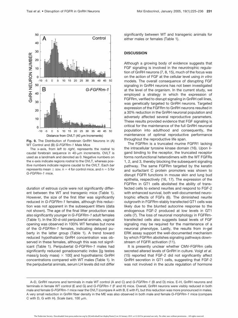
To examine whether the loss of GnRH neurons wasbrain region specific, the distribution of GnRH neuronsin WT and G-FGFRm-1 mice was analyzed in definedareas. The distribution of GnRH neurons was not sig-nificantly different between the G-FGFRm-1 and WTmale mice (Fig. 9). Both showed a typical pattern ofGnRH neuron distribution, with fewer GnRH neuronsanterior to the OVLT, an abrupt increase near theOVLT, and a gradual decrease posterior to OVLT (Fig.9). The distribution patterns of GnRH neurons in fe-male WT and G-FGFRm-1 mice were similar to males(data not shown).
Reproductive Phenotype of G-FGFRm-1 Mice
The observation that transgenic mice possessed fewerGnRH neurons suggests their fertility may be dis-rupted. To verify this, several reproductive parameterswere examined in male and female WT and G-FGFRm-1 mice (Table 1). In adult females, fertility(number of days required to produce one litter) and the
Fig. 6. Localization of Transgene Expression in GnRH Neurons of G-FGFRm-1 Mice by in Situ HybridizationmRNA for GnRH and the transgene were labeled by specific riboprobes on adjacent serial sections from the POA of
G-FGFRm-1 (A and C) and WT mice (B and D). In the G-FGFRm-1 mouse, eight GnRH neurons (A) were labeled, and six of thesewere also labeled for the transgene (C). Arrows in A point to GnRH neurons devoid of the transgene mRNA. In the WT mouse,four GnRH neurons (B) were labeled, whereas no labeling was observed for the transgene (D). Scale bar, 40 �m.
Fig. 7. Total Number of GnRH Neurons in the Forebrain ofAdult WT Control and G-FGFRm-1 Mice
A significant difference in GnRH neuronal number wasobserved between WT control and G-FGFRm-1 mice inmales and females combined (**, P � 0.001), in males only (**,P � 0.001), and in females only (*, P � 0.05). Each barrepresents mean � SEM. n � 4 and 5 for WT control andG-FGFRm-1 females, respectively; n � 5 for both WT controland G-FGFRm-1 males.
Tsai et al. • Disruption of FGFR in GnRH Neurons Mol Endocrinol, January 2005, 19(1):225–236 229
The Endocrine Society. Downloaded from press.endocrine.org by [${individualUser.displayName}] on 24 January 2015. at 12:26 For personal use only. No other uses without permission. . All rights reserved.

Fig. 8. Representative Photomicrographs of GnRH Neurons at the Plane of the OVLT (A, B, E, and F), and GnRH Axon Terminalsin the Median Eminence (C, D, G, and H)
230 Mol Endocrinol, January 2005, 19(1):225–236 Tsai et al. • Disruption of FGFR in GnRH Neurons
The Endocrine Society. Downloaded from press.endocrine.org by [${individualUser.displayName}] on 24 January 2015. at 12:26 For personal use only. No other uses without permission. . All rights reserved.
duration of estrous cycle were not significantly differ-ent between the WT and transgenic mice (Table 1).However, the size of the first litter was significantlyreduced in G-FGFRm-1 females, although this reduc-tion was not apparent in the subsequent litters (datanot shown). The age of the final litter production wasalso significantly younger in G-FGFRm-1 adult females(Table 1). In the 30-d-old peripubertal animals, vaginalopening was observed in 100% WT females but noneof the G-FGFRm-1 females, indicating delayed pu-berty in the latter group (Table 1). A trend towardreduced hypothalamic GnRH concentration was ob-served in these females, although this was not signif-icant (Table 1). Peripubertal G-FGFRm-1 males hadsignificantly reduced gonadosomatic index [(g testesmass/g body mass) � 100] and hypothalamic GnRHconcentrations compared with WT males (Table 1). Inthe peripubertal animals, serum LH levels did not differ
significantly between WT and transgenic animals foreither males or females (Table 1).
DISCUSSION
Although a growing body of evidence suggests thatFGF signaling is involved in the neurotrophic regula-tion of GnRH neurons (7, 8, 15), much of the focus wason the action of FGF at the cellular level using in vitromodels. The overall consequence of disrupting FGFsignaling in GnRH neurons has not been investigatedat the level of the organism. In the current study, weemployed a strategy in which the expression ofFGFRm, verified to disrupt signaling in GnRH cell lines,was genetically targeted to GnRH neurons. Targetedexpression of the FGFRm to GnRH neurons resulted ina 30% reduction in the GnRH neuronal population andadversely affected several reproductive parameters.These results provided evidence that FGF signaling iscritical for the maintenance of the full GnRH neuronalpopulation into adulthood and consequently, themaintenance of optimal reproductive performancethroughout the reproductive life span.
The FGFRm is a truncated murine FGFR1 lackingthe intracellular tyrosine kinase domain (16). Upon li-gand binding to the receptor, the truncated receptorforms nonfunctional heterodimers with the WT FGFRs1, 2, and 3, thereby blocking the subsequent signalingpathway. The same FGFRm targeted by the keratinand surfactant C protein promoters was shown todisrupt FGFR functions in mouse skin and lung budepithelia, respectively (16, 17). The expression of theFGFRm in GT1 cells abolished the ability of trans-fected cells to extend neurites and respond to FGF-2with enhanced survival, both well-documented neuro-trophic effects of FGFs (6). The diminished neuriteoutgrowth in FGFRm-stably transfected GT1 cells waslikely due to the blunted autocrine response to theendogenous FGF-2 produced at low levels in GT1cells (7). The loss of neuronal morphology in FGFRm-transfected cells also suggests basal levels of FGFsignaling may be required for the maintenance of aneuronal phenotype. Lastly, the results from in-gelERK assay support the well-documented mechanismby which FGFRm abolishes signaling pathways down-stream of FGFR activation (11).
It is presently unclear whether CMV-FGFRm cellssecreted altered levels of GnRH in culture. Voigt et al.(15) reported that FGF-2 did not significantly affectGnRH secretion in GT1 cells, suggesting that FGF-2was not involved in the acute regulation of hormone
A–D, GnRH neurons and terminals in male WT control (A and C) and G-FGFRm-1 (B and D) mice. E–H, GnRH neurons andterminals in female WT control (E and G) and G-FGFRm-1 (F and H) mice. Overall, GnRH neurons were visibly reduced in bothmale and female G-FGFRm-1 mice near the OVLT (compare A with B, E with F), but this reduction was more pronounced in males.A very small reduction in GnRH fiber density in the ME was also observed in both male and female G-FGFRm-1 mice (compareC with D, G with H). Scale bars, 100 �m.
Fig. 9. The Distribution of Forebrain GnRH Neurons in (A)WT Control and (B) G-FGFRm-1 Male Mice
The x-axis, from left to right, represents the rostral tocaudal forebrain sequence in 40-�m increments. OVLT isused as a landmark and denoted as 0. Negative numbers onthe x-axis indicate regions rostral to the OVLT, whereas pos-itive numbers indicate regions caudal to the OVLT. Each barrepresents mean � SEM. n � 4 for control mice, and n � 5 forG-FGFRm-1 mice.
Tsai et al. • Disruption of FGFR in GnRH Neurons Mol Endocrinol, January 2005, 19(1):225–236 231
The Endocrine Society. Downloaded from press.endocrine.org by [${individualUser.displayName}] on 24 January 2015. at 12:26 For personal use only. No other uses without permission. . All rights reserved.

release. Interestingly, the same study also reportedincreased mRNA levels of prohormone convertase 2, akey enzyme required for the initial processing of theGnRH prohormone, in GT1 cells treated with FGF-2(15). The latter raised the possibility that GnRH neu-rons, under the long-term disruption of FGF signaling,may process the GnRH prohormone differently, lead-ing to an altered levels of mature peptide release.
Northern blot analysis of tissue RNA from WT andG-FGFRm mice demonstrated the presence of twoendogenous FGFR1 transcripts in all animals. The4.3-kb transcript was widely documented and repre-sented the membrane spanning IIIc variant of FGFR1(18, 19). The 1.8-kb transcript may represent the IIIavariant of the secreted FGFR1 (14). This FGFR1 wasfound in the brain (14) and may have cross-hybridizedwith our probe. G-FGFRm-1 animals also expressed asingle FGFRm transcript consistent with the expectedsize of the transgene in the brain, where GnRH neu-rons reside. Further examination with in situ hybridiza-tion verified that the transgene was indeed expressedexclusively in GnRH neurons of G-FGFRm-1 mice. Incontrast with this expected expression pattern, G-FGFRm-5 animals expressed an additional splice vari-ant of the FGFRm (1.0 kb) and expressed the trans-gene ectopically in the hindbrain. It is worthmentioning that our initial observations showed thatG-FGFRm-5 animals had a similar reduction in thenumber of GnRH neurons (data not shown). However,it was impossible to determine whether the reductionin GnRH neurons was specific to the expression of thetransgene in GnRH neurons or the consequence of theectopic expression of the transgene.
G-FGFRm-1 mice exhibited an average of 30% re-duction in the size of GnRH neuronal population, withthe reduction being more prominent in males thanfemales. Interestingly, we found that WT females hadsignificantly fewer GnRH neurons (Fig. 7; P � 0.05 byStudent’s t test) than males. This particular sexualdimorphism has been reported in mice (20) and mayhave contributed to the sex difference in GnRH neuronreduction observed here. These results were also con-sistent with our observation that the reduction in hy-
pothalamic GnRH concentrations was more pro-nounced in transgenic males than females (see Table1). Although a modest reduction in GnRH fiber densitywas also observed in the ME of the G-FGFRm-1 mice,the ability of GnRH axons to target the ME did notappear grossly altered because a large number ofaxons still reached the ME. We believe the reducedfiber density was primarily due to the reduction in thenumber of GnRH neurons. It is also possible that thedisruption of FGFR function resulted in the diminishedability of GnRH neurons to branch axons, leading todecreased levels of fiber intensity. Our previous ob-servation that FGF-2 significantly promoted neuritebranching in cultures of primary GnRH neurons sup-ports this possibility (8).
The uniform reduction in the number of GnRH neu-rons in all brain regions of the G-FGFRm-1 mice sug-gests the size of the original GnRH neuronal popula-tion that migrated into the forebrain may have beenreduced. Several explanations could account for thisreduction. We previously showed that FGF signalingwas required for the specification of GnRH neuronalfate in the olfactory placode (8). In the absence of anFGF signal, the majority of GnRH neurons failed toemerge from their birthplace. However, the expressionof FGFRm in G-FGFRm-1 mice is coupled to the ac-tivation of the GnRH promoter; thus, one would notexpect FGFRm to be expressed and FGFR functiondisrupted until after GnRH neurons are fully specified.A more plausible explanation for the reduced GnRHneuronal population is that in the absence of FGFsignaling, a subpopulation of GnRH neurons failed tosurvive. To date, few studies have investigated thesurvival of GnRH neurons after fate specification. Theprevailing view was that 800 GnRH neurons were bornin the olfactory placode, and all 800 GnRH neuronssurvived and developed normally to reach the fore-brain. However, an observation made by Wu et al. (21)revealed that the GnRH neuronal population actuallyreached a total of 2000 at embryonic d 12.75. Thisnumber then declined to 1100 in postnatal animals,and to 800 in adults. This observation suggested thatless than 50% of the original GnRH neuronal popula-
Table 1. Reproductive Parameters of WT and G-FGFRm-1 Female and Male Mice
Females (n � 4–8) Males (n � 4–6)
WT G-FGFRm-1 Control G-FGFRm-1
Fertility (d/litter) 28.3 � 3.3 25.4 � 1.3 – –Length of estrous cycle (d) 5.6 � 1.4 4.8 � 0.6 – –Number of pups in the first litter 8.1 � 0.6 6.4 � 0.4a – –Age of last litter production (d) 392 � 14 289 � 38a – –Percent vaginal opening at 30 d (%) 100 (8/8) 0 (0/8)a – –GSI at 30 d (%) – – 0.64 � 0.02 0.51 � 0.02a
30-d-old Hypothalamic GnRH (pg/�g protein) 1.4 � 0.3 0.8 � 0.1 1.3 � 0.2 0.5 � 0.1a
30-d-old Plasma LH (ng/ml) 0.15 � 0.01 0.15 � 0.01 0.19 � 0.03 0.28 � 0.07
All values were expressed as mean � SEM.a P � 0.05 compared with the controls of the same sex.ND, Not detectable. Parameters not measured or not applicable are denoted by –.
232 Mol Endocrinol, January 2005, 19(1):225–236 Tsai et al. • Disruption of FGFR in GnRH Neurons
The Endocrine Society. Downloaded from press.endocrine.org by [${individualUser.displayName}] on 24 January 2015. at 12:26 For personal use only. No other uses without permission. . All rights reserved.

tion survived to adulthood. Moreover, it underscoredthe importance of survival-enhancing factors in main-taining and determining the final number of GnRHneurons. FGF-2, a potent survival enhancing factor inGT1 cells (Ref. 7 and current study), may be critical inpromoting the survival of GnRH neurons, especiallyduring the developmental period when a large numberof GnRH neurons vanished in the forebrain.
Despite the absence of approximately 30% of GnRHneurons in the forebrain of G-FGFRm-1 mice, theseanimals were initially fertile. This observation is notsurprising considering findings in the hypogonadal(hpg) mouse. The hpg mice harbored a deletion in theGnRH gene that resulted in the inability to synthesizethe mature GnRH peptide. The successful transplan-tation of just one to three detectable GnRH neuronswas effective in restoring their fertility (22–24). Thus,although reduced, the number of GnRH neuronspresent in G-FGFRm-1 mice was clearly sufficient formaintaining reproduction under laboratory conditions.It is important to point out that although these animalsreproduced, the level of reproductive activity was re-duced compared with controls. In particular, the de-layed puberty, a reduction in the size of the first litter,and early reproductive senescence were all clear signsthat the function of the GnRH system at the beginningand the end of the reproductive life span has beenmarkedly compromised.
One might question why serum LH levels in trans-genic mice remained unchanged despite the reducedGnRH neuron number and content. A recent study (25)reported similar observations in mice harboring a mu-tation in carboxypeptidase E and thus could not pro-cess GnRH normally. These mice had very low bioac-tive GnRH concentrations in the hypothalamus,displayed a number of reproductive defects, but hadnormal circulating LH. One way these animals com-pensated for low GnRH was by increasing pituitarysensitivity to GnRH (25). Another study on these micereported similar reduction in TRH due to processingdefect, but again, the level of circulating TSH wasnormal (26). These results, together with ours, clearlydemonstrated the ability of animals to develop pitu-itary-level compensatory mechanisms to cope withdiminished levels of releasing hormones.
Our current study provides the first evidence, at thelevel of the organism, that FGF signaling is involved inthe maintenance of GnRH neurons. The significantreduction in the number of GnRH neurons may reflecta decrease in the survival of GnRH neurons duringdevelopment. Several explanations could account forwhy only 30% of GnRH neurons were affected. First,the extent of FGFR disruption may vary greatly amongindividual GnRH neurons depending on the ratio ofFGFRm to WT FGFR expression. It has been specu-lated that the FGFRm would have to be expressed atlevels five to 10 times higher than the WT receptor tocompletely inhibit signaling (11). Due to the heteroge-neous nature of the GnRH neuronal population (27–30), the level of GnRH gene expression is likely to vary
among cells. Consequently, the number of FGFRmexpressed per cell is likely to vary because the expres-sion of this transgene is driven by the GnRH genepromoter. Furthermore, variations in the number ofendogenous WT FGFRs among GnRH neurons couldresult in different FGFRm to WT FGFRs ratios, leadingto unequal degree of disruption. Secondly, FGFRfunction may be involved in the direct regulation ofonly a fraction of GnRH neurons. In support of thisnotion, Gill et al. (8) has demonstrated the presence ofFGFRs1 and 3 in only 20–60% of embryonic and adultGnRH neurons. The consequence of disrupting FGFRfunction would therefore be negligible for GnRH neu-rons lacking FGFRs or for those expressing function-ally insignificant levels of FGFRs.
If not all GnRH neurons require FGF signaling tomature, there must be other regulatory factors capableof supporting the GnRH system. We hypothesize thatthis redundancy, commonly observed in the develop-ing CNS, is critically important in safeguarding theGnRH system under a disrupted environment. Neuro-trophic factors such as IGF-I (31–36), IGF-II (31, 37),and brain-derived neurotrophic factor (38) have allbeen shown to regulate GnRH neuronal function andmay protect the GnRH system from complete obliter-ation when FGF signaling is disrupted. Interestingly,IGF-I has also been shown to act synergistically withFGF-2 to regulate the function of GT1 cells (35), sug-gesting the collaborative nature of neurotrophic factoractions. The strong evolutionary need for an exces-sively robust GnRH system for the successful propa-gation of species underscores the importance of thisredundancy. Species with a GnRH system that couldbe rendered nonfunctional because the result of asingle regulatory anomaly will not survive the test oftime. Even under this type of redundancy, our resultshave unequivocally demonstrated that FGF signalingis one of the important components involved in theformation of a neuroendocrine system critical for ver-tebrate reproduction.
MATERIALS AND METHODS
Animals
Control and transgenic mice were derived from the mating ofC57BL/6J and DBA/2J mice, and their offspring. All micewere housed in the animal facility under a 12-h light, 12-hdark cycle and fed water and rodent chow ad libitum. Allanimal procedures complied with protocols approved by theInstitutional Animal Care and Use Committee of the Univer-sity of California at San Francisco and University of Coloradoat Boulder.
Construction of Plasmids
The details of the FGFRm cDNA were described previously(16). Briefly, a 1.1-kb FGFRm, a truncated dominant-negativemurine FGFR1 cDNA (exon IIIc variant) lacking the intracel-lular tyrosine kinase domain, was ligated downstream of0.65-kb rabbit �-globin intron (39). The 0.63-kb human GH
Tsai et al. • Disruption of FGFR in GnRH Neurons Mol Endocrinol, January 2005, 19(1):225–236 233
The Endocrine Society. Downloaded from press.endocrine.org by [${individualUser.displayName}] on 24 January 2015. at 12:26 For personal use only. No other uses without permission. . All rights reserved.

gene polyadenylation (poly A) sequence was inserted at the3� end of the FGFRm cDNA. The FGFRm expression con-struct (Fig. 1A) was cloned into the pBluescript KS� vector(Stratagene, La Jolla, CA).
For the stable transfection of GT1 cells, a partial FGFRmconstruct, containing only the �-globin intron and FGFRm,was excised with XbaI and BglII, blunted with Klenow, andligated into the pRc/CMV expression vector (Invitrogen LifeTechnologies, Carlsbad, CA). The resulting plasmid (CMV-FGFRm) was linearized for transfections by digestion withBglII (Fig. 1B).
For construction of a FGFRm transgene that would targetGnRH neurons, a 2.3-kb fragment of the rat GnRH promoter/enhancer region (G) was inserted via an XbaI site upstream ofthe rabbit �-globin intron of the FGFRm expression con-struct. The GnRH promoter/enhancer region consists of afragment from �2987 to �1172 appended to �441 to �104and contains a 731-bp deletion (13). This GnRH promoter/enhancer fragment was used successfully to target the ex-pression of simian virus 40 T antigen to mouse GnRH neuronsfor the generation of GT1 cells (13). For the production oftransgenic mice, the resulting plasmid was excised with KpnIand SalI to generate a transgenic fragment (G-FGFRm; Fig.1C) for the pronuclear injection of one-cell embryos.
GT1 Cells and Transfection
GT1–7 cells (passages 10–23) were maintained in a GT1medium consisting of the DMEM H21 (DMEM:H21; Universityof California at San Francisco Cell Culture Facility) supple-mented with 5% fetal calf serum, 5% horse serum (HyClone,Logan, UT), and penicillin/streptomycin. GT1–7 cells weretransfected with linearized CMV-FGFRm or empty vector(CMV-Null) using DOTAP liposomal transfection reagent ac-cording to the manufacturer’s instructions (Roche, Indianap-olis, IN). Stably transfected cells were selected by culturingcells in GT1 medium containing 450 �g/ml G418 (InvitrogenLife Technologies). After 1 month, single GT1 colonies wereisolated from the culture dish with a sterile pipette tip andtransferred to a 12-well plate. One clone was selected forcells transfected with the CMV-Null vector and three clonesfor the CMV-FGFRm.
Cell Survival Assay
The responsiveness of transfected cells to FGF-2 was exam-ined using a cell survival assay described previously (7).Briefly, transfected cells were serum-starved for 4 d in theabsence or presence of various doses of recombinant humanFGF-2 (Promega, Madison, WI). Cells were trypsinized on d 0(before serum starvation) and d 4, and counted with a hemo-cytometer. Percent cell survival was calculated by dividingthe cell number at d 4 by the cell number at d 0.
In-Gel ERK Assay
The ability of FGFRm to disrupt endogenous FGFR function,and thus downstream signaling events, was monitored in thetransfected cells by the in-gel ERK assay described previ-ously (40). We examined the activity levels of p42 and p44ERKs, both of which were robustly activated in GT1 cells bythe administration of FGF-2 (7). Details of this assay weredescribed elsewhere (7).
Generation and Screening of Transgenic Mice
The G-FGFRm transgene was injected into fertilized one-cellembryos as described previously (13). The F2 embryos werederived from the mating of C57BL/6J and DBA/2J mice.Animals were screened for the presence of the transgene by
PCR of the genomic DNA isolated from tail biopsies. ForPCR, we used a 5� primer directed to the rabbit �-globinintron and a 3� primer directed to the FGFRm region. Animalspositive for the transgene were designated as G-FGFRmmice. All G-FGFRm mice were bred to homozygosity bycrossing heterozygous G-FGFRm mice. Homozygosity wasconfirmed when a test cross between the G-FGFRm mouseand a nontransgenic mate generated offspring that were100% transgenic. The offspring of nontransgenic littermatesproduced during the heterozygote � heterozygote crosseswere used as control WT animals to match the genetic back-ground of G-FGFRm mice.
Immunocytochemistry and Quantification ofGnRH Neurons
Three- to 6-month-old WT and G-FGFRm mice were per-fused intracardially with 20 ml of 0.1 M PBS followed by 30 mlof 4% paraformaldehyde in 0.1 M PBS. Brains were dissectedand postfixed in 4% paraformaldehyde overnight at 4 C, andcryoprotected in 20% sucrose. Forty-micrometer floatingsections were cut using a cryostat. Sections were washedwith 0.5% hydrogen peroxide in 0.1 M PBS containing 0.4%Triton X-100 (PBST) for 10 min to the quench the endogenousperoxidase activity, rinsed five times with PBST, and incu-bated for 48 h at 4 C in PBST containing an anti-GnRHantibody (LR-1, a gift of Dr. R. Benoit, Montreal GeneralHospital, Montreal, Quebec, Canada; 1:10,000) and 4% nor-mal donkey serum. After incubation, sections were washedwith PBST and incubated with a biotinylated donkey-antirab-bit IgG (Jackson ImmunoResearch, West Grove, PA; 1:200),washed, and incubated with the Vectastain avidin-biotincomplex reagent (Vector Laboratories, Burlingame, CA) for45 min. Sections were washed and the immunoreactivityvisualized using diaminobenzidine as the chromagen. Afterthe color reaction, sections were washed, mounted on slides,dehydrated through an ascending series (70–100%) of etha-nol, cleared in Histoclear (National Diagnostics, Atlanta, GA),and coverslipped.
GnRH neurons were counted under a Nikon Eclipse E800microscope (Nikon, Melville, NY). Only cells with darklystained cytoplasm and clear nuclei were scored. To accu-rately assess the total number of GnRH neurons in a singleanimal, cells counts were made in every section ranging fromthe diagonal band of Broca through the ME.
Northern Blot Analysis of Transgene Expression inTransfected Cells and Mice
Total RNA isolated from transfected GT1 cells and from tis-sues of the control WT and G-FGFRm mice were subjected tonorthern blot analysis to detect the expression of the FGFRmtransgene. Fifteen micrograms of total RNA were fractionatedon a 1% agarose/formaldehyde gel and transferred onto anylon transfer membrane (MSI, Westboro, MA) using thecapillary blotting method. A randomly primed 32P-labeledcDNA probe corresponding to the exon IIIc variant of themurine FGFR1 was generated by the NEBlot Kit (New En-gland Biolabs, Beverly, MA). The membrane was hybridizedwith the FGFR1 probe at 68 C for 1 h. The membrane wasthen washed twice in 2� saline sodium citrate/0.1% sodiumdodecyl sulfate for 15 min at room temperature and once in0.1� saline sodium citrate/0.1% sodium dodecyl sulfate for15 min at 65 C. Hybridization signals were visualized by theexposure of the blot to Kodak X-OMAT AR film (EastmanKodak, Rochester, NY) or the Packard (Meriden, CT) Cyclonephosphorimager. The Northern blot of mouse tissue RNA wasstripped in 50% formamide/2� saline sodium phosphateEDTA at 65 C and reprobed for the presence of glyceralde-hyde phosphate dehydrogenase, an internal loading control,using the same procedure.
234 Mol Endocrinol, January 2005, 19(1):225–236 Tsai et al. • Disruption of FGFR in GnRH Neurons
The Endocrine Society. Downloaded from press.endocrine.org by [${individualUser.displayName}] on 24 January 2015. at 12:26 For personal use only. No other uses without permission. . All rights reserved.

In Situ Hybridization Analysis of Transgene Expressionin GnRH Neurons
The specificity of the transgene expression was analyzed byin situ hybridization on adjacent 10-�m frozen serial coronalsections from four 2-month-old homozygous G-FGFRm andfour 2-month-old WT mice. A detailed protocol for the in situanalysis was described previously (41). Briefly, a total of ninepairs of adjacent sections through the POA were used for theevaluation of each animal. Sections were acetylated, dehy-drated progressively, and hybridized overnight at 55 C withthe probe for either GnRH or the transgene after the protocolsuggested by the manufacturer (Roche). To localize themRNA for the FGFRm transgene, a 73-bp fragment corre-sponding to the 5� end of the human GH poly A fragment wasused to generate a digoxigenin-labeled antisense riboprobe.This region was transcribed and was contained within thetransgene mRNA. GnRH neurons were identified with a345-bp digoxigenin-labeled antisense riboprobe to the ratGnRH cDNA (42). The sense strand for both probes was usedas a control. For each pair of adjacent sections, one sectionwas hybridized with the GnRH riboprobe, whereas the adja-cent section was hybridized with the transgene riboprobe.After hybridization, the slides were washed under high strin-gency and processed directly for detection of the digoxigeninsignal. The sections were examined with a Leica (Bannock-burn, IL) DMR photomicroscope under bright-field optics.
Monitoring the Reproductive Function of Female andMale WT and Transgenic Mice
Reproductive function of adult WT and G-FGFRm femaleswere assessed by four criteria: the length of the estrous cycle,the mean duration required for the production of one litter, thesize of the first litter, and the age of the female when the finallitter was produced. The length of the estrous cycle wasassessed by vaginal smears obtained daily for 2 wk on fe-males between 3 and 4 months of age. To assess the averageduration required to produce a litter, the number of littersproduced by females (between 3 and 8 months of age) in theconstant presence of a male was recorded and normalizedfor the duration. We retired the female breeders when theyfailed to produce litters for three months. Thus, the final litterwas defined as the litter before the breeding female wasretired. Pubertal onset was assessed by measuring the inci-dence of vaginal opening in 30-d-old females, and gonado-somatic index in 30-d-old males.
Hormone Measurements
Hypothalamic GnRH concentrations in WT and G-FGFRmmales was measured by a GnRH RIA (43). Serum LH wasmeasured by a sensitive sandwich immunoassay describedpreviously (44).
Statistical Analysis
Differences among multiple groups were analyzed by theone-way ANOVA followed by the Tukey’s post hoc test. Dif-ference between two groups was analyzed by the Student’st test. Difference in percent animals with vaginal openingbetween WT and transgenic animals was analyzed by theFisher’s exact test. Differences were considered significantwhen P � 0.05.
Acknowledgments
We thank Amy Choi, Mai Hashimoto, and Jeremy Jones forthe expert technical assistance.
Received August 23, 2004. Accepted September 20, 2004.
Address all correspondence and requests for reprints to:Pei-San Tsai, Department of Integrative Physiology, 114Clare Small, University of Colorado, Boulder, Colorado80309-0354. E-mail: [email protected].
This work was supported by funding: National ScienceFoundation IBN-9996398 (to P.-S.T.), National Institutes ofHealth (NIH) HD-042634 (to P.-S.T.), NIH HD-08924 (toR.I.W.), NIH HD-11979 (to Reproductive Endocrinology Cen-ter Core Facilities), and the Ligand and Assay Core of Coop-erative Agreement U54 HD28934.
REFERENCES
1. Schwanzel-Fukuda M, Pfaff DW 1989 Origin of luteinizinghormone-releasing hormone neurons. Nature 338:161–164
2. Wray S, Grant P, Gainer H 1989 Evidence that cellsexpressing luteinizing hormone-releasing hormonemRNA in the mouse are derived from progenitor cells inthe olfactory placode. Proc Natl Acad Sci USA 86:8132–8136
3. Whitlock KE, Wolf CD, Boyce ML 2003 Gonadotropin-releasing hormone (GnRH) cells arise from cranial neuralcrest and adenohypophyseal regions of the neural platein the zebrafish, Danio rerio. Dev Biol 257:140–152
4. Schwanzel-Fukuda M 1999 Origin and migration of lu-teinizing hormone-releasing hormone neurons in mam-mals. Microsc Res Tech 44:2–10
5. Barbacid M 1994 The Trk family of neurotrophin recep-tors. J Neurobiol 25:1386–1403
6. Ford-Perriss M, Abud H, Murphy M 2001 Fibroblastgrowth factors in the developing central nervous system.Clin Exp Pharmacol Physiol 28:493–503
7. Tsai PS, Werner S, Weiner RI 1995 Basic fibroblastgrowth factor is a neurotropic factor in GT1 gonado-tropin-releasing hormone neuronal cell lines. Endocrinol-ogy 136:3831–3838
8. Gill JC, Moenter SM, Tsai PS 2004 Developmental reg-ulation of gonadotropin-releasing hormone neurons byfibroblast growth factor signaling. Endocrinology 124:3830–3839
9. Dode C, Levilliers J, Dupont JM, De Paepe A, Le Du N,Soussi-Yanicostas N, Coimbra RS, Delmaghani S, Com-pain-Nouaille S, Baverel F, Pecheux C, Le Tessier D,Cruaud C, Delpech M, Speleman F, Vermeulen S,Amalfitano A, Bachelot Y, Bouchard P, Cabrol S, CarelJC, Delemarre-van de Waal H, Goulet-Salmon B, KottlerML, Richard O, Sanchez-Franco F, Saura R, Young J,Petit C, Hardelin JP 2003 Loss-of-function mutations inFGFR1 cause autosomal dominant Kallmann syndrome.Nat Genet 33:463–465
10. Sato N, Katsumata N, Kagami M, Hasegawa T, Hori N,Kawakita S, Minowada S, Shimotsuka A, Shishiba Y,Yokozawa M, Yasuda T, Nagasaki K, Hasegawa D, Ha-segawa Y, Tachibana K, Naiki Y, Horikawa R, Tanaka T,Ogata T 2004 Clinical assessment and mutation analysisof Kallmann syndrome 1 (KAL1) and fibroblast growthfactor receptor 1 (FGFR1, or KAL2) in five families and18 sporadic patients. J Clin Endocrinol Metab 89:1079–1088
11. Amaya E, Musci TJ, Kirschner MW 1991 Expression of adominant negative mutant of the FGF receptor disruptsmesoderm formation in Xenopus embryos. Cell 66:257–270
12. Ueno H, Gunn M, Dell K, Tseng Jr A, Williams L 1992 Atruncated form of fibroblast growth factor receptor 1inhibits signal transduction by multiple types of fibroblastgrowth factor receptor. J Biol Chem 267:1470–1476
13. Mellon PL, Windle JJ, Goldsmith PC, Padula CA, RobertsJL, Weiner RI 1990 Immortalization of hypothalamicGnRH neurons by genetically targeted tumorigenesis.Neuron 5:1–10
Tsai et al. • Disruption of FGFR in GnRH Neurons Mol Endocrinol, January 2005, 19(1):225–236 235
The Endocrine Society. Downloaded from press.endocrine.org by [${individualUser.displayName}] on 24 January 2015. at 12:26 For personal use only. No other uses without permission. . All rights reserved.
14. Duan DS, Werner S, Williams LT 1992 A naturally occur-ring secreted form of fibroblast growth factor (FGF) re-ceptor 1 binds basic FGF in preference over acidic FGF.J Biol Chem 267:16076–16080
15. Voigt P, Ma YJ, Gonzalez D, Fahrenbach WH, WetselWC, Berg-von der Emde K, Hill DF, Taylor KG, Costa ME,Seidah NG, Ojeda SR 1996 Neural and glial-mediatedeffects of growth factors acting via tyrosine kinase re-ceptors on luteinizing hormone-releasing hormone neu-rons. Endocrinology 137:2593–2605
16. Werner S, Weinberg W, Liao X, Peters KG, Blessing M,Yuspa SH, Weiner RL, Williams LT 1993 Targeted ex-pression of a dominant-negative FGF receptor mutant inthe epidermis of transgenic mice reveals a role of FGF inkeratinocyte organization and differentiation. EMBO J12:2635–2643
17. Peters K, Werner S, Liao X, Wert S, Whitsett J, WilliamsL 1994 Targeted expression of a dominant negative FGFreceptor blocks branching morphogenesis and epithelialdifferentiation of the mouse lung. EMBO J 13:3296–3301
18. Yamaguchi TP, Conlon RA, Rossant J 1992 Expressionof the fibroblast growth factor receptor FGFR-1/flg dur-ing gastrulation and segmentation in the mouse embryo.Dev Biol 152:75–88
19. Jin Y, Pasumarthi KB, Bock ME, Lytras A, Kardami E,Cattini PA 1994 Cloning and expression of fibroblastgrowth factor receptor-1 isoforms in the mouse heart:evidence for isoform switching during heart develop-ment. J Mol Cell Cardiol 26:1449–1459
20. Grober MS, Winterstein GM, Ghazanfar AA, EroschenkoVP 1998 The effects of estradiol on gonadotropin-releas-ing hormone neurons in the developing mouse brain. GenComp Endocrinol 112:356–363
21. Wu TJ, Gibson MJ, Rogers MC, Silverman AJ 1997 Newobservations on the development of the gonadotropin-releasing hormone system in the mouse. J Neurobiol33:983–998
22. Krieger DT, Perlow MJ, Gibson MJ, Davies TF, Zimmer-man EA, Ferin M, Charlton HM 1982 Brain grafts reversehypogonadism of gonadotropin releasing hormone defi-ciency. Nature 298:468–471
23. Gibson MJ, Krieger DT, Charlton HM, Zimmerman EA,Silverman AJ, Perlow MJ 1984 Mating and pregnancycan occur in genetically hypogonadal mice with preopticarea brain grafts. Science 225:949–951
24. Gibson MJ, Charlton HM, Perlow MJ, Zimmerman EA,Davies TF, Krieger DT 1984 Preoptic area brain grafts inhypogonadal (hpg) female mice abolish effects of con-genital hypothalamic gonadotropin-releasing hormone(GnRH) deficiency. Endocrinology 114:1938–1940
25. Srinivasan S, Bunch DO, Fen Y, Rodriguiz RM, Li M,Ravenell RL, Luo GX, Arimura A, Fricker LD, Eddy EM,Wetsel WC 2004 Deficits in reproduction and pro-gonadotropin-releasing hormone processing in maleCpefat mice. Endocrinology 145:2023–2034
26. Nillni EA, Xie W, Mulcahy L, Sanchez VC, Wetsel WC2002 Deficiencies in pro-thyrotropin-releasing hormoneprocessing and abnormalities in thermoregulation inCpefat/fat mice. J Biol Chem 277:48587–48595
27. Wray S, Hoffman G 1986 Postnatal morphologicalchanges in rat LHRH neurons correlated with sexualmaturation. Neuroendocrinology 43:93–97
28. Simonian SX, Skynner MJ, Sieghart W, Essrich C, Lus-cher B, Herbison AE 2000 Role of the GABA(A) receptor�2 subunit in the development of gonadotropin-releasinghormone neurons in vivo. Eur J Neurosci 12:3488–3496
29. Schwarting GA, Kostek C, Bless EP, Ahmad N, Tobet SA2001 Deleted in colorectal cancer (DCC) regulates themigration of luteinizing hormone-releasing hormone neu-rons to the basal forebrain. J Neurosci 21:911–919
30. Sim JA, Skynner MJ, Herbison AE 2001 Heterogeneity inthe basic membrane properties of postnatal gonado-tropin-releasing hormone neurons in the mouse. J Neu-rosci 21:1067–1075
31. Olson BR, Scott DC, Wetsel WC, Elliot SJ, Tomic M,Stojilkovic S, Nieman LK, Wray S 1995 Effects of insulin-like growth factors I and II and insulin on the immortalizedhypothalamic GTI-7 cell line. Neuroendocrinology 62:155–165
32. Ochoa A, Domenzain C, Clapp C, Martinez de la EscaleraG 1997 Differential effects of basic fibroblast growthfactor, epidermal growth factor, transforming growth fac-tor-�, and insulin-like growth factor-I on a hypothalamicgonadotropin-releasing hormone neuronal cell line.J Neurosci Res 49:739–749
33. Zhen S, Zakaria M, Wolfe A, Radovick S 1997 Regulationof gonadotropin-releasing hormone (GnRH) gene ex-pression by insulin-like growth factor I in a culturedGnRH-expressing neuronal cell line. Mol Endocrinol 11:1145–1155
34. Longo KM, Sun Y, Gore AC 1998 Insulin-like growthfactor-I effects on gonadotropin-releasing hormone bio-synthesis in GT1–7 cells. Endocrinology 139:1125–1132
35. Gallo F, Morale MC, Tirolo C, Testa N, Farinella Z, AvolaR, Beaudet A, Marchetti B 2000 Basic fibroblast growthfactor priming increases the responsiveness of immor-talized hypothalamic luteinizing hormone releasing hor-mone neurones to neurotrophic factors. J Neuroendocri-nol 12:941–959
36. Miller BH, Gore AC 2001 Alterations in hypothalamicinsulin-like growth factor-I and its associations with go-nadotropin releasing hormone neurones during repro-ductive development and ageing. J Neuroendocrinol 13:728–736
37. Anderson RA, Zwain IH, Arroyo A, Mellon PL, Yen SS1999 The insulin-like growth factor system in the GT1–7GnRH neuronal cell line. Neuroendocrinology 70:353–359
38. Cronin AS, Horan TL, Spergel DJ, Brooks AN, HastingsMH, Ebling FJ 2004 Neurotrophic effects of BDNF onembryonic gonadotropin-releasing hormone (GnRH)neurons. Eur J Neurosci 20:338–344
39. O’Hare K, Benoist C, Breathnach R 1981 Transformationof mouse fibroblasts to methotrexate resistance by arecombinant plasmid expressing a prokaryotic dihydro-folate reductase. Proc Natl Acad Sci USA 78:1527–1531
40. Vietor I, Schwenger P, Li W, Schlessinger J, Vilcek J 1993Tumor necrosis factor-induced activation and increasedtyrosine phosphorylation of mitogen-activated protein(MAP) kinase in human fibroblasts. J Biol Chem 268:18994–18999
41. Paruthiyil S, El Majdoubi M, Conti M, Weiner RI 2002Phosphodiesterase expression targeted to gonadotro-pin-releasing hormone neurons inhibits luteinizing hor-mone pulses in transgenic rats. Proc Natl Acad Sci USA99:17191–17196
42. Adelman JP, Mason AJ, Hayflick JS, Seeburg PH 1986Isolation of the gene and hypothalamic cDNA for thecommon precursor of gonadotropin-releasing hormoneand prolactin release-inhibiting factor in human and rat.Proc Natl Acad Sci USA 83:179–183
43. Pak TR, Lynch GR, Tsai PS 2001 Testosterone and es-trogen act via different pathways to inhibit puberty in themale Siberian hamster (Phodopus sungorus). Endocri-nology 142:3309–3316
44. Fallest PC, Trader GL, Darrow JM, Shupnik MA 1995Regulation of rat luteinizing hormone � gene expressionin transgenic mice by steroids and a gonadotropin-releasing hormone antagonist. Biol Reprod 53:103–109
Molecular Endocrinology is published monthly by The Endocrine Society (http://www.endo-society.org), the foremostprofessional society serving the endocrine community.
236 Mol Endocrinol, January 2005, 19(1):225–236 Tsai et al. • Disruption of FGFR in GnRH Neurons
The Endocrine Society. Downloaded from press.endocrine.org by [${individualUser.displayName}] on 24 January 2015. at 12:26 For personal use only. No other uses without permission. . All rights reserved.