Negative Feedback Regulation of FGF Signaling Levels by Pyst1/MKP3 in Chick Embryos
10
Current Biology, Vol. 13, 1009–1018, June 17, 2003, 2003 Elsevier Science Ltd. All rights reserved. DOI 10.1016/S0960-9822(03)00381-6 Negative Feedback Regulation of FGF Signaling Levels by Pyst1/MKP3 in Chick Embryos played by dual specificity MAP kinase phosphatases in regulating developmental outcomes in vertebrates. Maxwell C. Eblaghie, 1,4 J. Simon Lunn, 1,2,4 Robin J. Dickinson, 3 Andrea E. Mu ¨ nsterberg, 1,5 Juan-Jose Sanz-Ezquerro, 1 Elizabeth R. Farrell, 1 Joanne Mathers, 3 Stephen M. Keyse, 3, * Introduction Kate Storey, 1,2, * and Cheryll Tickle 1, * 1 Division of Cell and Developmental Biology We wish to understand how cell-cell signaling in devel- 2 Neural Development Group oping embryos coordinates growth, differentiation, and Division of Cell and Developmental Biology morphogenesis. Fibroblast growth factors (FGFs) com- School of Life Sciences prise a major family of signaling molecules which are University of Dundee used in many different developmental contexts. Studies Dow Street in mice, Xenopus, Drosophila, and C. elegans demon- Dundee DD1 5EH strate that FGF signaling is mediated via tyrosine kinase United Kingdom receptors (FGFRs) that can act through a number of 3 Cancer Research UK transduction pathways, including the highly conserved Molecular Pharmacology Unit Ras-ERK mitogen-activated protein kinase (MAPK) sig- Biomedical Research Centre naling cascade [1–4]. Here we focus on FGF signaling Level 5, Ninewells Hospital in neural induction and limb bud outgrowth in chick Dundee DD1 9SY embryos, where downstream pathways that mediate United Kingdom these events are not known. Recent work has revealed that FGF signaling is nega- tively regulated by complex intracellular systems which Summary include Sprouty, Sef, Spred, and FRS2 [5–11]. Further- more, expression of Sprouty and Sef is induced by acti- Background: The importance of endogenous antago- vation of the MAP kinase cascade itself, demonstrating nists in intracellular signal transduction pathways is be- that these operate in negative feedback loops [7, 8, 12]. coming increasingly recognized. There is evidence in Sef coimmunoprecipitates with FGFR [8] while Sprouty2 cultured mammalian cells that Pyst1/MKP3, a dual spec- prevents activation of raf [13] in some contexts (also ificity protein phosphatase, specifically binds to and in- see [14]), indicating that there may be multiple points activates ERK1/2 mitogen-activated protein kinases of regulation at different levels within the FGF signaling (MAPKs). High-level Pyst1/Mkp3 expression has re- pathway. cently been found at many sites of known FGF signaling Biochemical assays and studies in cultured mamma- in mouse embryos, but the significance of this associa- lian cells have identified phosphatases that specifically tion and its function are not known. act on the ERK1/2 MAP kinases [15]. These include the Results: We have cloned chicken Pyst1/Mkp3 and closely related enzymes Pyst1/MKP3, Pyst2/MKPX, and show that high-level expression in neural plate corre- Pyst3/MKP4, which constitute a distinct subfamily of lates with active MAPK. We show that FGF signaling dual specificity MAP kinase phosphatases (MKPs). regulates Pyst1 expression in developing neural plate Pyst1/MKP3 binds selectively to ERK2, which results in and limb bud by ablating and/or transplanting tissue catalytic activation of the phosphatase [16, 17], and sources of FGFs and by applying FGF protein or a spe- expression of Pyst1 in mammalian cells specifically cific FGFR inhibitor (SU5402). We further show by blocks activation and nuclear translocation of ERK2 [16, applying a specific MAP kinase kinase inhibitor 18]. All of these observations strongly suggest that Pyst1 (PD184352) that Pyst1 expression is regulated via the acts as an intracellular brake to signal transduction MAPK cascade. Overexpression of Pyst1 in chick em- through the Ras/MAPK pathway in mammalian cells. bryos reduces levels of activated MAPK in neural plate However, nothing is known about the regulation of Pyst1 and alters its morphology and retards limb bud out- expression or its relationship with MAPK signaling in growth. vivo, and direct evidence for a physiological role for Conclusions: Pyst1 is an inducible antagonist of FGF Pyst1 in the regulation of MAPK signaling during verte- signaling in embryos and acts in a negative feedback brate development or in adult tissues is currently loop to regulate the activity of MAPK. Our results dem- lacking. onstrate both the importance of MAPK signaling in neu- Recently we reported that Pyst1/MKP3 is strikingly ral induction and limb bud outgrowth and the critical role expressed in many sites of known FGF signaling in mouse embryos [19]. This association suggests that Pyst1 is involved in the regulation of FGF signaling dur- *Correspondence: [email protected] (S.M.K.); k.g.storey@ ing vertebrate development. In order to explore the func- dundee.ac.uk (K.S.); [email protected] (C.T.) tional role of Pyst1 in the embryo, we isolated the chick 4 These authors contributed equally to this work. homolog of Pyst1 and investigated whether expression 5 Present address: School of Biological Sciences, University of East Anglia, Norwich NR4 7TJ, United Kingdom. of this gene in the developing neural plate and limb buds
-
Upload
eastanglia -
Category
Documents
-
view
1 -
download
0
Transcript of Negative Feedback Regulation of FGF Signaling Levels by Pyst1/MKP3 in Chick Embryos
Current Biology, Vol. 13, 1009–1018, June 17, 2003, 2003 Elsevier Science Ltd. All rights reserved. DOI 10.1016/S0960-9822(03)00381-6
Negative Feedback Regulation of FGF SignalingLevels by Pyst1/MKP3 in Chick Embryos
played by dual specificity MAP kinase phosphatases inregulating developmental outcomes in vertebrates.
Maxwell C. Eblaghie,1,4 J. Simon Lunn,1,2,4
Robin J. Dickinson,3 Andrea E. Munsterberg,1,5
Juan-Jose Sanz-Ezquerro,1 Elizabeth R. Farrell,1
Joanne Mathers,3 Stephen M. Keyse,3,*IntroductionKate Storey,1,2,* and Cheryll Tickle1,*
1Division of Cell and Developmental BiologyWe wish to understand how cell-cell signaling in devel-2 Neural Development Groupoping embryos coordinates growth, differentiation, andDivision of Cell and Developmental Biologymorphogenesis. Fibroblast growth factors (FGFs) com-School of Life Sciencesprise a major family of signaling molecules which areUniversity of Dundeeused in many different developmental contexts. StudiesDow Streetin mice, Xenopus, Drosophila, and C. elegans demon-Dundee DD1 5EHstrate that FGF signaling is mediated via tyrosine kinaseUnited Kingdomreceptors (FGFRs) that can act through a number of3 Cancer Research UKtransduction pathways, including the highly conservedMolecular Pharmacology UnitRas-ERK mitogen-activated protein kinase (MAPK) sig-Biomedical Research Centrenaling cascade [1–4]. Here we focus on FGF signalingLevel 5, Ninewells Hospitalin neural induction and limb bud outgrowth in chickDundee DD1 9SYembryos, where downstream pathways that mediateUnited Kingdomthese events are not known.
Recent work has revealed that FGF signaling is nega-tively regulated by complex intracellular systems which
Summary include Sprouty, Sef, Spred, and FRS2� [5–11]. Further-more, expression of Sprouty and Sef is induced by acti-
Background: The importance of endogenous antago- vation of the MAP kinase cascade itself, demonstratingnists in intracellular signal transduction pathways is be- that these operate in negative feedback loops [7, 8, 12].coming increasingly recognized. There is evidence in Sef coimmunoprecipitates with FGFR [8] while Sprouty2cultured mammalian cells that Pyst1/MKP3, a dual spec- prevents activation of raf [13] in some contexts (alsoificity protein phosphatase, specifically binds to and in- see [14]), indicating that there may be multiple pointsactivates ERK1/2 mitogen-activated protein kinases of regulation at different levels within the FGF signaling(MAPKs). High-level Pyst1/Mkp3 expression has re- pathway.cently been found at many sites of known FGF signaling Biochemical assays and studies in cultured mamma-in mouse embryos, but the significance of this associa- lian cells have identified phosphatases that specificallytion and its function are not known. act on the ERK1/2 MAP kinases [15]. These include theResults: We have cloned chicken Pyst1/Mkp3 and closely related enzymes Pyst1/MKP3, Pyst2/MKPX, andshow that high-level expression in neural plate corre- Pyst3/MKP4, which constitute a distinct subfamily oflates with active MAPK. We show that FGF signaling dual specificity MAP kinase phosphatases (MKPs).regulates Pyst1 expression in developing neural plate Pyst1/MKP3 binds selectively to ERK2, which results inand limb bud by ablating and/or transplanting tissue catalytic activation of the phosphatase [16, 17], andsources of FGFs and by applying FGF protein or a spe- expression of Pyst1 in mammalian cells specificallycific FGFR inhibitor (SU5402). We further show by blocks activation and nuclear translocation of ERK2 [16,applying a specific MAP kinase kinase inhibitor 18]. All of these observations strongly suggest that Pyst1(PD184352) that Pyst1 expression is regulated via the acts as an intracellular brake to signal transductionMAPK cascade. Overexpression of Pyst1 in chick em- through the Ras/MAPK pathway in mammalian cells.bryos reduces levels of activated MAPK in neural plate However, nothing is known about the regulation of Pyst1and alters its morphology and retards limb bud out- expression or its relationship with MAPK signaling ingrowth. vivo, and direct evidence for a physiological role forConclusions: Pyst1 is an inducible antagonist of FGF Pyst1 in the regulation of MAPK signaling during verte-signaling in embryos and acts in a negative feedback brate development or in adult tissues is currentlyloop to regulate the activity of MAPK. Our results dem- lacking.onstrate both the importance of MAPK signaling in neu- Recently we reported that Pyst1/MKP3 is strikinglyral induction and limb bud outgrowth and the critical role expressed in many sites of known FGF signaling in
mouse embryos [19]. This association suggests thatPyst1 is involved in the regulation of FGF signaling dur-
*Correspondence: [email protected] (S.M.K.); k.g.storey@ ing vertebrate development. In order to explore the func-dundee.ac.uk (K.S.); [email protected] (C.T.)
tional role of Pyst1 in the embryo, we isolated the chick4 These authors contributed equally to this work.homolog of Pyst1 and investigated whether expression5 Present address: School of Biological Sciences, University of East
Anglia, Norwich NR4 7TJ, United Kingdom. of this gene in the developing neural plate and limb buds
Current Biology1010
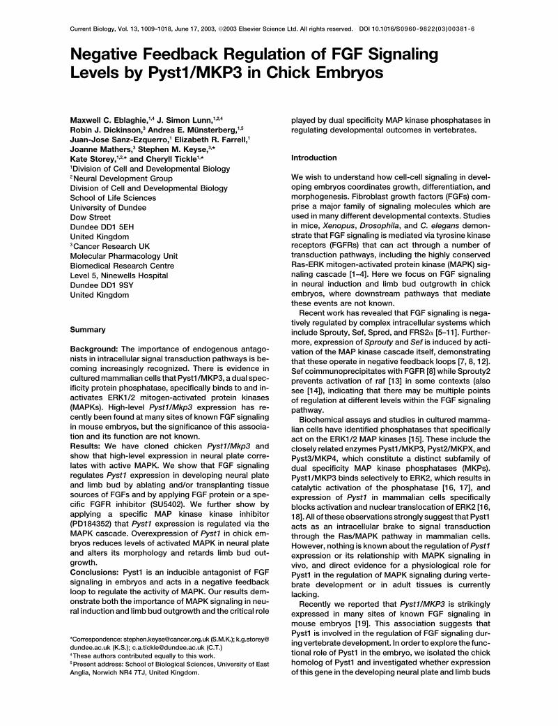
Figure 1. Expression Pattern of Chick Pyst1
(A) Pyst1 expression at Eyal-Giladi stage X.(A�) Transverse section (TS) showing Pyst1expression in epiblast layer. (B-D) Pyst1 ex-pression from HH3 to HH4 is gradually re-stricted to the neural plate and is absent inprimitive streak (ps). (D� and D″) TS showingPyst1 in neural plate and not in primitivestreak. (E) Activated MAPK is detected in theneural plate and primitive streak. (E� and E″)TS. Brown staining in periphery is due to trap-ping of reagents by yolk and not expressionin the extra-embryonic epiblast (see Supple-mental Figure S1B). (F) Pyst1 expression inthe presumptive forelimb (fl) and hindlimb (hl),as well as midbrain-hindbrain isthmus regionand somites at HH15. (G) Double in situ hy-bridization showing mesenchymal expres-sion of Pyst1 (blue) and ectodermal expres-sion of Fgf8 (magenta) in HH20 limb buds.(H and H�) Restriction of Pyst1 to distal limbmesoderm of older limb bud at stage HH24�.(H�) TS of limb bud in (H), showing Pyst1 ex-pression absent in dorsal and ventral ecto-derm (white arrowheads) and apical ectoder-mal ridge (black arrowhead). D, dorsal; Di,distal; Pr, proximal; V, ventral.
depends on FGF-mediated activation of MAPK. Finally, and 4 [1], Pyst1 is expressed more or less throughoutthe mesoderm (Figure 1G). As limb buds elongate, Pyst1we tested the developmental consequences of overex-
pressing Pyst1 in these regions. expression becomes confined to distal limb mesodermbeneath the apical ridge which continues to expressFgf8 (Figures 1H and 1H�).Results
This tight correlation between Pyst1 expression andtissue sources of FGF signaling led us to test whetherPyst1/MKP3 from chickens is highly similar to homolo-
gous proteins in human, mouse, rat, and frog (see Supple- these cell populations induce and/or maintain Pyst1 ex-pression in neural plate and limb bud mesoderm, re-mental Figure S1A available with this article online at
http://www.current-biology.com/content/supplemental). spectively, and whether FGF signaling is involved.To determine whether the node can induce Pyst1 ex-As in mouse [19], Pyst1 expression in chick embryos is
associated with many sites of FGF signaling. Here we pression, we used a well-established neural inductionassay in which we transplanted HH3 nodes into hostfocus on FGF signaling from Hensen’s node, which initi-
ates neural development [20–22], and from limb apical HH3 extra-embryonic epiblast [20, 29]. In nearly allcases, ectopic Pyst1 expression is induced within 4 hrectodermal ridge, which mediates bud outgrowth, ac-
companied by progressive formation of structures along in epiblast overlying the donor node (20/21 cases; Fig-ures 2A and 2B). Quail donor nodes were used to confirmthe long axis of the limb [23]. Pyst1 expression is first
detected in the epiblast layer of pre-streak Eyal-Giladi Pyst1 induction in chick hosts (7/8 cases; SupplementalFigures S2A, S2A�, and S2B). In contrast, grafts of HH3stage X embryos (Figures 1A and 1A�) and becomes
gradually restricted to neural plate in a pattern that re- posterior primitive streak which is not normally flankedby Pyst1-expressing cells only occasionally elicit ex-sembles the FGF-inducible preneural gene Sox3 [21].
By Hamilton and Hamburger stages (HH) 4, Pyst1 is pression (2/9 cases; data not shown). These findingsindicate that the node is a specific source of Pyst1-expressed in neural plate close to the node (tip of primi-
tive streak) (Figures 1B–1D, 1D�, and 1D″), which ex- inducing signals and place Pyst1 in the group of preneu-ral genes expressed as an early response to neural in-presses FGF2 and Fgfs3, 4, and 8 [21, 24–26]. Many cells
responding to FGF signaling in early embryos signal via ducing signals (see [21]).To test whether FGF signals are sufficient to initiatethe Ras/MAPK cascade (e.g., [7, 27]) and, consistent
with this, the activated form of MAPK is detected in the Pyst1 expression in extra-embryonic epiblast, beadssoaked in FGFs 4, 8, or 7 were grafted to this region.primitive streak and overlaps with Pyst1 in the early
neural plate (Figures 1E, 1E�, and 1E″; also see Supple- FGF4 beads induce Pyst1 within 1 hr (8/8 cases, 1 hr;15/19 cases, 2 hr), and this gene is strongly expressedmental Figure S1B).
Pyst1 is expressed in presumptive limb-forming re- after 4 hr (33/35 cases; Figure 2C), while FGF8, a lesspotent mitogen than FGF4 [30], elicits ectopic Pyst1gions in chick embryos (Figure 1F) at a time when Fgf8 is
expressed in neighboring intermediate mesoderm [28]. expression after 2 hr (4/8 cases; Figure 2D). In all cases(n � 4), control PBS beads do not induce Pyst1 (FigureThen, as limb buds form with an apical ridge at the tip
which expresses several different Fgfs, including Fgf8 2E). In contrast, FGF7, which has a restricted FGFR
FGF Signaling and Pyst1/MKP31011
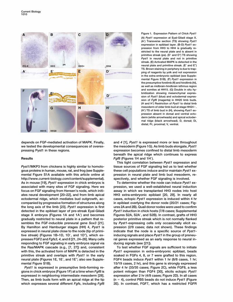
Figure 2. Pyst1 Induction by Node Is Mimicked by FGF Signaling
Whole-mount in situ hybridization with Pyst1 probe (purple/blue). (A and B) The node induces ectopic Pyst1 in extra-embryonic epiblast. (A)Chick donor node (DN) strongly induces ectopic Pyst1. (B) TS through chick DN (asterisk) in (A). (C and D) FGF mimics ability of node toinduce Pyst1. (C) FGF4 bead after 4 hr. (D) FGF8 bead after 2 hr. (E) PBS beads do not induce Pyst1. (F) Exposure to FGF4 (black asterisks)and FGFR inhibitor SU5402 (red asterisk) after 4 hr inhibits ectopic induction of Pyst1 (compare with contralateral FGF4 bead) and reducesendogenous Pyst1 expression in neural plate (broken black line indicates normal edge of neural plate).
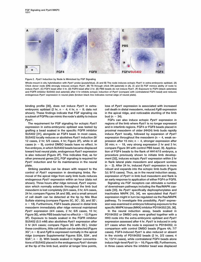
binding profile [30], does not induce Pyst1 in extra- loss of Pyst1 expression is associated with increasedcell death in distal mesoderm, reduced Fgf8 expressionembryonic epiblast (2 hr, n � 4; 4 hr, n � 8; data not
shown). These findings indicate that FGF signaling via in the apical ridge, and noticeable stunting of the limbbud (n � 34).a subset of FGFRs can mimic the node’s ability to induce
Pyst1. FGFs can also induce ectopic Pyst1 expression inregions of the limb where Pyst1 is no longer expressedThe requirement for FGF signaling for ectopic Pyst1
expression in extra-embryonic epiblast was tested by and in interlimb regions. FGF4 or FGF8 beads placed inproximal mesoderm of older (HH24) limb buds rapidlygrafting a bead soaked in the specific FGFR inhibitor
SU5402 [31], alongside an FGF4 bead. In most cases, induce Pyst1 locally, followed by expansion of Pyst1expression throughout the mesoderm (n � 4, weak ex-SU5402 locally reduces or abolishes Pyst1 induction (9/
12 cases, 2 hr; 5/5 cases, 4 hr; Figure 2F), while in all pression after 15 min; n � 4, stronger expression after30 min; n � 18, very strong expression 2 hr and 3 hr;cases (n � 9), control DMSO beads have no effect. In
five embryos, in which SU5402 beads became displaced compare Figure 3H with control PBS bead, 3I). Applica-tion of FGF4 beads to the flank of HH14/15 embryos, atoward host neural plate, endogenous Pyst1 expression
is also reduced (Figure 2F). This indicates that, as for procedure previously shown to initiate limb develop-ment [32], induces ectopic Pyst1 expression within 2 hrother preneural genes [21], FGF signaling is required for
Pyst1 induction and for its maintenance in the neural in flank lateral plate mesoderm and adjacent somites(n � 2). After 24 hr, induced Pyst1 expression is moreplate.
Striking parallels can be drawn with respect to the robust and expands into the ectopic limb buds (Figure3J; 9/15 cases). Thus, as in the neural induction assay,control of Pyst1 expression in developing limbs. Re-
moval of the apical ridge from early limb buds reduces expression of Pyst1 in limb bud mesoderm and flank isan early response to application of either FGF4 or FGF8.endogenous Pyst1 expression within an hour (data not
shown). Three hours after ridge removal, Pyst1 expres- Signaling via FGF receptors can stimulate a numberof downstream pathways including the Ras/MAPK cas-sion which normally extends throughout the limb bud
mesoderm is lost completely (3/4 cases, 3 hr; 4/4 cases, cade [33]. As Pyst1 specifically dephosphorylates andinactivates MAPK [16, 34], we speculated that Pyst124 hr; compare Figures 3A and 3B), and a small amount
of cell death can be detected at the tip by Nile Blue expression might in turn be regulated via the Ras/MAPKpathway. To investigate this possibility, Pyst1 expres-Sulfate staining (compare Figures 3C, 3C�, 3D, and 3D�;
n � 18). Furthermore, FGF4 beads placed in distal limb sion was examined in embryos following exposure to thespecific MAPK kinase (MKK) inhibitor PD184352 [35, 36].mesoderm immediately after ridge removal maintained
Pyst1 expression (4/4 cases, 2 hr; 6/6 cases, 10 hr; In the neural induction assay, beads soaked inPD184352 or DMSO only were grafted together with aFigure 3E), while PBS beads had no effect (n � 12; Figure
3F). Exposure to beads soaked in the FGFR inhibitor HH3 node into the extra-embryonic epiblast and Pyst1expression assessed after 4 hr. Pyst1 is induced in onlySU5402 (0.5 mM) also abolishes Pyst1 expression after
3 hr (4/4 cases; compare Figure 3G with 3G�). Under 2/7 cases when the node is exposed to PD184352, incomparison with control DMSO beads (Figure 4A; 7/7these conditions, little cell death can be detected (Figure
3G″; n � 8) and Fgf8 is expressed normally in the apical cases). FGF4-induced Pyst1 is also reduced or absentin the vicinity of PD184352 beads (2 hr, 2/2 cases; 4ridge (compare Supplemental Figures S3A, S3A�, and
S3A″; n � 2). With beads soaked in higher concentra- hr, 13/14 cases), while contralateral FGF4 beads aloneinduce high-level Pyst1 (n � 16; Figure 4B). Furthermore,tions of SU5402 placed in the endogenous Pyst1 domain
at the tip of the limb bud, and/or at longer time points, in three cases where the inhibitor bead was displaced
Current Biology1012
Figure 3. Endogenous Pyst1 Expression inthe Limb Is Maintained by FGF Signaling inthe Apical Ridge
(A) Endogenous Pyst1 expression in controllimb bud at HH22. (B) Loss of Pyst1 expres-sion 24 hr following apical ectodermal ridgeremoval (-AER; broken red line). (C and C�)Detection of cell death by Nile blue sulfate(NBS) staining in a control untreated HH22limb bud. A high level of apoptosis normallyoccurs in apical ridge cells. (D and D�) Apo-ptosis 6 hr following apical ridge removal(broken black line) is confined to scatteredcells in limb tip. (E) Maintenance of Pyst1 ex-pression by an FGF4 bead (asterisk) 10 hrfollowing ridge removal (broken red line). (F)PBS control bead (asterisk) does not maintainPyst1 after ridge removal (broken black line).(G, G�, and G″) Complete loss of Pyst1 ex-pression following treatment with SU5402bead. (G) Endogenous Pyst1 expression incontralateral untreated limb at HH21. (G�)Limb treated with bead soaked in 0.5 mMSU5402 bead (asterisk) for 3 hr. (G″) Samelimb as in (G�) stained for cell death with NBS.(H) Ectopic Pyst1 expression (arrowhead) 3hr after an FGF4 bead (asterisk) was placedproximally in HH24 limb bud. (I) PBS controlbead (asterisk) does not expand distal endog-enous Pyst1 expression. (J) Dorsal view.FGF4 bead implanted in flank of HH15 em-bryo for 24 hr. Pyst1 expression in ectopiclimb bud (arrowhead). El, ectopic limb bud;fl, wing bud; hl, leg bud.
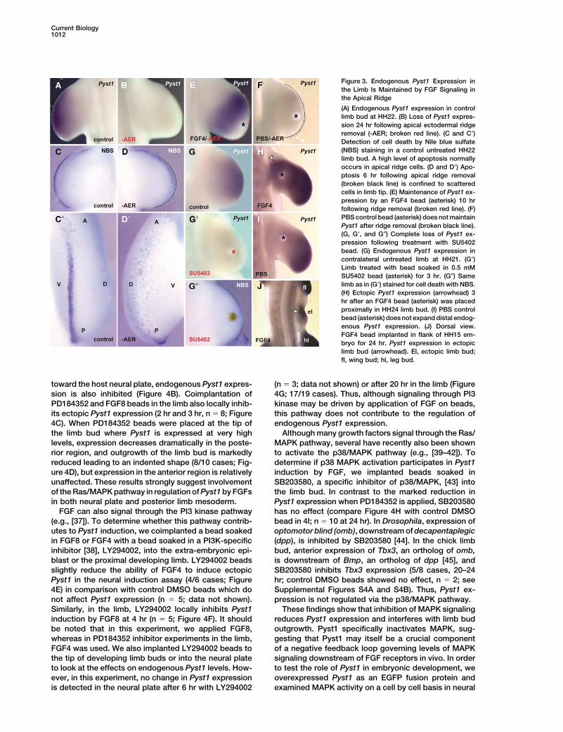
toward the host neural plate, endogenous Pyst1 expres- (n � 3; data not shown) or after 20 hr in the limb (Figure4G; 17/19 cases). Thus, although signaling through PI3sion is also inhibited (Figure 4B). Coimplantation of
PD184352 and FGF8 beads in the limb also locally inhib- kinase may be driven by application of FGF on beads,this pathway does not contribute to the regulation ofits ectopic Pyst1 expression (2 hr and 3 hr, n � 8; Figure
4C). When PD184352 beads were placed at the tip of endogenous Pyst1 expression.Although many growth factors signal through the Ras/the limb bud where Pyst1 is expressed at very high
levels, expression decreases dramatically in the poste- MAPK pathway, several have recently also been shownto activate the p38/MAPK pathway (e.g., [39–42]). Torior region, and outgrowth of the limb bud is markedly
reduced leading to an indented shape (8/10 cases; Fig- determine if p38 MAPK activation participates in Pyst1induction by FGF, we implanted beads soaked inure 4D), but expression in the anterior region is relatively
unaffected. These results strongly suggest involvement SB203580, a specific inhibitor of p38/MAPK, [43] intothe limb bud. In contrast to the marked reduction inof the Ras/MAPK pathway in regulation of Pyst1 by FGFs
in both neural plate and posterior limb mesoderm. Pyst1 expression when PD184352 is applied, SB203580has no effect (compare Figure 4H with control DMSOFGF can also signal through the PI3 kinase pathway
(e.g., [37]). To determine whether this pathway contrib- bead in 4I; n � 10 at 24 hr). In Drosophila, expression ofoptomotor blind (omb), downstream of decapentaplegicutes to Pyst1 induction, we coimplanted a bead soaked
in FGF8 or FGF4 with a bead soaked in a PI3K-specific (dpp), is inhibited by SB203580 [44]. In the chick limbbud, anterior expression of Tbx3, an ortholog of omb,inhibitor [38], LY294002, into the extra-embryonic epi-
blast or the proximal developing limb. LY294002 beads is downstream of Bmp, an ortholog of dpp [45], andSB203580 inhibits Tbx3 expression (5/8 cases, 20–24slightly reduce the ability of FGF4 to induce ectopic
Pyst1 in the neural induction assay (4/6 cases; Figure hr; control DMSO beads showed no effect, n � 2; seeSupplemental Figures S4A and S4B). Thus, Pyst1 ex-4E) in comparison with control DMSO beads which do
not affect Pyst1 expression (n � 5; data not shown). pression is not regulated via the p38/MAPK pathway.These findings show that inhibition of MAPK signalingSimilarly, in the limb, LY294002 locally inhibits Pyst1
induction by FGF8 at 4 hr (n � 5; Figure 4F). It should reduces Pyst1 expression and interferes with limb budoutgrowth. Pyst1 specifically inactivates MAPK, sug-be noted that in this experiment, we applied FGF8,
whereas in PD184352 inhibitor experiments in the limb, gesting that Pyst1 may itself be a crucial componentof a negative feedback loop governing levels of MAPKFGF4 was used. We also implanted LY294002 beads to
the tip of developing limb buds or into the neural plate signaling downstream of FGF receptors in vivo. In orderto test the role of Pyst1 in embryonic development, weto look at the effects on endogenous Pyst1 levels. How-
ever, in this experiment, no change in Pyst1 expression overexpressed Pyst1 as an EGFP fusion protein andexamined MAPK activity on a cell by cell basis in neuralis detected in the neural plate after 6 hr with LY294002
FGF Signaling and Pyst1/MKP31013
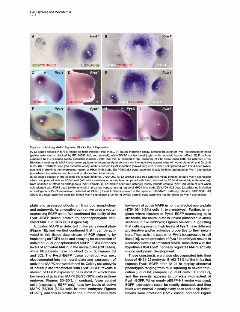
Figure 4. Inhibiting MAPK Signaling Blocks Pyst1 Expression
(A–D) Beads soaked in MAPK kinase-specific inhibitor, PD184352. (A) Neural induction assay. Ectopic induction of Pyst1 expression by node(yellow asterisks) is blocked by PD184352 (left; red asterisk), while DMSO control bead (right; white asterisk) has no effect. (B) Four hourexposure to FGF4 beads (white asterisks) induces Pyst1, but this is inhibited in the presence of PD184352 bead (left; red asterisk; 4 hr).Blocking signaling via MAPK also downregulates endogenous Pyst1 (broken red line indicates normal edge of neural plate). (C and D) Limbbuds. (C) PD184352 bead (red asterisk) locally inhibits ectopic Pyst1 induction (arrowhead) at 2 hr when coimplanted with FGF4 bead (whiteasterisk) in proximal nonexpressing region of HH24 limb buds. (D) PD184352 bead (asterisk) locally inhibits endogenous Pyst1 expression(arrowhead) in posterior limb bud and produces limb indentation.(E–G) Beads soaked in the specific PI3 kinase inhibitor, LY294002. (E) LY294002 bead (red asterisk) mildly inhibits ectopic Pyst1 expressionwhen coimplanted with an FGF4 bead (left; white asterisk) in neural plate compared with Pyst1 induced by FGF4 alone (right; white asterisk).Note absence of effect on endogenous Pyst1 domain. (F) LY294002 bead (red asterisk) locally inhibits ectopic Pyst1 induction at 3 hr whencoimplanted with FGF8 bead (white asterisk) in proximal nonexpressing region of HH24 limb buds. (G) LY294002 bead (asterisk); no inhibitionof endogenous Pyst1 expression detected at 24 hr. (H and I) Beads soaked in the specific p38/MAPK pathway inhibitor, SB203580. (H)SB203580 bead (asterisk) does not inhibit Pyst1 expression at 24 hr. (I) DMSO control bead (asterisk) has no effect on Pyst1 expression.
plate and assessed effects on limb bud morphology low levels of active MAPK in nontransfected neural plate(475/1064 (45%) cells in two embryos). Further, in re-and outgrowth. As a negative control, we used a vector
expressing EGFP alone. We confirmed the ability of the gions where clusters of Pyst1-EGFP-expressing cellsare found, the neural plate is kinked (observed in 48/54Pyst1-EGFP fusion protein to dephosphorylate acti-
vated MAPK in COS cells (Figure 5A). sections in five embryos; Figures 5D–5D″), suggestingthat cells expressing high levels of Pyst1 have differentActivated MAPK is detected in the early neural plate
(Figure 1E), and we first confirmed that it can be acti- proliferative and/or adhesive properties to their neigh-bors. Thus, as is the case when Pyst1 is expressed in cellvated in this tissue downstream of FGF signaling by
implanting an FGF4 bead and assaying for expression of lines [16], overexpression of Pyst1 in embryos results indecreased levels of activated MAPK, consistent with theactivated, dual-phosphorylated MAPK. FGF4 increases
levels of activated MAPK in the neural plate (7/8 cases), hypothesis that Pyst1 normally regulates MAPK activityduring embryonic development.while PBS beads have no effect (n � 5; Figures 5B
and 5C). The Pyst1-EGFP fusion construct was next These constructs were also electroporated into limbbuds of HH21-22 embryos. 51/63 (81%) of the limbs thatelectroporated into the neural plate and expression of
activated MAPK analyzed after 6 hr. Cell by cell analysis express Pyst1-EGFP after 12–24 hr display abnormalphenotypes ranging from mild squaring to severe trun-of neural plate transfected with Pyst1-EGFP reveals a
mosaic of EGFP expressing cells most of which have cation (Figure 6A; compare Figure 6B with 6B� and 6B″),and the severity appears to correlate with extent oflow levels of activated MAPK (68/75 (90%) cells in three
embryos; Figures 5D–5D″). In contrast, fewer control Pyst1-EGFP. When empty pEGFP-N1 vector was used,EGFP expression could be readily detected, and limbcells (expressing EGFP only) have low levels of active
MAPK (68/109 (62%) cells in three embryos; Figures buds were normal in nearly every case and no tip inden-tations were produced (15/17 cases; compare Figure5E–5E″), and this is similar to the number of cells with
Current Biology1014
observed in any of the truncated limb buds in distalregions of the bud expressing Pyst1-EGFP (compareuntreated limb in Figures 7B and 7C with Pyst1-EGFPelectroporated limb in 7B� and 7C�). Thus, Pyst1 overex-pression is not accompanied by cell death.
Discussion
These results place a dual specificity MAPK phospha-tase in a physiological context in a vertebrate and showthat Pyst1 expression in neural plate and limb is regu-lated by FGFs. When tissues expressing FGFs are re-moved or beads soaked in the FGFR inhibitor, SU5402,are applied, Pyst1 expression is abolished. We haveshown in the limb that this change in Pyst1 expressioncannot be accounted for by cell death. Application ofFGFs can maintain endogenous Pyst1 expression in thelimb or can induce it very rapidly in ectopic locations inboth early embryos and limbs, showing that expressionof Pyst1 is an early response to FGF signaling. Further-more, using specific inhibitors for the classical MAPKcascade, the PI3 kinase and p38 MAPK pathways, weshow that maintenance of endogenous Pyst1 expres-sion in the neural plate and posterior limb mesodermby FGF occurs by a direct feedback loop involving theclassical pathway. Although PD184352 inhibits Pyst1 inneural plate and posterior limb, unexpectedly, anteriorlimb Pyst1 expression is relatively unaffected. It may besignificant that different Fgfs are known to be expressedin anterior and posterior apical ridge (reviewed in [1]).
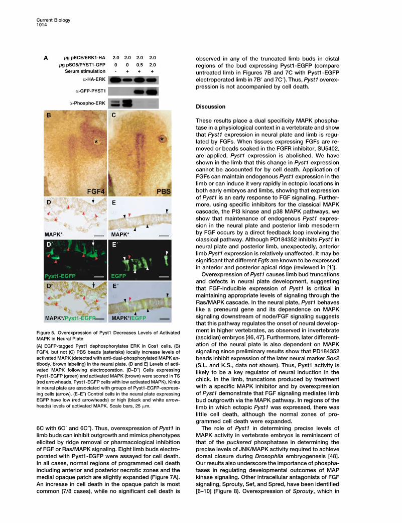
Overexpression of Pyst1 causes limb bud truncationsand defects in neural plate development, suggestingthat FGF-inducible expression of Pyst1 is critical inmaintaining appropriate levels of signaling through theRas/MAPK cascade. In the neural plate, Pyst1 behaveslike a preneural gene and its dependence on MAPKsignaling downstream of node/FGF signaling suggeststhat this pathway regulates the onset of neural develop-ment in higher vertebrates, as observed in invertebrateFigure 5. Overexpression of Pyst1 Decreases Levels of Activated(ascidian) embryos [46, 47]. Furthermore, later differenti-MAPK in Neural Plateation of the neural plate is also dependent on MAPK(A) EGFP-tagged Pyst1 dephosphorylates ERK in Cos1 cells. (B)signaling since preliminary results show that PD184352FGF4, but not (C) PBS beads (asterisks) locally increase levels of
activated MAPK (detected with anti-dual-phosphorylated MAPK an- beads inhibit expression of the later neural marker Sox2tibody, brown labeling) in the neural plate. (D and E) Levels of acti- (S.L. and K.S., data not shown). Thus, Pyst1 activity isvated MAPK following electroporation. (D–D″) Cells expressing likely to be a key regulator of neural induction in thePyst1-EGFP (green) and activated MAPK (brown) were scored in TS
chick. In the limb, truncations produced by treatment(red arrowheads, Pyst1-EGFP cells with low activated MAPK). Kinkswith a specific MAPK inhibitor and by overexpressionin neural plate are associated with groups of Pyst1-EGFP-express-of Pyst1 demonstrate that FGF signaling mediates limbing cells (arrow). (E–E″) Control cells in the neural plate expressing
EGFP have low (red arrowheads) or high (black and white arrow- bud outgrowth via the MAPK pathway. In regions of theheads) levels of activated MAPK. Scale bars, 25 �m. limb in which ectopic Pyst1 was expressed, there was
little cell death, although the normal zones of pro-grammed cell death were expanded.
The role of Pyst1 in determining precise levels of6C with 6C� and 6C″). Thus, overexpression of Pyst1 inlimb buds can inhibit outgrowth and mimics phenotypes MAPK activity in vertebrate embryos is reminiscent of
that of the puckered phosphatase in determining theelicited by ridge removal or pharmacological inhibitionof FGF or Ras/MAPK signaling. Eight limb buds electro- precise levels of JNK/MAPK activity required to achieve
dorsal closure during Drosophila embryogenesis [48].porated with Pyst1-EGFP were assayed for cell death.In all cases, normal regions of programmed cell death Our results also underscore the importance of phospha-
tases in regulating developmental outcomes of MAPincluding anterior and posterior necrotic zones and themedial opaque patch are slightly expanded (Figure 7A). kinase signaling. Other intracellular antagonists of FGF
signaling, Sprouty, Sef, and Spred, have been identifiedAn increase in cell death in the opaque patch is mostcommon (7/8 cases), while no significant cell death is [6–10] (Figure 8). Overexpression of Sprouty, which in
FGF Signaling and Pyst1/MKP31015
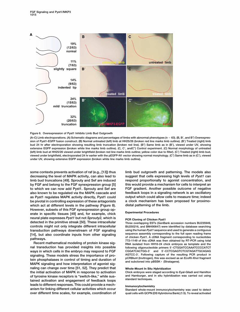
Figure 6. Overexpression of Pyst1 Inhibits Limb Bud Outgrowth
(A–C) Limb electroporations. (A) Schematic diagrams and percentages of limbs with abnormal phenotypes (n � 63). (B, B�, and B″) Overexpres-sion of Pyst1-EGFP fusion construct. (B) Normal untreated (left) limb at HH25/26 (broken red line marks limb outline). (B�) Treated (right) limbbud 24 hr after electroporation showing resulting limb truncation (broken red line). (B″) Same limb as in (B�), viewed under UV, showingextensive EGFP expression (broken white line marks limb outline). (C, C�, andC″) Control experiment. (C) Normal morphology of untreated(left) limb bud at HH25/26 viewed under brightfield (broken red line marks limb outline; yellow color due to filter). (C�) Treated (right) limb bud,viewed under brightfield, electroporated 24 hr earlier with the pEGFP-N1 vector showing normal morphology. (C″) Same limb as in (C�), viewedunder UV, showing extensive EGFP expression (broken white line marks limb outline).
some contexts prevents activation of raf (e.g., [13]) thus limb bud outgrowth and patterning. The models alsosuggest that cells expressing high levels of Pyst1 candecreasing the level of MAPK activity, can also lead torespond proportionally to agonist concentration, andlimb bud truncations [49]. Sprouty and Sef are inducedthis would provide a mechanism for cells to interpret anby FGF and belong to the FGF synexpression group [5]FGF gradient. Another possible outcome of negativeto which we can now add Pyst1. Sprouty and Sef arefeedback loops in a signaling network is an oscillatoryalso known to be regulated via the MAPK cascade andoutput which could allow cells to measure time; indeedas Pyst1 regulates MAPK activity directly, Pyst1 coulda clock mechanism has been proposed for proximo-be pivotal in controlling expression of these antagonistsdistal patterning of the limb.which act at different levels in the pathway (Figure 8).
However, subsets of this FGF synexpression group op-Experimental Procedureserate in specific tissues [49] and, for example, chick
neural plate expresses Pyst1 but not Sprouty2, which is PCR Cloning of Chicken Pyst1detected in the primitive streak [50]. These intracellular Three overlapping ESTs (GenBank accession numbers BU235949,controls might not only integrate different intracellular BU250216, and BM490647) were identified by database searching
using the human Pyst1 sequence and used to generate a contiguoustransduction pathways downstream of FGF signalingsequence assembly corresponding to the full open reading frame[14], but also coordinate inputs from other signalingof chicken Pyst1. A cDNA fragment corresponding to nucleotidespathways. 772–1149 of this cDNA was then obtained by RT-PCR using total
Recent mathematical modeling of protein kinase sig- RNA isolated from HH16-24 chick embryos as template and thenal transduction has provided insights into possible following oligonucleotide primers 5�-CTGGATCCAAATCCCCATCT
CGGATCACTGG-3� and 5�-CCTCGAGTCTCGTAGATTGCAGAGways in which cells in the embryo may respond to FGFAGTCC-3�. Following capture of the resulting PCR product insignaling. These models stress the importance of pro-pCRBlunt (Invitrogen), this was excised as an EcoRI-XhoI fragmenttein phosphatases in control of timing and duration ofand subcloned into pBSSK� (Stratagene).
MAPK signaling and how interpretation of agonist sig-naling can change over time [51, 52]. They predict that Whole-Mount In Situ Hybridization
Chick embryos were staged according to Eyal-Giladi and Hamiltonthe initial activation of MAPK in response to activationand Hamburger, and in situ hybridization was carried out usingof tyrosine kinase receptors is “switch-like,” while sus-standard techniques.tained activation and engagement of feedback loops
leads to different responses. This could provide a mech- Immunocytochemistryanism for linking different cellular activities which occur Standard whole-mount immunocytochemistry was used to detect
quail cells with QCPN (DS Hybridoma Bank) (1:5). To reveal activatedover different time scales, for example, coordination of
Current Biology1016
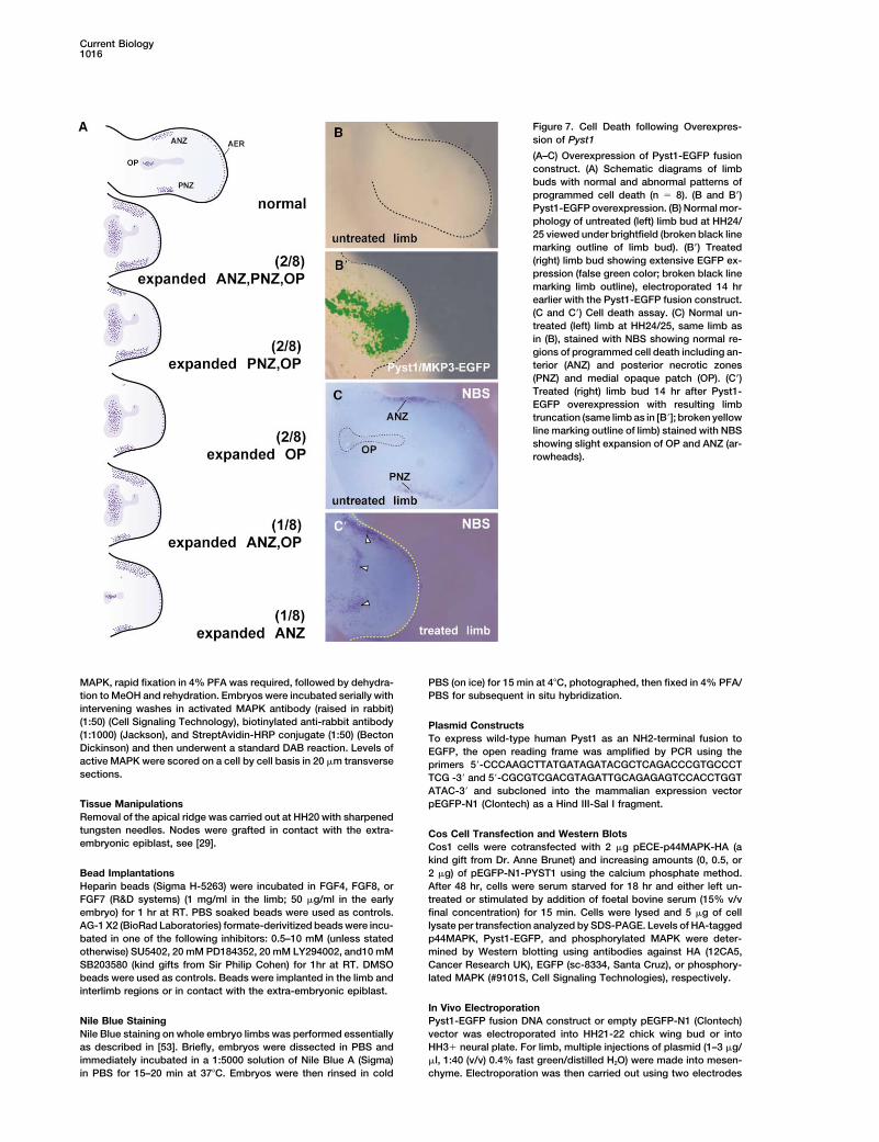
Figure 7. Cell Death following Overexpres-sion of Pyst1
(A–C) Overexpression of Pyst1-EGFP fusionconstruct. (A) Schematic diagrams of limbbuds with normal and abnormal patterns ofprogrammed cell death (n � 8). (B and B�)Pyst1-EGFP overexpression. (B) Normal mor-phology of untreated (left) limb bud at HH24/25 viewed under brightfield (broken black linemarking outline of limb bud). (B�) Treated(right) limb bud showing extensive EGFP ex-pression (false green color; broken black linemarking limb outline), electroporated 14 hrearlier with the Pyst1-EGFP fusion construct.(C and C�) Cell death assay. (C) Normal un-treated (left) limb at HH24/25, same limb asin (B), stained with NBS showing normal re-gions of programmed cell death including an-terior (ANZ) and posterior necrotic zones(PNZ) and medial opaque patch (OP). (C�)Treated (right) limb bud 14 hr after Pyst1-EGFP overexpression with resulting limbtruncation (same limb as in [B�]; broken yellowline marking outline of limb) stained with NBSshowing slight expansion of OP and ANZ (ar-rowheads).
MAPK, rapid fixation in 4% PFA was required, followed by dehydra- PBS (on ice) for 15 min at 4�C, photographed, then fixed in 4% PFA/PBS for subsequent in situ hybridization.tion to MeOH and rehydration. Embryos were incubated serially with
intervening washes in activated MAPK antibody (raised in rabbit)(1:50) (Cell Signaling Technology), biotinylated anti-rabbit antibody Plasmid Constructs(1:1000) (Jackson), and StreptAvidin-HRP conjugate (1:50) (Becton To express wild-type human Pyst1 as an NH2-terminal fusion toDickinson) and then underwent a standard DAB reaction. Levels of EGFP, the open reading frame was amplified by PCR using theactive MAPK were scored on a cell by cell basis in 20 �m transverse primers 5�-CCCAAGCTTATGATAGATACGCTCAGACCCGTGCCCTsections. TCG -3� and 5�-CGCGTCGACGTAGATTGCAGAGAGTCCACCTGGT
ATAC-3� and subcloned into the mammalian expression vectorpEGFP-N1 (Clontech) as a Hind III-Sal I fragment.Tissue Manipulations
Removal of the apical ridge was carried out at HH20 with sharpenedtungsten needles. Nodes were grafted in contact with the extra- Cos Cell Transfection and Western Blotsembryonic epiblast, see [29]. Cos1 cells were cotransfected with 2 �g pECE-p44MAPK-HA (a
kind gift from Dr. Anne Brunet) and increasing amounts (0, 0.5, or2 �g) of pEGFP-N1-PYST1 using the calcium phosphate method.Bead Implantations
Heparin beads (Sigma H-5263) were incubated in FGF4, FGF8, or After 48 hr, cells were serum starved for 18 hr and either left un-treated or stimulated by addition of foetal bovine serum (15% v/vFGF7 (R&D systems) (1 mg/ml in the limb; 50 �g/ml in the early
embryo) for 1 hr at RT. PBS soaked beads were used as controls. final concentration) for 15 min. Cells were lysed and 5 �g of celllysate per transfection analyzed by SDS-PAGE. Levels of HA-taggedAG-1 X2 (BioRad Laboratories) formate-derivitized beads were incu-
bated in one of the following inhibitors: 0.5–10 mM (unless stated p44MAPK, Pyst1-EGFP, and phosphorylated MAPK were deter-mined by Western blotting using antibodies against HA (12CA5,otherwise) SU5402, 20 mM PD184352, 20 mM LY294002, and10 mM
SB203580 (kind gifts from Sir Philip Cohen) for 1hr at RT. DMSO Cancer Research UK), EGFP (sc-8334, Santa Cruz), or phosphory-lated MAPK (#9101S, Cell Signaling Technologies), respectively.beads were used as controls. Beads were implanted in the limb and
interlimb regions or in contact with the extra-embryonic epiblast.
In Vivo ElectroporationPyst1-EGFP fusion DNA construct or empty pEGFP-N1 (Clontech)Nile Blue Staining
Nile Blue staining on whole embryo limbs was performed essentially vector was electroporated into HH21-22 chick wing bud or intoHH3� neural plate. For limb, multiple injections of plasmid (1–3 �g/as described in [53]. Briefly, embryos were dissected in PBS and
immediately incubated in a 1:5000 solution of Nile Blue A (Sigma) �l, 1:40 (v/v) 0.4% fast green/distilled H2O) were made into mesen-chyme. Electroporation was then carried out using two electrodesin PBS for 15–20 min at 37�C. Embryos were then rinsed in cold

FGF Signaling and Pyst1/MKP31017
Received: December 23, 2002Revised: April 17, 2003Accepted: April 17, 2003Published: June 17, 2003
References
1. Martin, G.R. (1998). The roles of FGFs in early development ofvertebrate limbs. Genes Dev. 12, 1571–1586.
2. Umbhauer, M., Marshall, C.J., Mason, C.S., Old, R.W., andSmith, J.C. (1995). Mesoderm induction in Xenopus caused byactivation of MAP kinase. Nature 376, 58–62.
3. Michelson, A.M., Gisselbrecht, S., Buff, E., and Skeath, J.B.(1998). Heartbroken is a specific downstream mediator of FGFreceptor signalling in Drosophila. Development 125, 4379–4389.
4. Borland, C.Z., Schutzman, J.L., and Stern, M.J. (2001). Fibro-blast growth factor signaling in Caenorhabditis elegans. Bioes-says 23, 1120–1130.
5. Niehrs, C., and Meinhardt, H. (2002). Modular feedback. Nature417, 35–36.
6. Hacohen, N., Kramer, S., Sutherland, D., Hiromi, Y., and Kras-now, M.A. (1998). sprouty encodes a novel antagonist of FGFsignaling that patterns apical branching of the Drosophila air-ways. Cell 92, 253–263.
7. Furthauer, M., Lin, W., Ang, S.L., Thisse, B., and Thisse, C.(2002). Sef is a feedback-induced antagonist of Ras/MAPK-mediated FGF signalling. Nat. Cell Biol. 4, 170–174.
8. Tsang, M., Friesel, R., Kudoh, T., and Dawid, I.B. (2002). Identifi-cation of Sef, a novel modulator of FGF signalling. Nat. CellBiol. 4, 165–169.
9. Kovalenko, D., Yang, X., Nadeau, R.J., Harkins, L.K., and Friesel,R. (2003). Sef inhibits fibroblast growth factor signaling by inhib-iting FGFR1 tyrosine phosphorylation and subsequent ERK acti-vation. J. Biol. Chem. 278, 14087–14091.Figure 8. Inhibitory Feedback Loops Downstream of FGF Signaling
10. Wakioka, T., Sasaki, A., Kato, R., Shouda, T., Matsumoto, A.,Factors regulating signal propagation via MAP kinase cascadeMiyoshi, K., Tsuneoka, M., Komiya, S., Baron, R., and Yoshi-
downstream of FGF receptor activation. Previously identified inhibi-mura, A. (2001). Spred is a Sprouty-related suppressor of Ras
tory factors Sef [7–9], Spred [10], and Sprouty [6] (but see [14])signalling. Nature 412, 647–651.
(yellow) act at different levels within FGF signaling pathway, al-11. Lax, I., Wong, A., Lamothe, B., Lee, A., Frost, A., Hawes, J., and
though the biochemical basis of these inhibitory activities is not yetSchlessinger, J. (2002). The docking protein FRS2alpha controls
clear. In contrast, PYST1/MKP3 is a known specific inhibitor ofa MAP kinase-mediated negative feedback mechanism for sig-
MAPK signaling through dephosphorylation of the T-E-Y MAPK acti-naling by FGF receptors. Mol. Cell 10, 709–719.
vation motif [16, 34]. As with Sef and Sprouty, it is expressed down- 12. Ozaki, K., Kadomoto, R., Asato, K., Tanimura, S., Itoh, N., andstream of FGF-induced MAPK signals and thus constitutes a novel Kohno, M. (2001). ERK pathway positively regulates the expres-feedback loop governing activity of this pathway (red arrows). Points sion of Sprouty genes. Biochem. Biophys. Res. Commun. 285,of action for pharmacological inhibitors SU5402 and PD184352 1084–1088.(blue) are indicated at level of FGF receptor and MEK, respectively. 13. Yusoff, P., Lao, D.H., Ong, S.H., Wong, E.S., Lim, J., Lo, T.L.,
Leong, H.F., Fong, C.W., and Guy, G.R. (2002). Sprouty2 inhibitsthe Ras/MAP kinase pathway by inhibiting the activation of Raf.J. Biol. Chem. 277, 3195–3201.(2.5 mm apart) placed at anterior and posterior edges of wing bud
14. Nutt, S.L., Dingwell, K.S., Holt, C.E., and Amaya, E. (2001). Xeno-and 30–50V administered in 2–3 pulses of 50 ms using a CUY-21pus Sprouty2 inhibits FGF-mediated gastrulation movementspulse generator. For neural plate, embryos prepared in vitro (follow-but does not affect mesoderm induction and patterning. Genesing EC culture methods) were electroporated in a custom built cham-Dev. 15, 1152–1166.ber (a kind gift of Ivor Mason) and exposed to 2 pulses of 50 ms �
15. Keyse, S.M. (2000). Protein phosphatases and the regulation of10V using an Intracel Intracept TSS10 pulse generator.mitogen-activated protein kinase signalling. Curr. Opin. CellBiol. 12, 186–192.
16. Groom, L.A., Sneddon, A.A., Alessi, D.R., Dowd, S., and Keyse,Supplemental DataS.M. (1996). Differential regulation of the MAP, SAP and RK/Supplemental Figures for this article are available at http://www.p38 kinases by Pyst1, a novel cytosolic dual-specificity phos-current-biology.com/content/supplemental.phatase. EMBO J. 15, 3621–3632.
17. Camps, M., Nichols, A., Gillieron, C., Antonsson, B., Muda, M.,Acknowledgments Chabert, C., Boschert, U., and Arkinstall, S. (1998). Catalytic
activation of the phosphatase MKP-3 by ERK2 mitogen-acti-We are grateful for the technical assistance of Graham Christie, vated protein kinase. Science 280, 1262–1265.Fiona MacIsaac, and Pam Halley. M.C.E. is supported by a student- 18. Brunet, A., Roux, D., Lenormand, P., Dowd, S., Keyse, S., andship from the Anatomical Society of Great Britain and Ireland and Pouyssegur, J. (1999). Nuclear translocation of p42/p44 mito-the British Council. J.S.L. is a BBSRC Student. R.J.D. is supported by gen-activated protein kinase is required for growth factor-a studentship from the Biomedical Research Centre and by Cancer induced gene expression and cell cycle entry. EMBO J. 18,Research UK. A.E.M. was a Wellcome Trust RCDF. J.M. is supported 664–674.by a studentship from the Imperial Cancer Research Fund (now 19. Dickinson, R.J., Eblaghie, M.C., Keyse, S.M., and Morriss-Kay,Cancer Research UK). S.M.K. is supported by Cancer Research UK. G.M. (2002). Expression of the ERK-specific MAP kinase phos-K.S. is an MRC Senior Research Fellow. C.T. is a Royal Society phatase PYST1/MKP3 in mouse embryos during morphogene-
sis and early organogenesis. Mech. Dev. 113, 193–196.Research Professor and supported by the MRC.

Current Biology1018
20. Storey, K.G., Goriely, A., Sargent, C.M., Brown, J.M., Burns, scatter factor activates proliferation in melanoma cells throughp38 MAPK, ATF-2 and cyclin D1. Oncogene 21, 1000–1008.H.D., Abud, H.M., and Heath, J.K. (1998). Early posterior neural
tissue is induced by FGF in the chick embryo. Development 41. Shimoke, K., and Kudo, M. (2002). 1-Methyl-4-phenyl-1,2,3,6-tetrahydropyridine has a transient proliferative effect on PC12h125, 473–484.
21. Streit, A., Berliner, A.J., Papanayotou, C., Sirulnik, A., and Stern, cells and nerve growth factor additively promotes this effect:possible involvement of distinct mechanisms of activation ofC.D. (2000). Initiation of neural induction by FGF signalling be-
fore gastrulation. Nature 406, 74–78. MAP kinase family proteins. Brain Res. Dev. Brain Res. 133,105–114.22. Wilson, S.I., Graziano, E., Harland, R., Jessell, T.M., and Edlund,
T. (2000). An early requirement for FGF signalling in the acquisi- 42. Morooka, T., and Nishida, E. (1998). Requirement of p38 mito-gen-activated protein kinase for neuronal differentiation in PC12tion of neural cell fate in the chick embryo. Curr. Biol. 10,
421–429. cells. J. Biol. Chem. 273, 24285–24288.43. Cuenda, A., Rouse, J., Doza, Y.N., Meier, R., Cohen, P., Gal-23. Niswander, L., Tickle, C., Vogel, A., Booth, I., and Martin, G.R.
(1993). FGF-4 replaces the apical ectodermal ridge and directs lagher, T.F., Young, P.R., and Lee, J.C. (1995). SB 203580 is aspecific inhibitor of a MAP kinase homologue which is stimu-outgrowth and patterning of the limb. Cell 75, 579–587.
24. Riese, J., Zeller, R., and Dono, R. (1995). Nucleo-cytoplasmic lated by cellular stresses and interleukin-1. FEBS Lett. 364,translocation and secretion of fibroblast growth factor-2 during 229–233.avian gastrulation. Mech. Dev. 49, 13–22. 44. Adachi-Yamada, T., Nakamura, M., Irie, K., Tomoyasu, Y., Sano,
25. Mahmood, R., Kiefer, P., Guthrie, S., Dickson, C., and Mason, Y., Mori, E., Goto, S., Ueno, N., Nishida, Y., and Matsumoto, K.I. (1995). Multiple roles for FGF-3 during cranial neural develop- (1999). p38 mitogen-activated protein kinase can be involvedment in the chicken. Development 121, 1399–1410. in transforming growth factor beta superfamily signal transduc-
26. Shamim, H., and Mason, I. (1999). Expression of Fgf4 during tion in Drosophila wing morphogenesis. Mol. Cell. Biol. 19,early development of the chick embryo. Mech. Dev. 85, 189–192. 2322–2329.
27. Christen, B., and Slack, J.M.W. (1999). Spatial response to fibro- 45. Tumpel, S., Sanz-Ezquerro, J.J., Isaac, A., Eblaghie, M.C., Dob-blast growth factor signalling in Xenopus embryos. Develop- son, J., and Tickle, C. (2002). Regulation of Tbx3 expression byment 126, 119–125. anteroposterior signalling in vertebrate limb development. Dev.
28. Crossley, P.H., Minowada, G., MacArthur, C.A., and Martin, G.R. Biol. 250, 251–262.(1996). Roles for FGF8 in the induction, initiation, and mainte- 46. Kim, G.J., and Nishida, H. (2001). Role of the FGF and MEKnance of chick limb development. Cell 84, 127–136. signaling pathway in the ascidian embryo. Dev. Growth Differ.
29. Storey, K.G., Crossley, J.M., De Robertis, E.M., Norris, W.E., 43, 521–533.and Stern, C.D. (1992). Neural induction and regionalisation in 47. Hudson, C., Darras, S., Caillol, D., Yasuo, H., and Lemaire, P.the chick embryo. Development 114, 729–741. (2003). A conserved role for the MEK signalling pathway in neural
30. Ornitz, D.M., Xu, J., Colvin, J.S., McEwen, D.G., MacArthur, tissue specification and posteriorisation in the invertebrateC.A., Coulier, F., Gao, G., and Goldfarb, M. (1996). Receptor chordate, the ascidian Ciona intestinalis. Development 130,specificity of the fibroblast growth factor family. J. Biol. Chem. 147–159.271, 15292–15297. 48. Martin-Blanco, E., Gampel, A., Ring, J., Virdee, K., Kirov, N.,
31. Mohammadi, M., McMahon, G., Sun, L., Tang, C., Hirth, P., Yeh, Tolkovsky, A.M., and Martinez-Arias, A. (1998). puckered en-B.K., Hubbard, S.R., and Schlessinger, J. (1997). Structures of codes a phosphatase that mediates a feedback loop regulatingthe tyrosine kinase domain of fibroblast growth factor receptor JNK activity during dorsal closure in Drosophila. Genes Dev.in complex with inhibitors. Science 276, 955–960. 12, 557–570.
32. Cohn, M.J., Izpisua Belmonte, J.C., Abud, H., Heath, J.K., and 49. Minowada, G., Jarvis, L.A., Chi, C.L., Neubuser, A., Sun, X.,Tickle, C. (1995). Fibroblast growth factors induce additional Hacohen, N., Krasnow, M.A., and Martin, G.R. (1999). Vertebratelimb development from the flank of chick embryos. Cell 80, Sprouty genes are induced by FGF signaling and can cause739–746. chondrodysplasia when overexpressed. Development 126,
33. Kouhara, H., Hadari, Y.R., Spivak-Kroizman, T., Schilling, J., 4465–4475.Bar-Sagi, D., Lax, I., and Schlessinger, J. (1997). A lipid- 50. Chambers, D., and Mason, I. (2000). Expression of sprouty2anchored Grb2-binding protein that links FGF-receptor activa- during early development of the chick embryo is coincident withtion to the Ras/MAPK signaling pathway. Cell 89, 693–702. known sites of FGF signalling. Mech. Dev. 91, 361–364.
34. Muda, M., Theodosiou, A., Rodrigues, N., Boschert, U., Camps, 51. Heinrich, R., Neel, B.G., and Rapoport, T.A. (2002). MathematicalM., Gillieron, C., Davies, K., Ashworth, A., and Arkinstall, S. models of protein kinase signal transduction. Mol. Cell 9,(1996). The dual specificity phosphatases M3/6 and MKP-3 are 957–970.highly selective for inactivation of distinct mitogen-activated 52. Bhalla, U.S., Ram, P.T., and Iyengar, R. (2002). MAP kinaseprotein kinases. J. Biol. Chem. 271, 27205–27208. phosphatase as a locus of flexibility in a mitogen-activated pro-
35. Sebolt-Leopold, J.S., Dudley, D.T., Herrera, R., Van Becelaere, tein kinase signaling network. Science 297, 1018–1023.K., Wiland, A., Gowan, R.C., Tecle, H., Barrett, S.D., Bridges, 53. Sanz-Ezquerro, J.J., and Tickle, C. (2000). Autoregulation of ShhA., Przybranowski, S., et al. (1999). Blockade of the MAP kinase expression and Shh induction of cell death suggest a mecha-pathway suppresses growth of colon tumors in vivo. Nat. Med.
nism for modulating polarising activity during chick limb devel-5, 810–816.
opment. Development 127, 4811–4823.36. Davies, S.P., Reddy, H., Caivano, M., and Cohen, P. (2000).
Specificity and mechanism of action of some commonly usedprotein kinase inhibitors. Biochem. J. 351, 95–105.
37. Ong, S.H., Hadari, Y.R., Gotoh, N., Guy, G.R., Schlessinger, J.,and Lax, I. (2001). Stimulation of phosphatidylinositol 3-kinaseby fibroblast growth factor receptors is mediated by coordi-nated recruitment of multiple docking proteins. Proc. Natl. Acad.Sci. USA 98, 6074–6079.
38. Vlahos, C.J., Matter, W.F., Hui, K.Y., and Brown, R.F. (1994). Aspecific inhibitor of phosphatidylinositol 3-kinase, 2-(4-morpho-linyl)-8-phenyl-4H–1-benzopyran-4-one (LY294002). J. Biol.Chem. 269, 5241–5248.
39. Hayama, M., Inoue, R., Akiba, S., and Sato, T. (2002). ERK andp38 MAP kinase are involved in arachidonic acid release in-duced by H2O2 and PDGF in mesangial cells. Am. J. Physiol.Renal Physiol. 282, F485–F491.
40. Recio, J.A., and Merlino, G. (2002). Hepatocyte growth factor/