Human cerebellar responses to brush and heat stimuli in healthy and neuropathic pain subjects
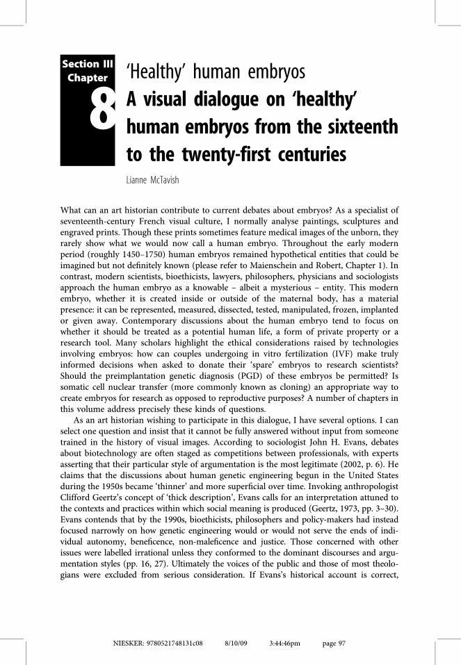
‘Healthy’ human embryosA visual dialogue on ‘healthy’human embryos from the sixteenthto the twenty-first centuriesLianne McTavish
What can an art historian contribute to current debates about embryos? As a specialist ofseventeenth-century French visual culture, I normally analyse paintings, sculptures andengraved prints. Though these prints sometimes feature medical images of the unborn, theyrarely show what we would now call a human embryo. Throughout the early modernperiod (roughly 1450–1750) human embryos remained hypothetical entities that could beimagined but not definitely known (please refer to Maienschein and Robert, Chapter 1). Incontrast, modern scientists, bioethicists, lawyers, philosophers, physicians and sociologistsapproach the human embryo as a knowable – albeit a mysterious – entity. This modernembryo, whether it is created inside or outside of the maternal body, has a materialpresence: it can be represented, measured, dissected, tested, manipulated, frozen, implantedor given away. Contemporary discussions about the human embryo tend to focus onwhether it should be treated as a potential human life, a form of private property or aresearch tool. Many scholars highlight the ethical considerations raised by technologiesinvolving embryos: how can couples undergoing in vitro fertilization (IVF) make trulyinformed decisions when asked to donate their ‘spare’ embryos to research scientists?Should the preimplantation genetic diagnosis (PGD) of these embryos be permitted? Issomatic cell nuclear transfer (more commonly known as cloning) an appropriate way tocreate embryos for research as opposed to reproductive purposes? A number of chapters inthis volume address precisely these kinds of questions.
As an art historian wishing to participate in this dialogue, I have several options. I canselect one question and insist that it cannot be fully answered without input from someonetrained in the history of visual images. According to sociologist John H. Evans, debatesabout biotechnology are often staged as competitions between professionals, with expertsasserting that their particular style of argumentation is the most legitimate (2002, p. 6). Heclaims that the discussions about human genetic engineering begun in the United Statesduring the 1950s became ‘thinner’ and more superficial over time. Invoking anthropologistClifford Geertz’s concept of ‘thick description’, Evans calls for an interpretation attuned tothe contexts and practices within which social meaning is produced (Geertz, 1973, pp. 3–30).Evans contends that by the 1990s, bioethicists, philosophers and policy-makers had insteadfocused narrowly on how genetic engineering would or would not serve the ends of indi-vidual autonomy, beneficence, non-maleficence and justice. Those concerned with otherissues were labelled irrational unless they conformed to the dominant discourses and argu-mentation styles (pp. 16, 27). Ultimately the voices of the public and those of most theolo-gians were excluded from serious consideration. If Evans’s historical account is correct,
Section IIIChapter
8
NIESKER: 9780521748131c08 8/10/09 3:44:46pm page 97
then any attempt by me or another art historian to claim jurisdiction over an establishedquestion relating to the modern or ‘healthy’ embryo would not enrich the discussion. Thisapproach might add an interesting visual dimension to the debate, but would not challengeexisting structures of knowledge and authority.
Alternatively, as an art historian, I could draw on my particular visual training tosuggest questions that might not have caught the attention of those fully engaged in currentdeliberations about the healthy embryo. This method would eschew the usual strategies forclaiming authority, such as refuting the arguments made by another scholar, or defending anovel thesis from premises to conclusions, using as evidence examples and counter-examples. Instead, as an art historian, I would take seriously my position as outsider to thecomplex scholarly discussions concerning the embryo, while recognizing that this outsiderstatus carries its own kind of authority and perhaps even a romantic hint of rebellion.Nevertheless contributions from that position could form part of Evans’s call for a thickerdebate about biotechnology and might even allow greater participation in that debate fromvarious members of the public.
In this chapter, I adopt this second approach by both engaging in and addressing formsof visual experiment. I begin by juxtaposing a typical image of an unborn human figurefrom the early modern period with a representation of the human embryo that circulatestoday. Placing these images side by side, I consider how they are alike and dissimilar,thereby initiating a dialogue between them. This comparative technique was developedduring the early twentieth century by the Swiss art critic Heinrich Wölfflin (1915). He wasprincipally concerned with the stylistic differences between Renaissance and seventeenth-century paintings, but art historians have since extended his method to assess changes inhistorical context, intended audience, the depiction of women, and many other issues.
When offering my course on Italian Renaissance art, I encourage students to examineimages in relation to other images by projecting two slides on the screen and announcing‘This is the question.’ My primary goal is to teach students how to perform the kind ofinformed looking crucial to the discipline of art history. This method recognizes thatalthough visual perception might seem to be a strictly natural process, it is in fact ahistorically and culturally learned activity (Foster, 1988; Crary, 1990; Jay, 1993; McTavish,2006). Anthropologist Sarah Franklin makes a similar point by drawing attention to theskilled looking enacted by modern scientists. She describes how she struggled to identify thenuclei in micromanipulated embryonic cells, and succeeded only by following the verbalinstructions of embryologist Dr Sue Pickering (Franklin, 2003, pp. 77–80). Along similarlines, my written comparison of an early modern engraving and a modern image of thehuman embryo addresses the kind of looking that each representation entails, whileencouraging readers to see the representations in new ways.
In the second section of this chapter, I consider visual experiments undertaken by otherscholars, namely those artists active in the area of production known as ‘bioart’, sometimescalled biotech art (Poissant and Daubner, 2005; Kac, 2007). When defined broadly, bioartrefers to artistic work that engages with biology across various media, including writtentexts and digital images (Anker and Nelkin, 2004). A number of art critics affirm, however,that bioart more accurately describes work involving living organisms, such as plants,animals, bacteria and tissue culture (Vita-More, 2007, pp. 173–4). To address both kinds ofbioart, this section analyses the work of Australian artist Patricia Piccinini, primarilyconsisting of silicone and acrylic sculptures that portray various forms of geneticmanipulation. I also investigate work that eschews traditional artistic methods by using
NIESKER: 9780521748131c08 8/10/09 3:44:49pm page 98
98
L. McTavish
living tissue as a material, focusing on the research of Canadians Shawn Bailey and JenniferWillet. These bioartists sometimes parody the official discourse of biotechnology and atother times work in conjunction with scientists. Their results can undermine distinctionsbetween artistic and scientific practice, revealing that scientific research is often embodied,accidental and passionate, despite the pervasive representation of science as rational, cooland calculating. I contend that bioart not only offers new ways of thinking about thequestions usually posed in relation to embryos, especially with regard to ethics, but alsomoves beyond these perspectives to invite diverse audiences to reconsider the impact ofbiotechnology on society and their everyday lives.
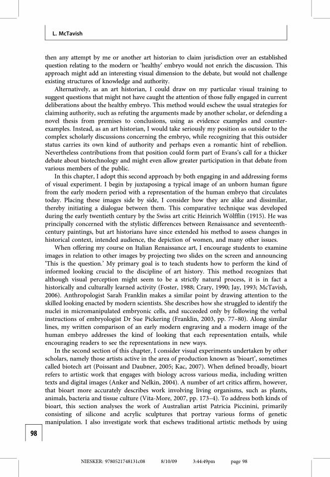
Regarding the human embryoFourteen engraved images of the unborn enrich La Pratique des accouchemens [The practiceof childbirth], an obstetrical treatise published in 1694 by French surgeon man-midwifePhilippe Peu. All the images depict playful toddlers in contorted postures floating insidespacious egg-shaped wombs (Fig. 8.1). These miniature humans are clearly at risk ofstrangulation, and one even pulls at the umbilical cord wrapped around its neck. At thesame time, references to danger are belied by the blissful expressions on their faces. Theengravings in Peu’s treatise are remarkably beautiful, but they are not unique. Repre-sentations of child-like figures in perilous situations routinely appear in obstetrical treatises
Fig. 8.1. Unborn figures, from Philippe Peu’s La Pratiquedes accouchemens (Paris, 1694). (Courtesy of the EdwardG. Miner Library, University of Rochester Medical Center,Rochester, NY.)
NIESKER: 9780521748131c08 8/10/09 3:44:49pm page 99
99
A visual dialogue on ‘healthy’ human embryos

published in Europe between 1550 and 1750 (McTavish, 2005). Sometimes the normal birthposition is pictured, and the unborn figure’s limbs are drawn in to its torso. Such repre-sentations provide, however, a point of contrast for the unnatural positions that prevail inthe treatises. Unborn figures are usually shown at risk: appendages flung in all directions,one hand extending towards the mouth of the womb, in a breech position with arms raisedoverhead, or with feet and hands presenting together.
Peu’s engraving of endangered unborn figures would seem to have little in commonwith the modern photograph of an early ‘at-risk’ human embryo in the process of havingone cell removed for PGD testing (Fig. 8.2). This image portrays a blastomere biopsy; thetransparent embryo attached by suction to a holding pipette on the left while a single cell isaspirated into a hollow micropipette on the right. Whereas the early modern engraving wasproduced by the hand of an anonymous commissioned artist, this image was created withvarious technologies, including the fluorescence microscopy that magnified the embryo,controlled the light necessary to see it and then directed the results for capture by aphotographic medium (Cox, 2007). The resultant flattened image features clean sharp lines,conveying a certain coldness and precision. In contrast, the early modern engraving is moresensually appealing with its rich texture, finely modelled unborn figures and meticulouslyrendered placentas.
The most notable difference between the two images, however, is their subject matter.Peu’s engraving does not portray embryos; it displays fully formed fetuses that look morelike children than what we could expect to find in the womb. Embryology was indeedpractised during the early modern period, with scholars generally adhering to either epi-genetic or preformationist theories, yet the imaginative ideas of early modern embryologistshad remarkably little impact on obstetrical treatises, the type of publication that includedengravings like Peu’s (Dunstan, 1990; Pinto-Correia, 1997; Roger, 1997; Hopwood, 2002).The authors of these books, mostly French and mostly surgeons, defended well into theeighteenth century the Galenic theory of conception as resulting from the mixture of maleand female seed (McTavish, 2005, pp. 203–6). In 1573, barber–surgeon Ambroise Paréexplained how the male and female seed actively combined and fermented in the womb.First, three bubbles formed the rudimentary beginnings of the organs, and then the bonesand interconnecting channels were covered with a protective skin (Paré, 1573, pp. 37–48).Paré was nevertheless unusual in the attention he paid to early stages of development.Most authors of obstetrical treatises did not describe the contents of women’s bodies duringthe initial stages of pregnancy, offering practical advice about the signs of pregnancy,difficult labours and postpartum complaints to an audience consisting of male medical
Fig. 8.2. Embryo undergoing biopsy for the purposeof preimplantation genetic diagnosis (PGD). (Courtesyof Dr Joyce Harper.)
NIESKER: 9780521748131c08 8/10/09 3:45:00pm page 100
100
L. McTavish
practitioners, female midwives, pregnant women, lay people and even readers in search of asex manual (Erickson, 1982).
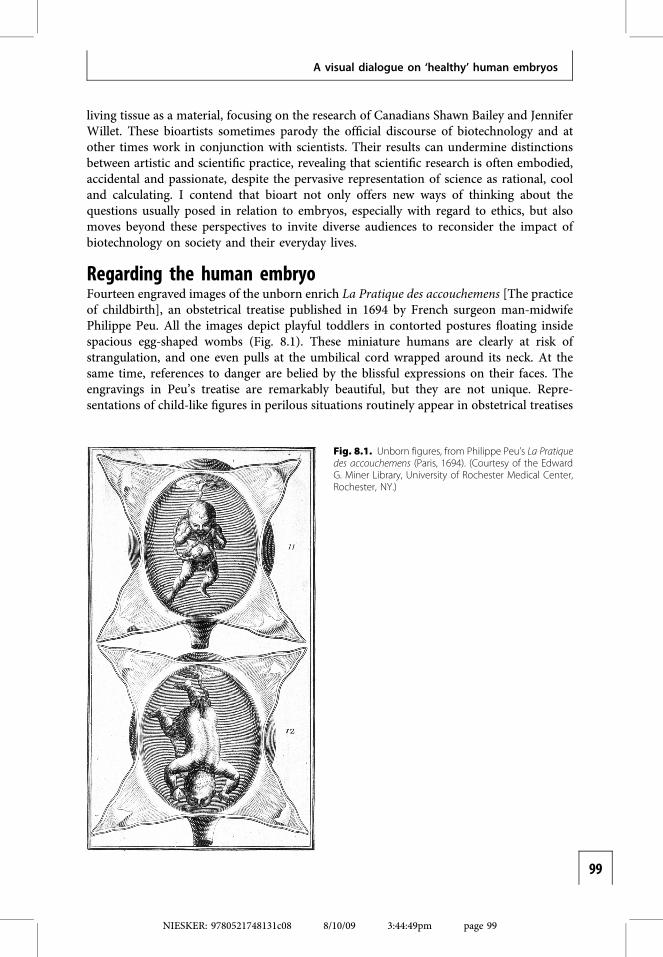
During the early modern period medical practitioners were cautious about identifyingwhat might be found inside the female body. In 1609, the French royal surgeon JacquesGuillemeau explained that he would look ridiculous if after having assured a woman thatshe was pregnant, her womb produced only menses, water or noisy winds (p. 2). As late asthe eighteenth century, surgeon Guillaume Mauquest de La Motte insisted that pregnancycould not be verified until the fourth month because until that date the contents of theuterus were minuscule and although the womb might harbour a fetus, it might just as likelycontain a mass of unformed flesh called a mole, or water, wind, and retained menses (1729,p. 49). According to historian Barbara Duden, pregnancy was confirmed exclusively bywomen during the early modern period, when they felt ‘quickening’ or movement insidetheir bodies (1991, pp. 158–61). She argues that the human embryo was not actuallyinvented until 1799 when Frankfurt physician and anatomist Samuel Thomas Soemmerringpublished drawings of a series of male and female embryos and fetuses, lined up by age andsize, in his Icones embryonum humanorum [Images of human embryos] (Duden, 1999).Wanting to exclude the subjective human eye, Soemmerring instructed his artist to producea measured elevation of each embryo by drawing it as if seen from a series of right angles atan infinite distance. Duden contends that because Soemmerring’s resulting plates portrayedvirtual embryos in a way that no eye could ever really see them, the human embryoappeared as a fact and an object for the first time in history (Fig. 8.3).
The modern photograph of an embryo undergoing a biopsy adheres to the principlesoutlined by Soemmerring, employing complex technologies to depict an embryo that thenaked eye could never see, apparently removing subjective human perception from theimaging process. The resulting representation seems simply to provide an accurate image ofthe embryo in the laboratory. This image can function as a ‘fact’ because it is assumed to beindexical, referring to the semiotic definition of index as a type of visual sign dependent ona real contiguity between the image and the object it portrays. American semioticianCharles Sanders Peirce explains the index by describing a piece of mould marked by abullet-hole: ‘without the shot there would have been no hole; but there is a hole there,whether anybody has the sense to attribute it to a shot or not’ (1991, pp. 239–40). Fin-gerprints and footprints are other examples of indexical signs because they depend on thematerial presence of the original object and provide evidence of that object even if they donot resemble it in an obvious way. In contemporary western culture, photographs are oftenassumed to be indexical in this manner, providing inscriptions of the real world, as ifmaterial objects had literally touched or been physically imprinted on the film. Art historianJoel Snyder (2004) points out that photographs were historically considered unreliable, andnot accepted as evidence in American courtrooms until the 1880s. Despite numerous effortsby scholars to challenge the truth value of the photographic medium, many still commonlyassume that photographs unaltered by technologies such as Photoshop can be taken atface value.
The image in Peu’s obstetrical treatise is not indexical. In fact, Peu insisted that thevisual contents of his engravings had little connection with the actual contents of women’sbodies. No early modern surgeon argued that unborn figures resembled fully formedchildren performing acrobatics inside gigantic wombs. On the contrary, they wrote aboutthe cramped quarters of fetuses, noting that the unborn figure in the womb was ‘curled andbent like a round ball’ (Arons, 1994, p. 3). At least one surgeon asserted that standardized
NIESKER: 9780521748131c08 8/10/09 3:45:01pm page 101
101
A visual dialogue on ‘healthy’ human embryos
Fig. 8.3. Engraving by the Klauber brothers, after drawings by Christian Koeck, from Samuel ThomasSoemmerring’s Icones embryonum humanorum (Frankfurt am Main, 1799). (By permission of the Syndics ofCambridge University Library.)
NIESKER: 9780521748131c08 8/10/09 3:45:02pm page 102
102
L. McTavish
images, such as the ones in Peu’s book, were therefore ‘useless’, and all agreed that theinterior of the female body was essentially a dark, mysterious realm eluding the human eye(Dionis, 1718, pp. xj–xij). French surgeon men-midwives nevertheless proclaimed that thefemale body could be discovered by their perceptive hands, which acted as substitutes fortheir eyes. When recounting their work in the lying-in room, the men described performingmanual examinations to ‘discover’ the state of labour, ‘look for’ the distressed child and‘observe’ its posture (Portal, 1685, pp. 2, 79; Peu, 1694, p. 408; Mauquest de La Motte, 1746,p. 359). Peu provided detailed guidelines encouraging surgeons to distinguish between thedifferent parts of the child by means of touch alone: for example, the head could berecognized by its round, hard skull: the eyes by their number, the cavity of their sockets,elevation of the eyeballs (1694, pp. 51–54). Clearly, an image of the unborn child emergedfrom the physical manipulation of the womb, not from any direct visual access.
Why, then, were images of the unborn commonly included in early modern obstetricaltreatises? What was their purpose? Peu provided an exceptionally detailed account of theengravings in his own publication. He argued that although his plates depicted only some ofthe situations in which he had found children ‘reduced’ by twisted umbilical cords, they‘could serve as a principle idea for conceptualizing an infinity of other possible [situations]’(1694, p. 441). Peu described the images as diagrams, typically associated with theexpression of scientific ideas. By distilling the scientist’s observations into a simple formula,diagrams provide a principle that can be tested in subsequent research. According toPeirce, diagrams are iconic signs, based on resemblance between the sign and the objectportrayed, yet they ‘resemble their objects not at all in looks; it is only in respect to therelations of their parts that their likeness consists’ (1931–5, p. 157). Diagrams supportcreative thinking because they can be observed and contemplated to discover unnoticedrelations amongst the parts of the object portrayed. Peirce’s description of diagrams is inkeeping with Peu’s account of his images of the unborn as ideas enabling male midwives toimagine malpresentations they had not previously encountered. According to Peu, the earlymodern engraving is a conceptual tool enabling thought, not an inscription of reality.
This comparison between an early modern engraving and a modern photographhighlights the vast and perhaps insurmountable differences between them. The imagesdiverge in terms of their aesthetics, modes of production, subject matter and the ways inwhich they communicate meaning. This comparison risks reinforcing the commonsensenotion that current scientific knowledge has progressed well beyond the muddled ideas ofthe past. Peu’s understanding of the images in his obstetrical treatise was, however, verysophisticated. He described them as useful reference points allowing medical practitionersto make sense of the unknown. Though art historians and other scholars have long arguedthat the visual realm actively creates meaning, this realm is often denigrated in contem-porary western culture, associated with ‘mindless entertainment’ in discussions of popularculture, or assumed merely to offer a passive reflection of knowledge in certain scholarlypractices, including some scientific ones (Debord, 1970; Adorno and Horkheimer, 1993;Stafford, 1994).
Despite these important differences, the two images have much in common. They bothdepend, for example, on the absence of the female body, most notable in the modernphotograph of the embryo, which focuses on cells not on the portrayal of human bodies.The composition of the image places the embryo in the centre, separating it from thephysicality of the outside world. This detachment enables the embryo to become a thing inand of itself, worthy of visual scrutiny. Various scholars have argued that the perceived
NIESKER: 9780521748131c08 8/10/09 3:45:16pm page 103
103
A visual dialogue on ‘healthy’ human embryos
relationship between the human embryo and the maternal body is crucial to currentdiscussions of human embryonic stem cell research and reproductive technologies. In herdiscussion of the American legal system, law professor Radhika Rao points out that anembryo located within a woman’s body is characterized as part of her body, forestallingprotectionist legislation that would infringe on her right to bodily autonomy and sexequality (please refer to Rao, Chapter 3). Once that embryo is separated from the maternalbody, however, ‘others’, including government agencies, can formulate policies designed tomanage or protect it. The status of human embryos thus changes according to their context,a claim made more broadly by Sarah Franklin, who describes them as a ‘vast and diversepopulation, imaged, imagined and archived in media as diverse as liquid nitrogen, DVDs,virtual libraries, t-shirts, logos and brandnames’ (Fausto-Sterling, 2003; Franklin, 2006a,p. 168).
The detachment and subsequent mobility of the embryo contributes to the embryo-centric nature of current debates about its rights and identity. According to bioethicistJackie Leach Scully and colleagues, this focus marginalizes those who supply the repro-ductive material (gametes) needed to create human embryos, including couples asked todonate their ‘spare’ IVF embryos (see Scully, Rehmann-Sutter and Porz, Chapter 2). Alsoregularly forgotten in debates about stem cells are women who undergo hormone injectionsand surgical procedures to supply eggs for various kinds of research (Stoyle, 2005; Dick-enson, 2006). Jeff Nisker and Roxanne Mykitiuk contend that many altruistic egg donationsoccur within coercive conditions, settings often shaped by the hierarchy of the doctor–patient relationship (please refer to Mykitiuk and Nisker Chapter 9). This potential forcoerced donations was recently highlighted in the scandal involving South Korean bio-medical scientist Woo-Suk Hwang, who claimed to have created human embryonic stemcells by cloning (Hwang, 2004; Hwang, Roh, Lee et al., 2005). After the journal Scienceretracted his original article because it was based on fabricated data, Dr Hwang admittedthat two of his graduate students had donated their eggs for his research (Holden, 2005;Kennedy, 2006). Hundreds of Korean women subsequently agreed to provide eggs withoutcompensation, an overt indication of their support for Dr Hwang but also perhaps animplicit assertion of their ability to make informed decisions (Holden, 2005). In any case,women made a brief appearance in the public debate about stem cell research.
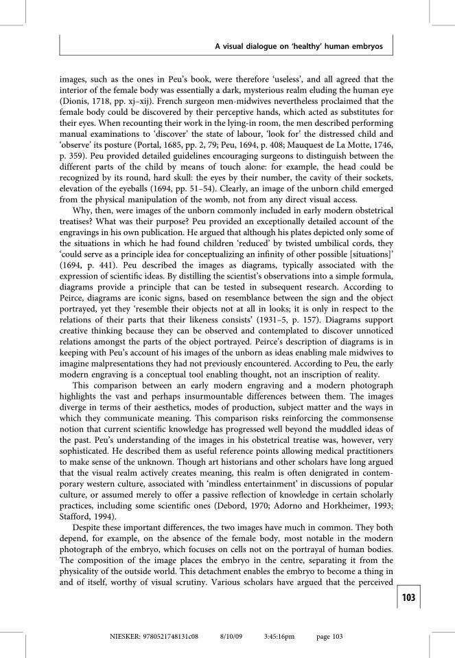
Although no sign of the female body is evident in the photograph of the biopsiedembryo, references to women’s bodies remain in the early modern engravings found inobstetrical treatises. Commonly, the maternal body appears in the fragmented forms ofdissected wombs, umbilical cords and placentas. The female body rarely appears in itsentirety. The book published by English midwife Jane Sharp in 1671 shows an engraving ofa pregnant woman as a plant producing fruit; the healthy child is both dependent on andcreated by her fertile maternal body (Fig. 8.4). Peu’s more typical engraving is vastlydifferent, depicting badly positioned, unhealthy fetuses in need of medical intervention.This emphasis on endangered unborn figures corresponds with the efforts made by malemidwives to expand their practices within the birthing room. Authoritative female mid-wives controlled childbirth throughout the early modern period, and men were summonedprimarily in cases of emergency, to remove impacted fetuses from the womb using hooks orhead pullers. Struggling to portray themselves as skilled birthing assistants, male midwivesclaimed that they could legitimately enter the lying-in room in difficult cases when the‘natural’ efforts of both the maternal body and the female midwife had failed (McTavish,2005, pp. 199–201). French surgeons drew attention to their skilled hands which could ‘see’
NIESKER: 9780521748131c08 8/10/09 3:45:16pm page 104
104
L. McTavish
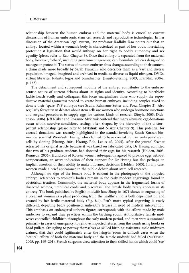
the unborn figure and turn it in the womb for a podalic (feet-first) delivery, a method theyclaimed surpassed the abilities of the average female midwife. The reduction of the preg-nant woman to a womb in Peu’s engraving thus creates an operative space for the entry ofthis medical expert. Though the surgeon’s presence is merely implied as necessary, it isconcretely represented in other early modern obstetrical engravings. The plates in Frenchman-midwife Cosme Viardel’s treatise of 1671, for example, depict male hands entering thewomb to manoeuvre malpositioned children (Fig. 8.5).
Manipulating hands are equally implied in the modern photograph, which shows anembryo framed by two micropipettes. Referring to a similar image, Franklin designatesthem the ‘two helping hands’ of science (2005, p. 66). Whereas in Viardel’s engraving, themale hand operates on the principle of synecdoche to represent the whole surgeon, in themodern photograph the technological tools invoke a more abstract entity: ‘science’. In bothcases, however, the hands are shown intervening to alter the unborn entity. Unlike the barehands of the French surgeon, the tools that remove one cell from the embryo to test for
Fig. 8.4. Fertilewoman, from JaneSharp’s TheCompleat Midwife’sCompanion(London, 1724).(Courtesy of theWellcome Library,London.)
NIESKER: 9780521748131c08 8/10/09 3:45:17pm page 105
105
A visual dialogue on ‘healthy’ human embryos
genetic abnormalities are antiseptic and fleshless. The embodied subjectivity of the scientistor medical practitioner holding the tools is bracketed out of the image. This denial ofembodiment accords with arguments made by sociologists such as Bruno Latour and SteveWoolgar (1986) and Steven Shapin (1999). Their ethnographic and historical studies revealthat for the final results to be accepted as scientific facts, the researcher needs to makeinvisible the messiness of laboratory life – its inconclusive data, mistakes and accidents.
Though the photographic image reinforces an official representation of ‘science’, it doesnot necessarily correspond with scientists themselves and how they characterize their ownresearch. Scientists especially adept at manoeuvring minute, delicate cells are said to have‘good hands’, a description recognizing their skill as embodied. Franklin explains thatDr Pickering is credited with possessing ‘green fingers’ because she seems particularly ableto grow stem cells (2006b, p. 79). This appellation refers not only to her body but also to her
Fig. 8.5. Unborn figure from Cosme Viardel’sObservations sur la pratique des accouchements naturels,contre nature et monstrueux (Paris, 1673). (Courtesy of theNational Library of Medicine, Bethesda, MD.)
NIESKER: 9780521748131c08 8/10/09 3:45:21pm page 106
106
L. McTavish
nurturing and even maternal qualities, as she seeds cells into the feeder beds. Thesubjectivity of scientists is foregrounded when they grade embryos during the IVF process,deciding which ones are ‘good looking’ and thus ideal for transfer to the womb. Goodembryos are robust, displaying clear, even, well-rounded development, whereas ‘poorlooking’ embryos are uneven, opaque, and slow to divide (Franklin, 2006c, pp. 145, 151).Embryologists are quick to admit, however, that the visual evaluation of an embryo’smorphology may not provide reliable information; embryos that ‘look like crap’ but areimplanted as a last resort can turn out to be viable (Franklin, 2006b, p. 82).
To this point, I have discussed both the engraving and the photograph in relation totheir immediate contexts, namely the politics of early modern childbirth and the modernlaboratories where human embryos and human embryonic stem cells are manipulated.What happens when these images are removed from these contexts and made to signifydifferently? In a sense, I have already performed such a decontextualization by separatingeach image from the series of which it was originally a part. In Peu’s obstetrical treatise, theengraving was positioned alongside 13 similar plates of malpresenting fetuses, combiningwith them to portray childbirth as an overwhelmingly dangerous activity. The modernphotograph of the embryo was likewise originally displayed within a sequence of imagesshowing the entire biopsy procedure; it followed a depiction of a micro-needle opening thezona pellucida of the embryo to enable the subsequent cell removal (Verlinksy and Kuliev,2000). By selecting and juxtaposing the two images, I have created new meanings for both, astated part of my goals in this chapter.
Art historians argue that visual images refer to other images more than to the externalworld (Panofsky, 1962; Bal and Bryson, 1991). According to the theory of intertextuality, alltexts depend on a host of conventions, codes and other texts for their existence. Any writtentext or visual image therefore contains an unavoidable multiplicity of references, withsignifiers referring to other signifiers and meaning constantly deferred (Kristeva, 1980;Derrida, 1986; Allen, 2000). Thus, Peu’s early modern image looks different to modernviewers from how it looked to early modern viewers. It is now a relatively ‘open’ textbecause of its superficial resemblance to modern fetal imagery, especially the depictionspromoted by those opposed to legal abortion. Drawing on these visual similarities, literarycritic Karen Newman (1996) argues that the fetus has long been visualized as an inviolable,rights-Bearing individual. Though the early modern fetus was never understood in thismodern way, Newman’s observations may seem correct because Peu’s engraving showswell-developed unborn figures detached from the maternal body, recalling Lennart Nilsson’sphotographs of embryos and fetuses shown floating against celestial backgrounds, producedfor Lifemagazine in 1965. According to anti-abortionists, Nilsson’s images provide irrefutableevidence of ‘life’ in the womb, although many of the specimens he photographed had beensurgically removed from the womb and were in fact dead (Stabile, 1993). Despite the effortsof anti-abortionists to insist that such fetal imagery has only one obvious meaning, therepresentations regularly appear in popular culture, in advertisements for movies and cars(Mink, 1985; Taylor, 1992; Duden, 1994; Kaplan, 1994).
The modern photograph of an embryo might seem, however, to be less open to suchmultiple meanings and popular appropriations (Fiske, 1989). In 2000, scholars ClareWilliams, Jenny Kitzinger and Lesley Henderson undertook a quantitative study of themedia in the United Kingdom to discover how scientific images of embryos were beingused. They found that anti-abortionists and others opposed to human embryonic stem cellresearch did not deploy photographs of early embryos as part of their political strategies.
NIESKER: 9780521748131c08 8/10/09 3:45:24pm page 107
107
A visual dialogue on ‘healthy’ human embryos
The representations were instead reproduced by proponents of human embryonic stem cellresearch because ‘the pre-14-day embryo looks nothing like its 12-week-old counterpartused by anti-abortionists to display perfectly formed fingers and toes’ (Williams, Kitzingerand Henderson, 2003, p. 801). This observation indicates that relatively new images ofembryos are interpreted in relation to well-known fetal imagery. According to the authors,multi-celled embryos resemble alien blobs more than human beings, an understandingencouraged when the photographs are reproduced in colour to show the saturated hues ofthe stains applied by scientists to make the parts of the embryo more visibly distinct(Verlinksy and Kuliev, 2000). These examples suggest that modern depictions of embryosare indeed read intertextually and can convey numerous meanings, as suggested by Franklinin her account of the discursive nature of embryos (Franklin, 2006a).
Malleability is highlighted in the photograph of the blastomere biopsy, revealing whatMaienschein and Robert call the ‘constructed’ embryo (please refer to Maienschein andRobert, Chapter 1). In this image the embryo itself is portrayed as an open text that can berewritten, a possibility that excites researchers but frightens others, including some membersof the public. Those whose world-view depends on beliefs in an immutable embryo imaginethat the absent body holding the ‘helping hands of science’ belongs to a mad scientist bent ondestroying vulnerable embryos. When images of early embryos leave the settings of thelaboratory and the scientific journal, they are understood in relation to a wide range ofsources, including science fiction literature and films, leading to interpretations varyingaccording to the viewers’ particular cultural backgrounds, knowledge and life experiences(Hall, 1993). Efforts by scientists and other scholars to inform an ‘ignorant’ public about thetrue or singular meaning of these images will fail unless they acknowledge the complexity ofvisual communication and reception. Contemporary artists, however, are formulatingalternative methods for engaging the public with biotechnology, both critically and visually.
Artistic interventions in biotechnologyThe idea that scientific imagery and practice can be resignified is key to bioart, a long-standing practice whose origins are often located in the 1930s, when American photographerEdward Steichen employed genetic manipulations to create gigantic, hybrid delphiniums(Gedrim, 2007). Now, a range of contemporary artists both interact with and criticize bio-technology – by culturing bacteria, sculpting with proteins, fusing their white blood cells withmice myeloma or cloning walnut trees – sometimes from within its structures and sometimesfrom the outside (Kac, 2007). Though the resulting artistic works and performances mayseem esoteric to those unaccustomed to the art world, they are largely meant to question theconstruction of expertise, allowing a broader public to have access to the founding conceptsand practices of technoscience. Canadian bioartist Jennifer Willet acknowledges that althoughsome of her work is available on the Internet, it still might not reach a mass audience. Shehopes it will nevertheless have a ‘trickle down effect’ by encouraging increased discussion anddebate about biotechnology in the long run (personal communication, 14 February 2008).
Instead of working with living tissue, renowned artist Patricia Piccinini uses modernmaterials, including silicon and acrylic resin, to create sculptural installations that explorethe themes of genetic engineering and biotechnology. Her show We Are Family was fea-tured in the Australian pavilion of the 2003 Venice Biennale – one of the world’s mostimportant recurring exhibitions of contemporary art. Piccinini’s sculptures representbizarre, hybrid creatures, complete with soft flesh and real human hair. Her 2002 Still Life
NIESKER: 9780521748131c08 8/10/09 3:45:24pm page 108
108
L. McTavish
with Stem Cells portrays a life-sized young girl surrounded by corporeal lumps adornedwith mouth-like openings (website reference). Although viewers may find the ‘stem cell’blobs repulsive, the girl affectionately hugs one to her chest while resting her hand onanother, treating it like a family pet. This sculptural group is in dialogue with the genre ofscience fiction, inviting viewers to fantasize about what might happen in the future if thehuman body becomes simply another kind of material to be manipulated (Michael, 2002;McTavish, 2003; Goriss-Hunter, 2004; Lauritzen, 2005). Yet Piccinini’s creatures remaindomesticated (at least for now), providing a vision of genetic modification that is neitherutopian nor dystopian (Haraway, 2007).
By titling her installation ‘still life’, Piccinini places it within an art historical tradition.Like paintings of fruit and flowers that purport to show a slice of everyday life, Piccinini’swork displays a banal scene from modern suburbia: a young child playing as if in a domesticinterior. Any sense of normality, however, is countered by the undefined forms that act as hercompanions, existing somewhere between the human and non-human realms. The overalleffect is uncanny, for the spectator confronts a scene that is simultaneously familiar andstrange. This effect is conveyed to a large degree by the work’s hyper-real details, whichinclude hair follicles and wrinkled skin. According to art historian Norman Bryson (1981),the realist aesthetic functions by providing an excess of information, divorcing details fromnarrative goals and didactic lessons so that they seem unmotivated and thus true. This realityeffect is enhanced by the tactility of Piccinini’s work, which like the comparable sculptures ofDuane Hanson and Ron Mueck, invites viewers to respond physically with a shiver of disgustand delight. The realism of the modern photograph of an early human embryo discussedabove operates differently. Though also dependent on apparently neutral details to conveyaccuracy, it effaces signs of embodiment, distancing viewers from the image rather thaninviting their identification with it. Piccinini’s sculptural installation plays with the signifyingsystems used by science, reshaping them to convey other meanings.
Artists Shawn Bailey and Jennifer Willet consider a different form of scientific aestheticsby exploring the economic goals of biotechnology. Visitors to their virtual laboratory calledBIOTEKNICA (www.bioteknica.org) are greeted by a professional logo before enteringthe website to find linear diagrams of molecules and photographs of specimen bottles.BIOTEKNICA’s ‘corporate history’ identifies it as an international bioengineering firm thatdevelops ‘patent and copyright applications for several pre-fetal genome mutations, andpost-fetal cellular processes, and related digital imaging technologies’. Such boastful andvague commentary is undermined by a subsequent description of BIOTEKNICA as afiction designed in 2000 by Bailey and Willet to parody, and thus invite criticism of, typicalforms of biotechnical self-presentation. BIOTEKNICA has nevertheless attracted invest-ment offers, a development troubling to its creators because the corporate vision includes‘placing the power of genetic supremacy in the hands of humanity itself’ (BIOTEKNICA).
In this work, Bailey and Willet engage with the increasing commodification of hybridentities, DNA, cells, and human tissue. Scholars such as Margaret Lock (2001) and Cath-erine Waldby and Robert Mitchell (2006) have written arguments about the transformationof the gift economy that once structured exchanges of human tissue. Bioartists Bailey andWillet participate in this analysis, inviting viewers to do the same. BIOTEKNICA’s virtuallaboratory software allows visitors to create their own products, simulating the role of thescientist–entrepreneur. The site allows visitors to custom design digital images of teratoma –cancerous growths containing hair, skin and possibly stem cells – by mixing the ingredientsand then ‘breeding’ them (www.bioteknica.org/index2.html). Whereas Piccinini’s work elicits
NIESKER: 9780521748131c08 8/10/09 3:45:24pm page 109
109
A visual dialogue on ‘healthy’ human embryos

visceral reactions from viewers and suggests ownership by having the little girl embrace andcaress the stem cell blobs, BIOTEKNICA allows its audience to perform virtual experiments,combining different elements to create new forms of ‘life’. According to the artists, BIO-TEKNICA ‘avoids prescriptive critical mantras’, focusing instead on allowing viewers toformulate their own conclusions about the procedures and protocols presented (Willet andBailey, 2007).
In 2004, Bailey and Willet moved beyond conventional forms of artistic creation anddisplay to adopt a critical participatory methodology. They began to grow material teratomatissue cultures at SymbioticA, the Art and Science Collaborative Research Laboratory at theUniversity of Western Australia. With Oron Catts and Ionat Zurr from the Tissue Cultureand Art Project, they cultivated cells of P19 mouse teratoma (Willet and Bailey, 2007).Teratomas fascinate the artists because they can be considered a natural type of cloning,genetically identical to humans (Winters, 2006). The ambiguous status of these tumours haslong been compelling to medical practitioners, who during the early modern period con-sidered them a monstrous form of reproduction, even when created outside of the womb. Forexample, in a medical journal from 1697, editor Claude Brunet reported that when surgeonsamputated and then dissected the swollen testicle of a Cistercian monk, they removed a fleshymass replete with bones and an afterbirth, proving that male pregnancy was possible (Brunet,1697). The appeal of unnatural or monstrous reproduction has not diminished, as indicatedby the popularity of Piccinini’s stem cell blobs, sometimes categorized as a version of the‘monstrous cute’ (Goriss-Hunter, 2004). Nothing, however, is cute about Bailey and Willet’sexamination of monstrosity, which produced what they call tissue culture sculptures, made byencouraging the mouse teratoma cells to attach to polymer scaffolds.
Modern responses to the diverse field of bioart depend on the work in question and thecritic examining it. Art historians, like myself, are intrigued by the use of living tissue as anartistic material because it is relatively new and challenges traditional definitions of theartist even as it raises ethical implications by blurring distinctions between artistic andscientific research, an idea supported by bioethicist Arthur Caplan who supports theblending of art and science but argues that it is an ethical abuse to ‘use genetics simply forartistic exhibitionism’ (Allmendinger, 2001). Other commentators argue that bioart com-plies with biotechnology instead of opening it to question. According to American bioartistAdam Zaretsky (2005), ‘some say that biology as an arts process is merely a promotionaltool for big science’. Willet reports that this same perspective has been used at scholarlyconferences to condemn the participatory work of herself and Bailey in scientific labora-tories. Their various collaborations with biologists have been compared to embeddedjournalism, when news reporters experience armed conflict from the inside by travellingwith the military. Critics contend that just as these journalists are constrained by regula-tions and ultimately come to identify with the military unit in which they are embedded,so too will bioartists become part of biotechnology, overwhelmed by its powerful allure(J. Willet, personal communication, 14 February 2008).
Nevertheless opportunities can be gained by working from the inside rather than as aperpetual outsider or layperson, the position embraced by Piccinini (Piccinini and Orgaz,2007). Feminist scholar Evelyn Fox Keller has advocated transforming science from within,arguing that science is a distinctive practice with its own logical and empirical constraints(1992, pp. 2–3). As a trained physicist, Keller has critiqued the masculinist nature ofdominant science, while considering the way in which language shapes scientific practices.Bioartists are unlikely, however, to be identified as scientists, even when they have been
NIESKER: 9780521748131c08 8/10/09 3:45:24pm page 110
110
L. McTavish
professionally trained to grow tissue culture. Bailey and Willet, for example, are positionedon the margins of the laboratories in which they work, as outsiders temporarily welcomedinside. I contend that this intermediary status is nevertheless enabling, allowing bioartistsboth to examine and to attempt to perform the habitus promoted in particular scientificrealms. French sociologist Pierre Bourdieu defines habitus as a system of dispositions oracquired modes of perception, belief, thought, and action (1984, p. 34). Dispositions varyaccording to the specific ‘fields’ in which they are developed. According to Bourdieu, a field‘is a space in which a game takes place, a field of objective relations between individuals orinstitutions who are competing for the same stake’ (p. 197). Fields are social systems thatfunction according to specific rules, and people engage on a daily basis with any number ofthem – the family, specific leisure activities, their profession. These social systems competefor legitimacy or symbolic capital by interacting with the often implicit rules of the game,which they learn through experience. Instead of being obligated to follow these rules, indi-viduals are in constant negotiation with them (Moi, 1991). Within technoscience, legitimacyis acquired in some obvious ways: by attaining an education, placing publications in par-ticular venues, attracting significant research funds, and creating deliverables that can bepatented. Legitimacy is also achieved more tacitly, through proper bodily comportment,modes of speech, and patterns of thought. Bioartists can contemplate these embodied aspectsof biotechnology by means of their physical and intellectual interactions within laboratories.At the same time, these artists may learn to reflect on the distinctive habitus of the art world,for they ultimately need to earn symbolic capital in that field if they hope to continue theirartistic careers.
No doubt the interaction of artists with scientists encourages some sympathies betweenthe two groups. Attending to the habitus promoted by particular laboratories foregroundsscientists as embodied individuals who are continually both constrained and produced bythe rules of their field. These interactions distinguish actual scientists from the generalizedentities known as ‘biotechnology’ and ‘science’, potentially resulting in more accurateunderstandings of both terms. My own limited experiences interacting with physicians,biologists and veterinarians at various collaborative conferences has had this effect,revealing the scientists as passionately devoted to their research. These cross-disciplinarydiscussions have also uncovered surprising links between the present and past. Modernscientists regularly spoke about their material engagement with and understanding of theirresearch subjects, whether animal or oocyte, just as early modern medical practitionersearned authority primarily through their physical experiences with matter, gained throughthe sense of touch (McTavish, 2005, pp. 87–8, 154–63).
Some forms of bioart work both to break down and to highlight the distinctionsbetween art and science, distinctions not yet constructed during the early modern period.Artists are able to position themselves in relation to science primarily because both fieldsprivilege hands-on knowledge, originality, and creativity. Yet a number of artists have alsofound that adopting the role of artist–researcher or artist–scientist allows them to apply forfinancial support from agencies that have not traditionally recognized the value of the visualarts. Contemporary funding structures have therefore encouraged the emergence of theartist as researcher, providing both opportunities and potential restraints. Bailey and Willetdescribe, for example, the ethics approval they needed to obtain before undertaking workwith human cell sources. They found it difficult to explain their artistic goals in thestandardized terms of health benefits, outcomes, and deliverables. In the end, they con-sidered the lengthy ethics procedure that continued from 2004 to 2005 a performance piece
NIESKER: 9780521748131c08 8/10/09 3:45:24pm page 111
111
A visual dialogue on ‘healthy’ human embryos

in itself (www.bioteknica.org/index2.html). According to the artists, this process allowedthem to engage fully with the methods of the scientific community. I contend that it alsoencouraged them to recognize the ‘rules of the game’ of a different field, which was initiallyforeign and uncomfortable.
ConclusionsThis chapter has brought together apparently diverse things – an early modern print and amodern photograph, art and science – in order to consider both what is distinctive about eachone and the sometimes surprising ways in which they overlap. My first question askedwhether an early modern engraving of unborn figures and a modern microscopic visual-ization of a human embryo could speak to each other. Despite their differences, I showed thatboth representations featured a largely absent maternal body which allowed the interventionsof an other body to claim authority over the products of conception; both images alsoproduced arguments about identity, based on assertions about the necessity of medicalprocedures required to manipulate unborn creatures; both likewise acquired meaning inrelation to what was outside of them, primarily other images and historical discoursesconcerned with who should properly control the realm of reproduction. My comparativemethod highlighted the complexity and variety of visual forms of communication, whileundermining simple understandings of progress that delegate the early modern period to thedistant past, utterly divorced from contemporary scientific practice. Early modern medicalpractitioners, such as Peu, arguably had an advanced understanding of the way in whichvisual representations can actively produce rather than merely reflect knowledge.
My second question asked what artists could contribute to exploring the visualdimensions of current debates about biotechnology. This section did not directly concernthe cultural construction of healthy and unhealthy human embryos, but instead turned tothe wider domain of technoscience that underpins many of the current discussions aboutthese beings. Although only a small selection of bioart production was discussed, it revealedthat artists are materially exploring the manipulations of biotechnology, encouraging moresophisticated understandings of both science and scientists, while drawing attention to thehabitus developed in laboratories. Audiences will be disappointed by bioart if they areseeking dogmatic positions either for or against stem cell research, the creation of animal–human hybrids, or particular reproductive technologies. For the most part, the artistsinvolved do not make prescriptive arguments but instead offer new ways of looking at andthinking about various forms of biotechnology. I contend that this kind of practice has theability to open new questions, offering a ‘thicker’ debate about biotechnology, particularlywith regard to ethical issues.
ReferencesAdorno, T. and Horkheimer, M. (1993). The
culture industry: enlightenment as massDeception. In The Cultural Studies Reader,ed. S. During. New York: Routledge,pp.29–43.
Allen, G. (2000). Intertextuality. London:Routledge.
Allmendinger, U. (2001). One small hop forAlba, one large hop for mankind. NY Arts
Magazine, 6 (6). Available at www.ekac.org/ulli.html (accessed 30 January 2008).
Anker, S. and Nelkin, D. (2004). The MolecularGaze: Art in the Genetic Age. Cold SpringHarbor, NY: Cold Spring Harbor LaboratoryPress.
Arons, W. (trans. and intro.) (1994). EuchariusRösslin: When Midwifery Became the MalePhysician’s Province. Jefferson, NC:McFarland.
NIESKER: 9780521748131c08 8/10/09 3:45:24pm page 112
112
L. McTavish
Bal, M. and Bryson, N. (1991). Semiotics and arthistory. Art Bulletin, 73 (2), 174–208.
Bourdieu, P. (1984). Questions de sociologie.Paris: Minuit.
Brunet, C. (1697). D’une grossesse d’home [sic].Le Progrès de la Médecine. Paris, 62–5.
Bryson, N. (1981). Word and Image: FrenchPainting of the Ancien Régime. Cambridge:Cambridge University Press.
Cox, G. (2007). Optical Imaging Techniques in CellBiology. Boca Raton, FL: Taylor and Francis.
Crary, J. (1990). Techniques of the Observer: OnVision and Modernity in the NineteenthCentury. Cambridge, MA: MIT Press.
Debord, G. (1970). Society of the Spectacle.Detroit: Black and Red.
Derrida, J. (1986). Différance. In Critical TheorySince 1965, eds. H. Adams and L. Searle.Tallahassee, FL: Florida State UniversityPress, pp.120–36.
Dickenson, D. (2006) The lady vanishes: what’smissing in the stem cell debate? Journal ofBioethical Inquiry, 3, 43–54.
Dionis, P. (1718). Traité général desaccouchemens. Paris.
Duden, B. (1994). The fetus as an object of ourtime. Res, 25 (Spring), 132–5.
Duden, B. (1991). The Woman beneath the Skin:A Doctor’s Patients in Eighteenth CenturyGermany. Cambridge, MA: HarvardUniversity Press.
Duden, B. (1999). The fetus on the ‘farther shore’:toward a history of the unborn. In FetalSubjects, Feminist Positions, eds. L.M. Morganand M.W. Michaels. Philadelphia, PA:University of Pennsylvania Press, pp.12–25.
Dunstan, R. (ed.) (1990). The Human Embryo:Aristotle and the Arabic and EuropeanTraditions. Exeter, UK: University of ExeterPress.
Erickson, R.A. (1982). ‘The books of generation’:some observations on the style of the Britishmidwife books, 1671–1764. In Sexuality inEighteenth-Century Britain, ed. P.-G. Boucé.Manchester, UK: Manchester UniversityPress, pp.74–94.
Evans, J.H. (2002). Playing God?: Human GeneticEngineering and the Rationalization of PublicBioethical Debate. Chicago, IL: University ofChicago Press.
Fausto-Sterling, A. (2003). Science matters,culture matters. Perspectives in Biology andMedicine, 46, 109–24.
Fiske, J. (1989). Understanding Popular Culture.Boston, MA: Unwin Hyman.
Foster, H. (ed.) (1988). Vision and Visuality.Seattle, WA: Bay Press.
Franklin, S. (2003). Rethinking nature/culture:anthropology and the new genetics.Anthropological Theory, 3, 65–85.
Franklin, S. (2005). Stem cells R us: emergent lifeforms and the global biological. In GlobalAssemblages: Technology, Politics, and Ethicsas Anthropological Problems, eds. A. Ongand S.J. Collier. Oxford, UK: Blackwell,pp.59–77.
Franklin, S. (2006a). The cyborg embryo: ourpath to transbiology. Theory, Culture andSociety, 23(7–8), 167–87.
Franklin, S. (2006b). Embryonic economies: thedouble reproductive value of stem cells.Biosocieties, 1, 71–90.
Franklin, S. (2006c). Born and Made: AnEthnography of Preimplantation GeneticDiagnosis. Princeton, NJ: PrincetonUniversity Press.
Gedrim, R.J. (2007). Edward Steichen’s 1935exhibition of delphinium blooms: an art offlower breeding. In Signs of Life: Bio Art andBeyond, ed. E. Kac. Cambridge, MA: MITPress, pp.347–69.
Geertz, C. (1973). Thick description: toward aninterpretive theory of culture. In TheInterpretation of Cultures: Selected Essays, ed.C. Geertz. New York: Basic Books, pp.3–30.
Goriss-Hunter, A. (2004). Slippery mutantsperform and wink at maternal insurrections:Patricia Piccinini’s monstrous cute.Continuum: Journal of Media and CulturalStudies, 18 (4), 541–53.
Guillemeau, J. (1609). De l’heureux accouchementdes femmes.Paris.
Hall, S. (1993). Encoding, decoding. In TheCultural Studies Reader, ed. S. During.New York: Routledge, pp.90–103.
Haraway, D. (2007). Speculative fabulations fortechnoculture’s generations. In (Tender)Creature Exhibition Catalogue. Vitoria,Spain: Artium, The Centro-Museo Vasco deArte Contemporaneo.
Holden, C. (2005). Korean cloner admitslying about oocyte donations. Science, 310,1402–3.
Hopwood, N. (2002). Embryos in Wax: Modelsfrom the Ziegler Studio. Cambridge, UK:Whipple Museum of the History of Science.
NIESKER: 9780521748131c08 8/10/09 3:45:26pm page 113
113
A visual dialogue on ‘healthy’ human embryos
Hwang, W.-S. (2004). Evidence of a pluripotentembryonic stem cell line derived from acloned blastocyst. Science, 303, 1669–74.
Hwang, W.-S., Roh, S.I., Lee, B.C. Kang, S.K.,Dwon, D.K., Kim, S. et. al. (2005). Patient-specific embryonic stem cells derived fromhuman SCNT blastocysts. Science, 308,1777–83.
Jay, M. (1993). Downcast Eyes: The Denigrationof Vision in Twentieth-Century FrenchThought. Berkeley, CA: University ofCalifornia Press.
Kac, E. (ed.) (2007). Signs of Life: Bio Art andBeyond. Cambridge, MA: MIT Press.
Kaplan, E.A. (1994). Look who’s talking, indeed:fetal images in recent North American visualculture. In Mothering: Ideology, Experience,and Agency, eds. E. Nakano Glenn, G. Changand L. Rennie Forcey. New York: Routledge,pp.121–37.
Keller, E.F. (1992). Secrets of Life, Secrets ofDeath: Essays on Language, Gender andScience. New York: Routledge.
Kennedy, D. (2006). Editorial retraction. Science,311, 355.
Kristeva, J.(1980). Desire in Language: A SemioticApproach to Literature and Art. New York:Columbia University Press.
Latour, B. and Woolgar, S. (1986). LaboratoryLife: The Construction of Scientific Facts.Princeton, NJ: Princeton University Press.
Lauritzen, P. (2005). Stem cells, biotechnology,and human rights: implications for aposthuman future. Hastings Center Report,35 (2), 25–33.
Lock, M. (2001). The alienation of body tissueand the biopolitics of immortalized stem celllines. Body and Society, 7(2–3), 63–91.
Mauquest de La Motte, G. (1729). Traité completdes accouchemens. Paris.
Mauquest de La Motte, G. (1746). A GeneralTreatise of Midwifry, Thomas Tomkyns,trans. London.
McTavish, L. (2003). Art and technology at theVenice Biennale. Canadian MedicalAssociation Journal, 169, 322–3.
McTavish, L. (2005). Childbirth and the Displayof Authority in Early Modern France.Aldershot, UK: Ashgate.
McTavish, L. (2006). Learning to see in NewBrunswick, 1862–1929. Canadian HistoricalReview, 87, 553–81.
Michael, L. (2002). We Are Family ExhibitionCatalogue. Sydney: Museum ofContemporary Art.
Mink, B. (1985). American Cancer Society’s‘smoking fetus’. American Cinematographer,66, 77–80.
Moi, T. (1991). Appropriating Bourdieu:feminist theory and Pierre Bourdieu’ssociology of culture. New Literary History,22, 1017–49.
Newman, K. (1996). Fetal Positions:Individualism, Science, Visuality. Stanford,CA: Stanford University Press.
Nilsson, L. (1965). The drama of life before birth.Life, 30 April, 54–69.
Panofsky, E. (1962). Studies in Iconology:Humanistic Themes in the Art of theRenaissance. New York: Harper and Row.
Paré, A. (1573). Deux livres de chirurgie, de lagénération de l’homme. Paris.
Peirce, C.S. (1991). Sign. In Peirce on Signs:Writings on Semiotic by Charles SandersPeirce, ed. J. Hoopes. Chapel Hill, NC:University of North Carolina Press,pp.239–40.
Peirce, C.S. (1931–5). Collected Papers, vols. 1–6,eds. C. Hartshorne and P. Weiss. Cambridge,MA: Belknap Press of Harvard UniversityPress.
Peu, P. (1694). La Pratique des accouchemens.Paris.
Piccinini, P. and Orgaz, L.F. (2007). Thenaturally artificial world. In (Tender)Creature Exhibition Catalogue. Vitoria,Spain: Artium, The Centro-Museo Vasco deArte Contemporaneo.
Pinto-Correia, C. (1997). The Ovary of Eve: Eggand Sperm and Preformation. Chicago, IL:University of Chicago Press.
Poissant, L. and Daubner, E. (eds.) (2005). Art etbiotechnologies. Sainte-Foy, PQ: Presses del’Université du Québec.
Portal, P. (1685). La Pratique des accouchemens.Paris.
Roger, J. (1997). The Life Sciences in Eighteenth-Century French Thought, R. Ellrich, trans.Stanford, CA: Stanford University Press.
Sharp, J. (1671). The Midwives Book. London.Shapin, S. (1999) The house of experiment in
17th century England. In The Science StudiesReader, ed. M. Biagioli. New York:Routledge, pp.479–504.
NIESKER: 9780521748131c08 8/10/09 3:45:26pm page 114
114
L. McTavish
Snyder, J. (2004). Res ipsa loquitur. In Thingsthat Talk: Object Lessons from Art andScience, ed. L. Daston. New York: ZoneBooks, pp.195–221.
Stabile, C.A. (1993). Shooting the mother:fetal photography and the politics ofdisappearance. Camera Obscura, 28,179–205.
Stafford, B. (1994). Artful Science: Enlightenment,Entertainment, and the Eclipse of VisualEducation. Cambridge, MA: MIT Press.
Stoyle, J. (2005). Stem cells: where the newesttechnology meets the oldest profession.Theology and Sexuality, 11, 77–96.
Taylor, J.S. (1992). The public fetus and thefamily car: from abortion politics to a Volvoadvertisement. Public Culture, 4, 67–80.
Verlinsky, Y. and Kuliev, A. (2000). An Atlas ofPreimplantation Genetic Diagnosis. NewYork: Parthenon.
Viardel, C. (1671). Observations sur la pratiquedes accouchemens naturels, contre nature etmonstrueux. Paris.
Vita-More, N. (2007). Brave bioart 2: sheddingthe bio, amassing the nano, and cultivatingpost-human Life. Technoetic Arts, 5, 171–86.
Waldby, C. and Mitchell, R. (2006). TissueEconomies: Blood, Organs, and Cell Lines inLate Capitalism. Durham, NC: DukeUniversity Press.
Willet, J. and Bailey, S. (2007). BIOTEKNICA:teratogenic strategies for critical bioartproduction. In MutaMorphosis: ChallengingArts and Science. Prague: CIANT. Availableat jenniferwillet.com/terotogenic.html(accessed 24 February 2008).
Williams, C., Kitzinger, J. and Henderson, L.(2003). Envisaging the embryo in stem cellresearch: rhetorical strategies and mediareporting of the ethical debates. Sociology ofHealth and Illness, 25, 793–814.
Winters, R. (2006). Bioteknica: consumers orderdesigner organisms. Concordia Journal, 1.Available at cjournal.concordia.ca/journalarchives/2005–06/apr_6/006678.shtml ( accessed 25 February 2008).
Wölfflin, H. (1915). KunstgeschichtlicheGrundbegriffe. Munich: Bruckmann.
Zaretsky, A. (2005). Bioart in question. CIAC’sElectronic Magazine, 23. Available at www.ciac,ca/magazine/archives/no_23/en/entrevue.htm (accessed 1 February 2008).
NIESKER: 9780521748131c08 8/10/09 3:45:27pm page 115
115
A visual dialogue on ‘healthy’ human embryos
Copyright © 2022 FDOKUMEN