Overlapping functions of Pea3 ETS transcription factors in FGF signaling during zebrafish...
161
OVERLAPPING FUNCTIONS OF PEA3 ETS TRANSCRIPTION FACTORS IN FGF SIGNALING DURING ZEBRAFISH DEVELOPMENT by Wade A. Znosko Bachelor of Science in Biology, Susquehanna University, 2004 Submitted to the Graduate Faculty of the School of Arts and Sciences in partial fulfillment of the requirements for the degree of Doctor of Philosophy in Biology University of Pittsburgh 2010
-
Upload
independent -
Category
Documents
-
view
0 -
download
0
Transcript of Overlapping functions of Pea3 ETS transcription factors in FGF signaling during zebrafish...

OVERLAPPING FUNCTIONS OF PEA3 ETS TRANSCRIPTION FACTORS IN FGF SIGNALING DURING ZEBRAFISH DEVELOPMENT
by
Wade A. Znosko
Bachelor of Science in Biology, Susquehanna University, 2004
Submitted to the Graduate Faculty of
the School of Arts and Sciences in partial fulfillment
of the requirements for the degree of
Doctor of Philosophy in Biology
University of Pittsburgh
2010

ii
UNIVERSITY OF PITTSBURGH
SCHOOL OF ARTS AND SCIENCES
This thesis was presented
by
Wade A. Znosko
It was defended on
June 1, 2010
and approved by
Deborah Chapman, Ph.D., Associate Professor, Department of Biological Sciences
Jeffrey Brodsky, Ph.D., Professor and Avinoff Chair, Department of Biological Sciences
Gerard Campbell, Ph.D., Associate Professor, Department of Biological Sciences
Philip Auron, Ph.D., Professor and Chair, Department of Biological Sciences, Duquesne
University
Thesis Advisor: Michael Tsang Ph.D., Assistant Professor, Department of Developmental
Biology

iii
Copyright © by Wade A. Znosko
2010
*All images found within this dissertation have been used with permission from the parties
owning the copyright information.

iv
Fibroblast Growth Factors (FGFs) are secreted molecules that activate the RAS/mitogen-
activated protein kinase (MAPK) signaling pathway to establish dorsal polarity, maintain the
isthmic organizer, and assure proper ventricle formation in the zebrafish. The mechanism of FGF
regulation of these processes and the transcription factors involved are still unclear. Expression
of the zebrafish PEA3 family of ETS transcription factors, Etv5, Erm, and Pea3, is responsive to
FGF signaling, and these factors are likely transcriptional effectors of this pathway. I have
determined the role of PEA3 ETS factors in FGF signaling and gene regulation through gain-
and loss-of-function studies. Ectopic expression of a constitutively activated form of Etv5
induced FGF target transcripts, dual specificity phosphatase 6 (dusp6) and similar expression to
fgfs (sef). The simultaneous knock-down of Etv5, Erm, and Pea3 produced phenotypes
reminiscent of the fgf8 mutant, including the disruption of the mid-hindbrain boundary,
diminished cardiac progenitors, and left/right patterning defects. Furthermore, the expression of
FGF target genes was abolished in Etv5/Erm/Pea3 depleted embryos. To understand how FGF
signaling and PEA3 ETS factors control gene expression, the transcriptional regulation of dusp6
was studied in mouse and zebrafish. Conserved Pea3/ETS binding sites were identified within
the dusp6 promoter, and reporter assays show that one of these sites is required for dusp6
induction by FGFs in both species. In addition, I demonstrated the interaction of PEA3 ETS
OVERLAPPING FUNCTIONS OF PEA3 ETS TRANSCRIPTION FACTORS IN FGF SIGNALING DURING ZEBRAFISH DEVELOPMENT
Wade A. Znosko, PhD
University of Pittsburgh, 2010

v
factors with the dusp6 promoter both in vitro and in vivo. These results revealed the requirement
of ETS factors in transducing FGF signals in developmental processes.

vi
TABLE OF CONTENTS
TABLE OF CONTENTS ........................................................................................................... VI
PREFACE ................................................................................................................................... XV
1.0 INTRODUCTION ........................................................................................................ 1
1.1 THE FIBROBLAST GROWTH FACTOR (FGF) SIGNALING PATHWAY
............................................................................................................................... 1
1.2 THE REGULATION OF FGF SIGNALING ................................................... 4
1.3 THE ROLE OF FGF SIGNALING IN DEVELOPMENT AND DISEASE .. 7
1.3.1 FGF Signaling in Human Genetic Disorders ................................................ 7
1.3.2 FGF Signaling in Axis Formation .................................................................. 8
1.3.3 FGF Signaling in Limb Development ............................................................ 9
1.3.4 FGF signaling in mid-hindbrain (MHB) formation in Zebrafish ............. 10
1.3.5 FGF Signaling in Heart Development in Zebrafish ................................... 12
1.3.5.1 FGF Signaling during Early Heart Development ............................ 12
1.3.5.2 FGF Signaling during Late Heart Development .............................. 18
1.3.6 FGF Signaling in Breaking Lateral Symmetry in Zebrafish ..................... 20
1.3.6.1 Breaking Symmetry in a Developing Organism ............................... 21
1.3.6.2 The Role of Cilia in Breaking Symmetry .......................................... 22

vii
1.3.6.3 Kupffer’s Vesicle is a C iliated Organ th at B reaks S ymmetry in
Zebrafish ............................................................................................................. 22
1.3.6.4 The Importance of FGF Signaling in Asymmetry ........................... 23
1.4 THE FAMILY OF ETS TRANSCRIPTION FACTORS .............................. 25
1.4.1 The Sub-family of PEA3 ETS Transcription Factors ................................ 26
1.4.2 Etv5 is a Member of the PEA3 ETS Factors ............................................... 30
1.5 AIMS OF DISSERTATION RESEARCH ...................................................... 33
2.0 MATERIALS AND METHODS .............................................................................. 36
2.1 ZEBRAFISH MAINTENANCE ....................................................................... 36
2.2 RNA INJECTIONS AND IN SITU HYBRIDIZATIONS ............................. 36
2.3 IMMUNOFLUORESCENCE .......................................................................... 37
2.4 ANTISENSE MORPHOLINO INJECTIONS ................................................ 38
2.5 GENERATION OF DUSP6 REPORTER CONSTRUCTS .......................... 38
2.6 BCI TREATMENT ........................................................................................... 40
2.7 MAMMALIAN CEL L AND XENOPUS EXPLANT CULT URES AND
LUCIFERASE ASSAYS .................................................................................................... 40
2.8 ELECTROPHORETIC MOBILITY SHIFT ASSAYS (EMSA) .................. 41
2.9 CHROMATIN IMMUNOPRECIPITATION (CHIP) ASSAY IN MOUSE 42
3.0 ANALYSIS OF POST TRANSLATIONAL REGULATION OF ETS FACTORS
TO MODULATE FGF SIGNALING ....................................................................................... 45
3.1 INTRODUCTION ............................................................................................. 45
3.1.1 PEA3 ETS Factors have Similar Expression to One Another and to FGF
Ligands ........................................................................................................................ 45

viii
3.2 ECTOPIC EXPRESSION OF ALTERED FORMS OF ETV5 RESULTS IN
MISEXPRESSION OF FGF TARGET GENES ............................................................. 48
3.3 ETV5 IS PHOSPHORYLATED VIA ERK ON SPECIFIC RESIDUES TO
RELAY FGF SIGNALS ..................................................................................................... 53
3.4 PEA3 IS P HOSPHORYLATED ON A S PECIFIC S ERINE RES IDUE TO
RELAY FGF SIGNALING ............................................................................................... 57
3.5 DISCUSSION ..................................................................................................... 58
3.5.1 PEA3 ETS Factors are Transcriptional Activators of FGF Signaling ..... 58
3.5.2 PEA3 ETS Factors are Activated Via ERK Phosphorylation ................... 59
3.5.3 Other Modes of Post-Translational Modification in ETS Factors ............ 60
4.0 THE ROLE O F ETS T RANSCRIPTION F ACTORS I N FGF-MEDIATED
DEVELOPMENTAL PROCESSES ......................................................................................... 62
4.1 INTRODUCTION ............................................................................................. 62
4.2 GENERATION OF ANTI SENSE M ORPHOLINOS S PECIFICALLY
TARGETED TO EACH INDIVIDUAL PEA3 ETS TRANSCRIPTION FACTOR ... 63
4.3 PEA3 E TS F ACTORS F UNCTION RED UNDANTLY A S
TRANSCRIPTIONAL MEDIATORS OF FGF SIGNALING ...................................... 65
4.3.1 PEA3 ETS Factors are Critical in Establishing the MHB in Zebrafish ... 68
4.3.1.1 Expression Patterns of MHB Genes are Reduced in EtsMO-injected
Embryos .............................................................................................................. 68
4.3.1.2 PEA3 ETS Factors are Required for MHB formation .................... 70
4.3.1.3 Ets Morphant Phenotypes are rescued with ETS Factors ............... 71
4.3.2 A Role for PEA3 ETS Factors during Cardiac Development ................... 72

ix
4.3.2.1 PEA3 ETS Factors are Spatially and Temporally Located to Play a
Role in Cardiac Development ........................................................................... 73
4.3.2.2 A Role for PEA3 ETS Factors Maintaining Cardiac Progenitors .. 74
4.3.2.3 The Role of PEA3 ETS factors in Late Heart Development ........... 77
4.3.2.4 FGF Signaling is Necessary for Proper Heart Size and Looping ... 78
4.3.2.5 Heart Looping is Regulated by PEA3 ETS Factors ......................... 81
4.3.3 PEA3 ETS Factors Function in L/R Asymmetry ....................................... 83
4.3.3.1 PEA3 ETS Factors are Not Required for KV Formation ............... 83
4.3.3.2 Cilia F ormation w ithin KV is Sensitive t o P EA3 E TS F actor
Expression .......................................................................................................... 85
4.3.3.3 Disrupting C ilia F ormation A lters L /R P atterning in 3Ets
Morphants .......................................................................................................... 86
4.4 DISCUSSION ..................................................................................................... 88
4.4.1 The Role of ETS Factors in FGF-Mediated Developmental Processes .... 88
4.4.2 Redundant Func tions of PE A3 E TS T ranscription Fa ctors T hroughout
Development ............................................................................................................... 90
5.0 CHARACTERIZATION O F PEA3 E TS TRA NSCRIPTION F ACTOR
BINDING TO AN FGF DOWNSTREAM TARGET ............................................................. 93
5.1 INTRODUCTION ............................................................................................. 93
5.2 USING XENOPUS ANIMAL CAP E XPLANTS TO DE TERMINE TH E
LOCATION O F CI S-REGULATORY ELEM ENTS WITHIN TH E DUSP6
PROMOTER ....................................................................................................................... 94

x
5.3 FGF REG ULATORY ELEM ENTS W ERE F OUND WITHIN A 2 KB
REGION OF THE DUSP6 PROMOTER ........................................................................ 96
5.4 TWO PUTATIVE PEA3 ETS BINDING SITES LOCATED WITHIN THE
2KB REGION OF THE DUSP6 PROMOTER REGULATE TRANSCRIPTION ..... 97
5.5 PEA3 E TS F ACTOR F UNCTION I S CO NSERVED B ETWEEN
ZEBRAFISH AND MOUSE ............................................................................................ 100
5.6 PEA3 ETS PROTEIN DIRECTLY BINDS TO THE DUSP6 PROMOTER
.............................................................................................................................103
5.7 DISCUSSION ................................................................................................... 107
5.7.1 PEA3 E TS Fa ctors B ind t o t he Dusp6 Promoter D irectly a t a S pecific
Binding Site ............................................................................................................... 107
5.7.2 The D irect B inding o f PE A3 E TS Fa ctors t o t he Dusp6 Promoter i s
Conserved between Zebrafish and Mouse ............................................................. 108
6.0 CONCLUSIONS AND FUTURE PROSPECTUS ................................................ 110
6.1 PEA3 ETS F ACTORS A RE PHOSPHORYLATED AT S PECIFIC
RESIDUES TO REGULATE TARGET GENE EXPRESSION ................................. 110
6.1.1 Verification of Multiple ERK Phosphorylation Sites on Etv5 ................. 112
6.2 MULTIPLE RO LES O F PE A3 E TS FA CTORS D URING
DEVELOPMENT ............................................................................................................. 113
6.2.1 Further A nalysis o f E TS F actors i n F GF-mediated D evelopmental
Processes ................................................................................................................... 114
6.2.2 FGF Signaling and Cilia Development ...................................................... 116

xi
6.3 THE BI NDING OF ETS T RANSCRIPTION F ACTORS TO
DOWNSTREAM TARGETS .......................................................................................... 118
6.3.1 Binding Partners of ETS Transcription Factors ...................................... 119
6.3.2 Identification of PEA3 ETS Transcription Factor Targets ..................... 120
6.4 SUMMARY ...................................................................................................... 122
BIBLIOGRAPHY ..................................................................................................................... 123

xii
LIST OF FIGURES
Figure 1: FGF Signal Transduction Pathway. ................................................................................ 4
Figure 2: Model of Early Myocardial Morphogenesis. ............................................................... 14
Figure 3: Expression Patterns within the ALPM of a Somitogenesis Stage Zebrafish Embryo. . 16
Figure 4: FGF Signaling Controls Proper Cilia Formation in KV. .............................................. 25
Figure 5: A Model of PEA3 ETS Transcription Factor Regulation. ........................................... 28
Figure 6: Zebrafish Etv5 Expression. .......................................................................................... 31
Figure 7: Expression of etv5 is Regulated via FGF Signaling. .................................................... 32
Figure 8: Comparative Expression of PEA3 ETS Transcription Factors during Zebrafish
Development. ................................................................................................................................ 47
Figure 9: Etv5 Expression Constructs. ......................................................................................... 49
Figure 10: Etv5 Functions as a Positive Effector in FGF Signaling. ........................................... 53
Figure 11: Putative ERK Phosphorylation Sites on Etv5/Erm. ................................................... 54
Figure 12: Any One of Three Conserved Residues is Sufficient to Activate Downstream Targets
of FGF Signaling. .......................................................................................................................... 57
Figure 13: One residue in Pea3 is sufficient to regulate FGF signaling effects. ......................... 58
Figure 14: Specificity of PEA3 ETS Transcription Factor MOs. ................................................ 65
Figure 15: PEA3 ETS Factors Function Redundantly to Mediate FGF Signaling. ..................... 67

xiii
Figure 16: PEA3 ETS Factors are Required to Regulate Gene Expression within the MHB. .... 69
Figure 17: PEA3 ETS Factors are Required for Proper MHB formation. ................................... 70
Figure 18: PEA3 ETS Factors rescue the MHB Phenotype of 3Ets morphants. ......................... 72
Figure 19: PEA3 ETS Factors are Expressed within the ALPM during Early Somitogenesis. ... 74
Figure 20: PEA3 ETS Factors are Required to Maintain Cardiac Progenitors. ........................... 77
Figure 21: PEA3 ETS Factors are Required for Late Heart Development. ................................. 78
Figure 22: Hyper-activation of FGF Signaling with BCI Increases Cardiac Size. ...................... 80
Figure 23: Late Cardiac Development is Sensitive to the Timing of FGF Hyper-activation. ..... 81
Figure 24: Proper Cardiac Looping Requires PEA3 ETS Factors. .............................................. 82
Figure 25: PEA3 ETS Factors are Not Needed to Properly Form KV. ....................................... 84
Figure 26: PEA3 ETS Factors are Required for Ciliogenesis in the KV. .................................... 86
Figure 27: spaw Expression is Completely Randomized in 3EtsMO-injected Embryos. ............ 87
Figure 28: Model of ETS Factor Regulation on Cardiac Development. ..................................... 89
Figure 29: Diagram of Xenopus laevis Animal Cap Assay. ........................................................ 95
Figure 30: fgf8 Activates the Dusp6 promoter. ............................................................................ 97
Figure 31: A Conserved Putative PEA3 ETS Binding Site within the Dusp6 Promoter is Critical
for FGF-Mediated Induction of Luciferase Activity. ................................................................... 98
Figure 32: FGF Signaling Regulates Dusp6 Transcription Through the Activity of PEA3 ETS
Factors. ........................................................................................................................................ 100
Figure 33: Conservation of PEA3 and ERM Function on Mouse and Zebrafish Dusp6
Promoters. ................................................................................................................................... 101
Figure 34: Requirement for the Conserved Pea3B Site in the Mouse Dusp6 Promoter. ........... 102
Figure 35: GST Purification of Etv5:Ets Binding Domain Protein. .......................................... 103

xiv
Figure 36: Etv5-Ets Domain Binds to the Dusp6 Promoter. ..................................................... 105
Figure 37: ETV4 Directly Binds to the Dusp6 Promoter in vivo in Mouse. .............................. 107
Figure 38: 3Ets Morphants Exhibit an Overall Reduction in the Number of Cardiomyocytes. 115
Figure 39: Putative IRF2 and HNF Sites may be Co-Factor Binding Sites with Pea3 to Activate
Dusp6. ......................................................................................................................................... 120
Figure 40: Method of ChIP-chip Technology. ........................................................................... 121

xv
PREFACE
The work contained within this thesis would not have been possible without the help and support
from many different people.
First, people on the science side of things:
Dr. Michael Tsang, thank you for taking a chance on me. I’m not sure where I would
have ended up if you hadn’t welcomed me into you lab, but I’m thankful you did. I have
enjoyed working with you in the lab and have learned a tremendous amount while here. Your
support in both research and during my job hunting has been wonderful, and I am grateful for
your help and understanding both inside and outside of the lab. I think we’ve come a long way
in the lab since I started, and it has been exciting to be your first graduate student and see how
the lab has expanded since then. Any graduate student would be lucky to have you as a mentor,
and you’ve provided me with a great example of how I should present myself as I move along in
this field of academia. I look forward to continued collaborations with you in the future.
Thanks to our collaborators at the University of Utah, Anne Moon, Shibin Yu,
and Kirk Thomas for working together with us to create a great story.
To both my thesis and teaching committees, Drs. Jeff Brodsky, Debbie Chapman, Gerard
Campbell, and Phil Auron, thank you for critical evaluations, insights, discussions, a kick in the
pants when needed, and helping me to achieve my goals both in my graduate school career and
beyond.

xvi
To the rest of the Tsang lab and our neighboring zebrafish labs, both past and
present: Dr. Neil Hukriede, Azzurra Missinato, Manush Sayd Mohammed, Hiba Codore, Takaki
Shima, Gabriela Molina, Kyle Sheetz, Natasha Goldberg, Warren Tsang, Chengjian Li, Manasi
Mayekar, Subramaniam Sanker, Uma Sankar Perunthottathu, Eric De Groh, Chiara Cosentino,
Ceci Cirio, Rachel Jackson, and Caroline Haldin for help with experiments, critical discussions,
and putting up with me while I wrote my thesis. Carsten Stuckenholz, you were my go-to guy
when I had problems ranging from my iPod to my Westerns and everything in between. Thanks
for all the help. Lisa Antoszewski, thanks for all the science and non-science discussions and
taking the majority of my complaining. Continue the Rita’s tradition without me, and next time
you’re traveling to UNC, stop in Farmville on the way down. Dave Grainy and Gretchen
Blasko, without you two, my fish would be in shambles. As a warning to all of you, even though
I’ll be in VA next year, the March Madness Zebrafish Freaks Tournament will continue. Get
your game faces on.
To Cathy Barr and the rest of the graduate secretarial staff at BioSci, thanks for keeping
me on top of things. I’m sure it wasn’t easy to keep track of me all the time, but without you
guys, I would have been in the dark.
People on the non-science side of things:
Mom and Dad, thanks for being my biggest supporters in everything I do. Without your
love and support I wouldn’t be where I am today. You should be proud of how the three boys
ended up. Not too shabby. Your job is finally done! You can kind of relax now. Brent, you’ve
been enormously helpful prior to Pitt, at Pitt, and I know it’ll continue post-Pitt. Having you to
bounce ideas/questions/concerns/thoughts off of has been so great. You’re a great role model for

xvii
me and your support and encouragement have been amazing. Jen, thanks for your support and
encouragement and well as taking care of the DF. That’s a full time job. Kevin and Shelly, I am
grateful for your love and support during my time at Pitt. It’s nice living so close to you guys
and being able to get together often. We’ll have to do some road trips now!
The Alaskans, Matt and Sandlin, it has been great having a friendship with you guys.
You’ve opened me up to some interesting experiences that I wouldn’t have done otherwise,
including eating peach cobbler, replacing a serpentine belt, and walking around on glaciers in
Juneau. Tuesdays won’t be the same anymore, but pantaloons for life!
Lastly, I don’t know if I would have made it this far without Jessi. Thanks for all your
encouragement, support, love, laughs, and most importantly food and homemade ice cream
(minus the strawberries, please). It has been an amazing several years and I’m so excited for our
next adventure in VA. It is going to be great! Thanks for always sticking by my side and being
my biggest fan. Love you!
To all those random people who have made my time at Pitt a little more sunny in this
mostly dreary city: DB, Matt Anderson, Chetro, Chuck, Mad Dog, Natey, Jeanne, Dave, Mr.
Smalls, Zach, Matty Pryor, Ingrid, Joshua Radin, Howie, Sam Beam, John Nolan, Meiko, Matt
Nathanson, Nick Zuber, Hulk Hogan, JD, and Lu.

1
1.0 INTRODUCTION
The fundamental question in developmental biology involves determining how a single,
undifferentiated fertilized egg can undergo changes that require cell proliferation, differentiation,
migration, and apoptosis to ultimately form a multi-cellular organism with several types of
tissues, organ systems, and functionality. These processes involve a variety of different
signaling cascades and transduction pathways to insure proper development. To understand the
role of these transduction pathways, we must look at the function of individual components of
these cascades. Utilizing zebrafish as a model system, my research focuses on the importance of
FGF signaling, and more specifically the activation and function of ETS transcription factors
within this pathway to lead to proper zebrafish development.
1.1 THE FIBROBLAST GROWTH FACTOR (FGF) SIGNALING PATHWAY
Fibroblast growth factors (FGFs) are a family of 22 small polypeptide growth factors (in
humans) that are critical for proper development. Of these, 18 are true secreted ligands
(Furthauer et al., 2004; Itoh, 2007; Ornitz and Itoh, 2001; Sekine et al., 1999; Sun et al., 1999).
FGFs have been identified in both invertebrates and vertebrates, ranging from nematodes to
humans, but have not yet been identified in unicellular organisms such as Escherichia coli or
Saccharomyces cerevisiae (http://ncbi.nlm.nih.gov/Genbank/idex.html). In invertebrates,

2
Drosophila have only 3 Fgfs, and only 2 have been discovered in C. elegans (Itoh and Ornitz,
2004). In contrast, among vertebrates, a large number of Fgf genes have been identified.
Zebrafish and Xenopus have 10 and 6 Fgfs respectively, while mice and humans both have 22
genes (Itoh and Ornitz, 2004). This expansion of Fgf genes has been hypothesized to occur
simultaneously with a phase of global gene duplications that took place during the emergence of
vertebrates (Coulier et al., 1997). Across species, most orthologous FGF proteins have a high
degree of conservation (>50% amino acid sequence identity), and can be classified into seven
groups or subfamilies that share sequence similarity combined with biochemical and
developmental properties. All subfamilies share domain similarity and have a high affinity for
heparin (Maruoka et al., 1998; Xu et al., 2000).
FGF ligands bind to a family of transmembrane protein tyrosine kinase receptors
(FGFRs), all of which contain a heparin-binding sequence and extracellular immunoglobulin-like
(Ig) domains. The Ig domains are connected by a single pass transmembrane region to a
cytoplasmic tyrosine kinase domain that serves as the intracellular signal transducer (Johnson et
al., 1990; Lee et al., 1989; Ullrich and Schlessinger, 1990). Four mammalian FGFR genes give
rise to a large number of receptor isoforms due to alternative splicing of pre-messenger RNAs, or
by the expression of different FGFR genes. This process regulates the number of Ig domains
(two or three) and the specific sequence of Ig domain III (IIIa, IIIb, and IIIc isoforms) (Ornitz,
2000; Powers et al., 2000). Ornitz et al. (1996) determined the specificity of different FGFs for
receptor isoforms by overexpressing these isoforms in Baf3 cells, a murine bone marrow derived
cell line which does not normally express FGFRs. The results indicated that diversity in FGF
signaling is obtained by different FGFs binding to different receptor splice variants and different
fgfr gene products. All FGFRs exist as inactivated monomers, only activated when two

3
molecules of FGF bind to the Ig domains of the receptor, leading to homodimerization. This
dimerization allows the intracellular domains to come together, causing
transautophosphorylation of critical tyrosine residues (Schlessinger et al., 2000) (Figure 1A).
FGFRs, as all receptor tyrosine kinases, transfer extracellular signals to various
cytoplasmic signal transduction pathways via tyrosine phosphorylation. After ligand binding and
dimerization at the cell membrane, the receptor is capable of phosphorylating specific tyrosine
residues on their own and each other’s cytoplasmic tails (Lemmon and Schlessinger, 1994).
Phosphorylated tyrosine residues, in turn, recruit other signaling molecules to the activated
receptors to propagate the signal through a variety of possible transduction pathways (Pawson,
1995) (Figure 1A). The activated tyrosine kinase receptor recruits target proteins of signaling
cascades to its cytoplasmic tail and modifies them by phosphorylation. Several signaling
cascades are activated in this way, including the phospholipase C gamma (PLC-γ),
phosphatidylinositol-3 kinase (PI3K) which activates Akt/protein kinase B, and the rat sarcoma
homologue (RAS)/mitogen-activated protein kinase (MAPK) cascade. Within this signaling
arm, phosphorylation of a MAPK protein, extracellular signal-regulated protein kinase (ERK),
results in the ability of ERK to enter the nucleus, and modify transcription factors, thus leading
to gene expression (Figure 1 A) (Dailey et al., 2005; Powers et al., 2000; Tsang and Dawid,
2004).

4
Figure 1: FGF Signal Transduction Pathway.
(A) Schematic representation of the major signal transduction pathways activated upon FGF
ligand binding to the transmembrane receptor. (B) Feedback attenuators of the FGF pathway,
including Spry, Sef, Mkp1 (also known as Dusp1), and Mkp3 (also known as Dusp6), function to
limit FGF target gene expression in the FGF/RAS/MAPK pathway.
1.2 THE REGULATION OF FGF SIGNALING
Due to FGF signaling influencing multiple transduction cascades, tight control of the signal is
essential to regulate the many FGF-mediated developmental processes (Thisse and Thisse, 2005;
Tsang and Dawid, 2004). To limit FGF signaling, several feedback attenuators have been
identified within this pathway. Negative feedback regulators, such as the Sprouty (Spry) family

5
of genes, Dual Specificity Phosphatase 6 (Dusp6), and Similar Expression to FGFs (Sef)
function to regulate the RAS/MAPK signaling arm of the FGF pathway at multiple points
(Furthauer et al., 2002; Furthauer et al., 2001; Hacohen et al., 1998; Tsang et al., 2002; Tsang et
al., 2004) (Figure 1B). Spry proteins are evolutionarily conserved and contain four members
(Spry1-4) in mammals (Dikic and Giordano, 2003). Regulated by tyrosine phosphorylation on
their invariant tyrosine phosphorylation site (Tyr55), Spry proteins have been shown to be both
induced by and antagonize FGF signaling (Furthauer et al., 2001; Furthauer et al., 2004;
Mailleux et al., 2001; Minowada et al., 1999; Nutt et al., 2001). In studies using both mouse
(Spry2) and Xenopus (Xspry1) protein, association was identified between the Spry protein and
the SH2 domain of Grb2 after FGFR-induced phosphorylation of Spry at Tyr55 (Hanafusa et al.,
2002). Due to this, Grb2 can no longer interact with FRS2 (Fibroblast Growth Factor Receptor
Substrate 2) and the transducing FGF signal is repressed (Figure 1B). In addition, Spry2 and
Spry4 have been shown to interact with Raf through a motif in the C-terminal domain, thus
indicating multiple levels of functional inhibition by Spry proteins within the FGF pathway
(Figure 1B).
Dusp6 is a member of a family of phosphatases that specifically inactivates
phosphorylated forms of ERK (Farooq et al., 2001; Muda et al., 1996). Since Dusp6 itself is
regulated via RAS/MAPK signaling, studies in mouse and zebrafish have indicated that they
represent feedback modulators of FGF signaling (Eblaghie et al., 2003; Kawakami et al., 2003;
Tsang et al., 2004). In chick, ectopic expression of Dusp6 in the limb bud results in limb
outgrowth disruption, a characteristic phenotype of blocking FGF signaling (Eblaghie et al.,
2003; Kawakami et al., 2003). In addition, Dusp6 limits FGF activity in the zebrafish embryo,
resulting in a disruption of dorsal-ventral polarity (Tsang et al., 2004).

6
Similarly, Sef protein, conserved among zebrafish, mouse, and human, functions as an
antagonist of FGF signaling by interfering with FGF signal transduction at the level of MEK
(Furthauer et al., 2002), and/or FGFR1/2 (Kovalenko et al., 2003; Tsang et al., 2002; Xiong et
al., 2003). Ectopically expressing sef leads to a ventralized phenotype in zebrafish, consisting of
a reduced tail and cyclopia, opposite of phenotypes caused by ectopic expression of fgf8,
indicating the role of Sef as an antagonist to FGF signaling (Furthauer et al., 2002; Tsang et al.,
2002).
In addition, several positive regulators of the FGF pathway have been studied. XFLRT3
is a transmembrane protein induced after activation of FGF signaling and down-regulated after
inhibition of this pathway. In gain- and loss-of-function studies in Xenopus, FLRT3 was shown
to mimic FGF signaling functions, thus indicating a positive regulatory role for this protein
(Bottcher et al., 2004). PEA3 ETS (E26 transformation-specific) factors are thought to function
as transcriptional regulators of the FGF pathway that allow proper signaling levels to be reached
and maintained during development (Munchberg et al., 1999; Raible and Brand, 2001; Roehl and
Nusslein-Volhard, 2001). Due to the importance of the correct regulation of FGF signaling
during embryogenesis, the proper balance of both positive and negative feedback attenuators are
necessary to achieve proper signaling levels in time and space during development.

7
1.3 THE ROLE OF FGF SIGNALING IN DEVELOPMENT AND DISEASE
1.3.1 FGF Signaling in Human Genetic Disorders
The roles of FGF signaling in human development and disease have been intensely studied.
Specific mutations among the FGFRs have been involved in multiple genetic disorders. In
humans, point mutations within the critical Ig domains of FGFR1, FGFR2, and FGFR3 are
associated with the development of Apert, Crouzon, and Pfeiffer syndromes, resulting in
premature closure of the joints in the skull, which inhibits proper brain formation and growth
(Chen and Deng, 2005; Coumoul and Deng, 2003). Unique to Apert syndrome, patients also
display syndactyly of the limbs. This dominantly acting mutation involves specific mutations on
two adjacent residues on FGFR2, S252 and P253, an area that lies in the linker region between
IgII and IgIII (Bellus et al., 1996; Park et al., 1995; Wilkie et al., 1995). Although the ligands
bind to the receptor under the correct stoichiometry (2:2), there is a decrease in the dissociation
kinetics of FGFR2, leading to activation of FGF signaling under conditions where availability of
ligand binding is limited (Anderson et al., 1998). Interestingly, a subset of patients (10%)
diagnosed with Apert syndrome also developed cardiovascular abnormalities, including a
narrowing of portions of the aorta, resulting in death (Cohen and Kreiborg, 1993; Skidmore et
al., 2003). Furthermore, Cardio-facio-cutaneous (CFC) syndrome is due to mutations
downstream in the FGF pathway. Again, this defect is associated with craniofacial and atrial
malformations as a result of missense mutations in KRAS, MEK1, and MEK2 (Niihori et al.,
2006; Rodriguez-Viciana et al., 2006). Further investigations into these mutations revealed a
hyperactivation within the FGF signaling pathway due to a hyper-phosphorylation of ERK
(Niihori et al., 2006; Rodriguez-Viciana et al., 2006).

8
1.3.2 FGF Signaling in Axis Formation
In addition, the importance of FGF signaling in multiple developmental processes including axis
formation and limb morphogenesis is highly conserved across other species. Early during
development, evidence suggests maternal β-catenin initially established the dorsal-ventral (D/V)
axis among vertebrates. In concert with the Wnt signaling pathway, β-catenin is prevented from
being sequestered to the cytoplasm and can accumulate in the nucleus within the presumptive
dorsal region of the embryo. This asymmetric nuclear localization of β-catenin is the earliest
marker of the D/V axis (Dougan et al., 2003; Schneider et al., 1996). Soon after the mid-blastula
transition, β-catenin activates the expression of a number of zygotic genes, including bozozok,
chordin, squint, and FGF signals within dorsal blastomeres (Dougan et al., 2003; Fekany et al.,
1999; Feldman et al., 1998; Furthauer et al., 2004; Kelly et al., 2000; Raible and Brand, 2001;
Roehl and Nusslein-Volhard, 2001). This induction and patterning of the mesoderm was one of
the earliest events in which FGF signaling was found to be essential. In Xenopus, FGF was
shown to induce mesoderm formation, and when inhibiting components of the FGF pathway,
mesoderm formation was blocked, inducing gastrulation and posterior defects (LaBonne et al.,
1995; MacNicol et al., 1993; Umbhauer et al., 1995; Whitman and Melton, 1992). Furthermore,
zebrafish have been extensively studied to address the involvement of FGFs in establishing the
D/V axis. BMP acts as a morphogen secreted ventrally within the developing zebrafish embryo
to specify ventral cell fates. This restricted ventral expression coincides with FGF activity from
the dorsal side of the embryo, suggesting FGF signaling is implicated in the dorsal down-
regulation of BMP gene expression (Schmid et al., 2000). Consistent with this, general
activation of FGF signaling within the entire embryo inhibits BMP gene expression in the whole

9
blastula, and inhibition of FGF signaling causes BMP gene expression to expand dorsally
(Furthauer et al., 2004). Therefore, it can be concluded that FGFs act upstream of ventral
morphogens and are one of the initial signals for establishing D/V patterning.
1.3.3 FGF Signaling in Limb Development
Evidence has also been shown that FGF signaling plays a direct role in limb initiation and
morphogenesis. Initial limb development is described as the formation of a bud, containing
lateral plate mesoderm (LPM) cells and the overlying surface ectoderm. The cells within this
bud proliferate and eventually give rise to the skeletal framework and connective tissue of the
mature limb, whereas the muscle within this limb is derived from migrating cells from the
somites (Chevallier et al., 1977; Christ et al., 1977). Limb induction is initially triggered by a
combination of FGF8 and FGF10, where FGF8 induces the formation of the apical ectodermal
ridge (AER). This region of cells undergoes cell proliferation and outgrowth as a result of
FGF2/4/8 signaling in concert with sonic hedgehog (Crossley et al., 1996; Fallon et al., 1994;
Laufer et al., 1994). Studies using mice further address the importance of FGFs during limb
development. In the absence of FGF8 in mice, the proximal elements of the limb are reduced or
completely absent. However, distal elements form normally; an indication that other FGFs are
involved in this process (Lewandoski et al., 2000; Moon and Capecchi, 2000). Furthermore, in
FGF8 mutant mice, the total size of the limb bud is smaller. Since this phenotype is detected
immediately following the onset of FGF8 expression, the possibility that FGF8 alters cell
proliferation or cell death can be excluded, indicating instead that FGF8 affects morphogenetic
movements and cell adhesion (Sun et al., 2002). Initial studies looking at another FGF ligand
expressed within the limb bud, FGF4 indicated that knocking out this protein had no effect on

10
fore- or hindlimb formation (Moon et al., 2000; Sun et al., 2000). However, in additional studies
where fgf4 was expressed in place of fgf8, all of the skeletal defects caused by inactivation of
fgf8 are rescued, conclusively demonstrating that FGF4 can functionally replace FGF8 in limb
skeletal development (Lu et al., 2006). This result reiterates the importance of FGF signaling in
limb formation, and also the importance of the temporal/spatial specificity of FGFRs due to the
multiple functionalities of FGF ligands.
1.3.4 FGF signaling in mid-hindbrain (MHB) formation in Zebrafish
Creation of a large number of different cell types within the nervous system requires both cell
intrinsic programs and coordination between neighboring cells. Generating cell diversity with the
vertebrate central nervous system begins as early in development as gastrulation. Specification
of the individual parts of the zebrafish brain, the forebrain, midbrain, and hindbrain, occurs at
this time in response to several signaling pathways including FGFs, retinoic acid, Nodals, and
Wnt proteins (Wilson et al., 2002; Wilson and Rubenstein, 2000). Here, designated populations
of cells exist in the neural plate that influence cell fate in surrounding neural plate cells.
Following the establishment of the initial primordia, each brain region is thought to develop
largely independently under the influence of local organizing centers. One of these organizing
centers, the isthmic organizer (IsO), is located between the midbrain and rhombomere 1 (r1) of
the hindbrain, a region commonly referred to as the mid-hindbrain boundary. Within this region,
three of the four FGFRs, Fgfr1, Fgfr2, and Fgfr3, are expressed, with Fgfr1 being the most
diffuse (Blak et al., 2005; Trokovic et al., 2005; Walshe and Mason, 2000). Thus, inactivation of
Fgfr1 in mouse mutants causes the most dramatic effects on midbrain-r1 development (Blak et
al., 2007). Furthermore, Fgf8 is highly expressed in the most anterior of the hindbrain (Crossley

11
and Martin, 1995; Heikinheimo et al., 1994). Ectopic FGF8 can mimic an IsO tissue transplant,
and can transform cell identity into isthmic, r1, or midbrain fate in the posterior hindbrain region
(Crossley et al., 1996; Martinez et al., 1999). In chick, when two differentially spliced isoforms
of FGF8, FGF8a and FGF8b, are misexpressed, expansion of the midbrain and transformation of
the midbrain into cerebellum occurs, respectively (Sato et al., 2001).
In addition to defining the cell fates of the midbrain-r1 region, FGF signaling is also
important for induction and patterning of adjacent brain units. When MHB tissue is transplanted
in a more caudal region of the forebrain primordium, MHB markers are not only expressed in the
in transplanted tissue, but also in the surrounding forebrain tissue (Bally-Cuif et al., 1992;
Gardner and Barald, 1991; Martinez et al., 1991). Similarly, transplantation of the MHB cells
into an area far removed from the brain region, such as the dorsal spinal chord, leads to an
induction of a second cerebellum (Martinez et al., 1995). In Fgfr1 mouse mutants, a coherent
border between the cells of the midbrain and r1 is lost and the two populations appear to mix
with one another. This phenotype is similar to embryos defective in heparin sulphate in the
neuroectoderm, in which heparin in critical in allowing FGFR dimerization and phosphorylation
(Inatani et al., 2003). The zebrafish fgf8 mutant acerebellar (ace) can first be recognized at the
5-somite stage, where a thicker neural keel in the developing midbrain is formed, followed by a
lack of MHB constriction in the pharyngula stage (Brand et al., 1996). Due to this, ace embryos
have a tectum that appears to be larger than their wildtype siblings. This further provides
evidence of the role of FGF8 in compartmentalization of brain units in vertebrates.

12
1.3.5 FGF Signaling in Heart Development in Zebrafish
Organogenesis is a highly complex developmental process that involves specification and
differentiation of multiple cell lineages. Furthermore, assembly of these different cell types
requires detailed regulation of cell movements and cell interactions. Coordinating patterning and
morphogenesis is critical for proper organ formation. The vertebrate heart is the first organ to
both form and function within an embryo, and thus has become a highly studied organ. Even a
simple structure, such as the embryonic heart tube, contains multiple cell types including
myocardiocytes and endocardiocytes. Further diversification creates subpopulations such as
ventricular and atrial myocardiocytes, that have distinct physiological characteristics (Franco et
al., 1998; Marques and Yelon, 2009; Satin et al., 1988). Heart development within the zebrafish
can be categorized into early and late heart development. The role of FGFs has been shown to
be critical in both of these phases of heart development.
1.3.5.1 FGF Signaling during Early Heart Development
Through fate mapping studies and utilization of laser-mediated activation of caged fluorescein,
both atrial and ventricular progenitor populations have been identified as early as 40% epiboly,
just prior to gastrulation within the zebrafish embryo. Myocardial progenitors are located within
the first four tiers of blastomeres on both sides of the embryo, 60-140o from the dorsal midline, in
an area termed the lateral marginal zone (LMZ) (Keegan et al., 2004; Warga and Nusslein-
Volhard, 1999) (Figure 2A-B). These cardiac progenitor cells are intermingled with progenitors
of other lineages found within the LMZ, including endoderm, endothelium, pectoral fin
mesenchyme, blood, head muscle, and pharyngeal tissue (Keegan et al., 2004; Kimmel et al.,
1990; Warga and Nusslein-Volhard, 1999). But, despite this mingling of different cell types,

13
ventricular and atrial myocardial progenitors remain relatively organized and compact during
and following gastrulation (Keegan et al., 2004) (Figure 2C-F). Thus, even during these early
stages, myocardial fates appear to be imparted to a population of cells within this marginal zone
(Lee et al., 1994; Stainier et al., 1993). However, transplantation experiments using blastula
staged cells indicate cells will adopt a new fate when placed in a new location, suggesting these
cells are plastic in nature, often referred to as progenitor cells (Lee et al., 1994; Stainier et al.,
1993). At the conclusion of gastrulation, the cardiac progenitors have migrated to two parallel
populations of cells on either side of the midline in the anterior portion of the lateral plate
mesoderm (LPM), where the ventricular cells are more medially positioned and the atrial cells
are more laterally positioned (Keegan et al., 2004) (Figure 2G -H). Eventually, these two
populations of cells will coalesce at the midline of the embryo, due to migration of the entire
LPM, to form a heart cone, where ventricular and atrial populations will form the inner and outer
cardiac cone, respectively (Keegan et al., 2004; Yelon et al., 1999) (Figure 2I).

14
Figure 2: Model of Early Myocardial Morphogenesis.
The spatial organization of myocardial progenitor cells during early zebrafish heart development
indicates orderly migration and segregation based upon heart chamber progenitor cells. (A-I)
Schematic representation of regions of ventricular myocardial progenitors (red) and atrial
myocardial progenitors (yellow). (A) Zones containing both populations are indicated by red
and yellow stripes. (B-F; lateral views, dorsal to the right) As development progresses from
40% epiboly (B), shield (C), 70% epiboly (D), 85% epiboly (E), and tailbud stage (F), two
distinct populations of myocardial progenitor cells can be identified. (G-I; dorsal views, anterior
toward the top) Later in development, these progenitor cells are found within the LPM at 5
somites (G), 15 somites (H), and 22 somites (I), when the heart tube begins to form (Figure from
Keegan et al., 2004; used with permission).

15
Although fate mapping studies indicate where myocardial progenitor cells are at early
developmental stages, this does not indicate specifically when myocardial specification occurs.
But as early as somitogenesis, the LPM already has an elaborate gene expression pattern,
specifically along the anterior-posterior axis (Figure 3). gata4, a transcription factor shown to
be required for proper heart development, is expressed in two bilateral populations of cells
located in a large portion of the anterior LPM (ALPM) (Ho and Kimmel, 1993; Kuo et al., 1997;
Molkentin et al., 1997). The expression pattern of nkx2.5, a homeodomain containing
transcription factor also considered a marker for precardiac mesoderm, is co-expressed within a
subpopulation of gata4-expressing cells, more posterior and medial in orientation (Figure 3 )
(Chen and Fishman, 1996; Evans, 1999; Lee et al., 1996; Lyons et al., 1995; Serbedzija et al.,
1998; Yelon et al., 1999). The expression of another precardiac helix-loop-helix transcription
factor gene, hand2, also overlaps with gata4 expression in the ALPM, but is restricted to more
posterior and lateral regions (Angelo et al., 2000; Yelon et al., 2000). Conversely, a population
of cells most anterior in the ALPM expresses the transcription factor scl, known to be required
for the formation of endothelial lineages, blood, and vessel (Figure 3) (Gering et al., 1998; Liao
et al., 1998).

16
Figure 3: E xpression P atterns w ithin t he A LPM o f a S omitogenesis S tage Zebrafish
Embryo.
A dorsal view schematic of a somitogenesis stage zebrafish embryo, where anterior is toward the
top (denoted by ‘A’) and posterior is toward the bottom (denoted by ‘P’). gata4 expression
outlines the entire ALPM. The ALPM can be further subdivided based on expression patterns of
other transcription factors. Cells located posterior in the ALPM expressing nkx2.5 (medially)
and hand2 (laterally) will give rise to heart progenitor cells, while ALPM cells located more
anterior, expressing scl, will give rise to blood and vessel lineages.
The availability of zebrafish mutants and the ease of manipulating signaling pathways in
zebrafish have revealed the importance of these transcription factors on cardiac progenitor cells.
For example, a zebrafish mutant, swirl, contains a mutation in the bmp2b gene. In addition to
altered dorsoventral axis patterning, as is expected when modulating BMP signaling,
homozygous mutants also have a severe reduction in nkx2.5-expressing precardiac mesoderm.
This gross lack of precardiac mesoderm early in development eventually leads to embryonic

17
death due to the formation of a deformed heart (Kishimoto et al., 1997; Reiter et al., 2001).
Furthermore, another mutant, Oep (one eyed pinhead), a critical cofactor in Nodal signaling
expressed both maternally and zygotically in zebrafish, also exhibits heart defects (Gritsman et
al., 1999; Zhang et al., 1998). Mutant embryos lacking only zygotic oep have a drastic reduction
of nkx2.5 expression during somitogenesis. Later in development this early signaling defect
results in altered myocardial differentiation, specifically in the ventricle. Cardia bifida, the result
of the two heart progenitor populations remaining separate and not forming a single heart tube, is
a common phenotype among these mutants (Reiter et al., 2001).
Using zebrafish, mouse, and Drosophila mutants, the link between FGF signaling and
early heart development has been described. In Drosophila, mutants in FGF receptor 1 (Fr1 or
heartless) display a lack of cell fate organization in several lineages, including the heart and
dorsal somatic cells (Gisselbrecht et al., 1996). This defect is likely a result from the failure of
the mesoderm to spread over the ectoderm and receive patterning signals. Heart precursor cells
cannot be identified in these embryos including nkx2.5 (tinman), and heart-specific genes are not
even expressed (Beiman et al., 1996; Gisselbrecht et al., 1996; Shishido et al., 1997). In mice,
due to the early embryonic lethality of fgfr1-/- mice, fgfr1-/- embryonic stem (ES) cells are
examined for their potential to differentiate into cardiomyocytes in vitro. Less than 10% of the
embryoid bodies from ES cells formed clusters of pulsating cardiomyocytes in fgfr1-/- when
compared to greater than 90% observed in fgfr1+/- embryoid bodies. Accordingly, fgfr1-/-
embryoid bodies lack expression of early cardiac transcription factors nkx2.5 and d-hand
(Dell'Era et al., 2003). In zebrafish, fgf8 expression can be detected in nkx2.5-expressing
precardiac mesoderm and neighboring cells during early somitogenesis stages. The zebrafish fgf8
mutant (ace) exhibits a loss of cardiac precursors due to a decrease in nkx2.5 and gata4

18
expression (Araki and Brand, 2001; Reifers et al., 1998). Furthermore, Fgf8-soaked beads near
the ALPM can slightly expand the expression domain of nkx2.5 in ace mutants (Reifers et al.,
2000). From these experiments, it was concluded that FGF signaling is critical for early heart
development.
1.3.5.2 FGF Signaling during Late Heart Development
Following cardiac fusion in the developing zebrafish heart, the cardiac cone extends, and by 24
hours post fertilization (hpf), has converted into a linear heart tube (Yelon et al., 1999). As the
myocardial tube extends, discrete ventricle and atrial ends form, each containing a subset of
genes that are chamber-specific. For example, atrial myosin heavy chain (amhc) is only
expressed in the atrium, while ventricular myosin heavy chain (vmhc) is only expressed in the
ventricle (Yelon et al., 1999). The heart tube will begin to beat immediately after formation,
driving circulation with regular contractions by 24hpf (Warren and Fishman, 1998). Between 24
and 48hpf, the linear heart tube will bend, causing a distinct division between the ventricle and
atrium, creating an S-shaped heart (Chen et al., 1997; Chin et al., 2000). Due to this looping, the
ventricle now lies to the right and dorsal of the atrium, and the two-chambered heart of the
zebrafish adult is now formed.
Since chamber-specific markers are known, several studies have examined the effect of
altered signaling on each individual chamber of the heart. Cells within each chamber can be
counted, and experiments have indicated that about 205 cardiomyocytes exist in the looped heart,
with the larger ventricle containing slightly more cells (115 cardiomyocytes) compared to the
smaller atrium (90 cardiomyocytes) (Marques et al., 2008; Marques and Yelon, 2009). Marques
and Yelon (2009) recently investigated the roles of BMP signaling on the lineages of each of the
chambers of the zebrafish heart. Mutation in the type I BMP receptor alk8 resulted in a

19
reduction of cardiomyocytes, but this reduction is restricted only to the atrium. Conversely,
increasing BMP signaling within the embryo increases the total cardiomyocytes with a more
pronounced increase within the atrial portion of the heart. In addition to mutations causing
changes in chamber/heart size, shape is also largely affected by altering signaling during heart
development. The heart and soul mutation (has) was originally described to have a small heart
phenotype (Stainier et al., 1996). Further analysis determined this smaller size was not due to a
reduced number of cardiomyocytes, but rather to a gross malformation of the heart tube. The
atrium, which normally lies posterior to the ventricle, surrounds the ventricle in has mutants
(Fishman and Chien, 1997; Yelon et al., 1999).
FGF signaling has also been shown to play an important role in these later stages of heart
development. In addition to the role of fgf8 in cardiac precursor development (see 1.3.2.1), fgf8
is more specifically required for proper ventricle formation in later stages. In ace embryos, the
resulting ventricle is greatly diminished, in addition to lacking proper heart looping (Reifers et
al., 2000). This is consistent with the predominant expression of fgf8 in the ventricle.
Furthermore, an allelic series of mouse mutants hypomorphic for Fgf8 display left-right
asymmetry defects in addition to hypoplastic right ventricle and outflow tract, indicating the
importance of the specific dosage of FGF8 protein (Abu-Issa et al., 2002). Consistent with the
differential effect of FGF signaling on each of the chambers of the heart, exogenous FGF2 or
FGF4 in chick embryos promotes ventricle-specific gene expression (VMHC1) and decreases
atrial-specific gene expression (AMHC1) (Lopez-Sanchez et al., 2002). Most recently, Marques
et al. (2008) determined that FGF signaling initially regulates heart size and chamber
proportionality during cardiac specification. Later in heart development, FGF signaling refines
the ventricle by regulating the cell number after differentiation. Thus, this single pathway can

20
act to coordinate organ size and proportion. This was further confirmed in our lab, where we
hyper-activated FGF signaling in zebrafish during development. We found that the overall heart
size was greatly expanded, specifically in the ventricle upon hyper-activation of FGF signaling.
Importantly, it was also concluded that the size of the heart is sensitive to the temporal increase
in FGF signaling, where increasing FGF signaling at early somite stages causes the greatest
increase in heart size (Molina et al., 2009a). Thus, not only is the amount of FGF signaling
important for the size and proportion of the heart, but also the temporal distribution of FGF
signaling during development is critical for proper cardiac formation.
A number of zebrafish mutants exhibiting defects in cardiac looping have been described
(Bisgrove et al., 2000; Chen et al., 1997; Chin et al., 2000). In some cases, mutant hearts were
looped in the opposite direction, where the ventricle is positioned to the left of the atrium. These
mutants are thought to have defects in the initial assignment of the embryonic left-right (L/R)
axis. In other mutants, hearts fail to loop, and remain as a straight heart. In these less severe
phenotypes, defects could arise from molecular mechanisms that allow the heart to interpret L/R
cues (Bisgrove et al., 2000; Chen et al., 1997; Chin et al., 2000). Interestingly, several of these
mutations also cause defects in L/R morphogenesis of endodermal organs, such as the liver and
gut, suggesting a common mechanism for generating all asymmetries (Bisgrove et al., 2000;
Chen et al., 1997; Chin et al., 2000).
1.3.6 FGF Signaling in Breaking Lateral Symmetry in Zebrafish
Most vertebrates outwardly appear bilaterally symmetric, however internal asymmetries exist
along the L/R axis. This is revealed by the asymmetric placement of organs along the midline,
such as the heart, liver, and stomach. Exactly how this asymmetry is established during early

21
embryogenesis is still under debate, but it is accepted that the L/R axis is defined after both the
dorsal-ventral (D/V) and anterior-posterior (A/P) axes (Capdevila et al., 2000; Takaoka et al.,
2007). The initial evidence for L/R patterning in all vertebrates is asymmetric gene expression in
the LPM, which will eventually lead to proper organ laterality in fish, frog, chick, and mouse
(Bisgrove and Yost, 2001; Burdine and Schier, 2000; Speder and Noselli, 2007).
1.3.6.1 Breaking Symmetry in a Developing Organism
The initial break in symmetry has been thought to involve a leftward flow of extra-embryonic
fluid around the embryonic node at the tip of the primitive streak that would transport molecules
to act as ‘handed’ determinants (Brown et al., 1991; Brown and Wolpert, 1990). For example,
nodal flow is thought to push molecules located around the node to one side of the embryo.
Members of the TGF-β family, such as Nodal, Lefty1, and Lefty2 are thought to be
asymmetrically expressed on the left side of the embryo due to this flow (Meno et al., 1997;
Meno et al., 1996; Zhou et al., 1993). Nodal regulates its own expression in a positive feedback
loop and activates Lefty2 expression on the left side of the lateral plate mesoderm. Lefty2 then
acts as a feedback inhibitor of Nodal (Adachi et al., 1999; Norris and Robertson, 1999; Saijoh et
al., 1999). Lefty1 has been found to function as a midline barrier that prevents left-side-specific
signaling from crossing the midline. Lefty1 mutant mice have bilateral expression of Lefty2 and
Nodal, and have pulmonary left-isomerism (Meno et al., 1998). The Nodal signal is further
mediated by a homeobox transcription factor, Pitx2, to further drive situs specific
morphogenesis, although many of the downstream targets of Pitx2 are not yet known (Campione
et al., 1999; Pagan-Westphal and Tabin, 1998; Piedra et al., 1998; Yoshioka et al., 1998). Pitx2
is responsible for generating left-sided morphology of several visceral organs. For example,

22
Pitx2-null mutant mice display right isomerism in the lung, with each lung containing four lobes
(Gage et al., 1999; Kitamura et al., 1999; Lin et al., 1999; Lu et al., 1999).
1.3.6.2 The Role of Cilia in Breaking Symmetry
A link between cilia and L/R specification has been suspected since the discovery of Kartagener
syndrome, a rare human genetic disorder identified by situs inversion accompanied by a loss of
motility of respiratory cilia and sperm flagella (Afzelius, 1976). Furthermore, nodal flow is
impaired in mouse mutants that show situs defects, supporting the necessity for nodal flow in
L/R determination. Further investigation determined these mice lacked the primary cilia found
within the node, and consequently the nodal flow was absent (Marszalek et al., 1999; Nonaka et
al., 1998; Takeda et al., 1999). It has been shown that cilia within the mouse node rotate in a
clock-wise direction and are tilted toward the posterior. Therefore, the flow above the individual
cilia is rotational, but the rightward swing of the cilium close to the cell surface retards the fluid.
Thus, the net flow at the node is from right to left (Nonaka et al., 2005; Okada et al., 2005). This
nodal flow is considered to be conserved among Xenopus, medaka, and rabbit (Blum et al., 2007;
Okada et al., 2005; Schweickert et al., 2007), and has been proposed as the process that breaks
L/R symmetry for all vertebrates (Nonaka et al., 1998; Nonaka et al., 2005; Okada et al., 2005).
1.3.6.3 Kupffer’s Vesicle is a Ciliated Organ that Breaks Symmetry in Zebrafish
In zebrafish, a transient ciliated organ called Kupffer’s vesicle (KV) is derived from the dorsal
forerunner cells (DFC), and the current dogma assumes that KV is analogous to the mouse node
(Cooper and D'Amico, 1996; D'Amico and Cooper, 1997; Essner et al., 2002). DFCs are
initially formed in the dorsal germ ring prior to gastrulation, and migrate attached to the surface
epithelium. During somitogenesis, these cells coalesce into a single rosette-like epithelial

23
structure that will differentiate into KV with a ciliated lumen (Oteiza et al., 2008). However, the
fluid dynamics inside KV is somewhat different than the mouse node. The ciliated surface
within the mouse node is relatively flat, while the KV is shaped more as a sphere, with cilia
projecting both from the dorsal roof and the ventral floor (Amack et al., 2007; Kreiling et al.,
2007). In both the floor and the roof, the cilia are posteriorly pointed and rotate clockwise when
viewed apically. Microinjection of beads into the KV indicated that the cilia cause a net circular
flow, but the local flow differs in direction depending on the location within the vesicle. The
plane of the circular net flow is tilted within the KV, and thus cells in the anterior-dorsal region
experience a local dominant leftward flow (Okabe et al., 2008). Thus, it can be hypothesized
that the net flow in the zebrafish KV is analogous to flow in the mouse node in terms of L/R
patterning even though the ciliated structures have differing architectures. Studies have shown
that altering cilia within the KV of developing zebrafish embryos affect L/R asymmetry. A KV-
specific knock-down of left-right dynein-related1 (lrdr1), a motor protein critical for cilia
movement in zebrafish, results in randomization of the situs of the heart and gut.
1.3.6.4 The Importance of FGF Signaling in Asymmetry
Recent reports implicate FGF signaling in playing a major role in cilia number and length, and
thus L/R asymmetry. Under normal conditions, it was observed that in the mouse node, local
microvilli would release membranous parcels, termed ‘nodal vesicular proteins’ (NVPs) that
consists of lipophilic granules sheathed by an outer membrane. These NVPs would be released
and flow down the stream of nodal flow. Finally, the NVPs were fragmented by ciliated surfaces
into several smaller particles in proximity to the left wall (Tanaka et al., 2005). When treating
the embryos with an FGF receptor inhibitor, SU5402, the release of NVPs was silenced,
indicating the importance of FGF signaling in L/R asymmetry. In a recent report from Hong and

24
Dawid (2008), two FGF target genes, ier2 and fibp1, were shown to be involved in laterality
determination. Knocking down these genes individually or in combination significantly
decreased the amount of cilia found within the KV. Furthermore, this phenotype can be rescued
by injection of ier2 and fibp1 mRNA. In addition, Neugebauer et al. (2009) provides evidence
that FGF signaling regulates cilia length in a variety of epithelia. By knocking down expression
of fgfr1 only in the DFCs, the expression of spaw (the zebrafish homologue of Nodal) was
completely randomized and this phenotype was accompanied with significantly shorter cilia. At
mid-gastrula stages, global knockdown of fgfr1 decreased monocilia length found in the kidney
and otic vesicle as well. In addition, it was found that foxj1, a transcription factor required for
proper ciliogenesis, was decreased in these embryos (Neugebauer et al., 2009). Previous studies
have implicated FGF ligands Fgf8 and Fgf24 as signaling through FGFR1. In addition to fgf8
and fgf24 having overlapping expression patterns in and around DFCs and KV in zebrafish, fgf24
mutants (ikarus) and fgf8 mutants (ace) display L/R patterning defects (Draper et al., 2003;
Fischer et al., 2003; Scholpp et al., 2004; Zhang et al., 2006). A current model has been
designed based on this data, whereby Fgf8 and Fgf24 ligands will signal through FGFR1, which
will activate transcription factors (such as foxj1) that will regulate intraflagellar transport genes
to maintain proper motile cilia (Figure 4)(Neugebauer et al., 2009).

25
Figure 4: FGF Signaling Controls Proper Cilia Formation in KV.
Proposed mechanism whereby FGF signaling controls the formation of motile cilia. FGF ligands
signal through FGFR1, activating downstream transcription factors (TF). These transcription
factors will activate ciliary genes to maintain proper cilia formation (Figure from Neugebauer et
al., 2009; used with permission).
1.4 THE FAMILY OF ETS TRANSCRIPTION FACTORS
The ETS-domain family of eukaryotic transcription factors consists of over 30 members found in
a diverse group of organisms ranging from sponges to humans (Degnan et al., 1993; Laudet et
al., 1993). Originally identified through sequence homology with the v-ets oncogene encoded by
the E26 (E twenty-six) erythroblastosis virus in chickens, the ETS family of transcription factors
has been shown to be important for cell proliferation, differentiation, and migration (Kobberup et
al., 2007; Wasylyk et al., 1998). Importantly in humans, chromosomal translocations that
generate ETS fusion proteins have been identified as the causative agent for myelogenous
leukemia and Ewing’s sarcoma (Golub et al., 1995; Golub et al., 1994; May et al., 1993).
Furthermore, ETS factors are overexpressed in oncogene-induced mouse mammary tumors, and

26
expression of an inhibitor form of an ETS factor reduces the number and size of tumors
(Shepherd et al., 2001).
All ETS family members contain the ETS domain, an 85 amino acid winged-helix-turn-
helix (wHTH) domain that, with the exception of one member, GABPα, binds DNA as a
monomer. This domain binds to a 5’-RGAA/T-3’ core DNA sequence. Nucleotides flanking this
core site influence binding affinity and the specificity of an ETS family member for that
particular site (Graves and Petersen, 1998). More specifically, helix α3 recognizes the GA core
site while the surrounding β-hairpin and loop make multiple contacts to nucleotides flanking the
GA core. Thus, through structural studies, it is suggested DNA contacts coupled with the
sequence-dependent DNA structure combine to give individual innate ETS protein specificity
(Kodandapani et al., 1996; Mo et al., 1998; Mo et al., 2000). In addition, the low specificity and
binding affinity of ETS proteins to individual target sites is compensated for with the interaction
of neighboring proteins. For example, Elk-1 ETS factors will commonly interact with SRF
proteins. Only through this interaction will Elk-1 proceed to bind to and induce c-fos promoter
activation (Buchwalter et al., 2004; Shore and Sharrocks, 1994). ETS proteins are thought to act
as either transcriptional activators or repressors, and many are targets of signal transduction
pathways. Biologically, ETS-domain transcription factors appear to have distinct roles in
regulating differentiation and proliferation during embryonic development and in the adult
organism.
1.4.1 The Sub-family of PEA3 ETS Transcription Factors
ETS transcription factors can be further sub-classified based on the high amino acid conservation
of the ETS DNA binding domain and the conservation of other domains and motifs. For

27
example, the PEA3 subfamily of ETS transcription factors contains significant sequence
similarity within the ETS domain (>95% amino acid identity), a 72 amino acid N-terminal
transcriptional activation domain, and a short C-terminal domain. This subfamily consists of
three members, Polyomavirus enhancer activator 3 (PEA3/ETV4), Er81, and ETS-related
molecule (ERM). These proteins are phosphorylated via activated ERKs, a key component of
the FGF signaling pathway, and Protein kinase A, which will then facilitate interactions with
DNA to induce gene expression (O'Hagan et al., 1996; Oikawa and Yamada, 2003; Sharrocks,
2001). Therefore, FGF activity is eventually relayed into the regulation of downstream target
gene expression due to the modification of PEA3 ETS factors via activated ERKs. For example,
ERK2 has been shown to directly phosphorylate GST-Erm proteins. Furthermore, the
RAS/MAPK pathway in vivo can increase Erm reporter gene activation (Janknecht et al., 1996).
Interestingly, through domain mapping studies specific motifs have been defined within
the Erm amino terminus that inhibit Erm from binding to target DNA sequences (Laget et al.,
1996). Previous in vitro studies indicate in resting cells that Erm is folded in an inactive state,
but upon ERK phosphorylation undergo a conformational change, allowing the ETS domain to
be exposed to bind DNA and regulate gene expression (Laget et al., 1996; O'Hagan et al., 1996;
Oikawa and Yamada, 2003; Sharrocks, 2001)(Figure 5 A&B). ERKs, being serine threonine
kinases, are likely to phosphorylate PEA3 ETS factors at these residues (O'Hagan et al., 1996)
but the specific sites of phosphorylation, in addition to how these factors activate gene
transcription, is still unknown.

28
Figure 5: A Model of PEA3 ETS Transcription Factor Regulation.
(A) PEA3 proteins are in a folded confirmation where the DNA binding domain (red) is
interacting with the inhibitory domain (blue) to prevent binding to the promoter of downstream
targets. (B) Phosphorylation by ERK at threonine/serine sites induces a conformational change,
allowing the DNA binding domain to be freed and bind to ETS binding sites on promoters,
activating transcription of downstream targets. The black and green boxes represent the
conserved N-terminal and C-terminal domains, respectively.
To determine the importance of PEA3 factors in developmental processes, all PEA3
members have been inactivated via gene knock-out techniques in mice. Single knock-outs of
Erm, Pea3, or Er81 resulted in developmentally normal mice surviving to adulthood, although
mice were mutant in motor neuron differentiation and spermatogonial stem cell renewal (Chen et
al., 2005; Haase et al., 2002; Hippenmeyer et al., 2005; Livet et al., 2002). This lack of gross
developmental defects can be explained due to the overlapping expression pattern of ETS
factors. Redundancy may allow for compensation of individual deletion of ETS genes, whereby
knocking out only one PEA3 ETS factor will not have an overall large effect on development.

29
More recent studies in mice used conditional knockouts of two ETS transcription factors,
Etv5 and Etv4 (the ortholog of zebrafish Pea3) to examine the effects of eliminating two ETS
transcription factors in specific tissues. Inactivating Etv4/5 in the mouse limb bud causes
preaxial polydactyly. This was determined to be a result of overexpression of sonic hedgehog
(Shh) signaling, indicating a role for PEA3 ETS factors and FGF signaling by inhibiting Shh
expression (Zhang et al., 2009). Another mouse study generated a compound Etv4-/- and Etv5+/-
mouse knock-out, in which the mice showed a complete absence of kidney formation (Lu et al.,
2009). These two experiments indicate the redundancy of this family of transcription factors,
whereby expression of two family members must be reduced to generate gross phenotypic
effects. Alternative studies with these factors depend upon the generation of dominant negative
or constitutive active constructs. These studies have indicated the importance of PEA3 factors in
both relaying and restricting FGF signaling in the chick somites (Brent and Tabin, 2004) and
relaying FGF signaling in mouse lung outgrowth (Liu et al., 2003).
In zebrafish, erm, pea3, and Er81 developmental expression has been described (Kudoh
et al., 2001; Munchberg et al., 1999; Raible and Brand, 2001; Roehl and Nusslein-Volhard,
2001; Roussigne and Blader, 2006). pea3 and erm are spatially restricted to the mesoderm and
anterior lateral plate mesoderm (ALPM) in the developing embryo, a region where cardiac
progenitor cells have been fate mapped. These areas are spatially consistent with fgf3 and fgf8
expression, alluding that these transcription factors are important within the FGF signaling
pathway (Munchberg et al., 1999; Raible and Brand, 2001; Roehl and Nusslein-Volhard, 2001).
Furthermore, manipulating the amount of FGF signaling an embryo will receive can alter the
expression of pea3 and erm (Munchberg et al., 1999; Raible and Brand, 2001; Roehl and
Nusslein-Volhard, 2001). In contrast to pea3 and erm expression, Er81 is not limited to domains

30
where fgf ligands are localized and suppression of FGF signaling does not affect Er81 expression
(Roussigne and Blader, 2006). Thus, Er81 has probably evolved to serve a separate function in
zebrafish that is distinct from mouse and Xenopus laevis orthologs (Chotteau-Lelievre et al.,
2001; Munchberg and Steinbeisser, 1999; Roussigne and Blader, 2006).
1.4.2 Etv5 is a Member of the PEA3 ETS Factors
From a random in situ hybridization screen in zebrafish, genes that exhibited similar expression
patterns to fgf3 and fgf8 throughout embryonic development were identified. Genes displaying
this similar expression pattern were classified into the FGF syn-expression group (Kudoh et al.,
2001; Tsang et al., 2002; Tsang et al., 2004). Another factor, etv5 (ETS-variant 5), was
identified that was classified into the FGF syn-expression group. Performing a sequence
alignment, Etv5 was placed into the PEA3 subfamily of ETS transcription factors due to the high
conservation of the amino-terminal acidic domain, the DNA binding inhibitory domain, the ETS
DNA binding domain, and the C-terminal region. In mouse and human, only one Etv5/Erm gene
has been identified (Liu et al., 2003). In zebrafish, due to genome duplication, two ETV5/ERM
genes exist, etv5 and erm (also known as etv5a and etv5b) , in addition to Er81 and pea3 (Kudoh
et al., 2001; Roussigne and Blader, 2006).
Expression analysis was performed to determine the expression pattern of etv5 during
development. Beginning at two hours after initiation of zygotic transcription, expression of etv5
was similar to that of fgf3 and fgf8. During blastula stages, expression is within the germ ring,
where cardiac progenitors are located. At gastrula stages, etv5 is also located in the organizer
region, an area with high levels of FGF signaling (Furthauer et al., 2002; Furthauer et al., 2004;
Keegan et al., 2004; Tsang et al., 2002; Tsang et al., 2004). Later during development, etv5 is
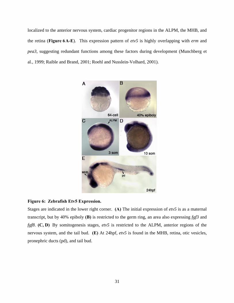
31
localized to the anterior nervous system, cardiac progenitor regions in the ALPM, the MHB, and
the retina (Figure 6A-E). This expression pattern of etv5 is highly overlapping with erm and
pea3, suggesting redundant functions among these factors during development (Munchberg et
al., 1999; Raible and Brand, 2001; Roehl and Nusslein-Volhard, 2001).
Figure 6: Zebrafish Etv5 Expression.
Stages are indicated in the lower right corner. (A) The initial expression of etv5 is as a maternal
transcript, but by 40% epiboly (B) is restricted to the germ ring, an area also expressing fgf3 and
fgf8. (C, D) By somitogenesis stages, etv5 is restricted to the ALPM, anterior regions of the
nervous system, and the tail bud. (E) At 24hpf, etv5 is found in the MHB, retina, otic vesicles,
pronephric ducts (pd), and tail bud.

32
In addition, FGFs were shown to regulate etv5 expression during zebrafish development.
By injecting fgf8 mRNA into zebrafish embryos, FGF signaling can be experimentally altered.
Under these circumstances, expansion of etv5 expression resulted at gastrula stages (Figure
7A,B). Conversely, over-expression of dusp6 (also known as mkp3), a molecule that inhibits
MAPK signaling, suppresses etv5 at shield stage (Figure 7 C,D). Taken together, these
experiments indicate etv5 reacts similarly to changes in FGF signaling during development as
does erm and pea3, further indicating that etv5 is a member of the PEA3 subfamily of ETS
transcription factors (Munchberg et al., 1999; Raible and Brand, 2001; Roehl and Nusslein-
Volhard, 2001).
Figure 7: Expression of etv5 is Regulated via FGF Signaling.
(A,B) fgf8 overexpression results in expansion of etv5 at gastrula stages. (C,D) Conversely,
blocking FGF signaling mediated via a feedback inhibitor dusp6, etv5 expression is inhibited, as
indicated by the red arrows.

33
In my thesis work, I focused on the role of PEA3 ETS transcription factors in FGF
signaling and embryogenesis in the zebrafish. Through the generation of constitutively active
Etv5 constructs, I determined that the over-expression of etv5 induced expression of the FGF-
regulated transcripts dusp6 and sef. Conversely, decreasing expression of PEA3 transcription
factors by antisense morpholino injections disrupted the MHB development, heart formation, and
L/R patterning, similar to phenotypes observed in fish and mouse mutants of FGF signaling. In
addition, I determined the essential DNA sequences required for FGF-mediated induction of
dusp6 using luciferase reporter assays. Electrophoretic Mobility Shift Assays (EMSAs) showed
that the Etv5 ETS DNA binding domain can directly bind to a putative ETS site within the
Dusp6 promoter, and mutating this site eliminated binding. Finally, in collaboration with Anne
Moon at the University of Utah, we show that the orthologous region of the mouse Dusp6
promoter is regulated by PEA3 and Erm via conserved ETS sites and is bound by PEA3 in vivo
in tissues undergoing active FGF signaling during mouse embryogenesis. These results indicate
the importance of ETS factors in relaying FGF signals during development and provide insights
as to how FGF target genes are regulated by them.
1.5 AIMS OF DISSERTATION RESEARCH
In an effort to determine the transcription factors responsible for regulating FGF responses in
zebrafish, the aims of my thesis work have been three-fold: 1) to determine the post-translational
mechanisms that regulate ETS transcription factors, 2) to analyze the role of ETS transcription
factors in development, and 3) to determine how ETS transcription factors regulate gene
expression.

34
Aim 1 – Analyze the post-translational regulation of ETS factors to modulate FGF signaling
FGF activity regulates gene expression through the modification of PEA3 ETS
transcription factors. In vitro studies have shown that ERK2 can directly phosphorylate GST-
Erm fusion proteins and that activation of the RAS/MAPK pathway can increase reporter gene
activation by Erm (Janknecht et al., 1996). In addition, mapping studies have indicated that Erm
has an inhibitory region within the amino terminus that prevents the ETS domain from binding to
a target DNA sequence. After ERK2 phosphorylation, a conformational change releases this
inhibition and reveals the ETS DNA binding domain (Laget et al., 1996). As a result, the model
of PEA3 activity can be described as these proteins being natively folded in an inactive state,
where the inhibitory region is in contact with the DNA binding domain. Upon activation by
ERK phosphorylation, a conformational change occurs that allows the ETS DNA binding
domain to be accessible for DNA binding (Laget et al., 1996). Since ERKs are members of the
serine threonine kinase family, I hypothesize that PEA3 ETS factors are likely to be
phosphorylated at these residues.
Aim 2 – Role of ETS transcription factors in FGF-regulated developmental processes.
Two approaches have been used to determine the role of ETS genes in development,
including gain- and loss-of-function studies. Fusion constructs utilizing the VP16 transactivation
domain were used in over-expression studies, while antisense morpholino oligonucleotides
(MOs) targeting specific ETS factors were employed to knock down expression. In the over-
expression experiments, I focused on the expression of downstream FGF targets, while in MO-
injected embryos I examined downstream FGF targets in addition to proper formation of the
brain, ear, and heart. These aspects of development are altered in zebrafish fgf8 mutants. Similar

35
types of defects recognized in the EtsMO-injected fish can indicate the importance of ETS
transcription factors in FGF-regulated developmental processes.
Aim 3 – Characterization of ETS transcription factor binding
A key to understanding FGF signaling is to identify the cis-regulatory elements in FGF-
target genes. To address this, I have generated deletion luciferase reporter constructs within the
Dusp6 promoter to determine the essential DNA sequences that are required for FGF-mediated
induction of luciferase activity. By using these constructs in Xenopus animal caps assays, I can
measure luciferase protein activity just 6 hours after zygotic transcription initiation, thus
allowing direct measurement of the activation of Dusp6 by FGFs. The luciferase constructs were
injected in the presence or absence of FGFs. In addition, EMSAs and ChIP assays were utilized
to determine direct binding of ETS factors to a specific region of the Dusp6 promoter.

36
2.0 MATERIALS AND METHODS
2.1 ZEBRAFISH MAINTENANCE
Danio rerio were maintained at 28.5oC on a 14hour light/10 hour dark cycle. Wildtype (Oregon
AB*), ace, Tg(Cmlc2:DsRednuc), and Tg(Dusp6:d2GFP)pt6 were used for experiments.
Embryos were staged as described by Kimmel et al. (1995) and Brand et al. (1996).
2.2 RNA INJECTIONS AND IN SITU HYBRIDIZATIONS
These procedures were performed as described previously (Tsang et al., 2000) with the following
modifications for RNA injections: Wildtype AB* zebrafish embryos were injected with etv5
(100pg), etv5:VP16 (75pg), etv5:EnR (150pg), or mutated forms of etv5; etv5:T135D,
etv5:T139D, etv5:S142D (50pg) at the 1-2 cell stage. All mutations within Etv5 were generated
as described in the Quick-Change II Site-directed Mutagenesis Kit (Stratagene) using the
primers:
5’-GGGTTCAAGCCCATTGGACCCTCCCTCGGACCCC-3’ (forward etv5:T135D),
5’-GGGGTCCGAGGGAGGGTCCAATGGCTTGAACCC-3’ (reverse etv5:T135D),
5’-ACTCCTCCCTCGGACCCCGTCTCCCCATGT-3’ (forward etv5:T139D),
5’-ACATGGGGAGACGGGGTCCGAGGGAGGAGT-3’ (reverse etv5:T139D),

37
5’-CCCTCGACGCCCGTCGACCCATGTGTCCCCAGC-3’ (forward etv5:S142D),
5’-GCTGGGGACACATGGGTCGACGGGCGTCGAGGG-3’ (reverse etv5:S142D).
Zebrafish embryos were fixed with 4% paraformaldehyde and followed by methanol
storage. Fixed embryos were analyzed by whole mount in situ hybridization using the protocol
described previously (Kudoh et al., 2001). The following antisense riboprobes were generated in
this study; etv5 (Kudoh et al., 2001), erm (Munchberg et al., 1999), pea3 (Brown et al., 1998),
fgf3 (Kiefer et al., 1996), fgf8 (Reifers et al., 1998), dusp6 (Kawakami et al., 2003; Tsang et al.,
2004), sef (Tsang et al., 2002), bmp4 (Hwang et al., 1997), chordin (Schulte-Merker et al., 1997),
pax2a (Krauss et al., 1991), her5 (Bally-Cuif et al., 2000), amhc (Berdougo et al., 2003), vmhc
(Yelon et al., 1999), nkx2.5 (Chen and Fishman, 1996), gata4 (Reiter et al., 1999), scl (Liao et
al., 1998), spaw (Long et al., 2003) and hand2 (Yelon et al., 2000).
2.3 IMMUNOFLUORESCENCE
Whole mount immunofluorescence was performed to detect monocilia in Kupffer’s vesicle as
described (Yamauchi et al., 2009). Monoclonal acetylated tubulin primary antibody (1:1000;
Sigma) and Cy3-conjugated anti mouse IgG secondary antibody (1:500; Jackson
ImmunoResearch Laboratories) was used in this study. Images were acquired on an Olympus
Fluoview1000 instrument using an UplanSapo 20X (NA 0.75) objective. Data was acquired
using the FV10-ASW software and z-stacks compressed using ImageJ software (NIH).

38
2.4 ANTISENSE MORPHOLINO INJECTIONS
Morpholino oligos (MOs) were designed and purchased from Gene Tools (www.gene-
tools.com):
etv5MO sequence, 5’-ATCCGTCCATGTCACCTGGGTCTTC-3’;
etv5/ermMO sequence, 5’-TGCTGGTCATAAAATCCGTCCATGT-3’;
fgf8MO sequence, 5’-GAGTCTCATGTTTATAGCCTCAGTA-3’;
and ContMO sequence, 5’-CCTCTTACCTCAGTTACAATTTATA-3’.
From Open Biosystems (www.openbiosystems.com):
ermMO sequence, 5’-AACCCATCCATGTCGCTTGCTTCTC-3’;
pea3MO sequence, 5’-ATCCATGCCTTAACCGTTTGTGGTC-3’;
ContMO (10ng), fgf8MO (1-3ng), single EtsMO (5ng), 2EtsMO (2.5ng of each EtsMO), or
3EtsMO (1.3ng of each EtsMO) were injected into the yolk of 1-8 cell stage AB* or
Tg(d2EGFP)pt6 embryos as described (Molina et al., 2007).
2.5 GENERATION OF DUSP6 REPORTER CONSTRUCTS
Zebrafish Dusp6 promoter sequences were PCR amplified and directionally cloned into the
pENTR Gateway vector (Invitrogen) with the following primers:
Forward primers: 5Kb: 5'- CACCGACCGGTAGTGAATTTTGATTTGAAC-3';
4Kb: 5'-CACCGGCCTAGTCGGCACTCAAACCAGTGA-3';
3Kb: 5'-CACCACTGTGGCATTACAGTGACAGGCCCG-3';

39
2Kb: 5'-CACCCTGCGCAGAAGTTCACTTAGACAGTG-3';
1Kb: 5'-CACCCCACACTGAACTGAGCTAAACTGAAC-3';
Reverse primer: Dusp6 Rev: 5'-GGTACCGTGAGACCTTAAAACTGCGG-3'.
The promoter sequences were verified and subcloned into a Gateway-modified pGL3-
promoter (Promega) by the Gateway System (Invitrogen). Pea3A and Pea3B mutant reporter
constructs were generated using QuickChange II with:
MutA: 5’- CACTCGCACTCCTCCGGCCGTCCCGTGAAGCGCCTCTCG-3’;
MutB: 5’-CCGCTGATCCGGCGCGGCCCGTCCTTTCCGTTTTTGTG-3’,
A 782bp fragment of DNA 5' to the mouse Dusp6 gene was amplified by PCR and
cloned into pGL3-Promoter. The primers were:
5’-ccctggtaccGTACCGTTGGATTAGCATTTAACACTTCGT (sense, UCSC genome
browser Chr10: chr10:98,725,230)
5’- ccctagatctAGTCTAGCGGCTCTTAATCCTC (anti-sense, UCSC genome browser
Chr10: 98,726,011)
Upper-case letters indicate genomic sequence. Amplification was performed with Pfx Platinum
polymerase (Invitrogen) and 0.1µg mouse DNA (C57Bl/6) under manufacturer conditions. The
single reaction product was purified by Qiaquick spin column (Qiagen), digested with Acc65I
and BglII (New England Biolabs), and cloned into pGL3-Promoter. Mutagenesis of the
conserved putative PEA3/ERM binding sites 1-3 was performed with a Quick-Change
Lightening Kit (Agilent) with mutagenic oligonucleotides designed by the manufacturer and
plasmids were sequenced.

40
2.6 BCI TREATMENT
Ten embryos were placed into each well of a 24-well plate in 200 µl of E3 (5 mM NaCl, 0.17
mM KCl, 0.33 mM CaCl2, 0.33 mM MgSO4), and a 0.5% (v/v) DMSO solution was added along
with BCI [(E)-2-benzylidene-3-(cyclohexylamino)-2,3-dihydro-1H-inden-1-one] at 10 µM.
Embryos were incubated in this compound from 1 somite or 8 somite stages until 24hpf, then
washed and incubated in E3 until 48hpf and fixed. BCI (also known as NSC150117) was
identified as a compound that enhances fluorescence in treated transgenic embryos. Each
experiment was repeated four times to show the reproducibility of the assay.
2.7 MAMMALIAN CELL AND XENOPUS EXPLANT CULTURES AND
LUCIFERASE ASSAYS
Human 293T cells (ATCC) were grown at 37°C, 5% CO2 in DMEM (GIBCO) containing 10%
FBS, 100U/ml penicillin and streptomycin. Cells were seeded into 24-well plates and were 70–
80% confluence at the time of transient transfection using FuGene6 (Roche). Each well received
100ng of pGL3-Control luciferase reporter, 10ng of pRL-TK vector (Promega) encoding Renilla
luciferase and increasing amounts of Pea3 (Open Biosystems, Clone ID: 3854349) or Erm (Open
Biosystems, Clone ID: 4036564) expression vector. The total amount of DNA in each well was
adjusted to 310ng with pcDNA3. The cells were harvested after 48h using Passive Lysis buffer
(Promega).
Xenopus laevis 2-cell stage embryos were injected with fgf8 RNA (25pg), Dusp6:luc
construct (125pg), and pCMV-Renilla (50pg) into each cell. In other experiments, etv5 (50pg),

41
etv5:VP16 (30pg) or etv5:T135D (40pg) was injected in place of fgf8 RNA. Animal caps were
dissected at stage 8.5 and cultured for 6h. Animal cap lysates were prepared from a population
of four animal caps. In both cell and frog assays, luciferase activities were determined using
Dual Luciferase Reporter Assay System (Promega). The data were normalized by calculating the
ratio of firefly luciferase to Renilla luciferase. A two-sample equal variance T-test using a two-
tailed distribution was applied to analyze the data for statistical significance.
2.8 ELECTROPHORETIC MOBILITY SHIFT ASSAYS (EMSA)
5’ labeled biotinylated and standard oligonucleotides (Midland Certified) 5’-
GTTTGTTTGCACTCCGCT-3’ (forward) and 5’-TTTGTCATTCACAAAAAC-3’ (reverse)
were used to generate biotinylated and competitor PCR amplified Dusp6 promoter. For
oligonucleotide competition assays, 18mer Pea3B site competitors were generated:
5’-TCCGGAGCG-GAAATTCCT-3’ (forward)
5’-AGGAATTTCCGCTCCGGA-3’ (reverse).
A random sequence competitor oligonucleotide was generated using:
5’-CCCTCGACGCCCGTCGACCCATGTGTCCCCAGC-3’ (forward)
5’-GCTGGGGACACATGGGTCGACGGGCGTCGAGGG-3’ (reverse).
GST-Etv5-ETS DNA binding domain protein was expressed in bacteria (BL21) cells
(Invitrogen) and batch purified with GST-sepharose beads (Amersham). Biotin labeled DNA
was incubated with Etv5-ETS protein (10ug) and resolved on a 6% native polyacrylamide gel.
The products were transferred to a nylon membrane, cross-linked by a UV Stratalinker
(Stratagene) and detected with streptavidin-HRP (Pierce). Competition assays were performed

42
with unlabeled oligos or PCR-amplified Dusp6 promoter at 1-500 fold molar excess to labeled
probes.
2.9 CHROMATIN IMMUNOPRECIPITATION (CHIP) ASSAY IN MOUSE
Chromatin was immunoprecipitated using a variation of the protocol described at
http://www.cellsignal.com. Briefly, 100 E9.5 mouse embryos pharyngeal arch regions 2–6 were
dissected, minced into fine slurry, cross-linked in 2% formaldehyde for 30 min, washed with ice-
cold PBS, pelleted, and re-suspended in 1% NP-40 non-denaturing lysis buffer with protease
inhibitors (Roche, 1-836-153). The chromatin DNA was digested with micrococcal nuclease
(New England Biolabs, M0247S) for 5 min to generate fragments ranging from 150 to 900 bp
and diluted in ChIP buffer with protease inhibitors. Two percent of the diluted supernatant was
kept as input control. Sample was incubated at 4 °C overnight with antibody against Pea3 (Etv4)
(Santa Cruz, sc-113 X). A normal mouse IgG (Santa Cruz, sc-2025) was used as negative
control.
Complexes were precipitated with Dynabeads® Pan Mouse IgG (Invitrogen, 110-41).
Immunoprecipitated chromatin was washed, eluted in elution buffer, incubated at 65 °C for 8 h,
and treated with proteinase K. DNA was purified by phenol extraction and ethanol precipitation.
Two different Dusp6 primer sets were employed to determine if the putative PEA3 binding sites
in the Dusp6 promoter were enriched in chromatin immunoprecipitated with anti-ETV4 antibody
relative to distant sequences at this locus: the “Dusp6 prom” set amplifies the highly conserved
region of the mouse Dusp6 promoter:
43
5′ AGTGCCCTGGTTTATGTGC 3′ (chr10: 98,725,599-98,725,618, sense),
5′ CGGGAGGAAGGAGAAAGAA 3′ (chr10: 98,725,599-98,725,618, anti-sense).
The “Dusp6 neg” negative control primer set amplifies a non conserved region 5′ to the Dusp6
gene:
5′ AAGGCCGAGGAAAAGACTTC3′ (chr10: 98,721,292-98,721,311, sense),
5′ ACCCGTGTTACTGGAGATCG′ (chr10: 98,721,426-98,721,445, anti-sense).
An additional negative control primer set was employed that amplifies a region upstream of β-
actin gene:
5′ GTGCTTAAGAGTCCACTATGAGGG3′ (sense),
5′ TCCACTCGCAATCATATACTTAGG3′ (anti-sense).
Equal quantities of input and immunoprecipitated DNA samples were subject to PCR (35
cycles), electrophoresed on a 2% agarose gel and visualized by ethidium bromide staining.
To quantitate the enrichment of the Dusp6 promoter region in the ChIPed region,
quantitative PCR was performed in the Bio-Rad iCycler IQ™ Multicolor Real-Time PCR
Detection System using 25 µl IQ™ SYBR® Green Supermix (Bio-Rad, 170-8882), 250 nM of
each primer and 2 μl of immunoprecipitated and input DNA samples. The amplification ramp
included an initial hold of 5 min at 94 °C, followed by a three step cycle consisting of
denaturation at 94 °C (30 s), annealing at 57 °C (30 s) and extension at 72 °C (30 s); the
amplification fluorescence was read at the end of the cycle. The Ct values and standard
deviations were analyzed using a method modified from that of SuperArray Biosciences:
(http://www.workingthebench.com/search/label/chromatin%immunoprecepitation). This
protocol correctly propagates the standard errors in the PCR data and permits quantitation of the
Dusp6 target amplicon in the ETV4 ChIPed sample relative to that ChIPed by a nonspecific

44
antibody (normalized to the actin negative control amplicon to control for total DNA quantity in
each sample), and expressed as a percent of amplicon detected in the input sample. The
enrichment was nearly 7-fold and this was highly reproducible over multiple ChIP experiments
with this tissue.

45
3.0 ANALYSIS OF POST TRANSLATIONAL REGULATION OF ETS FACTORS
TO MODULATE FGF SIGNALING
3.1 INTRODUCTION
Several members of the mouse PEA3 ETS proteins have been characterized, providing some
evidence for the role of these transcription factors in FGF signaling and development (Brent and
Tabin, 2004; Liu et al., 2003; Mao et al., 2009; Zhang et al., 2009). In Aim 1 of my thesis
research, I wanted to validate that these factors are responsive to FGF signaling in zebrafish, and
thus are effectors in this specific pathway. In addition, although ETS factors have been shown to
be activated via phosphorylation (Janknecht et al., 1996; Laget et al., 1996), the specific
phosphorylation sites on ETS factors remain unknown. Aim 1 examines putative
phosphorylation sites on Etv5 and Pea3 and the role of these sites on ETS activation and FGF
signaling.
3.1.1 PEA3 ETS Factors have Similar Expression to One Another and to FGF Ligands
A phylogenetic analysis of selected PEA3 ETS factors in mouse, human, and zebrafish indicate
that the newly identified zebrafish Etv5 is most similar to zebrafish Erm based on amino acid
substitutions (Figure 8 A). In zebrafish, due to genome duplication, there are two individual
factors, Erm and Etv5, while in human and mouse, only one factor exists, ERM/ETV5 (Kudoh et

46
al., 2001; Roussigne and Blader, 2006)(Figure 8A). Amino acid alignment between the three
PEA3 ETS transcription factors in zebrafish shows high sequence conservation, especially within
the DNA binding domain, acidic domain, and carboxyl region (Figure 8 B). In addition, the
expression pattern of etv5, erm, and pea3 is similar throughout zebrafish development.
Furthermore, the expression pattern of two fgf ligands, fgf3 and fgf8, are similar to that of etv5,
erm, and pea3 throughout development, indicating a potential importance of ETS factors in FGF
signaling. An example of these overlapping expression patterns during somitogenesis stages is
indicated in Figure 8C-G. These findings are akin to what has been described for pea3 and erm
regulation by FGF signaling, further indicating that PEA3 ETS genes are regulated in a similar
manner (Munchberg et al., 1999; Raible and Brand, 2001; Roehl and Nusslein-Volhard, 2001).
In contrast to these three PEA3 ETS members, the other family member in zebrafish, Er81, has
an expression pattern not limited to the domains where fgf ligands are expressed, and altering
FGF signaling does not affect Er81 expression (Roussigne and Blader, 2006). It is likely that
Er81 has evolved in zebrafish to serve a separate function that is distinct from its orthologs in
mouse and Xenopus laevis (Chotteau-Lelievre et al., 2001; Munchberg and Steinbeisser, 1999;
Roussigne and Blader, 2006).

47
Figure 8: Comparative Expression of PEA3 ETS Transcription Factors during Zebrafish
Development.
(A) Phylogenetic alignment of selected zebrafish (red), mouse, and human PEA3 ETS factors.
(B) Diagram of structural features of PEA3 ETS proteins showing the conserved acidic region
(acidic), ETS DNA binding domain (Ets), and carboxyl region (Ct). Percent identities between
Etv5 ETS domain to Pea3 and Erm are listed. (C-G) Lateral views at 10-somite stage. In situ
probes indicated on the bottom right corner. PEA3 ETS transcription factors have overlapping
expression patterns and are similar to that of fgf3 and fgf8 ligands.

48
3.2 ECTOPIC EXPRESSION OF ALTERED FORMS OF ETV5 RESULTS IN
MISEXPRESSION OF FGF TARGET GENES
To determine the function of Etv5 during development, expression constructs were generated for
ectopic expression experiments by subcloning the Etv5 open reading frame into the pCS2+
plasmid. This plasmid allows for generation of mRNA suitable for microinjection studies in
zebrafish embryos. Previous findings indicated that ETS factors contain an auto-inhibitory
domain that prevents binding to the promoter, however, upon RAS/MAPK signaling, the
inhibition is released and promoter binding can occur (Greenall et al., 2001; Laget et al., 1996;
O'Hagan et al., 1996)(Figure 5 ). Due to this, it was hypothesized that overexpression of full-
length Etv5 alone would not be active in this assay, unless stimulated by RAS/MAPK signaling.
To overcome the activity of the auto-inhibitory domain, a VP16 fusion construct was
generated. Herpes simplex virus-encoded protein VP16 is a potent activator that can control
transcription of early viral genes through the interaction of host factors (Wilson et al., 1997).
VP16 is a strong activation domain that functions in many organisms when tethered to
independent DNA binding domains (Cousens et al., 1989; Sadowski et al., 1988; Triezenberg et
al., 1988). Based on this property, fusion of VP16 with DNA binding domains of transcription
factors can be used as a model to investigate transcription activation. In this case, VP16
transcriptional activation motif was fused to the Etv5 ETS DNA binding domain, generating a
constitutively active form of Etv5 (Etv5:VP16) (Figure 9).

49
Figure 9: Etv5 Expression Constructs.
Diagram of Etv5, indicating the acidic region (AR), DNA inhibitory domain (DID), ETS binding
domain (ETS), and carboxyl region (CT) represented as colored boxes. Engrailed Repressor and
VP16 constructs generated by fusion to the binding domain are represented below the full length
Etv5.
Injection of full-length etv5 mRNA at the 1-cell stage (100-300pg) did not have any affect
on development, as indicated by in situ hybridizations at shield stage, during the start of
gastrulation. When compared to uninjected embryos, the expression of FGF target genes dusp6
(Figure 10A-B,Q) and sef (Figure 10E-F,Q) was not affected upon injection of full-length etv5.
In addition, since FGF signaling has been implicated in dorsal-ventral polarity (Furthauer et al.,
2004; Schmid et al., 2000; Tsang et al., 2004), a dorsally expressed gene, chordin (chd), and a
ventrally expressed gene, bmp4, were also examined. In full-length etv5-injected embryos, no
changes occurred in dorsal-ventral patterning when compared to uninjected embryos. The
expression patterns of chd and bmp4 were similar in these embryos (Figure 10I-J,M-N,Q).
To determine if PEA3 ETS factors can regulate the expression of downstream FGF
targets, I employed gain-of-function studies using etv5:VP16. Injection of etv5:VP16 (75pg) at
the 1-cell stage dramatically induced dusp6 and sef at shield stage (Figure 10 C,G,Q). In

50
conjunction with these results, embryos exhibited drastically dorsalized phenotypes, indicated by
altered expression of chd and bmp4 (Figure 10K,O,Q). The etv5:VP16-induced phenotypes are
identical to activation of the FGF pathway by fgf8 mRNA injections (Figure 7). These results
indicate that the fusion of the Etv5 ETS domain to the VP16 transactivation motif is sufficient to
generate a constitutively active form of Etv5, and altering Etv5 expression has an impact on FGF
signaling.
The lack of gross developmental phenotypes in Erm or Pea3 knock-out mice suggests
that these genes are functionally redundant (Chen et al., 2005; Laing et al., 2000). In zebrafish,
since etv5, erm, and pea3 have overlapping expression patterns throughout development (Figure
8), and these genes appear to be regulated by FGF signaling (Figure 7 ), therefore redundant
functionality may also be present here. One approach to study the function of PEA3 ETS factors
and to circumvent redundancy has been to generate a dominant negative construct. Previously,
PEA3 ETS factors have been described to function as activators and ectopically expressing an
engrailed repressor (EnR) fusion construct in the mouse lung and in chick somites yielded
phenotypes opposite of constitutively active versions (Brent and Tabin, 2004; Liu et al., 2003).
Engrailed (En) is a powerful Drosophila repressor homeodomain protein required for proper
segmentation and posterior identity (Jaynes and O'Farrell, 1991). It has been shown to
efficiently repress the activity of a variety of transcription factors. Two separate domains within
En are responsible for a majority of the activity, ultimately resulting in transcriptional repression
through histone deacetylation and modification of chromatin structure (Chen et al., 1999;
Jimenez et al., 1997; Tolkunova et al., 1998; Vinters et al., 1999).
A dominant negative version of Etv5 was constructed by fusion of the Etv5 ETS domain
to the Engrailed transcriptional repressor domain, etv5:EnR (Figure 9). Injection of etv5:EnR

51
(150pg) into 1-cell stage embryos dramatically diminished dusp6 and sef at shield stage,
completely opposite to the phenotypes generated by ectopic expression of etv5:VP16 (Figure
10D,H,Q). This suggests that ectopic expression of etv5:EnR blocks FGF signaling. In
conjunction with these results, embryos exhibited drastically ventralized phenotypes, indicated
by misexpression of chd and bmp4 (Figure 10L,P,Q). These results suggest that this construct is
acting in a dominant negative fashion as has been described with Pea3 and Erm Engrailed fusion
constructs in chick and mouse (Chen et al., 2005; Laing et al., 2000). Thus ectopic expression of
etv5:EnR is sufficient to block FGF signaling and PEA3 ETS genes are required to relay
transcriptional events mediated by FGF signals.

52

53
Figure 10: Etv5 Functions as a Positive Effector in FGF Signaling.
(A-L) Lateral views and (M-P) animal views at shield stage. In situ probes and constructs
injected are indicated on the left and above, respectively. Expression of downstream targets of
FGF signaling, dusp6 and sef, are unaltered in etv5-injected embryos (A,B,E,F), but were
induced by etv5:VP16 mRNA injections (C,G). Ectopic expression of dominant negative
(etv5:EnR) mRNA blocks dusp6 and sef expression (D,H). Expression of bmp4 and chordin was
unchanged in etv5-injected embryos (I,J,M,N). A dorsalized phenotype was noted in
etv5:VP16-injected embryos (K,O) and a ventralized phenotype upon etv5:EnR mRNA
injections (L,P). (U) Frequency of phenotypes elicited by microinjection of mRNAs indicated.
3.3 ETV5 IS PHOSPHORYLATED VIA ERK ON SPECIFIC RESIDUES TO RELAY
FGF SIGNALS
The above results suggest Etv5 is non-functional when over-expressed in the native full-length
form, and must be activated in some way in order to function. This is similar to previous
observations that PEA3 and ERM contain auto-inhibitory domains that prevent these factors
from binding to DNA (Janknecht et al., 1996; Laget et al., 1996). Since previous evidence has
indicated PEA3 ETS factors play a role in FGF signaling in mice (Brent and Tabin, 2004; Liu et
al., 2003; Mao et al., 2009; Zhang et al., 2009), this suggests that Etv5 must be activated by post-
translational mechanisms such as ERK phosphorylation (see F igure 1 ). Two previous
experiments in vitro have shown the importance of ERK phosphorylation on ETS factor function
(Laget et al., 1996; O'Hagan et al., 1996), but an in vivo analysis of ERK phosphorylation in
zebrafish, along with the identities of specific phosphorylated residues on zebrafish ETS factors,
has yet to be determined.

54
Scanning the Etv5 protein sequence using ScanSite and Netphos2.0 software, three ERK
signature phosphorylation motifs were identified. Two putative threonine sites (T135 and T139)
and one putative serine (S142) site were found in Etv5 in regions that are similar to the
consensus ERK phosphorylation site (PXS/TP)(Gonzalez et al., 1991; Songyang et al., 1996).
These putative threonine/serine phospho-acceptor sites are absolutely conserved across species,
supporting they are likely to play an important role in Etv5/Erm function (Figure 11) . Since
these regions of sequence are identical in Etv5 and Erm in zebrafish, these are likely to be the
same phospho-acceptor sites for both family members. To determine the importance of these
sites, I generated mutations in each of them to aspartic acid, which would mimic the introduction
of a negative charged phosphate on the sites (etv5:T135D; etv5:T139D; etv5:S142D)(Figure 11).
Figure 11: Putative ERK Phosphorylation Sites on Etv5/Erm.
Alignment of conserved putative ERK phosphorylation sites in Etv5/Erm. Constitutively active
Etv5 constructs with threonines and serines mutated to aspartic acid are also indicated.
Regardless of which conserved site was mutated, injection of any of the phospho-
acceptor mutations (50pg) at the 1-cell stage dramatically induced dusp6 and sef at shield stage
(Figure 12A-D,F-I,U). In conjunction with these results, embryos exhibited drastically

55
dorsalized phenotypes, indicated by expansion and suppression of chd and bmp4 expression,
respectively (Figure 1 2K-N,P-S,U). The introduction of a double mutation, in which both
threonines are mutated to aspartic acid in Etv5 (Etv5:2T>D), also induced downstream targets
and caused a dorsalization phenotype (Figure 12E,J,O,T,U). These phospho-acceptor-induced
phenotypes indicate ETS factors can function as a protein that hyperactivates FGF signaling, and
a single mutation on any of these 3 conserved serines/threonines was sufficient to generate a
constitutively active Etv5. Thus, I have confirmed Etv5 functions as a transcriptional activator in
FGF signaling, and identified 3 key residues that control Etv5 activity.

56

57
Figure 12: Any O ne of T hree C onserved R esidues i s Sufficient to A ctivate Downstream
Targets of FGF Signaling.
(A-O) Lateral views and (P-T) animal views at shield stage. In situ probes and constructs
injected are indicated on the left and above, respectively. Expression of downstream targets of
FGF signaling, dusp6 and sef were induced by mRNA injections of Etv5:T135D (A,B,F,G),
Etv5:T139D (C,H), and Etv5:S142D (D,I). Mutation of two of these residues (Etv5:2T>D)
displayed similar expansion (E,J). Expression of bmp4 and chordin indicates a dorsalized
phenotype was noted in all phospho-acceptor forms (K-T). (U) Frequency of phenotypes
elicited by microinjection of mRNAs indicated.
3.4 PEA3 IS PHOSPHORYLATED ON A SPECIFIC SERINE RESIDUE TO RELAY
FGF SIGNALING
Although Etv5 and Erm have similar conserved residues that are responsive to phosphorylation
(Figure 11 and 12), less sequence conservation is apparent between Etv5/Erm and Pea3 outside
of the ETS DNA binding domain. Similar methods were used to find potential ERK
phosphorylation sites using ScanSite and NetPhos2.0 within Pea3. A conserved serine residue
(S100) was found contained in an ERK phosphorylation consensus sequence (PXS/TP) in Pea3
(Gonzalez et al., 1991; Songyang et al., 1996). Using site-directed mutagenesis, this residue was
mutated to aspartic acid to generate a phospho-mimic version of Pea3 (Pea3:S100D). Injection
of this construct into 1-cell stage embryos suppressed bmp4 expression, while chd expression
was expanded at shield stage. This dorsalized phenotype is again reminiscent of an
overabundance of FGF signaling within the embryo, suggesting that this PEA3 ETS family
member also plays a role in transcribing the FGF signal (Figure 13). In addition, this provides

58
evidence that I have identified at leat one site within Pea3 that appears to be sensitive to ERK
phosphorylation.
Figure 13: One residue in Pea3 is sufficient to regulate FGF signaling effects.
(A,B) Lateral views and (C,D) animal views at shield stage. In situ probes and constructs
injected are indicated on the left and above, respectively. Mutation of one residue in Pea3, S100,
to a phospho-mimic form (Pea3:S100D) results in a dorsalized phenotype, with expression of
bmp4 reduced (A,B) and expression of chordin expanded (C,D). This phenotype is similar to
overexpressing fgf8 in embryos.
3.5 DISCUSSION
3.5.1 PEA3 ETS Factors are Transcriptional Activators of FGF Signaling
In Aim 1, I showed that PEA3 ETS factors function to mediate the FGF pathway transcriptional
response in zebrafish. In agreement with previous studies in mice (Brent and Tabin, 2004; Liu et
al., 2003; Mao et al., 2009; Zhang et al., 2009), ETS factors are transcriptional activators of FGF
signaling in zebrafish. Injection of a constitutively active form of Etv5 (etv5:VP16) in embryos

59
showed an expansion of FGF target domains, dusp6 and sef. In addition, chd was expanded and
bmp4 reduced in these embryos, an indication of dorsalization. This is reminiscent of
overexpression of fgf8 in embryos, another indication that ETS factors relay FGF signals.
Conversely, a dominant negative form of Etv5 (etv5:EnR) diminished downstream FGF targets
when injected into embryos. Ventralized phenotypes were also apparent in these embryos by
analysis of bmp4 and chd.
3.5.2 PEA3 ETS Factors are Activated Via ERK Phosphorylation
In addition, I have identified three conserved residues (T135, T139, S142) in Etv5 that are
located outside of the ETS DNA binding domain and are part of the ERK consensus sequence
(PXS/TP). Mutations of these sites to generate phospho-mimics activated downstream FGF
targets when injected into embryos. Since high levels of amino acid sequence homology exist
between Etv5 and Erm, these sites are likely to be phosphorylated in both Etv5 and Erm.
Interestingly, phospho-mimic mutations in any one of these three residues results in large
expansion of downstream FGF targets, indicating that all three sites may be direct targets of ERK
phosphorylation. Injection of either threonine (135 or 139) mutants resulted in the same level of
expansion of dusp6 and sef, suggesting that each site elicits a complete response, and no single
site is preferentially favorable to another. This was the first study to indicate specific ERK
phosphorylation sites on ETS factors that can activate downstream FGF targets in vivo.
Previous studies have indicated that FGF-dependent activation of ERK2 was essential to
induce Dusp6 under cell culture conditions (Ekerot et al., 2008). Further in vivo approaches
examined a member of the Ets subfamily of ETS transcription factors, Ets1, in sea urchins to
determine how this protein is activated. In this study, high levels of ERK were localized within

60
cells that will give rise to the urchin skeleton, termed primary mesenchyme cells
(PMCs)(Rottinger et al., 2004). Ets1 was found to be a putative target for ERK phosphorylation,
containing a single consensus site for phosphorylation via ERK. Overexpression of ets1
increases the amount of PMCs, and this effect is inhibited when treating embryos with ERK
inhibitor. Furthermore, mutating the consensus ERK binding sequence in Ets1 to aspartic acid or
alanine resulted in constitutively active or dominant negative affects, respectively (Rottinger et
al., 2004). Combining my data from Aim 1 with this data, it can be concluded that ETS
transcription factors in several subfamilies and in multiple organisms are activated in the same
way, via ERK phosphorylation. Interestingly, only one ERK phosphorylation site was identified
on Ets1, while in Etv5, I have identified three putative ERK phosphorylation sites, all of which
appear individually and collectively to regulate FGF downstream targets.
3.5.3 Other Modes of Post-Translational Modification in ETS Factors
Since Etv5 contains multiple sites for ERK phosphorylation, it is likely that other types of post-
translational modifications are used in combination to temporally and spatially control activities
of PEA3 ETS transcription factors. Recent studies indicate Small Ubiquitin-like MOdifier
proteins (SUMO proteins) can control the activity of Pea3 and Erm (Degerny et al., 2005; Guo
and Sharrocks, 2009). Sumoylation has been shown to impart repressive properties on
transcriptional regulatory proteins (Gill, 2005; Girdwood et al., 2004), but has also been
associated with transcriptional activation (Liang et al., 2004; Terui et al., 2004; Vrieseling and
Arber, 2006). It was found that in the case of Pea3, SUMO modification takes place both in vivo
and in vitro on multiple SUMO binding sites (ψKXEXXSP) within the N-terminal region.
Importantly, this sumoylation of Pea3 was promoted by the activation of the ERK/MAPK

61
pathway, thus the same pathway may control two types of post-translational modifications,
phosphorylation and sumoylation. Sumoylation of Pea3 results in ubiquitination and
destabilization of the protein. This destabilization is required to control the activation of
downstream promoters (Guo and Sharrocks, 2009). These results fit well with conclusions from
Aim 1. When Etv5 phospho-mimics are injected into embryos, a strong, global activation of
downstream targets occurs (Figure 12 and 13). In addition, multiple ERK phosphorylation sites
are found on Etv5 that each can elicit a strong response (Figure 1 2). Due to the powerful
response induced from ERK phosphorylation, it is likely that other post-translational
modifications, such as sumoylation, are required to regulate the activities of PEA3 ETS
transcription factors throughout development.

62
4.0 THE ROLE OF ETS TRANSCRIPTION FACTORS IN FGF-MEDIATED
DEVELOPMENTAL PROCESSES
4.1 INTRODUCTION
Results from over-expression assays (Aim 1) suggest that PEA3 ETS transcription factors play a
crucial role in relaying FGF signals during development. However, these results do not address
the requirement for these factors in developmental processes. To ascertain the requirement for
specific ETS factors during development, antisense Morpholino oligonucleotide technology
(MO) was utilized in zebrafish. Antisense MOs are commercially generated oligonucleotides
(GeneTools, Inc) that are customized to target the AUG codon of a specific mRNA. Annealing
of the MO to the mRNA prevents ribosomal binding, blocking mRNA translation, and thus
generation of the specific protein is knocked-down. The morpholine rings that make up the MO
backbone, instead of deoxyribose rings, prevent nucleases and other enzymes within the cell
from recognizing and breaking down the MO. These molecules can freely diffuse between the
cytosol and the nucleus (www.gene-tools.com). Antisense MOs have been extensively utilized
to knock-down gene function in zebrafish and Xenopus embryos (Heasman et al., 2000;
Nasevicius and Ekker, 2000).
Specifically, since ETS factors are thought to function in FGF signaling, I examined
FGF-mediated developmental processes in zebrafish. Due to the established large role of FGF

63
signaling on MHB formation (Blak et al., 2007; Blak et al., 2005; Crossley et al., 1996; Martinez
et al., 1999; Trokovic et al., 2005; Walshe and Mason, 2000) and a critical role in early and late
zebrafish heart development, (Araki and Brand, 2001; Marques and Yelon, 2009; Molina et al.,
2009a; Reifers et al., 1998; Reifers et al., 2000), I examined to role of ETS factors in these
processes.
4.2 GENERATION OF ANTISENSE MORPHOLINOS SPECIFICALLY TARGETED
TO EACH INDIVIDUAL PEA3 ETS TRANSCRIPTION FACTOR
To determine the requirement of PEA3 ETS factors for FGF-mediated developmental processes,
antisense MOs were generated that inhibited translation of members of the PEA3 ETS genes.
Since sequence conservation and similar expression patterns during development suggest that the
three ETS factors may perform redundant or overlapping functions, it was imperative that each
MO generated was specific for only one PEA3 ETS family member. Each antisense MO
targeted the AUG codon of etv5, erm, or pea3, and one MO targeted both etv5 and erm
(etv5/ermMO), given the high degree of identity between these two genes. To confirm MO
specificity, 2-cell stage embryos were co-injected with MOs and a construct containing the 5’
UTR of an ETS factor fused to enhanced green fluorescent protein (EGFP)(Figure 14A ). All
combinations of MO and EGFP construct were injected into embryos, and were screened at
shield stage for the presence of EGFP. A representation of the etv5MO specificity analysis is
shown in Figure 14B . After co-injection of etv5MO and etv5:EGFP, EGFP expression was
eliminated in these embryos showing the MO was functioning properly. Furthermore, this MO
did not inhibit EGFP expression in other 5’UTR Ets:EGFP-injected embryos, indicating the

64
specificity of the etv5MO (Figure 14B ). At shield stage, each individual MO suppressed
expression of its respective Ets:EGFP fusion protein and not the other related constructs (Figure
14C), indicating that each MO was effective and specific.

65
Figure 14: Specificity of PEA3 ETS Transcription Factor MOs.
(A) Experimental design to determine the specificity of each of the PEA3 ETS MOs. (B)
Brightfield live images of shield stage embryos on the left and EGFP expression on the right.
The construct and MO injected are indicated in the upper right corner. Embryos co-injected with
the 5’UTR erm:EGFP DNA and etv5MO at 2-4 cell stages or with 5’UTR pea3:EGFP DNA and
etv5MO at 2-4 cell stages expressed EGFP. In contrast, over 90% of embryos microinjected
with 5’UTR etv5:EGFP DNA and etv5MO did not show GFP expression, indicating the
efficiency of etv5MO. (C) A graphical representation of all possible combinations of ETS MO
and Ets:EGFP, supporting that each MO is specific for the knock-down of only one PEA3 ETS
family member.
4.3 PEA3 ETS FACTORS FUNCTION REDUNDANTLY AS TRANSCRIPTIONAL
MEDIATORS OF FGF SIGNALING
Microinjection of etv5MO into a 2-cell stage embryo resulted in minor effects on dusp6 or sef
expression at shield stage (Figure 15A,B,E,F,Q). This is in agreement with previous mouse
studies. Single knock-outs of Erm, Pea3, or Er81 results in developmentally normal mice
surviving to adulthood, although mice were mutant in motor neuron differentiation and
spermatogonial stem cell renewal (Chen et al., 2005; Haase et al., 2002; Hippenmeyer et al.,
2005; Livet et al., 2002). This lack of gross developmental defects could be explained due to the
overlapping expression pattern of ETS factors. Redundancy may allow for compensation of
individual deletion of ETS genes, whereby knocking out only one PEA3 ETS factor will not
have an extensive effect on development. Due to this effect, I performed microinjections
targeting two ETS factors, Etv5 and Erm (2EtsMO). It is important to note, that the total
concentration of MO injected with the 2EtsMO was identical to the etv5MO injections. The

66
knock-down of both etv5 and erm resulted in a mild suppression of these FGF target genes
(Figure 15C,G,Q). Furthermore, the concerted depletion of all three factors dramatically
diminished both dusp6 and sef expression, even with lower concentrations of MO targeting
individual ETS factors (Figure 1 5D,H,Q). Similar results were observed with bmp4 and chd
expression at shield stage. MO injections against a single family member had no impact on
bmp4 or chd expression (Figure 15I,J,M,N,Q), however injections targeting multiple members
of PEA3 ETS factors, such as etv5 and erm (termed 2EtsMO throughout this dissertation), or
injections targeting all three members (termed 3EtsMO throughout this dissertation) resulted in a
disruption of dorso-ventral patterning (Figure 15 K,L,O,P,Q). The expansion of bmp4
concomitant with the suppression of chd expression indicates a ventralized phenotype, a
common result of reduced FGF signaling. These results indicate that not only are PEA3 ETS
factors important in mediating FGF signaling, but also act in a redundant manner.

67
Figure 15: PEA3 ETS Factors Function Redundantly to Mediate FGF Signaling.
(A-P) Lateral views at shield stage. In situ probes and MO injected are indicated on the left and
above, respectively. Expression of downstream targets of FGF signaling, dusp6 and sef
remained similar to ContMO upon etv5MO injection (A,B,E,F), but were reduced when multiple
ETS family members being knocked-down (C,D,G,H). Expression of bmp4 and chordin
indicates a ventralized phenotype, but only after multiple family members have been target via
MOs (I-P). (U) Frequency of phenotypes elicited by microinjection of MOs indicated.

68
4.3.1 PEA3 ETS Factors are Critical in Establishing the MHB in Zebrafish
Due to the known importance of FGF signaling on the formation of the MHB (Blak et al., 2005;
Crossley and Martin, 1995; Heikinheimo et al., 1994; Trokovic et al., 2005; Walshe and Mason,
2000), expression patterns of genes critical for the proper formation of the MHB were analyzed
after MO injection. Embryos were injected with MOs at the 2-cell stage and in situ
hybridizations were performed at 28hpf, a timepoint when the MHB region of the brain is
properly formed. Overall phenotypic morphology of the MHB was also evaluated. This analysis
determined the importance of ETS transcription factors in a specific FGF-mediated
developmental process.
4.3.1.1 Expression Patterns of MHB Genes are Reduced in EtsMO-injected Embryos
Targeted depletion of a single family member or two family members did not result in
discernable disruption of the expression of two MHB critical genes, her5 and pax2a (Figure
16A-C,E-G,M). In contrast, the knock-down of all three family members resulted in substantial
reduction in her5 and pax2a expression, indicating a disruption in MHB formation (Figure
16D,H,M). Interestingly, expression of dusp6 is even more sensitive to the knock-down of ETS
factors, having almost completely reduced expression when only knocking down two factors
(Figure 16I -M). Although dusp6 is expressed in other domains at 28hpf, the most dramatic
reduction of expression in MO-injected embryos occurred within the MHB region. These results
collectively indicate the importance of ETS factors in regulating gene expression within the
MHB.

69
Figure 16: PEA3 ETS Factors are Required to Regulate Gene Expression within the MHB.
(A-L) Lateral views of 28hpf embryos. In situ probes and MOs injected are indicated on the left
and above, respectively. Expression of genes localized within the MHB are unaltered in
etv5MO-injected embryos (A,B,E,F,I,J). 2EtsMO mildly reduces her5 and pax2a expression
(C,G,M), while greatly reducing dusp6 expression within the MHB region (K,M). 3EtsMO-
injected embryos have reduction in all MHB markers examined (D,H,L,M). (M) Frequency of
phenotypes elicited by microinjection of MOs indicated. Red arrowheads mark loss of gene
expression within the MHB.

70
4.3.1.2 PEA3 ETS Factors are Required for MHB formation
Due to the large reduction of gene expression within the MHB upon knock-down of PEA3 ETS
factors (Figure 16), a phenotypic analysis was then used to examine the overall formation of the
MHB in the absence of ETS factors. Upon etv5MO injections, a well-formed MHB can still be
seen clearly separating the midbrain from the hindbrain (Figure 17A ,B). After injection of
2EtsMO, the MHB structure breaks down drastically (Figure 17C) in 23% of embryos, and this
phenotype is intensified in 3EtsMO-injected embryos (Figure 17D ), apparent in 63% of
embryos. These defects are similar to the fgf8 zebrafish mutant, acerebellar (ace)(Picker et al.,
1999; Reifers et al., 1998), further indication of the importance of these factors in the FGF
pathway. These results reiterate that PEA3 ETS transcription factors are functionally redundant,
and are necessary in an FGF-mediated developmental event, the formation of the zebrafish
MHB.
Figure 17: PEA3 ETS Factors are Required for Proper MHB formation.
(A-D) Brightfield lateral views of 28hpf embryos. (A) Injection of etv5MO does not disrupt
MHB formation when compared to ContMO. MHB breakdown is apparent in 2EtsMO-injected
embryos (C) and is further intensified in 3EtsMO-injected embryos (D). Red arrows mark the
MHB region.

71
4.3.1.3 Ets Morphant Phenotypes are rescued with ETS Factors
To confirm that the MHB phenotypes observed were the specific result of EtsMO-injections,
rescue experiments were performed. Since the MOs have strong sequence specificity prior to
and including the AUG codon region of each of these factors, tagged mRNA versions in which
the tag is located just upstream of the the AUG codon should be resistant to MO recognition, and
thus knockdown of the protein is inhibited. When performing co-injections of 3EtsMO with
MO-resistant etv5 (HAetv5) or pea3 (HApea3) mRNA, the MHB phenotype of the 3EtsMO was
rescued (Figure 18A,B,D,E; 73% rescued with HAetv5, n=71 and 66% rescued with HApea3,
n=44). Furthermore, the co-injection of HAetv5 and HApea3 mRNA together did not result in an
additive or synergistic rescue of 3Ets morphants, suggesting that either gene can compensate for
the loss of the other (Figure 18F ; 69% rescue, n=56). These results provide further evidence
indicating the redundant nature of this family of factors. Rescue attempts with random mRNA,
such as GFP, did not rescue the 3EtsMO MHB phenotype (Figure 18C). To further examine
this phenotypic rescue on the mRNA level, in situ results indicate induced expression of dusp6
upon HAetv5/HApea3 injection in 3Ets morphants (Figure 18G-I). These data further exemplify
that PEA3 ETS factors function redundantly and are required in the FGF-mediated
developmental process of MHB formation.

72
Figure 18: PEA3 ETS Factors rescue the MHB Phenotype of 3Ets morphants.
(A-I) Lateral views of 28hpf embryos. Injection of antisense MOs targeting pea3, erm and etv5
(3EtsMO) resulted in MHB defects (B) as compared to uninjected controls (A). 3EtsMO-induced
MHB defects can be rescued by co-injection of HAetv5 and/or HApea3 mRNA (D-F) but not by
GFP mRNA (C). Expression of dusp6 is diminished in 3EtsMO-injected embryos (G,H). Co-
injection of HAetv5 and HApea3 rescued dusp6 expression (I). Red arrowhead marks loss of
MHB in MO-injected embryos.
4.3.2 A Role for PEA3 ETS Factors during Cardiac Development
In mice, altering gene dosage of Fgf8 or the source of Fgf8 protein demonstrated a role for Fgf8
in cardiovascular development (Abu-Issa et al., 2002; Frank et al., 2002; Macatee et al., 2003;
Meyers and Martin, 1999; Park et al., 2006). More recently, conditional ablation of Fgf8 and Fgf
receptors with different Cre driver lines revealed that Fgf8 regulates expression of Pea3 and Erm
in heart precursors and is required for outflow tract formation (Ilagan et al., 2006; Park et al.,

73
2006; Park et al., 2008). These studies determined the temporal and spatial role for Fgf8 derived
from both the endoderm and mesoderm for proper morphogenesis of the heart, and showed that
Fgf8 is required for proliferation and survival of cardiac progenitors in mice (Ilagan et al., 2006;
Park et al., 2006). In addition, FGF signaling in zebrafish is necessary to specify and maintain
cardiac progenitor cells during somitogenesis stages (Marques et al., 2008; Molina et al., 2009a;
Molina et al., 2009b; Reifers et al., 2000). These studies lead to the analysis of the roles that
PEA3 ETS factors may play in these processes.
4.3.2.1 PEA3 ETS Factors are Spatially and Temporally Located to Play a Role in Cardiac
Development
Based upon previous fate mapping studies in zebrafish, heart precursor cells are within the
anterior lateral plate mesoderm (ALPM) during early somitogenesis stages. These cells can be
identified in two subpopulations, running parallel and on either side of the notochord from the
anterior to posterior region (Lee et al., 1994; Stainier et al., 1993). For PEA3 ETS factors to play
a role in maintaining cardiac progenitors, they must also be within the ALPM during early
somitogenesis stages. In situ analysis of the three ETS factors at 8-somite stage shows
expression of each of these factors within the ALPM region (Figure 19) . All three factors
appear to have the same level of expression within this region, indicating they all may be
important in this process. Due to this expression pattern, ETS factors may play a role in
maintenance of cardiac progenitors and early heart development.

74
Figure 19: PEA3 ETS F actors a re E xpressed w ithin t he ALPM d uring E arly
Somitogenesis.
Lateral views of 8-somite stage embryos. In situ analysis indicates etv5, erm, and pea3
expression within the ALPM during 8-somite stage, where cardiac progenitors are located. The
inserts show a dorsal view of the magnified ALPM only (identified by the red asterisk), indicated
by the two parallel subpopulations of cells.
4.3.2.2 A Role for PEA3 ETS Factors Maintaining Cardiac Progenitors
Injection of etv5MO mildly reduced expression of a cardiac specific transcription factor, nkx2.5
(Figure 2 0A,B,Y). However, injection of 2EtsMO or 3EtsMO markedly reduced this cardiac
population, a phenotype that was similar to fgf8MO-injected embryos or in ace mutants (Figure
20C-F,Y). Gata4, a gene expressed throughout the entire ALPM, was also reduced in both
2EtsMO- and 3EtsMO-injected embryos, and was comparable to fgf8-deficient embryos (Figure
20G-L,Y) (Draper et al., 2001; Marques et al., 2008; Serbedzija et al., 1998). I next analyzed
expression of hand2, a marker for lateral cardiac progenitors in the ALPM (Yelon et al., 2000).
Complimentary to the observed reduction of nkx2.5+ cells, hand2 expression was also reduced
with the knock-down of multiple PEA3 factors or in fgf8-deficient embryos (Figure 20M-R,Y).
As has been seen earlier, these factors appear to function redundantly to maintain the cardiac

75
progenitor populations, where the most dramatic changes are seen in 2EtsMO- and 3EtsMO-
injected embryos.
Within the ALPM, an important interplay between the hematopoietic/vascular cells and
cardiac progenitors exists to maintain the size of both populations (Keegan et al., 2004;
Schoenebeck et al., 2007). I analyzed expression of scl, an endothelial transcription factor, in
3EtsMO-injected embryos to determine if these factors play a role in endothelial lineages within
the ALPM. Knock-down of PEA3 ETS factors resulted in an expansion of scl expression at the
10-somite stage (Figure 2 0S-V,Y). Most striking was that the depletion of etv5 alone was
sufficient to expand scl populations, indicating that this precursor population is particularly
sensitive to Etv5 activity (Figure 20T). Similar MO injections against only erm or pea3 had no
effect on scl expression (data not shown), indicating the importance of a single, specific ETS
family member, etv5, in restricting endothelial domains within the ALPM. Injection of 2EtsMO
or 3EtsMO elicited stronger expansion of hematopoietic and vessel lineages as also noted in fgf8-
depleted or ace embryos, implicating that the loss of FGF signaling expanded endothelial
lineages (Fig. 4W-Y). Taken together, these results indicate that fgf ligands signal through ETS
factors within the process of early heart development. ETS factors then play a pivotal role in
maintaining cardiac progenitor identity. When these factors are knocked down, cardiac
progenitors cannot be properly maintained, allowing an adjacent population of cells, expressing
scl, to have an increased expression domain.

76

77
Figure 20: PEA3 ETS Factors are Required to Maintain Cardiac Progenitors.
(A-X) Dorsal views at 10-somite stage. In situ probe listed on left and MO above. (A-D) nkx2.5
expression was reduced as multiple PEA3 ETS factors were knocked down. This was similar to
fgf8MO knock-down and in ace mutants (E-F). (G-J;M-P) gata4 expression (G-J) and hand2
expression (M-P) were also reduced after PEA3 ETS depletion (J,P). A similar phenotype was
observed in fgf8MO knock-downs and in ace mutants (K,L,Q,R). (S-V) Dorsal view of scl
expression indicated expansion of endothelial lineages in EtsMO injections (T-V) as indicated by
arrowheads that mark the caudal limit in uninjected embryos (S). Similar results were noted in
fgf8MO and ace embryos suggesting that FGF signaling is required to maintain cardiac
progenitors and to limit endothelial lineages to the rostral ALPM (W,X). (Y) Graph providing
quantitative data for MO experiments.
4.3.2.3 The Role of PEA3 ETS factors in Late Heart Development
Since ETS factors were critical in maintaining cardiac progenitors, I next determined if the loss
of cardiac progenitors at early somitogenesis stages resulted in later heart defects. I assayed
expression of two specific cardiac differentiation markers at 24hpf, ventricular myosin heavy
chain (vmhc) for ventricular tissue, and for atria, atrial myosin heavy chain (amhc) (Berdougo et
al., 2003; Yelon et al., 1999). In etv5MO-injected embryos, amhc was expressed in a population
of cells just below the left eye (Figure 21 B), resembling observations in ContMO-injected
embryos (Figure 2 1A). In the 2EtsMO and 3EtsMO-injected embryos, diffuse amhc staining
was noted at the midline, suggesting disruption of heart tube morphogenesis (Figure 21C,D). In
ContMO- and etv5MO-injected embryos, vmhc expression outlined the heart tube as it “jogged”
to the left (Figure 2 1E,F). However in 2EtsMO- or 3EtsMO-injected embryos, ventricle
morphology was altered and was predominantly located at the midline between the eyes (Figure
21G,H). Thus, by disruption of cardiac maintenance earlier in development when knocking
down ETS factors, late heart defects are apparent. Due to a decreased population of cardiac cells

78
during early heart development upon EtsMO injections, when these cell migrate to form the heart
tube, they may not be capable to migrate correctly. This lack of cells and cell-cell contacts may
be the cause of the lack of migration of the heart tube toward the left eye at 24hpf. Again, it can
be noted that no alteration in phenotype in either amhc- or vmhc-expressing cells is apparent
until multiple ETS factors are knocked down.
Figure 21: PEA3 ETS Factors are Required for Late Heart Development.
(A-H) In situ hybridization showing cardiac gene expression at 24hpf. Knock-down of multiple
PEA3 ETS factors altered amhc (D; red arrowhead indicates midline of embryo) and vmhc
expression (H; cell population and spatial arrangement in yellow bracket).
4.3.2.4 FGF Signaling is Necessary for Proper Heart Size and Looping
One of the final stages in embryonic heart development is cardiac looping. At 48hpf, the linear
heart tube will begin to bend to cause a distinct division between the ventricle and atrium,
creating an S-shaped heart (Chen et al., 1997; Chin et al., 2000). Due to this looping, the
ventricle now lies to the right and dorsal of the atrium, and the two-chambered heart of the

79
zebrafish adult is now formed. In some cases, zebrafish with heart mutations will have hearts
looped in the opposite direction, where the ventricle is positioned to the left of the atrium. This
is commonly referred to “L-looping”, since the mutant heart resembles the letter ‘L’, whereas
wildtype hearts undergo what is called “D-looping”, the final heart shape after looping
resembling the letter ‘D’. These mutants are thought to have defects in the initial assignment of
the embryonic left-right (L/R) axis. In other mutants, hearts fail to loop, and remain as a straight
heart. In these less severe phenotypes, defects could arise from molecular mechanisms that
allow the heart to interpret L/R cues (Bisgrove et al., 2000; Chen et al., 1997; Chin et al., 2000).
Interestingly, several of these mutations also cause defects in L/R morphogenesis of endodermal
organs, such as the liver and gut, suggesting a common mechanism for generating all
asymmetries (Bisgrove et al., 2000; Chen et al., 1997; Chin et al., 2000).
Previous studies examined the role of FGF signaling on heart size and looping during late
heart development, but most of these studies examined heart defects due to decreasing FGF
signals (see Section 1.3.5.2). Recently in our lab, using a transgenic zebrafish chemical screen, a
small molecule inhibitor of Dusp6 was identified: (E)-2-benzylidene-3-(cyclohexylamino)-2,3-
dihydro-1H-inden-1-one (BCI) (Molina et al., 2009a). BCI treatment blocked Dusp6 activity
and enhanced FGF target gene expression in zebrafish embryos. This small molecule could now
be used to examine the effects of hyper-activation of FGF signaling upon late heart development.
Treating embryos at 40% epiboly for eight hours with 10µM BCI showed a drastic increase in
heart size at 48hpf when looking at both vmhc (Figure 22A,B) and cardiac myosin light chain 2
(cmlc2), marking myosin in all cardiac cells (Figure 22C,D).

80
Figure 22: Hyper-activation of FGF Signaling with BCI Increases Cardiac Size.
(A-D) In situ hybridization showing cardiac gene expression at 48hpf. vmhc (A,B) and cmlc2
(C,D) expression were drastically increased upon treatment of BCI for eight hours. Red
arrowheads indicate heart region. Scale bar, 250 µM.
Examining this effect of hyper-activation of FGF signaling more closely, it was
determined that the developmental time period when embryos are treated with BCI has unique
impacts on heart development. When treating embryos with BCI early in somitogenesis (1
somite stage) through 24hpf, the heart greatly increases in size, indicated by an increase in cmlc2
expression (Figure 23 A,B). However, when hyper-activating FGF signaling during mid-
somitogenesis (8 somite stage), the heart does not change in size, but the looping of the heart is
reversed. Instead of the common D-looping phenotype (Figure 23C), embryos are displaying a
reversed, L-looping phenotype (Figure 23D).

81
Figure 23: Late Cardiac Development is Sensitive to the Timing of FGF Hyper-activation.
(A-D) In situ hybridization showing cardiac gene expression at 48hpf. cmlc2 expression
indicates a large increase in heart size when FGF signaling is hyper-activated during early
somitogenesis (A,B), while heart looping is effected when FGF signaling is hyper-activated
during mid-somitogenesis (C,D).
4.3.2.5 Heart Looping is Regulated by PEA3 ETS Factors
Previous studies examining embryos with decreased FGF signaling (Abu-Issa et al., 2002;
Lopez-Sanchez et al., 2002; Marques et al., 2008; Molina et al., 2009a; Reifers et al., 2000) or
increasing FGF activity (Molina et al., 2009a) displayed late heart defects. Thus, it appears that
a proper level of FGF signaling must be maintained during development to form a proper heart.
Too little or too much FGF signaling can result in mispatterning of the heart, such as heart
looping defects. Since altered FGF signaling was shown to have an impact on heart looping, I
next examined if FGF signals were transcribed through PEA3 ETS factors to have an effect on
heart looping events. If so, altering expression of ETS factors should cause heart looping
defects. To examine if the knock-down of ETS factors affect heart looping, in situ

82
hybridizations with cmlc2 expression at 48hpf were performed. Since cmlc2 is expressed within
the entire heart, looping of the heart is easily visible. Disruption of cardiac looping in 3EtsMO-
injected larvae was identified, resulting in either non-looping straight hearts or reversed looping
hearts (Figure 24A -D; non-looping, straight heart phenotypes not shown). The instances of
hearts with altered looping increases as multiple ETS factors are knocked down (Figure 24E).
These data suggested that PEA3 ETS factors are required for proper heart morphogenesis. Thus,
ETS factors are likely to relay signals from Fgf8, Fgf24 and Fgf4, all ligands shown to be
necessary to establish proper L/R asymmetry (Neugebauer et al., 2009; Yamauchi et al., 2009).
Figure 24: Proper Cardiac Looping Requires PEA3 ETS Factors.
(A-D) In situ hybridization showing cardiac gene expression at 48hpf. Cardiac looping was
severely affected in 2EtsMO- (C) and 3EtsMO-injected embryos (D) as shown by expression of
cmlc2. (E) Quantification of effects on heart looping in MO-injected embryos.

83
4.3.3 PEA3 ETS Factors Function in L/R Asymmetry
Given the L/R patterning defects observed in the knock-down of PEA3 ETS factors (Figure 24),
I Their role in asymmetry formation during development was next investigated. Even though
outwardly symmetrical, internal asymmetries exist within the zebrafish, such as the sidedness of
the gut and the directional looping of the heart. The initial break in symmetry is thought to be
due to nodal flow (Brown et al., 1991; Brown and Wolpert, 1990), where the node region of the
embryo gives bilateral symmetry and midline axis to the embryo. This break in asymmetry has
been thought to involve a leftward flow of extra-embryonic fluid around the node that would
transport molecules, which could act as a ‘handed’ determinant (Brown et al., 1991; Brown and
Wolpert, 1990). For example, nodal flow is thought to push molecules located around the mouse
node to one side of the embryo. In zebrafish, a transient ciliated organ called Kupffer’s vesicle
(KV) is thought to be analogous to the mouse node (Cooper and D'Amico, 1996; D'Amico and
Cooper, 1997; Essner et al., 2002). Examining the effect, if any, of PEA3 ETS factors on KV
formation and function may explain the L/R heart defects seen in Ets morphants (Figure 24).
4.3.3.1 PEA3 ETS Factors are Not Required for KV Formation
Our lab has recently generated a transgenic reporter line, Tg(dusp6:d2EGFP)pt6, that expresses
d2EGFP in response to FGF signaling (Molina et al., 2007). To generate these transgenic fish, a
10Kb fragment that included the 5' untranslated sequence within exon I of Dusp6 was subcloned
into a vector containing d2EGFP, a gene that encodes a destabilized green fluorescent protein
that has a two hour half-life. Since dusp6 is controlled by FGF signaling throughout
development (Tsang et al., 2004), expression of d2EGFP is only present in FGF expression
domains (Molina et al., 2007). Expression of d2EGFP is initiated as early as 4hpf within the

84
future dorsal region of the embryo, where fgf3 and fgf8 are initially expressed. At later stages,
d2EGFP is detected within the MHB, pharyngeal endoderm, otic vesicle, hindbrain, and KV,
structures that correlate with the expression of fgf ligands and their receptors. Injection of
3EtsMO into Tg(dusp6:d2EGFP)pt6 embryos resulted in decreased GFP fluorescence within the
MHB, rhombomere 4, and KV, suggesting that PEA3 ETS proteins are required for GFP reporter
gene expression (Figure 25A,B). This was to be expected, since I had already determined PEA3
ETS factors are necessary to transcribe the FGF signal (Figures 10, 12, and 13). However, even
though GFP expression, and thus FGF signaling, was extinguished in the KV, the vesicle did
form properly. This can be seen in brightfield images, where the spherical structure is
completely formed in 3Ets morphants (Figure 25A ’,B’). Therefore, FGF signaling and PEA3
ETS factors are not required for KV formation.
Figure 25: PEA3 ETS Factors are Not Needed to Properly Form KV.
(A,B) Lateral views of Tg(dusp6:EGFP)pt6 embryos at 8 somite stage. (A’,B’) Zoomed, ventral
views of the spherical KV. (A) Wildtype embryos show d2EGFP expression within KV along
with other FGF-expressing regions. (A’) KV is formed properly and highly expresses d2EGFP.
(B) Embryos injected with 3EtsMO show grossly normal formation of Kupffer’s vesicle (B’),
even though FGF signaling was suppressed (B,B’). Red arrowheads indicate the MHB, a region
of high FGF activity. Yellow arrows indicate KV.

85
4.3.3.2 Cilia Formation within KV is Sensitive to PEA3 ETS Factor Expression
L/R patterning defects are apparent in ETS-depleted embryos, even though KV appears to have
developed properly. Since KV appeared to develop and form properly, I next analyzed the cilia
found within KV. In both the KV floor and the roof, the cilia are posteriorly pointed and rotate
clockwise when viewed apically. Microinjection of beads into the KV indicated that the cilia
cause a net circular flow, but the local flow differs in direction depending on the location within
the vesicle. The plane of the circular net flow is tilted within the KV, and thus cells in the
anterior-dorsal region experience a local dominant leftward flow (Okabe et al., 2008). Studies
have shown that altering cilia within the KV of developing zebrafish embryos affect L/R
asymmetry. More specifically, knocking down FGF signaling has been shown to affect cilia
formation (Hong and Dawid, 2009; Neugebauer et al., 2009; Yamauchi et al., 2009).
Analysis of cilia in the KV of 3EtsMO-injected embryos revealed a significant decrease
in cilia number (Figure 26A ,B,E; p = 5.21 x10⁻⁸). In addition, expression of foxj1a, a
transcription factor shown to be involved in ciliogenesis and localized in cells that give rise to
the KV (Bonnafe et al., 2004; Brody et al., 2000), was reduced or absent in 3EtsMO-injected
embryos, implicating the importance of ETS transcription factors in cilia formation (Figure
26C,D; 33% with reduced foxj1a expression in 3EtsMO-injected embryos, n=112). A recent
study shows a similar decrease in foxj1a expression when fgfr1 was depleted (Neugebauer et al.,
2009).

86
Figure 26: PEA3 ETS Factors are Required for Ciliogenesis in the KV.
(A,B) Confocal images of KV at 12 somite stage, ventral view. Cilia number in the KV was
reduced in 3EtsMO–injected embryo (B) compared to ContMO-injected embryos when analyzed
using acetylated tubulin (A). (C,D) In situ hybridization at 90% epiboly, lateral view.
Expression of foxj1a is significantly reduced in 3EtsMO-injected embryos (D) when compared to
ContMO-injected embryos (C). (E) Graph showing average number of cilia in MO-injected
embryos. Error bars indicate the standard deviation.
4.3.3.3 Disrupting Cilia Formation Alters L/R Patterning in 3Ets Morphants
Recent reports indicate the complexity of all cilia beating in varying ways, depending on the
location within the KV, to cause a global leftward flow (Amack et al., 2007; Kreiling et al.,
2007; Okabe et al., 2008). To determine if the lack of KV cilia within 3EtsMO-injected embryos
can be attributed to the L/R asymmetry defects in heart looping, spaw, a zebrafish Nodal gene,
was analyzed. spaw is normally expressed in the left lateral plate mesoderm (Long et al., 2003)
and is the first marker of asymmetry in the zebrafish. In 3EtsMO-injected embryos, spaw

87
expression was completely randomized; often absent, bilateral, or right-sided. This is consistent
with disruption of upstream initiation of the L/R cascade (Figure 27A-D). These L/R defects
increase as multiple ETS factors are knocked down (Figure 27E). Interestingly, knocking down
Fgf8 only does not cause as drastic of an effect in L/R patterning as injection of 3EtsMO (Figure
27E). This may indicate that more ligands in addition to Fgf8 signal through ETS factors to play
a role in L/R patterning. This finding agrees with a current model, whereby Fgf24 ligands in
addition to Fgf8 ligands will signal through FGFR1 and activate transcription factors that will
regulate intraflagellar transport genes to maintain proper motile cilia (Figure 4)(Neugebauer et
al., 2009).
Figure 27: spaw Expression is Completely Randomized in 3EtsMO-injected Embryos.
(A-D) In situ hybridization showing spaw expression at 18 somites, dorsal view. spaw
expression is completely randomized in 3EtsMO-injected embryos (B-D) when compared to
ContMO-injected embryos (A). Graph depicting L/R defects in PEA3 ETS- and Fgf8-depleted
embryos.

88
4.4 DISCUSSION
4.4.1 The Role of ETS Factors in FGF-Mediated Developmental Processes
In Aim 2 I showed that PEA3 ETS factors function to mediate the transcriptional response of the
FGF pathway in zebrafish. Antisense MO injections blocking the translation of erm, etv5, and
pea3 resulted in suppression of FGF target genes as well as a MHB defect in the zebrafish
embryo. The importance of these ETS factors in heart development was revealed by the loss of
cardiac progenitors in PEA3 ETS factor-depleted zebrafish embryos, consistent with a critical
role for these factors downstream of FGFs in mice (Liu et al., 2003; Lu et al., 2009; Zhang et al.,
2009). In zebrafish, experimental manipulations that expand cardiac progenitors negatively
affected the blood and vessel lineages, and vice versa (Molina et al., 2009a; Molina et al., 2009b;
Schoenebeck et al., 2007). Given that Fgf8 and PEA3 ETS factors play an important role in
maintaining cardiac identity, I reasoned that the knock-down of PEA3 ETS factors decrease
cardiac progentior populations. Indeed, the concerted depletion of PEA3 ETS factors resulted in
a decrease in cardiac progenitors. This was concomitant with an expansion of blood and vessel
progenitors as marked by expanded scl expression. Interestingly, one ETS factor, Etv5, was
shown to be critical for proper scl expression, indicating that, although PEA3 ETS factors appear
to function redundantly in most FGF-mediated developmental processes, this specific factor
appears to be solely responsible for proper endothelial expression within the ALPM. Thus, FGF
activity is required to maintain cardiac progenitor populations and suppress endothelial
differentiation within the caudal domain of the ALPM (Figure 28).

89
Figure 28: Model of ETS Factor Regulation on Cardiac Development.
Fgf lignds signal through PEA3 ETS factors that will maintain cardiac identity. When ETS
factors are knocked down, this allows the blood and vessel populations to expand, while cardiac
lineages are reduced.
Recent studies have highlighted a role for FGF signaling in L/R patterning of the
zebrafish embryo (Albertson and Yelick, 2005; Hong and Dawid, 2009; Neugebauer et al., 2009;
Yamauchi et al., 2009). The knock-down of either Fgf ligands or two effectors of the FGF
pathway did not disturb formation of the KV, but did disrupt monocilia formation (Hong and
Dawid, 2009; Neugebauer et al., 2009; Yamauchi et al., 2009). Similarly, in this study, depletion
of PEA3 ETS factors did not disrupt KV formation, but cilia number was markedly reduced.
This is responsible for the laterality defects such as alteration of spaw expression and both the
cardiac jogging and looping defects. Interestingly, among these recent studies, the role of FGF
signaling in proper monocilia formation within the KV has ranged from contributing to cilia
length (Neugebauer et al., 2009; Yamauchi et al., 2009) to contributing to the total number of

90
cilia within the KV (Hong and Dawid, 2009). Due to multiple cilial phenotypes displayed when
knocking down FGF signaling, it can be hypothesized that the specificity in patterning of KV
monocilia may involve several Fgf ligands and/or transcription factors. In addition, it could also
be hypothesized that Fgf ligands have morphogenic effects on cilia formation, whereby a certain
level of FGF signaling must reached for proper cilial number, while another level of FGF
signaling is necessary for proper cilial length. Within this study, I have demonstrated that the
PEA3 ETS transcription factors relay the FGF signal to allow for formation of the proper number
of cilia within KV.
4.4.2 Redundant Functions of PEA3 ETS Transcription Factors Throughout
Development
Erm/Etv5 or Pea3 gene knock-out approaches in mice resulted in developmentally normal
animals that survived to adulthood, but developed motor neuron differentiation defects,
spermatogonial stem cell renewal defects, and movement disorders (Arber et al., 2000; Chen et
al., 2005; Livet et al., 2002). The lack of gross developmental phenotypes in these knock-out
mice suggested that these genes may function redundantly in mediating FGF signaling. To
circumvent redundancy among PEA3 factors, engrailed repressor fusion constructs were
ectopically expressed in both mouse and chick embryos (Brent and Tabin, 2004; Liu et al.,
2003). The repression of multiple PEA3 ETS family members using this technique overcame
redundancy to show a role for these factors in somite and lung development (Brent and Tabin,
2004; Liu et al., 2003). Further evidence of their redundant function was shown by ectopic
expression of a dominant negative version of Etv5 in the mouse limb which resulted in shortened
limbs and polydactyly (Mao et al., 2009). This was confirmed by the knock-out of both Etv4 and

91
Etv5 in early mesodermal lineages that also resulted in limb and digit defects (Zhang et al.,
2009). In the studies presented in this dissertation, antisense morpholinos allowed the targeted
depletion of multiple members of the PEA3 family. In general, knocking down only one
member of this family rarely resulted in developmental defects, but the knock-down of two or
three members resulted in distinct phenotypes.
Functional redundancy within another ETS sub-family of transcription factors in
zebrafish has also been described (Pham et al., 2007). This study examined four ETS family
members expressed in the vasculature: fli1, fli1b, ets1, and etsrp (Pham et al., 2007). Using an
antisense MO approach to knock down expression of all four genes, both vascular and
hematopoietic development was disrupted, showing the importance of these genes for vessel
sprouting and circulation. Interestingly, a hierarchy was observed, whereby the knock-down of
estrp showed stronger phenotypes than a single knock-down of fli1, fli1b, or ets1 (Pham et al.,
2007). Similarly, a reduction of etv5 was shown in our study to have a strong effect on the scl
expression domain, indicating a hierarchy may also exist between members of the PEA3 ETS
factors. In conclusion, we have defined the importance of PEA3 ETS transcription factors in
mediating FGF signaling during development.
My data suggest that in some instances the knock-down of two PEA3 ETS proteins was
sufficient to reveal a phenotype in a majority of the injected embryos, as noted with the
expression of cardiac progenitor markers, implicating an overlapping function between these
factors. Given that knock-down studies can be difficult to quantitate, a more effective means is
to generate genetic nulls and determine the relative contribution of each PEA3 ETS factor to
FGF signaling and development. One interesting observation that demonstrates ETS overlapping
roles comes from a recent study in mouse kidney formation. Compound heterozygotes of two

92
factors (Etv4+/-; Etv5+/-) or Etv4 nulls resulted in a small percentage of embryos showing renal
defects. In contrast, Etv4-/-; Etv5+/- mice exhibited a complete lack of kidneys, suggesting that
a full compliment of Etv5 can function to restore normal kidney development, but in a reduced
state, it cannot (Lu et al., 2009). Thus, gene dosage may play a critical role in how these factors
bind to promoters and regulate transcription and development. A more detailed gene expression
profiling and readout is required to correlate gene activity with developmental outcome.

93
5.0 CHARACTERIZATION OF PEA3 ETS TRANSCRIPTION FACTOR BINDING
TO AN FGF DOWNSTREAM TARGET
5.1 INTRODUCTION
In the previous two aims, I have determined both how ETS factors are activated, and how their
activation plays a role in FGF-mediated developmental processes. However, there is a lack of
evidence that indicates PEA3 ETS factors directly regulate FGF-mediated gene transcription. In
Aim 3, I will identify cis-regulatory elements within an FGF target gene, Dusp6, and illustrate
how ETS factors bind to these elements within this promoter. To achieve this, luciferase reporter
constructs containing the Dusp6 promoter were generated to determine the essential DNA
sequences that are required for FGF-mediated induction of luciferase expression. By using these
constructs in a Xenopus animal cap assay, I can measure luciferase activity soon after the initial
zygotic transcription takes place; providing a direct measure of promoter activation. This allows
a direct analysis of transcription factor binding to a promoter to activate transcription in an in
vivo system. In addition, Electrophoretic Mobility Shift Assays (EMSAs) were utilized to
determine direct binding of ETS factors to a specific region of the Dusp6 promoter. To further
verify my conclusions, this aim will also contain data generated from a collaboration with Dr.
Anne Moon at the University of Utah, where ETS transcription factor binding to the Dusp6
promoter was analyzed in mouse to demonstrate evolutionary conservation. In addition, a ChIP
94
assay was performed to analyze this transcription factor binding to the promoter within mouse
embryonic tissues.
5.2 USING XENOPUS ANIMAL CAP EXPLANTS TO DETERMINE THE
LOCATION OF CIS-REGULATORY ELEMENTS WITHIN THE DUSP6 PROMOTER
The process by which FGF signaling is relayed into a transcriptional response in development is
not fully defined. Because Dusp6 is a known target of FGF activity (Molina et al., 2007; Tsang
et al., 2004), I focused on identifying cis-regulatory elements within the Dusp6 promoter to
determine if PEA3 ETS factors can directly regulate its transcription. Previously, transgenic
zebrafish FGF reporter lines were generated in our lab (Tg(dusp6:EGFP)pt6) and it was
determined that a 10Kb sequence upstream of Exon 1 in Dusp6 was sufficient to drive GFP
reporter expression in an FGF-dependent manner (Molina et al., 2007). Thus, all of the elements
required for transcription factor binding to this promoter are contained within this 10Kb region
of sequence. To determine which area of the 10Kb region contains cis-regulatory elements,
luciferase reporter constructs were generated by inserting varying lengths of Dusp6 upstream
sequence (1Kb to 10Kb) into a pGL3 luciferase reporter vector. These constructs were then used
in Xenopus laevis explant cultures.
The Xenopus explant assay is an ideal and widely used system for analysis of
transcription factor activation of a promoter in vivo (Casey et al., 1999; Friedle et al., 1998;
Rebbert and Dawid, 1997; Rogers et al., 2009; Watanabe et al., 2002). The major advantage of
this system is that luciferase protein activity can be measured just 6 hours after zygotic
transcription initiation, thus allowing the direct measure of the activation of Dusp6 by FGFs.

95
The luciferase reporter constructs were co-injected into the animal pole of 2-cell stage
Xenopus embryos in either the presence or absence of fgf8 mRNA. pCMV-Renilla was also co-
injected to normalize for injection variability in the dual luciferase reporter assay system
(Promega). The embryos were then cultured until Stage 8.5 (just prior to initiation of zygotic
transcription) at which point the animal regions were explanted (Figure 29) . The explanted
animal caps were cultured for 6 hours, then harvested and measured for luciferase activity. Due
to the short length of the culture, the animal caps are naive to other signaling except for what has
been injected into the embryo, thus a very direct effect of these injections can be measured.
Important to note, lysates were prepared from a population of four animal caps, and several
different lysates were analyzed during multiple injection sessions to account for variability
between experiments.
Figure 29: Diagram of Xenopus laevis Animal Cap Assay.
An overview of the protocol for Xenopus animal cap assays. A Dusp6 promoter fragment (1Kb
– 10Kb) with a luciferase reporter was injected (+/- fgf8) into each cell of a 2-cell stage Xenopus
embryo. Just prior to zygotic transcription (Stage 8.5) the animal cap is removed and cultured
for 6 hours. Lysate was made from four caps and analyzed for luciferase activity. The colored
boxes on the luciferase construct represent potential regions where ETS factors may bind to the
promoter.

96
5.3 FGF REGULATORY ELEMENTS WERE FOUND WITHIN A 2KB REGION OF
THE DUSP6 PROMOTER
In animal caps co-injected with fgf8 mRNA, an approximate 3-fold increase in luciferase reporter
activity was detected in all Dusp6 promoter constructs (10Kb – 1Kb) as compared to animal caps
injected with Dusp6 promoter constructs alone. This is a valid positive fold increase in animal
cap assays based on previous studies (Casey et al., 1999; Friedle et al., 1998; Rebbert and
Dawid, 1997; Rogers et al., 2009; Watanabe et al., 2002). This suggests each of these reporter
constructs contain putative responsive elements that are sensitive to FGF signals and must be
contained within a 1Kb region of the Dusp6 promoter (n>4 for each construct; Figure 30 shows
typical results from 5Kb-1Kb Dusp6 promoters). Since each promoter assay is a different
injection experiment, comparisons cannot be made between different promoters, but trends can
be evaluated based on luciferase induction of each promoter in the +/- of fgf8.

97
Figure 30: fgf8 Activates the Dusp6 promoter.
Luciferase activity measured with 5Kb-1Kb Dusp6 promoter constructs in the presence or
absence of fgf8. Each promoter was activated at least 2-3 fold in the presence of fgf8, indicating
cis-regulatory elements are located within the 1Kb region of the Dusp6 promoter. Error bars
indicate standard deviation.
5.4 TWO PUTATIVE PEA3 ETS BINDING SITES LOCATED WITHIN THE 2KB
REGION OF THE DUSP6 PROMOTER REGULATE TRANSCRIPTION
Due to an increase in luciferase activity in the presence of fgf8 upon injection of the 1Kb Dusp6
promoter construct, it was hypothesized that a population FGF responsive elements are located
within this 1Kb upstream promoter sequence. Using a bioinformatics approach to identify cis-
elements within the Dusp6 promoter that may be important for FGF regulated expression, I
compared this 1Kb stretch of promoter region from several species (zebrafish, pufferfish, mouse,
and human) based on sequence identity. Significant homology was identified within these
vertebrate promoters within the 1Kb region, indicating that this non-coding sequence of DNA
has been conserved through several hundred million years since their divergence (Figure 31A).
Further analysis revealed some of the conserved sequences represent known binding sites for
transcription factors (http://www.ifti.org/cgi-bin/ifti/Tfsitescan.pl). From this approach, several
putative transcription factor binding sites were identified within this region, including two PEA3
ETS binding sites (referred to Pea3A being the upstream site and Pea3B being the downstream
site)(Figure 31A). These sites were also described in the mouse Dusp6 promoter (Ekerot et al.,
2008).

98
To determine if these core ETS 5’-GGAA-3’ recognition sequences represents important
regulatory sites for FGF responsiveness, two new 2Kb luciferase constructs (Pea3MutA and
Pea3MutB) were generated in which these putative sites were mutated (Figure 31A; sequence of
mutated residues are indicated in red). Through these luciferase assays, it was determined that
the Pea3B site, which contains three perfectly conserved core 5’-GGAA-3’ motifs, was critical
for the FGF-mediated induction of luciferase activity. Conversely, mutating the Pea3A site had
no effect on luciferase activity (Figure 31B). Of note, the sequences flanking this putative ETS
binding site are also well conserved from zebrafish to human, suggesting that these regions may
be potential binding sites for protein partners of Pea3 ETS factors.
Figure 31: A Conserved Putative PEA3 ETS Binding S ite within the Dusp6 Promoter i s
Critical for FGF-Mediated Induction of Luciferase Activity.
(A) Alignment of the Dusp6 promoter from several vertebrate species showing the conserved
putative ETS binding sites (highlighted in purple) that were mutated, as shown in red below the
sequence. (B) Luciferase activity measured with the 2Kb Dusp6 promoter (WT) constructs in
the presence or absence of fgf8, indicating the requirement for the Pea3B site (MutB). A two-
sample equal variance T-test using a two-tailed distribution was applied to analyze for statistical
significance. Error bars indicate standard deviation.

99
Since PEA3 ETS factors are expressed within the same temporal and spatial domains as
dusp6, it is likely that these factors are direct regulators of dusp6 expression in vivo. To test this,
I co-injected etv5:VP16 instead of fgf8, along with the 2Kb Dusp6 reporter into the Xenopus
animal pole and measured luciferase activity six hours after zygotic transcription. Ectopic
expression of etv5:VP16 resulted in a dramatic 5-fold induction of luciferase activity when
compared to the injection of the Dusp6 reporter construct alone (Figure 32A). To confirm the
hypothesis that etv5:VP16 binds to Pea3 sites, I co-injected etv5:VP16 and the Pea3Mut reporters
and measured luciferase activity. As predicted, the integrity of the Pea3B site was important for
etv5:VP16 to activate luciferase expression (Figure 3 2A). Furthermore, etv5:T135D increased
luciferase activity and was dependent upon the Pea3B sequence (Figure 3 2B). These results
indicate that FGF signaling directly regulates Dusp6 transcription through the activity of PEA3
ETS factors in zebrafish. In addition, co-injection of etv5 with the Dup6 reporter did not
increase luciferase activity (Figure 32A ,B), further implicating the importance of post-
translational modifications of etv5 for transcriptional activity.

100
Figure 32: F GF Signaling Regulates Dusp6 Transcription Through the Activity of PEA3
ETS Factors.
(A) etv5:VP16 activated the Dusp6 reporter (WT), and mutation of the Pea3B (MutB) site
diminished this activity. (B) Ectopic expression of etv5:T135D activated the Dusp6 reporter
(WT), but not the mutant Pea3B reporter (MutB). A two-sample equal variance T-test using a
two-tailed distribution was applied to analyze for statistical significance. Error bars indicate
standard deviation.
5.5 PEA3 ETS FACTOR FUNCTION IS CONSERVED BETWEEN ZEBRAFISH
AND MOUSE
In collaboration with Dr. Anne Moon at the University of Utah, mammalian PEA3 factors were
tested to determine if they could drive reporter gene expression from the zebrafish promoter,
indicating conservation between zebrafish and mouse PEA3 ETS factor function. 293T cells
were co-transfected with Pea3 or Erm, the zebrafish Dusp6 promoters, and Renilla control

101
expression plasmid to account for transfection variability. Both PEA3 and ERM were capable of
inducing reporter gene expression controlled by the zebrafish promoter, and this was dependent
on the Pea3B site (Figure 33).
Figure 33: C onservation o f PE A3 a nd E RM Func tion o n M ouse a nd Zebrafish Dusp6
Promoters.
Mouse PEA3 and ERM transactivate the zebrafish Dusp6 promoter via a conserved putative
PEA3/ERM binding site. HEK293 cells were cotransfected with the zebrafish 2Kb promoter in
the PGL3 reporter and two concentrations of the mouse PEA3 or ERM expression plasmids.
Both PEA3 and ERM significantly increased luciferase activity from the zebrafish promoter. The
Pea3MutB diminished the response to PEA3 and ERM. Error bars indicate standard deviation.
Furthermore, the mouse Dusp6 promoter was isolated and generated luciferase reporter
constructs with 0.7Kb of upstream promoter sequence, where the critical putative Pea3 binding
domain is located in zebrafish. Ectopic expression of mouse PEA3 or ERM transactivated the
promoter, and this activity was also dependent on the exact Pea3B sequence found in the
zebrafish Dusp6 promoter (Figure 34A,B). These results reveal the evolutionary conservation of

102
PEA3 ETS factors between mouse and zebrafish, and that proteins from both species can activate
reporter genes from two diverse promoters.
Figure 34: Requirement for the Conserved Pea3B Site in the Mouse Dusp6 Promoter.
Transactivation of a 781bp fragment of the mouse Dusp6 promoter by PEA3 and ERM is
dependent on a region containing three consecutive putative PEA3/ERM core binding motifs
(bold type). This region was also conserved in zebrafish (Pea3B). (A) Interspecies alignment
and sequence of mutations (red type) generated in putative core motifs. (B) HEK293 cells were
co-transfected with plasmids containing the 781bp Dusp6 promoter luciferase reporter, the
Renilla transfection control reporter, and Pea3 or Erm. Maximal response to PEA3 requires all
three 5' GGAA 3' (5' TTCC 3') core motifs, while response to ERM requires sites 1 and 2. Error
bars indicate standard deviation.

103
5.6 PEA3 ETS PROTEIN DIRECTLY BINDS TO THE DUSP6 PROMOTER
Xenopus animal cap luciferase assays indicated the requirement of a putative Pea3 binding site
on the Dups6 promoter to activate transcription in vivo, however evidence for direct transcription
factor binding has not been reported. To demonstrate this, I used biotin-labeled electrophoretic
mobility shift assays (EMSAs) to show direct binding of Etv5 Ets domain to the Pea3B site in
vitro. To generate a purified Etv5 protein fragment, GST-Etv5-Ets DNA binding domain protein
was expressed in bacteria (BL21) cells (Invitrogen) and batch purified with GST-sepharose
beads (Amersham)(Figure 35).
Figure 35: GST Purification of Etv5:Ets Binding Domain Protein.
An Etv5 protein fragment containing the entire Ets DNA binding domain was generated by
expressing GST-Etv5-Ets DNA binding domain protein in BL21 cells and batch purified with
GST-speharose beads. Elutes 1-8 are shown on an SDS PAGE gel, where elute 6 was used for
the Etv5 protein in EMSA studies.

104
Biotin-labeled oligos that flank the Pea3B site (60bp) were synthesized and used to
amplify this region by PCR (Figure 36A). Recombinant GST-Etv5:Ets domain protein interacted
with the biotin-labeled Dusp6 promoter DNA, causing a band shift (Figure 36D ; lane 2). The
bound DNA was competed by increasing amounts of non-biotin labeled promoter (Figure 36D;
lanes 3-7). In addition, another more specific competitor was generated against only the Pea3B
sequence. This Pea3B-18mer oligo (Figure 3 6C) also competed Etv5 ETS domain binding
(Figure 3 6D; lanes 8-12), indicating that the direct binding of Etv5 to this promoter is at the
specific putative Pea3B site. Conversely, a random sequence used as a control competitor did
not compete with the biotin-labeled Dusp6 fragment (Figure 36D; lanes 13-17). Furthermore, to
verify the importance of the Pea3B site for Etv5 binding, a 60bp biotin-labeled Pea3B mutant
Dusp6 promoter was used in EMSAs (Figure 36B). Etv5:Ets protein did not shift the Pea3B
mutant DNA (Figure 36D; lane 18,19), verifying that mutating this specific putative binding site
within the promoter will not promote Etv5 binding.

105
Figure 36: Etv5-Ets Domain Binds to the Dusp6 Promoter.
(A) Biotin labeled PCR product. (B) Biotin labeled mutant Pea3B Dusp6 promoter fragment,
with the mutated residues identified in red. (C) Pea3B 18bp fragment used in competition assays.
Underlined sequence represent the putative Pea3B binding site (D) EMSA using Dusp6 promoter
biotin fragment and GST-Etv5:ETS protein. The Etv5-ETS domain bound to the 60bp Dusp6
promoter containing the Pea3B site (lane 2). This binding was competed with the non-biotin
labeled 60bp Dusp6 promoter (lanes 3-7) and the Pea3B 18bp fragment (lanes 8-12). A random
oligonucleotide sequence did not compete this binding (lanes 13-17). Binding of Etv5-ETS to
the Pea3B mutant Dusp6 promoter was not observed (lanes 18-19).
To determine if PEA3 ETS factors can directly bind to the mouse Dusp6 promoter in
vivo, Chromatin Immunoprecipitation (ChIP) assays were performed as part of a collaboration
with Dr. Anne Moon’s laboratory from the University of Utah. In this assay, pharyngeal tissue
was isolated from E9.5 mouse embryos, a region with active FGF signaling that expresses high

106
levels of Etv4 (an ortholog of Pea3 in zebrafish), Etv5, and Dusp6 (Dickinson et al., 2002; Ilagan
et al., 2006; Park et al., 2006). By ChIP and PCR, it was found that the Pea3B binding site was
enriched with specific ETV4 antibodies, suggesting that ETV4 directly binds to the Dusp6
promoter in vivo (Figure 37 A-C). qPCR quantitation revealed a 7-fold enrichment of this
element in the ETV4 ChIPed sample compared to the negative control (Figure 3 7D). Taken
together, this shows that Dusp6 is directly regulated by PEA3 ETS factors during zebrafish and
mouse embryonic development.

107
Figure 37: ETV4 Directly Binds to the Dusp6 Promoter in vivo in Mouse.
The mouse Dusp6 promoter region bearing highly conserved PEA3 binding sites is enriched in
chromatin immunoprecipitated from mouse embryonic tissues with anti-mouse ETV4 antibody.
(A) Agarose gel showing PCR products obtained from equal amounts of input, mouse IgG
ChIPed, and anti-ETV4 ChIPed DNAs with the Dusp6 promoter primers that amplify the region
containing the putative PEA3 binding sites. (B) Agarose gel showing PCR products obtained on
the same DNA samples as in A with Dusp6 control primers, amplifying a region of the Dusp6
promoter not containing the Pea3B site. (C) Agarose gel showing PCR products obtained on the
same DNA samples as in A with βactin control primers. Only the Dusp6 promoter was
differentially precipitated by the anti-ETV4 antibody. B and C control primers give identical
bands as they detect the non-specific, background precipitated DNA. (D) Graphical
representation of quantitative PCR results show nearly 7-fold enrichment of the amplicon
containing the highly conserved region of the Dusp6 promoter in ETV4 ChIPed DNA compared
to IgG negative control expressed relative to that detected in input. m, marker; bl, blank lane.
Error bars indicate standard deviation.
5.7 DISCUSSION
5.7.1 PEA3 ETS Factors Bind to the Dusp6 Promoter Directly at a Specific Binding Site
The process by which FGF signaling is relayed into a transcriptional response in development is
not fully defined. Because Dusp6 is a known target of FGF activity, I focused on identifying cis-
elements within the Dusp6 promoter to determine if PEA3 ETS factors can directly regulate its
transcription (Molina et al., 2007; Tsang et al., 2004). I identified several conserved putative
ETS binding sites within 1Kb upstream of the Dusp6 transcription start site and through reporter
and binding assays, indentified one specific site that was critical for reporter gene activity and in
vitro binding. This site is identical to what has been recently described in the mouse Dusp6

108
promoter (Ekerot et al., 2008). Ekerot et al. (2008) had demonstrated by ChIP studies in
NIH3T3 cells that both Ets1 and Ets2 can bind to this particular region. Our studies demonstrate
for the first time that Pea3 can bind to the Dusp6 promoter in vivo and provide strong evidence
that FGF signaling regulates Dusp6 transcription by PEA3 ETS factors during development.
These differences may reflect varying expression levels of these ETS factors between NIH3T3
cells and mouse pharyngeal tissue. Since Fgf8, Etv4, Etv5 and Dusp6 are strongly expressed in
pharyngeal arch, our results provide strong evidence for the direct binding of ETV4 to the Dusp6
promoter (Dickinson et al., 2002; Ilagan et al., 2006; Park et al., 2006). In contrast, both Ets1 and
Ets2 transcripts are predominantly expressed in vascular and lymphatic tissues and not co-
expressed with Dusp6 (Maroulakou et al., 1994). However, in NIH3T3 cells, expression of Ets1
and Ets2 may play a greater role in regulating Dusp6 expression in vitro.
5.7.2 The Direct Binding of PEA3 ETS Factors to the Dusp6 Promoter is Conserved
between Zebrafish and Mouse
The results in this aim suggest the evolutionary conserved binding of PEA3 ETS factors to the
Dusp6 promoter at a specific ETS binding site in zebrafish and mouse. Interestingly, since the
conserved ETS domain was used in the EMSA studies, this indicates that all three of the PEA3
ETS factors have the potential of binding to a specific site within the Dups6 promoter (since the
ETS binding domain is ~95% conserved between the three family members)(Figure 3 6).
However, luciferase assays performed using the mouse Dusp6 promoter indicated a larger
increase in luciferase activity with the co-transfection of Pea3 compared to the co-transfection of
Erm (Figure 34) . This may indicated preferential binding of Pea3 at this site, but further
analysis, such as competition assays, will need to be performed to address this question. In

109
addition, the region at which the mutation is generated within the putative Pea3 site on the Dusp6
promoter appears to be important (Figure 34). Mutations generated within the most extreme 5’
end or the central region of the putative Pea3 binding site inhibits both Pea3 and Erm activity,
while mutations generated in the extreme 3’ end of the Pea3 binding site only inhibit Pea3
binding based upon luciferase activity. This again may be due to preferential binding of Pea3 to
this site, or it could be the result of other binding co-factors found near the 3’ end of the putative
binding site that are necessary to facilitate Erm binding. When the extreme 3’ region of the
binding site is mutated, this may alter the binding of co-factors. The hypothesis of other co-
factors functioning with PEA3 ETS factors will be analyzed more in the Discussion of this
thesis.
110
6.0 CONCLUSIONS AND FUTURE PROSPECTUS
In this thesis work, the importance of the PEA3 ETS transcription factor family in FGF signaling
has been described. Specific residues were isolated on each family member that are
phosphorylated via ERKs and have an effect on expression of downstream FGF targets.
Mutating these residues to a constitutive active form will increase expression levels of FGF
targets. Furthermore, I have also determined that PEA3 ETS factors play a role in FGF-mediated
developmental processes, such as MHB formation, early and late heart development, and L/R
patterning. Finally, a PEA3 ETS binding motif was identified on the Dusp6 promoter, and the
binding of Etv5 to this motif will increase dusp6 expression both in zebrafish and in mouse.
Thus, within this dissertation, I have determined how these factors are activated, the roles of
these family members during development, and how these factors regulate expression of
downstream targets.
6.1 PEA3 ETS FACTORS ARE PHOSPHORYLATED AT SPECIFIC RESIDUES TO
REGULATE TARGET GENE EXPRESSION
A protein alignment using zebrafish, mouse, and human Etv5 and Erm identified conserved
sequences outside of the DNA binding domain within PEA3 ETS family members. Using
ScanSite and Netphos2.0 software, specific sites of phosphorylation via ERKs was predicted.

111
Two threonines (T135 and T139) and one serine (S142) were identified that are conserved
among human, mouse, and zebrafish, and are predicted to be phosphorylated by ERKs within
Etv5 and Erm. In addition, similar sites have been shown to be direct targets of MEK/ERK
signaling in ets1, another ETS factor in a different subfamily, within sea urchins (Rottinger et al.,
2004). To analyze the importance of these conserved residues in ERK phosphorylation events, I
performed site-directed mutagenesis on Etv5 to generate constructs that mimic the charged
nature ERK phosphorylation events at these sites. These constructs, when injected into zebrafish
embryos, showed a large increase of expression of FGF downstream target genes dusp6 and sef
at shield stage, supporting the charge hypothesis for activation. More importantly, these results
were verified upon injection of a constitutively active construct (VP16:Etv5), also showing an
increase of dusp6 and sef at shield stage.
Interestingly, another member of the PEA3 ETS subfamily, Pea3, has only one conserved
serine (S100) that was verified to be important in ERK phosphorylation events. Several
explanations may indicate why multiple ERK phosphorylation sites exist in some family
members (Etv5/Erm), while only one putative ERK phosphorylation site was determined in
another family member, Pea3. Since Etv5 contains multiple sites for ERK phosphorylation, it is
not unlikely that other types of post-translational modifications are used in combination to
temporally and spatially control activities of PEA3 ETS transcription factors. It was found that
in the case of Pea3, SUMO modification takes place both in vivo and in vitro on multiple SUMO
attachment sites within the N-terminal region. Importantly, sumoylation of Pea3 was promoted
by the activation of the ERK/MAPK pathway, thus the same pathway may control two types of
post-translational modifications, phosphorylation and sumoylation. (Guo and Sharrocks, 2009).
Due to the powerful response induced from ERK phosphorylation in Etv5 it is likely that other

112
post-translational modifications, such as sumoylation, are required to regulate the activities of
Etv5 throughout development.
6.1.1 Verification of Multiple ERK Phosphorylation Sites on Etv5
Although multiple putative ERK phosphorylation sites elicit an FGF response, all of these
putative sites may not be phosphorylated during FGF signaling. In addition, preferential
phosphorylation may occur at one site compared to others. Since no zebrafish-specific
antibodies exist for Etv5, I have been attempting to generate specific antibodies for
unphosphorylated and phosphorylated forms of Etv5 (BioSynthesis, Inc). Column purified
rabbit bleeds for the peptide KPLTPPSTPVSPC, an unphosphorylated version of Etv5, in
addition to two phosphorylated peptide versions, KPL(pT)PPS(pT)PVSPC and
TPPS(pT)PV(pS)PCVPS, have been generated. To test the functionality of these antibodies,
Western blot analysis will be performed blotting with these bleeds when running a purified form
of Etv5 on an SDS-PAGE gel. When examining for specificity of the phosphorylated versus
unphosphorylated versions of these bleeds, the product of phosphorylation assays with Etv5 can
be examined on a Western blot. If these antibodies are specific for phosphorylation of Etv5,
immunohistochemistry will be performed on whole zebrafish embryos during different stages in
development when FGF signaling is critical. These experiments can thus determine which sites
on Etv5 are phosphorylated during different developmental processes to relay FGF signals.
Interestingly, the sites of ERK phosphorylation on Etv5 may vary depending on which
developmental processes are occurring at specific timepoints. These antibodies can also be
critical in providing other insights into PEA3 ETS transcription factors, which will be discussed
later within the section.

113
6.2 MULTIPLE ROLES OF PEA3 ETS FACTORS DURING DEVELOPMENT
Microinjection of etv5MO, ermMO or pea3MO alone did not significantly affect development of
the zebrafish embryo. Since the expression patterns of these genes are identical during
development, it is likely ETS factors function redundantly, so that the loss of one gene can be
compensated by other ETS factors. This same redundancy was determined when studying ETS
factors in mouse (Arber et al., 2000; Chen and Deng, 2005; Livet et al., 2002). To circumvent
redundancy, injections of a MO targeting both erm and etv5 together or a combination of MOs
targeting all three family members (3EtsMO) were performed. The injection of the 3EtsMO
resulted in a decrease in downstream FGF targets, malformations of the MHB, and alterations in
the early and late heart development. These morphant phenotypes are very similar to the
zebrafish fgf8 mutant or embryos injected with fgf8MO, indicating that the ETS genes are likely
to be critical components of the FGF signal transduction pathway.
Functional redundancy within other ETS transcription factors in zebrafish has also been
described (Pham et al., 2007). A recent study examined four ETS family members expressed in
the vasculature: fli1, fli1b, ets1, and etsrp (Pham et al., 2007). Using an antisense MO approach
to knock down expression of all four genes, both vascular and hematopoietic development was
disrupted, showing the importance of these genes for vessel sprouting and circulation.
Interestingly, a hierarchy was observed, whereby the knock-down of estrp showed stronger
phenotypes than a single knock-down of fli1, fli1b, or ets1 (Pham et al., 2007). Similarly, a
reduction of etv5 was shown in our study to have a strong effect on the scl expression domain,
indicating a hierarchy may also exist between members of the PEA3 ETS factors. In conclusion,
we have defined the importance of PEA3 ETS transcription factors in mediating FGF signaling
during development.

114
6.2.1 Further Analysis of ETS Factors in FGF-mediated Developmental Processes
Within this work, PEA3 ETS transcription factors have been implicated in early and late heart
development, but further investigation is necessary to determine how these factors affect overall
heart development. Tg(cmlc2:dsRednuc) embryos express dsRed in the nucleus of cells found
only within the heart. This transgenic line will be used to further evaluate the effect of ETS
factors on heart development. From my previous results, it was concluded that FGF signaling
and ETS factors play a role in heart shape, size, and looping. Utilizing EtsMO injections within
Tg(cmlc2:dsrednuc) embryos, the alterations in heart development will be attributed to either a
change in the number of cardiac cells, or a change in size of cells that make up the heart. Using
this transgenic line in conjunction with chamber-specific heart antibodies, such as S46, the effect
of ETS factors on heart development can be further analyzed within each chamber. Preliminary
studies examining the heart of Tg(cmlc2:dsrednuc) embryos indicate a strong reduction in the
number of cells making up the embryonic heart upon injection of the 3EtsMO (Figure 38). In
addition, this transgenic line can be valuable for small molecule screening. To determine the
effects of small molecules on FGF signaling or specifically on heart development,
Tg(cmlc2:dsrednuc) embryos can be used as a quick and easy assay to find molecules that alter
heart development, just as Tg(dusp6:EGFP)pt6 has been used previously in small molecule
screens for modulators of FGF signaling (Molina et al., 2009a).

115
Figure 38: 3Ets Morphants E xhibit an O verall R eduction i n t he N umber o f
Cardiomyocytes.
Tg(cmlc2:dsrednuc) embryos at 52hpf, anterior view. When compared to ContMO, the overall
number of cardiomyocytes are reduced in 3EtsMO-injected embryos, in addition to having
misshaped hearts.
In this work, I have determined a role for PEA3 ETS factors in early and late cardiac
formation and looping. However, these effects were seen upon injection of MO at 2-cell stage.
Evidence has shown that EtsMO also induced early patterning defects that are characteristic of
altering early FGF signaling in the embryo. Thus, these heart defects may be secondary effects of
disrupting FGF signaling early in development. To circumvent the early polarity defects
generated by these MOs, I attempted to employ a strategy that allows for temporal activation of
ETS constructs at time points when cardiac progenitors are initially specified, and during
migration and differentiation of these cells into a functioning heart. Hormone inducible Etv5
fusion constructs were generated that have been successfully employed with Erm in tissue
culture studies (Pelczar et al., 1997). This technique has also proven to be successful with the
study of other transcription factors in zebrafish and Xenopus laevis embryos (Kolm and Sive,
1995; Picard, 1994). The constitutively active and dominant negative version of etv5 (Figure 9)
were fused to the hormone-binding domain of estrogen receptor (ERtm). This mutant estrogen

116
receptor contains a domain that interacts with the cytoplasmic heat shock proteins, and anchors
proteins containing this domain to the cytoplasm. Upon the addition of 4-OH-tamoxifen (10-7M),
the binding of the ER domain to Hsp90 will be disrupted, thus allowing transcription factors to
enter the nucleus to function (Pelczar et al., 1997). With this approach, etv5:VP16 and etv5:EnR
can be activated later in development, when heart formation begins. Thus, the temportal
activation of constitutive active and dominant negative forms of an ETS factor can be examined
during heart development. Although, in my studies, the activation of the transcription factors via
tamoxifen was not easily controlled, and activation was occurring even in the absence of
tamoxifen. To get around this problem, BCI was used to hyperactivate FGF signaling (Figures
22 and 23), although this is not analogous to altering PEA3 ETS factors directly. Future studies
can take advantage of recent technology where MOs have been synthesized to be
photoactivatable. Here, MOs would be generated containing an inhibition linker that is cleavable
when light-triggered (Shestopalov et al., 2007). This caged reagent would allow temporal gene
regulation in vivo and allow a direct analysis of PEA3 ETS transcription factors specifically on
heart development.
6.2.2 FGF Signaling and Cilia Development
Two recent studies indicate a role for FGF signaling during cilia development. However, these
two studies have different conclusions on the function of FGFs during ciliogenesis. Hong and
Dawid (2008) indicate that the knock-down of Fgf8 via antisense MOs significantly decreased
the number of cilia present within the KV. Conversely, Neugabauer et al (2009) demonstrate
that knocking down fgfr1, the receptor through which Fgf8 signals, does not decrease cilia
number, but diminishes cilia length in several ciliated organs within the zebrafish, including the

117
otic vesicle and KV. Furthermore, it is shown that ace fish (mutant for fgf8) do not have a
decrease in the length of cilia unless these mutants are also injected with fgf24MO. Thus, this
study concluded that Fgf8 and Fgf24 both signal through FGFR1 to activate ciliary transcription
factors that control cilia length.
In the work within this dissertation, I have identified a family of transcription factors that
have overlapping expression patterns with fgf8, an indication that this ligand is important for
relaying the FGF signal. PEA3 ETS factors were shown to play a role in the number of cilia
present within KV. However, my work also indicates that fgf8MO has minimal affects on cilia
length in morphants. Therefore, it is possible that multiple FGF ligands regulate the activation of
PEA3 ETS factors in ciliogenesis. Further investigation with Fgf24 is necessary to determine the
role of this ligand in ETS factor activation. If so, this would agree with the current model
proposed by Neugabauer et al (2009) (Figure 4) and define ETS factors as being another family
of ciliary transcription factors.
Important to note, although the knock-down of Fgf8 does not alter the formation, size, or
number of cilia with KV, it has not been determined if these cilia are functional. Within the
zebrafish KV, a complicated network of cilia movements occur, normally in a net counter-
clockwise direction when viewed from the apical side, to cause fluid flow in a leftward direction
(Kramer-Zucker et al., 2005). As development progresses, the length of cilia within the KV
fluctuates until the KV collapses around 18 somite stage (Essner et al., 2005; Okabe et al., 2008).
Many genes have been identified to be important for the proper movement of cilia, including
motor genes, such as leucine-rich repeat-containing 50, lrrc50, an outer-arm dynein subunit
(Sullivan-Brown et al., 2008; van Rooijen et al., 2008) and dnah9 (also known as lrdr), a
member of the dynein family (Kawakami et al., 2005). Expression of both lrrc50 and lrdr have

118
been shown to be important for cilia motility. Further studies are necessary to determine the
roles, if any, of PEA3 ETS factors, Fgf8, and Fgf24 on the expression of genes involved in cilia
motility.
6.3 THE BINDING OF ETS TRANSCRIPTION FACTORS TO DOWNSTREAM
TARGETS
Studies using Dusp6 genomic DNA have indicated that a 10kb fragment upstream and including
the first exon contains sequences that recapitulate endogenous dusp6 expression. Using
fragments from this putative promoter to drive luciferase reporter expression, I analyzed
transcriptional regulation of dusp6 by FGFs in Xenopus animal caps assays. Data indicated a
2.5-3 fold increase in luciferase expression in the presence of fgf8. Consequently, several
putative cis-elements within the FGF responsive dusp6 promoter were identified that are highly
conserved among humans, fish, and mouse. These results revealed a requirement of at least one
transcription start site proximal specific ETS binding site (Pea3B) for the FGF-mediated
induction of luciferase. Microinjection of etv5:VP16 or etv5:S142D increased luciferase activity
and this was dependent on the Pea3B ETS binding site, as a mutation of this sequence
suppressed luciferase induction. Further analysis using both zebrafish and mouse promoters in
EMSA and ChIP assays reiterated the importance of the same Pea3 site within the mouse Dusp6
promoter, indicating the evolutionary conservation of this site. However, this does not negate
the potential importance of any potential putative Pea3 sites upstream of the 2Kb promoter
region that was analyzed.

119
6.3.1 Binding Partners of ETS Transcription Factors
In addition to multiple putative PEA3 ETS binding sites within a 2Kb region of the Dusp6
promoter, several other putative sites for transcription factors were also present; among these
were IRF2 and HNF-3E sites. The IRF2 site is particularly intriguing due to its proximity to the
Pea3B putative site (3 nucleotides downstream). Mutating each of the putative binding sites in
Xenopus animal cap explant assays indicate that each of these sites are important for activation
of Dusp6. This result provides evidence that these sequences may be potential binding sites for
protein partners of ETS factors (Figure 39). Since ETS factors have a low binding specificity to
targets (all that is required is a core RGAA/T sequence for ETS factors and a core GGAA/T
sequence of PEA3 ETS factors), interactions with neighboring proteins appears critical. For
example, the Elk subclass of ETS factors must form a complex with Srf to cause activation on
the c-fos promoter (Buchwalter et al., 2004). In addition, Escalante et al (2002) have solved the
crystal structure of Pu.1, another ETS factor within the Spi subfamily, and IRF4 bound to their
respective cis-elements to form a ternary complex to regulate immunoglobulin light chain
lambda expression (Escalante et al., 2002a; Escalante et al., 2002b). Therefore, it is tempting to
speculate that other ETS factors may interact with IRF proteins to regulated gene expression, and
these elements within the Dusp6 promoter represent another example for ETS and IRF factor
cooperation.

120
Figure 39: P utative I RF2 a nd HN F S ites m ay b e C o-Factor B inding Si tes w ith Pe a3 t o
Activate Dusp6.
fgf8 could activate the Dusp6 reporter (WT), and mutation of the IRF2 site or the HNF site
diminished this activity. Error bars indicate standard deviation.
6.3.2 Identification of PEA3 ETS Transcription Factor Targets
With the availability of potential zebrafish specific Etv5 antibodies (see 6.1.1), the implications
of other possible experiments in the future, including ChIP assays using whole embryo lysates,
are possible. Several studies have successfully established protocols to detect transcription
factor binding to promoters in whole embryos (Havis et al., 2006; Hirayama et al., 2005; Kim et
al., 2004; Park et al., 2005). To determine the importance of PEA3 ETS factors during different
times in development, embryos can be lysed at different stages, thus determining the temporal
activation of Etv5 by FGF signaling to regulate gene expression, such as dusp6, in vivo. A more

121
global evaluation of the DNA-protein interactions with PEA3 ETS factors can also be examined
with the advent of new technologies such as ChIP-chip or ChIP-Seq. ChIP-chip technology has
recently been performed using zebrafish embryos (Wardle et al., 2006), where genome-wide
DNA-protein interactions could be identified (Figure 40). Importantly, these experiments have
the potential to identify direct targets of ETS transcription factors in vivo in addition to
identifying potential binding partners with these factors, adding a more global context to the
importance of PEA3 ETS factors during development, both within the FGF signaling pathway
and among other pathways.
Figure 40: Method of ChIP-chip Technology.
Overview of ChIP-chip methodology to identify genome-wide interactions with PEA3 ETS
factors. Using whole embryo lysates, formaldehyde cross-linking is performed followed by
shearing of the DNA. Using potential zebrafish Etv5 antibodies, DNA-Etv5 complexes can be
purified. Upon reverse cross-linking, DNA fragments can be fluorescently tagged and amplified.
These fragments can then be hybridized to a zebrafish DNA microarray (Wardle et al., 2006),
and analyzed for protein-Etv5 interactions (Figure from Thomas Hentrich; used with
permission).

122
6.4 SUMMARY
PEA3 ETS factors are a subfamily of transcription factors involved in cell proliferation, growth,
and apoptosis. In this thesis, the importance of these factors within the FGF signaling pathway
was examined. The way these factors were activated via ERK phosphorylation was described, in
addition to the roles ETS factors played during FGF-mediated developmental processes.
Futhermore, the way in which ETS factors bind to downstream FGF targets was also identified.
It will be interesting to further examine the importance of ETS factors during development,
specifically in heart formation and L/R patterning. Importantly, the identification of other
genome-wide targets of PEA3 ETS factors using ChIP-chip or ChIP-Seq technology can have
far-ranging advances in both FGF signaling and developmental biology.

123
BIBLIOGRAPHY
Abu-Issa, R., Smyth, G., Smoak, I., Yamamura, K., Meyers, E. N., 2002. Fgf8 is required for pharyngeal arch and cardiovascular development in the mouse. Development. 129, 4613-25.
Adachi, H., Saijoh, Y., Mochida, K., Ohishi, S., Hashiguchi, H., Hirao, A., Hamada, H., 1999. Determination of left/right asymmetric expression of nodal by a left side-specific enhancer with sequence similarity to a lefty-2 enhancer. Genes Dev. 13, 1589-600.
Afzelius, B. A., 1976. A human syndrome caused by immotile cilia. Science. 193, 317-9.
Albertson, R. C., Yelick, P. C., 2005. Roles for fgf8 signaling in left-right patterning of the visceral organs and craniofacial skeleton. Dev Biol. 283, 310-21.
Amack, J. D., Wang, X., Yost, H. J., 2007. Two T-box genes play independent and cooperative roles to regulate morphogenesis of ciliated Kupffer's vesicle in zebrafish. Dev Biol. 310, 196-210.
Anderson, J., Burns, H. D., Enriquez-Harris, P., Wilkie, A. O., Heath, J. K., 1998. Apert syndrome mutations in fibroblast growth factor receptor 2 exhibit increased affinity for FGF ligand. Hum Mol Genet. 7, 1475-83.
Angelo, S., Lohr, J., Lee, K. H., Ticho, B. S., Breitbart, R. E., Hill, S., Yost, H. J., Srivastava, D., 2000. Conservation of sequence and expression of Xenopus and zebrafish dHAND during cardiac, branchial arch and lateral mesoderm development. Mech Dev. 95, 231-7.
Araki, I., Brand, M., 2001. Morpholino-induced knockdown of fgf8 efficiently phenocopies the acerebellar (ace) phenotype. Genesis. 30, 157-9.
Arber, S., Ladle, D. R., Lin, J. H., Frank, E., Jessell, T. M., 2000. ETS gene Er81 controls the formation of functional connections between group Ia sensory afferents and motor neurons. Cell. 101, 485-98.
Bally-Cuif, L., Alvarado-Mallart, R. M., Darnell, D. K., Wassef, M., 1992. Relationship between Wnt-1 and En-2 expression domains during early development of normal and ectopic met-mesencephalon. Development. 115, 999-1009.

124
Bally-Cuif, L., Goutel, C., Wassef, M., Wurst, W., Rosa, F., 2000. Coregulation of anterior and posterior mesendodermal development by a hairy-related transcriptional repressor. Genes Dev. 14, 1664-77.
Beiman, M., Shilo, B. Z., Volk, T., 1996. Heartless, a Drosophila FGF receptor homolog, is essential for cell migration and establishment of several mesodermal lineages. Genes Dev. 10, 2993-3002.
Bellus, G. A., Gaudenz, K., Zackai, E. H., Clarke, L. A., Szabo, J., Francomano, C. A., Muenke, M., 1996. Identical mutations in three different fibroblast growth factor receptor genes in autosomal dominant craniosynostosis syndromes. Nat Genet. 14, 174-6.
Berdougo, E., Coleman, H., Lee, D. H., Stainier, D. Y., Yelon, D., 2003. Mutation of weak atrium/atrial myosin heavy chain disrupts atrial function and influences ventricular morphogenesis in zebrafish. Development. 130, 6121-9.
Bisgrove, B. W., Essner, J. J., Yost, H. J., 2000. Multiple pathways in the midline regulate concordant brain, heart and gut left-right asymmetry. Development. 127, 3567-79.
Bisgrove, B. W., Yost, H. J., 2001. Classification of left-right patterning defects in zebrafish, mice, and humans. Am J Med Genet. 101, 315-23.
Blak, A. A., Naserke, T., Saarimaki-Vire, J., Peltopuro, P., Giraldo-Velasquez, M., Vogt Weisenhorn, D. M., Prakash, N., Sendtner, M., Partanen, J., Wurst, W., 2007. Fgfr2 and Fgfr3 are not required for patterning and maintenance of the midbrain and anterior hindbrain. Dev Biol. 303, 231-43.
Blak, A. A., Naserke, T., Weisenhorn, D. M., Prakash, N., Partanen, J., Wurst, W., 2005. Expression of Fgf receptors 1, 2, and 3 in the developing mid- and hindbrain of the mouse. Dev Dyn. 233, 1023-30.
Blum, M., Andre, P., Muders, K., Schweickert, A., Fischer, A., Bitzer, E., Bogusch, S., Beyer, T., van Straaten, H. W., Viebahn, C., 2007. Ciliation and gene expression distinguish between node and posterior notochord in the mammalian embryo. Differentiation. 75, 133-46.
Bonnafe, E., Touka, M., AitLounis, A., Baas, D., Barras, E., Ucla, C., Moreau, A., Flamant, F., Dubruille, R., Couble, P., Collignon, J., Durand, B., Reith, W., 2004. The transcription factor RFX3 directs nodal cilium development and left-right asymmetry specification. Mol Cell Biol. 24, 4417-27.
Bottcher, R. T., Pollet, N., Delius, H., Niehrs, C., 2004. The transmembrane protein XFLRT3 forms a complex with FGF receptors and promotes FGF signalling. Nat Cell Biol. 6, 38-44.
Brand, M., Heisenberg, C. P., Jiang, Y. J., Beuchle, D., Lun, K., Furutani-Seiki, M., Granato, M., Haffter, P., Hammerschmidt, M., Kane, D. A., Kelsh, R. N., Mullins, M. C., Odenthal, J.,

125
van Eeden, F. J., Nusslein-Volhard, C., 1996. Mutations in zebrafish genes affecting the formation of the boundary between midbrain and hindbrain. Development. 123, 179-90.
Brent, A. E., Tabin, C. J., 2004. FGF acts directly on the somitic tendon progenitors through the Ets transcription factors Pea3 and Erm to regulate scleraxis expression. Development. 131, 3885-96.
Brody, S. L., Yan, X. H., Wuerffel, M. K., Song, S. K., Shapiro, S. D., 2000. Ciliogenesis and left-right axis defects in forkhead factor HFH-4-null mice. Am J Respir Cell Mol Biol. 23, 45-51.
Brown, L. A., Amores, A., Schilling, T. F., Jowett, T., Baert, J. L., de Launoit, Y., Sharrocks, A. D., 1998. Molecular characterization of the zebrafish PEA3 ETS-domain transcription factor. Oncogene. 17, 93-104.
Brown, N. A., McCarthy, A., Wolpert, L., 1991. Development of handed body asymmetry in mammals. Ciba Found Symp. 162, 182-96; discussion 196-201.
Brown, N. A., Wolpert, L., 1990. The development of handedness in left/right asymmetry. Development. 109, 1-9.
Buchwalter, G., Gross, C., Wasylyk, B., 2004. Ets ternary complex transcription factors. Gene. 324, 1-14.
Burdine, R. D., Schier, A. F., 2000. Conserved and divergent mechanisms in left-right axis formation. Genes Dev. 14, 763-76.
Campione, M., Steinbeisser, H., Schweickert, A., Deissler, K., van Bebber, F., Lowe, L. A., Nowotschin, S., Viebahn, C., Haffter, P., Kuehn, M. R., Blum, M., 1999. The homeobox gene Pitx2: mediator of asymmetric left-right signaling in vertebrate heart and gut looping. Development. 126, 1225-34.
Capdevila, J., Vogan, K. J., Tabin, C. J., Izpisua Belmonte, J. C., 2000. Mechanisms of left-right determination in vertebrates. Cell. 101, 9-21.
Casey, E. S., Tada, M., Fairclough, L., Wylie, C. C., Heasman, J., Smith, J. C., 1999. Bix4 is activated directly by VegT and mediates endoderm formation in Xenopus development. Development. 126, 4193-200.
Chen, C., Ouyang, W., Grigura, V., Zhou, Q., Carnes, K., Lim, H., Zhao, G. Q., Arber, S., Kurpios, N., Murphy, T. L., Cheng, A. M., Hassell, J. A., Chandrashekar, V., Hofmann, M. C., Hess, R. A., Murphy, K. M., 2005. ERM is required for transcriptional control of the spermatogonial stem cell niche. Nature. 436, 1030-4.
Chen, G., Fernandez, J., Mische, S., Courey, A. J., 1999. A functional interaction between the histone deacetylase Rpd3 and the corepressor groucho in Drosophila development. Genes Dev. 13, 2218-30.

126
Chen, J. N., Fishman, M. C., 1996. Zebrafish tinman homolog demarcates the heart field and initiates myocardial differentiation. Development. 122, 3809-16.
Chen, J. N., van Eeden, F. J., Warren, K. S., Chin, A., Nusslein-Volhard, C., Haffter, P., Fishman, M. C., 1997. Left-right pattern of cardiac BMP4 may drive asymmetry of the heart in zebrafish. Development. 124, 4373-82.
Chen, L., Deng, C. X., 2005. Roles of FGF signaling in skeletal development and human genetic diseases. Front Biosci. 10, 1961-76.
Chevallier, A., Kieny, M., Mauger, A., 1977. Limb-somite relationship: origin of the limb musculature. J Embryol Exp Morphol. 41, 245-58.
Chin, A. J., Tsang, M., Weinberg, E. S., 2000. Heart and gut chiralities are controlled independently from initial heart position in the developing zebrafish. Dev Biol. 227, 403-21.
Chotteau-Lelievre, A., Dolle, P., Peronne, V., Coutte, L., de Launoit, Y., Desbiens, X., 2001. Expression patterns of the Ets transcription factors from the PEA3 group during early stages of mouse development. Mech Dev. 108, 191-5.
Christ, B., Jacob, H. J., Jacob, M., 1977. Experimental analysis of the origin of the wing musculature in avian embryos. Anat Embryol (Berl). 150, 171-86.
Cohen, M. M., Jr., Kreiborg, S., 1993. Visceral anomalies in the Apert syndrome. Am J Med Genet. 45, 758-60.
Cooper, M. S., D'Amico, L. A., 1996. A cluster of noninvoluting endocytic cells at the margin of the zebrafish blastoderm marks the site of embryonic shield formation. Dev Biol. 180, 184-98.
Coulier, F., Pontarotti, P., Roubin, R., Hartung, H., Goldfarb, M., Birnbaum, D., 1997. Of worms and men: an evolutionary perspective on the fibroblast growth factor (FGF) and FGF receptor families. J Mol Evol. 44, 43-56.
Coumoul, X., Deng, C. X., 2003. Roles of FGF receptors in mammalian development and congenital diseases. Birth Defects Res C Embryo Today. 69, 286-304.
Cousens, D. J., Greaves, R., Goding, C. R., O'Hare, P., 1989. The C-terminal 79 amino acids of the herpes simplex virus regulatory protein, Vmw65, efficiently activate transcription in yeast and mammalian cells in chimeric DNA-binding proteins. EMBO J. 8, 2337-42.
Crossley, P. H., Martin, G. R., 1995. The mouse Fgf8 gene encodes a family of polypeptides and is expressed in regions that direct outgrowth and patterning in the developing embryo. Development. 121, 439-51.
Crossley, P. H., Minowada, G., MacArthur, C. A., Martin, G. R., 1996. Roles for FGF8 in the induction, initiation, and maintenance of chick limb development. Cell. 84, 127-36.

127
D'Amico, L. A., Cooper, M. S., 1997. Spatially distinct domains of cell behavior in the zebrafish organizer region. Biochem Cell Biol. 75, 563-77.
Dailey, L., Ambrosetti, D., Mansukhani, A., Basilico, C., 2005. Mechanisms underlying differential responses to FGF signaling. Cytokine Growth Factor Rev. 16, 233-47.
Degerny, C., Monte, D., Beaudoin, C., Jaffray, E., Portois, L., Hay, R. T., de Launoit, Y., Baert, J. L., 2005. SUMO modification of the Ets-related transcription factor ERM inhibits its transcriptional activity. J Biol Chem. 280, 24330-8.
Degnan, B. M., Degnan, S. M., Naganuma, T., Morse, D. E., 1993. The ets multigene family is conserved throughout the Metazoa. Nucleic Acids Res. 21, 3479-84.
Dell'Era, P., Ronca, R., Coco, L., Nicoli, S., Metra, M., Presta, M., 2003. Fibroblast growth factor receptor-1 is essential for in vitro cardiomyocyte development. Circ Res. 93, 414-20.
Dickinson, R. J., Eblaghie, M. C., Keyse, S. M., Morriss-Kay, G. M., 2002. Expression of the ERK-specific MAP kinase phosphatase PYST1/MKP3 in mouse embryos during morphogenesis and early organogenesis. Mech Dev. 113, 193-6.
Dikic, I., Giordano, S., 2003. Negative receptor signalling. Curr Opin Cell Biol. 15, 128-35.
Dougan, S. T., Warga, R. M., Kane, D. A., Schier, A. F., Talbot, W. S., 2003. The role of the zebrafish nodal-related genes squint and cyclops in patterning of mesendoderm. Development. 130, 1837-51.
Draper, B. W., Morcos, P. A., Kimmel, C. B., 2001. Inhibition of zebrafish fgf8 pre-mRNA splicing with morpholino oligos: a quantifiable method for gene knockdown. Genesis. 30, 154-6.
Draper, B. W., Stock, D. W., Kimmel, C. B., 2003. Zebrafish fgf24 functions with fgf8 to promote posterior mesodermal development. Development. 130, 4639-54.
Eblaghie, M. C., Lunn, J. S., Dickinson, R. J., Munsterberg, A. E., Sanz-Ezquerro, J. J., Farrell, E. R., Mathers, J., Keyse, S. M., Storey, K., Tickle, C., 2003. Negative feedback regulation of FGF signaling levels by Pyst1/MKP3 in chick embryos. Curr Biol. 13, 1009-18.
Ekerot, M., Stavridis, M. P., Delavaine, L., Mitchell, M. P., Staples, C., Owens, D. M., Keenan, I. D., Dickinson, R. J., Storey, K. G., Keyse, S. M., 2008. Negative-feedback regulation of FGF signalling by DUSP6/MKP-3 is driven by ERK1/2 and mediated by Ets factor binding to a conserved site within the DUSP6/MKP-3 gene promoter. Biochem J. 412, 287-98.
Escalante, C. R., Brass, A. L., Pongubala, J. M., Shatova, E., Shen, L., Singh, H., Aggarwal, A. K., 2002a. Crystal structure of PU.1/IRF-4/DNA ternary complex. Mol Cell. 10, 1097-105.

128
Escalante, C. R., Shen, L., Escalante, M. C., Brass, A. L., Edwards, T. A., Singh, H., Aggarwal, A. K., 2002b. Crystallization and characterization of PU.1/IRF-4/DNA ternary complex. J Struct Biol. 139, 55-9.
Essner, J. J., Amack, J. D., Nyholm, M. K., Harris, E. B., Yost, H. J., 2005. Kupffer's vesicle is a ciliated organ of asymmetry in the zebrafish embryo that initiates left-right development of the brain, heart and gut. Development. 132, 1247-60.
Essner, J. J., Vogan, K. J., Wagner, M. K., Tabin, C. J., Yost, H. J., Brueckner, M., 2002. Conserved function for embryonic nodal cilia. Nature. 418, 37-8.
Evans, S. M., 1999. Vertebrate tinman homologues and cardiac differentiation. Semin Cell Dev Biol. 10, 73-83.
Fallon, J. F., Lopez, A., Ros, M. A., Savage, M. P., Olwin, B. B., Simandl, B. K., 1994. FGF-2: apical ectodermal ridge growth signal for chick limb development. Science. 264, 104-7.
Farooq, A., Chaturvedi, G., Mujtaba, S., Plotnikova, O., Zeng, L., Dhalluin, C., Ashton, R., Zhou, M. M., 2001. Solution structure of ERK2 binding domain of MAPK phosphatase MKP-3: structural insights into MKP-3 activation by ERK2. Mol Cell. 7, 387-99.
Fekany, K., Yamanaka, Y., Leung, T., Sirotkin, H. I., Topczewski, J., Gates, M. A., Hibi, M., Renucci, A., Stemple, D., Radbill, A., Schier, A. F., Driever, W., Hirano, T., Talbot, W. S., Solnica-Krezel, L., 1999. The zebrafish bozozok locus encodes Dharma, a homeodomain protein essential for induction of gastrula organizer and dorsoanterior embryonic structures. Development. 126, 1427-38.
Feldman, B., Gates, M. A., Egan, E. S., Dougan, S. T., Rennebeck, G., Sirotkin, H. I., Schier, A. F., Talbot, W. S., 1998. Zebrafish organizer development and germ-layer formation require nodal-related signals. Nature. 395, 181-5.
Fischer, S., Draper, B. W., Neumann, C. J., 2003. The zebrafish fgf24 mutant identifies an additional level of Fgf signaling involved in vertebrate forelimb initiation. Development. 130, 3515-24.
Fishman, M. C., Chien, K. R., 1997. Fashioning the vertebrate heart: earliest embryonic decisions. Development. 124, 2099-117.
Franco, D., Lamers, W. H., Moorman, A. F., 1998. Patterns of expression in the developing myocardium: towards a morphologically integrated transcriptional model. Cardiovasc Res. 38, 25-53.
Frank, D. U., Fotheringham, L. K., Brewer, J. A., Muglia, L. J., Tristani-Firouzi, M., Capecchi, M. R., Moon, A. M., 2002. An Fgf8 mouse mutant phenocopies human 22q11 deletion syndrome. Development. 129, 4591-603.

129
Friedle, H., Rastegar, S., Paul, H., Kaufmann, E., Knochel, W., 1998. Xvent-1 mediates BMP-4-induced suppression of the dorsal-lip-specific early response gene XFD-1' in Xenopus embryos. EMBO J. 17, 2298-307.
Furthauer, M., Lin, W., Ang, S. L., Thisse, B., Thisse, C., 2002. Sef is a feedback-induced antagonist of Ras/MAPK-mediated FGF signalling. Nat Cell Biol. 4, 170-4.
Furthauer, M., Reifers, F., Brand, M., Thisse, B., Thisse, C., 2001. sprouty4 acts in vivo as a feedback-induced antagonist of FGF signaling in zebrafish. Development. 128, 2175-86.
Furthauer, M., Van Celst, J., Thisse, C., Thisse, B., 2004. Fgf signalling controls the dorsoventral patterning of the zebrafish embryo. Development. 131, 2853-64.
Gage, P. J., Suh, H., Camper, S. A., 1999. Dosage requirement of Pitx2 for development of multiple organs. Development. 126, 4643-51.
Gardner, C. A., Barald, K. F., 1991. The cellular environment controls the expression of engrailed-like protein in the cranial neuroepithelium of quail-chick chimeric embryos. Development. 113, 1037-48.
Gering, M., Rodaway, A. R., Gottgens, B., Patient, R. K., Green, A. R., 1998. The SCL gene specifies haemangioblast development from early mesoderm. EMBO J. 17, 4029-45.
Gill, G., 2005. Something about SUMO inhibits transcription. Curr Opin Genet Dev. 15, 536-41.
Girdwood, D. W., Tatham, M. H., Hay, R. T., 2004. SUMO and transcriptional regulation. Semin Cell Dev Biol. 15, 201-10.
Gisselbrecht, S., Skeath, J. B., Doe, C. Q., Michelson, A. M., 1996. heartless encodes a fibroblast growth factor receptor (DFR1/DFGF-R2) involved in the directional migration of early mesodermal cells in the Drosophila embryo. Genes Dev. 10, 3003-17.
Golub, T. R., Barker, G. F., Bohlander, S. K., Hiebert, S. W., Ward, D. C., Bray-Ward, P., Morgan, E., Raimondi, S. C., Rowley, J. D., Gilliland, D. G., 1995. Fusion of the TEL gene on 12p13 to the AML1 gene on 21q22 in acute lymphoblastic leukemia. Proc Natl Acad Sci U S A. 92, 4917-21.
Golub, T. R., Barker, G. F., Lovett, M., Gilliland, D. G., 1994. Fusion of PDGF receptor beta to a novel ets-like gene, tel, in chronic myelomonocytic leukemia with t(5;12) chromosomal translocation. Cell. 77, 307-16.
Gonzalez, F. A., Raden, D. L., Davis, R. J., 1991. Identification of substrate recognition determinants for human ERK1 and ERK2 protein kinases. J Biol Chem. 266, 22159-63.
Graves, B. J., Petersen, J. M., 1998. Specificity within the ets family of transcription factors. Adv Cancer Res. 75, 1-55.

130
Greenall, A., Willingham, N., Cheung, E., Boam, D. S., Sharrocks, A. D., 2001. DNA binding by the ETS-domain transcription factor PEA3 is regulated by intramolecular and intermolecular protein.protein interactions. J Biol Chem. 276, 16207-15.
Gritsman, K., Zhang, J., Cheng, S., Heckscher, E., Talbot, W. S., Schier, A. F., 1999. The EGF-CFC protein one-eyed pinhead is essential for nodal signaling. Cell. 97, 121-32.
Guo, B., Sharrocks, A. D., 2009. Extracellular signal-regulated kinase mitogen-activated protein kinase signaling initiates a dynamic interplay between sumoylation and ubiquitination to regulate the activity of the transcriptional activator PEA3. Mol Cell Biol. 29, 3204-18.
Haase, G., Dessaud, E., Garces, A., de Bovis, B., Birling, M., Filippi, P., Schmalbruch, H., Arber, S., deLapeyriere, O., 2002. GDNF acts through PEA3 to regulate cell body positioning and muscle innervation of specific motor neuron pools. Neuron. 35, 893-905.
Hacohen, N., Kramer, S., Sutherland, D., Hiromi, Y., Krasnow, M. A., 1998. sprouty encodes a novel antagonist of FGF signaling that patterns apical branching of the Drosophila airways. Cell. 92, 253-63.
Hanafusa, H., Torii, S., Yasunaga, T., Nishida, E., 2002. Sprouty1 and Sprouty2 provide a control mechanism for the Ras/MAPK signalling pathway. Nat Cell Biol. 4, 850-8.
Havis, E., Anselme, I., Schneider-Maunoury, S., 2006. Whole embryo chromatin immunoprecipitation protocol for the in vivo study of zebrafish development. Biotechniques. 40, 34, 36, 38 passim.
Heasman, J., Kofron, M., Wylie, C., 2000. Beta-catenin signaling activity dissected in the early Xenopus embryo: a novel antisense approach. Dev Biol. 222, 124-34.
Heikinheimo, M., Lawshe, A., Shackleford, G. M., Wilson, D. B., MacArthur, C. A., 1994. Fgf-8 expression in the post-gastrulation mouse suggests roles in the development of the face, limbs and central nervous system. Mech Dev. 48, 129-38.
Hippenmeyer, S., Vrieseling, E., Sigrist, M., Portmann, T., Laengle, C., Ladle, D. R., Arber, S., 2005. A developmental switch in the response of DRG neurons to ETS transcription factor signaling. PLoS Biol. 3, e159.
Hirayama, J., Cardone, L., Doi, M., Sassone-Corsi, P., 2005. Common pathways in circadian and cell cycle clocks: light-dependent activation of Fos/AP-1 in zebrafish controls CRY-1a and WEE-1. Proc Natl Acad Sci U S A. 102, 10194-9.
Ho, R. K., Kimmel, C. B., 1993. Commitment of cell fate in the early zebrafish embryo. Science. 261, 109-11.
Hong, S. K., Dawid, I. B., 2009. FGF-dependent left-right asymmetry patterning in zebrafish is mediated by Ier2 and Fibp1. Proc Natl Acad Sci U S A. 106, 2230-5.

131
Hwang, S. P., Tsou, M. F., Lin, Y. C., Liu, C. H., 1997. The zebrafish BMP4 gene: sequence analysis and expression pattern during embryonic development. DNA Cell Biol. 16, 1003-11.
Ilagan, R., Abu-Issa, R., Brown, D., Yang, Y. P., Jiao, K., Schwartz, R. J., Klingensmith, J., Meyers, E. N., 2006. Fgf8 is required for anterior heart field development. Development. 133, 2435-45.
Inatani, M., Irie, F., Plump, A. S., Tessier-Lavigne, M., Yamaguchi, Y., 2003. Mammalian brain morphogenesis and midline axon guidance require heparan sulfate. Science. 302, 1044-6.
Itoh, N., 2007. The Fgf families in humans, mice, and zebrafish: their evolutional processes and roles in development, metabolism, and disease. Biol Pharm Bull. 30, 1819-25.
Itoh, N., Ornitz, D. M., 2004. Evolution of the Fgf and Fgfr gene families. Trends Genet. 20, 563-9.
Janknecht, R., Monte, D., Baert, J. L., de Launoit, Y., 1996. The ETS-related transcription factor ERM is a nuclear target of signaling cascades involving MAPK and PKA. Oncogene. 13, 1745-54.
Jaynes, J. B., O'Farrell, P. H., 1991. Active repression of transcription by the engrailed homeodomain protein. EMBO J. 10, 1427-33.
Jimenez, G., Paroush, Z., Ish-Horowicz, D., 1997. Groucho acts as a corepressor for a subset of negative regulators, including Hairy and Engrailed. Genes Dev. 11, 3072-82.
Johnson, D. E., Lee, P. L., Lu, J., Williams, L. T., 1990. Diverse forms of a receptor for acidic and basic fibroblast growth factors. Mol Cell Biol. 10, 4728-36.
Kawakami, Y., Raya, A., Raya, R. M., Rodriguez-Esteban, C., Belmonte, J. C., 2005. Retinoic acid signalling links left-right asymmetric patterning and bilaterally symmetric somitogenesis in the zebrafish embryo. Nature. 435, 165-71.
Kawakami, Y., Rodriguez-Leon, J., Koth, C. M., Buscher, D., Itoh, T., Raya, A., Ng, J. K., Esteban, C. R., Takahashi, S., Henrique, D., Schwarz, M. F., Asahara, H., Izpisua Belmonte, J. C., 2003. MKP3 mediates the cellular response to FGF8 signalling in the vertebrate limb. Nat Cell Biol. 5, 513-9.
Keegan, B. R., Meyer, D., Yelon, D., 2004. Organization of cardiac chamber progenitors in the zebrafish blastula. Development. 131, 3081-91.
Kelly, C., Chin, A. J., Leatherman, J. L., Kozlowski, D. J., Weinberg, E. S., 2000. Maternally controlled (beta)-catenin-mediated signaling is required for organizer formation in the zebrafish. Development. 127, 3899-911.

132
Kiefer, P., Mathieu, M., Mason, I., Dickson, C., 1996. Secretion and mitogenic activity of zebrafish FGF3 reveal intermediate properties relative to mouse and Xenopus homologues. Oncogene. 12, 1503-11.
Kim, S. W., Park, J. I., Spring, C. M., Sater, A. K., Ji, H., Otchere, A. A., Daniel, J. M., McCrea, P. D., 2004. Non-canonical Wnt signals are modulated by the Kaiso transcriptional repressor and p120-catenin. Nat Cell Biol. 6, 1212-20.
Kimmel, C. B., Warga, R. M., Schilling, T. F., 1990. Origin and organization of the zebrafish fate map. Development. 108, 581-94.
Kishimoto, Y., Lee, K. H., Zon, L., Hammerschmidt, M., Schulte-Merker, S., 1997. The molecular nature of zebrafish swirl: BMP2 function is essential during early dorsoventral patterning. Development. 124, 4457-66.
Kitamura, K., Miura, H., Miyagawa-Tomita, S., Yanazawa, M., Katoh-Fukui, Y., Suzuki, R., Ohuchi, H., Suehiro, A., Motegi, Y., Nakahara, Y., Kondo, S., Yokoyama, M., 1999. Mouse Pitx2 deficiency leads to anomalies of the ventral body wall, heart, extra- and periocular mesoderm and right pulmonary isomerism. Development. 126, 5749-58.
Kobberup, S., Nyeng, P., Juhl, K., Hutton, J., Jensen, J., 2007. ETS-family genes in pancreatic development. Dev Dyn. 236, 3100-10.
Kodandapani, R., Pio, F., Ni, C. Z., Piccialli, G., Klemsz, M., McKercher, S., Maki, R. A., Ely, K. R., 1996. A new pattern for helix-turn-helix recognition revealed by the PU.1 ETS-domain-DNA complex. Nature. 380, 456-60.
Kolm, P. J., Sive, H. L., 1995. Efficient hormone-inducible protein function in Xenopus laevis. Dev Biol. 171, 267-72.
Kovalenko, D., Yang, X., Nadeau, R. J., Harkins, L. K., Friesel, R., 2003. Sef inhibits fibroblast growth factor signaling by inhibiting FGFR1 tyrosine phosphorylation and subsequent ERK activation. J Biol Chem. 278, 14087-91.
Kramer-Zucker, A. G., Olale, F., Haycraft, C. J., Yoder, B. K., Schier, A. F., Drummond, I. A., 2005. Cilia-driven fluid flow in the zebrafish pronephros, brain and Kupffer's vesicle is required for normal organogenesis. Development. 132, 1907-21.
Krauss, S., Johansen, T., Korzh, V., Fjose, A., 1991. Expression of the zebrafish paired box gene pax[zf-b] during early neurogenesis. Development. 113, 1193-206.
Kreiling, J. A., Williams, G., Creton, R., 2007. Analysis of Kupffer's vesicle in zebrafish embryos using a cave automated virtual environment. Dev Dyn. 236, 1963-9.
Kudoh, T., Tsang, M., Hukriede, N. A., Chen, X., Dedekian, M., Clarke, C. J., Kiang, A., Schultz, S., Epstein, J. A., Toyama, R., Dawid, I. B., 2001. A gene expression screen in zebrafish embryogenesis. Genome Res. 11, 1979-87.

133
Kuo, C. T., Morrisey, E. E., Anandappa, R., Sigrist, K., Lu, M. M., Parmacek, M. S., Soudais, C., Leiden, J. M., 1997. GATA4 transcription factor is required for ventral morphogenesis and heart tube formation. Genes Dev. 11, 1048-60.
LaBonne, C., Burke, B., Whitman, M., 1995. Role of MAP kinase in mesoderm induction and axial patterning during Xenopus development. Development. 121, 1475-86.
Laget, M. P., Defossez, P. A., Albagli, O., Baert, J. L., Dewitte, F., Stehelin, D., de Launoit, Y., 1996. Two functionally distinct domains responsible for transactivation by the Ets family member ERM. Oncogene. 12, 1325-36.
Laing, M. A., Coonrod, S., Hinton, B. T., Downie, J. W., Tozer, R., Rudnicki, M. A., Hassell, J. A., 2000. Male sexual dysfunction in mice bearing targeted mutant alleles of the PEA3 ets gene. Mol Cell Biol. 20, 9337-45.
Laudet, V., Niel, C., Duterque-Coquillaud, M., Leprince, D., Stehelin, D., 1993. Evolution of the ets gene family. Biochem Biophys Res Commun. 190, 8-14.
Laufer, E., Nelson, C. E., Johnson, R. L., Morgan, B. A., Tabin, C., 1994. Sonic hedgehog and Fgf-4 act through a signaling cascade and feedback loop to integrate growth and patterning of the developing limb bud. Cell. 79, 993-1003.
Lee, K. H., Xu, Q., Breitbart, R. E., 1996. A new tinman-related gene, nkx2.7, anticipates the expression of nkx2.5 and nkx2.3 in zebrafish heart and pharyngeal endoderm. Dev Biol. 180, 722-31.
Lee, P. L., Johnson, D. E., Cousens, L. S., Fried, V. A., Williams, L. T., 1989. Purification and complementary DNA cloning of a receptor for basic fibroblast growth factor. Science. 245, 57-60.
Lee, R. K., Stainier, D. Y., Weinstein, B. M., Fishman, M. C., 1994. Cardiovascular development in the zebrafish. II. Endocardial progenitors are sequestered within the heart field. Development. 120, 3361-6.
Lemmon, M. A., Schlessinger, J., 1994. Regulation of signal transduction and signal diversity by receptor oligomerization. Trends Biochem Sci. 19, 459-63.
Lewandoski, M., Sun, X., Martin, G. R., 2000. Fgf8 signalling from the AER is essential for normal limb development. Nat Genet. 26, 460-3.
Liang, M., Melchior, F., Feng, X. H., Lin, X., 2004. Regulation of Smad4 sumoylation and transforming growth factor-beta signaling by protein inhibitor of activated STAT1. J Biol Chem. 279, 22857-65.
Liao, E. C., Paw, B. H., Oates, A. C., Pratt, S. J., Postlethwait, J. H., Zon, L. I., 1998. SCL/Tal-1 transcription factor acts downstream of cloche to specify hematopoietic and vascular progenitors in zebrafish. Genes Dev. 12, 621-6.

134
Lin, C. R., Kioussi, C., O'Connell, S., Briata, P., Szeto, D., Liu, F., Izpisua-Belmonte, J. C., Rosenfeld, M. G., 1999. Pitx2 regulates lung asymmetry, cardiac positioning and pituitary and tooth morphogenesis. Nature. 401, 279-82.
Liu, Y., Jiang, H., Crawford, H. C., Hogan, B. L., 2003. Role for ETS domain transcription factors Pea3/Erm in mouse lung development. Dev Biol. 261, 10-24.
Livet, J., Sigrist, M., Stroebel, S., De Paola, V., Price, S. R., Henderson, C. E., Jessell, T. M., Arber, S., 2002. ETS gene Pea3 controls the central position and terminal arborization of specific motor neuron pools. Neuron. 35, 877-92.
Long, S., Ahmad, N., Rebagliati, M., 2003. The zebrafish nodal-related gene southpaw is required for visceral and diencephalic left-right asymmetry. Development. 130, 2303-16.
Lopez-Sanchez, C., Climent, V., Schoenwolf, G. C., Alvarez, I. S., Garcia-Martinez, V., 2002. Induction of cardiogenesis by Hensen's node and fibroblast growth factors. Cell Tissue Res. 309, 237-49.
Lu, B. C., Cebrian, C., Chi, X., Kuure, S., Kuo, R., Bates, C. M., Arber, S., Hassell, J., MacNeil, L., Hoshi, M., Jain, S., Asai, N., Takahashi, M., Schmidt-Ott, K. M., Barasch, J., D'Agati, V., Costantini, F., 2009. Etv4 and Etv5 are required downstream of GDNF and Ret for kidney branching morphogenesis. Nat Genet. 41, 1295-302.
Lu, M. F., Pressman, C., Dyer, R., Johnson, R. L., Martin, J. F., 1999. Function of Rieger syndrome gene in left-right asymmetry and craniofacial development. Nature. 401, 276-8.
Lu, P., Minowada, G., Martin, G. R., 2006. Increasing Fgf4 expression in the mouse limb bud causes polysyndactyly and rescues the skeletal defects that result from loss of Fgf8 function. Development. 133, 33-42.
Lyons, I., Parsons, L. M., Hartley, L., Li, R., Andrews, J. E., Robb, L., Harvey, R. P., 1995. Myogenic and morphogenetic defects in the heart tubes of murine embryos lacking the homeo box gene Nkx2-5. Genes Dev. 9, 1654-66.
Macatee, T. L., Hammond, B. P., Arenkiel, B. R., Francis, L., Frank, D. U., Moon, A. M., 2003. Ablation of specific expression domains reveals discrete functions of ectoderm- and endoderm-derived FGF8 during cardiovascular and pharyngeal development. Development. 130, 6361-74.
MacNicol, A. M., Muslin, A. J., Williams, L. T., 1993. Raf-1 kinase is essential for early Xenopus development and mediates the induction of mesoderm by FGF. Cell. 73, 571-83.
Mailleux, A. A., Tefft, D., Ndiaye, D., Itoh, N., Thiery, J. P., Warburton, D., Bellusci, S., 2001. Evidence that SPROUTY2 functions as an inhibitor of mouse embryonic lung growth and morphogenesis. Mech Dev. 102, 81-94.

135
Mao, J., McGlinn, E., Huang, P., Tabin, C. J., McMahon, A. P., 2009. Fgf-dependent Etv4/5 activity is required for posterior restriction of Sonic Hedgehog and promoting outgrowth of the vertebrate limb. Dev Cell. 16, 600-6.
Maroulakou, I. G., Papas, T. S., Green, J. E., 1994. Differential expression of ets-1 and ets-2 proto-oncogenes during murine embryogenesis. Oncogene. 9, 1551-65.
Marques, S. R., Lee, Y., Poss, K. D., Yelon, D., 2008. Reiterative roles for FGF signaling in the establishment of size and proportion of the zebrafish heart. Dev Biol. 321, 397-406.
Marques, S. R., Yelon, D., 2009. Differential requirement for BMP signaling in atrial and ventricular lineages establishes cardiac chamber proportionality. Dev Biol. 328, 472-82.
Marszalek, J. R., Ruiz-Lozano, P., Roberts, E., Chien, K. R., Goldstein, L. S., 1999. Situs inversus and embryonic ciliary morphogenesis defects in mouse mutants lacking the KIF3A subunit of kinesin-II. Proc Natl Acad Sci U S A. 96, 5043-8.
Martinez, S., Crossley, P. H., Cobos, I., Rubenstein, J. L., Martin, G. R., 1999. FGF8 induces formation of an ectopic isthmic organizer and isthmocerebellar development via a repressive effect on Otx2 expression. Development. 126, 1189-200.
Martinez, S., Marin, F., Nieto, M. A., Puelles, L., 1995. Induction of ectopic engrailed expression and fate change in avian rhombomeres: intersegmental boundaries as barriers. Mech Dev. 51, 289-303.
Martinez, S., Wassef, M., Alvarado-Mallart, R. M., 1991. Induction of a mesencephalic phenotype in the 2-day-old chick prosencephalon is preceded by the early expression of the homeobox gene en. Neuron. 6, 971-81.
Maruoka, Y., Ohbayashi, N., Hoshikawa, M., Itoh, N., Hogan, B. L., Furuta, Y., 1998. Comparison of the expression of three highly related genes, Fgf8, Fgf17 and Fgf18, in the mouse embryo. Mech Dev. 74, 175-7.
May, W. A., Gishizky, M. L., Lessnick, S. L., Lunsford, L. B., Lewis, B. C., Delattre, O., Zucman, J., Thomas, G., Denny, C. T., 1993. Ewing sarcoma 11;22 translocation produces a chimeric transcription factor that requires the DNA-binding domain encoded by FLI1 for transformation. Proc Natl Acad Sci U S A. 90, 5752-6.
Meno, C., Ito, Y., Saijoh, Y., Matsuda, Y., Tashiro, K., Kuhara, S., Hamada, H., 1997. Two closely-related left-right asymmetrically expressed genes, lefty-1 and lefty-2: their distinct expression domains, chromosomal linkage and direct neuralizing activity in Xenopus embryos. Genes Cells. 2, 513-24.
Meno, C., Saijoh, Y., Fujii, H., Ikeda, M., Yokoyama, T., Yokoyama, M., Toyoda, Y., Hamada, H., 1996. Left-right asymmetric expression of the TGF beta-family member lefty in mouse embryos. Nature. 381, 151-5.
136
Meno, C., Shimono, A., Saijoh, Y., Yashiro, K., Mochida, K., Ohishi, S., Noji, S., Kondoh, H., Hamada, H., 1998. lefty-1 is required for left-right determination as a regulator of lefty-2 and nodal. Cell. 94, 287-97.
Meyers, E. N., Martin, G. R., 1999. Differences in left-right axis pathways in mouse and chick: functions of FGF8 and SHH. Science. 285, 403-6.
Minowada, G., Jarvis, L. A., Chi, C. L., Neubuser, A., Sun, X., Hacohen, N., Krasnow, M. A., Martin, G. R., 1999. Vertebrate Sprouty genes are induced by FGF signaling and can cause chondrodysplasia when overexpressed. Development. 126, 4465-75.
Mo, Y., Vaessen, B., Johnston, K., Marmorstein, R., 1998. Structures of SAP-1 bound to DNA targets from the E74 and c-fos promoters: insights into DNA sequence discrimination by Ets proteins. Mol Cell. 2, 201-12.
Mo, Y., Vaessen, B., Johnston, K., Marmorstein, R., 2000. Structure of the elk-1-DNA complex reveals how DNA-distal residues affect ETS domain recognition of DNA. Nat Struct Biol. 7, 292-7.
Molina, G., Vogt, A., Bakan, A., Dai, W., de Oliveira, P. Q., Znosko, W., Smithgall, T. E., Bahar, I., Lazo, J. S., Day, B. W., Tsang, M., 2009a. Zebrafish chemical screening reveals an inhibitor of Dusp6 that expands cardiac cell lineages. Nat Chem Biol. doi:10.1038/nchembio.190.
Molina, G., Vogt, A., Bakan, A., Dai, W., Queiroz de Oliveira, P., Znosko, W., Smithgall, T. E., Bahar, I., Lazo, J. S., Day, B. W., Tsang, M., 2009b. Zebrafish chemical screening reveals an inhibitor of Dusp6 that expands cardiac cell lineages. Nat Chem Biol. 5, 680-7.
Molina, G. A., Watkins, S. C., Tsang, M., 2007. Generation of FGF reporter transgenic zebrafish and their utility in chemical screens. BMC Dev Biol. 7, 62.
Molkentin, J. D., Lin, Q., Duncan, S. A., Olson, E. N., 1997. Requirement of the transcription factor GATA4 for heart tube formation and ventral morphogenesis. Genes Dev. 11, 1061-72.
Moon, A. M., Boulet, A. M., Capecchi, M. R., 2000. Normal limb development in conditional mutants of Fgf4. Development. 127, 989-96.
Moon, A. M., Capecchi, M. R., 2000. Fgf8 is required for outgrowth and patterning of the limbs. Nat Genet. 26, 455-9.
Muda, M., Boschert, U., Dickinson, R., Martinou, J. C., Martinou, I., Camps, M., Schlegel, W., Arkinstall, S., 1996. MKP-3, a novel cytosolic protein-tyrosine phosphatase that exemplifies a new class of mitogen-activated protein kinase phosphatase. J Biol Chem. 271, 4319-26.
Munchberg, S. R., Ober, E. A., Steinbeisser, H., 1999. Expression of the Ets transcription factors erm and pea3 in early zebrafish development. Mech Dev. 88, 233-6.

137
Munchberg, S. R., Steinbeisser, H., 1999. The Xenopus Ets transcription factor XER81 is a target of the FGF signaling pathway. Mech Dev. 80, 53-65.
Nasevicius, A., Ekker, S. C., 2000. Effective targeted gene 'knockdown' in zebrafish. Nat Genet. 26, 216-20.
Neugebauer, J. M., Amack, J. D., Peterson, A. G., Bisgrove, B. W., Yost, H. J., 2009. FGF signalling during embryo development regulates cilia length in diverse epithelia. Nature. 458, 651-4.
Niihori, T., Aoki, Y., Narumi, Y., Neri, G., Cave, H., Verloes, A., Okamoto, N., Hennekam, R. C., Gillessen-Kaesbach, G., Wieczorek, D., Kavamura, M. I., Kurosawa, K., Ohashi, H., Wilson, L., Heron, D., Bonneau, D., Corona, G., Kaname, T., Naritomi, K., Baumann, C., Matsumoto, N., Kato, K., Kure, S., Matsubara, Y., 2006. Germline KRAS and BRAF mutations in cardio-facio-cutaneous syndrome. Nat Genet. 38, 294-6.
Nonaka, S., Tanaka, Y., Okada, Y., Takeda, S., Harada, A., Kanai, Y., Kido, M., Hirokawa, N., 1998. Randomization of left-right asymmetry due to loss of nodal cilia generating leftward flow of extraembryonic fluid in mice lacking KIF3B motor protein. Cell. 95, 829-37.
Nonaka, S., Yoshiba, S., Watanabe, D., Ikeuchi, S., Goto, T., Marshall, W. F., Hamada, H., 2005. De novo formation of left-right asymmetry by posterior tilt of nodal cilia. PLoS Biol. 3, e268.
Norris, D. P., Robertson, E. J., 1999. Asymmetric and node-specific nodal expression patterns are controlled by two distinct cis-acting regulatory elements. Genes Dev. 13, 1575-88.
Nutt, S. L., Dingwell, K. S., Holt, C. E., Amaya, E., 2001. Xenopus Sprouty2 inhibits FGF-mediated gastrulation movements but does not affect mesoderm induction and patterning. Genes Dev. 15, 1152-66.
O'Hagan, R. C., Tozer, R. G., Symons, M., McCormick, F., Hassell, J. A., 1996. The activity of the Ets transcription factor PEA3 is regulated by two distinct MAPK cascades. Oncogene. 13, 1323-33.
Oikawa, T., Yamada, T., 2003. Molecular biology of the Ets family of transcription factors. Gene. 303, 11-34.
Okabe, N., Xu, B., Burdine, R. D., 2008. Fluid dynamics in zebrafish Kupffer's vesicle. Dev Dyn. 237, 3602-12.
Okada, Y., Takeda, S., Tanaka, Y., Belmonte, J. C., Hirokawa, N., 2005. Mechanism of nodal flow: a conserved symmetry breaking event in left-right axis determination. Cell. 121, 633-44.
Ornitz, D. M., 2000. FGFs, heparan sulfate and FGFRs: complex interactions essential for development. Bioessays. 22, 108-12.

138
Ornitz, D. M., Itoh, N., 2001. Fibroblast growth factors. Genome Biol. 2, REVIEWS3005.
Oteiza, P., Koppen, M., Concha, M. L., Heisenberg, C. P., 2008. Origin and shaping of the laterality organ in zebrafish. Development. 135, 2807-13.
Pagan-Westphal, S. M., Tabin, C. J., 1998. The transfer of left-right positional information during chick embryogenesis. Cell. 93, 25-35.
Park, E. J., Ogden, L. A., Talbot, A., Evans, S., Cai, C. L., Black, B. L., Frank, D. U., Moon, A. M., 2006. Required, tissue-specific roles for Fgf8 in outflow tract formation and remodeling. Development. 133, 2419-33.
Park, E. J., Watanabe, Y., Smyth, G., Miyagawa-Tomita, S., Meyers, E., Klingensmith, J., Camenisch, T., Buckingham, M., Moon, A. M., 2008. An FGF autocrine loop initiated in second heart field mesoderm regulates morphogenesis at the arterial pole of the heart. Development. 135, 3599-610.
Park, J. I., Kim, S. W., Lyons, J. P., Ji, H., Nguyen, T. T., Cho, K., Barton, M. C., Deroo, T., Vleminckx, K., Moon, R. T., McCrea, P. D., 2005. Kaiso/p120-catenin and TCF/beta-catenin complexes coordinately regulate canonical Wnt gene targets. Dev Cell. 8, 843-54.
Park, W. J., Meyers, G. A., Li, X., Theda, C., Day, D., Orlow, S. J., Jones, M. C., Jabs, E. W., 1995. Novel FGFR2 mutations in Crouzon and Jackson-Weiss syndromes show allelic heterogeneity and phenotypic variability. Hum Mol Genet. 4, 1229-33.
Pawson, T., 1995. Protein modules and signalling networks. Nature. 373, 573-80.
Pelczar, H., Albagli, O., Chotteau-Lelievre, A., Damour, I., de Launoit, Y., 1997. A conditional version of the Ets transcription factor Erm by fusion to the ligand binding domain of the oestrogen receptor. Biochem Biophys Res Commun. 239, 252-6.
Pham, V. N., Lawson, N. D., Mugford, J. W., Dye, L., Castranova, D., Lo, B., Weinstein, B. M., 2007. Combinatorial function of ETS transcription factors in the developing vasculature. Dev Biol. 303, 772-83.
Picard, D., 1994. Regulation of protein function through expression of chimaeric proteins. Curr Opin Biotechnol. 5, 511-5.
Picker, A., Brennan, C., Reifers, F., Clarke, J. D., Holder, N., Brand, M., 1999. Requirement for the zebrafish mid-hindbrain boundary in midbrain polarisation, mapping and confinement of the retinotectal projection. Development. 126, 2967-78.
Piedra, M. E., Icardo, J. M., Albajar, M., Rodriguez-Rey, J. C., Ros, M. A., 1998. Pitx2 participates in the late phase of the pathway controlling left-right asymmetry. Cell. 94, 319-24.
Powers, C. J., McLeskey, S. W., Wellstein, A., 2000. Fibroblast growth factors, their receptors and signaling. Endocr Relat Cancer. 7, 165-97.

139
Raible, F., Brand, M., 2001. Tight transcriptional control of the ETS domain factors Erm and Pea3 by Fgf signaling during early zebrafish development. Mech Dev. 107, 105-17.
Rebbert, M. L., Dawid, I. B., 1997. Transcriptional regulation of the Xlim-1 gene by activin is mediated by an element in intron I. Proc Natl Acad Sci U S A. 94, 9717-22.
Reifers, F., Bohli, H., Walsh, E. C., Crossley, P. H., Stainier, D. Y., Brand, M., 1998. Fgf8 is mutated in zebrafish acerebellar (ace) mutants and is required for maintenance of midbrain-hindbrain boundary development and somitogenesis. Development. 125, 2381-95.
Reifers, F., Walsh, E. C., Leger, S., Stainier, D. Y., Brand, M., 2000. Induction and differentiation of the zebrafish heart requires fibroblast growth factor 8 (fgf8/acerebellar). Development. 127, 225-35.
Reiter, J. F., Alexander, J., Rodaway, A., Yelon, D., Patient, R., Holder, N., Stainier, D. Y., 1999. Gata5 is required for the development of the heart and endoderm in zebrafish. Genes Dev. 13, 2983-95.
Reiter, J. F., Verkade, H., Stainier, D. Y., 2001. Bmp2b and Oep promote early myocardial differentiation through their regulation of gata5. Dev Biol. 234, 330-8.
Rodriguez-Viciana, P., Tetsu, O., Tidyman, W. E., Estep, A. L., Conger, B. A., Cruz, M. S., McCormick, F., Rauen, K. A., 2006. Germline mutations in genes within the MAPK pathway cause cardio-facio-cutaneous syndrome. Science. 311, 1287-90.
Roehl, H., Nusslein-Volhard, C., 2001. Zebrafish pea3 and erm are general targets of FGF8 signaling. Curr Biol. 11, 503-7.
Rogers, C. D., Harafuji, N., Archer, T., Cunningham, D. D., Casey, E. S., 2009. Xenopus Sox3 activates sox2 and geminin and indirectly represses Xvent2 expression to induce neural progenitor formation at the expense of non-neural ectodermal derivatives. Mech Dev. 126, 42-55.
Rottinger, E., Besnardeau, L., Lepage, T., 2004. A Raf/MEK/ERK signaling pathway is required for development of the sea urchin embryo micromere lineage through phosphorylation of the transcription factor Ets. Development. 131, 1075-87.
Roussigne, M., Blader, P., 2006. Divergence in regulation of the PEA3 family of ETS transcription factors. Gene Expr Patterns. 6, 777-82.
Sadowski, I., Ma, J., Triezenberg, S., Ptashne, M., 1988. GAL4-VP16 is an unusually potent transcriptional activator. Nature. 335, 563-4.
Saijoh, Y., Adachi, H., Mochida, K., Ohishi, S., Hirao, A., Hamada, H., 1999. Distinct transcriptional regulatory mechanisms underlie left-right asymmetric expression of lefty-1 and lefty-2. Genes Dev. 13, 259-69.

140
Satin, J., Fujii, S., DeHaan, R. L., 1988. Development of cardiac beat rate in early chick embryos is regulated by regional cues. Dev Biol. 129, 103-13.
Sato, T., Araki, I., Nakamura, H., 2001. Inductive signal and tissue responsiveness defining the tectum and the cerebellum. Development. 128, 2461-9.
Schlessinger, J., Plotnikov, A. N., Ibrahimi, O. A., Eliseenkova, A. V., Yeh, B. K., Yayon, A., Linhardt, R. J., Mohammadi, M., 2000. Crystal structure of a ternary FGF-FGFR-heparin complex reveals a dual role for heparin in FGFR binding and dimerization. Mol Cell. 6, 743-50.
Schmid, B., Furthauer, M., Connors, S. A., Trout, J., Thisse, B., Thisse, C., Mullins, M. C., 2000. Equivalent genetic roles for bmp7/snailhouse and bmp2b/swirl in dorsoventral pattern formation. Development. 127, 957-67.
Schneider, S., Steinbeisser, H., Warga, R. M., Hausen, P., 1996. Beta-catenin translocation into nuclei demarcates the dorsalizing centers in frog and fish embryos. Mech Dev. 57, 191-8.
Schoenebeck, J. J., Keegan, B. R., Yelon, D., 2007. Vessel and blood specification override cardiac potential in anterior mesoderm. Dev Cell. 13, 254-67.
Scholpp, S., Groth, C., Lohs, C., Lardelli, M., Brand, M., 2004. Zebrafish fgfr1 is a member of the fgf8 synexpression group and is required for fgf8 signalling at the midbrain-hindbrain boundary. Dev Genes Evol. 214, 285-95.
Schulte-Merker, S., Lee, K. J., McMahon, A. P., Hammerschmidt, M., 1997. The zebrafish organizer requires chordino. Nature. 387, 862-3.
Schweickert, A., Weber, T., Beyer, T., Vick, P., Bogusch, S., Feistel, K., Blum, M., 2007. Cilia-driven leftward flow determines laterality in Xenopus. Curr Biol. 17, 60-6.
Sekine, K., Ohuchi, H., Fujiwara, M., Yamasaki, M., Yoshizawa, T., Sato, T., Yagishita, N., Matsui, D., Koga, Y., Itoh, N., Kato, S., 1999. Fgf10 is essential for limb and lung formation. Nat Genet. 21, 138-41.
Serbedzija, G. N., Chen, J. N., Fishman, M. C., 1998. Regulation in the heart field of zebrafish. Development. 125, 1095-101.
Sharrocks, A. D., 2001. The ETS-domain transcription factor family. Nat Rev Mol Cell Biol. 2, 827-37.
Shepherd, T. G., Kockeritz, L., Szrajber, M. R., Muller, W. J., Hassell, J. A., 2001. The pea3 subfamily ets genes are required for HER2/Neu-mediated mammary oncogenesis. Curr Biol. 11, 1739-48.
Shestopalov, I. A., Sinha, S., Chen, J. K., 2007. Light-controlled gene silencing in zebrafish embryos. Nat Chem Biol. 3, 650-1.

141
Shishido, E., Ono, N., Kojima, T., Saigo, K., 1997. Requirements of DFR1/Heartless, a mesoderm-specific Drosophila FGF-receptor, for the formation of heart, visceral and somatic muscles, and ensheathing of longitudinal axon tracts in CNS. Development. 124, 2119-28.
Shore, P., Sharrocks, A. D., 1994. The transcription factors Elk-1 and serum response factor interact by direct protein-protein contacts mediated by a short region of Elk-1. Mol Cell Biol. 14, 3283-91.
Skidmore, D. L., Pai, A. P., Toi, A., Steele, L., Chitayat, D., 2003. Prenatal diagnosis of Apert syndrome: report of two cases. Prenat Diagn. 23, 1009-13.
Songyang, Z., Lu, K. P., Kwon, Y. T., Tsai, L. H., Filhol, O., Cochet, C., Brickey, D. A., Soderling, T. R., Bartleson, C., Graves, D. J., DeMaggio, A. J., Hoekstra, M. F., Blenis, J., Hunter, T., Cantley, L. C., 1996. A structural basis for substrate specificities of protein Ser/Thr kinases: primary sequence preference of casein kinases I and II, NIMA, phosphorylase kinase, calmodulin-dependent kinase II, CDK5, and Erk1. Mol Cell Biol. 16, 6486-93.
Speder, P., Noselli, S., 2007. Left-right asymmetry: class I myosins show the direction. Curr Opin Cell Biol. 19, 82-7.
Stainier, D. Y., Fouquet, B., Chen, J. N., Warren, K. S., Weinstein, B. M., Meiler, S. E., Mohideen, M. A., Neuhauss, S. C., Solnica-Krezel, L., Schier, A. F., Zwartkruis, F., Stemple, D. L., Malicki, J., Driever, W., Fishman, M. C., 1996. Mutations affecting the formation and function of the cardiovascular system in the zebrafish embryo. Development. 123, 285-92.
Stainier, D. Y., Lee, R. K., Fishman, M. C., 1993. Cardiovascular development in the zebrafish. I. Myocardial fate map and heart tube formation. Development. 119, 31-40.
Sullivan-Brown, J., Schottenfeld, J., Okabe, N., Hostetter, C. L., Serluca, F. C., Thiberge, S. Y., Burdine, R. D., 2008. Zebrafish mutations affecting cilia motility share similar cystic phenotypes and suggest a mechanism of cyst formation that differs from pkd2 morphants. Dev Biol. 314, 261-75.
Sun, X., Lewandoski, M., Meyers, E. N., Liu, Y. H., Maxson, R. E., Jr., Martin, G. R., 2000. Conditional inactivation of Fgf4 reveals complexity of signalling during limb bud development. Nat Genet. 25, 83-6.
Sun, X., Mariani, F. V., Martin, G. R., 2002. Functions of FGF signalling from the apical ectodermal ridge in limb development. Nature. 418, 501-8.
Sun, X., Meyers, E. N., Lewandoski, M., Martin, G. R., 1999. Targeted disruption of Fgf8 causes failure of cell migration in the gastrulating mouse embryo. Genes Dev. 13, 1834-46.
Takaoka, K., Yamamoto, M., Hamada, H., 2007. Origin of body axes in the mouse embryo. Curr Opin Genet Dev. 17, 344-50.

142
Takeda, S., Yonekawa, Y., Tanaka, Y., Okada, Y., Nonaka, S., Hirokawa, N., 1999. Left-right asymmetry and kinesin superfamily protein KIF3A: new insights in determination of laterality and mesoderm induction by kif3A-/- mice analysis. J Cell Biol. 145, 825-36.
Tanaka, Y., Okada, Y., Hirokawa, N., 2005. FGF-induced vesicular release of Sonic hedgehog and retinoic acid in leftward nodal flow is critical for left-right determination. Nature. 435, 172-7.
Terui, Y., Saad, N., Jia, S., McKeon, F., Yuan, J., 2004. Dual role of sumoylation in the nuclear localization and transcriptional activation of NFAT1. J Biol Chem. 279, 28257-65.
Thisse, B., Thisse, C., 2005. Functions and regulations of fibroblast growth factor signaling during embryonic development. Dev Biol. 287, 390-402.
Tolkunova, E. N., Fujioka, M., Kobayashi, M., Deka, D., Jaynes, J. B., 1998. Two distinct types of repression domain in engrailed: one interacts with the groucho corepressor and is preferentially active on integrated target genes. Mol Cell Biol. 18, 2804-14.
Triezenberg, S. J., Kingsbury, R. C., McKnight, S. L., 1988. Functional dissection of VP16, the trans-activator of herpes simplex virus immediate early gene expression. Genes Dev. 2, 718-29.
Trokovic, R., Jukkola, T., Saarimaki, J., Peltopuro, P., Naserke, T., Weisenhorn, D. M., Trokovic, N., Wurst, W., Partanen, J., 2005. Fgfr1-dependent boundary cells between developing mid- and hindbrain. Dev Biol. 278, 428-39.
Tsang, M., Dawid, I. B., 2004. Promotion and attenuation of FGF signaling through the Ras-MAPK pathway. Sci STKE. 2004, pe17.
Tsang, M., Friesel, R., Kudoh, T., Dawid, I. B., 2002. Identification of Sef, a novel modulator of FGF signalling. Nat Cell Biol. 4, 165-9.
Tsang, M., Kim, R., de Caestecker, M. P., Kudoh, T., Roberts, A. B., Dawid, I. B., 2000. Zebrafish nma is involved in TGFbeta family signaling. Genesis. 28, 47-57.
Tsang, M., Maegawa, S., Kiang, A., Habas, R., Weinberg, E., Dawid, I. B., 2004. A role for MKP3 in axial patterning of the zebrafish embryo. Development. 131, 2769-79.
Ullrich, A., Schlessinger, J., 1990. Signal transduction by receptors with tyrosine kinase activity. Cell. 61, 203-12.
Umbhauer, M., Marshall, C. J., Mason, C. S., Old, R. W., Smith, J. C., 1995. Mesoderm induction in Xenopus caused by activation of MAP kinase. Nature. 376, 58-62.
van Rooijen, E., Giles, R. H., Voest, E. E., van Rooijen, C., Schulte-Merker, S., van Eeden, F. J., 2008. LRRC50, a conserved ciliary protein implicated in polycystic kidney disease. J Am Soc Nephrol. 19, 1128-38.

143
Vinters, H. V., Park, S. H., Johnson, M. W., Mischel, P. S., Catania, M., Kerfoot, C., 1999. Cortical dysplasia, genetic abnormalities and neurocutaneous syndromes. Dev Neurosci. 21, 248-59.
Vrieseling, E., Arber, S., 2006. Target-induced transcriptional control of dendritic patterning and connectivity in motor neurons by the ETS gene Pea3. Cell. 127, 1439-52.
Walshe, J., Mason, I., 2000. Expression of FGFR1, FGFR2 and FGFR3 during early neural development in the chick embryo. Mech Dev. 90, 103-10.
Wardle, F. C., Odom, D. T., Bell, G. W., Yuan, B., Danford, T. W., Wiellette, E. L., Herbolsheimer, E., Sive, H. L., Young, R. A., Smith, J. C., 2006. Zebrafish promoter microarrays identify actively transcribed embryonic genes. Genome Biol. 7, R71.
Warga, R. M., Nusslein-Volhard, C., 1999. Origin and development of the zebrafish endoderm. Development. 126, 827-38.
Warren, K. S., Fishman, M. C., 1998. "Physiological genomics": mutant screens in zebrafish. Am J Physiol. 275, H1-7.
Wasylyk, B., Hagman, J., Gutierrez-Hartmann, A., 1998. Ets transcription factors: nuclear effectors of the Ras-MAP-kinase signaling pathway. Trends Biochem Sci. 23, 213-6.
Watanabe, M., Rebbert, M. L., Andreazzoli, M., Takahashi, N., Toyama, R., Zimmerman, S., Whitman, M., Dawid, I. B., 2002. Regulation of the Lim-1 gene is mediated through conserved FAST-1/FoxH1 sites in the first intron. Dev Dyn. 225, 448-56.
Whitman, M., Melton, D. A., 1992. Involvement of p21ras in Xenopus mesoderm induction. Nature. 357, 252-4.
Wilkie, A. O., Slaney, S. F., Oldridge, M., Poole, M. D., Ashworth, G. J., Hockley, A. D., Hayward, R. D., David, D. J., Pulleyn, L. J., Rutland, P., et al., 1995. Apert syndrome results from localized mutations of FGFR2 and is allelic with Crouzon syndrome. Nat Genet. 9, 165-72.
Wilson, A. C., Freiman, R. N., Goto, H., Nishimoto, T., Herr, W., 1997. VP16 targets an amino-terminal domain of HCF involved in cell cycle progression. Mol Cell Biol. 17, 6139-46.
Wilson, S. W., Brand, M., Eisen, J. S., 2002. Patterning the zebrafish central nervous system. Results Probl Cell Differ. 40, 181-215.
Wilson, S. W., Rubenstein, J. L., 2000. Induction and dorsoventral patterning of the telencephalon. Neuron. 28, 641-51.
Xiong, S., Zhao, Q., Rong, Z., Huang, G., Huang, Y., Chen, P., Zhang, S., Liu, L., Chang, Z., 2003. hSef inhibits PC-12 cell differentiation by interfering with Ras-mitogen-activated protein kinase MAPK signaling. J Biol Chem. 278, 50273-82.

144
Xu, J., Liu, Z., Ornitz, D. M., 2000. Temporal and spatial gradients of Fgf8 and Fgf17 regulate proliferation and differentiation of midline cerebellar structures. Development. 127, 1833-43.
Yamauchi, H., Miyakawa, N., Miyake, A., Itoh, N., 2009. Fgf4 is required for left-right patterning of visceral organs in zebrafish. Dev Biol.
Yelon, D., Horne, S. A., Stainier, D. Y., 1999. Restricted expression of cardiac myosin genes reveals regulated aspects of heart tube assembly in zebrafish. Dev Biol. 214, 23-37.
Yelon, D., Ticho, B., Halpern, M. E., Ruvinsky, I., Ho, R. K., Silver, L. M., Stainier, D. Y., 2000. The bHLH transcription factor hand2 plays parallel roles in zebrafish heart and pectoral fin development. Development. 127, 2573-82.
Yoshioka, H., Meno, C., Koshiba, K., Sugihara, M., Itoh, H., Ishimaru, Y., Inoue, T., Ohuchi, H., Semina, E. V., Murray, J. C., Hamada, H., Noji, S., 1998. Pitx2, a bicoid-type homeobox gene, is involved in a lefty-signaling pathway in determination of left-right asymmetry. Cell. 94, 299-305.
Zhang, J., Talbot, W. S., Schier, A. F., 1998. Positional cloning identifies zebrafish one-eyed pinhead as a permissive EGF-related ligand required during gastrulation. Cell. 92, 241-51.
Zhang, X., Ibrahimi, O. A., Olsen, S. K., Umemori, H., Mohammadi, M., Ornitz, D. M., 2006. Receptor specificity of the fibroblast growth factor family. The complete mammalian FGF family. J Biol Chem. 281, 15694-700.
Zhang, Z., Verheyden, J. M., Hassell, J. A., Sun, X., 2009. FGF-regulated Etv genes are essential for repressing Shh expression in mouse limb buds. Dev Cell. 16, 607-13.
Zhou, X., Sasaki, H., Lowe, L., Hogan, B. L., Kuehn, M. R., 1993. Nodal is a novel TGF-beta-like gene expressed in the mouse node during gastrulation. Nature. 361, 543-7.