Vibration isolation systems for the beam ... - Caltech AUTHORS
Upload
khangminh22Category
view
0download
0
739RESEARCH ARTICLE
INTRODUCTIONMost Fibroblast growth factors (FGFs) have N-terminal signalpeptides and are secreted from cells, allowing binding to andactivation of their cell surface receptors (FGFRs). Studies invertebrates, as well as simpler invertebrate model systems, haveyielded valuable insights into the functions of this important genefamily in multiple biological processes (Huang and Stern, 2005;Rottinger et al., 2008; Thisse and Thisse, 2005). In embryos, thesefunctions include mesoderm induction and patterning, cell growth,migration and differentiation. Later functions include organformation and maintenance, neuronal differentiation and survival,wound healing and malignant transformation (Chen and Deng,2005; Coumoul and Deng, 2003; Eswarakumar et al., 2005). FGFsoften signal directionally across epithelial-mesenchymal boundariesand exhibit diverse and dynamic patterns of expression that are bothspatially and temporally restricted. Tightly regulated expression isone mechanism that helps to ensure the specificity of FGF signalingthrough FGFRs. An additional level of specificity is most likelyimparted by their divergent amino acid sequences, resulting indifferential binding affinity for the FGFR, heparan sulfateproteoglycan (HSPG)-interaction specificity (a co-factor involvedin FGF-FGFR interaction), and/or FGF ligand range of action (e.g.diffusibility).
FGF and FGFR genes have been identified in organisms rangingfrom the nematode and fly to mouse and human. There are twoFGFs and one FGFR in Caenorhabditis elegans, and three FGFs andtwo FGFRs in Drosophila melanogaster, as compared with 24 FGFsand four FGFRs (three of which exhibit alternative splicing) in
humans and mice (Birnbaum et al., 2005; Huang and Stern, 2005).In vertebrates, over 100 potential FGF-FGFR complexes arepredicted (Zhang et al., 2006). Thus, the expectation is that with somany combinations possible, tight regulation of FGF activity andreceptor specificity must exist to regulate signaling. Many FGF-FGFR interactions have been studied in the vertebrate system, butthe genetic redundancy can make dissection of the functionalcontribution of particular FGF-FGFR interactions challenging (e.g.Mariani et al., 2008). Often, more than four ligands interact with aparticular FGFR isoform at any one time.
Drosophila is an excellent model system for studying FGFsignaling, especially now that it appears that the full repertoire ofFGF ligands and receptors has been identified (reviewed by Huangand Stern, 2005). Relatively few FGF-FGFR interactions arepossible, with only three FGF ligands [Pyramus (Pyr), Thisbe (Ths)and Branchless (Bnl)] and two FGF receptors [Heartless (Htl) andBreathless (Btl)] (reviewed by Ornitz and Itoh, 2001; Szebenyi andFallon, 1999). The FGFR Btl, and its FGF ligand Bnl, controltracheal branching in the embryo, mesoderm migration over malegenital discs, and air sac formation in the larva (Ahmad and Baker,2002; Sato and Kornberg, 2002; Sutherland et al., 1996). Thepreliminary function of the Htl FGFR is to control mesodermmigration during gastrulation (Beiman et al., 1996; Gisselbrecht etal., 1996; Shishido et al., 1997). Later in development, among otherfunctions, Htl is also required for the differentiation of cells thatform the heart and hindgut musculature (Michelson et al., 1998; SanMartin and Bate, 2001). The ligands for the Htl FGFR in Drosophilahad remained elusive, until pyr and ths were identified by genomicscreens (Stathopoulos et al., 2004; Gryzik and Muller, 2004). Pyrand Ths share homology with the FGF8 family of vertebrate FGFs.Genetic evidence suggests that this pair of invertebrate FGFsfunctions through a single FGFR, Htl, to control cell migration anddifferentiation, as a deficiency mutant that removes both ligandsphenocopies the htl mutant (Stathopoulos et al., 2004; Gryzik and
FGF ligands in Drosophila have distinct activities required tosupport cell migration and differentiationSnehalata Kadam, Amy McMahon, Phoebe Tzou and Angelike Stathopoulos*
Fibroblast growth factor (FGF) signaling controls a vast array of biological processes including cell differentiation and migration,wound healing and malignancy. In vertebrates, FGF signaling is complex, with over 100 predicted FGF ligand-receptorcombinations. Drosophila melanogaster presents a simpler model system in which to study FGF signaling, with only three ligandsand two FGF receptors (FGFRs) identified. Here we analyze the specificity of FGFR [Heartless (Htl) and Breathless (Btl)] activation byeach of the FGF ligands [Pyramus (Pyr), Thisbe (Ths) and Branchless (Bnl)] in Drosophila. We confirm that both Pyr and Ths canactivate Htl, and that only Bnl can activate Btl. To examine the role of each ligand in supporting activation of the Htl FGFR, weutilize genetic approaches that focus on the earliest stages of embryonic development. When pyr and ths are equivalentlyexpressed using the Gal4 system, these ligands support qualitatively different FGFR signaling responses. Both Pyr and Ths functionin a non-autonomous fashion to support mesoderm spreading during gastrulation, but Pyr exhibits a longer functional range. pyrand ths single mutants exhibit defects in mesoderm spreading during gastrulation, yet only pyr mutants exhibit severe defects indorsal mesoderm specification. We demonstrate that the Drosophila FGFs have different activities and that cell migration anddifferentiation have different ligand requirements. Furthermore, these FGF ligands are not regulated solely by differentialexpression, but the sequences of these linked genes have evolved to serve different functions. We contend that inherent propertiesof FGF ligands make them suitable to support specific FGF-dependent processes, and that FGF ligands are not alwaysinterchangeable.
KEY WORDS: Cell migration and differentiation, Drosophila, FGF signaling, Ligand-receptor interactions
Development 136, 739-747 (2009) doi:10.1242/dev.027904
California Institute of Technology, Division of Biology MC114-96, 1200 EastCalifornia Boulevard, Pasadena, CA 91125, USA.
*Author for correspondence (e-mail: [email protected])
Accepted 30 December 2008 DEVELO
PMENT

740
Muller, 2004). Thus, Drosophila melanogaster provides a uniqueopportunity within an invertebrate model system, amenable togenetic approaches, to gain insights into why multiple FGF ligandsare utilized to activate the same receptor isoform, as is typically thecase in vertebrates.
A question in the FGF field is whether the specificity of receptor-ligand interactions is accomplished through the differentialexpression of ligands or through differences in the signalingproperties of the ligand proteins themselves. An impressive analysisof all vertebrate FGF-FGFR interactions was recently completed, inwhich the binding specificities of ligand-receptor interactions wereexamined in tissue culture (Ornitz et al., 1996; Zhang et al., 2006).Specificity of receptor-ligand interactions was demonstrated in thissystem; however, how this relates to in vivo processes is, for themost part, undetermined. Only a limited number of in vivo studieshave been conducted to analyze the function of particular FGF-FGFR interactions (e.g. Rentzsch et al., 2008; Yang et al., 2002). Inthis study, we examine the individual functions of the FGF ligandsPyr and Ths in Drosophila in order to define whether these ligandshave distinct functions and/or function redundantly, and to gaininsights into why multiple ligands are typically involved inactivating a particular FGFR coordinately. We find that the regulatedexpression of ligands and their divergent protein sequence bothcontribute to the specificity of FGF-FGFR interactions.
MATERIALS AND METHODSFly stocks and genetic approachesUnless otherwise noted, fly stocks were obtained from the Bloomington StockCenter and were reared under standard conditions at 25°C. The genotype ‘wildtype’ refers to the yw genetic background. Df(2R)BSC25 has been described(Stathopoulos et al., 2004). The Gal4 drivers zenVRE.Kr-Gal4 and sim-Gal4were obtained from M. Frasch (University of Erlangen-Nuremberg, Germany)and Stephen Crews (University of North Carolina, Chapel Hill, NC, USA),respectively. Two PiggyBac insertion stocks were obtained from the ExelixisCollection Stock Center at Harvard Medical School (Thibault et al., 2004); wedetermined that P02915 is located in the first intron of pyr, and confirmed thatP02026 is located in the second intron of ths.
Df(2R)ths238 is a deletion of the ths gene that was created in the courseof this study through a genetic screen for male-specific recombination (seePreston et al., 1996) using P9.1.2, a viable P-element insertion located~400 bp upstream from the ths transcription start site, as mapped by RACE(see Fig. 2A). Line P9.1.2 resulted from mobilization of P10004, a viableinsertion located ~900 bp upstream from the ths transcription start site, toremove 500 bp of ths upstream sequence. Deletion breakpoints wereconfirmed using inverse PCR, in the case that the P-element was retained[i.e. Df(2R)BSC25 and Df(2R)ths238].
Df(2R)pyr36 is a deletion of the pyr gene that was created by mobilizationof the P9.1.2 insertion and screening for excision events. The breakpoints ofthis deficiency were mapped by complementation using available zygoticlethal insertions (including walEY09961 and walk14026); we contend that theDf(2R)pyr36 does not extend past the walrus (wal) and ths genes. The500 bp of ths upstream sequence that was removed in the creation of lineP9.1.2 is also absent from Df(2R)pyr36. Yet, ths transcript levels anddomains of expression appear similar to those of the wild type inDf(2R)pyr36/Df(2R)pyr36 mutant embryos (expression up to stage 15 wasexamined; see Fig. 2F and data not shown). It is formally possible that cis-regulatory sequences controlling expression at later stages were removed.However, as the phenotypes exhibited by Df(2R)pyr36 are subtle relative toDf(2R)BSC25, and as the FGF-homologous portion of Ths is located at itsN-terminus, it is likely that Df(2R)pyr36 does not remove ths codingsequence.
UAS-bnl and UAS-ths have been described previously (Stathopoulos etal., 2004; Sutherland et al., 1996). UAS-pyr was generated during the courseof this work. No EST is available for pyr. Therefore, the 5� and 3� ends ofthe pyr gene were determined by RACE (Stathopoulos et al., 2004). Primersbased on this sequence were used to PCR amplify the intact pyr gene. PCR
of the intact full-length gene from the RACE cDNA with a single primer pairwas not possible, probably owing to repeats within the gene. Instead, twoseparate PCR reactions were performed to isolate sequence spanning thefull-length gene (one to amplify the 5� end from cDNA and the other toobtain sequence corresponding to the last exon from genomic DNA). Thetwo products were ligated in a three-way ligation to reconstitute the full-length pyr coding sequence.
UAS-pyr and UAS-ths fly stocks were constructed using standardmethods (Spralding and Rubin, 1982). For the genetic rescue experiments,virgins of the following genetic backgrounds (#1) Df(2R)BSC25 sim-Gal4/CyO ftz-lacZ, (#2) Df(2R)BSC25/CyO wg-lacZ; zenVRE.Kr-Gal4 and(#3) Df(2R)BSC25/CyO wg-lacZ; 69B-Gal4 were crossed with males from(#4) Df(2R)BSC25/CyO wg-lacZ; UAS-pyr, (#5) Df(2R)BSC25/CyO wg-lacZ; UAS-ths, (#6) Df(2R)BSC25/CyO wg-lacZ; UAS-bnl, (#7)Df(2R)BSC25/CyO ftz-lacZ; UAS-pyr, (#8) Df(2R)BSC25/CyO ftz-lacZ;UAS-ths or (#9) Df(2R)BSC25/CyO ftz-lacZ; UAS-bnl.
Quantitative PCREmbryos were manually separated and flash frozen using liquid nitrogen.Three biological replicates of each sample (i.e. 20 dechorionated embryosof stage 9/10) were obtained. Total RNA isolation was carried out usingthe RNeasy Micro Kit (Qiagen) and quantified using a ND-1000spectrophotometer (NanoDrop Technologies). One microgram of total RNAwas used for reverse transcription to synthesize cDNA using the Gene RacerSuperscript III RT Module Kit (Invitrogen). The quantitative PCR (qPCR)reaction was performed in 96-well format using LightCycler SYBR Green480 (Roche). The qPCR was set up as follows: 95°C for 5 minutes; 35 cyclesof 95°C for 20 seconds, 55°C for 20 seconds, and 72°C for 30 seconds; anda final 72°C for 10 minutes. For quantifying the transcript levels inectopically expressing pyr and ths lines, the data were normalized to thetranscript level of a housekeeping gene, Elongation factor 1 (Ishimoto et al.,2005). In order to quantify the levels of ‘ectopic’ pyr and ths expressionsupported by the Gal4 driver and transgenes containing UAS, we used twist-Gal4 and UASlacZ as a control and subtracted the endogenous pyr and thstranscript levels; the domain of ectopic expression supported by twist-Gal4is significant, as compared with expression supported by zenVRE.Kr-Gal4and sim-Gal4, and thereby allowed for better resolution of the levels ofectopic expression.
The lines chosen [pyrline1: UAS-pyr:AMS330(III) and thsline1: UAS-ths:AMS289.22(III)] exhibit comparable levels of ectopic expression asassayed by qPCR (see Fig. S4 in the supplementary material) and supportequivalent Eve+ expression when driven by 69B-Gal4 (see Fig. S3D,E in thesupplementary material). In another set of insertions, approximately twiceas much ectopic ths was supported, relative to the levels of pyr (see Fig. S4in the supplementary material, thsline2 and pyrline2); nevertheless, the sameresults were obtained with this other set of lines in the zenVRE.Kr-Gal4-driven rescue experiments (data not shown). Therefore, we contend that theability of pyr alone to support the migration of mesoderm cells at a distanceis not due to differences in expression levels resulting from positional effectson transgene expression.
Immunohistochemistry, in situ hybridization and sectioningThe following antibodies were used: guinea pig anti-Twist (1:200; from M.Levine, UC Berkeley, CA, USA), mouse anti-dpERK (1:200; Sigma), rabbitanti-β-galactosidase (1:200; Molecular Probes), rabbit anti-Eve (1:400; fromM. Frasch) and monoclonal 2A12 (1:3; Developmental Studies HybridomaBank). In situ hybridization (Kosman et al., 2004; Lehmann and Tautz, 1994),double antibody plus in situ hybridization detection of protein and transcriptlevels (Frasch, 1995), and staining of tracheal branches using 2A12 antibody(Sutherland et al., 1996) were conducted as previously described.
For sectioning, embryos were sorted for appropriate stage and genotype(i.e. using balancer chromosomes containing reporters), and embedded inacetone-Araldite (Electron Microscopy Sciences) as described (Leptin andGrunewald, 1990). Sections (10 μm) were cut using a Historange 2218microtome.
Because variability is present in the Df(2R)BSC25 mutant background, asystem of quantitation of mutant phenotype was devised. We scoredmesoderm spreading phenotypes in the deficiency and the ectopically
RESEARCH ARTICLE Development 136 (5)
DEVELO
PMENT

expressing lines (see Figs S5 and S6 in the supplementary material).Clumping was used as a measure of the ability of ligands to attract cells tothe ventral midline; the multilayer phenotype was scored by assessing theability of ligands to attract cells to the dorsal-most ectoderm.
RESULTSpyr and ths are dynamically expressed withinDrosophila embryosIt is likely that Pyr and Ths are ligands for the Htl FGF receptor, asthese genes are expressed near sites of Htl expression (Stathopouloset al., 2004). During gastrulation, pyr and ths are expressed in distinctdomains within the ectoderm at the surface of the embryo (see Fig.1A-C). Early in development, ths is expressed in ventral regions (Fig.1F,G) and pyr is expressed in dorsal regions (Fig. 1D,E). Onceinvagination is complete and all mesoderm cells have entered theinterior of the embryo, these cells proceed to migrate along the innerectodermal surface, forming a monolayer. Activation of the FGFR Htlis thought to trigger the spreading of the mesoderm across theunderlying ectoderm (Fig. 1A). The dynamic expression domains ofthe ligands suggested a possible mechanism by which directionalinformation could be imparted to the mesoderm cells moving beneath,causing these cells to move in the dorsal direction.
Ligand choice and expression domain areimportant for FGF signalingTo determine whether the distinct domains of ligand expression areimportant for proper mesoderm migration, we altered their expressiondomains and assayed effects on mesoderm spreading. Using the 69B-Gal4 driver, the bnl, pyr and ths FGF-encoding genes were ectopicallyexpressed throughout the entire ectoderm (Fig. 1H-M). For pyr andths, several transgenic lines were first compared with the control forpositional effects that might influence the levels of gene expression,and a set of lines was chosen that exhibited comparable levels ofectopic expression by Gal4 as assayed by qPCR (see Fig. S4 in thesupplementary material). Ectopic expression of Pyr throughout theectoderm resulted in mesoderm migration defects (Fig. 1J,K; n=9).The pyr ectopic expression defect was specific; mesoderm spreadingoccurred, but a cell monolayer was not formed (Fig. 1K). By contrast,ectopic expression of Bnl or Ths in the same manner had little to noeffect on mesoderm monolayer formation (Fig. 1G,I and Fig. 1L,M;n=6 and n=10, respectively).
The MAPK pathway is activated in migrating mesodermal cells asa result of FGFR receptor tyrosine kinase activation, and leads to dualphosphorylation of MAPK [also known as ERK (Gabay et al., 1997)and Rolled – FlyBase]. A monoclonal antibody that recognizes thisactivated form of MAPK (dpERK) can be used to monitor activationof this pathway within cells. dpERK staining is observed withingastrulating embryos at the leading edge of the migrating mesodermcells (see Fig. S1A in the supplementary material), but is lost in theabsence of Htl activation (Michelson et al., 1998; Stathopoulos et al.,2004). In embryos ectopically expressing pyr or ths via twist-Gal4,which supports their expression throughout the mesoderm, dpERK isectopically expressed throughout all mesoderm cells (see Fig. S1C,Ein the supplementary material) (Stathopoulos et al., 2004). However,when the 69B-Gal4 driver was used to ectopically express pyr or ths,only the ectopic expression of pyr supported ectopic activation ofERK throughout the entire mesoderm (compare Fig. S1D with Fig.S1F in the supplementary material). Ectopic expression of bnl had noeffect: dpERK remained localized at the leading edge of the migratingmesoderm [compare Fig. S1B with Fig. S1A (see arrowheads) in thesupplementary material]. Depending on where the ligands wereexpressed, differences in the capacity of Pyr versus Ths to activatesignaling were observed, whereas Bnl had no effect. Furthermore, theability of these ligands to support ectopic dpERK expression wasfound to correlate with the inhibition of mesoderm migration (see Fig.1; data not shown).
We then assayed the ability of Pyr and Ths to influence tracheaformation, a function that is supported by Bnl-Btl. Ectopicexpression of bnl via 69B-Gal4 leads to a substantial increase intracheal branching (compare Fig. S2B with Fig. S2A in thesupplementary material) (Sutherland et al., 1996). However, no sucheffect on the trachea was observed upon ectopic expression of pyror ths (see Fig. S2C,D in the supplementary material), suggestingthat neither Pyr nor Ths can activate the Btl FGFR.
Isolation and initial characterization of pyr andths single mutantsMesoderm spreading defects are observed in htl mutants and in adeficiency mutant, Df(2R)BSC25, that removes both pyr and ths(Fig. 2A) (Beiman et al., 1996; Gisselbrecht et al., 1996;Stathopoulos et al., 2004). We sought to test further the requirementfor Pyr or Ths in activation of the Htl FGFR by analysis of single-
741RESEARCH ARTICLEDrosophila FGFs have distinct activities
Fig. 1. Dynamic expression patterns of pyr andths regulate mesoderm migration. (A,B,D-M)Drosophila embryo cross-sections; (A-C,D,F,H,J,L)stage 8 embryos; (E,G,I,K,M) stage 9/10 embryos.(C) Whole-mount embryo oriented with anterior tothe left and dorsal up. (A) Schematic representationof a stage 8 embryo in cross-section depictingexpression patterns of pyr (blue) and ths (red).(B,C) During gastrulation, pyr (blue) and ths (red)are expressed in distinct domains of the neurogenicectoderm at the surface of the developing embryo,concurrent with spreading of mesoderm cellswithin the interior of the embryo.(D-G) Endogeneous pyr (D,E) and ths (F,G)expression patterns (blue). (H-M) Ectopic expressionof bnl (H,I), pyr (J,K), ths (L,M) in the ectodermusing the 69B-Gal4 driver (blue).
DEVELO
PMENT

742
mutant phenotypes. First, we identified two PiggyBac insertionslocated in proximity to pyr and ths (see Fig. 2A). The line containingthe pyr insertion, pyr02915, is zygotically lethal, whereas the linecontaining the ths insertion, ths02026, is only semi-lethal [i.e.homozygous flies are obtained at very low frequencies (~0.5%) andare short-lived]. Each insertion disrupts expression of the respectivegene, and we detect no effect on expression of the other ligand (seeFig. 2E,G, compare with Fig. 2A-C). As phenotypes associated withinsertions can vary, we chose to identify true null alleles.
Deletions of the entire pyr or ths coding sequences were created(see Fig. 2A and Materials and methods). Because pyr and ths arelinked, deletions resulting from mobilization of the insertion P10004had the potential to remove either ths or pyr sequences. Throughseveral rounds of male-specific recombination screening, we wereable to identify a deletion that removes the ths gene in its entirety,which we named Df(2R)ths238. The entire ths coding sequence, aswell as ten proximal genes, are absent, yet the entire pyr locus remainsintact. Using excision-based screening with the same P9.1.2 insertion,we obtained a deletion that removes sequence upstream of ths,including the entire pyr gene and five proximal genes, which wenamed Df(2R)pyr36.
Complementation tests were conducted to determine geneticinteractions between our allelic series of pyr and ths mutants. Webelieve that the pyr02915 allele represents a pyr null, as no expressionof pyr was detected at any time-point examined. It is unclear whetherthe ths02026 allele represents a ths null, as expression could be detectedin the visceral mesoderm at later stages (data not shown). Df(2R)pyr36and Df(2R)ths238 represent null alleles of pyr and ths, respectively,because the entire coding region of each gene is absent; however, ofnote is the fact that other genes are also removed by these ~100 kbdeletions (see Materials and methods for details). This allelicseries (i.e. PiggyBac insertions: weak alleles; Df(2R)pyr36 and
Df(2R)ths238 deficiencies: strong alleles; Df(2R)BSC25 deficiency:double mutant; see Fig. 2A) provided the first opportunity to analyzetransheterozygous combinations in order to gain insights into pyr andths single-mutant phenotypes. In both pyr and ths single mutants, themesoderm spreading was aberrant (Fig. 3G-J, compare with Fig.3A,B). The mesoderm was multilayered and a monolayer rarelyformed. However, neither mutant exhibited a phenotype as severe asthat of the Df(2R)BSC25 or htl mutants (Fig. 3C-F), suggesting a rolefor both Pyr and Ths in guiding mesoderm spreading.
Importance of positional information in thefunction of ligands during mesoderm spreadingTo determine whether the localized expression of each ligand isimportant for its function, we took an ectopic expression approachusing Gal4 to promote expression of a given ligand in the pyr andths double-mutant background. Df(2R)BSC25 removes both pyr andths, and 16 other genes, none of which has known embryonicphenotypes (Fig. 2A) (Stathopoulos et al., 2004). The sim-Gal4driver (Xiao et al., 1996) delivers ligands at the ventral midlinewhere single-minded (sim) is normally expressed (see Fig. 4F).Alternatively, the zenVRE.Kr-Gal4 driver can be used to deliverligands to the dorsal domain of the ectoderm within the lower halfof gastrulating embryos (see Fig. 4K) (Frasch, 1995).
When the sim-Gal4 driver was used to express either pyr or ths inthe Df(2R)BSC25 mutant background (Fig. 4F), no rescue ofmesoderm spreading was observed (Fig. 4G,H). In fact, clumps ofmesoderm cells were observed overlying the site of Pyr ectopicexpression, suggesting that this ligand had attracted mesoderm cellsto this position (Fig. 4G). This phenotype appeared to be moresevere than that of Df(2R)BSC25 mutants (Fig. 4G,H, compare withFig. 4B,C; see Fig. S5 in the supplementary material; P<0.01).Ectopic expression of ths in this manner also had a weak effect, as
RESEARCH ARTICLE Development 136 (5)
Fig. 2. Summary of pyr and ths single-mutantisolation: genetic alleles and expressionanalysis. (A) The genomic region containing theDrosophila pyr and ths loci, and P-element/PiggyBacinsertions and deficiencies identified in this area.Df(2R)BSC25 deletes ~220 kb (Stathopoulos et al.,2004). P9.1.2 deletes ~500 bp located ~400 bpupstream of the ths promoter, and was achievedthrough a male-specific recombination screen usingthe P10004 insertion. Df(2R)ths238 deletes ~100 kbincluding the entire ths coding sequence.Df(2R)pyr36 removes ~100 kb, including the entirepyr coding sequence. (B-G) Double in situhybridization using riboprobes to detect expressionof both pyr (blue) and ths (red) transcripts withinembryos at stage 10. Embryos are oriented withanterior to the left and dorsal up. Co-expressionpatterns are depicted in wild-type embryos (B-D). Inpyr02915 (E) and Df(2R)pyr36 (F) mutant embryos, nopyr expression is detected, yet ths expression appearsnormal. In ths02026 mutant embryos, no thsexpression is detected, yet the pyr expression domainappears normal (G).
DEVELO
PMENT

cells tended to pool at the ventral midline (Fig. 4H), but this effectwas not supported by statistical significance as the Df(2R)BSC25mutants themselves exhibit variability (Fig. 4B,C and see Fig. S5 inthe supplementary material).
Different results were obtained using the zenVRE.Kr-Gal4 driver.In this case, ectopic expression of either pyr or ths did appear sufficientto direct mesoderm cells toward dorsal regions of the ectoderm in thelower ventral half of Df(2R)BSC25 mutant embryos (Fig. 4L,M; n=17and n=19, respectively), within the domain of ectopic expressionsupported by the driver (see Fig. 4K). In Df(2R)BSC25 mutant
embryos, some spreading toward dorsal regions was observed in only28% of the embryos examined (n=10 of 36 embryos scored in total)(Fig. 4B and see Fig. S5 in the supplementary material); therefore, this‘rescue’ in the lower half of the embryos by pyr and ths is significant(see Figs S5 and S6 in the supplementary material). Surprisingly,however, ectopic expression of pyr in this manner was also capable ofdirecting the movement of mesoderm cells in the top half of embryos,at a distance from the source of pyr expression supported byzenVRE.Kr-Gal4. For ths ectopic expression, the effect on the top halfwas not as strong and exhibited more variability (see Figs S5 and S6
743RESEARCH ARTICLEDrosophila FGFs have distinct activities
Fig. 3. Pyr and Ths are both required for normal mesoderm spreading. Drosophila embryos were stained with anti-Twist antibody andsectioned as described (see Materials and methods). (A,B) Wild-type embryos; (C,D) htlAB42 mutant; (E,F) Df(2R)BSC25 mutant, which lacks both pyrand ths; (G,H) pyr single mutant (pyr02915/pyr02915); (I,J) ths single mutant [Df(2R)ths238/Df(2R)ths238]. (A,B) In wild-type embryos at stage 8, theinvaginated tube collapses and the mesodermal cells start migrating (A). At stage 9/10, the spreading of the mesoderm is complete, resulting in theformation of a single layer along the internal surface of the ectoderm (B). (C-J) In mutants, the invaginated tube appears to have collapsed normallyat stage 8 (C,E,G,I), but the mesoderm cells exhibit spreading defects that present as a multilayered phenotype at stage 9-10 (D,F,H,J). Cells do notappear to migrate toward dorsal regions and a monolayer is not formed.
Fig. 4. The specific expression domain of pyr is required for normal mesoderm spreading. (B,C,G,H,L,M) Drosophila embryos stained withanti-Twist antibody (brown) to detect mesoderm cells and hybridized with the specified riboprobes by in situ hybridization to detect transcripts(blue); the percentage of embryos exhibiting the mutant phenotype shown is indicated. (D,E,I,J,N,O) Embryos stained with anti-dpERK antibody.Cross-section views are of stage 9/10 embryos. (A) Schematic of a Df(2R)BSC25 mutant embryo in cross-section; no ectopic expression.(B,C) Variation in the phenotype of the Df(2R)BSC25 mutant background. Often, spreading can occur, but monolayer formation is defective (B).Alternatively, neither spreading nor monolayer formation occurs (C). (D) dpERK staining is observed in the leading edge in the wild-type embryo (seearrowhead) (Gabay et al., 1997). (E) dpERK staining is absent in Df(2R)BSC25 embryos (Stathopoulos et al., 2004). (F) Schematic of ectopicexpression of either ligand (pyr or ths) in the ventral midline of Df(2R)BSC25 mutants using the sim-Gal4 driver (Xiao et al., 1996). (G-J) Ectopicexpression of pyr (G,I) or ths (H,J) in the ventral midline using sim-Gal4 in the Df(2R)BSC25 mutant background. (K) Schematic of ectopic expressionof either ligand within the dorsal ectoderm of Df(2R)BSC25 mutants using the zenVRE.Kr-Gal4 driver (Frasch, 1995). (L-O) Ectopic expression of pyr(L,N) or ths (M,O) in the dorsal-lateral region of the ectoderm using zenVRE.Kr-Gal4 in the Df(2R)BSC25 mutant background. D
EVELO
PMENT

744
in the supplementary material). In Df(2R)BSC25, as well as in htlAB42
mutants, such a ‘mixed’ phenotype was not typical, as both top andbottom halves of the embryo exhibited similar phenotypes (see Fig.4L,M, compare with Fig. 4C).
Collectively, these results suggest that localized pyr expression isimportant to support mesoderm spreading, as zenVRE.Kr-Gal4 isable to promote spreading, whereas sim-Gal4 is not, and also suggestthe hypothesis that Pyr protein can function at a distance, whereasThs functions more locally. To test this, embryos were assayed fordpERK activity (Fig. 4D-O). Both pyr and ths expression throughsim-Gal4 was able to support dpERK activation (Fig. 4I,J).Therefore, the weaker effect of ths is unlikely to be due to aninability to activate the receptor in this domain. Moreover, pyrexpression through zenVRE.Kr-Gal4 supported strong activation ofdpERK throughout the lower half of the embryos (Fig. 4N), whereasthe effect of ectopic expression of ths in this same domain was morelimited (Fig. 4O). Both pyr and ths expression supported dpERKactivation at the leading edge of mesoderm cells in the top half ofembryos (Fig. 4N,O). Mesoderm cells in the top half of the embryoare able to receive both Pyr and Ths signals, but only a subset of thecells migrates toward the dorsal ectoderm in response to Ths (seeDiscussion).
FGF ligand specificity during mesodermdifferentiationAfter mesoderm cell spreading has been completed, both Pyr andThs are expressed in regions of the dorsal ectoderm that abut asubset of the mesoderm cells (Fig. 2D and see Fig. S3A,B in thesupplementary material). pyr is present in more-dorsal regions ofthe ectoderm, overlying sites of dorsal mesoderm specification;limited expression of ths is also seen in this same region (Fig. 2D).The mesoderm cells that come into contact with the dorsalectoderm receive an inductive signal from Decapentaplegic (Dpp),a Drosophila TGFβ homolog (reviewed by Frasch, 1999).Thereby, expression of genes such as even skipped (eve) and
tinman, which are required for the differentiation of cardiac andpericardial tissues, depend on Dpp as well as on Htl-dependentFGF signaling.
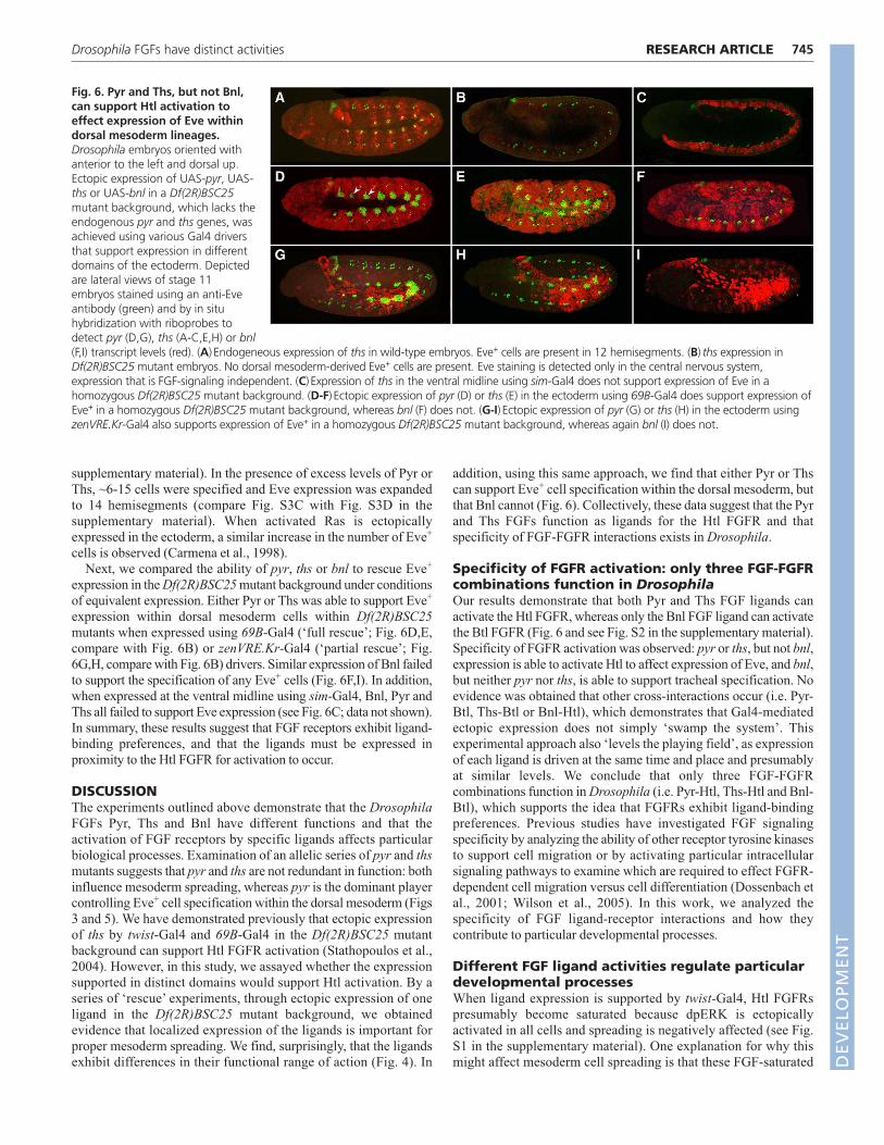
We sought to define the role of each ligand in the specification ofdorsal mesoderm cell lineages within germ-band elongated embryos.Normally, Eve is expressed within 12 hemisegments. In pyr singlemutants, often only a few hemisegments exhibited Eve expression(Fig. 5D,E, compare with Fig. 5A), whereas within ths single-mutantembryos, only subtle defects in Eve+ cell specification were observed(Fig. 5G,H, compare with Fig. 5A). The pyr mutant phenotype wasmuch stronger than the phenotype exhibited by ths single mutants atall stages examined, and even when comparisons were made betweenthe weakest pyr allele (pyr02915) and the strongest ths allele[Df(2R)ths238] (Fig. 5C,F, compare with Fig. 5I). Moreover, thephenotype of Df(2R)pyr36 was consistently stronger than that ofpyr02915, suggesting that either pyr02915 is not a null allele or that thsgene function also supports Eve+ cell specification. Although Pyr hasthe more dominant role, Ths probably also plays a role in supportingEve expression because the Df(2R)BSC25 mutant phenotype is moresevere than that of Df(2R)pyr36 mutants. No Eve+ cells are presentwithin the dorsal mesoderm of Df(2R)BSC25 mutants, a phenotypesimilar to that of htl mutants (Fig. 6B) (Stathopoulos et al., 2004).
To investigate the roles of Pyr and Ths in controlling celldifferentiation, which might differ from their roles during cellmigration, we focused on how ectopic expression of these ligandsaffects specification of Eve+ cells within the dorsal mesoderm of theembryo. We have demonstrated previously that when Ths isectopically expressed in the ectoderm using the 69B-Gal4 driver,ectopic Eve+ cells result; this is similar to the phenotype observedwhen constitutively activated Htl is ectopically expressed in thesame manner (see Fig. S3E in the supplementary material)(Stathopoulos et al., 2004). We found that ectopic expression of Pyrdriven by 69B-Gal4 also increases the number of Eve+ cells (see Fig.S3D in the supplementary material). Normally, three to four Eve+
cells are specified in 12 hemisegments (see Fig. S3C in the
RESEARCH ARTICLE Development 136 (5)
Fig. 5. The Pyr FGF ligand is necessary for the differentiation of dorsal mesoderm cell lineages. Drosophila embryos were stained with an anti-Eve antibody to examine specification of dorsal mesoderm lineages. Depicted are wild-type embryos at stage 11 (A) and stage 14 (C); Df(2R)BSC25mutant embryos, which lack both pyr and ths genes, at stage 11 (B); pyr single mutants at stage 11 (D,E) and stage 14 (F); and ths single mutants atstage 11 (G,H) and stage 14 (I). The insets display Eve+ cell clusters at 10� magnification. (A,C) In wild-type embryos at stage 11 (A), there are 11independent clusters of three Eve+ cells each within the dorsal somatic mesoderm. At stage 14 (C), these join to form a continuous row of heartprogenitors. (B) Eve+ clusters are absent from the dorsal mesoderm of Df(2R)BSC25 mutant embryos. (D-F) In both the weakest (pyr02915/pyr02915) andthe strongest [Df(2R)pyr36/Df(2R)pyr36] alleles of pyr single mutants (D and E, respectively), the number of Eve+ clusters is significantly reduced,resulting in gaps within the row of heart progenitors at stage 14 (F). (G-I) By contrast, within ths mutant embryos at stage 11, when either a weakmutant [Df(2R)ths238/ths02026] or the strongest alleles [Df(2R)ths238/Df(2R)ths238] are examined (G and H, respectively), there are only subtle effectson Eve+ cell specification (see inset; often two Eve+ cells are present instead of three). At later stages, defects are more apparent (I).
DEVELO
PMENT
supplementary material). In the presence of excess levels of Pyr orThs, ~6-15 cells were specified and Eve expression was expandedto 14 hemisegments (compare Fig. S3C with Fig. S3D in thesupplementary material). When activated Ras is ectopicallyexpressed in the ectoderm, a similar increase in the number of Eve+
cells is observed (Carmena et al., 1998).Next, we compared the ability of pyr, ths or bnl to rescue Eve+
expression in the Df(2R)BSC25 mutant background under conditionsof equivalent expression. Either Pyr or Ths was able to support Eve+
expression within dorsal mesoderm cells within Df(2R)BSC25mutants when expressed using 69B-Gal4 (‘full rescue’; Fig. 6D,E,compare with Fig. 6B) or zenVRE.Kr-Gal4 (‘partial rescue’; Fig.6G,H, compare with Fig. 6B) drivers. Similar expression of Bnl failedto support the specification of any Eve+ cells (Fig. 6F,I). In addition,when expressed at the ventral midline using sim-Gal4, Bnl, Pyr andThs all failed to support Eve expression (see Fig. 6C; data not shown).In summary, these results suggest that FGF receptors exhibit ligand-binding preferences, and that the ligands must be expressed inproximity to the Htl FGFR for activation to occur.
DISCUSSIONThe experiments outlined above demonstrate that the DrosophilaFGFs Pyr, Ths and Bnl have different functions and that theactivation of FGF receptors by specific ligands affects particularbiological processes. Examination of an allelic series of pyr and thsmutants suggests that pyr and ths are not redundant in function: bothinfluence mesoderm spreading, whereas pyr is the dominant playercontrolling Eve+ cell specification within the dorsal mesoderm (Figs3 and 5). We have demonstrated previously that ectopic expressionof ths by twist-Gal4 and 69B-Gal4 in the Df(2R)BSC25 mutantbackground can support Htl FGFR activation (Stathopoulos et al.,2004). However, in this study, we assayed whether the expressionsupported in distinct domains would support Htl activation. By aseries of ‘rescue’ experiments, through ectopic expression of oneligand in the Df(2R)BSC25 mutant background, we obtainedevidence that localized expression of the ligands is important forproper mesoderm spreading. We find, surprisingly, that the ligandsexhibit differences in their functional range of action (Fig. 4). In
addition, using this same approach, we find that either Pyr or Thscan support Eve+ cell specification within the dorsal mesoderm, butthat Bnl cannot (Fig. 6). Collectively, these data suggest that the Pyrand Ths FGFs function as ligands for the Htl FGFR and thatspecificity of FGF-FGFR interactions exists in Drosophila.
Specificity of FGFR activation: only three FGF-FGFRcombinations function in DrosophilaOur results demonstrate that both Pyr and Ths FGF ligands canactivate the Htl FGFR, whereas only the Bnl FGF ligand can activatethe Btl FGFR (Fig. 6 and see Fig. S2 in the supplementary material).Specificity of FGFR activation was observed: pyr or ths, but not bnl,expression is able to activate Htl to affect expression of Eve, and bnl,but neither pyr nor ths, is able to support tracheal specification. Noevidence was obtained that other cross-interactions occur (i.e. Pyr-Btl, Ths-Btl or Bnl-Htl), which demonstrates that Gal4-mediatedectopic expression does not simply ‘swamp the system’. Thisexperimental approach also ‘levels the playing field’, as expressionof each ligand is driven at the same time and place and presumablyat similar levels. We conclude that only three FGF-FGFRcombinations function in Drosophila (i.e. Pyr-Htl, Ths-Htl and Bnl-Btl), which supports the idea that FGFRs exhibit ligand-bindingpreferences. Previous studies have investigated FGF signalingspecificity by analyzing the ability of other receptor tyrosine kinasesto support cell migration or by activating particular intracellularsignaling pathways to examine which are required to effect FGFR-dependent cell migration versus cell differentiation (Dossenbach etal., 2001; Wilson et al., 2005). In this work, we analyzed thespecificity of FGF ligand-receptor interactions and how theycontribute to particular developmental processes.
Different FGF ligand activities regulate particulardevelopmental processesWhen ligand expression is supported by twist-Gal4, Htl FGFRspresumably become saturated because dpERK is ectopicallyactivated in all cells and spreading is negatively affected (see Fig.S1 in the supplementary material). One explanation for why thismight affect mesoderm cell spreading is that these FGF-saturated
745RESEARCH ARTICLEDrosophila FGFs have distinct activities
Fig. 6. Pyr and Ths, but not Bnl,can support Htl activation toeffect expression of Eve withindorsal mesoderm lineages.Drosophila embryos oriented withanterior to the left and dorsal up.Ectopic expression of UAS-pyr, UAS-ths or UAS-bnl in a Df(2R)BSC25mutant background, which lacks theendogenous pyr and ths genes, wasachieved using various Gal4 driversthat support expression in differentdomains of the ectoderm. Depictedare lateral views of stage 11embryos stained using an anti-Eveantibody (green) and by in situhybridization with riboprobes todetect pyr (D,G), ths (A-C,E,H) or bnl(F,I) transcript levels (red). (A) Endogeneous expression of ths in wild-type embryos. Eve+ cells are present in 12 hemisegments. (B) ths expression inDf(2R)BSC25 mutant embryos. No dorsal mesoderm-derived Eve+ cells are present. Eve staining is detected only in the central nervous system,expression that is FGF-signaling independent. (C) Expression of ths in the ventral midline using sim-Gal4 does not support expression of Eve in ahomozygous Df(2R)BSC25 mutant background. (D-F) Ectopic expression of pyr (D) or ths (E) in the ectoderm using 69B-Gal4 does support expression ofEve+ in a homozygous Df(2R)BSC25 mutant background, whereas bnl (F) does not. (G-I) Ectopic expression of pyr (G) or ths (H) in the ectoderm usingzenVRE.Kr-Gal4 also supports expression of Eve+ in a homozygous Df(2R)BSC25 mutant background, whereas again bnl (I) does not.
DEVELO
PMENT

746
mesoderm cells may no longer be competent to respond toendogenous ligands that provide directional cues. Recently, we haveshown that movement of the mesoderm cells during gastrulation isin fact directional (McMahon et al., 2008). Pyr and Ths ligands aredifferentially expressed during gastrulation and this might providethe necessary positional information required to direct migration ofthe mesoderm (see Fig. 7A).
We propose that Pyr and Ths have different activities that fulfilaspects of FGFR activation required to support cell migration(Fig. 7A). Ectopic expression of Pyr within the ectodermnegatively affects mesoderm spreading (Fig. 1H-M), whichsuggests that the refined expression domain of pyr within cells ofthe dorsal ectoderm is normally required to guide the mesodermcells toward dorsal regions. However, even though ectopicexpression of ths in the ectoderm has no effect on mesodermspreading, ths mutants also exhibit defects in mesodermspreading, demonstrating that both genes are required, perhaps tocontrol different aspects of the migration. Our ‘rescue’experiments using the zenVRE.Kr-Gal4 driver support the viewthat Pyr has a longer functional range than Ths (Fig. 4N,O). Thesedifferences in range of function might correlate with differentdiffusion capabilities, but an alternative explanation is that theligands activate the receptor with different affinities. Additionalexperiments will be necessary to distinguish their exact functionsand to uncover the molecular basis for the differential functionsof Pyr and Ths; we suggest that in vivo imaging and quantitativeanalysis (McMahon et al., 2008) of single-mutant phenotypes willprovide insights.
With regard to the FGF-dependent cell differentiation, our‘rescue’ experiments suggest that ectopic expression of either Pyr orThs is sufficient to support Eve+ cell specification (Fig. 6D,E). Thereason why loss of ths has less of an effect on Eve+ cell specificationis most likely because pyr is prominently expressed in the vicinityof the future Eve+ cells; normally, Pyr supports this function, but Thscan support this activity if presented at sufficient levels within the
correct domain. Furthermore, we propose that FGF signaling mightnot play an instructive role in supporting eve expression (Fig. 7B).Other signaling pathways already provide positional informationrequired for the specification of Eve+ cells; FGF signaling pathwayactivation might simply serve a permissive role, and in this contexteither ligand would suffice.
Conclusion: implications for vertebrate biology?We have used Drosophila to study FGF signaling and determinewhy multiple ligands are utilized to activate the same receptor. Itwill be informative to obtain additional insights into how theseligands differ in their activities. The expression domains of theligands do confer information that is important for controllingdevelopmental processes, but their individual protein sequencesalso impart differential functionality. For instance, the ligandsmight exhibit different affinities for specific HSPGs that influencetheir range of diffusion, or the proteins themselves may havedifferent stabilities. Future experiments will also define the FGFligand preferences that exist to support FGF signaling at later stagesof development.
At least 15 human genetic diseases result from mutations withinFGFR genes and each disease is caused by a different mutationshown to affect receptor activation (Chen and Deng, 2005). Severalmutations in FGFR lead to an expansion of FGF ligand-bindingpreference (Ornitz, 2005); however, it is still not clear why differentmutations yield different syndromes. Continuing this work in orderto understand how different FGF ligands activate the same receptorto effect different outcomes is an important goal, as this may provideinsights into why different mutations in the same FGFR lead tovarious dysplasias and diseases (Wilkie, 2005).
We thank S. Crews, M. Frasch, E. Giniger, M. Krasnow, M. Levine, A.Michelson, the Bloomington Stock Center, and Harvard Exelixis DistributionCenter for providing antibodies and fly stocks; Manfred Frasch and theStathopoulos laboratory members, especially Sarah Payne, for discussions andcomments on the manuscript; Leslie Dunipace for conducting the inverse PCRexperiments; Smadar Ben-Tabou de-Leon for advice on the qPCR experiments;and Jagan Srinivasan for help with graphical representations. This work wassupported by grants to A.S. from the NIH (R01 GM078542), the SearleScholars Program, and the March of Dimes (Basil O’Conner Starter ScholarAward, 5-FY06-12). Deposited in PMC for release after 12 months.
Supplementary materialSupplementary material for this article is available athttp://dev.biologists.org/cgi/content/full/136/5/739/DC1
ReferencesAhmad, S. M. and Baker, B. S. (2002). Sex-specific deployment of FGF signaling
in Drosophila recruits mesodermal cells into the male genital imaginal disc. Cell109, 651-661.
Beiman, M., Shilo, B. Z. and Volk, T. (1996). Heartless, a Drosophila FGF receptorhomolog, is essential for cell migration and establishment of several mesodermallineages. Genes Dev. 10, 2993-3002.
Birnbaum, D., Popovici, C. and Roubin, R. (2005). A pair as a minimum: thetwo fibroblast growth factors of the nematode Caenorhabditis elegans. Dev.Dyn. 232, 247-255.
Carmena, A., Gisselbrecht, S., Harrison, J., Jimenez, F. and Michelson, A. M.(1998). Combinatorial signaling codes for the progressive determination of cellfates in the Drosophila embryonic mesoderm. Genes Dev. 12, 3910-3922.
Chen, L. and Deng, C. X. (2005). Roles of FGF signaling in skeletal developmentand human genetic diseases. Front. Biosci. 10, 1961-1976.
Coumoul, X. and Deng, C. X. (2003). Roles of FGF receptors in mammaliandevelopment and congenital diseases. Birth Defects Res. C Embryo Today 69,286-304.
Dossenbach, C., Rock, S. and Affolter, M. (2001). Specificity of FGF signaling incell migration in Drosophila. Development 128, 4563-4572.
Eswarakumar, V. P., Lax, I. and Schlessinger, J. (2005). Cellular signaling byfibroblast growth factor receptors. Cytokine Growth Factor Rev. 16, 139-149.
Frasch, M. (1995). Induction of visceral and cardiac mesoderm by ectodermal Dppin the early Drosophila embryo. Nature 374, 464-467.
RESEARCH ARTICLE Development 136 (5)
Fig. 7. A model for FGF signaling through Heartless. (A) Thelocation of the Ths and Pyr expression domains is important for theproper regulation of mesoderm migration. Both ligands are required,presumably because they have different activities. (B) Duringspecification of dorsal mesoderm lineages, including specification ofEve+ cells (orange), FGFs feed into an array of signaling molecules[Wingless (Wg) and Dpp] necessary to specify dorsal mesodermlineages. We suggest that any FGF ligand (red), expressed in the regionof Dpp and Wg overlap and able to activate the respective FGFR, wouldsuffice to support cell differentiation. Wg, Wingless.
DEVELO
PMENT

Frasch, M. (1999). Intersecting signalling and transcriptional pathways inDrosophila heart specification. Semin. Cell Dev. Biol. 10, 61-71.
Gabay, L., Seger, R. and Shilo, B. Z. (1997). MAP kinase in situ activation atlasduring Drosophila embryogenesis. Development 124, 3535-3541.
Gisselbrecht, S., Skeath, J. B., Doe, C. Q. and Michelson, A. M. (1996).heartless encodes a fibroblast growth factor receptor (DFR1/DFGF-R2) involved inthe directional migration of early mesodermal cells in the Drosophila embryo.Genes Dev. 10, 3003-3017.
Gryzik, T. and Muller, H. A. (2004). FGF8-like1 and FGF8-like2 encode putativeligands of the FGF receptor Htl and are required for mesoderm migration in theDrosophila gastrula. Curr. Biol. 14, 659-667.
Huang, P. and Stern, M. J. (2005). FGF signaling in flies and worms: more andmore relevant to vertebrate biology. Cytokine Growth Factor Rev. 16, 151-158.
Ishimoto, H., Takahashi, K., Ueda, R. and Tanimura, T. (2005). G-proteingamma subunit 1 is required for sugar reception in Drosophila. EMBO J. 24,3259-3265.
Kosman, D., Mizutani, C. M., Lemons, D., Cox, W. G., McGinnis, W. and Bier,E. (2004). Multiplex detection of RNA expression in Drosophila embryos. Science305, 846.
Lehmann, R. and Tautz, D. (1994). In situ hybridization to RNA. Methods CellBiol. 44, 575-598.
Leptin, M. and Grunewald, B. (1990). Cell shape changes during gastrulation inDrosophila. Development 110, 73-84.
Mariani, F. V., Ahn, C. P. and Martin, G. R. (2008). Genetic evidence that FGFshave an instructive role in limb proximal-distal patterning. Nature 453, 401-405.
McMahon, A., Supatto, W., Fraser, S. E. and Stathopoulos, A. (2008).Dynamic analyses of Drosophila gastrulation provide insights into collective cellmigration. Science 322, 1546-1550.
Michelson, A. M., Gisselbrecht, S., Zhou, Y., Baek, K. H. and Buff, E. M.(1998). Dual functions of the heartless fibroblast growth factor receptor indevelopment of the Drosophila embryonic mesoderm. Dev. Genet. 22, 212-229.
Ornitz, D. M. (2005). FGF signaling in the developing endochondral skeleton.Cytokine Growth Factor Rev. 16, 205-213.
Ornitz, D. M. and Itoh, N. (2001). Fibroblast growth factors. Genome Biol. 2,reviews3005.1-3005.12.
Ornitz, D. M., Xu, J., Colvin, J. S., McEwen, D. G., MacArthur, C. A., Coulier,F., Gao, G. and Goldfarb, M. (1996). Receptor specificity of the fibroblastgrowth factor family. J. Biol. Chem. 271, 15292-15297.
Preston, C. R., Sved, J. A. and Engels, W. R. (1996). Flanking duplications anddeletions associated with P-induced male recombination in Drosophila. Genetics144, 1623-1638.
Rentzsch, F., Fritzenwanker, J. H., Scholz, C. B. and Technau, U. (2008). FGFsignalling controls formation of the apical sensory organ in the cnidarianNematostella vectensis. Development 135, 1761-1769.
Rottinger, E., Saudemont, A., Duboc, V., Besnardeau, L., McClay, D. andLepage, T. (2008). FGF signals guide migration of mesenchymal cells, controlskeletal morphogenesis and regulate gastrulation during sea urchindevelopment. Development 135, 353-365.
San Martin, B. and Bate, M. (2001). Hindgut visceral mesoderm requires anectodermal template for normal development in Drosophila. Development 128,233-242.
Sato, M. and Kornberg, T. B. (2002). FGF is an essential mitogen andchemoattractant for the air sacs of the drosophila tracheal system. Dev. Cell 3,195-207.
Shishido, E., Ono, N., Kojima, T. and Saigo, K. (1997). Requirements ofDFR1/Heartless, a mesoderm-specific Drosophila FGF-receptor, for the formationof heart, visceral and somatic muscles, and ensheathing of longitudinal axontracts in CNS. Development 124, 2119-2128.
Spralding, A. C. and Rubin, G. M. (1982). Genetic transformation of Drosophilawith transposable element vectors. Science 218, 348-353.
Stathopoulos, A., Tam, B., Ronshaugen, M., Frasch, M. and Levine, M.(2004). pyramus and thisbe: FGF genes that pattern the mesoderm ofDrosophila embryos. Genes Dev. 18, 687-699.
Sutherland, D., Samakovlis, C. and Krasnow, M. A. (1996). branchless encodesa Drosophila FGF homolog that controls tracheal cell migration and the patternof branching. Cell 87, 1091-1101.
Szebenyi, G. and Fallon, J. F. (1999). Fibroblast growth factors as multifunctionalsignaling factors. Int. Rev. Cytol. 185, 45-106.
Thibault, S. T., Singer, M. A., Miyazaki, W. Y., Milash, B., Dompe, N. A.,Singh, C. M., Buchholz, R., Demsky, M., Fawcett, R., Francis-Lang, H. L. etal. (2004). A complementary transposon tool kit for Drosophila melanogasterusing P and piggyBac. Nat. Genet. 36, 283-287.
Thisse, B. and Thisse, C. (2005). Functions and regulations of fibroblast growthfactor signaling during embryonic development. Dev. Biol. 287, 390-402.
Wilkie, A. O. (2005). Bad bones, absent smell, selfish testes: the pleiotropicconsequences of human FGF receptor mutations. Cytokine Growth Factor Rev.16, 187-203.
Wilson, R., Vogelsang, E. and Leptin, M. (2005). FGF signalling and themechanism of mesoderm spreading in Drosophila embryos. Development 132,491-501.
Xiao, H., Hrdlicka, L. A. and Nambu, J. R. (1996). Alternate functions of thesingle-minded and rhomboid genes in development of the Drosophila ventralneuroectoderm. Mech. Dev. 58, 65-74.
Yang, X., Dormann, D., Munsterberg, A. E. and Weijer, C. J. (2002). Cellmovement patterns during gastrulation in the chick are controlled by positiveand negative chemotaxis mediated by FGF4 and FGF8. Dev. Cell 3, 425-437.
Zhang, X., Ibrahimi, O. A., Olsen, S. K., Umemori, H., Mohammadi, M. andOrnitz, D. M. (2006). Receptor specificity of the fibroblast growth factor family.The complete mammalian FGF family. J. Biol. Chem. 281, 15694-15700.
747RESEARCH ARTICLEDrosophila FGFs have distinct activities
DEVELO
PMENT
Copyright © 2022 FDOKUMEN