k-FGF protoncogene expression is associated
9
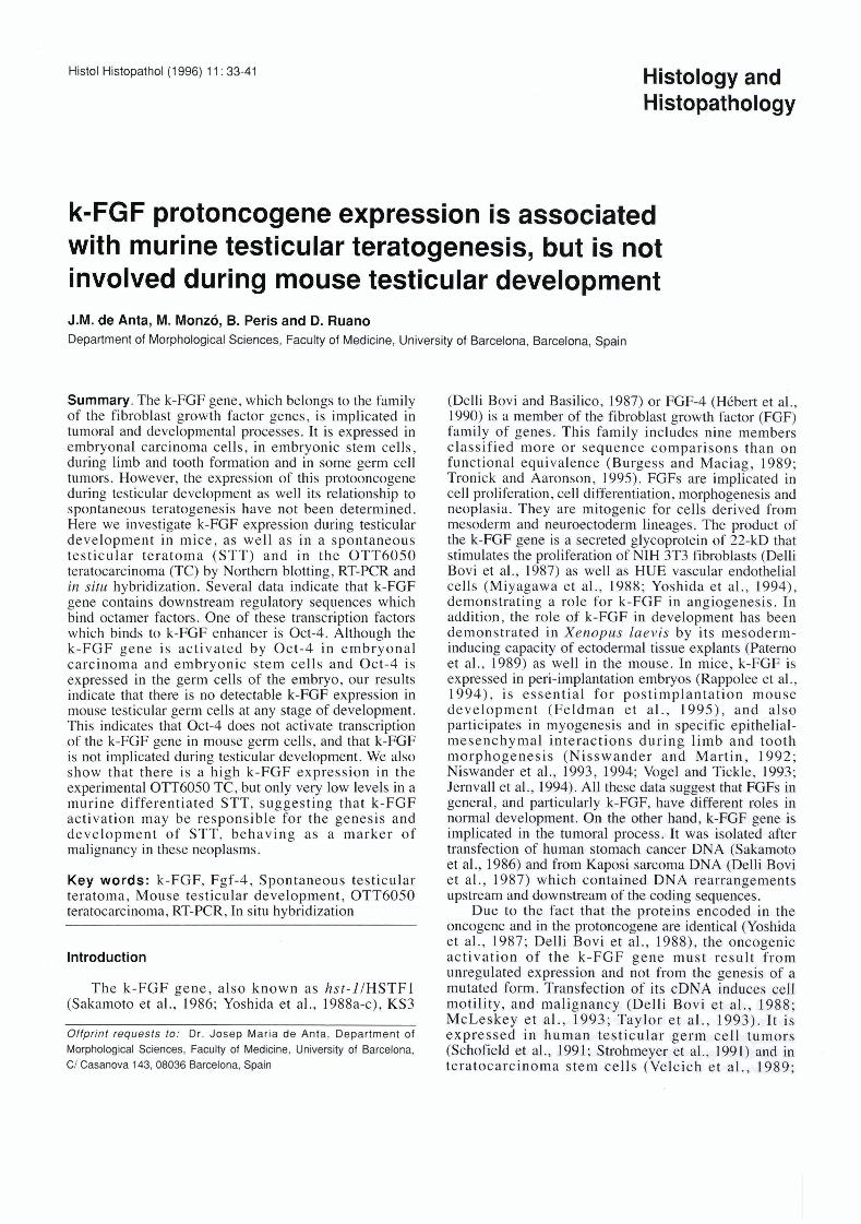
Histol Histopathol (1 996) 11 : 33-41 Histology and Histopathology k-FGF protoncogene expression is associated with murine testicular teratogenesis, but is not involved during mouse testicular development J.M. de Anta, M. Monzó, B. Peris and D. Ruano Department of Morphological Sciences, Faculty of Medicine, University of Barcelona, Barcelona, Spain Summary. The k-FGF gene, which belongs to the family of the fibroblast growth factor genes, is implicated in tumoral and developmental processes. It is expressed in embryonal carcinoma cells, in embryonic stem cells, during limb and tooth formation and in some germ cell tumors. However, the expression of this protooncogene during testicular development as well its relationship to spontaneous teratogenesis have not been determined. Here we investigate k-FGF expression during testicular development in mice, as well as in a spontaneous testicular teratoma (STT) and in the OTT6050 teratocarcinoma (TC) by Northern blotting, RT-PCR and in situ hybridization. Severa1 data indicate that k-FGF gene contains downstream regulatory sequences which bind octamer factors. One of these transcription factors which binds to k-FGF enhancer is Oct-4. Although the k-FGF gene is activated by Oct-4 in embryonal carcinoma and embryonic stem cells and Oct-4 is expressed in the germ cells of the embryo, our results indicate that there is no detectable k-FGF expression in mouse testicular germ cells at any stage of development. This indicates that Oct-4 does not activate transcription of the k-FGF gene in mouse germ cells, and that k-FGF is not implicated during testicular development. We also show that there is a high k-FGF expression in the experimental OTT6050 TC, but only very low levels in a murine differentiated STT, suggesting that k-FGF activation may be responsible for the genesis and development of STT, behaving as a marker of malignancy in these neoplasms . Key words: k-FGF, Fgf-4, Spontaneous testicular teratoma, Mouse testicular development, OTT6050 teratocarcinoma, RT-PCR, In situ hybridization lntroduction The k-FGF gene, also known as hst-lIHSTF1 (Sakamoto et al., 1986; Yoshida et al., 1988a-c), KS3 Offpr~nt requests to: Dr. Josep Maria de Anta, Department of Morphological Sciences, Faculty of Medicine, University of Barcelona, C/ Casanova 143,08036 Barcelona, Spain (Delli Bovi and Basilico, 1987) or FGF-4 (Hébert et al., 1990) is a member of the fibroblast growth factor (FGF) family of genes. This family includes nine members classified more or sequence comparisons than on functional equivalente (Burgess and Maciag, 1989; Tronick and Aaronson, 1995). FGFs are implicated in cell proliferation, cell differentiation, morphogenesis and neoplasia. They are mitogenic for cells derived from mesoderm and neuroectoderm lineages. The product of the k-FGF gene is a secreted glycoprotein of 22-kD that stimulates the proliferation of NIH 3T3 fibroblasts (Delli Bovi et d . , 1987) as well as HUE vascular endotheliai cells (Miyagawa et al., 1988; Yoshida et al., 1994), demonstrating a role for k-FGF in angiogenesis. In addition, the role of k-FGF in development has been demonstrated in Xenopus laevis by its mesoderm- inducing capacity of ectodermal tissue explants (Paterno et al., 1989) as well in the mouse. In mice, k-FGF is expressed in peri-implantation embryos (Rappolee et al., 1994), is essential for postimplantation mouse development (Feldman et al., 1995), and also participates in myogenesis and in specific epithelial- mesenchymal interactions during limb and tooth morphogenesis (Nisswander and Martin, 1992; Niswander et al., 1993, 1994; Vogel and Tickle, 1993; Jernvall et al., 1994). Al1 these data suggest that FGFs in general, and particularly k-FGF, have different roles in normal development. On the other hand, k-FGF gene is implicated in the tumoral process. It was isolated after transfection of human stomach cancer DNA (Sakamoto et al., 1986) and from Kaposi sarcoma DNA (Delli Bovi et al., 1987) which contained DNA rearrangements upstrearn and downstream of the coding sequences. Due to the fact that the proteins encoded in the oncogene and in the protoncogene are identical (Yoshida et al., 1987; Delli Bovi et al., 1988), the oncogenic activation of the k-FGF gene must result from unregulated expression and not from the genesis of a mutated form. Transfection of its cDNA induces cell motility, and malignancy (Delli Bovi et al., 1988; McLeskey et al., 1993; Taylor et al., 1993). It is expressed in human testicular germ cell tumors (Schofield et al., 1991; Strohmeyer et al., 1991) and in teratocarcinoma stem cells (Velcich et al., 1989;
-
Upload
khangminh22 -
Category
Documents
-
view
0 -
download
0
Transcript of k-FGF protoncogene expression is associated
Histol Histopathol (1 996) 11 : 33-41 Histology and Histopathology
k-FGF protoncogene expression is associated with murine testicular teratogenesis, but is not involved during mouse testicular development J.M. de Anta, M. Monzó, B. Peris and D. Ruano Department of Morphological Sciences, Faculty of Medicine, University of Barcelona, Barcelona, Spain
Summary. The k-FGF gene, which belongs to the family of the fibroblast growth factor genes, is implicated in tumoral and developmental processes. It is expressed in embryonal carcinoma cells, in embryonic stem cells, during limb and tooth formation and in some germ cell tumors. However, the expression of this protooncogene during testicular development as well its relationship to spontaneous teratogenesis have not been determined. Here we investigate k-FGF expression during testicular development in mice, as well as in a spontaneous testicular teratoma (STT) and in the OTT6050 teratocarcinoma (TC) by Northern blotting, RT-PCR and in situ hybridization. Severa1 data indicate that k-FGF gene contains downstream regulatory sequences which bind octamer factors. One of these transcription factors which binds to k-FGF enhancer is Oct-4. Although the k-FGF gene is activated by Oct-4 in embryonal carcinoma and embryonic stem cells and Oct-4 is expressed in the germ cells of the embryo, our results indicate that there is no detectable k-FGF expression in mouse testicular germ cells at any stage of development. This indicates that Oct-4 does not activate transcription of the k-FGF gene in mouse germ cells, and that k-FGF is not implicated during testicular development. We also show that there is a high k-FGF expression in the experimental OTT6050 TC, but only very low levels in a murine differentiated STT, suggesting that k-FGF activation may be responsible for the genesis and development of STT, behaving as a marker of malignancy in these neoplasms .
Key words: k-FGF, Fgf-4, Spontaneous testicular teratoma, Mouse testicular development, OTT6050 teratocarcinoma, RT-PCR, In situ hybridization
lntroduction
The k-FGF gene, also known as hst-lIHSTF1 (Sakamoto et al., 1986; Yoshida et al., 1988a-c), KS3
Offpr~nt requests to: Dr . Josep Maria de Anta, Department of Morphological Sciences, Faculty of Medicine, University of Barcelona, C/ Casanova 143,08036 Barcelona, Spain
(Delli Bovi and Basilico, 1987) or FGF-4 (Hébert et al., 1990) is a member of the fibroblast growth factor (FGF) family of genes. This family includes nine members classified more or sequence comparisons than on functional equivalente (Burgess and Maciag, 1989; Tronick and Aaronson, 1995). FGFs are implicated in cell proliferation, cell differentiation, morphogenesis and neoplasia. They are mitogenic for cells derived from mesoderm and neuroectoderm lineages. The product of the k-FGF gene is a secreted glycoprotein of 22-kD that stimulates the proliferation of NIH 3T3 fibroblasts (Delli Bovi et d . , 1987) as well as HUE vascular endotheliai cells (Miyagawa et al., 1988; Yoshida et al., 1994), demonstrating a role for k-FGF in angiogenesis. In addition, the role of k-FGF in development has been demonstrated in Xenopus laevis by its mesoderm- inducing capacity of ectodermal tissue explants (Paterno et al., 1989) as well in the mouse. In mice, k-FGF is expressed in peri-implantation embryos (Rappolee et al., 1994), is essential for postimplantation mouse development (Feldman et al., 1995), and also participates in myogenesis and in specific epithelial- mesenchymal interactions during limb and tooth morphogenesis (Nisswander and Martin, 1992; Niswander et al., 1993, 1994; Vogel and Tickle, 1993; Jernvall et al., 1994). Al1 these data suggest that FGFs in general, and particularly k-FGF, have different roles in normal development. On the other hand, k-FGF gene is implicated in the tumoral process. It was isolated after transfection of human stomach cancer DNA (Sakamoto et al., 1986) and from Kaposi sarcoma DNA (Delli Bovi et al., 1987) which contained DNA rearrangements upstrearn and downstream of the coding sequences.
Due to the fact that the proteins encoded in the oncogene and in the protoncogene are identical (Yoshida et al., 1987; Delli Bovi et al., 1988), the oncogenic activation of the k-FGF gene must result from unregulated expression and not from the genesis of a mutated form. Transfection of its cDNA induces cell motility, and malignancy (Delli Bovi et al., 1988; McLeskey et al. , 1993; Taylor et al., 1993). It is expressed in human testicular germ cell tumors (Schofield et al., 1991; Strohmeyer et al., 1991) and in teratocarcinoma stem cells (Velcich et al. , 1989;
k-FGF expression in testicular development and teratogenesis
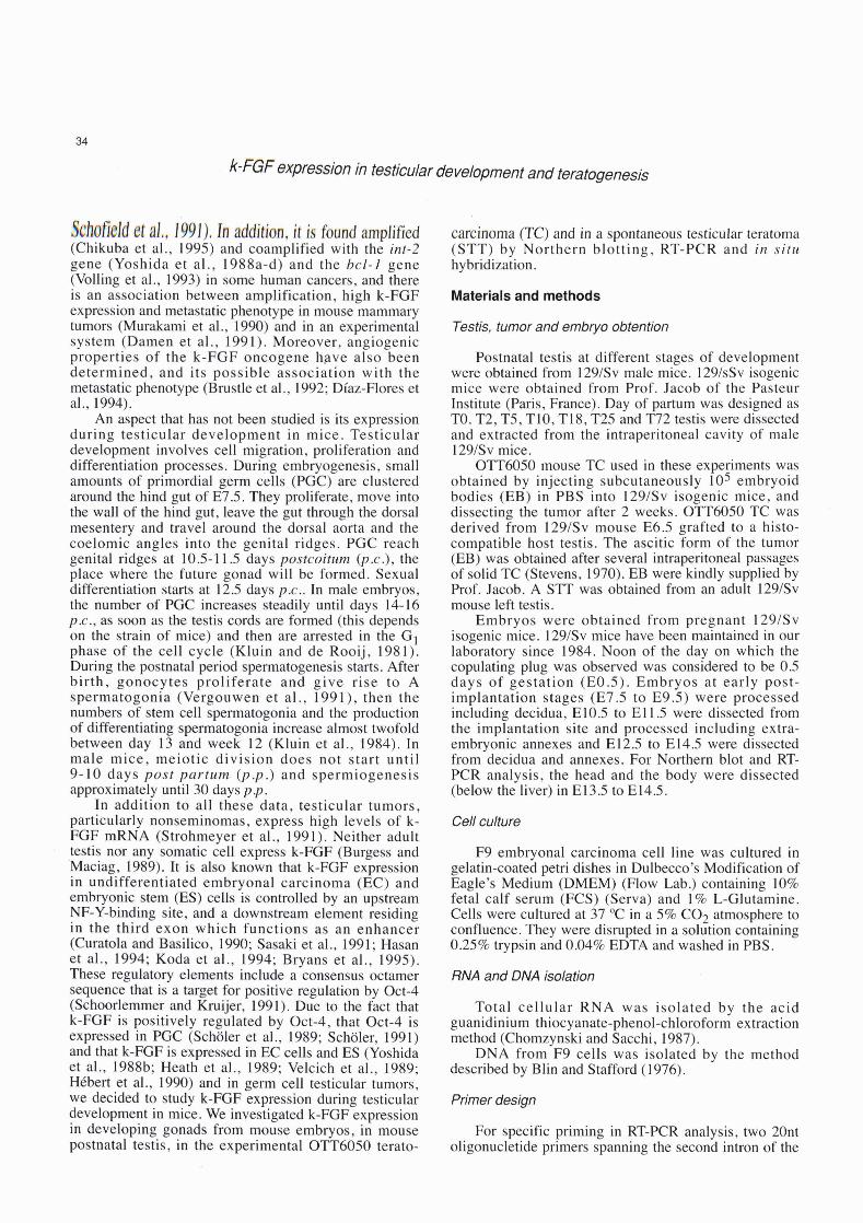
#~ht&d 8t d., 1991). In addiiion, it is faund amplified (Chikuba et al., 1995) and coamplified with the int-2 gene (Yoshida et al., 1988a-d) and the bcl-I gene (Volling et al., 1993) in some human cancers, and there is an association between amplification, high k-FGF expression and metastatic phenotype in mouse mammary tumors (Murakami et al., 1990) and in an experimental system (Damen et al., 1991). Moreover, angiogenic properties of the k-FGF oncogene have also been determined, and its possible association with the metastatic phenotype (Brustle et al., 1992; Díaz-Flores et al., 1994).
An aspect that has not been studied is its expression during testicular development in mice. Testicular development involves cell migration, proliferation and differentiation processes. During embryogenesis, small amounts of primordial germ cells (PGC) are clustered around the hind gut of E7.5. They proliferate, move into the wall of the hind gut, leave the gut through the dorsal mesentery and travel around the dorsal aorta and the coelomic angles into the genital ridges. PGC reach genital ridges at 10.5-1 1.5 days postcoitum @.c.), the place where the future gonad will be formed. Sexual differentiation starts at 12.5 days p.c.. In male embryos, the number of PGC increases steadily until days 14-16 p.c., as soon as the testis cords are formed (this depends on the strain of mice) and then are arrested in the G1 phase of the cell cycle (Kluin and de Rooij, 198 1). During the postnatal period spermatogenesis starts. After birth, gonocytes proliferate and give rise to A spermatogonia (Vergouwen et al ., 199 1 ), then the numbers of stem cell spermatogonia and the production of differentiating spermatogonia increase almost twofold between day 13 and week 12 (Kluin et al., 1984). In male mice, meiotic division does not start until 9-10 days post partum (p.p.) and spermiogenesis approximately until30 days p.p.
In addition to al1 these data, testicular tumors, particularly nonseminomas, express high levels of k- FGF mRNA (Strohmeyer et al., 1991). Neither adult testis nor any somatic cell express k-FGF (Burgess and 'Maciag, 1989). It is also known that k-FGF expression in undifferentiated embryonal carcinoma (EC) and embryonic stem (ES) cells is controlled by an upstream NF-Y-binding site, and a downstream element residing in the third exon which functions as an enhancer (Curatola and Basilico, 1990; Sasaki et al., 1991; Hasan et al., 1994; Koda et al., 1994; Bryans et al., 1995). These regulatory elements include a consensus octamer sequence that is a target for positive regulation by Oct-4 (Schoorlemmer and Kmijer, 1991). Due to the fact that k-FGF is positively regulated by Oct-4, that Oct-4 is expressed in PGC (Scholer et al., 1989; Scholer, 1991) and that k-FGF is expressed in EC cells and ES (Yoshida et al., 1988b; Heath et al., 1989; Velcich et al., 1989; Hébert et al., 1990) and in germ cell testicular tumors, we decided to study k-FGF expression during testicular development in mice. We investigated k-FGF expression in developing gonads from mouse embryos, in mouse postnatal testis, in the experimental OTT6050 terato-
carcinoma (TC) and in a spontaneous testicular teratoma (STT) by Northern blotting, RT-PCR and in situ hybridization.
Materials and methods
Testis, tumor and embryo obtention
Postnatal testis at different stages of development were obtained from 1291Sv male mice. 1291sSv isogenic mice were obtained from Prof. Jacob of the Pasteur Institute (Paris, France). Day of partum was designed as TO. T2, T5, T10, T18, T25 and T72 testis were dissected and extracted from the intraperitoneal cavity of male 1291Sv mice.
O'TT6050 mouse TC used in these experiments was obtained by injecting subcutaneously lo5 embryoid bodies (EB) in PBS into 129ISv isogenic mice, and dissecting the tumor after 2 weeks. OTT6050 TC was derived from 1291Sv mouse E6.5 grafted to a histo- compatible host testis. The ascitic form of the tumor (EB) was obtained after severa1 intraperitoneal passages of solid TC (Stevens, 1970). EB were kindly supplied by Prof. Jacob. A STT was obtained from an adult 1291Sv mouse left testis.
Embryos were obtained from pregnant 129ISv isogenic mice. 129ISv mice have been maintained in our laboratory since 1984. Noon of the day on which the copulating plug was observed was considered to be 0.5 days of gestation (E0.5). Embryos at early post- implantation stages (E7.5 to E9.5) were processed including decidua, E10.5 to E1 1.5 were dissected from the implantation site and processed including extra- embryonic annexes and E1 2.5 to E14.5 were dissected from decidua and annexes. For Northern blot and RT- PCR analysis, the head and the body were dissected (below the liver) in E13.5 to E14.5.
Cell culture
F9 embryonal carcinoma cell line was cultured in gelatin-coated petri dishes in Dulbecco's Modification of Eagle's Medium (DMEM) (Flow Lab.) containing 10% fetal calf serum (FCS) (Serva) and 1% L-Glutamine. Cells were cultured at 37 "C in a 5% C02 atmosphere to confluence. They were disrupted in a solution containing 0.25% trypsin and 0.04% EDTA and washed in PBS.
RNA and DNA isolation
Total cellular RNA was isolated by the acid guanidinium thiocyanate-phenol-chloroform extraction method (Chomzynski and Sacchi, 1987).
DNA from F9 cells was isolated by the method described by Blin and Stafford (1976).
Primer design
For specific priming in RT-PCR analysis, two 20nt oligonucletide primers spanning the second intron of the
k-FGF expression in testicular development and teratogenesis
mouse k-FGF gene (Brookes et al., 1989), RTKF2 S'TGGTGAGCATCTTCGGAGTG 5' and RTKFl 5' TTCATGGTAGGCGACACTCG 3' were designed and produced in a DNA synthesizer (Applied Biosystems).
RT- PCR reaction
For the reverse transcriptase reaction, 100 ng of RNA samples were heated at 95 "C for 10 min, cooled on ice and cDNA synthesis was performed at 42 "C for 30 min in 50mM KCI, lOmM Tris-HC1 pH 9.0, 0.1% Triton X-100, 1.5mM MgC12 using 1mM dNTPs (Pharmacia), 20 U RNasin (Promega), 100 pmoles downstream primer (RTKFl) and 10 U AMV reverse transcriptase (Promega). Then the reaction mixture was heated at 95 "C for 10 min, chilled on ice and 100 pmoles upstream primer (RTKF2) and 1 U AmpTaq DNA pol (Perkin Elmer Cetus) were added for a normal PCR reaction. The thermal profile used was 95 "C denaturation for 1 min, annealing of primers at 68 "C for 1 min and extension of the primers at 72 "C for 1 min. This cycle was repeated 22 times.
Southern blotting
One tenth of the RT-PCR reaction was fractioned in a 1.8% agarose TBE gel, stained with ethidium bromide, photographed, treated with 0.25N HCl, 1M NaCYO.5M NaOH, equilibrated in 0.5M Tris-HCI pH 7.411.5M NaCl and blotted to Genescreen membranes (Dupont) in 10xSSC. DNA was imrnobilized by baking the membrane at 80 "C. Hybridization was performed in 50% formamide, 6xSSPE, 1 % SDS, 50 pglml denatured salmon sperm DNA at 42 "C for 16 to 24 h. A Aval1 155 pb mouse genornic k-FGF fragment including 3rd coding exon and 2nd intron sequences was used as a probe. This probe recognized k-FGF-specific genomic and RNA-amplified fragments, but not priming sequences. This was labelled to a specific activity of 2x10⁶ cpmlml using a random primer DNA Labeling kit (Boehringer Mannheim) and a 3 2 ~ - d ~ ~ ~ (Amersham). After hybridization, filters were washed and exposed to XAR films (Kodak) at -80 "C using intensifying screens, and developed.
RNA probes and Northern blotting
A mouse 417 bp HindllIXbal fragment from pXMT2-1.58kFGF plasmid that included the 2nd exon and a part of intronic sequences, was subcloned in pBluescript 11 SK + (Stratagene) giving rise to pBSkfHX. pBSkfHX was linealized with Hindll and Xbal. a 3 2 ~ - ~ ~ ~ and a 3 2 ~ - ~ ~ ~ labelled single stranded antisense and sense k-FGF riboprobes were transcribed using T3 and T7 RNA pol res ectively.
As a positive control. a aq2P-UTP labelled human B- actin cRNA was used as a probe. The human B-actin cDNA cloned in pGEM4Z (Promega) was linealized with EcoRI and Hindlll before being transcribed using
T7 RNA pol and SP6 RNA pol to create antisense and sense single-stranded ridoprobes. Both were alkaline hydrolized to reduce probes to an average size of 400 nt.
For Northern blotting experiments, 25 p g of total RNA were denatured in 50% formamide, 2.2M formaldehyde, 20mM MOPS pH 7.0, 5mM sodium acetate, and lmM EDTA. RNA samples were diluted in loading buffer containing 0.1 % bromophenol blue and 10% glycerol and electrophoresed in 1% agarose- formaldehyde-MOPS gel. The gel was transferred onto Genescreen membranes (Dupont) in 10xSSC. Hybridization was performed in 50% formamide, 5x Denhardt's solution, SxSSPE, 1% SDS, 200 pglml denatured, and fragmented salmon sperm DNA at 42 "C for 16 to 24 h.
Riboprobes were used at a specific activity of 2x10~ cpmlml. Washes were made at high stringency conditions and autoradiography was performed as Southern blots. For B-actin hybridization, k-FGF riboprobe was removed from the filter by washing in 50% formamide, 6xSSPE for 40 min at 65 "C.
In situ hybridization
Testis, tumors and embryos were fixed in 4% paraformaldehyde, 5mM MgC12 in PBS, dehydrated, embedded in paraffin wax and sectioned at 6 pm. In situ hybridization conditions were performed at high stringency conditions according to the method of Frohman et al. (1990). k-FGF and B-actin riboprobes were used at a specific activity of lo5 cpmtpl. Slides were exposed to an autorradiographic emulsion (NTB2, Kodak) 1: 1 diluted in 0.6M ammonium acetate at 4 "C in the dark between three to four weeks (k-FGF) or one week (human B-actin) before being developed and stained with hematoxylin-eosin.
Results
Northern blot analysis of kFGF mRNA in postnatal testis, 0776050 TC, a 129/Sv STT, F9 cells and embryos I
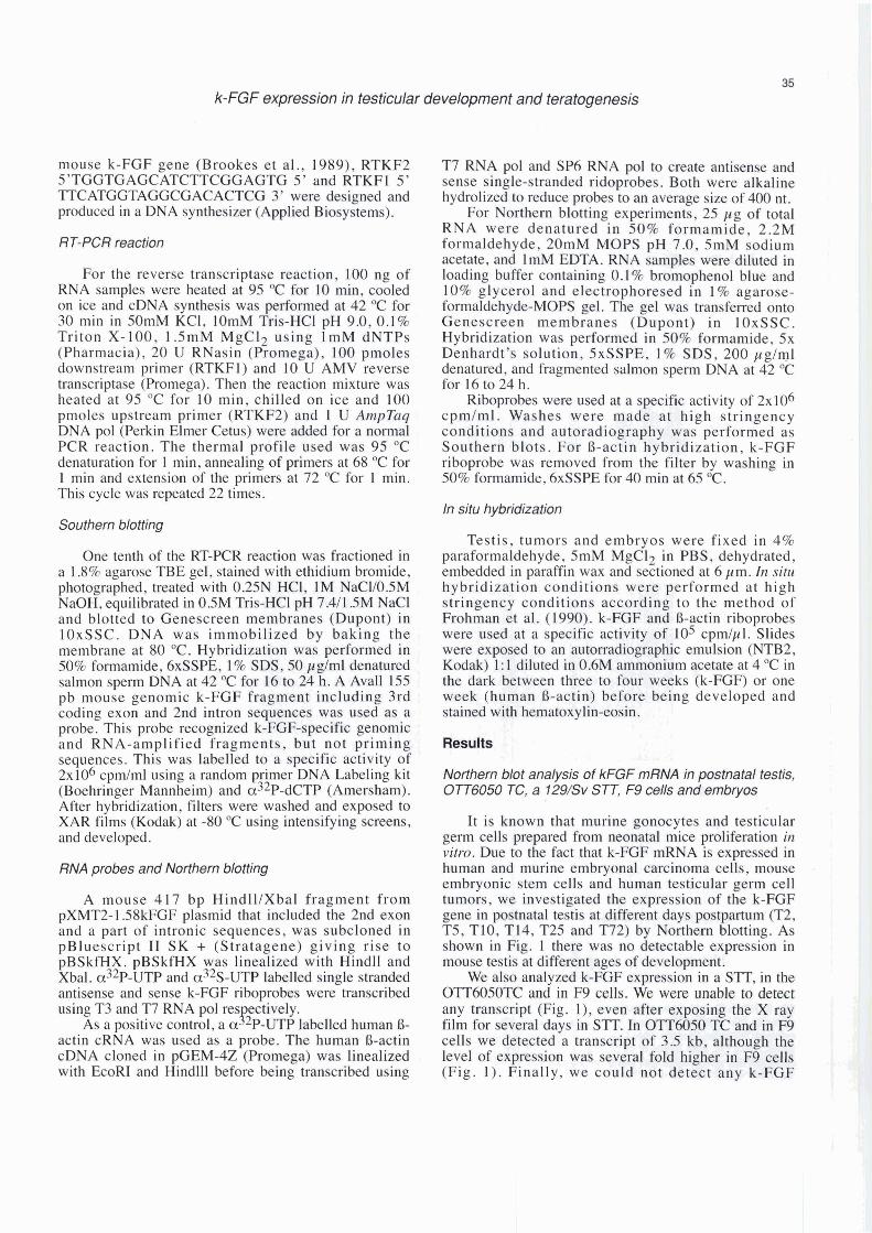
It is known that murine gonocytes and testicular germ cells prepared from neonatal mice proliferation in vitro. Due to the fact that k-FGF mRNA is expressed in human and murine embryonal carcinoma cells, mouse embryonic stem cells and human testicular germ cell tumors, we investigated the expression of the k-FGF gene in postnatal testis at different days postpartum (T2, T5, T10, T14, T25 and T72) by Northern blotting. As shown in Fig. 1 there was no detectable expression in
I mouse testis at different ages of development.
We also analyzed k-FGF expression in a STT, in the OTTóO50TC and in F9 cells. We were unable to &tect any transcript (Fig. l), even after exposing the X ray 1 film for several days in S R . In O'iT6050 TC and in F9 cells we detected a transcript of 3.5 kb, although the leve1 of expression was severa1 fold higher in F9 cells (Fig. 1). Finally, we could not detect any k-FGF
k-FGF expression in testicular development and teratogenesis
2 3 4 6 6- 7 1) O 10 11 12 13 14 15 16 17 78 lo
Flg. 1. Northern bbt anatysis of RNA samples from F9 cells (lane l ) , OTT6050 TC (lane 2), 129/Sv STT (lane 3), testls of 2,5, 10, 14,25 and 72 days p.c. (lanes 4 to 9) and mwse embryos of 7.5 to 12 days (lanes 10 to 15) and head of 13.lday and 14.5day embryos (lanes 16 and 18) and body of 13.5 days and 14.5-day embryos (lanes 17 and 19). 25 yg of total RNA are electrophoresed in each line. A aP32-UTP-labelled antisense k-FGF nboprobe is used, as desdbed in matenals and meíhods. Film was expooed overnight. The same membrane was reprobed with the antisense human O-adin níprobe.
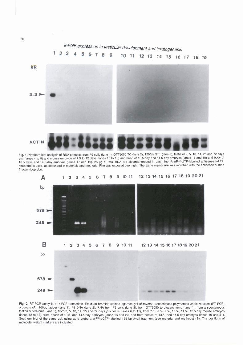
Fig. 2. RT-PCR analysis of k-FGF transcripts. Ethidium bromide-stained agarose gel of reverse transcriptase-polymerase chain reaction (RT-PCR) products (A). 100bp ladder (lane l ) , F9 DNA (lane 2), RNA from F9 cells (lane 3). from OTT6050 teratocarcinoma (lane 4), from a spontaneous testicular teratoma (lane 5), from 2, 5, 10, 14, 25 and 72 days p.p. testis (lanes 6 lo l l ) , from 7.5-, 8.5-, 9.5-, 10.5-, 11.5-. 12.5-day mouse embryos (lanes 12 to 17), from heads of 13.5- and 14.5-day embryos (lanes 18 and 20) and from bodies of 13.5- and 14.5-day embryos (lanes 19 and 21). Southern blot of the same gel, using as a probe a aJ2P-dCTP-labelled 155 bp Avall fragment (see material and methods) (B). The positions of molecular weight markers are indicated.
k-FGF expression in testicular development and teratogenesis
transcript in postimplantation mouse embryos (E7.5 to E12.5 and from head and body of E13.5 to E14.5) on Northem blots under our experimental conditions (Fig. 1).
These results showed that k-FGF rnRNA expression was apparently restricted to undifferentiated F9 cells and OTT6050 TC, being undetectable in testis, STT and embryos under our experimental conditions. However, the possibility that expression was restricted to few clumps of cells of the testis and of the S'IT, which could not be detected by this technique, was not ruled out.
RT-PCR analysis of postnatal testis, a 129/Sv STT, 077-6050 TC, F9 cells and embryos
Amplification of RNA-specific sequences via cDNA (RT-PCR) is a powerful and very sensitive method to
analyze RNA (Kawasaki et al., 1988). Due to the fact that we could not detect k-FGF mRNA in postnatal testis, STT or embryos, we decided to analyze the presence of k-FGF transcripts by RT-PCR. The analysis of the PCR products revealed (Fig. 2) a 249 bp visible band on the agarose gel and a strong signal onto the blot corresponding to the specific prirned RNAs in F9 cells and OTT6050 TC. We also detected a clearly visible fragment in postimplantation mouse ernbryos and only a faint band in the STT on the blot. However, we did not observe appreciable expression in T2 testis. As a control of k-FGF-specific priming 500 ng of F9 DNA was used, detecting a band of 649 bp as expected (Fig. 2).
In situ hybridizafion in testis, fumors and embryo sections
Using testis sections of different postnatal ages (T2,
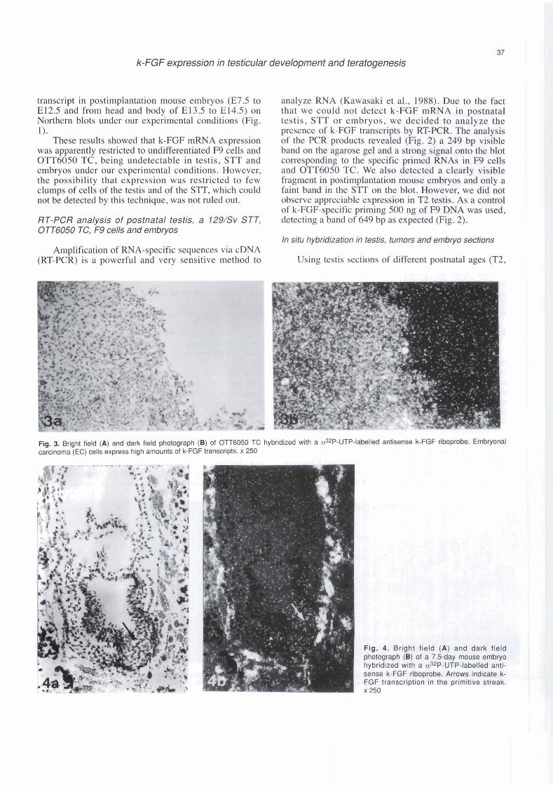
Fig. 3. Bright field (A) and dark field photograph (B) of OlT6050 TC hybridized wRh a a32P-UTP-labelled antisense k-FGF riboprobe. Embryonal carcinoma (EC) cells express high amounts of k-FGF transcripts. x 250
Fig. 4. Bright field (A) and dark field photograph (E) of a 7.5-day mouse embryo hybridized with a a32P-UTP-labelled anti- sense k-FGF riboprobe. Arrows indicate k- FGF transcription in the primitive streak x 250
k-FGF ~XP~@SS¡O~ h testcular development and teratogenesis
B, no, m, ?gl) and bybri&Pbg wíth &e antisense k- H3P'ribpmbe, we cwld not &tect any expression, even a;Ber e x p i n g the slides for one month, as expected
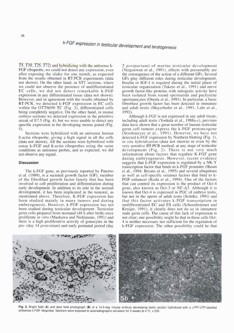
'tíie m I @ a M in RT-KX experiments (data a@ s h ) . Qn ttre o k r hmd, in Sl'T sections, where we mdd not observe tk p m n c e of undrfferentiated EC calle, we did not detsct remarkable Ir-FGF expmsim in m y dlffenxtthted t h u e (data not shown). Hm&ver, and in t with h m h obtained by BT"-FCR, we d e t e ~ t d k-F@F expre%~*sian in EC cells aitbin ik OTTWm TC (Fig. 3), differentiated cells being c m p k b l y negative. Bn &e other hand, in mouse embryo &ons we detwted expmsion in the primitive s$ealr of E75 (Fig. 4), but we were unhle to detect any s p B c e x p s i o n in the ckve1oping mowe gonad (Fig. 5).
Sections were hybridized with an antisense human B-actin riboprobe, giving a high signal in al1 the cells
w m hybridiized with u s i q the same
e x p a d , we did
l b k-FOP et al. (1!3%9), i9 of the fibroblwt geowth factor family that has bean inwobcí ra mU plihration ourd diff~fekltisrtion during ear1y QBvzyl-nt. In tdctíiam to its roke In the normal d e w : l ~ w , it has begn h p l i a t e d 21 the h~moral, as mentimd &ave. Tbrefwre, E-FGF exprmsion has bean studied nainly in many turnors and during embryogenesis. However, k-FGF expression has not been studied during testicular development. Testicular germ cells prepared from mwtai (48 h after birth) mice proliferate in vino (Maekawa and Nishimune, 191) and there is a high proliferative activity of gomqms ín tha pre- (day 14 postcoitutn) and early po"stmtd periBd (day
2 postpartum) of murine testicular development (Vergouwen et al., 1991), effects with presumably are the consequence of the action of a different GFs. Severa1 GFs play different roles during testicular development. Insulin or IGF-1 is required during the initial phase of testicular organization (Taketo et al., 1991) and nerve growth factor-like proteins with mitogenic activity have been isolated from round spermatids and pachytene spermatocytes (Onoda et al., 1991). In particular, a basic fibroblast growth factor has been detected in immature and adult testis (Mayerhofer et al., 1991; Lahr et al., 1992).
Although k-FGF is not expressed in any adult tissue, including adult testis (Yoshida et al., 1988a-c), previous data have shown that a great number of human testicular germ cell tumors express the k-FGF protoncogene (Strohmeyer et al., 1991). However, we have not detected k-FGF expression by Northern blotting (Fig. l), in situ hybridization (data not shown) or even by the very sensitive RT-PCR method, at any stage of testicular development (Fig. 2). There is not very much information about factors that regulate K-FGF gene during embryogenesis. However, recent evidence suggests that k-FGF expression is regulated by a NK-Y transcription factor that binds to k-FGF promoter (Hasan et al., 1994; Bryans et al., 1995) and several ubiquitous as well as cell-specific octamer factors that bind to k- FGF enhancer (Koda et al., 1994). One of the factors that can control its expression is the product of Oct-4 gene, also known as Oct-3 or NF-A3. Although it is known that Oct-4 is expressed in PGC of embryo testis, but not in the sperm of adult testis (Scholer, 1991) and that this factor activates k-FGF transcription in undifferentiated EC and ES cells (Schoorlemmer and Kruijer, 1991), it clearly does not do so in immature male germ cells. The cause of this lack of expression is not clear; one possibility might be that in these cells Oct- 4 is neither necessary nor sufficient for the regulation of k-FGF expression. The other possibility could be that
6. bk'i (A) md derk.fW -gph @$ ed ri 14.[j-dBy i'imm embryo dewbpfng testis section hybridlzed wíth a aaP-UTP-labelled antiseiwrek-K3fribopFobe.~onswen, b I c e m u ~ f o r 3 ~ a t 4 O C . x 2 5 0
k-FGF expression in testicular development and teratogenesis
after birth, with the appearance of the A spermatogonia and the onset of spermatogenesis, Oct-4 was downregulated, k-FGF transcription not being activated. It should be noted that Oct-4 is a target for negative regulation by retinoic acid (RA) (Okamoto et al., 1990), which in tum mediates its action through RA receptors. Vanbeek and Meistrich (1992) reported the irnportance of retinoic acid in the maintenance of spermatogenesis. Therefore, it should be of interest to analyze RA and RA receptor expression in mouse testis after birth and in early spermatogenesis (prepubertal stage).
In a spontaneous, 1291Sv mouse testicular teratoma (STI'), we did not detect appreciable k-FGF expression by Northem blotting, although we detected a faint band after blotting the products of the RT-PCR reaction and hybridizing with a specific probe (Fig. 2). This fact indicates that probably only some small groups of un- differentiated cells, which were not included in the analyzed STT sections, express k-FGF, reflecting the conversion to benignancy of the tumor. This clearly associates k-FGF with the degree of undifferentiation of this tumor. Supporting this idea, k-FGF is expressed in primary human testicular tumors (Yoshida et al., 1988a- d; Schofield et al., 1991 ; Strohmeyer et al., 1991).
We used the experimental OTT6050 TC as a positive control for k-FGF expression. We detected expression in the stem cells of the tumor (EC cells), but not in the differentiated tissue by in situ hybridization. The fact that k-FGF is expressed in the stem cells of the tumor and that it is known that this GF acts via an autocrine mechanism (Delli Bovi et al., 1988; Talarico and Basilico, 1991; Moscatelli, 1994), suggest a role of k- FGF in O'IT6050 TC in vivo growth.
By Northern blot analysis, we also detected a different level of expression between the nullipotent F9 cell line and the multipotent OTT6050 TC. This result is not surprising and may be explained because OTI'6050 TC contained some differentiated cells which are known not to express k-FGF, and also because there are differences in the level of k-FGF expression between several EC cell lines, as previously demonstrated by Schoorlemmer and Kruijer (1991).
In agreement with the results previously reported by Niswander and Martin (1992) indicating k-FGF expression during gastnilation, myogenesis and limb and tooth development, we detected k-FGF transcripts in mouse embryos from E7.5 to E12.5 (Figs. 2, 4) and in the head and body of E1 3.5 and E14.5 (Fig. 2) by RT- PCR. Using an antisense riboprobe for the k-FGF gene, we confirmed some of these results by detecting k-FGF expression in the dista1 part of the primitive streak of E7.5 (Fig. 4). This zone of the primitive streak contains cells that are fated to become embryonic, but not extraembryonic mesoderm. As discussed by Niswander and Martin (1992), this finding suggests that k-FGF is involved in the specification of some cells within the streak as well as in mesoderm induction in mammals, as demonstrated in amphibia by Paterno et al. (1989). In E7.5, the base of the allantosis, the area where PGC are
located, is completely negative, indicating that PGC do not express the k-FGF gene. This result is surprising, because k-FGF is directly transactivated by Oct-4, which is known to be expressed in PGC and in the mouse gonad (Scholer, 199 1). However, some evidence suggests that there may be an interaction between Oct-4 and an ElA-like factor. One could speculate that Oct-4 could be complexed by an E1A-like factor, avoiding it binding to the k-FGF enhancer. Indeed, E1A acts as a bridging factor for transcriptional.regulation by Oct-4 (Scholer et al., 1991). Another possibility for this lack of k-FGF expression in PGC could be addressed to the presence of a transcriptional repressor. This lack of k- FGF expression in the developing gonad was also maintained in later stages (E12.5, E13.5 and E14.5 mouse embryo sections).
These results indicate that although k-FGF gene is not involved in mouse testicular development, it is involved in mesodermal induction and in the growth of the experimental OTT6050 TC. At the same time, the detection of very low levels of k-FGF transcription in STI', its absence in differentiated cells of STT, as well as its expression in undifferentiated OTT6050 TC cells suggest that the activation of this gene is probably related to the genesis and progression of STI'.
Acknowledgements. We gratefully thank Prof. A. Alonso for his helpful technical assistance and Dr. J.K. Heath for providing PXMT2-1.58 k- FGF plasmid. We would also like to thank to the Comissió lnterdepartamental de Recerca i Innovació Tecnológica (CIRIT). from the Generalitat de Catalunya for financia1 assistance.
References
Blin N. and Stafford D.W. (1976). lsolation of high-molecular-weight DNA. Nucleic Acids Res. 3, 2303.
Brookes S., Smith R., Thurlow J., Dickson C. and Peters G. (1989). The mosue homologue of the hst/k-FGF: sequence, genome organization and location relative to int-2. Nucleic Acids Res. 17, 4037-4035.
Brustle O., Aguzzi A.. Talarico D., Basilico C., Kleihues P. and Wiestler O.D. (1992). Angiogenic acitivity of the K-fgflhst oncogene in neutral transplants. Oncogene 7, 11 77-1 183.
Bryans M., Lucas J.M., Knobloch T.J., Wilkie N.M. and Lang J.C. (1 995). Regulation of FGF-4 enhancer activity by transcription factor NF-Y. Biochem. Biophys. Res. Commun. 21 T, 519-527.
Burgess W.H. and Maciag T. (1989). The heparln binding (fibroblast) growth factor family of proteins. Annu. Rev. B ' i e m . 58,575-606.
Chikuba K., Saito T., Uchino S., Sato K., Miyahara M., Tsuda H., Hirohashi S. and Kobayashi M. (1995). High amplificatton of the hst- 1 gene correlates with haematogenous recurrence after curative resection of oesophageal carcinoma. Br. J. Surg. 82,364-367.
Curatola A.M. and Basilico C. (1990). Expression fo the K-fgf proto- onmgene is controlled by 3' regulatory elements whih are specific for ernbyronai caricnoma cells. Mol. Cell. Bid. 10, 2475-2484.
Chmozynski P. and Sacchi N. (1987). Single-step method of RNA isolation by acid guanidinium thiocyanate-phenol-chloroform extradon. Anal. Biochem. 162, 156-1 59.
-. ' ~ x P ~ @ s s ~ o ~ in testicular development and teratogenesis
Damen J.E., Greenberg A.H. and Wright J.A. (1991). Transformation and amplification of !he K-fgf pmto-oncogene in NIH-3T3 ceils, and induction of metastatic potential. Biochem. Biophys. Acta 1097, 103- 110.
Delli Bovi P. and Basilico C. (1987). lsolation of rearranged human transforming gene following transfection of Kaposi sarcoma DNA. Proc. Natl. Acad. Sci. USA 84,5660-5664.
Delli Bovi P., Curatola A.M., Kern F.G., Greco A., lttmann M. and Basilica C. (1987). An oncogene isdated by transfection of Kaposi's sarcoma DNA enwdes a growth factor that is a member of the FGF famBy. Cell50,729-737.
Delli Bovi P., Curatola A.M., Mewman K.M., Sato Y., Moscatelli D., Hewick R.M.. Rifkin D.B. and Basilico C. (1988). Processing, secretion and biological properties of a novel growth factor of the firoblast growth factor family with onwgenic potential. Mol. Cell. Biol. 8, 2933-2941.
Díaz-Flores L., GutiBrrez R. and Varela H. (1994). Angiogenesis: an update. Histol. Histopathol. 9, 807-843.
Feldman B., Poueymirou W., Papaioannou V.E., DeChiara T.M. and Goldbarb M. (1995). Requirement of FGF-4 for postimplantation mouse development. Science 267,246-249.
Frohrnan M.A, Boyle M. and Martin G.R. (1990). lsolation of the mouse Hox-2.9 gene: analysis of embyronic expression suggests that positional information along the anterior-posterior axis is specified by mesoderm. Development 110,589-607.
Hasan S., Koda T. and Kakinuma M. (1994). An upstream NF-Y-binding site is required for transcriptional activation from the hst promoter in F9 embryonal carcinoma cells. J. Biol. Chem. 269,25042-25048.
Heath J.K., Paterno G.D., Lindon A.C. and Edwards D.R. (1 989). Expression of multiple heparin-binding growth factor species by murine embryonal carcinoma and embryonic stem cells. Development 107, 1 13-1 22.
HBbert J.M., B a s i l i C., Goldfarb M., Haub 0. and Martin G.R. (1 990). Isolaüon of cDNAs encoding four mouse FGF family members and characterization of their expression patterns during ernbyrogenesis. Dev. Biol. 138,454-463.
Jernvall J., Kettunen P., Karavanova l., Martin L.B. and Thesleff 1. (1994). Evidence for the role of the enamel knot as a control center in mammalian tooth cusp formation: non-dividing cells express growth stimulating Fgf-4 gene. Int. J. Dev. Biol. 38,463-489.
Kawasaki ES., Clark S.S., Coyne M.Y., Smith S.D., Champlin R., Witte O.N. and McCormick F.P. (1988). Diagnosis of chronic myeloid and acute lymphocytic leukemias by detection of leukemia-specific mRNA sequences amplified h vitro. Proc. Natl. Acad. Sci. USA 85, 5698-5702.
Kluin Ph.M. and de Rooij D.G. (1981). A comparison between the morphology and cell kinetics of gonocytes and adult type un- differentiated spermatogonia in the mouse. Int. J. Androl. 4, 475- 493.
Kluin Ph.M., Kreamer M.F. and de Rooij D.G. (1984). Prollferation of spermatogonia and Sertoli cells in rnaturing mice. Anat. Embryol. 169,73-78.
Koda T., Hasan S., Sasaki A., Arimura Y. and Kakinuma M. (1994). Regulatory sequences required for hst-1 expression in embryonal carcinoma cells. FEBS Lett. 342,71-75.
Lahr G., Mayerhoffer A., Seidl K., Bucher S.. Grothe C., Knkhel W. and Gratzl M. (1992). Basic fibroblast growth factor in rodent testis. presence of bFGF mRNA and of a 30 kDA bFGF protein in pachytene spermatocytes. FEBS Lett. 302,43-46.
Maekawa M. and Nishimune Y. (1991). In-vitro proliferation of germ cells and supporting cells in the neonatal mouse testis. Cell Tissue Res. 265,551 -554.
Mayerhofer A., Russell L.D., Grothe C., Rudolf M. and Gratzl M. (1991). presence and localization of a 30-kDa basic fibroblast growth factor- like protein in rodents testes. Endocrinology 129, 921-924.
McLeskey S.W., Kurebayashi J., Honig S.F., Zwiebel J., Lippman M.E., Dickson R.B. and Kern F.G. (1993). Fibroblast growth factor 4 transfection of MCF-7 cells produces cell iines that are tumorigenic and metastatic in ovariectomized or tamoxifen-treated athymic nude mice. Cancer Res. 53,2168-21 77.
Miyagawa K., Sakamoto H., Yoshida T., Yamashita Y., Mitsui Y.. Furusawa M., Maeda S., Takaku F., SugimuraT. and Terada M. (1988). hst-1 transforming protein: expression in silkworm cells and characterization as a novel heparin-binding growth factor. Oncogene 3,383-389.
Moscatelli D. (1994). Autocrine downregulation of fibroblast growth factor receptors in F9 teratocarcinoma cells. J. Cell. Physiol. 160, 555-562.
Murakami A.. Tanaka H. and Matsuzawa A. (1990). Association of hst gene expression with metastatic phenotype in mouse mammary tumors. Cell Growth Differ. 1, 225-231.
Niswander L. and Martin G.R. (1992). Fgf-4 expression during gastrulation, myogenesis, limb and tooth development in the mouse. Development 114, 755-768.
Niswander L., Tickle C., Vogel A., Booth l. and Martin G.R. (1993). FGF- 4 replaces the apical ectodermal ridge and directs outgrowth and patterning of the limb. Cell75,579-587.
Niswander L., Tickle C., Vogel A. and Martin G.R. (1994). Function of FGF-4 in limb developrnent. Mol. Reprod. Dev. 39,8349.
Okamoto K., Okazawa H., Okuda A., Sakai M., Muramatsu M. and Hamada H. (1990). A novel octamer binding transcription factor is differentially expressed in mouse embyronic cells. Cell 60, 461 -472.
Onoda M., Pflug B. and Djakiew D. (1991). Germ cell mitogenic activity is associated with neme growth factor-like protein(s). J. Cell Physiol. 149,536-543.
Paterno G.D., Gillespie L.L., Dixon M., Slack J.M.W. and Heath J.K. (1989). Mesoderm-inducing properties of lnt-2 and kFGF: two oncogene encoded growth factors related to FGF. Development 106,79-84.
Rappolee D.A., Basilico C.. Patel Y. and Werb Z. (1994). Expression and function of FGF-4 in peri-implantation development in mouse embryos. Development 120,2259-2269.
Sakamoto H., Mori M., Taira M., Yoshida T., Matsukawa S., Shimizu K., Sekiguchi M., Terada M. and Sugimura T. (1986). Transforming gene from human stomach cancers and a noncancerous portion of stomach mucosa. Proc. Natl. Acad. Sci. USA 83, 3997- 4001.
Sasaki A., Kubo M., Hasan S., Yano Y. and Kakinuma M. (1991). Regulation of human hst expression by an enhancer element residing in the third exon. Jpn. J. Cancer Res. 82, 1191-1 195.
Schofield P.N.. EkstrUm T.J., Granerus M. and EngstrUm W. (1991). Differentiation associated modulation of K-FGF expression in a human teratocarcinoma cell line and in primary germ cell tumours. FEBS Lett. 280,8-10.
SchUler H.R. (1991). Octamania: The POU factors in murine development. TIG 7,323-329.
SchUler H.R., Hatzopoulos A.K., Balling R., Suzuki N. and Gruss P. (1989). A family of octamer-specific proteins present during mouse
k-FGF expression in testicular development and teratogenesis
embryogenesis: evidence for germline-specific expression of an Oct factor. EMBO J. 8, 2543-2550.
Schbler H.R., Ciesiolka T. and Gruss P. (1991). A nexus between Oct-4 and ElA: implications for gene regulation in embryonic stem cells. Cell66,291-304.
Schoorlemmer J. and Kruijer W. (1991). Octamer-dependent regulation of the kFGF gene in embyronal carcinoma and embryonic stern cells. Mech. Dev. 36, 75-86.
Stevens L.C. (1970). The development of transplantable terato- carcinomas from intratesticular grafts of pre- and postimplantation mouse embryos. Dev. Biol. 21,364-382.
Strohmeyer T., Peter S., Hartmann M., Munemitsu S., Ackermann R., Ullrich A. and Slarnon D.J. (1991). Expression of the hst-1 and c-kit protooncogenes in human testicular germ cell tumors. Cancer Res. 51, 1811-1816.
Taketo T., Saeed J., Roberge S., Matsuo N. and Koide S. (1991). Regulation of testicular differentiation and testosterone production in the fetal mouse gonad in vitro. J. Steroid Biochem. Mol. Biol. 38, 523-531.
Talarico D. and Basilico C. (1991). The K-fgflhst oncogene induces transforrnation through an autocrine mechanism that requires extracellular stirnulation of the mitogenic pathway. Mol. Cell. Biol. 11, 1138-1 145.
Taylor W.R., Greenberg A.H., Turley E.A. and Wright J.A. (1993). Cell motility, invasion. and malignancy induced by overexpression of K- FGF or bFGF. Exp. Cell Res. 204, 295-301.
Tronick S.R. and Aaronson S.A. (1995). Growth factors and signal transduction. In: The molecular basis of cancer. Mendelson J., Howley S., Israel C. and Liotta L.A. (ed). W.B. Saunders Company. Philadelphia, London, Toronto, Montreal, Sidney, Tokyo. pp 117-142.
Vanbeek M.E.A.B. and Meistrich M.L. (1992). Spermatogenesis in retinol-deficient rats maintained on retinoic acid. J. Reprod. Fert. 94, 327-336. '
Velcich A., Delli Bovi P., Mansukhani A., Ziff E.B. and Basilico C. (1989). Expression of the K-fgf protonocgene is repressed during
differentiaiton of F9 cells. Oncogene Res. 5, 31 -37. Vergouwen R.P.F.A., Jacobs S.G.P.M., Huiskamp R., Davids J.A.G.
and de Rooij D.G. (1991). Proliferative activity of gonocytes, sertoli cells and interstitial cells during testicular development in rnice. J. Reprod. Fert. 93, 233-243.
Vogel A. and Tickle C. (1993). FGF-4 maintains polarizing activity of posterior limb bud cells in vivo and in vitro. Development 119, 199- 206.
Volling P., Jungehulsing M., Jud<er M., Stutzer H., Diehl V. and Tesch H. (1993). Coamplification of the hst and bcl-1 oncogenes in advanced squamous cell carcinomas of the head and neck. Eur. J. Cancer 29A, 383-389.
Yoshida T., Miyagawa K., Odagiri H., Sakamoto H., Little P., Terada M. and Sugimura T. (1987). Genomic sequence of hst, a transfonning gene encoding a protein homologous to fibroblast growth factors and the int-2-encoded protein. Proc. Natl. Acad. Sci. USA 84, 7305- 7309.
Yoshida T., Tsutsumi M., Sakamoto H., Miyagawa K., Teshima S., Sugimura T. and Terada (1988a). Expression of HSTl onmgene in human germ cell tumors. Biochern. Biophys. Res. Commun. 155, 1324-1 329.
Yoshida T., Muramatsu H., Muramatsu T., Sakamoto H., Katoh O., Sugimura T. and Terada (1988b). Differential expression of two homologous and clustered oncogenes, hs t l and int2, during differentiation of F9 cells. Biochem. Biophys. Res. Cornrnun. 157, 618-625.
Yoshida M.C., Wada M., Satoh H., Yoshida T., Sakamoto H., Miyagawa K., Yokota J., Koda T., Kakinurna M., Sugimura T. and Terada M. (1988~). Human HSTl (HSTF1) gene maps to chromosome band 1 lq13 and coamplifies with the INT2 gene in human cancer. Proc. Natl. Acad. Sci. USA 85,4861 -4864.
Yoshida T., Ishimaru K., Sakamoto H., Yokota J., Hirohashi S., Igarashi K. and Sudo K. (19894). Angiogenic activ'Ry of the recombinant hst-1 protein. Cancer Lett. 83,261 -268.
Accepted June 9,1996


![FYdgYdcYf]k Yf\ FYdgYj]f]k - Samagra](https://static.fdokumen.com/doc/165x107/631f358f13819e2fbb0fa1db/fydgydcyfk-yf-fydgyjfk-samagra.jpg)