Role of FGF/FGFR signaling in skeletal development ... - Nature
24
REVIEW ARTICLE Role of FGF/FGFR signaling in skeletal development and homeostasis: learning from mouse models Nan Su * , Min Jin * and Lin Chen Fibroblast growth factor (FGF)/fibroblast growth factor receptor (FGFR) signaling plays essential roles in bone development and diseases. Missense mutations in FGFs and FGFRs in humans can cause various congenital bone diseases, including chondrodysplasia syndromes, craniosynostosis syndromes and syndromes with dysregulated phosphate metabolism. FGF/FGFR signaling is also an important pathway involved in the maintenance of adult bone homeostasis. Multiple kinds of mouse models, mimicking human skeleton diseases caused by missense mutations in FGFs and FGFRs, have been established by knock-in/out and transgenic technologies. These genetically modified mice provide good models for studying the role of FGF/FGFR signaling in skeleton development and homeostasis. In this review, we summarize the mouse models of FGF signaling-related skeleton diseases and recent progresses regarding the molecular mechanisms, underlying the role of FGFs/FGFRs in the regulation of bone development and homeostasis. This review also provides a perspective view on future works to explore the roles of FGF signaling in skeletal development and homeostasis. Bone Research (2014) 2, 14003; doi:10.1038/boneres.2014.3; Published online 29 April 2014 INTRODUCTION Skeletons are formed through two distinct developmental modes, namely intramembranous ossification and endo- chondral ossification. The former is directly accomplished by osteoblast differentiation from mesenchymal cells; the latter involves initial differentiation of mesenchymal cells into chondrocytes to form a cartilage template and sub- sequent replacement by bone. 1 The cranium and medial clavicles are formed through intramembranous ossifica- tion. Long bones, including the appendicular skeleton, facial bones and vertebrae, are formed through endo- chondral ossification. 2–3 Various signaling molecules control the process of skel- eton development, such as fibroblast growth factor (FGF), wingless-type MMTV integration site family members (Wnt) and bone morphogenetic protein (BMP) signaling path- ways. Among these signaling pathways, FGF/fibroblast growth factor receptor (FGFR) signaling is very essential. The 22 members of the FGF family mediate their cellular responses by binding to FGFRs. There are four distinct FGF receptors with differential FGF-binding properties. 4–5 A typ- ical FGFR contains an extracellular ligand-binding domain, a transmembrane region and an intracellular divided tyrosine kinase domain. FGFs bind to the extracel- lular domain of FGFRs and induce the phosphorylation of tyrosine residues in the intracellular domain of FGFRs. The activated FGFRs recruits target proteins to its cytoplas- mic tail and modifies these proteins by phosphorylation, 6 leading to the activation of intracellular downstream sig- naling pathways, such as mitogen-activated protein kinase (Ras/MAPK), phosphoinositide 3-kinase/Akt (also known as protein kinase B), phospholipase C and protein kinase C pathways. Furthermore, FGF signaling can also stimulate the signal transducers and activators of tran- scription (STAT) 1/p21 pathway 2,7 (Figure 1). Multiple kinds of mouse models with genetic modifications of FGF/FGFR have been generated. In our review, we summarize the use of these mouse models in the research of the role of FGF/FGFR signaling in skeleton development and homeostasis. ROLE OF FGFRS IN BONE GENETIC DISEASES AND HOMEOSTASIS FGFR1 FGFR1 is first expressed in the early limb bud. 8–10 At the epi- physeal growth plate, FGFR1 is expressed in perichondrium, *These authors contributed equally to this review. Center of Bone Metabolism and Repair, State Key Laboratory of Trauma, Burns and Combined Injury, Trauma Center, Institute of Surgery Research, Daping Hospital, Third Military Medical University, Chongqing, 400042, China. Correspondence: L Chen ([email protected]) Received: 9 November 2013; Revised: 29 November 2013; Accepted: 3 December 2013; Uncorrected proof published 9 April 2014 OPEN Citation: Bone Research (2014) 2, 14003; doi:10.1038/boneres.2014.3 ß 2014 Sichuan University. All rights reserved 2095-4700/14 www.boneresearch.org
-
Upload
khangminh22 -
Category
Documents
-
view
1 -
download
0
Transcript of Role of FGF/FGFR signaling in skeletal development ... - Nature
REVIEW ARTICLE
Role of FGF/FGFR signaling in skeletal development and
homeostasis: learning from mouse models
Nan Su*, Min Jin* and Lin Chen
Fibroblast growth factor (FGF)/fibroblast growth factor receptor (FGFR) signaling plays essential roles in bonedevelopment and diseases. Missense mutations in FGFs and FGFRs in humans can cause various congenital bonediseases, including chondrodysplasia syndromes, craniosynostosis syndromes and syndromes with dysregulatedphosphate metabolism. FGF/FGFR signaling is also an important pathway involved in the maintenance of adultbone homeostasis. Multiple kinds of mouse models, mimicking human skeleton diseases caused by missensemutations in FGFs and FGFRs, have been established by knock-in/out and transgenic technologies. Thesegenetically modified mice provide good models for studying the role of FGF/FGFR signaling in skeletondevelopment and homeostasis. In this review, we summarize themousemodels of FGF signaling-related skeletondiseases and recent progresses regarding the molecular mechanisms, underlying the role of FGFs/FGFRs in theregulation of bone development and homeostasis. This review also provides a perspective view on futureworks toexplore the roles of FGF signaling in skeletal development and homeostasis.
Bone Research (2014) 2, 14003; doi:10.1038/boneres.2014.3; Published online 29 April 2014
INTRODUCTIONSkeletons are formed through two distinct developmental
modes, namely intramembranous ossification and endo-
chondral ossification. The former is directly accomplished
by osteoblast differentiation from mesenchymal cells; the
latter involves initial differentiation of mesenchymal cells
into chondrocytes to form a cartilage template and sub-
sequent replacement by bone.1 The cranium and medial
clavicles are formed through intramembranous ossifica-
tion. Long bones, including the appendicular skeleton,
facial bones and vertebrae, are formed through endo-
chondral ossification.2–3
Various signaling molecules control the process of skel-
eton development, such as fibroblast growth factor (FGF),
wingless-typeMMTV integration site family members (Wnt)
and bone morphogenetic protein (BMP) signaling path-
ways. Among these signaling pathways, FGF/fibroblast
growth factor receptor (FGFR) signaling is very essential.
The 22 members of the FGF family mediate their cellular
responses by binding to FGFRs. There are four distinct FGF
receptorswithdifferential FGF-bindingproperties.4–5 A typ-
ical FGFR contains an extracellular ligand-binding
domain, a transmembrane region and an intracellular
divided tyrosine kinase domain. FGFs bind to the extracel-
lular domain of FGFRs and induce the phosphorylation
of tyrosine residues in the intracellular domain of FGFRs.
The activated FGFRs recruits target proteins to its cytoplas-
mic tail and modifies these proteins by phosphorylation,6
leading to the activation of intracellular downstream sig-
naling pathways, such as mitogen-activated protein
kinase (Ras/MAPK), phosphoinositide 3-kinase/Akt (also
known as protein kinase B), phospholipase C and protein
kinase C pathways. Furthermore, FGF signaling can also
stimulate the signal transducers and activators of tran-
scription (STAT) 1/p21 pathway2,7 (Figure 1). Multiple kinds
of mouse models with genetic modifications of FGF/FGFR
have been generated. In our review, we summarize the
use of these mouse models in the research of the role
of FGF/FGFR signaling in skeleton development and
homeostasis.
ROLE OF FGFRS IN BONE GENETIC DISEASES ANDHOMEOSTASISFGFR1
FGFR1 is first expressed in the early limb bud.8–10 At the epi-
physeal growth plate, FGFR1 is expressed in perichondrium,
*These authors contributed equally to this review.
Center of BoneMetabolismandRepair, State Key Laboratoryof Trauma, BurnsandCombined Injury, TraumaCenter, Instituteof Surgery Research,
Daping Hospital, Third Military Medical University, Chongqing, 400042, China.
Correspondence: L Chen ([email protected])
Received: 9 November 2013; Revised: 29 November 2013; Accepted: 3 December 2013; Uncorrected proof published 9 April 2014
OPEN Citation: Bone Research (2014) 2, 14003; doi:10.1038/boneres.2014.3� 2014 Sichuan University. All rights reserved 2095-4700/14
www.boneresearch.org

prehypertrophic and hypertrophic chondrocytes.9,11–12
FGFR1 is also expressed in osteoblasts and osteocytes
(Table 1).13–16
A series of mousemodels of Fgfr1 have been generated
to genetically dissect the functions of Fgfr1 during gastru-
lation and later developmental processes. Fgfr1-deficient
(Fgfr12/2) embryos display severegrowth retardation, and
died prior to or during gastrulation because of intrinsic
blocks in mesodermal differentiation.17–18 Deletion of the
Ig domain IIIc of Fgfr1 (Fgfr1IIIc) leads to gastrulation
defects resembling the Fgfr12/2 alleles. However, mice
with Fgfr1IIIb ablation are viable and fertile, suggesting
that IIIc is the dominant isoform for the majority of FGFR1
functions in embryogenesis.19 Chimeras were generated
by injecting Fgfr12/2 embryonic stem cells into wild-type
blastocysts to circumvent the gastrulation defect. The
milder mutant chimeras exhibit deformed limb buds and
varyingdegrees of reduction in limb skeletal elements.19–21
Mice with targeted deletion of FGFR1 in all limb bud
mesenchymal cells (via T (brachyury)-cre),22 or posterior
limb budmesenchyme (via Shh-cre)23 were used to further
study the role of FGFR1 in limb development. T-cre;
Fgfr1 mice die at birth and show reduced limb skeleton,
misshapen forelimb/hindlimb bud and missing digits,
whereas Shh-cre Fgfr1 mice display normal limb bud size,
but missed a digit.10 Li et al.24 assessed the roles of FGFR1
signaling in forelimb and hindlimb development by disrupt-
ing this gene, using AP2-Cre and Hoxb6-Cre transgenic
mice that express Cre recombinase in complementary
temporal and spatial patterns during limb bud formation.
The results indicate that disruption of Fgfr1 at an earlier
stage, prior to thickening of limb mesenchyme, results in
lg
lg
lg
lg
lg
lg
FGF
FGFR
JM
TK TK FRS2
PI3K
Grb Sos Ras
Raf
MEK1/2
ERK1/2
AKT
Chondrocytes
Proliferation Differentiation Apoptosis
STAT1
p21
PDK
TK TK
JM
PKC
Src
Shc
PLCg
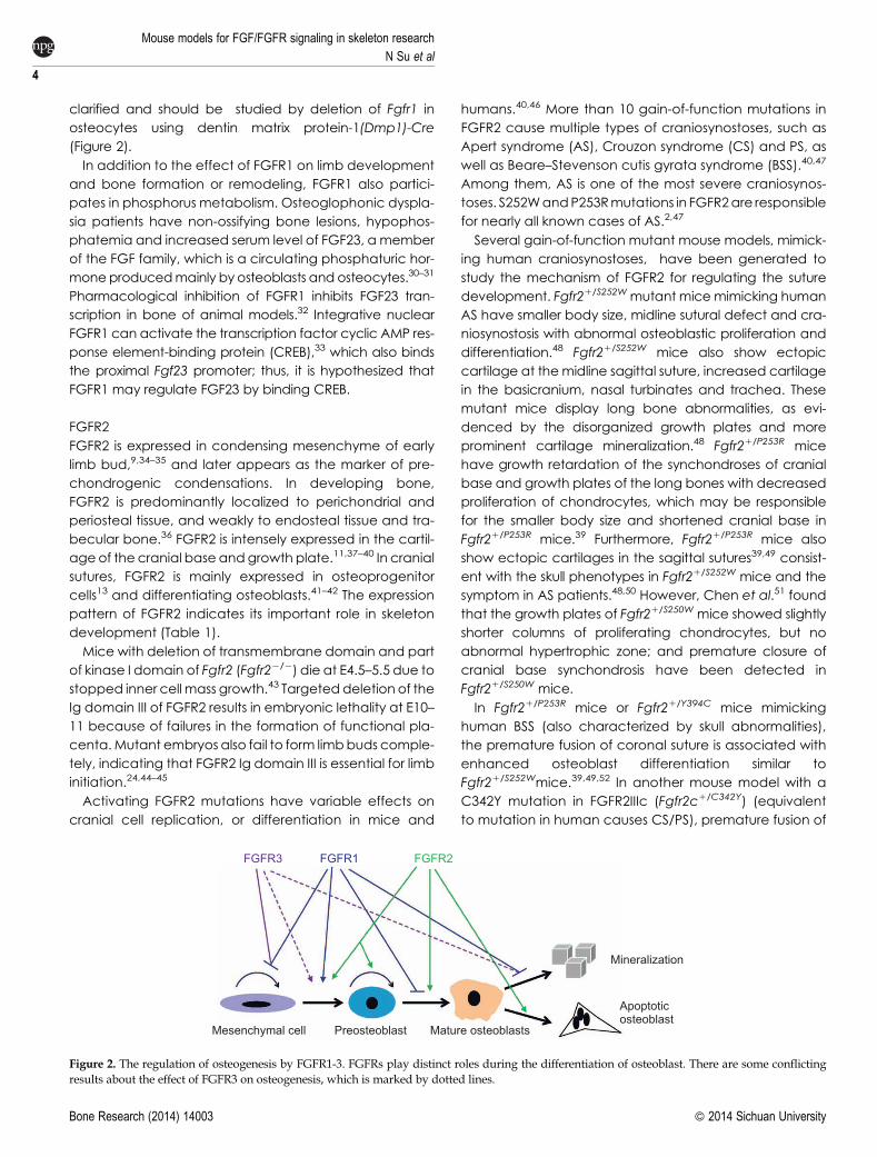
Figure 1. Signaling pathways activated by FGF/FGFR. FGFs induce dimerization, kinase activation and transphosphorylation of tyrosine residues ofFGFRs, leading to activation of downstream signaling pathways.Multiple pathways are stimulated by FGF/FGFR signaling such as Ras-MAP kinase,PI-3 kinase/AKT and PLC-c pathways. Furthermore, FGF signaling can also stimulate STAT1/p21 pathway. FGF/FGFR signaling also phosphor-ylates the Shc and Src protein. FGF/FGFR play crucial roles in the regulation of proliferation, differentiation and apoptosis of chondrocytes viadownstream signaling pathways.
Mouse models for FGF/FGFR signaling in skeleton research
N Su et al
2
Bone Research (2014) 14003 � 2014 Sichuan University

more severe defects, characterized by malformation of
the apical ectodermal ridge (AER).
FGF receptor-specific substrates (Frs) act as theprincipal
mediators for FGFR1 signal transduction. Mice that lack
the Frs-binding site on FGFR1 (Fgfr1DFrs/DFrs) die during late
embryogenesis, and exhibit defects in neural tube closure,
and in the development of the tail bud and pharyngeal
arches. However, mutant FGFR1 still has functions during
gastrulationand somitogenesis, indicating thatdistinct sig-
nal transduction mechanisms of FGFR1 signaling in differ-
ent developmental contexts.25
Osteoglophonic dysplasia (OD) patients, resulting from
activating mutations of FGFR1, exhibit rhizomelic dwarf-
ism,26 indicating that FGFR1 is a negative regulator of long
bone growth. Embryos with conditional deletion of Fgfr1 in
osteochondro-progenitor cell lineages show increased
height of the hypertrophic zone due to delayed degrada-
tion, or maturation of hypertrophic chondrocytes, or
decreased osteoclastogenesis.15
Studies in humans andmice also reveal that FGFR1 play
crucial role inbone formation.Again-of-functionmutation
in FGFR1 (P252R) leads to Pfeiffer syndrome (PS), one type
of craniosynostoses, characterized by premature fusion of
oneor several calvarial sutures.27 Several activatingmuta-
tions of FGFR1 in OD patients also lead to craniosynostosis
in addition to rhizomelic dwarfism.26Mice carrying a P250R
mutation in FGFR1 were generated to mimic human PS.
Studies using these mutant mice uncovered that FGFs/
FGFR1 signals may regulate intramembranous bone
formation.28
Jacob et al.15 found that adult mice, with deletion of
Fgfr1, exhibited increased bone mass. Deletion of Fgfr1,
in osteochondro-progenitor cells in mice (via Col2-cre),
leads to increased proliferation and delayed differenti-
ation, andmatrix mineralization of osteoblasts, while inac-
tivation of Fgfr1 in differentiated osteoblasts (viaCol1-cre)
causes accelerated osteoblast mineralization differenti-
ation.15 It has been proposed that FGFR1 promotes the
differentiation of mesenchymal progenitors into preosteo-
blasts, but inhibits the proliferation of mesenchymal pro-
genitor cells, as well as the maturation and mineralization
of osteoblasts.15 Impaired osteoclast activity is another
reason for increased bone mass in mice with Fgfr1-
deficient in differentiated osteoblasts. To explore the direct
effect of FGFR1 on osteoclasts, Lu et al.29 generated mice
with targeted deletion of Fgfr1 in bonemarrowmonocytes
and osteoclasts using LysM-cre. The mutant mice exhibit
increased bone mass, impaired osteoclast formation and
activity indicating the positive regulation of FGFR1 on
osteoclasts. The role of FGFR1 in osteocytes is still not
Table 1. The expression patterns of FGFs/FGFRs during skeleton development.2,7,11,31,46,60,161–162,204,262–263
FGFs/
FGFRs Limb bud Osteoblast lineage Cartilage Cranial bone Receptor specificity
FGF2 Developing condensation Periosteal cells, Osteoblasts in
trabecular bone
Perichondrium, Chondrocytes Mesenchymal cells in the suture FGFR1, FGFR2, FGFR3c,
FGFR4
FGF4 Posterior AER at E10.5-
11.0
Sutural mesenchyme in early
craniofacial skeletogenesis
FGFR1c, FGFR2c, FGFR3c
FGF7 Loose mesenchyme Perichondrium FGFR2b
FGF8 AER Cortical bone at embryonic stage Perichondrium, Chondrocytes Osteoblasts FGFR2c, FGFR3c, FGFR4
FGF9 AER, Developing
condensation
Periosteum, Primary spongiosa Perichondrium, Chondrocyte
primordia
Mesenchyme of suture in early
craniofacial development
stages
FGFR2c, FGFR3, FGFR4
FGF10 Lateral plate mesoderm FGFR2b
FGF18 Perichondrium and
presumptive joint
positions
Chondrocytes, Mesenchymal cells in the suture
separating the two osteogenic
fronts
FGFR2c, FGFR3c
FGF21 Chondrocytes FGFR1-4
FGF23 Osteoblasts, Osteocytes Resting and hypertrophic zone FGFR1, FGFR3c, FGFR4
FGFR1 Mesenchyme (IIIc) Osteoblasts in trabecular bone,
Osteocytes
Prehypertrophic and hypertrophic
chondrocytes of growth plate,
Perichondrium, Cartilage of the
cranial base
Dura mater and periosteum,
Calvarial mesenchyme and
later in osteoblasts
FGFR2 AER (IIIb), Early limb
bud mesenchyme
(IIIc)
Periosteum, Trabecular bone
(IIIc), Osteocytes
Prechondrogenic condensation,
Resting zone of growth plate,
Perichondrium, Cartilage of the
cranial base
Proliferating osteoprogenitor
cells and differentiating
osteoblasts
FGFR3 Center of themesenchyme
condensation
Osteoblasts, Osteocytes Resting zone and proliferating
chondrocytes of growth plate,
Cartilage of the cranial base
Low levels in sutural osteogenic
fronts at late stages of
development
FGFR4 Strictly in osteoblasts between the
periosteal and endosteal layers
Resting and proliferative zones
of growth plate
Mouse models for FGF/FGFR signaling in skeleton researchN Su et al
3
� 2014 Sichuan University Bone Research (2014) 14003
clarified and should be studied by deletion of Fgfr1 in
osteocytes using dentin matrix protein-1(Dmp1)-Cre
(Figure 2).
In addition to the effect of FGFR1 on limb development
and bone formation or remodeling, FGFR1 also partici-
pates in phosphorus metabolism. Osteoglophonic dyspla-
sia patients have non-ossifying bone lesions, hypophos-
phatemia and increased serum level of FGF23, a member
of the FGF family, which is a circulating phosphaturic hor-
moneproducedmainly by osteoblasts and osteocytes.30–31
Pharmacological inhibition of FGFR1 inhibits FGF23 tran-
scription in bone of animal models.32 Integrative nuclear
FGFR1 can activate the transcription factor cyclic AMP res-
ponse element-binding protein (CREB),33 which also binds
the proximal Fgf23 promoter; thus, it is hypothesized that
FGFR1 may regulate FGF23 by binding CREB.
FGFR2
FGFR2 is expressed in condensing mesenchyme of early
limb bud,9,34–35 and later appears as the marker of pre-
chondrogenic condensations. In developing bone,
FGFR2 is predominantly localized to perichondrial and
periosteal tissue, and weakly to endosteal tissue and tra-
becular bone.36 FGFR2 is intensely expressed in the cartil-
age of the cranial base andgrowth plate.11,37–40 In cranial
sutures, FGFR2 is mainly expressed in osteoprogenitor
cells13 and differentiating osteoblasts.41–42 The expression
pattern of FGFR2 indicates its important role in skeleton
development (Table 1).
Mice with deletion of transmembrane domain and part
of kinase I domain of Fgfr2 (Fgfr22/2) die at E4.5–5.5 due to
stopped inner cell mass growth.43 Targeteddeletion of the
Ig domain III of FGFR2 results in embryonic lethality at E10–
11 because of failures in the formation of functional pla-
centa.Mutant embryos also fail to form limbbuds comple-
tely, indicating that FGFR2 Ig domain III is essential for limb
initiation.24,44–45
Activating FGFR2 mutations have variable effects on
cranial cell replication, or differentiation in mice and
humans.40,46 More than 10 gain-of-function mutations in
FGFR2 cause multiple types of craniosynostoses, such as
Apert syndrome (AS), Crouzon syndrome (CS) and PS, as
well as Beare–Stevenson cutis gyrata syndrome (BSS).40,47
Among them, AS is one of the most severe craniosynos-
toses. S252WandP253Rmutations in FGFR2are responsible
for nearly all known cases of AS.2,47
Several gain-of-function mutant mouse models, mimick-
ing human craniosynostoses, have been generated to
study the mechanism of FGFR2 for regulating the suture
development. Fgfr21/S252W mutant mice mimicking human
AS have smaller body size, midline sutural defect and cra-
niosynostosis with abnormal osteoblastic proliferation and
differentiation.48 Fgfr21/S252W mice also show ectopic
cartilage at themidline sagittal suture, increased cartilage
in the basicranium, nasal turbinates and trachea. These
mutant mice display long bone abnormalities, as evi-
denced by the disorganized growth plates and more
prominent cartilage mineralization.48 Fgfr21/P253R mice
have growth retardation of the synchondroses of cranial
base and growth plates of the long bones with decreased
proliferation of chondrocytes, which may be responsible
for the smaller body size and shortened cranial base in
Fgfr21/P253R mice.39 Furthermore, Fgfr21/P253R mice also
show ectopic cartilages in the sagittal sutures39,49 consist-
ent with the skull phenotypes in Fgfr21/S252W mice and the
symptom in AS patients.48,50 However, Chen et al.51 found
that the growth plates of Fgfr21/S250W mice showed slightly
shorter columns of proliferating chondrocytes, but no
abnormal hypertrophic zone; and premature closure of
cranial base synchondrosis have been detected in
Fgfr21/S250W mice.
In Fgfr21/P253R mice or Fgfr21/Y394C mice mimicking
human BSS (also characterized by skull abnormalities),
the premature fusion of coronal suture is associated with
enhanced osteoblast differentiation similar to
Fgfr21/S252Wmice.39,49,52 In another mouse model with a
C342Y mutation in FGFR2IIIc (Fgfr2c1/C342Y) (equivalent
to mutation in human causes CS/PS), premature fusion of
FGFR3 FGFR1 FGFR2
Mesenchymal cell Preosteoblast Mature osteoblasts
Mineralization
Apoptoticosteoblast
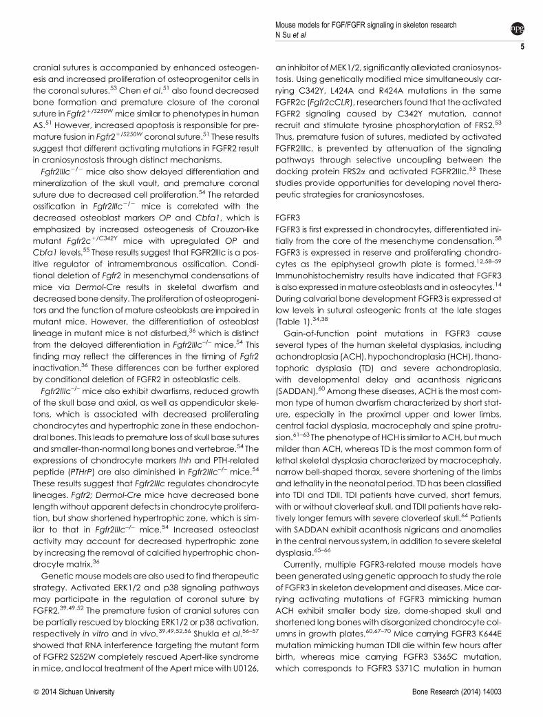
Figure 2. The regulation of osteogenesis by FGFR1-3. FGFRs play distinct roles during the differentiation of osteoblast. There are some conflictingresults about the effect of FGFR3 on osteogenesis, which is marked by dotted lines.
Mouse models for FGF/FGFR signaling in skeleton research
N Su et al
4
Bone Research (2014) 14003 � 2014 Sichuan University
cranial sutures is accompanied by enhanced osteogen-
esis and increased proliferation of osteoprogenitor cells in
the coronal sutures.53 Chen et al.51 also found decreased
bone formation and premature closure of the coronal
suture in Fgfr21/S250W mice similar to phenotypes in human
AS.51 However, increased apoptosis is responsible for pre-
mature fusion in Fgfr21/S250W coronal suture.51 These results
suggest that different activating mutations in FGFR2 result
in craniosynostosis through distinct mechanisms.
Fgfr2IIIc2/2 mice also show delayed differentiation and
mineralization of the skull vault, and premature coronal
suture due to decreased cell proliferation.54 The retarded
ossification in Fgfr2IIIc2/2 mice is correlated with the
decreased osteoblast markers OP and Cbfa1, which is
emphasized by increased osteogenesis of Crouzon-like
mutant Fgfr2c1/C342Y mice with upregulated OP and
Cbfa1 levels.55 These results suggest that FGFR2IIIc is a pos-
itive regulator of intramembranous ossification. Condi-
tional deletion of Fgfr2 in mesenchymal condensations of
mice via Dermol-Cre results in skeletal dwarfism and
decreasedbonedensity. The proliferation of osteoprogeni-
tors and the function of mature osteoblasts are impaired in
mutant mice. However, the differentiation of osteoblast
lineage in mutant mice is not disturbed,36 which is distinct
from the delayed differentiation in Fgfr2IIIc–/– mice.54 This
finding may reflect the differences in the timing of Fgfr2
inactivation.36 These differences can be further explored
by conditional deletion of FGFR2 in osteoblastic cells.
Fgfr2IIIc–/– mice also exhibit dwarfisms, reduced growth
of the skull base and axial, as well as appendicular skele-
tons, which is associated with decreased proliferating
chondrocytes and hypertrophic zone in these endochon-
dral bones. This leads to premature loss of skull base sutures
and smaller-than-normal long bones and vertebrae.54 The
expressions of chondrocyte markers Ihh and PTH-related
peptide (PTHrP) are also diminished in Fgfr2IIIc–/– mice.54
These results suggest that Fgfr2IIIc regulates chondrocyte
lineages. Fgfr2; Dermol-Cre mice have decreased bone
lengthwithout apparent defects in chondrocyte prolifera-
tion, but show shortened hypertrophic zone, which is sim-
ilar to that in Fgfr2IIIc–/– mice.54 Increased osteoclast
activity may account for decreased hypertrophic zone
by increasing the removal of calcified hypertrophic chon-
drocyte matrix.36
Geneticmousemodels arealso used to find therapeutic
strategy. Activated ERK1/2 and p38 signaling pathways
may participate in the regulation of coronal suture by
FGFR2.39,49,52 The premature fusion of cranial sutures can
be partially rescued by blocking ERK1/2 or p38 activation,
respectively in vitro and in vivo.39,49,52,56 Shukla et al.56–57
showed that RNA interference targeting the mutant form
of FGFR2 S252W completely rescued Apert-like syndrome
inmice, and local treatment of the Apertmicewith U0126,
an inhibitor ofMEK1/2, significantly alleviated craniosynos-
tosis. Using genetically modified mice simultaneously car-
rying C342Y, L424A and R424A mutations in the same
FGFR2c (Fgfr2cCLR), researchers found that the activated
FGFR2 signaling caused by C342Y mutation, cannot
recruit and stimulate tyrosine phosphorylation of FRS2.53
Thus, premature fusion of sutures, mediated by activated
FGFR2IIIc, is prevented by attenuation of the signaling
pathways through selective uncoupling between the
docking protein FRS2a and activated FGFR2IIIc.53 These
studies provide opportunities for developing novel thera-
peutic strategies for craniosynostoses.
FGFR3
FGFR3 is first expressed in chondrocytes, differentiated ini-
tially from the core of the mesenchyme condensation.58
FGFR3 is expressed in reserve and proliferating chondro-
cytes as the epiphyseal growth plate is formed.12,58–59
Immunohistochemistry results have indicated that FGFR3
is alsoexpressed inmatureosteoblasts and inosteocytes.14
During calvarial bone development FGFR3 is expressed at
low levels in sutural osteogenic fronts at the late stages
(Table 1).34,38
Gain-of-function point mutations in FGFR3 cause
several types of the human skeletal dysplasias, including
achondroplasia (ACH), hypochondroplasia (HCH), thana-
tophoric dysplasia (TD) and severe achondroplasia,
with developmental delay and acanthosis nigricans
(SADDAN).60 Among these diseases, ACH is themost com-
mon type of human dwarfism characterized by short stat-
ure, especially in the proximal upper and lower limbs,
central facial dysplasia, macrocephaly and spine protru-
sion.61–63 ThephenotypeofHCH is similar toACH,butmuch
milder than ACH, whereas TD is the most common form of
lethal skeletal dysplasia characterized by macrocephaly,
narrow bell-shaped thorax, severe shortening of the limbs
and lethality in the neonatal period. TD has beenclassified
into TDI and TDII. TDI patients have curved, short femurs,
with or without cloverleaf skull, and TDII patients have rela-
tively longer femurs with severe cloverleaf skull.64 Patients
with SADDAN exhibit acanthosis nigricans and anomalies
in the central nervous system, in addition to severe skeletal
dysplasia.65–66
Currently, multiple FGFR3-related mouse models have
been generated using genetic approach to study the role
of FGFR3 in skeleton development anddiseases. Mice car-
rying activating mutations of FGFR3 mimicking human
ACH exhibit smaller body size, dome-shaped skull and
shortened long bones with disorganized chondrocyte col-
umns in growth plates.60,67–70 Mice carrying FGFR3 K644E
mutation mimicking human TDII die within few hours after
birth, whereas mice carrying FGFR3 S365C mutation,
which corresponds to FGFR3 S371C mutation in human
Mouse models for FGF/FGFR signaling in skeleton researchN Su et al
5
� 2014 Sichuan University Bone Research (2014) 14003

TDI, exhibit skeletal dysplasia more severe than ACH.71–72
FGFR3 negatively regulates chondrogenesis of long bones
by affecting the proliferative activity and differentiation of
chondrocytes. A number of reports have demonstrated
that FGFR3 signaling inhibits chondrocyte proliferation
through STAT1 signaling by inducing the expression of cell
cycle suppressor genes such as the CDK inhibitor p21.73–76
Loss of Stat1 restored the reduced chondrocyte prolifera-
tion in ACH mice, but did not rescue the reduced hyper-
trophic zone or the delayed formation of secondary
ossification centers in ACH mice. The expression of a con-
stitutively active mutant of MEK1 in chondrocytes of Fgfr3-
deficient mice inhibits skeletal overgrowth, strongly sug-
gesting that FGFR3 inhibits chondrocyte differentiation
through the ERK/MAPK pathway.76 In contrast, evidence
suggests that FGFR3 promote chondrocyte terminal
hypertrophic differentiation.77–78 Conversely, mice car-
rying targeted deletion of FGFR3 exhibit overgrowth of
long bone, wider hypertrophic zone, proliferative zone
and enhanced proliferative activity of chondrocytes.59,79
Moreover, the activity and the signaling outcomes of
the FGFR3 pathway during chondrogenesis are also influ-
enced by many intracellular and extracellular signals.
Activated FGFR3 inhibits BMP4 expression in post-natal
mouse growth plates,80 while BMP treatment rescues the
retarded growth of long bone in ACH mouse model.77
These studies emphasize the antagonistic interaction
between FGFR3 and BMP signaling in the control of
chondrogenesis. Moreover, IHH expression is reduced in
mice carrying activating FGFR3.80 PTHrP partially reverses
the inhibition of long bone growth caused by FGFR3
activation.72 It was suspected that FGFR3 signaling may
act upstream of the IHH/PTHrP system in regulating the
onset of hypertrophic differentiation.77 In addition, it was
reported that IGF1 prevents the apoptosis, induced by
FGFR3 mutation, through the phosphoinositide 3-kinase
pathway and MAPK pathways.81
FGFR3 signaling is also an important regulator of osteo-
genesis. Chondrocyte-specific activationof FGFR3 inmice
causes premature synchondrosis closure and enhanced
osteoblast differentiation around synchondroses. Prema-
ture synchondrosis closure is alsoobserved in the spineand
cranial base in human cases of homozygous ACH and TD,
as well as in mouse models of ACH, with increased bone
formation.70,72,82 Activated FGFR3 leads to decreased
bone mass by regulating both osteoblast and osteoclast
activities.83–84 Mice lacking FGFR3 also have decreased
bone mineral density and osteopenia.14,85 FGFR3 can
inhibit proliferation of BMSCs in vitro.83,85 However, both
deletion and activation of FGFR3 can lead to increased
differentiation, but impaired mineralization of osteoblasts
(Figure 2).83,85 The reasons for these seemingly inconsistent
results need to be explored.
Given its causal role in some skeletal disorders, including
ACH, FGFR3and/or its downstreampathways, areattract-
ive targets for therapy. C-type natriuretic peptide is a
newly identified potential therapeutic antagonist of
FGFR3 signaling that alleviates the dwarfism phenotype
of mice mimicking human ACH through its inhibition on
FGFR3/MAPK pathway.86–87 It was reported that parathy-
roid hormone (PTH) (1–34) stimulates the longitudinal bone
growth in rats and improves the growth of the cultured
femurs from mice carrying a gain-of-function mutation
(G380R) of FGFR3.88–89 In addition, we have found prev-
iously that PTHrP partially reversed the shortening of cul-
tured bone rudiments from ACH mice.72 Recently, we
found that systemic intermittent injection of PTH (1–34)
can rescue the lethal phenotype of TDII mice and signifi-
cantly alleviate the retarded skeleton development of
ACH mice.90 We also have identified a novel inhibitory
peptide for FGFR3 signaling, which alleviated the bone
growth retardation in bone rudiments from mice mimick-
ing human TDII and reversed the neonatal lethality of TDII
mice.91
FGFR4
In addition to its expression in the resting and proliferative
zones of growth plates,11 FGFR4 is also highly expressed in
rudimentary membranous bone and strictly localized in
osteoblasts between the periosteal and endosteal layers
(Table 1).92 Interestingly, Fgfr4-deficient mice are devel-
opmentally normal, but the Fgfr3/Fgfr4 double null mice
growmore slowly.93 However, the effect of FGFR4 onbone
development remains unclear and needs further studies.
FGFS PARTICIPATE IN SKELETON DEVELOPMENT ANDBONE METABOLISMFGF2
FGF2 is one of the earliest members identified in the FGF
polypeptide family, and is expressed in majority of cells
and tissues including limb bud, chondrocytes and osteo-
blasts. FGF2 is stored in the extracellular matrix.11,94–96
FGF2 contributes to the growth and patterning of the
limb.96 Overexpression of human FGF2 in mice (TgFGF2)
results in dwarfism, with shortening and flattening of long
bones and moderate macrocephaly.97 Deletion of Stat1
leads to a significant correction of the chondrodysplasic
phenotype of TgFGF2 mice.98 These results indicate the
essential role of STAT1 in FGF-mediated regulation of epi-
physeal growthplates. Fgf2-knockout (Fgf22/2)micehave
normal limbs. The normal skeleton in Fgf22/2 mice indi-
cates that the function of FGF2 may be replaced by
FGF8 and FGF4,99 which is also expressed in the limb bud.
FGF2 also plays important roles in bone homeostasis.
Deletion of Fgf2 in mice leads to decreased bone mass,
bone formation and mineralization.95,100 Endogenous
Mouse models for FGF/FGFR signaling in skeleton research
N Su et al
6
Bone Research (2014) 14003 � 2014 Sichuan University

FGF2 promotes the differentiation of bonemarrow stromal
cells (BMSCs) into osteoblasts, since FGF2deficiency results
in adipogenesis and reducedosteogenesis of BMSCs.95,101
Similar to Fgf22/2 mice, TgFGF2 mice also have reduced
bonemass,whichmay result from impairedendochondrol
ossification, or continuous exposure to high levels of FGF2
in vivo.14,102 Targeted overexpression of FGF2 in chondro-
cytes and osteoblasts should provide important informa-
tion about the role of FGF2 in dwarfism and bone
formation.102
Other important factors for bone homeostasis also exert
their effects through FGF2. PTH and BMP2-induced bone
formation in Fgf22/2micearegreatly impaired, andosteo-
clast formation stimulated by PTH and BMP2 are also dis-
rupted in Fgf22/2 bonemarrow stromal cultures.103–105 The
impaired bone anabolic effect of PTH in Fgf22/2 mice is
associated with reduced expression of activating tran-
scription factor 4, a critical regulator for osteoblast differ-
entiation and function.106 Furthermore, prostaglandin F2a
also induces osteoblast proliferation through endogenous
FGF2.107
FGF2 has three isoforms: a lowmolecular weight isoform
(lmw, 18 kDa) and two high molecular weight isoforms
(hmw, 21 and 22 kDa). FGF2lmw is secreted and activates
FGFRs, whereas FGF2hmw remains intranuclear. Their roles
in bone formation are largely unknown. Transgenic mice
with targetedoverexpressionof FGF2lmwandFGF2hmw in
immature and mature osteoblast lineage (via Col3.6-cre)
are used to elucidate the differential functions of FGF2 iso-
forms inbone formation.108–109Col3.6-FGF2lmwmicehave
increased bone mineral density (BMD), bone mass and
enhanced mineralization of BMSCs, which is related to
the reduced expression of the Wnt antagonist secreted
frizzled receptor 1.110 In contrast to TgFGF2lmw mice,
Fgf2lmw2/2 mice show significantly reduced BMD and
impaired mineralization.108
Col3.6-FGF2hmw mice display dwarfism, decreased
BMD, increased FGF23 level, hypophosphatemia and rick-
ets/osteomalacia, which is similar to X-linked hypopho-
sphatemia (XLH).109–110 A potential mechanism is that
FGF2enhances FGF23/FGFR1/KLOTHOsignaling,and then
downregulates renal Na1/Pi cotransporter NPT2a, causing
Pi wasting, osteomalacia and decreased BMD.109 The
upregulation of FGF23 level by FGF2hwm depends on
FGFR1/MAPK pathway.110 These studies indicate that
FGF2 isoforms have important effects on bone homeosta-
sis and different FGF2 isoforms perform distinct roles.
FGF4
Vertebrate limb development largely depends on signals
from the AER. During limb development, FGF4 is first
expressed in the developing murine forelimb bud at
E10.0. Its expression is strongest in the posterior AER at
E10.5–11.0 and is undetectable at E12.0.111 FGF4 provides
mitogenic and morphogenic signals to regulate normal
limb development.111–112 Fgf4 knockout (Fgf42/2) mice
die on E4.5 (early embryonic stages),113 preventing the
direct evaluation of FGF4 function in the developing limb.
Mice with targeted deletion of Fgf4 in limbs (via Rarb-Cre)
are viable and have normal skeletal patterns.111 The
expression pattern of Sonic hedgehog (Shh), another key
signaling molecule in AER maintenance, is normal in the
limb buds, suggesting that FGF4–Shh feedback loop is not
essential for limb development.
In addition to its essential roles in the AER of normal
embryo, FGF4 can also promote intramembranous ossi-
fication and participate in the development of calvarial
bone. FGF4 is expressed in sutural mesenchyme during
early craniofacial skeletogenesis.60 Treatment with FGF4
on developing mouse coronal suture leads to synostotic
coronal sutures accompanied by the induction of apop-
tosis and accelerated mineralization.114 FGF4 can also
cause premature suture fusionwith increased cell prolifera-
tion, both in cultured calvaria and in mice.115 Furthermore,
systemic administration of FGF4 and its 134 amino-acid
residues leads to increased bone formation in rats and
mice in vivo.116 FGF4 can also promote BMSC proliferation
in vitro,117–118 and strongly stimulate Runx2 expression in
osteoblast-like MC3T3-E1 and murine premyoblast C2C12
cells.119 However, studies especially genetic studies on the
role of FGF4 in bone formation, are still lacking.
FGF8
FGF8 is expressed throughout theAER, indicating its import-
ant role in limb development.120–122 Mice with deleted
Fgf8 show early embryonic lethality before limb develop-
ment.123–124 Lewandoski et al. generated mice with tar-
geted deletion of Fgf8124 (via Msx2-cre) in limb bud.112
These mice display failed limb development with substan-
tial reduction in limb-bud size, andhypoplasia or aplasia of
specific skeletal elements.112 However, theMsx2 promoter
drived cre is not expressed sufficiently early to completely
ablate Fgf8 function during forelimb formation, which
results in a complex forelimb phenotype. Using Rarb-Cre
mice, Fgf8 is conditionally deleted in the developing fore-
limb AER. These mice have severe forelimb deformity,
including the absence of radius and first digit.125–126
In addition to its important role in limb development,
FGF8 also regulates osteoblast and chondrocyte differ-
entiation. FGF8 is expressed in chondrocytes and peri-
chondrium of dorsal costal bone, as well as in the
osteoblast compartment of calvarial bone in cortical
bone and the growth plate of developing bones.60,127
FGF8 can effectively predetermine mouse BMSCs and
C2C12 cell line to differentiate to osteoblasts and increase
bone formation in vitro.128–129 However, Lin et al.130 found
Mouse models for FGF/FGFR signaling in skeleton researchN Su et al
7
� 2014 Sichuan University Bone Research (2014) 14003
that FGF8 stimulated the proliferation of MC3T3E1 or prim-
ary rat osteogenic cells, but inhibited osteogenic differ-
entiation and mineralization. These controversial results
may be attributed to the different cells used in in vitro
experiments. As to cartilage, FGF8 can promote the
degradationof cartilageandexacerbationofosteoarthri-
tis.131 However, the influence of FGF8 on bone and cartil-
age remains unclear.
FGF9
FGF9 has the highest affinity to FGFR3, and can also bind
FGFR2 with a lower affinity (Table 1).132 FGF9 is broadly
expressed in different tissues including in AER, perichon-
drium/periosteum, chondrocytes of growth plate, as well
as primary spongiosa.133–135
Colvin et al.136 generated Fgf9 knockout (Fgf92/2) mice
and showed that deletion of Fgf9 alleles led to lethality at
the neonatal stage mainly due to malformations of the
lung, and causing male-to-female sex reversal.136–137
Fgf92/2 mice display disproportionate shortening of the
proximal skeletal elements (rhizomelia), but the limb bud
development and mesenchymal condensations are nor-
mal.135 These results indicate that loss of Fgf9 in AER does
not lead to limb patterning defects that primarily affected
mesenchymal condensation. The rhizomelia results from
the loss of Fgf9 functionaftermesenchymal condensation.
Similarly, transgenic mice, with overexpression of Fgf9 in
chondrocytes (Col2a1–Fgf9), also show dwarfism, short
limb and vertebral defect because of the reduced prolif-
eration and terminal differentiation of chondrocytes.
These results are similar to bone phenotypes, caused
by activated FGFR3.133 These seemingly inconsistent
results between Fgf9 null and transgenic mice may
result from distinct effect of FGF9 on different stages of
skeletogenesis.
In addition, Fgf92/2 mice also show impaired osteogen-
esis, which may be secondary to the earlier defective
chondrogenesis and vascularization,135 or FGF9 may
directly regulate osteogenesis, as demonstratedby in vitro
calvarial bone cell culture studies.138 Furthermore, the loss
of Fgf9 results inadeficiencyofosteoclasts in theperichon-
drium and primary spongiosa of developing bone.135
These findings suggest that FGF9 can positively regulate
osteogenesis and osteoclastogenesis in endochondral
ossification.
FGF9 is also expressed in the mesenchyme of suture in
the early craniofacial development stages.115 By contrast
to its promoting effects on osteogenesis in endochondral
ossification, targeted overexpression of FGF9 in cranial
mesenchymal cells leads to a switch from intramembra-
nous to endochondral ossification inmouse parietal bones,
indicating that FGF9 may regulate bone development by
affecting the direction of mesenchyme differentiation.139
Recently, missense mutations in FGF9 have been iden-
tified to result in elbow-knee synostosis, premature fusionof
cranial sutures inmice140 andmultiple synostosis syndrome
in humans.141 These data further suggest the important
effect of FGF9 on bone development.
However, the different impacts of FGF9 on different
stages of limb development and the direct effect of
FGF9 on adult bone homeostasis are still unclear.
Targeteddeletion of Fgf9 in different stagesandcells using
Fgf9CKOmice142 are necessary to answer these questions
in the future.
FGF10
FGF10 is expressed in the lateral plate mesoderm and
serves as a mesenchymally expressed limb bud ini-
tiator,44,143–144 and the expression persists in the mesench-
yme under AER after initial limb bud formation. FGF10 acts
epistatically at the upstreamof FGF8.145 Positive feedback
exists between FGF8 and FGF10, which is essential for limb
development.44 To define the role of FGF10, Fgf10 knock-
out (Fgf102/2) mouse strain was generated. These mice
show complete absence of fore- and hindlimbs, and
die after birth associated with complete absence of
lungs.145–146 The limb bud formation in Fgf102/2 embryos
is initiatedbut outgrowthof the limbbuds is impaired,while
theclavicle formation is normal.146However, the impactof
FGF10 on postnatal bone development and modeling
remains unclear.
FGF18
FGF18 is expressed in osteogenic mesenchymal cells and
differentiating osteoblasts of developing calvaria, in the
perichondrium and joints, as well as growth plates of
developing long bones.11,147–148
Fgf18 knockout (Fgf182/2) mice die shortly after birth,
and display expanded zones of proliferating and hyper-
trophic chondrocytes with increased chondrocyte prolif-
erationanddifferentiation, similar to that observed inmice
lacking Fgfr3.147–148 Bone cultures of fetal mouse tibias
treated with FGF18 show decreased bone length and
hypertrophic differentiation of chondrocytes.87,149 These
studies demonstrate the inhibitory effect of FGF18 in chon-
drogenesis. In contrast to the negative role of FGF18 in
chondrogenesis found in Fgf182/2 mice or FGF18-treated
cultured bone, the proliferation and differentiation of
primary chondrocytes and prechondrocytic ATDC5 cells
are stimulated by FGF18 treatment in vitro.150 FGF18 also
enhances BMP function and stimulate chondrogenesis in
earlier stages of cartilage formationby suppressingnoggin
expression.151 These seemingly contradictory data sug-
gest that the in vivo role of FGF18 in chondrogenesis need
to be further studied. In addition, FGF18 regulates bone
development by inducing skeletal vascularization and
Mouse models for FGF/FGFR signaling in skeleton research
N Su et al
8
Bone Research (2014) 14003 � 2014 Sichuan University
subsequent recruitment and formation of osteoclasts in
developing long bone.152
Fgf182/2 mice also show delayed suture closure with
decreased proliferation of calvarial osteogenic mesen-
chymal cells and delayed osteogenic differentiation.
The calvarial bone mineralization in Fgf182/2 mice is also
decreased.148,152 The delayed osteogenic differentiation
is also observed in the developing long bones of Fgf182/2
mice.152 In vitro studies show that FGF18 treatment results in
enhancedproliferation ofMC3T3-E1 cells andperichondrial
cells in cultured metatarsals,150 supporting the promoting
effect of FGF18 on osteogenesis. These data indicate that
FGF18maybean importantmodulator for both endochon-
dral and intramembranous bone formation in adult mice.
Although FGF18 is a key regulator for chondrogenesis,
osteogenesis and vascularization of early skeleton
development, the mechanism and the direct effect of
FGF18 on the three critical stages in skeleton devel-
opmental or bone homeostasis at adult period need to
be further studied.
FGF21
FGF21 is amemberof the FGF19/21/23 subfamily that func-
tions as an endocrine hormone.153–154 FGF21 is a powerful
regulator of glucoseand lipidmetabolism.155–158 Recently,
FGF21 has also been found to participate in bone home-
ostasis. The overexpression of Fgf21 in liver driven by Apoe
promoter in transgenic mice show decreased bonemass,
impaired bone formation and increased osteoclast func-
tion, which is consistent with the phenotypes of mice
with pharmacological FGF21 treatment. In contrast,
Fgf212/2 mice have increased bone mass with improved
osteogenesis anddecreasedosteoclast function. Thepos-
sible mechanism is that FGF21 stimulates adipogenesis
from bone marrow mesenchymal stem cells by potentiat-
ing the activity of peroxisome proliferator-activated
receptor c, but inhibits osteoblastogenesis.159 These results
indicate that FGF21 is a negative regulator of bone turn-
over and a key integrator of bone and energy metabol-
ism, and underscores the importance of the whole body
energy metabolism in bone physiology.159
Furthermore, FGF21 is expressed in the growth
plate,160–161 and is associated with reduced skeletal
growth and growth hormone (GH) insensitivity caused by
undernutrition. After food restriction, FGF21 expression is
increased in the tibial growth plates of mice. Fgf212/2
mice exhibit greater body and tibia growth than their
wild-type controls after food restriction because of
reduced GH binding and GH receptor expression in the
liver and in the growth plates of wild-type mice, but not
in that of Fgf212/2mice.161 FGF21 also has direct effect on
chondrocytes. Higher concentrations of FGF21 inhibit
chondrocyte proliferation and differentiation by reducing
GH binding in cultured chondrocytes.160 FGFR1 may par-
ticipate as receptors of FGF21 in the regulation of chon-
drocytes by FGF21.160,162
Owen et al.163 found that physiological levels of FGF21
regulate the HPA axis and glucocorticoid levels, as well as
the kisspeptin pathway in female fertility, which may also
have effect on bone homeostasis.
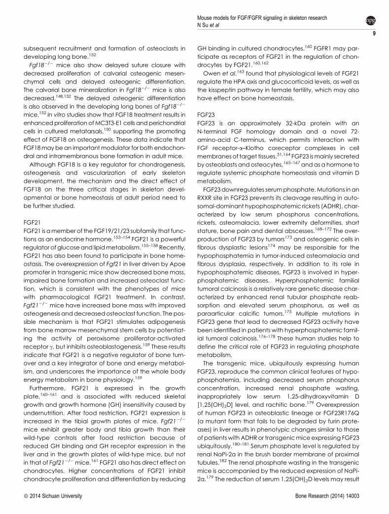
FGF23
FGF23 is an approximately 32-kDa protein with an
N-terminal FGF homology domain and a novel 72-
amino-acid C-terminus, which permits interaction with
FGF receptor-a–Klotho coreceptor complexes in cell
membranes of target tissues.31,164 FGF23 ismainly secreted
byosteoblasts andosteocytes,165–167 andasahormone to
regulate systemic phosphate homeostasis and vitamin D
metabolism.
FGF23downregulates serumphosphate.Mutations inan
RXXR site in FGF23 prevents its cleavage resulting in auto-
somal-dominant hypophosphatemic rickets (ADHR), char-
acterized by low serum phosphorus concentrations,
rickets, osteomalacia, lower extremity deformities, short
stature, bone pain and dental abscesses.168–172 The over-
production of FGF23 by tumors173 and osteogenic cells in
fibrous dysplastic lesions174 may be responsible for the
hypophosphatemia in tumor-induced osteomalacia and
fibrous dysplasia, respectively. In addition to its role in
hypophosphatemic diseases, FGF23 is involved in hyper-
phosphatemic diseases. Hyperphosphatemic familial
tumoral calcinosis is a relatively rare genetic disease char-
acterized by enhanced renal tubular phosphate reab-
sorption and elevated serum phosphorus, as well as
paraarticular calcific tumors.175 Multiple mutations in
FGF23 gene that lead to decreased FGF23 activity have
been identified in patients with hyperphosphatemic famil-
ial tumoral calcinosis.176–178 These human studies help to
define the critical role of FGF23 in regulating phosphate
metabolism.
The transgenic mice, ubiquitously expressing human
FGF23, reproduce the common clinical features of hypo-
phosphatemia, including decreased serum phosphorus
concentration, increased renal phosphate wasting,
inappropriately low serum 1,25-dihydroxyvitamin D
[1,25(OH)2D] level, and rachitic bone.179 Overexpression
of human FGF23 in osteoblastic lineage or FGF23R176Q
(a mutant form that fails to be degraded by furin prote-
ases) in liver results in phenotypic changes similar to those
of patientswith ADHRor transgenicmice expressing FGF23
ubiquitously.180–181 Serum phosphate level is regulated by
renal NaPi-2a in the brush border membrane of proximal
tubules.182 The renal phosphate wasting in the transgenic
mice is accompanied by the reduced expression of NaPi-
2a.179 The reduction of serum 1,25(OH)2D levels may result
Mouse models for FGF/FGFR signaling in skeleton researchN Su et al
9
� 2014 Sichuan University Bone Research (2014) 14003
from a significant decrease in renal mRNA level for 25-
hydroxyvitamin D-1a-hydroxylase (1a-OHase) and a sim-
ultaneous elevation of 24-hydroxylase mRNA, induced by
increased serum level of FGF23 (Figure 3).183
Consistently, Fgf23 knockout (Fgf232/2) mice have
opposite features including significantly increased serum
levels of phosphate, calcium and 1,25(OH)2D because of
the upregulated renal phosphate reabsorption and
enhanced expression of renal 1a-OHase, respectively.184
The Fgf232/2 mice also exhibit premature aging-like phe-
notypes including reduced lifespan, infertility, osteoporosis
and renal dysfunction.184 The elimination or reduction of
vitamin D activity from Fgf232/2 mice can rescue the pre-
mature aging-like features and ectopic calcifications.
These in vivo experimental data strongly support the very
essential roles of FGF23 in the regulation of phosphate
homeostasis, vitamin D activity and in the pathogenesis
of premature aging.185
Recent studies have indicated the regulation of iron on
FGF23. Reduced serum iron concentrations are strongly
correlated with increased serum FGF23 in ADHR
patients,186 and C-terminal FGF23 is negatively correlated
with ferritin.187 To investigate the effect of iron on the
development of the ADHR phenotype, R176Q-Fgf23
knock-in mice mimicking human ADHR are generated
and placed on control or low-iron diets.188–189 R176Q-
Fgf23 knock-in mice on low-iron diet have elevated intact
C-terminal Fgf23 with hypophosphatemic osteomalacia
and low serum 1,25(OH)2D. Iron chelation in vitro results in
a significantly increased Fgf23 mRNA level that depends
on MAPK signaling.189 However, the mechanism for the
regulation of FGF23 by iron is still unclear.
Increased FGF23 level is also found in patientswith hypo-
phosphatemic diseases including XLH and autosomal
dominant hypophosphatemic rickets (ARHR). XLH is
causedby inactivatingmutations in phosphate regulating
gene with homologies to endopeptidases on the X chro-
mosome (PHEX).190–191 Mice with ablation of Phex gene
(Hyp mice) have increased FGF23 expression and hypo-
phosphatemia.192 Both the serum phosphate levels and
skeletal changes in Hyp mice can be reversed by intro-
ducing Fgf23 null mutation into Hyp mice,166,193–194 indi-
cating that enhanced FGF23 level is responsible for the
hypophosphatemia in XLH patients and Hyp mice. The
increased FGF23 level is due to the improved Fgf23
expression, but not decreased degradation.165,194–195
ARHR results from missense mutations in DMP-1. Dmp1
knockout mice exhibit hypophosphatemic rickets and
osteomalacia similar to ARHR patients.196–197 Both Dmp1
null mice and patients with ARHR show elevated serum
FGF23 levels. Considering the role of FGF23 in ADHR and
other hypophosphatemic diseases, ARHR has been
proposed to be associated with excessive actions of
FGF23.
FGF23 also participates in some clinical pathological
processes, in addition to its role in genetic diseases. In
patients with chronic kidney disease (CKD), FGF23 level is
elevateddue to increased serumcalciumandphosphate
concentrations and PTH,31,198 and is associated with
increased FGF23 transcription in bone.199 Some research-
ers proposed that FGF23 might be an early biomarker for
earlier interventions in CKD.200 However, the reason for the
high serum levels of FGF23 in CKD is unclear. Furthermore,
elevated level of FGF23 in CKD patients have been linked
to greater risks of left ventricular hypertrophy (LVH).201–202
Using animal models, Faul et al.203 found that increased
level of FGF23 inmice resulted inpathological hypertrophy
of cardiomyocytes and LVH. To avoid redundancy and
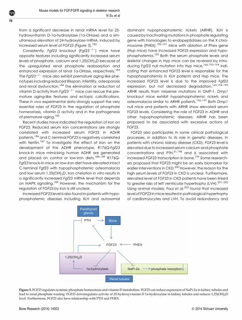
PTH
FGF23
1,25(OH)2D
1a-hydroxylase NaPi-2a
Renal tubules
Bone
Parathyroidglands
phosphate resorption
PHEX
Figure 3. FGF23 regulates systemic phosphate homeostasis and vitaminDmetabolism. FGF23 can reduce expression of NaPi-2a in kidney tubules andlead to renal phosphate wasting. FGF23 downregulates activity of 25-hydroxyvitamin D 1a-hydroxylase in kidney tubules and reduces 1,25(OH)2Dlevel. Furthermore, FGF23 also have relationship with PTH and PHEX.
Mouse models for FGF/FGFR signaling in skeleton research
N Su et al
10
Bone Research (2014) 14003 � 2014 Sichuan University
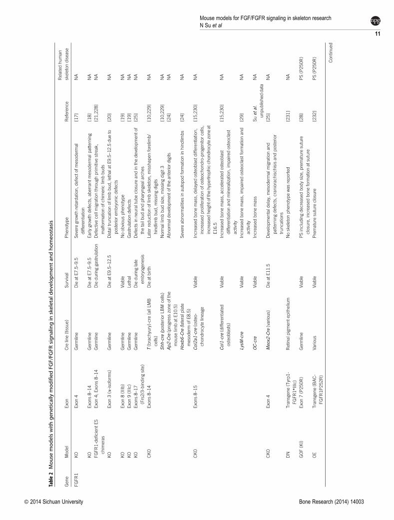
Table2
Mouse
modelswithgen
eticallymodifiedFG
F/FG
FRsignalingin
skeletaldevelopmen
tan
dhomeo
stasis
Gene
Model
Exon
Cre
line(tissue)
Survival
Phenotype
Reference
Relatedhuman
skeletondisease
FGFR1
KO
Exon4
Germ
line
DieatE7.5–9.5
Severe
growth
retardation,defectofmesoderm
al
differentiation
[17]
NA
KO
Exons8–14
Germ
line
DieatE7.5–9.5
Earlygrowth
defects,aberrantmesoderm
alp
atterning
[18]
NA
FGFR1-deficientES
chim
eras
Exon4;Exons8–14
Germ
line
Dieduringgastrulation
Defectivecellmigrationthroughprimitive
streak,
malform
ationofchim
ericlim
bbuds
[21,228]
NA
KO
Exon3(a-isoform
s)Germ
line
DieatE9.5–12.5
Distaltruncationoflim
bbud,lethalatE9.5–12.5
dueto
posteriorembryonicdefects
[20]
NA
KO
Exon8(IIIb)
Germ
line
Viable
Noobviousphenotype
[19]
NA
KO
Exon9(IIIc)
Germ
line
Lethal
Gastrulationdefects
[19]
NA
KO
Exons8–17
(Frs2/3-bindingsite)
Germ
line
Dieduringlate
embryogenesis
Defectsin
neuraltubeclosure
andin
thedevelopmentof
thetailbudandpharyngealarches
[25]
NA
CKO
Exons8–14
T(brachyury)-cre
(allLMB
cells)
Dieatbirth
Laterreductionoflim
bskeleton,misshapenforelim
b/
hindlim
bbud,missingdigits
[10,229]
NA
Shh-cre
(posteriorLBM
cells)
Norm
allim
bbudsize,missingdigit3
[10,229]
NA
Ap2-Cre
(progress
zoneofthe
mouse
limbatE10.5)
Abnorm
ald
evelopmentoftheanteriordigits
[24]
NA
Hoxb6-Cre
(lateralp
late
mesoderm
ofE8.5)
Severe
abnorm
alitiesin
autopodform
ationin
hindlim
bs
[24]
NA
CKO
Exons8–15
Col2a1
-cre
(osteo-
chondrocytelineage
Viable
Increasedbonemass,delayedosteoblastdifferentiation,
increasedproliferationofosteochondro-progenitorcells,
increasedheightofthehypertrophicchondrocytezoneat
E16.5
[15,230]
NA
Col1-cre
(differentiated
osteoblasts)
Viable
Increasedbonemass,acceleratedosteoblast
differentiationandmineralization,im
pairedosteoclast
activity
[15,230]
NA
LysM-cre
Viable
Increasedbonemass,im
pairedosteoclast
form
ationand
activity
[29]
NA
OC-cre
Viable
Increasedbonemass
Suet
al.
unpublisheddata
NA
CKO
Exon4
Meox2
-Cre
(various)
DieatE11.5
Developmentald
elay,mesoderm
alm
igrationand
patterningdefects,craniorachischisisandposterior
truncations
[25]
NA
DN
Transgene(Tyrp1-
FGFR1*IIIc)
Retin
alp
igmentepith
elium
Noskeletonphenotypewasreported
[231]
NA
GOF(KI)
Exon7(P250R)
Germ
line
Viable
PSincludingdecreasedbodysize,premature
suture
closure,increasedboneform
ationatsuture
[28]
PS(P250R)
OE
Transgene(BAC-
FGFR1P252R)
Various
Viable
Premature
suture
closure
[232]
PS(P250R)
Contin
ued
Mouse models for FGF/FGFR signaling in skeleton researchN Su et al
11
� 2014 Sichuan University Bone Research (2014) 14003
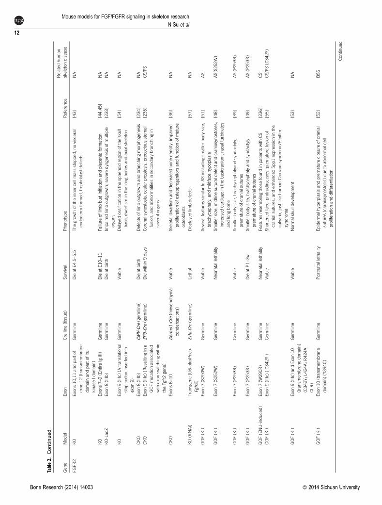
Table2.
Continued
Gene
Model
Exon
Cre
line(tissue)
Survival
Phenotype
Reference
Relatedhuman
skeletondisease
FGFR2
KO
Exons10,11andpartof
exon12(transm
embrane
domain
andpartofits
kinase
Idomain)
Germ
line
DieatE4.5–5.5
Thegrowth
oftheinnercellmass
stopped,novisceral
endoderm
form
ed,trophoblastdefects
[43]
NA
KO
Exons7–9(EntireIg
III)
Germ
line
DieatE10–11
Failure
oflim
bbudinitiationandplacenta
form
ation
[44,45]
NA
KO-LacZ
Exon8(IIIb)
Germ
line
Dieatbirth
Impairedlim
boutgrowth,severe
dysgenesisofmultiple
organs
[233]
NA
KO
Exon9(IIIc)(A
translational
stopcodoninsertedinto
exon9)
Germ
line
Viable
Delayedossificationin
thesphenoid
regionoftheskull
base,dwarfism
inthelongbonesandaxialskeleton
[54]
NA
CKO
Exon8(IIIb)
CMV-Cre
(germ
line)
Dieatbirth
Defectsoflim
boutgrowth
andbranchingmorphogenesis
[234]
NA
CKO
Exon9(IIIc)(Resultingin
a
GOFmutationassociated
with
exonsw
itchingwith
in
theFgfr2gene)
ZP3-Cre
(germ
line)
Diewith
in9days
Coronalsynostosis,
ocularproptosis,precocioussternal
fusion,andabnorm
alitiesin
secondary
branchingin
severalorgans
[235]
CS/PS
CKO
Exons8–10
Dermo1
-Cre
(mesenchym
al
condensations)
Viable
Skeletald
warfism
anddecreasedbonedensity,im
paired
proliferationofosteoprogenitors
andfunctionofmature
osteoblasts
[36]
NA
KD(RNAi)
Transgene(U
6-ploxP
neo-
Fgfr2)
EIIa-Cre
(germ
line)
Lethal
Displayedlim
bdefects
[57]
NA
GOF(KI)
Exon7(S250W)
Germ
line
Viable
Severalfeaturessimilarto
ASincludingsm
allerbodysize,
brachycephaly,andmidfacehypoplasia
[51]
AS
GOF(KI)
Exon7(S252W)
Germ
line
Neonatallethality
Smallersize,midlinesuturald
efectandcraniosynostoses,
increasedcartilagein
thebasicranium,nasalturbinates
andlongbone
[48]
AS(S252W)
GOF(KI)
Exon7(P253R)
Germ
line
Viable
Smallerbodysize,brachycephalyandsyndactyly,
premature
ofcranialsutures
[39]
AS(P253R)
GOF(KI)
Exon7(P253R)
Germ
line
DieatP1–3w
Smallerbodysize,brachycephalyandsyndactyly,
premature
ofcranialsutures
[49]
AS(P253R)
GOF(ENU-induced)
Exon7(W
290R)
Germ
line
Neonatallethality
Featuresresemblingthose
foundin
patientswith
CS
[236]
CS
GOF(KI)
Exon9(IIIc)(C342Y)
Germ
line
Viable
Shortenedface,protrudingeyes,premature
fusionof
cranialsutures,andenhancedSpp1expressionin
the
calvaria,justlikehumanCrouzonsyndrome/Pfeiffer
syndrome
[55]
CS/PS(C342Y)
GOF(KI)
Exon9(IIIc)andExon10
(transm
embranedomain)
(C342Y;L424A;R424A,
CLR)
Germ
line
Viable
Norm
alskulldevelopment
[53]
NA
GOF(KI)
Exon10(transm
embrane
domain)(Y394C)
Germ
line
Postnatallethality
Epiderm
alh
yperplasiaandpremature
closure
ofcranial
sutures(craniosynostosis)
dueto
abnorm
alcell
proliferationanddifferentiation
[52]
BSS
Contin
ued
Mouse models for FGF/FGFR signaling in skeleton research
N Su et al
12
Bone Research (2014) 14003 � 2014 Sichuan University

Table2.
Continued
Gene
Model
Exon
Cre
line(tissue)
Survival
Phenotype
Reference
Relatedhuman
skeletondisease
FGFR3
KO
Exon5
Germ
line
Viable
Boneovergrowth,decreasedbonemass
[79]
CATSHLsyndrome
KO
From
Ig-likedomain
IIto
thetransm
embrane
domain
Germ
line
Viable
Boneovergrowth,defectivebonemineralizationand
osteopenia,earlyarthritis,deafness
[59,85,237]
CATSHLsyndrome
KO(a
stopcodon
inserted)
Exon8(IIIb)
Germ
line
Viable
Noobviousphenotype
[238]
NA
KO(a
stopcodon
inserted)
Exon9(IIIc)
Germ
line
Viable
Skeletalovergrowth,decreasedboneminerald
ensity
[238]
NA
CKO
Exons9–10
EIIa-Cre
Viable
Increasedlength
oflongboneanddecreasedbonemineral
density
[239]
NA
GOF(KI)
Exon7(P244R)
Germ
line
Viable
Abnorm
alcraniofacialm
orphology
[240]
MS(P250R)
GOF(KI)
Exon9(Y367C)
Germ
line
Viable
(dieat6–8weeks
afterbirth)
Skeletald
ysplasiamore
severe
thanACH
[241]
TDI(Y373C)
GOF(KI)
Exon10(S365C)
Germ
line
Viable
Skeletald
ysplasiamore
severe
thanACH
[72]
TDI
GOF(KI)
Exon10(G369C)
Germ
line
Viable
Macrocephalyandshortenedlim
bsdueto
retarded
endochondralb
onegrowth
andpremature
closure
of
cranialb
ase
synchondroses
[70]
ACH(G375R)
GOF(KI)
Exon10(G374R)
Germ
line
Viable
Smallsize,shorttail,macrocephalyanddome-shaped
heads,thenarrowerepiphysealgrowth
platesand
decreasedhypertrophicchondrocytezone
[67,68]
ACH(G380R)
GOF(KI)
K644EcDNAknock-in
Germ
line
Viable
Retardationofbonegrowth,macrocephalyandshortening
ofthelongbonesresemblingACHpatients
[73]
ACH
GOF(KI)
Exon15(K644E)
Germ
line
Neonatallethality
Diewith
infewhours
afterbirth,skeletald
ysplasiamore
severe
thanACH
[71]
TDII(K650E)
GOF(KI)
Exon15(K644M)
Germ
line
Viable
Acanthosisnigricansandanomaliesin
centraln
ervous
system
inadditionto
severe
skeletald
ysplasia
[242]
SADDAN
OE
Transgene(Col2-G374R)
Chondrocyte
Viable
Micearedwarfed,w
ithaxial,appendicularandcraniofacial,
skeletalh
ypoplasia
[80]
ACH
OE
Transgene(FGFR3-
hG380R)
Germ
line
Viable
Disproportionate
dwarfism
similarto
those
ofhuman
achondroplasia
[243]
ACH
FGFR4
KO
Exon6(IgII)
Germ
line
Viable
Morphologically
norm
al,noobviousdefectsin
skeleton
[93]
NA
KI
Exon8(G385R)
Germ
line
Viable
Skeletonphenotypenotreported
[244]
NA
FGFR3/
FGFR4
DoubleKO
Germ
line
Viable
Neonatalgrowth
retardation,lungabnorm
alities
[93]
NA
FGF1
KO
Exon1
Germ
line
Viable
Noobviousphenotype
[206]
NA
Contin
ued
Mouse models for FGF/FGFR signaling in skeleton researchN Su et al
13
� 2014 Sichuan University Bone Research (2014) 14003
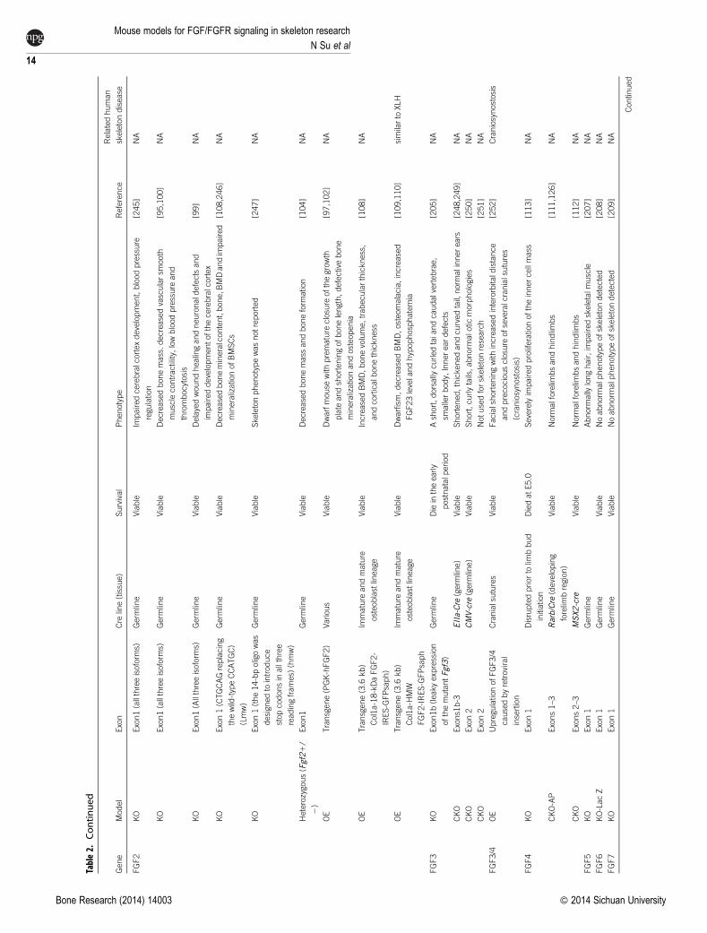
Table2.
Continued
Gene
Model
Exon
Cre
line(tissue)
Survival
Phenotype
Reference
Relatedhuman
skeletondisease
FGF2
KO
Exon1(allthreeisoform
s)Germ
line
Viable
Impairedcerebralcortexdevelopment,bloodpressure
regulation
[245]
NA
KO
Exon1(allthreeisoform
s)Germ
line
Viable
Decreasedbonemass.decreasedvascularsm
ooth
musclecontractility,lowbloodpressure
and
thrombocytosis
[95,100]
NA
KO
Exon1(Allthreeisoform
s)Germ
line
Viable
Delayedwoundhealingandneuronald
efectsand
impaireddevelopmentofthecerebralcortex
[99]
NA
KO
Exon1(CTGCAGreplacing
thewild-typeCCATGC)
(Lmw)
Germ
line
Viable
Decreasedbonemineralcontent,bone,B
MDandim
paired
mineralizationofBMSCs
[108,246]
NA
KO
Exon1(the14-bpoligowas
designedto
introduce
stopcodonsin
allthree
readingframes)
(hmw)
Germ
line
Viable
Skeletonphenotypewasnotreported
[247]
NA
Heterozygous(Fgf21/
2)
Exon1
Germ
line
Viable
Decreasedbonemass
andboneform
ation
[104]
NA
OE
Transgene(PGK-hFGF2)
Various
Viable
Dwarfmouse
with
premature
closure
ofthegrowth
plate
andshorteningofbonelength,defectivebone
mineralizationandosteopenia
[97,102]
NA
OE
Transgene(3.6
kb)
Col1a-18-kDaFGF2-
IRES-GFPsaph)
Immature
andmature
osteoblastlineage
Viable
IncreasedBMD,bonevolume,trabecularthickness,
andcorticalb
onethickness
[108]
NA
OE
Transgene(3.6
kb)
Col1a-H
MW
FGF2-IRES-GFPsaph
Immature
andmature
osteoblastlineage
Viable
Dwarfism,decreasedBMD,osteomalacia,increased
FGF23levelandhypophosphatemia
[109,110]
similarto
XLH
FGF3
KO
Exon1b(leakyexpression
ofthemutantFgf3)
Germ
line
Diein
theearly
postnatalp
eriod
Ashort,dorsally
curledtaiandcaudalvertebrae,
smallerbody,Innereardefects
[205]
NA
CKO
Exons1b-3
EIIa-Cre
(germ
line)
Viable
Shortened,thickenedandcurvedtail,norm
alinnerears
[248,249]
NA
CKO
Exon2
CMV-cre
(germ
line)
Viable
Short,curlytails,abnorm
alotic
morphologies
[250]
NA
CKO
Exon2
Notusedforskeletonresearch
[251]
NA
FGF3/4
OE
UpregulationofFGF3/4
causedbyretroviral
insertion
Cranialsutures
Viable
Facialshorteningwith
increasedinterorbitald
istance
andprecociousclosure
ofseveralcranialsutures
(craniosynostosis)
[252]
Craniosynostosis
FGF4
KO
Exon1
Disruptedpriorto
limbbud
initiation
DiedatE5.0
Severelyim
pairedproliferationoftheinnercellmass
[113]
NA
CKO-AP
Exons1–3
Rarb/Cre
(developing
forelim
bregion)
Viable
Norm
alforelim
bsandhindlim
bs
[111,126]
NA
CKO
Exons2–3
MSX2-cre
Viable
Norm
alforelim
bsandhindlim
bs
[112]
NA
FGF5
KO
Exon1
Germ
line
Abnorm
ally
longhair,im
pairedskeletalm
uscle
[207]
NA
FGF6
KO-LacZ
Exon1
Germ
line
Viable
Noabnorm
alp
henotypeofskeletondetected
[208]
NA
FGF7
KO
Exon1
Germ
line
Viable
Noabnorm
alp
henotypeofskeletondetected
[209]
NA
Contin
ued
Mouse models for FGF/FGFR signaling in skeleton research
N Su et al
14
Bone Research (2014) 14003 � 2014 Sichuan University
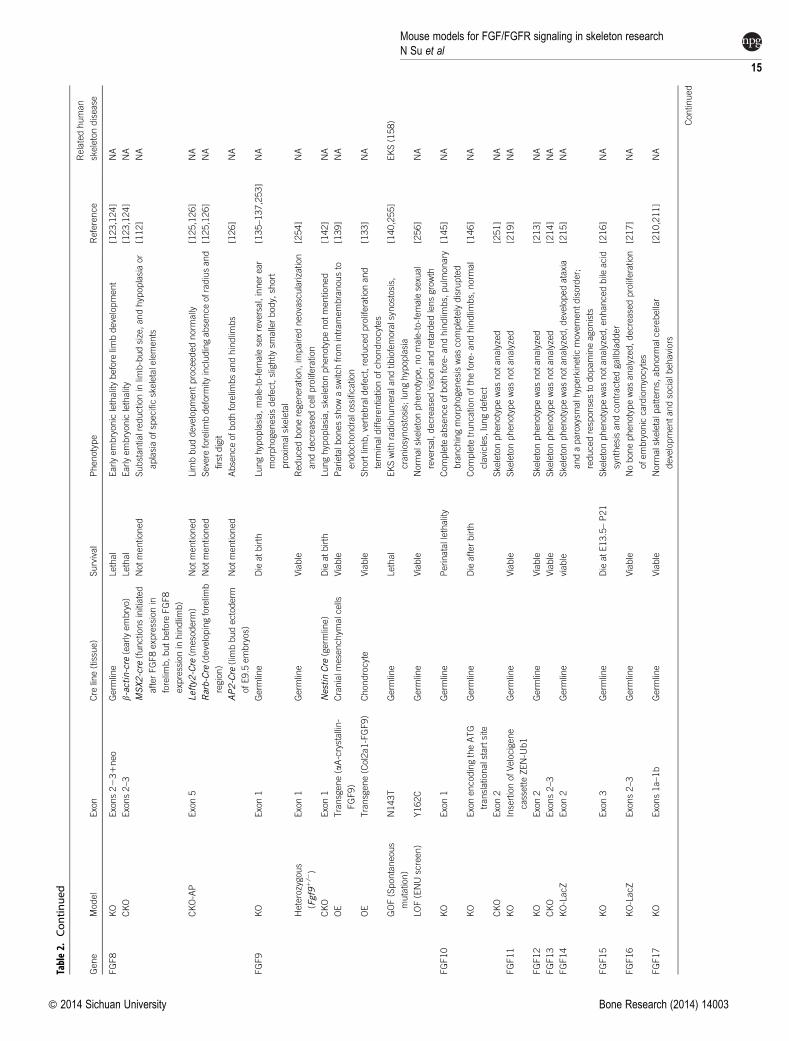
Table2.
Continued
Gene
Model
Exon
Cre
line(tissue)
Survival
Phenotype
Reference
Relatedhuman
skeletondisease
FGF8
KO
Exons2231neo
Germ
line
Lethal
Earlyembryoniclethalitybefore
limbdevelopment
[123,124]
NA
CKO
Exons2–3
b-actin-cre
(earlyembryo)
Lethal
Earlyembryoniclethality
[123,124]
NA
MSX2-cre
(functionsinitiated
afterFGF8expressionin
forelim
b,butbefore
FGF8
expressionin
hindlim
b)
Notmentioned
Substantialreductionin
limb-budsize,andhypoplasiaor
aplasiaofspecificskeletalelements
[112]
NA
CKO-AP
Exon5
Lefty2-Cre
(mesoderm
)Notmentioned
Lim
bbuddevelopmentproceedednorm
ally
[125,126]
NA
Rarb-Cre
(developingforelim
b
region)
Notmentioned
Severe
forelim
bdeform
ityincludingabsenceofradiusand
firstdigit
[125,126]
NA
AP2-Cre
(lim
bbudectoderm
ofE9.5
embryos)
Notmentioned
Absenceofboth
forelim
bsandhindlim
bs
[126]
NA
FGF9
KO
Exon1
Germ
line
Dieatbirth
Lunghypoplasia,male-to-femalesexreversal,innerear
morphogenesisdefect,slightly
smallerbody,short
proximalskeletal
[135–137,253]
NA
Heterozygous
(Fgf91/2)
Exon1
Germ
line
Viable
Reducedboneregeneration,im
pairedneovascularization
anddecreasedcellproliferation
[254]
NA
CKO
Exon1
NestinCre
(germ
line)
Dieatbirth
Lunghypoplasia,skeletonphenotypenotmentioned
[142]
NA
OE
Transgene(aA-crystallin-
FGF9)
Cranialm
esenchym
alcells
Viable
Parietalb
onesshowasw
itchfrom
intramembranousto
endochondralossification
[139]
NA
OE
Transgene(Col2a1-FGF9)
Chondrocyte
Viable
Shortlim
b,vertebrald
efect,reducedproliferationand
term
inald
ifferentiationofchondrocytes
[133]
NA
GOF(Spontaneous
mutation)
N143T
Germ
line
Lethal
EKSwith
radiohumeralandtib
iofemoralsynostosis,
craniosynostosis;
lunghypoplasia
[140,255]
EKS(158)
LOF(ENUscreen)
Y162C
Germ
line
Viable
Norm
alskeletonphenotype,nomale-to-femalesexual
reversal,decreasedvisionandretardedlensgrowth
[256]
NA
FGF10
KO
Exon1
Germ
line
Perinatallethality
Complete
absenceofboth
fore-andhindlim
bs,pulm
onary
branchingmorphogenesiswascompletelydisrupted
[145]
NA
KO
ExonencodingtheATG
translationalstartsite
Germ
line
Dieafterbirth
Complete
truncationofthefore-andhindlim
bs,norm
al
clavicles,lungdefect
[146]
NA
CKO
Exon2
Skeletonphenotypewasnotanalyzed
[251]
NA
FGF11
KO
InsertionofVelocigene
cassetteZEN-U
b1
Germ
line
Viable
Skeletonphenotypewasnotanalyzed
[219]
NA
FGF12
KO
Exon2
Germ
line
Viable
Skeletonphenotypewasnotanalyzed
[213]
NA
FGF13
CKO
Exons2–3
Viable
Skeletonphenotypewasnotanalyzed
[214]
NA
FGF14
KO-LacZ
Exon2
Germ
line
viable
Skeletonphenotypewasnotanalyzed,developedataxia
andaparoxysm
alh
yperkinetic
movementdisorder;
reducedresponsesto
dopamineagonists
[215]
NA
FGF15
KO
Exon3
Germ
line
DieatE13.5–P21
Skeletonphenotypewasnotanalyzed,enhancedbile
acid
synthesisandcontractedgallbladder
[216]
NA
FGF16
KO-LacZ
Exons2–3
Germ
line
Viable
Nobonephenotypewasanalyzed,decreasedproliferation
ofembryoniccardiomyocytes
[217]
NA
FGF17
KO
Exons1a–1b
Germ
line
Viable
Norm
alskeletalp
atterns,
abnorm
alcerebellar
developmentandsocialb
ehaviors
[210,211]
NA
Contin
ued
Mouse models for FGF/FGFR signaling in skeleton researchN Su et al
15
� 2014 Sichuan University Bone Research (2014) 14003

Table2.
Continued
Gene
Model
Exon
Cre
line(tissue)
Survival
Phenotype
Reference
Relatedhuman
skeletondisease
FGF18
KO
Exon3
Germ
line
Diejustbefore
or
atbirth
Impairedossificationandincreasedchondrocyte
proliferation,decreasedalveolarspacesin
thelung
[148,257]
NA
KO-LacZ
Exon1
Germ
line
Dieafterbirth
Impairedossificationandincreasedchondrocyte
proliferation,respiratory
failure
[147,152]
NA
Heterozygous
Exon3
Germ
line
Viable
Reducedboneregeneration
[258]
NA
CKO
Exon1
Germ
line
Evennotbeusedin
bonedevelopment
[259]
NA
FGF20
KO-LacZ
Exon1
Germ
line
Viable
Nobonephenotypewasanalyzed,deafness
[218]
NA
FGF21
KO
Exons2,partofexon1
andexon3
Germ
line
Viable
Greaterbodyandtib
iagrowth
afterfoodrestriction
[157,161]
NA
KO(LacZ)
Exons1–3
Germ
line
Viable
Skeletonphenotypewasnotreported
[156]
NA
CKO
Exons1–3
Meox-cre(Germ
line)
Viable
Increasedbonemass,metabolic
defectsincluding
decreasedcirculatin
gglucose
levelandoxygen
consumption
[155,159]
NA
OE
Transgene(Apoe-FGF21)
Liver
Viable
Decreasedbonemass,increasedosteoblastandbone
resorption
[158,159]
NA
OE
Transgene(Apoe-hFGF21)
Liver
Viable
Skeletonphenotypewasnotreported
[260]
NA
FGF22
KO
Exon1andpartofExon2
Germ
line
Viable
Norm
alskeletalp
atterns;decreasedsusceptib
ility
to
pharm
acologically
inducedseizures
[212]
NA
KO
Exons1–3
Germ
line
Viable
Norm
alskeletalp
atterns;decreasedincidenceoftumors
bychemicalinduction
[261]
NA
FGF23
KO
Exon1
Germ
line
Viable
Increasedserum
levelsofphosphate,calcium
and
1,25(OH) 2D,severe
growth
retardationwith
abnorm
al
bonephenotype
[184]
NA
KO-Lacz
Exons1–3
Germ
line
Viable
Hyperphosphatemiaandim
pairedskeletogenesis
[193]
NA
KO-eGFP
Exon1
Germ
line
Viable
Hyperphosphatemiaandim
pairedskeletogenesis
[166]
NA
OE
Transgene(CAG-hFGF23)
Various
Viable
Hypophosphatemia,lowserum
1,25(OH) 2Dlevel,and
rachiticbone,growth
retardation
[179]
ADHR
OE
Transgene(Col1a-hFGF23)
Osteoblasticlineage
Viable
Smallerbody,decreasedserum
phosphateconcentrations,
lowserum
1,25(OH) 2Dlevel
[180]
ADHR
OE
Transgene(Apoe3-
hFGF23*R176Q)
Liver
Viable
Hypophosphatemia,lowserum
1,25(OH) 2Dleveland
rachiticbone
[181]
ADHR
GOF(KI)
Knockin
(R176Q-hFGF23)
Germ
line
Viable
IncreasedserumlevelofFGF23,h
ypophosphatemiandlow
serum
1,25(OH) 2vitamin
Dafterreceivinglow-irondiets
[188,189]
ADHR
Abbreviations:
ACH,achon
droplasia;
ADHR,au
tosomal
dom
inan
thypop
hosphatem
icrickets;
AS,Apertsyndrome;
BSS,Beare–S
tevensoncu
tisgyrata
syndrome;
CATSHL,camptodactyly,tallstature
andhearingloss;CKO,
conditionalkn
ockout;CS,C
rouzonsyndrome;
EKS,e
lbow
knee
synostosis;GOF,gainof
function;K
D,knockd
own;K
I,kn
ock-in;K
O,knockout;LMB,lim
bbudmesen
chym
e;LOF,lossof
function;M
S,M
uen
kesyndrome;
NA,n
ot
applicable;OE,overexpression;PS,Pfeiffersyndrome;
SADDAN,severe
achon
droplasiawithdevelop
men
taldelay
andacan
thosis
nigricans;
TD,thanatop
horic
dysplasia;
XLH,X-linkedhypop
hosphatem
ia.
Mouse models for FGF/FGFR signaling in skeleton research
N Su et al
16
Bone Research (2014) 14003 � 2014 Sichuan University
give full attention of the exciting results from FGF23 studies,
we encourage you to read the recently published review
by Quarles and Bhattacharyya.31,204
OTHER FGFSIn addition to the FGFs mentioned above, the roles of
majority of these 22 FGFs are not defined in skeleton
development and homeostasis. Researchers have gener-
ated knockout or CKO mouse models of these FGFs
(Table 2). Fgf3 knockoutmice showa short, dorsally curled
tail, caudal vertebrae and smaller body.205 Some mouse
models show normal skeleton phenotypes, such as mice
lacking FGF1, FGF5, FGF6, FGF7, FGF17 or FGF22.206–212 The
skeleton phenotypes of knockout mice lacking FGF11–
FGF16, or FGF20, are still not analyzed.213–219 The effect of
these FGFs onbonedevelopment or homeostasis needbe
further studied.
CONCLUSIONSStudies in human patients and mouse models with FGFs/
FGFRsmutations have shown important roles of FGF signal-
ing in skeletal development, genetic skeletal diseases and
bone homeostasis. So, FGF/FGFR signalings will be attract-
ive targets for treating bone related diseases. FGF/FGFR
signals control the balance among skeletal cell growth,
differentiation and apoptosis during development and
adult homeostasis, as well as regulate systemic phosphate
homeostasis. However,manyunresolved issues still need to
be explored.
Many studies have investigated the role of FGFRs in
endochondral and intramembranous bone formation
during development, but the effects of FGFRs on osteo-
clasts, especially on osteocytes, have not be clarified.
Osteocytes are themost abundant and longest-living cells
in the adult skeleton and have essential roles in bone
homeostasis.220–221 Thus, uncovering the impact of FGFRs
on osteocytes using osteocyte-specific Cremice is critical.
Compensation, or crosstalk, may occur between differ-
ent FGFRs during skeleton development. For example,
conditional knock out of Fgfr1 in mature osteoblasts leads
to increased FGFR3 expression,15 whereas both cultured
bone marrow stromal cells from Fgfr3 null mice, or mice
carrying gain-of-function mutation in FGFR3, have
increased expression of FGFR1.83,85 Crossing between
mouse strains harboring various FGFRs mutations is extre-
mely important to elucidate the interactions between dif-
ferent FGFRs.
So far, only part of the 22 known FGF ligands have been
shown to be essential for skeletal development, such as
FGF8, FGF9 and FGF10. However, the mechanisms remain
unclear because most of the knockout mice die before or
after birth. Conditional deletion of these FGFs using bone
cell-specificCremice is necessary to study their roles during
bone development. The function of other unexplored FGFs
in skeletogenesis remains to be discovered. Furthermore,
which FGFRs are the relatively specific receptors of these
unexplored FGFs during bone development and metabo-
lism are unknown. Crossing mouse strains harboring differ-
ent FGFs mutations with FGFRs mutant mouse models is
necessary to discover the interactions between FGFs and
FGFRs in skeleton development and homeostasis.
Recently, studies have indicated that the bone is closely
related with whole-organism physiology.222 For example,
bone can regulate energy metabolism, male reproduc-
tion and hematopoiesis.223–224 Some hormone secreted
fromother organs or tissues also haveeffect onbone, such
as Leptin secreted by adipocyte.222 In addition, systemic
disease also influence skeleton such as CDK225 and
inflammatory disease.226–227 The roles of FGF signaling in
the effect of systemic diseases on bone or bone on
whole-organism physiology remain unclear and need fur-
ther exploration.
Conflict of InterestThe authors declare no conflict of interest.
AcknowledgementsThe work is supported by grants from the National Natural ScienceFoundation of China (81030036, 81270012, 81170809), the Special Funds forMajor State Basic Research Program of China (973 Program) (2014CB942904)and the Committee of Science and Technology of Chongqing (CSTC2011jjA1468), the foundation from national key laboratory (SKLZZ201017).In addition, we apologized to those whose work could not be cited due tospace constraints.
References1 Karsenty G, Wagner EF. Reaching a genetic and molecular
understanding of skeletal development. Dev Cell 2002; 2: 389–406.2 Su N, Du X, Chen L. FGF signaling: its role in bone development and
human skeleton diseases. Front Biosci 2008; 13: 2842–2865.3 Chen L, Deng CX. Roles of FGF signaling in skeletal development and
human genetic diseases. Front Biosci 2005; 10: 1961–1976.4 Johnson DE,Williams LT. Structural and functional diversity in the FGF
receptor multigene family. Adv Cancer Res 1993; 60: 1–41.5 Ornitz DM, Xu J, Colvin JS et al. Receptor specificity of the fibroblast
growth factor family. J Biol Chem 1996; 271: 15292–15297.6 Powers CJ, McLeskey SW, Wellstein A. Fibroblast growth factors, their
receptors and signaling. Endocrine Relat Cancer 2000; 7: 165–197.7 Ornitz DM. FGF signaling in the developing endochondral skeleton.
Cytokine Growth Factor Rev 2005; 16: 205–213.8 Yamaguchi TP, Conlon RA, Rossant J. Expression of the fibroblast
growth factor receptor FGFR-1/flg during gastrulation andsegmentation in the mouse embryo. Dev Biol 1992; 152: 75–88.
9 Peters KG,Werner S, Chen G,Williams LT. Two FGF receptor genes aredifferentially expressed in epithelial and mesenchymal tissues duringlimb formation and organogenesis in the mouse.Development 1992; 114:233–243.
10 Verheyden JM, Lewandoski M, Deng C, Harfe BD, Sun X. Conditionalinactivation of Fgfr1 inmouse defines its role in limb bud establishment,outgrowth and digit patterning. Development 2005; 132: 4235–4245.
Mouse models for FGF/FGFR signaling in skeleton researchN Su et al
17
� 2014 Sichuan University Bone Research (2014) 14003
11 Lazarus JE, Hegde A, Andrade AC, Nilsson O, Baron J. Fibroblastgrowth factor expression in the postnatal growth plate. Bone 2007; 40:577–586.
12 Szebenyi G, Savage MP, Olwin BB, Fallon JF. Changes in the expressionof fibroblast growth factor receptors mark distinct stages ofchondrogenesis in vitro and during chick limb skeletal patterning. DevDyn 1995; 204: 446–456.
13 Iseki S, Wilkie AO, Morriss-Kay GM. Fgfr1 and Fgfr2 have distinctdifferentiation- and proliferation-related roles in the developingmouse skull vault. Development 1999; 126: 5611–5620.
14 Xiao L, Naganawa T, Obugunde E et al. Stat1 controls postnatal boneformation by regulating fibroblast growth factor signaling inosteoblasts. J Biol Chem 2004; 279: 27743–27752.
15 Jacob AL, Smith C, Partanen J, Ornitz DM. Fibroblast growth factorreceptor 1 signaling in the osteo-chondrogenic cell lineage regulatessequential steps of osteoblast maturation. Dev Biol2006; 296: 315–328
16 Kyono A, Avishai N, Ouyang Z, Landreth GE, Murakami S. FGF andERK signaling coordinately regulate mineralization-related genes andplay essential roles in osteocyte differentiation. J Bone Miner Metab 2012;30: 19–30.
17 DengCX,Wynshaw-Boris A, ShenMM,DaughertyC,OrnitzDM, LederP. Murine FGFR-1 is required for early postimplantation growth andaxial organization. Genes Dev 1994; 8: 3045–3057.
18 Yamaguchi TP, Harpal K, Henkemeyer M, Rossant J. fgfr-1 is requiredfor embryonic growth and mesodermal patterning during mousegastrulation. Genes Dev 1994; 8: 3032–3044.
19 Partanen J, Schwartz L, Rossant J. Opposite phenotypes of hypomorphicand Y766 phosphorylation site mutations reveal a function for Fgfr1 inanteroposterior patterning ofmouse embryos.Genes Dev 1998; 12: 2332–2344.
20 Xu X, Li C, Takahashi K, Slavkin HC, Shum L, Deng CX. Murinefibroblast growth factor receptor 1alpha isoforms mediate noderegression and are essential for posterior mesoderm development. DevBiol 1999; 208: 293–306.
21 Deng C, Bedford M, Li C et al. Fibroblast growth factor receptor-1(FGFR-1) is essential for normal neural tube and limb development.Dev Biol 1997; 185: 42–54.
22 Perantoni AO, Timofeeva O, Naillat F et al. Inactivation of FGF8 in earlymesoderm reveals an essential role in kidney development.Development2005; 132: 3859–3871.
23 Harfe BD, Scherz PJ,Nissim S, TianH,McMahonAP, TabinCJ. Evidencefor an expansion-based temporal Shh gradient in specifying vertebratedigit identities. Cell 2004; 118: 517–528.
24 Li C, Xu X, Nelson DK, Williams T, Kuehn MR, Deng CX. FGFR1function at the earliest stages of mouse limb development plays anindispensable role in subsequent autopod morphogenesis.Development 2005; 132: 4755–4764.
25 Hoch RV, Soriano P. Context-specific requirements for Fgfr1 signalingthrough Frs2 and Frs3 during mouse development. Development 2006;133: 663–673.
26 WhiteKE, Cabral JM,Davis SI et al.Mutations that cause osteoglophonicdysplasia define novel roles for FGFR1 in bone elongation. Am J HumGenet 2005; 76: 361–367.
27 Roscioli T, Flanagan S, Kumar P et al. Clinical findings in a patient withFGFR1 P252R mutation and comparison with the literature. Am J MedGenet 2000; 93: 22–28.
28 Zhou YX, Xu X, Chen L, Li C, Brodie SG, Deng CX. A Pro250Argsubstitution in mouse Fgfr1 causes increased expression of Cbfa1 andpremature fusion of calvarial sutures. Hum Mol Genet 2000; 9:2001–2008.
29 Lu X, SuN, Yang J et al. Fibroblast growth factor receptor 1 regulates thedifferentiation and activation of osteoclasts through Erk1/2 pathway.Biochem Biophys Res Commun 2009; 390: 494–499.
30 YuX,White KE. Fibroblast growth factor 23 and its receptors.Ther ApherDial 2005; 9: 308–312.
31 Quarles LD. Skeletal secretion of FGF-23 regulates phosphate andvitamin D metabolism. Nat Rev Endocrinol 2012; 8: 276–286.
32 Wohrle S, Bonny O, Beluch N et al. FGF receptors control vitamin D andphosphate homeostasis by mediating renal FGF-23 signaling andregulating FGF-23 expression in bone. J Bone Miner Res 2011; 26: 2486–2497.
33 Peng H, Myers J, Fang X et al. Integrative nuclear FGFR1 signaling(INFS) pathway mediates activation of the tyrosine hydroxylase geneby angiotensin II, depolarization andprotein kinaseC. JNeurochem 2002;81: 506–524.
34 Delezoide AL, Benoist-Lasselin C, Legeai-Mallet L et al. Spatio-temporalexpression of FGFR 1, 2 and 3 genes during human embryo-fetalossification. Mech Dev 1998; 77: 19–30.
35 Orr-Urtreger A, Bedford MT, Burakova T et al. Developmentallocalization of the splicing alternatives of fibroblast growth factorreceptor-2 (FGFR2). Dev Biol 1993; 158: 475–486.
36 Yu K, Xu J, Liu Z et al.Conditional inactivation of FGF receptor 2 revealsan essential role for FGF signaling in the regulation of osteoblastfunction and bone growth. Development 2003; 130: 3063–3074.
37 Rice DP, Rice R, Thesleff I. Fgfr mRNA isoforms in craniofacial bonedevelopment. Bone 2003; 33: 14–27.
38 RiceDP,Aberg T,ChanY et al. Integration of FGFandTWIST in calvarialbone and suture development. Development 2000; 127: 1845–1855.
39 Yin L, Du X, Li C et al.APro253Argmutation in fibroblast growth factorreceptor 2 (Fgfr2) causes skeleton malformation mimicking humanApert syndrome by affecting both chondrogenesis and osteogenesis.Bone 2008; 42: 631–643.
40 Wilkie AO. Bad bones, absent smell, selfish testes: the pleiotropicconsequences of human FGF receptor mutations. Cytokine GrowthFactor Rev 2005; 16: 187–203.
41 Britto JA, Chan JC, Evans RD, Hayward RD, Thorogood P, Jones BM.Fibroblast growth factor receptors are expressed in craniosynostoticsutures. Plast Reconstr Surg 1998; 101: 540–543.
42 Britto JA, Evans RD, Hayward RD, Jones BM. From genotype tophenotype: the differential expression of FGF, FGFR, and TGFbetagenes characterizes human cranioskeletal development and reflectsclinical presentation in FGFR syndromes. Plast Reconstr Surg 2001; 108:2026–2039; discussion 2040–2026..
43 Arman E, Haffner-Krausz R, Chen Y, Heath JK, Lonai P. Targeteddisruption of fibroblast growth factor (FGF) receptor 2 suggests a rolefor FGF signaling in pregastrulationmammalian development.ProcNatlAcad Sci USA 1998; 95: 5082–5087.
44 Xu X, Weinstein M, Li C et al. Fibroblast growth factor receptor 2(FGFR2)-mediated reciprocal regulation loop between FGF8and FGF10 is essential for limb induction. Development 1998; 125:753–765.
45 Li X, Chen Y, Scheele S et al. Fibroblast growth factor signaling andbasement membrane assembly are connected during epithelialmorphogenesis of the embryoid body. J Cell Biol 2001; 153: 811–822.
46 Marie PJ, Coffin JD, Hurley MM. FGF and FGFR signaling inchondrodysplasias and craniosynostosis. J Cell Biochem 2005; 96: 888–896.
47 Cunningham ML, Seto ML, Ratisoontorn C, Heike CL, Hing AV.Syndromic craniosynostosis: from history to hydrogen bonds. OrthodCraniofac Res 2007; 10: 67–81.
Mouse models for FGF/FGFR signaling in skeleton research
N Su et al
18
Bone Research (2014) 14003 � 2014 Sichuan University

48 Wang Y, Xiao R, Yang F et al. Abnormalities in cartilage and bonedevelopment in the Apert syndrome FGFR2(1/S252W) mouse.Development 2005; 132: 3537–3548.
49 Wang Y, Sun M, Uhlhorn VL et al. Activation of p38 MAPK pathway inthe skull abnormalities of Apert syndrome Fgfr2(1P253R) mice. BMC Dev
Biol 2010; 10: 22.50 Kreiborg S, Aduss H, Cohen MM Jr. Cephalometric study of the Apert
syndrome in adolescence and adulthood. J Craniofac Genet Dev Biol 1999;19: 1–11.
51 Chen L, Li D, Li C, Engel A, Deng CX. A Ser252Trp [corrected]substitution in mouse fibroblast growth factor receptor 2 (Fgfr2)results in craniosynostosis. Bone 2003; 33: 169–178.
52 WangY,ZhouX,Oberoi K et al.p38 Inhibition ameliorates skin and skullabnormalities in Fgfr2 Beare–Stevenson mice. J Clin Invest 2012; 122:2153–2164.
53 Eswarakumar VP, Ozcan F, Lew ED et al. Attenuation of signalingpathways stimulated by pathologically activated FGF-receptor 2mutants prevents craniosynostosis. Proc Natl Acad Sci USA 2006; 103:18603–18608.
54 Eswarakumar VP, Monsonego-Ornan E, Pines M, Antonopoulou I,Morriss-Kay GM, Lonai P. The IIIc alternative of Fgfr2 is a positiveregulator of bone formation. Development 2002; 129: 3783–3793.
55 Eswarakumar VP, Horowitz MC, Locklin R, Morriss-Kay GM, Lonai P.A gain-of-function mutation of Fgfr2c demonstrates the roles of thisreceptor variant in osteogenesis. Proc Natl Acad Sci USA 2004; 101:12555–12560.
56 Shukla V, Coumoul X, Wang RH, Kim HS, Deng CX. RNA interferenceand inhibition of MEK–ERK signaling prevent abnormal skeletalphenotypes in a mouse model of craniosynostosis. Nat Genet 2007; 39:1145–1150.
57 Coumoul X, Shukla V, Li C, Wang RH, Deng CX. Conditionalknockdown of Fgfr2 in mice using Cre-LoxP induced RNAinterference. Nucleic Acids Res 2005; 33: e102.
58 Peters K, Ornitz D, Werner S, Williams L. Unique expression pattern ofthe FGF receptor 3 gene duringmouse organogenesis.Dev Biol 1993; 155:423–430.
59 Colvin JS, Bohne BA, Harding GW, McEwen DG, Ornitz DM. Skeletalovergrowth and deafness in mice lacking fibroblast growth factorreceptor 3. Nat Genet 1996; 12: 390–397.
60 Ornitz DM, Marie PJ. FGF signaling pathways in endochondral andintramembranous bone development and human genetic disease.Genes Dev 2002; 16: 1446–1465.
61 Rousseau F, Bonaventure J, Legeai-Mallet L et al.Mutations in the geneencoding fibroblast growth factor receptor-3 in achondroplasia. Nature
1994; 371: 252–254.62 Bellus GA, Hefferon TW, Ortiz de Luna RI et al. Achondroplasia is
defined by recurrent G380R mutations of FGFR3. Am J Hum Genet
1995; 56: 368–373.63 Passos-BuenoMR,WilcoxWR, Jabs EW, Sertie AL, Alonso LG, KitohH.
Clinical spectrum of fibroblast growth factor receptor mutations. Hum
Mutat 1999; 14: 115–125.64 Rousseau F, el Ghouzzi V, Delezoide AL, Legeai-Mallet L, Le Merrer M,
Munnich A, Bonaventure J. Missense FGFR3 mutations create cysteineresidues in thanatophoric dwarfism type I (TD1).HumMol Genet 1996; 5:509–512.
65 Bellus GA, Bamshad MJ, Przylepa KA et al. Severe achondroplasia withdevelopmental delay and acanthosis nigricans (SADDAN): phenotypicanalysis of a new skeletal dysplasia caused by a Lys650Met mutation infibroblast growth factor receptor 3. Am J Med Genet 1999; 85: 53–65.
66 Tavormina PL, Bellus GA, Webster MK et al. A novel skeletal dysplasiawith developmental delay and acanthosis nigricans is caused by aLys650Met mutation in the fibroblast growth factor receptor 3 gene.Am J Hum Genet 1999; 64: 722–731.
67 Wang JM, Du XL, Li CL et al. Gly374Arg mutation in Fgfr3 causesachondroplasia in mice. Zhonghua Yi Xue Yi Chuan Xue Za Zhi 2004;21: 537–541. Chinese.
68 Wang Y, Spatz MK, Kannan K et al. Amouse model for achondroplasiaproduced by targeting fibroblast growth factor receptor 3.ProcNatl Acad
Sci USA 1999; 96: 4455–4460.69 Naski MC, Colvin JS, Coffin JD, Ornitz DM. Repression of hedgehog
signaling and BMP4 expression in growth plate cartilage by fibroblastgrowth factor receptor 3. Development 1998; 125: 4977–4988.
70 Chen L, Adar R, Yang X et al. Gly369Cys mutation in mouse FGFR3causes achondroplasia by affecting both chondrogenesis andosteogenesis. J Clin Invest 1999; 104: 1517–1525.
71 Iwata T, Chen L, Li C et al. A neonatal lethal mutation in FGFR3uncouples proliferation and differentiation of growth platechondrocytes in embryos. Hum Mol Genet 2000; 9: 1603–1613.
72 Chen L, Li C, Qiao W, Xu X, Deng C. A Ser(365)--.Cys mutation offibroblast growth factor receptor 3 in mouse downregulates Ihh/PTHrP signals and causes severe achondroplasia. Hum Mol Genet
2001; 10: 457–465.73 Li C, Chen L, Iwata T, Kitagawa M, Fu XY, Deng CX. A Lys644Glu
substitution in fibroblast growth factor receptor 3 (FGFR3) causesdwarfism in mice by activation of STATs and ink4 cell cycle inhibitors.Hum Mol Genet 1999; 8: 35–44.
74 SuWC,KitagawaM,XueN et al.Activation of Stat1 bymutant fibroblastgrowth-factor receptor in thanatophoric dysplasia type II dwarfism.Nature 1997; 386: 288–292.
75 Sahni M, Ambrosetti DC, Mansukhani A, Gertner R, Levy D, Basilico C.FGF signaling inhibits chondrocyte proliferation and regulates bonedevelopment through the STAT-1 pathway. Genes Dev 1999; 13: 1361–1366.
76 Murakami S, Balmes G, McKinney S, Zhang Z, Givol D, deCrombrugghe B. Constitutive activation of MEK1 in chondrocytescauses Stat1-independent achondroplasia-like dwarfism and rescuesthe Fgfr3-deficient mouse phenotype. Genes Dev 2004; 18: 290–305.
77 MininaE, Kreschel C,NaskiMC,OrnitzDM,VortkampA. Interaction ofFGF, Ihh/Pthlh, and BMP signaling integrates chondrocyteproliferation and hypertrophic differentiation.Dev Cell 2002; 3: 439–449.
78 Dailey L, Laplantine E, Priore R, Basilico C. A network of transcriptionaland signaling events is activated by FGF to induce chondrocyte growtharrest and differentiation. J Cell Biol 2003; 161: 1053–1066.
79 Deng C, Wynshaw-Boris A, Zhou F, Kuo A, Leder P. Fibroblast growthfactor receptor 3 is a negative regulator of bone growth. Cell 1996; 84:911–921.
80 Naski MC, Ornitz DM. FGF signaling in skeletal development. FrontBiosci 1998; 3: 781–794.
81 KoikeM,YamanakaY, InoueM,TanakaH,NishimuraR,SeinoY. Insulin-like growth factor-1 rescues the mutated FGF receptor 3 (G380R)expressing ATDC5 cells from apoptosis through phosphatidylinositol 3-kinase and MAPK. J Bone Miner Res 2003; 18: 2043–2051.
82 Matsushita T, Wilcox WR, Chan YY et al. FGFR3 promotessynchondrosis closure and fusion of ossification centers through theMAPK pathway. Hum Mol Genet 2009; 18: 227–240.
83 Su N, Sun Q, Li C et al. Gain-of-function mutation in FGFR3 in miceleads to decreased bone mass by affecting both osteoblastogenesis andosteoclastogenesis. Hum Mol Genet 2010; 19: 1199–1210.
Mouse models for FGF/FGFR signaling in skeleton researchN Su et al
19
� 2014 Sichuan University Bone Research (2014) 14003
84 Marie PJ, Miraoui H, Severe N. FGF/FGFR signaling in bone formation:progress and perspectives. Growth Factors 2012; 30: 117–123.
85 Valverde-Franco G, Liu H, Davidson D et al. Defective bonemineralization and osteopenia in young adult FGFR32/2 mice. HumMol Genet 2004; 13: 271–284.
86 Yasoda A, Komatsu Y, Chusho H et al. Overexpression of CNP inchondrocytes rescues achondroplasia through a MAPK-dependentpathway. Nat Med 2004; 10: 80–86.
87 Ozasa A, Komatsu Y, Yasoda A et al. Complementary antagonisticactions between C-type natriuretic peptide and the MAPK pathwaythrough FGFR-3 in ATDC5 cells. Bone 2005; 36: 1056–1064.
88 Ogawa T, Yamagiwa H, Hayami T et al. Human PTH (1–34) induceslongitudinal bone growth in rats. J Bone Miner Metab 2002; 20: 83–90.
89 Ueda K, Yamanaka Y, Harada D, Yamagami E, Tanaka H, Seino Y. PTHhas the potential to rescue disturbed bone growth in achondroplasia.Bone 2007; 41: 13–18.
90 Xie Y, Su N, Jin M et al. Intermittent PTH (1–34) injection rescues theretarded skeletal development and postnatal lethality of micemimicking human achondroplasia and thanatophoric dysplasia. HumMol Genet 2012; 21: 3941–3955.
91 Jin M, Yu Y, Qi H et al. A novel FGFR3-binding peptide inhibitsFGFR3 signaling and reverses the lethal phenotype of mice mimickinghuman thanatophoric dysplasia. Hum Mol Genet 2012; 21: 5443–5455.
92 Cool S, Jackson R, Pincus P, Dickinson I, Nurcombe V. Fibroblastgrowth factor receptor 4 (FGFR4) expression in newborn murinecalvaria and primary osteoblast cultures. Int J Dev Biol 2002; 46: 519–523.
93 Weinstein M, Xu X, Ohyama K, Deng CX. FGFR-3 and FGFR-4 functioncooperatively to direct alveogenesis in the murine lung. Development1998; 125: 3615–3623.
94 Fei Y, Hurley MM. Role of fibroblast growth factor 2 andWnt signalingin anabolic effects of parathyroid hormone on bone formation. J CellPhysiol 2012; 227: 3539–3545.
95 MonteroA, Okada Y, TomitaM et al.Disruption of the fibroblast growthfactor-2 gene results in decreased bone mass and bone formation. J ClinInvest 2000; 105: 1085–1093.
96 Fallon JF, Lopez A, Ros MA, SavageMP, Olwin BB, Simandl BK. FGF-2:apical ectodermal ridge growth signal for chick limb development.Science 1994; 264: 104–107.
97 Coffin JD, Florkiewicz RZ, Neumann J et al.Abnormal bone growth andselective translational regulation in basic fibroblast growth factor (FGF-2) transgenic mice. Mol Biol Cell. 1995; 6: 1861–1873.
98 Sahni M, Raz R, Coffin JD, Levy D, Basilico C. STAT1 mediates theincreased apoptosis and reduced chondrocyte proliferation in miceoverexpressing FGF2. Development 2001; 128: 2119–2129.
99 Ortega S, IttmannM, Tsang SH, Ehrlich M, Basilico C. Neuronal defectsand delayed wound healing in mice lacking fibroblast growth factor 2.Proc Natl Acad Sci USA 1998; 95: 5672–5677.
100 Zhou M, Sutliff RL, Paul RJ et al. Fibroblast growth factor 2 control ofvascular tone. Nat Med 1998; 4: 201–207.
101 Xiao L, Sobue T, Esliger A et al.Disruption of the Fgf2 gene activates theadipogenic and suppresses the osteogenic program in mesenchymalmarrow stromal stem cells. Bone 2010; 47: 360–370.
102 Sobue T, Naganawa T, Xiao L et al.Over-expression of fibroblast growthfactor-2 causes defective bone mineralization and osteopenia intransgenic mice. J Cell Biochem 2005; 95: 83–94.
103 Sabbieti MG, Agas D, Xiao L et al. Endogenous FGF-2 is criticallyimportant in PTH anabolic effects on bone. J Cell Physiol 2009; 219:143–151.
104 Naganawa T, Xiao L, Abogunde E et al. In vivo and in vitro comparison ofthe effects of FGF-2 null and haplo-insufficiency on bone formation inmice. Biochem Biophys Res Commun 2006; 339: 490–498.
105 Okada Y, Montero A, Zhang X et al. Impaired osteoclast formation inbone marrow cultures of Fgf2 null mice in response to parathyroidhormone. J Biol Chem 2003; 278: 21258–21266.
106 Fei Y, Xiao L, Hurley MM. The impaired bone anabolic effect of PTH inthe absence of endogenous FGF2 is partially due to reduced ATF4expression. Biochem Biophys Res Commun 2011; 412: 160–164.
107 SabbietiMG,AgasD,Marchetti L et al. Signaling pathways implicated inPGF2alpha effects on Fgf21/1 and Fgf22/2 osteoblasts. J Cell Physiol 2010;224: 465–474.
108 Xiao L, Liu P, Li X et al. Exported 18-kDa isoform of fibroblast growthfactor-2 is a critical determinant of bone mass in mice. J Biol Chem 2009;284: 3170–3182.
109 Xiao L, Naganawa T, Lorenzo J, Carpenter TO, Coffin JD, Hurley MM.Nuclear isoforms of fibroblast growth factor 2 are novel inducers ofhypophosphatemia via modulation of FGF23 and KLOTHO. J BiolChem 2010; 285: 2834–2846.
110 Xiao L, Esliger A,HurleyMM.Nuclear fibroblast growth factor 2 (FGF2)isoforms inhibit bone marrow stromal cell mineralization throughFGF23/FGFR/MAPK in vitro. J Bone Miner Res 2013; 28: 35–45.
111 Moon AM, Boulet AM, Capecchi MR. Normal limb development inconditional mutants of Fgf4. Development 2000; 127: 989–996.
112 Lewandoski M, Sun X, Martin GR. Fgf8 signalling from the AER isessential for normal limb development. Nat Genet 2000; 26: 460–463.
113 Feldman B, Poueymirou W, Papaioannou VE, DeChiara TM, GoldfarbM. Requirement of FGF-4 for postimplantation mouse development.Science 1995; 267: 246–249.
114 Mathijssen IM, van Leeuwen H, Vermeij-Keers C, Vaandrager JM. FGF-4 or FGF-2 administration induces apoptosis, collagen type I expression,and mineralization in the developing coronal suture. J Craniofac Surg2001; 12: 399–400.
115 KimHJ, Rice DP, Kettunen PJ, Thesleff I. FGF-, BMP- and Shh-mediatedsignalling pathways in the regulation of cranial suture morphogenesisand calvarial bone development. Development 1998; 125: 1241–1251.
116 Kuroda S, Kasugai S, Oida S, Iimura T, Ohya K, Ohyama T. Anaboliceffect of aminoterminally truncated fibroblast growth factor 4 (FGF4) onbone. Bone 1999; 25: 431–437.
117 Choi SC, Kim SJ, Choi JH, Park CY, ShimWJ, LimDS. Fibroblast growthfactor-2 and -4 promote the proliferation of bone marrowmesenchymalstem cells by the activation of the PI3K–Akt and ERK1/2 signalingpathways. Stem Cells Dev 2008; 17: 725–736.
118 Farre J, Roura S, Prat-Vidal C et al. FGF-4 increases in vitro expansion rateof human adult bone marrow-derived mesenchymal stem cells. GrowthFactors 2007; 25: 71–76.
119 Kim HJ, Kim JH, Bae SC, Choi JY, Ryoo HM. The protein kinase Cpathway plays a central role in the fibroblast growth factor-stimulatedexpression and transactivation activity of Runx2. J Biol Chem 2003; 278:319–326.
120 Heikinheimo M, Lawshe A, Shackleford GM, Wilson DB, MacArthurCA. Fgf-8 expression in the post-gastrulationmouse suggests roles in thedevelopment of the face, limbs and central nervous system. Mech Dev1994; 48: 129–138.
121 Mahmood R, Bresnick J, Hornbruch A et al. A role for FGF-8 in theinitiation and maintenance of vertebrate limb bud outgrowth. CurrBiol 1995; 5: 797–806.
122 Crossley PH, Minowada G, MacArthur CA, Martin GR. Roles for FGF8in the induction, initiation, andmaintenance of chick limb development.Cell 1996; 84: 127–136.
Mouse models for FGF/FGFR signaling in skeleton research
N Su et al
20
Bone Research (2014) 14003 � 2014 Sichuan University
123 Sun X, Meyers EN, Lewandoski M, Martin GR. Targeted disruption ofFgf8 causes failure of cell migration in the gastrulating mouse embryo.Genes Dev 1999; 13: 1834–1846.
124 Meyers EN, Lewandoski M, Martin GR. An Fgf8 mutant allelic seriesgenerated by Cre- and Flp-mediated recombination. Nat Genet 1998; 18:136–141.
125 MoonAM, Capecchi MR. Fgf8 is required for outgrowth and patterningof the limbs. Nat Genet 2000; 26: 455–459.
126 Boulet AM,MoonAM,Arenkiel BR, CapecchiMR. The roles of Fgf4 andFgf8 in limb bud initiation and outgrowth. Dev Biol 2004; 273: 361–372.
127 Xu J, Lawshe A, MacArthur CA, Ornitz DM. Genomic structure,mapping, activity and expression of fibroblast growth factor 17. MechDev 1999; 83: 165–178.
128 Valta MP, Hentunen T, Qu Q et al. Regulation of osteoblastdifferentiation: a novel function for fibroblast growth factor 8.Endocrinology 2006; 147: 2171–2182.
129 Omoteyama K, Takagi M. FGF8 regulates myogenesis and inducesRunx2 expression and osteoblast differentiation in cultured cells. J CellBiochem 2009; 106: 546–552.
130 Lin JM,CallonKE,Lin JS et al.Actionsof fibroblast growth factor-8 in bonecells in vitro. Am J Physiol Endocrinol Metab 2009; 297: E142–E150.
131 Uchii M, Tamura T, Suda T, Kakuni M, Tanaka A, Miki I. Role offibroblast growth factor 8 (FGF8) in animal models of osteoarthritis.Arthritis Res Ther 2008; 10: R90.
132 Hecht D, ZimmermanN, BedfordM,Avivi A, YayonA. Identification offibroblast growth factor 9 (FGF9) as a high affinity, heparin dependentligand for FGF receptors 3 and 2 but not for FGF receptors 1 and 4.Growth Factors 1995; 12: 223–233.
133 Garofalo S, Kliger-Spatz M, Cooke JL et al. Skeletal dysplasia anddefective chondrocyte differentiation by targeted overexpression offibroblast growth factor 9 in transgenic mice. J Bone Miner Res 1999;14: 1909–1915.
134 Colvin JS, Feldman B, Nadeau JH, Goldfarb M, Ornitz DM. Genomicorganization and embryonic expression of the mouse fibroblast growthfactor 9 gene. Dev Dyn 1999; 216: 72–88.
135 Hung IH, Yu K, Lavine KJ, Ornitz DM. FGF9 regulates earlyhypertrophic chondrocyte differentiation and skeletal vascularizationin the developing stylopod. Dev Biol 2007; 307: 300–313.
136 Colvin JS,WhiteAC, Pratt SJ, OrnitzDM. Lung hypoplasia and neonataldeath in Fgf9-null mice identify this gene as an essential regulator oflung mesenchyme. Development 2001; 128: 2095–2106.
137 Colvin JS, Green RP, Schmahl J, Capel B, Ornitz DM. Male-to-femalesex reversal in mice lacking fibroblast growth factor 9. Cell 2001; 104:875–889.
138 Fakhry A, Ratisoontorn C, Vedhachalam C et al. Effects of FGF-2/-9 incalvarial bone cell cultures: differentiation stage-dependent mitogeniceffect, inverse regulation of BMP-2 and noggin, and enhancement ofosteogenic potential. Bone 2005; 36: 254–266.
139 Govindarajan V, Overbeek PA. FGF9 can induce endochondralossification in cranial mesenchyme. BMC Dev Biol 2006; 6: 7.
140 Harada M, Murakami H, Okawa A et al. FGF9 monomer-dimerequilibrium regulates extracellular matrix affinity and tissue diffusion.Nat Genet 2009; 41: 289–298.
141 WuXL,GuMM,Huang L et al.Multiple synostoses syndrome is due to amissensemutation in exon 2 of FGF9 gene.Am JHumGenet 2009; 85: 53–63.
142 Lin Y, Liu G, Wang F. Generation of an Fgf9 conditional null allele.Genesis 2006; 44: 150–154.
143 Martin GR. The roles of FGFs in the early development of vertebratelimbs. Genes Dev 1998; 12: 1571–1586.
144 Ohuchi H, Nakagawa T, Yamamoto A et al. The mesenchymal factor,FGF10, initiates and maintains the outgrowth of the chick limb budthrough interaction with FGF8, an apical ectodermal factor.Development 1997; 124: 2235–2244.
145 Min H, Danilenko DM, Scully SA et al. Fgf-10 is required for both limband lung development and exhibits striking functional similarity toDrosophila branchless. Genes Dev 1998; 12: 3156–3161.
146 SekineK, Ohuchi H, FujiwaraM et al. Fgf10 is essential for limb and lungformation. Nat Genet 1999; 21: 138–141.
147 Liu Z, Xu J, Colvin JS, Ornitz DM. Coordination of chondrogenesis andosteogenesis by fibroblast growth factor 18.Genes Dev 2002; 16: 859–869.
148 Ohbayashi N, Shibayama M, Kurotaki Y et al. FGF18 is required fornormal cell proliferation and differentiation during osteogenesis andchondrogenesis. Genes Dev 2002; 16: 870–879.
149 Mukherjee A, Dong SS, Clemens T, Alvarez J, Serra R. Co-ordination ofTGF-beta and FGF signaling pathways in bone organ cultures.MechDev2005; 122: 557–571.
150 Shimoaka T, Ogasawara T, Yonamine A et al. Regulation of osteoblast,chondrocyte, and osteoclast functions by fibroblast growth factor (FGF)-18in comparison with FGF-2 and FGF-10. J Biol Chem 2002; 277: 7493–7500.
151 Reinhold MI, Abe M, Kapadia RM, Liao Z, Naski MC. FGF18 repressesnoggin expression and is induced by calcineurin. J Biol Chem 2004; 279:38209–38219.
152 Liu Z, Lavine KJ, Hung IH, Ornitz DM. FGF18 is required for earlychondrocyte proliferation, hypertrophy and vascular invasion of thegrowth plate. Dev Biol 2007; 302: 80–91.
153 Goetz R, Beenken A, Ibrahimi OA et al. Molecular insights into theklotho-dependent, endocrine mode of action of fibroblast growthfactor 19 subfamily members. Mol Cell Biol 2007; 27: 3417–3428.
154 Itoh N, Ornitz DM. Functional evolutionary history of the mouse Fgfgene family. Dev Dyn 2008; 237: 18–27.
155 Potthoff MJ, Inagaki T, Satapati S et al. FGF21 induces PGC-1alpha andregulates carbohydrate and fatty acid metabolism during the adaptivestarvation response. Proc Natl Acad Sci USA 2009; 106: 10853–10858.
156 Hotta Y, Nakamura H, Konishi M et al. Fibroblast growth factor 21regulates lipolysis in white adipose tissue but is not required forketogenesis and triglyceride clearance in liver. Endocrinology 2009; 150:4625–4633.
157 Badman MK, Koester A, Flier JS, Kharitonenkov A, Maratos-Flier E.Fibroblast growth factor 21-deficient mice demonstrate impairedadaptation to ketosis. Endocrinology 2009; 150: 4931–4940.
158 Inagaki T, Dutchak P, Zhao G et al. Endocrine regulation of the fastingresponse by PPARalpha-mediated induction of fibroblast growth factor21. Cell Metab 2007; 5: 415–425.
159 WeiW, Dutchak PA,Wang X et al. Fibroblast growth factor 21 promotesbone loss bypotentiating the effects of peroxisomeproliferator-activatedreceptor gamma. Proc Natl Acad Sci USA 2012; 109: 3143–3148.
160 Wu S, Levenson A, Kharitonenkov A, de Luca F. Fibroblast growthfactor 21 (FGF21) inhibits chondrocyte function and growth hormoneaction directly at the growth plate. J Biol Chem 2012; 287: 26060–26067.
161 Kubicky RA, Wu S, Kharitonenkov A, de Luca F. Role of fibroblastgrowth factor 21 (FGF21) in undernutrition-related attenuation ofgrowth in mice. Endocrinology 2012; 153: 2287–2295.
162 Kliewer SA, Mangelsdorf DJ. Fibroblast growth factor 21: frompharmacology to physiology. Am J Clin Nutr 2010; 91: 254S–257S.
163 Owen BM, Bookout AL, Ding X et al. FGF21 contributes toneuroendocrine control of female reproduction. Nat Med 2013; 19:1153–1156.
164 Yamashita T, Yoshioka M, Itoh N. Identification of a novel fibroblastgrowth factor, FGF-23, preferentially expressed in the ventrolateral
Mouse models for FGF/FGFR signaling in skeleton researchN Su et al
21
� 2014 Sichuan University Bone Research (2014) 14003
thalamic nucleus of the brain. Biochem Biophys Res Commun 2000; 277:494–498.
165 Liu S, Guo R, Simpson LG, Xiao ZS, Burnham CE, Quarles LD.Regulation of fibroblastic growth factor 23 expression but notdegradation by PHEX. J Biol Chem 2003; 278: 37419–37426.
166 Liu S, Zhou J, Tang W, Jiang X, Rowe DW, Quarles LD. Pathogenic roleof Fgf23 in Hyp mice. Am J Physiol Endocrinol Metab 2006; 291: E38–E49.
167 Yoshiko Y, Wang H, Minamizaki T et al. Mineralized tissue cells are aprincipal source of FGF23. Bone 2007; 40: 1565–1573.
168 Econs MJ, McEnery PT. Autosomal dominant hypophosphatemicrickets/osteomalacia: clinical characterization of a novel renalphosphate-wasting disorder. J Clin Endocrinol Metab 1997; 82: 674–681.
169 Bianchine JW, Stambler AA, Harrison HE. Familial hypophosphatemicrickets showing autosomal dominant inheritance.BirthDefectsOrigArtic
Ser 1971; 7: 287–295.170 Consortium A. Autosomal dominant hypophosphataemic rickets is
associated with mutations in FGF23. Nat Genet 2000; 26: 345–348.171 Shimada T, Muto T, Urakawa I et al. Mutant FGF-23 responsible for
autosomal dominant hypophosphatemic rickets is resistant toproteolytic cleavage and causes hypophosphatemia in vivo.Endocrinology 2002; 143: 3179–3182.
172 White KE, Carn G, Lorenz-Depiereux B, Benet-Pages A, Strom TM,Econs MJ. Autosomal-dominant hypophosphatemic rickets (ADHR)mutations stabilize FGF-23. Kidney Int 2001; 60: 2079–2086.
173 Shimada T, Mizutani S, Muto T et al. Cloning and characterization ofFGF23 as a causative factor of tumor-induced osteomalacia. Proc Natl
Acad Sci USA 2001; 98: 6500–6505.174 Riminucci M, Collins MT, Fedarko NS et al. FGF-23 in fibrous dysplasia
of bone and its relationship to renal phosphate wasting. J Clin Invest
2003; 112: 683–692.175 Lyles KW, Halsey DL, Friedman NE, Lobaugh B. Correlations of
serum concentrations of 1,25-dihydroxyvitamin D, phosphorus, andparathyroid hormone in tumoral calcinosis. J Clin Endocrinol Metab
1988; 67: 88–92.176 Benet-Pages A, Orlik P, Strom TM, Lorenz-Depiereux B. An FGF23
missense mutation causes familial tumoral calcinosis withhyperphosphatemia. Hum Mol Genet 2005; 14: 385–390.
177 Araya K, Fukumoto S, Backenroth R et al.Anovel mutation in fibroblastgrowth factor 23 gene as a cause of tumoral calcinosis. J Clin Endocrinol
Metab 2005; 90: 5523–5527.178 Larsson T, Yu X, Davis SI et al. A novel recessive mutation in fibroblast
growth factor-23 causes familial tumoral calcinosis. J Clin Endocrinol
Metab 2005; 90: 2424–2427.179 Shimada T, Urakawa I, Yamazaki Y et al. FGF-23 transgenic mice
demonstrate hypophosphatemic rickets with reduced expression ofsodium phosphate cotransporter type IIa. Biochem Biophys Res
Commun 2004; 314: 409–414.180 Larsson T, Marsell R, Schipani E et al. Transgenic mice expressing
fibroblast growth factor 23 under the control of the alpha1(I) collagenpromoter exhibit growth retardation, osteomalacia, and disturbedphosphate homeostasis. Endocrinology 2004; 145: 3087–3094.
181 Bai X, Miao D, Li J, Goltzman D, Karaplis AC. Transgenic miceoverexpressing human fibroblast growth factor 23 (R176Q) delineate aputative role for parathyroid hormone in renal phosphate wastingdisorders. Endocrinology 2004; 145: 5269–5279.
182 Beck L, Karaplis AC, Amizuka N, Hewson AS, Ozawa H, TenenhouseHS. Targeted inactivation of Npt2 in mice leads to severe renalphosphate wasting, hypercalciuria, and skeletal abnormalities. ProcNatl Acad Sci USA 1998; 95: 5372–5377.
183 Shimada T, Hasegawa H, Yamazaki Y et al. FGF-23 is a potent regulatorof vitamin D metabolism and phosphate homeostasis. J Bone Miner Res
2004; 19: 429–435.184 Shimada T, Kakitani M, Yamazaki Y et al. Targeted ablation of Fgf23
demonstrates an essential physiological role of FGF23 in phosphate andvitamin D metabolism. J Clin Inves 2004; 113: 561–568.
185 Razzaque MS, Lanske B. Hypervitaminosis D and premature aging:lessons learned from Fgf23 and Klotho mutant mice. Trends Mol Med
2006; 12: 298–305.186 Imel EA, Peacock M, Gray AK, Padgett LR, Hui SL, Econs MJ. Iron
modifies plasma FGF23 differently in autosomal dominanthypophosphatemic rickets and healthy humans. J Clin Endocrinol
Metab 2011; 96: 3541–3549.187 Durham BH, Joseph F, Bailey LM, Fraser WD. The association of
circulating ferritin with serum concentrations of fibroblast growthfactor-23 measured by three commercial assays. Ann Clin Biochem
2007; 44: 463–466.188 Clinkenbeard EL, FarrowEG, Summers LJ et al.Neonatal iron deficiency
causes abnormal phosphate metabolism by elevating FGF23 in normaland ADHR mice. J Bone Miner Res 2014; 29: 361–369
189 Farrow EG, Yu X, Summers LJ et al. Iron deficiency drives an autosomaldominant hypophosphatemic rickets (ADHR) phenotype in fibroblastgrowth factor-23 (Fgf23) knock-inmice.ProcNatl Acad SciUSA 2011; 108:E1146–E1155.
190 Yamazaki Y, Okazaki R, Shibata M et al. Increased circulatory level ofbiologically active full-length FGF-23 in patientswith hypophosphatemicrickets/osteomalacia. J Clin Endocrinol Metab 2002; 87: 4957–4960.
191 Fukumoto S, Yamashita T. Fibroblast growth factor 23 in oncogenicosteomalacia and X-linked hypophosphatemia. N Engl J Med 2003;349: 505–506; author reply 505–506.
192 Strom TM, Francis F, Lorenz B et al. Pex gene deletions in Gy and Hypmice provide mouse models for X-linked hypophosphatemia. HumMol
Genet 1997; 6: 165–171.193 Sitara D, Razzaque MS, Hesse M et al. Homozygous ablation of
fibroblast growth factor-23 results in hyperphosphatemia andimpaired skeletogenesis, and reverses hypophosphatemia in Phex-deficient mice. Matrix Biol 2004; 23: 421–432.
194 Owen C, Chen F, Flenniken AM et al. A novel Phex mutation in a newmouse model of hypophosphatemic rickets. J Cell Biochem 2012; 113:2432–2441.
195 Martin A, Liu S, David V et al. Bone proteins PHEX and DMP1 regulatefibroblastic growth factor Fgf23 expression in osteocytes through acommon pathway involving FGF receptor (FGFR) signaling. FASEB J
2011; 25: 2551–2562.196 Feng JQ, Ward LM, Liu S et al. Loss of DMP1 causes rickets and
osteomalacia and identifies a role for osteocytes in mineralmetabolism. Nat Genet 2006; 38: 1310–1315.
197 Lorenz-Depiereux B, BastepeM, Benet-Pages A et al.DMP1mutations inautosomal recessive hypophosphatemia implicate a bonematrix proteinin the regulation of phosphate homeostasis. Nat Genet 2006; 38: 1248–1250.
198 Larsson T, Nisbeth U, Ljunggren O, Juppner H, Jonsson KB. Circulatingconcentration of FGF-23 increases as renal function declines in patientswith chronic kidney disease, but does not change in response tovariation in phosphate intake in healthy volunteers. Kidney Int 2003;64: 2272–2279.
199 Stubbs JR, He N, Idiculla A et al. Longitudinal evaluation of FGF23changes and mineral metabolism abnormalities in a mouse model ofchronic kidney disease. J Bone Miner Res 2012; 27: 38–46.
Mouse models for FGF/FGFR signaling in skeleton research
N Su et al
22
Bone Research (2014) 14003 � 2014 Sichuan University
200 Quarles LD. The bone and beyond: ‘Dem bones’ are made formore thanwalking. Nat Med 2011; 17: 428–430.
201 Mirza MA, Larsson A, Melhus H, Lind L, Larsson TE. Serum intactFGF23 associate with left ventricular mass, hypertrophy and geometryin an elderly population. Atherosclerosis 2009; 207: 546–551.
202 Gutierrez OM, Januzzi JL, Isakova T et al. Fibroblast growth factor 23and left ventricular hypertrophy in chronic kidney disease. Circulation2009; 119: 2545–2552.
203 Faul C, Amaral AP, Oskouei B et al. FGF23 induces left ventricularhypertrophy. J Clin Invest 2011; 121: 4393–4408.
204 Bhattacharyya N, Chong WH, Gafni RI, Collins MT. Fibroblast growthfactor 23: state of the field and future directions. Trends Endocrinol Metab2012; 23: 610–618.
205 Mansour SL, Goddard JM, Capecchi MR. Mice homozygous for atargeted disruption of the proto-oncogene int-2 have developmentaldefects in the tail and inner ear. Development 1993; 117: 13–28.
206 Miller DL, Ortega S, Bashayan O, Basch R, Basilico C. Compensation byfibroblast growth factor 1 (FGF1) does not account for the mildphenotypic defects observed in FGF2 null mice. Mol Cell Biol 2000; 20:2260–2268.
207 Hebert JM, Rosenquist T, Gotz J, Martin GR. FGF5 as a regulator of thehair growth cycle: evidence from targeted and spontaneous mutations.Cell 1994; 78: 1017–1025.
208 Fiore F, Planche J, Gibier P, Sebille A, deLapeyriere O, Birnbaum D.Apparent normal phenotype of Fgf62/2 mice. Int J Dev Biol 1997; 41:639–642.
209 Guo L, Degenstein L, Fuchs E. Keratinocyte growth factor is required forhair development but not forwoundhealing.Genes Dev 1996; 10: 165–175.
210 Xu J, Liu Z,OrnitzDM. Temporal and spatial gradients of Fgf8 and Fgf17regulate proliferation and differentiation of midline cerebellarstructures. Development 2000; 127: 1833–1843.
211 Scearce-Levie K, Roberson ED, Gerstein H et al. Abnormal socialbehaviors in mice lacking Fgf17. Genes Brain Behav 2008; 7: 344–354.
212 Terauchi A, Johnson-Venkatesh EM, Toth AB, Javed D, Sutton MA,Umemori H. Distinct FGFs promote differentiation of excitatory andinhibitory synapses. Nature 2010; 465: 783–787.
213 Goldfarb M, Schoorlemmer J, Williams A et al. Fibroblast growth factorhomologous factors control neuronal excitability throughmodulation ofvoltage-gated sodium channels. Neuron 2007; 55: 449–463.
214 Wu QF, Yang L, Li S et al. Fibroblast growth factor 13 is a microtubule-stabilizing protein regulating neuronal polarization and migration. Cell2012; 149: 1549–1564.
215 WangQ, Bardgett ME,WongM et al.Ataxia and paroxysmal dyskinesiain mice lacking axonally transported FGF14. Neuron 2002; 35: 25–38.
216 Inagaki T, Choi M, Moschetta A et al. Fibroblast growth factor 15functions as an enterohepatic signal to regulate bile acid homeostasis.Cell Metab 2005; 2: 217–225.
217 Hotta Y, Sasaki S, Konishi M et al. Fgf16 is required for cardiomyocyteproliferation in themouse embryonic heart.DevDyn 2008; 237: 2947–2954.
218 Barak H, Huh SH, Chen S et al. FGF9 and FGF20 maintain the stemnessof nephron progenitors in mice and man. Dev Cell 2012; 22: 1191–1207.
219 Velocigene. Alleles produced for the KOMP project by Velocigene(Regeneron Pharmaceuticals). MGI Direct Data Submission 2008.
220 Neve A, Corrado A, Cantatore FP. Osteocytes: central conductors ofbone biology in normal and pathological conditions. Acta Physiol 2012;204: 317–330.
221 Galli C, Passeri G, Macaluso GM. Osteocytes andWNT: the mechanicalcontrol of bone formation. J Dent Res 2010; 89: 331–343.
222 Karsenty G, Ferron M. The contribution of bone to whole-organismphysiology. Nature 2012; 481: 314–320.
223 Karsenty G. Bone endocrine regulation of energy metabolism and malereproduction. C R Biol 2011; 334: 720–724.
224 Zhang J, Niu C, Ye L et al. Identification of the haematopoietic stem cellniche and control of the niche size. Nature 2003; 425: 836–841.
225 Sardiwal S, Magnusson P, Goldsmith DJ, Lamb EJ. Bone alkalinephosphatase in CKD-mineral bone disorder. Am J Kidney Dis 2013; 62:810–822.
226 Clowes JA, Riggs BL, Khosla S. The role of the immune system in thepathophysiology of osteoporosis. Immunol Rev 2005; 208: 207–227.
227 Mundy GR. Osteoporosis and inflammation. Nutr Rev 2007; 65: S147–S151.
228 Ciruna BG, Schwartz L, Harpal K, Yamaguchi TP, Rossant J. Chimericanalysis of fibroblast growth factor receptor-1 (Fgfr1) function: a role forFGFR1 in morphogenetic movement through the primitive streak.Development 1997; 124: 2829–2841.
229 Xu X, QiaoW, Li C, Deng CX. Generation of Fgfr1 conditional knockoutmice. Genesis 2002; 32: 85–86.
230 Pirvola U, Ylikoski J, Trokovic R, Hebert JM, McConnell SK, Partanen J.FGFR1 is required for the development of the auditory sensoryepithelium. Neuron 2002; 35: 671–680.
231 Rousseau B, Dubayle D, Sennlaub F et al.Neural and angiogenic defectsin eyes of transgenic mice expressing a dominant-negative FGF receptorin the pigmented cells. Exp Eye Res 2000; 71: 395–404.
232 Hajihosseini MK, Lalioti MD, Arthaud S et al. Skeletal development isregulated by fibroblast growth factor receptor 1 signalling dynamics.Development 2004; 131: 325–335.
233 Revest JM, Spencer-Dene B, Kerr K et al. Fibroblast growth factorreceptor 2-IIIb acts upstream of Shh and Fgf4 and is required for limbbudmaintenance but not for the induction of Fgf8, Fgf10,Msx1, or Bmp4.Dev Biol 2001; 231: 47–62.
234 deMoerlooze L, Spencer-Dene B, Revest JM,HajihosseiniM, Rosewell I,Dickson C. An important role for the IIIb isoform of fibroblast growthfactor receptor 2 (FGFR2) in mesenchymal–epithelial signalling duringmouse organogenesis. Development 2000; 127: 483–492.
235 Hajihosseini MK, Wilson S, de Moerlooze L, Dickson C. A splicingswitch and gain-of-function mutation in FgfR2-IIIc hemizygotes causesApert/Pfeiffer-syndrome-like phenotypes. Proc Natl Acad Sci USA 2001;98: 3855–3860.
236 Mai S, Wei K, Flenniken A et al. The missense mutation W290R in Fgfr2causes developmental defects from aberrant IIIb and IIIc signaling. DevDyn 2010; 239: 1888–1900.
237 Valverde-Franco G, Binette JS, Li W et al. Defects in articular cartilagemetabolism and early arthritis in fibroblast growth factor receptor 3deficient mice. Hum Mol Genet. 2006; 15: 1783–1792.
238 Eswarakumar VP, Schlessinger J. Skeletal overgrowth is mediated bydeficiency in a specific isoform of fibroblast growth factor receptor 3.Proc Natl Acad Sci USA 2007; 104: 3937–3942.
239 Su N, Xu X, Li C et al. Generation of Fgfr3 conditional knockout mice.Int J Biol 2010; 6: 327–332.
240 Twigg SR, Healy C, Babbs C et al. Skeletal analysis of the Fgfr3(P244R)mouse, a genetic model for theMuenke craniosynostosis syndrome.DevDyn 2009; 238: 331–342.
241 Pannier S, Couloigner V, Messaddeq N et al. Activating Fgfr3 Y367Cmutation causes hearing loss and inner ear defect in a mouse model ofchondrodysplasia. Biochim Biophys Acta 2009; 1792: 140–147.
242 Iwata T, Li CL, Deng CX, Francomano CA. Highly activated Fgfr3withthe K644M mutation causes prolonged survival in severe dwarf mice.Hum Mol Genet 2001; 10: 1255–1264.
243 Segev O, Chumakov I, Nevo Z et al. Restrained chondrocyteproliferation and maturation with abnormal growth plate
Mouse models for FGF/FGFR signaling in skeleton researchN Su et al
23
� 2014 Sichuan University Bone Research (2014) 14003
vascularization and ossification in human FGFR-3(G380R) transgenicmice. Hum Mol Genet 2000; 9: 249–258.
244 Seitzer N, Mayr T, Streit S, Ullrich A. A single nucleotide change in themouse genome accelerates breast cancer progression. Cancer Res 2010;70: 802–812.
245 Dono R, Texido G, Dussel R, Ehmke H, Zeller R. Impaired cerebralcortex development and blood pressure regulation in FGF-2-deficientmice. EMBO J 1998; 17: 4213–4225.
246 Garmy-Susini B, Delmas E, Gourdy P et al. Role of fibroblast growthfactor-2 isoforms in the effect of estradiol on endothelial cell migrationand proliferation. Circ Res 2004; 94: 1301–1309.
247 AzharM, YinM, ZhouM et al.Gene targeted ablation of highmolecularweight fibroblast growth factor-2. Dev Dyn 2009; 238: 351–357.
248 Alvarez Y, Alonso MT, Vendrell V et al. Requirements for FGF3 andFGF10 during inner ear formation. Development 2003; 130: 6329–6338.
249 Holzenberger M, Lenzner C, Leneuve P et al. Cre-mediated germlinemosaicism: a method allowing rapid generation of several alleles of atarget gene. Nucleic Acids Res 2000; 28: E92.
250 Hatch EP, Noyes CA, Wang X, Wright TJ, Mansour SL. Fgf3 is requiredfor dorsal patterning and morphogenesis of the inner ear epithelium.Development 2007; 134: 3615–3625.
251 Urness LD, Paxton CN, Wang X, Schoenwolf GC, Mansour SL. FGFsignaling regulates otic placode induction and refinement bycontrolling both ectodermal target genes and hindbrain Wnt8a. DevBiol 2010; 340: 595–604.
252 Carlton MB, Colledge WH, Evans MJ. Crouzon-like craniofacialdysmorphology in the mouse is caused by an insertional mutation atthe Fgf3/Fgf4 locus. Dev Dyn 1998; 212: 242–249.
253 Pirvola U, Zhang X, Mantela J, Ornitz DM, Ylikoski J. Fgf9 signalingregulates inner ear morphogenesis through epithelial-mesenchymalinteractions. Dev Biol 2004; 273: 350–360.
254 Behr B, Leucht P, Longaker MT, Quarto N. Fgf-9 is required forangiogenesis and osteogenesis in long bone repair. Proc Natl Acad SciUSA 2010; 107: 11853–11858.
255 Murakami H, Okawa A, Yoshida H, Nishikawa S, Moriya H, Koseki H.Elbow knee synostosis (Eks): a new mutation on mouse Chromosome14. Mamm Genome 2002; 13: 341–344.
256 Puk O, Moller G, Geerlof A et al. The pathologic effect of a novelneomorphic Fgf9(Y162C) allele is restricted to decreased vision andretarded lens growth. PLoS ONE 2011; 6: e23678.
257 UsuiH, ShibayamaM,OhbayashiN, KonishiM, Takada S, ItohN. Fgf18is required for embryonic lung alveolar development. Biochem Biophys
Res Commun 2004; 322: 887–892.258 Longaker MT, Behr B, Sorkin M, Manu A, Lehnhardt M, Quarto N. Fgf-
18 is required for osteogenesis but not angiogenesis during long bonerepair. Tissue Eng Part A 2011; 17: 2061–2069.
259 Kimura-UekiM,OdaY,Oki J et al.Hair cycle restingphase is regulatedbycyclic epithelial FGF18 signaling. J Invest Dermatol 2012; 132: 1338–1345.
260 Kharitonenkov A, Shiyanova TL, Koester A et al. FGF-21 as a novelmetabolic regulator. J Clin Invest 2005; 115: 1627–1635.
261 Jarosz M, Robbez-Masson L, Chioni AM, Cross B, Rosewell I, Grose R.Fibroblast growth factor 22 is not essential for skin development andrepair but plays a role in tumorigenesis. PLoS ONE 2012; 7: e39436.
262 Eswarakumar VP, Lax I, Schlessinger J. Cellular signaling by fibroblastgrowth factor receptors. Cytokine Growth Factor Rev 2005; 16: 139–149.
263 Raimann A, Ertl DA, Helmreich M, Sagmeister S, Egerbacher M,Haeusler G. Fibroblast growth factor 23 and Klotho are present in thegrowth plate. Connect Tissue Res 2013; 54: 108–117.
This work is licensed under a Creative Commons Attribution-
NonCommercial-NoDerivs 3.0 Unported License. The images or other
third partymaterial in this article are included in the article’s Creative Commons license,
unless indicated otherwise in the credit line; if the material is not included under the
Creative Commons license, users will need to obtain permission from the license
holder to reproduce the material. To view a copy of this license, visit http://
creativecommons.org/licenses/by-nc-nd/3.0/
Mouse models for FGF/FGFR signaling in skeleton research
N Su et al
24
Bone Research (2014) 14003 � 2014 Sichuan University