Imaging of Congenital Skeletal Disorders - Thieme Connect
17
Imaging of Congenital Skeletal Disorders Maria Pilar Aparisi Gómez, MBChB, FRANZCR 1,2 Giovanni Trisolino, MD 3 Luca Sangiorgi, MD 4 Giuseppe Guglielmi, MD 5,6 Alberto Bazzocchi, MD, PhD 7 1 Department of Radiology, National Women’s Ultrasound, Auckland City Hospital, Auckland, New Zealand 2 Department of Radiology, Hospital Vithas Nueve de Octubre, Valencia, Spain 3 Pediatric Orthopedics and Traumatology, IRCCS Istituto Ortopedico Rizzoli, Bologna, Italy 4 Rare Skeletal Diseases, IRCCS Istituto Ortopedico Rizzoli, Bologna, Italy 5 Department of Radiology, Hospital San Giovanni Rotondo, San Giovanni Rotondo, Italy 6 Department of Radiology, University of Foggia, Foggia, Italy 7 Diagnostic and Interventional Radiology, IRCCS Istituto Ortopedico Rizzoli, Bologna, Italy Semin Musculoskelet Radiol 2021;25:22–38. Address for correspondence Alberto Bazzocchi, MD, PhD, Diagnostic and Interventional Radiology, IRCCS Istituto Ortopedico Rizzoli, Via G. C. Pupilli 1, 40136 Bologna, Italy (e-mail: [email protected]). The constitutional disorders of bone are divided into two large groups: the dysostoses and the osteochondrodysplasias. Dysostoses are the result of abnormalities in embryogen- esis that occur from week 3 to week 8 of development in utero. They result in defective morphogenesis of the skeletal system, with changes in the bones (dysostoses may involve one or multiple bones) that may progress locally but will not spread to involve previously normal bones or joints. The phenotype of dysostoses is static through life. 1 Due to their very early onset in most cases, it is almost always possible to make a prenatal diagnosis. Osteochondrodysplasias are the result of the expression of gene mutations and classified into two different groups: the dysplasias, which consist of abnormalities of bone or cartilage growth, and the osteodystrophies, which are abnormalities of bone or cartilage texture. The largest group are the dysplasias. The phenotypes in osteochondrodysplasias evolve through life, with the possi- bility that previously unaffected bones may be involved at later stages. Due to the variable time of onset, the diagnosis may be made prenatally (generally in the most severe cases), at birth, or later. Certainty in the diagnosis is occasionally only achieved as the patient grows and the disease evolves. 2 Based on radiologic, molecular, and biochemical criteria, > 450 different osteochondrodysplasias exist. 3,4 The overall prevalence has been estimated as 2.3 to 7.6 in 10,000 births. 5,6 The diagnosis of dysplasia in many cases is done prenatally. Certainly the most severe cases, which may be lethal perinatally, Keywords ► musculoskeletal ► rare disease ► radiography ► skeletal dysplasia ► congenital Abstract Osteochondrodysplasias are the result of the expression of gene mutations. The phenotypes in osteochondrodysplasias evolve through life, with the possibility that previously unaffected bones may be involved at later stages of growth. Due to the variable time of onset, the diagnosis may be made prenatally, at birth, or later. Certainty in the diagnosis is sometimes only achieved as the patient matures and the disease evolves. Radiographic evaluation is a fundamental part of the diagnostic work- up of congenital skeletal disorders and in most cases the first tool used to arrive at a diagnosis. This review describes the imaging characteristics, specific signs, and evolution of several skeletal dysplasias in which diagnosis may be directly or indirectly suggested by radiologic findings. A definitive accurate diagnosis of a congenital skeletal abnormality is necessary to help provide a prognosis of expected outcomes and to counsel parents and patients. Issue Theme Imaging of Pediatric MSK Diseases; Guest Editors, Alberto Bazzocchi, MD, PhD and Giuseppe Guglielmi, MD © 2021. Thieme. All rights reserved. Thieme Medical Publishers, Inc., 333 Seventh Avenue, 18th Floor, New York, NY 10001, USA DOI https://doi.org/ 10.1055/s-0041-1723964. ISSN 1089-7860. 22 This document was downloaded for personal use only. Unauthorized distribution is strictly prohibited.
-
Upload
khangminh22 -
Category
Documents
-
view
1 -
download
0
Transcript of Imaging of Congenital Skeletal Disorders - Thieme Connect

Imaging of Congenital Skeletal DisordersMaria Pilar Aparisi Gómez, MBChB, FRANZCR1,2 Giovanni Trisolino, MD3 Luca Sangiorgi, MD4
Giuseppe Guglielmi, MD5,6 Alberto Bazzocchi, MD, PhD7
1Department of Radiology, National Women’s Ultrasound, AucklandCity Hospital, Auckland, New Zealand
2Department of Radiology, Hospital Vithas Nueve de Octubre,Valencia, Spain
3Pediatric Orthopedics and Traumatology, IRCCS Istituto OrtopedicoRizzoli, Bologna, Italy
4Rare Skeletal Diseases, IRCCS Istituto Ortopedico Rizzoli, Bologna,Italy
5Department of Radiology, Hospital San Giovanni Rotondo, SanGiovanni Rotondo, Italy
6Department of Radiology, University of Foggia, Foggia, Italy7Diagnostic and Interventional Radiology, IRCCS Istituto OrtopedicoRizzoli, Bologna, Italy
Semin Musculoskelet Radiol 2021;25:22–38.
Address for correspondence Alberto Bazzocchi, MD, PhD, Diagnosticand Interventional Radiology, IRCCS Istituto Ortopedico Rizzoli, Via G.C. Pupilli 1, 40136 Bologna, Italy (e-mail: [email protected]).
The constitutional disorders of bone are divided into two largegroups: the dysostoses and the osteochondrodysplasias.
Dysostoses are the result of abnormalities in embryogen-esis that occur from week 3 to week 8 of development inutero. They result in defective morphogenesis of the skeletalsystem, with changes in the bones (dysostoses may involveone or multiple bones) that may progress locally but will notspread to involve previously normal bones or joints. Thephenotype of dysostoses is static through life.1 Due to theirvery early onset in most cases, it is almost always possible tomake a prenatal diagnosis.
Osteochondrodysplasias are the result of the expression ofgene mutations and classified into two different groups: thedysplasias, which consist of abnormalities of bone or cartilage
growth, and the osteodystrophies, which are abnormalities ofbone or cartilage texture.
The largest group are the dysplasias. The phenotypes inosteochondrodysplasias evolve through life, with the possi-bility that previously unaffected bones may be involved atlater stages. Due to the variable time of onset, the diagnosismay bemade prenatally (generally in themost severe cases),at birth, or later. Certainty in the diagnosis is occasionallyonly achieved as the patient grows and the disease evolves.2
Based on radiologic, molecular, and biochemical criteria,>450 different osteochondrodysplasias exist.3,4 The overallprevalence has been estimated as 2.3 to 7.6 in 10,000 births.5,6
The diagnosis of dysplasia in many cases is done prenatally.Certainly themost severecases,whichmaybelethalperinatally,
Keywords
► musculoskeletal► rare disease► radiography► skeletal dysplasia► congenital
Abstract Osteochondrodysplasias are the result of the expression of gene mutations. Thephenotypes in osteochondrodysplasias evolve through life, with the possibility thatpreviously unaffected bones may be involved at later stages of growth. Due to thevariable time of onset, the diagnosis may be made prenatally, at birth, or later.Certainty in the diagnosis is sometimes only achieved as the patient matures and thedisease evolves. Radiographic evaluation is a fundamental part of the diagnostic work-up of congenital skeletal disorders and in most cases the first tool used to arrive at adiagnosis. This review describes the imaging characteristics, specific signs, andevolution of several skeletal dysplasias in which diagnosis may be directly or indirectlysuggested by radiologic findings. A definitive accurate diagnosis of a congenital skeletalabnormality is necessary to help provide a prognosis of expected outcomes and tocounsel parents and patients.
Issue Theme Imaging of Pediatric MSKDiseases; Guest Editors, AlbertoBazzocchi, MD, PhD and GiuseppeGuglielmi, MD
© 2021. Thieme. All rights reserved.Thieme Medical Publishers, Inc.,333 Seventh Avenue, 18th Floor,New York, NY 10001, USA
DOI https://doi.org/10.1055/s-0041-1723964.ISSN 1089-7860.
22
Thi
s do
cum
ent w
as d
ownl
oade
d fo
r pe
rson
al u
se o
nly.
Una
utho
rized
dis
trib
utio
n is
str
ictly
pro
hibi
ted.
are normally evident on antenatal scans, although some nonle-thal dysplasias may already be evident at that point. In othercases of dysplasia (obviously nonlethal), manifestations onlyappear later, and the diagnosis is achieved from the evaluationof clinical and family history, physical examination, radiologicassessment, and molecular/biochemical tests.
Radiologic evaluation is a fundamental part of the diag-nostic work-up. This review focuses on how to recognize arange of skeletal dysplasias that are fundamentally nonle-thal, not necessarily diagnosed in utero, in which diagnosismay be oriented toward or directly suggested by radiologicfindings and specific signs. We based our review of imagingfindings on anatomical involvement, at the general and bonelevel, and intrinsic bone characteristics. Note that this articlecomplements the article on embryology of the skeletalsystem in this issue of the journal.
Diagnosis
Approach to DiagnosisThe number of different entities is phenomenal (461).4 Theclassification changes as molecular bases are described. The2019 revision of the nosology and classification of geneticskeletal disorders has 42 groups of conditions, essentially thesame as the 2015 revision7 but two more than in 2010.3
Pathogenic variants affecting 437 different genes werefound in 425 of the 461 (92%) of these conditions. Approxi-mately 100 of the 461 diseases present prenatally.8
The revisions of this classification are frequent and veryrelevant; they are invaluable diagnostic aids. The classifica-tion is not always based on the same criteria; some diseasesare grouped based on the causal gene, and others are listedtogether because they have similar radiologic features.Some others are grouped based on their clinical course(lethality) or because they involve similar parts of theskeleton. Roughly the first eight groups of the classificationare grouped based on molecular background, and thefollowing 34 are organized according to clinical and radio-logic presentation.
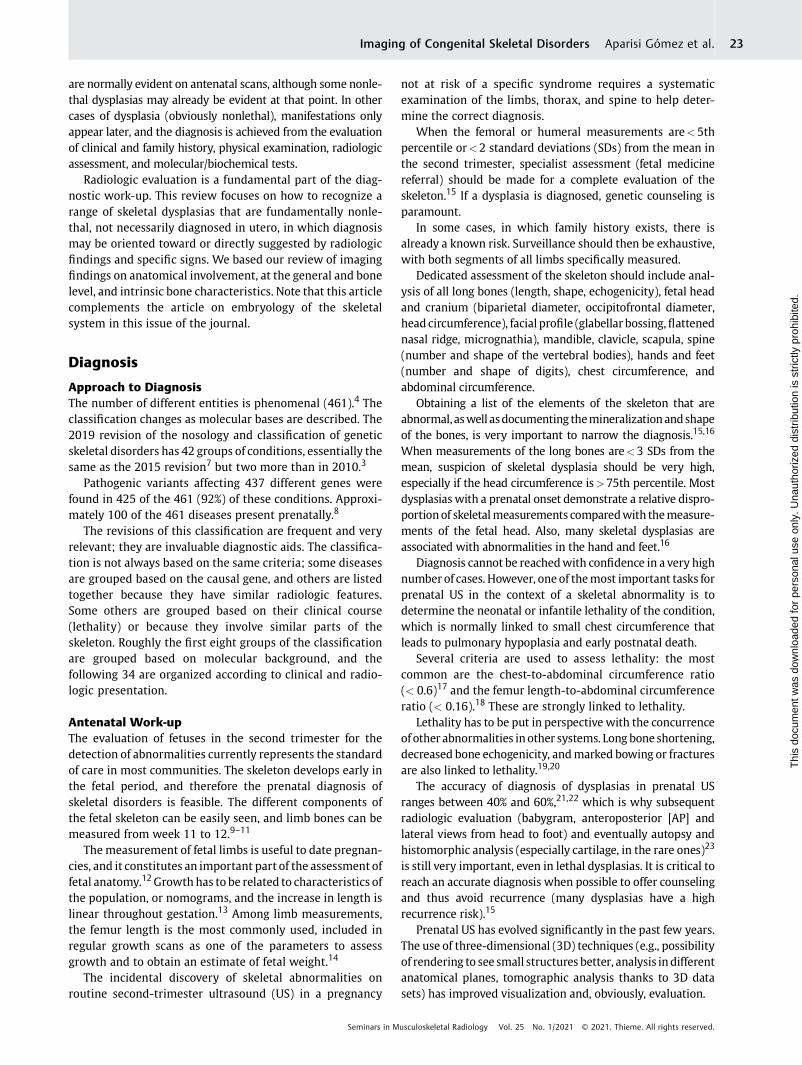
Antenatal Work-upThe evaluation of fetuses in the second trimester for thedetection of abnormalities currently represents the standardof care in most communities. The skeleton develops early inthe fetal period, and therefore the prenatal diagnosis ofskeletal disorders is feasible. The different components ofthe fetal skeleton can be easily seen, and limb bones can bemeasured from week 11 to 12.9–11
The measurement of fetal limbs is useful to date pregnan-cies, and it constitutes an important part of the assessment offetal anatomy.12Growth has to be related to characteristics ofthe population, or nomograms, and the increase in length islinear throughout gestation.13 Among limb measurements,the femur length is the most commonly used, included inregular growth scans as one of the parameters to assessgrowth and to obtain an estimate of fetal weight.14
The incidental discovery of skeletal abnormalities onroutine second-trimester ultrasound (US) in a pregnancy
not at risk of a specific syndrome requires a systematicexamination of the limbs, thorax, and spine to help deter-mine the correct diagnosis.
When the femoral or humeral measurements are<5thpercentile or<2 standard deviations (SDs) from the mean inthe second trimester, specialist assessment (fetal medicinereferral) should be made for a complete evaluation of theskeleton.15 If a dysplasia is diagnosed, genetic counseling isparamount.
In some cases, in which family history exists, there isalready a known risk. Surveillance should then be exhaustive,with both segments of all limbs specifically measured.
Dedicated assessment of the skeleton should include anal-ysis of all long bones (length, shape, echogenicity), fetal headand cranium (biparietal diameter, occipitofrontal diameter,head circumference), facial profile (glabellar bossing,flattenednasal ridge, micrognathia), mandible, clavicle, scapula, spine(number and shape of the vertebral bodies), hands and feet(number and shape of digits), chest circumference, andabdominal circumference.
Obtaining a list of the elements of the skeleton that areabnormal,aswell asdocumenting themineralizationandshapeof the bones, is very important to narrow the diagnosis.15,16
When measurements of the long bones are<3 SDs from themean, suspicion of skeletal dysplasia should be very high,especially if the head circumference is>75th percentile. Mostdysplasias with a prenatal onset demonstrate a relative dispro-portionof skeletalmeasurements comparedwith themeasure-ments of the fetal head. Also, many skeletal dysplasias areassociated with abnormalities in the hand and feet.16
Diagnosis cannot be reachedwith confidence in a very highnumber of cases. However, one of themost important tasks forprenatal US in the context of a skeletal abnormality is todetermine the neonatal or infantile lethality of the condition,which is normally linked to small chest circumference thatleads to pulmonary hypoplasia and early postnatal death.
Several criteria are used to assess lethality: the mostcommon are the chest-to-abdominal circumference ratio(< 0.6)17 and the femur length-to-abdominal circumferenceratio (< 0.16).18 These are strongly linked to lethality.
Lethality has to be put in perspective with the concurrenceofother abnormalities inother systems. Long bone shortening,decreased bone echogenicity, andmarked bowing or fracturesare also linked to lethality.19,20
The accuracy of diagnosis of dysplasias in prenatal USranges between 40% and 60%,21,22 which is why subsequentradiologic evaluation (babygram, anteroposterior [AP] andlateral views from head to foot) and eventually autopsy andhistomorphic analysis (especially cartilage, in the rare ones)23
is still very important, even in lethal dysplasias. It is critical toreach an accurate diagnosis when possible to offer counselingand thus avoid recurrence (many dysplasias have a highrecurrence risk).15
Prenatal US has evolved significantly in the past few years.The use of three-dimensional (3D) techniques (e.g., possibilityof rendering to see small structures better, analysis in differentanatomical planes, tomographic analysis thanks to 3D datasets) has improved visualization and, obviously, evaluation.
Seminars in Musculoskeletal Radiology Vol. 25 No. 1/2021 © 2021. Thieme. All rights reserved.
Imaging of Congenital Skeletal Disorders Aparisi Gómez et al. 23
Thi
s do
cum
ent w
as d
ownl
oade
d fo
r pe
rson
al u
se o
nly.
Una
utho
rized
dis
trib
utio
n is
str
ictly
pro
hibi
ted.
The use of low-dose24–26 and ultra-low-dose computedtomography (CT)27 was explored and proven to be veryhelpful.28 The main advantage of the use of low-dose CTfor the evaluation of fetuses is the exquisite depiction of fetalbones and the possibility of complete 3D rendering of theskeleton. Images can be rotated in space and postprocessedto focus on particular sections and to obtain adequate detail.This is an important advantagewith respect to dedicated US,in which the maternal habitus and the position of the fetushave a great impact on visualization.
Low-dose CT allows retrospective study of the skeletonthat is useful in cases inwhich fetuses have to be fragmentedon evacuation, to help the geneticist and pathologist in thereconstruction. The possibility to obtain 3D renderings of thefull skeleton is useful for obstetricians and orthopaedicsurgeons to plan delivery and potentially postnatal surgery,and it is also very helpful for parents to understand theextent of the anomalies.26
The use of CT is a serious decision, normally made by amultidisciplinary team of sonographers, radiologists, fetalmedicine experts, obstetricians, geneticists, and surgeons,because it requires radiation. The potential benefits need tobe carefully weighed against risks. The study is usuallyconsidered if the diagnosis has not been achievedwith highlydetailed specialist US examination and parents agree in aninformed consent.
Multiple studies have evaluated the performance of low-dose fetal CT, placing it at a superior level thanUS.25,28 Ruanoet al,29 in a comparative study of 33 patients, reported thattwo-dimensional US had an accuracy of 51%; 3DUS, 77%; andfetal CT, 94%.
Miyazaki et al30 compared its performance with postnatalradiological skeletal survey, finding that CT had a 94% rate ofidentification of cardinal findings and a 100% accuracy indiagnosing skeletal dysplasias in individual patients (includingcommon and uncommon dysplasias). In 59% of the cases, fetalCT changed the diagnosis obtained with US, with importantconsequences.Anadditional advantageof fetalCT is that itmayreveal additional findings, narrowing the differential.25
According to the American College of Radiology/Societyfor Pediatric Radiology guidelines for imaging of a pregnantpatient, the effect of a 50-mGy radiation dose is negligible.The American College of Obstetricians and Gynaecologists31
released a statement that exposure to<5 rad (50mGy) is notassociated with an increase in fetal anomalies or pregnancyloss. This position is shared by the International Commissionon Radiological Protection and the National Council onRadiation Protection.
If a fetus is not exposed to radiation during pregnancy, thechance of being born with no malformations is 96%, thechance of being bornwith no childhood cancer is 99.93%, andthe cumulative chance of being born with no malformationand no childhood cancer is 95.93%.With a radiation of 5mSv,these percentages decrease to 95.99%, 99.89%, and 95.88%,respectively. With a dose of 50 mSv, they decrease to 95.90%,99.51%, and 95.43%, respectively.32
The International Commission on Radiological Protectionstates that “Whenpregnantwomenrequireabdominalorpelvic
diagnostic X-ray examinations in which the X-ray beam irradi-ates the fetus directly, special care has to be taken to ascertainthat the X-ray examination is indeed indicated at that time andthat it cannot be delayed until after the pregnancy. Commonly,the radiation risk to the fetus is much less than that of notmaking a necessary diagnosis. In such cases, care should betaken to minimize the absorbed dose in the fetus. However,alterations in technique should not unduly reduce the diagnos-tic value of the X-ray examination.”33
To optimize the risk-benefit ratio, these studies should beperformed in scanners with multiple detectors (as many aspossible, to reduce scanning time and therefore avoid move-ment artifact), using low peak kilovoltage (as low as possiblefor patient habitus), decreased pitch in association with rapidgantry rotation (to minimize scanning time and optimizesignal-to-noise and contrast-to-noise ratios for dose), andmanipulating the automatic tube current-time setting orautomaticexposure control (milliamperesper second, keepingin mind the potential increase in noise).
A very important factor to decrease radiation dose is theshape of the iterative reconstruction (IR), which belongs tothe postprocessing phase and as such does not involveradiation, but allows adjustments in protocols so that lowerdoses can be used, preserving the level of accuracy. Forexample, Imai et al27 proposed a threshold of 0.5 mSv asthe minimal radiation dose for ultra-low-dose fetal CTwithout losing image quality, using model-based iterativereconstruction (MBIR), a specific tool for postprocessing.
Low-dose CT is performed during the second and thirdtrimester of pregnancy (ideally from week 20 but not beforeweek 18)25,26 when organogenesis is already complete. Inmost cases, indication of low-dose CT is limited to motherswith a challenging body habitus. In these patients, unfortu-nately, the radiation dose has to be larger than in normalbody habitus patients to be able to traverse the maternalabdomen and adjust for noise, which increases radiation tothe fetus. It is important to highlight that if correct reason-able indications are followed, fetal low-dose CT is normallyperformed in a pathologic context of severe abnormalities,most of which are lethal.26
Radiologic EvaluationIf a skeletal dysplasia is suspected, a skeletal survey needs to beperformed. It consistsofa seriesof radiographs that samplethestructure and morphology of a wide range of bone structures.Ideally this should include the following images2,34:
Skull (AP and lateral)Thoracolumbar spine (AP and lateral)Chest (AP)Pelvis (AP)One upper limb (AP)One lower limb (AP)Left hand (AP)
The left hand is included to assess bone age. This isimportant in some cases inwhich it is necessary to relativizefindings to the stage of normal growth.35 For example, if achild is short constitutionally, while the fingers may be short
Seminars in Musculoskeletal Radiology Vol. 25 No. 1/2021 © 2021. Thieme. All rights reserved.
Imaging of Congenital Skeletal Disorders Aparisi Gómez et al.24
Thi
s do
cum
ent w
as d
ownl
oade
d fo
r pe
rson
al u
se o
nly.
Una
utho
rized
dis
trib
utio
n is
str
ictly
pro
hibi
ted.

in comparison with another child of the same age, they maybe normal for the child, related to height. Bone age may alsobe obtained from the foot and ankle, or the knee.
If the limbs are visibly asymmetric or if epiphyseal involve-ment or stippling is suspected, views of both limbs (upper andlower) should be obtained for more accurate assessment. Insome cases, it may also be useful to obtain dedicated views(projections) that would better display the abnormality.
If other family members are affected, or at least suspectedto have the same condition, it may be useful to obtainradiologic surveys (and previous imaging) from them. Thismay offer an insight on future appearances, for example, andaid with diagnosis and prognosis. Inherently, it also givesinformation (or confirmation) on the pattern of inheritance.
Dysplasias are evolving diseases.When a diagnosis cannotbe reached initially, it is helpful to repeat the survey, but itshould not be done too early. Most centers would not repeatin<12 months.2
Serial radiographs and comparison are essential to evaluateevolutionandcomplications.Animportantconsiderationis thatearly radiographs are very useful. The ideal age for recognitionofmost dysplasias is before the closingof the growth epiphyses,after which a radiologic diagnosis may be impossible.36
Approach to Radiologic AnalysisAny radiologist specialized in the analysis of musculoskeletal(MSK) structures, regardless of their experiencewith pediatricpatients, is likely to encounter clinical questions regardingskeletal dysplasias and will have to evaluate skeletal surveysperformed with this purpose.
An important consideration is that orthopaedic manage-ment does not necessarily require a complete diagnosis ofcertainty. The patient can be treated with the clinical andradiologic approach to the problem, and in this sense, the roleof the MSK radiologist is very helpful as the usual correlate ofthe orthopaedic surgeon. Analysis of findings from the pureMSK perspective is important to speed up treatment.
Final diagnosis is still necessary to aid the pediatrician andgeneticist and provide a prognosis in terms of expectedoutcomes in adulthood, and to counsel parents and patient.In this sense, a careful radiologic assessment is also para-mount. An accurate diagnosis also opens up possibilities ofsupport, psychological but also in many instancesinstitutional/economical, which is paramount.
For this same reason, misdiagnosis (in the sense of failureto label an entity correctly) may pose a tremendous problemwith devastating consequences for the patient and families,either not allowing them to benefit from the best manage-ment approaches or creating a stigma.
A reasonable general approach to the analysis of radio-logic findings was suggested by Offiah and Hall,2 based onthe simplicity of the A, B, C, D mnemonic. “A” stands foranatomical involvement, which is really helpful as a startingpoint for classification. “B” stands for bone characteristicsthat will involve the analysis of five “S”: structure, shape,size, sum, and soft tissues.
Structure refers to bone density (deficient or excessive) andpresence of tumors (exostoses or internal abnormalities).
Shape comprises a large list of descriptive terms affectingportions or the whole bone (some of these descriptions arevery helpful for classification because they are typical ofcertain groups). Size abnormalitiesmay be absolute or relativeto other bones in the individual. Sum refers to the “number” ofbones: excessive, absent, or fused. Soft tissue refers to theanalysis of other findings in radiographs: wasting, excessivesoft tissues, calcifications, and contractures.
“C” stands for complications that may be fractures,subluxations/luxations, scoliosis, limb discrepancies, anddevelopment of malignancy. “D” stands for death, whetherthe findings are compatible with life, which is critical forcounseling.
All these characteristics, in careful analysis, are very helpfultonarrowsuspicionof thediagnosis,whichultimatelyneeds tobe genetically confirmed/correlated.
Partially based on the general approach just outlined, wedescribe the most frequently encountered dysplasias inwhich diagnosis may be narrowed or suggested by radiologicfindings. We grossly base our review of imaging findings onanatomical involvement, at the general and bone level, andintrinsic bone characteristics. We also focus on the ones thatdo not correspond to the lethal type, which are normallydiagnosed in the antenatal period.
Imaging Characteristics of Dysplasias
Dysplasias with Shortening of BonesIn dysplasias with shortening of bones, metaphyseal abnor-malities are predominant, impairing normal growth ofbones. The shortening of the limbs can be rhizomelic, asseen in achondroplasia, hypochondroplasia, and thanato-phoric dysplasia (lethal), or be mesomelic or acromelic, asseen in chondroectodermal dysplasia (Ellis-Van Creveld syn-drome), Jeune’s/asphyxiating thoracic dysplasia (lethal), andshort-rib polydactyly dysplasias.
AchondroplasiaAchondroplasia is themost common nonlethal dysplasia. It isinherited in an autosomal dominant fashion; 80% occursporadically. The cause is a mutation in chromosome 4involving the fibroblast growth factor receptor 3 (FGFR3).37
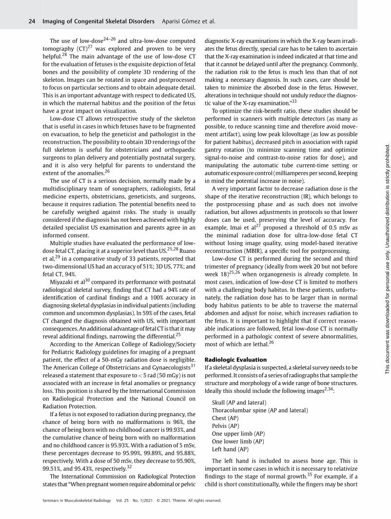
The typical features are present at birth. Prenatal diagno-sis may be challenging, given that this condition may only berevealed in the third trimester when it becomes evidentthere is shortening of the long bones. The abnormalitiesinvolve the limbs, spine, pelvis, and skull37,38 (►Fig. 1).
There is symmetrical shortening of all the long bones, withthe proximal portions and the lower limbs more affected(rhizomelia). Themetaphyses are splayed, with normal epiph-yses. In the immature skeleton, the epiphyses appear closer tothe metaphyses, and therefore articular (cartilage) spacesappear widened. On occasion the metaphysis appears to“wrap” around the epiphysis, with a “ball-socket” appearance.This appearance is frequently seen in the distal femur andattenuates/resolves with skeletal maturation.
The hand bones are tubular shaped and thick (second tofifthmetacarpals andproximal phalangesmore affected),with
Seminars in Musculoskeletal Radiology Vol. 25 No. 1/2021 © 2021. Thieme. All rights reserved.
Imaging of Congenital Skeletal Disorders Aparisi Gómez et al. 25
Thi
s do
cum
ent w
as d
ownl
oade
d fo
r pe
rson
al u
se o
nly.
Una
utho
rized
dis
trib
utio
n is
str
ictly
pro
hibi
ted.
a pronounced irreducible gap on extension of the second andthird fingers that makes them look like a “trident.”
The pelvis is short and broad (“champagne glass” appear-ances) due to squaring of the iliac wings, “elephant-earshaped.” The inferior margins of the iliac wings and acetab-ular roofs are flattened and horizontal.
Exaggerated sacral tilt and a protruding sacral promontoryare characteristic. There is associated dorsolumbar kyphosco-liosis when sitting, with lumbar hyperlordosis on a standingposition. The overall length of the spine is normal, as arevertebral bodyheights.An importantfinding is theprogressivecaudal decrease in the interpeduncular space in the lumbarspine that will become more conspicuous with age, with
development of spinal stenosis. In the lateral view, the verte-bral bodies may have a “bullet-shaped” configuration, with arounded anterior aspect and a scalloped posterior aspect.
Theskullhasanarrowedbase,withnarrowingof theforamenmagnum. The vault is expanded with frontal bossing. There ismidface underdevelopment, relative to the size of the vault.
Prenatally, it may eventually pose challenges to be distin-guished from thanatophoric dysplasia. In childhood, the differ-ential diagnosis has to be made with hypochondroplasia.39
HypochondroplasiaHypochondroplasia is a milder form of achondroplasia,caused by a mutation in the gene encoding FGFR3.39 Other
Fig. 1 Achondroplasia throughdifferent stages ofdevelopment. Thetypical features of achondroplasia are alreadypresent at birth. (a) Radiographof lowerlimbs of a 2-month-oldgirl. There is symmetrical shortening of all the long bones,with the proximal portions and the lower limbsmoreaffected (rhizomelia).Themetaphyses alreadyappear splayed. (b) There is exaggerated sacral tilt andaprotrudingsacral promontory, seen in the lateral spineview. (c)Detail of legradiograph in a 4-year-old boy. In the immature skeleton, the epiphyses appear closer to themetaphyses, and therefore articular spaces appear widened. (d)Anteroposterior (AP) radiograph of pelvis and lower limbs in a 6-year-old girl demonstrate the typical appearances of the pelvis, short and broad(“champagne glass” appearances) with “elephant-ear shaped” iliac wings. The inferior margins of the iliac wings and acetabular roofs are flattened andhorizontal. (e) Lateral view of the spine of a 9-year-old boy. The overall length of the spine is normal, as are vertebral body heights. The vertebral bodiesmayhavea “bullet-shaped” configuration,with a roundedanterior aspect (arrows) anda scalloped posterior aspect (dotted arrows). (f) OnAP view, an importantfinding is the progressive caudal decrease in the interpeduncular space in the lumbar spine that will becomemore conspicuouswith age, with developmentof spinal stenosis (thin arrows show normal distance; dotted arrows demonstrate a level of decrease of the interpeduncular space).
Seminars in Musculoskeletal Radiology Vol. 25 No. 1/2021 © 2021. Thieme. All rights reserved.
Imaging of Congenital Skeletal Disorders Aparisi Gómez et al.26
Thi
s do
cum
ent w
as d
ownl
oade
d fo
r pe
rson
al u
se o
nly.
Una
utho
rized
dis
trib
utio
n is
str
ictly
pro
hibi
ted.
mutations in chromosome 4 that do not involve this gene butanother (SHOX) that also affect stature were described.40
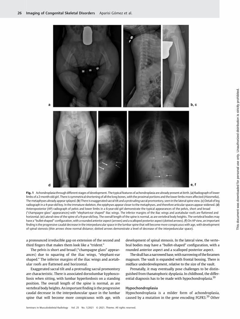
Manifestations appear later than achondroplasia, withshort stature and limb shortening that becomes evident atage 2 to 4 (►Fig. 2). The findings in the spine are similar toachondroplasia. Interpeduncular distance is also reducedcaudally, but spinal stenosis is less common. In the limbs,there can be rhizomelia but also mesomelia.41
The skull, pelvis, and hands are essentially normal. Theremay bemildmacrocephaly andmild brachydactyly involvingall metacarpals and phalanges.42
Chondroectodermal Dysplasia (Ellis-Van Creveld)Chondroectodermal dysplasia is recessively inherited, due toa mutation in the EVC gene in chromosome 4.43 The condi-tion is evident at birth, with the detection of dysplastic nails,teeth, polydactyly, and congenital cardiac defects (60% ofindividuals).44 The most common one is a defect of primaryatrial septation, with a common atrium as a result.45
There is mesomelia and acromelia, with postaxial hexadac-tyly in hands and feet, and carpal fusion that involves thecapitate and hamate. The pelvis is short with flared iliacwings, a narrow base, and hook-like projection from theacetabulum, forming a “trident” acetabula. The spine remainsnormal throughout.
Other findings are genu varum and acro-osteolysis,synmetacarpalism and synphalangism due to progressiveinvolvement.46
Jeune’s dysplasia and short-rib dysplasia with or withoutpolydactyly may have similar radiologic features, but theinvolvement of other organs like teeth, nails, and the heartare typical of chondroectodermal dysplasia.
Dysplasias with Abnormal EpiphysesIn somedysplasias, abnormal epiphyses are the predominantfinding. In these cases, the abnormalities lead to earlyosteoarthritis and joint deformities.
This is a broad group. The involvement may be isolated tothe epiphysis, as in chondrodysplasia punctata. In someentities, there is concomitant involvement of the spine(platyspondyly), as in the type II collagenopathies (spondy-loepiphyseal dysplasia congenital and tarda, Kniest dysplasia,and achondrogenesis type II). In some other cases, alongwith epiphyseal involvement, there may be metaphysealinvolvement (spondyloepimetaphyseal dysplasia, multipleepiphyseal dysplasia, pseudoachondroplasia, mucopolysac-charidoses, diastrophicdysplasia, andachondrogenesis type I).
Chondrodysplasia PunctataChondrodysplasia punctata (CDP) is a genetically heteroge-neous dysplasia (different genetic causes), with the commoncharacteristic of stippling of the epiphyses. The most com-monly encountered one is X-linked dominant, also called theConradi-Hunermann type.47,48
This condition is almost exclusively seen in females andnormally lethal in males. Radiographically, there is stipplingof the epiphyses that typically involves hands and feet,rhizomelia, transient congenital ichthyosis, patchy alopecia,cataracts, and midface hypoplasia. In the spine, there isstippling of the end plates and bodies that disappears overtime, with development of kyphoscoliosis. These infantshave normal mental development.
Another type is rhizomelic chondrodysplasia punctata(RCDP), associated with a peroxisomal enzyme disorderand autosomal recessive inheritance.49 There are severaltypes (I, II, III); the most common is type I. It is characterizedby rhizomelia, broad nasal bridge, epicanthus, high-archedpalate, dysplastic ears, micrognathia, congenital contrac-tures, and ocular problems. Contrary to the more benignConradi-Hunermann type, stippling involves large joints andspares hands and feet, and in the spine, there is no stippling,but there are coronal clefts. Normally there is severe mentalretardation and spasticity. It is lethal, with patients notsurviving beyond the first decade.49
Another very rare type of CDP of genetic origin isthe brachytelephalangic type, with X-linked recessive inheri-tance.50 CDP can also occur in cases of embryotoxicity bywarfarin (milder type, similar to Conradi-Hunermann) and ininfants from mothers with autoimmune diseases (systemiclupus erythematosus) with features similar to RCDP, but withlonger survival.51
When stippling of the epiphyses is encountered radio-graphically, the most important differential is between
Fig. 2 Hypochondroplasia. Anteroposterior radiograph of the lowerlimbs in an 11-year-old boy. Manifestations appear later than achon-droplasia, with short stature and limb shortening that becomesevident as the child starts to grow.
Seminars in Musculoskeletal Radiology Vol. 25 No. 1/2021 © 2021. Thieme. All rights reserved.
Imaging of Congenital Skeletal Disorders Aparisi Gómez et al. 27
Thi
s do
cum
ent w
as d
ownl
oade
d fo
r pe
rson
al u
se o
nly.
Una
utho
rized
dis
trib
utio
n is
str
ictly
pro
hibi
ted.

Conradi-Hunermann type and RCDP, given the prognosisis quite different. Distribution of the stippling is really helpful(Conradi-Hünermann typically involves hands, feet, andspine).
Spondyloepiphyseal Dysplasia Congenita (SEDC)Spondyloepiphyseal dysplasia congenita (SEDC) is a type IIcollagenopathy, with autosomal inheritance. The typicaldeformities of SEDC begin before birth and include malfor-mations that predominantly involve the spine, hips, andknees, and abnormalities that affect the eyes (myopia andvitreoretinal degeneration, retinal detachment) and ears(progressive sensorineural hearing loss). Malar hypoplasiaand cleft palate are also seen. Intelligence is unaffected, butthere may be delays in attaining milestones, due to delayedgrowth, muscular hypotonia, and spinal malformations. Gaitmay be abnormal, with “waddling.”52,53
Typical features are a very short stature (disproportionatedwarfism, arms appear long compared with torso), withflattened vertebral bodies and abnormal epiphyses. Thetypical radiologic feature is delayed ossification of theepiphyses, not present at birth.52
Manifestations mainly involve the spine. The vertebrae arebulbous, pear-shaped at birth, and then flatten, in keepingwith severe platyspondyly. The intervertebral spaces are verythin. Over time, kyphoscoliosis develops, as well as lumbarlordosis and atlantoaxial instability secondary to odontoidhypoplasia, with risk of compressive myelopathy.53
Pubic bones are absent at birth, and the iliac wings areshort and broad. The roofs of the acetabulum are flat as aconsequence.
The epiphyses of the knee and the calcaneum are notpresent at birth. The ossification of the carpal and tarsalbones is delayed but not that of other bones in the handsand feet.
The skull is usually large and dolichocephalic, and there isrhizomelic shortening of the limbs, more in the lower thanthe upper. The metaphyses appear widened because of theabnormal epiphyses.
Joint stiffness and deformities (coxa vara, genu varum orvalgum, luxations), and early-onset osteoarthritis develop overtime.
Spondyloepiphyseal Dysplasia TardaSpondyloepiphyseal dysplasia tarda (SEDT) is a conditionwitha diverse inheritance (X-linked recessive, classically, butautosomal recessive and dominant have also been described).As X-linked recessive, it only affects males.
The age of presentation, as the name indicates, is laterthan in the congenital form, � 5 to 10 years, but it can bevariable, even in the second decade of life.54 Appearances atbirth are normal.
In the spine, there is platyspondyly, with hyperostosis inthe posterior two thirds of the bodies that gives them“humped” appearances. Progressive narrowing of the inter-peduncular distances was also described.55
The pelvis is small, with mild epiphyseal irregularity, lead-ing to early osteoarthritis in thehips, knees, and ankles. Hands,
feet, and skull are typically not involved. Cleft palate and retinaldetachment, typical of SEDC, are not seen in SEDT.53
Kniest DysplasiaKniest dysplasia is another type II collagenopathy, with anautosomal dominant inheritance. It is similar to SEDC, with amarkedly short trunk and delayed ossification at birth andinfancy.56
In addition to the features of SEDC, the femurs aredumbbell-shaped, and there is coronal clefting of the spineat birth. With development, the epiphyses become enlarged,becoming “mega-epiphyses,” with cloud-like calcificationsin the metaphyses.
In the hands, the metacarpals and phalanges are abnor-mal, with flattening and enlargement of the ends, whichmakes the metacarpophalangeal and proximal interphalan-geal joints look bulbous.57
Achondrogenesis Types I and IIAchondrogenesis types I and II are the most severe types ofchondrodysplasia and lethal before or soon after birth. Theyare normally diagnosed in utero.
Type I is subdivided into IA and IB.58 In type I, there isdeficient ossification in the lumbar, sacral, pubic, and ischialbones. There is also severemicromelia and a large head, withedema in the soft tissues.59 The ribs are thin, with multiplefractures (can be mistaken for osteogenesis imperfecta).Type II is characterized by absent ossification of the spine,the sacrum, and pubic bones.
The trunk is short, small,with aprominenthydropic-appear-ing abdomen, in both types. Hypochondrogenesis belongs tothisgroup,withamilderphenotype,60andisgenetically related.
Spondyloepimetaphyseal DysplasiaSpondyloepimetaphyseal dysplasia (SEMD) is a generallydescriptive term for a group of entities that demonstrateradiologic abnormalities in the spine, epiphyses, and meta-physes, and they have different phenotypes, mechanisms ofinheritance, and particular radiologic findings. There aremany different variants.
One of the specific variants is the Strudwick type.61 It ischaracterized by severe dwarfism, pectus carinatum, and scoli-osis thatmayevolvetosevereandcreateproblems inadulthood.
Short limbs and delayed epiphyseal maturation are alreadypresent at birth. During infancy, the disorder cannot bedistinguished from SEDC (cleft palate and retinal detachment,typical of SEDC, are also present in Strudwick SEMD).
The typical radiographic feature is the development inearly childhood of irregular sclerotic changes in the meta-physes of the long bones. This pattern (called “dappling”)results from a mix of regions of osteosclerosis and osteope-nia.62 It is usually moremarked in the ulna and fibula than inthe radius and tibia (►Fig. 3).
Some variants of SEMD are well delineated, but in a greatnumber of cases, the approach is basically descriptive(►Fig. 4). As a general rule, in patients in which the epiphy-seal component is predominant, there will be early develop-ment of osteoarthritis.
Seminars in Musculoskeletal Radiology Vol. 25 No. 1/2021 © 2021. Thieme. All rights reserved.
Imaging of Congenital Skeletal Disorders Aparisi Gómez et al.28
Thi
s do
cum
ent w
as d
ownl
oade
d fo
r pe
rson
al u
se o
nly.
Una
utho
rized
dis
trib
utio
n is
str
ictly
pro
hibi
ted.
Multiple Epiphyseal DysplasiaMultiple epiphyseal dysplasia (EDM) is a genetically heteroge-neous condition, with multiple types. Most are of autosomaldominant inheritance, but recessive forms are also possible.63
It normally presents at 2 to 4 years of agewhen the child startsto walk.
Symptoms include joint pain, in hips and knees, a wad-dling walk, and short stature as adults. As in other entitiesinvolving the epiphyses, there is early onset of osteoarthritis.In recessive inheritance, there may be malformations of thehands, feet, knees, and other abnormalities. Other clinicalfindings include myopia, sensorineural hearing impairment,and abnormalities of T-cell physiology. Rarely, there can beanonychia (absent nails).64,65
There is bilateral and symmetric irregularity of the epiph-yses of the hips, knees, ankles, shoulders, elbows, wrists,hands, and feet (►Fig. 5). In the ankle, there is lateraltibiotalar inclination, with the lateral part of the distal tibialepiphysis thinner than the medial, and the talus is shaped toaccommodate.
A typical finding is the double-layered patella (patho-gnomonic), seen in the lateral view64,65 (►Fig. 6). Thiswas also described in pseudoachondroplasia,66 likelyexplained by mutations affecting the same genes (geno-typic alleles).
The spine is only mildly involved (there is no platyspon-dyly), with anterior wedging, mild end-plate irregularity,and Schmorl’s nodes, resembling Scheuermann’s disease.
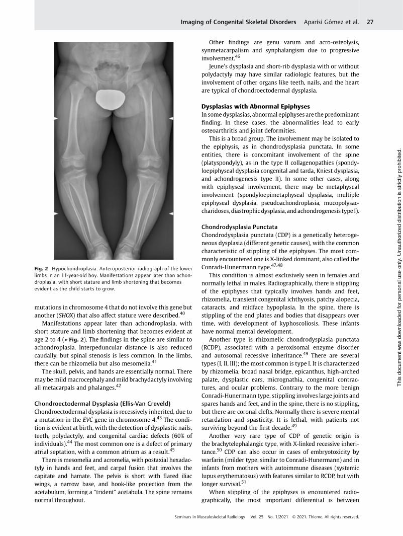
Fig. 3 Spondyloepimetaphyseal dysplasia, Strudwick type. (a) Anteroposterior (AP) radiograph of the lower limbs in a 7-year-old girldemonstrates the typical radiographic feature of “dappling,” resulting from a mix of regions of osteosclerosis and osteopenia in all themetaphyses included in the projection, more conspicuous around the knee. (b) This is seen in detail in the radiograph of the right knee. There isa degree of valgus deformity. Short limbs and delayed epiphyseal maturation are already present at birth. (c) Sagittal proton-density view frommagnetic resonance imaging of the right knee, performed when the girl was 10, demonstrates the irregularity in the distal metaphysis of thefemur and proximal metaphysis of the tibia. In the proximal tibia there are ossified foci within the metaphysis (arrows), and the epiphysis has anabnormal orientation, almost vertical (signaled by the dotted arrow). The girl underwent corrective tibial osteotomies. (d) AP radiograph of thespine of the same girl at 10 years of age. There is marked thoracolumbar scoliosis. “Dappling” is present in the humeral metaphyses, and note theirregularity of the epiphyses. The femoral epiphyses are practically not present, with marked irregularity of the metaphyses (“dappling” is againseen). Note how the iliac wings are short and broad. The roofs of the acetabulum are flat, as a consequence of this and the irregular femoralepiphyses. (e) Three years later, by the time the girl is 13, the scoliosis has mildly progressed. The appearances of the epiphyses and metaphysesof the humeri and femurs are similar to previous images, markedly irregular. Computed tomography (f) coronal reconstruction and (g) sagittalreconstruction, acquired at 17 years of age. The femoral heads are underdeveloped (practically absent) and the acetabular roofs are flat. Thereare also segments of platyspondyly and residual fragmentation of the vertebral bodies (arrows).
Seminars in Musculoskeletal Radiology Vol. 25 No. 1/2021 © 2021. Thieme. All rights reserved.
Imaging of Congenital Skeletal Disorders Aparisi Gómez et al. 29
Thi
s do
cum
ent w
as d
ownl
oade
d fo
r pe
rson
al u
se o
nly.
Una
utho
rized
dis
trib
utio
n is
str
ictly
pro
hibi
ted.

PseudoachondroplasiaPseudoachondroplasia has an autosomal dominant inheri-tance. The age of presentation is the same as EDM (2–4 years,as the child starts to walk).
Subjects have a disproportionate short stature (armsappear long compared with torso), with normal facialfeatures and head size. Intelligence is preserved. The fingers,wrists, elbows, and knees are markedly lax. Joint pain is acommon complaint, and earlyonset of osteoarthritis involvesevery joint.
EDM and pseudoachondroplasia share many traits. Bothare genotypic alleles (involving mutations of the samegenes).67 However, clinical and radiologic manifestationsare more severe in pseudoachondroplasia (►Fig. 7).
In the spine, the vertebrae have an oval shape, with atongue-like projection in the anterior aspect that is patho-
gnomonic of this entity but disappears over time withdevelopment of platyspondyly. As a result, the trunk is short.
Scoliosis, excessive kyphosis or lordosis, and cervicalspine instability are seen as complications. The transientnature of the pathognomonic sign highlights the importanceof obtaining radiographs early in this condition (►Fig. 8).
An important typical feature is joint-ligament laxity, todistinguish pseudoachondroplasia from other entities.The differential of pseudoachondroplasia includes EDM andachondroplasia. In EDM, there is no platyspondyly and double-layeredpatella. Also, joints are not lax. In achondroplasia, thereisnoplatyspondyly, theskull is abnormal, andonlymetaphysesare affected. The interpeduncular distance is decreased, andthe hand and pelvis have typical appearances (trident hand,champagne glass pelvis). In pseudoachondroplasia, there isplatyspondyly, epiphyses are involved, the skull is normal, and
Fig. 4 Spondyloepimetaphyseal dysplasia. Sporadic presentation. First visit at 7 years of age. The diagnosis was confirmed by clinical andradiographic findings, even though the molecular tests (COMP and MANT3) were negative. (a) Anteroposterior (AP) radiograph of the pelvis at8 years of age demonstrates marked irregularity of the metaphyses and practically nonexistence of the epiphyses that cannot be seen to beossified. The acetabular roof is flat, and the iliac wings are short and broad. (b) AP radiograph of the right knee (age 8) demonstrates markedirregularity of the metaphysis in the distal femur and proximal tibia; the epiphyses are much less irregular. (c) The patient underwent hemi-epiphyseal stapling for correcting genu valgum at the age of 9. The genu valgum progressively corrected: (d), age 10 years; (e), age 11 years, butan important genu recurvatum was still evident on lateral weight-bearing radiograph (f).
Seminars in Musculoskeletal Radiology Vol. 25 No. 1/2021 © 2021. Thieme. All rights reserved.
Imaging of Congenital Skeletal Disorders Aparisi Gómez et al.30
Thi
s do
cum
ent w
as d
ownl
oade
d fo
r pe
rson
al u
se o
nly.
Una
utho
rized
dis
trib
utio
n is
str
ictly
pro
hibi
ted.

the specific abnormalities in thehand (trident hand) and pelvis(champagne pelvis) are not present.
MucopolysaccharidosesMucopolysaccharidoses represent a groupof storage disordersin which there are mutations in the genes that encode lyso-somal enzymes with a role in the degradation of glycosami-noglycans (GAGs) ormucopolysaccharides. As a result, there isdeposit of GAGs in various tissues. Typically, there is a coarseface, mental retardation, and hepatosplenomegaly.
Another name for these disorders is dysostoses multiplexbecause they are associated with multiple skeletalabnormalities.
If amucopolysaccharidosis is suspected, the initialwork-upincludes urinalysis that will demonstrate elevated GAGs.
Studies of enzyme activity are performed in cultured fibro-blasts or leucocytes.
Common features of mucopolysaccharidoses are epiphysealabnormalities, pointed appearances of the base of the meta-carpals, and beaking in the spine (inferior aspect of the bodies,with superior notching) (►Fig. 9). There are evolving jointcontractureswithout inflammation.68Osteoporosis isalso seen.
Hurler’s syndrome (mucopolysaccharidosis type I) is auto-somal recessive in inheritance. Signs start to appear over thefirst 2 years of life. There is corneal clouding, coronary arterynarrowing, endocardial fibroelastosis, and valvular disease.69
This translates into cardiomegaly on radiographs.The head is large with frontal bossing. There is a “J-shaped”
sella that occurs due to the deposit of GAGs in the pituitarygland. The ribs are broad anteriorly but thin posteriorly, with a
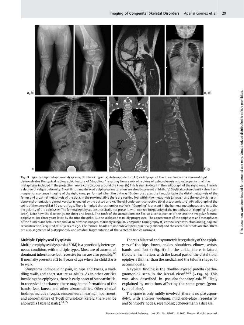
Fig. 5 Multiple epiphyseal dysplasia. (a) Weight-bearing anteroposterior radiograph of the lower limbs of an 11-year-old girl with bilateral andsymmetrical irregularity of the epiphyses of the hips, knees, and ankles. Note the lateral tibiotalar inclination, with the lateral part of the distaltibial epiphysis thinner than the medial and the talus shaped to accommodate. (b) Detail of a coronal T1 image from magnetic resonanceimaging performed at age 12 demonstrates marked irregularity of the epiphysis, with appearances that suggest fragmentation: patchyossification. (c) On coronal proton-density fat sat image there is mild effusion, and the epiphyseal cartilage is inhomogeneous. (d) Coronalreconstruction from computed tomography at age 17 demonstrates the abnormal shape of the femoral heads (flattened) as well as a flatacetabulum. On the left, there is development of subchondral bone cysts as a sign of osteoarthritis.
Fig. 6 Multiple epiphyseal dysplasia. (a) Lateral knee radiograph of a 7-year-old girl demonstrates pathognomonic appearances of a double-layered patella. Progressive knee flexion deformity required correction at age 8, with anterior epiphyseal stapling on the distal femur.Progressive contracture and deformity of the joints is a feature that differentiates multiple epiphyseal dysplasia from pseudoachondroplasia.Unfortunately, the stapling was unsuccessful. (b) Anteroposterior radiographs at age 10 demonstrate shortening of the femur. The markedirregularity of the femoral epiphyses is also noticeable. (c) Lateral view of the right knee better demonstrates the double-layered patella.
Seminars in Musculoskeletal Radiology Vol. 25 No. 1/2021 © 2021. Thieme. All rights reserved.
Imaging of Congenital Skeletal Disorders Aparisi Gómez et al. 31
Thi
s do
cum
ent w
as d
ownl
oade
d fo
r pe
rson
al u
se o
nly.
Una
utho
rized
dis
trib
utio
n is
str
ictly
pro
hibi
ted.
configuration that makes them resemble oars. The lateral endsof the clavicles are hypoplastic, with small scapulae. The lengthof the limbs is normal, but the diaphyses are widened, more soin the upper limbs. The distal radius and ulna may arch toconverge.
In thehands, thetubularbonesarenormallyshortandwide,and the metacarpals are pointy at the base and broad at thehead.
In the spine, L1 or L2 are hypoplastic and slightlymisaligned, with the appearance of dorsolumbar kyphosis.There is also anteroinferior beaking, as mentioned. There isno platyspondyly. There can be atlantoaxial instability. Theiliac wings are flared out, the acetabular roofs are shallow,and there is delayed ossification of the femoral heads.70
Morquio’s syndrome (mucopolysaccharidosis type IV) isalso autosomal recessive in inheritance. Pathologically, thereis intracellular accumulation of keratan sulfate and chon-droitin-6-sulfate.
In this case, there are dental abnormalities and cornealclouding with normal intelligence.71 The skull is enlarged,
there is dorsolumbar kyphosis and atlantoaxial instability,with a normal sella, and there is platyspondyly, with centralbeaking.
In the hands, the typical pointy appearance of the metacar-palbase is found,butadditionally, there is irregularanddelayedossification of the carpal and tarsal bones. The epiphyses areenlarged, and the metaphyses widened, to accommodate.There is delayed ossification of the femoral heads, and as aconsequence, the acetabula are poorly developed, leading toarthropathy.72
There is delayed ossification of the femoral heads, and as aconsequence, the acetabula are poorly developed, leading toarthropathy.72
Attenuated types of Morquio and type VI mucopolysac-charidosis have been described, in which involvement islimited to femoral heads.73,74
Diastrophic DysplasiaDiastrophic dysplasia is characterized by scoliosis thatresults in short stature, bilateral clubfeet, premature
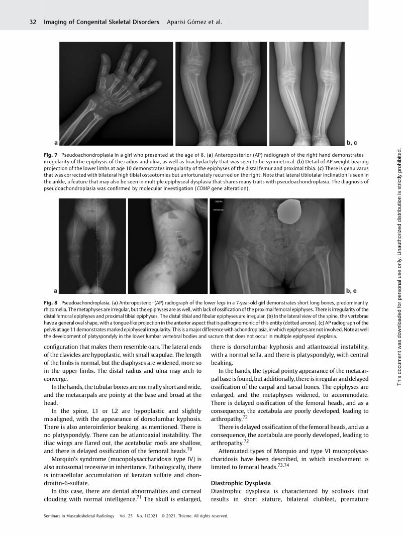
Fig. 7 Pseudoachondroplasia in a girl who presented at the age of 8. (a) Anteroposterior (AP) radiograph of the right hand demonstratesirregularity of the epiphysis of the radius and ulna, as well as brachydactyly that was seen to be symmetrical. (b) Detail of AP weight-bearingprojection of the lower limbs at age 10 demonstrates irregularity of the epiphyses of the distal femur and proximal tibia. (c) There is genu varusthat was corrected with bilateral high tibial osteotomies but unfortunately recurred on the right. Note that lateral tibiotalar inclination is seen inthe ankle, a feature that may also be seen in multiple epiphyseal dysplasia that shares many traits with pseudoachondroplasia. The diagnosis ofpseudoachondroplasia was confirmed by molecular investigation (COMP gene alteration).
Fig. 8 Pseudoachondroplasia. (a) Anteroposterior (AP) radiograph of the lower legs in a 7-year-old girl demonstrates short long bones, predominantlyrhizomelia. Themetaphyses are irregular, but the epiphyses areaswell, with lackofossificationof the proximal femoral epiphyses. There is irregularity of thedistal femoral epiphyses and proximal tibial epiphyses. The distal tibial and fibular epiphyses are irregular. (b) In the lateral view of the spine, the vertebraehave a general oval shape, with a tongue-like projection in the anterior aspect that is pathognomonic of this entity (dotted arrows). (c) AP radiograph of thepelvis at age11demonstratesmarkedepiphyseal irregularity. This is amajordifferencewithachondroplasia, inwhichepiphysesarenot involved.Noteaswellthe development of platyspondyly in the lower lumbar vertebral bodies and sacrum that does not occur in multiple epiphyseal dysplasia.
Seminars in Musculoskeletal Radiology Vol. 25 No. 1/2021 © 2021. Thieme. All rights reserved.
Imaging of Congenital Skeletal Disorders Aparisi Gómez et al.32
Thi
s do
cum
ent w
as d
ownl
oade
d fo
r pe
rson
al u
se o
nly.
Una
utho
rized
dis
trib
utio
n is
str
ictly
pro
hibi
ted.

calcification of costal cartilages, occasional cleft palate, and atypical “hitchhiker thumb” appearance due to deformity ofthe first metacarpal.75 This is very characteristic and allowsprenatal diagnosis. Historically, because of the findings,these patientswere diagnosedwith arthrogryposis (multiplecontractures).
Dysplasias with Altered Bone DensityThese dysplasias are genetically heterogeneous, causedby multiple mutations. Due to the genetic complexity,the focus is on the clinical manifestations to reach aprognosis.
The prototype of reduced bone density is osteogenesisimperfecta. There are other syndromes, labeled as syndromes,with congenital brittle bones that also show decreased bonedensity.
The prototype of increased bone density is osteopetrosis,but osteosclerotic dysplasias may be further classified intothree groups, with endochondral bone formation, such asosteopetrosis, pyknodysostosis, bone island, osteopoikilosisand osteopathia striata, intramembranous bone formation,like progressive diaphyseal dysplasia (Camurati-Engel-mann’s disease), Caffey’s disease, and mixed sclerosing,such as melorheostosis and overlap syndromes.
Fig. 10 Osteogenesis imperfecta type I. (a) Lateral radiograph of the left leg of a 3-week-old boy with osteogenesis imperfecta type I demonstrates smallregions of early periosteal bone formation adjacent to the cortex of the distal metaphysis of the tibia (dotted arrows). (b) Weeks later (lateral radiograph),there is hypertrophic callus formation, with florid periosteal reaction, and a transverse lucent fracture line still visible through the distal tibia (arrow). Thepatient sustained multiple fractures of the tibias that were treated conservatively. He is on a background treatment of neridronate (bisphosphonate). (c)Anteroposterior radiograph of the leg of the same boy at age 6 demonstrates the typical “zebra lines” (band-like areas of increased opacity) seen inosteogenesis imperfecta patients treated with bisphosphonates. These are the result of failure of remodeling of the primary spongiosa into secondaryspongiosa in thephysis, associatedwith the cycles/doses of treatment. Note the decreasedmineralization of theboneand thebowingof the tibia and fibula.
Fig. 9 Mucopolysaccharidosis. (a) Lateral spine radiograph in a 3-year-old boy with mucopolysaccharidosis type II–III, demonstrating “beaking”in the inferior aspect of the vertebral bodies, with superior notching (dotted arrows). (b) Left wrist anteroposterior radiograph demonstratesirregularity in the epiphyses of the radius and ulna, with slight arching of the distal ulna. (c) Radiograph of the right hand (age 4) demonstratesthe typical appearances of the metacarpals that are pointy toward the base and broad distally. The epiphyseal abnormalities, pointedappearances of the base of the metacarpals and beaking in the spine, are common features of the different types of mucopolysaccharidoses.
Seminars in Musculoskeletal Radiology Vol. 25 No. 1/2021 © 2021. Thieme. All rights reserved.
Imaging of Congenital Skeletal Disorders Aparisi Gómez et al. 33
Thi
s do
cum
ent w
as d
ownl
oade
d fo
r pe
rson
al u
se o
nly.
Una
utho
rized
dis
trib
utio
n is
str
ictly
pro
hibi
ted.
Osteogenesis ImperfectaIn osteogenesis imperfecta, inheritance is variable, autoso-mal dominant or recessive. The disorder is due to a mutationin type I procollagen genes. There is decreased bone massand bone fragility, as a consequence.
Extraskeletal manifestations include blue sclerae, denti-nogenesis imperfecta, and deafness.
The severity is variable, and there are eight types. Type II,for example, is lethal perinatally. In order of severity, VIII, III(progressively deforming), VII, VI, V, and IV follow.76 Type I isthe less severe type (►Fig. 10).
Typical radiologic features are diffuse osteopenia, with verythin cortices and multiple fractures. Healing of these fracturesoccurs with exuberant callus (“pseudotumor”). There may bedeformities andpseudoarthrosis.Vertebrae are also osteopenic,and collapse,withbiconcave appearances. In theskull, there aremultiple wormian bones, the calvarium is lucent, sinuses areenlarged, and there isplatybasia (flatteningof the skull base). Inthe pelvis, protrusio acetabuli is common. The femur has thetypical appearance of a “shepherd’s crook” (►Fig. 11).
In the differential, nonaccidental injuries have to beconsidered. Other entities with decreased bone density arehypophosphatasia, juvenile idiopathic osteoporosis, and othersyndromes with congenital brittle bones77 that are labeled assyndromes resembling osteogenesis imperfecta. In the latter,mutations do not involve the type I procollagen genes.
OsteopetrosisIn osteopetrosis, there is failure of normal osteoclasticresorption of bone. The result is increased density in themedullary portions of the bones. Cortices are spared.78
There is a also lot of genetic heterogenicity, as well asclinical presentations. The most severe type is recessive, ofearly onset, and causes bone marrow failure, with progres-sive alterations in the blood cell counts (due to the oblitera-tion of the medullary canals) and early death.
In the skull, there is diffuse sclerosis that involves thebase and vault. There is progressive narrowing of theforamina at the base that causes cranial nerve impingementand related symptoms. The mandible is prognathic.
Mandibular osteomyelitis incidence is increased. In thelimbs, there is also diffuse sclerosis and metaphyseal flaring(“Erlenmeyer flask deformity”)79 with a high incidence offractures. Healing is normal, but callus formation isdefective.
There is also a “bonewithin bone” appearance, seen in thespine, pelvis, and short tubular bones. In the spine, vertebraehave “sandwich” appearances, due to end-plate sclerosis.In the pelvis, there are multiple sclerotic lines parallel to theiliac crest.
A group of entities, the craniotubular dysplasias, resembleosteopetrosis and pose a challenge in differential diagnosis.They have similar cranial and metaphyseal features.80 Theinvolvement of tubular bones is different, vertebrae arenormal, and there is no disturbance to hematopoiesis, featuresthat can help with distinction.
PycnodysostosisPycnodysostosis is a condition with autosomal recessiveinheritance that presents in early childhood, with a triad ofincreased bone density, fractures, and dwarfism with shortlimbs.81
The skull demonstrates base and vault sclerosis, withopen sutures and fontanelles and multiple wormian bones.There is mandibular hypoplasia.
In the limbs, there is sclerosis of long bones. Limb length isdecreased. The pelvis is sclerotic, with small and shallowacetabulum.
Acro-osteolysis is seen in the hands. The medullary cavityis maintained, and there may be an associated Madelung’sdeformity.
Themain differential is osteopetrosis, and distinctionmaybe made by the appearances of the skull with open sutures,the small mandible, and the acro-osteolysis.78
OsteopoikilosisOsteopoikilosis is of autosomal dominant inheritance, morecommon in males, and benign. The typical features aremultiple small (1–10mm) dense concretions at the end ofthe long bones often just deep to the cortex, in the carpal and
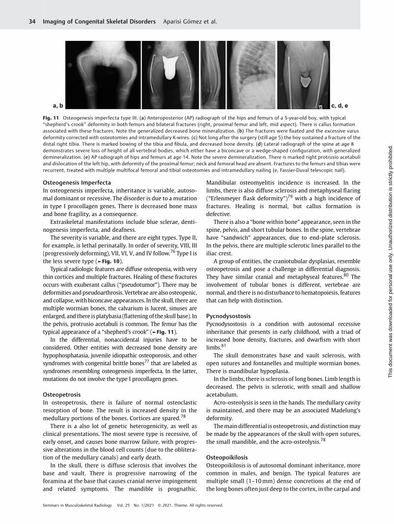
Fig. 11 Osteogenesis imperfecta type III. (a) Anteroposterior (AP) radiograph of the hips and femurs of a 5-year-old boy, with typical“shepherd’s crook” deformity in both femurs and bilateral fractures (right, proximal femur and left, mid aspect). There is callus formationassociated with these fractures. Note the generalized decreased bone mineralization. (b) The fractures were fixated and the excessive varusdeformity corrected with osteotomies and intramedullary K-wires. (c) Not long after the surgery (still age 5) the boy sustained a fracture of thedistal right tibia. There is marked bowing of the tibia and fibula, and decreased bone density. (d) Lateral radiograph of the spine at age 8demonstrates severe loss of height of all vertebral bodies, which either have a biconcave or a wedge-shaped configuration, with generalizeddemineralization. (e) AP radiograph of hips and femurs at age 14. Note the severe demineralization. There is marked right protrusio acetabuliand dislocation of the left hip, with deformity of the proximal femur; neck and femoral head are absent. Fractures to the femurs and tibias wererecurrent, treated with multiple multifocal femoral and tibial osteotomies and intramedullary nailing (e, Fassier-Duval telescopic nail).
Seminars in Musculoskeletal Radiology Vol. 25 No. 1/2021 © 2021. Thieme. All rights reserved.
Imaging of Congenital Skeletal Disorders Aparisi Gómez et al.34
Thi
s do
cum
ent w
as d
ownl
oade
d fo
r pe
rson
al u
se o
nly.
Una
utho
rized
dis
trib
utio
n is
str
ictly
pro
hibi
ted.

tarsal bones, in a periacetabular location and in the sub-glenoid area.82 These are symmetrical and uniform. Theseconcretions represent multiple benign enostoses.
The condition is asymptomatic, incidentally found inradiographs. The enostoses appear during childhood anddo not regress with adulthood. In adulthood, they may bemistaken by other entities such as metastases, hence theimportance of accurate diagnosis.
Osteopoikilosis is often found with osteopathia striataandmelorheostosis, probably representing a spectrum of thesame condition, also called mixed sclerosing bone dysplasia,due to a mutation of the LEMD3 gene.83
The association of osteopoikilosis with disseminated con-nective tissue and cutaneous nevi, on extremities and trunk,that have a yellowish shade is known as Buschke-Ollendorffsyndrome (disseminated dermatofibrosis lenticularis).84
Osteopathia StriataThe typical form of osteopathia striata is also known asVoorhoeve’s disease and is benign, characteristically involvingthe epiphysis and metaphysis of tubular bones, happening inany age group.
The condition is usually asymptomatic, although somepatients describe joint discomfort. There is bilateral involve-ment of long bones, pelvis, and scapulae, with multiple thinvertical sclerotic lines in the metaphysis, extending into thediaphysis78 (►Fig. 12).
Osteopathia striata may be found associated withcutaneous manifestations, such as hypopigmented or hyper-pigmented lesions, known as Goltz’s syndrome or focaldermal hypoplasia.85 This disorder follows an X-linkeddominant inheritance pattern and is more commonly seenin males.
Osteopathia striata with cranial sclerosis is a differentclinical entity that is very rare.
This condition is of X-linked dominant inheritance, morecommon in females, and more severe and with highermortality in males.86 The condition affects multiple systems.
There may be sclerosis of the long bones and the skull,with cranial nerve compression, and there are cases ofcleft palate.87
Progressive Diaphyseal DysplasiaProgressive diaphyseal dysplasia is of autosomal dominantinheritance and also called Camurati-Engelmann’s disease.The hallmark of the disease is increased bone density,affecting the tibia, femur, humerus, ulna, and radius. Insome cases, pelvis and skull are also affected.
Patients develop pain, a waddling gait, muscle weakness,and limiting tiredness.
Neurologic problems caused by increased intracranialpressure and cranial nerve compromise may also be seen,such as headaches, visual and hearing loss, vertigo, tinnitus,and facial palsy. Scoliosis, contractures, and valgum of theknees and pes planus may also be present.82,88
Radiographically, there is bilateral symmetrical enlarge-ment,with increaseddensityof thediaphysisof the longbonesthat begins in themidportion and extends in a fusiform shapetoward both ends. Metaphyses may be involved, but typically,epiphyses are not.82,88
Caffey’s DiseaseCaffrey’s disease is an autosomal inherited condition (domi-nant and recessive forms) that presents early in life (beforethe fifth month), with hyperirritability, soft tissue swelling,bone lesions, and typical mandible involvement. Acutemanifestations are inflammatory, with fever and hot, tenderswelling.89
The ulna, tibia, clavicle scapulae, and ribs may also beinvolved.
Radiologically, there is diffuse cortical thickening, due tosubperiosteal new bone formation that typically involves thediaphysis, sparing metaphyses and epiphyses (►Fig. 13).
The condition usually resolves spontaneously by 2 years ofage. Despite its appearances in acute stages, the bones returnto completely normal in subsequent studies.
MelorheostosisMelorheostosismay present sporadically or be inherited. It isa benign condition with pain and soft tissue contractures asclinical features.
Radiologically, there is cortical thickening, in a wavy,characteristic pattern, a “flowing wax candle” appearance.
Fig. 12 Osteopathia striata. (a) Anteroposterior radiograph of the pelvis in a 5-year-old boy. Typical appearances of multiple thin verticalsclerotic lines in the proximal metaphysis of the femur that extend into the diaphysis. (b) Note the slight irregularity in the metaphysis, moreevident on coronal computed tomography image.
Seminars in Musculoskeletal Radiology Vol. 25 No. 1/2021 © 2021. Thieme. All rights reserved.
Imaging of Congenital Skeletal Disorders Aparisi Gómez et al. 35
Thi
s do
cum
ent w
as d
ownl
oade
d fo
r pe
rson
al u
se o
nly.
Una
utho
rized
dis
trib
utio
n is
str
ictly
pro
hibi
ted.
Distribution is asymmetric and can be mono- or poly-ostotic or monomelic. If monomelic, it typically involves thelower limb. Other bones like the skull, ribs, spine, and shorttubular bones are occasionally affected. In children, thepattern of involvement is endosteal, and in adults, perioste-al82 (►Fig. 14).
Melorheostosis may be found with osteopoikilosis andosteopathia striata, probably representing a spectrum of thesame condition called mixed sclerosing bone dysplasia, dueto a mutation of the LEMD3 gene.83
Conclusion
The diagnosis of the most severe congenital abnormalities ofthe skeletal system is normally done prenatally, but in thecases of nonlethal dysplasias, manifestations will onlyappear later, and the diagnosis will be achieved from theevaluation of clinical and family history, physical
examination, radiologic assessment, and molecular/bio-chemical tests.
Radiographic evaluation is a fundamental part of thediagnostic work-up of congenital skeletal disorders, and inmost cases it is the first tool to orient a diagnosis.
This review has described the imaging characteristics,specific signs, and evolution in several skeletal dysplasias inwhich diagnosis may be oriented or directly suggested byradiologic findings. We based our review of imaging findingson anatomical involvement, at the general and bone level,and intrinsic bone characteristics.
Definitive diagnosis of congenital skeletal abnormalities isnecessary to enable the pediatrician and geneticist to offer aprognosis in terms of expected outcomes in adulthood, andto counsel parents and patients.
Conflict of InterestNone declared.
Fig. 14 Melorheostosis. (a) Anteroposterior (AP) radiograph of the knees of a 12-year-old girl, with typical features of cortical thickening, which is subtle,endosteal (dotted arrow), in the lateral aspect of the distal right femur. There is also a sclerotic regionwith lobulated contours (arrow) in the lateral aspect ofthedistal femoral epiphysis. (b) AP radiographof the right kneeatage17demonstratesprogressionof the cortical thickening,which ismainlyendosteal, anddisplays a mildly wavy contour. The thickening extends to the epiphysis, where it becomes confluent with the sclerotic region in the lateral aspect of theepiphysis. Regions of cortical thickening have also become apparent in the proximal fibula (bold arrowhead). (c) AP radiograph of the leg at age 19 showsgeneral cortical thickening in the fibula, withwavy contours, the typical “flowing wax candle” appearances (arrowhead). The thickening is periosteal. (d) APradiograph of the knee at age 22 demonstrates generalized progression of the cortical thickening (arrowheads). Distribution is asymmetric and can bemonostotic or polyostotic or monomelic. If monomelic, it typically involves the lower limb, as in this case. The diagnosis was confirmed by clinical andradiographic findings, even though the molecular tests (LEMD3) were negative.
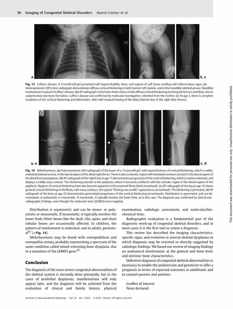
Fig. 13 Caffey’s disease. A 2-month-old girl presented with hyperirritability, fever, and regions of soft tissue swelling with inflammatory signs. (a)Anteroposterior (AP) chest radiograph demonstrates diffuse cortical thickening in both humeri, left clavicle, and in themandible (dotted arrow). Mandibleinvolvement is typical ofCaffey’s disease. (b) AP radiographof the lower limbs shows similar diffuse cortical thickening involving the femurs and tibias, due tosubperiosteal new bone formation. Caffey’s disease was confirmed by molecular investigation, inherited from the mother. (c) At age 3, there is completeresolution of the cortical thickening and deformities, with mild residual bowing of the tibias (lateral view of the right tibia shown).
Seminars in Musculoskeletal Radiology Vol. 25 No. 1/2021 © 2021. Thieme. All rights reserved.
Imaging of Congenital Skeletal Disorders Aparisi Gómez et al.36
Thi
s do
cum
ent w
as d
ownl
oade
d fo
r pe
rson
al u
se o
nly.
Una
utho
rized
dis
trib
utio
n is
str
ictly
pro
hibi
ted.

References1 Hall CM. International nosology and classification of constitutional
disorders of bone (2001). Am J Med Genet 2002;113(01):65–772 Offiah AC, Hall CM. Radiological diagnosis of the constitutional dis-
ordersof bone.As easyasA, B, C?Pediatr Radiol 2003;33(03):153–1613 Warman ML, Cormier-Daire V, Hall C, et al. Nosology and classifi-
cation of genetic skeletal disorders: 2010 revision. Am J MedGenet A 2011;155A(05):943–968
4 Mortier GR, Cohn DH, Cormier-Daire V, et al. Nosology andclassification of genetic skeletal disorders: 2019 revision. Am JMed Genet A 2019;179(12):2393–2419
5 Orioli IM, Castilla EE, Barbosa-Neto JG. The birth prevalence ratesfor the skeletal dysplasias. J Med Genet 1986;23(04):328–332
6 Andersen PE Jr, HaugeM. Congenital generalised bone dysplasias:a clinical, radiological, and epidemiological survey. J Med Genet1989;26(01):37–44
7 Bonafe L, Cormier-Daire V, Hall C, et al. Nosologyand classificationof genetic skeletal disorders: 2015 revision. Am J Med Genet A2015;167A(12):2869–2892
8 Calder AD, Offiah AC. Foetal radiography for suspected skeletaldysplasia: technique, normal appearances, diagnostic approach.Pediatr Radiol 2015;45(04):536–548
9 Chitty LS, Altman DG. Charts of fetal size: limb bones. BJOG 2002;109(08):919–929
10 Olsen BR, Reginato AM, Wang W. Bone development. Annu RevCell Dev Biol 2000;16:191–220
11 van Zalen-Sprock RM, Brons JT, van Vugt JM, van der Harten HJ,van Geijn HP. Ultrasonographic and radiologic visualization of thedeveloping embryonic skeleton. UltrasoundObstet Gynecol 1997;9(06):392–397
12 Altman DG, Chitty LS. Charts of fetal size: 1. Methodology. Br JObstet Gynaecol 1994;101(01):29–34
13 Exacoustos C, Rosati P, Rizzo G, Arduini D. Ultrasound measure-ments of fetal limb bones. UltrasoundObstet Gynecol 1991;1(05):325–330
14 Chitty LS, AltmanDG,Henderson A, Campbell S. Charts of fetal size:4. Femur length. Br J Obstet Gynaecol 1994;101(02):132–135
15 Krakow D, Lachman RS, Rimoin DL. Guidelines for the prenataldiagnosis of fetal skeletal dysplasias. Genet Med 2009;11(02):127–133
16 Pilu G, Nicolaides KH. Diagnosis of Fetal Abnormalities: The 18–23-Week Scan. Nashville, TN: Parthenon Publication Group; 1999
17 Yoshimura S, Masuzaki H, Gotoh H, Fukuda H, Ishimaru T.Ultrasonographic prediction of lethal pulmonary hypoplasia:comparison of eight different ultrasonographic parameters. AmJ Obstet Gynecol 1996;175(02):477–483
18 Rahemtullah A, McGillivray B, Wilson RD. Suspected skeletaldysplasias: femur length to abdominal circumference ratio canbe used in ultrasonographic prediction of fetal outcome. Am JObstet Gynecol 1997;177(04):864–869
19 Gaffney G, Manning N, Boyd PA, Rai V, Gould S, Chamberlain P.Prenatal sonographic diagnosis of skeletal dysplasias—a report ofthe diagnostic and prognostic accuracy in 35 cases. Prenat Diagn1998;18(04):357–362
20 Savarirayan R, Rossiter JP, Hoover-Fong JE, et al;Skeletal DysplasiaManagement Consortium. Best practice guidelines regardingprenatal evaluation and delivery of patients with skeletal dyspla-sia. Am J Obstet Gynecol 2018;219(06):545–562
21 Parilla BV, Leeth EA, Kambich MP, Chilis P, MacGregor SN. Ante-natal detection of skeletal dysplasias. J Ultrasound Med 2003;22(03):255–258; quiz 259–261
22 Doray B, Favre R, Viville B, Langer B, Dreyfus M, Stoll C. Prenatalsonographic diagnosis of skeletal dysplasias. A report of 47 cases.Ann Genet 2000;43(3-4):163–169
23 Eckalbar WL, Fisher RE, Rawls A, Kusumi K. Scoliosis and seg-mentation defects of the vertebrae.Wiley Interdiscip Rev Dev Biol2012;1(03):401–423
24 Adler-Levy Y, Yagel S, Nadjari M, Bar-ziv Y, Simanovsky N, Hiller N.Use of low dose computed tomography with 3D reconstructionsfor the prenatal evaluation of suspected skeletal dysplasia. IsrMed Assoc J 2015;17(01):42–46
25 Victoria T, Epelman M, Bebbington M, et al. Low-dose fetal CT forevaluation of severe congenital skeletal anomalies: preliminaryexperience. Pediatr Radiol 2012;42(Suppl 1):S142–S149
26 Victoria T, Zhu X, Lachman R, et al. What is new in prenatal skeletaldysplasias? AJR Am J Roentgenol 2018;210(05):1022–1033
27 Imai R, Miyazaki O, Horiuchi T, et al. Ultra-low-dose fetal CTwithmodel-based iterative reconstruction: a prospective pilot study.AJR Am J Roentgenol 2017;208(06):1365–1372
28 Cassart M, Massez A, Cos T, et al. Contribution of three-dimen-sional computed tomography in the assessment of fetal skeletaldysplasia. Ultrasound Obstet Gynecol 2007;29(05):537–543
29 Ruano R, Molho M, Roume J, Ville Y. Prenatal diagnosis of fetalskeletal dysplasias by combining two-dimensional and three-dimensional ultrasound and intrauterine three-dimensional he-lical computer tomography. Ultrasound Obstet Gynecol 2004;24(02):134–140
30 Miyazaki O, Nishimura G, Sago H, Horiuchi T, Hayashi S, Kosaki R.Prenatal diagnosis of fetal skeletal dysplasia with 3D CT. PediatrRadiol 2012;42(07):842–852
31 ACOG Committee Opinion #299: Guidelines for Diagnostic Imag-ing During Pregnancy. Obstet Gynecol 2004;104(03):647
32 McCollough CH, Schueler BA, Atwell TD, et al. Radiation exposureand pregnancy: when should we be concerned? Radiographics2007;27(04):909–917; discussion 917–918
33 Valentin J. Editorial: BACK TO BASICS. Ann ICRP 2000;30(01):iii–iv
34 Dighe M, Fligner C, Cheng E, Warren B, Dubinsky T. Fetal skeletaldysplasia: an approach to diagnosis with illustrative cases. Radio-graphics 2008;28(04):1061–1077
35 Mazzanti L, Matteucci C, Scarano E, Tamburrino F, Ragni MC,Cicognani A. Auxological and anthropometric evaluation in skel-etal dysplasias. J Endocrinol Invest 2010;33(6, Suppl):19–25
36 Kozlowski K. The radiographic clues in the diagnosis of bonedysplasias. Pediatr Radiol 1985;15(01):1–3
37 Bellus GA, Hefferon TW, Ortiz de Luna RI, et al. Achondroplasia isdefined by recurrent G380Rmutations of FGFR3. Am J HumGenet1995;56(02):368–373
38 Langer LO Jr, Baumann PA, Gorlin RJ. Achondroplasia. Am JRoentgenol Radium Ther Nucl Med 1967;100(01):12–26
39 Bellus GA, McIntosh I, Smith EA, et al. A recurrent mutation in thetyrosine kinase domain of fibroblast growth factor receptor 3causes hypochondroplasia. Nat Genet 1995;10(03):357–359
40 Song S-H, Balce GCE, Agashe MV, et al. New proposed clinico-radiologic and molecular criteria in hypochondroplasia: FGFR 3genemutations are not the only cause of hypochondroplasia. Am JMed Genet A 2012;158A(10):2456–2462
41 Bober MB, Bellus GA, Nikkel SM, Tiller GE. Hypochondroplasia. In:Adam MP, Ardinger HH, Pagon RA, et al., eds. GeneReviews.University of WashingtonSeattle1993www.ncbi.nlm.nih.gov/books/NBK1477/. Accessed November 26, 2020
42 Hall BD, Spranger J. Hypochondroplasia: clinical and radiologicalaspects in 39 cases. Radiology 1979;133(01):95–100
43 PolymeropoulosMH, Ide SE,WrightM, et al. The gene for the Ellis-van Creveld syndrome is located on chromosome 4p16. Genomics1996;35(01):1–5
44 Ruiz-Perez VL, Ide SE, Strom TM, et al. Mutations in a new gene inEllis-van Creveld syndrome and Weyers acrodental dysostosis.Nat Genet 2000;24(03):283–286
45 Hills CB, Kochilas L, Schimmenti LA, Moller JH. Ellis-van Creveldsyndrome and congenital heart defects: presentation of an addi-tional 32 cases. Pediatr Cardiol 2011;32(07):977–982
46 Rudnik-Schöneborn S, Zerres K, Graul-Neumann L, Wiegand S,Mellerowicz H, Hehr U. Two adult patients with Ellis-van Creveld
Seminars in Musculoskeletal Radiology Vol. 25 No. 1/2021 © 2021. Thieme. All rights reserved.
Imaging of Congenital Skeletal Disorders Aparisi Gómez et al. 37
Thi
s do
cum
ent w
as d
ownl
oade
d fo
r pe
rson
al u
se o
nly.
Una
utho
rized
dis
trib
utio
n is
str
ictly
pro
hibi
ted.
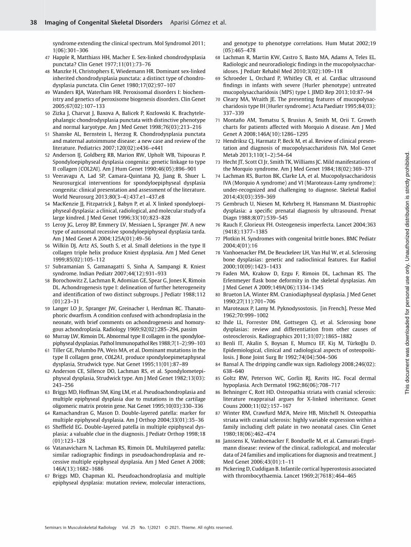
syndrome extending the clinical spectrum. Mol Syndromol 2011;1(06):301–306
47 Happle R, Matthiass HH, Macher E. Sex-linked chondrodysplasiapunctata? Clin Genet 1977;11(01):73–76
48 Manzke H, Christophers E, Wiedemann HR. Dominant sex-linkedinherited chondrodysplasia punctata: a distinct type of chondro-dysplasia punctata. Clin Genet 1980;17(02):97–107
49 Wanders RJA, Waterham HR. Peroxisomal disorders I: biochem-istry and genetics of peroxisome biogenesis disorders. Clin Genet2005;67(02):107–133
50 Zizka J, Charvat J, Baxova A, Balicek P, Kozlowski K. Brachytele-phalangic chondrodysplasia punctata with distinctive phenotypeand normal karyotype. Am J Med Genet 1998;76(03):213–216
51 Shanske AL, Bernstein L, Herzog R. Chondrodysplasia punctataand maternal autoimmune disease: a new case and review of theliterature. Pediatrics 2007;120(02):e436–e441
52 Anderson IJ, Goldberg RB, Marion RW, Upholt WB, Tsipouras P.Spondyloepiphyseal dysplasia congenita: genetic linkage to typeII collagen (COL2AI). Am J Hum Genet 1990;46(05):896–901
53 Veeravagu A, Lad SP, Camara-Quintana JQ, Jiang B, Shuer L.Neurosurgical interventions for spondyloepiphyseal dysplasiacongenita: clinical presentation and assessment of the literature.World Neurosurg 2013;80(3–4):437.e1–437.e8
54 MacKenzie JJ, Fitzpatrick J, Babyn P, et al. X linked spondyloepi-physeal dysplasia: a clinical, radiological, andmolecular studyof alarge kindred. J Med Genet 1996;33(10):823–828
55 Leroy JG, Leroy BP, Emmery LV, Messiaen L, Spranger JW. A newtype of autosomal recessive spondyloepiphyseal dysplasia tarda.Am J Med Genet A 2004;125A(01):49–56
56 Wilkin DJ, Artz AS, South S, et al. Small deletions in the type IIcollagen triple helix produce Kniest dysplasia. Am J Med Genet1999;85(02):105–112
57 Subramanian S, Gamanagatti S, Sinha A, Sampangi R. Kniestsyndrome. Indian Pediatr 2007;44(12):931–933
58 Borochowitz Z, Lachman R, Adomian GE, Spear G, Jones K, RimoinDL. Achondrogenesis type I: delineation of further heterogeneityand identification of two distinct subgroups. J Pediatr 1988;112(01):23–31
59 Langer LO Jr, Spranger JW, Greinacher I, Herdman RC. Thanato-phoric dwarfism. A condition confused with achondroplasia in theneonate, with brief comments on achondrogenesis and homozy-gous achondroplasia. Radiology 1969;92(02):285–294, passim
60 Murray LW, Rimoin DL. Abnormal type II collagen in the spondyloe-piphysealdysplasias. Pathol ImmunopatholRes1988;7(1–2):99–103
61 Tiller GE, Polumbo PA, Weis MA, et al. Dominant mutations in thetype II collagen gene, COL2A1, produce spondyloepimetaphysealdysplasia, Strudwick type. Nat Genet 1995;11(01):87–89
62 Anderson CE, Sillence DO, Lachman RS, et al. Spondylometepi-physeal dysplasia, Strudwick type. Am J Med Genet 1982;13(03):243–256
63 Briggs MD, Hoffman SM, King LM, et al. Pseudoachondroplasia andmultiple epiphyseal dysplasia due to mutations in the cartilageoligomeric matrix protein gene. Nat Genet 1995;10(03):330–336
64 Ramachandran G, Mason D. Double-layered patella: marker formultiple epiphyseal dysplasia. Am J Orthop 2004;33(01):35–36
65 Sheffield EG. Double-layered patella in multiple epiphyseal dys-plasia: a valuable clue in the diagnosis. J Pediatr Orthop 1998;18(01):123–128
66 Vatanavicharn N, Lachman RS, Rimoin DL. Multilayered patella:similar radiographic findings in pseudoachondroplasia and re-cessive multiple epiphyseal dysplasia. Am J Med Genet A 2008;146A(13):1682–1686
67 Briggs MD, Chapman KL. Pseudoachondroplasia and multipleepiphyseal dysplasia: mutation review, molecular interactions,
and genotype to phenotype correlations. Hum Mutat 2002;19(05):465–478
68 Lachman R, Martin KW, Castro S, Basto MA, Adams A, Teles EL.Radiologic and neuroradiologic findings in the mucopolysacchar-idoses. J Pediatr Rehabil Med 2010;3(02):109–118
69 Schroeder L, Orchard P, Whitley CB, et al. Cardiac ultrasoundfindings in infants with severe (Hurler phenotype) untreatedmucopolysaccharidosis (MPS) type I. JIMD Rep 2013;10:87–94
70 Cleary MA, Wraith JE. The presenting features of mucopolysac-charidosis type IH (Hurler syndrome). Acta Paediatr 1995;84(03):337–339
71 Montaño AM, Tomatsu S, Brusius A, Smith M, Orii T. Growthcharts for patients affected with Morquio A disease. Am J MedGenet A 2008;146A(10):1286–1295
72 Hendriksz CJ, Harmatz P, Beck M, et al. Review of clinical presen-tation and diagnosis of mucopolysaccharidosis IVA. Mol GenetMetab 2013;110(1–2):54–64
73 Hecht JT, Scott CI Jr, Smith TK,Williams JC. Mild manifestations ofthe Morquio syndrome. Am J Med Genet 1984;18(02):369–371
74 Lachman RS, Burton BK, Clarke LA, et al. MucopolysaccharidosisIVA (Morquio A syndrome) and VI (Maroteaux-Lamy syndrome):under-recognized and challenging to diagnose. Skeletal Radiol2014;43(03):359–369
75 Gembruch U, Niesen M, Kehrberg H, Hansmann M. Diastrophicdysplasia: a specific prenatal diagnosis by ultrasound. PrenatDiagn 1988;8(07):539–545
76 Rauch F, Glorieux FH. Osteogenesis imperfecta. Lancet 2004;363(9418):1377–1385
77 Plotkin H. Syndromes with congenital brittle bones. BMC Pediatr2004;4(01):16
78 Vanhoenacker FM, De Beuckeleer LH, Van Hul W, et al. Sclerosingbone dysplasias: genetic and radioclinical features. Eur Radiol2000;10(09):1423–1433
79 Faden MA, Krakow D, Ezgu F, Rimoin DL, Lachman RS. TheErlenmeyer flask bone deformity in the skeletal dysplasias. AmJ Med Genet A 2009;149A(06):1334–1345
80 Brueton LA, Winter RM. Craniodiaphyseal dysplasia. J Med Genet1990;27(11):701–706
81 Maroteaux P, Lamy M. Pyknodysostosis. [in French]. Presse Med1962;70:999–1002
82 Ihde LL, Forrester DM, Gottsegen CJ, et al. Sclerosing bonedysplasias: review and differentiation from other causes ofosteosclerosis. Radiographics 2011;31(07):1865–1882
83 Benli IT, Akalin S, Boysan E, Mumcu EF, Kiş M, Türkoğlu D.Epidemiological, clinical and radiological aspects of osteopoiki-losis. J Bone Joint Surg Br 1992;74(04):504–506
84 Bansal A. The dripping candle wax sign. Radiology 2008;246(02):638–640
85 Goltz RW, Peterson WC, Gorlin RJ, Ravits HG. Focal dermalhypoplasia. Arch Dermatol 1962;86(06):708–717
86 Behninger C, Rott HD. Osteopathia striata with cranial sclerosis:literature reappraisal argues for X-linked inheritance. GenetCouns 2000;11(02):157–167
87 Winter RM, Crawfurd Md’A, Meire HB, Mitchell N. Osteopathiastriata with cranial sclerosis: highly variable expression within afamily including cleft palate in two neonatal cases. Clin Genet1980;18(06):462–474
88 Janssens K, Vanhoenacker F, Bonduelle M, et al. Camurati-Engel-mann disease: review of the clinical, radiological, and moleculardata of 24 families and implications for diagnosis and treatment. JMed Genet 2006;43(01):1–11
89 Pickering D, Cuddigan B. Infantile cortical hyperostosis associatedwith thrombocythaemia. Lancet 1969;2(7618):464–465
Seminars in Musculoskeletal Radiology Vol. 25 No. 1/2021 © 2021. Thieme. All rights reserved.
Imaging of Congenital Skeletal Disorders Aparisi Gómez et al.38
Thi
s do
cum
ent w
as d
ownl
oade
d fo
r pe
rson
al u
se o
nly.
Una
utho
rized
dis
trib
utio
n is
str
ictly
pro
hibi
ted.