Dental JOURNAL - Thieme Connect
8
Casting of metal copings has traditionally been carried out by lost wax (LW) technique given by Taggart in 1907. Initially noble alloys were used for fabricating metal copings because of their 8 ease of use and biocompatibility. However with time manufacturers shifted to less costly alternatives like the base metal alloys. Casting of all base metal alloys is more technique sensitive compared to casting of noble alloys because of their high melting range and oxidation of base metal alloys during 8-11 casting. In addition, due to their high hardness, grinding of base metals especially Co-Cr alloys, to finish castings is comparatively time consuming for 12 dental laboratories. Such limitations of casting procedures has led to introduction TO EVALUATE THE MARGINAL FIT OF METAL COPINGS FABRICATED BY CONVENTIONAL CASTING PROCEDURE AND DIRECT METAL LASER SINTERING TECHNOLOGY – AN IN VITRO STUDY 1 2 3 4 5 Monica Sharma , Ajay Bansal , Sunny Panthi , Shefali S Malik , Atulya Sharma 1 Senior Lecturer, Dept. of Prosthodontics, Bhojia Dental College and Hospital, Himachal Pradesh, India 2 Reader, Dept. of Prosthodontics, Bhojia Dental College and Hospital, Himachal Pradesh,India 3 Senior Lecturer, Dept. of Prosthodontics, Bhojia Dental College and Hospital, Himachal Pradesh, India 4 Senior Lecturer, Dept. of Prosthodontics, Bhojia Dental College and Hospital, Himachal Pradesh, India 5 Senior Lecturer, Dept. of Prosthodontics, Bhojia Dental College and Hospital, Himachal Pradesh, India Corresponding Author: Monica Sharma E-mail: [email protected] th Received: 25 December 2016 rd Accepted: 3 April 2017 th Online: 20 May 2017 ORIGINAL ARTICLE www.djas.co.in ISSN No-2321-1482 DJAS 5(I), 39-46, 2017 All rights are reserved Dental JOURNAL of A d v a n c e S t u d i e s ABSTRACT Purpose: The purpose of this in vitro study was to evaluate marginal fit of cobalt- chromium (Co-Cr) copings fabricated by direct metal laser sintering system (DMLS) and conventional lost-wax technique (LW). Materials and method: Forty tooth preparations were carried out over extracted mandibular molars. They were divided into two groups A and B of 20 each. For group A Co-Cr copings were fabricated by direct metal laser sintering (DMLS) and for group B by lost wax technique (LW). Glass –ionomer cement (GIC) was used to tack the copings over their preparations. Marginal fit was then evaluated directly under the stereomicroscope. Results: The mean marginal gap of group A was 27.9 + 2.4 μm and group B was 40.4 μm. Statistical analysis using t - test showed highly significant difference (P>.05) between the marginal mean of the DMLS (group A) compared to LW (group B). Conclusion: The DMLS copings demonstrated superior marginal fit compared to that of conventional Co-Cr casted copings. Keywords: Cobalt Chromium, Direct metal laser sintering, Porcelain fused metal crowns INTRODUCTION Metal fused to ceramic prosthesis is still the most widely used restoration material for fabricating complete coverage crowns and partial fixed dental 1 prostheses. A porcelain fused to metal prosthesis is made by sintering layers of porcelain over metal coping. An essential factor for the success of a metal–ceramic fixed dental prosthesis is marginal fit of 2,3 the metal coping. Excessive marginal discrepancy for crowns increases cement dissolution and microleakage and can 4 cause inflammation of the vital pulp. Poor marginal adaptation of crowns increases 5,6 plaque retention, changes the composition 7 of the subgingival microflora, and can result in the onset of periodontal disease. 39
-
Upload
khangminh22 -
Category
Documents
-
view
2 -
download
0
Transcript of Dental JOURNAL - Thieme Connect

Casting of metal copings has
traditionally been carried out by lost wax
(LW) technique given by Taggart in 1907.
Initially noble alloys were used for
fabricating metal copings because of their 8
ease of use and biocompatibility.
However with time manufacturers shifted
to less costly alternatives like the base
metal alloys. Casting of all base metal
alloys is more technique sensitive
compared to casting of noble alloys
because of their high melting range and
oxidation of base metal alloys during 8-11
casting. In addition, due to their high
hardness, grinding of base metals
especially Co-Cr alloys, to finish castings
is comparatively time consuming for 12 dental laboratories. Such limitations of
casting procedures has led to introduction
TO EVALUATE THE MARGINAL FIT OF METAL COPINGS FABRICATED BY CONVENTIONAL CASTING PROCEDURE AND DIRECT
METAL LASER SINTERING TECHNOLOGY – AN IN VITRO STUDY1 2 3 4 5Monica Sharma , Ajay Bansal , Sunny Panthi , Shefali S Malik , Atulya Sharma
1Senior Lecturer, Dept. of Prosthodontics, Bhojia Dental College and Hospital, Himachal Pradesh, India2Reader, Dept. of Prosthodontics, Bhojia Dental College and Hospital, Himachal Pradesh,India3Senior Lecturer, Dept. of Prosthodontics, Bhojia Dental College and Hospital, Himachal Pradesh, India4Senior Lecturer, Dept. of Prosthodontics, Bhojia Dental College and Hospital, Himachal Pradesh, India5Senior Lecturer, Dept. of Prosthodontics, Bhojia Dental College and Hospital, Himachal Pradesh, India
Corresponding Author: Monica Sharma
E-mail: [email protected]
thReceived: 25 December 2016rdAccepted: 3 April 2017th Online: 20 May 2017
ORIGINAL ARTICLEwww.djas.co.in ISSN No-2321-1482
DJAS 5(I), 39-46, 2017All rights are reserved
Dental JOURNAL of A d v a n c e S t u d i e s
ABSTRACT
Purpose: The purpose of this in vitro study was to evaluate marginal fit of cobalt- chromium (Co-Cr) copings fabricated by direct metal laser sintering system (DMLS) and conventional lost-wax technique (LW). Materials and method: Forty tooth preparations were carried out over extracted mandibular molars. They were divided into two groups A and B of 20 each. For group A Co-Cr copings were fabricated by direct metal laser sintering (DMLS) and for group B by lost wax technique (LW). Glass –ionomer cement (GIC) was used to tack the copings over their preparations. Marginal fit was then evaluated directly under the stereomicroscope. Results: The mean marginal gap of group A was 27.9 + 2.4 µm and group B was 40.4 µm. Statistical analysis using t - test showed highly significant difference (P>.05) between the marginal mean of the DMLS (group A) compared to LW (group B). Conclusion: The DMLS copings demonstrated superior marginal fit compared to that of conventional Co-Cr casted copings.
Keywords: Cobalt Chromium, Direct metal laser sintering, Porcelain fused metal crowns
INTRODUCTION
Metal fused to ceramic prosthesis
is still the most widely used restoration
material for fabricating complete
coverage crowns and partial fixed dental 1
prostheses. A porcelain fused to metal
prosthesis is made by sintering layers of
porcelain over metal coping. An essential
factor for the success of a metal–ceramic
fixed dental prosthesis is marginal fit of 2,3the metal coping. Excessive marginal
discrepancy for crowns increases cement
dissolution and microleakage and can 4cause inflammation of the vital pulp. Poor
marginal adaptation of crowns increases 5,6
plaque retention, changes the composition 7
of the subgingival microflora, and can
result in the onset of periodontal disease.
39

of direct metal laser-sintering (DMLS) system to
dentistry.
The newly developed direct metal laser-
sintering (DMLS) system is a rapid prototyping
method for fabricating metal products directly from
computer-aided design (CAD) data. Automated
fabrication is accomplished layer-by-layer by
selectively fusing together metal powders with the help
of a laser beam. Advantages of the DMLS system
include ease of fabrication, use of fully automated 3
system, and comparatively shorter working time.
Also, metal copings fabricated with DMLS have been
reported to have satisfactory mechanical and chemical 13-16properties.
While an essential condition for a successful
dental prosthesis is good marginal fit, there is little data
on the marginal fit of fixed dental prostheses (FDPs) 3,8,17-21fabricated by the DMLS system.
The purpose of this in vitro study was to
evaluate and compare the marginal fit of Co-Cr
copings, fabricated by direct metal laser-sintering
technology and conventional lost wax technique. The
hypothesis for the study was that marginal fit of
copings fabricated with both the techniques i.e. DMLS
and lost wax techinque would be same.
MATERIAL AND METHOD
Forty caries-free natural mandibular molars,
extracted for reasons other than the study, were
selected. They were cleaned and stored in 0.1% thymol
solution (Amrit chemicals Ltd,India) at room
temperature throughout the course of the study, to
prevent them from drying and becoming brittle. Each
tooth was mounted over self-cure acrylic resin mount
(Self cure, DPI, India).
Tooth preparation and Die Fabrication
The tooth preparation of all the fourty
specimens was done following standard preparation 22
protocol, using a high-speed angled handpiece and
diamond rotary cutting instruments under water
cooling. A 1.2 mm wide, smooth, continuous, shoulder
finish line was prepared, 2 mm above cemento-enamel
junction and an occlusal reduction of 1.5 mm was done
(Figure 1). The convergence angle of the preparation
Figure 1: Prepared teeth
corresponded to the convergence angle of diamond
rotary cutting instrument. New diamond bur was used
for every tooth preparation. To control the amount of
tooth reduction while doing tooth preparation silicone
putty index (Putty, 3M EPSE, Germany) was used and
a periodontal probe was used to gauge the tooth
reduction. Impressions were made for each sample in
custom tray fabricated with self cure resin(DPI) using
medium-body polyvinyl siloxane impression material
(Aquasil Monophase, Dentsply, USA). Dies were
poured in type IV die-stone (Kalrock, Kalabhai, India)
(Figure 2). Specimens were then divided into two
Figure 2: Stone Dies
Dental Journal of Advance Studies Vol. 5 (Issue I) 2017
40
groups (A&B) of twenty sample each over which
copings with direct metal laser sintering and
conventional casting were fabricated.
Fabrication of copings
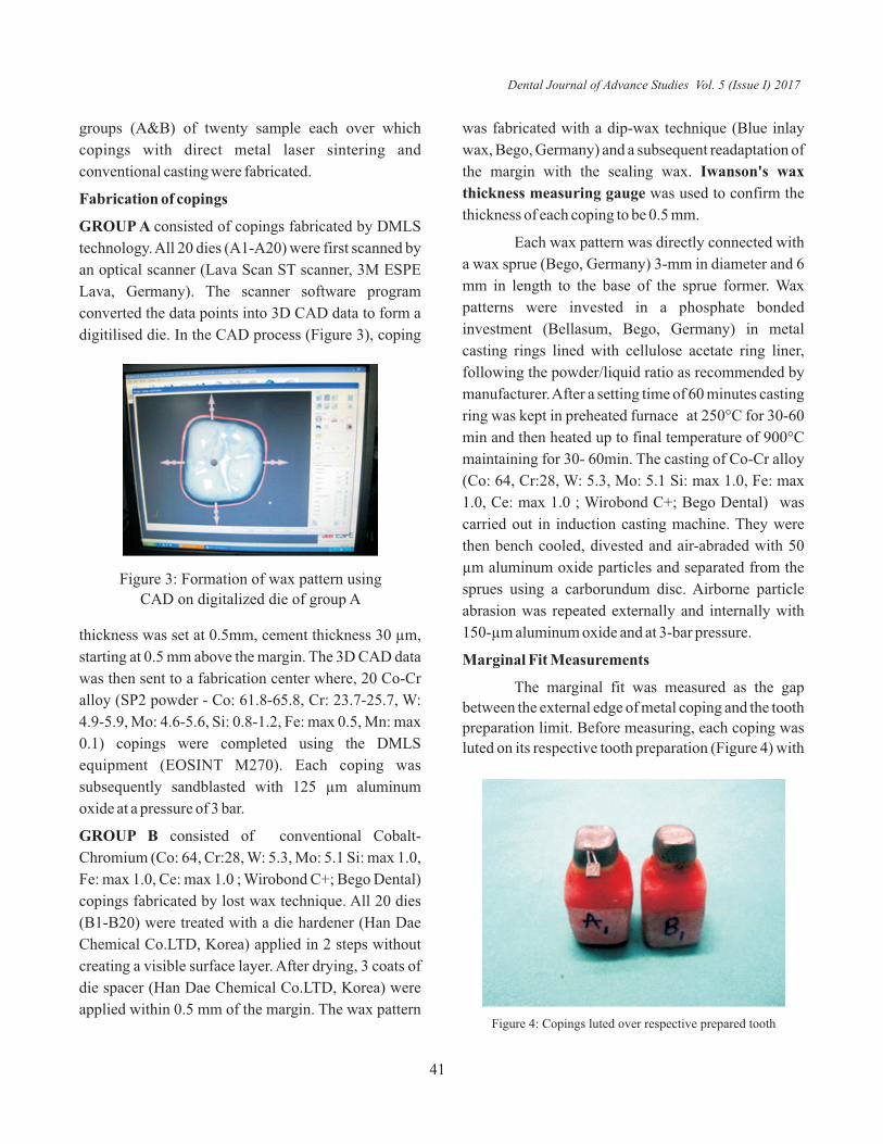
GROUP A consisted of copings fabricated by DMLS
technology. All 20 dies (A1-A20) were first scanned by
an optical scanner (Lava Scan ST scanner, 3M ESPE
Lava, Germany). The scanner software program
converted the data points into 3D CAD data to form a
digitilised die. In the CAD process (Figure 3), coping
was fabricated with a dip-wax technique (Blue inlay
wax, Bego, Germany) and a subsequent readaptation of
the margin with the sealing wax. Iwanson's wax
thickness measuring gauge was used to confirm the
thickness of each coping to be 0.5 mm.
Each wax pattern was directly connected with
a wax sprue (Bego, Germany) 3-mm in diameter and 6
mm in length to the base of the sprue former. Wax
patterns were invested in a phosphate bonded
investment (Bellasum, Bego, Germany) in metal
casting rings lined with cellulose acetate ring liner,
following the powder/liquid ratio as recommended by
manufacturer. After a setting time of 60 minutes casting
ring was kept in preheated furnace at 250°C for 30-60
min and then heated up to final temperature of 900°C
maintaining for 30- 60min. The casting of Co-Cr alloy
(Co: 64, Cr:28, W: 5.3, Mo: 5.1 Si: max 1.0, Fe: max
1.0, Ce: max 1.0 ; Wirobond C+; Bego Dental) was
carried out in induction casting machine. They were
then bench cooled, divested and air-abraded with 50
µm aluminum oxide particles and separated from the
sprues using a carborundum disc. Airborne particle
abrasion was repeated externally and internally with
150-µm aluminum oxide and at 3-bar pressure.
Marginal Fit Measurements
The marginal fit was measured as the gap
between the external edge of metal coping and the tooth
preparation limit. Before measuring, each coping was
luted on its respective tooth preparation (Figure 4) with
Figure 3: Formation of wax pattern using
CAD on digitalized die of group A
thickness was set at 0.5mm, cement thickness 30 µm,
starting at 0.5 mm above the margin. The 3D CAD data
was then sent to a fabrication center where, 20 Co-Cr
alloy (SP2 powder - Co: 61.8-65.8, Cr: 23.7-25.7, W:
4.9-5.9, Mo: 4.6-5.6, Si: 0.8-1.2, Fe: max 0.5, Mn: max
0.1) copings were completed using the DMLS
equipment (EOSINT M270). Each coping was
subsequently sandblasted with 125 µm aluminum
oxide at a pressure of 3 bar.
GROUP B consisted of conventional Cobalt-
Chromium (Co: 64, Cr:28, W: 5.3, Mo: 5.1 Si: max 1.0,
Fe: max 1.0, Ce: max 1.0 ; Wirobond C+; Bego Dental)
copings fabricated by lost wax technique. All 20 dies
(B1-B20) were treated with a die hardener (Han Dae
Chemical Co.LTD, Korea) applied in 2 steps without
creating a visible surface layer. After drying, 3 coats of
die spacer (Han Dae Chemical Co.LTD, Korea) were
applied within 0.5 mm of the margin. The wax pattern Figure 4: Copings luted over respective prepared tooth
Dental Journal of Advance Studies Vol. 5 (Issue I) 2017
41

minimum amount of GIC (Glass Ionomer, GC
Corporation, Japan) applied over the occlusal surface
of the prepared tooth and secured with finger pressure 23for 3 min. The marginal gap was then examined using
stereomicroscope ×40 (Magnus, Olympus, Tokyo,
Japan), and digital images were captured at each of the
four surfaces mesial, distal, buccal and lingual. The flat
base of the specimens along each surface prevented the
specimens from moving and kept each surface
perpendicular to the objective lens. Using image
analysing software, five measurements were made at
each of the four positions (Figure 5, 6) for a total of 20
measurements per coping.
RESULTS
In total 800 marginal gap measurements were
made for all the samples. The mean marginal gap
values for each specimen of group A and group B are
given in the table I and table II. The over all mean
marginal gap of group A was 27.9+2.4 µm and group B
was 40.4+ 6 µm (Table-III & Graph-I). Statistical
analysis using t - test showed (Table VI) highly
significant difference between the overall mean
marginal gap of group A compared to group B.Figure 5: Marginal gap measurement for Group A using stereomicroscope
Figure 6: Marginal gap measurement for Group B using stereomicroscope
GROUP A (DMLS) MEAN MARGINAL GAP
STANDARD DEVIATION
MAXIMUM MINIMUM MEDIAN
A1 25.3605 5.23 35.18 16.15 25.155 A2 29.732 6.906 39.96 19.42 31.09 A3 27.663 6.852 40.1 17.63 29.24 A4 24.602 4.637 34.71 15.67 23 A5 25.898 6.157 36.83 17.49 24.33 A6 26.814 5.838 35.99 17.11 27.64 A7 24.48 4.652 34.56 17.68 23.2 A8 24.62 4.2173 34.23 16.11 24.195 A9 29.551 6.141 38.96 19.56 30.83 A10 30.518 5.130 39.75 23.17 29.06 A11 27.17 4.37 37.89 20.87 26.17 A12 27.12 5.186 37.66 19.34 26.045 A13 27.53 4.460 37.84 21.56 27.085 A14 28.423 4.893 38.91 21.31 28.825 A15 30.861 5.239 39.88 21.34 31.995 A16 33.455 5.756 42.88 21.56 32.77 A17 28.997 5.758 38.91 18.91 29.155 A18 26.889 4.243 36.67 19.47 26.28 A19 29.492 4.811 39.65 19.23 28.96 A20 29.414 6.022 41.67 21.56 29.165
Table 1: Results for Group A
Dental Journal of Advance Studies Vol. 5 (Issue I) 2017
42
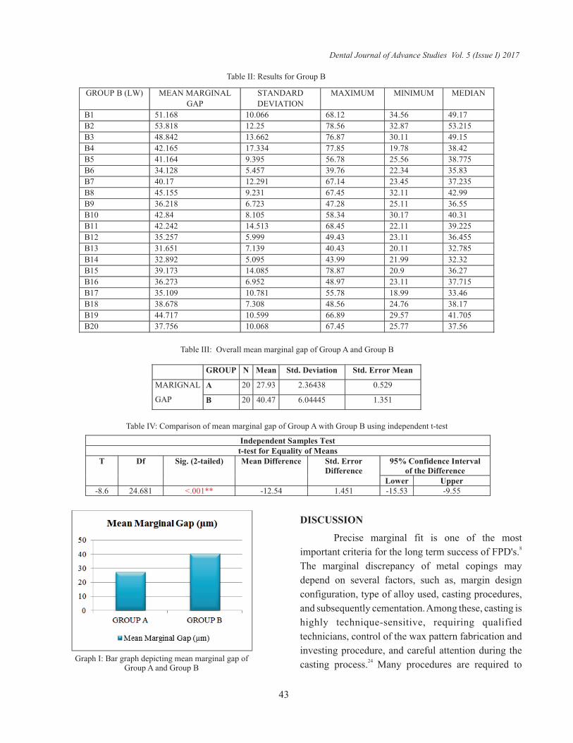
GROUP B (LW) MEAN MARGINAL
GAP
STANDARD
DEVIATION
MAXIMUM MINIMUM MEDIAN
B1 51.168 10.066 68.12 34.56 49.17
B2 53.818 12.25 78.56 32.87 53.215
B3 48.842 13.662 76.87 30.11 49.15
B4 42.165 17.334 77.85 19.78 38.42
B5 41.164 9.395 56.78 25.56 38.775
B6 34.128 5.457 39.76 22.34 35.83
B7 40.17 12.291 67.14 23.45 37.235
B8 45.155 9.231 67.45 32.11 42.99
B9 36.218 6.723 47.28 25.11 36.55
B10 42.84 8.105 58.34 30.17 40.31
B11 42.242 14.513 68.45 22.11 39.225
B12 35.257 5.999 49.43 23.11 36.455
B13 31.651 7.139 40.43 20.11 32.785
B14 32.892 5.095 43.99 21.99 32.32
B15 39.173 14.085 78.87 20.9 36.27
B16 36.273 6.952 48.97 23.11 37.715
B17 35.109 10.781 55.78 18.99 33.46
B18 38.678 7.308 48.56 24.76 38.17
B19 44.717 10.599 66.89 29.57 41.705
B20 37.756 10.068 67.45 25.77 37.56
Table II: Results for Group B
DISCUSSION
Precise marginal fit is one of the most 8
important criteria for the long term success of FPD's.
The marginal discrepancy of metal copings may
depend on several factors, such as, margin design
configuration, type of alloy used, casting procedures,
and subsequently cementation. Among these, casting is
highly technique-sensitive, requiring qualified
technicians, control of the wax pattern fabrication and
investing procedure, and careful attention during the 24
casting process. Many procedures are required to
Table III: Overall mean marginal gap of Group A and Group B
GROUP N Mean Std. Deviation Std. Error Mean
MARIGNAL
GAP
A 20 27.93 2.36438 0.529
B 20 40.47 6.04445 1.351
Table IV: Comparison of mean marginal gap of Group A with Group B using independent t-test
Independent Samples Test t-test for Equality of Means
T Df Sig. (2-tailed) Mean Difference Std. Error Difference
95% Confidence Interval of the Difference
Lower Upper -8.6 24.681 <.001** -12.54 1.451 -15.53 -9.55
Graph I: Bar graph depicting mean marginal gap of Group A and Group B
Dental Journal of Advance Studies Vol. 5 (Issue I) 2017
43

execute this process, which increases the possibility of
mistakes affecting the accuracy and fit of copings
made. To overcome these flaws, a new additive
technique for forming the metal substructures has been
introduced. Direct Metal Laser Sintering (DMLS) is a
promising new technology that may avoid the 25
distortions inherent to casting procedures. The CAD
process of producing copings by DMLS technique
using automated scanning process and powerful CAD
software offers many advantages such as complete
control over the framework and coping designing,
margin placement and cement space maintenance. To
date, very few studies have been published on the
marginal fit of FPD's made by DMLS. Therefore, this
study was carried out to assess the marginal fit of metal
copings made by new DMLS technique compared with
conventional casting procedure.
Co-Cr was used to fabricate the metal copings
in the study, because currently they are used more
commonly than Ni-Cr alloys for fixed prosthesis.
Electrochemical studies show that Co-Cr alloys are
more resistant to corrosion than Ni-Cr alloys. Nickel
based alloys have a greater sensitization potential than
cobalt-chromium alloys, whereas Co-Cr alloy allergies 12, 24are rare. .
The clinical objective is to have the lowest marginal
gap that still allows proper seating of the coping. All
marginal fit values obtained for both the techniques in
this study were within clinical permissible limits, that 28is, 120µm . The marginal fit of 20 samples of DMLS
technique varied between 24-33.5 µm as compared to
conventional casting whose marginal fit varied from
34-54 µm. This shows that consistent and better
marginal fit values can be obtained with the DMLS
technique. The comparison of overall mean marginal
fit of copings made by DMLS technique was
significantly superior to that of LW group (p>0.001).
This is inaccordance to the findings of Ortop et al, 21Huang et al. and Xu et al. who recorded lower
discrepancies for laser sintered Co-Cr than for cast Co-11,18
Cr. Li et alobserved 220 metal crowns for 24 months
in order to evaluate the clinical effect of laser sintered
Co-Cr and cast Co-Cr metal crowns and found that
laser sintered Co-Cr metal crowns resulted in better 29marginal fit than cast Co-Cr metal crowns.
DMLS technique has its further advantages
like less time in fabrication and a uniform thickness of
metal coping. Reducing the technicians work in
adjusting the coping to desired thickness and a uniform
thickness of ceramic layer can be added.
DMLS is a relatively new method compared to
conventional casting procedure. The ?t of CAD/CAM
prosthetic frameworks may rely on the precision of the
scanner that reads the abutments, on the ways in which
the software can transform the scanning data into a 3D
model on the computer, and on the accuracy of the
machine that uses CAM to produce objects from the 8
CAD data. The use of the DMLS technology may
result in the most predictable fabrication method under
the tested experimental conditions. However, clinical
implementation of the DMLS system seems to require
further investigation, because studies of restoration
longevity are scarce.
LIMITATIONS OF THE STUDY
The tooth preparations for all the 40 specimens
were carried out manually to simulate the oral
environment which may have incorporated operator
errors, but the preparations closely simulated the
clinical conditions.
CONCLUSION
Within the limitations of this in vitro
investigation, the following conclusions were drawn:
1. Both the casting techniques exhibited different
results, disaproving the null hypothesis.
2. Copings fabricated with DMLS technique had
better mnarginal fit when compared to copings
fabricated from conventional lost wax technique.
The reference drawn from this study strongly
suggests use of DMLS technique in place of
conventional lost wax technique to fabricate copings
for FPD in routinue dental practice.
Dental Journal of Advance Studies Vol. 5 (Issue I) 2017
44

REFERENCES
1) Petteno D, Schierano G, Bassi F, Bresciano ME, Carossa S.
Comparison of marginal fit of 3 different metal-ceramic
systems: an in vitro study. Int J Prosthodont 2000; 13:405-408.
2) Hunter AJ, Hunter AR. Gingival margins for crowns: a review
and discussion. Part 2. Discrepancies and configurations. J
Prosthet Dent 1990;64:636–42.
3) Kim KB, Kim WC, Kim HY, Kim JH. An evaluation of
marginal fit of three-unit fixed dental prostheses fabricated by
direct metal laser sintering system. Dent Mater
2013;29(7):e91-6.
4) Bindl A, Mormann WH. Marginal and internal fit of all-
ceramic CAD/CAM crowncopings on chamfer preparations. J
OralRehabil 2005;32:441-7.
5) Valderhaug J, Birkeland JM. Periodontal conditions in
patients 5 years following insertion of fixed prostheses.
Pocket depth and loss of attachment. J Oral Rehabil 1976;3:
237-43.
6) Valderhaug J, Heloe LA. Oral hygiene in a group of supervised
patients with fixed prostheses. J Periodontol 1977;48:221-4.
7) Lang NP, Kiel RA, Anderhalden K. Clinical and
microbiological effects of subgingival restorations with
overhanging or clinically perfect margins. J Clin Periodontol
1983;10:563-78.
8) Ucar Y, Akova T, Akyil M, Brantley W. Internal fit evaluation
of crowns prepared using a new dental crown fabrication
technique: laser-sintered Co–Cr crowns. J Prosthet Dent
2009;102:253–259.
9) Faucher RR, Nicholls JI. Distortion related to margin design in
porcelainfused-to-metal restorations. J Prosthet Dent
1980;43:149-55.
10) Buchanan WT, Svare CW, Turner KA. The effect of repeated
firings and strength on marginal distortion in two ceramometal
systems. J Prosthet Dent 1981;45:502-6.
11) Huang Z,Lu Zhang, Zhu J, Zhang X. Clinical marginal and
internal fit of metal ceramic crowns fabricated with a selective
laser melting technology. J Prosthet Dent 2015;113:623-27.
12) Anusavice KJ. Phillips' science of dental materials. 11th ed.
Phialdelphia: W.B. Saunders; 2003. p. 565, 584, 585.
13) Vandenbroucke B, Kruth JP. Selective laser melting of
biocompatible metals for rapid manufacturing of medical
parts. Rapid Prototyping J 2007;13:196-203.
14) Wu L, Zhu H, Gai X, Wang Y. Evaluation of the mechanical
properties and porcelain bond strength of cobalt-chromium
dental alloy fabricated by selective laser melting. J Prosthet
Dent 2014;111:51-5.
15) Yang X, Xiang N, Wei B. Effect of fluoride content on ion
release from castand selective laser melting-processed Co-Cr-
Mo alloys. J Prosthet Dent2014;112:1212-6.
16) Zeng L, Xiang N, Wei B. A comparison of corrosion resistance
of cobaltchromium-molybdenum metal ceramic alloy
fabricated with selectivelaser melting and traditional
processing. J Prosthet Dent 2014;112:1217-24.
17) Quante K, Ludwig K, Kern M. Marginal and internal fit of
metal–ceramic crowns fabricated with a new laser melting
technology. Dent Mater 2008;24:1311–1315.
18) Ortorp OA, Jonsson D, Mouhsen A, Von Steyern PV. The fit of
cobalt–chromium three-unit fixed dental prostheses
fabricated with four different techniques: A comparative in
vitro study. Dent Mater 2011;27:356-363.
19) Oyagüe RC, Sánchez-Turrión A, López-Lozano JF, Suárez-
García MJ. Vertical discrepancy and microleakage of laser-
sintered and vacuum-castimplant-supported structures luted
with different cement types. J Dent 2012;40(2):123-30.
20) Kim KB, Kim WC, Kim HY, Kim JH. Evaluation of the
marginal and internal gap of metal-ceramic crown fabricated
with a selective laser sintering technology: two- and three-
dimensional replica techniques. J Adv Prosthodont 2013; 5(2):
179–186.
21) Xu D,XiangN,Wei B. The marginal fit of selective laser
melting- fabricated metal crowns: an in vitro study. J Prosthet
Dent 2014;112:1437-1440.
22) Shillingburg HT. Fundamentals of fixed prosthosthodontics.
4th ed. USA: Quintessence publishing; 2012.p.149-164.
23) Att W, Komine F, Gerds T, Strub JR. Marginal adaptation of
three different zirconium dioxide three-unit fixed dental
prostheses. J Prosthet Dent 2009;101:239–247.
24) Bhaskaran E, Azhagarasan NS, Miglani S, Gajapathi
B.Comparative Evaluation of Marginal and Internal Gap of
Co–Cr Copings Fabricated from Conventional Wax Pattern,
3D Printed Resin Pattern and DMLS Tech: An In Vitro Study. J
Indian ProsthodontSoc 2013; 13(3): 189-195.
25) Akova T, Ucar Y, Tukay A, Balkaya MC, Brantley WA.
Comparison of the bond strength of laser-sintered and cast
Dental Journal of Advance Studies Vol. 5 (Issue I) 2017
45

base metal dental alloys to porcelain. Dent Mater
2008;24(10):1400-4.
26) Beschnidt SM, Strub JR. Evaluation of the marginal accuracy
of different all-ceramic crown systems after simulation in the
artificial mouth. J Oral Rehabil 1999;26(7):582-93.
27) Nawafleh NA, Mack F, Evans J, Mackay J, Hatamleh MM.
Accuracy and reliability of methods to measure marginal
adaptation of crowns and FDPs: a literature review. J
Prosthodont 2013;22(5):419-28
28) McLean JW, von Fraunhofer JA. The estimation of cement
film thickness by an in vivo technique. Br Dent J
1971;131:107-11.
29. Li JM, Wang WQ, Ma JY. Comparison of the clinical effects of
selective laser melting deposition basal crowns and cobalt
chromium alloy base crowns. Shanghai Kou Qiang Yi Xue
2014;23:350-3.
Dental Journal of Advance Studies Vol. 5 (Issue I) 2017
Source of Support: Nil, Conflict of Interest: None Declared
46