Which Is Better PCI or CABG for LMT Lesion and CTO of RCA ...
Upload
khangminh22Category
view
4download
0
The Large Focal Isolated Chondral LesionJorge Chahla, MD, PhD1 Brady T. Williams, MD2 Adam B. Yanke, MD, PhD1 Jack Farr3
1Department of Orthopedic Surgery, Rush University Medical Center,Chicago, Illinois
2Department of Orthopedic Surgery, University of Colorado, Aurora,Colorado
3Knee Preservation and Cartilage Restoration Center, OrthoIndy,Indianapolis, Indiana
J Knee Surg
Address for correspondence Jorge Chahla, MD, PhD, Department ofOrthopedic Surgery, Rush University Medical Center, 1611W HarrisonStreet, Suite 300, Chicago Excelsior, IL 55331(e-mail: [email protected]).
Treatment of large articular cartilage defects of the knee canbe difficult, particularly in young athletic patients. Suchlesions are common in young individuals (<40 years), withan overall incidence of 4.2 to 6.2%, and up to 36% inathletes.1–5 In a series of nearly 1,000 arthroscopies, full-thickness cartilage defects meeting criteria for repair werefound in 11% of knees, with 55% of lesions being greater than2 cm2.2 If left unaddressed, cartilage defects canworsen overtime and may progress to diffuse degenerative changes.6
Surgical management of larger FCDs has evolved over thelast decade with the advent of improved biotechnology andsurgical techniques to address FCDs with promising out-comes reported in the literature. In particular, there has beena shift toward reparative and regenerative procedures in aneffort to restore cartilage, improve patient symptoms, andreduce morbidity.
Currently, several procedures are considered when treat-ing large FCDs (>2.5 cm2) that have demonstrated favorableand reproducible outcomes.7–16 Commonly employed pro-cedures include osteochondral allograft transplantation(OCA), matrix-induced autologous chondrocyte implanta-tion (MACI/ACI),minced cartilage procedures, cryopreservedosteochondral surface allografts, and augmented marrowstimulation in combination with extracellular matrixscaffolds. Given the array of treatment options, the challengelies in determining which intervention or combination ofinterventions is most appropriate given patient- and defect-specific characteristics, while considering important comor-bidities such as mechanical alignment, meniscal pathology,and ligamentous stability.
The purpose of this narrative review is to describe currentconcepts in the treatment of large FCDs, providing an
Keywords
► cartilage► focal chondral defect► osteochondral► knee► regenerative
Abstract Focal chondral defects (FCDs) of the knee can be a debilitating condition that canclinically translate into pain and dysfunction in young patients with high activitydemands. Both the understanding of the etiology of FCDs and the surgical manage-ment of these chondral defects has exponentially grown in recent years. This isreflected by the number of surgical procedures performed for FCDs, which is nowapproximately 200,000 annually. This fact is also apparent in the wide variety ofavailable surgical approaches to FCDs. Although simple arthroscopic debridement ormicrofracture are usually the first line of treatment for smaller lesions, chondral lesionsthat involve a larger area or depth require restorative procedures such as osteochondralallograft transplantation or other cell-based techniques. Given the prevalence of FCDsand the increased attention on treating these lesions, a comprehensive understandingof management from diagnosis to rehabilitation is imperative for the treating surgeon.This narrative review aims to describe current concepts in the treatment of large FCDsthrough providing an algorithmic approach to selecting interventions to address theselesions as well as the reported outcomes in the literature.
receivedDecember 27, 2019accepted after revisionJune 25, 2020
© 2021. Thieme. All rights reserved.Thieme Medical Publishers, Inc.,333 Seventh Avenue, 18th Floor,New York, NY 10001, USA
DOI https://doi.org/10.1055/s-0041-1735278.ISSN 1538-8506.
Original Article
Thi
s do
cum
ent w
as d
ownl
oade
d fo
r pe
rson
al u
se o
nly.
Una
utho
rized
dis
trib
utio
n is
str
ictly
pro
hibi
ted.
Article published online: 2021-09-10

algorithmic approach to selecting interventions to addressthese lesions along with reviewing the reported outcomes inthe literature. In addition, both conservative and surgicalapproaches to the treatment of these defects are described,as well as recommended postoperative rehabilitation.
Diagnosis
Given the progressive nature of these lesions, successfultreatment of FCDs is predicated on diagnosis early in thedisease process to provide a window of opportunity forintervention. Early diagnosis can allow for more treatmentoptions to restore articular surfaces, contact pressures, andkinematics. In addition to the timing of the diagnosis, it isequally important to establish an etiology to prevent pro-gression and recurrence insofar as it is able to be addressed(e.g., weight loss, repairable meniscal tears, ligament injury,and malalignment). Missed or delayed diagnoses can poten-tially have significant consequences on patient function andquality of life, as symptoms continue to progress, possiblycontributing to downstream osteoarthritis (OA).17,18 To en-sure accurate and timely diagnosis, cliniciansmust perform acomprehensive assessment including a thorough history andphysical exam, radiographs, and magnetic resonance imag-ing (MRI) when indicated.
Patient HistoryEvaluation of a patient with knee pain begins with a thor-ough patient history including a detailed characterization ofthe pain and associated symptoms, historic and presentactivity level, prior injuries, and any previous treatments.Pain and swelling are the most common presenting symp-toms in patients with FCDs. Details of the pain, includingonset, location, and associated symptoms can lend insightinto the underlying diagnosis. For example, gradual onset ismore commonly seen in conditions such as osteochondritisdissecans, while sudden onset pain is more commonly seenin acute injury and trauma. Although it should be noted thatan acute traumatic eventmaynot be the cause of the FCD, butrather the provocative event that uncovered a previouslyasymptomatic FCD.
Intuitively, the location of the pain and the correspondingposition of the knee helps identify the site of injury. Pain isoften localized to the affected compartment with jointloading, which is distinct from the diffuse pain secondaryto progressive osteoarthritis and synovitis. For patients withpatellofemoral FCDs, pain is typically anterior, but can alsoinclude retro- and peripatellar pain, and even popliteal-areapain in the case of trochlear defects. Since the cartilage itselflacks innervation, it is thought that the pain is a summationof inputs from a variety of sources, including synovialinflammation and overloading of the subchondral bone.19
In addition to current activity limitations, history shouldinclude a detailed account of a patient’s prior activities.Participation in athletics and injuries should be elicited,given that sport is a common inciting and exacerbatingactivity.1,2,20 A systematic review of the literature reporteda prevalence of full-thickness chondral defects in more than
one-third of athletes identified.1 In addition to providingclues to themechanism and location of injury, prior activitiesmay also help identify patient-specific goals of treatment.Lastly, it is important to know what has been done beforeincluding prior injections, surgeries, and physical therapy.
Physical ExamPatients with FCDs often do not have specific physical examfindings. Regardless, a systematic approach should be takento ensure thorough assessment including inspection andpalpation, range of motion, ligamentous stability, alignment,including gait and patellar tracking, and manual or instru-mented strength assessment.21 Inspection and palpationmay reveal varying degrees of swelling, joint effusions, andjoint line tenderness, which may often be more pronouncedover the lesion itself. Ligamentous stability of the knee isimportant to assess as ligament injury or gross laxity may becontributing to altered kinematics and cartilage loading.Similarly, mechanical alignment, gait analysis, rotationaldeformity, and muscular imbalance can also provide valu-able information with respect to cartilage loading and po-tential lesion locations. Gait patterns can provide additionalclues as to the location of lesions, including intoeing andabductor weakness in patellofemoral FCDs.
ImagingAlthough physical exam findings can be suggestive of FCDs,imaging is required to determine the location and severity ofthese lesions. Imaging begins with weight-bearing plainradiographs to evaluate alignment and degenerativechanges. Most commonly, views include standing full lengthanteroposterior films from the hip to the ankle, lateral andpatellofemoral sunrise views of the knee, anteroposteriorprojections in full extension, and posteroanterior views inflexion.22 Lesions are not best visualized on X-ray, althoughsome lesions, such a larger osteochondritis dissecans (OCD)lesions, are often visible on plain films. However, plains filmsprovide additional valuable information regarding degener-ative changes and mechanical alignment that may requiresurgical correction.23 Properly calibrated radiographs canalso be used for preoperative planning of meniscal trans-plants to ensure correct size-matching of allografts.24
Magnetic resonance imaging is useful for the evaluation ofarticular cartilage and subchondral bone. However, clini-cians should be aware that MRI findings can be misleading,both underestimating the size of the lesion and often failingto correlatewith clinical symptoms.25,26 To more completelyevaluate the articular cartilage, additional MR imaging tech-niques have been developed. These techniques include T2mapping and delayed gadolinium-enhanced MRI of cartilage(dGEMRIC). Both techniques are useful in assessing specificbiochemical properties of cartilage with biomechanicalimplications. T2 mapping provides quantitative data thatcan be used tomeasure collagen content, whichmay provideapplications for postoperative evaluation for both quantityandquality of defectfilling.27–29 In contrast, dGEMRIC is usedtomeasure glycosaminoglycan content, which can be used toassess compressive stiffness of cartilage.27,30–32
The Journal of Knee Surgery © 2021. Thieme. All rights reserved.
Large Focal Isolated Chondral Lesion Chahla et al.
Thi
s do
cum
ent w
as d
ownl
oade
d fo
r pe
rson
al u
se o
nly.
Una
utho
rized
dis
trib
utio
n is
str
ictly
pro
hibi
ted.

Role of ArthroscopyAlthough patient history, physical exam, and imaging can besupportive of the diagnosis, arthroscopy remains the goldstandard for diagnosing the size and depth of FCDs. Duringarthroscopy, the depth can be reported by using one ofseveral cartilage grading scales, the most common beingthe International Cartilage Repair Society (ICRS) and Outer-bridge Criteria. In addition to being diagnostic, arthroscopycan be therapeutic, allowing for simultaneous debridementof unstable lesions and treatment of other intra-articularpathology, such asmeniscal tears, thatmayalso contribute tothe symptomatology.
Treatment
Conservative ManagementDebate exists surrounding the role of conservative andsymptomatic management of FCDs given the possible pro-gressive nature of such lesions. Patients may report symp-tomatic relief from a variety of medications and injectionsincluding nonsteroidal anti-inflammatory drugs, over thecounter supplements (glucosamine and chondroitin), intra-articular injections (corticosteroids and hyaluronic acid),and biologics (platelet rich plasma and bonemarrowaspirateconcentrate). Activity modification, weight loss, strengthen-ing and physical therapy, and bracing may also improvesymptoms. The long-term implications of conservative man-agement are still largely unknown given that the rapidity ofprogression is unclear. Several studies have looked at thetreatment of OCD lesions and the impact of fragment remov-al, effectively creating a focal cartilage defect. Followingremoval, Shelbourne et al demonstrated good function andoutcomes at midterm follow-up. However, joint space nar-
rowing and symptoms were not reliably predicted based onfactors such as defect size.33 Other case series followingdebridement and fragment removal have demonstrated arange of outcomes, including inconsistencies between pa-tient reported function and evidence of progression. Forexample, Murry et al reported on long-term outcomes on aseries of 32 knees, in which the overall mean American KneeSociety Score (179) was indicative of good clinical function.Yet, radiographic evidence of early degenerative joint diseasewas present in more than 70% of patients at long-termfollow-up (>11 years).34 It was noted that smaller lesions,stable (fragment preserved), and medial condyle lesions hadbetter prognoses.
Review of the FCD literature reveals similar trends ofprogression. A recent systematic review of patients withuntreated FCDs reported that patients were more likely toexperience progression of cartilage damage; however, radio-graphic evidence of OA was not uniformly evident within2 years of follow-up.18 Beyond 2 years, limited data exist.Messner and Maletius reported on long-term outcomes(14-year follow-up) on a small series of athletes with radio-graphic confirmation of isolated chondral lesions. Despitethe majority (78.6%) of patients reporting good knee func-tion, more than half of the patients demonstrated radio-graphic progression, with 42.9% demonstrating a reductionin joint space.35 This is in line with a growing body ofevidence supporting surgical intervention of symptomaticFCDs to prevent progression of both symptoms and cartilagedegeneration, with the goal of delaying or preventing theneed for subsequent arthroplasty procedures.33,35–37 How-ever, limited and conflicting data exist regarding the devel-opment or progression of radiographic evidence ofosteoarthritis following cartilage procedures.38–45
Fig. 1 Treatment algorithm outlining concurrent pathology and lesion specific characteristics and their impact on surgical decision-making andtreatment options for focal chondral defects.
The Journal of Knee Surgery © 2021. Thieme. All rights reserved.
Large Focal Isolated Chondral Lesion Chahla et al.
Thi
s do
cum
ent w
as d
ownl
oade
d fo
r pe
rson
al u
se o
nly.
Una
utho
rized
dis
trib
utio
n is
str
ictly
pro
hibi
ted.

Surgical TreatmentThe goal of surgical treatment of FCDs is anatomic restora-tion of the joint surface and subchondral bone, to recreatenormal biomechanical loading and contact pressures acrossthe joint. The surgical intervention is determined based onthe size, depth, location of the lesion, and other patientfactors (►Fig. 1). However, when deciding on a surgicalintervention, the constellation of concurrent knee pathologyspecific to each patient must factor into the treatmentalgorithm. This includes malalignment, concomitant menis-cal pathology, and ligamentous injury, all of which can beaddressed simultaneously in single stage procedures. In thecase of meniscal pathology, studies to date confirm thatcartilage procedures, such as osteochondral allograft trans-plantation and autologous chondrocyte implantation, can beperformed in combination with meniscus allograft trans-plantation, with reliable results comparable to isolated car-tilage procedures.46–49 Similarly, mechanical axismalalignment can be addressed with the appropriately indi-cated osteotomy to correct joint loading profiles and contactpressures.50 The importance of concurrently correctingalignment has been demonstrated both by improved out-comes in patients who were treated with single stage proxi-mal tibial osteotomy (PTO) and cartilage procedures, andinferior outcomes in patients where malalignment was notaddressed.51,52 Similarly, patellar FCDs with evidence ofmaltracking can also be simultaneously correctedwith ante-romedializing transfer of the tibial tuberosity.53,54 Lastly, anyligamentous pathology or laxity must be considered andcorrected to restore knee kinematics and optimize thesurvivorship of any cartilage procedure. Perhaps the mostwell-documented example of this is the interplay of anteriorcruciate ligament (ACL) reconstruction and cartilage proce-dures. Wang et al compared ACL-reconstructed and ACL-intact patients following OCA procedures, reporting no sig-nificant differences at 2 years.55 Following comprehensiveconsideration and plans for correction of these contributingfactors, attention can then be turned to the FCD lesion itself.
A range of treatment options exist for FCDs that aredictated based on lesion specific factors including size,depth, and location, in addition to patient characteristicssuch as age, activity level, symptomatology, compliance, andpatient preference. In the context of large FCDs (>2.5 cm2),many of the options that exist for smaller lesions (<2 cm2),such as debridement, abrasion arthroplasty, microfracture,and subchondral drilling, are not viable options. Largerlesions are more appropriately treated with osteochondralallograft transplantation (OCA), matrix-induced autologouschondrocyte implantation (MACI/ACI), minced cartilage pro-cedures, cryopreserved osteochondral surface allografts, andaugmented marrow stimulation with extracellular matrixscaffolds.
Osteochondral AllograftOsteochondral allografting is a widely used technique fortreatment of a variety of chondral defects of the femoralcondyles, trochlea, or patella either as primary treatment oras a revision procedure for prior cartilage surgeries. Given
that the allograft source eliminates issues of donor sitemorbidity, OCAs are useful in the treatment of large FCDs.Furthermore, OCAs can also accommodate lesions with sub-chondral involvement or bone loss that may exist in revisioncases from prior cartilage procedures or OCDs. Surgicaltechniques for OCA fall into three categories, cylindricalpress-fit plugs, oblong press-fit plugs, or free-shell grafts,and are largely dictated by the size and location of the lesion.Plugs are typically obtained and cut from hemicondylarallografts; however, in cases of smaller lesions, fresh precutOCA cores may also be used.56 The technical aspects of theseprocedures have beenwell documented and described in theliterature. The press-fit technique is preferred when possibleand eliminates the need for additional fixation such asheadless screws, or pins (►Fig. 2). The press-fit approachcan also be implemented in a snowman or oblong configu-ration to adequately cover larger lesions. Instances in whichthe press-fit technique cannot be implemented includeposterior lesions where the joint surface cannot be easilyaccessed perpendicularly, or lesions of the tibial plateau. Inthe free-shell technique, a donor graft is matched to thedefect site and fixed with screws. Lesions of the plateau canalso be addressed through grafting of a size-matched tibialplateau.57
Autologous Chondrocyte/Matrix-AssociatedAutologous Chondrocyte ImplantationAutologous chondrocyte implantation (ACI) is a two-stageprocedure in which chondrocytes are harvested from theknee, typically from the femoral notch or another nonpri-mary weight-bearing surface, followed by enzymatic
Fig. 2 Photograph of a press-fit osteochondral allograft for a largefocal chondral defect of the medial femoral condyle of a left knee.Press-fit osteochondral allografts eliminate the need for additionalfixation such as headless screws.
The Journal of Knee Surgery © 2021. Thieme. All rights reserved.
Large Focal Isolated Chondral Lesion Chahla et al.
Thi
s do
cum
ent w
as d
ownl
oade
d fo
r pe
rson
al u
se o
nly.
Una
utho
rized
dis
trib
utio
n is
str
ictly
pro
hibi
ted.

processing, culture, and finally, reintroduction at the site ofthe defect. The chondrocytes are contained within the defectby using a periosteal or collagen membrane patch (►Fig. 3).Matrix-associated autologous chondrocyte implantation(MACI) represents an evolution of this technique utilizinga porcine collagen membrane scaffold in attempts to applyautologous chondrocytesmore evenly, reduce extrusion, andeliminate the need for a patch.58–60 It is also postulated thatthe scaffold may also act as barrier to fibroblast mediatedrepair.61
Surgical techniques combining the use of ACI/MACI withbone grafting have also been described for patients withlarge, deep lesions affecting the subchondral bone.62 In suchcases, subchondral defects can be filledwith cancellous boneor bicortical bone graft followed by subsequent autologouschondrocyte implantation. Zellner et al reported on a seriesof patients treated with this approach in which the meantotal size of the defects was 6.7 cm2 (range¼3–14 cm2), witha mean depth of 12mm.62 Such techniques have beenproposed to circumvent the need for osteochondral allog-rafts, which may have limited availability and potentiallycarry additional infectious disease transmission risk.
Minced CartilageThe use of minced cartilage dates back to 1983 when it wasinitially described by Albrecht et al,63 demonstrating im-proved healing of osteochondral defects compared withfibrin. In current practice, minced cartilage procedures com-monly utilize allograft articular cartilage from juveniledonors due to chondrocyte density and proliferative capacity(DeNovo Natural Tissue).64,65 Intraoperatively, the mincedcartilage can be prepared within the defect or using a moldon the back table. The 1-mm3 cartilage particles are subse-quently inserted into the defect, ensuring they are seatedbeneath the rim of the defect, followed by sealing with afibrin glue. The knee is taken through a range of motion toassess stability and ensure the graft site does not sit proudthat would result in increased loading and stress. Similar
techniques have been described with autograft cartilage(Cartilage Autograft Implantation System, CAIS,Depuy/Mitek, Raynham, MA) in which articular cartilage isharvested intraoperatively from a minimally load-bearingsurface, such as the notch or peripheral trochlea.66 Theharvested cartilage is then minced and secured to a scaffoldwith a fibrin sealant. The construct can then be trimmed andtransferred into the defect, cartilage-side down, and affixedwith biodegradable staple anchors.
Cryopreserved Osteochondral AllograftsCyropreserved osteochondral surface allografts are com-posed of human hyaline articular cartilage and have similarindications to those previously described for ACI/MACI inwhich there is an isolated and contained lesionwithminimalsubchondral bone loss.67 The graft itself serves as a scaffoldcomposed of extracellular matrix in combinationwith chon-drogenic growth factors, proteins, and viable chondrocytes.Some of the currently available products include ProChon-drix CR (Allosource, Centennial, CO) and Cartiform (ArthrexInc., Naples, FL). These grafts have a shelf life of approxi-mately 2 years and can be easily cut to fit the defect and fixedwith varying combinations of fibrin glue, sutures, or sutureanchors. These products offer additional advantages relativeto ACI/MACI in that they can be used in a single stageprocedure. These techniques can also be used in conjunctionwith other marrow stimulation techniques such asmicrofracture.
Augmented Marrow StimulationOther extracellular matrix-based techniques have been de-scribed, including BioCartilage (Arthrex Inc., Naples, FL) andChondro-Gide (Geistlich Pharma AG). The BioCartilage ex-tracellular matrix is developed from cartilage allograft and iscomprised of type II collagen, proteoglycans, and othercartilaginous growth factors intended to serve as a scaffoldwhen performed in conjunction with marrow stimulationtechniques such as microfracture. Purported advantagesinclude the opportunity for single stage procedures withoutrisk of immunogenicity that can occur from other allograftsources. Chondro-Gide is a membrane composed of collagenI/III and is used in combination with microfracture, in atechnique described as autologous matrix-induced chondro-genesis (AMIC).
Rehabilitation
Rehabilitation following surgical treatment of large FCDs isdictated both by the location of the lesion and the procedureperformed, with patellofemoral lesions prioritizing weightbearing and protected range of motion and vice versa fortibiofemoral lesions. Additional factors that impact thepatient specific postoperative rehabilitation program in-clude body mass index (BMI), preoperative activitylevel/sport, repair technique, defect location, and concomi-tant procedures.68 In general, patients progress throughgraduated use of a continuous passive motion (CPM) ma-chine 6 to 8 hours/day for the first 4 to 6 weeks, with
Fig. 3 Medial view of a left knee demonstrating the final product of anautologous chondrocyte implantation repair of a large focal chondraldefect of the patella.
The Journal of Knee Surgery © 2021. Thieme. All rights reserved.
Large Focal Isolated Chondral Lesion Chahla et al.
Thi
s do
cum
ent w
as d
ownl
oade
d fo
r pe
rson
al u
se o
nly.
Una
utho
rized
dis
trib
utio
n is
str
ictly
pro
hibi
ted.

Table
1Clin
ical
outcom
esfollo
wingsu
rgical
trea
tmen
tof
largefoca
lcho
ndrald
efec
ts
Stud
y(yea
r)Trea
tmen
tAge(m
ean,y)
Patien
ts(kne
es)
Duration
offollo
w-up
Outcomes/Con
clusion
Osteo
chon
drala
llograft
tran
splantation
Thom
aset
al(201
9)8
OCA(M
FC,L
FC,P,
Tr)
31.7
6146
.2mo
VASpa
inim
prov
edfrom
4.10
�2.17
to2.68
�2.73
6(9.8%)requ
ired
revision
chon
dral
proce
dures
39(63.9%
)returned
tomilitary
duties
Balazs
etal
(201
8)72
OCA(M
FC,L
FC,P,
Tr)
22.8
11Min.1
y80
%returned
toplay
atpriorlevelo
fplay
(NBA
orco
llegiate)
Med
iantime-to-returnwas
14mo(ran
ge¼6–
26)
Cotteret
al(201
8)81
OCA(M
FC,L
FC,P,
Tr)
31.9
22Sn
owman
:7.4
yMultifoca
l:6.4y
Unico
ndylar
snow
man
OCAgrafts
had44
.4%
reop
erationrate,an
d33
.3%failu
rerate
(mea
n¼7.7�5.5y)
MultifocalO
CAgrafts
had20
%reop
erationratesan
d6.7%
failu
re(n
¼1)
at4.5y
Fran
ket
al(201
8)76
OCA(M
FC,L
FC,P,
Tr)
<40
y(27.6)
>40
y(44.9)
170
5.0y
Nodifferen
cesin
reop
erationrate,time-to-
reop
eration,
orfailu
reratesbe
twee
npa
tien
ts<40
and>40
yat
thetimeof
surgery
Patien
ts>40
yha
dhigh
erKO
OSsymptom
subs
cores
atfina
lfollow-up
Fran
ket
al(201
8)74
OCA(M
FC,L
FC)�
MAT
31.7
100
4.9y
Nodifferen
cein
reop
erationrates,
time-to-
reop
eration,
failu
rerates,
ofPR
Oscores
with
conc
urrent
MAT
Failu
rerate
of14
%in
both
grou
ps
Tírico
etal
(201
8)73
OCA(M
FC,L
FC,P,
Tr,TP
)�ACL
35.0
34.8
31OCAþACL
62OCA
6.2y
Nodifferen
cein
clinical
failu
rerates(9.7%)or
reop
erationrates(33.5%
)5-
and10
-ysurvivorsh
ipwere94
.7an
d82
.3%forthe
OCAþACLgrou
pan
d93
.4an
d79
.6%fortheOCA
grou
p,respec
tively
Wan
get
al(201
8)75
OCA(M
FC,L
FC,P,
Tr)
4851
(52)
3.6y
21kn
ees(40%
)requ
ired
reop
eration,
14(27%
)de
emed
clinical
failu
resat
amea
nof
33mo
2-an
d4-ysu
rvivorsh
iprateswere88
and73
%,
respec
tively
Wan
get
al(201
8)77
OCA(M
FC,L
FC,Tr)
35.4
314.1y
Inpa
tien
tswithBM
I>30
(mea
n¼32
.9kg
/m2),2-
and
5-ygraftsu
rvivorsh
ipwere87
and83
%,resp
ective
ly
McC
arthyet
al(201
7)9
OCA(M
FC,L
FC)
19.2
13(14)
5.9y
7returned
tosp
ortat
7.9�3.5mo,
5of
which
returned
topreinjuryleve
lofplay
Improve
men
tswereno
tedin
allP
ROsexce
ptKO
OS-
Sport,WOMAC-Stiffness,an
dSF
-12men
talsub
scales
Nielsen
etal
(201
7)71
OCA(M
FC,L
FC,P,
Tr,TP
)31
.214
2(149
)6y
75.2%of
knee
sreturned
tosp
ortor
recrea
tiona
lac
tivity
25.5%un
derw
entad
dition
alsu
rgery,
9.4%
deem
edallograft
failu
res
The Journal of Knee Surgery © 2021. Thieme. All rights reserved.
Large Focal Isolated Chondral Lesion Chahla et al.
Thi
s do
cum
ent w
as d
ownl
oade
d fo
r pe
rson
al u
se o
nly.
Una
utho
rized
dis
trib
utio
n is
str
ictly
pro
hibi
ted.

Table
1(Con
tinue
d)
Stud
y(yea
r)Trea
tmen
tAge(m
ean,y)
Patien
ts(kne
es)
Duration
offollo
w-up
Outcomes/Con
clusion
Nue
lleet
al(201
7)82
OCA(M
FC,L
FC)
34.2
7519
.5mo
Successde
fine
das
VASim
prov
emen
tof
2pts(or0
overall)
Preo
perative
activity
leve
l,BM
I<35
,and
graft
storag
e<28
dwereassociated
withgrea
terratesof
success
Wan
get
al(201
7)55
OCA(M
FC,L
FC,P,
Tr)�
ACL
36.2
753.9y
Survivorship
ofOCAwas
90an
d96
%at
2yan
d79
and
85%at
5yin
ACL-intact
andACL-reco
nstruc
ted
patien
ts,resp
ective
ly
Raz
etal
(201
4)79
OCA(M
FC,L
FC)
6328
21.8
y13
/58faile
dat
amea
nof
11y
Survivorship
of91
%at
10y,
84%at
15y,
69%at
20y,
and59
%pred
ictedat
25y
Krychet
al(201
2)78
OCA(M
FC,L
FC,Tr)
4332
.92.5y
Return
tosportwas
possiblein
88%,w
ithfullreturn
topreinjuryleve
lin79
%,w
ithaverag
etime-to-returnof
9.6�3.0mo
Age
�25yan
ddu
rationof
symptom
s>12
mo
nega
tively
impa
cted
return
toplay
Autolog
ousch
ond
rocy
tean
dmatrix-associated
autologo
usch
ondrocy
teim
plan
tation
Ebertet
al(201
7)87
MACI(MFC
,LFC,P,
Tr)
37.8
194
Min.2
yMACIfor
thetrea
tmen
tofP
FJde
fectswithco
ncurrent
correc
tion
ofmaltrac
king
resu
ltsin
compa
rable
clinical
andradiolog
ical
outcom
esco
mpa
redwith
femoral
cond
ylede
fects
At24
mo,
theov
erallM
RIc
ompo
site
scorewas
classified
asgo
od/ex
celle
ntin
98TF
patien
ts(77%
)an
d54
PFpa
tien
ts(81%
)
Nietham
mer
etal
(201
7)83
MACI(MFC
,LFC,P)
<20
y(16)
Adu
lt(36.7)
<20
y(40)
Adu
lt(40)
Min.3
yChildrenan
dad
olesce
ntsha
dsu
perior
IKDCan
dVA
Spa
inscores
atallp
ostope
rative
timepo
ints
compa
red
withad
ults
Zelln
eret
al(201
7)62
MACIþ
bone
grafting
(MFC
,LFC,P,
Tr)
28.2
462y
Sign
ificant
improv
emen
tsin
IKDCan
dCincinn
ati
scores
atfollo
w-up
Subc
hond
ralb
onerege
neration
andintegrationof
bone
grafts
MOCARTscoreof
82.6
at1ywitho
utade
terioration
atthelaterfollo
w-uptimepo
int
Revision
requ
ired
in4pa
tien
ts(8.7%)
Bian
tet
al(201
4)16
ACI(MFC
,LFC,P,
Tr)
30.2
104
10.4
y26
%faile
dat
amea
n5.7y
Ofgrafts
that
didno
tfail,
resultswereexce
llent
(63%
),go
od(25%
),fair(8%),an
dpo
or(4%)
Naw
azet
al(201
4)15
ACIv
ersusMACI(MFC
,LFC,P
,Tr)
3482
76.2y
Ove
rallgraftsurvival
rateswere78
.2%at
5yan
d50
.7%at
10y,withno
sign
ifica
ntdifferen
cesbe
twee
nACIa
ndMACI
(Con
tinue
d)
The Journal of Knee Surgery © 2021. Thieme. All rights reserved.
Large Focal Isolated Chondral Lesion Chahla et al.
Thi
s do
cum
ent w
as d
ownl
oade
d fo
r pe
rson
al u
se o
nly.
Una
utho
rized
dis
trib
utio
n is
str
ictly
pro
hibi
ted.

Table
1(Con
tinue
d)
Stud
y(yea
r)Trea
tmen
tAge(m
ean,y)
Patien
ts(kne
es)
Duration
offollo
w-up
Outcomes/Con
clusion
5times
grea
terfailu
rein
patien
tswithprior
rege
nerative
proc
edureco
mpa
redwithun
trea
ted
lesion
s
Zaket
al(201
4)13
MACI(MFC
,LFC,Tr,P)
30.8
28Min.2y
Sign
ificant
improv
emen
tsin
allP
ROsat
2y,
exce
ptKO
OS-symptom
ssu
bscale
At2y,mea
nMOCART
scores
was
73.2
�12
.4an
d3D
MOCARTscorewas
73.4
�9.7
Beriset
al(201
2)86
ACI(MFC
,LFC,Tr)
28.9
42(45)
96mo
Sign
ificant
improv
emen
tsin
allPRO
sat
fina
lfollow-up
5pa
tien
tsun
derw
entreop
eration,
2de
emed
failu
res
dueto
graftde
gene
rationor
detach
men
t
Moradi
etal
(201
2)88
ACI(MFC
,LFC)
30.5
239.9y
Sign
ificant
improv
emen
tin
allc
linical
outcom
epa
rameters
Deterioration
betw
eeninterm
ediate
andfina
lfollow-
up Youn
ger
patien
ts,sm
allerde
fect,an
dsh
orter
duration
ofsymptom
sha
dgrea
terbe
nefit
52.3%of
patien
tsha
dco
mpletefilling
ofde
fect
onfina
lfollow-upMRI
McN
ickleet
al(200
9)85
ACI(MFC
,LFC,P,
Tr)
30.3
137(140
)4.3y
Sign
ificant
improv
emen
tin
allP
ROs
75%co
mpletely/mos
tlysatisfied
16%requ
ired
debridem
ent,6.4%
wereclinicalfailu
res
Age
andWCarepred
ictors
ofpo
stop
erative
Lysh
olm’s
score
Niemey
eret
al(200
8)90
ACIa
ndMACI(MFC
,LFC,P,
Tr)
35.2
309
4.5y
Prim
aryco
mplications
includ
esymptom
atic
hype
rtroph
y,disturbed
fusion
,de
lamination,
and
graftfailu
reCom
plicationrate
andhy
pertrophy
werehigh
erfor
perios
teum
-cov
erACIa
ndforpa
tella
rde
fects
Rosenb
erger
etal
(200
8)84
ACI(MFC
,LFC,P,
Tr,TP
)48
.656
4.7y
72%repo
rted
good/exce
llent
outcom
es8(14%
)faile
dan
d24
(43%
)requ
ired
addition
alarthroscop
icproc
edures
Mince
dca
rtila
ge
Wan
get
al(201
8)93
Juve
nile
allograft
(DeN
ovoNT)
(P,Tr)
29.9
273.84
yIKDCan
dKO
S-ADLscores
continue
dto
improve
until
2ypo
stop
erativelywithno
sign
ificant
improv
emen
tin
VAS
69.2%of
lesion
sde
mon
stratedat
least67
%de
fect-
filling
Farr
etal
(201
4)92
Juve
nile
allograft
(DeN
ovoNT)
(MFC
,LFC,Tr)
3725
Min.2
ySign
ificant
improv
emen
tin
IKDCan
dKO
OSsu
bscales
at2ywithsign
ificant
improv
emen
tsas
earlyas
3mo
MRI
demon
stratedgo
odde
fect
filling
withim
prov
ing
The Journal of Knee Surgery © 2021. Thieme. All rights reserved.
Large Focal Isolated Chondral Lesion Chahla et al.
Thi
s do
cum
ent w
as d
ownl
oade
d fo
r pe
rson
al u
se o
nly.
Una
utho
rized
dis
trib
utio
n is
str
ictly
pro
hibi
ted.

Table
1(Con
tinue
d)
Stud
y(yea
r)Trea
tmen
tAge(m
ean,y)
Patien
ts(kne
es)
Duration
offollo
w-up
Outcomes/Con
clusion
T2-w
eighted
scores
at2y(inc
reasingpe
rcen
tage
approx
imatinglevelsof
norm
alarticu
larcartila
ge)
Coleet
al(201
1)66
Autog
raft
(CAIS)vs.m
icrofrac
ture
(FC,T
r)33
.3vs
32.7
29Min.2
ySign
ificantly
grea
terim
prov
emen
tsin
IKDCan
dva
riou
sKO
OSsu
bscalesat
12moin
CAIS
group
.Sign
ificant
differen
cesweremaintaine
dat
24mo.
Cryop
reserved
osteoc
hond
rala
llografts
Melug
inet
al(202
0)95
Cryop
reserved
allograft
(Cartiform
)(P,Tr)
3119
3.5y
Sign
ificant
improv
emen
tsin
VR-12,
IKDC,K
OOS,
and
Tegn
er’s
scores
(minim
um2y)
21.1%reop
erationrate,12
.5%co
nversion
rate
topa
tello
femoral
arthroplasty
Van
gsne
sset
al(201
8)94
Cryop
reserved
allograft
(Cartiform
)(M
FC,TP
)20
,28
,52
3Min.2
y3pa
tien
ts(2
MFC
,1TP
)de
mon
strating
symptom
atic
improv
emen
t,return
toac
tivities,a
ndMRIfi
ndings
indicative
ofgo
odde
fect
filling
Aug
men
tedmarrow
stim
ulation
DeGirolam
oet
al(201
9)10
2AMIC
vs.A
MIC
þBM
AC(M
FC,L
FC,P
FJ)
3024
100mo
Both
AMIC
andAMIC
þBM
ACde
mon
strated
compa
rableim
prov
emen
tsin
pain
andfunc
tion
lastingas
long
as9y
AMIC
þBM
ACtrea
tedgrou
pha
dhigh
erLysh
olm’s
andlower
VASscores
at12
mo
Fossum
etal
(201
9)10
4AMIC
vs.A
CI-C(M
FC,LFC,P,
Tr)
38.3
vs.3
7.2
412y
Sign
ificant
improv
emen
tsin
clinical
outcom
es(KOOS,
Lysholm
,VAS)
withno
sign
ifica
ntdifferen
ces
betw
eengrou
ps2pa
tien
tsin
AMIC
trea
tedgroup
progressedto
arthroplasty
Bertho
etal
(201
8)10
0AMIC
(MFC
,LFC,P)
2913
2y
Sign
ificant
improv
emen
tsin
IKDCan
dKO
OS
2pa
tien
tswithpo
orou
tcom
es,1ha
dmultipleprior
proc
edures,theothe
rwas
51yoldwith6.9cm
2
defect
area
Schiavon
eet
al(201
8)10
1AMIC
(MFC
,LFC,P,
Tr)
3921
7y
Sign
ificant
improv
emen
tsin
IKDCan
dLysh
olm’s
scores
atfina
lfollow-up
76.2%satisfied
orex
trem
elysatisfied
,66.6%
show
edgo
odqu
alityrepa
irtissue
onMRI
Volzet
al(201
7)10
3AMIC
vs.m
icrofrac
ture
(MFC
,LFC,P,
Tr)
3747
5y
Sign
ificant
improv
emen
tsin
modified
Cincinn
atia
ndmod
ified
ICRS
pain
at2yforbo
thgroup
s.AMIC
trea
tedpa
tien
tsha
dsign
ifica
ntly
highe
rmea
nmod
ified
Cincinn
ati’s
scores
at5y
AMIC
trea
tedpa
tien
tsha
dgrea
terpropo
rtionof
patien
tswith>2/3de
fect
filling
at2an
d5y
Abbrev
iation
s:ACI,au
tologou
sch
ondroc
yteim
plan
tation
;ACI-C,a
utolog
ousch
ondroc
yteim
plan
tationwithco
llage
npa
tch;
ACL,
anterior
cruc
iate
ligam
ent;AMIC,a
utolog
ousmatrix-induc
edch
ondrog
enesis;
BMAC,b
onemarrow
aspirate
conc
entrate;
BMI;bo
dymassinde
x;FC
,fem
oralco
ndyle;
ICRS;
internationa
lcartilage
repairsociety;
IKDC,Interna
tion
alKne
eDocu
men
tation
Com
mitteescore;
KOOS,
knee
injury
andosteoa
rthritisou
tcom
escore;KO
S-ADL,kn
eeou
tcom
esurvey
–ac
tivities
ofda
ilyliving;LFC
,lateralfemoralcon
dyle,M
AT,m
eniscalallo
grafttran
splantation;
MFC
,med
ialfem
oralco
ndyle;MOCART
,Mag
netic
reso
nanc
eob
servationof
cartila
gerepairtissue
;MRI,m
agne
ticresona
nceim
aging;N
BA,n
ationa
lbaske
tballassoc
iation
;P,p
atella;P
F,pa
tello
femoral;P
FJ,p
atellofemoraljoint;PR
O(s),pa
tien
trep
orted
outcom
e
The Journal of Knee Surgery © 2021. Thieme. All rights reserved.
Large Focal Isolated Chondral Lesion Chahla et al.
Thi
s do
cum
ent w
as d
ownl
oade
d fo
r pe
rson
al u
se o
nly.
Una
utho
rized
dis
trib
utio
n is
str
ictly
pro
hibi
ted.
incremental increases toward full weight-bearing at 6 to12 weeks.69,70
Clinical Outcomes
Osteochondral Allograft TransplantationOutcomes following OCA have been reported for a range ofpatient ages, BMI, activity levels, sport participation, andconcomitant injuries demonstrating good to excellent out-comes and high rates of return to sport.8,9,55,71–78 Outcomesdata have also highlighted benefits in the form of durabilityof symptom relief and graft survivorship (►Table 1). A largedatabase study of 1,608 OCA procedures reported a 12.2%reoperation rate within 2 years.11 Similarly, within theliterature, survivorship of patellofemoral OCA procedureshas been reported to be 87.9% at 5 years and 77.2% at10 years.12 Longer term follow-up of smaller samples (58patients) have demonstrated reported rates of survivorshipbeyond 20 years, with 91, 84, 69, and 59% survivorshipreported at 10, 15, 20, and 25 years, respectively.79 Aspreviously mentioned, functional improvements translateto high rates of return to sport. A recent systematic review byCrawford et al reported rates of return to sport of 75 to 82%from a pooled sample of 772 patients with average defectsizes ranging from 2.4 to 9.6 cm2.10
Importantly, OCA has also been routinely employed as asalvage procedure for prior failed cartilage procedures.Merkely et al performed a matched-group analysis of prima-ry OCA versus OCA revision after failed ACI, demonstratingno significant differences in patient reported outcomes,reoperation, or failure rates at final follow-up, concludingthat OCA performs similarly as a revision procedure as it doesfor the primary treatment of large cartilage defects.80 How-ever, it should be noted that equivalent outcomes have notbeen observed for larger defects requiring snowman grafting.In a small patient series, reoperation and failure rates ofsnowman grafting have been reported to be as high as 44 and33% respectively at 7.7�5.5 years, with all failed patientsconverting to arthroplasty procedures.81 Although patientsdid report improvement in clinical outcomes, failure andreoperations rates for overlapping grafts are higher thanthose for isolated lesions. Similarly, other patient and allo-graft characteristics may also increase the risk of failureincluding higher BMI (>35), patient activity, allografts stored>28 days, and other baseline comorbidities at the time ofsurgery.82
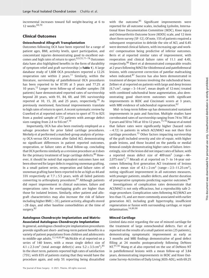
Autologous Chondrocyte Implantation and Matrix-Associated Autologous Chondrocyte ImplantationIn general, autologous chondrocyte implantation proceduresprovide significant short- and long-term patient benefits in avariety of patient populations from children and adolescentsto middle-age adults.13,14,83,84 McNickle et al reported on aseries of 140 knees, with a mean single defect size of4.1�2.3 cm2 (total average defect(s) area 5.2�3.5 cm2).85
In the short term, patients reported high rates of satisfaction(75%), with 83% of patients stating that they would have theprocedure again, and only 5% reporting being dissatisfied
with the outcome.85 Significant improvements werereported for all outcome scales, including Lysholm, Interna-tional Knee Documentation Committee (IKDC), Knee injuryand Osteoarthritis Outcome Score (KOOS) scale, and 12 itemshort form survey (SF-12). Of note, 15% of patients required asubsequent reoperation to debride the site of ACI, and 6.4%were deemed clinical failures, with increasing age andwork-ers’ compensation being predictive of inferior outcomes.Beris et al reported similar rates of improvement, withreoperation and clinical failure rates of 11.1 and 4.4%,respectively.86 Ebert et al demonstrated comparable resultsat 2 years followingMACI for tibiofemoral and patellofemorallesions, with concurrent correction of patellar maltrackingwhen indicated.87 Success has also been demonstrated intreatment of deeper lesions involving the subchondral bone.Zellner et al reported on patients with large and deep lesions(6.7 cm2, range¼3–14 cm2, mean depth of 12mm) treatedwith combined subchondral bone augmentation, also dem-onstrating good short-term outcomes with significantimprovements in IKDC and Cincinnati scores at 3 years,with MRI evidence of subchondral regeneration.62
Mid- to long-term follow-up has demonstrated sustainedimprovements in pain and function. Multiple studies havecorroborated rates of survivorship ranging from 74 to 78% at5 years and 50 to 74% at 10 to 12 years.15,16Nawaz et al notedthat failure rates were significantly higher (hazard ratio¼4.72) in patients in which ACI/MACI was not their firstcartilage procedure.15 Other factors impacting survivorshipof the graft included severity and site of repair, with highergrade lesions, and those located on the patella or medialfemoral condyle demonstrating higher rates of failure. Inter-estingly, size of the lesion did not impact rates of failure, witha reported mean defect size of 409mm2 (range¼44–2,075mm2).15 Moradi et al reported on 7- to 14-year out-comes following first generation ACI treatment of lesionswith a mean size of 4.3�2 cm2 (range¼2.5–11.25 cm2),noting significant improvement in all outcomes measures,with younger patients, smaller defects, and shorter durationof preoperative symptoms predicting superior outcomes.88
Investigations of complication rates demonstrate thatACI/MACI is not only efficacious, but a reproducibly safe 2-stage procedure. Complication rates following ACI/MACI areless than 1%, and are more commonly associated with earliergeneration ACI, including graft hypertrophy, insufficientregeneration or fusion with surrounding cartilage, or repairdelamination.14,89,90
Minced CartilageLimited data exist regarding the use of minced cartilage forthe treatment of large osteochondral defects. Farr et alreported on the results of a small patient series (25 patients),demonstrating symptomatic improvement as early as3 months and MRI findings demonstrative of good defectfilling at 24 months postoperatively following DeNovoNT.91,92 Wang et al also reported on the use of DeNovo NTin patellofemoral lesions with a mean follow-up of 3.84years, demonstrating improvements in IKDC and Knee Out-come Survey-Activities of Daily Living (KOS-ADL), with 69.2%
The Journal of Knee Surgery © 2021. Thieme. All rights reserved.
Large Focal Isolated Chondral Lesion Chahla et al.
Thi
s do
cum
ent w
as d
ownl
oade
d fo
r pe
rson
al u
se o
nly.
Una
utho
rized
dis
trib
utio
n is
str
ictly
pro
hibi
ted.

of lesions demonstrating filling greater than two-thirds.93
Using autologous cartilage harvest intraoperatively, Coleet al reported on 2-year outcomes after CAIS in a series of29 patients randomized to either CAIS or microfracture,demonstrating significantly higher IKDC scores beginningat 12 months in the CAIS group compared with microfrac-ture.66 Other significant differences were also noted invarious KOOS subscales (symptoms and stiffness, pain, ac-tivities of daily living, and sports and recreation) at12 months, and knee-related quality of life at 18 months.All of these differences were maintained at 24 months.
Cryopreserved Osteochondral AllograftsGiven the relative novelty of the technology compared withother surgical techniques, clinical outcomes following im-plantation of cryopreserved osteochondral allografts arecurrently limited. Vangsness et al reported on three patientstreated with Cartiform (Osiris Therapeutics, Inc., Columbia,MD), including two treated for lesions of the medial femoralcondyle and one treated for a lesion of the tibial plateau,demonstrating symptomatic improvement, return to activi-ties, and MRI findings indicative of good defect filling out to2 years postopeatively.94Melugin et al reported on a series of19 patients with patellofemoral defects treated with Carti-form (Osiris Therapeutics, Inc., Columbia, MD). Patientsdemonstrated significant improvements in VR-12, IKDC,KOOS, and Tegner’s scores at a minimum of 24 monthsfollow-up. However, there was a 21.1% reoperation rateand 12.5% conversion rate to patellofemoral arthroplasty.95
Augmented Marrow StimulationOutcomes after surgery with extracellular matrix scaffoldsare largely limited to animal models and case series. Severaltechniques are described in the literature of combined Bio-Cartilage and microfracture techniques; however, outcomedata are limited.96–98 Other techniques, such as AMIC utiliz-ing the Chondro-Gide collagenmembrane havemore clinicaloutcomes data, demonstrating significant improvements inclinical and functional outcomes based on systematic re-view.99 Bertho et al reported on preliminary results in 13patients with a mean defect area of 3.7 cm2 treated withAMIC. At a minimum of 1-year follow-up, patients reportedsignificant improvements in IKDC andKOOS scores.100 Schia-vone Panni et al reported on a series of 21 patients treatedwith AMIC for full thickness lesions >2 cm2, demonstratingsignificant improvements in Lysholm and IKDC with anaverage of 7 years of follow-up. The same study also reported76.2% patient satisfaction rates with 66.6% of patient dem-onstrating reduced defect size and subchondral edema onMRI.101 Another study performed by de Girolamo et al with asimilar length of follow-up suggested that bone marrowaspirate concentrate (BMAC) may help augment functionalimprovements and pain relief in the short term (12months).102 Autologous matrix-induced chondrogenesishas also been compared with other treatments in multiplerandomized trials. Volz et al compared AMIC to microfrac-ture in a series of 47 patients with a mean defect size of3.6�1.6 cm2.103 At 2 years, all groups demonstrated signifi-
cant improvements in modified Cincinnati score and modi-fied ICRS score for pain. At 5 years, improvements were stillnoted for all groups; however, AMIC-treated subjects hadsignificantly higher Modified Cincinnati scores. On 2- and5-year MRI, AMIC-treated groups also had a greater propor-tion of subjects with >2/3 defect filling. In another random-ized trial, Fossum et al compared 2-year outcomes in 41patients treated with either AMIC or ACI covered with acollagen patch with mean total defect sizes of 5.2�2.4 and4.9�4.4 cm2, respectively.104 At 2-year follow-up, bothgroups demonstrated significant improvements in clinicalscores (KOOS, Lysholm, and VAS pain) with no significantdifferences between groupswith respect to themagnitude ofimprovement; however, two patients in the AMIC groupprogressed to arthroplasty by 2-year follow-up.
Conclusion
Focal chondral defects (FCDs) of the kneewith accompanyingpain and dysfunction can be debilitating conditions affectingyoung active patients. Optimal outcomes are dependent oncomplete integration of clinical care from a timely andaccurate diagnosis to selection of a patient- and defect-specific surgical intervention, through postoperative reha-bilitation and return to activities. For lesions that involve alarger chondral area, a variety of well-established complexrestorative procedures exist such as OCA and ACI/MACI, inaddition to other emerging resurfacing technologies. Out-comes data have demonstrated reproducible results includ-ing long-term relief of symptoms and return to activities.Given the array of treatment options, the challenge lies indetermining which intervention or combination of interven-tions is most appropriate, given patient- and defect-specificcharacteristics, while considering important comorbiditiessuch as mechanical alignment, meniscal pathology, andligamentous status.
FundingNone.
Conflict of InterestJ.C. is a board or committee member of the AmericanOrthopaedic Society for Sports Medicine (AOSSM), Ar-throscopy Association of North America (AANA), and theInternational Society of Arthroscopy, Knee Surgery, andOrthopaedic Sports Medicine (ISAKOS); and is a paidconsultant for Arthrex, Inc, CONMED Linvatec, Ossur,and Smith & Nephew. A.B.Y. receives research supportfrom Arthrex, Inc, Organogenesis, and Vericel; is an un-paid consultant for Patient IQ, Smith & Nephew, andSparta Biomedical; is a paid consultant for CONMEDLinvatec, JRF Ortho, and Olympus; and receives stock orstock options from Patient IQ. J.F. receives research sup-port from Active Implants, Arthrex, Inc, Episurf, Fidia, JRFOrtho, Moximed, Novartis, Organogenesis, Samumed, Inc,Vericel, and ZimmerBiomet; is a paid consultant forAesculap/B.Braun, Cartiheal, Cook Biotech, Exactech,Moximed, Inc, Organogenesis, Regentis, Samumed, Inc,
The Journal of Knee Surgery © 2021. Thieme. All rights reserved.
Large Focal Isolated Chondral Lesion Chahla et al.
Thi
s do
cum
ent w
as d
ownl
oade
d fo
r pe
rson
al u
se o
nly.
Una
utho
rized
dis
trib
utio
n is
str
ictly
pro
hibi
ted.

and ZKR orthopedics; is on the editorial or governingboard of the American Journal of Orthopedics, and Car-tialge; is a paid presenter or speaker for Arthrex, Inc,Moximed, Inc, Organogenesis, and Vericel; receives IProyalties from Arthrex, Inc, Biopoly, LLC, and Organogen-esis; receives stock or stock options from MedShape, Inc,and Ortho Regenerative Tech; and receives publishingroyalties, financial or material support from Springer,and Thieme Medical Publishers, Inc.
References1 Flanigan DC, Harris JD, Trinh TQ, Siston RA, Brophy RH. Preva-
lence of chondral defects in athletes’ knees: a systematic review.Med Sci Sports Exerc 2010;42(10):1795–1801
2 Arøen A, Løken S, Heir S, et al. Articular cartilage lesions in 993consecutive knee arthroscopies. Am J Sports Med 2004;32(01):211–215
3 Curl WW, Krome J, Gordon ES, Rushing J, Smith BP, Poehling GG.Cartilage injuries: a review of 31,516 knee arthroscopies. Ar-throscopy 1997;13(04):456–460
4 Hjelle K, Solheim E, Strand T, Muri R, Brittberg M. Articularcartilage defects in 1,000 knee arthroscopies. Arthroscopy 2002;18(07):730–734
5 Zamber RW, Teitz CC, McGuire DA, Frost JD, Hermanson BK.Articular cartilage lesions of the knee. Arthroscopy 1989;5(04):258–268
6 Davies-Tuck ML, Wluka AE, Wang Y, et al. The natural history ofcartilage defects in peoplewith knee osteoarthritis. Osteoarthri-tis Cartilage 2008;16(03):337–342
7 Behery O, Siston RA, Harris JD, Flanigan DC. Treatment ofcartilage defects of the knee: expanding on the existing algo-rithm. Clin J Sport Med 2014;24(01):21–30
8 Thomas D, Shaw KA, Waterman BR. Outcomes after fresh osteo-chondral allograft transplantation for medium to large chondraldefects of the knee. Orthop J Sports Med 2019;7(03):2325967119832299
9 McCarthy MA, Meyer MA, Weber AE, et al. Can competitiveathletes return to high-level play after osteochondral allografttransplantation of the knee? Arthroscopy 2017;33(09):1712–1717
10 Crawford ZT, Schumaier AP, Glogovac G, Grawe BM. Return tosport and sports-specific outcomes after osteochondral allografttransplantation in the knee: a systematic review of studies withat least 2 years’ mean follow-up. Arthroscopy 2019;35(06):1880–1889
11 Frank RM, McCormick F, Rosas S, et al. Reoperation rates aftercartilage restoration procedures in the knee: analysis of a largeUS commercial database. Am J Orthop 2018;47(06):
12 Chahla J, Sweet MC, Okoroha KR, et al. Osteochondral allografttransplantation in the patellofemoral joint: a systematic review.Am J Sports Med 2018:363546518814236
13 Zak L, Albrecht C,WondraschB, et al. Results 2 years aftermatrix-associated autologous chondrocyte transplantation using thenovocart 3D scaffold: an analysis of clinical and radiologicaldata. Am J Sports Med 2014;42(07):1618–1627
14 Harris JD, Siston RA, Pan X, Flanigan DC. Autologous chondrocyteimplantation: a systematic review. J Bone Joint Surg Am 2010;92(12):2220–2233
15 Nawaz SZ, Bentley G, Briggs TW, et al. Autologous chondrocyteimplantation in the knee: mid-term to long-term results. J BoneJoint Surg Am 2014;96(10):824–830
16 Biant LC, Bentley G, Vijayan S, Skinner JA, Carrington RW. Long-term results of autologous chondrocyte implantation in the kneefor chronic chondral and osteochondral defects. Am J Sports Med2014;42(09):2178–2183
17 Widuchowski W, Widuchowski J, Faltus R, et al. Long-termclinical and radiological assessment of untreated severe carti-lage damage in the knee: a natural history study. Scand J Med SciSports 2011;21(01):106–110
18 Houck DA, Kraeutler MJ, Belk JW, Frank RM, McCarty EC, Brav-man JT. Do focal chondral defects of the knee increase the risk forprogression to osteoarthritis? A review of the literature. Orthop JSports Med 2018;6(10):2325967118801931
19 Dye SF. The pathophysiology of patellofemoral pain: a tissuehomeostasis perspective. Clin Orthop Relat Res 2005;(436):100–110
20 Gomoll AH,Minas T, Farr J, Cole BJ. Treatment of chondral defectsin the patellofemoral joint. J Knee Surg 2006;19(04):285–295
21 Mall NA, Harris JD, Cole BJ. Clinical evaluation and preoperativeplanning of articular cartilage lesions of the knee. J Am AcadOrthop Surg 2015;23(10):633–640
22 Rosenberg TD, Paulos LE, Parker RD, Coward DB, Scott SM. Theforty-five-degree posteroanterior flexion weight-bearing radio-graph of the knee. J Bone Joint Surg Am 1988;70(10):1479–1483
23 Godin JA, Hussain ZB, Sanchez A, et al. Multicompartmentalosteochondral allografts of knee and concomitant high tibialosteotomy. Arthrosc Tech 2017;6(05):e1959–e1965
24 Samitier G, Alentorn-Geli E, Taylor DC, et al. Meniscal allografttransplantation. Part 1: systematic review of graft biology, graftshrinkage, graft extrusion, graft sizing, and graft fixation. KneeSurg Sports Traumatol Arthrosc 2015;23(01):310–322
25 Gomoll AH, Yoshioka H, Watanabe A, Dunn JC, Minas T. Preoper-ative measurement of cartilage defects by MRI underestimateslesion size. Cartilage 2011;2(04):389–393
26 O’ConnorMA, PalaniappanM, KhanN, Bruce CE. Osteochondritisdissecans of the knee in children: a comparison of MRI andarthroscopic findings. J Bone Joint Surg Br 2002;84(02):258–262
27 Gillis A, Bashir A, McKeon B, Scheller A, Gray ML, Burstein D.Magnetic resonance imaging of relative glycosaminoglycan dis-tribution in patients with autologous chondrocyte transplants.Invest Radiol 2001;36(12):743–748
28 Potter HG, Foo LF. Magnetic resonance imaging of articularcartilage: trauma, degeneration, and repair. Am J Sports Med2006;34(04):661–677
29 Stelzeneder D, Shetty AA, Kim SJ, et al. Repair tissue quality afterarthroscopic autologous collagen-induced chondrogenesis(ACIC) assessed via T2� mapping. Skeletal Radiol 2013;42(12):1657–1664
30 Tiderius CJ, Tjörnstrand J, Akeson P, Södersten K, Dahlberg L,Leander P. Delayed gadolinium-enhanced MRI of cartilage(dGEMRIC): intra- and interobserver variability in standardizeddrawing of regions of interest. Acta Radiol 2004;45(06):628–634
31 Young AA, Stanwell P, Williams A, et al. Glycosaminoglycancontent of knee cartilage following posterior cruciate ligamentrupture demonstrated by delayed gadolinium-enhanced mag-netic resonance imaging of cartilage (dGEMRIC): a case report. JBone Joint Surg Am 2005;87(12):2763–2767
32 Kurkijärvi JE, Nissi MJ, Kiviranta I, Jurvelin JS, Nieminen MT.Delayed gadolinium-enhanced MRI of cartilage (dGEMRIC) andT2 characteristics of human knee articular cartilage: topograph-ical variation and relationships to mechanical properties. MagnReson Med 2004;52(01):41–46
33 Shelbourne KD, Jari S, Gray T. Outcome of untreated traumaticarticular cartilage defects of the knee: a natural history study. JBone Joint Surg Am 2003;85-A(Suppl 2):8–16
34 Murray JR, Chitnavis J, Dixon P, et al. Osteochondritis dissecans ofthe knee; long-term clinical outcome following arthroscopicdebridement. Knee 2007;14(02):94–98
35 Messner K, Maletius W. The long-term prognosis for severedamage to weight-bearing cartilage in the knee: a 14-yearclinical and radiographic follow-up in 28 young athletes. ActaOrthop Scand 1996;67(02):165–168
The Journal of Knee Surgery © 2021. Thieme. All rights reserved.
Large Focal Isolated Chondral Lesion Chahla et al.
Thi
s do
cum
ent w
as d
ownl
oade
d fo
r pe
rson
al u
se o
nly.
Una
utho
rized
dis
trib
utio
n is
str
ictly
pro
hibi
ted.

36 Henn RF III, Gomoll AH. A review of the evaluation and manage-ment of cartilage defects in the knee. Phys Sportsmed 2011;39(01):101–107
37 Everhart JS, Abouljoud MM, Kirven JC, Flanigan DC. Full-thick-ness cartilage defects are important independent predictivefactors for progression to total knee arthroplasty in older adultswith minimal to moderate osteoarthritis: data from the osteo-arthritis initiative. J Bone Joint Surg Am 2019;101(01):56–63
38 Ogura T, Merkely G, Bryant T, Winalski CS, Minas T. Autologouschondrocyte implantation “segmental-sandwich” technique fordeep osteochondral defects in the knee: clinical outcomes andcorrelation with magnetic resonance imaging findings. Orthop JSports Med 2019;7(05):2325967119847173
39 Ferruzzi A, Buda R, Cavallo M, Timoncini A, Natali S, Giannini S.Cartilage repair procedures associated with high tibial osteot-omy in varus knees: clinical results at 11 years’ follow-up. Knee2014;21(02):445–450
40 Knutsen G, Drogset JO, Engebretsen L, et al. A randomized trialcomparing autologous chondrocyte implantation with micro-fracture. Findings at five years. J Bone Joint Surg Am 2007;89(10):2105–2112
41 Knutsen G, Drogset JO, Engebretsen L, et al. A randomizedmulticenter trial comparing autologous chondrocyte implanta-tion with microfracture: long-term follow-up at 14 to 15 years. JBone Joint Surg Am 2016;98(16):1332–1339
42 Ekman E, Mäkelä K, Kohonen I, Hiltunen A, Itälä A Favourablelong-term functional and radiographical outcome after osteoau-tograft transplantation surgery of the knee: a minimum 10-yearfollow-up. Knee Surg Sports Traumatol Arthrosc 2018;26(12):3560–3565
43 Gudas R, Kalesinskas RJ, Kimtys V, et al. A prospective random-ized clinical study of mosaic osteochondral autologous trans-plantation versus microfracture for the treatment ofosteochondral defects in the knee joint in young athletes.Arthroscopy 2005;21(09):1066–1075
44 Gudas R, Gudaite A, Pocius A, et al. Ten-year follow-up of aprospective, randomized clinical study of mosaic osteochondralautologous transplantation versus microfracture for the treat-ment of osteochondral defects in the knee joint of athletes. Am JSports Med 2012;40(11):2499–2508
45 Ulstein S, Årøen A, Røtterud JH, Løken S, Engebretsen L, Heir S.Microfracture technique versus osteochondral autologous trans-plantation mosaicplasty in patients with articular chondrallesions of the knee: a prospective randomized trial with long-term follow-up. Knee Surg Sports Traumatol Arthrosc 2014;22(06):1207–1215
46 Rue JP, Yanke AB, Busam ML, McNickle AG, Cole BJ. Prospectiveevaluation of concurrent meniscus transplantation and articularcartilage repair: minimum 2-year follow-up. Am J Sports Med2008;36(09):1770–1778
47 Frank RM, Cole BJ. Meniscus transplantation. Curr Rev Muscu-loskelet Med 2015;8(04):443–450
48 Abrams GD, Hussey KE, Harris JD, Cole BJ. Clinical results ofcombined meniscus and femoral osteochondral allograft trans-plantation: minimum 2-year follow-up. Arthroscopy 2014;30(08):964–70.e1
49 Farr J, Rawal A, Marberry KM. Concomitant meniscal allografttransplantation and autologous chondrocyte implantation:min-imum 2-year follow-up. Am J Sports Med 2007;35(09):1459–1466
50 Marti RK, Verhagen RA, Kerkhoffs GM, Moojen TM. Proximaltibial varus osteotomy. Indications, technique, and five to twen-ty-one-year results. J Bone Joint Surg Am 2001;83(02):164–170
51 Bode G, Schmal H, Pestka JM, Ogon P, SüdkampNP, Niemeyer P. Anon-randomized controlled clinical trial on autologous chon-drocyte implantation (ACI) in cartilage defects of the medialfemoral condyle with or without high tibial osteotomy in
patients with varus deformity of less than 5°. Arch OrthopTrauma Surg 2013;133(01):43–49
52 Kahlenberg CA, Nwachukwu BU, Hamid KS, Steinhaus ME,Williams RJ III. Analysis of outcomes for high tibial osteotomiesperformed with cartilage restoration techniques. Arthroscopy2017;33(02):486–492
53 Farr J. Autologous chondrocyte implantation improves patello-femoral cartilage treatment outcomes. Clin Orthop Relat Res2007;463(463):187–194
54 Cotter EJ, Waterman BR, Kelly MP, Wang KC, Frank RM, Cole BJ.Multiple osteochondral allograft transplantation with concomi-tant tibial tubercle osteotomy for multifocal chondral disease ofthe knee. Arthrosc Tech 2017;6(04):e1393–e1398
55 Wang D, Eliasberg CD, Wang T, et al. Similar outcomes afterosteochondral allograft transplantation in anterior cruciate lig-ament-intact and -reconstructed knees: a comparativematched-group analysis with minimum 2-year follow-up. Ar-throscopy 2017;33(12):2198–2207
56 Jones KJ, Mosich GM, Williams RJ. Fresh precut osteochondralallograft core transplantation for the treatment of femoralcartilage defects. Arthrosc Tech 2018;7(08):e791–e795
57 Godin JA, Frangiamore S, Chahla J, Cinque ME, DePhillipo NN,LaPrade RF. Tibial allograft transfer for medial tibial plateauresurfacing. Arthrosc Tech 2017;6(03):e661–e665
58 Bartlett W, Skinner JA, Gooding CR, et al. Autologous chondro-cyte implantation versus matrix-induced autologous chondro-cyte implantation for osteochondral defects of the knee: aprospective, randomised study. J Bone Joint Surg Br 2005;87(05):640–645
59 Sohn DH, Lottman LM, Lum LY, et al. Effect of gravity onlocalization of chondrocytes implanted in cartilage defects.Clin Orthop Relat Res 2002;(394):254–262
60 Gikas PD, Bayliss L, Bentley G, Briggs TW. An overview ofautologous chondrocyte implantation. J Bone Joint Surg Br2009;91(08):997–1006
61 Frenkel SR, Toolan B, Menche D, Pitman MI, Pachence JM.Chondrocyte transplantation using a collagen bilayer matrixfor cartilage repair. J Bone Joint Surg Br 1997;79(05):831–836
62 Zellner J, Grechenig S, Pfeifer CG, et al. Clinical and radiologicalregeneration of large and deep osteochondral defects of the kneeby bone augmentation combined with matrix-guided autolo-gous chondrocyte transplantation. Am J Sports Med 2017;45(13):3069–3080
63 Albrecht F, Roessner A, Zimmermann E. Closure of osteochondrallesions using chondral fragments and fibrin adhesive. ArchOrthop Trauma Surg 1983;101(03):213–217
64 Bonasia DE, Martin JA, Marmotti A, et al. Cocultures of adult andjuvenile chondrocytes compared with adult and juvenile chon-dral fragments: in vitro matrix production. Am J Sports Med2011;39(11):2355–2361
65 McCormick F, Yanke A, Provencher MT, Cole BJ. Minced articularcartilage–basic science, surgical technique, and clinical applica-tion. Sports Med Arthrosc Rev 2008;16(04):217–220
66 Cole BJ, Farr J, Winalski CS, et al. Outcomes after a single-stageprocedure for cell-based cartilage repair: a prospective clinicalsafety trial with 2-year follow-up. Am J SportsMed 2011;39(06):1170–1179
67 Woodmass JM, Melugin HP,Wu IT, Saris DBF, Stuart MJ, Krych AJ.Viable osteochondral allograft for the treatment of a full-thick-ness cartilage defect of the patella. Arthrosc Tech 2017;6(05):e1661–e1665
68 Mithoefer K, Hambly K, Logerstedt D, Ricci M, Silvers H, DellaVilla S. Current concepts for rehabilitation and return to sportafter knee articular cartilage repair in the athlete. J Orthop SportsPhys Ther 2012;42(03):254–273
69 Seo S-S, Kim C-W, Jung D-W.Management of focal chondral lesionin the knee joint. Knee Surg Relat Res 2011;23(04):185–196
The Journal of Knee Surgery © 2021. Thieme. All rights reserved.
Large Focal Isolated Chondral Lesion Chahla et al.
Thi
s do
cum
ent w
as d
ownl
oade
d fo
r pe
rson
al u
se o
nly.
Una
utho
rized
dis
trib
utio
n is
str
ictly
pro
hibi
ted.

70 Jones DG, Peterson L. Autologous chondrocyte implantation.Instr Course Lect 2007;56:429–445
71 Nielsen ES, McCauley JC, Pulido PA, Bugbee WD. Return to sportand recreational activity after osteochondral allograft transplan-tation in the knee. Am J Sports Med 2017;45(07):1608–1614
72 Balazs GC,WangD, Burge AJ, Sinatro AL,WongAC,Williams RJ III.Return to play among elite basketball players after osteochondralallograft transplantation of full-thickness cartilage lesions.Orthop J Sports Med 2018;6(07):2325967118786941
73 Tírico LEP, McCauley JC, Pulido PA, Bugbee WD. Does anteriorcruciate ligament reconstruction affect the outcome of osteo-chondral allograft transplantation?: a matched cohort studywith a mean follow-up of 6 years Am J Sports Med 2018;46(08):1836–1843
74 Frank RM, Lee S, Cotter EJ, Hannon CP, Leroux T, Cole BJ. Out-comes of osteochondral allograft transplantationwith andwith-out concomitant meniscus allograft transplantation: acomparative matched group analysis. Am J Sports Med 2018;46(03):573–580
75 Wang D, Kalia V, Eliasberg CD, et al. Osteochondral allografttransplantation of the knee in patients aged 40 years and older.Am J Sports Med 2018;46(03):581–589
76 Frank RM, Cotter EJ, Lee S, Poland S, Cole BJ. Do outcomes ofosteochondral allograft transplantation differ based on age andsex?: a comparative matched group analysis Am J Sports Med2018;46(01):181–191
77 Wang D, Rebolledo BJ, Dare DM, et al. Osteochondral allografttransplantation of the knee in patients with an elevated bodymass index. Cartilage 2018:1947603518754630
78 Krych AJ, Robertson CM, Williams RJ IIICartilage Study Group.Return to athletic activity after osteochondral allograft trans-plantation in the knee. Am J SportsMed 2012;40(05):1053–1059
79 Raz G, Safir OA, Backstein DJ, Lee PT, Gross AE. Distal femoralfresh osteochondral allografts: follow-up at a mean of twenty-two years. J Bone Joint Surg Am 2014;96(13):1101–1107
80 Merkely G, Ogura T, Ackermann J, Barbieri Mestriner A, GomollAH. Clinical outcomes after revision of autologous chondrocyteimplantation to osteochondral allograft transplantation for largechondral defects: a comparative matched-group analysis. Carti-lage 2019:1947603519833136
81 Cotter EJ, Hannon CP, Christian DR, et al. Clinical outcomes ofmultifocal osteochondral allograft transplantation of the knee:an analysis of overlapping grafts and multifocal lesions. Am JSports Med 2018;46(12):2884–2893
82 Nuelle CW, Nuelle JA, Cook JL, Stannard JP. Patient factors, donorage, and graft storage duration affect osteochondral allograftoutcomes in knees with or without comorbidities. J Knee Surg2017;30(02):179–184
83 Niethammer TR, Holzgruber M, Gülecyüz MF, Weber P, Pietsch-mann MF, Müller PE. Matrix based autologous chondrocyteimplantation in children and adolescents: a match paired analy-sis in a follow-up over three years post-operation. Int Orthop2017;41(02):343–350
84 Rosenberger RE, Gomoll AH, Bryant T, Minas T. Repair of largechondral defects of the knee with autologous chondrocyteimplantation in patients 45 years or older. Am J Sports Med2008;36(12):2336–2344
85 McNickle AG, L’Heureux DR, Yanke AB, Cole BJ. Outcomes ofautologous chondrocyte implantation in a diverse patient pop-ulation. Am J Sports Med 2009;37(07):1344–1350
86 Beris AE, Lykissas MG, Kostas-Agnantis I, Manoudis GN. Treat-ment of full-thickness chondral defects of the knee with autolo-gous chondrocyte implantation: a functional evaluation withlong-term follow-up. Am J Sports Med 2012;40(03):562–567
87 Ebert JR, Schneider A, FallonM,Wood DJ, Janes GC. A comparisonof 2-year outcomes in patients undergoing tibiofemoral orpatellofemoral matrix-induced autologous chondrocyte implan-tation. Am J Sports Med 2017;45(14):3243–3253
88 Moradi B, Schönit E, Nierhoff C, et al. First-generation autologouschondrocyte implantation in patients with cartilage defects ofthe knee: 7 to 14 years’ clinical andmagnetic resonance imagingfollow-up evaluation. Arthroscopy 2012;28(12):1851–1861
89 Gowd AK, Cvetanovich GL, Liu JN, et al. Management of chondrallesions of the knee: analysis of trends and short-term compli-cations using the national surgical quality improvement pro-gram database. Arthroscopy 2019;35(01):138–146
90 Niemeyer P, Pestka JM, Kreuz PC, et al. Characteristic complica-tions after autologous chondrocyte implantation for cartilagedefects of the knee joint. Am J Sports Med 2008;36(11):2091–2099
91 Farr J, Cole BJ, Sherman S, Karas V. Particulated articular carti-lage: CAIS and DeNovo NT. J Knee Surg 2012;25(01):23–29
92 Farr J, Tabet SK,Margerrison E, Cole BJ. Clinical, radiographic, andhistological outcomes after cartilage repair with particulatedjuvenile articular cartilage: a 2-year prospective study. Am JSports Med 2014;42(06):1417–1425
93 Wang T, BelkinNS, BurgeAJ, et al. Patellofemoral cartilage lesionstreated with particulated juvenile allograft cartilage: a prospec-tive study with minimum 2-year clinical and magnetic reso-nance imaging outcomes. Arthroscopy 2018;34(05):1498–1505
94 Vangsness CT Jr, Higgs G, Hoffman JK, et al. Implantation of anovel cryopreserved viable osteochondral allograft for articularcartilage repair in the knee. J Knee Surg 2018;31(06):528–535
95 Melugin HP, Ridley TJ, Bernard CD, et al. Prospective outcomes ofcryopreserved osteochondral allograft for patellofemoral carti-lage defects at minimum 2-year follow-up. Cartilage 2020:1947603520903420
96 Carter AH, Guttierez N, Subhawong TK, et al. MR imaging ofBioCartilageaugmentedmicrofracturesurgeryutilizing2DMOCARTand KOOS scores. J Clin Orthop Trauma 2018;9(02):146–152
97 Wang KC, Frank RM, Cotter EJ, Christian DR, Cole BJ. Arthroscopicmanagement of isolated tibial plateau defect with microfractureand micronized allogeneic cartilage-platelet-rich plasma ad-junct. Arthrosc Tech 2017;6(05):e1613–e1618
98 Schallmo MS, Marquez-Lara A, Luo TD, Rosas S, Stubbs AJ.Arthroscopic treatment of hip chondral defect with microfrac-ture and platelet-rich plasma-infused micronized cartilage allo-graft augmentation. Arthrosc Tech 2018;7(04):e361–e365
99 SteinwachsMR, Gille J, VolzM, et al. Systematic reviewandmeta-analysis of the clinical evidence on the use of autologous matrix-induced chondrogenesis in the knee. Cartilage 2019:1947603519870846
100 Bertho P, Pauvert A, Pouderoux T, Robert HOrthopaedics andTraumatology Society of Western France (SOO) Treatment oflarge deep osteochondritis lesions of the knee by autologousmatrix-induced chondrogenesis (AMIC): Preliminary results in13 patients. Orthop Traumatol Surg Res 2018;104(05):695–700
101 Schiavone Panni A, Del Regno C, Mazzitelli G, D’Apolito R, CoronaK, VassoM. Good clinical resultswith autologousmatrix-inducedchondrogenesis (Amic) technique in large knee chondral defects.Knee Surg Sports Traumatol Arthrosc 2018;26(04):1130–1136
102 de Girolamo L, Schönhuber H, Viganò M, et al. Autologousmatrix-induced chondrogenesis (AMIC) and AMIC enhanced byautologous concentrated bone marrow aspirate (BMAC) allowfor stable clinical and functional improvements at up to 9 yearsfollow-up: results from a randomized controlled study. J ClinMed 2019;8(03):E392
103 VolzM, Schaumburger J, Frick H, Grifka J, Anders S. A randomizedcontrolled trial demonstrating sustained benefit of autologousmatrix-induced chondrogenesis over microfracture at five years.Int Orthop 2017;41(04):797–804
104 Fossum V, Hansen AK, Wilsgaard T, Knutsen G. Collagen-coveredautologous chondrocyte implantation versus autologousmatrix-induced chondrogenesis: a randomized trial comparing 2 meth-ods for repair of cartilage defects of the knee. Orthop J SportsMed 2019;7(09):2325967119868212
The Journal of Knee Surgery © 2021. Thieme. All rights reserved.
Large Focal Isolated Chondral Lesion Chahla et al.
Thi
s do
cum
ent w
as d
ownl
oade
d fo
r pe
rson
al u
se o
nly.
Una
utho
rized
dis
trib
utio
n is
str
ictly
pro
hibi
ted.
Copyright © 2022 FDOKUMEN